RUSSIAN_Matasov Adequate method of single-moment large intestine resection
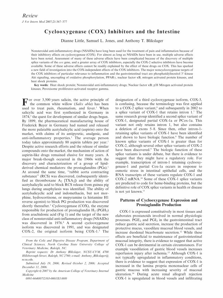
Cyclooxygenase (COX) Inhibitors and the Intestine
Dianne Little, Samuel L. Jones, and Anthony T. Blikslager
Nonsteroidal anti-inflammatory drugs (NSAIDs) have long been used for the treatment of pain and inflammation because of
their inhibitory effects on cyclooxygenase (COX). For almost as long as NSAIDs have been in use, multiple adverse effects
have been noted. Assessment of many of these adverse effects have been complicated because of the discovery of multiple
splice variants of the cox gene, and a greater array of COX inhibitors, especially the COX-2 selective inhibitors have become
available. Some of these adverse effects cannot be readily explained by the effect of these drugs on COX. This has sparked
a new field of investigation into the COX-independent effects of the COX inhibitors. The major noncyclooxygenase targets of
the COX inhibitors of particular relevance to inflammation and the gastrointestinal tract are phosphatidylinositol 39-kinase
Akt signaling, uncoupling of oxidative phosphorylation, PPARc, nuclear factor kB, mitogen activated protein kinases, and
heat shock proteins.
Key words: Heat shock protein; Nonsteroidal anti-inflammatory drugs; Nuclear factor kB; p38 Mitogen activated protein
kinases; Peroxisome proliferator-activated receptor gamma.
F or over 3,500 years salicin, the active principle ofthe common white willow (Salix abla) has been
used to treat pain, rheumatism, and fever.1 Whensalicylic acid was first synthesized in Germany in1874,2 the quest for development of similar drugs began.By 1899, the pharmaceutical manufacturing house ofFrederick Bayer in Germany had named and releasedthe more palatable acetylsalicylic acid (aspirin) onto themarket, with claims of its antipyretic, analgesic, andanti-inflammatory properties.1 The average persontoday takes approximately 80 aspirin tablets per year.1
Despite active research efforts and the release of similarcompounds onto the market, the mechanism of action ofaspirin-like drugs remained elusive for many years. Amajor break-though occurred in the 1960s with thediscovery and characterization of a group of lipid-derived chemical mediators: the prostaglandins (PGs).1
At around the same time, ‘‘rabbit aorta contractingsubstance’’ (RCS) was discovered, (subsequently identi-fied as thromboxane A2 [TXA2]) and the ability ofacetylsalicylic acid to block RCS release from guinea piglungs during anaphylaxis was identified. The ability ofacetylsalicyclic acid and indomethacin, but not mor-phine, hydrocortisone, or mepyramine (a histamine H1reverse agonist) to block PG production was discoveredshortly thereafter.3 Cyclooxygenase (COX), the enzymeresponsible for production of prostaglandin H2 (PGH2)from arachidonic acid (Fig 1) and the target of the newclass of nonsteroidal anti-inflammatory drugs (NSAIDs)was discovered in 1976.4 A second cyclooxygenaseisoform was discovered in 1991, and was designatedCOX-2, the original isoform being COX-1.5 The
designation of a third cyclooxygenase isoform, COX-3,is confusing, because the terminology was first appliedto a COX-2 splice variant,6 and subsequently in 2002 toa splice variant of COX-1 that retains intron 1.7 Thesame research group identified a second splice variant ofCOX-1, designated partial COX-1a or PCox-1a. Thisvariant not only retains intron 1, but also containsa deletion of exons 5–8. Since then, other intron-1-retaining splice variants of COX-1 have been identifiedand shown to have biologic function.8 The number ofknown splice variants of COX-1 is greater than forCOX-2, although several other splice variants of COX-2have been discovered.9 The biologic function of thesesplice variants is under debate, but there is evidence tosuggest that they might have a regulatory role. Forexample, transcription of intron-1 retaining cyclooxy-genase-1 and partial Cox-1a occurs in response toosmotic stress in intestinal epithelial cells, and theRNA transcripts of these variants regulate COX-1 andCOX-2 mRNA.10 Some of these COX-1 splice variantsare predicted to code for heme-binding proteins, but thedefinitive role of COX splice variants in health or diseaseis not yet known.11
Patterns of Cyclooxygenase Expression andProstaglandin Production
COX-1 is expressed constitutively in most tissues andelaborates prostanoids involved in normal physiologicprocesses. PGE2 and PGI2 in the gastrointestinal tractreduce gastric acid secretion, increase the production ofprotective mucus, vasodilate mucosal blood vessels, andincrease duodenal bicarbonate secretion.12 While theseeffects are beneficial to maintenance of gastrointestinalmucosal integrity, there is evidence to suggest that activeCOX-1 can be detrimental in certain circumstances. Forexample vasodilation of gastric blood vessels increasesreperfusion injury after ischemia.13 Although COX-1 isnot typically upregulated in inflammatory conditions,there is evidence to suggest that expression of COX-1 isincreased in the lamina propria mononuclear cells ofgastric mucosa with increasing severity of mucosalulceration.14 During acute renal allograft rejectionCOX-1 is upregulated in blood vessels and infiltrating
From the Colic and Digestive Disease Program, Department of
Clinical Sciences, North Carolina State University College of
Veterinary Medicine, Raleigh, NC.
Reprint requests: Dr Anthony Blikslager, NCSU-CVM, 4700
Hillsborough Street, Raleigh, NC 2760; e-mail: Anthony_Blikslager@
ncsu.edu.
Submitted July 19, 2006; Revised October 2, 2006; Accepted
December 11, 2006.
Copyright E 2007 by the American College of Veterinary Internal
Medicine
0891-6640/07/2103-0001/$3.00/0
ReviewJ Vet Intern Med 2007;21:367–377

interstitial cells.15 The mechanisms of COX-1 upregula-tion have been substantially less well studied than themechanisms of COX-2 upregulation, despite the impor-tance of COX-1 inhibition as a target of cardiovasculartherapy. However, expression of both COX-1 and COX-2 isoforms are increased in equine jejunal mucosa,immediately after the end of 2 hours of ischemia.16
Furthermore, COX-1 expression is upregulated inovarian cancer, and several cell lines increase expressionof COX-1 when stimulated with phorbol-esters; severaltranscription factor binding sites have been identified aspart of these studies.17 Further work is required toelucidate the pathways that regulate COX-1 expressionin nonmalignant states, and is likely to be forthcominggiven the increasing number of conditions in whichupregulation of expression of COX-1 appears to occur.
COX-2 was originally thought to function only as aninducible isoenzyme and indeed, upregulation of local invivo COX-2 expression occurs in both acute and chronic
Fig 1. Pathways of prostanoid synthesis. Arachidonic acid,
produced from metabolism of membrane phospholipids or linoleic
acid, is metabolized to the PGs by the COX isoenzymes. The
species of PG produced depends on the presence of a specific PG
synthase colocalized with COX. ‘‘X2’’ 5 D,E,F, I or Thromboxane
A2. PGD2 and PGE2 may be further metabolized to the
cyclopentenone prostaglandins. Concentrations of arachidonic
acid are high during reperfusion injury, and in the presence of
COX inhibitors, metabolism is preferentially directed down the
cytochrome P450 (epoxygenase) pathway to produce HETE and
EET and the LOX pathway to produce HETEs, HPETE, and
leukotrienes, including LTA4. Linoleic acid metabolism by LOX
produces HODE and lipoxins. See the abbreviation side bar for
definition.
Abbreviation Definition
Akt Protein kinase B
ATF-2 Activating transcription factor 2
ATP Adenosine triphosphate
COX Cyclooxygenase
EET epoxytrienoic acids
ERK extracellular regulated kinase
HETE hydroxyeicosatetraenoic acids
HO-1 Hemoxygenase-1
HODE hydroxyoctadecadienoic acid
HPETE hydroperoxyeicosatetraenoic acids
HSF-1 Heat shock factor-1
HSP Heat shock protein
IkB I kappa B
ICAM-1 intercellular adhesion molecule-1
IFNc Interferon gamma
IKK IkB kinase
IL-1 Interleukin-1
IL-4 Interleukin-4
IL-6 Interleukin-6
JNK c-Jun N-Terminal kinase
LOX Lipoxygenase
LTA4 Leukotriene A4
MAPK(s) Mitogen-activated protein kinase(s)
NFkB Nuclear factor kB
NHE3 Na+ - H+ Exchanger 3
NSAIDs Nonsteroidal anti-inflammatory drugs
PDK-1 3-phosphoinositide-dependent kinase-1
PGA1 Prostaglandin A1
PGA2 Prostaglandin A2
PGB2 Prostaglandin B2
PGC2 Prostaglandin C2
PGD2 Prostaglandin D2
PGE2 Prostaglandin E2
PGF2 Prostaglandin F2
PGG2 Prostaglandin G2
PGH2 Prostaglandin H2
PGI2 Prostacyclin
PGJ2 Prostaglandin J2
PGs Prostaglandins
P Phosphorylated
PI3K phosphatidylinositol 39-kinase
PIP2 Phosphatidylinositol Phosphate PIP2 5
Phosphatidylinositol bisphosphate
PPARa Peroxisome proliferator-activated receptor
alpha
PPARb Peroxisome proliferator-activated receptor
beta
PPARc Peroxisome proliferator-activated receptor
gamma
PPAR(s) Peroxisome proliferator-activated receptor(s)
PPRE PPAR response element
PTEN Phosphatase and tensin Homolog
RCS Rabbit aorta contracting substance
RXR retinoic acid receptor
STAT Signal Transducers and Activators of Tran-
scription
TER transepithelial electrical resistance
TGFb Transforming Growth Factor beta
TNFa tumor necrosis factor
TXA2 Thromboxane A2
TZDs thiazolidinediones
368 Little, Jones, and Blikslager

inflammation and in ischemia, stimulated by pro-inflammatory cytokines and mitogens. Upregulation ofCOX-2 expression is also pivotal in the pathogenesis ofcancer. For example, PGE2 elaborated by COX-2increases tumor cell proliferation and inhibits apopto-sis.18 Early work evaluating the function and distribu-tion of COX-2 suggested that there was no constitutiveexpression, but COX-2 has subsequently been found tobe constitutively expressed in many tissue types. Forexample in the gastrointestinal tract, constitutive ex-pression of COX-2 has been identified in the mouseproximal colon.19 In the rat, the highest level of COX-2expression in the intestine is observed at the ileocecaljunction, in the lamina propria of the apical villus ratherthan the epithelial cells themselves.20 However, the roleof COX-2 expression in the presence of COX inhibitorsbecomes even more complex, for example, inhibition ofCOX-2 delays healing of gastric ulcers and mayexacerbate colitis.21,22 COX-2 expression is upregulatedand occurs rapidly after treatment with COX-1 selectiveor nonselective COX inhibitors; this upregulation isblocked by pretreatment with PGE2, suggesting that thelack of PGE2 may be a trigger for COX-2 proteinexpression.12
The array of prostaglandins produced by cycloox-ygenase depends not only on expression or upregula-tion of the specific COX-1 or COX-2 isoforms, but alsoon the expression and localization of specific prosta-glandin synthases. For example an inducible mem-brane-associated form of PGE2 synthase is coupled toCOX-2, but the constitutively expressed cytosolic PGEsynthase is coupled to COX-1.23 The activity of specificprostaglandin synthases in large part determineswhether increased expression of COX-2 is beneficialor detrimental. For example, in a model of cardiacinflammation induced by endotoxemia, PGE2 synthaseand COX-2 expression both peak by 4 hours afterendotoxin administration, but PGD2 synthase expres-sion does not peak until 48 hours after endotoxinadministration.24 PGE2 contributes to inflammationand hyperalgesia, suggesting that inhibition of COX-2activity and PGE2 production in a clinical patient inthe early stages of inflammation would be beneficial.However, the PGD2 metabolite, 15-deoxy-delta12,14
PGJ2 is not only a potent agonist of the largely anti-inflammatory peroxisome proliferator-activated recep-tor gamma (PPARc) (see later discussion) but can actindependently of PPARc to inhibit PGE2 synthasedirectly in rat chondrocytes, thus blocking productionof pro-inflammatory prostaglandins, and can alsoinhibit activation and nuclear translocation of nuclearfactor kB (NFkB) in the chondrocyte.25,26 Therefore,COX-2 inhibition during the resolution phase ofinflammation may be detrimental, because synthesisof valuable anti-inflammatory prostaglandins is inhib-ited. These studies suggest that in the future, morework will be directed at evaluation of the role ofspecific inhibitors of COX isoenzymes and PGsynthases at different stages of various pathologicprocesses to determine optimal therapeutic regimensfor clinical cases.
Nonsteroidal Anti-inflammatory Drugs:Cyclooxygenase Inhibition
Both COX-1 and COX-2 exist as membrane bounddimers.27 Arachidonic acid enters the active site viaa hydrophobic channel. Acetylsalicylic acid irreversiblyblocks access to the hydrophobic channel via acetylationof serine 530 (COX-1) or Serine 516 (COX-2).27 ForNSAIDs with a carboxylate group, there is significantcharge-charge interaction with arginine 120, a residuewithin the hydrophobic channel. Some COX-2 specificinhibitors lack the carboxylate group, which probablycontributes to their selectivity. COX-1 has bulky iso-leucine residues at positions 434 and 523 whereas COX-2 has smaller valine residues at these sites. This smalldifference in amino acid composition along with severalconformational changes accounts for the difference inspecificity of COX-1 and COX-2 inhibitors; the presenceof valine residues at these sites in COX-2 allows a side-pocket to form, that is part of the active site of COX-2selective drugs. The larger isoleucine residues close downthe entrance to this side-pocket in COX-1, preventingbinding of COX-2 selective drugs.28 Furthermore, atamino acid 513, COX-2 has a charged arginine residueand COX-1 an aromatic histidine residue; the arginineresidue is able to interact with the sulphonamide groupspresent on many COX-2 selective inhibitors, but theimidazole ring of the histidine residue in COX-1 isunable to interact with COX-2 inhibitors.29 Additionalhydrogen bonding of COX-2 inhibitors to residuespresent in COX-2 but not COX-1 also contributes totheir selectivity. Binding of COX inhibitors to the activesite of the membrane bound cyclooxygenase isoenzymesprevents cycling of the membrane derived arachidonicacid through the ‘‘U’’ shaped active site of thecyclooxygenase enzyme, and prevents formation of thearomatic ring that characterizes the prostanoids.28
Nonsteroidal Anti-inflammatory Drugs:Cyclooxygenase Independent Effects
Just as the cyclooxygenase gene and its expression arefar more complex than originally expected, the mechan-isms of action of cyclooxygenase inhibitors are provingto be equally complex. Much of this work was initiatedwhen several epidemiologic and clinical studies demon-strated the role of cyclooxygenase inhibitors in theprevention of cancer. This work revealed that COX-2 isupregulated in many types of neoplasia.30 For example,celecoxib, a COX-2 selective inhibitor was recentlyapproved by the US Federal Drug Administration forits demonstrated efficacy as an adjuvant treatment offamilial adenomatous polyposis, a condition in whichup-regulation of COX-2 is well recognized.31 In murineembryonic cell lines with no cyclooxygenase expression,both nonselective NSAIDs, including indomethacin,suldinac, piroxicam, and ibuprofen, and the selectiveCOX-2 inhibitor, NS-398 have antineoplastic andantiproliferative effects.32 Additionally there is evidencefrom other areas of investigation to suggest thatnoncyclooxygenase targets may play an important role
NSAIDs and Intestine 369

in the mechanism of action of the NSAIDs. Salicylicacid is considered to be a COX inhibitor and has anti-inflammatory properties, even though it does notpossess the acetyl group of acetylsalicylic acid, andtherefore does not inhibit cyclooxygenase by acetyla-tion.33 At therapeutic concentrations, S-flurbiprofeninhibits cyclooxygenase, but R-flurbiprofen, the otherenantiomer of flurbiprofen lacks the ability to inhibitcyclooxygenase activity and therefore is unable to blockPGE2 release in a rat model of paw inflammation.Nonetheless, R-flurbiprofen is almost as effective asdexamethasone at reducing inflammation.34
Active research in the field of chemo-prevention istargeting discovery of alternative drugs to NSAIDs toprovide the benefits of these drugs for prevention ofcancer, without the deleterious side-effects of long-termadministration of NSAIDs on the gastrointestinal tractand the cardiovascular system. Therefore there is muchactive research to determine the noncyclooxygenasemechanisms of action of cyclooxygenase inhibitors.
The major noncyclooxygenase targets of the cycloox-ygenase inhibitors of particular relevance to the intestineare phosphatidylinositol 39-kinase (PI3K)/Akt signaling,
uncoupling of oxidative phosphorylation, PPARc, NFkB,the mitogen activated protein kinases (MAPKs), and theheat shock protein (HSP) response.
PI3K/Akt Signaling
PI3K/Akt signaling is the final common pathway ofa number of cytokine and tyrosine kinase pathways and iskey in the regulation of cell proliferation, transcription,translation, growth, and apoptosis.35,36 Dysregulated upre-gulation of Akt is key for 3 critical characteristics ofneoplasia: angiogenesis, tumor invasiveness, and pro-liferation.37 Celecoxib is a direct inhibitor of 3-phosphoi-nositide-dependent kinase-1 (PDK-1) (Fig 2A), an up-stream regulator of Akt signaling and therefore thispathway is a major noncyclooxygenase target of celecoxibthat partially explains the antiproliferative effects of thisdrug on cancer cells.30,36 I kappa B kinase (IKK) isa downstream effector of Akt. Phosphorylation of IKKresults in IKK activation and phosphorylation of I kappa B(IkB), which leads to nuclear translocation of NFkB andtranscription of NFkB genes, including cox-2. Thereforeinhibition of PDK-1 by celecoxib results ultimately in
Fig 2. External stimuli, such as growth factors, activate the PI3K signaling pathway. (A) A downstream effector of this pathway, Akt
activates IKK, which phosphorylates IkB bound to the p65 and p50 subunits of NFkB. Phosphorylated IkB disassociates from NFkB, and
allows translocation of the p65 and p50 subunits into the nucleus where they bind and increase transcription of many genes, including cox-2.
Celecoxib is a direct inhibitor of PDK1, which is required for Akt activation, thus celecoxib inhibits upregulation of cox-2 transcription that
occurs in response to extracellular stimuli. Other NSAIDs, such as sulindac, ibuprofen, sodium salicylate, and aspirin can directly inhibit
disassociation of phosphorylated IkB from the p65 and p50 subunits of NFkB. (B) An alternate mechanism of action of celecoxib on the
PI3K pathway is via production of oxygen free radicals, which stimulate activity of PDK1, and activates JNK, which in turn increases
activity of HO-1, an enzyme critical for protection against ischemia-reperfusion injury and for its other anti-inflammatory effects. For
a complete list of abbreviations refer to the abbreviation side bar on page 368.
370 Little, Jones, and Blikslager

down-regulation of COX-2 expression. Rofecoxib, anoth-er COX-2 selective coxib drug has no effect on PDK-1activity,38 suggesting that each NSAID should be evaluatedcarefully in the circumstances in which it will be used for itscyclooxygenase independent effects.
Findings from studies performed in a mesangial cellline provided conflicting results in that celecoxib in-creased activation of the PI3K pathway and phosphor-ylation of Akt leading to phosphorylation of JNK,a mitogen-activated protein kinase (MAPK) (Fig 2B).39
Not surprisingly, downstream effectors of JNK, c-Junand ATF-2 also demonstrated increased phosphorylationafter treatment of mesangial cells with celecoxib, effectsthat could be blocked with specific inhibition of JNK.39
Nevertheless, these effects on the Akt/PI3K pathway wereindirect, because celecoxib treatment enhanced oxygenfree radical generation, which in turn leads to increasedphosphorylation of Akt and activation of the PI3Kpathway. Specific inhibition of PI3K leads to a reductionin JNK phosphorylation and reduced expression ofhemoxygenase-1 (HO-1), reversing the effects of celecoxibtreatment on HO-1 expression.39 Increases in HO-1expression caused by celecoxib treatment would beexpected to increase heme degradation, and increase theproduction of reaction products including carbon mon-oxide, biliverdin, and iron. Carbon monoxide and theother products of heme degradation have importantantioxidant, anti-inflammatory, and cytoprotective func-tions and are protective in ischemia/reperfusion injuryand inflammation in the intestine.40,41 Therefore fromthese studies, celecoxib may be expected to be protectiveagainst ischemia/reperfusion injury.
The effect of NSAIDs on Akt activity in different celllines is particularly interesting in relation to the effect ofNSAIDs on intestinal repair and differentiation. Aktexpression is high in cells that are fully differentiated,compared to cells in a less differentiated state, andtherefore NSAIDs may have differential effects atdifferent levels of the villus-crypt axis.42 In intestinalepithelial cells, both PI3K and Akt are associated withlipid rafts, microdomains of the membrane that areenriched in glycosphingolipids and cholesterol. Lipidrafts cluster cell surface receptors, membrane proteins,and signaling molecules within a small defined area.43
PI3K and Akt are associated with the Na+ - H+ exchanger3 (NHE3) within the lipid rafts, and act to increase brushborder sodium absorption in the absence of PTEN.42
PI3K is required for prostaglandin-mediated recovery ofbarrier function in ischemia-injured porcine ileum, andinhibition retards normal formation of the tight junctionafter ischemic-injury.44,45 The role of specific NSAIDs onthis pathway in relation to recovery of intestinal barrierfunction after ischemic injury may be worthy of furtherinvestigation, particularly given the very specific effects ofa given NSAID on different cell lines.
Uncoupling of Oxidative Phosphorylation
Substantial work has focused on the role of cycloox-ygenase inhibition on arachidonic acid accumulationand on uncoupling of mitochondrial oxidative phos-
phorylation, particularly in the postischemic heart.Arachidonic acid accumulates in cardiac tissue duringreperfusion injury, and COX inhibition blocks eicosa-noid production and leads to further increases inconcentrations of arachidonic acid.46 In the presence ofCOX inhibition, arachidonic acid metabolism is redir-ected to the lipoxygenase and cytochrome P450 (epox-ygenase) pathways (Fig 1) to produce alternate arachi-donic acid eicosanoids.47,48 High arachidonic acid levelsalso inhibit mitochondrial oxidative phosphorylationand increase generation of reactive oxygen species.49
Furthermore, several NSAIDs, including indomethacin,aspirin, diclofenac, the coxibs, meloxicam, and theselective COX-2 inhibitor SC-236 directly uncouple orinhibit mitochondrial oxidative phosphorylation.49–52
The effects of cyclooxygenase inhibitors on coupling ofoxidative phosphorylation are tissue and dose depen-dent. For example, indomethacin, but not celecoxibuncouples oxidative phosphorylation in the rat smallintestine.52 At clinically relevant concentrations melox-icam and piroxicam have no effect on oxidativephosphorylation, and do not uncouple oxidative phos-phorylation until concentrations are approximately 3times the therapeutic anti-inflammatory serum concen-trations.53 The effect of cyclooxygenase inhibitors suchas acetylsalicylic acid on oxidative phosphorylation isrelated to their structure rather than their effects oncyclooxygenase, and requires a carboxylic acid group. Incontrast, neither piroxicam nor meloxicam have a car-boxylic acid group.47 Furthermore, as a result ofuncoupling of oxidative phosphorylation, adenosinetriphosphate production from mitochondria is reducedand intestinal permeability increases.52
Further work focusing on the effects of structure andphysico-chemical properties of the cyclooxygenaseinhibitors on their noncyclooxygenase effects and pro-or antioxidant capacity was initiated when the COX-2selective coxib, rofecoxib, but not celecoxib was foundto increase the risk of atherothrombotic cardiovascularevents.54 The COX-2 selective coxib drugs are dividedinto 2 classes, the sulfones (rofecoxib, etoricoxib) andthe sulfonamides (celecoxib, valdecoxib). The sulfonecoxibs reduce the low-density lipoprotein (LDL) anti-oxidant capacity of human plasma, by acting as pro-oxidants whereas the sulfonamide coxibs (celecoxib,valdecoxib), meloxicam, diclofenac, naproxen, andibuprofen do not. The pro-oxidant activity of thesulfone coxibs occurs through increased rate of free-radical production via a non-COX dependent mecha-nism. Furthermore, the sulfone coxibs increase concen-trations of isoprostanes (free-radical prostaglandinisomers generated nonenzymatically from arachidonicacid), and change the electron density patterns withinthe phospholipid bilayers of the cell membrane whereasthe sulfonamide coxibs do not.54
Peroxisome Proliferator-ActivatedReceptor Activity
Peroxisome proliferator-activated receptors (PPARs)are ligand-activated transcription factors of the nuclear
NSAIDs and Intestine 371

receptor family.55 They bind to sequence-specific DNAresponse elements (PPAR response elements) as a het-erodimer with the retinoic acid receptor and therebyexert their effect on target gene transcription (Fig 3).56
PPARc can also inhibit gene transcription through theNFkB, STAT and AP-1 signaling pathways.57 Therehave been 3 isoforms of PPAR isolated: PPARa,PPARb, and PPARc.58 PPARa is expressed in hepato-cytes, the proximal tubule cells of the kidney, and incardiomyocytes. PPARc is expressed in adipocytes,mammary epithelial cells, vascular endothelial cells,smooth muscle cells, hepatocytes, and enterocytes, andPPARb is ubiquitously expressed.59 The roles of PPARaand PPARb in inflammation have been less well studiedthan the role of PPARc. PPARc expression is down-regulated by lipopolysaccharide, tumor necrosis factor(TNFa), IL-1, and IL-6. In contrast IL-4 inducesPPARc expression.56 PPARc inhibits the expression ofinducible nitric oxide synthase, and blocks the actions ofseveral pro-inflammatory cytokines, such as TNFa,IFNc, TGFb, IL-1, and IL-6.60 In smooth muscle cells,PPARc inhibits COX-2 expression, but in mammaryepithelial cells PPARc increases COX-2 transcription.56
These paradoxical results could be explained bydifferences in kinetics between the NFkB pathway andthe classic PPAR response element (PPRE).56 Recentwork has demonstrated the importance of PPARc ininflammation and in intestinal ischemia/reperfusioninjury. PPARc knock-out mice demonstrate exacerbatedgastric ischemia/reperfusion injury compared to wildtype mice. Additionally, PPARc activation reduced theseverity of gastric ischemia/reperfusion injury, andblocked the upregulation of intercellular adhesionmolecule-1 (ICAM-1), and TNFa via inhibition of IkBdegradation and inhibition of NFkB activation.61,62
PPARc directly inhibits NFkB through binding of thep65 subunit of NFkB (Fig 3).63 PPARc containsa consensus site for the MAPKs, which when phos-phorylated results in inhibition of PPARc activity, thusproviding an important means of regulating PPARcduring the pro-inflammatory state (Fig 3).64
PPARc agonists are divided into 2 classes, thethiazolidinediones (TZDs) and the non-TZDs.65 Thenon-TZDs include several long chain fatty acids, 15-deoxy D12,14 PGJ2, and certain NSAIDs. 15-Deoxy D12,14
PGJ2 attenuates the severity of gastric ischemia-reperfu-sion injury in rats via a PPARc-dependent mechanism.66
Paradoxically, not only can COX-2 inhibition reduce 15-deoxy D12,14 PGJ2 synthesis and therefore block 15-deoxyD12,14 PGJ2 induced inhibition of NFkB and PGE2
synthase activity discussed earlier, but cyclooxygenaseinhibitors can directly activate PPARc. For example, theCOX-2 selective inhibitor celecoxib directly activatesPPARc in renal mesangial cells, an effect that is blockedby albumin.67 Furthermore, in this study, albuminblocked the effects of the natural ligand 15-deoxy D12,14
PGJ2 on PPARc activity.67 However, in tissues sur-rounded by interstitial fluid, celecoxib may be able toactivate PPARc without the inhibitory effects ofalbumin. In order of potency, naproxen, indomethacin,and ibuprofen were all found to activate PPARc at
doses relevant to COX-1 and COX-2 inhibition.60
Fenoprofen and flufenamic acid also bind and activatePPARc, but with less potency than indomethacin.68 Incontrast, acetylsalicylic acid and sodium salicylate haveno effect on PPARc activity, and sulindac decreasesPPARc activity.33
In Caco-2 intestinal epithelial cells, the NSAIDsmefenamic acid, meclofenamate, NS-398 (a specificCOX-2 inhibitor), sulindac, flurbiprofen, and ibuprofentreatment all resulted in upregulation of COX-2 proteinand mRNA expression, but had no effect on COX-1expression suggesting a transcriptional rather thana translational effect. Treatment with indomethacin,piroxicam, naproxen, aspirin, or ketorolac did notchange COX-1 or COX-2 expression. COX-2 transcrip-tion was dependent on the presence of the PPRE,lending support to the hypothesis that certain NSAIDsmodulate COX-2 transcription via a PPARc dependentmechanism.69 In a rodent model of intestinal ischemia/reperfusion, NS-398 not only reversed the deleteriouseffects of COX-2 activity on inflammation, injury, and
Fig 3. PPARc is activated by a number of ligands, including the
cyclopentenone prostaglandin 15-deoxy D12,14 PGJ2 and the non-
steroidal anti-inflammatory drugs (NSAIDs) indomethacin, cel-
ecoxib, naproxen, and ibuprofen. Activated PPARc blocks nuclear
translocation of NFkB, and gene transcription, but on the other
hand can bind, as a heterodimer with retinoic acid receptor (RXR)
to sequence specific PPAR response elements (PPRE) to increase
gene transcription of products such as cox-2. See abbreviation side-
bar on page 368 for definition of abbreviations.
372 Little, Jones, and Blikslager

impaired transit, but induced expression and nucleartranslocation of PPARc.70 Therefore administration ofCOX-2 specific NSAIDs with PPARc ligand activitymay be protective in intestinal ischemia/reperfusion.70
Interestingly, intraluminal glutamine increased PPARcactivity, and was protective in intestinal ischemia/reperfusion injury suggesting an alternate mechanismof restoring intestinal function after ischemia/reperfu-sion.71
Given that 15-deoxy D12,14 PGJ2 is a more potentPPARc agonist than the NSAIDs investigated so far,particularly at doses sufficient for cyclooxygenase in-hibition it is possible that the agonist effect of NSAIDson PPARc activity is insufficient to offset the reductionin PPARc activity caused by inhibition of 15-deoxy D12,14
PGJ2 synthesis by the NSAIDs.33 Further evaluation ofthe interaction between 15-deoxy D12,14 PGJ2, theNSAIDs, and PPARc is required.
NFkB Activity
The NFkB transcription factors are central coordi-nators of a wide variety of cellular processes. NFkB israpidly activated in response to a variety of pro-inflammatory stimuli, and is the driving force for theinduction of a wide variety of genes involved in theinflammatory cascade through binding of NFkB sub-units to DNA (Fig 2A). Deletion of IkappaB kinase(IKK) prevents the systemic inflammatory response thattypically results in multiple organ dysfunction syndromeafter intestinal ischemia/reperfusion injury. However,deletion of IKK from enterocytes results in dramaticincrease in rate of apoptosis of enterocytes fromreperfused intestinal mucosa. Therefore NFkB hasa critical role in the pathogenesis of multiple organdysfunction syndrome after ischemia/reperfusion injury,but also a critical role in protecting enterocytes fromapoptosis in response to environmental stressors.72
Many NSAIDs can influence the NFkB pathway viatheir interactions with Akt/PI3K as discussed earlier(Fig 2A), but sodium salicylate and aspirin are the beststudied of the NSAIDs in relation to their direct effecton NFkB activity. Both aspirin and salicylate inhibitLPS or cytokine induced activation of NFkB bypreventing IkB phosphorylation and degradation(Fig 2A), though these effects were only seen at theupper limits of anti-inflammatory and therapeuticdoses.33 Ibuprofen, sulindac, flunixin meglumine, andflurbiprofen all also inhibit NFkB activation, butindomethacin, ketoprofen, and ketorolac do not.33,73
Ibuprofen and sulindac act via a similar mechanism assalicylate to inhibit IkB, but flurbiprofen does not. Themechanism of action of flurbiprofen appears to be viadirect interaction with NFkB or by interaction withnuclear chaperones, such as HSP70, that facilitatenuclear translocation of NFkB.33 Meloxicam and in-domethacin both block LPS-induced increases in NFkBactivation in murine peritoneal macrophages.74 Melox-icam was also found to inhibit adhesion of poly-morphonuclear leukocytes to human synovial cells andICAM-1 expression via inhibition of NFkB activity.75
Rofecoxib inhibited NFkB activation in lipopolysaccha-ride-treated murine macrophages and reduced induciblenitric oxide synthase and COX-2 expression.76 Inphorbol 12-myristate 13-acetate (PMA)-stimulated hu-man monocytes, however rofecoxib did not inhibitTNFa release from activated human monocytes, eventhough NFkB activation was also inhibited.77 In TNF-a stimulated tumor cells, aspirin, ibuprofen, sulindac,phenylbutazone, naproxen, indomethacin, diclofenac,and celecoxib in increasing order of potency inhibitedIKK, and NFkB regulated COX-2 expression.78
In contrast, in TNF-a stimulated H-29 cells, a coloncancer cell line, increased levels of NFkB translocationwere identified after simultaneous treatment withdiclofenac, compared to TNF-a stimulation alone, butdiclofenac by itself had no effect on NFkB trans-location.79 In addition, high doses of celecoxib causeactivation of NFkB translocation rather than inhibi-tion.80 Incubation of hepatocytes with salicylate, aspirin,indomethacin, ibuprofen, or a rofecoxib derivative didnot inhibit NFkB translocation or expression of NFkB-dependent genes, such as inducible nitric oxidesynthase.81 These data suggest that NSAIDs may havedifferential effects on NFkB depending on the degree ofcell stimulation or differentiation, and on the cell lineevaluated.
Cyclopentenone prostaglandins such as 15-deoxy D12,14
PGJ2 and PGA1 and PGA2 elaborated during arachido-nic acid metabolism also inhibit NFkB via PPARc-independent mechanisms involving direct inhibition ofIKK activation (Fig 3), or direct binding to target DNAsequences of NFkB, thereby inhibiting NFkB binding.26
In contrast, PGE2 enhances NFkB activity.79 Thesefindings collaborate well with the role of COX-2 ininflammation, an early pro-inflammatory activity anda later anti-inflammatory response discussed earlier.
The Mitogen Activated Protein Kinases
There are 3 major MAPK cascades that lead toaltered gene expression: extracellular signal regulatedkinase (ERK) 1/2, JNK and p38 MAPK. They areactivated by a variety of cytokines, and pathogenicstimuli such as lipopolysaccharide. The p38 MAPKpathway is critical for stabilization of many pro-inflammatory mRNA transcripts such as COX-2mRNA. Furthermore, MAPK frequently phosphorylateand activate cytoplasmic transcription factors, such asNFkB, leading to increases in gene transcription.However, activation of transcription factors such asNFkB are not required for p38 MAPK-inducedupregulation of COX-2 expression in response tolipopolysaccharide in intestinal epithelial cells, and p38MAPK can activate the cox-2 gene promoter directly.82
In some cases, salicylate and aspirin can blockactivation of ERK-1 and ERK-2, and some COX-2selective drugs can also inhibit ERK-2. Salicylate mayalso activate both p38 MAPK and JNK in certaincircumstances.33 Indomethacin treatment results inprolonged activation of the MAPK in colon cancercells.83 Other than those mentioned there appear to be
NSAIDs and Intestine 373

relatively few studies that evaluate specifically the effectsof the NSAIDs on either MAPK expression or activity.The cyclopentenone prostaglandin 15-deoxy D12,14 PGJ2
can activate JNK, which leads to activation oftranscription factors such as c-Jun and activation ofAP-1 mediated transcription.26 MAPK are intimately
involved in recovery of epithelial barrier function afterischemic injury. Inhibition of p38 MAPK or ERK 1/2both inhibit in vitro recovery of ischemia-injuredporcine ileum, but only inhibition of p38 MAPKblocked the upregulation of COX-2 expression thatoccurred in ischemic injury.84 Administration of exoge-nous PGs ameliorated the response of the injured tissueto p38 or ERK 1/2 inhibition and permitted recovery ofintestinal barrier function. This work also revealed thatJNK negatively regulated COX-2 expression in ische-mia-injured intestine.
Fig 4. (A) Transepithelial electrical resistance (TER) of ischemia-
injured porcine ileum mounted in Ussing chambers to measure
effect of in vitro treatments on barrier function. Low TER indicates
a ‘‘leaky’’ epithelial barrier. Ischemia-injured porcine ileum treated
with indomethacin to block endogenous prostaglandin production
does not demonstrate in vitro recovery of barrier function (– N –).
Ischemia-injured porcine ileum treated with exogenous prostaglan-
dins (PGs) after a 30-minute equilibration period demonstrates
a rapid recovery of barrier function, indicated by a rapid rise in
TER (–#–). This recovery of barrier function is preceded by a rapid
increase in short circuit current (Isc) (B), a measure of luminal
chloride secretion. Application of the specific extracellular signal
regulated kinase 1/2 inhibitor PD98059 to ischemia-injured ileum
before treatment with PGs (—,–) results in further increases in
TER and barrier function, caused by further increases in Isc.
Fig 5. Application of the specific extracellular signal regulated
kinase 1/2 inhibitor PD98059 to control uninjured porcine ileum
treated with indomethacin had no effect on transepithelial electrical
resistance (A) or an increase in short circuit current (Isc) (B).
374 Little, Jones, and Blikslager
When similar tissue was treated with the non-selectivecyclooxygenase inhibitor indomethacin to block endog-enous prostanoid production, before treatment with thespecific ERK 1/2 inhibitor PD-98059, recovery of barrierfunction as assessed by transepithelial electrical re-sistance (TER) (Fig 4A) was further enhanced by theaddition of exogenous PGs. This increase in TER wascaused by further stimulation of short circuit current(Isc) than that caused by addition of PGs alone(Fig 4B). These data indicate that p38 MAPK has anadditional role in recovery of intestinal barrier func-tion as a downstream effector of prostaglandins.Addition of the specific ERK 1/2 inhibitor to un-injuredtissue treated with indomethacin had no effect onintestinal barrier function or on short circuit current(Figs 5A,B).
The Heat Shock Protein Response
Heat shock proteins (HSP) are a well-conservedgroup of transcriptional activators that are essentialfor cell survival in response to a variety of externalstressors. Many of the heat shock proteins also functionas chaperones for the folding, sorting, and assembly ofproteins. The heat shock response is particularlyimportant for example in cellular response to ischemia/reperfusion, and in the rat gastric mucosa, HSP 70, 27,86, 90, 60, and 8 gene expression are all upregulatedafter ischemia and reperfusion.85 Pretreatment of HeLacells with indomethacin reduces the time and tempera-ture required for induction of the heat shock responsethrough reducing the threshold of heat shock factor-1(HSF-1) activation and binding to DNA; HSF-1 isa major transcription factor for the heat shock proteins,and acts as an inducer of heat shock genes and inhibitorof cytokine gene expression.86 Both indomethacin andibuprofen cause HSP 70 to localize to the nucleus.87
Sodium salicylate activates HSF-1, and aspirin, ibupro-fen, and piroxicam can induce HSP 70 mRNAexpression.33,88 The interaction of the HSP with theNSAIDs and their role in response of the gastrointes-tinal tract to injury requires further investigation.
To summarize, the concept that the NSAIDs are purecyclooxygenase inhibitors is overly simplistic, and con-sideration should be given to the multiple other effectsthat they may exert in other signaling pathways orexperimental models. The physicochemical properties ofthe drug itself, and the concentration of the drug at thetissue level should also be considered in ascertaining thelikely effects of a given NSAID on a specific pathway.Further research is required to elucidate the noncycloox-ygenase effects of the different cyclooxygenase inhibitorson the gastrointestinal tract and consideration should begiven to these factors when considering the therapeuticand potential adverse side-effects of the NSAIDs.
References
1. Vane JR. The fight against rheumatism: From willow bark to
COX-1 sparing drugs. J Physiol Pharm 2000;51:573–586.
2. Vane JR, Botting RM. The mechanism of action of aspirin.
Thromb Res 2003;110:255–258.
3. Vane JR. Inhibition of prostaglandin synthesis as a mecha-
nism of action of aspirin-like drugs. Nat New Biol 1971;231:
232–235.
4. Hemler M, Lands WEM, Smith WL. Purification of the
cyclo-oxygenase that forms prostaglandins: Demonstration of two
forms of iron in the holoenzyme. J Biol Chem 1976;251:5575–5579.
5. Xie W, Chipman JG, Robertson DL, et al. Expression of
a mitogen-responsive gene encoding prostaglandin synthesis is
regulated by mRNA splicing. Proc Natl Acad Sci USA 1991;88:
2692–2696.
6. Willoughby DA, Moore AR, Colville-Nash PR. COX-1,
COX-2 and COX-3 and the future treatment of chronic in-
flammatory disease. Lancet 2000;355:646–648.
7. Chandrasekharan NV, Dai H, Roos KL, et al. Cox-3, a cyclo-
oxygenase-1 variant inhibited by acetaminophen and other
analgesic/antipyretic drugs: Cloning, structure and expression.
Proc Natl Acad Sci USA 2002;99:13926–13931.
8. Qin N, Zhang SP, Reitz TL, et al. Cloning, expression, and
functional characterization of human cyclooxygenase-1 splicing
variants: Evidence for intron 1 retention. J Pharmacol Exp Ther
2005;315:1298–1305.
9. Davies NM, Good RL, Roupe KA, et al. Cyclooxygenase-3:
Axiom, dogma, anomaly, enigma or splice error? - Not as easy as 1,
2, 3. J Pharm Pharmaceut Sci 2004;7:217–226.
10. Nurmi JT, Puolakkainen PA, Rautonen NE. Intron 1
retaining cyclooxygenase 1 splice variant is induced by osmotic
stress in human intestinal epithelial cells. Prostaglandins Leukot
Essent Fatty Acids 2005;73:343–350.
11. Roos KLT, Simmons DL. Cyclooxygenase variants: The
role of alternative splicing. Biochem Biophys Res Comm 2005;338:
62–69.
12. Martin GR, Wallace JL. Gastrointestinal inflammation: A
central component of mucosal defense and repair. Exp Biol Med
(Maywood) 2006;231:130–137.
13. Hiratsuka T, Futagami S, Tatsuguchi A, et al. COX-1 and
COX-2 conversely promote and suppress ischemia-reperfusion
gastric injury in mice. Scand J Gastroenterol 2005;40:903–913.
14. Bhandari P, Bateman AC, Mehta RL, et al. Mucosal
expression of cyclooxygenase isoforms 1 and 2 is increased with
worsening damage to the gastric mucosa. Histopathology 2005;46:
280–286.
15. Hoffmann U, Banas B, Kruger B, et al. Expression of
cyclooxygenase-1 and cyclooxygenase-2 in human renal allograft
rejection—A prospective study. Transpl Int 2006;19:203–212.
16. Tomlinson JE, Wilder BO, Young KM, et al. Effects of
flunixin meglumine or etodolac treatment on mucosal recovery of
equine jejunum after ischemia. Am J Vet Res 2004;65:761–769.
17. DeLong CJ, Smith WL. An intronic enhancer regulated
COX-1 gene expression. Biochem Biophys Res Commun 2005;338:
53–61.
18. Kashfi K, Rigas B. Is COX-2 a ‘collateral’ target in cancer
prevention? Biochem Soc Trans 2005;33:724–727.
19. Porcher C, Horowitz B, Ward SM, et al. Constitutive and
functional expression of cyclooxygenase 2 in the murine proximal
colon. Neurogastroenterol Motil 2004;16:785–799.
20. Haworth R, Oakley K, McCormack N, et al. Differential
expression of COX-1 and COX-2 in the gastrointestinal tract of the
rat. Toxicol Pathol 2005;33:239–245.
21. Reuter BK, Asfaha S, Buret A, et al. Exacerbation of
inflammation-associated colonic injury in rat through inhibition of
cyclooxygenase-2. J Clin Invest 1996;98:2076–2085.
22. Schmassmann A, Peskar BM, Stettler C, et al. Effects of
inhibition of prostaglandin endoperoxide synthase-2 in chronic
gastro-intestinal ulcer models in rats. Br J Pharmacol 1998;123:
795–804.
23. Murakami M, Kambe T, Shimbara S, et al. Functional
coupling between various phospholipase A2s and cyclooxygenases
NSAIDs and Intestine 375

in immediate and delayed prostanoid biosynthetic pathways. J Biol
Chem 1999;274:3103–3115.
24. Schuligoi R, Grill M, Heinemann A, et al. Sequential
induction of prostaglandin E and D synthases in inflammation.
Biochem Biophys Res Commun 2005;335:684–689.
25. Bianchi A, Moulin D, Sebillaud S, et al. Contrasting effects
of peroxisome-proliferator-activated receptor (PPAR) gamma
agonists on membrane-associated prostaglandin E2 synthase-1 in
IL-1beta-stimulated rat chondrocytes: Evidence for PPARgamma-
independent inhibition by 15-deoxy-Delta12,14 prostaglandin J2.
Arthritis Res Ther 2005;7:R1325–1337.
26. Straus DS, Glass CK. Cyclopentenone prostaglandins: New
insights on biological activities and cellular targets. Med Res Rev
2001;21:185–210.
27. Fitzgerald GA, Loll P. COX in a crystal ball: Current status
and future promise of prostaglandin research. J Clin. Invest 2001;
107:1335–1337.
28. Bertolini A, Ottani A, Sandrini M. Dual acting anti-
inflammatory drugs: A reappraisal. Pharmacol Res 2000;44:437–450.
29. Kurumbail RG, Stevens AM, Gierse JK, et al. Structural
basis for selective inhibition of cyclooxygenase-2 by anti-inflam-
matory agents. Nature 1996;384:644–648.
30. Kulp SK, Yang YT, Hung CC, et al. 3-Phosphoinositide-
dependent protein kinase-1/Akt signaling represents a major
cyclooxygenase-2-independent target for celecoxib in prostate
cancer cells. Cancer Res 2004;64:1444–1451.
31. Steinbach G, Lynch PM, Phillips RK, et al. The effect of
celecoxib, a cyclooxygenase-2 inhibitor, in familial adenomatous
polyposis. N Engl J Med 2000;342:1946–1952.
32. Zhang X, Morham SG, Langenbach R, et al. Malignant
transformation and antineoplastic actions of nonsteroidal antiin-
flammatory drugs (NSAIDs) on cyclooxygenase-null embryo
fibroblasts. J Exp Med 1999;190:451–459.
33. Tegeder I, Pfeilschifter J, Geisslinger G. Cyclooxygenase-
independent actions of cyclooxygenase inhibitors. FASEB J 2001;
15:2057–2072.
34. Geisslinger G, Ferreira SH, Menzel S, et al. Antinociceptive
actions of R(-)-flurbiprofen–a non-cyclooxygenase inhibiting 2-
arylpropionic acid—in rats. Life Sci 1994;54:PL173–PL177.
35. Osaki M, Oshimura M, Ito H. PI3K-Akt pathway: Its
functions and alterations in human cancer. Apoptosis 2004;9:
667–676.
36. Zhu J, Huang JW, Tseng PH, et al. From the cycloox-
ygenase-2 inhibitor celecoxib to a novel class of 3-phosphoinosi-
tide-dependent protein kinase-1 inhibitors. Cancer Res 2004;64:
4309–4318.
37. Di Cristofano A, Pandolfi PP. The multiple roles of PTEN
in tumor suppression. Cell 2000;100:387–390.
38. Patel MI, Subbaramaiah K, Du B, et al. Celecoxib inhibits
prostate cancer growth: Evidence of a cyclooxygenase-2-indepen-
dent mechanism. Clin Cancer Res 2005;11:1999–2007.
39. Hou CC, Hung SL, Kao SH, et al. Celecoxib induces heme-
oxygenase expression in glomerular mesangial cells. Ann NY Acad
Sci 2005;1042:235–245.
40. Attuwaybi BO, Kozar RA, Moore-Olufemi SD, et al. Heme
oxygenase-1 induction by hemin protects against gut ischemia/
reperfusion injury. J Surg Res 2004;118:53–57.
41. Deshane J, Wright M, Agarwal A. Heme oxygenase-1
expression in disease states. Acta Biochim Pol2005, 52:273–284.
42. Li X, Leu S, Cheong A, et al. Akt2, phosphatidylinositol 3-
kinase, and PTEN are in lipid rafts of intestinal cells: Role in
absorption and differentiation. Gastroenterology 2004;126:
122–135.
43. Simons K, Toomre D. Lipid rafts and signal transduction.
Nat rev mol Cell Biol 2000;1:31–49.
44. Blikslager AT, Roberts MC, Argenzio RA. Prostaglandin-
induced recovery of barrier function in porcine ileum is triggered
by chloride secretion. Am J Physiol Gastrointest Liver Physiol
1999;276:G28–G36.
45. Little D, Dean RA, Young KM, et al. Phosphatidylinositol-
3-kinase (PI3’K) signaling is required for prostaglandin-induced
mucosal recovery in ischemia-injured porcine ileum. Am J Physiol
Gastrointest Liver Physiol 2003;284:G46–56.
46. Van der Vusse GJ, Reneman RS, Van Bilsen M. Accumu-
lation of arachidonic acid in ischemic/reperfused cardiac tissue:
Possible causes and consequences. Prostaglandins Leukot Essent
Fatty Acids 1997;57:85–93.
47. Heindl B, Becker BF. Aspirin, but not the more selective
cyclooxygenase (COX)-2 inhibitors meloxicam and SC58125,
aggravates postischaemic cardiac dysfunction, independent of
COX function. Naunyn Schmiedebergs Arch Pharmacol 2001;363:
233–40.
48. Yu Z, Ng VY, Su P, et al. Induction of renal cytochrome
P450 arachidonic acid epoxygenase activity by dietary c-Linolenic
acid. J Pharmacol Exp Ther 2006;317:732–738.
49. Fosslien E. Cardiovascular complications of non-steroidal
anti-inflammatory drugs. Ann Clin Lab Sci 2005;35:347–385.
50. Krause MM, Brand MD, Krauss S, et al. Nonsteroidal anti-
inflammatory drugs and a selective cyclooxygenase 2 inhibitor
uncouple mitochondria in intact cells. Arthritis Rheum 2003;48:
1438–1444.
51. Petrescu I, Tarba C. Uncoupling effects of diclofenac and
aspirin in the perfused liver and isolated hepatic mitochondria of
rat. Biochim Biophys Acta 1997;1318:385–394.
52. Tibble JA, Sigthorsson G, Foster R, et al. Comparison of
the intestinal toxicity of celecoxib, a selective COX-2 inhibitor, and
indomethacin in the experimental rat. Scand J Gastroenterol 2000;
35:802–807.
53. Moreno-Sanchez R, Bravo C, Vasquez C, et al. Inhibition
and uncoupling of oxidative phosphorylation by nonsteroidal anti-
inflammatory drugs. Biochem Pharmacol 1999;57:743–752.
54. Walter MF, Jacob RF, Day CA, et al. Sulfone COX-2
inhibitors increase susceptibility of human LDL and plasma to
oxidative modification: Comparison to sulfonamide COX-2
inhibitors and NSAIDs. Atherosclerosis 2004;177:235–243.
55. Issemann I, Green S. Activation of a member of the steroid
hormone receptor superfamily by peroxisome proliferators. Nature
1990;347:645–650.
56. Chinetti G, Fruchart JC, Staels B. Peroxisome proliferator-
activated receptors (PPARs): Nuclear receptors at the crossroads
between lipid metabolism and inflammation. Inflamm Res 2000;49:
497–505.
57. Kliewer SA, Forman BM, Blumberg B, et al. Differential
expression and activation of a family of murine peroxisome
proliferator-activated receptors. Proc Nat Acad Sci USA 1994;91:
7355–7359.
58. Delerive P, Fruchart J-C, Staels B. Peroxisome proliferator-
activated receptors in inflammation control. J Endocrin 2001;169:
453–459.
59. Kersten S, Desvergne B, Wahli W. Roles of PPARs in
health and disease. Nature 2000;405:421–424.
60. Jaradat MS, Wongsud B, Phornchirasilp S, et al. Activation
of peroxisome proliferator-activated receptor isoforms and in-
hibition of prostaglandin H2 synthases by ibuprofen, naproxen,
and indomethacin. Biochem Pharmacol 2001;62:1587–1595.
61. Boyault S, Bianchi A, Moulin D, et al. 15-deoxy-D12,14-
prostaglandin J2 inhibits IL-1b-induced IKK enzymatic activity
and IkBa degradation in rat chondrocytes through a PPARc-
independent pathway. FEBS Letters 2004;572:33–40.
62. Nakajima A, Wada K, Miki H, et al. Endogenous PPARc
mediates anti-inflammatory activity in murine ischemia-reperfu-
sion injury. Gastroenterology 2001;120:460–469.
63. Chen F, Wang M, O’Connor JP, et al. Phosphorylation
of PPARc via active ERK1/2 leads to its physical association
376 Little, Jones, and Blikslager

with p65 and inhibition of NF-kB. J Cell Biochem 2003;90:
732–744.
64. Adams M, Reginato MJ, Shao D, et al. Transcriptional
activation by peroxisome proliferator-activated receptor gamma is
inhibited by phosphorylation at a consensus mitogen-activated
protein kinase site. J Biol Chem 1997;272:5128–5132.
65. Feinstein DL, Spagnolo A, Akar C, et al. Receptor-
independent actions of PPAR thiazolidinedione agonists: Is
mitochondrial function the key? Biochem Pharm 2005;70:177–188.
66. Takagi T, Naito Y, Ichikawa H, et al. A PPAR-gamma
ligand, 15-deoxy-Delta 12,14-prostaglandin J(2), inhibited gastric
mucosal injury induced by ischemia-reperfusion in rats. Redox Rep
2004;9:376–381.
67. Lopez-Parra M, Claria J, Titos E, et al. The selective
cyclooxygenase-2 inhibitor celecoxib modulates the formation of
vasoconstrictor eicosanoids and activates PPARc. Influence of
albumin. J Hepatol 2005;42:75–81.
68. Lehmann JM, Lenhard JM, Oliver BB, et al. Peroxisome
proliferator-activated receptors alpha and gamma are activated by
indomethacin and other non-steroidal anti-inflammatory drugs.
J Biol Chem 1997;272:3406–3410.
69. Meade EA, McIntyre TM, Zimmerman GA, et al.
Peroxisome proliferators enhance cyclooxygenase-2 expression in
epithelial cells. J Biol Chem 1999;274:8328–8334.
70. Sato N, Kozar RA, Zou L, et al. Peroxisome proliferator-
activated receptor gamma mediates protection against cycloox-
ygenase-2-induced gut dysfunction in a rodent model of mesenteric
ischemia/reperfusion. Shock 2005;24:462–469.
71. Sato N, Moore FA, Kone BC, et al. Differential induction
of PPARc by luminal glutamine and iNOS by luminal arginine in
the rodent post ischemic small bowel. Am J Physiol Gastrointest
Liver Physiol 2006;290:616–623.
72. Chen LW, Egan L, Li ZW, et al. The two faces of IKK and
NF-kB inhibition: Prevention of systemic inflammation but
increased local injury following intestinal ischemia-reperfusion.
Nat Med 2003;9:575–581.
73. Ahn KS, Aggarwal BB. Transcription factor NF-kB: A
sensor for smoke and stress signals. Ann NY Acad Sci 2005;1056:
218–233.
74. Hu YF, Guo Y, Cheng GF. Inhibitory effects of in-
domethacin and meloxicam on NF-kappa B in mouse peritoneal
macrophages. Yao Xue Xue Bao 2001;36:161–164.
75. Li LC, Hou Q, Guo Y, et al. Inhibitory effect of meloxicam
on human polymorphonuclear leukocyte adhesion to human
synovial cell. Yao Xue Xue Bao 2002;37:103–107.
76. Callejas NA, Fernandez-Martinez A, Castrillo A, et al.
Selective inhibitors of cyclooxygenase-2 delay the activation of
nuclear factor kB and attenuate the expression of inflammatory
genes in murine macrophages treated with lipopolysaccharide. Mol
Pharmacol 2003;63:671–677.
77. Lavagno L, Gunella G, Bardelli C, et al. Anti-inflammatory
drugs and tumor necrosis factor-a production from monocytes:
Role of transcription factor NFkB and implication for rheumatoid
arthritis therapy. Eur J Pharm 2004;501:199–208.
78. Takada Y, Bhardwaj A, Potdar P, et al. Nonsteroidal anti-
inflammatory agents differ in their ability to suppress NF-kappaB
activation, inhibition of expression of cyclooxygenase-2 and cyclin
D1, and abrogation of tumor cell proliferation. Oncogene 2004;23:
9247–9258.
79. Poligone B, Baldwin AS. Positive and negative regulation of
NF-kB by COX-2. Roles of different prostaglandins. J Biol Chem
2001;276:38658–38664.
80. Niederberger E, Tegeder I, Vetter G, et al. Celecoxib loses
its anti-inflammatory efficacy at high doses through activation of
NFkB. FASEB J 2001;15:1622–1824.
81. Callejas NA, Casado M, Bosca L, et al. Absence of nuclear
factor kB inhibition by NSAIDs in hepatocytes. Hepatology
2002;35:341–348.
82. Grishin AV, Wang J, Potoka DA, et al. Lipopolysaccharide
induces cyclooxygenase-2 in intestinal epithelium via a noncanoni-
cal p38 MAPK pathway. J Immunol 2006;176:580–588.
83. Kim TI, Jin SH, Kang EH, et al. The role of mitogen-
activated protein kinases and their relationship with NF-kB and
PPARc in indomethacin-induced apoptosis of colon cancer cells.
Ann NY Acad Sci 2002;973:241–245.
84. Shifflett DE, Jones SL, Moeser AJ, et al. Mitogen-activated
protein kinases regulate COX-2 and mucosal recovery in ischemic-
injured porcine ileum. Am J Physiol Gastrointest Liver Physiol
2004;286:G906–G913.
85. Naito Y, Mizushima K, Yoshikawa T. Global analysis of
gene expression in gastric ischemia-reperfusion: A future thera-
peutic direction for mucosal protective drugs. Dig Dis Sci 2005;50
Suppl 1:S45–55.
86. Locke JE, Bradbury CM, Wei SJ, et al. Indomethacin
lowers the threshold thermal exposure for hyperthermic radio-
sensitization and heat-shock inhibition of ionizing radiation-
induced activation of NF-kappaB. Int J Radiat Biol 2002;78:
493–502.
87. Lagunas L, Bradbury CM, Laszlo A, et al. Indomethacin
and ibuprofen induce Hsc70 nuclear localization and activation of
the heat shock response in HeLa cells. Biochem Biophys Res
Commun 2004;313:863–870.
88. Nousby JN, Cahil CM, Chu B, et al. Non-steroidal anti-
inflammatory drugs inhibit the expression of cytokines that induce
HSP70 in human monocytes. Cytokine 1999;11:347–358.
NSAIDs and Intestine 377
Copyright © 2022 FDOKUMEN