Expression of iron absorption genes in mouse large intestine
Upload
independentCategory
view
0download
0
Molecular Brain Research, 2 (1987) 137-148 137 Elsevier
BRM 70037
Developmental expression of the VIP-gene in brain and intestine
I l lana Gozes 1, Yael Shani I and William H. Rost6ne 2 1Department of Hormone Research, The Weizmann Institute of Science, Rehovot (Israel) and
21. N. S. E. R.M. U.55, HOpital Saint-Antoine, Paris (France)
(Accepted 27 January 1987)
Key words: Peptide histidine methionine-amide (PHM); Rat cerebral cortex; RNA hybridization probes (riboprobes); RNA blot hybridization; Vasoactive intestinal peptide (VIP)
Vasoactive intestinal peptide (VIP) is a major regulatory peptide in the nervous system, playing a role in normal brain activity. VIP levels change dramatically during postnatal rat brain development, raising the question of how these changes are regulated. To study VIP-gene expression, a sensitive RNA detection assay which uses in vitro-transcribed RNA hybridization probes, corresponding to 4 exons of th e VIP-gene, was adapted. Results show that the major VIP-mRNA was 2000-2100 bases long in the rat. The amounts of this RNA varied markedly with development. In the frontal cortex of the rat brain, the 2000-2100-base mRNA increased by at least 5- fold from birth to 3-4 days, showing a maximal content at 14-16 days. VIP-mRNA synthesis therefore apparently precedes peptide synthesis by several days, as VIP in the rat cortex begins to increase only at about 7 days of age. Similarly, in the parietal cortex, VIP- mRNA was detected by 3 days of age. However, the increase in the mRNA content from 3 to 14 days of age was greater than in the frontal cortex, while almost no VIP-mRNA was detected in the newborn rat parietal cortex. In contrast, the hypothalamus and intes- tine contained significant quantities of VIP-mRNA at birth, the hypothalamic levels in newborns being much higher than anticipated from the peptide levels. In the hippocampus, the major peak in VIP-mRNA content occurred at 8 days of age. Taken together, these results indicate local controls of VIP-gene expression and a developmentally associated role for VIP-gene products. As the VIP- mRNA levels did not always parallel the peptide levels, regulation at the post-transcriptional stage may be essential for normal VIP function.
INTRODUCTION
VIP is a 28 amino acid pept ide , originally isolated
from the duodenum 47 and la ter found in the central
and per iphera l nervous systems. This pep t ide ap-
pears to play a role as a neuronal messenger 42. It is
present in neurons 35, whithin synaptic vesicles 12,25,
and can be re leased upon electrical s t imulat ion 44-46.
Specific recep tor sites for VIP have been demon-
strated in brain 5° and these sites may be coupled to
adenylate cyclase 11. As a neuromodula to r , VIP may
act synergistically with norep inephr ine 14,28, and
modula te the binding activities of both acetylcho-
line 27 and serotonin 43. In the brain, VIP has a discrete
regional distr ibution with the highest concentrat ions
in the cerebral cortex, hypotha lamus , amygdala , and hippocampus 42'44. Radio immunoassays have re-
vealed significant changes in the VIP content of these
brain structures during deve lopment 13,32,37. In the pe-
r ipheral nervous system, the intest ine is densely in-
nervated by VIP neurons 44 which may be associated
with differential secretory processes 29 and gastroin-
testinal peristalsis 21,46. While VIP is almost undetect-
able in brain before birth 13,32,37 it is found in the intes-
tine of the fetus 13,26. However , measurements of the
pept ide content may reflect modif icat ions in synthe-
sis as well as in metabol ism and it is thus of interest to
measure the V I P - m R N A levels in specific tissues,
which will de te rmine the extent of VIP-gene expres-
sion.
We have recently isolated and de te rmined the
structure of the human VIP gene 6,18. This gene is
composed of 7 exons, each of which encodes a dis-
tinct functional region of the VIP precursor protein or its m R N A 6,18,52. One of the exons of the VIP-gene
codes for VIP and an adjacent exon codes for a VIP-
Correspondence: I. Gozes, Department of Hormone Research, The Weizmann Institute of Science, Rehovot 76100, Israel.
0169-328X/87/$03.50 © 1987 Elsevier Science Publishers B.V. (Biomedical Division)
138
related peptide, PHM 6. PHM (abbreviated from: Peptide having an amino terminal Histidine and a carboxy terminal Methionine amide, in human, or the rat analogue: PHI, Peptide having an amino ter- minal Histidine and a carboxy terminal Isoleucine amide) is a 27 amino acid peptide closely related in structure and activity to VIP 5'24. Thus, the cloned gene can be used to identify both VIP and PHM/I en- coding sequences in human as well as in rat because of a 80-90% homology between the VIP-PHM/I coding sequences of these species 36. As VIP and PHI are not always detected in equimolar ratios 3, it is of interest to investigate whether the respective mRNA contains both exons or alternative splicing occurs. Since VIP-PHI-mRNA is a rare RNA transcript (es- timated at 0.005% of the total mRNA in rat cerebral cortex36), a sensitive method for its detection was adapted. This assay used in vitro transcribed RNA probes corresponding to the coding exons of the VIP- gene. Results show that the VIP-mRNA was 2000-2100 bases long and contained PHM/I coding sequences in all brain structures and intestinal tissue tested. This mRNA underwent marked variations and differential expression during postnatal matura- tion of brain and intestine. Thus, our results suggest local environmental controls of VIP-gene expres- sion, in different neuronal populations.
MATERIALS AND METHODS
Animals Wistar-derived male rats from the colony of the
Department of Hormone Research, were killed at different ages by decapitation. Brains and sections of the intestine (mainly the small intestine, designated also as gut), were removed immediately, dissected and frozen in liquid nitrogen, and kept at -130 °C un- til use.
Materials Enzymes and plasmids were purchased from New
England Biolabs (Beverly, MA) and Promega Bio- tec. (Madison, WI). [a-32p]UTP (~800 Ci/mmol) and [a-32p]dCTP (~3000 Ci/mmol) were obtained from Amersham (U.K.).
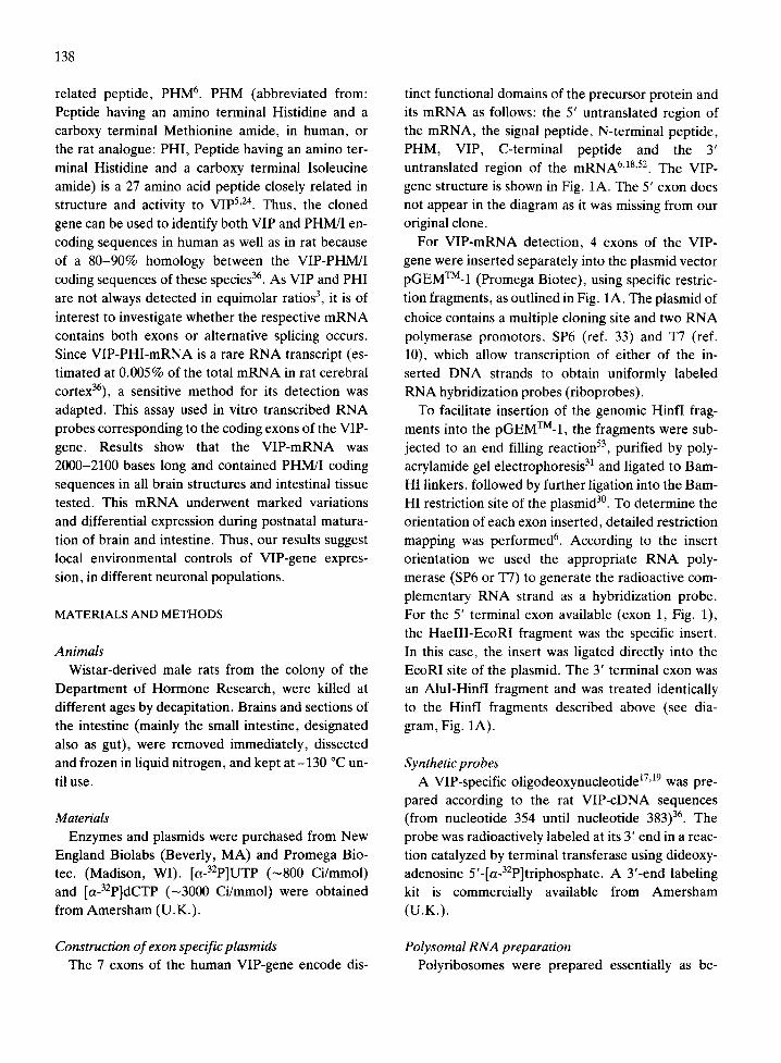
Construction of exon specific plasmids The 7 exons of the human VIP-gene encode dis-
tinct functional domains of the precursor protein and its mRNA as follows: the 5' untranslated region of the mRNA, the signal peptide, N-terminal peptide, PHM, VIP, C-terminal peptide and the 3' untranslated region of the mRNA 6'18,52. The VIP-
gene structure is shown in Fig. 1A. The 5' exon does not appear in the diagram as it was missing from our original clone.
For VIP-mRNA detection, 4 exons of the VIP- gene were inserted separately into the plasmid vector pGEMTM-1 (Promega Biotec), using specific restric- tion fragments, as outlined in Fig. 1A. The plasmid of
choice contains a multiple cloning site and two RNA polymerase promotors, SP6 (ref. 33) and T7 (ref. 10), which allow transcription of either of the in- serted DNA strands to obtain uniformly labeled RNA hybridization probes (riboprobes).
To facilitate insertion of the genomic Hinfl frag- ments into the pGEMTM-1, the fragments were sub- jected to an end filling reaction 53, purified by poly- acrylamide gel electrophoresis 31 and ligated to Bam- HI linkers, followed by further ligation into the Bam- HI restriction site of the plasmid 3°. To determine the orientation of each exon inserted, detailed restriction mapping was performed 6. According to the insert orientation we used the appropriate RNA poly- merase (SP6 or T7) to generate the radioactive com- plementary RNA strand as a hybridization probe. For the 5' terminal exon available (exon 1, Fig. 1), the HaelII-EcoRI fragment was the specific insert. In this case, the insert was ligated directly into the EcoRI site of the plasmid. The 3' terminal exon was an AluI-HinfI fragment and was treated identically to the Hinfl fragments described above (see dia- gram, Fig. 1A).
Synthetic probes A VIP-specific oligodeoxynucleotide 17A9 was pre-
pared according to the rat VIP-cDNA sequences (from nucleotide 354 until nucleotide 383) 36. The probe was radioactively labeled at its 3! end in a reac- tion catalyzed by terminal transferase using dideoxy- adenosine 5'-[a-32p]triphosphate. A 3'-end labeling kit is commercially available from Amersham (U.K.).
Polysomal RNA preparation Polyribosomes were prepared essentially as be-

fore 48, except that sodium deoxychola te was added
to disrupt microsomes 49. Poly(A)-conta in ing R N A
was f ract ionated on ol igo(dT) cellulose 2, after
E D T A unfolding of the po lyr ibosome 49. Polysomal
R N A was further analyzed as descr ibed below for to-
tal RNA.
RNA blot hybridization analysis Total R N A was isolated by urea- l i th ium chloride
extractionL Poly(A)-conta in ing R N A was separa ted
from the bulk of r ibosomal R N A by ol igo(dT) cellu-
lose chromatography z. R N A samples were then sub-
jected to e lect rophores is on 1.5% agarose slab gels.
139
The concentrat ions of the R N A samples were deter-
mined pr ior to the gel applicat ion by absorbance
measurements at 260 nm. Identical quanti t ies (10 #g)
of poly(A)-r ich R N A were appl ied to each gel lane.
Following electrophoresis , the R N A was t ransferred
onto nitrocellulose filters 17,19. Ribosomal R N A was
used for size markers on all gels.
Radioact ive complementary R N A probes were
synthesized according to the protocol provided by
Promega Biotec in the presence of [32p]UTP (800
Ci/mmol, Amersham) . Hybr id iza t ion was pe r fo rmed
according to Promega Biotec in a medium containing
50% formamide , 50 mM sodium phosphate p H 6.5,
A VIP-GENE
= , ~ ~, , ~ , I LU I 3E , II , I I , ,, E o I E x o n 2 E x o n 3 E x n 4 E x o n 5 Exon 6
-~0i['~'-' 25 K b--I'OgK b "1~ 1~ ~ "t~'-"~--i4 Kb "-~" J'~---18 Kb ~J i
iiiiii!iiiiiiill
5000 j
21oo, D. iiiii ~
. . . iiiii~i!! ¸̧
Liiiiill : !i !!i i ~ ~ !!
t S00 b I B
S i z e ( b a s e s ) # . . . . ,
5 0 0 0 - -
! K b - ~ - I r V I P 2 I 0 0 - -
:: f ....
I 2 3
P o l y s o m a l R N A
Fig. 1. Hybridization of isolated exons of the VIP-gene to a 2000-2100-base brain mRNA. Poly(A)-containing mRNA was prepared from the cerebral cortex (enriched for the frontal area) of 30-day-old rats. The RNA was thereafter resolved on agarose formaldehyde gels and then transferred onto nitrocellulose filters. Ribosomal RNA was employed for size markers. A: the VIP-gene structure (lack- ing the 5' exon which corresponds to our original clone) is shown in the diagram 6'1s. To identify VIP-mRNA, plasmids containing spe- cific exons of the gene were constructed. Arrows show the specific exons each inserted into the plasmid pGEMTU-1 and cloned. Specif- ic radiolabeled RNA hybridization probes (riboprobes) were obtained by in vitro transcription. The autoradiogram depicts hybridiza- tion with the 4 different riboprobes shown on the diagram. Numbers indicate size in bases. Open arrows indicate the highest molecular weight hybridizing RNA band and the mature mRNA band. B: autoradiogram showing hybridization with the rat VIP-specific oligo- deoxynucleotide probe to poly(A)-containing polysomal RNA preparation (lane 1) as compared to total cellular poly(A)-containing RNA (lane 2) and to RNA devoid of poly(A) tails obtained by oligo(dT) cellulose chromatography 2 as a negative control (lane 3). RNA was prepared from the cerebral cortex of 8-day-old rats. Hybridization was performed as before 17'19, at 50 °C, using 1 x 10⁶ cprn/ml hybridization mixture of the labeled probe.

140
0.8 M NaC1, 1 mM EDTA, 0.05% BSA, 0.05% Fi- coil, 0.05% polyvinylpyrrolidone and 200 ~g/ml poly(A) and labeled probe (1 x 10⁶ cpm/ml) for 16 h
at 50 °C. The filters were then washed with 0.5 x SSC (1 x SSC = 0.15 M NaCl, 0.015 M sodium cit- rate) and 0.1% NaDodSO 4 at 65 °C, for about 3 h with several changes. Exposure time was usually 16 h a t -70 °C, using Kodak XAR5 film and Du Pont Cro- nex intensifying screens.
When nick-translated probes were used, DNA was labeled according to Rigby et al. 41 and hybridization was performed at 37 °C in 50% formamide, 10% dextran sulfate, 1% NaDodSO4, 20 mM Tris-HCl, pH 7.1, 1 M NaC1, 200 ~g/ml poly(A) and labeled probe at 1 x 10⁶ cpm/ml. Washing with 0.1 x SSC
and 0.2% NaDodSO4 was carried out at 50 °C. Auto- radiograms were densitometrically scanned at 560 r im.
When synthetic probes were used, hybridization was performed as previously described 17'19, at 50 °C.
RESULTS
Construction of exon specific RNA detection probes VIP-mRNA was detected by exon-specific RNA
probes (riboprobes). The sensitivity of the measure- ments using RNA probes was shown to increase sig- nificantly 23 due to lack of renaturation of the conven- tionally used complementary DNA strands during the hybridization procedure 23'41. The availability of
exon-specific hybridization probes (Fig. 1A) should contribute to the precision of the identification of the corresponding mRNA.
Identification of VIP-mRNA in the rat cerebral cortex
mRNA prepared from rat frontal cortex 1'2 was fractionated by electrophoresis on agarose gels 17'19. The RNA bands were blotted onto nitrocellulose fil- ters and hybridized with the specific complementary RNA fragments. Fig. 1A demonstrates that a major VIP-mRNA, hybridizing to the 4 exon-specific probes, was 2000-2100 bases long in the frontal cere- bral cortex of the rat. Other higher molecular weight VIP-related RNA species were apparent as well, the largest form being -7000 bases long. The apparent difference in the intensity of the bands using the dif- ferent exon-specific probes (Fig. 1A) could result from differential exon processing (alternative
processing); or from processing intermediates; or from differences in the hybridization stability of the various probes to the multiple RNA forms. It is also possible that some of the probes tested recognize ad- ditional sequences that are not necessarily products of the V1P gene, but of related genes or of human-re- lated sequences, although our probes do not recog- nize related genes on DNA blot hybridization. Hy- bridization with the probe corresponding to exon 6 (Fig. 1) resulted in a reduced intensity of the 2000-2100-base RNA as compared to results ob-
tained with the other probes. This could be due to the somewhat lower degree of homology (human to rat) of this exon-specific probe (about 10% lower than the other exon-specific riboprobes36). Although this probe, corresponding to exon 6, is longer than the other probes used and does not contain intron se- quences, it contains shorter linear stretches of homo- logy (human to rat) than the other probes 36. To avoid hybridization mismatches, in the following studies we routinely used the riboprobes corresponding to exons 3 and 4 (VIP and PHM, Fig. 1A).
To rule out any possibility of non-specific hybridi- zation, we also used the opposite polarity RNA probe (the sense strand). When such hybridization was conducted with the probe corresponding to the VIP-encoding exon (exon 4) for example, no hybridi- zation signal was obtained. In addition, since VIP- mRNA seems to have a size similar to 18S ribosomal RNA (2000-2100 bases), and one high molecular weight hybridizing band is ~5000 bases long, similar to 28S ribosomal RNA, we routinely employed ribo- somal RNA as a control for the detection of possible non-specific hybridization. Our results indicated no significant hybridization to the ribosomal RNA (e.g.
Figs. 1B and 2B). As an additional, independent control, we synthe-
sized an oligodeoxynucleotide (30 bases) with se- quence complementary to the VIP-encoding mRNA of the rat 36. This probe was then used to identify the mature VIP-mRNA in the cerebral cortex. We pre- pared poly(A)-rich mRNA from polysomes of 8-day- old rat cerebral cortex and compared the hybridiza- tion pattern to that of total cellular poly(A)-rich mRNA. The only band apparent upon hybridization with the synthetic VIP-probe was the -2100-base band, indicating that this band represents the mature VIP-mRNA (Fig. 1B).

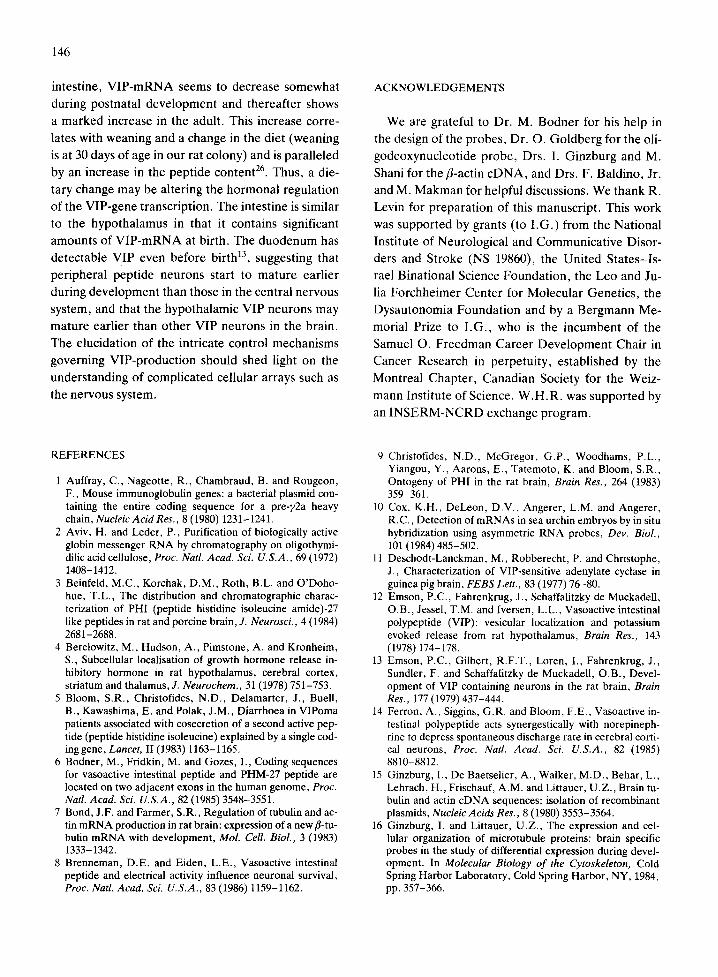
Differential expression of VIP-mRNA in the rat brain during development
The system of choice for initial studies of VIP-gene regulation was the frontal region of the rat cerebral cortex. This brain area is particularly rich in VIP- containing neurons 13'35'37, moreover, in this brain re- gion VIP increases several-fold from birth to matura- tion 13'32'37. To investigate whether the developmen-
tal increase in VIP is paralleled by a change at the mRNA level, we prepared poly(A)-containing RNA from rat frontal cortex using newborn, 3-, 14-, 16-and 30-day-old rats (Fig. 2A). These RNA preparations were subjected to agarose gel electrophoresis fol- lowed by hybridization to the VIP-encoding exon (exon 4) probe. By densitometric scanning of autora- diograms, we quantitated the developmental changes depicted in Fig. 2A. Thus, if we used the newborn mRNA amount as the reference lane (as shown in Fig. 2A being almost equal to the back- ground), the -2100-base mRNA had increased 5-10-fold at the age of 3-4 days, 10-fold or more at 14-16 days and decreased somewhat at 30 days. The high molecular weight RNA forms (5000 bases and 7000 bases), which at birth may represent up to 80% of the total VIP-encoding RNA species, decreased gradually to about 30% at the age of 14 days and to about 20% at 30 days of age. Similar results were ob- tained in 3 independent experiments using various quantities of RNA on the agarose gels. To investi- gate whether the -2100 base VIP-related RNA is in- deed the mature VIP-mRNA, poly(A)-containing RNA was prepared from cortical polysomes and sub- jected to RNA blot analysis. A rat VIP-specific oli- godeoxynucleotide probe specifically designed to
identify rat VIP-mRNA sequences was used as a hy- bridization probe, resulting in exclusive hybridiza- tion to the 2000-2100-base band (Fig. 1B), which is therefore identified as the mature VIP-mRNA. Thus, the mature VIP-mRNA levels were low in the newborn rat cerebral cortex, in parallel with the low level of the peptide 37. We discovered substantial hy- bridization to the 2000-2100-base VIP-mRNA in the brain of 3-day-old rats, suggesting that mRNA syn- thesis precedes VIP synthesis by several days, as the VIP levels in rat cortex begin to increase only at about 7 days of age 32,37. VIP-mRNA content seems to reach a peak at the age of 14 days which may be just prior to a peak in the VIP peptide synthesis 37.
141
To evaluate VIP-gene expression in other brain areas containing the peptide, poly(A)-containing RNA was extracted from parietal cerebral cortex, hypothalamus, hippocampus and amygdala of new- born, 3-, 5-, 8-, 14-, 21- and 90-day-old rats (about 30 rats for each time point). RNA samples were then subjected to Northern blot analysis. Identical amounts of RNA (10/~g) were applied per gel slot; thus, all measurements reflect the amount of VIP- mRNA relative to the total poly(A)-containing- RNA population. Using the VIP-exon-specific ribo- probe, we identified the VIP-mRNA as a 2000-2100- base RNA band in all brain areas tested (Fig. 2B). Densitometric scanning quantitating the devel- opmental changes in the 2000-2100-base VIP- mRNA (Fig. 2C) revealed, in the parietal cerebral cortex, a gradual increase from 0.05 arbitrary absor- bance units in newborn rats to 0.14 at 3 days of age, then to 2.0 at 14 days of age, followed by a decrease in the adult VIP-mRNA. A very similar pattern of a gradual developmental increase in the 2000-2100- base VIP-mRNA of the parietal cerebral cortex was observed using a rat VIP specific oligodeoxynucleo- tide probe (Fig. 2D). In fact, the relative changes in the -2100-base VIP-mRNA were identical using either the riboprobes or the synthetic probe. Thus, the riboprobes (human derived) can be used to iden- tify mature VIP-mRNA in the rat. The results ob- tained for the parietal cortex were similar to those found for the frontal cortex, although the VIP- mRNA content seemed to increase more between birth and 3-4 days of age in the frontal area of the cortex than in the parietal part. As found for the cor- tex, a peak of VIP-mRNA concentration was also
seen in the amygdala at 14 days of age (data not shown). In the hippocampus, VIP-mRNA concen- trations followed a similar pattern, but the maximal level amount was observed earlier, at 8 days of age (0.62 arbitrary absorbance units compared to 0.02 in newborns). In the hypothalamus, a significant amount of VIP-mRNA was observed at birth, which is in contrast to all other brain areas tested, and is much higher than anticipated when compared to the amount of the peptide measured 37. Indeed there is -1 .5 times more VIP-mRNA in the newborn rat hy- pothalamus than in that of the 8-day-old rat, while the peptide concentrations show an opposite trend13. 37.

142
A
Size(bases) :~
5000-- ~
Frontal Cortex
2100 - -
O
i iiii/
B
Cortex
Size(bases)
5O
VIP 2 I
ACTIN
5000--
41140~ 2100- -
Hypothalamus Hippocampus
I I I I I I Hippocompus
vI
', j i 1
e~
I 3 5 B 14 2 1 0 d u l l
3 14 16 30
Age (days)
Hypotho lamus
VIP
I 5 8 14 21 adul t
Age (days}
C o r t e x
B 14 zl d I
Age (days)
I 3 5 8 1 4 2 1 9 0
D
Size(bas
500 (
VIP 2 I O(
5 8 ~ 4 z l 9 o ( - 1 i 3 5 e ~ 4 z 0 9 o
Age (days)
Cor tex
I 5 8 1 4 9 0
Age ( d a y s )
Fig. 2. VIP gene expression is developmentally regulated. A: poly(A)-rich RNA was extracted from rat frontal cortex (as in Fig. 1) at certain postnatal ages (newborns - indicated as 1 on the figure - other numbers indicate 3 days, 14 days, 16 days and 30 days). Experi- ments were carried out as described in Fig. 1, using the VIP exon (exon 4) complementary RNA probe as a hybridization probe. As a control, the same blots were hybridized with cDNA probes containing fl-actin and a-tubulin encoding sequenceslS,16; there was a marked reduction in these RNA populations with development, as expected 48. B: poly(A)-rich m R N A from different brain areas of newborn, 3-, 5-, 8-, 14-, 21- and 90-day-old male rats were subjected to agarose gel electrophoresis. Hybridization was performed with the riboprobe corresponding to the VIP-encoding exon (exon 4). The resulting autoradiograms are shown. Ages are depicted in num- bers below. Ribosomal RNA (RNA devoid of poly(A) tails obtained by oligo(dT) cellulose chromatography 2) was also used as a nega- tive control for hybridization. An example is given with hypothalamic RNA denoted as ( - ) in the figure. The same blots were washed for two 15 min periods at 95-100 °C in a solution containing 0.1% NaDodSO 4 and 0.1 x SCC, which removed all hybridization, as seen following autoradiography. Rehybridization with nick translated/3-actin c D N A 15'16'39, is depicted in Fig. 2C. C: the autoradio- grams in Fig. 2B were densitometrically scanned at 560 nm and results (for the VIP riboprobe hybridization) are presented graphical- ly. Densitometric traces shown are horizontal scans along the 2100-base RNA band of the different ages. All the blots were rehybri- dized with the riboprobe corresponding to the PHM encoding exon (exon 3, Fig. 1A) (an example of the autoradiogram obtained is seen in Fig. 5) and an example of the quantitation pattern is seen for the hippocampus. (Adult = 90 days of age.) D: a similar blot to that used in Fig. 2B, containing parietal cortex RNA samples, was used to hybridize to a rat VIP-specific oligodeoxynucleotide probe (as in Fig. 1B), except that exposure time was at least 5 times longer (more than 80 h, versus 16 h in Fig. 1B).

To verify the integrity of the mRNA preparation and the accuracy of the measurements, the same blots were hybridized with nick-translated fl-actin cDNA (Fig. 2B). Actin mRNA content has been shown to decrease gradually during brain matura- tion 7,48, and this expected pattern was apparent at
the mRNA level (Fig. 2B). VIP-mRNA levels were then compared to actin-mRNA levels, as measured by densitometric scanning of the above autoradio- grams (Figs. 2B, C and 4). Results were standardized using VIP hybridization levels in 90-day-old rats equal to actin hybridization levels at the same age of the same tissue. When results were calculated as such, the relative value of VIP-mRNA in the new- born hypothalamus was still very high. The increase during development in VIP-mRNA content relative to actin mRNA levels, appeared earlier in the hippo- campus and the hypothalamus than in the parietal cortex (Fig. 4), similar to the results obtained above, relative to the total mRNA (Fig. 2C). In contrast, highest concentrations of brain VIP-mRNA in com- parison to actin mRNA were at 21 days and not at 14 days (as compared to total mRNA), probably re- flecting a decrease in the actin-mRNA at 21 days, whereas the VIP-mRNA was still at high levels.
The 2000-2100-base VIP-encoding RNA was not the only RNA band visualized upon hybridization with the VIP specific probe. Higher molecular weight RNA species were detected as well, not only in the frontal cortex (Fig. 2A) but also in the parietal cor- tex, hypothalamus and hippocampus. In the hypo- thalamus, these bands are rather abundant and can represent more than 50% of the hybridization during development (in the newborn as well as in the adult), while in the hippocampus the abundancy of the high
molecular weight RNA forms is lower and tends to decrease with development, from ~30% at 5 days to - 1 0 % of the total hybridization at 14 days of age. The parietal cortex was very similar to the frontal cortex in its abundance of the higher molecular weight RNA. These bands are specific for the VIP probe as they do not cross-hybridize with actin cDNA (Fig. 2B). However, these bands may repre- sent either, intron-containing, processing interme- diates or gene-products related to the human VIP- gene. Indeed, hybridization to rat VIP-specific exon sequences (Fig. 2D) resulted in the almost exclusive detection of the ~2100-base RNA and only trace
143
amounts of higher molecular weight species were seen, much less than with the riboprobes. We thus quantitated only the 2000-2100-base mRNA as we have proven it to represent the rat specific mature VIP-mRNA (see also Fig. 1B).
With longer exposure times, a lower molecular weight RNA band (~1600 bases long) was observed as well, especially in the parietal cortex, and also with the synthetic rat specific probe (Fig. 2D). This band may represent a non-abundant VIP-related RNA (data not shown, and see below).
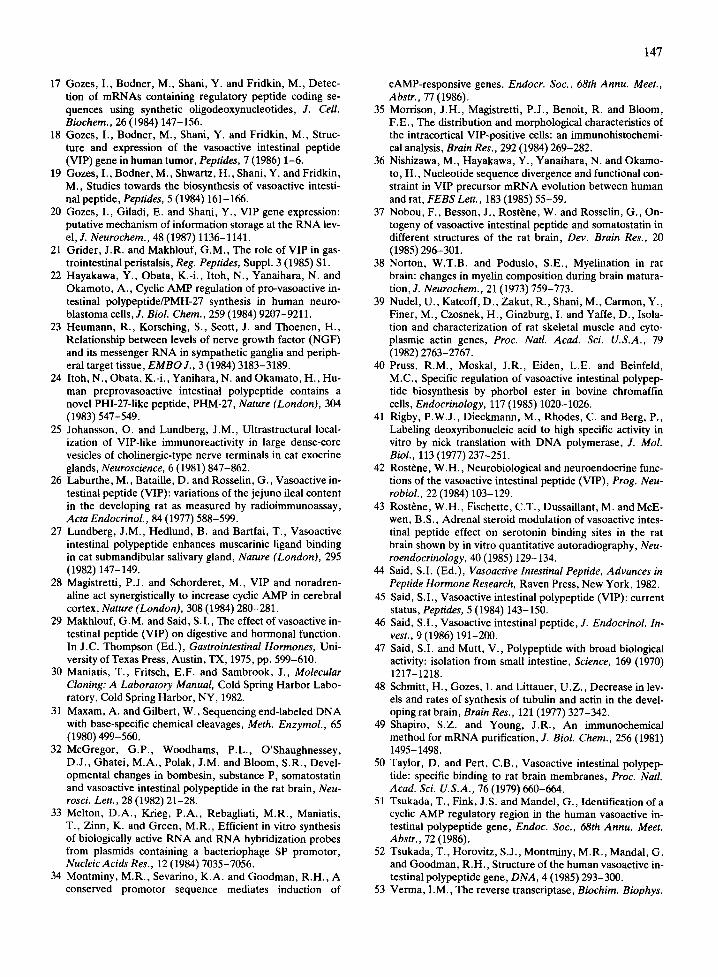
Expression of VIP-mRNA in the rat intestine during development
Poly(A)-rich RNA was isolated from the intestine of newborn 3-, 5-, 8-, 14-, 21- and 90-day-old rats.
In tes t ine
Size(boses)
5 0 0 0
VIP 2 I O0
ACTIN
5000--
2 1 0 0 - -
I 3 5 8 14 21 9 0
Age(days)
Fig. 3. Developmental expression of VIP-mRNA in the intes- tine. Poly(A)-rich mRNA was extracted from intestines of newborn, 3-, 5-, 8-, 14-, 21- and 90-day-old male rats, and treated as in Fig. 2B; autoradiograms are shown. (Adult = 90 days of age.)

144
Northern blot hybridizat ion with the VIP-specific ri-
boprobe revealed substantial amounts of the
2000-2100-base V I P - m R N A in newborn intestine,
decreasing somewhat during postnatal development
and increasing 3-fold in the adult (Fig. 3). A n addi-
tional hybridizing R N A band of - 1 6 0 0 bases was ob-
served at 90 days of age. Such a small R N A could re-
sult from a shorter po ly (A) tail, a lack of one or more
of the exons due to exon exclusion during R N A
processing, or a shorter 3' untransla ted region (there
are two polyadenyla t ion sites in the rat VIP-PHI-
cDNA36). As noted above, a similar R N A band was
observed in the cortex. However , in contrast to the
._c
500
4 0 0
300
2 0 0
I 0 0
I I I I I
Cortex
)othalomus
Intestine
5 I0 15 20 90 Age (days)
Fig. 4. VIP-mRNA in comparison to fl-actin mRNA during on- togeny. The autoradiograms depicted in Fig. 2B for fl-actin hy- bridization were scanned and the values obtained were used for calculations of the 2000-2100-base VIP-mRNA (Fig. 2C) to actin-mRNA ratios. At the adult stage, the ratio of VIP hybri- dization to actin hybridization was arbitrarily designated as 100% and all ratios were calculated accordingly (see Results). The actual values obtained for the percent of VIP/actin hybridi- zation of the adult brain (90-day-old rats) were: 35% for the hy- pothalamus, 45% for the hippocampus and 75% for the parie- tal cerebral cortex. The results for the intestine are obtained from Fig. 3 in which the adult ratio of VIP to actin hybridization was 196%.
i ¸
PHMI12100--
Z n t e s t i n e C o r t e x
S i z e ( b a s e s )
5 0 0 0 - -
!ii iii !! !iii i ilii I 3 5 8 14 2f 9 0 I 3 5 8 14 21 9 0
Age (days)
Fig. 5. VIP-mRNA contains PHM/I coding sequences in brain and intestine at all ages tested. The same experiment per- formed in Figs. 2B and 3 was repeated with the PHM-specific riboprobe (exon 3). Two example autoradiograms of parietal cortex and intestine are shown in the figure.
brain, hardly any high molecular weight VIP rela ted
R N A was detected in the intestine. When VIP-
m R N A concentrat ions in the intestine were com-
pared to actin m R N A concentrat ions, lower ratios
were obtained relative to the brain (Fig. 4). These re-
sults may reflect the higher actin concentrat ions in
the intestine than in the brain.
VIP-mRNA contains PHM/I coding sequences To find out whether V I P - m R N A contained PHM/I
coding sequences in all systems tested we hybridized
the blots with the P H M specific r iboprobe (exon 3)
which is highly homologous to PHI and hybridizes
with the V I P - m R N A 36 (Fig. 1A). Results obta ined
with the PHM r iboprobe were very similar to those
found with the VIP probe (e.g. Fig. 2C, hippocam-
pus; Fig. 5, par ietal cortex and intestine). It should
be noted that the autoradiogram of the P H M hybridi-
zation was exposed for about a third of the exposure
t ime of that of the VIP-specific hybridizat ion, result-
ing in lower intensity of the specific bands (Fig. 2C).
However , the ratio of VIP to PHM/I hybridizat ion of
the 2000-2100-base R N A band remained constant ,
as expected for an m R N A encoding both peptides.
Using a P/HM-riboprobe, high molecular weight
R N A barids were sometimes less prominent than
with the 'VIP- r iboprobe (Figs. 1A and 5), reflecting
different hybridizat ion efficiencies of the probes or
differential R N A processing of the various exons, as
discussed above. The lower molecular weight R N A
of ~1600 bases ment ioned above was seen at relativ-

ely higher VIP-RNA concentrations (e.g. in 14- and 21-day-old rat parietal cortex and 90-day-old rat in- testine, Fig. 5 and also with the rat specific synthetic probe, Fig. 2D).
DISCUSSION
Using 4 different exon-specific RNA hybridization probes, we have identified a 2000-2100-base VIP- mRNA in all brain areas which have been shown to contain high levels of V I P 13'32'37. The overall increase
in VIP-mRNA during brain development generally parallels the peptide increase as measured by radio- immunoassay, suggesting control at the transcriptio- nal level. However, the increase in the mRNA seems to precede, sometimes by several days, the increase in the peptide level, as for example, in the frontal area of the rat cerebral cortex (Fig. 2A). In addition, in the hypothalamus, the high level of VIP-mRNA observed at birth does not correspond to the low level of the peptide at that age 13'37.
The finding of large quantities of VIP-mRNA a few days prior to the actual appearance of high levels
of the peptide is intriguing. Similar findings were ob- tained with other proteins during brain devel- opment a6 suggesting a more general phenomenon of regulation at the translational level. Alternatively, other peptide products of the VIP-gene may appear at an early age. Thus, it is possible that VIP is unsta- ble in the cortex at 3 days of age while other peptide products of the protein precursor 24 are stable. How- ever, when PHI levels were measured during brain development they seemed to essentially parallel the changes observed for VIP 9. Moreover, VIP-mRNA, as compared to total brain mRNA concentration, de- creased after reaching a peak between one and two weeks of age, while the peptide content might still be increasing. These observations, coupled with the age-dependent differential onset of high concentra- tions of VIP-mRNA in various brain regions, suggest local regulation of VIP-gene expression and addi- tional controls at the translational and post-transla- tional (including peptide turnover and secretion) lev- els. As for controls at the peptide level, measure- ments comparing VIP content to PHI show that they are not always in equimolar ratios in various brain areas3; our results, in following brain development of the rat, suggest an mRNA which contains both coding
145
sequences. However, mRNAs containing either the VIP or the PHM/I exons are possible and they may be similar in size to the mRNA containing both cod- ing sequences, as each of these exons is very short (Fig. 1A).
At the RNA processing level, the multiple hybridi- zation bands obtained with the specific VIP probes may be intermediates in the synthesis of mature VIP- mRNA or sequence-related products of other genes. The -7000-base VIP-related RNA may be close to the primary transcript as the human VIP-gene spans -9000 bases 52. Indeed, Sa-nuclease analysis TM and molecular cloning experiments 2°, identified VIP pre- cursor RNA in a VIP-producing human tumor. In ad- dition, the ~1600-base VIP and PHM related RNA observed in the intestine and cerebral cortex (Figs. 3 and 5) may also be a result of differential RNA processing. A similar --1600 base RNA was pre- viously detected using a mixture of VIP-specific syn- thetic oligodeoxynucleotides with sequences de- duced from the amino acid sequence of the pep- tidel7.19.
It is tempting to hypothesize that some ontogeni- cally related control mechanisms may be operating at
the VIP-RNA transcriptional level. Nobou et al. 37 suggested that hypercorticism and hypocorticism may modify the postnatal evolution of VIP, implying a role for corticoids in the developmental patterns of VIP. In addition, hypothyroidism produced a de- crease in both the concentration of VIP and the num- ber of VIP-stained neurons in the developing cor- tex 54, suggesting a possible role for thyroid hormones in regulating VIP expression. In general, both cAMP 22 and an increase in protein kinase C activity 4° probably induce VIP-gene transcription. The cAMP site was identified as containing an 8-base self-com- plementary sequence 5 '-TGACGTCA-3' which is present in the VIP-gene as well as in other cAMP in- ducible genes 34,51.
The variations in the concentrations of VIP- mRNA in the different brain regions during devel- opment and the relative increase in the mRNA con- tent seem to coincide with the period of most rapid brain growth in the rat when there is extensive axonal growth 38 and synaptogenesis 4. Thus, VIP and PHM/I may have a regulatory role in the process of brain dif- ferentiation 8. In contrast to the maturation surge in VIP-mRNA in the postnatal developing brain, in the
146
intestine, V I P - m R N A seems to decrease somewhat
during postnatal deve lopment and thereaf ter shows
a marked increase in the adult. This increase corre-
lates with weaning and a change in the diet (weaning
is at 30 days of age in our rat colony) and is para l le led
by an increase in the pept ide content 26. Thus, a die-
tary change may be altering the hormonal regulat ion
of the VIP-gene transcription. The intestine is similar
to the hypothalamus in that it contains significant
amounts of V I P - m R N A at birth. The duodenum has
detectable VIP even before birth 13, suggesting that
per ipheral pept ide neurons start to mature ear l ier
during development than those in the central nervous
system, and that the hypothalamic VIP neurons may
mature earl ier than other VIP neurons in the brain.
The elucidation of the intricate control mechanisms
governing VIP-product ion should shed light on the
understanding of complicated cellular arrays such as
the nervous system.
ACKNOWLEDGEMENTS
We are grateful to Dr. M. Bodner for his help in
the design of the probes , Dr. O. Goldberg for the oli-
godeoxynucleot ide probe , Drs. 1. Ginzburg and M.
Shani for the fl-actin c D N A , and Drs. F. Baldino, Jr.
and M. Makman for helpful discussions. We thank R.
Levin for prepara t ion of this manuscript . This work
was suppor ted by grants (to I .G . ) from the National
Institute of Neurological and Communicat ive Disor-
ders and Stroke (NS 19860), the Uni ted S ta t e s - I s -
rael Binat ional Science Foundat ion , the Leo and Ju-
lia Forchheimer Center for Molecular Genetics , the
Dysautonomia Foundat ion and by a Bergmann Me-
morial Prize to I .G. , who is the incumbent of the
Samuel O. F reedman Career Deve lopment Chair in
Cancer Research in perpetui ty , establ ished by the
Montreal Chapter , Canadian Society for the Weiz-
mann Insti tute of Science. W . H . R . was suppor ted by
an I N S E R M - N C R D exchange program.
REFERENCES
1 Auffray, C., Nageotte, R., Chambraud, B. and Rougeon, F., Mouse immunoglobulin genes: a bacterial plasmid con- taining the entire coding sequence for a pre-72a heavy chain, Nucleic Acid Res., 8 (1980) 1231-1241.
2 Aviv, H. and Leder, P., Purification of biologically active globin messenger RNA by chromatography on oligothymi- dilic acid cellulose, Proc. Natl. Acad. Sci. U.S.A., 69 (1972) 1408-1412.
3 Beinfeld, M.C., Korchak, D.M., Roth, B.L. and O'Doho- hue, T.L., The distribution and chromatographic charac- terization of PHI (peptide histidine isoleucine amide)-27 like peptides in rat and porcine brain, J. Neurosci., 4 (1984) 2681-2688.
4 Berelowitz, M., Hudson, A., Pimstone, A. and Kronheim, S., Subcellular localisation of growth hormone release in- hibitory hormone in rat hypothalamus, cerebral cortex, striatum and thalamus, J. Neurochem., 31 (1978) 751-753.
5 Bloom, S.R., Christofides, N.D., Delamarter, J., Buell, B., Kawashima, E. and Polak, J.M., Diarrhoea in VIPoma patients associated with cosecretion of a second active pep- tide (peptide histidine isoleucine) explained by a single cod- ing gene, Lancet, II (1983) 1163-1165.
6 Bodner, M., Fridkin, M. and Gozes, I., Coding sequences for vasoactive intestinal peptide and PHM-27 peptide are located on two adjacent exons in the human genome, Proc. Natl. Acad. Sci. U.S.A., 82 (1985) 3548-3551.
7 Bond, J.F. and Farmer, S.R., Regulation of tubulin and ac- tin mRNA production in rat brain: expression of a new fl-tu- bulin mRNA with development, Mol. Cell. Biol., 3 (1983) 1333-1342.
8 Brenneman, D.E. and Eiden, L.E., Vasoactive intestinal peptide and electrical activity influence neuronal survival, Proc. Natl. Acad. Sci. U.S.A., 83 (1986) 1159-1162.
9 Christofides, N.D., McGregor, G.P., Woodhams, P.L., Yiangou, Y., Aarons, E., Tatemoto, K. and Bloom, S.R., Ontogeny of PHI in the rat brain, Brain Res., 264 (1983) 359-361.
10 Cox, K.H., DeLeon, D.V., Angerer, L.M. and Angerer, R.C., Detection of mRNAs in sea urchin embryos by in situ hybridization using asymmetric RNA probes, Dev. Biol., 101 (1984) 485-502.
11 Deschodt-Lanckman, M., Robberecht, P. and Christophe, J., Characterization of VIP-sensitive adenylate cyclase in guinea pig brain, FEBS Lett., 83 (1977) 76-80.
12 Emson, P.C., Fahrenkrug, J., Schaffalitzky de Muckadell, O.B., Jessel, T.M. and Iversen, L.L., Vasoactive intestinal polypeptide (VIP): vesicular localization and potassium evoked release from rat hypothalamus, Brain Res., 143 (1978) 174-178.
13 Emson, P.C., Gilbert, R.F.T., Loren, I., Fahrenkrug, J., Sundler, F. and Schaffalitzky de Muckadell, O.B., Devel- opment of VIP containing neurons in the rat brain, Brain Res., 177 (1979) 437-444.
14 Ferron, A., Siggins, G.R. and Bloom, F.E., Vasoactive in- testinal polypeptide acts synergestically with norepineph- rine to depress spontaneous discharge rate in cerebral corti- cal neurons, Proc. Natl. Acad. Sci. U.S.A., 82 (1985) 8810-8812.
15 Ginzburg, I., De Baetselier, A., Walker, M.D., Behar, L., Lehrach, H., Frischauf, A.M. and Littauer, U.Z., Brain tu- bulin and actin cDNA sequences: isolation of recombinant plasmids, Nucleic Acids Res., 8 (1980) 3553-3564.
16 Ginzburg, I. and Littauer, U.Z., The expression and cel- lular organization of microtubule proteins: brain specific probes in the study of differential expression during devel- opment. In Molecular Biology o f the Cytoskeleton, Cold Spring Harbor Laboratory, Cold Spring Harbor, NY, 1984, pp. 357-366.
17 Gozes, I., Bodner, M., Shani, Y. and Fridkin, M., Detec- tion of mRNAs containing regulatory peptide coding se- quences using synthetic oligodeoxynucleotides, J. Cell. Biochem., 26 (1984) 147-156.
18 Gozes, I., Bodner, M., Shani, Y. and Fridkin, M., Struc- ture and expression of the vasoactive intestinal peptide (VIP) gene in human tumor, Peptides, 7 (1986) 1-6.
19 Gozes, I., Bodner, M., Shwartz, H., Shani, Y. and Fridkin, M., Studies towards the biosynthesis of vasoactive intesti- nal peptide, Peptides, 5 (1984) 161-166.
20 Gozes, I., Giladi, E. and Shani, Y., VIP gene expression: putative mechanism of information storage at the RNA lev- el, J. Neurochem., 48 (1987) 1136-1141.
21 Grider, J.R. and Makhlouf, G.M., The role of VIP in gas- trointestinal peristalsis, Reg. Peptides, Suppl. 3 (1985) S1.
22 Hayakawa, Y., Obata, K.-i., Itoh, N., Yanaihara, N. and Okamoto, A., Cyclic AMP regulation of pro-vasoactive in- testinal polypeptide/PMH-27 synthesis in human neuro- blastoma cells, J. Biol. Chem., 259 (1984) 9207-9211.
23 Heumann, R., Korsching, S., Scott, J. and Thoenen, H., Relationship between levels of nerve growth factor (NGF) and its messenger RNA in sympathetic ganglia and periph- eral target tissue, EMBO J., 3 (1984) 3183-3189.
24 Itoh, N., Obata, K.-i., Yanihara, N. and Okamato, H., Hu- man preprovasoactive intestinal polypeptide contains a novel PHI-27-1ike peptide, PHM-27, Nature (London), 304 (1983) 547-549.
25 Johansson, O. and Lundberg, J.M., Ultrastructural local- ization of VIP-like immunoreactivity in large dense-core vesicles of cholinergic-type nerve terminals in cat exocrine glands, Neuroscience, 6 (1981) 847-862.
26 Laburthe, M., Bataille, D. and Rosselin, G., Vasoactive in- testinal peptide (VIP): variations of the jejuno ileal content in the developing rat as measured by radioimmunoassay, Acta Endocrinol., 84 (1977) 588-599.
27 Lundberg, J.M., Hedlund, B. and Bartfai, T., Vasoactive intestinal polypeptide enhances muscarinic ligand binding in cat submandibular salivary gland, Nature (London), 295 (1982) 147-149.
28 Magistretti, P.J. and Schorderet, M., VIP and noradren- aline act synergistically to increase cyclic AMP in cerebral cortex, Nature (London), 308 (1984) 280-281.
29 Makhlouf, G.M. and Said, S.I., The effect of vasoactive in- testinal peptide (VIP) on digestive and hormonal function. In J.C. Thompson (Ed.), Gastrointestinal Hormones, Uni- versity of Texas Press, Austin, TX, 1975, pp. 599-610.
30 Maniatis, T., Fritsch, E.F. and Sambrook, J., Molecular Cloning: A Laboratory Manual, Cold Spring Harbor Labo- ratory, Cold Spring Harbor, NY, 1982.
31 Maxam, A. and Gilbert, W., Sequencing end-labeled DNA with base-specific chemical cleavages, Meth. Enzymol., 65 (1980) 499-560.
32 McGregor, G.P., Woodhams, P.L., O'Shaughnessey, D.J., Ghatei, M.A., Polak, J.M. and Bloom, S.R., Devel- opmental changes in bombesin, substance P, somatostatin and vasoactive intestinal polypeptide in the rat brain, Neu- rosci. Lett., 28 (1982) 21-28.
33 Melton, D.A., Krieg, P.A., Rebagliati, M.R., Maniatis, T., Zinn, K. and Green, M.R., Efficient in vitro synthesis of biologically active RNA and RNA hybridization probes from plasmids containing a bacteriophage SP promotor, Nucleic Acids Res., 12 (1984) 7035-7056.
34 Montminy, M.R., Sevarino, K.A. and Goodman, R.H., A conserved promotor sequence mediates induction of
147
cAMP-responsive genes. Endocr. Soc., 68th Annu. Meet., Abstr., 77 (1986).
35 Morrison, J.H., Magistretti, P.J., Benoit, R. and Bloom, F.E., The distribution and morphological characteristics of the intracortical VIP-positive cells: an immunohistochemi- cal analysis, Brain Res., 292 (1984) 269-282.
36 Nishizawa, M., Hayakawa, Y., Yanaihara, N. and Okamo- to, H., Nudeotide sequence divergence and functional con- straint in VIP precursor mRNA evolution between human and rat, FEBS Lett., 183 (1985) 55-59.
37 Nobou, F., Besson, J., Rost~ne, W. and Rosselin, G., On- togeny of vasoactive intestinal peptide and somatostatin in different structures of the rat brain, Dev. Brain Res., 20 (1985) 296-301.
38 Norton, W.T.B. and Poduslo, S.E., Myelination in rat brain: changes in myelin composition during brain matura- tion, J. Neurochem., 21 (1973) 759-773.
39 Nudel, U., Katcoff, D., Zakut, R., Shani, M., Cartoon, Y., Finer, M., Czosnek, H., Ginzburg, I. and Yaffe, D., Isola- tion and characterization of rat skeletal muscle and cyto- plasmic actin genes, Proc. Natl. Acad. Sci. U.S.A., 79 (1982) 2763-2767.
40 Pruss, R.M., Moskal, J.R., Eiden, L.E. and Beinfeld, M.C., Specific regulation of vasoactive intestinal polypep- tide biosynthesis by phorbol ester in bovine chromaffin cells, Endocrinology, 117 (1985) 1020-1026.
41 Rigby, P.W.J., Dieckmann, M., Rhodes, C. and Berg, P., Labeling deoxyribonucleic acid to high specific activity in vitro by nick translation with DNA polymerase, J. Mol. Biol., 113 (1977) 237-251.
42 Rost~ne, W.H., Neurobiological and neuroendocrine func- tions of the vasoactive intestinal peptide (VIP), Prog. Neu- robiol., 22 (1984) 103-129.
43 Rost~ne, W.H., Fischette, C.T., Dussaillant, M. and McE- wen, B.S., Adrenal steroid modulation of vasoactive intes- tinal peptide effect on serotonin binding sites in the rat brain shown by in vitro quantitative autoradiography, Neu- roendocrinology, 40 (1985) 129-134.
44 Said, S.I. (Ed.), Vasoactive Intestinal Peptide, Advances in Peptide Hormone Research, Raven Press, New York, 1982.
45 Said, S.I., Vasoactive intestinal polypeptide (VIP): current status, Peptides, 5 (1984) 143-150.
46 Said, S.I., Vasoactive intestinal peptide, J. Endocrinol. In- vest., 9 (1986) 191-200.
47 Said, S.I. and Mutt, V., Polypeptide with broad biological activity: isolation from small intestine, Science, 169 (1970) 1217-1218.
48 Schmitt, H., Gozes, I. and Littauer, U.Z., Decrease in lev- els and rates of synthesis of tubulin and actin in the devel- oping rat brain, Brain Res., 121 (1977) 327-342.
49 Shapiro, S.Z. and Young, J.R., An immunochemical method for mRNA purification, J. Biol. Chem., 256 (1981) 1495-1498.
50 Taylor, D. and Pert, C.B., Vasoactive intestinal polypep- tide: specific binding to rat brain membranes, Proc. Natl. Acad. Sci. U.S.A., 76 (1979) 660-664.
51 Tsukada, T., Fink, J.S. and Mandel, G., Identification of a cyclic AMP regulatory region in the human vasoactive in- testinal polypeptide gene, Endoc. Soc., 68th Annu. Meet. Abstr., 72 (1986).
52 Tsukada, T., Horovitz, S.J., Montminy, M.R., Mandal, G. and Goodman, R.H., Structure of the human vasoactive in- testinal polypeptide gene, DNA, 4 (1985) 293-300.
53 Verma, I.M., The reverse transcriptase, Biochim. Biophys.

148
Acta, 473 (1977) 1-38. 54 Woodhams, P.L., McGovern, J., McGregor, G.P.,
O'Shaughnessey, D.J., Ghatei, M.A., Blank, M.A., Adri- an, T.H., Lee, Y., Polak, J.M., Bloom, S.R. and Balazs,
R., Effects of changes in neonatal thyroid status on the de- velopment of neuropeptide systems in the rat brain, Int. J. Dev. Neurosci., 1 (1983) 155-164.
Copyright © 2022 FDOKUMEN