Death Inducer-Obliterator 1 Triggers Apoptosis after Nuclear Translocation and Caspase Upregulation
11
10.1128/MCB.23.9.3216-3225.2003. 2003, 23(9):3216. DOI: Mol. Cell. Biol. de Buitrago and Carlos Martínez-A David García-Domingo, Dorian Ramírez, Gonzalo González Caspase Upregulation Apoptosis after Nuclear Translocation and Death Inducer-Obliterator 1 Triggers http://mcb.asm.org/content/23/9/3216 Updated information and services can be found at: These include: REFERENCES http://mcb.asm.org/content/23/9/3216#ref-list-1 at: This article cites 42 articles, 23 of which can be accessed free CONTENT ALERTS more» articles cite this article), Receive: RSS Feeds, eTOCs, free email alerts (when new http://journals.asm.org/site/misc/reprints.xhtml Information about commercial reprint orders: http://journals.asm.org/site/subscriptions/ To subscribe to to another ASM Journal go to: on October 16, 2014 by guest http://mcb.asm.org/ Downloaded from on October 16, 2014 by guest http://mcb.asm.org/ Downloaded from
Transcript of Death Inducer-Obliterator 1 Triggers Apoptosis after Nuclear Translocation and Caspase Upregulation

10.1128/MCB.23.9.3216-3225.2003.
2003, 23(9):3216. DOI:Mol. Cell. Biol. de Buitrago and Carlos Martínez-ADavid García-Domingo, Dorian Ramírez, Gonzalo González Caspase UpregulationApoptosis after Nuclear Translocation and Death Inducer-Obliterator 1 Triggers
http://mcb.asm.org/content/23/9/3216Updated information and services can be found at:
These include:
REFERENCEShttp://mcb.asm.org/content/23/9/3216#ref-list-1at:
This article cites 42 articles, 23 of which can be accessed free
CONTENT ALERTS more»articles cite this article),
Receive: RSS Feeds, eTOCs, free email alerts (when new
http://journals.asm.org/site/misc/reprints.xhtmlInformation about commercial reprint orders: http://journals.asm.org/site/subscriptions/To subscribe to to another ASM Journal go to:
on October 16, 2014 by guest
http://mcb.asm
.org/D
ownloaded from
on O
ctober 16, 2014 by guesthttp://m
cb.asm.org/
Dow
nloaded from

MOLECULAR AND CELLULAR BIOLOGY, May 2003, p. 3216–3225 Vol. 23, No. 90270-7306/03/$08.00�0 DOI: 10.1128/MCB.23.9.3216–3225.2003Copyright © 2003, American Society for Microbiology. All Rights Reserved.
Death Inducer-Obliterator 1 Triggers Apoptosis after NuclearTranslocation and Caspase Upregulation
David García-Domingo, Dorian Ramírez,† Gonzalo Gonzalez de Buitrago,and Carlos Martínez-A*
Department of Immunology and Oncology, Centro Nacional de Biotecnología/CSIC, UniversidadAutonomo de Madrid, Campus de Cantoblanco, E-28049 Madrid, Spain
Received 22 July 2002/Returned for modification 4 September 2002/Accepted 4 February 2003
Death inducer-obliterator 1 (DIO-1) is a gene that is upregulated early in apoptosis. Here we report that inhealthy cells, the DIO-1 gene product was located in the cytoplasm, where it formed oligomers. After inter-leukin-3 starvation or c-Myc-induced apoptosis in serum-free conditions, DIO-1 translocated to the nucleus,where it upregulated caspase levels and activity. A nuclear localization signal deletion mutant (DIO-1�NLS)was unable to translocate to the nuclear compartment in the absence of interleukin-3 and failed to upregulateprocaspase levels or trigger cell death. In addition, cells stably expressing DIO-1�NLS were protected fromapoptosis induced by interleukin-3 withdrawal. These results indicate that DIO-1 has a relevant role inregulating the early stages of cell death.
Apoptosis, or programmed cell death, has a major role innormal development, tissue homeostasis, defense against viralinvasion, immune modulation, and, when dysregulated, mod-ulation of autoimmune and clonal or neoplastic diseases (15,17, 30). Apoptosis is characterized by cell shrinkage, chromatincondensation, internucleosomal DNA cleavage, membraneblebbing, and the formation of apoptotic bodies that are pha-gocytosed by other cells (8, 38). These morphological changesare orchestrated by the activity of a family of aspartate-specificproteases called caspases (4, 7, 34).
Caspases are produced in cells as catalytically inactive zy-mogens, or procaspases, composed of three subunits, a pro-domain and two catalytic subdomains, known as the large andsmall subunits (1). Procaspases must be proteolytically pro-cessed to become active proteases. An effector caspase, forexample caspase 3, is activated by an initiator caspase, such ascaspase 9, through proteolytic cleavage at specific internal Aspresidues to give rise to the two subunits of the mature caspase.Once activated, the effector caspases cleave a broad spectrumof cellular targets, leading ultimately to cell death (38). Acti-vation of the initiator caspases is in turn regulated by upstreamprotein complexes. In the case of the so-called “extrinsic”pathway, activation of death receptors such as Fas/CD95 andtumor necrosis factor receptor 1 after binding of their respec-tive ligands induces recruitment of caspase 8 (FLICE) via theadapter molecule FADD (Fas-associated protein with deathdomain) (27). Caspase 8 can activate effector caspases eitherdirectly (3, 27) or indirectly by cleaving Bid and inducing therelease of mitochondrial cytochrome c (18, 25).
In the case of the “intrinsic” death receptor-independentpathway, apoptotic cell death is induced directly by death stim-uli and is also regulated by adapter complexes. This is the case
of one of the major routes to caspase activation pathways,triggered by cytochrome c release from the mitochondrial in-termembrane space into the cytosol (23). Cytosolic cytochromec promotes assembly of a protein complex called the apopto-some, which includes caspase 9 bound to the CED-4 homologApaf-1 (19, 42), inducing autoactivation of procaspase 9 (33,37). Following activation, caspase 9 cleaves and activates procas-pase 3 (19, 42), giving rise to a proteolytic cascade involvingmultiple caspases.
DIO-1 was identified by a differential display approach (21)in WOL-1 pre-B cells induced to undergo apoptosis by inter-leukin-7 (IL-7) starvation (9). Its predicted amino acid se-quence showed transcriptional activation domains, a canonicalbipartite nuclear localization signal (NLS), a PhD finger, and acarboxy-terminal lysine-rich region. DIO-1 mRNA was up-regulated soon after apoptotic induction by several stimuli,including removal of IL-7, addition of dexamethasone orgamma interferon in WOL-1 cells, immunoglobulin M (IgM)receptor cross-linking in WEHI-231 cells, or c-myc activationunder serum-free conditions in the absence of p53 expressionin MEF(10.1)Val5MycER cells. Overexpression of DIO-1 incells or misexpression in chick limbs induced massive apoptosisin the absence of any apoptotic stimuli; this could be inhibitedby Bcl-2 overexpression or incubation with the general caspaseinhibitor benzyloxycarbonyl-Val-Ala-Asp-fluoromethyl ketone(z-VAD-fmk). These results suggested that DIO-1-inducedapoptosis requires caspase activation. Furthermore, overex-pression of a DIO-1 deletion mutant lacking both NLSs failedto induce cell death, linking its lack of lethality to an inabilityto translocate.
Here we studied the mechanism by which DIO-1 inducesapoptosis and the importance of its subcellular localization.We generated several tagged constructs and analyzed the sub-cellular distribution pattern of both wild-type and mutantDIO-1 in various apoptotic situations, revealing nuclear trans-location as the main regulatory event in the DIO-1-activatedapoptotic pathway. We provide evidence that DIO-1 translo-cation boosts the apoptotic machinery by upregulating protein
* Corresponding author. Mailing address: Department of Immunol-ogy and Oncology, Centro Nacional de Biotecnología/CSIC, UAMCampus de Cantoblanco, E-28049 Madrid, Spain. Phone: 34 91 585 4559. Fax: 34 91 372 04 93. E-mail: [email protected].
† Present address: University of Michigan Medical School, Ann Ar-bor, MI 48109.
3216
on October 16, 2014 by guest
http://mcb.asm
.org/D
ownloaded from

levels of procaspase 3 and 9, which enhances their apoptosis-inducing activity.
MATERIALS AND METHODS
Expression plasmids. DIO-1�NLSpcDNA3 was generated from DIO-1cloned in pcDNA3 by reverse PCR with the circular plasmid as the template andthe primers 5�-GAAGATTCTGCCGAAACTGGG-3� and 5�-AAGTTCCTTCAACGTAAGG-3�, which span the NLSs and the connecting sequence; the PCRproduct was permitted to further self-ligate. Both N-terminally Flag-taggedDIO-1 and DIO-1�NLS were PCR amplified from their corresponding pcDNA3constructs with a proof-reading polymerase and the primers 5�-GCGGATCCGATGATAAAGGGCACCTG-3� and 5�-CGACCTCGAGTTACCAAGGCCTAAAACTG-3� for in-frame reading. The PCR products were digested withBamHI and XhoI and subcloned into the pCMV-Tag 2B vector (Stratagene).DIO-1/Myc was generated similarly after PCR amplification with the primers5�-TGGAATTCCACCATGGATGATAAAGGGCAC-3� and 5�-GCTCTAGACCAAGGCCTAAAACTG-3� from DIO-1pcDNA3, digested with EcoRI andXbaI, and subcloned into the pEF4/Myc-His A vector (Invitrogen). All constructswere verified by nucleotide sequencing.
Cell culture. FL5.12 and MEF(10.1)Val5MycER cells were cultured as de-scribed previously (9). 293T cells were cultured in Dulbecco’s modified Eagle’smedium supplemented with 10% fetal bovine serum, 2 mM L-glutamine, andantibiotics.
Antibodies and reagents. Production of the polyclonal antibody against murineDIO-1 amino acids 58 to 72 has been described (9). Polyclonal rabbit anti-mousecaspase 3 was the kind gift of T. Mak and R. Hakem (Ontario Cancer Institute,Toronto, Canada); anti-caspase 9 was the kind gift of D. R. Green and B. Wolf(39). Anti-Flag M2 monoclonal antibody (Sigma), anti-Myc 9E10 monoclonalantibody (Santa Cruz Biotechnologies), protein A-agarose (Sigma), protein G-agarose (Sigma), antiphosphoserine sampler kit (Biomol), antiphosphoserinemonoclonal antibody (Calbiochem), and antiphosphothreonine monoclonal an-tibody (Calbiochem) were purchased as indicated. z-VAD-fmk was purchasedfrom Bachem.
Establishment of FL5.12 cells stably overexpressing DIO-1�NLS. FL5.12 cells(3 � 106) were electroporated with 10 �g of DIO-1�NLS construct or pcDNA3.Transfected cells were resuspended in 48 ml of complete medium and distributedin a 48-well plate. Selection was performed after 24 h by removing the mediumand adding fresh complete medium containing 1 mg of G418 per ml. Selectionwas carried out for 30 days, and DIO-1�NLS expression was determined byreverse transcription-PCR and Western blotting.
Subcellular fractionation. FL5.12 cells (2 � 107) were harvested and washedin ice-cold phosphate-buffered saline (PBS), and pellets were resuspended in5 volumes of ice-cold buffer A (10 mM HEPES [pH 8.0], 0.5 M sucrose, 1 mMEDTA, 0.5 mM spermidine, 0.15 mM spermine, 15 mM KCl, 0.5 mM dithio-threitol, 10 �g of aprotinin per ml, 10 �g of pepstatin A per ml, 10 �g of leu-peptin per ml, 1 mM phenylmethylsulfonyl fluoride [PMSF], 100 �M Na3VO4,and 10 mM NaF). After incubation on ice for 15 min, cells were lysed by threefreeze-thaw cycles and centrifuged (1,000 � g, 10 min, 4°C), and the nuclearpellet was resuspended in buffer B (10 mM piperazine-N,N�-bis(2-ethanesulfonicacid) [PIPES, pH 7.4], 80 mM KCl, 20 mM NaCl, 5 mM sodium EGTA, 250 mMsucrose, 1 mM dithiothreitol, 10 �g of aprotinin per ml, 10 �g of pepstatin A perml, 10 �g of leupeptin per ml, 1 mM PMSF, 100 �M Na3VO4, and 10 mM NaF)at 8.5 � 107 nuclei/ml. Samples were separated by sodium dodecyl sulfate-polyacrylamide gel electrophoresis (SDS-PAGE) under reducing conditions. Pu-rity of the cytosolic and nuclear fractions was tested by Western blotting withcompartment-specific antibodies to procyclic acidic repetitive protein and cal-pain-1 (not shown).
Transfections and immunoprecipitation. Transient transfections in FL5.12and MEF(10.1)Val5MycERcells were performed by electroporation as describedpreviously (9). 293T cells were transiently transfected in six-well plates withLipofectamine Plus (Life Technologies) following the manufacturer’s protocol.Cytosolic extracts for immunoprecipitation were obtained after cell lysis in NP-40buffer (40 mM Tris-HCl [pH 8], 500 mM NaCl, 0.1% NP-40, 6 mM EDTA, 6 mMEGTA, 10 �g of aprotinin per ml, 10 �g of pepstatin A per ml, 10 �g of leupeptinper ml, 1 mM PMSF, 100 �M Na3VO4, and 10 mM NaF) or a digitonin-containing buffer (10 mM triethanolamine [pH 8], 150 mM NaCl, 1 mM EDTA,10% glycerol, 1% digitonin, 10 �g of aprotinin per ml, 10 �g of pepstatin A perml, 10 �g of leupeptin per ml, 1 mM PMSF, 100 �M Na3VO4, and 10 mM NaF).Immunoprecipitation was performed by preclearing lysates with 30 �l of proteinA/G-agarose (1 h, 4°C, with gentle rotation), followed by incubation with theappropriate antibody (10 �g/ml; 1 h, 4°C). After addition of protein A- or proteinG-agarose (30 �l), beads were pelleted by brief centrifugation and washed three
times in 50 mM Tris-HCl (pH 7.6) buffer, and the pellet was boiled in loadingbuffer and analyzed in Western blot.
Phosphatase treatment. 293T cells transiently transfected with Flag-taggedDIO-1 were harvested, washed in ice-cold PBS, and lysed in a digitonin-contain-ing buffer (40 mM Tris-HCl [pH 8], 50 mM NaCl, 2 mM MnCl2, 1% digitonin,10 �g of aprotinin per ml, 10 �g of pepstatin A per ml, 10 �g of leupeptin perml, 1 mM PMSF). After removal of cellular debris by centrifugation, lysates weretreated with 500 U of �-phosphatase (Calbiochem) (30 min, 30°C). The reactionwas terminated by addition of loading buffer and then analyzed in Western blot.
Western blot analysis. Cells for analysis of procaspase expression were washedwith PBS, and the pellet was suspended in lysis buffer (137 mM NaCl, 20 mMTris-HCl [pH 8], 1 mM MgCl2, 1 mM CaCl2, 10% glycerol, 1% NP-40, 0.5%deoxycholate, 0.1% SDS, 10 �g of aprotinin per ml, 10 �g of pepstatin A per ml,10 �g of leupeptin per ml, and 1 mM PMSF) for 30 min on ice. The proteincontent of the lysates was quantified with the Bio-Rad DC protein assay (Bio-Rad); after SDS-PAGE, proteins were transferred to nitrocellulose membranes(Bio-Rad). Equal protein loading was verified by Ponceau Red (Sigma) staining.Membranes were blocked overnight with 5% nonfat dry milk in TBS buffer (20mM Tris-HCl [pH 7.5], 150 mM NaCl); subsequent incubations and membranewashes were performed in TBS-T buffer (20 mM Tris-HCl [pH 7.5], 150 mMNaCl, and 0.2% Tween 20) containing 1% nonfat dry milk. After 2 h of antibodyincubation and 1 h of washing, blots were developed with peroxidase-conjugatedanti-rabbit or anti-mouse immunoglobulin antibodies (Dako), and proteins weredetected by an enhanced chemiluminescence system (ECL; Amersham).
Fluorescence microscopy. For immunofluorescence, cells were cultured oncoverslips, washed in PBS, fixed in 4% paraformaldehyde (15 min, room tem-perature), washed three times in PBT (PBS with 0.1% Tween 20), incubated in2% bovine serum albumin, and incubated for 1 h with anti-DIO-1 (1:100) oranti-Flag M2 (1:500) in PBT. After incubation, cells were washed three times inthe same buffer and incubated for 1 h with indocarbocyanine-conjugated sec-ondary antibodies (Jackson ImmunoResearch). After washing, samples wereincubated with Sybr Green (Molecular Probes) in PBS for DNA staining. SerialZ-sections were obtained with an Ar-Kr laser and a TCS-NT Leica confocalimaging system.
Enzyme assay for caspase activity. Cells were collected, washed with ice-coldPBS, and resuspended in extraction buffer (50 mM Tris-HCl [pH 7.6], 150 mMNaCl, 0.5 mM EDTA, 10 mM NaH2PO4, 10 mM Na2HPO4, 1% Nonidet P-40,0.4 mM Na3VO4, 1 mM PMSF, 10 �g of aprotinin per ml, 10 �g of pepstatin Aper ml, 10 �g of leupeptin per ml). After incubation (30 min, on ice), the celllysate was centrifuged (20,000 � g, 30 min), and the supernatant was used as thecytosolic extract. Five micrograms of cytosolic proteins, estimated by the bicin-choninic acid method (36), were diluted fivefold in assay buffer (25 mM HEPES[pH 7.5], 0.1% CHAPS, 10% sucrose, 10 mM dithiothreitol, and 0.1 mg ofovalbumin per ml) and incubated (1 h, 37°C) with 10 �M of the fluorescentsubstrate Ac-DEHD-AMC (acetyl-Asp-Glu-His-Asp-7-amino-4-methylcouma-rin), Ac-DEVD-AMC (acetyl-Asp-Glu-Val-Asp-7-amino-4-methylcoumarin),Ac-VEID-AMC (acetyl-Val-Glu-Ile-Asp-7-amino-4-methylcoumarin), or Ac-LEHD-AMC (acetyl-Leu-Glu-His-Asp-7-amino-4-methylcoumarin) to measurecaspase 2, caspase 3-like, caspase 6, and caspase 9 activity, respectively. Thereaction was terminated by addition of high-pressure liquid chromatography(HPLC) buffer (water-acetonitrile [75:25], 0.1% trifluoroacetic acid). Cleavedsubstrate fluorescence was determined by C18 reverse-phase HPLC with fluores-cence detection (338 nm excitation, 455 nm emission). Control experimentsconfirmed linearity with time and protein concentration of substrate release.
Apoptosis assay. Apoptosis was evaluated by staining cellular DNA contentwith the DNA intercalator propidium iodide in a semiautomatic procedure(DNA-Prep Reagents; Coulter), followed by analysis on an Epics XL flow cy-tometer (Coulter). Briefly, cells (105 to 106) were recovered by centrifugation,resuspended in 100 �l of PBS, permeabilized, and stained by adding 100 �l ofdetergent reagent followed by 1 ml of propidium iodide solution. After mixing,samples were incubated (37°C, 1 h) and analyzed by flow cytometry.
RESULTS
DIO-1 nuclear translocation following apoptotic stimula-tion requires the NLS. We previously reported differences inDIO-1 mRNA levels after induction of p53-dependent and-independent apoptosis in MEF(10.1)Val5MycER cells (9).DIO-1 transcripts were upregulated in apoptotic processes in ap53-independent fashion. The presence of two putative NLSsin the DIO-1 gene product suggested that it may be localized
VOL. 23, 2003 DIO-1 NUCLEAR TRANSLOCATION ACTIVATES CASPASES 3217
on October 16, 2014 by guest
http://mcb.asm
.org/D
ownloaded from

in the nucleus (9). To study its putative role as a transcrip-tion factor, we examined the subcellular localization patternof the DIO-1 gene product and its NLS deletion mutant(DIO-1�NLS). Immunofluorescence microscopy of healthyMEF(10.1)Val5MycER cells with an affinity-purified rabbitantiserum specific for a DIO-1 peptide (9) indicated a clearcytoplasmic pattern for the DIO-1 protein, which was nearlyabsent in the nucleus (Fig. 1A to C). p53-mediated triggeringof apoptosis showed no change in the DIO-1 localization pat-tern (Fig. 1D to F), although the cells underwent apoptosis(not shown). Cells cultured at 39°C to inactivate p53, then
induced to apoptosis by 17�-estradiol addition and fetal bovineserum starvation, showed DIO-1 translocation from cytoplasmto the nucleus (Fig. 1G to I). This was observed before celldeath was detectable by any method and in parallel to mRNAupregulation kinetics.
We examined the subcellular localization of the DIO-1�NLS mutant, but as the anti-DIO-1 antibody recognizesboth wild-type and mutant proteins, the mutant was Flagtagged (DIO-1�NLS/Flag) and transiently transfected intoMEF(10.1)Val5MycER cells. Apoptosis was induced 24 h post-transfection, and staining was performed at the same time
FIG. 1. Immunolocalization of endogenous DIO-1 under apoptotic conditions. (A to C) Viable (nonapoptotic) MEF(10.1)Val5MycER cellswere stained with anti-DIO-1 antibody (red); nuclei were stained with Sybr Green (green). (D to F) The same cells were induced to apoptosis bylowering the incubation temperature (32°C, 12 h) to activate p53 in the presence of 17�-estradiol (1 �M). Note the clear cytoplasmic pattern ofDIO-1, although some cells have already begun the apoptotic program. (G to I) The same cell line, incubated at 39°C to inactivate p53, was17�-estradiol treated (1 �M) and serum starved for 8 h, the time at which DIO-1 mRNA is upregulated. Note nuclear translocation of DIO-1 tothe intact, nonapoptotic nuclei.
3218 GARCIA-DOMINGO ET AL. MOL. CELL. BIOL.
on October 16, 2014 by guest
http://mcb.asm
.org/D
ownloaded from

points as for wild-type DIO-1. A clear cytoplasmic pattern wasobserved in healthy cells (Fig. 2A to C) and in those undergo-ing p53-induced apoptosis (Fig. 2D to F). DIO-1�NLS wasunable to translocate to the nucleus under conditions thatpromoted translocation of the wild-type form (Fig. 2G to I), aspredicted by its lack of NLS.
DIO-1 forms oligomers. To test for the association state ofDIO-1, we fused murine DIO-1 to N-terminal Flag (DIO-1/Flag) and C-terminal Myc (DIO-1/Myc) tags in different con-structs. The constructs were coexpressed by transient transfec-tion into human embryonic kidney 293T cells, and proteinswere isolated under nondenaturing conditions with buffers thatpermitted cytosolic extraction. Western blot analysis with the
anti-DIO-1 antibody (Fig. 3A) and antitag antibodies (Fig. 3B)confirmed expression in total lysates of cells transfected withthe appropriate plasmids, but not in cells transfected withcontrol plasmids. In all cases, a triplet was observed with veryclose bands. The triplets do not correspond to proteolytic deg-radation products, as they were detected equally well withantibodies to tags located at both ends of the protein, in thepresence of large amounts of protease inhibitors. These mul-tiple bands are more likely to represent posttranslational mod-ifications of DIO-1.
Oligomerization was determined in an immunoprecipitationassay performed with anti-Myc and resolved in SDS-polyacryl-amide gel electrophoresis (SDS-PAGE). Subsequent Western
FIG. 2. Immunolocalization of DIO-1�NLS in MEF(10.1)Val5MycER cells under apoptotic conditions. A DIO-1�NLS Flag-tagged constructwas transiently transfected into the cells, which were analyzed 36 h posttransfection. A selected field of positive cells is shown. (A to C) Viable cellswere stained with anti-Flag antibody (red) and Sybr Green (green). (D to F) Conditions as for Fig. 1D to F. (G to I) Conditions as for Fig. 1Gto I. The protein did not translocate to the nucleus in the presence of the stimulus that induced wild-type DIO-1 translocation.
VOL. 23, 2003 DIO-1 NUCLEAR TRANSLOCATION ACTIVATES CASPASES 3219
on October 16, 2014 by guest
http://mcb.asm
.org/D
ownloaded from

FIG. 3. DIO-1 forms oligomers. (A) Expression of DIO-1 gives rise to a three-band pattern in Western blot. Whole-cell extracts from 293T cellstransfected with vector plasmid (pcDNA3) or expression plasmids encoding Myc- or Flag-tagged DIO-1 were analyzed in Western blot with theanti-DIO-1 antibody. Molecular size markers are indicated at the left. (B) 293T cells were cotransfected with constructs encoding Myc- andFlag-tagged DIO-1 except for the last lane, in which only one construct was transfected as a negative immunoprecipitation control. After 48 h,extracts were immunoprecipitated with anti-Flag or anti-Myc monoclonal antibody. An irrelevant isotype-matched antibody was used as a control.Immunoprecipitates were analyzed by SDS-PAGE and blotted with anti-Flag (upper panel) or anti-Myc antibody (lower panel). Total cell extractswere also analyzed by SDS-PAGE and blotted with the same antibodies (left). (C) 293T cells were cotransfected with constructs encodingMyc-tagged DIO-1 and Flag-tagged DIO-1�NLS except for the last lane, in which only DIO-1/Myc was transfected. The experimental procedurewas identical to that for panel B, upper panel. All results are representative of three independent experiments.
3220
on October 16, 2014 by guest
http://mcb.asm
.org/D
ownloaded from

blotting with an anti-Flag antibody showed that DIO-1/Flagcoimmunoprecipitated with DIO-1/Myc, whereas negativecontrols gave no bands (Fig. 3B). The amount of coprecipi-tated material was distributed equally among the three bandsobserved. To verify this interaction, we performed reciprocalexperiments with anti-Flag to immunoprecipitate DIO-1/Flag,followed by blotting with anti-Myc. Concurring with the re-verse experiment, DIO-1/Myc coimmunoprecipitated specif-ically with DIO-1/Flag (Fig. 3B). Similar experiments withDIO-1/Myc and DIO-1�NLS/Flag showed that both constructscoimmunoprecipitated (Fig. 3C), as confirmed by the recipro-cal experiment (not shown). These results strongly suggest thatDIO-1 homo-oligomerizes and that deletion of both NLS re-gions and their interspace does not affect this interaction. Inaddition, the C-terminal end of the protein is not needed foroligomerization, as glutathione S-transferase fusions lacking
the terminal 86 amino acids interacted with endogenous DIO-1(not shown). The exact oligomerization site nonetheless re-mains to be determined. Based on computer predictions, thelysine-rich region, which resembles part of the c-myc dimeriza-tion site, may contribute to DIO-1 oligomerization; experi-ments are under way to analyze this possibility.
DIO-1 is present in multiple forms with distinct subcellularlocalizations. Triplet banding in Western blot, rather than asingle band, may be produced by different protein phosphory-lation states. To study this, Flag-tagged DIO-1 and DIO-1�NLS were transiently transfected in 293T cells and immu-noprecipitated with antiphosphoserine, antiphosphothreonine,and antityrosine antibodies. Expression of both constructsshowed the predicted three-band pattern (Fig. 4A), althoughonly the two upper bands immunoprecipitated with antiphos-phoserine and antiphosphothreonine (Fig. 4B). The lowest
FIG. 4. DIO-1 is present in several forms with distinct subcellular distribution. (A) Western blot of lysates from 293T cells transfected withempty vector (pCMV-Tag2B), Flag-tagged DIO-1, or Flag-tagged DIO-1�NLS. The membrane was blotted with anti-Flag antibody. A represen-tative result of five independent experiments is shown. Note the lower apparent molecular size of the deletion mutant and the three-band pattern.(B) 293T cells were transiently transfected with the indicated Flag-tagged constructs. Lysates were immunoprecipitated with antiphosphothreonine,antiphosphoserine, or an irrelevant control antibody and then blotted with anti-Flag monoclonal antibody. Only the upper and middle bands weredetected. (C) 293T cells were transiently transfected with Flag-tagged DIO-1. Untreated or �-phosphatase-treated lysates were separated bySDS-PAGE and then blotted with anti-Flag monoclonal antibody. Phosphatase treatment did not cause a mobility shift (upper panel). Dephos-phorylation of total cellular proteins was verified by blotting with antiphosphotyrosine antibody (lower panel). (D) FL5.12 cells deprived of IL-3for the times indicated were separated into cytosolic and nuclear fractions (Materials and Methods). Extracts were separated by SDS-PAGE andanalyzed by Western blotting with the anti-DIO-1 antibody.
VOL. 23, 2003 DIO-1 NUCLEAR TRANSLOCATION ACTIVATES CASPASES 3221
on October 16, 2014 by guest
http://mcb.asm
.org/D
ownloaded from
band was absent, suggesting that it corresponds to a proteinthat is not phosphorylated on serine/threonine, whereas thetwo upper bands appeared to correspond to phosphorylatedforms. Antiphosphotyrosine antibodies did not immunopre-cipitate DIO-1 (not shown). Phosphatase treatment of cellextracts did not result in the loss of the multiple bands (Fig.4C), indicating that the mobility shift was not caused by phos-phorylation alone. These results nonetheless show that DIO-1is present in multiple forms that differ in electrophoretic mo-bility. Of these, the larger forms are phosphorylated on serine/threonine residues.
To determine the role of the different DIO-1 forms in apo-ptosis, we examined their subcellular distribution and appear-ance under apoptotic conditions. Apoptosis was induced inFL5.12 cells by IL-3 withdrawal. Samples were taken at severaltime points, and the cytosolic and nuclear fractions were sep-arated by centrifugation, resolved in SDS-PAGE, and analyzedin Western blots with the anti-DIO-1 antibody (Fig. 4D).Healthy cells (lanes labeled 0 h) showed higher DIO-1 proteinlevels in the cytosol than in the nucleus, as observed in theconfocal images of MEF(10.1)Val5MycER cells; the threebands were present in the cytosolic fraction, whereas the upperband was nearly absent in the nucleus. With time, more cellsentered apoptosis, and a progressive decrease was observed inthe upper and middle cytosolic bands. At the same time, thenuclear fraction showed a clear increase in the lower band anda decrease in the middle band. These results suggest that thecytosolic form of DIO-1 is phosphorylated on serine/threonineand is predominant in healthy cells, whereas the unphosphor-ylated nuclear form is found under apoptotic conditions.
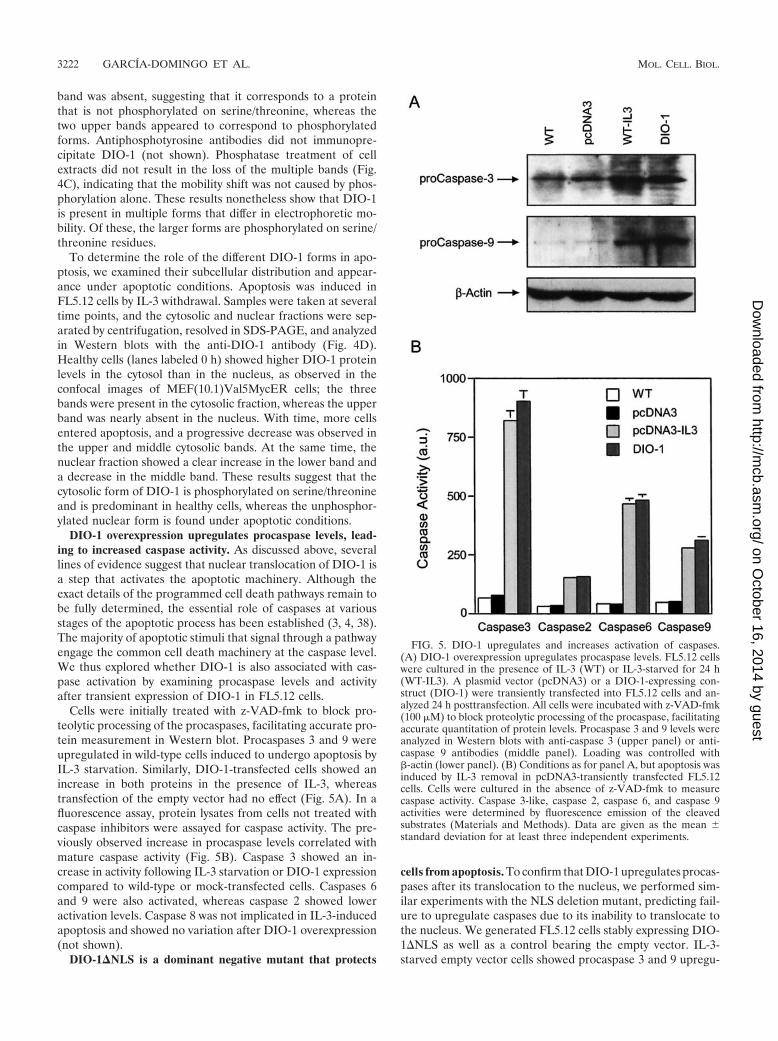
DIO-1 overexpression upregulates procaspase levels, lead-ing to increased caspase activity. As discussed above, severallines of evidence suggest that nuclear translocation of DIO-1 isa step that activates the apoptotic machinery. Although theexact details of the programmed cell death pathways remain tobe fully determined, the essential role of caspases at variousstages of the apoptotic process has been established (3, 4, 38).The majority of apoptotic stimuli that signal through a pathwayengage the common cell death machinery at the caspase level.We thus explored whether DIO-1 is also associated with cas-pase activation by examining procaspase levels and activityafter transient expression of DIO-1 in FL5.12 cells.
Cells were initially treated with z-VAD-fmk to block pro-teolytic processing of the procaspases, facilitating accurate pro-tein measurement in Western blot. Procaspases 3 and 9 wereupregulated in wild-type cells induced to undergo apoptosis byIL-3 starvation. Similarly, DIO-1-transfected cells showed anincrease in both proteins in the presence of IL-3, whereastransfection of the empty vector had no effect (Fig. 5A). In afluorescence assay, protein lysates from cells not treated withcaspase inhibitors were assayed for caspase activity. The pre-viously observed increase in procaspase levels correlated withmature caspase activity (Fig. 5B). Caspase 3 showed an in-crease in activity following IL-3 starvation or DIO-1 expressioncompared to wild-type or mock-transfected cells. Caspases 6and 9 were also activated, whereas caspase 2 showed loweractivation levels. Caspase 8 was not implicated in IL-3-inducedapoptosis and showed no variation after DIO-1 overexpression(not shown).
DIO-1�NLS is a dominant negative mutant that protects
cells from apoptosis. To confirm that DIO-1 upregulates procas-pases after its translocation to the nucleus, we performed sim-ilar experiments with the NLS deletion mutant, predicting fail-ure to upregulate caspases due to its inability to translocate tothe nucleus. We generated FL5.12 cells stably expressing DIO-1�NLS as well as a control bearing the empty vector. IL-3-starved empty vector cells showed procaspase 3 and 9 upregu-
FIG. 5. DIO-1 upregulates and increases activation of caspases.(A) DIO-1 overexpression upregulates procaspase levels. FL5.12 cellswere cultured in the presence of IL-3 (WT) or IL-3-starved for 24 h(WT-IL3). A plasmid vector (pcDNA3) or a DIO-1-expressing con-struct (DIO-1) were transiently transfected into FL5.12 cells and an-alyzed 24 h posttransfection. All cells were incubated with z-VAD-fmk(100 �M) to block proteolytic processing of the procaspase, facilitatingaccurate quantitation of protein levels. Procaspase 3 and 9 levels wereanalyzed in Western blots with anti-caspase 3 (upper panel) or anti-caspase 9 antibodies (middle panel). Loading was controlled with�-actin (lower panel). (B) Conditions as for panel A, but apoptosis wasinduced by IL-3 removal in pcDNA3-transiently transfected FL5.12cells. Cells were cultured in the absence of z-VAD-fmk to measurecaspase activity. Caspase 3-like, caspase 2, caspase 6, and caspase 9activities were determined by fluorescence emission of the cleavedsubstrates (Materials and Methods). Data are given as the mean �standard deviation for at least three independent experiments.
3222 GARCIA-DOMINGO ET AL. MOL. CELL. BIOL.
on October 16, 2014 by guest
http://mcb.asm
.org/D
ownloaded from

lation, whereas IL-3-starved cells expressing DIO-1�NLS didnot upregulate procaspase levels (Fig. 6A). In addition, cas-pase activity in IL-3-starved DIO-1�NLS-expressing cells wasclearly below the levels observed in DIO-1-transfected cells(Fig. 6B; compare to Fig. 5B). These low activity levels re-mained constant even after 48 h of IL-3 deprivation, whenmore than 60% of wild-type cells were apoptotic (not shown).The mutation thus failed to alter basal caspase levels. Takentogether, these data suggest that DIO-1 translocation to thenucleus is essential to induce upregulation of several procas-pases, to increase their activity, and to trigger the apoptoticpathway.
Given that the NLS mutation does not alter caspase levels,we tested whether it prevents apoptosis. FL5.12 wild-type cellswere IL-3 starved, and apoptosis was measured by propidiumiodide staining at 24 and 48 h postinduction (Fig. 6C). The cellsunderwent apoptotic cell death that increased with time, andsimilar results were obtained in FL5.12 pcDNA3 cells afterIL-3 withdrawal. When FL5.12 cells stably expressing DIO-1�NLS (FL5.12 DIO-1�NLS) were IL-3 starved, we observedprotection from apoptosis even after 48 h without the survivalfactor, with a reduction in the percentage of apoptotic cells ofapproximately 50% at 24 h and 66% at 48 h compared to cellsexpressing the empty vector. This behavior corresponds to adominant negative mutation that is able to block the apoptoticsignal transmitted by the wild-type form.
DISCUSSION
We report that DIO-1 translocation from the cytoplasm tothe nucleus is an important early event in activating theapoptotic machinery. DIO-1 is present in the cell in multipleforms, which can be distinguished on the basis of theirelectrophoretic mobilities and their distinct subcellular lo-calizations. We previously showed that DIO-1 mRNA andprotein levels were upregulated by serum withdrawal fromMEF(10.1)Val5MycER cells independently of p53-inducedapoptosis (9). Here we show that, in the same cell system andwith the same apoptotic stimulus, the DIO-1 gene producttranslocated to the nuclear compartment, whereas p53-in-duced cell death did not provoke alterations in its subcellularlocalization. The DIO-1 mutant lacking both NLSs (DIO-1�NLS) was unable to translocate to the nucleus or to triggerapoptosis, even in the presence of physiological death signals,and prevented IL-3 deprivation-induced cell death. The obser-
FIG. 6. DIO-1�NLS mutant is unable to upregulate and activatecaspases. (A) FL5.12 cells were untreated (WT) or stably transfectedwith an empty vector (pcDNA3) or a DIO-1�NLS-expressing con-struct. Where indicated, IL-3 was removed from culture medium, andanalysis was performed after 24 h. All cells were cultured in thepresence of z-VAD-fmk (100 �M). Lysates were resolved by SDS-PAGE, followed by Western blotting with anti-caspase 3 (upper panel)or anti-caspase 9 (middle panel) antibodies. Loading was controlledwith �-actin (lower panel). (B) Analysis of caspase activation. Samples
were collected from FL5.12 wild-type (WT) cells cultured in the pres-ence or absence of IL-3 and from the indicated stable FL5.12 cell linesin the absence of IL-3. Caspase activity was measured as in Fig. 5B.Results are expressed as the mean � standard deviation for at leastthree independent experiments. a.u., arbitrary units. (C) DIO-1�NLSis a dominant negative mutant that protects cells from growth factordeprivation-induced apoptosis. FL5.12 wild-type cells (WT) and theFL5.12pcDNA3, and FL5.12DIO-1�NLS stable cell lines were in-duced to undergo apoptosis by IL-3 removal. Cells were collected atthe times indicated, permeabilized, and propidium iodide stained (Ma-terials and Methods). Apoptosis corresponds to the amount of frag-mented DNA in the hypoploid sub-G0/G1 peak of the cell cycle. Valuesare expressed as percentages and represent the mean � standarddeviation for at least three independent experiments.
VOL. 23, 2003 DIO-1 NUCLEAR TRANSLOCATION ACTIVATES CASPASES 3223
on October 16, 2014 by guest
http://mcb.asm
.org/D
ownloaded from

vation that DIO-1 but not DIO-1�NLS overexpression up-regulated procaspase 3 and 9 levels and increased the activityof their mature forms establishes a link between the increase inDIO-1 levels and apoptosis induction (9). Although transcrip-tional activation by DIO-1 remains to be demonstrated, theneed for nuclear translocation suggests that DIO-1 actsthrough the induction of caspase promoters rather than exert-ing a direct effect on cytoplasmic caspases. These findings areconsistent with earlier studies reporting that transcriptionalactivation of caspases may be an important regulatory mecha-nism in programmed cell death (6, 12, 22, 29, 40).
One important model for caspase 9 activation involves itsrecruitment to Apaf-1 in the presence of ATP or dATP, fol-lowing cytochrome c release from mitochondria (19, 42). Otherauthors propose that caspase 9 is activated and apoptosis cantake place in the absence of cytochrome c (5, 10, 20, 32) afteroverexpression of the adaptor molecule Apaf-1 (16, 28), Nod-1(14), TMS-1 (26), or CED-4 (35, 41). Furthermore, Apaf-1mutants lacking the WD-40 repeats (WDR) constitutivelyself-associate and activate procaspase 9 independently of cy-tochrome c and dATP (13, 37). The Caenorhabditis elegansApaf-1 homolog CED-4 lacks the WDR, implying that it mayconstitutively activate the procaspase 9 homolog CED-3 andsuggesting that cytochrome c may not be required for CED-4-mediated CED-3 activation.
In our system, apoptosis induction via a DIO-1-triggeredincrease in procaspase 9 levels concurs with previous reportsthat procaspase 9 overexpression in vivo results in cell death,overriding the need for cytosolic cytochrome c (2, 14, 37). Weobserved that in MEF(10.1)Val5MycER and FL5.12 cells in-duced to apoptosis as well as in DIO-1-transfected cells, cyto-chrome c release was preceded by DIO-1 nuclear translocationand caspase upregulation (not shown). Caspase 9 activation isthus mediated by dimerization, and cytochrome c-induced re-cruitment by Apaf-1 creates high local caspase 9 concentra-tions that allow dimer-induced activation (31). This explainsthe caspase 9 activation observed prior to cytochrome c releasefrom mitochondria.
Change in subcellular distribution is an important regulatoryevent for many proteins involved in apoptosis, cell cycle, ortranscriptional regulation. Here we show that DIO-1 alsochanges its localization early in apoptosis induction and thatthis change is accompanied by an electrophoretic mobilityshift. Nonetheless, the exact nature of the signal leading tothese changes remains to be established. Coprecipitation ofdifferentially tagged proteins showed that DIO-1 forms homo-oligomers in vivo. As homo-oligomers were detected for allforms, the oligomerization state is unlikely to differ followingDIO-1 translocation to the nucleus. This has also been foundfor p53, which appears to be transported across the nuclearmembrane in a tetrameric form (11). Proteolytic processingcan also be ruled out as a regulatory mechanism, since allforms were detected with tags on either end of the protein.Although only the forms with lower electrophoretic mobilityappeared to be phosphorylated, phosphorylation alone is notthe basis of the mobility change. Additional modifications maythus be needed for the change in DIO-1 localization. In asimilar case, MDM-2-mediated nuclear export of p53 dependson multiple signals that include the DNA-binding domain,conformational change, and C-terminal ubiquitination (11,
24); ubiquitinated p53 is subsequently degraded by protea-somes in the cytosol. DIO-1 localization may be regulated in asimilar fashion. DIO-1 would thus be continuously modifiedand exported from the nucleus in healthy cells, appearing ascytosolic localization. In apoptotic cells, loss of the exportsignal would result in a net accumulation of the unmodifiedproduct in the nucleus.
DIO-1-mediated apoptosis requires caspase activation. Inthese experiments, neither procaspase upregulation nor cas-pase activation was detected after DIO-1�NLS overexpres-sion, indicating that this mutant acts in a dominant negativemanner, since it inhibits IL-3 withdrawal-induced apoptosis.This allows us to propose a model that explains the mechanismof DIO-1 activation and induction of cell death. In healthycells, the DIO-1 transcript and protein are expressed at lowbasal levels, and the protein is found in the cytosol. Followingan appropriate apoptotic stimulus, such as IL-3 starvation orc-myc induction in serum-free conditions, DIO-1 translocatesto the nucleus before the appearance of any classical indicatorsof apoptotic machinery activation, such as caspase activation,alteration of cell membrane polarity, nuclear disruption, orDNA laddering. This indicates that DIO-1 activation is a veryearly step in apoptosis induction.
In our model, the presence of DIO-1 in the nucleus leads toan increase in procaspase 3 and 9 levels, resulting in caspase 9activation. Activated caspase 9 can then activate procaspase 3.The processing of the large amounts of procaspase 3 gives riseto rapid accumulation of mature caspase 3, which acts as an-other amplification step due to caspase 3 feedback onto procas-pase 9 Asp-330 (37). Subsequent cytochrome c release frommitochondria would induce Apaf-1 oligomerization, amplify-ing the apoptotic signal. The DIO-1�NLS mutant could formstable oligomers with wild-type DIO-1 and block nuclear trans-location of the entire complex, preventing DIO-1-mediatedgene upregulation and inhibiting the cell death program.
ACKNOWLEDGMENTS
We thank B. B. Wolf, D. R. Green, T. W. Mak, and R. Hakem forgenerous gifts of reagents, A. Ruiz-Vela for excellent and continuousadvice, and A. Futterer and M. Torres, A. Ruiz-Vela, K. van Wely, andM. Campanero for critical reading of the manuscript. We also thankI. Lopez-Vidriero and M. C. Moreno-Ortiz for help with flow cytom-etry, all technical members of the department who aided with cellculture and general reagents, and C. Mark for editorial assistance.
D.G.D. is the recipient of a fellowship from the Spanish Ministeriode Educacion y Ciencia. This work was supported by grants from theMinisterio de Ciencia y Tecnología and the European Union (QLG1-CT-2001-01536). The Department of Immunology and Oncology wasfounded and is supported by the Spanish Council for Scientific Re-search (CSIC) and the Pharmacia Corporation.
REFERENCES
1. Alnemri, E. S., D. J. Livingston, D. W. Nicholson, G. Salvesen, N. A. Thorn-berry, W. W. Wong, and J. Yuan. 1996. Human ICE/CED-3 protease no-menclature. Cell 87:171.
2. Bertin, J., W. J. Nir, C. M. Fischer, O. V. Tayber, P. R. Errada, J. R. Grant,J. J. Keilty, M. L. Gosselin, K. E. Robison, G. H. Wong, M. A. Glucksmann,and P. S. DiStefano. 1999. Human CARD4 protein is a novel CED-4/Apaf-1cell death family member that activates NF-B. J. Biol. Chem. 274:12955–12958.
3. Boldin, M. P., T. M. Goncharov, Y. V. Goltsev, and D. Wallach. 1996.Involvement of MACH, a novel MORT1/FADD-interacting protease, inFas/APO-1- and TNF receptor-induced cell death. Cell 85:803–815.
4. Budihardjo, I., H. Oliver, M. Lutter, X. Luo, and X. Wang. 1999. Biochem-ical pathways of caspase activation during apoptosis. Annu. Rev. Cell Dev.Biol. 15:269–290.
3224 GARCIA-DOMINGO ET AL. MOL. CELL. BIOL.
on October 16, 2014 by guest
http://mcb.asm
.org/D
ownloaded from

5. Chauhan, D., T. Hideshima, S. Rosen, J. C. Reed, S. Kharbanda, and K. C.Anderson. 2001. Apaf-1/cytochrome c-independent and Smac-dependent in-duction of apoptosis in multiple myeloma (MM) cells. J. Biol. Chem. 276:24453–24456.
6. Dai, C., and S. B. Krantz. 1999. Interferon gamma induces upregulation andactivation of caspases 1, 3, and 8 to produce apoptosis in human erythroidprogenitor cells. Blood 93:3309–3316.
7. Earnshaw, W. C., L. M. Martins, and S. H. Kaufmann. 1999. Mammaliancaspases: structure, activation, substrates, and functions during apoptosis.Annu. Rev. Biochem. 68:383–424.
8. Ellis, R. E., J. Y. Yuan, and H. R. Horvitz. 1991. Mechanisms and functionsof cell death. Annu. Rev. Cell Biol. 7:663–698.
9. Garcia-Domingo, D., E. Leonardo, A. Grandien, P. Martinez, J. P. Albar,J. C. Izpisua-Belmonte, and C. Martinez-A. 1999. DIO-1 is a gene involvedin onset of apoptosis in vitro, whose misexpression disrupts limb develop-ment. Proc. Natl. Acad. Sci. USA 96:7992–7997.
10. Gross, A., J. Jockel, M. C. Wei, and S. J. Korsmeyer. 1998. Enforced dimer-ization of BAX results in its translocation, mitochondrial dysfunction andapoptosis. EMBO J. 17:3878–3885.
11. Gu, J., L. Nie, D. Wiederschain, and Z. M. Yuan. 2001. Identification of p53sequence elements that are required for MDM2-mediated nuclear export.Mol. Cell. Biol. 21:8533–8546.
12. Gupta, S., V. Radha, Y. Furukawa, and G. Swarup. 2001. Direct transcrip-tional activation of human caspase-1 by tumor suppressor p53. J. Biol. Chem.276:10585–10588.
13. Hu, Y., M. A. Benedict, L. Ding, and G. Nunez. 1999. Role of cytochrome cand dATP/ATP hydrolysis in Apaf-1-mediated caspase-9 activation and apo-ptosis. EMBO J. 18:3586–3595.
14. Inohara, N., T. Koseki, L. del Peso, Y. Hu, C. Yee, S. Chen, R. Carrio, J.Merino, D. Liu, J. Ni, and G. Nunez. 1999. Nod1, an Apaf-1-like activator ofcaspase-9 and nuclear factor-B. J. Biol. Chem. 274:14560–14567.
15. Jacobson, M. D., M. Weil, and M. C. Raff. 1997. Programmed cell death inanimal development. Cell 88:347–354.
16. Kamarajan, P., N. K. Sun, C. L. Sun, and C. C. Chao. 2001. Apaf-1 over-expression partially overcomes apoptotic resistance in a cisplatin-selectedHeLa cell line. FEBS Lett. 505:206–212.
17. Kerr, J. F., A. H. Wyllie, and A. R. Currie. 1972. Apoptosis: a basic biologicalphenomenon with wide-ranging implications in tissue kinetics. Br. J. Cancer26:239–257.
18. Li, H., H. Zhu, C. J. Xu, and J. Yuan. 1998. Cleavage of BID by caspase 8mediates the mitochondrial damage in the Fas pathway of apoptosis. Cell94:491–501.
19. Li, P., D. Nijhawan, I. Budihardjo, S. M. Srinivasula, M. Ahmad, E. S.Alnemri, and X. Wang. 1997. cytochrome c and dATP-dependent formationof Apaf-1/caspase-9 complex initiates an apoptotic protease cascade. Cell91:479–489.
20. Li, P. F., R. Dietz, and R. von Harsdorf. 1999. p53 regulates mitochondrialmembrane potential through reactive oxygen species and induces cyto-chrome c-independent apoptosis blocked by Bcl-2. EMBO J. 18:6027–6036.
21. Liang, P., and A. B. Pardee. 1992. Differential display of eukaryotic messen-ger RNA by means of the polymerase chain reaction. Science 257:967–971.
22. Liu, W., G. Wang, and A. G. Yakovlev. 2002. Identification and functionalanalysis of the rat caspase-3 gene promoter. J. Biol. Chem. 277:8273–8278.
23. Liu, X., C. N. Kim, J. Yang, R. Jemmerson, and X. Wang. 1996. Induction ofapoptotic program in cell-free extracts: requirement for dATP and cyto-chrome c. Cell 86:147–157.
24. Lohrum, M. A., D. B. Woods, R. L. Ludwig, E. Balint, and K. H. Vousden.
2001. C-terminal ubiquitination of p53 contributes to nuclear export. Mol.Cell. Biol. 21:8521–8532.
25. Luo, X., I. Budihardjo, H. Zou, C. Slaughter, and X. Wang. 1998. Bid, a Bcl2interacting protein, mediates cytochrome c release from mitochondria inresponse to activation of cell surface death receptors. Cell 94:481–490.
26. McConnell, B. B., and P. M. Vertino. 2000. Activation of a caspase-9-medi-ated apoptotic pathway by subcellular redistribution of the novel caspaserecruitment domain protein TMS1. Cancer Res. 60:6243–6247.
27. Muzio, M., A. M. Chinnaiyan, F. C. Kischkel, K. O’Rourke, A. Shevchenko,J. Ni, C. Scaffidi, J. D. Bretz, M. Zhang, R. Gentz, M. Mann, P. H. Krammer,M. E. Peter, and V. M. Dixit. 1996. FLICE, a novel FADD-homologousICE/CED-3-like protease, is recruited to the CD95 (Fas/APO-1) death-inducing signaling complex. Cell 85:817–827.
28. Perkins, C., C. N. Kim, G. Fang, and K. N. Bhalla. 1998. Overexpression ofApaf-1 promotes apoptosis of untreated and paclitaxel- or etoposide-treatedHL-60 cells. Cancer Res. 58:4561–4566.
29. Pohl, D., P. Bittigau, M. J. Ishimaru, D. Stadthaus, C. Hubner, J. W. Olney,L. Turski, and C. Ikonomidou. 1999. N-Methyl-D-aspartate antagonists andapoptotic cell death triggered by head trauma in developing rat brain. Proc.Natl. Acad. Sci. USA 96:2508–2513.
30. Raff, M. 1998. Cell suicide for beginners. Nature 396:119–122.31. Renatus, M., H. R. Stennicke, F. L. Scott, R. C. Liddington, and G. S.
Salvesen. 2001. Dimer formation drives the activation of the cell deathprotease caspase 9. Proc. Natl. Acad. Sci. USA 98:14250–14255.
32. Ruiz-Vela, A., G. Gonzalez de Buitrago, and C. Martinez-A. 1999. Implica-tion of calpain in caspase activation during B cell clonal deletion. EMBO J.18:4988–4998.
33. Salvesen, G. S., and V. M. Dixit. 1999. Caspase activation: the induced-proximity model. Proc. Natl. Acad. Sci. USA 96:10964–10967.
34. Salvesen, G. S., and V. M. Dixit. 1997. Caspases: intracellular signaling byproteolysis. Cell 91:443–446.
35. Shaham, S., and H. R. Horvitz. 1996. Developing Caenorhabditis elegansneurons may contain both cell-death protective and killer activities. GenesDev. 10:578–591.
36. Smith, P. K., R. I. Krohn, G. T. Hermanson, A. K. Mallia, F. H. Gartner,M. D. Provenzano, E. K. Fujimoto, N. M. Goeke, B. J. Olson, and D. C.Klenk. 1985. Measurement of protein with bicinchoninic acid. Anal. Bio-chem. 150:76–85.
37. Srinivasula, S. M., M. Ahmad, T. Fernandes-Alnemri, and E. S. Alnemri.1998. Autoactivation of procaspase-9 by Apaf-1-mediated oligomerization.Mol. Cell 1:949–957.
38. Thornberry, N. A., and Y. Lazebnik. 1998. Caspases: enemies within. Science281:1312–1316.
39. Wolf, B. B., J. C. Goldstein, H. R. Stennicke, H. Beere, G. P. Amarante-Mendes, G. S. Salvesen, and D. R. Green. 1999. Calpain functions in acaspase-independent manner to promote apoptosis-like events during plate-let activation. Blood 94:1683–1692.
40. Yakovlev, A. G., K. Ota, G. Wang, V. Movsesyan, W. L. Bao, K. Yoshihara,and A. I. Faden. 2001. Differential expression of apoptotic protease-activat-ing factor-1 and caspase-3 genes and susceptibility to apoptosis during braindevelopment and after traumatic brain injury. J. Neurosci. 21:7439–7446.
41. Yuan, J., and H. R. Horvitz. 1992. The Caenorhabditis elegans cell death geneCED-4 encodes a novel protein and is expressed during the period of ex-tensive programmed cell death. Development 116:309–320.
42. Zou, H., W. J. Henzel, X. Liu, A. Lutschg, and X. Wang. 1997. Apaf-1, ahuman protein homologous to C. elegans CED-4, participates in cytochromec-dependent activation of caspase-3. Cell 90:405–413.
VOL. 23, 2003 DIO-1 NUCLEAR TRANSLOCATION ACTIVATES CASPASES 3225
on October 16, 2014 by guest
http://mcb.asm
.org/D
ownloaded from