Modelling ascorbic acid thermal degradation and browning in orange juice under aerobic conditions
Upload
independentCategory
view
6download
0
RESEARCH
Ascorbic Acid Infusion Blunts CD40L Upregulation in PatientsUndergoing Coronary StentPasquale Pignatelli,1 Gaetano Tanzilli,2 Roberto Carnevale,1 Serena Di Santo,1 Lorenzo Loffredo,1
Andrea Celestini,1 Marco Proietti,1 Priscilla Tovaglia,1 Enrico Mangieri,2 Stefania Basili1 & Francesco Violi1
1 Prima Clinica Medica—Department of Experimental Medicine, University of Rome “La Sapienza,” Rome, Italy2 Department of the Heart and Great Vessels Attilio Reale, University of Rome “La Sapienza,” Rome, Italy
KeywordsAscorbic acid; CD40L; Oxidative stress;
Percutaneous coronary interventions, PCI;
Platelets.
CorrespondenceProfessor Francesco Violi,
Prima Clinica Medica,
Viale del Policlinico 155, Roma 00161,
Italy.
Tel.: +39064461933;
Fax: +39064461933;
E-mail: [email protected]
doi: 10.1111/j.1755-5922.2010.00168.x
SUMMARY
Objectives: To reduce the increase of oxidative stress and the upregulation ofCD40L during stenting procedure using ascorbic acid infusion. Background:CD40L upregulation occurring after coronary Percutaneous Coronary In-tervention predicts vascular events but the underlying mechanism is stillunclear. Methods: Fifty-six patients undergoing elective coronary stentingwere randomly allocated to intravenous infusion of the antioxidant ascorbicacid or placebo. Platelet CD40L and plasma levels of soluble CD40L and of8-hydroxy-2′-deoxyguanosine, a marker of oxidative stress, were measuredbefore and after coronary stenting. In vitro study was also done to measurereactive oxidant species and CD40L expression in platelets exposed to anoxia-reoxygenation. Results: Placebo-treated patients showed a significant increaseof platelet CD40L, soluble CD40L and 8-hydroxy-2′-deoxyguanosine com-pared to baseline values. Patients given ascorbic acid showed no change ofsoluble CD40L and platelet CD40L but a significant decrease of 8-hydroxy-2′-deoxyguanosine. After 60 and 120 min, soluble CD40L, platelet CD40Land 8-hydroxy-2′-deoxyguanosine were significantly lower in the ascorbicacid-treated group compared to the placebo-treated one. A significant correla-tion between platelet CD40L and soluble CD40L and between soluble CD40Land 8-hydroxy-2′-deoxyguanosine was observed. Platelets, in vitro exposed toanoxia-reoxygenation, had a burst of ROS and an upregulation of CD40L thatwere inhibited by ascorbic acid or apocynin, an inhibitor of NADPH oxidase.Conclusions: This study shows that in patients undergoing coronary stentingCD40L is upregulated with a mechanism which is likely mediated by oxidativestress.
Introduction
Despite intracoronary stents improved survival in pa-tients with coronary heart disease who underwent per-cutaneous coronary intervention (PCI), stent thrombosis(2.03%), and vascular events such as myocardial infarc-tion, stroke, and vascular death (11.9%) are still observedin a high percentage of patients undergoing PCI [1–3].Platelets have a key role in such poor outcomes as shownby the fact that reduction of vascular outcomes is relatedto the rate of platelet inhibition [3–5]. Knowledge of the
mechanisms of coronary stent-related platelet activationis still unclear.
CD40L is a cytokine of the tumor necrosis factor alpha(TNF)α super-family primarily identified in the immuno-system cells [6]. CD40L is expressed on platelet surfaceupon agonist stimulation and is released in the circu-lation as soluble form (sCD40L) [7]; more than 95%of the circulating CD40L stems from platelet activation[8]. CD40L is a potent inflammatory and procoagulantmolecule that is over-expressed in a series of clinical set-tings characterized by accelerated atherosclerosis, such as
Cardiovascular Therapeutics 29 (2011) 385–394 c© 2010 Blackwell Publishing Ltd 385

Ascorbic Acid, CD40L, and PCI P. Pignatelli et al.
hypercholesterolemia [9], diabetes [10], and smoking[11]. Prospective studies have shown that CD40L is pre-dictive of poor vascular outcome in patients with acutevascular events or at risk of both thromboembolic andatherothrombotic diseases [12–14]. Furthermore, in pa-tients undergoing PCI the increase of CD40L immedi-ately after stenting procedure seems to be associated withrestenosis [5,15]. Thus comprehension of the mechanismleading to the increase of CD40L could be relevant notonly to provide a mechanistic insight but also to eventu-ally develop novel therapeutic strategies. We, as well asothers, have reported that oxidative stress plays a majorrole in the upregulation of CD40L [16–18]. Interestingly,in patients undergoing PCI, a significant increase of ox-idative stress has been reported as a likely consequenceof the ischemia-reperfusion process occurring in this set-ting [19–22]. We, therefore, speculated that the increaseof oxidative stress may be pertinent in the upregulationof CD40L and that the use of an antioxidant during stent-ing procedure could minimize this phenomenon. To ex-plore the validity of such hypothesis we planned a pi-lot study consisting in measuring CD40L before and aftercoronary stenting in patients treated or not with ascor-bic acid, a known antioxidant that scavenges superoxideradicals [23]. This approach was based on the results of aprevious study demonstrating that intravenous infusionof 1 g ascorbic acid is associated with a significant decreaseof circulating sCD40L [24]. As ischemia-reperfusion phe-nomenon could be a part of the injury linked to PCI, weperformed in vitro study to see if anoxia-reoxygenationof platelets was associated with oxidative-stress mediatedCD40L upregulation.
Materials and Methods
Interventional Study
Study Population and Study Design
This prospective randomized pilot study included patients≥18 years of age with clinically stable class I or II ef-fort angina, a positive functional study for myocardialischemia and a single de novo lesion in a native coro-nary artery referred to Department of the Heart and GreatVessels “Attilio Reale,” University of Rome “La Sapienza,”Italy between June 2008 and January 2009.
Angiographic inclusion criteria were all type A ortype B coronary lesions, as described by the AmericanCollege Cardiology (ACC)/American Heart Association(AHA) Task Force [25], a target vessel reference diameterof 2.5–3.5 mm, a lesion length ≤33 mm, which would becovered with a single medicated stent, diameter stenoses≥70% and <100%, and a Thrombolysis In MyocardialInfarction grade >1 flow.
Procedural exclusion criteria were a target vessel diam-eter <2.5 mm or >4.0 mm (14 patients), type C lesions(four patients) and bifurcation lesions (four patients).Clinical exclusion criteria were: contraindication to as-pirin or clopidrogel (two patients), previous myocardialinfarction (three patients), graft vessel disease (four pa-tients), platelet count <100,000/mm3 (two patients), his-tory of bleeding diathesis (two patients), renal dysfunc-tion (creatinine levels >1.5 mg/dL) (one patient).
The study was approved by the University of Rome “LaSapienza” Ethical Committee, and written informed con-sent was obtained from all patients.
Fifty-six consecutive patients [47 male and 9 female,mean ages 67 (50–84) years] who met inclusion and ex-clusion criteria entered the study.
The PCI was carried out according to internationalguidelines, using a standard technique, through thefemoral route [25]. Systematic stent implantation wasachieved in all patients. The sheath was removed imme-diately at the end of the procedure in all cases. Routinecare before and after the procedure was undertaken forall patients, including pretreatment clopidogrel (600-mginitial bolus) 12 h before the procedure followed by 75 mgdaily for at least 1 year. In addition, all patients receivedaspirin, 100 mg daily, for at least 1 week before, with adose administered 12 h before stenting.
Serum cardiac-Troponin I (cTpI) was measured at base-line before the procedure, every 6 h over the next 2 daysand thereafter if abnormal values were present. Left ven-tricular ejection fraction (LVEF) was calculated by the bi-plane Simpson’s rule, as recommended by the AmericanSociety of Echocardiography at baseline and at 72 h afterPCI by operators unaware of the treatment allocation.
Angioplasty Procedure
Preparation and percutaneous access were performed ac-cording to standard hospital procedures. After percu-taneous access was obtained, an intravenous bolus of5000 U of unfractionated heparin was administered, withsufficient supplements (if necessary) to maintain an ac-tivated clotting time (ACT) ≥250 seconds during inter-ventions. A baseline angiography of the involved vesselwas performed in at least two near orthogonal views thatshowed the target lesion free of foreshortening or vesseloverlap, using a 6 F diagnostic catheter. The angiogramsincluded at least 2 cm of catheter to allow for accuratequantitative coronary angiographic measurements.
Selective angiography was performed with an auto-matic injector (ACIST HD101, Eden Prairie, Minnesota),by using a total volume of 10 mL iopromide (Ultravist370, Schering AG, Berlin, Germany), at a rate of 2.0 mL/sfor left coronary arteries, and a total volume of 8 mL
386 Cardiovascular Therapeutics 29 (2011) 385–394 c© 2010 Blackwell Publishing Ltd

P. Pignatelli et al. Ascorbic Acid, CD40L, and PCI
iopromide at a rate of 1.0 mL/s for right coronary ar-teries, at 450 PSI. Following identification of the tar-get lesion that meets all eligibility criteria, those patientswho continue to meet eligibility criteria were randomizedto placebo or ascorbic acid administration and receiveda unique study identification code. Thus, after baselinecollection of blood samples a sealed, opaque envelopescontaining a computer-generated random sequence wereused for randomization to an intravenous infusion ofascorbic acid (1 g/L at 24 mg/min) or placebo (salinesolution).
The target lesion was crossed with a 0.014 exchange-length guide wire and a single predilatation with an ap-propriately sized balloon was performed by inflating theballoon to the nominal pressure over a 15 sec period.Within 1 min from balloon dilatation a sirolimus-elutingstent (SES; CypherTM, Cordis, Johnson & Johnson) wasimplanted. Stent deployment was achieved with highpressure balloon inflation (more than 15 atm) lasting10 sec without any post-dilatation. No direct stenting wasperformed.
Laboratory Analysis
To ensure blind analysis, cardiologists sent tube identifiedby numerical code to the laboratory where biologists per-form analytical tests. The randomization list was unveiledafter that the analytical phase was terminated.
Blood Sampling Protocol for Platelet Isolation
Blood samples were drawn from an antecubital veinwith a 21-gauge needle and then mixed in a tube with0.13 mM/l sodium citrate (ratio 9:1) for plasma prepara-tion. Samples were collected before and 60, 120 min af-ter the procedure. To obtain platelet rich plasma (PRP)sample was centrifuged (15 min at 180 g) as previouslyreported [26].
sCD40L, 8-hydroxy-2′-deoxyguanosine (8OH-dG),high sensitivity C-reactive protein (hs-CRP), TNFα, andTroponin.
Blood samples were immediately centrifuged at 300 gfor 20 min at 4◦C, and the supernatant was collectedand stored at −80◦C until measurement. Plasma levels ofsCD40L, 8OH-dG and serum levels of hs-CRP were mea-sured with a commercial immunoassay (Tema Ricerches.r.l., Bologna, Italy). Intra-assay and interassay coeffi-cients of variation were 5 and 7% for sCD40L, 2.1 and4.5% for 8OH-dG, 8.3 and 7.8% for hsPCR, respectively.TNFα (Quantikine R&D Systems, Minneapolis, MN, USA)were measured with a commercial immunoassay (Intra-assay and interassay coefficients of variation were 5.3 and6.8%). cTp I levels were measured using an automated
enzyme immuno-assay system (Dimension RXL MAX,Siemens Healthcare diagnostic, Germany) with upperlimit of normal being 0.05 ng/mL in our laboratory.
Flow Cytometric Analysis of Platelet CD40LExpression
CD40L expression on platelet membranes was ana-lyzed with specific fluorescein isothiocyanate-labeledmonoclonal antibodies (mAbs; anti-CD40L antibody byBeckman Coulter). The binding of the primary antibodywas detected with a secondary antibody (anti-mouseIgG-FITC by Beckman Coulter). In all assays, an irrel-evant isotype-matched antibody was used as a negativecontrol.
Blood samples were centrifuged to obtain PRP and im-mediately fixed with tromboFix platelet stabilizer (Beck-man Coulter) (30 min at room temperature before anyother treatment). After fixing procedure, twenty micro-liters of mAb were added to 200 μL of platelet suspension(2 × 10⁸/mL). The unbound mAb was removed by addi-tion of 0.1% bovine serum albumin–phosphate bufferedsaline (PBS) and centrifugation at 300 g for 3 min (twice).Fluorescence intensity was analyzed by an Epics XL-MCLcytometer (Coulter Electronics) equipped with an argonlaser at 488 nm. For every histogram, 50,000 plateletswere counted to determine the proportion of positiveplatelets. Antibody reactivity is reported as mean fluo-rescence. Intra-assay coefficient of variation was 5%. Totest the effect of fixing procedure on the functional in-tegrity of platelet unfixed and fixed samples were in-cubated after centrifugation with mAB anti-PAC1-FITC(Beckman Coulter) (an antibody that recognizes an epi-tope on the glycoprotein IIb/IIIa of activated platelets)showing that PAC1 binding did not change before andafter centrifugation.
In Vitro Study
Blood Sampling Protocol and Platelet Preparation
Five non-smoking healthy volunteers (three males andtwo females, mean age 60 ± 3 years) who had not in-gested any drug known to interfere with platelet func-tion for at least 15 days, after having given informed con-sent, were enrolled for the study. All subjects had fastedfor 12 h before venipuncture and blood samples weredrawn between 8:00 and 9:00 am. Platelet suspensionwas obtained as described earlier with a final platelet con-centration of 2 × 10⁸/mL. Platelets were incubated withapocynin (25, 50, and 100 μM), an inhibitor of NADPHoxidase, Vitamin C (25, 50, and 100 μM) and apoc-ynin (25, 50, and 100 μM) plus Vitamin C (25, 50, and
Cardiovascular Therapeutics 29 (2011) 385–394 c© 2010 Blackwell Publishing Ltd 387

Ascorbic Acid, CD40L, and PCI P. Pignatelli et al.
100 μM). To keep platelets in anaerobic conditions, sam-ples were purged with a gentle stream of pure nitrogengas up to 20 min. Platelets were reoxygenated by re-exposure to atmospheric conditions. Platelets exposed toa gentle stream of room air were used as control.
Flow Cytometric Analysis of Platelet ReactiveOxidant Species (ROS) Formation and CD40L
To detect the platelet formation of ROS, the propertyof DCF-DA was used. This molecule rapidly diffusesacross cell membranes and is then trapped within thecell via a deacetylation reaction. In the presence ofhydrogen peroxide, this compound is oxidized to thehighly fluorescent dichlorofluorescein (DCF). DCFH-DAwas added to platelet suspension (final concentration:40 μmol/L) immediately after the anoxia phase; fluores-cence intensity was analyzed on an Epics XL-MCL cy-tometer (Coulter Electronics) equipped with an argonlaser at 510–550 nm (green). For every histogram, 50,000platelets were counted to determine the proportion ofpositive platelets. The fluorescent signal generated by theprobe was expressed as mean fluorescence. CD40L ex-pression on platelet membrane was analyzed as earlierdescribed.
Intra-assay coefficient of variation was 5%.
Statistical Analysis
Two-sided t-test was used to compare means. Re-sults were confirmed by nonparametric tests as Mann-Whitney U-test. Pearson chi-square test was used to com-pare proportions. Friedman testing was performed forevaluating differences over time within the group. Dif-ferences between paired data were tested by Wilcoxonrank testing. The Spearman correlation coefficient wascalculated to assess the correlation of absolute changefrom the baseline of sCD40L plasma levels (�sCD40L),8-OHdG plasma levels (�8-OHdG), platelet CD40Lexpression (�platelet CD40L expression), and LVEF(�LVEF). Data are presented as mean (1 SD) or as medianand interquartile range (IQR) (25th, 75th percentile).Statistical significance was defined at P < 0.05. Statisti-cal analysis was performed with SPSS 13.0 software forWindows.
Sample size determination: we computed the mini-mum sample size with respect to a two-tailed one-sampleStudent t-test with Welch correction, considering a (1)difference for CD40L variation to be detected between thepatients treated or not with Vitamin C |δ| ≥ 1.5, (2) stan-dard deviations SD = 1 for the control group and 1.2 forthe Vitamin C group, (3) type I error probability α = 0.05and power 1-β = 0.90. This resulted in a minimum sam-
ple size of n = 10 for control and n = 12 for Vitamin Cgroup, which are increased to n > 20.
Results
Characteristics of the Study Population
Table 1 shows baseline demographic and clinical charac-teristics of included patients. Angiographic and procedu-ral data are shown in Table 2. The two groups assignedto ascorbic acid or placebo did not differ in terms of car-diovascular risk factors and pharmacological therapy. Theimplantation of single stent was successful in all patientswith complete covering of the vessel lesion length andwithout visual residual stenosis within the stent. Fur-thermore, angiographic images obtained following stentdeployment did not show signs of endothelial dissectionin the areas proximal or distal to the stent struts. Noside effects were observed during or after ascorbic acidor placebo infusion.
Baseline mean values of LVEF were similar in bothVitamin C and control groups (52.3 ± 4.3% vs. 53.7% ±3.9%, P = ns).
Ascorbic acid assigned group showed, at the baseline,similar values of sCD40L (2.3 ± 1.4 vs. 2.3 ± 1.2 ng/mL,P = 0.959), 8-OHdG (3.7 ± 1.3 vs. 3.7 ± 1.1 ng/mL,P = 0.373), hs-CRP [Median (IQR):1.0 (0.71–1.90) vs.1.25 (0.80–2.00) mg/L, P = 0.451) and TNF-α [42.5(35.0–50.0) vs. 40.0 (40.0–50.0) pg/mL, P = 0.735) com-pared to placebo group. The platelet CD40L expressionwas similar the in two groups (4.2 ± 0.88 MF in ascorbicacid assigned group and 4.3 ± 0.78 MF in placebo group,P = 0.0724), the apparent trend in differences between
Table 1 Baseline clinical characteristics
Placebo P-value Vitamin C
(n = 28) (n = 28)
Age (years) 68 ± 9 >0.05 66 ± 8
Male, n (%) 23 (82) >0.05 24 (86)
Familiarity, n (%) 17 (61) >0.05 14 (50)
Hypertension, n (%) 20 (71) >0.05 20 (71)
Dyslipidemia, n (%) 20 (71) >0.05 19 (68)
Current smoker, n (%) 13 (46) >0.05 11 (39)
Diabetes mellitus, n (%) 15 (54) >0.05 18 (64)
Pharmacological therapy
Oral hypoglycaemic, n (%) 12 (43) >0.05 11 (39)
Insulin treatment, n (%) 2 (7) >0.05 3 (10)
Statins, n (%) 19 (68) >0.05 16 (57)
Nitrates, n (%) 13 (46) >0.05 16 (57)
ACE-inhibitors, n (%) 15 (54) >0.05 12 (43)
β-blockers, n (%) 9 (32) >0.05 10 (36)
Ca-antagonists, n (%) 7 (25) >0.05 6 (21)
388 Cardiovascular Therapeutics 29 (2011) 385–394 c© 2010 Blackwell Publishing Ltd

P. Pignatelli et al. Ascorbic Acid, CD40L, and PCI
Table 2 Angiographic characteristics and quantitative angiographic measurements of included subjects
Placebo (n = 28) P-value Vitamin C (n = 28)
Target vessel Left anterior descending artery, n (%) 18(64) >0.05 15 (54)
Left circumflex artery, n (%) 3 (11) >0.05 6 (21)
Right coronary artery, n (%) 7 (25) >0.05 7 (25)
Procedural data Diameter stenosis (%) 80.1 ± 9.7 >0.05 83.5 ± 11.5
Stent diameter (mm) 3.0 ± 0.39 >0.05 3.1 ± 0.37
Stent length (mm) 19.8 ± 7.1 >0.05 20.8 ± 7.5
Balloon pressure (atm) 16.5 ± 1.4 >0.05 16.9 ± 1.1
the two groups is well under the coefficient of variationof the method.
There were no adverse events during hospitalization inboth Vitamin C and placebo group.
Interventional Study
Serum cardiac-Troponin I and Left VentricularEjection Fraction
The median absolute increase after PCI in serum cTpIlevel was similar between controls and ascorbic acidgroup [Median (IQR): 0.027 (0.05–0.032) vs. 0.008(0.02–0.013) ng/mL, respectively, P = 0.0832].
After the intervention, LVEF improved in both groupswith a higher increase in Vitamin C treated patients(58.3 ± 2.9%) compared to the placebo one (54.1 ±4.7%) (P < 0.03). Vitamin C-treated patients showed agreater improvement of LVEF after procedure comparedto that observed in the control group (P < 0.01).
sCD40L Plasma Levels
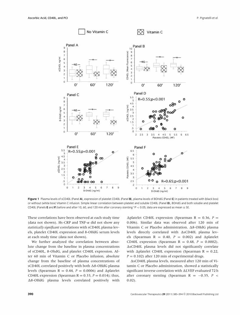
Compared to baseline, no statistically significant changesof sCD40L was found after 60 (2.2 ± 1.1 ng/mL, P =0.269) and 120 min (2.4 ± 1.0 ng/mL, P = 0.738) ofVitamin C infusion; conversely, a significant increase ofsCD40L was observed after 60 (3.2 ± 1.5 ng/mL, P =0.0007) and 120 min (3.4 ± 1.7 ng/mL, P = 0.0003) ofplacebo infusion (Figure 1, Panel A). Comparing sCD40Lvalues at 60 and 120 min after balloon inflation, theascorbic acid-treated group showed a significant lowervalue of sCD40L in respect to placebo group (P = 0.0057and P = 0.016, respectively) (Figure 1, Panel A). Similarresults were obtained in the subgroup of diabetic patients(n = 33; 60 min: P = 0.01; 120 min: P = 0.06).
Platelet CD40L Expression
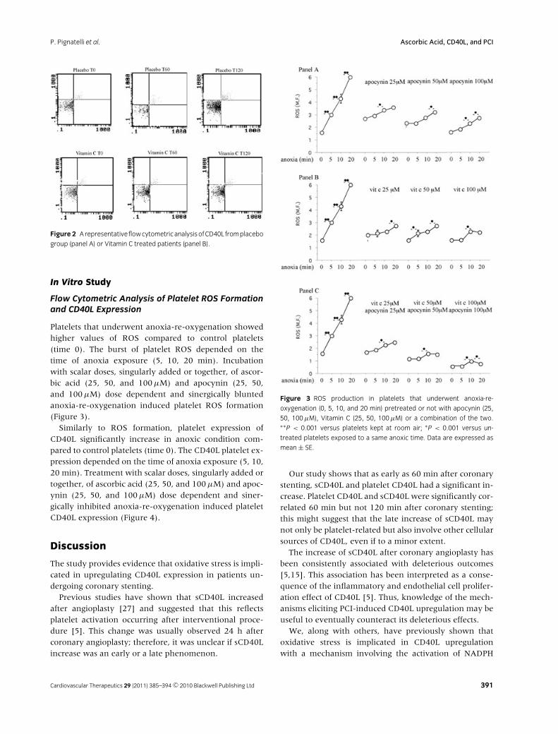
Compared to baseline, platelet expression of CD40L didnot significantly differ at 60 (3.8 ± 1.3 MF, P = 0.077)
and 120 min (3.8 ± 1.1 MF, P = 0.307) after bal-loon inflation in Vitamin C treated group (Figure 1,Panel B) (Figure 2, Panel A). On the other hand, in theplacebo group, at 60 (5.1 ± 1.3 MF, P = 0.005) and120 min (5.4 ± 1.2 MF, P = 0.0002) after balloon infla-tion, platelet CD40L expression was higher than baseline(Figure 1, Panel B) (Figure 2, Panel B). A significantdifference between groups was observed at 60 (P =0.0008) and 120 (P < 0.0001) min after balloon inflation(Figure 1, Panel B). Similar results were obtained in thesubgroup of diabetic patients (n = 33; 60 min: P = 0.036;120 min: P = 0.001).
8-hydroxy-2′-deoxyguanosine, hs-CRP, and TNF-α
Compared to baseline, 8-OHdG plasma levels significantlydecreased at 60 (2.6 ± 1.1, ng/mL, P < 0.0001) and120 min (2.7 ± 0.87 ng/mL, P = 0.0003) after ballooninflation in Vitamin C treated group (Figure 1, Panel C).In contrast, the placebo group at 60 (4.2 ± 1.1 ng/mL, P =0.045) and 120 min (4.6 ± 1.0 ng/mL, P = 0.003) afterballoon inflation, showed 8-OHdG plasma levels higherthan baseline (Figure 1, Panel C). A significant differencebetween groups was observed at 60 (P < 0.0001) and120 min (P < 0.0001) after balloon inflation (Figure 1,Panel C).
Hs-CRP [Vitamin C: 1.0 (0.67–1.60) and 1.37(1.0–2.0) mg/L, P > 0.05; Placebo: 1.30 (0.75–2.0) and1.25 (0.95–2.10) mg/L, P > 0.05] and TNF-α [Vitamin C:45.0 (40.0–60.0) and 43.5 (36.5–58.5) pg/mL, P > 0.05;Placebo: 40.0 (40.0–50.0) and 46.5 (40.0–58.0) pg/mL,P > 0.05] did not show any significant changes in bothgroups of treatment at 60 and 120 min after PCI.
Bivariate Analysis
Simple linear regression analysis showed an overall corre-lation between sCD40L plasma levels and platelet CD40Lexpression, sCD40L and 8-OHdG, and between 8-OHdGand CD40L platelet expression (Figure 1, Panels D–F).
Cardiovascular Therapeutics 29 (2011) 385–394 c© 2010 Blackwell Publishing Ltd 389
Ascorbic Acid, CD40L, and PCI P. Pignatelli et al.
Figure 1 Plasma levels of sCD40L (PanelA), expression of platelet CD40L (Panel B), plasma levels of 8OHdG (Panel C) in patients treated with (black box)
or without (white box) Vitamin C infusion. Simple linear correlation between platelet and soluble CD40L (Panel D), 8OHdG and both soluble and platelet
CD40L (Panels E and F) before and after 10, 60, and 120 min after coronary stenting ∗P < 0.05; data are expressed as mean ± SE.
These correlations have been observed at each study time(data not shown). Hs-CRP and TNF-α did not show anystatistically significant correlations with sCD40L plasma lev-els, platelet CD40L expression and 8-OHdG serum levelsat each study time (data not shown).
We further analyzed the correlation between abso-lute change from the baseline in plasma concentrationsof sCD40L, 8-OhdG, and platelet CD40L expression. Af-ter 60 min of Vitamin C or Placebo infusion, absolutechange from the baseline of plasma concentrations ofsCD40L correlated positively with both �8-OHdG plasmalevels (Spearman R = 0.44, P = 0.0006) and �plateletCD40L expression (Spearman R = 0.33, P = 0.014); thus,�8-OHdG plasma levels correlated positively with
�platelet CD40L expression (Spearman R = 0.36, P =0.006). Similar data was observed after 120 min ofVitamin C or Placebo administration. �8-OHdG plasmalevels directly correlated with �sCD40L plasma lev-els (Spearman R = 0.40, P = 0.002) and �plateletCD40L expression (Spearman R = 0.48, P = 0.0002).�sCD40L plasma levels did not significantly correlatewith �platelet CD40L expression (Spearman R = 0.22,P = 0.102) after 120 min of experimental drugs.
�sCD40L plasma levels, measured after 120 min of Vi-tamin C or Placebo administration, showed a statisticallysignificant inverse correlation with �LVEF evaluated 72 hafter coronary stenting (Spearman R = −0.35, P <
0.02).
390 Cardiovascular Therapeutics 29 (2011) 385–394 c© 2010 Blackwell Publishing Ltd
P. Pignatelli et al. Ascorbic Acid, CD40L, and PCI
Figure 2 ArepresentativeflowcytometricanalysisofCD40L fromplacebo
group (panel A) or Vitamin C treated patients (panel B).
In Vitro Study
Flow Cytometric Analysis of Platelet ROS Formationand CD40L Expression
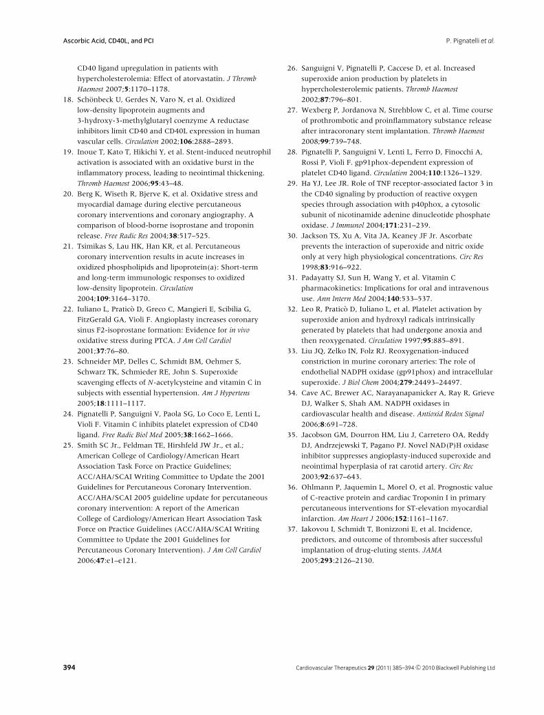
Platelets that underwent anoxia-re-oxygenation showedhigher values of ROS compared to control platelets(time 0). The burst of platelet ROS depended on thetime of anoxia exposure (5, 10, 20 min). Incubationwith scalar doses, singularly added or together, of ascor-bic acid (25, 50, and 100 μM) and apocynin (25, 50,and 100 μM) dose dependent and sinergically bluntedanoxia-re-oxygenation induced platelet ROS formation(Figure 3).
Similarly to ROS formation, platelet expression ofCD40L significantly increase in anoxic condition com-pared to control platelets (time 0). The CD40L platelet ex-pression depended on the time of anoxia exposure (5, 10,20 min). Treatment with scalar doses, singularly added ortogether, of ascorbic acid (25, 50, and 100 μM) and apoc-ynin (25, 50, and 100 μM) dose dependent and siner-gically inhibited anoxia-re-oxygenation induced plateletCD40L expression (Figure 4).
Discussion
The study provides evidence that oxidative stress is impli-cated in upregulating CD40L expression in patients un-dergoing coronary stenting.
Previous studies have shown that sCD40L increasedafter angioplasty [27] and suggested that this reflectsplatelet activation occurring after interventional proce-dure [5]. This change was usually observed 24 h aftercoronary angioplasty; therefore, it was unclear if sCD40Lincrease was an early or a late phenomenon.
Figure 3 ROS production in platelets that underwent anoxia-re-
oxygenation (0, 5, 10, and 20 min) pretreated or not with apocynin (25,
50, 100μM), Vitamin C (25, 50, 100μM) or a combination of the two.∗∗P < 0.001 versus platelets kept at room air; ∗P < 0.001 versus un-
treated platelets exposed to a same anoxic time. Data are expressed as
mean ± SE.
Our study shows that as early as 60 min after coronarystenting, sCD40L and platelet CD40L had a significant in-crease. Platelet CD40L and sCD40L were significantly cor-related 60 min but not 120 min after coronary stenting;this might suggest that the late increase of sCD40L maynot only be platelet-related but also involve other cellularsources of CD40L, even if to a minor extent.
The increase of sCD40L after coronary angioplasty hasbeen consistently associated with deleterious outcomes[5,15]. This association has been interpreted as a conse-quence of the inflammatory and endothelial cell prolifer-ation effect of CD40L [5]. Thus, knowledge of the mech-anisms eliciting PCI-induced CD40L upregulation may beuseful to eventually counteract its deleterious effects.
We, along with others, have previously shown thatoxidative stress is implicated in CD40L upregulationwith a mechanism involving the activation of NADPH
Cardiovascular Therapeutics 29 (2011) 385–394 c© 2010 Blackwell Publishing Ltd 391

Ascorbic Acid, CD40L, and PCI P. Pignatelli et al.
Figure 4 CD40L expression in platelets that underwent anoxia-re-
oxygenation (0, 5, 10, and 20 min) pretreated or not with apocynin (25,
50, 100μM), Vitamin C (25, 50, 100μM) or a combination of the two.∗∗P < 0.001 versus platelets kept at room air; ∗P < 0.001 versus un-
treated platelets exposed to a same anoxic time. Data are expressed as
mean ± SE.
oxidase [28,29]. Therefore, in patients lacking this en-zyme, platelet CD40L was down-regulated and normalplatelets, incubated in vitro with apocynin, an inhibitorof NADPH oxidase, showed lower expression of CD40L[28]. Also, intravenous infusion of 1 g ascorbic acid re-sulted in a significant reduction of both platelet CD40Land sCD40L [24].
Using different markers of oxidative stress, previousstudies showed that oxidative stress increased after PCIas a likely consequence of the burst of ROS occurring af-ter ischemia-reperfusion [19–22]. Based on these data weplanned a pilot study to see if ascorbic acid infusion wasable to counteract the increase of CD40L occurring afterPCI. We observed that such treatment was able to pre-vent both sCD40L and platelet CD40L expression occur-ring after coronary stenting with a mechanism that waslikely dependent upon its antioxidant effect. Thus intra-venous infusion of ascorbic acid was associated with a
significant reduction of 8-OHdG plasma levels. Even if ithas been underscored that ascorbic acid may exert an-tioxidant effect in vivo only at millimolar concentration[30], it is noteworthy that such concentration can beachieved in vivo when ascorbic acid is intravenously given[31]. So, it is plausible that the decrease of serum 8-OHdGmight be attributable to its antioxidant effect.
Together, these data lead to hypothesize that the burstof ROS occurring after coronary stenting may triggerplatelets CD40L upregulation. This hypothesis is con-sistent with the fact that sCD40L and platelet CD40Lshowed a significant correlation with 8-OHdG levels.
To further corroborate the role of ROS in upregulatingplatelet CD40L, the ischemia-reperfusion phenomenonoccurring after coronary stenting was mimicked in vitroby exposing platelets to anoxia-re-oxygenation. This phe-nomenon has been proved to be associated with NADPHoxidase-dependent ROS formation [32,33]. Through thisexperiment we demonstrated that platelets CD40L up-regulation occurs after platelet exposure to anoxia-re-oxygenation and that such effect was likely mediatedby NADPH oxidase-generated platelet ROS. Thus, aninhibitor of NADPH oxidase as well as ascorbic acid,prevented both platelet ROS and CD40L upregulationelicited by anoxia-re-oxygenation in a dose dependentand sinergical manner. As NADPH oxidase is the mostimportant cellular producer of superoxide anion [34] andascorbic acid is an antioxidant able to scavenge it [24], it isconceivable that anoxia-re-oxygenation-induced plateletCD40L upregulation is mediated by a burst of superoxideanion.
The study has limitations and implications. Even if wepostulate that NADPH oxidase- generating platelet ROSis responsible for enhanced oxidative stress and in turnplatelet CD40L upregulation, our study did not provideevidence that this enzymatic pathway is actually activatedafter coronary stenting. Despite the fact that experimen-tal study seems to support such hypothesis [35], furtherinvestigation in humans is necessary to explore the roleof NADPH oxidase in the ROS formation occurring aftercoronary stenting.
The lack of correlation between hs-CRP and CD40L ob-served in the present study could be explained by a dif-ferent pattern of platelet activation and inflammation inthe early phase after coronary stenting. In fact CRP serumlevels reach peak values at mean interval of 49 h aftercoronary stenting [36] while oxidative stress and CD40Lhave a much earlier increase.
We observed that LVEF, a good predictor of stentthrombosis [37], significantly and inversely correlatedwith plasma sCD40L after PCI and was moderately im-proved in Vitamin C treated group. Nevertheless, a longerfollow-up and a larger study need to be planned to
392 Cardiovascular Therapeutics 29 (2011) 385–394 c© 2010 Blackwell Publishing Ltd

P. Pignatelli et al. Ascorbic Acid, CD40L, and PCI
confirm the clinical validity of ascorbic acid infusion inthis setting.
As we used a standardized treatment including dual an-tiplatelet treatment with clopidogrel and aspirin, the en-hanced level of sCD40L after stenting procedure suggeststhat such therapeutic approach is insufficient to counter-act PCI-induced platelet CD40L release and that. ascorbicacid may represent a simple and cheap approach to coun-teract it.
In conclusion, we provide evidence that platelet CD40Lupregulation occurring after coronary stenting could beoxidative stress-mediated and could be dependent uponNADPH oxidase activation. Interventional trials withascorbic acid will be needed to evaluate if intravenous in-fusion of ascorbic acid could reduce vascular outcomes inpatients undergoing coronary stenting.
Acknowledgments
We are grateful to Alessio Farcomeni for support ofthe statistical analyses. We also thank Dr AndreaBrattini for the editing support. Grant from AteneoFederato 2007.(University of Rome La Sapienza) to F.V.
Author Contributions
Pasquale Pignatelli: study design and coordination.Gaetano Tanzilli: patients’ recruitment.Roberto Carnevale: laboratory experimental procedures.Serena Di Santo: laboratory experimental procedures.Lorenzo Loffredo: statistical analysis.Andrea Celestini: paper preparation, data elaboration.Marco Proietti: clinical data collection and elaboration.Priscilla Tovaglia: clinical data collection and elaboration.Enrico Mangieri: patients’ recruitment.Stefania Basili: study design and coordination.Francesco Violi: study design and paper preparation.
Conflict of Interest
None.
References
1. Katritsis DG, Meier B. Percutaneous coronary
intervention for stable coronary artery disease. J Am Coll
Cardiol 2008;52:889–893.
2. Cohen DJ, Bakhai A, Shi C, et al.; SIRIUS Investigators.
Cost-effectiveness of sirolimus-eluting stents for
treatment of complex coronary stenoses: Results from the
Sirolimus-Eluting Balloon Expandable Stent in the
treatment of patients with de novo native coronary artery
lesions (SIRIUS) trial. Circulation 2004;110:508–514.
3. Wiviott SD, Braunwald E, McCabe CH, et al.;
TRITON-TIMI 38 Investigators. Intensive oral antiplatelet
therapy for reduction of ischaemic events including stent
thrombosis in patients with acute coronary syndromes
treated with percutaneous coronary intervention and
stenting in the TRITON-TIMI 38 trial: A subanalysis of a
randomised trial. Lancet 2008;371:1353–1363.
4. Eisenstein EL, Anstrom KJ, Kong DF, et al. Clopidogrel
use and long-term clinical outcomes after drug-eluting
stent implantation. JAMA 2007;297:159–168.
5. Cipollone F, Ferri C, Desideri G, et al. Preprocedural level
of soluble CD40L is predictive of enhanced inflammatory
response and restenosis after coronary angioplasty.
Circulation 2003;108:2776–2782.
6. Noelle RJ, Ledbetter JA, Aruffo A. CD40 and its ligand, an
essential ligand-receptor pair for thymus-dependent
B-cell activation. Immunol Today 1992;13:431–433.
7. Henn V, Slupsky JR, Grafe M, Anagnostopoulos I, Forster
R, Muller-Berghaus G, Kroczek RA. CD40 ligand on
activated platelets triggers an inflammatory reaction of
endothelial cells. Nature 1998;391:591–594.
8. Freedman JE. CD40-CD40L and platelet function:
Beyond hemostasis. Circ Res 2003;92:944–946.
9. Sanguigni V, Ferro D, Pignatelli P, et al. CD40 ligand
enhances monocyte tissue factor expression and
thrombin generation via oxidative stress in patients with
hypercholesterolemia. J Am Coll Cardiol 2005;45:35–42.
10. Santilli F, Davı G, Consoli A, et al. Thromboxane-
dependent CD40 ligand release in type 2 diabetes
mellitus. Am Coll Cardiol 2006;47:391–397.
11. Harding SA, Sarma J, Josephs DH, et al. Upregulation of
the CD40/CD40 ligand dyad and platelet-monocyte
aggregation in cigarette smokers. Circulation
2004;109:1926–1929.
12. Ferro D, Loffredo L, Polimeni L, et al. Soluble CD40
ligand predicts ischemic stroke and myocardial infarction
in patients with nonvalvular atrial fibrillation. Arterioscler
Thromb Vasc Biol 2007;27:2763–2768.
13. Varo N, de Lemos JA, Libby P, et al. Soluble CD40L: Risk
prediction after acute coronary syndromes. Circulation
2003;108:1049–1052.
14. Heeschen C, Dimmeler S, Hamm CW, Van Den Brand
MJ, Boersma E, Zeiher AM, Simoons ML; CAPTURE
Study Investigators. Soluble CD40 ligand in acute
coronary syndromes. N Engl J Med 2003;348:1104–1111.
15. Yan JC, Ma GS, Zhu J, et al. The clinical implications of
increased coexpression of CD40-CD40 ligand system and
C-reactive protein in patients after percutaneous
coronary intervention. Clin Chim Acta 2006;374:140–141.
16. Rizvi M, Pathak D, Freedman JE, Chakrabarti S.
CD40–CD40 ligand interactions in oxidative stress,
inflammation and vascular disease. Trends Mol Med
2008;14:530–538.
17. Pignatelli P, Sanguigni V, Lenti L, Loffredo L, Carnevale
R, Sorge R, Violi F. Oxidative stress-mediated platelet
Cardiovascular Therapeutics 29 (2011) 385–394 c© 2010 Blackwell Publishing Ltd 393
Ascorbic Acid, CD40L, and PCI P. Pignatelli et al.
CD40 ligand upregulation in patients with
hypercholesterolemia: Effect of atorvastatin. J Thromb
Haemost 2007;5:1170–1178.
18. Schonbeck U, Gerdes N, Varo N, et al. Oxidized
low-density lipoprotein augments and
3-hydroxy-3-methylglutaryl coenzyme A reductase
inhibitors limit CD40 and CD40L expression in human
vascular cells. Circulation 2002;106:2888–2893.
19. Inoue T, Kato T, Hikichi Y, et al. Stent-induced neutrophil
activation is associated with an oxidative burst in the
inflammatory process, leading to neointimal thickening.
Thromb Haemost 2006;95:43–48.
20. Berg K, Wiseth R, Bjerve K, et al. Oxidative stress and
myocardial damage during elective percutaneous
coronary interventions and coronary angiography. A
comparison of blood-borne isoprostane and troponin
release. Free Radic Res 2004;38:517–525.
21. Tsimikas S, Lau HK, Han KR, et al. Percutaneous
coronary intervention results in acute increases in
oxidized phospholipids and lipoprotein(a): Short-term
and long-term immunologic responses to oxidized
low-density lipoprotein. Circulation
2004;109:3164–3170.
22. Iuliano L, Pratico D, Greco C, Mangieri E, Scibilia G,
FitzGerald GA, Violi F. Angioplasty increases coronary
sinus F2-isoprostane formation: Evidence for in vivo
oxidative stress during PTCA. J Am Coll Cardiol
2001;37:76–80.
23. Schneider MP, Delles C, Schmidt BM, Oehmer S,
Schwarz TK, Schmieder RE, John S. Superoxide
scavenging effects of N-acetylcysteine and vitamin C in
subjects with essential hypertension. Am J Hypertens
2005;18:1111–1117.
24. Pignatelli P, Sanguigni V, Paola SG, Lo Coco E, Lenti L,
Violi F. Vitamin C inhibits platelet expression of CD40
ligand. Free Radic Biol Med 2005;38:1662–1666.
25. Smith SC Jr., Feldman TE, Hirshfeld JW Jr., et al.;
American College of Cardiology/American Heart
Association Task Force on Practice Guidelines;
ACC/AHA/SCAI Writing Committee to Update the 2001
Guidelines for Percutaneous Coronary Intervention.
ACC/AHA/SCAI 2005 guideline update for percutaneous
coronary intervention: A report of the American
College of Cardiology/American Heart Association Task
Force on Practice Guidelines (ACC/AHA/SCAI Writing
Committee to Update the 2001 Guidelines for
Percutaneous Coronary Intervention). J Am Coll Cardiol
2006;47:e1–e121.
26. Sanguigni V, Pignatelli P, Caccese D, et al. Increased
superoxide anion production by platelets in
hypercholesterolemic patients. Thromb Haemost
2002;87:796–801.
27. Wexberg P, Jordanova N, Strehblow C, et al. Time course
of prothrombotic and proinflammatory substance release
after intracoronary stent implantation. Thromb Haemost
2008;99:739–748.
28. Pignatelli P, Sanguigni V, Lenti L, Ferro D, Finocchi A,
Rossi P, Violi F. gp91phox-dependent expression of
platelet CD40 ligand. Circulation 2004;110:1326–1329.
29. Ha YJ, Lee JR. Role of TNF receptor-associated factor 3 in
the CD40 signaling by production of reactive oxygen
species through association with p40phox, a cytosolic
subunit of nicotinamide adenine dinucleotide phosphate
oxidase. J Immunol 2004;171:231–239.
30. Jackson TS, Xu A, Vita JA, Keaney JF Jr. Ascorbate
prevents the interaction of superoxide and nitric oxide
only at very high physiological concentrations. Circ Res
1998;83:916–922.
31. Padayatty SJ, Sun H, Wang Y, et al. Vitamin C
pharmacokinetics: Implications for oral and intravenous
use. Ann Intern Med 2004;140:533–537.
32. Leo R, Pratico D, Iuliano L, et al. Platelet activation by
superoxide anion and hydroxyl radicals intrinsically
generated by platelets that had undergone anoxia and
then reoxygenated. Circulation 1997;95:885–891.
33. Liu JQ, Zelko IN, Folz RJ. Reoxygenation-induced
constriction in murine coronary arteries: The role of
endothelial NADPH oxidase (gp91phox) and intracellular
superoxide. J Biol Chem 2004;279:24493–24497.
34. Cave AC, Brewer AC, Narayanapanicker A, Ray R, Grieve
DJ, Walker S, Shah AM. NADPH oxidases in
cardiovascular health and disease. Antioxid Redox Signal
2006;8:691–728.
35. Jacobson GM, Dourron HM, Liu J, Carretero OA, Reddy
DJ, Andrzejewski T, Pagano PJ. Novel NAD(P)H oxidase
inhibitor suppresses angioplasty-induced superoxide and
neointimal hyperplasia of rat carotid artery. Circ Rec
2003;92:637–643.
36. Ohlmann P, Jaquemin L, Morel O, et al. Prognostic value
of C-reactive protein and cardiac Troponin I in primary
percutaneous interventions for ST-elevation myocardial
infarction. Am Heart J 2006;152:1161–1167.
37. Iakovou I, Schmidt T, Bonizzoni E, et al. Incidence,
predictors, and outcome of thrombosis after successful
implantation of drug-eluting stents. JAMA
2005;293:2126–2130.
394 Cardiovascular Therapeutics 29 (2011) 385–394 c© 2010 Blackwell Publishing Ltd
Copyright © 2022 FDOKUMEN