Protein disulphide isomerase mediates platelet aggregation and secretion
Upload
khangminh22Category
view
0download
0
Infusion of Platelet-derived Growth Factor or Basic FibroblastGrowth Factor Induces Selective GlomerularMesangial Cell Proliferation and Matrix Accumulation in RatsJurgen Floege, Eudora Eng, Bessie A. Young,t Charles E. Alpers, * Thomas B. Barrett, *Daniel F. Bowen-Pope,* and Richard J. JohnsonDivision ofNephrology, Medizinische Hochschule, Hannover 61, Germany; and tDivision ofNephrology, Department ofMedicine, and* Department ofPathology, University of Washington, Seattle, Washington 98195
Abstract
Mesangial cell (MC) proliferation and extracellular matrix ex-pansion are involved in the pathogenesis of glomerulosclerosisand renal failure. In vitro, PDGF and basic fibroblast growthfactor (bFGF) regulate MC proliferation and/or matrix pro-duction. To elucidate the role ofPDGF and bFGF in vivo, equi-molar concentrations of recombinant PDGF-BB or bFGF orvehicle were infused intravenously into rats over a 7-d period.Rats were either nonmanipulated ("normals") or had receiveda subnephritogenic dose of anti-MC antibody ("anti-Thy 1.1rats") before the infusion period. Glomerular cell proliferation(anti-proliferating cell nuclear antigen immunostaining) ondays 2, 4, and 7 was unchanged in vehicle-infused normals oranti-Thy 1.1 rats. PDGF infusion increased glomerular cellproliferation 32-fold in anti-Thy 1.1 rats and an 11-fold in nor-mals on day 2. bFGF increased glomerular cell proliferationfourfold in anti-Thy 1.1 rats but was ineffective in normals.Induction of cell proliferation in all kidneys was limited to theglomerulus. The majority of proliferating cells were identifiedas MC by double immunolabeling. No significant proteinuria,glomerular leukocyte, or platelet influx developed in any group.Glomerular matrix expansion with increased deposition of typeIV collagen, laminin, and fibronectin, as well as upregulatedlaminin and collagen IV mRNA expression was confined toPDGF-infused anti-Thy 1.1 rats. These results show thatPDGF and, to a lesser degree, bFGF are selectiveMC mitogensin vivo and that previous subclinical injury can enhance thisMC response. The data thereby support a role of these cyto-kines in the pathogenesis of glomerulosclerosis. (J. Clin. In-vest. 1993.92:2952-2962.) Key words: glomerulonephritis-glo-merulosclerosis * collagen - fibronectin * laminin
Introduction
Proliferation of intrinsic glomerular mesangial cells as well asexpansion ofthe mesangial extracellular matrix are central fea-tures of numerous experimental and human glomerular dis-eases ( 1-3). Both processes are thought to play an important
Address correspondence and reprint requests to R. J. Johnson, M.D.,Division of Nephrology RM-1 1, University of Washington, Seattle,WA 98195.
Receivedfor publication 20 May 1993 and in revisedform 26 Au-gust 1993.
role in the development of glomerular sclerosis and renal fail-ure ( 1, 3). Over the last five years, evidence has accumulated tolink cytokines to the regulation of mesangial cell proliferationand matrix synthesis (reviewed in references 1 and 4). How-ever, most of this evidence is correlative and a causal role ofcytokines has been difficult to demonstrate.
We (5), as well as others (6), have recently outlined criteriato establish a role for a cytokine in mediating a specific biologi-cal effect in glomerular disease. These criteria include (a) thedemonstration that the cytokine has the effect on target cells invitro; (b) the demonstration that the cytokine is released orexpressed in diseases and correlates with the proposed effect;(c) the demonstration that inhibition of the cytokine in vivoblocks the postulated effect of the cytokine in disease; and (d)the demonstration that administration of the cytokine in vivo(or overexpression in transgenic animals) reproduces the bio-logical effect.
In the case of two cytokines, PDGF and basic fibroblastgrowth factor (bFGF),' a number of these criteria have beenfulfilled and have linked them to mesangial cell proliferationand mesangial matrix accumulation: (a) Both PDGF (mainlythe BB isoform) and bFGF exhibit potent mitogenic effects oncultured mesangial cells (7-10). PDGF has also been shown tobe involved in the regulation oftheir matrix synthesis ( 11 ). (b)Numerous reports have shown that glomerular PDGF synthe-sis and PDGF receptor expression are increased in both experi-mental and human mesangial proliferative diseases (reviewedin references 5 and 6). Recently, we have also presented evi-dence that glomerular bFGF release and synthesis are in-creased in the rat mesangial proliferative anti-Thy 1.1 nephri-tis model ( 10). (c) Inhibition of PDGF with a neutralizingantibody led to significant reduction of both mesangial cellproliferation and matrix accumulation in the anti-Thy 1.1model ( 12).
In the present study, we have attempted to fulfill the fourthcriterion, namely to show that the exogenous administration ofPDGF or bFGF can induce biological effects in vivo. Previ-ously, limited data of ours had shown that a single bolus injec-tion ofbFGF into rats that had received a previous subnephri-togenic mesangial insult could induce low grade mesangial cellproliferation ( 10). Subnephritogenic injury was defined as themaximal dose of anti-mesangial cell antibody (anti-Thy 1.1 /antithymocyte serum) that could be administered without in-ducing significant morphological changes in the glomeruli. Inthe present study, we have expanded on this observation and
1. Abbreviations used in this paper: ATS, antithymocyte serum; bFGF,basic fibroblast growth factor; PAS, periodic acid-Schiff; PCNA, proli-ferating cell nuclear antigen; PDGF, platelet-derived growth factor.
2952 Floege, Eng, Young, Alpers, Barrett, Bowen-Pope, and Johnson
J. Clin. Invest.© The American Society for Clinical Investigation, Inc.002 1-9738/93/12/2952/1 1 $2.00Volume 92, December 1993, 2952-2962
have analyzed the effects of a 1 -wk infusion of recombinantPDGF-BB and/or bFGF on glomerular cell proliferation andmatrix accumulation. To evaluate the influence of the mesan-gial cell status on the cytokine responsiveness, we have investi-gated the effects of infusion of PDGF-BB and/or bFGF intoboth normal rats, as well as into rats that had previously re-ceived a subnephritogenic dose of antithymocyte serum(ATS). The results show that administration of PDGF-BB tonormal rats and especially to rats injected with subnephrito-genic doses of ATS can induce pronounced histologicalchanges in the glomerulus, namely increased mesangial cellproliferation and mesangial matrix accumulation, resemblingthose observed in mesangial proliferative glomerulonephritis.On a molar basis, PDGF-BB was considerably more potentthan bFGF. bFGF led to only mild mesangial cell proliferationin subnephritogenic ATS rats and did not induce significantchanges in normal rats. The data thereby provide further evi-dence for a causal role ofbFGF and, in particular, PDGF in themediation of mesangial cell proliferation and matrix expansionin vivo.
Methods
Experimental design. To evaluate the renal effects of PDGF andbFGF, 15 normal male Wistar rats (Simonson, Gilroy, CA) weighing180-220 g received a 7-d intravenous infusion of either PDGF-BB (n= 4), bFGF (n = 3), PDGF-BB plus bFGF (n = 3), or vehicle (n = 5).In addition, we studied 26 Wistar rats that had received a single intrave-nous injection of a subnephritogenic dose of goat anti-rat thymocyteserum at 30 min after the start ofthe infusion period (subnephritogenicATS rats) (infusion of PDGF-BB: n = 9; bFGF: n = 6; PDGF-BB plusbFGF: n = 5; vehicle: n = 6). The subnephritogenic dose of ATS (0.1ml/ 100 g body wt as opposed to the regularly used dose of0.5 ml/ 100 g[13]) was chosen as the maximum amount of ATS that could be ad-ministered without inducing an increase in the glomerularcell prolifera-tion rate. Cytokines or vehicle, respectively, were continuously infusedthrough a catheter in the left internal jugular vein with microosmoticpumps (filling volume = 200 Ml; delivery rate = I Mul/h) (Alzet Corp.,Palo Alto, CA). 24-h urinary protein excretion was determined on days1-2, 3-4, and 6-7 after the implantation ofthe pumps. Renal biopsies,as well as blood samples, for urea nitrogen and creatinine were ob-tained from each rat on days 2 and 4 and upon death (day 7).
In each kidney biopsy, we evaluated the total glomerular cell num-ber and the number of proliferating glomerular or tubulointerstitialcells (as defined by immunostaining for the proliferating cell nuclearantigen [PCNA] [13]). Mesangial cell proliferation and activationwere analyzed directly by double immunostaining for PCNA and theThy 1. 1 antigen on the mesangial cell surface ( 14), as well as by immu-nostaining for the glomerular de novo expression of a-smooth muscleactin ( 15). To exclude that increases in glomerular or tubulointerstitialcellularity and cell proliferation rate resulted from infiltrating leuko-cytes, the numbers of monocytes/macrophages, neutrophils, and lym-phocytes were also evaluated using specific monoclonal antibodies. Fur-thermore, immunostaining was performed to detect glomerular expres-sion of growth factors (PDGF and bFGF), their receptors (PDGFreceptor f subunit), and to assess glomerular extracellular matrix ex-pansion as reflected by immunostaining for type I and IV collagen,laminin, and fibronectin. To assess the intrinsic synthesis rate ofPDGFand laminin, in situ hybridization for PDGF B chain mRNA andNorthern analysis of total glomerular RNA for the expression of la-minin B2 chain mRNA was performed.
Additional experiments were designed to evaluate the mecha-nism(s) underlying the increased potency ofPDGF-BB in rats injectedwith subnephritogenic ATS. To investigate whether upregulation ofgrowth factor receptor expression might be involved, total glomerular
RNA was isolated from six normal Wistar rats and six rats 48 h after theinjection of subnephritogenic ATS. Northern and dot blots were thenprobed with a cDNA specific for PDGF receptor j3 subunit mRNA.Furthermore, we investigated whether the intravenous injection ofsub-nephritogenic ATS led to an increase of the glomerular procoagulantactivity, as assessed by immunostaining for platelets and fibrinogen,which might enhance mesangial cell proliferation. Finally, experimentswere performed to exclude the possibility that the injected ATS led torelease of mediators from thymocytes that might act synergisticallywith the infused PDGF. For this reason, nine rats received a subnephri-togenic ATS dose (50 Ml) via infusion into the left renal artery (16) withconcomitant drainage of the left renal vein, thereby preventing sys-temic effects of the infused ATS. Subsequently PDGF-BB or vehiclewas infused for 48 h. After death, glomerular cell proliferation was thenassessed separately in the left and right kidneys.
PDGF and bFGF infusion. The BB homodimer of recombinanthuman PDGF (mol mass = 28 kD) was a kind gift from C. Hart (Zy-mogenetics, Seattle, WA). Human recombinant bFGF (molecularmass = 18 kD) was kindly provided by R. Eure (Synergen, Boulder,CO). The amounts of cytokines infused were chosen in a way thatequimolar amounts were administered (40 Mg PDGF-BB/d and 25 AgbFGF/d). Both cytokines were dissolved in 10 mM PBS, pH 7.4. Asshown in Table I, PDGF-BB and, to a lesser degree, bFGF maintaintheir biological activity in this buffer if incubated at 370C for . 1 wk.Completeness of delivery of the cytokines in vivo was controlled byexamining the pumps at the end of the experiments.
Renal morphology. Tissue for light microscopy and immunoperox-idase staining was fixed in methyl Carnoy's solution (13) and embed-ded in paraffin. 4-Am sections were stained with the periodic acid-Schiff (PAS) reagent and counterstained with hematoxylin. In thePAS-stained sections the total number of nuclei per glomerular crosssection was counted. For each biopsy, > 20 cross sections (range = 20-50) ofconsecutive cortical glomeruli containing > 20 discrete capillarysegments each were evaluated by one ofthe authors, who was unawareofthe origin ofthe slides. Mesangiolysis was graded semiquantitativelyon a scale from 0 to 4+, as previously described (15). Glomerularmatrix expansion was assessed by staining sections with silver methen-amine.
Immunoperoxidase staining. 4-Am sections of methyl Carnoy'sfixed biopsy tissue were processed by a direct or indirect immunoper-oxidase technique as previously described (13). Primary antibodies
Table L Biological Activity ofRecombinantHuman PDGF-BB or bFGF
Relative potencyIncubation Incorporated (percent of freshly
Stimulus time [3Hlthymidine thawed)
cpm
None 5,300±900PDGF-BB(20 ng/ml) Freshly thawed 73,600±3,400 100
2 d at 37°C 91,200+7,300 1234 d at 37°C 85,000±1,400 1157 d at 370C 92,350±4,100 124
bFGF (30 ng/ml) Freshly thawed 69,000±8,000 1002 d at 37°C 80,000±3,000 1164 d at 370C 64,000±8,000 937 d at 370C 44,000±5,000 64
Data are mean±SD of four experiments each. Both cytokines were
incubated in 10 mM PBS, pH 7.4, for various lengths of time. Bioac-tivity was then determined by measuring [3H]thymidine incorpora-tion rates in serum-starved 3T3 fibroblasts stimulated for 24 h withthese cytokines as described (9).
Mesangial Cells In Vivo and Platelet-derived Growth Factor or Basic Fibroblast Growth Factor 2953
included 19A2 (Coulter Corp., Hialeah, FL), a murine IgM monoclo-nal antibody against human PCNA, which is expressed by activelyproliferating cells ( 17); a murine monoclonal antibody to an NH2-ter-minal synthetic decapeptide of a-smooth muscle actin (kind gift of G.Gabbiani, University ofGeneva, Geneva, Switzerland)( 18); EDl (Bio-products for Science, Indianapolis, IN), a murine monoclonal IgG to acytoplasmic antigen present in monocytes, macrophages, and dendriticcells (19); RP-3 (kind gift of F. Sendo, Yamagata University, Yama-gata, Japan), a murine monoclonal IgM antibody to rat neutrophils(20); OX-22 (Accurate Chemical Corp., Westbury, NY), a murinemonoclonal antibody to the high molecular weight form of the ratcommon leukocyte antigen expressed on B lymphocytes and most Tlymphocytes; PGF-007 (a kind gift of Mochida Pharmaceutical, To-kyo, Japan) a murine monoclonal IgG antibody to a 25-amino acidpeptide located near the COOH terminus ofthe human PDGF B chain(21 ); a rabbit polyclonal antibody to the f subunit ofthe PDGF-recep-tor as described elsewhere (22) (kindly provided by R. Seifert, Univer-sity of Washington, Seattle, WA); DE6, a murine monoclonal IgG,antibody against rh-bFGF (kindly provided by T. Reilly, Du Pont-Merck, Wilmington, DE) (23); an IgG fraction of polyclonal rabbitanti rat laminin (Chemicon, Temecula, CA); an IgG fraction of poly-clonal guinea pig anti rat type I collagen (24) kindly provided by L.Iruela-Arispe, University of Washington, Seattle, WA); a biotinylated(25) IgG fraction of polyclonal goat anti mouse type IV collagen(Southern Biotech, Birmingham, AL); an affinity purified IgG fractionof a polyclonal rabbit anti-rat fibronectin (Chemicon); a polyclonalgoat anti-rat fibrinogen antibody (Cappel Laboratories, Cochranville,PA); and PL-1, a murine monoclonal antibody against rat platelets(kind gift from W. W. Bakker, University of Groningen, Groningen,The Netherlands) (26).
For all biopsies, negative controls consisted of substitution of theprimary antibody with equivalent concentrations of an irrelevant mu-rine monoclonal antibody or normal rabbit IgG.
Mean values per biopsy were calculated for the number of prolifer-ating (PCNA+) cells and leukocytes per glomerular cross section (seecomment above). To obtain mean numbers of proliferating cells andinfiltrating leukocytes in the cortical tubulointerstitium, 30 grid fieldsmeasuring 0.1 mm2 each were evaluated. For the evaluation of theimmunoperoxidase stains for a-smooth muscle actin, PDGF B-chain,PDGF receptor 3 subunit, bFGF and the various matrix proteins, eachglomerulus was graded semiquantitatively as described previously(27), and the mean score per biopsy was calculated. Each score reflectsmainly changes in the extent rather than intensity of mesangial stain-ing. 0: diffuse, very, weak or absent mesangial staining. No localizedincreases of staining. 1+: Diffuse, weak mesangial staining with < 25%of the glomerular tuft showing focally increased staining. 2+: 25-50%of the glomerular tuft demonstrating a focal, strong staining. 3+: 50-75% of the glomerular tuft stained strongly in a focal manner. 4+:> 75% of the glomerular tuft stained strongly.
Immunohistochemical double staining. Double immunostainingfor the identification of the type of proliferating cells was performed asreported previously (28) by first staining the sections for proliferatingcells with 19A2, an IgM monoclonal antibody to PCNA, followed bystaining with the IgG1 monoclonal antibody OX-7 (Accurate ChemicalCorp.), an antibody against the Thy 1.1 antigen present on the mesan-gial cell membrane. Cells were identified as proliferating mesangialcells if they showed positive nuclear staining for PCNA and if the nu-cleus was completely surrounded by cytoplasm or cell membrane posi-tive for OX-7. Proliferating cells in which the PCNA-positive nucleusdid not border on the cytoplasm or cell membrane positive for OX-7were classified as nonmesangial. Proliferating (PCNA+) cells thatcould not be clearly identified as OX-7 positive or negative were consid-ered nonclassifiable.
In situ hybridization for PDGF B chain. In situ hybridization wasperformed on 4-,um sections of biopsy tissue fixed in buffered 10%formalin using a digoxigenin-labeled antisense cRNA probe for themurine PDGF B chain (derived from a cDNA kindly provided byCharles Stiles, Harvard University, Boston, MA) as described (29).Detection ofthe cRNA probe was performed with an alkaline phospha-
tase coupled antidigoxigenin antibody (Genius Nonradioactive Nu-cleic Acid Detection Kit; Boehringer-Mannheim GmbH, Mannheim,Germany) with subsequent color development. Controls consisted ofhybridization with a sense probe to matched serial sections, by hybrid-ization ofthe antisense probe to tissue sections that had been incubatedwith RNAse A before hybridization, or by deletion of the probe, anti-body, or color solution (29).
Preparation ofglomerular RNA and Northern analysis. Glomeruliwere isolated by differential sieving ( 15). Total RNA was extractedfrom glomeruli either as described (PDGF receptor) (30) or withRNAzol BT after the manufacturer's instructions (Cinna/BiotecxLaboratories, Friendswood, TX) (laminin B2 chain). The latter RNAobtained was further purified by a LiCl precipitation as described else-where (28). For Northern analysis, the RNA was denatured and 15,gg/lane were electrophoresed through a formaldehyde/agarose gel andtransferred to a nylon filter (Hybond NT; Amersham Corp., ArlingtonHeights, IL) as described ( 15). The cDNA probes used for Northernanalysis were as follows: (a) PDGF receptor (3 subunit: A 5.1-kb EcoRIfragment ofmouse PDGF receptor d subunit cDNA from plasmid pIBI(31 ) was used to detect the 5.7-kb rat PDGF receptor f3 subunit tran-script. (b) Laminin B2 chain: A 1.7-kb EcoRi/XbaI fragment ofmouselaminin B2 cDNA isolated from plasmid p 1298 was used to detect the8-kb laminin B2 transcript (32). The probe was a kind gift of Y. Ya-mada (National Institutes of Health, Bethesda, MD). (c) Type IV col-lagen: A 1.8-kb EcoRi/Hind III fragment of mouse al (IV) collagencDNA from plasmid pPE 123 was used for the detection ofthe 6.2- and6.8-kb type IV collagen transcripts. The probe was a kind gift of M.Kurkinen (Robert Wood Johnson Medical School, Piscataway, NJ).(d) 28S ribosomal RNA: A bovine 280-bp cDNA probe was used todetect 28S ribosomal RNA (kindly provided by Dr. Luisa Iruela-Arispeand Dr. Helene Sage, University of Washington, Seattle, WA) (33).
All probes were labeled with [a-32P]deoxycytidine 5-triphosphate(3,000 Ci/mmol, New England Nuclear, Boston, MA) by randomprimer extension. Positive controls included total RNA isolated frommurine BALB/c 3T3 fibroblasts (PDGF receptor ,3 subunit) and glo-meruli at 5 d after the induction of anti-Thy 1.1 glomerulonephritis inrats (laminin B2 chain).
To quantitate the amount ofmRNA for each sample, membraneswere prehybridized and hybridized as described ( 15 ) and autoradio-grams were obtained and signal intensity was determined by Phophor-ImagerN analysis (Molecular Dynamics, Sunnyvale, CA) ( 15). TotalRNA in each sample was then determined by reprobing with a cDNAspecific for 28S ribosomal RNA. All Northern analyses were repeatedtwo to three times with glomerular RNA preparations obtained fromdifferent sets of animals. Alternatively, dot blots were prepared as de-scribed (28) and analyzed using the same procedure as describedabove.
Miscellaneous measurements. Urinary protein was measured by amethod using sulfosalicylic acid (34) with a whole serum standard(Lab Trol; Dade Diagnostics, Aquado, Puerto Rico). Urea nitrogenand creatinine were measured in serum using an autoanalyzer (Beck-man Instruments, Brea, CA).
Statistical analysis. All values are expressed as mean±SD. Statisti-cal significance (defined as P < 0.05) was evaluated using the Student'st test or one-way ANOVA with modified t tests performed using theBonferroni correction (35).
Results
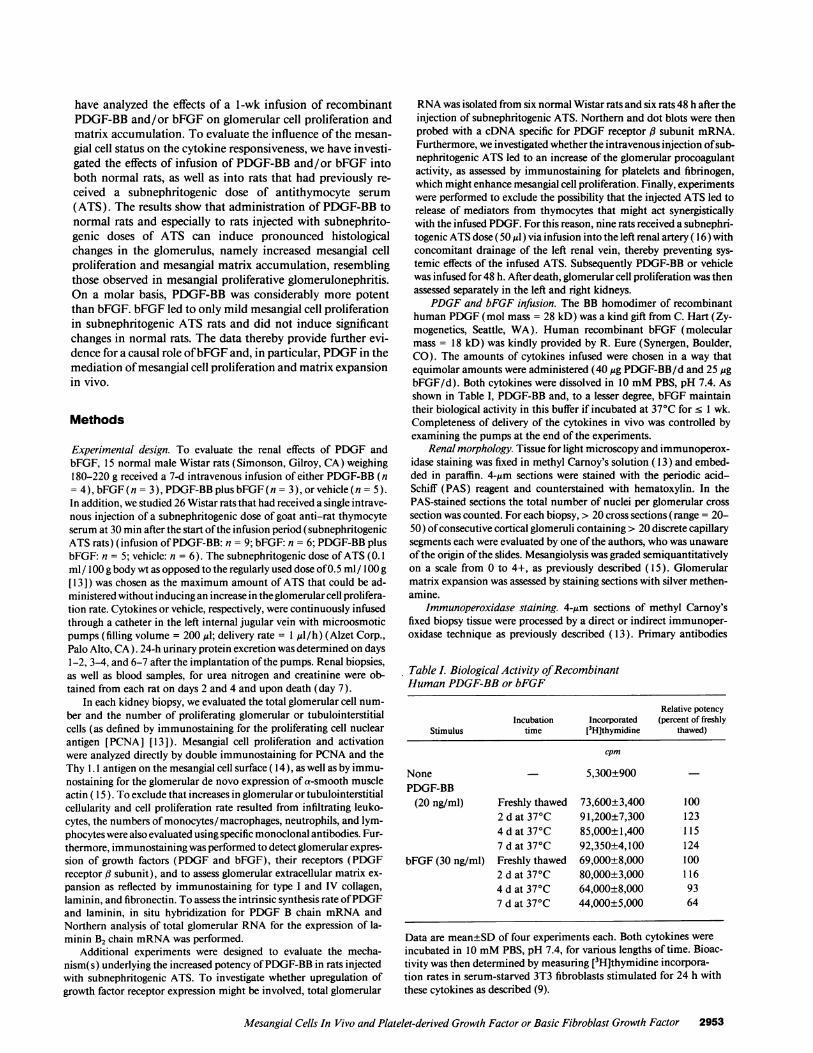
PDGF-BB infusion selectively induces glomerular hyperplasiain normal rats. PAS staining of renal biopsies obtained fromnormal rats infused with PDGF-BB (28 ng/min) or equimolarconcentrations of PDGF-BB (28 ng/min) and bFGF (17 ng/min) revealed mild glomerular hypercellularity with increasedtotal cell counts (Fig. 1), while no changes were noted in thetubulointerstitium and vessels. Renal biopsies of rats infusedwith bFGF ( 17 ng/min) or vehicle had a normal appearanceand normal glomerular cell counts (Fig. 1). No additive or
2954 Floege, Eng, Young, Alpers, Barrett, Bowen-Pope, and Johnson
n= (5) (4) (3) (3) (6) (9) (5) (6)
Figure 1. Total glomerular cellularity and cell proliferation: glomeru-lar total cell counts and proliferating cells (as defined by immuno-staining for PCNA) per glomerular cross-section in normal and sub-nephritogenic ATS rats receiving a l-wk infusion of either PDGF-BB(m), PDGF-BB plus bFGF (i), bFGF (m), or vehicle (o). *P < 0.05vs corresponding vehicle-infused group.
synergistic effect between PDGF-BB and bFGF was observed(Fig. 1). The number of proliferating glomerular cells in-creased up to eightfold in PDGF-BB infused rats, but not inbFGF-infused rats (Fig. 1). Again, no further increase was ob-served when PDGF-BB and bFGF were combined (Fig. 1).
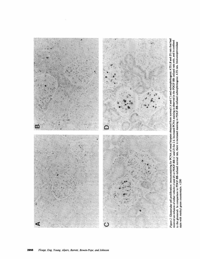
Fig. 2 shows that the induction of cell proliferation in thekidneys of PDGF-BB-infused rats was confined to glomeruli,while no change was observed in the cortical tubulointersti-tium (counts of proliferating cells in tubulointerstitial fieldsranged from 1.1±0.4 to 1.5±0.9 PCNA+ cells/0.1 mm2 at anyofthe time points examined). Furthermore, neither PDGF-BBnor bFGF infusion led to an apparent increase in smooth mus-cle cell proliferation in larger vessels within the kidney.
Mitogenicity ofPDGF-BB andbFGF in vivo depends on thecellular status. To evaluate the effect of a minimal mesangialcell insult at the start ofPDGF-BB and/or bFGF infusion, ratsreceived a subnephritogenic dose ofATS. Fig. 1 confirms thatthis ATS dose was subnephritogenic, since it did not inducesignificant cell proliferation within the glomeruli of vehicle-in-fused rats. PAS-stained tissue sections obtained from these lat-ter rats revealed rare mild mesangiolysis, which was, however,not sufficient to result in a significant increase ofthe mesangio-
lysis index. No other morphological abnormalities, increasedglomerular influx of macrophages and platelets, or depositionof fibrin was noted in the subnephritogenic ATS rats (data notshown).
Fig. 1 shows that in subnephritogenic ATS rats, total glo-merular cellularity increased significantly during the infusionof PDGF-BB. This increase was higher than that observed inPDGF-BB-infused normal rats, although the difference be-tween subnephritogenic ATS rats and normals was not statisti-cally significant. bFGF infusion led to a significant increase ofthe total glomerular cellularity after 4 d in subnephritogenicATS rats but not in normal rats (Fig. 1). No synergism be-tween the two cytokines was noted (Fig. 1 ). Counting ofproli-ferating cells in the glomerulus revealed a < 32-fold increase inPDGF-BB-infused subnephritogenic ATS rats and an < 4.5-fold increase in bFGF infused rats (Fig. 1). The changes ob-served were significantly higher than those observed in thecorresponding normal rats (P < 0.05 subnephritogenic ATSrats vs normal rats) at all time points. Proliferating cell countsin glomeruli ofsubnephritogenic ATS rats infused with PDGF-BB plus bFGF were comparable to those receiving PDGF-BBalone (Fig. 1).
As in the normal rats, the PDGF-BB- or bFGF-induced cellproliferation in the kidneys of subnephritogenic ATS rats wasconfined to the glomerulus (Fig. 2), and no significant alter-ations were observed in the cortical tubulointerstitium or inlarge vessels.
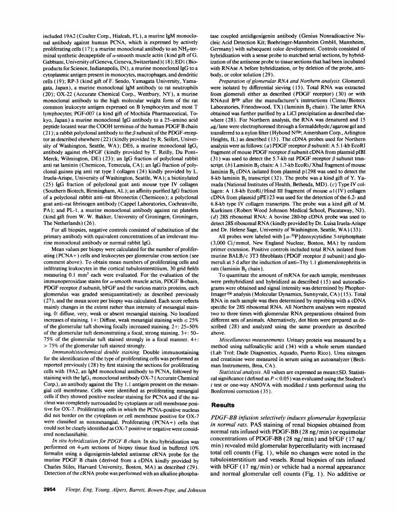
In both normal and subnephritogenic ATS rats, the in-creases in glomerular PCNA-positive cells were associated withan increased frequency of mitoses in the glomeruli. In sub-nephritogenic ATS rats receiving PDGF-BB, occasional glo-meruli contained more than one cell in mitosis (Fig. 3). Thiswas never observed in normal rats.
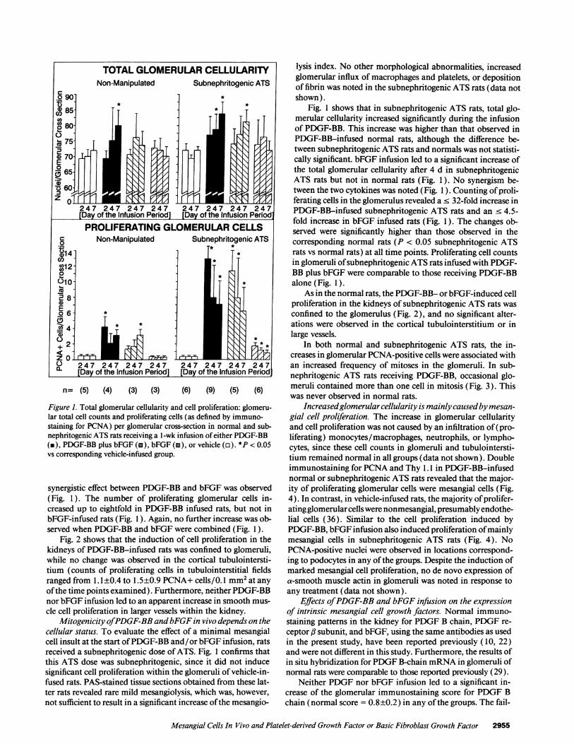
Increasedglomerular cellularity is mainlycausedbymesan-gial cell proliferation. The increase in glomerular cellularityand cell proliferation was not caused by an infiltration of(pro-liferating) monocytes/macrophages, neutrophils, or lympho-cytes, since these cell counts in glomeruli and tubulointersti-tium remained normal in all groups (data not shown). Doubleimmunostaining for PCNA and Thy 1.1 in PDGF-BB-infusednormal or subnephritogenic ATS rats revealed that the major-ity of proliferating glomerular cells were mesangial cells (Fig.4). In contrast, in vehicle-infused rats, the majority ofprolifer-ating glomerular cellswere nonmesangial, presumably endothe-lial cells (36). Similar to the cell proliferation induced byPDGF-BB, bFGF infusion also induced proliferation ofmainlymesangial cells in subnephritogenic ATS rats (Fig. 4). NoPCNA-positive nuclei were observed in locations correspond-ing to podocytes in any ofthe groups. Despite the induction ofmarked mesangial cell proliferation, no de novo expression ofa-smooth muscle actin in glomeruli was noted in response toany treatment (data not shown).
Effects ofPDGF-BB and bFGF infusion on the expressionof intrinsic mesangial cell growth factors. Normal immuno-staining patterns in the kidney for PDGF B chain, PDGF re-ceptor ,B subunit, and bFGF, using the same antibodies as usedin the present study, have been reported previously ( 10, 22)and were not different in this study. Furthermore, the results ofin situ hybridization for PDGF B-chain mRNA in glomeruli ofnormal rats were comparable to those reported previously (29).
Neither PDGF nor bFGF infusion led to a significant in-crease of the glomerular immunostaining score for PDGF Bchain (normal score = 0.8±0.2) in any ofthe groups. The fail-
Mesangial Cells In Vivo and Platelet-derived Growth Factor or Basic Fibroblast Growth Factor 2955
I~~~~~~~~~~~~~~~~~~~I
A
..1
1C *~ 9@ *,
i[4.''r 4:.*t5** ; 4 95'Sg7}1t
4 3<,** 1 s w -~~~~
I 4I
11 :1
.I
a
,N
*t j
ol
*I ... 4
0
.a
i
Ivv.0
4' 1 -~~~~~~~~~~ ~~ ~~~~~~~~~~~~~~~~~~~~~~~~~~~~~~~~~~~~~~~~~~~~~
tw
0'
i' i.
''0i
0
2956 Floege, Eng, Young, Alpers, Barrett, Bowen-Pope, and Johnson
S.4 0 b.*f JS
mo
laCIS0
'C'0 *Z
t ECd
<'0°m
4
.0
C
*=MS
C 1'0'C o
Cu = C
CU
0
- 4) U].0Do Cu'0
U)
C)'suAo C)Dr ,,U'S '--
6) '
om E
o
C o
.- Y C)
C) t. o Q
oD
O Cu)0
° .= Qto OCuK b~
a:-piC11
V,0 C;
Sc
I"e
.1
I1.
.0

Figure 3. Glomerular mitoses: PAS stain of a renal biopsy of aPDGF-BB-infused subnephritogenic ATS rat on day 2 after the startofthe infusion period. In the glomerular tuft, two cells in mitosis areobserved (arrows). x 1,000.
ure ofexogenously administered PDGF-BB orbFGF to induceglomerular PDGF synthesis was also supported by the resultsof in situ hybridization for PDGF B chain mRNA, which, likethe immunostaining, failed to reveal any change from the con-trol pattern in any of the experimental groups (data notshown). In contrast toPDGFB chain, the glomerular immuno-
F- PCNA +, Thy 1.1 - Non-Manipulated Rats_- PCNA +, Thy 1.1 +
1~aT~ I' LASubnephritogenic ATSI
d2 d4 d7 d2 d4 d7 d2 d4 d7 d2 d4 d7Vehicle PDGF PDGF+bFGF bFGF
Figure 4. Identification of proliferating glomerular cells: double im-munostaining for PCNA and the Thy 1.1 antigen expressed on mes-angial cell surfaces. Data are presented as counts of proliferating glo-merular cells per glomerular cross section that were either doublelabeled (i.e., proliferating mesangial cells) or not double labeled (i.e.,proliferating nonmesangial cells). It is apparent that the counts ofproliferating mesangial cells increase in the PDGF-BB-infusedgroups, as well as in bFGF-infused subnephritogenic ATS rats. Be-cause of the different histochemical technique (see Methods), PCNAcounts differ from those presented in Fig. 1.
staining scores for both PDGF receptor fl subunit and bFGFincreased in those groups where significant cell proliferationwas present (Fig. 5).
Neither PDGF nor bFGF infusion led to significant glo-merular deposition ofplatelets or to thrombus formation (datanot shown), both of which contain various mesangial cell mi-togens ( 1, 4).
Increased responsiveness ofsubnephritogenic ATS rats toPDGF is associated with a mild upregulation ofPDGF recep-tor. The injection of a subnephritogenic dose of ATS led to asmall increase in the levels of glomerular PDGF receptor #subunit mRNA on day 2 after the injection (Table II). Asshown in Fig. 5, there was also a small increase in the glomeru-lar staining score for PDGF receptor # subunit after the admin-istration ofsubnephritogenic ATS. In contrast, as noted above,no significant glomerular deposition of platelets or fibrinogenwas observed in these latter rats.
Fig. 6 shows that the renal responsiveness to PDGF-BB wasstill increased if a subnephritogenic dose ofATS was adminis-tered selectively into one kidney rather than systemically. Inthis case the response in the left kidney, which received theATS, was significantly higher than in the right nonperfusedkidney.
PDGF RECEPTOR B-SUBUNITNon-Manipulated
0
c0 3cm
S..
co0)@; 2-
E1
012 0
0
00
COCDc'2a
Subnephritogenic ATS
I~~~i247 247 247 247 247 247 247 247[Day of the Infusion Period] [Day of the Infusion Period]
Non-Manipulated
T
0
E0 1
0
247[Day of
n= (5)
bFGFSubnephritogenic ATS
I *
247 247 247 247 247 247fusion Period] [Day of the Infusion Period]
(3) (6) (9) (5) (6)
Figure S. Glomerular growth factor expression: glomerular immuno-staining scores for the expression of PDGF receptor , subunit andbFGF in normal or subnephritogenic ATS rats receiving a 1-wk infu-sion of either PDGF-BB (n), PDGF-BB plus bFGF (i), bFGF (i),or vehicle (o). *P < 0.05 vs corresponding vehicle-infused group.
Mesangial Cells In Vivo and Platelet-derived Growth Factor or Basic Fibroblast Growth Factor 2957
3-
2-0
(1 1-Co0L) 0OCu
7TE0( 6-
= 5.0)
. 4.z0a.
3'3.Ca" 2
oIqa- 1
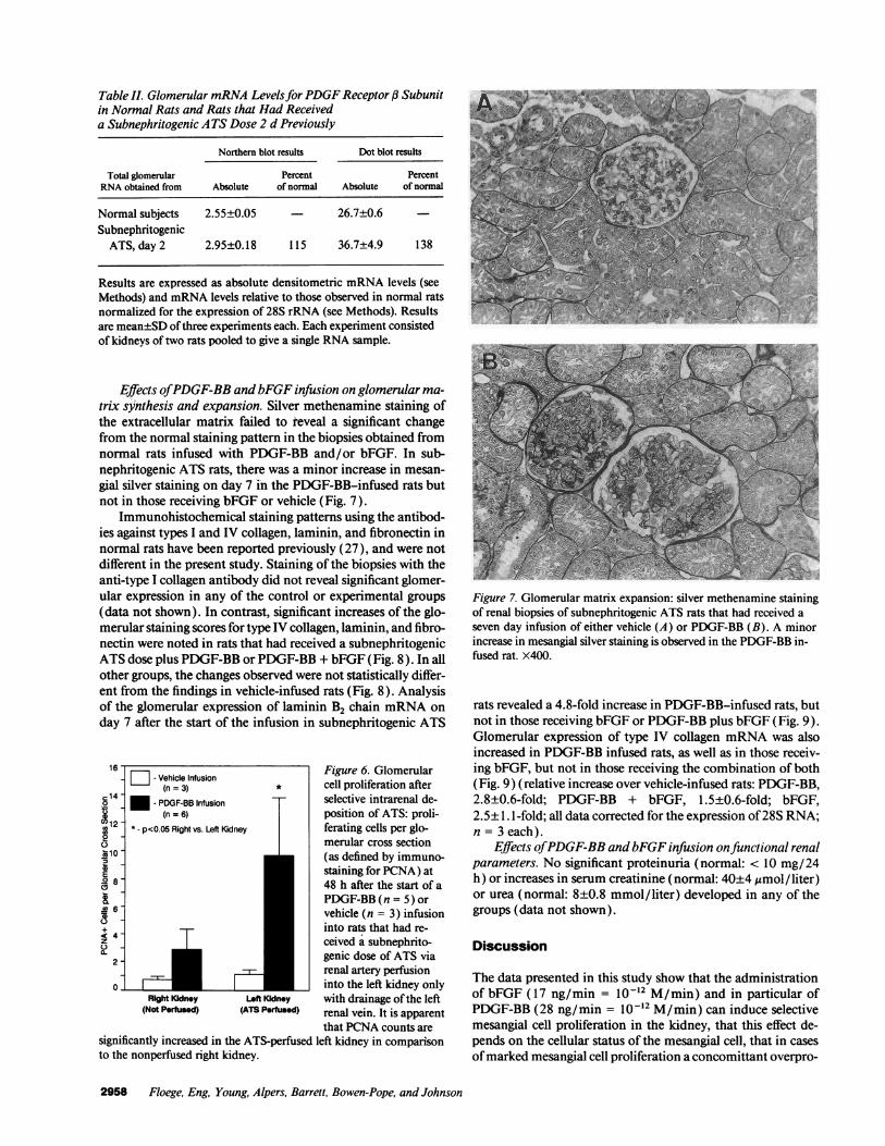
Table II. Glomerular mRNA LevelsforPDGF Receptor j3 Subunitin Normal Rats and Rats that Had Receiveda Subnephritogenic ATS Dose 2 d Previously
Northern blot results Dot blot results
Total glomerular Percent PercentRNA obtained from Absolute of normal Absolute of normal
Normal subjects 2.55±0.05 - 26.7±0.6SubnephritogenicATS, day 2 2.95±0.18 115 36.7±4.9 138
U.
Results are expressed as absolute densitometric mRNA levels (seeMethods) and mRNA levels relative to those observed in normal ratsnormalized for the expression of 28S rRNA (see Methods). Resultsare mean±SD ofthree experiments each. Each experiment consistedof kidneys of two rats pooled to give a single RNA sample.
Effects ofPDGF-BB and bFGF infusion on glomerular ma-trix synthesis and expansion. Silver methenamine staining ofthe extracellular matrix failed to reveal a significant changefrom the normal staining pattern in the biopsies obtained fromnormal rats infused with PDGF-BB and/or bFGF. In sub-nephritogenic ATS rats, there was a minor increase in mesan-gial silver staining on day 7 in the PDGF-BB-infused rats butnot in those receiving bFGF or vehicle (Fig. 7).
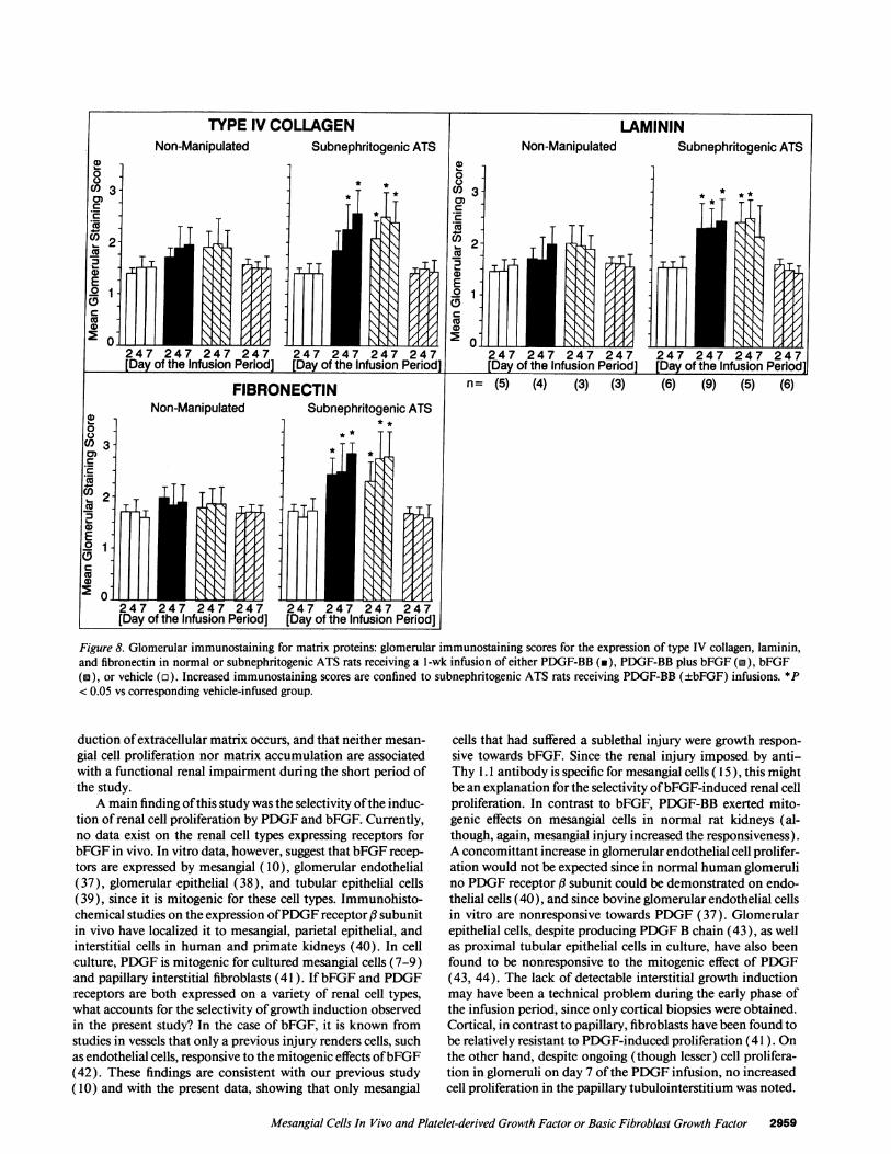
Immunohistochemical staining patterns using the antibod-ies against types I and IV collagen, laminin, and fibronectin innormal rats have been reported previously (27), and were notdifferent in the present study. Staining of the biopsies with theanti-type I collagen antibody did not reveal significant glomer-ular expression in any of the control or experimental groups(data not shown). In contrast, significant increases of the glo-merular staining scores for type IV collagen, laminin, and fibro-nectin were noted in rats that had received a subnephritogenicATS dose plus PDGF-BB or PDGF-BB + bFGF (Fig. 8). In allother groups, the changes observed were not statistically differ-ent from the findings in vehicle-infused rats (Fig. 8). Analysisof the glomerular expression of laminin B2 chain mRNA onday 7 after the start of the infusion in subnephritogenic ATS
16 - Figure 6. Glomerular_ Vehicle Infusion(n = 3) * cell proliferation after
.,14 n - PDGF-BB Infusion selective intrarenal de--1 (n = 6) position of ATS: proli-
* - p<O.05 Right vs. Left Kidney ferating cells per glo-22lo merular cross section
co (as defined by immuno-E _ staining for PCNA) at
48 h after the start of aPDGF-BB (n = 5) or
6 vehicle (n = 3) infusion+ into rats that had re-
2 ceived a subnephrito-a.
_ _ genic dose of ATS viarenal artery perfusion
0- _ _ into the left kidney onlyRight Kidney Left Kidney with drainage ofthe left
(Not Perfused) (ATS Peffused) renal vein. It is apparentthat PCNA counts are
significantly increased in the ATS-perfused left kidney in comparisonto the nonperfused right kidney.
-A
Figure 7. Glomerular matrix expansion: silver methenamine stainingof renal biopsies of subnephritogenic ATS rats that had received aseven day infusion of either vehicle (A) or PDGF-BB (B). A minorincrease in mesangial silver staining is observed in the PDGF-BB in-fused rat. x400.
rats revealed a 4.8-fold increase in PDGF-BB-infused rats, butnot in those receiving bFGF or PDGF-BB plus bFGF (Fig. 9).Glomerular expression of type IV collagen mRNA was alsoincreased in PDGF-BB infused rats, as well as in those receiv-ing bFGF, but not in those receiving the combination of both(Fig. 9) (relative increase over vehicle-infused rats: PDGF-BB,2.8±0.6-fold; PDGF-BB + bFGF, 1.5±0.6-fold; bFGF,2.5±1.1 -fold; all data corrected for the expression of28S RNA;n = 3 each).
Effects ofPDGF-BB andbFGF infusion onfunctional renalparameters. No significant proteinuria (normal: < 10 mg/24h) or increases in serum creatinine (normal: 40±4 pmol/ liter)or urea (normal: 8±0.8 mmol/liter) developed in any of thegroups (data not shown).
Discussion
The data presented in this study show that the administrationof bFGF (17 ng/min = IO-2 M/min) and in particular ofPDGF-BB (28 ng/min = 10-2 M/min) can induce selectivemesangial cell proliferation in the kidney, that this effect de-pends on the cellular status of the mesangial cell, that in casesofmarked mesangial cell proliferation a concomittant overpro-
2958 Floege, Eng, Young, Alpers, Barrett, Bowen-Pope, and Johnson
All
-_;=
M h64NF*Ew
MAWOF air e m s *SM .-, >~~~~~~~~~~~~~~~~~~~~~,;ad
I.-Cll--'%'-4.-R"
7.rn
MAN
n= (5) (4) (3) (3) (6) (9)
Figure 8. Glomerular immunostaining for matrix proteins: glomerular immunostaining scores for the expression of type IV collagen, laminin,and fibronectin in normal or subnephritogenic ATS rats receiving a l-wk infusion of either PDGF-BB (in), PDGF-BB plus bFGF (M), bFGF(a), or vehicle (o). Increased immunostaining scores are confined to subnephritogenic ATS rats receiving PDGF-BB (±bFGF) infusions. *P< 0.05 vs corresponding vehicle-infused group.
duction ofextracellular matrix occurs, and that neither mesan-gial cell proliferation nor matrix accumulation are associatedwith a functional renal impairment during the short period ofthe study.
A main finding ofthis study was the selectivity ofthe induc-tion of renal cell proliferation by PDGF and bFGF. Currently,no data exist on the renal cell types expressing receptors forbFGF in vivo. In vitro data, however, suggest that bFGF recep-tors are expressed by mesangial (10), glomerular endothelial(37), glomerular epithelial (38), and tubular epithelial cells(39), since it is mitogenic for these cell types. Immunohisto-chemical studies on the expression ofPDGF receptor f3 subunitin vivo have localized it to mesangial, parietal epithelial, andinterstitial cells in human and primate kidneys (40). In cellculture, PDGF is mitogenic for cultured mesangial cells (7-9)and papillary interstitial fibroblasts (41 ). If bFGF and PDGFreceptors are both expressed on a variety of renal cell types,what accounts for the selectivity ofgrowth induction observedin the present study? In the case of bFGF, it is known fromstudies in vessels that only a previous injury renders cells, suchas endothelial cells, responsive to the mitogenic effects ofbFGF(42). These findings are consistent with our previous study(10) and with the present data, showing that only mesangial
cells that had suffered a sublethal injury were growth respon-sive towards bFGF. Since the renal injury imposed by anti-Thy 1.1 antibody is specific for mesangial cells ( 15), this mightbe an explanation for the selectivity ofbFGF-induced renal cellproliferation. In contrast to bFGF, PDGF-BB exerted mito-genic effects on mesangial cells in normal rat kidneys (al-though, again, mesangial injury increased the responsiveness).A concomittant increase in glomerular endothelial cell prolifer-ation would not be expected since in normal human glomerulino PDGF receptor f3 subunit could be demonstrated on endo-thelial cells (40), and since bovine glomerular endothelial cellsin vitro are nonresponsive towards PDGF (37). Glomerularepithelial cells, despite producing PDGF B chain (43), as wellas proximal tubular epithelial cells in culture, have also beenfound to be nonresponsive to the mitogenic effect of PDGF(43, 44). The lack of detectable interstitial growth inductionmay have been a technical problem during the early phase ofthe infusion period, since only cortical biopsies were obtained.Cortical, in contrast to papillary, fibroblasts have been found tobe relatively resistant to PDGF-induced proliferation (41 ). Onthe other hand, despite ongoing (though lesser) cell prolifera-tion in glomeruli on day 7 ofthe PDGF infusion, no increasedcell proliferation in the papillary tubulointerstitium was noted.
Mesangial Cells In Vivo and Platelet-derived Growth Factor or Basic Fibroblast Growth Factor 2959
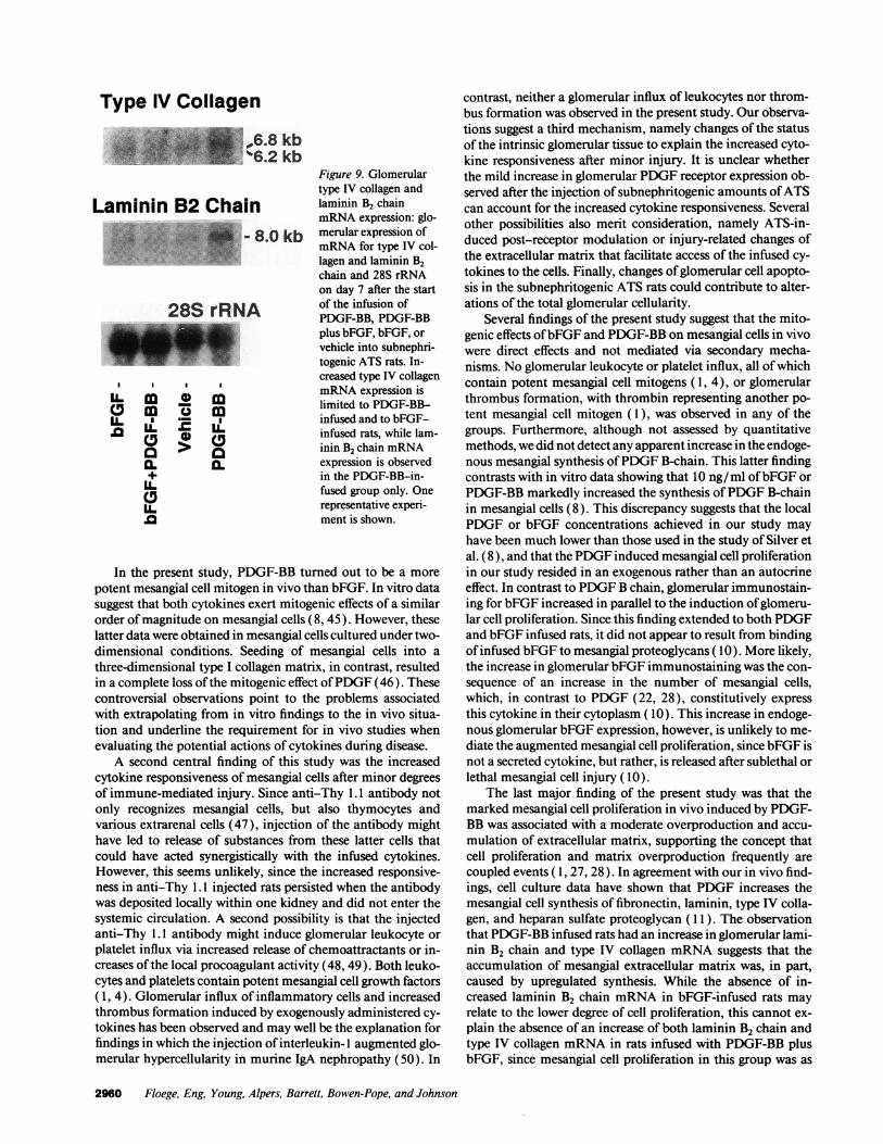
Figure 9. Glomerulartype IV collagen andlaminin B2 chainmRNA expression: glo-merular expression ofmRNA for type IV col-lagen and laminin B2chain and 28S rRNAon day 7 after the startof the infusion ofPDGF-BB, PDGF-BBplus bFGF, bFGF, orvehicle into subnephri-togenic ATS rats. In-creased type IV collagenmRNA expression islimited to PDGF-BB-infused and to bFGF-infused rats, while lam-inin B2 chain mRNAexpression is observedin the PDGF-BB-in-fused group only. Onerepresentative experi-ment is shown.
In the present study, PDGF-BB turned out to be a morepotent mesangial cell mitogen in vivo than bFGF. In vitro datasuggest that both cytokines exert mitogenic effects of a similarorder of magnitude on mesangial cells (8, 45). However, theselatter data were obtained in mesangial cells cultured under two-dimensional conditions. Seeding of mesangial cells into athree-dimensional type I collagen matrix, in contrast, resultedin a complete loss ofthe mitogenic effect ofPDGF (46). Thesecontroversial observations point to the problems associatedwith extrapolating from in vitro findings to the in vivo situa-tion and underline the requirement for in vivo studies whenevaluating the potential actions of cytokines during disease.
A second central finding of this study was the increasedcytokine responsiveness of mesangial cells after minor degreesof immune-mediated injury. Since anti-Thy 1.1 antibody notonly recognizes mesangial cells, but also thymocytes andvarious extrarenal cells (47), injection of the antibody mighthave led to release of substances from these latter cells thatcould have acted synergistically with the infused cytokines.However, this seems unlikely, since the increased responsive-ness in anti-Thy 1.1 injected rats persisted when the antibodywas deposited locally within one kidney and did not enter thesystemic circulation. A second possibility is that the injectedanti-Thy 1.1 antibody might induce glomerular leukocyte orplatelet influx via increased release of chemoattractants or in-creases of the local procoagulant activity (48, 49). Both leuko-cytes and platelets contain potent mesangial cell growth factors( 1, 4). Glomerular influx of inflammatory cells and increasedthrombus formation induced by exogenously administered cy-tokines has been observed and may well be the explanation forfindings in which the injection of interleukin- I augmented glo-merular hypercellularity in murine IgA nephropathy (50). In
contrast, neither a glomerular influx of leukocytes nor throm-bus formation was observed in the present study. Our observa-tions suggest a third mechanism, namely changes of the statusof the intrinsic glomerular tissue to explain the increased cyto-kine responsiveness after minor injury. It is unclear whetherthe mild increase in glomerular PDGF receptor expression ob-served after the injection of subnephritogenic amounts ofATScan account for the increased cytokine responsiveness. Severalother possibilities also merit consideration, namely ATS-in-duced post-receptor modulation or injury-related changes ofthe extracellular matrix that facilitate access of the infused cy-tokines to the cells. Finally, changes ofglomerular cell apopto-sis in the subnephritogenic ATS rats could contribute to alter-ations of the total glomerular cellularity.
Several findings of the present study suggest that the mito-genic effects ofbFGF and PDGF-BB on mesangial cells in vivowere direct effects and not mediated via secondary mecha-nisms. No glomerular leukocyte or platelet influx, all ofwhichcontain potent mesangial cell mitogens ( 1, 4), or glomerularthrombus formation, with thrombin representing another po-tent mesangial cell mitogen (1), was observed in any of thegroups. Furthermore, although not assessed by quantitativemethods, we did not detect any apparent increase in the endoge-nous mesangial synthesis ofPDGF B-chain. This latter findingcontrasts with in vitro data showing that 10 ng/ml ofbFGF orPDGF-BB markedly increased the synthesis ofPDGF B-chainin mesangial cells (8). This discrepancy suggests that the localPDGF or bFGF concentrations achieved in our study mayhave been much lower than those used in the study of Silver etal. (8), and that the PDGF induced mesangial cell proliferationin our study resided in an exogenous rather than an autocrineeffect. In contrast to PDGF B chain, glomerular immunostain-ing for bFGF increased in parallel to the induction ofglomeru-lar cell proliferation. Since this finding extended to both PDGFand bFGF infused rats, it did not appear to result from bindingof infused bFGF to mesangial proteoglycans ( 10). More likely,the increase in glomerular bFGF immunostaining was the con-sequence of an increase in the number of mesangial cells,which, in contrast to PDGF (22, 28), constitutively expressthis cytokine in their cytoplasm ( 10). This increase in endoge-nous glomerular bFGF expression, however, is unlikely to me-diate the augmented mesangial cell proliferation, since bFGF isnot a secreted cytokine, but rather, is released after sublethal orlethal mesangial cell injury (10).
The last major finding of the present study was that themarked mesangial cell proliferation in vivo induced by PDGF-BB was associated with a moderate overproduction and accu-mulation of extracellular matrix, supporting the concept thatcell proliferation and matrix overproduction frequently arecoupled events ( 1, 27, 28). In agreement with our in vivo find-ings, cell culture data have shown that PDGF increases themesangial cell synthesis of fibronectin, laminin, type IV colla-gen, and heparan sulfate proteoglycan ( 11). The observationthat PDGF-BB infused rats had an increase in glomerular lami-nin B2 chain and type IV collagen mRNA suggests that theaccumulation of mesangial extracellular matrix was, in part,caused by upregulated synthesis. While the absence of in-creased laminin B2 chain mRNA in bFGF-infused rats mayrelate to the lower degree of cell proliferation, this cannot ex-plain the absence of an increase of both laminin B2 chain andtype IV collagen mRNA in rats infused with PDGF-BB plusbFGF, since mesangial cell proliferation in this group was as
2960 Floege, Eng, Young, Alpers, Barrett, Bowen-Pope, and Johnson
Type IV Collagenf,_~~~~~.1.41 iI.i.*6bPi.86kb.,':^.,l.| ju.S`46.2 kb
Laminin B2 Chain28S rRN-8A0kb
28S rRNA
I I I
a m o mIL IL
0]0.+ILC,
pronounced as in rats infused with PDGF-BB only. One possi-ble explanation is that the concomitant infusion of PDGF-BBand bFGF led to a more transient increase ofglomerular lami-nin B2 chain and type IV collagen mRNA expression. We mayhave missed this, since we could only obtain RNA on day 7 ofthe infusion period. Future experiments will have to clarify thispoint.
Interestingly, the increase in extracellular matrix compo-nents in PDGF-BB- and bFGF-infused rats was limited to com-ponents of the normal mesangial matrix. Neither interstitial(type I) collagen nor the expression of a-smooth muscle actinwas induced, despite being present in several models ofglomer-ular diseases involving mesangial cell proliferation ( 15, 28).This suggests that mesangial cell proliferation and matrix ex-pansion can occur without the mesangial cell acquiring pheno-typic characteristics of "myofibroblasts" ( 51 ), and that otherstimuli may be necessary (e.g., hemodynamic alterations orother cytokines) to induce these changes.
In conclusion, this study provides further evidence thatPDGF and (to a lesser degree) bFGF may be involved in theincreased mesangial cell proliferation and matrix expansionthat occurs in a variety of glomerular diseases. Our findingsalso show that sublethal injury to glomeruli that has no, or onlyminimal, morphological correlates may induce glomerularchanges of a yet undefined nature, that render the cells moreresponsive to the action of inflammatory mediators. From aclinical point of view, this might explain how a minor renalinsult could have pronounced effects in the presence of a sub-clinical but ongoing autoimmune process. Finally, this studyprovides a further rationale to examine the effects ofPDGF orbFGF antagonists on the course of progressive glomerular dis-ease. However, as discussed elsewhere in more detail (5), be-fore such interventional trials are undertaken, more insightinto the question will have to be gained of when and how thephysiological, (e.g., regenerative) role of cytokines becomes apathological one.
Acknowledaments
The authors gratefully acknowledge the generous supply ofPDGF-BBby C. Hart (Zymogenetics, Seattle, WA) and of bFGF by R. Eure(Synergen, Boulder, CO). The help of Katherine Gordon, MonikaKregeler, and Heike Claus in performing the experiments is also grate-fully acknowledged.
This study was supported by U.S. Public Health Service grants DK-43422, DK-34198, DK-07467, DK-40802, GM-35501, HL-47151,HL-42270, a grant from the Northwest Kidney Foundation, and by apostdoctoral stipend of the Deutsche Forschungsgemeinschaft to J.Floege.
References
1. Striker, L. J., E. P. Peten, S. J. Elliot, T. Doi, and G. E. Striker. 1991.Mesangial cell turnover: effect ofheparin and peptide growth factors. Lab. Invest.64:446-456.
2. Alpers, C. E., K. L. Hudkins, A. M. Gown, and R. J. Johnson. 1992.Enhanced expression of "muscle-specific" actin in glomerulonephritis. KidneyInt. 41:1134-1142.
3. Klahr, S., G. Schreiner, and I. Ichikawa. 1988. The progression of renaldisease. N. Engl. J. Med. 318:1657-1666.
4. Floege, J., E. Eng, B. A. Young, and R. J. Johnson. 1993. Factors involvedin the regulation of glomerular mesangial cell proliferation in vitro and in vivo.Kidney Int. 43:S47-S54.
5. Johnson, R. J., J. Floege, W. G. Couser, and C. E. Alpers. 1993. Role ofplatelet-derived growth factor in glomerular disease. J. Am. Soc. Nephrol. 4:119-128.
6. Abboud, H. E. 1993. Nephrology forum: growth factors in glomerulonephri-tis. Kidney Int. 43:252-267.
7. Shultz, P. J., P. E. DiCorleto, B. J. Silver, and H. E. Abboud. 1988. Mesan-gial cells express PDGF mRNAs and proliferate in response to PDGF. Am. J.Physiol. 255:F674-F679.
8. Silver, B. J., F. E. Jaffer, and H. E. Abboud. 1988. Platelet-derived growthfactor synthesis in mesangial cells: induction by multiple peptide mitogens. Proc.Natl. Acad. Sci. USA. 86:1056-1060.
9. Floege, J., N. Topley, J. Hoppe, T. B. Barrett, and K. Resch. 1991. Mito-genic effect of platelet-derived growth factor in human glomerular mesangialcells: Modulation and/or suppression by inflammatory cytokines. Clin. Exp.Immunol. 86:334-341.
10. Floege, J., E. Eng, V. Lindner, B. A. Young, M. A. Reidy, and R. J.Johnson. 1992. Rat glomerular mesangial cells synthesize basic FGF. Release,upregulated synthesis and mitogenicity in mesangial proliferative glomerulone-phritis. J. Clin. Invest. 90:2362-2369.
11. Doi, T., H. Vlassara, M. Kirstein, Y. Yamada, G. E. Striker, and L. J.Striker. 1992. Receptor-specific increase in extracellular matrix production inmouse mesangial cells by advanced glycosilation end products is mediated viaplatelet-derived growth factor. Proc. Nadl. Acad. Sci. USA. 89:2873-2877.
12. Johnson, R. J., E. Raines, J. Floege, A. Yoshimura, P. Pritzl, C. E. Alpers,and R. Ross. 1992. Inhibition of mesangial cell proliferation and matrix expan-sion in glomerulonephritis in the rat by antibody to platelet derived growth factor.J. Exp. Med. 175:1413-1416.
13. Johnson, R. J., R. L. Garcia, P. Pritzl, and C. E. Alpers. 1990. Plateletsmediate glomerular cell proliferation in immune complex nephritis induced byanti-mesangial cell antibodies in the rat. Am. J. Pathol. 136:369-374.
14. Yamamoto, T., K. Yamamoto, K. Kawasaki, E. Yaoita, F. Shimizu, andI. Kihara. 1986. Immunoelectron microscopic demonstration of Thy- 1 antigenon the surface of mesangial cells in the rat glomerulus. Nephron. 43:293-298.
15. Johnson, R. J., H. Iida, C. E. Alpers, M. W. Majesky, S. M. Schwartz, P.Pritzl, K. Gordon, and A. M. Gown. 1991. Expression of smooth muscle cellphenotype by rat mesangial cells in immune complex nephritis. J. Clin. Invest.87:847-858.
16. Johnson, R. J., S. J. Klebanoff, R. F. Ochi, S. Adler, P. Baker, L. Sparks,and W. G. Couser. 1987. Participation of the myeloperoxidase-H202-halide sys-tem in immune complex nephritis. Kidney Int. 32:342-349.
17. Kurki, P., M. Vanderlaan, F. Dolbeare, J. Gray, and E. M. Tan. 1986.Expression of proliferating cell nuclear antigen (PCNA)/cyclin during the cellcycle. Exp. Cell Res. 166:209-219.
18. Skalli, O., P. Ropraz, A. Trzeciak, G. Benzonana, D. Gillessen, and G.Gabbiani. 1986. A monoclonal antibody against a-smooth muscle actin: a newprobe for smooth muscle differentiation. J. Cell Biol. 103:2787-2796.
19. Dijkstra, C. D., E. A. Dopp, P. Joling, and G. Kraal. 1985. The heterogene-ity ofmononuclear phagocytes in lymphoid organs: distinct macrophage popula-tions in the rat recognized by monoclonal antibodies ED 1, ED2, and ED3. Immu-nology. 54:589-599.
20. Sekiya, S., S. Gotoh, T. Yamashita, T. Watanabe, S. Saitoh, and F. Sendo.1989. Selective depletion of rat neutrophils by in vivo administration ofa mono-clonal antibody. J. Leukocyte Biol. 46:96-102.
21. Shiraishi, T., S. Morimoto, K. Itoh, H. Sato, K. Sugihara, T. Onishi, andT. Ogihara. 1989. Radioimmunoassay of human platelet-derived growth factorusing monoclonal antibodies toward a synthetic 73-97 fragment of its B-chain.Clin. Chim. Acta. 184:65-74.
22. Iida, H., R. Seifert, C. E. Alpers, R. G. K. Gronwald, P. E. Phillips, P.Pritzl, K. Gordon, A. M. Gown, R. Ross, D. F. Bowen-Pope, and R. J. Johnson.1991. Platelet-derived growth factor (PDGF) and PDGF receptor are induced inmesangial proliferative nephritis in the rat. Proc. Natl. Acad. Sci. USA. 88:6560-6564.
23. Reilly, T. M., D. S. Taylor, W. F. Herblin, M. J. Thoolen, A. T. Chiu,D. W. Watson, and P. B. M. W. M. Timmermans. 1989. Monoclonal antibodiesdirected against basic fibroblast growth factor which inhibit its biological activityin vitro and in vivo. Biochem. Biophys. Res. Commun. 2:736-743.
24. Fouser, L., L. Iruele-Arispe, P. Bornstein, and E. H. Sage. 1991. Transcrip-tional activity of the a, (I )-collagen promoter is correlated with the formation ofcapillary-like structures by endothelial cells in vitro. J. Biol. Chem. 266:18345-18351.
25. Bieber, F. 1985. Biotinylierung monoklonaler Antik6rper. In Monoklon-ale Antikorper. Herstellung und Charakterisierung. Peters H. Berlin, editor.Springer pp. 209-212.
26. Bagchus, W. M., M. F. Jeunink, J. Rozing, and J. D. Elema. 1989. Amonoclonal antibody against rat platelets. I. Tissue distribution in vitro and invivo. C/in. Exp. Immunol. 75:317-323.
27. Floege, J., R. J. Johnson, K. Gordon, H. lida, P. Pritzl, A. Yoshimura, C.Campbell, C. E. Alpers, and W. G. Couser. 1991. Increased synthesis ofextracel-lular matrix in mesangial proliferative nephritis. Kidney Int. 40:477-488.
28. Floege, J., M. W. Bums, C. E. Alpers, A. Yoshimura, P. Pritzl, K. Gordon,R. A. Seifert, D. F. Bowen-Pope, W. G. Couser, and R. J. Johnson. 1992. Glomer-ular cell proliferation and PDGF expression precedes glomerulosclerosis in theremnant kidney model. Kidney Int. 41:297-309.
Mesangial Cells In Vivo and Platelet-derived Growth Factor or Basic Fibroblast Growth Factor 2961
29. Yoshimura, A., K. Gordon, C. E. Alpers, J. Floege, P. Pritzl, R. Ross,W. G. Couser, D. F. Bowen-Pope, and R. J. Johnson. 1991. Demonstration ofPDGF B-chain mRNA in glomeruli in mesangial proliferative nephritis by in situHybridization. Kidney Int. 40:470-476.
30. Chomczynski, P., and N. Sacchi. 1987. Single-step method ofRNA isola-tion by acid guanidinium thiocyanate-phenol-chloroform extraction. Anal. Bio-chem. 162:156-159.
31. Yarden, Y., J. A. Escobedo, W. J. Kuang, T. L. Yang-Feng, T. 0. Daniel,P. M. Tremble, E. Y. Chen, M. E. Ando, R. N. Harkins, U. and Francke, et al.1986. Structure of the receptor of platelet derived growth factor helps define afamily of closely related growth factor receptors. Nature (Lond.). 323:226-232.
32. Sasaki, M., and Y. Yamada. 1987. The laminin B2 chain has a multido-main structure homologous to the B1 chain. J. Biol. Chem. 262:17111-17117.
33. Iruela-Arispe, M. L., C. A. Diglio, and E. H. Sage. 1991. Proteins byendothelial cells undergoing angiogenesis in vitro. Artherioscler. Thromb.21:805-815.
34. Bradley, G. M., and E. S. Benson. 1974. Examination of the urine. InTodd-Sanford Clinical Diagnosis by Laboratory Methods. I. Davidson and J. B.Henry, editors. W. B. Saunders Company, Philadelphia, PA. pp. 74-80.
35. Wallenstein, S., C. L. Zucker, and J. L. Fleiss. 1980. Some statisticalmethods useful in circulation research. Circ. Res. 47:1-9.
36. Pabst, R., and R. B. Sterzel. 1983. Cell renewal ofglomerular cell types innormal rats. An autoradiographic study. Kidney Int. 24:626-631.
37. Ballermann, B. J. 1989. Regulation of bovine glomerular endothelial cellgrowth in vitro. Am. J. Physiol. 256:C 182-C 189.
38. Takeuchi, A., N. Yoshizawa, M. Yamamoto, Y. Sawasaki, T. Oda, A.Senoo, H. Niwa, and Y. Fuse. 1992. Basic fibroblast growth factor promotesproliferation of rat glomerular visceral epithelial cells in vitro. Am. J. Pathol.141:107-116.
39. Zhang, G., T. Ichimura, A. Wallin, K. Mikio, and J. L. Stevens. 1991.Regulation of rat proximal tubule epithelial cell growth by fibroblast growthfactors, insulin-like growth factor- I and transforming growth factor-a, and analy-sis of fibroblast growth factors in rat kidney. J. Cell. Physiol. 148:295-305.
40. Alpers, C. E., R. A. Seifert, K. L. Hudkins, R. J. Johnson, and D. F.
Bowen-Pope. 1993. PDGF-receptor localizes to mesangial, parietal epithelial,and interstitial cells in human and primate kidneys. Kidney Int. 43:286-294.
41. Knecht, A., L. G. Fine, K. S. Kleinman, P. Rodemann, G. A. Miller,D. D. L. Woo, and J. T. Norman. 1991. Fibroblasts ofrabbit kidney in culture II.Paracrine stimulation of papillary fibroblasts by. PDGF. Am. J. Physiol.261:F292-F299.
42. Lindner, V., R. A. Majack, and M. A. Reidy. 1991. Basic fibroblast growthfactors mediates endothelial regrowth and proliferation in denuded arteries. J.Clin. Invest. 85:2004-2008.
43. Floege, J., R. J. Johnson, C. E. Alpers, C. A. Richardson, K. Gordon, andW. G. Couser. 1993. Visceral glomerular epithelial cells can proliferate in vivoand synthesize PDGF B-chain. Am. J. Pathol. 142:637-650.
44. Humes, H. D., T. F. Beals, D. A. Cieslinski, 1. 0. Sanchez, and T. P. Page.1991. Effects of transforming growth factor-j3, transforming growth factor-a, andother growth factors on renal proximal tubule cells. Lab. Invest. 64:538-545.
45. Issandou, M., and J. M. Darbon. 1991. Basic fibroblast growth factorstimulates glomerular mesangial cell proliferation through a protein kinase C-in-dependent pathway. Growth Factors. 5:255-264.
46. Madri, J. A., and M. Marx. 1992. Matrix composition, organization, andsoluble factors: modulators of microvascular cell differentiation in vitro. KidneyInt. 41:560-565.
47. Barclay, A. N. 1981. Different reticular elements in rat lymphoid tissueidentified by localization of Ia, Thy-l and MRC OX 2 antigens. Immunology.44:727-736.
48. Bagchus, W. M., M. F. Jeunink, and J. D. Elema. 1990. The mesangium inanti-Thy-l nephritis. Am. J. Pathol. 137:215-222.
49. Poelstra, K., M. J. Hardonk, J. Koudstaal, and W. W. Bakker. 1990.Intraglomerular platelet aggregation and experimental glomerulonephritis. Kid-ney Int. 37:1500-1508.
50. Montinaro, V., K. Hevey, L. Aventaggiato, K. Fadden, A. Esparza, A.Chen, D. S. Finbloom, and A. Rifai. 1992. Extrarenal cytokines modulate theglomerular response to IgA immune complexes. Kidney Int. 42:341-353.
51. Johnson, R. J., J. Floege, A. Yoshimura, H. lida, W. G. Couser, and C. E.Alpers. 1992. The activated mesangial cell: A glomerular "myofibroblast"? J.Am. Soc. Nephrol. 2:S190-S197.
2962 Floege, Eng, Young, Alpers, Barrett, Bowen-Pope, and Johnson
Copyright © 2022 FDOKUMEN