Effect of Glucose–Insulin–Potassium Infusion on Mortality in ...
16
Effect of Glucose–Insulin–Potassium Infusion on Mortality in Critical Care Settings: A Systematic Review and Meta-Analysis Michael A. Puskarich, MD, Michael S. Runyon, MD, Stephen Trzeciak, MD, MPH, Jeffrey A. Kline, MD, and Alan E. Jones, MD Department of Emergency Medicine, Carolinas Medical Center, Charlotte, North Carolina (Dr Puskarich, Dr Runyon, Dr Kline, Dr Jones) and Cooper University Hospital, Camden, New Jersey (Dr Trzeciak) Abstract This study seeks to measure the treatment effect of glucose–insulin–potassium (GIK) infusion on mortality in critically ill patients. A systematic review of randomized controlled trials is conducted, comparing GIK treatment with standard care or placebo in critically ill adult patients. The primary outcome variable is mortality. Two authors independently extract data and assess study quality. The primary analysis is based on the random effects model to produce pooled odds ratios (ORs) with 95% confidence intervals (CIs). The search yields 1720 potential publications; 23 studies are included in the final analysis, providing a sample of 22 525 patients. The combined results demonstrate no heterogeneity (P = .57, I 2 = 0%) and no effect on mortality (OR = 1.02; 95% CI, 0.93–1.11) with GIK treatment. No experimental studies of shock or sepsis populations are identified. This meta-analysis finds that there is no mortality benefit to GIK infusion in critically ill patients; however, study populations are limited to acute myocardial infarction and cardiovascular surgery patients. No studies are identified using GIK in patients with septic shock or other forms of circulatory shock, providing an absence of evidence regarding the effect of GIK as a therapy in patients with shock. Keywords Clinical research; critical care; emergency medicine; clinical trials; anesthesiology Infusion of a glucose–insulin–potassium (GIK) solution was first proposed in the 1960s as treatment for acute myocardial infarction (AMI) with the intention of providing electrical stability to the myocardium. 1 The proposed mechanisms of action of GIK therapy in cardiovascular disease include suppression of circulating levels and myocardial uptake of free fatty acids, which are toxic to ischemic myocardium; improvement in the efficiency of myocardial energy production through the provision of exogenous glucose; and stabilization of intracellular potassium, which may be depleted during times of myocardial ischemia. 2,3 Since the sentinel report, numerous randomized controlled trials have been performed using GIK. Pooled data have suggested morbidity and mortality benefits associated with GIK therapy in cardiothoracic surgery 4 and AMI 3 populations. The largest GIK study performed in AMI patients to date failed to find similar benefits. 2 Thus, there remain significant conflicting evidence and debate regarding the utility of GIK infusion in various patient populations. Address for correspondence: Alan E. Jones, MD, Department of Emergency Medicine, Carolinas Medical Center, PO Box 32861, Charlotte, NC 28232-2861; [email protected]. NIH Public Access Author Manuscript J Clin Pharmacol. Author manuscript; available in PMC 2010 August 10. Published in final edited form as: J Clin Pharmacol. 2009 July ; 49(7): 758–767. doi:10.1177/0091270009334375. NIH-PA Author Manuscript NIH-PA Author Manuscript NIH-PA Author Manuscript brought to you by CORE View metadata, citation and similar papers at core.ac.uk provided by IUPUIScholarWorks
-
Upload
khangminh22 -
Category
Documents
-
view
0 -
download
0
Transcript of Effect of Glucose–Insulin–Potassium Infusion on Mortality in ...
Effect of Glucose–Insulin–Potassium Infusion on Mortality inCritical Care Settings: A Systematic Review and Meta-Analysis
Michael A. Puskarich, MD, Michael S. Runyon, MD, Stephen Trzeciak, MD, MPH, Jeffrey A.Kline, MD, and Alan E. Jones, MDDepartment of Emergency Medicine, Carolinas Medical Center, Charlotte, North Carolina (DrPuskarich, Dr Runyon, Dr Kline, Dr Jones) and Cooper University Hospital, Camden, New Jersey(Dr Trzeciak)
AbstractThis study seeks to measure the treatment effect of glucose–insulin–potassium (GIK) infusion onmortality in critically ill patients. A systematic review of randomized controlled trials isconducted, comparing GIK treatment with standard care or placebo in critically ill adult patients.The primary outcome variable is mortality. Two authors independently extract data and assessstudy quality. The primary analysis is based on the random effects model to produce pooled oddsratios (ORs) with 95% confidence intervals (CIs). The search yields 1720 potential publications;23 studies are included in the final analysis, providing a sample of 22 525 patients. The combinedresults demonstrate no heterogeneity (P = .57, I2 = 0%) and no effect on mortality (OR = 1.02;95% CI, 0.93–1.11) with GIK treatment. No experimental studies of shock or sepsis populationsare identified. This meta-analysis finds that there is no mortality benefit to GIK infusion incritically ill patients; however, study populations are limited to acute myocardial infarction andcardiovascular surgery patients. No studies are identified using GIK in patients with septic shockor other forms of circulatory shock, providing an absence of evidence regarding the effect of GIKas a therapy in patients with shock.
KeywordsClinical research; critical care; emergency medicine; clinical trials; anesthesiology
Infusion of a glucose–insulin–potassium (GIK) solution was first proposed in the 1960s astreatment for acute myocardial infarction (AMI) with the intention of providing electricalstability to the myocardium.1 The proposed mechanisms of action of GIK therapy incardiovascular disease include suppression of circulating levels and myocardial uptake offree fatty acids, which are toxic to ischemic myocardium; improvement in the efficiency ofmyocardial energy production through the provision of exogenous glucose; and stabilizationof intracellular potassium, which may be depleted during times of myocardial ischemia.2,3Since the sentinel report, numerous randomized controlled trials have been performed usingGIK. Pooled data have suggested morbidity and mortality benefits associated with GIKtherapy in cardiothoracic surgery4 and AMI3 populations. The largest GIK study performedin AMI patients to date failed to find similar benefits.2 Thus, there remain significantconflicting evidence and debate regarding the utility of GIK infusion in various patientpopulations.
Address for correspondence: Alan E. Jones, MD, Department of Emergency Medicine, Carolinas Medical Center, PO Box 32861,Charlotte, NC 28232-2861; [email protected].
NIH Public AccessAuthor ManuscriptJ Clin Pharmacol. Author manuscript; available in PMC 2010 August 10.
Published in final edited form as:J Clin Pharmacol. 2009 July ; 49(7): 758–767. doi:10.1177/0091270009334375.
NIH
-PA Author Manuscript
NIH
-PA Author Manuscript
NIH
-PA Author Manuscript
brought to you by COREView metadata, citation and similar papers at core.ac.uk
provided by IUPUIScholarWorks
Extensive preclinical rationales support the use of GIK as an inotropic and metabolictherapy in several critical illnesses. The heart develops insulin resistance in shock andsepsis, and insulin-euglycemia produces a potent inotropic effect on the left ventricle.5,6Sepsis can lead to excessive phosphorylation of the pyruvate dehydrogenase complex, thusimpairing net carbohydrate oxidation in skeletal and heart muscle.7–9 Deficits incarbohydrate oxidation can be expected to be worsened by the effect of diabetes mellitus, acommon comorbidity in humans.10 Insulin reactivates the pyruvate dehydrogenase complex,via activation of pyruvate dehydrogenase phosphatase, to permit maximal glucose andlactate oxidation by heart muscle.11 Additionally, it can be hypothesized that via its actionon the tyrosine kinase pathway, insulin can reduce intracellular signals that lead to theproduction of inflammatory molecules that possess negative inotropic properties, includingtumor necrosis factor.12
Recognizing that numerous clinical trials of GIK infusion in various critically ill populationshave been conducted over the past 4 decades, we hypothesized that their results could beaggregated to produce more definitive conclusions about the treatment effect of GIKinfusion in the critically ill. Therefore, we conducted a systematic review and meta-analysisto examine the available evidence investigating GIK treatment of critically ill patients; ourgoal was to determine the treatment effect of such a strategy on mortality and to determinewhether certain predefined subpopulations (patients with septic shock or other forms ofcirculatory shock) are afforded survival benefit with GIK treatment.
METHODSSearch Strategy for Identification of Studies
We followed a written protocol that was designed in accordance with recommendedguidelines and finalized prior to beginning the study.13 We searched MEDLINE (1965 toApril 2008), EMBASE (1974 to April 2008), and CINHAL (1982 to April 2008) using thefollowing search terms: (critical care or intensive care unit or CABG or coronary arterybypass or cardiac surgery or heart surgery or myocardial infarction or stroke or sepsis orshock) and (GIK or glucose–insulin–potassium or glucose-insulin or glucose or insulin orhyperinsulinemia-euglycemia) and clinical trial. The search was updated in October 2008.To identify potential unpublished data, we contacted 2 experts in the field of critical care,searched abstracts from the Society of Critical Care Medicine and American College ofChest Physicians annual meetings from 1997 to 2007, reviewed published practiceguidelines for critical care from 2000 to 2007, and searched Web sites containing details forclinical trial registration. Additionally, reference lists of the articles chosen for inclusion andthe reference lists of previous reviews3,4,14,15 were screened to identify further studies forinclusion.
Inclusion CriteriaWe considered studies eligible for review, regardless of language or publication type, if theywere randomized controlled trials of human adults (age >17 years) with critical illness(defined as either patients enrolled in an intensive care unit [ICU] or operating room or acontrol group with a mortality rate of greater than or equal to 15%) that had (1) a clearlydefined control group in which patients received standard of care therapy or placebo and (2)an intervention group in which patients received a continuous infusion of insulin, glucose,and potassium. Reviews, correspondence, editorials, and nonhuman studies were excluded;however, their reference lists were screened, if relevant, to identify further studies forinclusion. We attempted to contact corresponding authors for clarification of missing orincomplete data.
Puskarich et al. Page 2
J Clin Pharmacol. Author manuscript; available in PMC 2010 August 10.
NIH
-PA Author Manuscript
NIH
-PA Author Manuscript
NIH
-PA Author Manuscript
Study Selection and Data AbstractionTwo reviewers (MAP and AEJ) independently screened the titles and abstracts of identifiedstudies for potential eligibility. After the relevance search, the 2 reviewers compared theirexclusion logs, and the κ statistic was calculated to assess interobserver agreement. Cases ofdisagreement were resolved by conference between the reviewers. If agreement could not bereached, the full manuscript was obtained for review.
The full manuscript of each study that passed the relevance screen was reviewed by 2 of theinvestigators (MAP and MSR). Study data were abstracted independently by each reviewerusing a standardized data collection form. In cases of disagreement in abstracted data, a thirdreviewer abstracted the data and consensus was reached by conference between the 3reviewers.
Assessment of QualityQuality was assessed using the Cochrane Collaboration’s tool for assessing the risk of biasevaluating 4 domains13:
A. Sequence generation: grade A = sequence was adequately generated, grade B =sequence was inadequately generated, grade C = unknown.
B. Concealment allocation: grade A = performed concealment allocation, grade B =did not use concealment allocation, grade C = unknown.
C. Blinding: grade A = investigators blinded to allocation, grade B = investigators notblinded to allocation, grade C = unknown.
D. Selective outcome reporting: grade A = free of suggestion of selective outcomereporting, grade B = suggestion of selective outcome reporting, grade C =unknown.
Main Outcome MeasureWe defined mortality at the end of the time frame reported by the authors to be the primarydependent variable for analysis. In the event authors reported mortality at more than onetime point, we used inhospital mortality preferentially.
Statistical AnalysisAll data were entered into StatsDirect version 2.7.2 (StatsDirect, Cheshire, UK).Heterogeneity was tested using a chi-square test with a P value <.10 to indicate significantheterogeneity between the trials. In addition, I2 was calculated to provide an estimate of thevariability across studies beyond that attributable to chance.16 The results of studies werepooled using a random effects model.17 The individual and pooled statistics were calculatedas odds ratios (ORs) with 95% confidence intervals (CIs). The validity of using funnel plotsand formal statistical methods to detect the presence of publication bias has recently beencalled into question; therefore, a funnel plot was constructed for visual inspection but we donot attempt to explain any asymmetry.18,19 Additionally, we collected data on the adverseevents of hypoglycemia and hyperkalemia, as defined by the authors of the respectivepublications, in the control and GIK arms. We report them as a proportion of the totalenrolled populations among the publications that reported adverse events and according tothe respective study group.
Subgroup and Sensitivity AnalysisGiven the large amount of clinical heterogeneity attributable to our definition of critically illpatients and the wide range of year of publication and practice style, we conducted the
Puskarich et al. Page 3
J Clin Pharmacol. Author manuscript; available in PMC 2010 August 10.
NIH
-PA Author Manuscript
NIH
-PA Author Manuscript
NIH
-PA Author Manuscript
following a priori defined subgroup analyses: (1) high-quality studies (studies rated grade Afor at least 3 of 4 quality domains previously discussed; (2) studies of patients with septicshock (using standard criteria); and (3) studies of patients with other forms of circulatoryshock (as defined by each study). Finally, a sensitivity analysis was performed to examinethe use of a fixed effect versus random effects model.
We performed a post hoc (not described in our original protocol) analysis of patients withacute myocardial infarction (AMI) only and those undergoing cardiovascular surgery (CVS)only. We chose to perform these subgroup analyses after data collection given that thesewere the only 2 patient populations represented in the included studies.
RESULTSSearch and Selection
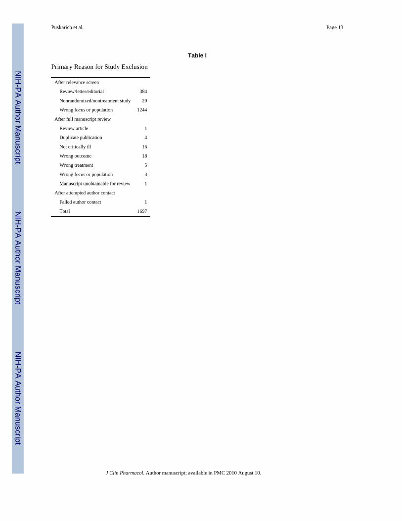
The comprehensive search yielded a total of 1720 relevant publications; details of the searchand study selection are provided in Figure 1 and Table I.
InclusionAfter the relevance search (blinded interobserver agreement of 98% with 30 disagreementsamong 1720 articles, κ = 0.79), a complete manuscript review was performed on 71 articles;1 article was unobtainable for review despite multiple requests to the National Libraries ofMedicine, Online Computer Library Center, Pramukh Swami Medical College Library(India), PSG Institute of Medical Sciences and Research (India), Regional Medical ResearchCentre for Tribals Central Library (India), KG Medical University (India), and e-mailrequests to the editor of the journal and all of the primary and secondary authors.
A total of 23 studies met our predefined criteria and were included in the final analysis,providing a total sample of 22 525 patients.2,20–41 Three full-length articles were translatedto English from French,23 Turkish,31 and Chinese.40 All 3 met our criteria and wereincluded in the final analysis.
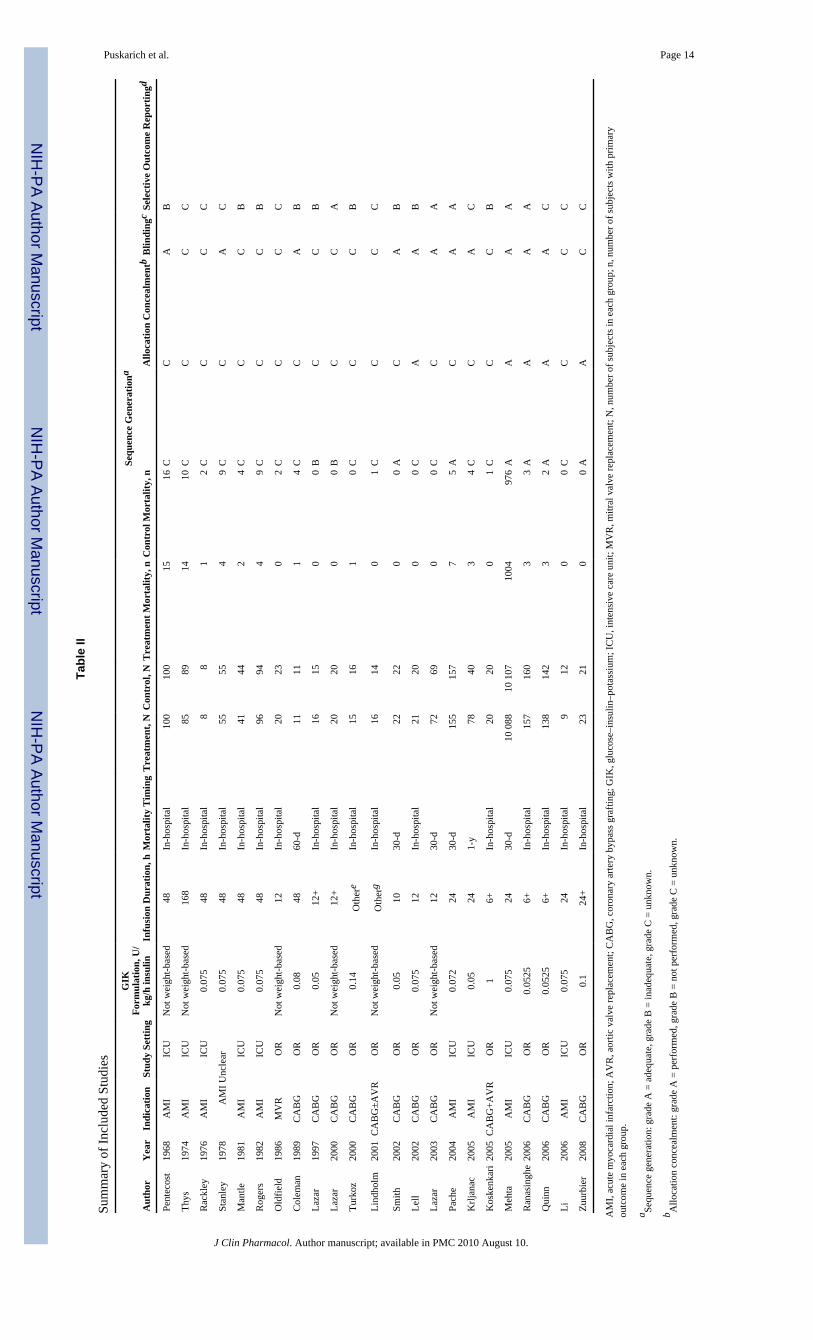
Study DescriptionsThe incidence of mortality in the studies ranged from 0 to 36% in the control group and 0 to16% in the study group. Nine studies took place in an ICU, 13 studies took place in anoperating room, and in 1 study the location was unclear but the predefined control groupmortality rate allowed inclusion of the study. Pertinent details of the included studies areprovided in Table II.
Quality AssessmentTwo authors (MAP, MSR) independently graded the publications on the measures ofsequence generation, concealment of allocation, blinding, and selective outcome reporting.Four studies were graded A in 3 of 4 criteria,2,35,38,39 meeting our definition of high quality.
AnalysesWhen all 23 studies were considered, there was no evidence of statistical heterogeneity (P= .57; I2 = 0%); however, there was clinical heterogeneity among the studies. The summaryOR demonstrates no significant reduction in mortality (OR = 1.02; 95% CI, 0.93–1.11)among subjects receiving GIK (Figure 2). To assess for potential publication bias, a funnelplot of all included studies is provided in Figure 3.
Subgroup analysis of high-quality studies (defined above and shown in Table 2), studieswith only AMI patients (10 studies, Table 2), and studies with only CVS patients (13
Puskarich et al. Page 4
J Clin Pharmacol. Author manuscript; available in PMC 2010 August 10.
NIH
-PA Author Manuscript
NIH
-PA Author Manuscript
NIH
-PA Author Manuscript
studies, Table 2) similarly showed no mortality reduction among subjects who received GIK(OR = 1.04; 95% CI, 0.95–1.14 for high-quality studies; OR = 0.98; 95% CI, 0.81–1.19 forAMI studies; and OR = 0.70; 95% CI, 0.29–1.72 for CVS studies). None of the above pointestimates were significantly different when using a fixed effect model.
Of the 22 525 patients included in the final analysis, 20 195 (89.7%) were enrolled in asingle, multi-center study of GIK in AMI patients.2 We performed a post hoc analysis todetermine whether this study was dominating the model because of its size. When this studywas excluded and the analysis repeated, there was no significant difference in the treatmenteffect (OR = 0.78; 95% CI, 0.54–1.11).
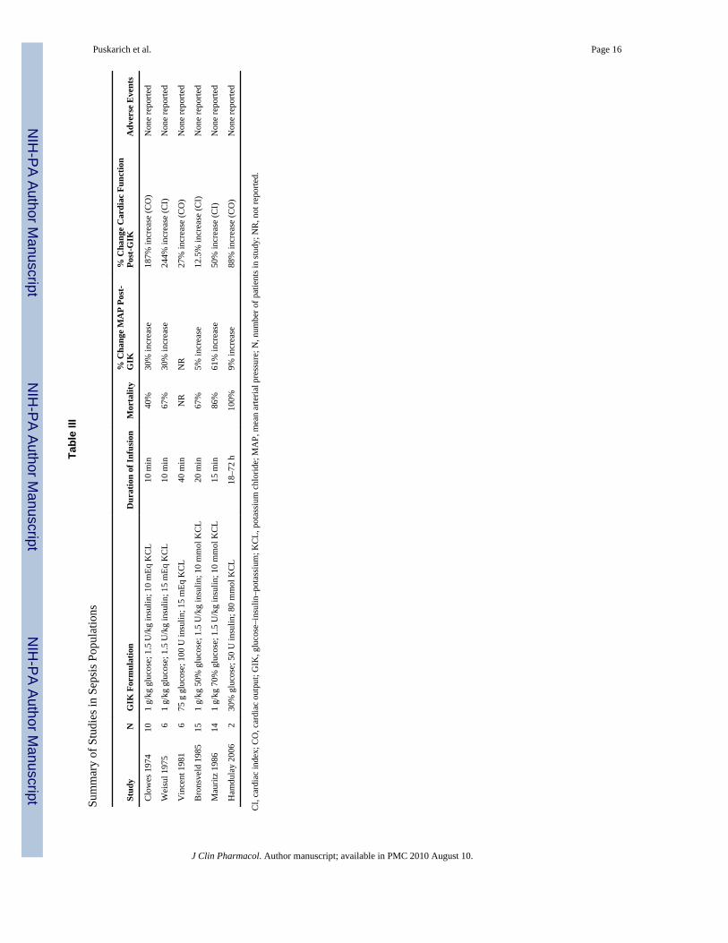
Table III provides a summary of all of the studies involving GIK administration to patientswith septic shock.42–47 None of the studies with septic shock populations were randomizedtrials; thus, none were appropriate for inclusion in our meta-analysis. Additionally, ourcomprehensive literature search did not identify any randomized trials of GIK treatment inother circulatory shock populations, and no subpopulations of circulatory shock patientswere reported in any of the studies meeting our inclusion criteria.
Six studies2,29,36,37,39,40 explicitly reported the number of occurrences of hypoglycemia,and among these studies the incidence of hypoglycemia in GIK patients was 0.5% (54/10324) and in controls was 0.1% (11/10 309). Four studies2,31,36,40 explicitly reported thenumber of occurrences of hyperkalemia, and among these studies the incidence ofhyperkalemia was 4.3% (434/10 166) in GIK patients and 1.6% (161/10 147) in controls.
DISCUSSIONThis meta-analysis evaluates the treatment effect of GIK therapy in critically ill patients.Using pooled data from 23 studies that randomized a total of 22 525 subjects, we found nosignificant effect on mortality (OR = 1.02; 95% CI = 0.93–1.11). We identified no studiesthat included septic shock or other forms of circulatory shock patients in an experimentaldesign. Of the studies reporting adverse events, hyperkalemia occurred more commonly thanhypoglycemia; however, both had low cumulative incidence.
This systematic review used both a comprehensive search and standard methods forsummarizing the treatment effect of GIK therapy in critically ill patients. The subgroupanalyses we present included the high-quality studies, studies with AMI populations only,and studies with CVS populations only. None of these analyses demonstrated significantmortality benefits associated with administration of GIK. Our other subgroup analyses wereto examine only populations of septic shock and other forms of circulatory shock; however,we were unable to identify any experimental studies examining these groups of patients.This was surprising to us given the amount of basic science research that supports thepotential physiological benefits of GIK therapy for septic shock.48–52 Because wespecifically sought out studies of septic shock and were unable to include any in theanalysis, this study adds to previously published meta-analyses of GIK by allowing aconclusion that the current literature provides an absence of evidence regarding the effect ofGIK as a therapy for septic shock.
It is important to note the clinical heterogeneity among the studies included in this analysis.All of the included studies were either in patients with the diagnosis of AMI (10 studies) orin CVS patients (13 studies). Thus, our results are limited to those populations and shouldnot be extrapolated to heterogeneous groups of critically ill patients or those with otherdiagnoses such as acute respiratory distress syndrome or sepsis. Furthermore, the studies inthis analysis span a time frame of 40 years. Many new therapeutic options, particularly formyocardial infraction and cardiothoracic surgery, have changed during this time. A
Puskarich et al. Page 5
J Clin Pharmacol. Author manuscript; available in PMC 2010 August 10.
NIH
-PA Author Manuscript
NIH
-PA Author Manuscript
NIH
-PA Author Manuscript
limitation of this meta-analysis is the possibility that co-interventions (eg, thrombolytic orpercutaneous coronary angioplasty) may have influenced the results of included studies;there was no practical way to capture or control for such interventions in this meta-analysis.
One previous meta-analysis, by Pittas et al, examined the effect of insulin therapy forcritically ill hospitalized patients.15 Pittas et al found that insulin therapy initiated in thehospital has beneficial effects on short-term mortality in critically ill patients. There areseveral plausible explanations for the different findings between the Pittas study and thepresent analysis. First, the Pittas analysis included studies in which patients were treatedwith any type or form of insulin therapy, whereas we limited our inclusion to only studiesthat administered a GIK solution. Second, the Pittas study used a broad definition of criticalillness by including all patients with AMI and stroke and those in the ICU or operatingroom. This broad inclusion may have resulted in misclassification of critical illness andaffected the results. The definition of critical illness used in the present analysis was moreconservative, most likely resulting in a more homogenous sample. Finally, the sample ofpatients in this meta-analysis is 5 times larger (22 525 vs 4733) than the sample of subjectsthat received GIK in the Pittas study, resulting in a much higher power to detect the truetreatment effect.
As previously mentioned, 20 195 of the 22 525 (89.7%) subjects included in this meta-analysis were from a single publication (Mehta et al2). In an attempt to control for thepotential for this study to dominate the analysis, we performed a subgroup analysis wherewe excluded this report from the analysis and re-performed the statistical model. Theresulting odds ratio was 0.78 (95% CI, 0.54–1.11), and although this was not statisticallysignificant, there is clearly a trend toward improvement in mortality with the odds ratio wellbelow 1 and the upper bounds of the confidence interval just above 1. This suggests that thislarge study had an important influence on our overall results; however, we are reassured bythe finding that the statistical significance of the subgroup analysis (nonsignificant mortalityreduction) was the same as our overall findings.
Any meta-analysis is prone to bias given its retrospective approach; however, we used anumber of important steps in an attempt to minimize the impact of bias. First, we followed aprotocol that was written prior to starting the search and analysis. Second, we usedindependent observers to both abstract data and grade study quality. Third, our meta-analysis addressed all of the suggested necessary components of the AMSTARmethodological quality measurement tool for systematic reviews.53
CONCLUSIONData synthesized from 23 randomized controlled trials demonstrated no treatment effect ofGIK therapy in critically ill patients; however, study populations included were limited toacute myocardial infarction and cardiovascular surgery patients. No studies were identifiedthat used GIK in patients with septic shock or other forms of circulatory shock, providing anabsence of evidence regarding GIK as a treatment for shock. Adverse events related to GIKinfusion were uncommon. Future studies should explore the potential benefits of GIK insepsis, shock, or other heterogeneous critically ill populations in experimental researchdesigns.
AcknowledgmentsThe authors thank the following corresponding authors: Shamir Mehta, Adnan Kastrati, and C. J. Zuurbier. Inaddition, we thank Ming Qi, MD, for help with article translation and both Bridget Loven and Leonora Kaufmannfor help with the literature search and article retrieval.
Puskarich et al. Page 6
J Clin Pharmacol. Author manuscript; available in PMC 2010 August 10.
NIH
-PA Author Manuscript
NIH
-PA Author Manuscript
NIH
-PA Author Manuscript
Financial disclosure: Alan E. Jones has received research support from Critical Biologics and HutchinsonTechnology, and Stephen Trzeciak has received research support from Novo Nordisk, Biosite, and Eli Lilly.
References1. Fishleder BL, Frieldand C, De MA, et al. Effects of an intravenous infusion of a potassium-glucose-
insulin solution on the electrocardiographic signs of myocardial infarction: a preliminary clinicalreport. Am J Cardiol 1962;9:166–181. [PubMed: 13914751]
2. Mehta SR, Yusuf S, Diaz R, et al. Effect of glucose-insulin-potassium infusion on mortality inpatients with acute ST-segment elevation myocardial infarction: the CREATE-ECLA randomizedcontrolled trial. JAMA 2005;293:437–446. [PubMed: 15671428]
3. Fath-Ordoubadi F, Beatt KJ. Glucose-insulin-potassium therapy for treatment of acute myocardialinfarction: an overview of randomized placebo-controlled trials. Circulation 1997;96:1152–1156.[PubMed: 9286943]
4. Bothe W, Olschewski M, Beyersdorf F, et al. Glucose-insulin-potassium in cardiac surgery: a meta-analysis. Ann Thorac Surg 2004;78:1650–1657. [PubMed: 15511450]
5. Law WR, McLane MP, Raymond RM. Effect of insulin on myocardial contractility during canineendotoxin shock. Cardiovasc Res 1988;22:777–785. [PubMed: 3076843]
6. Raymond RM, McLane MP, Law WR, et al. Myocardial insulin resistance during acute endotoxinshock in dogs. Diabetes 1988;37:1684–1688. [PubMed: 3056762]
7. Vary TC, Hazen S, Vary TC, et al. Sepsis alters pyruvate dehydrogenase kinase activity in skeletalmuscle. Mol Cell Biochem 1999;198:113–118. [PubMed: 10497885]
8. Vary TC. Sepsis-induced alterations in pyruvate dehydrogenase complex activity in rat skeletalmuscle: effects on plasma lactate [abstract]. Shock 1996;6:89–94. [PubMed: 8856841]
9. Vary TC, Siegel JH, Nakatani T, et al. Effect of sepsis on activity of pyruvate dehydrogenasecomplex in skeletal muscle and liver. Am J Physiol 1986;250:E634–E640. [PubMed: 3521310]
10. Kobayashi K, Neely JR. Effects of increased cardiac work on pyruvate dehydrogenase activity inhearts from diabetic animals. J Mol Cell Cardiol 1983;15:347–357. [PubMed: 6876184]
11. Hutson NJ, Kerbey AL, Randle PJ, et al. Regulation of pyruvate dehydrogenase by insulin action.Prog Clin Biol Res 1979;31:707–719. [PubMed: 231784]
12. Kumar A, Thota V, Dee L, et al. Tumor necrosis factor alpha and interleukin 1beta are responsiblefor in vitro myocardial cell depression induced by human septic shock serum. J Exp Med1996;183:949–958. [PubMed: 8642298]
13. Higgins, JPT.; Green, S., editors. Cochrane Handbook for Systematic Reviews of Interventions.Version 4.2.6. Chichester, UK: Wiley; 2006.
14. Schipke JD, Friebe R, Gams E. Forty years of glucose-insulin-potassium (GIK) in cardiac surgery:a review of randomized, controlled trials. Eur J Cardiothorac Surg 2006;29:479–485. [PubMed:16481185]
15. Pittas AG, Siegel RD, Lau J. Insulin therapy for critically ill hospitalized patients: a meta-analysisof randomized controlled trials. Arch Intern Med 2004;164:2005–2011. [PubMed: 15477435]
16. Higgins JP, Thompson SG, Deeks JJ, et al. Measuring inconsistency in meta-analyses. BMJ2003;327:557–560. [PubMed: 12958120]
17. Hunter JE, Schmidt FL. Fixed effects vs random effects meta-analysis models: implications forcumulative research knowledge. International Journal of Selection and Assessment 2000;8:275–292.
18. Terrin N, Schmid CH, Lau J. In an empirical evaluation of the funnel plot, researchers could notvisually identify publication bias. J Clin Epidemiol 2005;58:894–901. [PubMed: 16085192]
19. Tang JL, Liu JL. Misleading funnel plot for detection of bias in meta-analysis. J Clin Epidemiol2000;53:477–484. [PubMed: 10812319]
20. Pentecost BL, Mayne NM, Lamb P. Controlled trial of intravenous glucose, potassium, and insulinin acute myocardial infarction. Lancet 1968;1:946–948. [PubMed: 4171584]
21. Rackley CE, Rogers WJ, McDaniel HG, et al. Randomized study of glucose insulin potassium inpatients with acute myocardial infarction [abstract]. Clin Res 1976;24:421A.
Puskarich et al. Page 7
J Clin Pharmacol. Author manuscript; available in PMC 2010 August 10.
NIH
-PA Author Manuscript
NIH
-PA Author Manuscript
NIH
-PA Author Manuscript
22. Stanley AWH Jr, Prather JW. Glucose-insulin-potassium, patient mortality and the acutemyocardial infarction: results from a prospective randomized study [abstract]. Circulation1978;58(Supp 4 II):II–61. [PubMed: 29717]
23. Thys JP. The value of the polarizing treatment in myocardial infarction. Acta Cardiol 1974;29:19–29. [PubMed: 4544476]
24. Mantle JA, Rogers WJ, Smith LR, et al. Clinical effects of glucose-insulin-potassium on leftventricular function in acute myocardial infarction: results from a randomized clinical trial. AmHeart J 1981;102:313–324. [PubMed: 7023215]
25. Rogers J, McDaniel HG, Mantle JA, et al. Glucose-insulin-potassium infusion in acute myocardialinfarction-results of a prospective randomized study [abstract]. Clin Res 1982;30:216A.
26. Oldfield GS, Commerford PJ, Opie LH. Effects of preoperative glucose-insulin-potassium onmyocardial glycogen levels and on complications of mitral valve replacement. J ThoracicCardiovas Surg 1986;91:874–878.
27. Coleman GM, Gradinac S, Taegtmeyer H, et al. Efficacy of metabolic support with glucose-insulin-potassium for left ventricular pump failure after aortocoronary bypass surgery. Circulation1989;80:I91–I96. [PubMed: 2670331]
28. Lazar HL, Philippides G, Fitzgerald C, et al. Glucose-insulin-potassium solutions enhancerecovery after urgent coronary artery bypass grafting. J Thoracic Cardiovas Surg 1997;113:354–360.
29. Lazar HL, Chipkin S, Philippides G, et al. Glucose-insulin-potassium solutions improve outcomesin diabetics who have coronary artery operations. Ann Thorac Surg 2000;70:145–150. [PubMed:10921699]
30. Lazar HL, Chipkin SR, Fitzgerald CA, et al. Tight glycemic control in diabetic coronary arterybypass graft patients improves perioperative outcomes and decreases recurrent ischemic events.Circulation 2004;109:1497–1502. [PubMed: 15006999]
31. Turkoz A, Toprak HI, Sari S, et al. Glucose-insulin-potassium solution before cardiopulmonarybypass in coronary artery surgery. Turk Anest Rean Cem Mecnuiasi 2000;28:361–365.
32. Lindholm L, Bengtsson A, Hansdottir V, et al. Insulin (GIK) improves central mixed and hepaticvenous oxygenation in clinical cardiac surgery. Scand Cardiovasc 2001;35:347–352.
33. Smith A, Grattan A, Harper M, et al. Coronary revascularization: a procedure in transition fromon-pump to off-pump? The role of glucose-insulin-potassium revisited in a randomized, placebo-controlled study. J Cardiothorac Vasc Anesth 2002;16:413–420. [PubMed: 12154417]
34. Lell WA, Nielsen VG, McGiffin DC, et al. Glucose-insulin-potassium infusion for myocardialprotection during off-pump coronary artery surgery. Ann Thorac Surg 2002;73:1246–1251.[PubMed: 11998815]
35. Pache J, Kastrati A, Mehilli J, et al. A randomized evaluation of the effects of glucose-insulin-potassium infusion on myocardial salvage in patients with acute myocardial infarction treated withreperfusion therapy. Am Heart J 2004;148:e3. [PubMed: 15215812]
36. Krljanac G, Vasiljevic Z, Radovanovic M, et al. Effects of glucose-insulin-potassium infusion onST-elevation myocardial infarction in patients treated with thrombolytic therapy. Am J Cardiol2005;96:1053–1058. [PubMed: 16214437]
37. Koskenkari JK, Kaukoranta PK, Kiviluoma KT, et al. Metabolic and hemodynamic effects of high-dose insulin treatment in aortic valve and coronary surgery. Ann Thorac Surg 2005;80:511–517.[PubMed: 16039195]
38. Ranasinghe AM, Quinn DW, Pagano D, et al. Glucose-insulin-potassium and tri-iodothyronineindividually improve hemodynamic performance and are associated with reduced troponin Irelease after on-pump coronary artery bypass grafting. Circulation 2006;114:I245–I250. [PubMed:16820580]
39. Quinn DW, Pagano D, Bonser RS, et al. Improved myocardial protection during coronary arterysurgery with glucose-insulin-potassium: a randomized controlled trial. J Thorac Cardiovasc Surg2006;131:34–42. [PubMed: 16399292]
40. Yanhui L, Zhang L, Zhang H, et al. The effects of high concentration glucose-insulin-potassium(GIK) on the hemodynamics of patients with acute myocardial infarction. Chin J Emerg Med2006;15:152–155.
Puskarich et al. Page 8
J Clin Pharmacol. Author manuscript; available in PMC 2010 August 10.
NIH
-PA Author Manuscript
NIH
-PA Author Manuscript
NIH
-PA Author Manuscript
41. Zuurbier CJ, Hoek FJ, van DJ, et al. Perioperative hyperinsulinaemic normoglycaemic clampcauses hypolipidaemia after coronary artery surgery. Br J Anaesth 2008;100:442–450. [PubMed:18305079]
42. Clowes GH Jr. Energy metabolism in sepsis: treatment based on different patterns in shock andhigh output stage. Ann Surg 1974;179:684–696. [PubMed: 4823845]
43. Weisul JP, O’Donnell TF Jr, Stone MA, et al. Myocardial performance in clinical septic shock:effects of isoproterenol and glucose potassium insulin. J Surg Res 1975;18:357–363. [PubMed:1095826]
44. Vincent J-L, Dufaye P, Berre J, et al. Infusion of glucose and insulin in circulatory shock. Crit CareMed 1981;9:209.
45. Bronsveld W, van den Bos G. Use of glucose-insulin-potassium (GIK) in human septic shock. CritCare Med 1985;13:566–570. [PubMed: 3891230]
46. Mauritz W, Schindler I, Zadrobilek E, et al. Glucose-potassium-insulin in hypodynamic septicshock. Anaesthesist 1986;35:623–627. [PubMed: 3538938]
47. Hamdulay SS, Al-Khafaji A, Montgomery H. Glucose-insulin and potassium infusions in septicshock. Chest 2006;129:800–804. [PubMed: 16537885]
48. Archer LT, Beller BK, Drake JK, et al. Reversal of myocardial dysfunction in endotoxin shockwith insulin. Can J Physiol Pharmacol 1978;56:132–138. [PubMed: 346182]
49. Manny J, Rabinovici N, Manny N, et al. Effect of glucose-insulin-potassium on survival inexperimental endotoxic shock. Surg Gynecol Obstet 1978;147:405–409. [PubMed: 684594]
50. Bronsveld W, van Lambalgen AA, van den Bos GC, et al. Ventricular function, hemodynamics,and oxygen consumption during infusions of blood and glucose-insulin-potassium (GIK) in canineendotoxin shock. Circ Shock 1982;9:145–156. [PubMed: 7044599]
51. Tuynman HA, Thijs LG, Straub JP, et al. Effects of glucose-insulin-potassium (GIK) on theposition of the oxyhemoglobin dissociation curve, 2.3-diphosphoglycerate, and oxygenconsumption in canine endotoxin shock. J Surg Res 1983;34:246–253. [PubMed: 6339816]
52. Bronsveld W, van Lambalgen AA, van den Bos GC, et al. Effects of glucose-insulin-potassium(GIK) on myocardial blood flow and metabolism in canine endotoxin shock. Circ Shock1984;13:325–340. [PubMed: 6383652]
53. Shea BJ, Grimshaw JM, Wells GA, et al. Development of AMSTAR: a measurement tool to assessthe methodological quality of systematic reviews. BMC Med Res Methodol 2007;7:10. [PubMed:17302989]
Puskarich et al. Page 9
J Clin Pharmacol. Author manuscript; available in PMC 2010 August 10.
NIH
-PA Author Manuscript
NIH
-PA Author Manuscript
NIH
-PA Author Manuscript
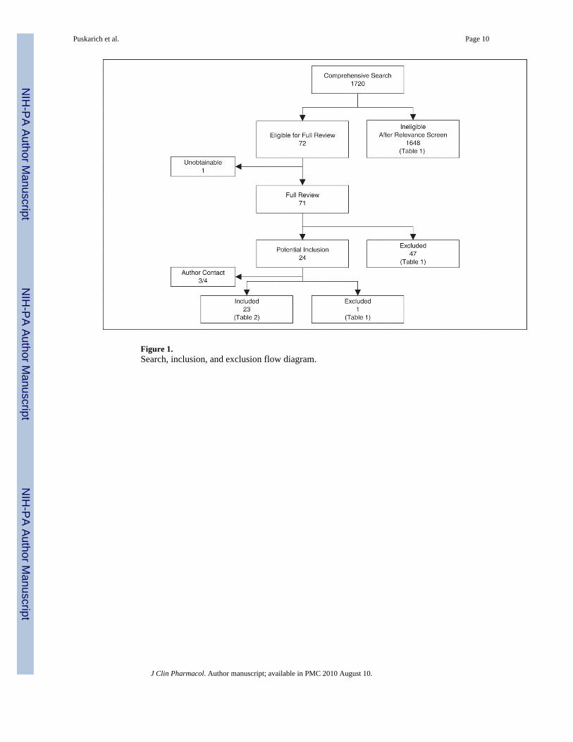
Figure 1.Search, inclusion, and exclusion flow diagram.
Puskarich et al. Page 10
J Clin Pharmacol. Author manuscript; available in PMC 2010 August 10.
NIH
-PA Author Manuscript
NIH
-PA Author Manuscript
NIH
-PA Author Manuscript
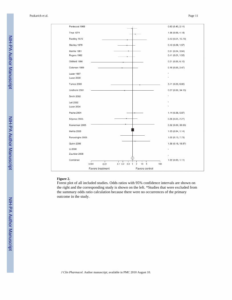
Figure 2.Forest plot of all included studies. Odds ratios with 95% confidence intervals are shown onthe right and the corresponding study is shown on the left. *Studies that were excluded fromthe summary odds ratio calculation because there were no occurrences of the primaryoutcome in the study.
Puskarich et al. Page 11
J Clin Pharmacol. Author manuscript; available in PMC 2010 August 10.
NIH
-PA Author Manuscript
NIH
-PA Author Manuscript
NIH
-PA Author Manuscript
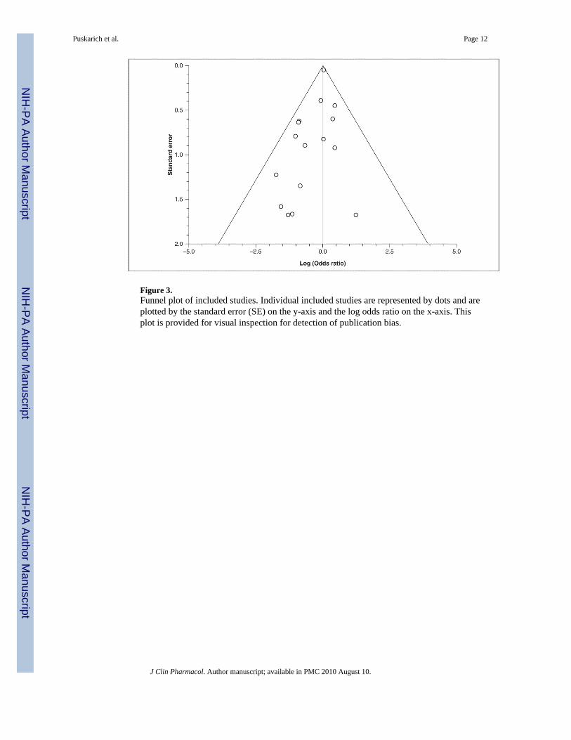
Figure 3.Funnel plot of included studies. Individual included studies are represented by dots and areplotted by the standard error (SE) on the y-axis and the log odds ratio on the x-axis. Thisplot is provided for visual inspection for detection of publication bias.
Puskarich et al. Page 12
J Clin Pharmacol. Author manuscript; available in PMC 2010 August 10.
NIH
-PA Author Manuscript
NIH
-PA Author Manuscript
NIH
-PA Author Manuscript
NIH
-PA Author Manuscript
NIH
-PA Author Manuscript
NIH
-PA Author Manuscript
Puskarich et al. Page 13
Table I
Primary Reason for Study Exclusion
After relevance screen
Review/letter/editorial 384
Nonrandomized/nontreatment study 20
Wrong focus or population 1244
After full manuscript review
Review article 1
Duplicate publication 4
Not critically ill 16
Wrong outcome 18
Wrong treatment 5
Wrong focus or population 3
Manuscript unobtainable for review 1
After attempted author contact
Failed author contact 1
Total 1697
J Clin Pharmacol. Author manuscript; available in PMC 2010 August 10.
NIH
-PA Author Manuscript
NIH
-PA Author Manuscript
NIH
-PA Author Manuscript
Puskarich et al. Page 14
Tabl
e II
Sum
mar
y of
Incl
uded
Stu
dies
Aut
hor
Yea
rIn
dica
tion
Stud
y Se
tting
GIK
Form
ulat
ion,
U/
kg/h
insu
linIn
fusi
on D
urat
ion,
hM
orta
lity
Tim
ing
Tre
atm
ent,
NC
ontr
ol, N
Tre
atm
ent M
orta
lity,
nC
ontr
ol M
orta
lity,
n
Sequ
ence
Gen
erat
iona
Allo
catio
n C
once
alm
entb
Blin
ding
cSe
lect
ive
Out
com
e R
epor
tingd
Pent
ecos
t19
68A
MI
ICU
Not
wei
ght-b
ased
48In
-hos
pita
l10
010
015
16C
CA
B
Thys
1974
AM
IIC
UN
ot w
eigh
t-bas
ed16
8In
-hos
pita
l85
8914
10C
CC
C
Rac
kley
1976
AM
IIC
U0.
075
48In
-hos
pita
l8
81
2C
CC
C
Stan
ley
1978
AM
I Unc
lear
0.07
548
In-h
ospi
tal
5555
49
CC
AC
Man
tle19
81A
MI
ICU
0.07
548
In-h
ospi
tal
4144
24
CC
CB
Rog
ers
1982
AM
IIC
U0.
075
48In
-hos
pita
l96
944
9C
CC
B
Old
field
1986
MV
RO
RN
ot w
eigh
t-bas
ed12
In-h
ospi
tal
2023
02
CC
CC
Col
eman
1989
CA
BG
OR
0.08
4860
-d11
111
4C
CA
B
Laza
r19
97C
AB
GO
R0.
0512
+In
-hos
pita
l16
150
0B
CC
B
Laza
r20
00C
AB
GO
RN
ot w
eigh
t-bas
ed12
+In
-hos
pita
l20
200
0B
CC
A
Turk
oz20
00C
AB
GO
R0.
14O
ther
eIn
-hos
pita
l15
161
0C
CC
B
Lind
holm
2001
CA
BG
±AV
RO
RN
ot w
eigh
t-bas
edO
ther
gIn
-hos
pita
l16
140
1C
CC
C
Smith
2002
CA
BG
OR
0.05
1030
-d22
220
0A
CA
B
Lell
2002
CA
BG
OR
0.07
512
In-h
ospi
tal
2120
00
CA
AB
Laza
r20
03C
AB
GO
RN
ot w
eigh
t-bas
ed12
30-d
7269
00
CC
AA
Pach
e20
04A
MI
ICU
0.07
224
30-d
155
157
75
AC
AA
Krlj
anac
2005
AM
IIC
U0.
0524
1-y
7840
34
CC
AC
Kos
kenk
ari
2005
CA
BG
+AV
RO
R1
6+In
-hos
pita
l20
200
1C
CC
B
Meh
ta20
05A
MI
ICU
0.07
524
30-d
10 0
8810
107
1004
976
AA
AA
Ran
asin
ghe
2006
CA
BG
OR
0.05
256+
In-h
ospi
tal
157
160
33
AA
AA
Qui
nn20
06C
AB
GO
R0.
0525
6+In
-hos
pita
l13
814
23
2A
AA
C
Li20
06A
MI
ICU
0.07
524
In-h
ospi
tal
912
00
CC
CC
Zuur
bier
2008
CA
BG
OR
0.1
24+
In-h
ospi
tal
2321
00
AA
CC
AM
I, ac
ute
myo
card
ial i
nfar
ctio
n; A
VR
, aor
tic v
alve
repl
acem
ent;
CA
BG
, cor
onar
y ar
tery
byp
ass g
rafti
ng; G
IK, g
luco
se–i
nsul
in–p
otas
sium
; IC
U, i
nten
sive
car
e un
it; M
VR
, mitr
al v
alve
repl
acem
ent;
N, n
umbe
r of s
ubje
cts i
n ea
ch g
roup
; n, n
umbe
r of s
ubje
cts w
ith p
rimar
you
tcom
e in
eac
h gr
oup.
a Sequ
ence
gen
erat
ion:
gra
de A
= a
dequ
ate,
gra
de B
= in
adeq
uate
, gra
de C
= u
nkno
wn.
b Allo
catio
n co
ncea
lmen
t: gr
ade
A =
per
form
ed, g
rade
B =
not
per
form
ed, g
rade
C =
unk
now
n.
J Clin Pharmacol. Author manuscript; available in PMC 2010 August 10.
NIH
-PA Author Manuscript
NIH
-PA Author Manuscript
NIH
-PA Author Manuscript
Puskarich et al. Page 15c B
lindi
ng: g
rade
A =
blin
ded,
gra
de B
= n
ot b
linde
d, g
rade
C =
unk
now
n.
d Sele
ctiv
e ou
tcom
e re
porti
ng: g
rade
A =
not
sugg
este
d, g
rade
B =
sugg
este
d, g
rade
C =
unk
now
n.
e GIK
adm
inis
tere
d fr
om in
duct
ion
of a
nest
hesi
a un
til th
e be
ginn
ing
of c
ardi
opul
mon
ary
bypa
ss (C
PB).
g GIK
adm
inis
tere
d fr
om 1
5 m
inut
es b
efor
e re
war
min
g un
til 3
0 m
inut
es a
fter C
PB d
isco
ntin
ued.
J Clin Pharmacol. Author manuscript; available in PMC 2010 August 10.
NIH
-PA Author Manuscript
NIH
-PA Author Manuscript
NIH
-PA Author Manuscript
Puskarich et al. Page 16
Tabl
e III
Sum
mar
y of
Stu
dies
in S
epsi
s Pop
ulat
ions
Stud
yN
GIK
For
mul
atio
nD
urat
ion
of In
fusi
onM
orta
lity
% C
hang
e M
AP
Post
-G
IK%
Cha
nge
Car
diac
Fun
ctio
nPo
st-G
IKA
dver
se E
vent
s
Clo
wes
197
410
1 g/
kg g
luco
se; 1
.5 U
/kg
insu
lin; 1
0 m
Eq K
CL
10 m
in40
%30
% in
crea
se18
7% in
crea
se (C
O)
Non
e re
porte
d
Wei
sul 1
975
61
g/kg
glu
cose
; 1.5
U/k
g in
sulin
; 15
mEq
KC
L10
min
67%
30%
incr
ease
244%
incr
ease
(CI)
Non
e re
porte
d
Vin
cent
198
16
75 g
glu
cose
; 100
U in
sulin
; 15
mEq
KC
L40
min
NR
NR
27%
incr
ease
(CO
)N
one
repo
rted
Bro
nsve
ld 1
985
151
g/kg
50%
glu
cose
; 1.5
U/k
g in
sulin
; 10
mm
ol K
CL
20 m
in67
%5%
incr
ease
12.5
% in
crea
se (C
I)N
one
repo
rted
Mau
ritz
1986
141
g/kg
70%
glu
cose
; 1.5
U/k
g in
sulin
; 10
mm
ol K
CL
15 m
in86
%61
% in
crea
se50
% in
crea
se (C
I)N
one
repo
rted
Ham
dula
y 20
062
30%
glu
cose
; 50
U in
sulin
; 80
mm
ol K
CL
18–7
2 h
100%
9% in
crea
se88
% in
crea
se (C
O)
Non
e re
porte
d
CI,
card
iac
inde
x; C
O, c
ardi
ac o
utpu
t; G
IK, g
luco
se–i
nsul
in–p
otas
sium
; KC
L, p
otas
sium
chl
orid
e; M
AP,
mea
n ar
teria
l pre
ssur
e; N
, num
ber o
f pat
ient
s in
stud
y; N
R, n
ot re
porte
d.
J Clin Pharmacol. Author manuscript; available in PMC 2010 August 10.