Potassium iodide capsule treatment of feline sporotrichosis
7
http://jfm.sagepub.com/ Journal of Feline Medicine and Surgery http://jfm.sagepub.com/content/early/2012/03/02/1098612X12441317 The online version of this article can be found at: DOI: 10.1177/1098612X12441317 published online 2 March 2012 Journal of Feline Medicine and Surgery Rodrigo C Menezes, Sandro A Pereira and Tânia M P Schubach Érica G Reis, Isabella D F Gremião, Amanda A B Kitada, Raphael F D B Rocha, Verônica S P Castro, Mônica B L Barros, Potassium iodide capsule treatment of feline sporotrichosis Published by: International Society of Feline Medicine American Association of Feline Practitioners and http://www.sagepublications.com can be found at: Journal of Feline Medicine and Surgery Additional services and information for http://jfm.sagepub.com/cgi/alerts Email Alerts: http://jfm.sagepub.com/subscriptions Subscriptions: http://www.sagepub.com/journalsReprints.nav Reprints: http://www.sagepub.com/journalsPermissions.nav Permissions: What is This? - Mar 2, 2012 OnlineFirst Version of Record >> by guest on March 25, 2012 jfm.sagepub.com Downloaded from
-
Upload
independent -
Category
Documents
-
view
1 -
download
0
Transcript of Potassium iodide capsule treatment of feline sporotrichosis
http://jfm.sagepub.com/Journal of Feline Medicine and Surgery
http://jfm.sagepub.com/content/early/2012/03/02/1098612X12441317The online version of this article can be found at:
DOI: 10.1177/1098612X12441317
published online 2 March 2012Journal of Feline Medicine and SurgeryRodrigo C Menezes, Sandro A Pereira and Tânia M P Schubach
Érica G Reis, Isabella D F Gremião, Amanda A B Kitada, Raphael F D B Rocha, Verônica S P Castro, Mônica B L Barros,Potassium iodide capsule treatment of feline sporotrichosis
Published by:
International Society of Feline Medicine
American Association of Feline Practitioners
and http://www.sagepublications.com
can be found at:Journal of Feline Medicine and SurgeryAdditional services and information for
http://jfm.sagepub.com/cgi/alertsEmail Alerts:
http://jfm.sagepub.com/subscriptionsSubscriptions:
http://www.sagepub.com/journalsReprints.navReprints:
http://www.sagepub.com/journalsPermissions.navPermissions:
What is This?
- Mar 2, 2012OnlineFirst Version of Record >>
by guest on March 25, 2012jfm.sagepub.comDownloaded from
Journal of Feline Medicine and Surgery0(0) 1 –6© ISFM and AAFP 2012Reprints and permission: sagepub.co.uk/journalsPermissions.navDOI: 10.1177/1098612X12441317jfms.com
IntroductionSporotrichosis is a mycosis caused by the dimorphic fungus Sporothrix schenckii, which infects humans, as well as other mammals, especially domestic cats. Classic infection is associated with traumatic, cutaneous inoculation of the fungus.1,2
Sporotrichosis occurs worldwide but is endemic in Latin America, India, South Africa and Japan. In the state of Rio de Janeiro, Brazil, a zoonotic form of this disease has been emerging over the last 12 years in a region with socioeconomic and environmental difficulties.3 In this epidemic, the domestic cat is involved in zoonotic transmission through scratches, bites and contact with lesion secretions. The potential of transmission is related to the high parasite burden in skin lesions in comparison to other animal species.3,4
Infections of S schenckii in cats range from a subclinical form that can progress to multiple skin lesions and fatal systemic involvement with or without extracutaneous signs, particularly respiratory signs Subcutaneous nodules and ulcers are the most frequent skin lesions in sick cats and can be found at three or more noncontiguous anatomical sites.5–7
Treatment of feline sporotrichosis presents a challenge as there is a limited number of oral antifungal agents, in
addition to the adverse effects and high cost.6 The azoles, ketoconazole and itraconazole, are the most common drugs for the treatment of feline sporotrichosis.8 Itraconazole is considered the drug of choice,9,10 especially when cost is not an issue.11 Although treatment with itraconazole has proved to be effective in cats, the clinical response is unsatisfactory in some cases, especially in those with respiratory signs and nasal mucosa lesions.6,12 In refractory cases to oral itraconazole, intralesional (IL) administration of amphotericin B6,13 or oral potassium iodide (KI) might be an alternative.14
Potassium iodide capsule treatment of feline sporotrichosis
Érica G Reis1, Isabella D F Gremião1, Amanda A B Kitada1, Raphael F D B Rocha1, Verônica S P Castro2, Mônica B L Barros3, Rodrigo C Menezes1, Sandro A Pereira1 and Tânia M P Schubach1
AbstractSporotrichosis is a mycosis caused by Sporothrix schenckii. The most affected animal is the cat; it has played an important role in the zoonotic transmission of this disease, especially in Rio de Janeiro, Brazil, since 1998. In order to evaluate the treatment of feline sporotrichosis with potassium iodide, an observational cohort was conducted in 48 cats with sporotrichosis at Instituto de Pesquisa Clínica Evandro Chagas, Fiocruz. All cats received potassium iodide capsules, 2.5 mg/kg to 20 mg/kg q24h. The cure rate was 47.9%, treatment failure was 37.5%, treatment abandonment was 10.4% and death was 4.2%. Clinical adverse effects were observed in 52.1% of the cases. Thirteen cats had a mild increase in hepatic transaminase levels during the treatment, six of them presented clinical signs suggestive of hepatotoxicity. Compared to previous studies with itraconazole and iodide in saturated solution, potassium iodide capsules are an alternative for feline sporotrichosis treatment.
Accepted: 15 February 2012
1 Laboratory of Clinical Research on Dermatozoonosis in Domestic Animals, Evandro Chagas Clinical Research Institute/Oswaldo Cruz Foundation, Rio de Janeiro, Brazil
2 Department of Small Animals, Federal University of Santa Maria, Camobi, 9, Veterinary Hospital, Santa Maria, Brazil
3 National School of Public Health Sergio Arouca, Oswaldo Cruz Foundation, Rio de Janeiro, Brazil
Corresponding author:Érica G Reis DVM MSc, Laboratory of Clinical Research on Dermatozoonosis in Domestic Animals, Evandro Chagas Clinical Research Institute/Oswaldo Cruz Foundation, Av Brazil, 4365, Manguinhos, 21045-900, Rio de Janeiro, BrazilEmail: [email protected]
441317 JFM0010.1177/1098612X12441317Reis et alJournal of Feline Medicine and Surgery2012
Original Article
by guest on March 25, 2012jfm.sagepub.comDownloaded from

2 Journal of Feline Medicine and Surgery 0(0)
KI, the first successful drug for the treatment of human sporotrichosis, continues to be used for fixed cutaneous and lymphocutaneous disease because of its low cost (compared to itraconazole) and its effectiveness.15 The mechanism of iodide action still remains poorly understood, but it seems that its therapeutic effect is mediated through modulation of the inflammatory response or enhancement of the immune defense mechanism.15,16 In vitro studies have suggested yeast cell damage through the conversion of KI to iodine. This conversion path for some unknown and appropriate concentrations could damage the yeast cell form in vivo.16
In feline sporotrichosis, there are few reports of cases having been treated with KI and the results are inconclusive.8 The lack of studies on KI in cats points to the need for systematic approaches to describe the efficacy of the drug for treating the disease. For public health purposes, a favorable response to KI in feline sporotrichosis could contribute to therapeutic regimen options, as well as reduce the risks of zoonotic transmission related to diseased cats, shorten the treatment period and diminish relapses. Thus, the purpose of this study was to evaluate the effectiveness of KI capsules in feline sporotrichosis treatment.
Materials and methodsBetween April 2010 and July 2011 an observational cohort was conducted with a sample of cats with sporotrichosis at the Laboratório de Pesquisa Clínica em Dermatozoonoses em Animais Domésticos (LAPCLINDERMZOO), Instituto de Pesquisa Clínica Evandro Chagas (IPEC)/(Fiocruz), Rio de Janeiro, Brazil. The study was approved by the Ethics Committee on the Use of Animals (CEUAFiocruz), number: LW 50/10. All procedures undertaken during the study were signed by the owners in terms of informed consent.
Fortyeight cats, of both sexes, with no previous history of oral antifungal therapy, were included according to the following criteria: weight >2 kg; sporotrichosis diagnosis confirmed by isolation of S schenckii in culture. The exclusion criteria were age <6 months and >9 years, pregnancy and lactation. Cats that did not improve within 1 month of KI treatment, or had worsening of the lesions regardless of the time of treatment, were excluded from the study and oral itraconazole monotherapy (50–100 mg/cat q24h) was initiated.
The cats underwent a clinical and dermatologic examination. Regarding the distribution of skin lesions,17 the cats were allocated into groups,: L1 (cutaneous lesions in only one site), L2 (cutaneous lesions in two nonadjacent sites) and L3 (cutaneous lesions in three or more nonadjacent sites).
Samples were collected from the ulcerated lesion and nasal cavities by sterile swab for mycological culture as follows: seeding on to Sabouraddextrose agar and
mycobiotic agar (Difco), incubated at 25ºC and observed during 4 weeks for fungal growth. Suspected isolates were subcultivated on potatodextroseagar medium (Difco) at 25ºC for macroscopic and microscopic morphological studies, and dimorphism was demonstrated by conversion to the yeastlike form on brain heart infusion agar medium (Difco) at 37ºC.17 Blood samples were collected for complete blood count and biochemistry (urea, creatinine, alanine transaminase, aspartate aminotransferase, alkaline phosphatase albumin) before and during the study, as well as for serological diagnosis for the feline immunodeficiency virus (FIV) and feline leukemia virus (FeLV) with an immunoenzymatic test (Snap Combo FeLV/FIV; Idexx Laboratories).
The therapeutic prescription for all cats was KI capsules supplied freeofcharge by the Clinical Pharmacy Service of IPEC/Fiocruz and was administered by the owners at home with food. A 5day scaling period of the drug was initially established with 2.5 mg/kg q24h. Then, doses were progressively increased at each 5day period until a clinical response was achieved or signs of toxicity appeared as follows: 5 mg/kg, 10 mg/kg, 15 mg/kg and 20 mg/kg q24h. Cats with mild adverse clinical effects had therapy suspended for 7 days and resumed 2.5 mg/kg dose increments until the that the highest dose that did not induce toxicity was attained. The cats were followed monthly by clinical examination and laboratory tests (hematology and biochemistry), and the owners were contacted weekly by telephone.
The criteria for cure were complete healing of the skin and mucosal lesions and remission of the extracutaneous clinical signs (ie, dyspnea, sneezing, nasal discharge,
Table 1 Sociodemographic characteristics, respiratory signs occurrence, adverse clinical effects, frequency of clinical cure and time to achieve cure in the observational cohort of cats with sporotrichosis treated with KI
Variables Cats treated with KI
Number of cats 48Male (%) 72.9Mongrel (%) 85.4Age in months (median) 24Weight in kg (median) 3.8Frequency of respiratory signs (%) 66.7Frequency of ACE (%) 52.1Frequency of FeLV antigen (%) 16.6Frequency of FIV antibodies (%) 8.3Frequency of clinical cure (%) 47.9Median time to achieve clinical cure (weeks) 19
ACE = adverse clinical effects
by guest on March 25, 2012jfm.sagepub.comDownloaded from
Reis et al 3
lymphangitis and regional lymphadenitis) presented initially. KI therapy was maintained for 2 months after clinical cure. A followup was carried out 3 months after the clinical cure.
The data collected from each animal were stored with the software Epidata 3.1. The simple frequencies of categorical variables [sex, general health, respiratory signs, presence, location and distribution of skin lesions, adverse clinical effects (ACE), laboratorial adverse effects and outcome] and measures of central tendency for quantitative variables (age, weight, monthly time to achieve cure and dose) were recorded with the software Statistical Package for the Social Sciences (SPSS 16.0).
The frequency of clinical characteristics of the cohort animals was described by univariate statistical analysis. The medians for time of treatment (in weeks) was used owing to nonparametric distribution in the KolmogorovSmirnov test. Fisher’s exact test was used to associate respiratory signs and treatment failure. A value of P <0.05 was considered significant.
ResultsFortyeight cats from the Rio de Janeiro metropolitan region were included in the study (Table 1). Thirtyfive cats (72.9%) exhibited good general health. Nasal
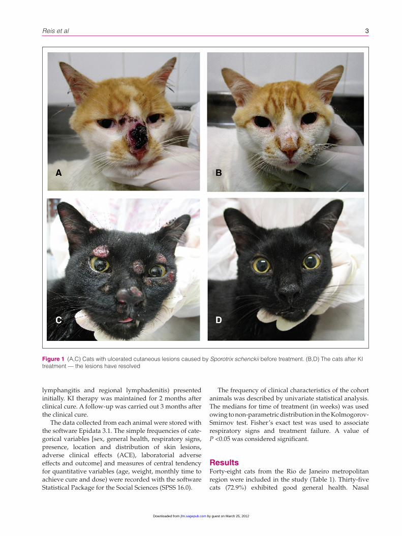
Figure 1 (A,C) Cats with ulcerated cutaneous lesions caused by Sporotrix schenckii before treatment. (B,D) The cats after KI treatment — the lesions have resolved
by guest on March 25, 2012jfm.sagepub.comDownloaded from
4 Journal of Feline Medicine and Surgery 0(0)
discharge (40.3%) was the most frequent respiratory sign and was not associated with treatment failure according to Fisher’s exact test (P = 1000).
Most lesions were located on the nasal mucosal (n = 26), right fore limb (n = 23), nasal bridge (n = 20) and head (n = 15). Cutaneous ulcers were observed in 34 (72.3%) animals, ulcers and nodules in 11 cats (23.4%) and nodules in two cats (4.3%).
Twentythree (47.9%) of the 48 cats achieved clinical cure (Figure 1) and 11 belonged to L1 group (Table 2). Treatment failure was observed in 18 animals: in 17 cats because of lack of clinical response and because of severe
adverse clinical effects in one cat. Five cats (10.4%) were lost during followup and two cats died (4.2%) with no association to sporotrichosis. Cats that tested positive for FeLV/FIV (Table 1) did not exhibit significant clinical or laboratory differences when compared with the FeLV/FIVnegative cats.
Hyporexia was the most frequent ACE (52%) sign related to treatment, followed by lethargy, weight loss, anorexia, vomiting and diarrhea. Thirteen cats (27.1%) had a mild increase in liver enzyme levels during the study,18 six of which displayed clinical signs suggestive of hepatotoxicity, such as hyporexia, lethargy and weight loss. No further relevant alterations were identified with respect to hematology or biochemistry.
The maximum dose at the beginning of treatment (median = 15.5 mg/kg) was similar to the discharge dose (median = 15.0 mg/kg). Only two cats had lower doses upon initiation and discharge — 5 mg/kg and 7 mg/kg, respectively. The variability of the dose of outcome (2.5–20 mg/kg) was higher than the initial dose (5–20 mg/kg), as five cats had a significant reduction in dose (Figure 2).
Fifteen cats out of 23 were followedup 3 months after clinical cure. No relevant alterations were identified in the routine hematology or biochemistry panel. One cat presented reactivation of the disease in the nasal mucosal 3 months after discharge, and eight were lost during followup.
DiscussionThis is the first report to evaluate KI capsules in cases of feline sporotrichosis in relation to clinical cure, adverse effects and reactivation. The treatment of feline sporotrichosis remains a challenging and longterm endeavor.12 Cats usually reject oral medication, especially in solution. This inspired us to manipulate KI in capsules, as it is convenient and easy to administer when compared with iodide in saturated solution (SSKI).
In this study, most animals had lesions in the nasal region. According to Malik et al,19 the nasal region of cats does not have a rich supply of blood nor does it possess easilyaccessible mobile skin, which would facilitate reconstructive surgical procedures. Thus, cure of infections in this specific area may be problematic.
Some authors have reported that the severity of feline sporotrichosis may be related to immunosupression caused by coinfection with FIV/FeLV.12,17 The small number of cats that tested positive in this study does not allow an association between the infection and outcome.
Respiratory signs were observed in most cats, mainly nasal discharge with no apparent link to treatment failure. However, the occurrence of respiratory signs is associated with treatment failure, according to previous
Table 2 Frequency of outcomes and length of treatment in weeks to achieve clinical cure according to the distribution of skin lesions in the observational cohort of cats with sporotrichosis treated with KI
Variables Distribution of skin lesions
L1 (n = 15)
L2 (n = 17)
L3 (n = 16)
Clinical cure 11 7 5Failure 3 9 6Other outcomes 1 1 5Median time to achieve clinical cure (weeks)
19 18 19
Others outcomes = death or abandonment
Figure 2 Median doses used at inclusion and discharge in 48 cats with sporotrichosis treated with KI capsules
by guest on March 25, 2012jfm.sagepub.comDownloaded from
Reis et al 5
studies.12,17 KI may represent a better alternative than itraconazole in cases with nasal mucosal involvement, although further clinical trials should be carried out to determine this hypothesis.
Previous studies have reported clinical cure in feline sporotrichosis with SSKI,20–22 even though others have claimed treatment to be unsuccessful.23,24 This study describes a large and wellcharacterized case series of cats treated with KI capsules, including a larger sample size, a different pharmaceutical form (capsules) and dose. Therefore, it was not possible to compare the cure rate with the veterinary literature, which was based only on isolated case reports. The treatment period ranged from 4 to 5 months, which is consistent with the period heretofore reported in literature.21,22,24 In addition, the animals belonging to the L1 group had a higher rate of clinical cure, as noted in previous studies.12,17
Regardless of the initial clinical condition, most of the cats in the failure group presented a worsening of the lesions after 1 month of therapy. KI therapy was discontinued and oral itraconazole was adopted for this group. Treatment abandonment was a result of noncompliance by owners.
Felines are susceptible to iodides and should be carefully monitored for adverse clinical signs, such as lethargy, anorexia, vomiting, diarrhea, spasms, hypothermia and cardiomyopathy.8 The clinical adversities evidenced in our study were consistent with the findings of other studies20,22 and hyporexia occurred in the majority of the cases. The increase in hepatic transaminase levels might be attributed to the use of KI being symptomatic in six cases.
Although the adverse clinical effects are common in cats treated with iodide, it does not preclude its application, as the toxic effects are reversible when the drug is suspended or administered in lower doses.24 Clinical and biochemical monitoring is strongly recommended, as well as the dosescaling period.
Continuous KI therapy could lead to interruption of the endogenous production of thyroid hormones and may also cause thyroiditis and/or hypo or hyperthyroidism, as observed in human patients. Thyroid stimulating hormone screening is prudent to ensure that thyroid function remains normal after 1 month of treatment.25
The dose of SSKI for feline sporotrichosis treatment according to the literature is 20 mg/kg q24h or q12h.21,24 Nevertheless, the median dose used to achieve cure with KI in capsules in our study was lower (15 mg/kg) than that established in the literature.
The clinical cure rate was similar to previous studies with itraconazole,12 which suggests that KI should be an alternative treatment for feline sporotrichosis. Further clinical trials should be carried out to compare the efficacy and safety of KI therapy and oral itraconazole.
Acknowledgements We thank the staff of LAPCLINDERMZOO, LABMICOL, LABCLIN, SEFARM and Raquel Vasconcelos from the Biostatistics Department of IPEC/Fiocruz.
TMP Schubach has a fellowship from the Technological and Scientific Development National Council (CNPq). We acknowledge Mitchell Raymond Lishon for the careful English review of the article.
Funding This study was partially supported by grants from Fundação de Apoio à Pesquisa do Estado do Rio de Janeiro (FAPERJ), Coordenação de Aperfeiçoamento de Pessoal de nível Superior (CAPES) and Programa de Incentivo à Pesquisa e ao Desenvolvimento Tecnológico (PIPDT)/IPECFiocruz.
Conflict of interest The authors do not have any potential conflicts of interest to declare.
References 1 Barros MB, Schubach AO, do Valle AC, Gutierrez Galhardo
MC, Conceicao-Silva F, Schubach TM, et al. Cat-transmitted sporotrichosis epidemic in Rio de Janeiro, Brazil: description of a series of cases. Clin Infect Dis 2004; 38: 529–535.
2 Rippon J. Sporotrichosis. In: Rippon J (ed), Medical mycology — The pathogenic fungi and the pathogenic actinomycetes. 3rd ed. Philadelphia: WB Saunders, 1988, pp 325–352.
3 Barros MB, Schubach TP, Coll JO, Gremiao ID, Wanke B and Schubach A. Sporotrichosis: development and chal-lenges of an epidemic. Rev Panam Salud Publica 2010; 27: 455–460.
4 Schubach TMP, Menezes RC and Wake B. Sporotrichosis. In: Greene C (ed), Infectious diseases of the dog and cat. 4rd ed. St. Louis: Elsevier Saunders, 2011, pp 645–650.
5 Schubach TM. Estudo clínico, laboratorial e epidemi-ológico da esporotricose felina na região metropolitana do Rio de Janeiro [Doutorado]. Rio de Janeiro: Fundação Oswaldo Cruz, 2004.
6 Gremiao I, Schubach T, Pereira S, Rodrigues A, Honse C and Barros M. Treatment of refractory feline sporotrichosis with a combination of intralesional amphotericin B and oral itraconazole. Aust Vet J 2011; 89: 346–351.
7 Pereira SA, Menezes RC, Gremiao ID, Silva JN, Honse Cde O, Figueiredo FB, et al. Sensitivity of cytopathological exami-nation in the diagnosis of feline sporotrichosis. J Feline Med Surg 2011; 13: 220–223.
8 Pereira SA, Schubach TMP, Gremião IDF, Silva DT, Figueiredo FB, Assis NV, et al. Therapeutic aspects of feline sporotri-chosis. Acta Sci Vet 2009; 37: 311–321.
9 Welsh RD. Sporotrichosis. J Am Vet Med Assoc 2003; 223: 1123–1126.
10 Madrid IM, Mattei A, Martins A, Nobre M and Meireles M. Feline sporotrichosis in the southern region of rio grande do sul, Brazil: clinical, zoonotic and therapeutic aspects. Zoonoses Public Health 2010; 57: 151–154.
11 Morris-Jones R. Sporotrichosis. Clin Exp Dermatol 2002; 27: 427–431.
12 Pereira SA, Passos SR, Silva JN, Gremiao ID, Figueiredo FB, Teixeira JL, et al. Response to azolic antifungal
by guest on March 25, 2012jfm.sagepub.comDownloaded from
6 Journal of Feline Medicine and Surgery 0(0)
agents for treating feline sporotrichosis. Vet Rec 2010; 166: 290–294.
13 Gremiao ID, Schubach TM, Pereira SA, Rodrigues AM, Chaves AR and Barros MB. Intralesional amphotericin B in a cat with refractory localised sporotrichosis. J Feline Med Surg 2009; 11: 720–723.
14 Kauffman CA, Bustamante B, Chapman SW and Pappas PG. Clinical practice guidelines for the management of sporo-trichosis: 2007 update by the Infectious Diseases Society of America. Clin Infect Dis 2007; 45: 1255–1265.
15 Sterling JB and Heymann WR. Potassium iodide in der-matology: a 19th century drug for the 21st century-uses, pharmacology, adverse effects, and contraindications. J Am Acad Dermatol 2000; 43: 691–697.
16 Torres-Mendoza BM, Vazquez-Valls E and Gonzalez-Mendoza A. Effect of potassium iodide on the immune response in the sporotrichosis. Rev Iberoam Micol 1997; 14: 98–100.
17 Schubach TM, Schubach A, Okamoto T, Barros MB, Figueiredo FB, Cuzzi T, et al. Evaluation of an epidemic of sporotrichosis in cats: 347 cases (1998–2001). J Am Vet Med Assoc 2004; 224: 1623–1629.
18 Center S. Interpretation of liver enzymes. Vet Clin Small Animals 2007; 37: 297–333.
19 Malik R, Pristas P and Javorsky P. Occurrence of plasmid-mediated ampicillin resistance among enterobacteria from the ovine rumen. Folia Microbiol (Praha) 2004; 49: 187–190.
20 Dunstan RW, Reimann KA and Langham RF. Feline sporo-trichosis. J Am Vet Med Assoc 1986; 189: 880–883.
21 Gonzalez Cabo JF, de las Heras Guillamon M, Latre Cequiel MV and Garcia de Jalon Ciercoles JA. Feline sporotrichosis: a case report. Mycopathologia 1989; 108: 149–154.
22 Burke M, Grauer G and Macy D. Succesful treatment of cuta-neolymphatic sporotrichosis in cat with ketoconazole and sodium iodine. J Am Anim Hosp Assoc 1982; 19: 542–547.
23 Mackay BM, Menrath VH, Ridley MF and Kelly WR. Sporotri-chosis in a cat. Aust Vet Pract 1986; 16: 3–5.
24 Dunstan RW, Langham RF, Reimann KA and Wakenell PS. Feline sporotrichosis: a report of five cases with transmis-sion to humans. J Am Acad Dermatol 1986; 15: 37–45.
25 Yamada K, Zaitz C, Framil VM and Muramatu LH. Cutane-ous sporotrichosis treatment with potassium iodide: a 24 year experience in Sao Paulo State, Brazil. Rev Inst Med Trop Sao Paulo 2011; 53: 89–93.
by guest on March 25, 2012jfm.sagepub.comDownloaded from


![2-[( E )-2-(4Ethoxyphenyl)ethenyl]-1-methylquinolinium iodide dihydrate](https://static.fdokumen.com/doc/165x107/631e216b05964b686800aa35/2-e-2-4ethoxyphenylethenyl-1-methylquinolinium-iodide-dihydrate.jpg)