Quality control of fibrinogen secretion in the molecular pathogenesis of congenital afibrinogenemia
Upload
khangminh22Category
view
1download
0
Fibrinogen, platelet and factor XIII
supplementation in cardiac surgery
In vitro and in vivo studies
Caroline Shams Hakimi
Department of Molecular and Clinical Medicine
Institute of Medicine
Sahlgrenska Academy at University of Gothenburg
Gothenburg 2017

Cover illustration: Graphite drawing of an activated platelet and fibrin threads drawn by the author
Fibrinogen, platelet and factor XIII supplementation in cardiac surgery © Caroline Shams Hakimi 2017 [email protected] ISBN 978-91-629-0300-8 (Print), 978-91-629-0301-5 (PDF) http://hdl.handle.net/2077/52857 Printed in Gothenburg, Sweden 2017 by BrandFactory AB

To Poja, Miryam and Meysam


Fibrinogen, platelet and factor XIII supplementation in cardiac surgery
In vitro and in vivo studies Caroline Shams Hakimi
Department of Molecular and Clinical Medicine, Institute of Medicine Sahlgrenska Academy at University of Gothenburg
Gothenburg, Sweden
ABSTRACT Background: There is a high risk of bleeding complications in cardiac surgery. Fibrinogen and platelet concentrates are often used to treat perioperative bleeding, but there is little information about its efficacy. The overall aim of this thesis project was to study the effects of fibrinogen, platelet and factor XIII concentrates on markers of hemostasis in blood samples from cardiac surgery patients.
Methods: Increasing doses of fibrinogen, platelets, and factor XIII were added to blood samples from patients or healthy volunteers (study I, III–V). In study II, blood samples from cardiac surgery patients with ongoing bleeding were analyzed before and after transfusion of fibrinogen and/or platelet concentrates. In all studies, platelet function was assessed with impedance aggregometry, and clot formation with thromboelastometry.
Results: Supplementation with fibrinogen improved clot formation while platelets improved both platelet aggregation and clot formation in blood samples form cardiac surgery patients (I). Fibrinogen to patients with ongoing bleeding improved clot formation and platelets improved platelet aggregation (II). Factor XIII supplementation to blood samples from cardiac and scoliosis surgery patients improved clot formation moderately (III). Supplementation with platelets improved platelet aggregation independently of antiplatelet therapy (IV). Time-dependent changes in platelet concentrates were detected with impedance aggregometry in vitro (V). The results predicted with moderate accuracy changes in aggregation after addition of the platelet concentrates to whole blood samples.
Conclusions: The results suggest that transfusion with fibrinogen or platelets improve hemostasis, whereas factor XIII should remain a secondary tool in the treatment of perioperative bleeding. Impedance aggregometry may be used to monitor the quality of stored platelet concentrates in vitro.
Keywords: Fibrinogen, platelets, factor XIII, platelet aggregation, clot formation, cardiac surgery.
ISBN: 978-91-629-0300-8 (Print), 978-91-629-0301-5 (PDF)


POPULÄRVETENSKAPLIG SAMMANFATTNING Inom hjärtkirurgi är det dessvärre ganska vanligt att patienten drabbas av en omfattande blödning i samband med operationen. Fem procent av patienterna blöder så mycket att de behöver tas tillbaka till operationssalen efteråt, och ungefär hälften av alla patienter behöver blodtransfusion. Stora blödningar ökar påtagligt risken för död och andra allvarliga komplikationer under och efter operationen.
Vid stora blödningar är det vanligt att man behandlar patienterna med transfusion av fibrinogen- eller trombocytkoncentrat för att minska blödningen, men det finns få studier som undersökt vad som händer i blodet när fibrinogen och trombocyter tillsätts. Fibrinogen är ett protein i blodet och en trombocyt är en celliknande beståndsdel av blodet, som båda hjälper till att levra blodet om det skulle uppstå en skada på ett blodkärl. Fibrinogen- och trombocytkoncentrat framställs av blod donerat av blodgivare. Fibrinogenkoncentrat har en lång hållbarhetstid, medan trombocytkoncentrat endast kan förvaras i upp till en vecka. Detta beror på att trombocyter har en begränsad överlevnadstid (i blodet lever de i 8–9 dagar) och att deras funktion försämras under förvaringen. Syftet med detta doktorandprojekt var att studera vad som händer med koagelbildningen (levringsförmågan) och trombocytfunktion (trombocyternas förmåga att binda samman till varandra) i blodprover efter tillsats av fibrinogen, trombocyter och faktor XIII (ett annat koagulationsfrämjande protein i blodet).
I den första studien tillsattes fibrinogen- och/eller trombocytkoncentrat till blodprover från patienter som just hjärtopererats. I studie II samlades blodprover vid två tillfällen från hjärtkirurgipatienter med pågående blödning; före och efter transfusion av fibrinogen- och/eller trombocytkoncentrat. I studie III tillsattes faktor XIII-koncentrat, ensamt eller tillsammans med fibrinogen eller trombocyter, till blodprover från hjärt- och ryggkirurgipatienter som just genomgått sin operation. I studie IV tillsattes trombocytkoncentrat till blodprover från patienter behandlade med olika läkemedel som hämmar trombocyterna. I studie V studerades trombocytkoncentrat vid olika tidpunkter under förvaringstiden. Dessutom studerades vad som händer med trombocytfunktionen i blodprover från friska frivilliga försökspersoner, när trombocytkoncentrat med olika lagringstid tillsattes blodproverna.

Trombocytfunktion och koagelbildning mättes i samtliga studier före och efter tillsats av fibrinogen, trombocyter eller FXIII. Trombocytfunktionen mättes med impedansaggregometri, en metod som mäter förändringen i elektriskt motstånd mellan elektrodpar i en testcell. Då trombocyter i blodprovet aktiveras och aggregerar på elektroderna ökar det elektriska motståndet, som omräknas till ett mått på trombocytfunktion. Koagelbildningen studerades med trombo-elastometri, där blod får koagulera i en testcell med en roterande pinne mitt i testcellen. Då blodet börjar koagulera bromsas pinnens rotation, och parametrar som tid till påbörjan av koagelbildning och maximal koagelstabilitet mäts.
Tillsats av fibrinogenkoncentrat i blodprover från hjärtkirurgipatienter förbättrade koagelbildningen och tillsats av trombocytkoncentrat förbättrade både koagelbildningen och trombocytfunktionen (studie I). Effekterna var dosberoende. När samma analyser gjordes före och efter transfusion av fibrinogen och trombocyter hos hjärtkirurgipatienter blev resultaten liknande (studie II). Förbättringen i trombocytfunktion och koagelbildning var associerad med minskad blödning. Faktor XIII-tillsats till blodprover från hjärt- och ryggkirurgipatienter förbättrade koagelbildningen, men effekten var mycket begränsad. När fibrinogen eller trombocyter tillsattes tillsammans med faktor XIII förbättrades koagelbildningen desto mer (studie III). Tillsats av trombocyter till blodprover från patienter behandlade med trombocythämmande läkemedel hade en förbättrande effekt, men effekten var måttlig, särskilt hos patienter behandlade med en viss typ av läkemedel, s.k. P2Y12-hämmare (studie IV). Förändringar i trombocytfunktion under förvaringstiden kunde följas med impedansaggregometri. Resultaten av undersökningarna kunde med måttlig precision förutspå förändringar i trombocytfunktion efter tillsats av trombocytkoncentraten till blodprover från friska frivilliga försökspersoner (studie V).
Sammanfattningsvis visar studierna att tillsats av trombocyter och fibrinogen förbättrade trombocytaggregation och koagelbildning, medan effekten av faktor XIII var begränsad. Trombocyttillsats i blodprover från patienter behandlade med P2Y12-hämmare hade dock en begränsad effekt. Impedansaggregometri kan användas för att mäta trombocyt-kvalitén i trombocytkoncentrat under förvaringen.

i
PREFACE The work in this thesis was performed at the Department of Cardiothoracic Surgery at Sahlgrenska University Hospital and financed by the Swedish Heart-Lung Foundation and Västra Götalandsregionen. I want to express my gratitude to my supervisor, Professor Anders Jeppsson, for giving me this opportunity and for all the support, enthusiasm, and inspiration. To Camilla Hesse, my co-supervisor, I want to give my thanks for all the help, encouragement and for sharing her knowledge. Many thanks also to all co-authors of the papers included in this thesis, for the great collaboration, including Inger Fagerberg Blixter, who also introduced me to laboratory work with blood samples.
To the research team, thank you for all the help throughout the years. I want to thank Christine Roman-Emanuel, Linda Thimour-Bergström, Erika Backlund Jansson, Åsa Israelsson-Skogsberg, Elisabeth Schyum, Anna Börjesson, Eva Lysell, Eva Berg, Susanne Nielsen, Sukhi Singh and Caroline Ivarsson. Colleagues at the administrative unit, thank you for providing me with an enjoyable working atmosphere.
To my friends and family, thank you for all the support and love. My husband Poja, I am so grateful for all the encouragement you have given me. You helped me manage through hard times. My children Miryam and Meysam, I am so lucky to have you in my life! To my mother Petra, thank you for always being there. Your help has been indispensable.


iii
LIST OF PUBLICATIONS This thesis is based on the following studies, referred to in the text by their Roman numerals.
I Shams Hakimi C, Fagerberg Blixter I, Hansson EC, Hesse C, Wallén H, Jeppsson A. Effects of fibrinogen and platelet supplementation on clot formation and platelet aggregation in blood samples from cardiac surgery patients. Thromb Res. 2014; 134: 985-900.
II Shams Hakimi C, Singh S, Hesse C, Jeppsson A. Effects of fibrinogen and platelet transfusion on hemostasis in cardiac surgery patients with ongoing bleeding. In manuscript.
III Shams Hakimi C, Carling MS, Hansson EC, Brisby H, Hesse C, Radulovic V, Jeppsson A. The effect of ex vivo factor XIII supplementation on clot formation in blood samples from cardiac and scoliosis surgery patients. Clin Appl Thromb Hemost. doi: 10.1177/1076029617713872. [Epub ahead of print].
IV Hansson EC*, Shams Hakimi C*, Åström-Olsson K, Hesse C, Wallén H, Dellborg M, Albertsson P, Jeppsson A. Effects of ex vivo platelet supplementation on platelet aggregability in blood samples from patients treated with acetylsalicylic acid, clopidogrel, or ticagrelor. Br J Anaesth. 2014; 112: 570-575. *Shared first authorship.
V Shams Hakimi C*, Hesse C*, Wallén H, Boulund F, Grahn A, Jeppsson A. In vitro assessment of platelet concentrates with multiple electrode aggregometry. Platelets. 2015; 26: 132-137. *Shared first authorship.

v
CONTENT
PREFACE ........................................................................................................ i
LIST OF PUBLICATIONS ............................................................................ iii
CONTENT ...................................................................................................... v
ABBREVIATIONS ........................................................................................ vii
1 INTRODUCTION ................................................................................... 1
1.1 Hemostasis ....................................................................................... 1 1.2 Cardiac surgery and bleeding........................................................... 4 1.3 Platelet inhibition ............................................................................ 5 1.4 Transfusion with blood products ..................................................... 7 1.5 Functional hemostatic tests ............................................................ 11 1.6 Study objectives .............................................................................. 17
2 AIMS ....................................................................................................... 19
3 METHODS ............................................................................................. 21
3.1 Participants ..................................................................................... 21 3.2 Study design and methodology ....................................................... 24 3.3 Analyses .......................................................................................... 31 3.4 Statistics ......................................................................................... 33
4 RESULTS ............................................................................................... 35
4.1 Supplementation with fibrinogen and/or platelets (study I) .......... 35 4.2 Transfusion with fibrinogen and/or platelets (study II) ................. 38 4.3 Addition of factor XIII, fibrinogen, and platelets (study III) ......... 42 4.4 Platelet inhibitors and supplementation with platelets (study IV) 44 4.5 Platelet concentrates and storage time (study V) .......................... 46
5 DISCUSSION .......................................................................................... 49
6 CONCLUSIONS ..................................................................................... 57
7 FUTURE PERSPECTIVES ................................................................... 59
REFERENCES ............................................................................................... 61


vii
ABBREVIATIONS AA Arachidonic acid
ADP Adenosine diphosphate
ASA Acetylsalicylic acid
AVR Aortic valve replacement
CABG Coronary artery bypass grafting
COX-1 Cyclooxygenase-1
CPB Cardiopulmonary bypass
DAPT Dual antiplatelet therapy
EDTA Ethylenediaminetetraacetic acid
FXIII Factor XIII
GPIIb/IIIa Glycoprotein IIb/IIIa
HES Hydroxyethyl starch
MI Myocardial infarction
MVR Mitral valve replacement
PBS Phosphate-buffered saline
PPP Platelet-poor plasma
PRP Platelet-rich plasma
TRAP Thrombin receptor-activating peptide-6
vWF von Willebrand factor


Caroline Shams Hakimi
1
Excessive bleeding during and after cardiac surgery is common, and is associated with an increased morbidity and mortality [7-10]. The cause of the bleeding can be both surgical and/or a compromised hemostasis.
In case of a blood vessel injury, vascular constriction, platelet plug formation, and coagulation contributes to the hemostasis of the affected area by temporarily decreasing the blood flow and closing the hole of the vessel [11].
The primary hemostasis consists of vasoconstriction and formation of a platelet plug. When a blood vessel is injured, the plasma protein von Willebrand factor (vWF) attaches to collagen that is exposed on the damaged vessel wall, and anchors the platelets to the vessel wall by binding to the glycoprotein platelet receptor Ib. In Figure 1, an activated platelet (middle) is visualized. Upon stimulation of platelet receptors, platelets are activated, turn irregularly shaped, and release contents from granules inside the platelets. From the dense granules, adenosine diphosphate (ADP), serotonin, thrombin, and thromboxane A2 are released, which activate adjacent platelets, resulting in activation of their glycoprotein IIb/IIIa (GPIIb/IIIa) receptors. From
-granules, vWF, factor V and XIII, and fibrinogen are released [11]. Several coagulation factor proteins are also present in the blood plasma. Platelets bind together with fibrinogen
Figure 1. A scanning electron micro-scope image of an activated platelet (middle) together with a red and a white blood cell [3].

Fibrinogen, platelet and factor XIII supplementation in cardiac surgery
2
(Figure 2), which attaches to GPIIb/IIIa receptors on the platelets. A platelet plug, although quite fragile, is formed [11].
To stabilize the platelet plug formed in primary hemostasis, coagulation, which is the secondary hemostasis, occurs at the location. A number of coagulation factor proteins are involved in the coagulation, which are successively activated in a chain reaction. These coagulation factors are located in the plasma and in the platelet granules.
The initiation phase of the coagulation starts with a contact between tissue factor, which is expressed on cells of the vessel wall, and the blood. The coagulation factor VII in the blood attaches to tissue factor, together they form a complex, and factor VII is activated. This complex then activates the coagulation factors IX and X. The activated factor X activates factor V, and these two factors forms a complex. The factor V is secreted from platelets and binds to a phospholipid membrane, thereby attaching the complex to platelets or a damaged vessel wall. When bound to this complex, activated factor X converts prothrombin to thrombin [11, 12].
The amplification phase starts with activation of factors V, VIII and XI and nearby platelets, which is induced by the thrombin formed in the initiation phase. The platelet activation makes the surface negatively charged. Coagulation factors forms complexes on this negatively
Figure 2. Cartoon representation of the fibrinogen molecule. Fibrinogen consist of two monomers, each composed of three chains which are coiled around each other. Upon thrombin cleavage, parts of the E domain (center of the fibrinogen molecule) are cleaved off and fibrin is formed [4].

Caroline Shams Hakimi
3
charged surface. Complexes of activated factors VIII and IX activate complexes of factor V and X, which in turn induces a massive conversion of prothrombin to thrombin [12]. The formed thrombin cleaves fibrinogen into fibrin monomers and converts factor XIII (Figure 3) to its active form. The fibrin monomers are then polymerized and the activated factor XIII crosslinks the fibrin polymers, forming a stable meshwork of fibrin fibers. The result is a mature blood clot [11]. A microscope picture of a blood clot can be seen in Figure 4.
To prevent spontaneous clot formation when not necessary, and to enable degradation of a blood clot after the wounded area is recovered, there are anticoagulant and fibrinolytic mechanisms that prevent a clot to be formed and degrade a blood clot, respectively. Substances such as
Figure 3. Cartoon representation of the factor XIII molecule [2].
Figure 4. A scanning electron microscope image of an activated platelet within a dense network of fibrin fibers and red blood cells in a blood clot. © Dr. Stanley Flegler/Visuals Unlimited, Inc., with permission [5].

Fibrinogen, platelet and factor XIII supplementation in cardiac surgery
4
antithrombin, protein C, protein S, and tissue factor pathway inhibitor are inhibitors of the clot formation located in the blood, which limit the coagulation to the area of the damaged vessel. On intact endothelium, prostacyclin, heparan sulfate, and nitric oxide inhibit platelet activation. There is also a fibrinolytic mechanism, dissolving formed fibrin preventing a clot to occlude a vessel at the injured region [12]. .
1.2 Cardiac surgery and bleeding The history of cardiac surgery goes back to the end of 19th century. The first worldwide reported successful cardiac operation was performed by the German surgeon Ludwig Rehn in 1896, when he managed to close a 1.5-cm stab wound in the right ventricle of the heart [13]. The first successful cardiac operation with the use of a heart-lung machine was performed by the American surgeon John Gibbon in 1953. The patient was an 18-year old woman with an atrial septum defect and symptoms of heart failure [14]. During the use of heart-lung machine, the anticoagulant heparin is crucial to prevent clot formation. Heparin pure enough for human use was prepared from mammal cadaver in 1935 [15]. Heart-lung machine, or cardiopulmonary bypass (CPB), is a technique used during most open-heart operations today, where oxygen-poor blood is led, most often from the right atrium of the heart into the machine, where it is oxygenated before it is pumped back, to arterial circulation, the ascending aorta in the vast majority of the cases. Hence, the heart-lung machine temporarily takes over the function of heart and lungs.
In cardiac surgery there is a high risk for bleeding complications. During cardiac surgery, with the use of CPB, the hemostasis is affected by exposure of the blood to artificial surfaces, hemodilution, consumption of coagulation factors (including fibrinogen and platelets), and sometimes hypothermia [16, 17]. Major bleeding in cardiac surgery is associated with a higher incidence of renal failure, stroke, sepsis, and a higher mortality [7-10]. Treatment with platelet inhibitors and anticoagulants, which are used to prevent thrombotic events, caused by undesired clot formation, may have a contributory effect to the impaired hemostasis and the increased bleeding [16, 18].

Caroline Shams Hakimi
5
Antiplatelet therapy is associated with an increased risk for spontaneous and perioperative bleeding complications. Dual antiplatelet therapy (DAPT) with acetylsalicylic acid (ASA) and a P2Y12-inhibitor, such as clopidogrel or ticagrelor, further reduces the risk of thrombotic events in acute coronary syndrome patients compared to ASA alone. However, the risk of surgical bleeding is significantly increased with DAPT [19, 20]. Platelet activation is inhibited by ASA by irreversible binding to cyclooxygenase-1 (COX-1), resulting in a reduced production of prostaglandin and thromboxane A2, substances responsible for platelet activation and aggregation. The dosage which is commonly used (75–100 mg daily) is sufficient for complete inhibition of COX-1. As platelets lack cellular nucleus, they are unable to synthesize new substances, such as COX-1 [21]. Therefore, the effect of ASA is maintained during the life span of the platelet, which is up to ten days [22]. Another group of platelet inhibitors is the P2Y12-inhibitors, which bind to the ADP platelet receptor P2Y12, and thereby reducing ADP-induced aggregation. Clopidogrel inhibits platelet aggregation by irreversible
Figure 5. The molecular structure of clopidogrel (top left) and its activation. The first activation step is an oxidation mediated by cytochrome P450. The two structures below are isomers with the same molecular formula. The last step is a hydrolysis, which gives the active metabolite (top right) [6].

Fibrinogen, platelet and factor XIII supplementation in cardiac surgery
6
binding to the P2Y12-receptors on platelets. Clopidogrel is a prodrug that undergoes extensive conversion in the liver before the active metabolite is formed, see Figure 5 [23]. Because of the extensive metabolism required, there is a variability in response. The
incidence of non-responders or poor-responders have been shown to be 20–40% [24]. Because of the irreversible binding of clopidogrel, the effect is maintained during the life span of the platelets. Ticagrelor (molecule shown in Figure 6) is another P2Y12-inhibitor which binds reversible to the receptors. The half-life of ticagrelor is 6-13 h [25]. Ticagrelor can move between P2Y12-receptors and has a rapid receptor kinetics; ticagrelor associates to P2Y12-receptors with a half-life of 4 min and dissociates with a half-life of 14 min [26]. Ticagrelor does not require extensive conversion in the liver, both its origin form and its active metabolite are potent platelet inhibitors [27]. There are no direct antidotes to ticagrelor or any other oral platelet inhibitor clinically
Figure 6. The ticagrelor molecule [1].
Figure 7. Schematic illustration of some of the platelet receptors and platelet activation pathways. The inhibitory pathways of acetylsalicylic acid (ASA), ticagrelor, and clopidogrel are also shown.

Caroline Shams Hakimi
7
available. Current guidelines recommend discontinuation of the P2Y12-inhibitor in elective surgery, but maintained treatment with ASA [28]. However, patients with ongoing or recently stopped DAPT undergoing acute surgery will have a maintained strong platelet inhibition during and after surgery, and accordingly a high risk for bleeding complications. In Figure 7, the inhibitory actions of ASA, clopidogrel, and ticagrelor are shown.
1.4 Transfusion with blood products Transfusion with blood components (red blood cells, platelets and plasma) and/or blood products (factor concentrates) is used to improve hemostasis in patients with ongoing bleeding. The first attempts to transfuse a human with animal blood occurred in 1667, and the first transfusion of human blood to human patients (woman with major post-partum hemorrhage) occurred in 1818. Some of the transfusions were successful, but many came with complications or were fatal [29]. In 1901, the four blood groups O, A, B, and AB were discovered by the Austrian Karl Landsteiner and his students, which subsequently increased the proportion of successful transfusions. In 1940, Landsteiner and Wiener discovered the Rhesus blood group system, which is the second most important blood group system (after the ABO system) in blood transfusion [29, 30]. In 1914, sodium citrate was found to function as a safe anticoagulant, which made it possible to store blood for transfusion at a later point in time [30]. Until 1940, whole blood or plasma was used for transfusion, but then fractionated blood products were developed, such as concentrates containing mainly fibrinogen, globulins or albumin. These fraction concentrates were easier to transport, which was beneficial during war situations. A couple of decades later, coagulation factor concentrates with factor VIII or IX became available for patients with hemophilia [30, 31]. These patients had earlier received plasma from animals, with allergic reactions as a result, and without treatment they seldom survived into adulthood. The first blood bank was established in London in 1921. Since then the transfusion medicine has developed, in terms of availability, durability, quality, and safety [30].

Fibrinogen, platelet and factor XIII supplementation in cardiac surgery
8
Although the improvement in safety, there is still a risk for complications associated with blood transfusions. Mild allergic symptoms occur in 1–3% of transfusions. However, alloimmunization to HLA (human leukocyte antigen) antigens occurs in less than 5% of patients transfused with platelet concentrates. The risk for serious adverse events (such as febrile non-hemolytic transfusion reactions, bacterial contamination, anaphylaxis and transfusion-related acute lung injury) is 0.1% for red blood cell transfusions and 0.04% for platelet concentrate transfusions [32, 33]. In a study performed at Sahlgrenska University Hospital in Gothenburg, Sweden, about 50–60% of all adult patients who underwent cardiac surgery during a 2-year period received at least one blood component [34], but there are large variations between different centers.
Platelet and fibrinogen transfusion is commonly used in the treatment of significant bleeding after cardiac surgery in many centers. Infusion of fibrinogen concentrate has been reported in up to 21% of cardiac surgery patients [35]. In an observational study of 102,470 patients undergoing coronary artery bypass graft surgery (CABG) in the United States, the incidence of perioperative platelet transfusion was 25% [36]. In a Danish prospective observational study with 811 cardiac surgery patients, 26% received platelet concentrates perioperative or during the first 24 postoperative hours [37].
Little is known about the efficacy of platelet transfusion in patients treated with platelet inhibitors, or if the efficacy varies between different antiplatelet therapies. It has been shown that the effects of one blood product to some degree can substitute the effects of another. For example, fibrinogen has been shown to decrease bleeding caused by hemodilution or thrombocytopenia in experimental models [38, 39], and to improve platelet aggregation ex vivo in samples from healthy subjects treated with GPIIb/IIIa inhibitors [40]. Plasma FXIII activity has been shown to be significantly correlated with bleeding volume after cardiac surgery [41-43], and to be reduced in surgical patients with unexpected bleeding [44]. In a study where patients were randomized to receive either FXIII concentrate or placebo at the start of their gastrointestinal

Caroline Shams Hakimi
9
cancer surgery, the decrease in clot stability and the perioperative blood loss was significantly less in the group that received FXIII [45]. However, little is known about the relationship between dose and response of platelet, fibrinogen and FXIII concentrates, the interaction between different blood products, and optimal combinations for transfusion.
Platelet concentrate When located in the blood vessels, the life span of the platelets is up to ten days [22]. The reference interval for platelet count is 165–387×109/L for women and 145–348×109/L for men [46], respectively, while the platelet count of platelet concentrates can be about 800–1800×109/L, depending on e.g. the preparation method and the donor(s) platelet count [47]. Transfusions of platelet concentrates have been used for decades, and their quality and safety have been improved over the years. For example preparation methods and storage medium have been elaborated, and pathogen reduction technologies have been introduced.
Platelet concentrates are stored at room temperature until use. The storage time varies and 30 years ago the storage time was only 3 days after preparation. Cryopreservation of platelet concentrates has been utilized, which allows for a longer storage period, however, the in vivo recovery of platelets has not been optimal [48]. Nowadays, the platelets are stored at room temperature for up to 7 days. Although improvements have been made, there is still a deterioration of the morphology and function of the platelets over the storage period, called platelet storage lesion. Microbial contamination is still an issue, because of the storage temperature [49, 50].
Platelet storage lesion may develop during the collection, processing, and storage of the platelet concentrate. Release of granule content may occur as a cause of high shear stress exposure during centrifugation. During storage, the platelets remain metabolically active, thereby keep consuming nutrients and producing metabolic products that may be harmful. Platelet-plasma interaction may activate coagulation factors. Some signs of platelet storage lesion that can be measured are;

Fibrinogen, platelet and factor XIII supplementation in cardiac surgery
10
decreasing levels in pH, oxygen, glucose, increasing levels in carbon dioxide, lactate, P-selectin surface expression, and decreased platelet aggregation [51]. Whether the platelets have undergone a shape change from discoid (their normal form) to spherical, which occurs at low pH or low storage temperatures (about 4°C), can be inspected by exposing the platelet unit to a light source while gently rotating it [51, 52]. Discoid platelets will then produce a visual “swirling” impression. The platelet morphology can also be determined by microscopy. Clinically, the only parameters routinely measured as a quality control are usually platelet count, concentrate volume, white blood cell count, and pH (when outdated) [51].
Platelet concentrates are either prepared from whole blood or with apheresis technique from blood donors. The platelets are stored in plasma and/or a platelet additive solution in plastic bags (Figure 8). The platelet concentrate can originate from one donor, or pooled from a number of donors.
Fibrinogen concentrates are prepared from plasma collected and pooled from a large number of blood donors. The reference interval for fibrinogen in the blood plasma is 1.8–3.8 g/L [46], and before administration, 1 g of fibrinogen is dissolved in 50 mL sterile water. The rationale of using fibrinogen concentrate in cardiac surgery patients is based on the association between pre- and postoperative plasma concentration of fibrinogen and postoperative bleeding volume as shown in several studies [53, 54]. Initial clinical studies with fibrinogen supplementation in patients with ongoing bleeding or as a pre-emptive treatment before surgery, showed promising results [55-57], which led to the hypothesis that fibrinogen could be used as a universal hemostatic
Figure 8. Platelet concentrates; buffy coat (left) and apheresis platelet concentrate (right). Photo by Camilla Hesse.

Caroline Shams Hakimi
11
agent [58]. However, more recent studies both in cardiovascular surgery [59, 60] and other fields [61] have not been able to confirm the initial results, although, in one study, fibrinogen concentrate was shown to decrease bleeding and transfusions after complex cardiac surgery [62].
Factor XIII concentrate FXIII concentrate is prepared from plasma collected and pooled from a large number of blood donors. The reference interval for plasma concentration of FXIII is 0.70–1.40 kIU/L [46]. Infusion with factor XIII concentrate is mainly used in individuals with inherited FXIII deficiency, which occurs in 0.5–1 in one million individuals. The target for prophylaxis treatment with FXIII concentrate in patients with inherited FXIII deficiency is 0.10–0.20 kIU/L, achieved by administration of 25–35 IU/kg every 4 to 6 weeks [63]. FXIII deficiency may also be acquired during massive bleeding. However, FXIII concentrate is rarely used as a first line treatment during peri- or postoperative bleeding. Little is known about the relationship between dose and response, and whether potential effects are preserved when FXIII is combined with another pro-hemostatic blood product such as fibrinogen or platelet concentrate.
1.5 Functional hemostatic tests There are several methods available for functional hemostasis testing, which assesses the platelet function or clot formation process. Below, some are described.
Platelet function testing The gold standard method used for the assessment of platelet function is light transmission aggregometry (LTA). There are many different methods available for platelet function measurement. Below, LTA and the methods platelet function analyzer (PFA), flow cytometry (which studies the surfaces of the platelets more than their function), and impedance aggregometry (the method used in this thesis project), are described, to indicate the difference in approaches utilized to assess platelet function.

Fibrinogen, platelet and factor XIII supplementation in cardiac surgery
12
LTA was developed in the 1960s by Born and O’Brien [64, 65] and is still the gold standard method used to study the platelet function. In this test, platelet-rich plasma (PRP) is prepared from citrated whole blood, and put into a cuvette with a magnetic stirring bar. Platelet aggregation is induced by the addition of an agonist, for example ADP, arachidonic acid (AA), collagen or epinephrine, and the temperature is regulated to 37°C. Optical density is used as a measurement of platelet aggregation, and the change in optical density of the PRP sample is recorded with a photometer. The optical density of platelet-poor plasma (PPP; from the same blood sample) is subtracted for adjustment. As the platelets aggregate in the cuvette, the optical density decreases [65]. The result is displayed as percentage aggregation, where 0% is minimum and 100% is maximum aggregation [66].
PFA has been regarded as a standardization of the method bleeding time, where a skin wound is made and the time until occlusion by a formed platelet plug is measured. In PFA, citrated whole blood is drawn, at high shear stress, through a capillary with a membrane which is coated with agonists (collagen+ADP or collagen+epinephrine) at the end. The time until closure of the capillary (CT; closure time), is used as a measure of platelet function [67].
In flow cytometry, the sample, usually anticoagulated whole blood, is passed through one or more laser beams, and different particles can be distinguished based on size, granularity, and signal intensity from different fluorescently labeled markers bound to the platelet [68]. The platelets can be studied in presence or absence of agonists. Fluorescently labeled antibodies are used which bind to antigens of interest (e.g. on fibrinogen receptors, collagen receptors or P-selectin secreted form the α-granules) on the platelet surface [68, 69]. Parameters of interest are commonly the percentage of platelets positive for the marker, and the median fluorescence intensity. Normally, platelets are analyzed separately (not attached to each other), however, flow cytometry can also be used to study platelet activation in microaggregates [68].

Caroline Shams Hakimi
13
In impedance aggregometry (one avalaible application is Multiplate®; Roche Diagnistics, Basel, Switzerland), the equivalent volume of saline and citrated or hirudin-anticoagulated whole blood are added to a test cell and an agonist is added to promote the platelets to aggregate to electrode pairs in the test cell. Pipetting is assisted with an automatized pipette connected to the computer, in which the measurement is recorded. The temperature is regulated to 37°C and the test cell content is continuously stirred with a magnetic stirring bar (Figure 9). As platelets aggregate on the two electrode pairs in the test cell, the impedance between the pairs increases, and the change in impedance over time is recorded and is displayed in a graph by two curves; one for each electrode pair. The area under the curve is calculated (the mean of the two curves) and reported in aggregation units (U) (Figure 9).
Two classical methods clinically used for the assessment of clot formation are activated partial thromboplastin time (APTT) and prothrombin time. Two other commonly used methods are thrombelastography and thromboelastometry, the latter used in this thesis project. All four methods are described below.
In APTT, intrinsically activated clot formation is measured. In the assay, PPP, a contact activator (for example kaolin or ellagic acid), and
Figure 9. Test cell used for impedance aggregomtry analysis with Multiplate® and representative aggregation curves. One curve is obtained from one of the two electrode pairs, and area under the curve is calculated from the mean of the two curves.

Fibrinogen, platelet and factor XIII supplementation in cardiac surgery
14
a phospholipid suspension are incubated at 37°C, followed by the addition of calcium. The time it takes from the addition of calcium for fibrin polymerization to occur is the APTT, which is either optically or mechanically measured. A prolonged APTT can be caused by deficiencies in coagulation factors II, V, VIII, IX, X, XI, XII, and fibrinogen [70].
In the prothrombin time, extrinsically activated clot formation is measured. In the assay, PPP is prepared from citrated whole blood and placed in a test cell together with calcium and a phospholipid suspension. Upon addition of tissue factor, clot formation is initiated and the time until fibrin polymerization occurs is determined by the optical density [70]. Prothrombin time can be assessed with the Owren or the Quick method. A prolonged prothrombin time can be caused by deficiencies in factors VII, X and prothrombin when the Owren method is used. When the Quick method is used, prothrombin time is also dependent on factor V and fibrinogen. The Owren method is less sensitive to interference from lupus antibodies or excess citrate, and is utilized in Scandinavian countries [71]. To standardize the prothrombin time assay, international normalized ratio (INR) was introduced. INR is the prothrombin time of the patient divided by the prothrombin time of normal plasma [70].
In thrombelastography (TEG®; Haemonetics Corporation, Braintree, MA, USA), whole blood is put in a test cell and clot formation is induced by addition of an agonist. During the measurement, the test cell oscillates (4.45°) in relation to a pin which is located in the test cell and connected to a torsion wire. While the blood starts to clot, the pin begins to follow the movement of the test cell, and this rotation is mechanically detected. The temperature is regulated to 37°C. The movement is calculated to a curve and numerical parameters such as reaction time and maximum amplitude are obtained. Reaction time is the time from the start of the analysis to when a 2 mm amplitude is reached and is measured in seconds. The maximum amplitude is also measured and reported in mm.

Caroline Shams Hakimi
15
Thromboelastometry (ROTEM®; Pentapharm GmbH, Munich, Germany) is developed from thrombelastography. It uses the same principle to measure clot formation, but the assay is somewhat different. The main difference with thromboelastometry is that with this device, the test cell is fixed while the pin oscillates, and as the blood starts to clot, the movement of the pin is gradually restricted. The movement of the pin is optically detected. Clotting time and maximum clot firmness are parameters correspondent to reaction time and maximum amplitude in thrombelastography. In Figure 10, visual descriptions of the assay and some of the result parameters are shown.
Advantages with the assays APTT and prothrombin time are that they can be used to detect a deficiency in most of the coagulation factors, or in vitamin K (because coagulation factors II, VII, IX, and X are vitamin K-dependent), or to monitor the effect from anticoagulation therapy (for example heparin and warfarin) [70, 72]. Limitations with the methods are that minor or modest deficiencies cannot be detected, and the required sample preparation to acquire plasma. The APTT assay has not yet been standardized, there may be differences in reagents, definitions of clotting time, and devices among centers [70, 71]. Some centers utilize APTT ratio to evaluate the effect of anticoagulant-treatment [73], however the results may still vary depending on the sensitivity of the reagent used, to the anticoagulant.
Cuvette
Detector
Heating block
4.75°
Light beam
Rotating pin
Mirror
Figure 10. Visualization of the thromboelastometry instrument and a representative graph. The curve is mirrored along the longitudinal axis. CT, clotting time; CFT, clot formation time; MCF, maximum clot firmness.

Fibrinogen, platelet and factor XIII supplementation in cardiac surgery
16
An advantage with thromboelastometry is the user-friendly automatized pipetting system. Advantages with thrombelastography and thrombo-elastometry are that no sample preparation is needed, that a first evaluation can be done after just ten minutes of analysis [74], and that a dysfunction in the clot formation process can be linked to for example the fibrin polymerization, platelet contribution to the clot, or that there is a heparin effect. Limitations with both assays are the hematocrit-dependency; that the clot formation improves with a decrease in hematocrit [75], which is not the case with in vivo coagulation. Thromboelastometry is not sensitive to pharmacological platelet inhibition, however a supplementary device is available for analysis of platelet function and platelet inhibition [76].

Caroline Shams Hakimi
17
1.6 Study objectives At the start of this thesis project, the effect on hemostasis after simultaneous supplementation of fibrinogen and platelet concentrates had not been compared to the effect of fibrinogen or platelets alone. However, it had previously been shown that supplementation with fibrinogen to blood samples was able to improve clot formation [77-79], and that platelet supplementation was able to increase platelet aggregation [80]. Study I was designed to investigate the effect of platelet and fibrinogen supplementation on platelet aggregation and clot formation in blood samples from cardiac surgery patients. Furthermore, we aimed to study whether simultaneous supplementation of fibrinogen and platelet concentrate would be more effective on platelet aggregation and clot formation compared to fibrinogen or platelet concentrate alone.
To study whether potential findings in study I was possible to translate into real-life, study II was conducted. In this study, blood samples from cardiac surgery patients with ongoing bleeding were analyzed before and after the transfusion of fibrinogen, platelets, or the combination of both.
The effect of FXIII supplementation on clot formation has been studied by others, but the results have been conflicting [77-79, 81-93]. The clot formation process had been studied after supplementation of FXIII, both added alone [77-79, 81-86, 88-93] and together with fibrinogen [77-79, 82, 84, 85, 87, 88], but not together with platelet concentrate. In some of the studies, supraphysiological doses of FXIII were used [77, 79, 84-86, 88]. In study III, blood samples from two markedly different groups of surgical patients: cardiac and scoliosis surgery patients were supplemented with clinically relevant doses of FXIII, alone or together with fibrinogen or platelets.
Cardiac patients treated with oral platelet inhibitors undergoing acute surgery have a high-grade platelet inhibition and there are no direct antidotes available. The use of platelet concentrates to improve platelet function in patients with low platelet count or known platelet dysfunction, e.g. in patients with antiplatelet therapy, is rarely debated,

Fibrinogen, platelet and factor XIII supplementation in cardiac surgery
18
although few investigations had been able to show that it is effective [94-96]. To investigate the efficacy, blood samples from patients treated with different platelet inhibitors were supplemented with platelet concentrate in study IV. Platelet aggregation was measured before and after platelet supplementation to blood samples from patients treated with ASA, ASA+clopidogrel, ASA+ticagrelor and from healthy volunteers.
If transfusion with platelet concentrate is not sufficient to improve platelet aggregation, one potential cause could be time-dependent degradation of the platelets in the concentrates. Study V was designed to investigate if impedance aggregometry, performed directly on platelet concentrates, can be used to monitor platelet function in stored platelet concentrates and if the results predict changes in platelet aggregation after supplementation of the platelet concentrate in an in vitro transfusion model.

Caroline Shams Hakimi
19
2 AIMS The aims of this thesis project were
1. To investigate if supplementation with fibrinogen and/or platelet concentrate improves platelet aggregation and clot formation in postoperative blood samples from cardiac surgery patients (study I)
2. To investigate if transfusion with fibrinogen and/or platelet concentrate improves platelet aggregation and clot formation in cardiac surgery patients with ongoing bleeding (study II)
3. To investigate if supplementation with increasing doses of FXIII concentrate improves clot formation in postoperative blood samples from cardiac and scoliosis surgery patients (study III)
4. To investigate if supplementation with increasing doses of platelet concentrates restores platelet function in blood samples from patients treated with different platelet inhibitors (study IV)
5. To investigate if impedance aggregometry can be used to monitor time-dependent changes in platelet concentrates (study V)

Fibrinogen, platelet and factor XIII supplementation in cardiac surgery
20

Caroline Shams Hakimi
21
3 METHODS
3.1 Participants All studies were approved by the Regional Research Ethics Committee in Gothenburg and performed in accordance with the Declaration of Helsinki. In all studies, patients were included after obtaining written informed consent. In study I, IV and V, only men without previous history of blood transfusion were included, to prevent reactions due to alloimmunization that could occur due to transfusion or pregnancy. Exclusion criteria for study I and III–V were known bleeding disorder, known renal or liver disease, and a platelet count <150×109/L (preoperative count for the surgery patients). The healthy volunteers in study IV and V had not consumed any drugs influencing bleeding and coagulation at least 7 days prior to the blood sample collection(s). In study II, inclusion criteria were ongoing significant bleeding after an open heart surgery procedure, where the responsible physicians had prescribed transfusion with either fibrinogen, platelets, or both fibrinogen and platelets. The only exclusion criterion was known bleeding disorder not caused by preoperative medication. While most patients in study II were under anesthesia at the time of transfusion, they were asked to participate and gave their written consent at a later date, according to the approval from the Regional Ethics Committee. A patient could only be included once, regardless of repeated transfusions. The decision to transfuse the patient was based on clinical signs, routine coagulation tests, blood gases and thromboelastometry results.
Characteristics for the patients included in the studies where fibrinogen, platelet or factor XIII concentrate were supplemented to blood samples, or fibrinogen and/or platelet concentrates were transfused, are summarized in Table 1.

Fibrinogen, platelet and factor XIII supplementation in cardiac surgery
22
Table 1. Patient characteristic for patients in study I, II, and III. Median and interquartile range (25th–75th percentile), or number and percentage.
Study I Study II Study III
Cardiac ex vivo
Cardiac in vivo Cardiac Scoliosis
n 15 41 9 10 Female/male gender 0/15 5/36 0/9 8/2 Age (years) 70 (61–76) 73 (65–76) 66 (65–70) 14 (14–17) BMI (kg/m2) 28 (26–31) 25 (24–28) 28 (25–29) 17 (17–19) Diabetes mellitus 4 (27%) 6 (15%) 2 (2%) 0 Smoker/former/non-smoker 1/9/4 0/19/22 0/7/2 0/1/9
Previous MI 9 (60%) 13 (32%) 4 (44%) 0 Procedure CABG 13 (87%) 18 (43%) 9 (100%) – AVR 1 (6.7%) 13 (32%) 0 – CABG + AVR 1 (6.7%) 7 (17%) 0 – MVR 0 2 (5%) 0 – AVR + MVR 0 1 (2%) 0 – Scoliosis surgery – – – 10 (100%) Preoperative lab results
Hemoglobin (g/L) 140 (132–148)
147 (136–150)
126 (122–137)
115 (113–118)
Platelet count (×109/L)
241 (190–289)
210 (176–252)
210 (172–229)
253 (218–291)
Plasma fibrinogen (g/L)
3.4 (3.0–4.2) 3.0 (2.6–3.6) 3.1 (2.7–3.3) 2.3 (2.0–2.8)
Key: AVR, aortic valve replacement; CABG, coronary artery bypass grafting; MI, myocardial infarction; MVR, mitral valve replacement.
Characteristics of the patients and the healthy volunteers included in the studies where platelet concentrates were supplemented to blood samples are summarized in Table 2.

Caroline Shams Hakimi
23
Table 2. Characteristics of patient groups and healthy volunteers whose blood samples were supplemented with platelet concentrates (study IV and V). Median and interquartile range (25th–75th percentile), or number.
Study IV Study V
Healthy volunteers
Patients treated with platelet
inhibitors
Healthy volunteers
n 10 40 9 Age (years) 55 (49–75) 68 (60–75) 37 (33–40) BMI (kg/m2) 24 (23–27) 26 (24–30) 27 (23–29) Smoker/former/non-smoker 0/0/10 2/21/17 0/0/9
Hemoglobin (g/L) 145 (139–151) 139 (132–145) 148 (144–152) Platelet count (×109/L) 224 (166–251) 231 (202–282) 212 (193–265)
Diabetes mellitus 0 6 (15%) 0 Previous MI 0 20 (50%) 0 Antiplatelet therapy ASA 0 40 (100%) 0 ASA + clopidogrel 0 15 (38%) 0
ASA + ticagrelor 0 15(38%) 0 Key: ASA, acetylsalicylic acid; MI myocardial infarction.

Fibrinogen, platelet and factor XIII supplementation in cardiac surgery
24
3.2 Study design and methodology
Study I Venous blood samples from the patients were collected after surgery, when the patient was off CPB (after heparin had been neutralized with protamine). Blood was collected in hirudin (>0.15 mg/L) and citrate (0.129 M) tubes, which were used for platelet aggregation and clot formation analysis, respectively. Blood was also collected in EDTA tubes for hemoglobin and platelet count.
Postoperatively, ten different samples were prepared for platelet aggregation and clot formation analysis: one baseline, three with increasing doses of fibrinogen (+0.5, 1.0, and 1.5 g/L) (Riastap®; CSL Behring GmbH, Marburg, Germany), three with increasing amounts of platelet concentrate (+46, 92, and 138 ×109/L), and three with increasing doses of both fibrinogen and platelets (Figure 11). The platelet concentrates were fresh (<6 h old) allogeneic ABO-compatible single-donor apheresis platelet concentrates, prepared at the regional blood bank, Sahlgrenska University Hospital, using Trima Accel system version 6.0 (Terumo BCT, Zaventem, Belgium) according to the manufacturer’s instruction. The platelet count of the donor before donation was at least 230×109/L. The target concentration was 1600×109/L for each platelet concentrate. The whole blood to citrate ratio was 10:1 and the platelets were stored in autologous plasma. The levels of A- and B-antibodies were checked in the concentrate of blood group O; only concentrates with a low level of antibodies (titre of <1/100 with indirect antiglobulin testing) were included. Phosphate-buffered saline (PBS; 140 mM NaCl and 10 mM Na3PO4, pH 7.4) was used in varying volumes to keep the hemodilution of the samples constant (23% by volume). The amounts of added fibrinogen (0.5–1.5 g/L) and platelets (46–138 ×109/L) correspond to approximately 2–5 fibrinogen and 2–5 units of platelet concentrate to a 70-kg patient.
Platelet aggregation was assessed with impedance aggregometry and clot formation with thromboelastometry, for the ten samples. Both methods are described in the Analyses section.

Caroline Shams Hakimi
25
Intraoperative bleeding was based on waste suction volume and sponges as estimated by the operation nurse. Postoperative bleeding was defined as mediastinal drain loss volume, measured hour by hour in the intensive care unit.
Blood samples were collected from an arterial line in a citrate tube (0.129 M, 2.7 mL) and a hirudin tube (>0.15 mg/L, 3 mL) for clot formation and platelet aggregation analyses, respectively. The samples were collected immediately before the transfusion and 15–30 minutes after the transfusion was completed. Blood samples for hemoglobin, plasma fibrinogen concentration and platelet count were also collected before and after the transfusion. In addition, bleeding volumes (chest drain output) were registered hour by hour before and after transfusion. The median bleeding volume during the two hours before and the two hours after transfusions were calculated.
Fibrinogen concentrate (Riastap®, CSL Behring GmbH, Marburg, Germany) was reconstituted in sterile water before administration, 1 g fibrinogen per 50 mL water. Platelet concentrates (apheresis, buffy-coat or interim platelet unit platelet concentrates) from the local blood bank were used. Platelet concentrates of all types were leucocyte-reduced (<1×10⁶ leucocytes per unit) and stored at 22°C in a platelet incubator (Helmer Agitator; Fenwal Europe, Mont Saint Guibert, Belgium) with horizontal agitation until use (up to 7 days after donation). The platelet concentrates contained approximately 250–300 × 109 platelets per unit.
Figure 11. Samples prepared for platelet aggregation and clot formation analysis in study I; one baseline, three with increasing doses of fibrinogen, three with increasing doses of platelets, and three with both fibrinogen and platelets.
Baseline Fibrinogen Platelets Fibrinogen + platelets

Fibrinogen, platelet and factor XIII supplementation in cardiac surgery
26
Impedance aggregometry was used to assess the platelet aggregation and thromboelastometry was used to assess the clot formation process. Hemoglobin levels, plasma fibrinogen concentration and platelet count were measured using standard clinical methods.
Study III In a prestudy, citrated whole blood samples from six healthy volunteers were supplemented with three doses of FXIII which increases the plasma concentration of FXIII by 0.20, 0.40, and 0.60 kIU/L (Concentration of the concentrate: 62.5 IU/mL; Fibrogammin®; CSL Behring, Marburg, Germany). The plasma FXIII concentrations were measured at the accredited coagulation laboratory at Sahlgrenska University Hospital, using the Berichrom FXIII assay (Dade Behring, Marburg, Germany) on the BCS XP instrument (Siemens Healthcare GnbH, Erlangen, Germany) (reference range 0.70–1.40 kIU/L). This prestudy was performed to evaluate the FXIII concentrate content.
In the main study, postoperative blood samples from the cardiac surgery patients were collected when the patient was off CPB (as in study I). Blood samples from the scoliosis surgery patients were collected directly after completion of surgery. All samples were collected from an arterial line, except in two scoliosis patients, where blood was collected from a peripheral vein catheter. Samples were collected in citrate tubes (0.129 M) for clot formation analysis, and for measurement of fibrinogen plasma concentration and FXIII activity. For measurement of fibrinogen and FXIII, the blood was centrifuged for 20 min at 2,000 × g within 30 min of collection and the plasma was stored at –80°C in polypropylene cryotubes for later analysis. Additional blood was collected in EDTA tubes for hemoglobin concentration and platelet count.
Ten different samples were prepared and analyzed with tromboelastomtry. The samples consisted of; one baseline and three with increasing doses of FXIII concentrate (+0.20, 0.40, and 0.60 kIU/L), alone together with a fixed dose of fibrinogen concentrate (+1.0 g/L) (Riastap®; CSL Behring) or fresh apheresis platelets (+92×109/L)

Caroline Shams Hakimi
27
from the regional blood bank (same as in study I, but the age of the concentrates here were <24 h) (Figure 12). The doses correspond to clinically relevant doses of approximately 1,050–3,150 IU FXIII concentrate, 3 g fibrinogen concentrate, and 3 units of single-donor apheresis platelets to a 70-kg patient. PBS was used in different volumes to maintain the same hemodilution (23% by volume) in the samples.
Fibrinogen concentration and FXIII activity were analyzed at the accredited coagulation laboratory at Sahlgrenska University Hospital. Fibrinogen plasma concentration (reference range when the study was performed was 2.0–4.5 g/L) was measured by the modified method of Clauss [97]. Hemoglobin concentration and platelet count were measured using standard clinical methods.
Venous blood samples from the healthy volunteers and the patients were collected in hirudin-anticoagulated tubes (>0.15 mg/L) and one EDTA tube two hours after intake of the morning dose of 75 mg ASA alone, or 75 mg ASA together with 75 mg clopidogrel (daily dose), or 90 mg ticagrelor (twice daily; 180 mg per day). The EDTA tube was used for measurement of hemoglobin and platelet count and the hirudin tubes were used for platelet aggregation analysis.
Three increasing amounts of single-donor apheresis platelet concentrate were added (same type and age as in study I) to the samples from each study individual (Figure 13). The baseline sample consisted of 1 mL blood and 300 μL PBS, and the samples with added platelets consisted of 300 μL blood and various amounts of platelet concentrate and PBS, to maintain the same hemodilution (23% by volume) in all samples. The
Figure 12. Samples prepared for clot formation analysis in study III; one baseline, three with increasing doses of factor XIII (FXIII), alone or together with a fixed dose of fibrinogen or platelets.
Fi 12 S l d f l f i l i i d III
Baseline FXIII FIB+ FXIII PLT + FXIII

Fibrinogen, platelet and factor XIII supplementation in cardiac surgery
28
amounts of platelets added to the blood samples were +46, 92, and 138 ×109/L, corresponding to approximately 2–5 units of single-donor apheresis platelets to a 70-kg patient.
To assess the platelet aggregation, the baseline and the samples with added platelets were analyzed with impedance aggregometry.
Platelet concentrates prepared with two different techniques; apheresis or pooled buffy coat, stored for one, three, five, or seven days were studied. The apheresis platelet concentrates were of the same type as in study I-IV, and preparation details are described in the method section for study I. The buffy coat-derived platelet concentrates were prepared from buffy coats donated by four regular donors of whole blood one day prior to the preparation. Whole blood units were collected in bottom-and-top bags (MacoPharma Nordic AB, Helsingborg, Sweden) withholding 63 mL citrate phosphate dextrose as anticoagulant. The whole blood units were hard-spin-centrifuged (4,880×g for 11 min at 23°C) and the buffy coat was separated from plasma and the red blood cells using a blood expander platform (Macopress Smart, MacoPharma Nordic AB). The buffy coats were then pooled together with platelet additive solution (SSP; MacoPharma Nordic AB) and processed in an automated blood component processing device (TACSI, Terumo BCT Europe, Zaventem, Belgium) to separate the remaining red cells. The residual plasma content was approximately 20%.
In the in vitro part of the study, platelet concentrates were analyzed. Samples from both types of concentrates were taken aseptically from the bag using a separate sampling bag on day one, three, five, and seven
Figure 13. Samples prepared for platelet aggregation analysis in study IV; baseline and three with increasing doses of platelet concentrate.
Baseline Platelets

Caroline Shams Hakimi
29
(n=13 for each day and type of platelet concentrate) after collection. The samples were transferred to small plastic tubes without any additives for assessment of platelet aggregation (Figure 14), and to EDTA tubes for measurement of platelet count. Platelet count was analyzed within one hour after sampling. Before each sample collection from the platelet concentrates, a visible “platelet swirl” was detected. The platelet concentrates were analyzed with impedance aggregometry.
In the ex vivo part of the study, whole blood samples from nine healthy volunteers were collected every second day: from the antecubital vein using a BD Vacutainer Eclipse blood collection needle (BD Diagnostics, NJ, USA). The blood samples were collected in hirudin tubes (>0.15 mg/L). Before analysis, the samples were diluted (20% by volume) with hydroxyethyl starch (HES) (Venofundin 60 mg/mL; B. Braun Melsungen AG, Melsungen, Germany), to resemble samples from patients with impaired platelet function. Samples for impedance aggregometry analysis were collected from the platelet concentrates on storage day one, three, five, and seven. Whole blood samples from three healthy volunteers were supplemented with the same preparation, one apheresis concentrate and one buffy coat concentrate, the same batches for every consecutive collection day.
For each healthy volunteer and day, three samples were prepared for platelet aggregation analysis: one baseline sample, one sample
BC AC Baseline BC AC
In vitro Ex vivo
Figure 14. Samples which were analyzed with impedance aggregometry in study V. This setup was used on day 1 after preparation of the platelet concentrates, and it was repeated on storage days 3, 5, and 7. Buffy coat platelet concentrates (BC) and apheresis platelet concentrates (AC) were analyzed in vitro and after ex vivo supplementation to diluted whole blood samples from healthy volunteers.
Fibrinogen, platelet and factor XIII supplementation in cardiac surgery
30
supplemented with apheresis platelets, and one supplemented with buffy coat platelets (Figure 14). The platelet-supplemented samples consisted of 1,080 µL of HES-diluted blood and 285 µL platelet concentrate, and the baseline sample consisted of 1,080 µL HES-diluted blood and 285 µL PBS (to keep the total volume constant). The amount of platelets in the concentrates was standardized to 700×109/L, so the increase in platelets was 146×109/L. If needed, PBS was used to dilute the platelet concentrates to the desired concentration. A sample in an EDTA tube was also collected from the healthy individuals, for measurement of hemoglobin and platelet count.

Caroline Shams Hakimi
31
3.3 Analyses Platelet function was analyzed with impedance aggregometry (Multiplate®; Roche Diagnistics, Basel, Switzerland). In this method, 300 µL saline and 300 µL whole blood are added to a test cell and incubated at 37°C for three minutes, and the test cell content is continuously stirred with a magnetic stirring bar. Then, an agonist is added to promote the platelets to aggregate on electrode pairs in the test cell, and the change in impedance over time is recorded for 6 minutes. In study I, III–V, the 300 µL blood used for impedance aggregometry analysis consisted of whole blood supplemented with PBS (baseline), fibrinogen, platelet or FXIII concentrate. For the analysis of platelet concentrates solely in study V, the 300 µL sample volume used in the assay consisted of 150 μL of the platelet concentrate and 150 μL PBS, for the apheresis concentrates, and 150 μL of the concentrate and 150 μL allogeneic plasma of blood group AB, for pooled buffy coat platelet concentrates. The area under the curve, reported in aggregation units (U), was used as a measure of platelet aggregation.
In study I–IV three test were used; ADP high sensitivity-test, ASPI-test, and TRAP-test. In ADP high sensitivity test, the agonist adenosine diphosphate (ADP, final concentration 6.3 µM) is used together with prostaglandin E1 (final concentration 9.4 nM) for increased sensitivity for P2Y12-dependent aggregation. In ASPI test, COX-1-dependent aggregation is assessed with arachidonic acid (AA, final concentration 0.48 mM) as agonist and it is ASA-sensitive. In TRAP test, thrombin receptor-activating peptide-6 (TRAP, final concentration 32 µM) stimulates the thrombin receptor PAR-1 and the test is sensitive to GPIIb/IIIa antagonists and has minor sensitivity for ASA and ADP receptor inhibitors. In study V, four test were used; ADP, ASPI, TRAP, and COL. In ADP test aggregation is induced with ADP (final concentration 6.5 µM). In COL test, aggregation is induced with collagen (final concentration 3.2 mg/L), which assesses COX-1-dependent aggregation.
The clot formation process was assessed with thromboelastometry (ROTEM®; Pentapharm GmbH, Munich, Germany). In this method,

Fibrinogen, platelet and factor XIII supplementation in cardiac surgery
32
300 μL whole blood, which is incubated at 37°C, is put in a test cell and an agonist is added. As the blood starts to clot, the movement of an oscillating pin inside the test cell is gradually restricted. The change in movement is optically detected and calculated to a graph. A number of parameters are obtained; two of them are clotting time and maximum clot firmness. Clotting time is obtained when an amplitude of 2 mm is reached and is measured in seconds, and maximum clot firmness is the amplitude of the graph, measured in mm. In study I, III–V, the 300 µL blood used for thromboelastometry analysis consisted of whole blood supplemented with PBS (baseline sample), fibrinogen, platelet or FXIII concentrate.
In study I, III–V, the tests Extem and Fibtem were used. In Extem, clot formation is extrinsically activated with tissue factor. In Fibtem, tissue factor is also used, but by adding cytochalasin D, the platelet contribution is eliminated in order to highlight the fibrin polymerization. In study II, the tests Extem, Fibtem, Intem and Heptem were used. Intem assesses clot formation by intrinsic activation and is sensitive to heparin. In Heptem, intrinsic activation is also assessed, but heparinase is also added. The test Intem and Heptem can be performed in pairs to estimate the extent of remaining heparin effect after protamine administration. The analysis was run for 30 minutes, and for all tests, maximum clot firmness is reported.

Caroline Shams Hakimi
33
3.4 Statistics Data are presented as median with interquartile range (25th–75th percentile), unless otherwise stated. Any p-value < 0.05 was considered to be statistically significant. There was not sufficient data available before the start of studies I and III–V to perform sample size calculations. In these studies, the number of included patients or healthy volunteers were chosen based on previous publications and own experiences.
Study I The effects of fibrinogen and/or platelet concentrate on clot formation and platelet aggregation were compared using two-way ANOVA on log-transformed data. Tukey’s method was used to control for mass significance. Comparisons between pre- and postoperative laboratory variables were performed with the paired Wilcoxon signed-rank-test. Statistical calculations were performed with SAS 9.3 (SAS Institute Inc., Cary, NC, USA) and SPSS 19 (IBM Corp., Armonk, NY, USA).
Study II Paired Wilcoxon signed-rank-test was used to compare laboratory variables, clot formation, platelet aggregation, and bleeding before and after transfusion. Mann-Whitney U-test was used for comparison of hemostasis between patients who did or did not receive red blood cells or plasma together with the transfusion of fibrinogen and/or platelet concentrate. Analyses were performed with SPSS 19 (IBM Corp., Armonk, NY, USA).
Study III For comparisons of laboratory variables within groups paired Wilcoxon signed-rank-test was used. A mixed model was used to evaluate the difference in thromboelastometry results among the blood samples prepared with increasing concentrations of FXIII concentrate, alone or together with fibrinogen or platelet concentrate. Treatment and baseline value were set as fixed effects, and dose as random effect. Analyses were performed with SPSS 20.0 (IBM Corp., Armonk, NY, USA) and SAS 9.4 (SAS Institute Inc., Cary, NC, USA).

Fibrinogen, platelet and factor XIII supplementation in cardiac surgery
34
Study IV Changes from baseline to samples with added platelet concentrates within groups were analyzed with the paired t-test. Comparisons of platelet aggregation at baseline between groups were done with one-way analysis of variance (ANOVA) (more than two groups) or with Student’s t-test (two-group comparisons). Differences in response to various platelet doses between groups were analyzed with ANOVA for repeated measurements followed by unpaired t-test as post hoc test. Statistical analyses were done with STATISTICA 10 (StatSoft, Tulsa, OK, USA).
Study V Estimation of the decrease in aggregation of the platelet concentrates over time was done using linear regression based on the impedance aggregometry data. For the ex vivo study, regression analysis was performed on data after adjusting for the time dependency of the added platelet concentrate, by subtraction of day 1 values from all samples. The correlation between in vitro aggregation of the platelet concentrates and changes in the whole blood aggregation after platelet supplementation was examined with Spearman rank-sum test on pooled data from all four time points. Variables at different time points were compared with Student’s t-test or paired t-test when appropriate. Statistical analyses were done in R 2.13.1 and with SPSS 19 (IBM Corp., Armonk, NY, USA).
Caroline Shams Hakimi
35
4 RESULTS
4.1 Supplementation with fibrinogen and/or platelets (study I)
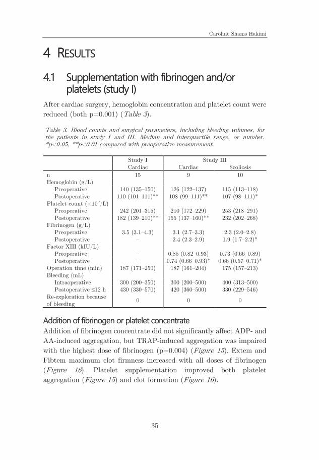
After cardiac surgery, hemoglobin concentration and platelet count were reduced (both p=0.001) (Table 3).
Table 3. Blood counts and surgical parameters, including bleeding volumes, for the patients in study I and III. Median and interquartile range, or number. *p<0.05, **p<0.01 compared with preoperative measurement.
Study I Study III Cardiac Cardiac Scoliosis n 15 9 10 Hemoglobin (g/L) Preoperative 140 (135–150) 126 (122–137) 115 (113–118) Postoperative 110 (101–111)** 108 (99–111)** 107 (98–111)* Platelet count (×109/L) Preoperative 242 (201–315) 210 (172–229) 253 (218–291) Postoperative 182 (139–210)** 155 (137–160)** 232 (202–268) Fibrinogen (g/L) Preoperative 3.5 (3.1–4.3) 3.1 (2.7–3.3) 2.3 (2.0–2.8) Postoperative – 2.4 (2.3–2.9) 1.9 (1.7–2.2)* Factor XIII (kIU/L) Preoperative – 0.85 (0.82–0.93) 0.73 (0.66–0.89) Postoperative – 0.74 (0.66–0.93)* 0.66 (0.57–0.71)* Operation time (min) 187 (171–250) 187 (161–204) 175 (157–213) Bleeding (mL) Intraoperative 300 (200–350) 300 (200–500) 400 (313–500) Postoperative ≤12 h 430 (330–570) 420 (360–500) 330 (229–546) Re-exploration because of bleeding 0 0 0
Addition of fibrinogen or platelet concentrate Addition of fibrinogen concentrate did not significantly affect ADP- and AA-induced aggregation, but TRAP-induced aggregation was impaired with the highest dose of fibrinogen (p=0.004) (Figure 15). Extem and Fibtem maximum clot firmness increased with all doses of fibrinogen (Figure 16). Platelet supplementation improved both platelet aggregation (Figure 15) and clot formation (Figure 16).

Fibrinogen, platelet and factor XIII supplementation in cardiac surgery
36
Supplementation with both fibrinogen and platelets improved platelet aggrega-tion induced by AA and TRAP with the medium and high doses (all p<0.001) (Figure 15B–C). ADP-induced aggregation increased with the highest dose (Figure 15A). Clot formation was improved (Figure 16). Compared to fibrinogen alone or platelets alone, the combination significantly improved Ex-tem maximum clot firmness for all doses (p 0.007) (Figure 16A). However, the combination was less effective in improving platelet aggregation com-pared to platelets alone for
Figure 15. Changes in platelet aggregation from baseline after addition of increasing doses of fibrinogen, platelet, or both, in blood samples from cardiac surgery patients (n=15). Median and inter-quartile range. Outliers (circles) are values >1.5 times the interquartile range away from the lower or upper quartile. *p<0.05, **p<0.01, ***p<0.001 compared with baseline.

Caroline Shams Hakimi
37
medium and high doses of ADP- and TRAP-induced aggregation (p 0.01) (Figure 15A and C).
Figure 16. Changes in maximum clot firmness from baseline after addition of three increasing doses of fibrinogen, platelet, or both fibrinogen and platelet concentrate in postoperative blood samples from cardiac surgery patients (n=15). Median and interquartile range. Outliers (circles) are values >1.5 times the interquartile range away from the lower or upper quartile. **p<0.01, ***p<0.001 compared with baseline.

Fibrinogen, platelet and factor XIII supplementation in cardiac surgery
38
Forty-one patients were included in study II. Fifteen received infusion of fibrinogen concentrate (median dose and range 2[1–3] g), 12 received platelet concentrate transfusion (2[1–3] units), and 14 patients received both fibrinogen and platelet concentrate (2[1–4] g and 2[1–3] units, respectively).
After infusion of fibrinogen concentrate, the plasma concentration of fibrinogen increased from 2.0 (1.8–2.3) to 2.6 (2.5–2.9) g/L (p=0.001). Fibtem maximum clot firmness was significantly increased (p=0.002). (Figure 17). Fibrinogen infusion did not significantly affect platelet aggregation (Figure 18).
Figure 17. Change in maximum clot firmness after transfusion with fibrinogen (FIB), platelets (PLT), or both fibrinogen and platelets. Median and interquartile range. Outliers (circles) are values >1.5 times the interquartile range away from the lower or upper quartile. **p<0.01 compared with baseline.

Caroline Shams Hakimi
39
Platelet concentrate transfusion increased platelet count from 168 (149–222) to 216 (182–274) × 109 platelets/L (p=0.006). Platelet transfusion significantly improved platelet aggregation induced by arachidonic acid (p=0.017) and TRAP (p=0.034), but aggregation induced by ADP was not significantly changed (Figure 18). Clot formation was not significantly affected by platelet transfusion (Figure 17).
After transfusion with fibrinogen and platelet concentrate, plasma fibrinogen concentration increased from 2.2 (1.8–2.3) to 2.7 (2.5–2.9) g/L (p=0.001), and the platelet count increased from 127 (120–174) to 192 (161–222) × 109 platelets/L (p=0.002). Fibrinogen and platelet transfusion significantly improved platelet aggregation induced with
Figure 18. Change in platelet aggregation induced by adenosine diphosphate (ADP), arachidonic acid, and thrombin receptor-activating peptide 6 (TRAP) after transfusion with fibrinogen (FIB), platelets (PLT), or both fibrinogen and platelets. Median and interquartile range. Outliers (circles) are values >1.5 times the interquartile range away from the lower or upper quartile. *p<0.05, **p<0.01 compared to baseline.

Fibrinogen, platelet and factor XIII supplementation in cardiac surgery
40
arachidonic acid, and TRAP (p=0.004 and 0.016, respectively), but not ADP-induced aggregation. (Figure 18). Extem, Fibtem and Intem maximum clot firmness were all improved after transfusion with fibrinogen and platelets (all p=0.001) (Figure 17).
Bleeding Overall, the bleeding volume decreased from 153 (69–243) before to 59 (39–108) mL/h (p<0.001) after transfusion (median and 25th–75th percentile). Fourteen of the 41 patients (34%) had sustained excessive bleeding despite the transfusion and proceeded to re-exploration. Surgical bleeding sources was found at re-exploration in 10/14 patients (71%).
Comparisons with in vitro study (study I) The administered doses of fibrinogen and platelet concentrates in study II were similar to the low doses which were supplemented to blood samples in study I, see Table 4.
Table 4. Doses of fibrinogen and platelet concentrates in study I and II, and the calculated and measured increases in fibrinogen concentration and platelet count in study II. *Corresponding dose administered to a 70-kg patient. Median and 25th–75th percentile.
Study I Study II
Treatment Dose* Median dose* Calculated increase Measured increase
Fibrinogen 2 g 1.6 (1.5–1.8) g 0.6 (0.5–0.6) g/L 0.6 (0.4–0.7) g/L
Platelets 2 units 1.7 (1.5–1.9) units 50 (46–58) ×109/L 32 (15–63) ×109/L
Fibrinogen + platelets
2 g, 2 units
1.9 (1.6–2.6) g, 1.8 (1.4–2.0)
units
0.7 (0.6–0.9) g/L, 55 (43–61) ×109/L
0.6 (0.4–0.8) g/L, 52 (33–69) ×109/L

Caroline Shams Hakimi
41
A brief summary of some of the functional hemostatic test results for study I and II is shown in Table 5.
Table 5. Changes in TRAP-induced aggregation and Fibtem maximum clot firmness after supplementation with fibrinogen and/or platelets (low doses in study I). Median and 25th–75th percentile. *p<0.05, **p<0.01, ***p<0.001 compared with baseline.
Study I Study II
Treatment Change in aggregation (U)
Change in clot stability (mm)
Change in aggregation (U)
Change in clot stability (mm)
Fibrinogen 4 (4–5)*** 3 (1–5)** Platelets 25 (16–32)*** 19 (1–21)* Fibrinogen + platelets 15 (5–21) 5 (5–6)*** 20 (12–27)* 4 (3–6)**

Fibrinogen, platelet and factor XIII supplementation in cardiac surgery
42
4.3 Addition of factor XIII, fibrinogen, and platelets (study III)
Hemoglobin concentration and FXIII activity decreased in both the cardiac and scoliosis surgery patients after surgery (Table 3). Platelet count was significantly reduced in cardiac surgery patients and fibrinogen concentration was reduced in scoliosis surgery patients.
Evaluation of the FXIII concentrate In the prestudy of study III, supplementation of increasing doses of FXIII concentrates increased with at least the levels of the calculated doses, see Table 6.
Table 6. Plasma concentrations for baseline and three samples supplemented with three increasing doses of factor XIII (FXIII) (+0.20, 0.40, and 0.60 kIU/L). Median and 25th–75th percentile.
Sample Measured plasma concentration (kIU/L)
Calculated plasma concentration (kIU/L)
Baseline 0.85 (0.74–0.88) FXIII low dose 1.14 (1.00–1.20) 1.05 (0.94–1.08) FXIII medium dose 1.41 (1.33–1.46) 1.25 (1.14–1.28) FXIII high dose 1.56 (1.47–1.65) 1.46 (1.44–1.49)
Cardiac surgery patients Supplementation of medium and high doses of FXIII resulted in an increased Fibtem maximum clot firmness compared to baseline (p=0.021 and p=0.003, respectively). There was an increase in Fibtem maximum clot firmness between the low and high dose of FXIII when it was added together with fibrinogen (p=0.002) (Figure 19A).
Scoliosis surgery patients Fibtem maximum clot firmness was significantly increased compared to baseline only when FXIII was added together with fibrinogen (p<0.001 for all three FXIII doses) or platelets (p=0.0019 for low dose FXIII and p<0.001 for medium and high doses). Fibrinogen enhanced the effect of FXIII supplementation only, with a significant increase between the low and high dose of FXIII (p<0.001) (Figure 19B).

Caroline Shams Hakimi
43
Figure 19. Change in Fibtem maximum clot firmness in blood samples from cardiac (A) and scoliosis surgery patients (B) after addition of increasing doses of factor XIII, alone or along with a fixed dose of fibrinogen (FIB) or platelets (PLT). *p<0.05, **p<0.01, ***p<0.001 relative to baseline. #p<0.05, ##p<0.01, ###p<0.001 compared with another dose of factor XIII (within treatment). Outliers (circles) are values >1.5 times the interquartile range away from the lower or upper quartile.
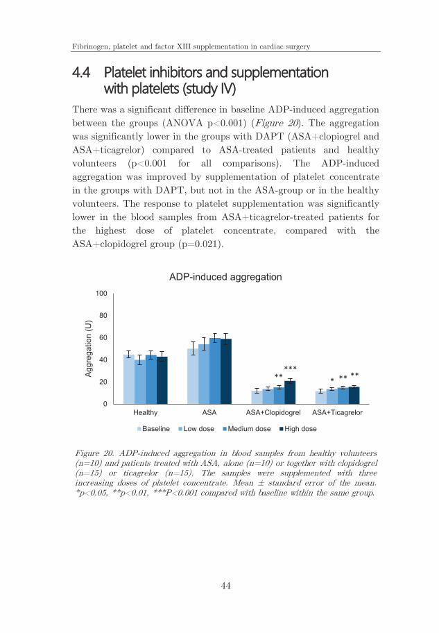
Fibrinogen, platelet and factor XIII supplementation in cardiac surgery
44
There was a significant difference in baseline ADP-induced aggregation between the groups (ANOVA p<0.001) (Figure 20). The aggregation was significantly lower in the groups with DAPT (ASA+clopiogrel and ASA+ticagrelor) compared to ASA-treated patients and healthy volunteers (p<0.001 for all comparisons). The ADP-induced aggregation was improved by supplementation of platelet concentrate in the groups with DAPT, but not in the ASA-group or in the healthy volunteers. The response to platelet supplementation was significantly lower in the blood samples from ASA+ticagrelor-treated patients for the highest dose of platelet concentrate, compared with the ASA+clopidogrel group (p=0.021).
Figure 20. ADP-induced aggregation in blood samples from healthy volunteers (n=10) and patients treated with ASA, alone (n=10) or together with clopidogrel (n=15) or ticagrelor (n=15). The samples were supplemented with three increasing doses of platelet concentrate. Mean ± standard error of the mean. *p<0.05, **p<0.01, ***P<0.001 compared with baseline within the same group.
0
20
40
60
80
100
Healthy ASA ASA+Clopidogrel ASA+Ticagrelor
Aggr
egat
ion
(U)
ADP-induced aggregation
Baseline Low dose Medium dose High dose
* **** *****

Caroline Shams Hakimi
45
AA-induced aggregation was significantly different at baseline among the groups (ANOVA p<0.001) (Figure 21). Compared to baseline in healthy volunteers, AA-induced aggregation was lower in all patient groups (all p<0.001). The baseline was lower in the patient groups with DAPT compared to patients treated with ASA only (p=0.001 and p=0.006 for clopidogrel and ticagrelor, respectively). Supplementation of platelets increased AA-induced aggregation in all groups, with the highest response in the ASA group and the lowest in the ASA+ticagrelor group. The response was significantly lower in the blood samples from patients treated with ASA+ticagrelor than in the ASA+clopidogrel group for all doses of platelet concentrate (p=0.025, p=0.040, and p=0.004, respectively).
Figure 21. Arachidonic acid-induced aggregation in blood samples from healthy volunteers (n=10) and patients treated with ASA, alone (n=10) or together with clopidogrel (n=15) or ticagrelor (n=15). The samples were supplemented with three increasing doses of platelet concentrate Mean ± standard error of the mean. *p<0.05, **p<0.01, ***P<0.001 compared with baseline within the same group.
0
20
40
60
80
100
Healthy ASA ASA+Clopidogrel ASA+Ticagrelor
Aggr
egat
ion
(U)
Arachidonic acid-induced aggregation
Baseline Low dose Medium dose High dose
***
***
**
***
******
***
***
***
***

Fibrinogen, platelet and factor XIII supplementation in cardiac surgery
46
The platelet aggregation of the buffy coat platelet concentrates was significantly reduced over time with all tested agonists; ADP, AA, TRAP and collagen (all p<0.001) (Figure 22). In the apheresis platelet concentrates, aggregation induced with ADP, AA and collagen was significantly reduced over time (p<0.001, p=0.005, and p<0.001, respectively), while TRAP-induced aggregation was not significantly different (p=0.20) (Figure 22).
Figure 22. In vitro platelet aggregation of the apheresis platelet concentrates (AC) and the buffy coat platelet concentrates (BC) after 1, 3, 5, and 7 storage days (n=13 for AC and n=13 for BC at each time point). Aggregation was induced with ADP, arachidonic acid, TRAP, and collagen. Mean ± standard error of the mean, and a linear regression model fitted to the data, are shown.

Caroline Shams Hakimi
47
Ex vivo aggregation The effect of supplemented buffy coat platelet concentrates in whole blood samples (change compared to baseline) significantly declined with storage time for aggregation induced with ADP, AA, and collagen (all p<0.001), but not for TRAP-induced aggregation (p=0.18) (Figure 23). The effect of apheresis platelets supplemented to whole blood samples declined with time for ADP-induced aggregation (p=0.010), while AA- TRAP- and collagen-induced aggregation did not change significantly (p=0.72, p=0.55, and p=0.11, respectively) (Figure 23).
There were significant correlations between in vitro aggregation of buffy coat platelet concentrates and the change in aggregation after supplementation to blood samples for aggregation induced with ADP (r=0.61, p<0.001), AA (r=0.66, p<0.001) and collagen (r=0.77, p<0.001) (pooled data from all four time points). For the apheresis platelet concentrations, there were significant correlations for ADP- (r=0.36, p=0.036) and collagen-induced (r=0.49, p=0.003) aggregation. The correlation coefficient for collagen-induced aggregation was r=0.67 (p<0.001), when including results for both buffy coat and apheresis platelet concentrates.

Fibrinogen, platelet and factor XIII supplementation in cardiac surgery
48
Figure 23. Platelet aggregation in whole blood samples from healthy volunteers (n=9 for each time point) after supplementation of apheresis platelet concentrates (AC) and buffy coat platelet concentrates (BC) after 1, 3, 5, and 7 storage days (n=3 for each type of concentrate and time point). Aggregation was induced with ADP, arachidonic acid, TRAP, and collagen. The baseline is aggregation in whole blood samples before addition of platelets. Mean ± standard error of the mean. Note that the lines shown are not regression lines. For regression analysis of this data, see Supplementary Figure of paper V.

Caroline Shams Hakimi
49
5 DISCUSSION The main findings in this thesis were; supplementation of fibrinogen concentrate improved clot formation and platelet concentrate improved platelet aggregation (study I and II); supplementation of FXIII caused a very limited improvement in clot formation (study III); platelet supplementation to blood samples from patients on DAPT was not able to restore ADP-induced aggregation (study IV); and the results of in vitro aggregation of platelet concentrates could with modest precision predict changes in aggregation after supplementation to blood samples (study V).
Fibrinogen and platelet concentrates (study I and II) Supplementation with fibrinogen to blood samples from cardiac surgery patients, and infusion of fibrinogen concentrate to cardiac surgery patient with ongoing bleeding significantly improved maximum clot firmness measured with the Extem and Fibtem assays in study I and Intem, Extem, and Fibtem in study II. These findings, together with the results from other in vitro and in vivo studies, clearly demonstrates that fibrinogen supplementation is able to improve clot stability [38, 55, 77-79, 88, 98-101]. Supplementation with platelets to blood samples increased both clot formation and platelet aggregation. In vivo transfusion of platelets to bleeding patients improved platelet aggregation induced with AA and TRAP, but not clot formation. Overall, the results are in line with other in vitro studies that also demonstrated improved clot formation [99] and platelet aggregation [80, 102, 103] after platelet supplementation to whole blood or plasma samples. To our knowledge, the effect of fibrinogen and platelet transfusion on platelet aggregation and clot formation has never been studied in cardiac surgery patients with ongoing bleeding before.
Potential differences between results after supplementation of prohemostatic substances obtained in in vitro and in vivo settings could have several explanations, such as variations in baseline clot formation and platelet aggregation parameters, differences in type and age of the platelet concentrates, and that there is a consumption of platelets and

Fibrinogen, platelet and factor XIII supplementation in cardiac surgery
50
coagulation factors during ongoing bleeding. As an example, the median age of the platelet concentrates in study II was 5 (3–6) days, whereas in study I, only fresh apheresis concentrates were used. Despite the large differences between the controlled in vitro study (study I) and the real-life study with patients with ongoing bleeding (study II), the effects of fibrinogen and platelet supplementation on platelet aggregation and clot formation showed similar patterns (Table 5). This suggests strongly that an in vitro model is appropriate and could be used for pre-clinical studies of prohemostatic drugs and substances.
Some of the cardiac surgery patients in study II also received red blood cells and/or plasma besides the fibrinogen and/or platelet transfusion. The influence of red blood cells and plasma on hemostasis has been investigated by Bochsen et al. [75]. They showed that an increased proportion of red blood cells and decreased proportion of plasma impaired clot stability and improved platelet aggregation [75]. In our study, the material was too small to enable statistical calculation of a potential effect of concomitant red cell or plasma transfusion on platelet aggregation or clot formation.
In study II, there was a lack of a control group not receiving fibrinogen or platelets. Since a time factor may contribute to a change in hemostasis and bleeding, a control group could have added important information. We deemed it unethical to include a control group not receiving the treatment, since transfusions with fibrinogen and platelets are included in our institutional protocol to treat excessive bleeding after cardiac surgery.
Factor XIII, fibrinogen, and platelet concentrates (study III) The effect on clot formation of FXIII supplementation, alone or together with fibrinogen, has been studied before with deviating results. Some studies have demonstrated an improvement in clotting time or clot stability after FXIII supplementation alone [78, 81, 83, 85, 86] or an enhanced effect when FXIII was added together with fibrinogen [77, 79, 82, 84, 87]. Other studies were unable to detect any effect of FXIII on clotting time or clot stability [89-93]. Some possible reasons for the

Caroline Shams Hakimi
51
discrepancies in results could be the varying doses of added FXIII, differences in study population, sample preparation (grade and type of hemodilution) and method used for clot formation assessment. There are also variations in FXIII activity at baseline before FXIII supplementation in the different studies. In fact, median FXIII activity ranged from <1% to 88% FXIII activity before supplementation [77, 79, 81-83, 86, 90, 91]. In study III, median FXIII activity before supplementation was about 70%, which is in the upper range of the reported studies.
In the prestudy of study III, the concentrations of FXIII were measured after supplementation of increasing doses of FXIII. The measured concentrations were at least the level of the calculated concentrations (Table 6). The content of the concentrate was thus reliable.
The effect of FXIII supplementation was limited in our study. Extem clotting time shortened by approximately 10% in both cardiac and scoliosis surgery groups (see paper III), and Fibtem clot firmness increased by 25% in the cardiac surgery group. When fibrinogen was added together with FXIII, the dose-dependent effect of FXIII on clot stability was maintained. However, the effect of fibrinogen or platelet supplementation was markedly larger than the effect of FXIII alone.
One important finding was that the effect of FXIII was similar in cardiac and scoliosis patients despite large differences between the two patient groups. The cardiac and scoliosis groups differed in patient characteristics and laboratory variables including age, gender, body mass index, and fibrinogen concentration. This indicates that the results regarding FXIII, but also fibrinogen and platelets, might be valid for surgical groups other than cardiac and scoliosis surgery patients.
Platelet inhibition and platelet concentrate supplementation (study IV) The use of platelet concentrates to improve platelet function in bleeding patients on antiplatelet therapy is rarely debated, even if the effect often is limited, especially in patients treated with P2Y12-inhibitors [103, 104]. Prior to the publication of study IV, few had investigated the efficacy of platelet supplementation or transfusion in patients on antiplatelet

Fibrinogen, platelet and factor XIII supplementation in cardiac surgery
52
therapy [94-96]. Since the publication of study IV, some new studies have been performed studying the effect of platelet supplementation in PRP or blood samples from patients treated with P2Y12-inhibitors [105-108] or PRP and blood samples from healthy volunteers supplemented with P2Y12-inhibitors [102]. Platelet supplementation in PRP samples from patients treated with clopidogrel or prasugrel improved platelet aggregation assessed with LTA [105, 106], but the effect was limited or absent in PRP or blood samples with platelet inhibition from ticagrelor [102, 106-108].
The somewhat lower effect of platelet supplementation in ticagrelor-treated in comparison to clopidogrel-treated patients in study IV may potentially be explained by the plasma concentrations of the P2Y12-inhibitor. In studies performed by Karaźniewicz-Łada et al. and Price et al., the plasma concentration of the active metabolite of clopidogrel was measured 2 h after a maintenance dose of 75 mg to about 5 ng/mL, whereas the plasma concentration of ticagrelor and its active metabolite 2 h after a maintenance dose of 90 mg (twice daily) were 750 and 210 ng/mL, respectively [109, 110]. The proportion of potent platelet inhibitor in the plasma seem to be higher for ticagrelor. Assuming that the concentration of ticagrelor was higher, this might indicate that newly supplemented platelets from concentrates in study IV could have been affected to a higher degree in blood samples from patients treated with ticagrelor compared with clopidogrel.
Platelet supplementation to blood samples from patients treated with P2Y12-inhibitos had a very limited effect on ADP-induced aggregation (study IV). However, addition of a high dose of platelet concentrate was able to restore both AA- and TRAP-induced aggregation (see paper IV), to at least the baseline level of healthy volunteers. As can be seen in the results of study V, the ability of platelet concentrates to improve ADP-induced aggregation after supplementation to blood samples was not as apparent compared with aggregation induced with other agonists. How large clinical impact this limited effect on ADP-induced aggregation has is not clear though. The impact on other pathways of

Caroline Shams Hakimi
53
platelet activation from the platelets in concentrates might potentially compensate.
The AA-induced aggregation was lower in patients on DAPT compared with ASA-treated patients (p=0.001 and 0.006 for ASA+clopidogrel and ASA+ticagrelor, respectively). In another study performed by our group, patients treated with ASA+ticagrelor were followed for up to four days after discontinuation of the ticagrelor-treatment (whereas the ASA-treatment was maintained [107]. Platelet aggregation was measured daily. Between 12 h and 96 h, the mean AA-induced aggregation was increased from 10±8 (standard deviation) U to 17±9 U (p=0.003), despite ongoing ASA-treatment [107]. The results from this study combined with the results of study IV, suggest that treatment with the ADP-inhibitors clopidogrel and ticagrelor may also affect COX-1-mediated platelet aggregation.
In vitro platelet aggregation in platelet concentrates – storage time and effect after supplementation to blood samples (study V) Attempts have been made to enable in vitro evaluation of the quality of the platelets in platelet concentrates during storage. A number of different methods, both routine assays and tests mainly used in research, have been used, but so far none of the assays has gained widespread clinical use. This is probably due to the fact that there is no strong evidence that there is a correlation between in vitro measurements of platelet concentrates and measurements after in vivo transfusion [49, 50, 111-114]. In study V, the platelet aggregation, assessed with impedance aggregometry, was gradually impaired over the storage time of the platelet concentrates. This is in accordance with a study performed by Ostrowski et al. [115]. In a more recently performed study, Glas et al. could not detect any decrease in platelet aggregation until storage day 12, however they assessed platelet aggregation in apheresis platelet concentrates with only two agonists; AA and TRAP [116]. With those agonists, neither we could detect any decline in aggregation of the apheresis platelets, during the seven days of storage which we analyzed. In a study where platelet aggregation was assessed in reconstituted whole blood, with apheresis platelet concentrates of varying ages

Fibrinogen, platelet and factor XIII supplementation in cardiac surgery
54
(storage day 0–5), the aggregation induced with ADP, AA and collagen was impaired with storage time, whereas TRAP-induced aggregation was maintained [117].
In study V, apheresis and buffy coat platelet concentrates did not only differ in preparation methodology, the storage medium also differed. The apheresis platelets were stored in autologous plasma, and the buffy coat platelets were stored in platelet additive solution and approximately 20% plasma. Because of the large differences, comparisons were only done within concentrate type. However, this was what the supply of platelet concentrates consisted of at the clinic during the time of the study.
We found that the in vitro results with modesty predicted the changes in platelet aggregation in a controlled in vitro transfusion model. The best correlation was achieved with the agonist collagen, which assesses the most general view of the platelet aggregation, as compared with the agonists ADP, AA, and TRAP, as collagen both function as a substrate for platelet adhesion and induces platelet activation [118].
One potential reason for the difficulty in predicting platelet recovery after in vivo transfusion is that the hemostasis of the recipient may vary, for example due to ongoing bleeding. In addition, according to our results, there was a recovery in platelet function after supplementation to blood samples (Figure 23), even if the in vitro results indicated poor aggregability (Figure 22). One reason could be the hematocrit-dependency in impedance aggregometry, since the hematocrit is zero in platelet concentrates [75, 95]. The capacity of the platelets to aggregate in vivo is dependent of the presence of a sufficient amount of red blood cells, as the larger red blood cells are centered in the vessels during blood flow, marginalizing the smaller platelets to the near-wall region [119].
At room temperature, the quality of platelets in concentrates deteriorates rapidly. Storage at cooler temperatures (at 2–8°C) of platelet concentrates have been applied. However, almost 50 years ago, Murphy and Gardner showed that the in vivo recovery of platelets was

Caroline Shams Hakimi
55
reduced after being stored at 4°C, compared with 22°C [52], leading to the guideline recommendation that the storage temperature should be 20–24°C. This is still the current guideline in most countries [120, 121]. Recently, the interest in cold storage has been raised again. Some efforts have been made to improve the function of cold stored platelets, which have shown promising in vitro results [122-125]. However, more studies are needed before cold storage of platelet concentrates can be implemented as clinical practice.
Limitations and strengths Limitations of the studies include the ex vivo measurements of platelet aggregation and clot formation. With these methods, interaction with blood vessels and vascular endothelium are lacking.
In all studies, platelet function was only assessed with one method: impedance aggregometry. The gold standard in assessment of platelet function is light transmission aggregometry (LTA). However, there are satisfactory correlation between impedance aggregometry and LTA [66, 126]. The advantage of impedance aggregometry over LTA is that platelet function is assessed in whole blood, and there is no need for preparation before analysis. In LTA on the other hand, PRP has to be prepared from whole blood. In this process, the function of the platelets can be affected. Compared with impedance aggregometry, LTA is also more time consuming and requires larger sample volumes of whole blood from the patient.
In study I and III, analysis were done on blood samples from cardiac and scoliosis surgery patients without excessive bleeding. The effect after supplementation with blood products would probably be more clinically relevant if the patients were more similar to patients who receive transfusions.
Approximately three in ten cardiac surgery patients are women [127]. Female gender was an exclusion criteria in study I, IV, V, and in cardiac patients in study III. Alloimmunization can occur after transfusion or pregnancy [128-130], and to avoid the risk of alloantibodies in the tested blood sample, which could interfere with the supplemented platelets, we
Fibrinogen, platelet and factor XIII supplementation in cardiac surgery
56
decided to only include men without previous history of transfusion. This was done to define the effects of the added blood products and minimizing factors that could interfere with the results. We acknowledge that this limit the applicability of our results to non-transfused men. An alternative would have been to include women in all studies, to make the results more representative. Larger groups could have been used to compensate for potential gender influences. Study II was an in vivo study where both men and women were included. In study III, the scoliosis surgery patient group consisted mainly of females, so in that group they were included.
In study III, eight teenaged females were included. Medication having a direct effect on hemostasis were exclusion criteria. However, hormonal contraceptives could potentially affect hemostasis, since there is a 3–8-fold increased incidence of venous thromboembolism (VTE) in women on combined hormonal contraceptives compared with women not using hormonal contraceptives The absolute incidence of VTE in women is however, very low; about two incidents per 10,000 woman-years [131, 132]. Unfortunately we did not have information whether any of the eight females used hormonal contraceptives.
Strengths of all in vitro studies (study I, III–V) includes the highly controlled circumstances; that every individual is their own control and that the same hemodilution is maintained between the different supplementations.
Strengths of study II include the prospective approach and the paired analyzes before and after the transfusion, making every patient their own control.

Caroline Shams Hakimi
57
6 CONCLUSIONS 1. Supplementation with fibrinogen to postoperative blood
samples from cardiac surgery patients improved clot formation, and supplementation with platelets improved both platelet aggregation and clot formation. The combination of fibrinogen and platelets was more effective than the individual components in improving clot formation.
2. Infusion with fibrinogen concentrate in postoperative cardiac surgery patients with ongoing bleeding improved clot formation, and platelet concentrate transfusion improved platelet aggregation. The combination of fibrinogen and platelets improved both platelet aggregation and clot formation.
3. Supplementation with FXIII concentrate to post-operative blood samples from cardiac and scoliosis surgery patients improved clot formation, but the effect was limited.
4. Supplementation with platelet concentrates to blood samples from patients treated with ADP-inhibitors was able to restore AA-induced platelet aggregation, but not ADP-induced platelet aggregation.
5. Impedance aggregometry in vitro could be used to follow platelet function in platelet concentrates over time, and the result could with modest precision predict changes in aggregation after ex vivo supplementation to whole blood samples.

Fibrinogen, platelet and factor XIII supplementation in cardiac surgery
58

Caroline Shams Hakimi
59
7 FUTURE PERSPECTIVES Bleeding will remain a major obstacle after major surgical procedures. New methods to prevent and treat excessive bleeding are therefore warranted. These methods includes refinement of the surgical techniques, new less invasive surgical approaches, improved monitoring of perioperative hemostasis and new products to improve coagulation and platelet function in bleeding patients. In this thesis we have investigated the effect of three pro-coagulant substances, i.e. fibrinogen, platelets and coagulation factor XIII, but more studies are needed. There are yet no large prospective randomized multicenter studies in surgical patients that have established the true value of fibrinogen supplementation in comparison with e.g. plasma-based transfusion protocols.
There are also gaps in evidence regarding the interaction between procoagulants, such as fibrinogen, platelets and FXIII, and red blood cells in the setting of peri- or postoperative bleeding. Red blood cells count may have direct effects on hemostasis but may also interfere with assays that assess hemostasis e.g. impedance aggregometry and thromboelastometry. Additional effects of red blood cells transfusion on top of fibrinogen, platelets and FXIII need to be investigated in studies with clinically relevant endpoints and the optimal hemoglobin level needs to be defined in patients with ongoing bleeding.
There is a large diversity among platelet concentrates at different centers, both in preparation methods, storage medium, volume and number of donors per unit, which all may influence the overall quality of the concentrate. Thorough comparisons of different platelet concentrates are needed to identify the optimal preparation and storage methods, which then potentially could be set as the international standard. To increase the shelf life (with the aim of reducing wastage and costs) of the platelet concentrates, the influence of storage temperature on post-transfusion survival needs to be studied more in detail. It is possible that differently stored concentrates are optimal for different patient categories, i.e. that patients with congenital or

Fibrinogen, platelet and factor XIII supplementation in cardiac surgery
60
acquired platelet deficiencies, may need for long lasting platelets, these patients could then receive room temperature stored platelet concentrates, while trauma and surgery patients, which need functional platelets momentarily, could receive cold stored platelets.
The in vitro aggregation of platelets stored in cold has shown to be better preserved than platelets stored in room temperature [133]. Studies should be performed to determine whether the change in platelet aggregation in direct association to the in vivo transfusion of cold stored platelet concentrates is superior, or at least comparable, to those stored at room temperature.
Impedance aggregometry results for study I, II, IV, and V indicates that the ADP-induced aggregation of platelet concentrates are not well-preserved. More studies evaluating ADP-induced aggregation are needed to determine the reason, and potentially found out how it can be maintained to a higher degree.

Caroline Shams Hakimi
61
REFERENCES 1. Structure of Ticagrelor; Brilique; AZD-6140; Brilinta; Possia. 2011 Oct
7 [cited 2017 Oct 19]. Available from: https://commons.wikimedia. org/wiki/File:Ticagrelor.svg.
2. Swaminathan, J and MSD staff at the European Bioinformatics Institute. Cartoon representation of the molecular structure of protein registered with 1f13 code. 2009 Mar 21 [cited 2017 Oct 19]. Available from: https://commons.wikimedia.org/wiki/File%3APDB1f13_EBI. jpg.
3. Electron Microscopy Facility at The National Cancer Institute at Frederick. From left to right: erythrocyte, thrombocyte, leukocyte. 2016 Jun 3 [cited 2017 Oct 19]. Available from: https://commons. wikimedia.org/wiki/File:Red_White_Blood_cells.jpg.
4. Cartoon representation of the molecular structure of protein registered with 1m1j code. 2009 Apr 2 [cited 2017 Oct 19]. Available from: https://commons.wikimedia.org/wiki/File%3APDB_1m1j_EBI.jpg.
5. Flegler S. Activated platelet (thrombocyte) within a dense network of fibrin fibers and red blood cells (erythrocytes) in a blood clot. SEM, X4000 at 100 x 125 mm 3048404.JPG, Editor: Visuals Unlimited, Inc.
6. Clopidogrel activation. 2011 Dec 24 [cited 2017 Oct 19]. Available from: https://commons.wikimedia.org/wiki/File:Clopidogrel_ activation.svg.
7. Coakley M, Hall JE, Evans C, et al. Assessment of thrombin generation measured before and after cardiopulmonary bypass surgery and its association with postoperative bleeding. J Thromb Haemost. 2011; 9(2): 282-292.
8. Frojd V and Jeppsson A. Reexploration for bleeding and its association with mortality after cardiac surgery. Ann Thorac Surg. 2016; 102(1): 109-117.
9. Karthik S, Grayson AD, McCarron EE, et al. Reexploration for bleeding after coronary artery bypass surgery: risk factors, outcomes, and the effect of time delay. Ann Thorac Surg. 2004; 78(2): 527-534; discussion 534.
10. Moulton MJ, Creswell LL, Mackey ME, et al. Reexploration for bleeding is a risk factor for adverse outcomes after cardiac operations. J Thorac Cardiovasc Surg. 1996; 111(5): 1037-1046.
11. Temenoff JS and Mikos AG. Biomaterials: The Intersection of Biology and Materials Science. 2008, Upper Saddle River, N.J: Pearson/Prentice Hall.
12. Albert J, Astermark J, Axelsson CG, et al., Hemostas vid allvarlig blödning. Vårdprogram utarbetat av arbetsgrupp inom Svenska Sällskapet för Trombos och Hemostas (SSTH). 2014: www.ssth.se.

Fibrinogen, platelet and factor XIII supplementation in cardiac surgery
62
13. Werner OJ, Sohns C, Popov AF, et al. Ludwig Rehn (1849-1930): the German surgeon who performed the worldwide first successful cardiac operation. J Med Biogr. 2012; 20(1): 32-34.
14. Cohn LH. Fifty years of open-heart surgery. Circulation. 2003; 107(17): 2168-2170.
15. Radegran K. The early history of cardiac surgery in Stockholm. J Card Surg. 2003; 18(6): 564-572.
16. Paparella D, Brister SJ, and Buchanan MR. Coagulation disorders of cardiopulmonary bypass: a review. Intensive Care Med. 2004; 30(10): 1873-1881.
17. Sniecinski RM and Levy JH. Bleeding and management of coagulopathy. J Thorac Cardiovasc Surg. 2011; 142(3): 662-667.
18. Reece MJ, Klein AA, Salviz EA, et al. Near-patient platelet function testing in patients undergoing coronary artery surgery: a pilot study. Anaesthesia. 2011; 66(2): 97-103.
19. Yusuf S, Zhao F, Mehta SR, et al. Effects of clopidogrel in addition to aspirin in patients with acute coronary syndromes without ST-segment elevation. N Engl J Med. 2001; 345(7): 494-502.
20. Wallentin L, Becker RC, Budaj A, et al. Ticagrelor versus clopidogrel in patients with acute coronary syndromes. N Engl J Med. 2009; 361(11): 1045-1057.
21. Patrono C. Chapter 53 - Aspirin A2 - Michelson, Alan D, in Platelets (Third Edition). 2013, Academic Press. p. 1099-1115.
22. Josefsson EC, Dowling MR, Lebois M, et al. Chapter 3 - The regulation of platelet life span A2 - Michelson, Alan D, in Platelets (Third Edition). 2013, Academic Press. p. 51-65.
23. Sangkuhl K, Klein TE, and Altman RB. Clopidogrel pathway. Pharmacogenet Genomics. 2010; 20(7): 463-465.
24. Brandt JT, Close SL, Iturria SJ, et al. Common polymorphisms of CYP2C19 and CYP2C9 affect the pharmacokinetic and pharmacodynamic response to clopidogrel but not prasugrel. J Thromb Haemost. 2007; 5(12): 2429-2436.
25. Wallentin L. P2Y(12) inhibitors: differences in properties and mechanisms of action and potential consequences for clinical use. Eur Heart J. 2009; 30(16): 1964-1977.
26. van Giezen JJJ, Nilsson L, Berntsson P, et al. Ticagrelor binds to human P2Y12 independently from ADP but antagonizes ADP-induced receptor signaling and platelet aggregation. J Thromb Haemost. 2009; 7: 1556-1565.
27. Anderson SD, Shah NK, Yim J, et al. Efficacy and safety of ticagrelor: a reversible P2Y12 receptor antagonist. Ann Pharmacother. 2010; 44(3): 524-537.
28. Hamm CW, Bassand JP, Agewall S, et al. ESC Guidelines for the management of acute coronary syndromes in patients presenting without persistent ST-segment elevation: The Task Force for the

Caroline Shams Hakimi
63
management of acute coronary syndromes (ACS) in patients presenting without persistent ST-segment elevation of the European Society of Cardiology (ESC). Eur Heart J. 2011; 32(23): 2999-3054.
29. Bird GW. The history of blood transfusion. Injury. 1971; 3(1): 40-44. 30. Giangrande PL. The history of blood transfusion. Br J Haematol. 2000;
110(4): 758-767. 31. Morfini M, Coppola A, Franchini M, et al. Clinical use of factor VIII
and factor IX concentrates. Blood Transfus. 2013; 11 Suppl 4: s55-63. 32. Despotis G, Eby C, and Lublin DM. A review of transfusion risks and
optimal management of perioperative bleeding with cardiac surgery. Transfusion. 2008; 48(1 Suppl): 2s-30s.
33. Bilgin YM, van de Watering LM, and Brand A. Clinical effects of leucoreduction of blood transfusions. Neth J Med. 2011; 69(10): 441-450.
34. Ternstrom L, Hyllner M, Backlund E, et al. A structured blood conservation programme reduces transfusions and costs in cardiac surgery. Interact Cardiovasc Thorac Surg. 2014; 19(5): 788-794.
35. Fassl J, Lurati Buse G, Filipovic M, et al. Perioperative administration of fibrinogen does not increase adverse cardiac and thromboembolic events after cardiac surgery. Br J Anaesth. 2015; 114(2): 225-234.
36. Bennett-Guerrero E, Zhao Y, O'Brien SM, et al. Variation in use of blood transfusion in coronary artery bypass graft surgery. Jama. 2010; 304(14): 1568-1575.
37. Ravn HB, Lindskov C, Folkersen L, et al. Transfusion requirements in 811 patients during and after cardiac surgery: a prospective observational study. J Cardiothorac Vasc Anesth. 2011; 25(1): 36-41.
38. Fries D, Innerhofer P, Reif C, et al. The effect of fibrinogen substitution on reversal of dilutional coagulopathy: an in vitro model. Anesth Analg. 2006; 102(2): 347-351.
39. Velik-Salchner C, Haas T, Innerhofer P, et al. The effect of fibrinogen concentrate on thrombocytopenia. J Thromb Haemost. 2007; 5(5): 1019-1025.
40. Li YF, Spencer FA, and Becker RC. Comparative efficacy of fibrinogen and platelet supplementation on the in vitro reversibility of competitive glycoprotein IIb/IIIa (alphaIIb/beta3) receptor-directed platelet inhibition. Am Heart J. 2001; 142(2): 204-210.
41. Chandler WL, Patel MA, Gravelle L, et al. Factor XIIIA and clot strength after cardiopulmonary bypass. Blood Coagul Fibrinolysis. 2001; 12(2): 101-108.
42. Shainoff JR, Estafanous FG, Yared JP, et al. Low factor XIIIA levels are associated with increased blood loss after coronary artery bypass grafting. J Thorac Cardiovasc Surg. 1994; 108(3): 437-445.
43. Ternstrom L, Radulovic V, Karlsson M, et al. Plasma activity of individual coagulation factors, hemodilution and blood loss after

Fibrinogen, platelet and factor XIII supplementation in cardiac surgery
64
cardiac surgery: a prospective observational study. Thromb Res. 2010; 126(2): e128-133.
44. Wettstein P, Haeberli A, Stutz M, et al. Decreased factor XIII availability for thrombin and early loss of clot firmness in patients with unexplained intraoperative bleeding. Anesth Analg. 2004; 99(5): 1564-1569; table of contents.
45. Korte WC, Szadkowski C, Gahler A, et al. Factor XIII substitution in surgical cancer patients at high risk for intraoperative bleeding. Anesthesiology. 2009; 110(2): 239-245.
46. Analysis records with reference intervals from the accredited clinical chemistry laboratory at Sahlgrenska University Hospital. 2017.
47. Shams Hakimi C, Hesse C, Wallen H, et al. In vitro assessment of platelet concentrates with multiple electrode aggregometry. Platelets. 2015; 26(2): 132-137.
48. Kelton JG and Blajchman MA. Platelet transfusions. Can Med Assoc J. 1979; 121(10): 1353-1358.
49. Devine DV and Serrano K. The platelet storage lesion. Clin Lab Med. 2010; 30(2): 475-487.
50. Shrivastava M. The platelet storage lesion. Transfus Apher Sci. 2009; 41(2): 105-113.
51. Perrotta PL, Parsons J, Rinder HM, et al. Chapter 62 - Platelet transfusion medicine A2 - Michelson, Alan D, in Platelets (Third Edition). 2013, Academic Press. p. 1275-1303.
52. Murphy S and Gardner FH. Effect of storage temperature on maintenance of platelet viability - Deleterious effect of refrigerated storage. N Engl J Med. 1969; 280(20): 1094-1098.
53. Gielen C, Dekkers O, Stijnen T, et al. The effects of pre- and postoperative fibrinogen levels on blood loss after cardiac surgery: a systematic review and meta-analysis. Interact Cardiovasc Thorac Surg. 2014; 18(3): 292-298.
54. Walden K, Jeppsson A, Nasic S, et al. Low preoperative fibrinogen plasma concentration is associated with excessive bleeding after cardiac operations. Ann Thorac Surg. 2014; 97(4): 1199-1206.
55. Fenger-Eriksen C, Jensen TM, Kristensen BS, et al. Fibrinogen substitution improves whole blood clot firmness after dilution with hydroxyethyl starch in bleeding patients undergoing radical cystectomy: a randomized, placebo-controlled clinical trial. J Thromb Haemost. 2009; 7(5): 795-802.
56. Karlsson M, Ternstrom L, Hyllner M, et al. Prophylactic fibrinogen infusion reduces bleeding after coronary artery bypass surgery. A prospective randomised pilot study. Thromb Haemost. 2009; 102(1): 137-144.
57. Rahe-Meyer N, Solomon C, Hanke A, et al. Effects of fibrinogen concentrate as first-line therapy during major aortic replacement

Caroline Shams Hakimi
65
surgery: a randomized, placebo-controlled trial. Anesthesiology. 2013; 118(1): 40-50.
58. Fenger-Eriksen C, Ingerslev J, and Sorensen B. Fibrinogen concentrate--a potential universal hemostatic agent. Expert Opin Biol Ther. 2009; 9(10): 1325-1333.
59. Jeppsson A, Walden K, Roman-Emanuel C, et al. Preoperative supplementation with fibrinogen concentrate in cardiac surgery: A randomized controlled study. Br J Anaesth. 2016; 116(2): 208-214.
60. Rahe-Meyer N, Levy JH, Mazer CD, et al. Randomized evaluation of fibrinogen vs placebo in complex cardiovascular surgery (REPLACE): a double-blind phase III study of haemostatic therapy. Br J Anaesth. 2016; 117(1): 41-51.
61. Wikkelso AJ, Edwards HM, Afshari A, et al. Pre-emptive treatment with fibrinogen concentrate for postpartum haemorrhage: randomized controlled trial. Br J Anaesth. 2015; 114(4): 623-633.
62. Ranucci M, Baryshnikova E, Crapelli GB, et al. Randomized, double-blinded, placebo-controlled trial of fibrinogen concentrate supplementation after complex cardiac surgery. J Am Heart Assoc. 2015; 4(6): e002066.
63. Lassila R. Clinical Use of Factor XIII Concentrates. Semin Thromb Hemost. 2016; 42(4): 440-444.
64. Platelet aggregation: Part II Some results from a new method of study. J Clin Pathol. 1962; 15(5): 452-455.
65. Born GV. Aggregation of blood platelets by adenosine diphosphate and its reversal. Nature. 1962; 194: 927-929.
66. Paniccia R, Antonucci E, Maggini N, et al. Comparison of methods for monitoring residual platelet reactivity after clopidogrel by point-of-care tests on whole blood in high-risk patients. Thromb Haemost. 2010; 104(2): 287-292.
67. Harrison P and Lordkipanidzé M. Chapter 26 - Clinical tests of platelet function A2 - Michelson, Alan D, in Platelets (Third Edition). 2013, Academic Press. p. 519-545.
68. Ramstrom S, Sodergren AL, Tynngard N, et al. Platelet function determined by flow cytometry: new perspectives? Semin Thromb Hemost. 2016; 42(3): 268-281.
69. Michelson AD, Barnard MR, Krueger LA, et al. Evaluation of platelet function by flow cytometry. Methods. 2000; 21(3): 259-270.
70. Luxemburg B, Krause M, and Lindhoff-Last E. Disorders of blood clotting [Basiswissen Gerinnungslabor]. Deutsches Arzteblatt. 2007; 104(21): A1489-A1498.
71. Larsen PB, Storjord E, Bakke A, et al. The microINR portable coagulometer: analytical quality and user-friendliness of a PT (INR) point-of-care instrument. Scand J Clin Lab Invest. 2017; 77(2): 115-121.

Fibrinogen, platelet and factor XIII supplementation in cardiac surgery
66
72. Lane PA and Hathaway WE. Vitamin K in infancy. J Pediatr. 1985; 106(3): 351-359.
73. Antovic A, Norberg EM, Berndtsson M, et al. Effects of direct oral anticoagulants on lupus anticoagulant assays in a real-life setting. Thromb Haemost. 2017; 117(9): 1700-1704.
74. Romlin BS, Wahlander H, Synnergren M, et al. Earlier detection of coagulopathy with thromboelastometry during pediatric cardiac surgery: a prospective observational study. Paediatr Anaesth. 2013; 23(3): 222-227.
75. Bochsen L, Johansson PI, Kristensen AT, et al. The influence of platelets, plasma and red blood cells on functional haemostatic assays. Blood Coagul Fibrinolysis. 2011; 22(3): 167-175.
76. Petricevic M, Konosic S, Biocina B, et al. Bleeding risk assessment in patients undergoing elective cardiac surgery using ROTEM((R)) platelet and Multiplate((R)) impedance aggregometry. Anaesthesia. 2016; 71(6): 636-647.
77. Haas T, Fries D, Velik-Salchner C, et al. The in vitro effects of fibrinogen concentrate, factor XIII and fresh frozen plasma on impaired clot formation after 60% dilution. Anesth Analg. 2008; 106(5): 1360-1365, table of contents.
78. Hanna J, Winstedt D, and Schott U. Fibrinogen and FXIII dose response effects on albumin-induced coagulopathy. Scand J Clin Lab Invest. 2013; 73(7): 553-562.
79. Schlimp CJ, Cadamuro J, Solomon C, et al. The effect of fibrinogen concentrate and factor XIII on thromboelastometry in 33% diluted blood with albumin, gelatine, hydroxyethyl starch or saline in vitro. Blood Transfus. 2013; 11(4): 510-517.
80. Hobl EL, Derhaschnig U, Firbas C, et al. Reversal strategy in antagonizing the P2Y12 -inhibitor ticagrelor. Eur J Clin Invest. 2013; 43(12): 1258-1261.
81. Grossmann E, Akyol D, Eder L, et al. Thromboelastometric detection of clotting Factor XIII deficiency in cardiac surgery patients. Transfus Med. 2013; 23(6): 407-415.
82. Kind SL, Spahn-Nett GH, Emmert MY, et al. Is dilutional coagulopathy induced by different colloids reversible by replacement of fibrinogen and factor XIII concentrates? Anesth Analg. 2013; 117(5): 1063-1071.
83. Nielsen VG, Gurley WQ, Jr., and Burch TM. The impact of factor XIII on coagulation kinetics and clot strength determined by thrombelastography. Anesth Analg. 2004; 99(1): 120-123.
84. Shenkman B, Einav Y, Livnat T, et al. In vitro evaluation of clot quality and stability in a model of severe thrombocytopenia: effect of fibrinogen, factor XIII and thrombin-activatable fibrinolysis inhibitor. Blood Transfus. 2014; 12(1): 78-84.

Caroline Shams Hakimi
67
85. Shenkman B, Livnat T, Lubetsky A, et al. The in-vitro effect of fibrinogen, factor XIII and thrombin-activatable fibrinolysis inhibitor on clot formation and susceptibility to tissue plasminogen activator-induced fibrinolysis in hemodilution model. Blood Coagul Fibrinolysis. 2012; 23(5): 370-378.
86. Theusinger OM, Baulig W, Asmis LM, et al. In vitro factor XIII supplementation increases clot firmness in Rotation Thromboelastometry (ROTEM). Thromb Haemost. 2010; 104(2): 385-391.
87. Winstedt D, Tynngard N, Olanders K, et al. Free oscillation rheometry monitoring of haemodilution and hypothermia and correction with fibrinogen and factor XIII concentrates. Scand J Trauma Resusc Emerg Med. 2013; 21: 20.
88. Calmette L, Martin AC, Le Bonniec B, et al. Ticagrelor reversal: in vitro assessment of four haemostatic agents. J Clin Pathol. 2017; 70(9): 733-739.
89. Dirkmann D, Gorlinger K, Gisbertz C, et al. Factor XIII and tranexamic acid but not recombinant factor VIIa attenuate tissue plasminogen activator-induced hyperfibrinolysis in human whole blood. Anesth Analg. 2012; 114(6): 1182-1188.
90. Fenger-Eriksen C, Tonnesen E, Ingerslev J, et al. Mechanisms of hydroxyethyl starch-induced dilutional coagulopathy. J Thromb Haemost. 2009; 7(7): 1099-1105.
91. Hvas AM, Andreasen JB, Christiansen K, et al. Ex-vivo response to blood products and haemostatic agents after paediatric cardiac surgery. Blood Coagul Fibrinolysis. 2013; 24(6): 587-592.
92. Rea CJ, Foley JH, Okaisabor O, et al. FXIII: mechanisms of action in the treatment of hemophilia A. J Thromb Haemost. 2014; 12(2): 159-168.
93. Schramko AA, Kuitunen AH, Suojaranta-Ylinen RT, et al. Role of fibrinogen-, factor VIII- and XIII-mediated clot propagation in gelatin haemodilution. Acta Anaesthesiol Scand. 2009; 53(6): 731-735.
94. Di Minno G, Silver MJ, and Murphy S. Monitoring the entry of new platelets into the circulation after ingestion of aspirin. Blood. 1983; 61(6): 1081-1085.
95. Pruller F, Drexler C, Archan S, et al. Low platelet reactivity is recovered by transfusion of stored platelets: a healthy volunteer in vivo study. J Thromb Haemost. 2011; 9(8): 1670-1673.
96. Vilahur G, Choi BG, Zafar MU, et al. Normalization of platelet reactivity in clopidogrel-treated subjects. J Thromb Haemost. 2007; 5(1): 82-90.
97. Clauss A. [Rapid physiological coagulation method in determination of fibrinogen]. Acta Haematol. 1957; 17(4): 237-246.
98. Cartwright BL, Kam P, and Yang K. Efficacy of fibrinogen concentrate compared with cryoprecipitate for reversal of the antiplatelet effect of

Fibrinogen, platelet and factor XIII supplementation in cardiac surgery
68
clopidogrel in an in vitro model, as assessed by multiple electrode platelet aggregometry, thromboelastometry, and modified thromboelastography. J Cardiothorac Vasc Anesth. 2015; 29(3): 694-702.
99. Ninivaggi M, Feijge MA, Baaten CC, et al. Additive roles of platelets and fibrinogen in whole-blood fibrin clot formation upon dilution as assessed by thromboelastometry. Thromb Haemost. 2014; 111(3): 447-457.
100. Shenkman B, Einav Y, Livnat T, et al. Rotation thromboelastometry analysis of clot formation and fibrinolysis in severe thrombocytopenia: effect of fibrinogen, activated prothrombin complex concentrate, and thrombin- activatable fibrinolysis inhibitor. Int J Lab Hematol. 2015; 37(4): 521-529.
101. Solomon C, Cadamuro J, Ziegler B, et al. A comparison of fibrinogen measurement methods with fibrin clot elasticity assessed by thromboelastometry, before and after administration of fibrinogen concentrate in cardiac surgery patients. Transfusion. 2011; 51(8): 1695-1706.
102. Martin AC, Berndt C, Calmette L, et al. The effectiveness of platelet supplementation for the reversal of ticagrelor-induced inhibition of platelet aggregation: An in-vitro study. Eur J Anaesthesiol. 2016; 33(5): 361-367.
103. O'Connor SA, Amour J, Mercadier A, et al. Efficacy of ex vivo autologous and in vivo platelet transfusion in the reversal of P2Y12 inhibition by clopidogrel, prasugrel, and ticagrelor: the APTITUDE study. Circ Cardiovasc Interv. 2015; 8(11): e002786.
104. Teng R, Carlson GF, Nylander S, et al. Effects of autologous platelet transfusion on platelet inhibition in ticagrelor-treated and clopidogrel-treated subjects. J Thromb Haemost. 2016; 14(12): 2342-2352.
105. Bhal V, Herr MJ, Dixon M, et al. Platelet function recovery following exposure to triple anti-platelet inhibitors using an in vitro transfusion model. Thromb Res. 2015; 136(6): 1216-1223.
106. Bonhomme F, Bonvini R, Reny JL, et al. Impact of non-inhibited platelet supplementation on platelet reactivity in patients treated with prasugrel or ticagrelor for an acute coronary syndrome: An ex vivo study. Platelets. 2015; 26(4): 324-330.
107. Hansson EC, Malm CJ, Hesse C, et al. Platelet function recovery after ticagrelor withdrawal in patients awaiting urgent coronary surgery. Eur J Cardiothorac Surg. 2017; 51(4): 633-637.
108. Zafar MU, Smith DA, Baber U, et al. Impact of timing on the functional recovery achieved with platelet supplementation after treatment with ticagrelor. Circ Cardiovasc Interv. 2017; 10(8).
109. Karazniewicz-Lada M, Danielak D, Burchardt P, et al. Clinical pharmacokinetics of clopidogrel and its metabolites in patients with cardiovascular diseases. Clin Pharmacokinet. 2014; 53(2): 155-164.

Caroline Shams Hakimi
69
110. Price MJ, Clavijo L, Angiolillo DJ, et al. A randomised trial of the pharmacodynamic and pharmacokinetic effects of ticagrelor compared with clopidogrel in Hispanic patients with stable coronary artery disease. J Thromb Thrombolysis. 2015; 39(1): 8-14.
111. Albanyan AM, Murphy MF, and Harrison P. Evaluation of the Impact-R for monitoring the platelet storage lesion. Platelets. 2009; 20(1): 1-6.
112. Ohto H and Nollet KE. Overview on platelet preservation: better controls over storage lesion. Transfus Apher Sci. 2011; 44(3): 321-325.
113. Panzer S and Jilma P. Methods for testing platelet function for transfusion medicine. Vox Sang. 2011; 101(1): 1-9.
114. Schubert P and Devine DV. Towards targeting platelet storage lesion-related signaling pathways. Blood Transfus. 2010; 8 Suppl 3: s69-72.
115. Ostrowski SR, Bochsen L, Windelov NA, et al. Hemostatic function of buffy coat platelets in additive solution treated with pathogen reduction technology. Transfusion. 2011; 51(2): 344-356.
116. Glas M, Bauer JV, Eichler H, et al. Impedance aggregometric analysis of platelet function of apheresis platelet concentrates as a function of storage time. Scand J Clin Lab Invest. 2016; 76(8): 664-670.
117. Ponschab M, Schlimp CJ, Zipperle J, et al. Platelet function in reconstituted whole blood variants: An observational study over 5 days of storage time. J Trauma Acute Care Surg. 2015; 79(5): 797-804.
118. Roberts DE, McNicol A, and Bose R. Mechanism of collagen activation in human platelets. J Biol Chem. 2004; 279(19): 19421-19430.
119. Fogelson AL and Neeves KB. Fluid mechanics of blood clot formation. Annu Rev Fluid Mech. 2015; 47: 377-403.
120. European Directorate for the Quality of Medicines & Health Care. Guide to the preparation, use and quality assurance of blood components. 18th edition. 2015.
121. Thomas S. Platelets: handle with care. Transfus Med. 2016; 26(5): 330-338.
122. Gitz E, Koekman CA, van den Heuvel DJ, et al. Improved platelet survival after cold storage by prevention of glycoprotein Ibα clustering in lipid rafts. Haematologica. 2012; 97(12): 1873-1881.
123. Handigund M, Bae TW, Lee J, et al. Evaluation of in vitro storage characteristics of cold stored platelet concentrates with N acetylcysteine (NAC). Transfus Apher Sci. 2016; 54(1): 127-138.
124. Pérez-Ceballos E, Rivera J, Lozano ML, et al. Evaluation of refrigerated platelet concentrates supplemented with low doses of second messenger effectors. Clin Lab Haematol. 2004; 26(4): 275-286.
125. Hoffmeister KM, Josefsson EC, Isaac NA, et al. Glycosylation restores survival of chilled blood platelets. Science. 2003; 301(5639): 1531-1534.
126. Sibbing D, Braun S, Jawansky S, et al. Assessment of ADP-induced platelet aggregation with light transmission aggregometry and multiple

Fibrinogen, platelet and factor XIII supplementation in cardiac surgery
70
electrode platelet aggregometry before and after clopidogrel treatment. Thromb Haemost. 2008; 99(1): 121-126.
127. Head SJ, Howell NJ, Osnabrugge RL, et al. The European Association for Cardio-Thoracic Surgery (EACTS) database: an introduction. Eur J Cardiothorac Surg. 2013; 44(3): e175-180.
128. Brown CJ and Navarrete CV. Clinical relevance of the HLA system in blood transfusion. Vox Sang. 2011; 101(2): 93-105.
129. De Clippel D, Baeten M, Torfs A, et al. Screening for HLA antibodies in plateletpheresis donors with a history of transfusion or pregnancy. Transfusion. 2014; 54(12): 3036-3042.
130. Kormoczi GF and Mayr WR. Responder individuality in red blood cell alloimmunization. Transfus Med Hemother. 2014; 41(6): 446-451.
131. Lidegaard O, Nielsen LH, Skovlund CW, et al. Venous thrombosis in users of non-oral hormonal contraception: follow-up study, Denmark 2001-10. BMJ. 2012; 344: e2990.
132. Raps M, Curvers J, Helmerhorst FM, et al. Thyroid function, activated protein C resistance and the risk of venous thrombosis in users of hormonal contraceptives. Thromb Res. 2014; 133(4): 640-644.
133. Getz TM, Montgomery RK, Bynum JA, et al. Storage of platelets at 4 degrees C in platelet additive solutions prevents aggregate formation and preserves platelet functional responses. Transfusion. 2016; 56(6): 1320-1328.
Copyright © 2022 FDOKUMEN