Exposure to acrolein by inhalation causes platelet activation
11
Exposure to acrolein by inhalation causes platelet activation Srinivas D. Sithu a,b , Sanjay Srivastava b , Maqsood A. Siddiqui b , Elena Vladykovskaya b , Daniel W. Riggs b , Daniel J. Conklin b , Petra Haberzettl b , Timothy E. O'Toole b , Aruni Bhatnagar b , Stanley E. D'Souza a, ⁎ a Department of Physiology and Biophysics, University of Louisville, Louisville, KY 40202, USA b Diabetes and Obesity Center, University of Louisville, Louisville, KY 40202, USA abstract article info Article history: Received 13 July 2010 Revised 16 July 2010 Accepted 16 July 2010 Available online 3 August 2010 Keywords: Acrolein Platelets Platelet factor 4 Fibrinogen binding Acrolein is a common air pollutant that is present in high concentrations in wood, cotton, and tobacco smoke, automobile exhaust and industrial waste and emissions. Exposure to acrolein containing environmental pollutants such as tobacco smoke and automobile exhaust has been linked to the activation of the coagulation and hemostasis pathways and thereby to the predisposition of thrombotic events in human. To examine the effects of acrolein on platelets, adult male C57Bl/6 mice were subjected acute (5 ppm for 6 h) or sub-chronic (1 ppm, 6 h/day for 4 days) acrolein inhalation exposures. The acute exposure to acrolein did not cause pulmonary inflammation and oxidative stress, dyslipidemia or induce liver damage or muscle injury. Platelet GSH levels in acrolein-exposed mice were comparable to controls, but acrolein-exposure increased the abundance of protein-acrolein adducts in platelets. Platelets isolated from mice, exposed to both acute and sub-chronic acrolein levels, showed increased ADP-induced platelet aggregation. Exposure to acrolein also led to an increase in the indices of platelet activation such as the formation of platelet-leukocyte aggregates in the blood, plasma PF4 levels, and increased platelet-fibrinogen binding. The bleeding time was decreased in acrolein exposed mice. Plasma levels of PF4 were also increased in mice exposed to environmental tobacco smoke. Similar to inhalation exposure, acrolein feeding to mice also increased platelet activation and established a pro-thrombotic state in mice. Together, our data suggest that acrolein is an important contributing factor to the pro-thrombotic risk in human exposure to pollutants such as tobacco smoke or automobile exhaust, or through dietary consumption. © 2010 Elsevier Inc. All rights reserved. Introduction Aldehydes are ubiquitous pollutants that are present in the air and in the drinking water. Large amounts of these chemicals are generated by the burning of fossil fuels and cooking. Aldehydes such as acrolein, malonaldehyde, and 4-hydroxy trans-2-nonenal (HNE) are generated endogenously during lipid peroxidation in biological systems (Adams and Klaidman, 1993; Uchida, 1999). Although these aldehydes are considered to be end-products of lipid peroxidation, they are highly reactive. They combine readily with nucleophilic sites in proteins, phospholipids and DNA to form covalent adducts that can induce structural damage or cause functional changes. An increase in endogenous production of aldehydes by oxidized lipids has been linked to the pathogenesis of cardiovascular, neuronal, neoplastic, and autoimmune diseases (Uchida, 1999). Of the several aldehydes present in the environment or generated in situ, acrolein is the most reactive. It is an unsaturated aldehyde and a common environmental and industrial pollutant and toxin which is present in high concentration in automobile exhaust, cigarette and wood smoke. Acrolein is also generated during the metabolism of several drugs (e.g., cyclophosphamide), toxins (e.g. allylamine), or atmospheric transformation of pollutants such as 1,3-butadiene (Kehrer and Biswal, 2000; Bhatnagar, 2006). Large amounts of acrolein are synthesized for industrial use in the production of acrylic acid, polymers and herbicides and it is used widely as a biocide against aqueous organisms and as a slimicide in the manufacture of paper (DeWoskin et al., 2003). Previous studies have shown that acrolein is a potent respiratory tract and eye irritant. Inhalation of acrolein induces asthma-like symptoms, pulmonary edema and respiratory distress and the presence of acrolein in inhaled air exacerbates asthma (Leikauf, 2002). In contrast to the well-described pulmonary effects of acrolein, less is known in regard to its toxicity in the vasculature. In vitro studies show that acrolein is cytotoxic to pulmonary artery endothelial cells (Kachel and Martin, 1994) and cardiac fibroblasts (Toraason et al., 1989). Acrolein evokes NO- and prostacyclin-dependent relaxation in isolated rat aortic rings (Tsakadze et al., 2003). In medium resistance arteries, acrolein caused vasodilatation of rodent mesenteric bed via an increase in endothelium-derived hyperpolarizaton factor (EDHF) Toxicology and Applied Pharmacology 248 (2010) 100–110 Abbreviations: ADP, Adenosine 5′-diphosphate; PRP, platelet rich plasma; WP, washed platelets; PF4, platelet factor 4. ⁎ Corresponding author. Department of Physiology and Biophysics, Health Sciences Center A-1115, University of Louisville, Louisville, KY 40292, USA. Fax: + 1 502 852 6239. E-mail address: [email protected] (S.E. D'Souza). 0041-008X/$ – see front matter © 2010 Elsevier Inc. All rights reserved. doi:10.1016/j.taap.2010.07.013 Contents lists available at ScienceDirect Toxicology and Applied Pharmacology journal homepage: www.elsevier.com/locate/ytaap
Transcript of Exposure to acrolein by inhalation causes platelet activation
Toxicology and Applied Pharmacology 248 (2010) 100–110
Contents lists available at ScienceDirect
Toxicology and Applied Pharmacology
j ourna l homepage: www.e lsev ie r.com/ locate /ytaap
Exposure to acrolein by inhalation causes platelet activation
Srinivas D. Sithu a,b, Sanjay Srivastava b, Maqsood A. Siddiqui b, Elena Vladykovskaya b, Daniel W. Riggs b,Daniel J. Conklin b, Petra Haberzettl b, Timothy E. O'Toole b, Aruni Bhatnagar b, Stanley E. D'Souza a,⁎a Department of Physiology and Biophysics, University of Louisville, Louisville, KY 40202, USAb Diabetes and Obesity Center, University of Louisville, Louisville, KY 40202, USA
Abbreviations: ADP, Adenosine 5′-diphosphate; PRwashed platelets; PF4, platelet factor 4.⁎ Corresponding author. Department of Physiology a
Center A-1115, University of Louisville, Louisville, KY6239.
E-mail address: [email protected] (S.E. D'Sou
0041-008X/$ – see front matter © 2010 Elsevier Inc. Adoi:10.1016/j.taap.2010.07.013
a b s t r a c t
a r t i c l e i n f oArticle history:Received 13 July 2010Revised 16 July 2010Accepted 16 July 2010Available online 3 August 2010
Keywords:AcroleinPlateletsPlatelet factor 4Fibrinogen binding
Acrolein is a common air pollutant that is present in high concentrations in wood, cotton, and tobaccosmoke, automobile exhaust and industrial waste and emissions. Exposure to acrolein containingenvironmental pollutants such as tobacco smoke and automobile exhaust has been linked to the activationof the coagulation and hemostasis pathways and thereby to the predisposition of thrombotic events inhuman. To examine the effects of acrolein on platelets, adult male C57Bl/6 mice were subjected acute(5 ppm for 6 h) or sub-chronic (1 ppm, 6 h/day for 4 days) acrolein inhalation exposures. The acuteexposure to acrolein did not cause pulmonary inflammation and oxidative stress, dyslipidemia or induceliver damage or muscle injury. Platelet GSH levels in acrolein-exposed mice were comparable to controls, butacrolein-exposure increased the abundance of protein-acrolein adducts in platelets. Platelets isolated frommice, exposed to both acute and sub-chronic acrolein levels, showed increased ADP-induced plateletaggregation. Exposure to acrolein also led to an increase in the indices of platelet activation such as theformation of platelet-leukocyte aggregates in the blood, plasma PF4 levels, and increased platelet-fibrinogenbinding. The bleeding time was decreased in acrolein exposed mice. Plasma levels of PF4 were also increasedin mice exposed to environmental tobacco smoke. Similar to inhalation exposure, acrolein feeding to micealso increased platelet activation and established a pro-thrombotic state in mice. Together, our data suggestthat acrolein is an important contributing factor to the pro-thrombotic risk in human exposure to pollutantssuch as tobacco smoke or automobile exhaust, or through dietary consumption.
P, platelet rich plasma; WP,
nd Biophysics, Health Sciences40292, USA. Fax: +1 502 852
za).
ll rights reserved.
© 2010 Elsevier Inc. All rights reserved.
Introduction
Aldehydes are ubiquitous pollutants that are present in the air andin the drinking water. Large amounts of these chemicals are generatedby the burning of fossil fuels and cooking. Aldehydes such as acrolein,malonaldehyde, and 4-hydroxy trans-2-nonenal (HNE) are generatedendogenously during lipid peroxidation in biological systems (Adamsand Klaidman, 1993; Uchida, 1999). Although these aldehydes areconsidered to be end-products of lipid peroxidation, they are highlyreactive. They combine readily with nucleophilic sites in proteins,phospholipids and DNA to form covalent adducts that can inducestructural damage or cause functional changes. An increase inendogenous production of aldehydes by oxidized lipids has beenlinked to the pathogenesis of cardiovascular, neuronal, neoplastic, andautoimmune diseases (Uchida, 1999). Of the several aldehydespresent in the environment or generated in situ, acrolein is the most
reactive. It is an unsaturated aldehyde and a common environmentaland industrial pollutant and toxin which is present in highconcentration in automobile exhaust, cigarette and wood smoke.Acrolein is also generated during the metabolism of several drugs(e.g., cyclophosphamide), toxins (e.g. allylamine), or atmospherictransformation of pollutants such as 1,3-butadiene (Kehrer andBiswal, 2000; Bhatnagar, 2006). Large amounts of acrolein aresynthesized for industrial use in the production of acrylic acid,polymers and herbicides and it is used widely as a biocide againstaqueous organisms and as a slimicide in the manufacture of paper(DeWoskin et al., 2003).
Previous studies have shown that acrolein is a potent respiratorytract and eye irritant. Inhalation of acrolein induces asthma-likesymptoms, pulmonary edema and respiratory distress and thepresence of acrolein in inhaled air exacerbates asthma (Leikauf,2002). In contrast to the well-described pulmonary effects of acrolein,less is known in regard to its toxicity in the vasculature. In vitro studiesshow that acrolein is cytotoxic to pulmonary artery endothelial cells(Kachel and Martin, 1994) and cardiac fibroblasts (Toraason et al.,1989). Acrolein evokes NO- and prostacyclin-dependent relaxation inisolated rat aortic rings (Tsakadze et al., 2003). In medium resistancearteries, acrolein caused vasodilatation of rodent mesenteric bed viaan increase in endothelium-derived hyperpolarizaton factor (EDHF)

Fig. 1. Acrolein exposure promotes platelet aggregation. Representative tracings ofADP-induced platelet aggregation in platelet rich plasma (PRP) in mice following theacute (5 ppm for 6 h; n=6; (i) or sub-chronic (1 ppm for 6 h/day for 4 days; n=6; (ii)exposure to acrolein by inhalation are illustrated. Mice exposed to filtered air (n=6 foreach group) served as controls. The insert in (i) shows a representative phase contrastimage of aggregate size following ADP-induced platelet aggregation in washed plateletsobtained from acrolein inhaled mice (1 ppm for 6 h/day for 4 days) and air inhaledcontrols.
101S.D. Sithu et al. / Toxicology and Applied Pharmacology 248 (2010) 100–110
(Awe et al., 2006). In vivo exposure to low doses of acrolein has beenassociated with vasopressor effects suggesting changes in systolicblood pressure (Egle and Hudgins, 1974; Green and Egle, 1983).Continuous 1 year oral exposure to acrolein in dogs revealed littletoxicity except for a mild and persistent depression of serum albumin,calcium, and a decrease in serum protein levels (Parent et al., 1992).Significantly, variable changes in coagulation times were observed,but the relevance of these findings to the systemic toxicity ofacrolein remains unclear. Given that exposure to pollutant mixturescontaining high levels of acrolein such as tobacco smoke or carexhaust are associated with increased thrombosis (Rahman andLaher, 2007), we studied the effects of acrolein on hemostasis. Wefocused on changes in blood platelets, because they are the primarycells that initiate coagulation to prevent excessive blood loss due tovessel injury. In addition, aberrant activation of platelets canincrease thrombus formation, disrupt the lining of the arterialwall, and accelerate atherogenesis. Therefore, an increase inthrombosis is strongly associated with increased risk of ischemicheart disease (Law andWald, 2003). In this study we have examinedthe effect of acrolein exposure on platelet activation in mice.
Methods
Animals. Male C57BL/6mice (16–20 weeks old), obtained from TheJackson Laboratory (Bar Harbor, ME), were housed under pathogen-free conditions in the University of Louisville vivarium undercontrolled temperature and 12 h light/12 h dark cycle. The micewere fed a standard chow diet (PicoLab Rodent Chow 20 containing4.5 % fat by weight and 0.02 % cholesterol).
Exposure to acrolein. For acrolein inhalation exposures, acroleinatmospheres were generated from liquid acrolein (Sigma) diluted indH2O (1:10) in a custom vapor system (Teague Inc.) using a primarychamber as a constant source. Acrolein vapors were diluted withHEPA-filtered room air in secondary chamber. Acrolein exposureconcentration (ppm) was continuously monitored using an in-linephotoionization detector (ppbRAE+, Rae Industries, Sunnyvale, CA)prior to delivery via a cage insert vapor delivery unit into a standardpolycarbonate rat cage (16"×8.75"×13.5") for exposures. Acroleinwas distributed through a fine mesh screen at 3 liters per min bydelivery units with a cyclone-type top that distributes air within 10%of the mean concentration at six locations in the cage. Exposure cageswere placed partially over heating pads (~24 °C). In the first protocol,mice were exposed to 5 ppm acrolein for 6 h (4,944±44 ppb) and inthe other to 1 ppm acrolein for 4 days (1,053±22 ppb). Mice exposedto filtered air served as controls. These exposure levels were inconcordance with recently reported studies on acrolein (Kasahara etal., 2008). For oral exposures, mice were gavage-fed acrolein (1–5 mg/kg) or tap water (vehicle; controls) as described before(Conklin et al., 2010) and euthanized at 4 or 24 h after exposure.
Exposure to tobacco smoke. Mice (n=6) were exposed to environ-mental tobacco smoke (ETS) for 5 h as described previously (Conklinet al., 2009). Tobacco smoke was generated from Kentucky 2R4Freference cigarettes (Tobacco Research Institute, University ofKentucky, Lexington, KY), using a smoke chamber (model TE-10,Teague Enterprises, Woodland, CA) and amixture of sidestream (89%)and mainstream (11%) CS was used. Each smoldering cigarette waspuffed for 2 s once every minute for a total of 8 puffs at a flow rate of1.05 l/min to provide a standard puff of 35 cm3. Ten 2R4F cigaretteswere burned at one time for 5 h. Mice (n=6) exposed to filtered airfor 5 h served as controls.
Isolation of platelets. Mice were anesthetized with sodium pento-barbital (0.1 ml; 50 mg/kg, i.p) immediately after acrolein exposures—5 ppm for 6 h or 1 ppm for 6 h/day for 4 days. Blood was collected
from the heart using a tuberculin syringewith a 20 gauge needle using1:9 (v/v) trisodium citrate (4%) as anti-coagulant (Srivastava et al.,1994). Blood was centrifuged at 180g for 20 min at 22 °C. Thesupernatant–platelet rich plasma (PRP)–was aspirated and centri-fuged at 1000g for 10 min to sediment platelets. The supernatant–platelet poor plasma (PPP)–was transferred to a fresh tube. Plateletcount in the PRP was adjusted to 3×10⁸ cells/ml using PPP. To obtainwashed platelets (WP), the platelets were washed three times withTyrode buffer (137 mM NaCl; 20 mM HEPES, 1 mM MgCl2, 2.7 mMKCl, 3.3 mM NaH2PO4, 5.6 mM glucose, 0.1% BSA; pH 7.4,) andreconstituted in Tyrode buffer at a density of 3×10⁸ cells/ml.
Platelet aggregation. Platelet aggregation was measured using afour-channel platelet aggregometer (Chrono-log Corp, Havertown,PA). PRP (adjusted to 3×10⁸ cells/ml) was incubated for 2 min at37 °C with continuous stirring at 1000 rpm, and then stimulated with
102 S.D. Sithu et al. / Toxicology and Applied Pharmacology 248 (2010) 100–110
adenosine 5’-diphosphate (ADP; 10 μM). The aggregation wasmeasured for 5 min by following change in absorbance (Srivastavaet al., 1994). For measuring platelet aggregation in washed platelets,CaCl2 (2 mM) and fibrinogen (200 μg/ ml) were added to the plateletsuspension prior to the stimulation with ADP. For each assay pooledblood from 4–6 mice was used.
Platelet-leukocyte aggregates. Blood was collected in Na4·EDTA(0.2 M; 16 μl/ml blood). Aliquots of whole blood (100 μl) werediluted with 4 volume of HEPES-Tyrode solution and the sampleswere fixed in 1% formaldehyde for 30 min at 4 °C. The red cellswere lysed by dilution in water. Cells were then collected bycentrifugation at 400g for 8 min, washed with HEPES-Tyrodesolution containing 1% BSA, and stained with FITC-labeled anti-CD41 and APC-labeled anti-CD11b or isotype matched controls for30 min on ice (Harding et al., 2007). The stained cells wereanalyzed on a Moflo flow cytometer (Dako) or a BD LSR II FlowCytometer (BD Biosciences). For each sample, a minimum of 10,000events were collected. Platelet-leukocyte aggregates were definedas those events which were positive for both platelets andleukocytes and expressed as a percentage of total events.
Protein-acrolein adducts and glutathione measurement. Levels ofprotein-acrolein adducts accumulated in washed platelets were
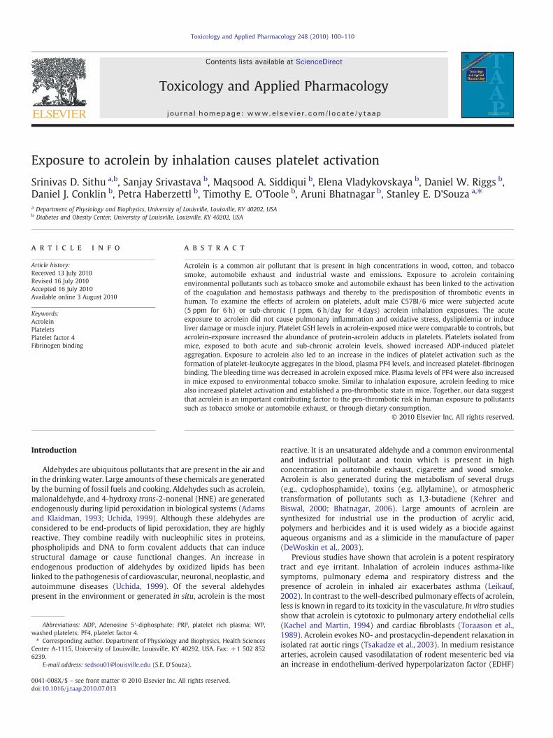
Fig. 2. Increased protein-acrolein adduct formation and no change in GSH levels in acroleplatelets obtained from mice exposed to acrolein (1 ppm/6 h/day for 4 days; n=3) or filtcytometric analysis of intracellular reduced glutathione levels in the platelets of mice exposewere labeled with monochlorobiamine (MCB) and mean fluorescence intensity of MCB waRepresentative illustration of fluorescence intensity of MCB in platelets isolated from contropanel (i) and the group data is shown in panel (ii) as percent CD41-MCB double positive c
measured by Western blotting, using the anti-KLH-acrolein antibody(Conklin et al., 2009). Intracellular glutathione levels in platelets weremeasured as described by Staal et al. (1990). Briefly, washed platelets(3×10⁸ platelets/mL) were labeled with anti-CD41-PE antibody andthen incubated with monochlorobimane (MCB; 20 μM) for 15 min at37 °C. Cells were excited at 394 nm and emission was acquired at490 nm. Data are presented as mean fluorescence intensity of MCB.
Platelet-fibrinogen binding. Aliquots of blood (100 μl) were incu-bated with PE-labeled rat-anti-mouse CD41 antibody for 20 min atroom temperature. Samples were then diluted with 4 volumes ofHEPES-Tyrode solution and incubated with Alexa-flour488 fibrinogen(20 μg/ml) for 10 min at room temperature. Samples were fixed withparaformaldehyde (2%) for 20 min. Erythrocytes were lysed by adding4.6 volumes of distilled water. Samples were centrifuged at 1000g for8 min and the pellet was washed with HEPES-Tyrode solutioncontaining 0.1% D-glucose and 0.1% BSA (FACS buffer). The pelletwas re-suspended in FACS buffer (1.0 ml) and analyzed by flowcytometry.
Platelet factor 4 assay. Plasma PF-4 levels were assayed by a sand-wich ELISA using DeoSet Mouse PF4/CXCL4 ELISA kit (R&D Systems,Minneapolis, MN) according to the manufacturer's instructions. Briefly,96-well ELISA plates were coated with a rat anti-mouse PF4 capture
in-inhaled mice. Panel A shows the representative Western blot of lysates of washedered air (n=3), and probed with anti-acrolein-KLH antibody. Panel B shows the flowd to acrolein (1 ppm/6 h/day for 4 days; n=5) or filtered air (n=5). Washed plateletss measured at excitation and emission wavelengths of 394 and 490 nm, respectively.l (air-exposed) and acrolein-exposed (1 ppm for 6 h/day for 4 days) mice is shown inells from total events. Values are mean±SEM.
103S.D. Sithu et al. / Toxicology and Applied Pharmacology 248 (2010) 100–110
antibody (2 μg/mL in PBS) for 16 h at room temperature. Wells werewashed free of theunbound antibody and blockedwith 1%BSA for 1 h atroom temperature. Plasma samples or the PF-4 standards wereincubated in the coated wells for 2 h at room temperature. Wells werewashed three times and then incubated with a biotinylated goat anti-mouse PF-4 antibody (100 ng/ml) for 2 h at room temperature. Wellswere washed again and then incubatedwith streptavidin conjugated tohorseradish-peroxidase for 20 min at room temperature. The washingstep was repeated and the substrate, tetramethylbenzidine (TMB) wasadded and the reaction was incubated for 15 min. The reaction wasstopped with 1 N H2SO4 and the color developed wasmeasured using amicroplate reader at 450 nm.
Expression of cytokines. Expression of pro-inflammatory cytokineswas measured in the lungs as described before (Srivastava et al., 2009).Lungswere snap-frozen immediately after harvesting. RNAwas isolatedusing Trizol reagent and 2.0 μg RNA from each was reverse transcribedwith AMV reverse transcriptase (Promega Corp., Madison, WI) at 42 °Cfor 60 min, followed by PCR amplification. Quantitative RT-PCR wascarried out in a BioRad Real Time PCR thermocycler using SYBR green/Fluorescein PCR master mix (SuperArray Biosciences Corp., Frederick,MD). Following primers were used: TNF-α-F:GCATGATCCGCGAC-GTGGAA and R: AGATCCATGCCGTTGGCCAG; IL-1β-F:CTCCATGAGCTT-TGTACAAGG and R: TGCTGATGTACCAGTTGGGG; MCP-1-F:ATGCAGGTCCCTGTCATG and R: GCTTGAGGTGGTTGTGGA and CSF-2-
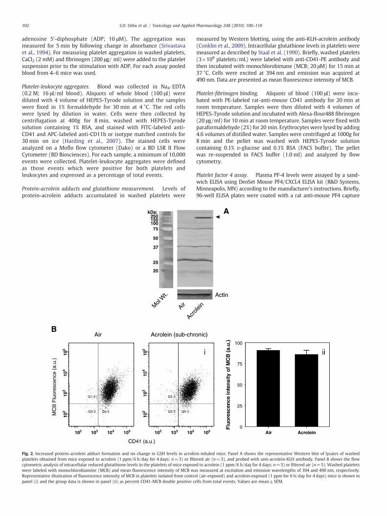
Fig. 3. Acrolein exposure increases platelet-fibrinogen binding. Panel A shows flow cytometchronic exposure to acrolein (1 ppm for 6 h/day for 4 days; n=6) or filtered air (n=6). Fscatter (FSC) and side scatter (SSC) of cells were used to distinguish platelets from other ceintensity (arbitrary units) of fibrinogen bound to CD41-positive platelets (i) and surface ex
F:TCGAGCAGGGTCTACGGGGC and R: AGCTGGCCTGGGCTTCCTCA. ForIL-6 measurement a commercial set of primers from Super ArrayBiosciences was used. HPRT1 was used as a house keeping gene (SuperArray Biosciences, Frederick, MD).
Measurements of malonyldialdehyde by gas chromatography-massspectrometry. Levels of malonyldialdehyde (MDA) in the lung weremeasured by gas chromatography-mass spectrometry as describedbefore (Srivastava et al., 2009).
PATHSCREEN. The following parameters of PATHSCREEN weremeasured in the plasma to examine systemic toxicity: levels ofalanine aminotransferase (ALT) and aspartate aminotransferase (AST)for liver damage, creatine kinase (CK) level to assess muscle and totalcholesterol and lipoproteins for dyslipidemia. Lipoprotein subclassesin the plasma were analyzed by NMR spectroscopy as describedbefore (Srivastava et al., 2009; Conklin et al., 2010).
Bleeding time measurement. Mice were anesthesized with an intra-peritoneal injection of avertin (250 mg/kg) to measure tail bleedingtime (Tranholm et al., 2003). Briefly, 5 mm of the distal tip of the tailwas briskly cut by a scalpel blade and immediately immersed into apre-warmed (37 °C) tube of 0.9 % saline. The tail was held near thebase to avoid any tourniquet effect and the time for the clotting ofvenous blood was recorded.
ric analysis of platelet-fibrinogen binding ex vivo in whole blood in mice following subor these assays PE-labeled anti-CD41 and FITC labeled fibrinogen were used. Forwardlls and cell debris. Panel B shows group data, presented as mean±SEM of fluorescencepression of CD41 (ii). §P≤0.02 versus control.
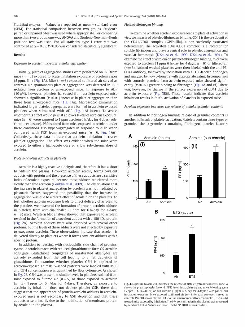
Fig. 4. Exposure to acrolein increases the release of platelet granular contents. Panel Ashows the plasma platelet factor 4 (PF4) levels in acrolein-treated mice following acute(5 ppm, 6 h; n=8; Ai) or sub-chronic (1 ppm, 6 h/day for 4 days; n=8; panel; Aii)inhalation exposure. Mice exposed to filtered air (n=8 for each protocol) served ascontrols. Panel B shows plasma PF4 levels in environmental tobacco smoke (ETS; n=6)treatedmice exposed by inhalation. The PF4 concentration in the plasma was measuredby sandwich ELISA. Values are mean±SEM. *P≤0.01 versus controls.
104 S.D. Sithu et al. / Toxicology and Applied Pharmacology 248 (2010) 100–110
Statistical analysis. Values are reported as mean±standard error(SEM). For statistical comparison between two groups, Student'spaired or unpaired t-test was used where appropriate. For comparingmore than two groups, one-way ANOVA and Student–Newman–Keulspost-hoc test was used. For all statistics, type I error rate wascontrolled at α=0.05. Pb0.05 was considered statistically significant.
Results
Exposure to acrolein increases platelet aggregation
Initially, platelet aggregation studies were performed on PRP frommice (n=6) exposed to acute inhalation exposure of acrolein vapor(5 ppm, 6 h) (Fig. 1A). Mice (n=6) exposed to filtered air served ascontrols. No spontaneous platelet aggregation was detected in PRPisolated from acrolein or air-exposed mice. In response to ADP(10 μM), however, platelets harvested from acrolein-exposed miceshowed a significant (Pb0.01) increase in platelet aggregation thanthose from air-exposed mice (Fig. 1Ai). Microscopic examinationindicated larger platelet aggregates were formed in acrolein exposedplatelets when stimulated with ADP (Fig. 1Ai insert). To assesswhether this effect would persist at lower levels of acrolein exposure,mice (n=6) were exposed to 1 ppm acrolein 6 h/day for 4 days (sub-chronic exposure). PRP isolated from mice exposed to acrolein underthese conditions also hyper-aggregated in response to ADP, whencompared with PRP from air-exposed mice (n=6; Fig. 1Aii).Collectively, these data indicate that acrolein inhalation increasesplatelet aggregation. The effect was evident when the mice wereexposed to either a high-acute dose or a low sub-chronic dose ofacrolein.
Protein-acrolein adducts in platelets
Acrolein is a highly reactive aldehyde and, therefore, it has a shorthalf-life in the plasma. However, acrolein readily forms covalentadducts with protein and the presence of these adducts are a sensitiveindex of acrolein exposure, because these adducts are cleared moreslowly than free acrolein (Conklin et al., 2009). The observations thatthe increase in platelet aggregation by acrolein was not mediated byplasmatic factors, suggested the possibility that the increase inaggregation was due to a direct effect of acrolein on the platelets. Totest whether acrolein exposure leads to direct delivery of acrolein tothe platelets, we measured the formation of protein-acrolein adductsin platelets from acrolein-inhaled (1 ppm for 6 h/day for 4 days;n=3) mice. Western blot analysis showed that exposure to acroleinresulted in the formation of a covalent adduct with a 150 kDa protein(Fig. 2A). Acrolein adducts were also observed with several otherproteins, but the levels of these adducts were not affected by exposureto exogenous acrolein. These observations indicate that acrolein isdelivered directly to platelets where it forms covalent adducts with aspecific protein.
In addition to reacting with nucleophilic side chain of proteins,cytosolic acrolein reacts with reduced glutathione to form GS-acroleinconjugate. Glutathione conjugates of unsaturated aldehydes areactively extruded from the cell leading to a net depletion ofglutathione. To examine whether platelet GSH is depleted inacrolein-exposed animals, washed platelets were labeled with MCBand GSH concentration was quantified by flow cytometry. As shownin Fig. 2B, GSH was present at similar levels in platelets isolated frommice exposed to filtered air (n=5) or those exposed to acrolein(n=5), 1 ppm for 6 h/day for 4 days. Therefore, as exposure toacrolein by inhalation does not deplete platelet GSH, these datasuggest that the appearance of protein-acrolein adducts in acrolein-exposed mice is not secondary to GSH depletion and that theseadducts arise primarily due to the modification of membrane proteinby acrolein in the plasma.
Platelet-fibrinogen binding
To examinewhether acrolein exposure leads to platelet activation invivo, wemeasured platelet-fibrinogen binding. CD41 is theα-subunit ofthe CD41/CD61 complex (GPllb–llla), a non-covalently associatedheterodimer. The activated CD41/CD61 complex is a receptor forsoluble fibrinogen and plays a central role in platelet aggregation andvascular hemostasis (D'Souza et al., 1990; D'Souza et al., 1991). Toexamine the effect of acrolein on platelet-fibrinogen binding, micewereexposed to acrolein (1 ppm 6 h/day for 4 days; n=6) or filtered air(n=6). Isolated washed platelets were then labeled with the anti-PE-CD41 antibody, followed by incubation with a FITC-labeled fibrinogenand analyzed by flow cytometrywith appropriate gating. In comparisonwith controls, platelets from acrolein-exposed mice showed signifi-cantly (Pb0.02) greater binding to fibrinogen (Fig. 3A and Bi). Therewas, however, no change in the surface expression of CD41 due toacrolein exposure (Fig. 3Bii). These results indicate that acroleininhalation results in in situ activation of platelets in exposed mice.
Acrolein exposure increases the release of platelet granular contents
In addition to fibrinogen binding, release of granular contents isanother hallmark of platelet activation. Platelets contain three types ofgranules—the α-granules (containing fibrinogen, platelet factor-4
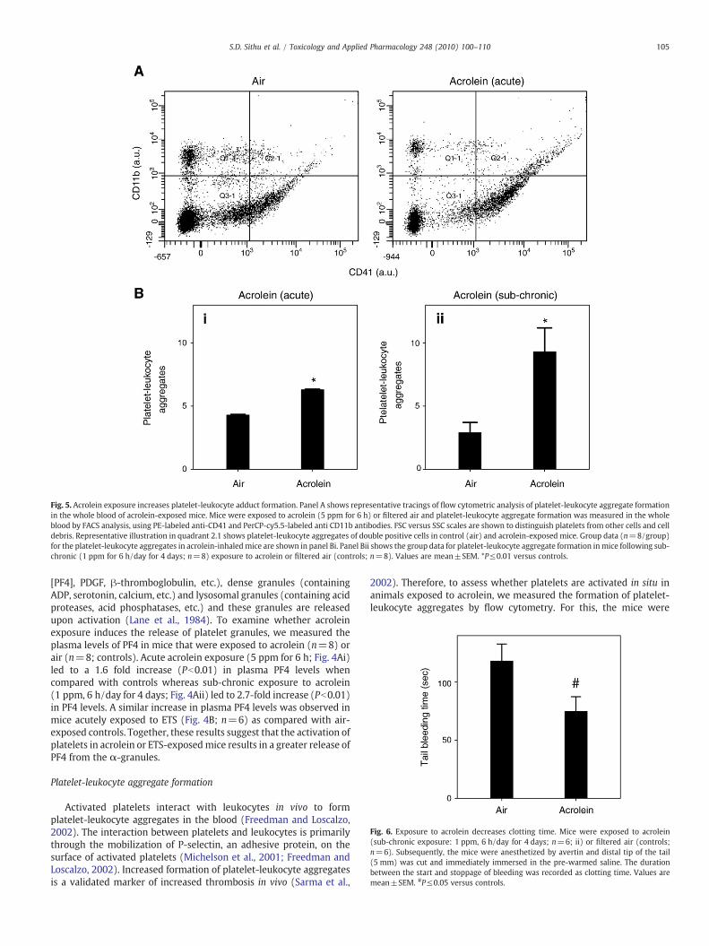
Fig. 5. Acrolein exposure increases platelet-leukocyte adduct formation. Panel A shows representative tracings of flow cytometric analysis of platelet-leukocyte aggregate formationin the whole blood of acrolein-exposed mice. Mice were exposed to acrolein (5 ppm for 6 h) or filtered air and platelet-leukocyte aggregate formation was measured in the wholeblood by FACS analysis, using PE-labeled anti-CD41 and PerCP-cy5.5-labeled anti CD11b antibodies. FSC versus SSC scales are shown to distinguish platelets from other cells and celldebris. Representative illustration in quadrant 2.1 shows platelet-leukocyte aggregates of double positive cells in control (air) and acrolein-exposed mice. Group data (n=8/group)for the platelet-leukocyte aggregates in acrolein-inhaledmice are shown in panel Bi. Panel Bii shows the group data for platelet-leukocyte aggregate formation inmice following sub-chronic (1 ppm for 6 h/day for 4 days; n=8) exposure to acrolein or filtered air (controls; n=8). Values are mean±SEM. *P≤0.01 versus controls.
Fig. 6. Exposure to acrolein decreases clotting time. Mice were exposed to acrolein(sub-chronic exposure: 1 ppm, 6 h/day for 4 days; n=6; ii) or filtered air (controls;n=6). Subsequently, the mice were anesthetized by avertin and distal tip of the tail(5 mm) was cut and immediately immersed in the pre-warmed saline. The durationbetween the start and stoppage of bleeding was recorded as clotting time. Values aremean±SEM. #P≤0.05 versus controls.
105S.D. Sithu et al. / Toxicology and Applied Pharmacology 248 (2010) 100–110
[PF4], PDGF, β-thromboglobulin, etc.), dense granules (containingADP, serotonin, calcium, etc.) and lysosomal granules (containing acidproteases, acid phosphatases, etc.) and these granules are releasedupon activation (Lane et al., 1984). To examine whether acroleinexposure induces the release of platelet granules, we measured theplasma levels of PF4 in mice that were exposed to acrolein (n=8) orair (n=8; controls). Acute acrolein exposure (5 ppm for 6 h; Fig. 4Ai)led to a 1.6 fold increase (Pb0.01) in plasma PF4 levels whencompared with controls whereas sub-chronic exposure to acrolein(1 ppm, 6 h/day for 4 days; Fig. 4Aii) led to 2.7-fold increase (Pb0.01)in PF4 levels. A similar increase in plasma PF4 levels was observed inmice acutely exposed to ETS (Fig. 4B; n=6) as compared with air-exposed controls. Together, these results suggest that the activation ofplatelets in acrolein or ETS-exposedmice results in a greater release ofPF4 from the α-granules.
Platelet-leukocyte aggregate formation
Activated platelets interact with leukocytes in vivo to formplatelet-leukocyte aggregates in the blood (Freedman and Loscalzo,2002). The interaction between platelets and leukocytes is primarilythrough the mobilization of P-selectin, an adhesive protein, on thesurface of activated platelets (Michelson et al., 2001; Freedman andLoscalzo, 2002). Increased formation of platelet-leukocyte aggregatesis a validated marker of increased thrombosis in vivo (Sarma et al.,
2002). Therefore, to assess whether platelets are activated in situ inanimals exposed to acrolein, we measured the formation of platelet-leukocyte aggregates by flow cytometry. For this, the mice were
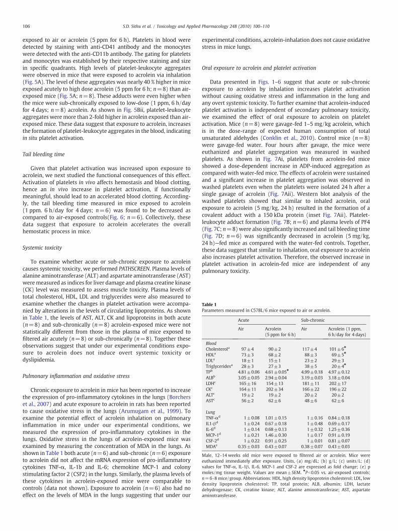
Table 1Parameters measured in C57BL/6 mice exposed to air or acrolein.
Acute Sub-chronic
Air Acrolein(5 ppm for 6 h)
Air Acrolein (1 ppm,6 h/day for 4 days)
BloodCholesterola 97±4 90±2 117±4 101±6⁎
HDLa 73±3 68±2 88±3 69±5⁎
LDLa 18±1 15±1 23±2 29±3Triglyceridesa 28±3 27±3 38±5 20±4⁎
TPb 4.81±0.06 4.61±0.05⁎ 4.99±0.18 4.97±0.12ALBb 3.05±0.05 2.94±0.04 3.19±0.03 3.18±0.04LDHc 165±16 154±13 181±11 202±17CKc 164±11 202±34 166±22 196±22ALTc 19±2 19±2 20±2 20±2ASTc 56±2 62±6 48±6 62±6
LungTNF-αd 1±0.08 1.01±0.15 1±0.16 0.84±0.18IL1-βd 1±0.24 0.67±0.18 1±0.48 0.69±0.17IL-6d 1±0.14 0.68±0.13 1±0.32 1.25±0.36MCP-1d 1±0.21 1.46±0.30 1±0.17 0.91±0.19CSF-2d 1±0.22 0.91±0.25 1±0.01 0.81±0.07MDAe 0.35±0.03 0.43±0.07 0.38±0.07 0.43±0.03
Male, 12–14 weeks old mice were exposed to filtered air or acrolein. Mice wereeuthanized immediately after exposure. Units, (a) mg/dL; (b) g/L; (c) units/L; (d)values for TNF-α, IL-1β, IL-6, MCP-1 and CSF-2 are expressed as fold change; (e) pmoles/mg tissue weight. Values are mean±SEM. ⁎Pb0.05 vs. air-exposed controls;n=6–8mice/group. Abbreviations: HDL, high density lipoprotein cholesterol; LDL, lowdensity lipoprotein cholesterol; TP, total protein; ALB, albumin; LDH, lactatedehydrogenase; CK, creatine kinase; ALT, alanine aminotransferase; AST, aspartateaminotransferase.
106 S.D. Sithu et al. / Toxicology and Applied Pharmacology 248 (2010) 100–110
exposed to air or acrolein (5 ppm for 6 h). Platelets in blood weredetected by staining with anti-CD41 antibody and the monocyteswere detected with the anti-CD11b antibody. The gating for plateletsand monocytes was established by their respective staining and sizein specific quadrants. High levels of platelet-leukocyte aggregateswere observed in mice that were exposed to acrolein via inhalation(Fig. 5A). The level of these aggregates was nearly 40 % higher in miceexposed acutely to high dose acrolein (5 ppm for 6 h; n=8) than air-exposed mice (Fig. 5A; n=8). These adducts were even higher whenthe mice were sub-chronically exposed to low-dose (1 ppm, 6 h/dayfor 4 days; n=8) acrolein. As shown in Fig. 5Bii, platelet-leukocyteaggregates weremore than 2-fold higher in acrolein exposed than air-exposed mice. These data suggest that exposure to acrolein, increasesthe formation of platelet-leukocyte aggregates in the blood, indicatingin situ platelet activation.
Tail bleeding time
Given that platelet activation was increased upon exposure toacrolein, we next studied the functional consequences of this effect.Activation of platelets in vivo affects hemostasis and blood clotting,hence an in vivo increase in platelet activation, if functionallymeaningful, should lead to an accelerated blood clotting. According-ly, the tail bleeding time measured in mice exposed to acrolein(1 ppm. 6 h/day for 4 days; n=6) was found to be decreased ascompared to air-exposed controls(Fig. 6; n=6). Collectively, thesedata suggest that exposure to acrolein accelerates the overallhemostatic process in mice.
Systemic toxicity
To examine whether acute or sub-chronic exposure to acroleincauses systemic toxicity, we performed PATHSCREEN. Plasma levels ofalanine aminotransferase (ALT) and aspartate aminotransferase (AST)weremeasured as indices for liver damage and plasma creatine kinase(CK) level was measured to assess muscle toxicity. Plasma levels oftotal cholesterol, HDL, LDL and triglycerides were also measured toexamine whether the changes in platelet activation were accompa-nied by alterations in the levels of circulating lipoproteins. As shownin Table 1, the levels of AST, ALT, CK and lipoproteins in both acute(n=8) and sub-chronically (n=8) acrolein-exposed mice were notstatistically different from those in the plasma of mice exposed tofiltered air acutely (n=8) or sub-chronically (n=8). Together theseobservations suggest that under our experimental conditions expo-sure to acrolein does not induce overt systemic toxicity ordyslipidemia.
Pulmonary inflammation and oxidative stress
Chronic exposure to acrolein in mice has been reported to increasethe expression of pro-inflammatory cytokines in the lungs (Borcherset al., 2007) and acute exposure to acrolein in rats has been reportedto cause oxidative stress in the lungs (Arumugam et al., 1999). Toexamine the potential effect of acrolein inhalation on pulmonaryinflammation in mice under our experimental conditions, wemeasured the expression of pro-inflammatory cytokines in thelungs. Oxidative stress in the lungs of acrolein-exposed mice wasexamined by measuring the concentration of MDA in the lungs. Asshown in Table 1 both acute (n=6) and sub-chronic (n=6) exposureto acrolein did not affect the mRNA expression of pro-inflammatorycytokines TNF-α, IL-1b and IL-6; chemokine MCP-1 and colonystimulating factor 2 (CSF2) in the lungs. Similarly, the plasma levels ofthese cytokines in acrolein-exposed mice were comparable tocontrols (data not shown). Exposure to acrolein (n=6) also had noeffect on the levels of MDA in the lungs suggesting that under our
experimental conditions, acrolein-inhalation does not cause oxidativestress in mice lungs.
Oral exposure to acrolein and platelet activation
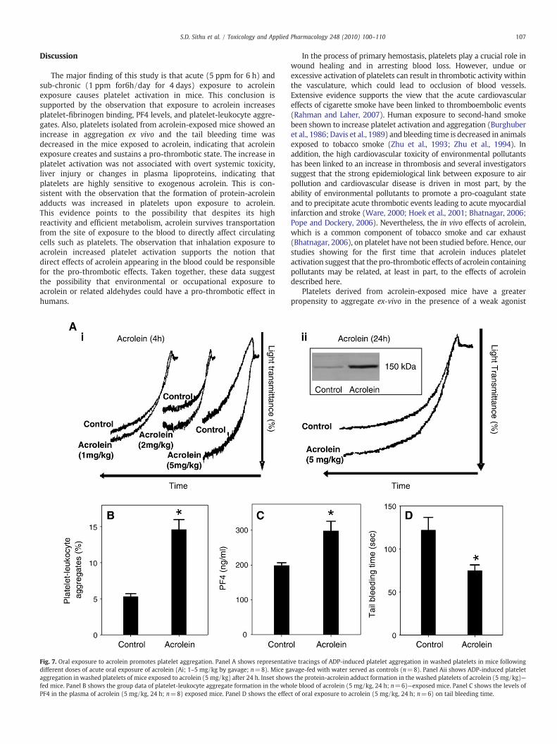
Data presented in Figs. 1–6 suggest that acute or sub-chronicexposure to acrolein by inhalation increases platelet activationwithout causing oxidative stress and inflammation in the lung andany overt systemic toxicity. To further examine that acrolein-inducedplatelet activation is independent of secondary pulmonary toxicity,we examined the effect of oral exposure to acrolein on plateletactivation. Mice (n=8) were gavage-fed 1–5 mg/kg acrolein, whichis in the dose-range of expected human consumption of totalunsaturated aldehydes (Conklin et al., 2010). Control mice (n=8)were gavage-fed water. Four hours after gavage, the mice wereeuthanized and platelet aggregation was measured in washedplatelets. As shown in Fig. 7Ai, platelets from acrolein-fed miceshowed a dose-dependent increase in ADP-induced aggregation ascompared with water-fed mice. The effects of acrolein were sustainedand a significant increase in platelet aggregation was observed inwashed platelets even when the platelets were isolated 24 h after asingle gavage of acrolein (Fig. 7Aii). Western blot analysis of thewashed platelets showed that similar to inhaled acrolein, oralexposure to acrolein (5 mg/kg, 24 h) resulted in the formation of acovalent adduct with a 150 kDa protein (inset Fig. 7Aii). Platelet-leukocyte adduct formation (Fig. 7B; n=6) and plasma levels of PF4(Fig. 7C; n=8)were also significantly increased and tail bleeding time(Fig. 7D; n=6) was significantly decreased in acrolein (5 mg/kg,24 h)—fed mice as compared with the water-fed controls. Together,these data suggest that similar to inhalation, oral exposure to acroleinalso increases platelet activation. Therefore, the observed increase inplatelet activation in acrolein-fed mice are independent of anypulmonary toxicity.
107S.D. Sithu et al. / Toxicology and Applied Pharmacology 248 (2010) 100–110
Discussion
The major finding of this study is that acute (5 ppm for 6 h) andsub-chronic (1 ppm for6h/day for 4 days) exposure to acroleinexposure causes platelet activation in mice. This conclusion issupported by the observation that exposure to acrolein increasesplatelet-fibrinogen binding, PF4 levels, and platelet-leukocyte aggre-gates. Also, platelets isolated from acrolein-exposed mice showed anincrease in aggregation ex vivo and the tail bleeding time wasdecreased in the mice exposed to acrolein, indicating that acroleinexposure creates and sustains a pro-thrombotic state. The increase inplatelet activation was not associated with overt systemic toxicity,liver injury or changes in plasma lipoproteins, indicating thatplatelets are highly sensitive to exogenous acrolein. This is con-sistent with the observation that the formation of protein-acroleinadducts was increased in platelets upon exposure to acrolein.This evidence points to the possibility that despites its highreactivity and efficient metabolism, acrolein survives transportationfrom the site of exposure to the blood to directly affect circulatingcells such as platelets. The observation that inhalation exposure toacrolein increased platelet activation supports the notion thatdirect effects of acrolein appearing in the blood could be responsiblefor the pro-thrombotic effects. Taken together, these data suggestthe possibility that environmental or occupational exposure toacrolein or related aldehydes could have a pro-thrombotic effect inhumans.
Fig. 7. Oral exposure to acrolein promotes platelet aggregation. Panel A shows representatidifferent doses of acute oral exposure of acrolein (Ai; 1–5 mg/kg by gavage; n=8). Mice gaggregation in washed platelets of mice exposed to acrolein (5 mg/kg) after 24 h. Inset showfed mice. Panel B shows the group data of platelet-leukocyte aggregate formation in the whPF4 in the plasma of acrolein (5 mg/kg, 24 h; n=8) exposed mice. Panel D shows the effec
In the process of primary hemostasis, platelets play a crucial role inwound healing and in arresting blood loss. However, undue orexcessive activation of platelets can result in thrombotic activity withinthe vasculature, which could lead to occlusion of blood vessels.Extensive evidence supports the view that the acute cardiovasculareffects of cigarette smoke have been linked to thromboembolic events(Rahman and Laher, 2007). Human exposure to second-hand smokebeen shown to increase platelet activation and aggregation (Burghuberet al., 1986; Davis et al., 1989) and bleeding time is decreased in animalsexposed to tobacco smoke (Zhu et al., 1993; Zhu et al., 1994). Inaddition, the high cardiovascular toxicity of environmental pollutantshas been linked to an increase in thrombosis and several investigatorssuggest that the strong epidemiological link between exposure to airpollution and cardiovascular disease is driven in most part, by theability of environmental pollutants to promote a pro-coagulant stateand to precipitate acute thrombotic events leading to acute myocardialinfarction and stroke (Ware, 2000; Hoek et al., 2001; Bhatnagar, 2006;Pope and Dockery, 2006). Nevertheless, the in vivo effects of acrolein,which is a common component of tobacco smoke and car exhaust(Bhatnagar, 2006), on platelet have not been studied before. Hence, ourstudies showing for the first time that acrolein induces plateletactivation suggest that the pro-thrombotic effects of acrolein containingpollutants may be related, at least in part, to the effects of acroleindescribed here.
Platelets derived from acrolein-exposed mice have a greaterpropensity to aggregate ex-vivo in the presence of a weak agonist
ve tracings of ADP-induced platelet aggregation in washed platelets in mice followingavage-fed with water served as controls (n=8). Panel Aii shows ADP-induced platelets the protein-acrolein adduct formation in the washed platelets of acrolein (5 mg/kg)—ole blood of acrolein (5 mg/kg, 24 h; n=6)—exposed mice. Panel C shows the levels oft of oral exposure to acrolein (5 mg/kg, 24 h; n=6) on tail bleeding time.

108 S.D. Sithu et al. / Toxicology and Applied Pharmacology 248 (2010) 100–110
(ADP) compared with platelets obtained from control, non-exposedmice (Fig. 1). Acrolein-induced augmentation of platelet aggregationwas observed in the PRP and with washed platelets. The observationthat platelet aggregation was increased even in the absence of plasmaindicates that acrolein directly affects platelets and that the effects ofacrolein are unlikely to be mediated by changes in the plasma. Nosignificant changes in plasma lipoproteins were observed in miceexposed to acrolein via inhalation, suggesting that acrolein-inducedplatelet activation is independent of the dyslipidemic effects ofacrolein. The selective and independent effect of acrolein on plateletsis similar to that of tobacco smoke. Even low dose exposure to tobaccosmoke significantly and rapidly increase platelet activation and muchof the ischemic heart disease risk of smoking could be ascribed to anincrease in platelet activation (Law and Wald, 2003). Moreover,exposure to tobacco smoke is an independent risk factor for heartdisease, which means that the effects of tobacco smoke are notmediated by other risk factors such as an increase in cholesterol orhypertension. Although smokers have higher levels of LDL-cholesteroland low levels of HDL than non-smokers, dyslipidemia accounts forb9 % of the cardiovascular risk of smoking (Craig et al., 1989).Similarly, cholesterol levels do not affect the risk associationbetween air pollutant exposure and cardiovascular disease (Milleret al., 2007). Thus, the effects of acrolein bear a striking resemblanceto those of tobacco smoke and air pollutants and appear to be drivenby a direct increase in pro-thrombotic indices rather than indirectchanges in blood lipoproteins.
We found that exposure to acrolein and ETS increased the plasmaconcentration of PF4. PF4 is mainly synthesized in megakaryocytes,and then stored in the α-granules of platelets. Upon plateletactivation PF4, released from the α-granules of platelets, binds toheparin-like molecules and prevents the activation of antithrombin(Denton et al., 1983); thereby facilitating thrombus formation at thesite of vessel injury. Our data complement recent studies showing thatacrolein in vitro inhibits plasma antithrombin activity by inhibitingheparin affinity (Gugliucci, 2008; Martinez-Martinez et al., 2009).Lower antithrombin activity levels could potentially increase throm-bin availability in the circulation, adding another pro-thromboticcomponent to the effects of acrolein exposure. PF4 has also beenreported to affect the anticoagulant activity of Activated Protein C(Preston et al., 2009). Apart for the well documented role of plateletsin thrombosis and hemostasis, emerging data also suggest thatplatelet activation could be involved in the development of athero-genesis (Massberg et al., 2002; Massberg et al., 2003). Significantly,PF4 has been shown to modulate the atherogenic process. ApoE-nullmice lacking PF4 show a significant decrease in aortic lesion formationas compared with ApoE (−/−) mice with PF4 (Sachais et al., 2007).The high levels of PF4 that we have observed in acrolein and ETSexposed mice in conjunction with the augmented activation ofplatelets suggest that acrolein exposure is not only thrombogenic,but could also be considered pro-atherogenic. In this regard it isimportant to note that the sub-chronic exposure to acrolein had amore pronounced effect on PF4 formation than the acute exposure,suggesting that the sustained exposure to lower levels of acroleincould be more deleterious than acute exposure.
Platelet activation leads to the mobilization of the granularprotein, P-selectin, on the cell membrane. Activated plateletsexpressing P-selectin interact with circulating leukocytes,through P-selectin glycoprotein ligand (PSGL) to form platelet-leukocyte aggregates. We observed a 1.5- to 3-fold increase in thenumber of platelet-leukocyte aggregates in the blood of acrolein-exposed mice, suggesting that acrolein exposure mediates theactivation of platelets in situ in the vasculature of the mice. Similarto PF4, sub-chronic exposure to acrolein had a more pronouncedeffect on platelet-leukocyte adduct formation than acute exposure,suggesting that continuous exposure to lower levels of acrolein ismore pro-thrombotic. The shortening of tail bleeding times observed
in acrolein-exposed mice further suggests that the exposure toacrolein causes pro-thrombotic shift in the platelet hemostaticparameters, affecting the in vivo coagulation process. Together,multiple parameters of platelet activation measured in this studyclearly show that acrolein exposure is pro-thrombotic in mice. Inhumans markers of platelet activation such as PF4 and platelet-leukocyte aggregate formation are associated with increased throm-botic disorders (Dole et al., 2005; McGregor et al., 2006; Kowalska etal., 2010), suggesting that human exposure to acrolein could be pro-thrombotic. Similar increase in platelet activation has been observedin the individuals exposed to ultrafine particles (Ruckerl et al., 2007),diesel exhaust (Lucking et al., 2008), or tobacco smoke (Harding etal., 2004).
High concentrations of acrolein are generated in cigarette smoke(Bhatnagar, 2006). In mainstream tobacco smoke, 100–600 μg ofacrolein are generated per cigarette (50–70 ppm) (Ghilarducci andTjeerdema, 1995; Smith and Fischer, 2001; Dong and Moldoveanu,2004). Due to incomplete combustion, side-stream smoke contains12-fold more acrolein than mainstream smoke (Ghilarducci andTjeerdema, 1995); consistent with the high cardiovascular risk ofsecond-hand tobacco smoke (Barnoya and Glantz, 2005). In smokyenvironments such as bars, restaurants, automobiles and trains wheresmoking is permitted, 20–300 μg/m3 (0.04 to 0.6 ppm) acrolein hasbeen detected (Badre et al., 1978). Along with other aldehydesacrolein is also generated in high concentrations in car exhaust and itaccounts for 1 mole % of the total aldehydes generated by car exhausts(20 to 60 mg/mile). Acrolein levels of 3 ppm have been measured in10 % of the personal monitors of firefighters and 0.1 to 1.3 ppmacrolein has been measured in kitchen ventilator outlets (Ghilarducciand Tjeerdema, 1995). Hence, acrolein at concentrations present inmainstream or sidestream tobacco smoke could increase thrombosisin exposed humans and contribute to the atherogenic effects ofenvironmental exposure to tobacco smoke or traffic-generatedpollutants. In this regard it is significant to point out that transientexposure to traffic pollutants is associated with an increase in the riskof myocardial infarction (Peters et al., 2004). Because myocardialinfarction is usually precipitated by the formation of an occlusivethrombus, it could be a reflection of an increase in thrombosis due toexposure to traffic-related pollutants which include acrolein.
Our data suggest that increased platelet activation inmice exposedto acrolein by inhalation are not due to pulmonary oxidative stress orinflammation. Exposure to acrolein under the experimental condi-tions did not affect the levels of MDA, measured as an index ofoxidative stress. On the contrary, it has been reported that in rats, 4 hexposure to acrolein (1 and 2 ppm) significantly increases oxidativestress in lungs (Arumugam et al., 1999). This could most likely due tospecies-dependent sensitivity to acrolein. The LD50 for 4 h exposureto acrolein in rats is 8 ppm (Carpenter et al., 1949), whereas the LD50for 6 h exposure of acrolein to mice is 66 ppm (Philippin et al., 1970;WHO, 1992). Also, we did not observe any change in the expressionof pro-inflammatory cytokines in the lungs of mice acutely or sub-chronically exposed to acrolein by inhalation. On the contrary, aprevious report showed that chronic inhalation to acrolein (2 ppmfor 6 h/day, 5 days per week for 12 weeks) increases the expressionof cytokines IL-10, IFN-g, IL-12p40, RANTES and MCP-1 in thelungs (Borchers et al., 2007). Together these observations suggestthat acrolein-induced platelet activation precedes pulmonaryinflammation.
Moreover we also observed that in addition to inhaled exposure,oral exposure to acrolein increases platelet activation and thrombosis.These data further strengthen the notion that acrolein-inducedplatelet activation is likely to be independent of secondary pulmonarytoxicity. Unsaturated aldehydes such as acrolein, crotonaldehyde, andhexenal are also natural constituents of several foods and theirconcentration in food and drink increases upon cooking, heating orfrying (Ghilarducci and Tjeerdema, 1995). Consequently, acrolein
109S.D. Sithu et al. / Toxicology and Applied Pharmacology 248 (2010) 100–110
consumption via food exceeds that due to inhalation even in smokers(Wang et al., 2008). Hence, our observation that ingestion of acroleinincreases platelet activation and thrombosis suggests that componentsof diet and food can be significant determinants of cardiovasculardisease risk. Importantly, the observation that both oral and inhalationexposure to acrolein increased thrombosis supports the notion thatdirect effects of acrolein appearing in the blood could be responsiblefor the pro-thrombotic effects of acrolein. This is consistent with theobservation that the formation of protein-acrolein adducts wasincreased in platelets upon exposure to acrolein. Similarly, it is alsopossible that acrolein generated endogenously during oxidative stress(Uchida, 1999) can increase platelet activation. Collectively, thisevidence points to the possibility that despites its high reactivity andefficient metabolism, acrolein survives transportation from the site ofexposure to the blood to directly affect circulating cells such asplatelets.
In summary, our data show that exposure to acrolein, either viainhalation or ingestion induces platelet activation and that this resultsin a pro-thrombotic state. We speculate that human exposure toacrolein might have pro-thrombotic effects similar to those observedin mice. Moreover, our data also support the idea that acrolein may beresponsible for the well-documented pro-thrombotic effects of director second-hand tobacco smoke exposure or exposure to pollutedenvironments rich in combustion products.
Acknowledgments
The authors are thankful to Mr. David Young and Ms. EricaWerkman for expert technical assistance. This work was supported inpart by NIH grants ES17260, ES11594, ES11860, HL 95593, HL55477,HL59378, HL89380 and RR 24489.
References
Adams Jr., J.D., Klaidman, L.K., 1993. Acrolein-induced oxygen radical formation. FreeRadic. Biol. Med. 15, 187–193.
Arumugam, N., Sivakumar, V., Thanislass, J., Pillai, K.S., Devaraj, S.N., Devaraj, H., 1999.Acute pulmonary toxicity of acrolein in rats—underlying mechanism. Toxicol. Lett.104, 189–194.
Awe, S.O., Adeagbo, A.S., D'Souza, S.E., Bhatnagar, A., Conklin, D.J., 2006. Acroleininduces vasodilatation of rodent mesenteric bed via an EDHF-dependentmechanism. Toxicol. Appl. Pharmacol. 217, 266–276.
Badre, R., Guillerm, R., Abran, N., Bourdin, M., Dumas, C., 1978. Atmospheric pollution bysmoking (author's transl). Ann. Pharm. Fr. 36, 443–452.
Barnoya, J., Glantz, S.A., 2005. Cardiovascular effects of secondhand smoke: nearly aslarge as smoking. Circulation 111, 2684–2698.
Bhatnagar, A., 2006. Environmental cardiology: studying mechanistic links betweenpollution and heart disease. Circ. Res. 99, 692–705.
Borchers, M.T., Wesselkamper, S.C., Harris, N.L., Deshmukh, H., Beckman, E., Vitucci, M.,Tichelaar, J.W., Leikauf, G.D., 2007. CD8+ T cells contribute to macrophageaccumulation and airspace enlargement following repeated irritant exposure. Exp.Mol. Pathol. 83, 301–310.
Burghuber, O.C., Punzengruber, C., Sinzinger, H., Haber, P., Silberbauer, K., 1986. Plateletsensitivity to prostacyclin in smokers and non-smokers. Chest 90, 34–38.
Carpenter, C.P., Smyth Jr., H.F., Pozzani, U.C., 1949. The assay of acute vapor toxicity, andthe grading and interpretation of results on 96 chemical compounds. J. Ind. Hyg.Toxicol. 31, 343–346.
Conklin, D.J., Haberzettl, P., Prough, R.A., Bhatnagar, A., 2009. Glutathione-S-transferaseP protects against endothelial dysfunction induced by exposure to tobacco smoke.Am. J. Physiol. Heart Circ. Physiol. 296, H1586–H1597.
Conklin, D.J., Barski, O.A., Lesgards, J.F., Juvan, P., Rezen, T., Rozman, D., Prough, R.A.,Vladykovskaya, E., Liu, S., Srivastava, S., Bhatnagar, A., 2010. Acrolein consumptioninduces systemic dyslipidemia and lipoprotein modification. Toxicol. Appl.Pharmacol. 243, 1–12.
Craig, W.Y., Palomaki, G.E., Haddow, J.E., 1989. Cigarette smoking and serum lipid andlipoprotein concentrations: an analysis of published data. BMJ 298, 784–788.
Davis, J.W., Shelton, L., Watanabe, I.S., Arnold, J., 1989. Passive smoking affectsendothelium and platelets. Arch. Intern. Med. 149, 386–389.
Denton, J., Lane, D.A., Thunberg, L., Slater, A.M., Lindahl, U., 1983. Binding of plateletfactor 4 to heparin oligosaccharides. Biochem. J. 209, 455–460.
DeWoskin, R.S.G.M., Pepelko, W., Strickland, J., 2003. Toxicological Review of Acrolein. USEnvironmental Protection Agency,Washington, DC, pp. 1–99 (CAS No. 107-102-108).
Dole, V.S., Bergmeier, W., Mitchell, H.A., Eichenberger, S.C., Wagner, D.D., 2005.Activated platelets induce Weibel–Palade-body secretion and leukocyte rolling invivo: role of P-selectin. Blood 106, 2334–2339.
Dong, J.Z., Moldoveanu, S.C., 2004. Gas chromatography-mass spectrometry of carbonylcompounds in cigarette mainstream smoke after derivatization with 2, 4-dinitrophenylhydrazine. J. Chromatogr. A 1027, 25–35.
D'Souza, S.E., Ginsberg, M.H., Burke, T.A., Plow, E.F., 1990. The ligand binding site of theplatelet integrin receptor GPIIb–IIIa is proximal to the second calcium bindingdomain of its alpha subunit. J. Biol. Chem. 265, 3440–3446.
D'Souza, S.E., Ginsberg, M.H., Matsueda, G.R., Plow, E.F., 1991. A discrete sequence in aplatelet integrin is involved in ligand recognition. Nature 350, 66–68.
Egle Jr., J.L., Hudgins, P.M., 1974. Dose-dependent sympathomimetic and cardioinhibitoryeffects of acrolein and formaldehyde in the anesthetized rat. Toxicol. Appl. Pharmacol.28, 358–366.
Freedman, J.E., Loscalzo, J., 2002. Platelet-monocyte aggregates: bridging thrombosisand inflammation. Circulation 105, 2130–2132.
Ghilarducci, D.P., Tjeerdema, R.S., 1995. Fate and effects of acrolein. Rev. Environ.Contam. Toxicol. 144, 95–146.
Green, M.A., Egle Jr., J.L., 1983. The effects of acetaldehyde and acrolein on bloodpressure in guanethidine-pretreated hypertensive rats. Toxicol. Appl. Pharmacol.69, 29–36.
Gugliucci, A., 2008. Antithrombin activity is inhibited by acrolein and homocysteinethiolactone: protection by cysteine. Life Sci. 82, 413–418.
Harding, S.A., Sarma, J., Josephs, D.H., Cruden, N.L., Din, J.N., Twomey, P.J., Fox, K.A.,Newby, D.E., 2004. Upregulation of the CD40/CD40 ligand dyad and platelet-monocyte aggregation in cigarette smokers. Circulation 109, 1926–1929.
Harding, S.A., Din, J.N., Sarma, J., Jessop, A., Weatherall, M., Fox, K.A., Newby, D.E., 2007.Flow cytometric analysis of circulating platelet-monocyte aggregates in wholeblood: methodological considerations. Thromb. Haemost. 98, 451–456.
Hoek, G., Brunekreef, B., Fischer, P., van Wijnen, J., 2001. The association betweenair pollution and heart failure, arrhythmia, embolism, thrombosis, and othercardiovascular causes of death in a time series study. Epidemiology 12,355–357.
Kachel, D.L., Martin 2nd, W.J., 1994. Cyclophosphamide-induced lung toxicity:mechanism of endothelial cell injury. J. Pharmacol. Exp. Ther. 268, 42–46.
Kasahara, D.I., Poynter, M.E., Othman, Z., Hemenway, D., van der Vliet, A., 2008. Acroleininhalation suppresses lipopolysaccharide-induced inflammatory cytokine productionbut does not affect acute airways neutrophilia. J. Immunol. 181, 736–745.
Kehrer, J.P., Biswal, S.S., 2000. The molecular effects of acrolein. Toxicol. Sci. 57,6–15.
Kowalska, M.A., Rauova, L., Poncz, M., 2010. Role of the platelet chemokine plateletfactor 4 (PF4) in hemostasis and thrombosis. Thromb. Res. 125, 292–296.
Lane, D.A., Ireland, H., Wolff, S., Ranasinghe, E., Dawes, J., 1984. Detection of enhanced invivo platelet alpha-granule release in different patient groups—comparison of beta-thromboglobulin, platelet factor 4 and thrombospondin assays. Thromb. Haemost.52, 183–187.
Law, M.R., Wald, N.J., 2003. Environmental tobacco smoke and ischemic heart disease.Prog. Cardiovasc. Dis. 46, 31–38.
Leikauf, G.D., 2002. Hazardous air pollutants and asthma. Environ. Health Perspect. 110(Suppl 4), 505–526.
Lucking, A.J., Lundback, M., Mills, N.L., Faratian, D., Barath, S.L., Pourazar, J., Cassee, F.R.,Donaldson, K., Boon, N.A., Badimon, J.J., Sandstrom, T., Blomberg, A., Newby, D.E.,2008. Diesel exhaust inhalation increases thrombus formation in man. Eur. Heart J.29, 3043–3051.
Martinez-Martinez, I., Ordonez, A., Guerrero, J.A., Pedersen, S., Minano, A., Teruel, R.,Velazquez, L., Kristensen, S.R., Vicente, V., Corral, J., 2009. Effects of acrolein, anatural occurring aldehyde, on the anticoagulant serpin antithrombin. FEBS Lett.583, 3165–3170.
Massberg, S., Brand, K., Gruner, S., Page, S., Muller, E., Muller, I., Bergmeier, W., Richter, T.,Lorenz,M.,Konrad, I.,Nieswandt, B., Gawaz,M., 2002.A critical roleofplatelet adhesion inthe initiation of atherosclerotic lesion formation. J. Exp. Med. 196, 887–896.
Massberg, S., Schulz, C., Gawaz, M., 2003. Role of platelets in the pathophysiology ofacute coronary syndrome. Semin. Vasc. Med. 3, 147–162.
McGregor, L., Martin, J., McGregor, J.L., 2006. Platelet-leukocyte aggregates and derivedmicroparticles in inflammation, vascular remodelling and thrombosis. Front. Biosci.11, 830–837.
Michelson, A.D., Barnard, M.R., Krueger, L.A., Valeri, C.R., Furman, M.I., 2001.Circulating monocyte-platelet aggregates are a more sensitive marker of in vivoplatelet activation than platelet surface P-selectin: studies in baboons, humancoronary intervention, and human acute myocardial infarction. Circulation 104,1533–1537.
Miller, K.A., Siscovick, D.S., Sheppard, L., Shepherd, K., Sullivan, J.H., Anderson, G.L.,Kaufman, J.D., 2007. Long-term exposure to air pollution and incidence ofcardiovascular events in women. N Engl J. Med. 356, 447–458.
Parent, R.A., Caravello, H.E., Long, J.E., 1992. Two-year toxicity and carcinogenicity studyof acrolein in rats. J. Appl. Toxicol. 12, 131–139.
Peters, A., von Klot, S., Heier, M., Trentinaglia, I., Hormann, A., Wichmann, H.E., Lowel,H., 2004. Exposure to traffic and the onset of myocardial infarction. N Engl J. Med.351, 1721–1730.
Philippin, C., Gilgen, A., Grandjean, E., 1970. Toxicological and physiological investigationon acroleine inhalation in the mouse. Int. Arch. Arbeitsmed. 26, 281–305.
Pope 3rd, C.A., Dockery, D.W., 2006. Health effects of fine particulate air pollution: linesthat connect. J. Air Waste Manage. Assoc. 56, 709–742.
Preston, R.J., Tran, S., Johnson, J.A., Ainle, F.N., Harmon, S., White, B., Smith, O.P., Jenkins,P.V., Dahlback, B., O'Donnell, J.S., 2009. Platelet factor 4 impairs the anticoagulantactivity of activated protein C. J. Biol. Chem. 284, 5869–5875.
Rahman, M.M., Laher, I., 2007. Structural and functional alteration of blood vesselscaused by cigarette smoking: an overview of molecular mechanisms. Curr. Vasc.Pharmacol. 5, 276–292.
110 S.D. Sithu et al. / Toxicology and Applied Pharmacology 248 (2010) 100–110
Ruckerl, R., Phipps, R.P., Schneider, A., Frampton, M., Cyrys, J., Oberdorster, G.,Wichmann, H.E., Peters, A., 2007. Ultrafine particles and platelet activation inpatients with coronary heart disease—results from a prospective panel study. PartFibre Toxicol. 4, 1.
Sachais, B.S., Turrentine, T., Dawicki McKenna, J.M., Rux, A.H., Rader, D., Kowalska, M.A.,2007. Elimination of platelet factor 4 (PF4) from platelets reduces atherosclerosis inC57Bl/6 and apoE−/− mice. Thromb. Haemost. 98, 1108–1113.
Sarma, J., Laan, C.A., Alam, S., Jha, A., Fox, K.A., Dransfield, I., 2002. Increased plateletbinding to circulating monocytes in acute coronary syndromes. Circulation 105,2166–2171.
Smith, C.J., Fischer, T.H., 2001. Particulate and vapor phase constituents of cigarettemainstream smoke and risk of myocardial infarction. Atherosclerosis 158,257–267.
Srivastava, S., Joshi, C.S., Sethi, P.P., Agrawal, A.K., Srivastava, S.K., Seth, P.K., 1994.Altered platelet functions in non-insulin-dependent diabetes mellitus (NIDDM).Thromb. Res. 76, 451–461.
Srivastava, S., Vladykovskaya, E., Barski, O.A., Spite, M., Kaiserova, K., Petrash, J.M.,Chung, S.S., Hunt, G., Dawn, B., Bhatnagar, A., 2009. Aldose reductase protectsagainst early atherosclerotic lesion formation in apolipoprotein E-null mice. Circ.Res. 105, 793–802.
Staal, F.J., Roederer, M., Herzenberg, L.A., 1990. Intracellular thiols regulate activation ofnuclear factor kappa B and transcription of human immunodeficiency virus. Proc.Natl Acad. Sci. USA 87, 9943–9947.
Toraason, M., Luken, M.E., Breitenstein, M., Krueger, J.A., Biagini, R.E., 1989. Comparative
toxicity of allylamine and acrolein in cultured myocytes and fibroblasts fromneonatal rat heart. Toxicology 56, 107–117.
Tranholm, M., Kristensen, K., Kristensen, A.T., Pyke, C., Rojkjaer, R., Persson, E., 2003.Improved hemostasis with superactive analogs of factor VIIa in a mouse model ofhemophilia A. Blood 102, 3615–3620.
Tsakadze, N.L., Srivastava, S., Awe, S.O., Adeagbo, A.S., Bhatnagar, A., D'Souza, S.E., 2003.Acrolein-induced vasomotor responses of rat aorta. Am. J. Physiol. Heart Circ.Physiol. 285, H727–H734.
Uchida, K., 1999. Current status of acrolein as a lipid peroxidation product. TrendsCardiovasc. Med. 9, 109–113.
Wang, G.W., Guo, Y., Vondriska, T.M., Zhang, J., Zhang, S., Tsai, L.L., Zong, N.C., Bolli, R.,Bhatnagar, A., Prabhu, S.D., 2008. Acrolein consumption exacerbates myocardialischemic injury and blocks nitric oxide-induced PKCepsilon signaling andcardioprotection. J. Mol. Cell. Cardiol. 44, 1016–1022.
Ware, J.H., 2000. Particulate air pollution and mortality—clearing the air. N Engl J. Med.343, 1798–1799.
WHO, 1992. Environmental Health Criteria 127, acrolein. World Health Oranization,Geneva.
Zhu, B.Q., Sun, Y.P., Sievers, R.E., Isenberg, W.M., Glantz, S.A., Parmley, W.W., 1993.Passive smoking increases experimental atherosclerosis in cholesterol-fed rabbits.J. Am. Coll. Cardiol. 21, 225–232.
Zhu, B.Q., Sun, Y.P., Sievers, R.E., Glantz, S.A., Parmley, W.W., Wolfe, C.L., 1994. Exposureto environmental tobacco smoke increases myocardial infarct size in rats.Circulation 89, 1282–1290.