Platelet-derived growth factor-induced Akt phosphorylation ...
Upload
independentCategory
view
6download
0
Research ArticlePlatelet Concentration in Platelet-Rich Plasma AffectsTenocyte Behavior In Vitro
Ilaria Giusti,1 Sandra D’Ascenzo,1 Annalisa Mancò,2 Gabriella Di Stefano,1
Marianna Di Francesco,1 Anna Rughetti,3 Antonella Dal Mas,4 Gianfranco Properzi,1
Vittorio Calvisi,1,2 and Vincenza Dolo1
1 Department of Life, Health and Environmental Sciences, University of L’Aquila, Italy2 Postgraduate School in Orthopedics and Traumatology, University of L’Aquila, Italy3 Immunotransfusion Medicine Unit, “San Salvatore” Hospital, L’Aquila, Italy4 Pathological Anatomy Unit, “San Salvatore” Hospital, L’Aquila, Italy
Correspondence should be addressed to Vincenza Dolo; [email protected]
Received 12 March 2014; Accepted 3 July 2014; Published 23 July 2014
Academic Editor: Mikel Sanchez
Copyright © 2014 Ilaria Giusti et al. This is an open access article distributed under the Creative Commons Attribution License,which permits unrestricted use, distribution, and reproduction in any medium, provided the original work is properly cited.
Since tendon injuries and tendinopathy are a growing problem, sometimes requiring surgery, new strategies that improveconservative therapies are needed. Platelet-rich plasma (PRP) seems to be a good candidate by virtue of its high content of growthfactors, most of which are involved in tendon healing. This study aimed to evaluate if different concentrations of platelets in PRPhave different effects on the biological features of normal human tenocytes that are usually required during tendon healing. Thedifferent platelet concentrations tested (up to 5 × 10⁶ plt/𝜇L) stimulated differently tenocytes behavior; intermediate concentrations(0.5 × 10⁶, 1 × 10⁶ plt/𝜇L) strongly induced all tested processes (proliferation, migration, collagen, and MMPs production) ifcompared to untreated cells; on the contrary, the highest concentration had inhibitory effects on proliferation and strongly reducedmigration abilities and overall collagen production but, at the same time, induced increasing MMP production, which could becounterproductive because excessive proteolysis could impair tendonmechanical stability.Thus, these in vitro data strongly suggestthe need for a compromise between extremely high and low platelet concentrations to obtain an optimal global effect when inducingin vivo tendon healing.
1. Introduction
Platelet-rich plasma (PRP) is a portion of the plasma fractionthat has a platelet concentration above baseline values (wholeblood) [1]. PRP is obtained by centrifugation of whole blood,which separates the various components of blood accordingto their specific weight, and its activation through an activa-tor, for example, thrombin, results in the formation of plateletgel (PG) [2]. Activation of platelets in PRP induces the releaseof several growth factors that are stored in 𝛼-granules, such asplatelet-derived growth factor (PDGF), transforming growthfactor- (TGF-) 𝛽, insulin-like growth factor (IGF), vascularendothelial growth factor (VEGF), fibroblast growth factor(FGF), and epidermal growth factor (EGF) [3].
The release of growth factors at the site of injury inhigher concentrations than those found in whole blood [4, 5]
helps the healing of tissues because these factors contributeto several required processes, such as cell proliferation,chemotaxis, cell differentiation, and angiogenesis [3, 6]. Innormal healing processes, activated platelets are trapped ina clot and release approximately 95% of the growth factors,stored in a presynthesized form in 𝛼-granules, within thefirst hour. Platelets then secrete additional growth factorsfor another 7 days, which sustains the healing process forlonger time [7]. These growth factors bind to receptorson cell membranes and activate the pathways involved intissue healing. Therefore, PG increases platelet numbers and,consequently, the concentration of growth factors at lesionsites to induce faster healing.
Even if the concentration of most of these growth factorsis higher in PRP than in respective serum, this is not true
Hindawi Publishing CorporationBioMed Research InternationalVolume 2014, Article ID 630870, 12 pageshttp://dx.doi.org/10.1155/2014/630870
2 BioMed Research International
for IGF, whose level is not elevated [8–12]. This circumstanceis very meaningful if we considered that, even if usually theconcentration and availability of circulating IGF vary greatlyamong individuals and are dependent on several factors(including sex, age, genetic influences, hormones, nutritionalstatus, and catabolic stressors) [13], there are some indicationsthat elevated IGF level could be in some way related to cancerdevelopment [11]; IGF level in PRP, as already said, is notsignificantly higher than circulating level and there is no sig-nificative systemic increase of IGF after PRP administrationwithout, consequently, an increased risk of cancer [11]. Thesafeness of PRP use is further highlighted if we consideredthat PRP is mainly an autologous product and, consequently,an excellent treatment from a safety point of view. Moreover,due to its low cost, availability, and safety, PRP has become aninteresting clinical tool as a source of growth factors and is,therefore, used in a wide range of surgery fields such as oral,periodontal, maxillofacial, cosmetic, general, and orthopedic[6, 14, 15]. Clinical applications range from the treatment ofnonhealing ulcers, chronic tendinopathy, and ligamentousand acute muscle injuries and as an adjuvant in bone graftingto intraoperative use in the reconstruction of anterior cruciateligaments and in joint arthroplasty or in the repair of rotatorcuffs, cartilage lesions, or tendons [6, 15].
Recently, interest in the use of PRP for tendon heal-ing has increased because growth factors that are releasedfrom platelets could be involved in tendon-repair processes.Tendon healing occurs through inflammation, proliferation,and remodeling phases that are regulated by several growthfactors, some of which are released from platelets [16]. TGF-𝛽 and IGF seem to be involved in fibroblast proliferation andmigration, and in the following increase in collagen synthesis;VEGF has an important role in angiogenesis induction;and PDGF is involved in tissue remodeling and helps tostimulate the production of other growth factors [3, 17, 18].To better understand the effect of PRP on human tendoncells, controlled clinical trials are important, but detailed invitro studies are also critical. In most of the studies that havebeen performed, the concentration of PRP was expressed asa percentage, making it impossible to understand how manyplatelets/𝜇L were effectively used [17, 19].
The aim of this study was to fully characterize theeffect of several specific concentrations of platelets (expressedas plt/𝜇L) from activated PRP (i.e., PG) on the cellularparameters of human tenocytes and on the expression ofmatrix metalloproteinases (MMPs) and collagen, which areusually used as markers of tendon cell biology [19].
2. Materials and Methods
2.1. Isolation and Culture of Human Tenocytes. Human ten-don samples were obtained during surgical reconstructionof the anterior cruciate ligament with gracilis and/or semi-tendinosus tendon autograft; informed consent was obtainedfrom all patients (Table 1). All samples used for the studywere considered surgical waste and would otherwise havebeen discarded. The tendon samples were cleaned of thesurrounding adipose tissue and peritendineum, minced, and
Table 1: Sex and age of cells’ donors.
Sample Donor sex Donor ageA Male 53B Male 39C Female 34D Male 25E Male 37
transferred to 6-well plates containing complete medium:DMEM/Ham’s F-12 culture medium (Euroclone SpA, Milan,Italy, and Sigma, St. Louis, MO, USA, resp.; 1 : 1 vol/vol)that was supplemented with 10% fetal bovine serum (FBS)(Euroclone SpA, Milan, Italy), 1 × penicillin/streptomycin(Euroclone SpA, Milan, Italy), 2mM glutamine (EurocloneSpA, Milan, Italy), 50 𝜇g/mL gentamicin (Sigma, St. Louis,MO, USA), and 2.5 𝜇g/mL amphotericin B (Sigma, St. Louis,MO, USA). Tenocytes migrated out of the minced tissueand adhered to the bottom of the well. The cells werethen successively cultured in the same medium, maintainedat 37∘C in a humidified atmosphere with 5% CO
2, and
trypsinized at subconfluency. The cells were used until thefourth passage.
2.2. Vimentin Immunostaining. Human primary gracilis and/or semitendinosus tenocytes were characterized by stainingfor vimentin. Briefly, cells were seeded onto glass slidesand grown until they reached 80% confluency; then, theywere fixed for 3min in ice-cold methanol and 2min in ice-cold acetone. After incubation in 10%-buffered formalin for5min, the slides were washed in water and stained usingthe EnVision FLEX, High pH system with Autostainer Link48 (Dako, Glostrup, Denmark), which is a high-sensitivity,two-step visualization system that uses a unique, enzyme-conjugated polymer backbone that also carries secondaryantibody molecules. The primary antibody that was usedwas the anti-vimentin mouse monoclonal antibody (ready-to-use for Autostainer Link, Clone V9, Dako, Glostrup, Den-mark). After counterstainingwith hematoxylin and eosin, theslides were covered with glass coverslips using an aqueous-mounting medium (Crystal/Mount, Biomeda Corporation,Foster City, CA, USA), and representative images wereobtained by contrast-phase microscopy.
2.3. Preparation of the Platelet-Gel-Released Supernatant.Whole blood (450mL) was collected using triple bags (Teru-flex with CPD/S.A.G.M., Terumo, Rome, Italy), and eachdonor provided consent according to current laws (DecreeLaw 3, March 2005, and Law 21, October 2005, n. 219)(Table 2). Fractionation was carried out by initial centrifuga-tion of the bag for 10min at 22∘C using a Heraeus Cryofuge6000i centrifuge (AHSI SpA, Massa Martana (PG), Italy) at462 g to obtain PRP and red-cell concentrates. Subsequently,the obtained PRP was subjected to a second centrifugationfor 6min at 22∘C at 3932 g to produce the platelet concentrateand platelet-poor plasma. Finally, the platelets were hyper-concentrated in 10–15mL of plasma, and PG was produced
BioMed Research International 3
Table 2: Properties of PRP.
PRP plt/𝜇L WBC/𝜇L Donor sex Donor ageA 5229000 17010 Male 52B 4341000 8100 Female 30C 5886000 24000 Male 50
by placing tubes of platelet concentrate in a Vacutainer Plus(Becton Dickinson, Plymouth, UK) containing 5 NIH unitsof thrombin and adding calcium gluconate (BioindustriaLaboratorio ItalianoMedicinali SpA, Novi Ligure (AL), Italy)at a 1 : 20 dilution. Subsequently the solution was allowed toclot for 5min at 37∘C, and the obtained clot was centrifugedfor 10min at 153 g to obtain a supernatant that was rich inthe growth factors that had been released from the activatedplatelets. The supernatant was subjected to a succession ofcentrifugations (10min each at 153 g, 1250 g, and 1770 g) toremove red cells, debris, and cellular stroma and was imme-diately used in the experimental tests. Because preliminaryfindings [8] showed that PG and PG-released supernatanthad the same effect on cellular parameters (i.e., morphologyand proliferation), all experiments were performed usingPG-released supernatants, rather than GP itself, for higherfeasibility in performing experiments. The initial concentra-tion of platelets in the platelet concentrates was different ineach preparation (ranging from approximately 4.5 × 10⁶ to6 × 106 plt/𝜇L); as showed in Table 2, white blood cells werealso present in PRP. To obtain different concentrations ofplt/𝜇L, the supernatant was diluted with medium that wassupplemented with 1% FBS.
2.4. Proliferation Assay. Cell proliferation was determinedusing a 2,3-bis(2-methoxy-4-nitro-5-sulfophenyl)-2H-tetra-zolium-5-carboxanilide (XTT) assay (Sigma, St. Louis, MO,USA). The metabolic reduction of XTT by living cellsproduces a colored, nontoxic, water-soluble formazan whosevalue, whenmeasured by an ELISA reader, is directly propor-tional to the number of viable cells. Briefly, 1,000 cells/wellwere seeded onto a 96-well plate, incubated for 24 h incompletemediumat 37∘Cand 5%CO
2to enable cell adhesion
and spreading, starved with serum-freemedium for 24 h, andthen treated with the PG-released supernatant by diluting theoriginal preparation with medium + 1% FBS to obtain dif-ferent platelet concentrations (0.5–5 × 10⁶ plt/𝜇L). Tenocytesmaintained in medium + 1% FBS or complete medium wereused as negative and positive controls, respectively. FBS (1%)provides enough nourishment to support cell viability butdoes not stimulate proliferation, while complete medium isknown to support cell proliferation. The cells were incubatedat 37∘C in a humidified atmosphere containing 5% CO
2for
72, 96, and 120 h. At the end of each period, an XTT assaywas performed, and the OD was evaluated at 450 nm. XTTtests were performed before the positive-control cells reachedconfluency to prevent possible artifactual decreases in theresults due to contact inhibition.
Each experiment was performed in triplicate andrepeated at least twice.The data are expressed as the means ±standard deviations.
2.5. In Vitro Scratch Wound Closure Assay. The in vitroscratch wound closure assay was used to study directionalcell migration in vitro and is based on the observation of cellmigration into a scratch “wound” that is created on a cellmonolayer. Tenocytes were cultured in 24-well microplatesunder normal culture conditions and allowed to reach maxi-mum confluency. A previously sterilized, round-tipped, steelneedle was used to create a wound of approximately 0.2mmin the cellular stratum; then, the microplates were washed (toremove debris and smooth the edge of the wound) 3 times(10min each) with medium + 1% FBS, and the cells werecultured in complete medium (positive control), medium +1% FBS (negative control), or in PG supernatant diluted toseveral concentrations (0.5–3 × 10⁶ plt/𝜇L) with the samemedium as the negative control. The status of the scratchwounds was monitored using phase-contrast microscopy atthe beginning of the assay and at regular intervals (0, 8, 22,30, and 46 h), and representative images were collected.
2.6. Gelatin Zymography. To analyze the release of MMPsfrom tenocytes, cells were seeded onto 6-well plates incompletemedium and incubated overnight at 37∘C and in 5%CO2to allow for cell adhesion and spreading. When the cells
were subconfluent, theywere starved for 24 h and successivelytreated with PG supernatant diluted with medium + 1% FBSto 0.5–3× 10⁶ plt/𝜇L for 72 h, and cells grown inmedium+ 1%FBS or in complete medium were used as negative and pos-itive controls, respectively. The cells were then washed withDMEM/F-12 and incubated for 24 h in complete medium inwhich FBS was replaced with 0.2% LEH (Lactalbumin Enzy-matic Hydrolysate, Sigma, St. Louis, MO, USA) to removethe contribution of MMPs from the serum. The recoveredsupernatants that contained MMPs were concentrated usingCentricon Ultracel YM-10 filters (Amicon Bioseparations;Millipore Corporations, MA, USA; cutoff: 10 kDa) and ana-lyzed via gelatin zymography. The samples were normalizedby volume.Gelatin zymographywas performedusing sodiumdodecyl sulfate-polyacrylamide gel (SDS-PAGE, 7.5%) thatwas copolymerized with 1mg/mL gelatin type B (Sigma, St.Louis, MO, USA), and the supernatants were diluted in SDS-PAGE sample buffer under nonreducing conditions withoutheating. After electrophoresis, the gels were washed twicefor 30 minutes in 2.5% Triton X-100 at room temperatureand incubated overnight in a collagenase buffer (50mMTris-HCl, pH 7.4, containing 5mMCaCl
2and 120mMNaCl) at
37∘C. The gels were then stained with Coomassie Blue R250 (Bio-Rad, Hercules, CA, USA) dissolved in a mixtureof methanol : acetic acid : water (4 : 1 : 5) for 1 h and weredestained in the same solution without dye. The gelatinaseactivities were visualized as distinct bands that indicatedproteolysis of the substrate.
2.7. Western Blotting. Western blotting was performed toanalyze themodulation of scleraxis and the release of collagen
4 BioMed Research International
type I from tenocytes after treatment with PG. Briefly, cellswere seeded on a 6-well plate in complete medium andincubated overnight at 37∘C and in 5% CO
2to allow for cell
adhesion and spreading. When the cells were subconfluent,they were starved for 24 h and treated. For collagen analysis,the tenocytes were treated with PG supernatant that wasdiluted with medium + 1% FBS to 0.5−3 × 10⁶ plt/𝜇L for 72 h,and cells grown in medium + 1% FBS or in complete mediumwere used as negative and positive controls, respectively.The cells were then washed with DMEM/F-12 and incubatedfor 72 h in complete medium to allow for the release ofcollagen. The recovered supernatants containing collagenwere concentrated using Centricon Ultracel YM-10 filters(Amicon Bioseparations, Millipore Corporations, MA, USA;cutoff: 10 kDa) and analyzed viawestern blotting.The sampleswere normalized by volume, resolved using sodium dodecylsulfate-5% polyacrylamide gel electrophoresis (SDS-PAGE)under native, nonreducing conditions, and transferred tonitrocellulose. Nonspecific binding sites were blocked by a1.5 h incubation with 10% nonfat dry milk in TBS-T (TBScontaining 0.5% Tween-20) at room temperature. The blotswere then incubated overnight with a rabbit antibody againsthuman type I collagen (both 1𝛼 and 2𝛼 subtypes) that wasdiluted 1 : 1,000 (Abcam,Cambridge,UK) at 4∘C, and this stepwas followed by incubation with a peroxidase-conjugatedsecondary antibody (Santa Cruz Biotechnology, Santa Cruz,CA, USA) in blocking buffer for 1 h at room temperature.After washing, the reactive bands were visualized using achemiluminescence detection kit (SuperSignal West FemtoChemiluminescent Substrate, Thermo Scientific, Rockford,IL, USA) and the gel documentation system Alliance LD2(Uvitec, Cambridge, UK). For scleraxis analysis, tenocyteswere treated with PG supernatant that was diluted withmedium + 1% FBS to 0.5−3 × 10⁶ plt/𝜇L for 18 h, andcells grown in medium + 1% FBS or in complete mediumwere used as negative and positive controls, respectively.Subsequently, total proteins were extracted from the teno-cytes using RIPA buffer containing 50mMTris-HCl, pH 7.6;150mMNaCl; 5mM EDTA; 1% Triton-X; 100mM sodiumfluoride (NaF); 2mM sodium orthovanadate (Na
3VO4);
2mM sodium pyrophosphate (NaPPi); 1mM phenylmethyl-sulphonyl fluoride (PMSF); and a classical protease-inhibitorcocktail (Sigma, St. Louis, MO, USA). Forty microgramsof total protein were electrophoresed by 12.5% SDS-PAGEunder nonreducing, denaturing conditions and transferredto nitrocellulose membranes (Schleicher & Schuell, Das-sel, Germany). Nonspecific binding sites were blocked byincubation with 10% nonfat dry milk in TBS-T containing0.5% Tween-20 for 1.5 h at room temperature. The blotwas incubated overnight with an antibody against humanscleraxis (anti-scleraxis rabbit polyclonal antibody 1 : 1,000;Abcam, Cambridge, UK) at 4∘, and this step was followedby an incubationwith peroxidase-conjugated secondary anti-body (Santa Cruz Biotechnology, Santa Cruz, CA, USA) inblocking buffer for 1 h at room temperature. After washing,reactive bands were visualized using a chemiluminescencedetection kit (SuperSignal West Femto ChemiluminescentSubstrate; Thermo Scientific, Rockford, IL, USA) and the
gel documentation system Alliance LD2 (Uvitec, Cambridge,UK). Normalization was performed accordingly using thesame protocol using a primary goat antibody that recognizedactin isoforms (Santa Cruz Biotechnology, Santa Cruz, CA,USA) and a secondary rabbit anti-goat HRP-conjugated anti-body (Millipore,Millipore Corporation, Billerica,MA,USA).Densitometric analysis of protein bands was performed usingAlliance LD2 gel documentation system or the Image Jpublic domain software. Relative values were calculated bycomparison with negative control, defined as 1, and, wherepossible, normalized by the corresponding values of loadingcontrol (actin).
2.8. Statistical Analysis. All data shown are from at least threeindependent experiments and are expressed as the mean ±SD. Data were analyzed by two-way ANOVA, followed byDunnett test, using the GraphPad Prism 4 software (Graph-Pad Inc., SanDiego, CA, USA). Statistical significance was setat 𝑃 < 0.05.
3. Results and Discussion
Tendon disorders account for a large percentage (30−50%)of all sports-related injuries and frequently lead to cessationof sport activities for long periods [20] because tendonhealing is naturally slow [21]. However, tendon injuries andtendinopathy are a growing problem not only in athletes butalso in elderly subjects who are still physically active [22].Conservative treatments are not always satisfying, and theyforce orthopedists to resort to surgery for some patients [20].Therefore, it is desirable to find new ways to improve con-servative therapy; in this context, PRP has raised increasedinterest due to its high content of growth factors, most ofwhich are involved in tendon healing [17].
Several studies on animal models showed that PRP-treated tendons healed in a shorter time and with betterresults regarding the quality of the tendon. In an establishedrat model of transected Achilles tendon, the mechanicalcharacteristics of the tendon improved after injection ofplatelet concentrate [23]. In the transected tendons of rabbits,PRP seems to be able to promote the formation of scar tissueof better histological quality and improve neovascularization,which accelerates the healing process (poor vascularity seemsto be one of themost important limiting factors in the healingcapacity of tendons) [24]; the same effect was confirmed forsurgically created, equine tendon lesions [25]. Injection ofPRP into an intact rabbit patellar tendon was able to inducecollagen remodeling and hypercellularity [26].
Some studies on humans have also been conductedand show an improvement in tendon healing when usingPRP [27–29]. At the same time, other studies showed nosignificant improvement in chronic Achilles tendinopathy[20, 30, 31] or rotator cuff tendon healing [32] after treatmentwith PRP when compared to controls. Some authors haveargued that it is difficult to correctly compare the few studiesthat have been performed in this field, and controlled studiesare still needed to determine the real effectiveness of PRPtreatment in tendon healing [33]. In addition to in vivo
BioMed Research International 5
(a)
Scleraxis
(b)
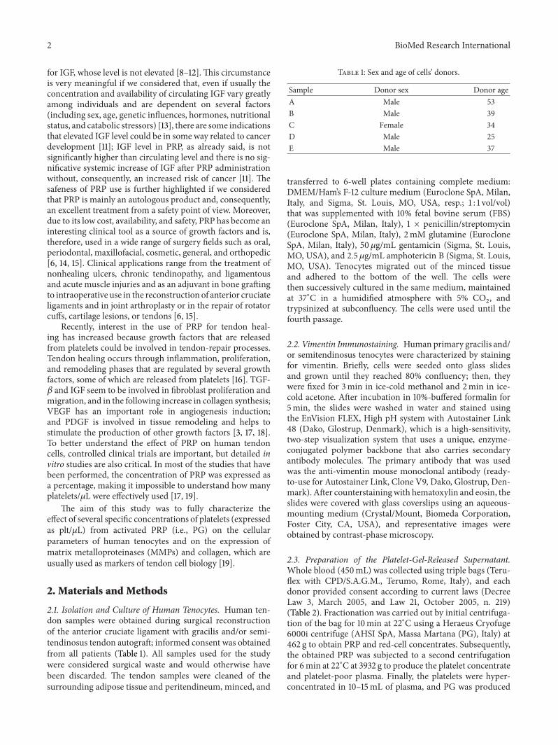
Figure 1: Tenocytes characterization. Tenocytes were characterized through vimentin immunostaining (a) and assay of scleraxis expressionby western blot (b). Magnification 200x.
studies, some in vitro investigations have been performedon tenocytes isolated from several tendon origins (healthytendons or rotator cuff tendons with degenerative tears), andthese studies showed that PRP stimulated cell proliferation[3, 34–36] and collagen production [3, 35] in tenocytes.It is important to consider, however, that tenocyte biologycould differ depending on donor age, anatomic origin, andstatus of the tendon (healthy, injured, or degenerated tendon)[35]. Most importantly, in some of these studies, the plateletconcentration of PRP was unclear, and it is critical to knowthe exact concentration of platelets to correctly comparestudies. It was clearly shown in other cell types involved inwound healing (such as endothelial cells and fibroblasts) thatspecific concentrations of platelets have different effects andthat excessively high concentrations could be less effective[5, 8, 37].This evidence suggests that excessively high concen-trations of platelets have an inhibitory effect on the wound-healing processes and are therefore counterproductive.
For these reasons, our purpose was to evaluate the in vitroeffect of different concentrations of platelets (and of differentgrowth factor concentrations) on the biological features oftenocytes. As the platelet source used was platelet gel, theactivated form of PRP that was obtained adding thrombinand calcium gluconate to the latter; more specifically, super-natant released from PG was used because it had alreadybeen shown that it had the same effect as PG on cellularparameters [8]. Therefore, for convenience, all experimentswere performed using PG-released supernatants. Plateletconcentration in the PG-released supernatants was expressedas plt/𝜇L because the concentration of the factors that arereleased from the platelets is assumed to be proportional tothe initial concentration of plt/𝜇L, and it was assumed thatthe supernatants would maintain the same concentration ofplt/𝜇L even if platelets were no longer present in the releasate.
The present study evaluated the ability of PG-releasedsupernatants to affect tenocyte activities that are required forthe tendon-healing process that occurs in three overlappingphases (inflammatory, proliferative, and remodeling phase).During these phases, in addition to other processes, theECM is produced and remodeled from tenocytes, and thecells proliferate and migrate into wounds [38, 39]. Therefore,
how different concentrations of platelets in PG-releasedsupernatants were able to condition proliferation, migrationinto wounds, and the production of collagen type I andgelatinases was evaluated.
3.1. Tenocyte Characterization. First, to confirm the identityof tenocytes, immunocytochemistry of vimentin, an inter-mediate filament that is characteristically found in cells ofmesenchymal origin and usually used as a tenocyte marker,was performed [40, 41]. The expression of this marker wasso high that counterstaining with hematoxylin and eosin wasnot perceptible (Figure 1(a)).
To further assess the identity of the isolated cells, thepresence of scleraxis, a transcription factor that is a highlyspecificmarker for tendons and ligaments, was evaluated [42,43]. Western blotting confirmed the presence of this markerat a molecular weight of approximately 40 kDa (Figure 1(b)).With regard to scleraxis, we found this marker at a molecularweight of approximately 40 kDa; however, this molecule,when in its monomeric form, has a molecular weight of22 kDa. Scleraxis belongs to a family of transcription factors(the basic helix-loop-helix [bHLH]) that are known to formheterodimers and homodimers [44, 45]; therefore, it ispossible that we revealed the dimeric form of this protein.The presence of these markers, however, confirmed that theisolated cells were tenocytes.
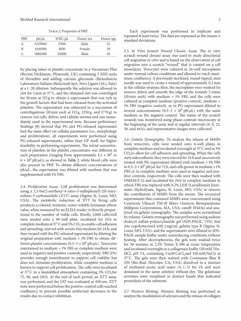
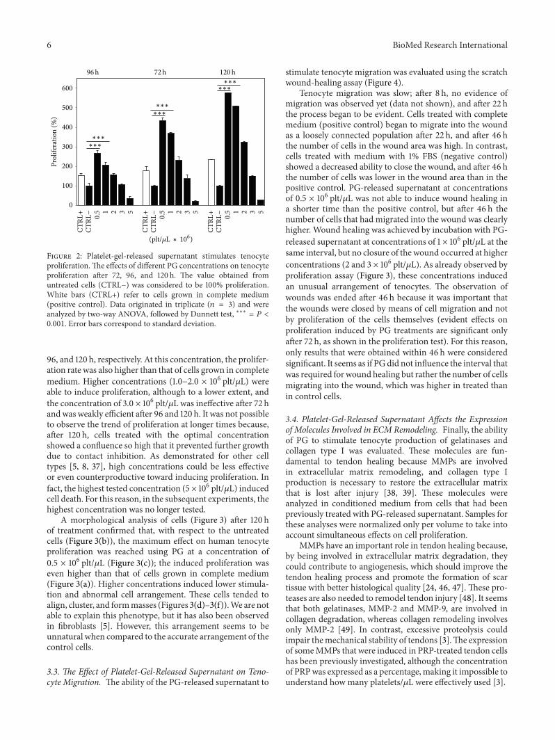
3.2. Platelet-Gel-Released Supernatant Stimulates TenocyteProliferation. Once the identity of the cells was confirmed,tenocyte proliferation in response to PG-released supernatantwas evaluated. Because the tenocyte doubling time is approx-imately 96 h (data not shown), proliferation was evaluated inthe treated cells at this time and 24 h after and before thattime. Cells were treatedwith diluted PG-released supernatantto obtain the indicated concentrations (0.5 × 10⁶, 1 × 10⁶, 2× 10⁶, 3 × 10⁶, and 5 × 10⁶ plt/𝜇L), and tests showed a dose-dependent response of the cells, with higher stimulation after120 h (Figure 2). However, at all of the tested intervals, theoptimal concentration seemed to be 0.5 × 10⁶ plt/𝜇L, whichresulted in a rate of proliferation that was approximately 2.6-,4.3-, and 5.8-fold higher than that of untreated cells after 72,
6 BioMed Research International
0
100
200
300
400
500
600
Prol
ifera
tion
(%)
CTRL+
CTRL−
∗∗∗
∗∗∗
∗∗∗
∗∗∗
∗∗∗
∗∗∗
0.5 1 2 3 5
CTRL+
CTRL−
0.5 1 2 3 5
CTRL+
CTRL−
0.5 1 2 3 5
96h 72h 120h
(plt/𝜇L ∗ 106)
Figure 2: Platelet-gel-released supernatant stimulates tenocyteproliferation.The effects of different PG concentrations on tenocyteproliferation after 72, 96, and 120 h. The value obtained fromuntreated cells (CTRL−) was considered to be 100% proliferation.White bars (CTRL+) refer to cells grown in complete medium(positive control). Data originated in triplicate (𝑛 = 3) and wereanalyzed by two-way ANOVA, followed by Dunnett test, ∗∗∗ = 𝑃 <0.001. Error bars correspond to standard deviation.
96, and 120 h, respectively. At this concentration, the prolifer-ation rate was also higher than that of cells grown in completemedium. Higher concentrations (1.0−2.0 × 10⁶ plt/𝜇L) wereable to induce proliferation, although to a lower extent, andthe concentration of 3.0 × 10⁶ plt/𝜇L was ineffective after 72 handwas weakly efficient after 96 and 120 h. It was not possibleto observe the trend of proliferation at longer times because,after 120 h, cells treated with the optimal concentrationshowed a confluence so high that it prevented further growthdue to contact inhibition. As demonstrated for other celltypes [5, 8, 37], high concentrations could be less effectiveor even counterproductive toward inducing proliferation. Infact, the highest tested concentration (5 × 10⁶ plt/𝜇L) inducedcell death. For this reason, in the subsequent experiments, thehighest concentration was no longer tested.
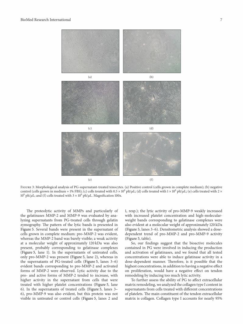
A morphological analysis of cells (Figure 3) after 120 hof treatment confirmed that, with respect to the untreatedcells (Figure 3(b)), the maximum effect on human tenocyteproliferation was reached using PG at a concentration of0.5 × 106 plt/𝜇L (Figure 3(c)); the induced proliferation waseven higher than that of cells grown in complete medium(Figure 3(a)). Higher concentrations induced lower stimula-tion and abnormal cell arrangement. These cells tended toalign, cluster, and formmasses (Figures 3(d)–3(f)).We are notable to explain this phenotype, but it has also been observedin fibroblasts [5]. However, this arrangement seems to beunnatural when compared to the accurate arrangement of thecontrol cells.
3.3. The Effect of Platelet-Gel-Released Supernatant on Teno-cyte Migration. The ability of the PG-released supernatant to
stimulate tenocyte migration was evaluated using the scratchwound-healing assay (Figure 4).
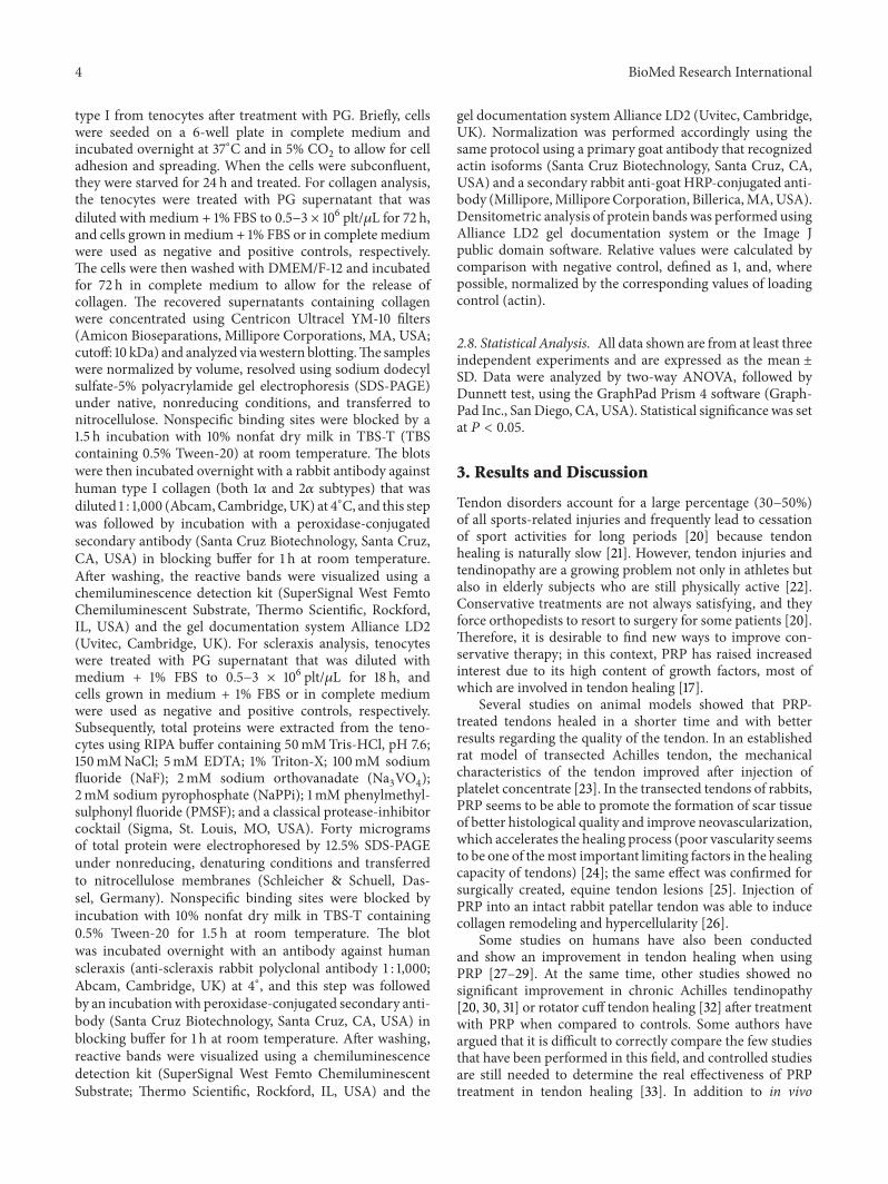
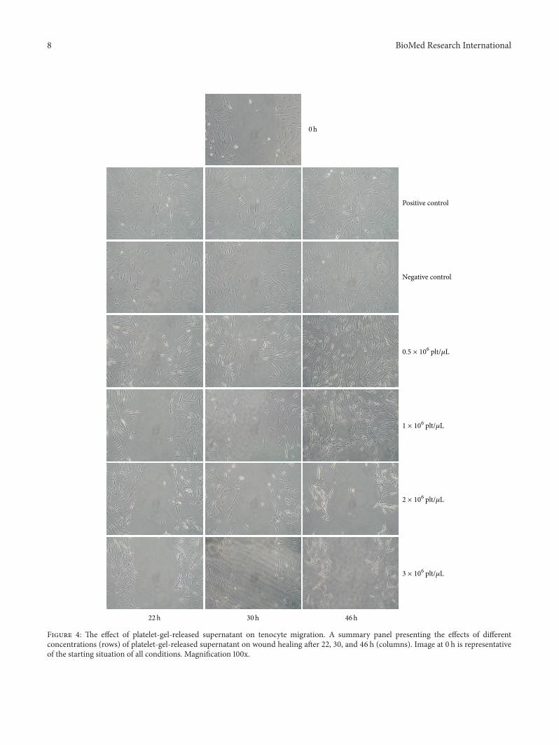
Tenocyte migration was slow; after 8 h, no evidence ofmigration was observed yet (data not shown), and after 22 hthe process began to be evident. Cells treated with completemedium (positive control) began to migrate into the woundas a loosely connected population after 22 h, and after 46 hthe number of cells in the wound area was high. In contrast,cells treated with medium with 1% FBS (negative control)showed a decreased ability to close the wound, and after 46 hthe number of cells was lower in the wound area than in thepositive control. PG-released supernatant at concentrationsof 0.5 × 10⁶ plt/𝜇L was not able to induce wound healing ina shorter time than the positive control, but after 46 h thenumber of cells that had migrated into the wound was clearlyhigher. Wound healing was achieved by incubation with PG-released supernatant at concentrations of 1 × 10⁶ plt/𝜇L at thesame interval, but no closure of the wound occurred at higherconcentrations (2 and 3 × 10⁶ plt/𝜇L). As already observed byproliferation assay (Figure 3), these concentrations inducedan unusual arrangement of tenocytes. The observation ofwounds was ended after 46 h because it was important thatthe wounds were closed by means of cell migration and notby proliferation of the cells themselves (evident effects onproliferation induced by PG treatments are significant onlyafter 72 h, as shown in the proliferation test). For this reason,only results that were obtained within 46 h were consideredsignificant. It seems as if PGdid not influence the interval thatwas required forwound healing but rather the number of cellsmigrating into the wound, which was higher in treated thanin control cells.
3.4. Platelet-Gel-Released Supernatant Affects the Expressionof Molecules Involved in ECM Remodeling. Finally, the abilityof PG to stimulate tenocyte production of gelatinases andcollagen type I was evaluated. These molecules are fun-damental to tendon healing because MMPs are involvedin extracellular matrix remodeling, and collagen type Iproduction is necessary to restore the extracellular matrixthat is lost after injury [38, 39]. These molecules wereanalyzed in conditioned medium from cells that had beenpreviously treated with PG-released supernatant. Samples forthese analyses were normalized only per volume to take intoaccount simultaneous effects on cell proliferation.
MMPs have an important role in tendon healing because,by being involved in extracellular matrix degradation, theycould contribute to angiogenesis, which should improve thetendon healing process and promote the formation of scartissue with better histological quality [24, 46, 47]. These pro-teases are also needed to remodel tendon injury [48]. It seemsthat both gelatinases, MMP-2 and MMP-9, are involved incollagen degradation, whereas collagen remodeling involvesonly MMP-2 [49]. In contrast, excessive proteolysis couldimpair themechanical stability of tendons [3].The expressionof someMMPs that were induced in PRP-treated tendon cellshas been previously investigated, although the concentrationof PRPwas expressed as a percentage,making it impossible tounderstand how many platelets/𝜇L were effectively used [3].
BioMed Research International 7
(a) (b)
(c) (d)
(e) (f)
Figure 3: Morphological analysis of PG-supernatant-treated tenocytes. (a) Positive control (cells grown in complete medium); (b) negativecontrol (cells grown in medium + 1% FBS); (c) cells treated with 0.5 × 10⁶ plt/𝜇L; (d) cells treated with 1 × 10⁶ plt/𝜇L; (e) cells treated with 2 ×10⁶ plt/𝜇L; and (f) cells treated with 3 × 10⁶ plt/𝜇L. Magnification 100x.
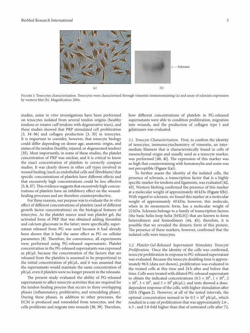
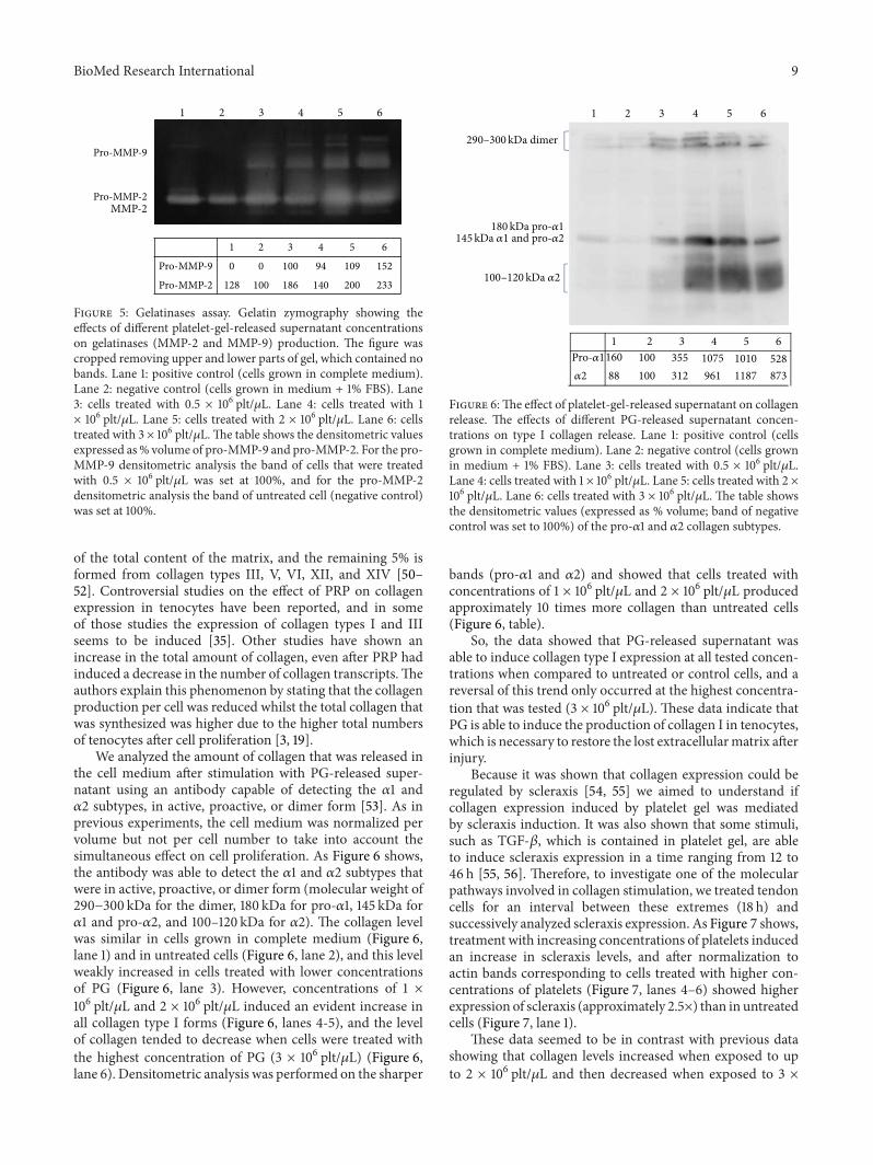
The proteolytic activity of MMPs and particularly ofthe gelatinases MMP-2 and MMP-9 was evaluated by ana-lyzing supernatants from PG-treated cells through gelatinzymography. The pattern of the lytic bands is presented inFigure 5. Several bands were present in the supernatant ofcells grown in complete medium: pro-MMP-2 was evident,whereas the MMP-2 band was barely visible; a weak activityat a molecular weight of approximately 120 kDa was alsopresent, probably corresponding to gelatinase complexes(Figure 5, lane 1). In the supernatants of untreated cells,only pro-MMP-2 was present (Figure 5, lane 2), whereas inthe supernatants of PG-treated cells (Figure 5, lanes 3–6)evident bands corresponding to pro-MMP-2 and activatedforms of MMP-2 were observed. Lytic activity due to thepro- and active forms of MMP-2 tended to increase, withhigher activity in the supernatant from cells that weretreated with higher platelet concentrations (Figure 5, lane6). In the supernatants of treated cells (Figure 5, lanes 3–6), pro-MMP-9 was also evident, but this protein was notvisible in untreated or control cells (Figure 5, lanes 2 and
1, resp.); the lytic activity of pro-MMP-9 weakly increasedwith increased platelet concentration and high-molecular-weight bands corresponding to gelatinase complexes werealso evident at a molecular weight of approximately 120 kDa(Figure 5, lanes 3–6). Densitometric analysis showed a dose-dependent trend of pro-MMP-2 and pro-MMP-9 activity(Figure 5, table).
So, our findings suggest that the bioactive moleculescontained in PG were involved in inducing the productionand activation of gelatinases, and we found that all testedconcentrations were able to induce gelatinase activity in adose-dependent manner. Therefore, is it possible that thehighest concentrations, in addition to having a negative effecton proliferation, would have a negative effect on tendonremodeling by inducing too much lytic activity.
To further assess the ability of PG to affect extracellularmatrix remodeling, we analyzed the collagen type I content insupernatants from cells treated with different concentrationsof platelets. The main constituent of the tendon extracellularmatrix is collagen. Collagen type I accounts for nearly 95%
8 BioMed Research International
Positive control
Negative control
0h
0.5 × 10⁶ plt/𝜇L
1 × 10⁶ plt/𝜇L
2 × 10⁶ plt/𝜇L
3 × 10⁶ plt/𝜇L
22h 46h30h
Figure 4: The effect of platelet-gel-released supernatant on tenocyte migration. A summary panel presenting the effects of differentconcentrations (rows) of platelet-gel-released supernatant on wound healing after 22, 30, and 46 h (columns). Image at 0 h is representativeof the starting situation of all conditions. Magnification 100x.
BioMed Research International 9
1 2 3 4 5 6
MMP-2
1 2 3 4 5 6
Pro-MMP-9
Pro-MMP-2
Pro-MMP-9
0 0 100 94 109 152
Pro-MMP-2 128 100 186 140 200 233
Figure 5: Gelatinases assay. Gelatin zymography showing theeffects of different platelet-gel-released supernatant concentrationson gelatinases (MMP-2 and MMP-9) production. The figure wascropped removing upper and lower parts of gel, which contained nobands. Lane 1: positive control (cells grown in complete medium).Lane 2: negative control (cells grown in medium + 1% FBS). Lane3: cells treated with 0.5 × 10⁶ plt/𝜇L. Lane 4: cells treated with 1× 10⁶ plt/𝜇L. Lane 5: cells treated with 2 × 10⁶ plt/𝜇L. Lane 6: cellstreated with 3 × 10⁶ plt/𝜇L.The table shows the densitometric valuesexpressed as% volume of pro-MMP-9 and pro-MMP-2. For the pro-MMP-9 densitometric analysis the band of cells that were treatedwith 0.5 × 10⁶ plt/𝜇L was set at 100%, and for the pro-MMP-2densitometric analysis the band of untreated cell (negative control)was set at 100%.
of the total content of the matrix, and the remaining 5% isformed from collagen types III, V, VI, XII, and XIV [50–52]. Controversial studies on the effect of PRP on collagenexpression in tenocytes have been reported, and in someof those studies the expression of collagen types I and IIIseems to be induced [35]. Other studies have shown anincrease in the total amount of collagen, even after PRP hadinduced a decrease in the number of collagen transcripts.Theauthors explain this phenomenon by stating that the collagenproduction per cell was reduced whilst the total collagen thatwas synthesized was higher due to the higher total numbersof tenocytes after cell proliferation [3, 19].
We analyzed the amount of collagen that was released inthe cell medium after stimulation with PG-released super-natant using an antibody capable of detecting the 𝛼1 and𝛼2 subtypes, in active, proactive, or dimer form [53]. As inprevious experiments, the cell medium was normalized pervolume but not per cell number to take into account thesimultaneous effect on cell proliferation. As Figure 6 shows,the antibody was able to detect the 𝛼1 and 𝛼2 subtypes thatwere in active, proactive, or dimer form (molecular weight of290−300 kDa for the dimer, 180 kDa for pro-𝛼1, 145 kDa for𝛼1 and pro-𝛼2, and 100–120 kDa for 𝛼2). The collagen levelwas similar in cells grown in complete medium (Figure 6,lane 1) and in untreated cells (Figure 6, lane 2), and this levelweakly increased in cells treated with lower concentrationsof PG (Figure 6, lane 3). However, concentrations of 1 ×10⁶ plt/𝜇L and 2 × 10⁶ plt/𝜇L induced an evident increase inall collagen type I forms (Figure 6, lanes 4-5), and the levelof collagen tended to decrease when cells were treated withthe highest concentration of PG (3 × 10⁶ plt/𝜇L) (Figure 6,lane 6). Densitometric analysis was performed on the sharper
1 2 3 4 5 6
1 2 3 4 5 6160 100 355 1075 1010 52888 100 312 961 1187 873
290–300kDa dimer
180 kDa pro-𝛼1145kDa 𝛼1 and pro-𝛼2
100–120 kDa 𝛼2
𝛼2
Pro-𝛼1
Figure 6:The effect of platelet-gel-released supernatant on collagenrelease. The effects of different PG-released supernatant concen-trations on type I collagen release. Lane 1: positive control (cellsgrown in complete medium). Lane 2: negative control (cells grownin medium + 1% FBS). Lane 3: cells treated with 0.5 × 10⁶ plt/𝜇L.Lane 4: cells treated with 1 × 10⁶ plt/𝜇L. Lane 5: cells treated with 2 ×10⁶ plt/𝜇L. Lane 6: cells treated with 3 × 10⁶ plt/𝜇L. The table showsthe densitometric values (expressed as % volume; band of negativecontrol was set to 100%) of the pro-𝛼1 and 𝛼2 collagen subtypes.
bands (pro-𝛼1 and 𝛼2) and showed that cells treated withconcentrations of 1 × 10⁶ plt/𝜇L and 2 × 10⁶ plt/𝜇L producedapproximately 10 times more collagen than untreated cells(Figure 6, table).
So, the data showed that PG-released supernatant wasable to induce collagen type I expression at all tested concen-trations when compared to untreated or control cells, and areversal of this trend only occurred at the highest concentra-tion that was tested (3 × 10⁶ plt/𝜇L). These data indicate thatPG is able to induce the production of collagen I in tenocytes,which is necessary to restore the lost extracellularmatrix afterinjury.
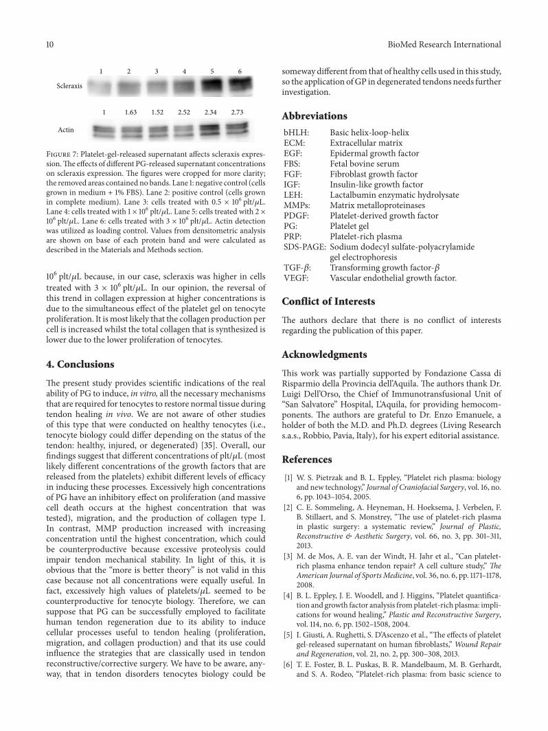
Because it was shown that collagen expression could beregulated by scleraxis [54, 55] we aimed to understand ifcollagen expression induced by platelet gel was mediatedby scleraxis induction. It was also shown that some stimuli,such as TGF-𝛽, which is contained in platelet gel, are ableto induce scleraxis expression in a time ranging from 12 to46 h [55, 56]. Therefore, to investigate one of the molecularpathways involved in collagen stimulation, we treated tendoncells for an interval between these extremes (18 h) andsuccessively analyzed scleraxis expression. As Figure 7 shows,treatment with increasing concentrations of platelets inducedan increase in scleraxis levels, and after normalization toactin bands corresponding to cells treated with higher con-centrations of platelets (Figure 7, lanes 4–6) showed higherexpression of scleraxis (approximately 2.5×) than in untreatedcells (Figure 7, lane 1).
These data seemed to be in contrast with previous datashowing that collagen levels increased when exposed to upto 2 × 10⁶ plt/𝜇L and then decreased when exposed to 3 ×
10 BioMed Research International
1 2 3 4 5 6
Scleraxis
Actin
1 2.732.341.52 2.521.63
Figure 7: Platelet-gel-released supernatant affects scleraxis expres-sion.The effects of different PG-released supernatant concentrationson scleraxis expression. The figures were cropped for more clarity;the removed areas contained no bands. Lane 1: negative control (cellsgrown in medium + 1% FBS). Lane 2: positive control (cells grownin complete medium). Lane 3: cells treated with 0.5 × 10⁶ plt/𝜇L.Lane 4: cells treated with 1 × 10⁶ plt/𝜇L. Lane 5: cells treated with 2 ×10⁶ plt/𝜇L. Lane 6: cells treated with 3 × 10⁶ plt/𝜇L. Actin detectionwas utilized as loading control. Values from densitometric analysisare shown on base of each protein band and were calculated asdescribed in the Materials and Methods section.
106 plt/𝜇L because, in our case, scleraxis was higher in cellstreated with 3 × 10⁶ plt/𝜇L. In our opinion, the reversal ofthis trend in collagen expression at higher concentrations isdue to the simultaneous effect of the platelet gel on tenocyteproliferation. It ismost likely that the collagen production percell is increased whilst the total collagen that is synthesized islower due to the lower proliferation of tenocytes.
4. Conclusions
The present study provides scientific indications of the realability of PG to induce, in vitro, all the necessary mechanismsthat are required for tenocytes to restore normal tissue duringtendon healing in vivo. We are not aware of other studiesof this type that were conducted on healthy tenocytes (i.e.,tenocyte biology could differ depending on the status of thetendon: healthy, injured, or degenerated) [35]. Overall, ourfindings suggest that different concentrations of plt/𝜇L (mostlikely different concentrations of the growth factors that arereleased from the platelets) exhibit different levels of efficacyin inducing these processes. Excessively high concentrationsof PG have an inhibitory effect on proliferation (and massivecell death occurs at the highest concentration that wastested), migration, and the production of collagen type I.In contrast, MMP production increased with increasingconcentration until the highest concentration, which couldbe counterproductive because excessive proteolysis couldimpair tendon mechanical stability. In light of this, it isobvious that the “more is better theory” is not valid in thiscase because not all concentrations were equally useful. Infact, excessively high values of platelets/𝜇L seemed to becounterproductive for tenocyte biology. Therefore, we cansuppose that PG can be successfully employed to facilitatehuman tendon regeneration due to its ability to inducecellular processes useful to tendon healing (proliferation,migration, and collagen production) and that its use couldinfluence the strategies that are classically used in tendonreconstructive/corrective surgery. We have to be aware, any-way, that in tendon disorders tenocytes biology could be
someway different from that of healthy cells used in this study,so the application ofGP in degenerated tendons needs furtherinvestigation.
Abbreviations
bHLH: Basic helix-loop-helixECM: Extracellular matrixEGF: Epidermal growth factorFBS: Fetal bovine serumFGF: Fibroblast growth factorIGF: Insulin-like growth factorLEH: Lactalbumin enzymatic hydrolysateMMPs: Matrix metalloproteinasesPDGF: Platelet-derived growth factorPG: Platelet gelPRP: Platelet-rich plasmaSDS-PAGE: Sodium dodecyl sulfate-polyacrylamide
gel electrophoresisTGF-𝛽: Transforming growth factor-𝛽VEGF: Vascular endothelial growth factor.
Conflict of Interests
The authors declare that there is no conflict of interestsregarding the publication of this paper.
Acknowledgments
This work was partially supported by Fondazione Cassa diRisparmio della Provincia dell’Aquila. The authors thank Dr.Luigi Dell’Orso, the Chief of Immunotransfusional Unit of“San Salvatore” Hospital, L’Aquila, for providing hemocom-ponents. The authors are grateful to Dr. Enzo Emanuele, aholder of both the M.D. and Ph.D. degrees (Living Researchs.a.s., Robbio, Pavia, Italy), for his expert editorial assistance.
References
[1] W. S. Pietrzak and B. L. Eppley, “Platelet rich plasma: biologyand new technology,” Journal of Craniofacial Surgery, vol. 16, no.6, pp. 1043–1054, 2005.
[2] C. E. Sommeling, A. Heyneman, H. Hoeksema, J. Verbelen, F.B. Stillaert, and S. Monstrey, “The use of platelet-rich plasmain plastic surgery: a systematic review,” Journal of Plastic,Reconstructive & Aesthetic Surgery, vol. 66, no. 3, pp. 301–311,2013.
[3] M. de Mos, A. E. van der Windt, H. Jahr et al., “Can platelet-rich plasma enhance tendon repair? A cell culture study,” TheAmerican Journal of SportsMedicine, vol. 36, no. 6, pp. 1171–1178,2008.
[4] B. L. Eppley, J. E. Woodell, and J. Higgins, “Platelet quantifica-tion and growth factor analysis fromplatelet-rich plasma: impli-cations for wound healing,” Plastic and Reconstructive Surgery,vol. 114, no. 6, pp. 1502–1508, 2004.
[5] I. Giusti, A. Rughetti, S. D’Ascenzo et al., “The effects of plateletgel-released supernatant on human fibroblasts,” Wound Repairand Regeneration, vol. 21, no. 2, pp. 300–308, 2013.
[6] T. E. Foster, B. L. Puskas, B. R. Mandelbaum, M. B. Gerhardt,and S. A. Rodeo, “Platelet-rich plasma: from basic science to
BioMed Research International 11
clinical applications,” American Journal of Sports Medicine, vol.37, no. 11, pp. 2259–2272, 2009.
[7] R. Landesberg, A. Burke, D. Pinsky et al., “Activation of platelet-rich plasma using thrombin receptor agonist peptide,” Journal ofOral andMaxillofacial Surgery, vol. 63, no. 4, pp. 529–535, 2005.
[8] I. Giusti, A. Rughetti, S. D’Ascenzo et al., “Identification of anoptimal concentration of platelet gel for promoting angiogen-esis in human endothelial cells,” Transfusion, vol. 49, no. 4, pp.771–778, 2009.
[9] A. S. Wasterlain, H. J. Braun, A. H. S. Harris, H. Kim, and J. L.Dragoo, “The systemic effects of platelet-rich plasma injection,”The American Journal of Sports Medicine, vol. 41, no. 1, pp. 186–193, 2013.
[10] L. V. Schnabel, H. O. Mohammed, B. J. Miller et al., “PlateletRich Plasma (PRP) enhances anabolic gene expression pat-terns in flexor digitorum superficialis tendons,” Journal ofOrthopaedic Research, vol. 25, no. 2, pp. 230–240, 2007.
[11] G. Schippinger, K. Oettl, F. Fankhauser, S. Spirk, W. Domej,and P. Hofmann, “Influence of intramuscular application ofautologous conditioned plasma on systemic circulating IGF-1,”Journal of Sports Science and Medicine, vol. 10, no. 3, pp. 439–444, 2011.
[12] B. L. Eppley, J. E. Woodell, and J. Higgins, “Platelet quantifi-cation and growth factor analysis from platelet-rich plasma:implications for wound healing,” Plastic and ReconstructiveSurgery, vol. 114, no. 6, pp. 1502–1508, 2004.
[13] C. J. Rosen andM. Pollak, “Circulating a new century,”Trends inEndocrinology and Metabolism, vol. 10, no. 4, pp. 136–141, 1999.
[14] S. Mehta and J. T. Watson, “Platelet rich concentrate: basic sci-ence and current clinical applications,” Journal of OrthopaedicTrauma, vol. 22, no. 6, pp. 432–438, 2008.
[15] S. E. Smith and T. S. Roukis, “Bone and wound healingaugmentation with platelet-rich plasma,” Clinics in PodiatricMedicine and Surgery, vol. 26, no. 4, pp. 559–588, 2009.
[16] N. Maffulli, U. G. Longo, and V. Denaro, “Novel approachesfor the management of tendinopathy,” Journal of Bone and JointSurgery, vol. 92, no. 15, pp. 2604–2613, 2010.
[17] T. Molloy, Y. Wang, and G. A. C. Murrell, “The roles of growthfactors in tendon and ligament healing,” Sports Medicine, vol.33, no. 5, pp. 381–394, 2003.
[18] E. V. Cheung, L. Silverio, and J. W. Sperling, “Strategies inbiologic augmentation of rotator cuff repair: a review,” ClinicalOrthopaedics and Related Research, vol. 468, no. 6, pp. 1476–1484, 2010.
[19] X. Wang, Y. Qiu, J. Triffitt, A. Carr, Z. Xia, and A. Sabok-bar, “Proliferation and differentiation of human tenocytes inresponse to platelet rich plasma: an in vitro and in vivo study,”Journal of Orthopaedic Research, vol. 30, no. 6, pp. 982–990,2012.
[20] R. J. De Vos, A. Weir, H. T. M. Van Schie et al., “Platelet-rich plasma injection for chronic Achilles tendinopathy: arandomized controlled trial,” Journal of the American MedicalAssociation, vol. 303, no. 2, pp. 144–149, 2010.
[21] M. Sanchez, E. Anitua, G. Orive, I. Mujika, and I. Andia,“Platelet-rich therapies in the treatment of orthopaedic sportinjuries,” Sports Medicine, vol. 39, no. 5, pp. 345–354, 2009.
[22] N. Zargar Baboldashti, R. C. Poulsen, S. L. Franklin, M. S.Thompson, and P. A. Hulley, “Platelet-rich plasma protectstenocytes from adverse side effects of dexamethasone andciprofloxacin,”TheAmerican Journal of Sports Medicine, vol. 39,no. 9, pp. 1929–1935, 2011.
[23] P. Aspenberg and O. Virchenko, “Platelet concentrate injectionimproves Achilles tendon repair in rats,” Acta OrthopaedicaScandinavica, vol. 75, no. 1, pp. 93–99, 2004.
[24] D. N. Lyras, K. Kazakos, D. Verettas et al., “The influence ofplatelet-rich plasma on angiogenesis during the early phase oftendon healing,” Foot and Ankle International, vol. 30, no. 11, pp.1101–1106, 2009.
[25] G. Bosch, M. Moleman, A. Barneveld, P. R. vanWeeren, and H.T. M. van Schie, “The effect of platelet-rich plasma on the neo-vascularization of surgically created equine superficial digitalflexor tendon lesions,” Scandinavian Journal of Medicine andScience in Sports, vol. 21, no. 4, pp. 554–561, 2011.
[26] J. G. Lane, R. M. Healey, D. C. Chase, and D. Amiel, “Use ofplatelet-rich plasma to enhance tendon function and cellular-ity,”TheAmerican Journal of Orthopedics, vol. 42, no. 5, pp. 209–214, 2013.
[27] M. Sanchez, E. Anitua, J. Azofra, I. Andıa, S. Padilla, and I.Mujika, “Comparison of surgically repaired Achilles tendontears using platelet-rich fibrin matrices,” American Journal ofSports Medicine, vol. 35, no. 2, pp. 245–251, 2007.
[28] K. Gaweda, M. Tarczynska, and W. Krzyzanowski, “Treatmentof achilles tendinopathy with platelet-rich plasma,” Interna-tional Journal of SportsMedicine, vol. 31, no. 8, pp. 577–583, 2010.
[29] K. Harmon, J. Drezner, and A. Rao, “Platelet rich plasma forchronic tendinopathy,” British Journal of Sports Medicine, vol.47, p. e2, 2013.
[30] S. de Jonge, R. J. de Vos, A. Weir et al., “One-year follow-up ofplatelet-rich plasma treatment in chronic achilles tendinopathy:a double-blind randomized placebo-controlled trial,” AmericanJournal of Sports Medicine, vol. 39, no. 8, pp. 1623–1629, 2011.
[31] T. Schepull, J. Kvist, H. Norrman, M. Trinks, G. Berlin, and P.Aspenberg, “Autologous platelets have no effect on the healingof human Achilles tendon ruptures: a randomized single-blindstudy,” The American Journal of Sports Medicine, vol. 39, no. 1,pp. 38–47, 2011.
[32] J. Chahal, G. S. vanThiel, N.Mall et al., “The role of platelet-richplasma in arthroscopic rotator cuff repair: a systematic reviewwith quantitative synthesis,” Arthroscopy—Journal of Arthro-scopic and Related Surgery, vol. 28, no. 11, pp. 1718–1727, 2012.
[33] J. Kaux and J. Crielaard, “Platelet-rich plasma application inthemanagement of chronic tendinopathies,”Acta OrthopaedicaBelgica, vol. 79, no. 1, pp. 10–15, 2013.
[34] E. Anitua, I. Andıa, M. Sanchez et al., “Autologous preparationsrich in growth factors promote proliferation and induce VEGFandHGF production by human tendon cells in culture,” Journalof Orthopaedic Research, vol. 23, no. 2, pp. 281–286, 2005.
[35] C. H. Jo, J. E. Kim, K. S. Yoon, and S. Shin, “Platelet-richplasma stimulates cell proliferation and enhances matrix geneexpression and synthesis in tenocytes from human rotator cufftendonswith degenerative tears,”TheAmerican Journal of SportsMedicine, vol. 40, no. 5, pp. 1035–1045, 2012.
[36] A. D. Mazzocca, M. B. R. McCarthy, D. M. Chowaniec et al.,“The positive effects of different platelet-rich plasma methodson human muscle, bone, and tendon cells,” American Journal ofSports Medicine, vol. 40, no. 8, pp. 1742–1749, 2012.
[37] A. Rughetti, I. Giusti, S. D’Ascenzo et al., “Platelet gel-releasedsupernatant modulates the angiogenic capability of humanendothelial cells,”BloodTransfusion, vol. 6, no. 1, pp. 12–17, 2008.
[38] P. Sharma and N. Maffulli, “Tendon injury and tendinopathy:Healing and repair,” Journal of Bone and Joint Surgery A, vol. 87,no. 1, pp. 187–202, 2005.
12 BioMed Research International
[39] R. James, G. Kesturu, G. Balian, and A. B. Chhabra, “Tendon:biology, biomechanics, repair, growth factors, and evolvingtreatment options,” The Journal of Hand Surgery, vol. 33, no. 1,pp. 102–112, 2008.
[40] M. Vetrano, F. d’Alessandro, M. R. Torrisi, A. Ferretti, M. C.Vulpiani, and V. Visco, “Extracorporeal shock wave therapypromotes cell proliferation and collagen synthesis of primarycultured human tenocytes,” Knee Surgery, Sports Traumatology,Arthroscopy, vol. 19, no. 12, pp. 2159–2168, 2011.
[41] L. J. Backman, G. Fong, G. Andersson et al., “Substance P isa mechanoresponsive, autocrine regulator of human tenocyteproliferation,” PLoS ONE, vol. 6, no. 11, Article ID e27209, 2011.
[42] R. Schweitzer, J. H. Chyung, L. C. Murtaugh et al., “Analysis ofthe tendon cell fate using scleraxis, a specificmarker for tendonsand ligaments,” Development, vol. 128, no. 19, pp. 3855–3866,2001.
[43] S. Pauly, F. Klatte, C. Strobel et al., “Characterization of tendoncell cultures of the human rotator cuff,” European Cells andMaterials, vol. 20, pp. 84–97, 2010.
[44] V. Lejard, G. Brideau, F. Blais et al., “Scleraxis and NFATc regu-late the expression of the pro-𝛼1(I) collagen gene in tendonfibroblasts,”The Journal of Biological Chemistry, vol. 282, no. 24,pp. 17665–17675, 2007.
[45] P. Cserjesi, D. Brown, K. L. Ligon et al., “Scleraxis: A basic helix-loop-helix protein that prefigures skeletal formation duringmouse embryogenesis,” Development, vol. 121, no. 4, pp. 1099–1110, 1995.
[46] M. A. Akhavani, B. Sivakumar, E. M. Paleolog, and N. Kang,“Angiogenesis and plastic surgery,” Journal of Plastic, Recon-structive & Aesthetic Surgery, vol. 61, no. 12, pp. 1425–1437, 2008.
[47] D. Lyras, K. Kazakos, D. Verettas et al., “Immunohistochemicalstudy of angiogenesis after local administration of platelet-richplasma in a patellar tendon defect,” International Orthopaedics,vol. 34, no. 1, pp. 143–148, 2010.
[48] T. M. Ritty and J. Herzog, “Tendon cells produce gelatinases inresponse to type I collagen attachment,” Journal of OrthopaedicResearch, vol. 21, no. 3, pp. 442–450, 2003.
[49] W. Oshiro, J. Lou, X. Xing, Y. Tu, and P. R. Manske, “Flexortendon healing in the rat: a histologic and gene expressionstudy,” Journal of Hand Surgery, vol. 28, no. 5, pp. 814–823, 2003.
[50] P. Kannus, “Structure of the tendon connective tissue,” Scandi-navian Journal of Medicine and Science in Sports, vol. 10, no. 6,pp. 312–320, 2000.
[51] G. P. Riley, “Gene expression and matrix turnover in overusedand damaged tendons,” Scandinavian Journal of Medicine &Science in Sports, vol. 15, no. 4, pp. 241–251, 2005.
[52] G. Gross and A. Hoffmann, “Therapeutic strategies for tendonhealing based on novel biomaterials, factors and cells,” Pathobi-ology, vol. 80, no. 4, pp. 203–210, 2013.
[53] H. Qiao, J. Bell, S. Juliao, L. Li, and J. M. May, “Ascorbic aciduptake and regulation of type I collagen synthesis in culturedvascular smoothmuscle cells,” Journal of Vascular Research, vol.46, no. 1, pp. 15–24, 2008.
[54] R. A. Bagchi and M. P. Czubryt, “Synergistic roles of scleraxisand Smads in the regulation of collagen 1𝛼2 gene expression,”Biochimica et Biophysica Acta—Molecular Cell Research, vol.1823, no. 10, pp. 1936–1944, 2012.
[55] Y. M. Farhat, A. A. Al-Maliki, T. Chen et al., “Gene expressionanalysis of the pleiotropic effects of TGF-𝛽1 in an in vitromodelof flexor tendon healing,” PLoS ONE, vol. 7, no. 12, Article IDe51411, 2012.
[56] Y. Liu, P. Cserjesi, A. Nifuji, E. N. Olson, and M. Noda,“Sclerotome-related helix-loop-helix type transcription factor(scleraxis) mRNA is expressed in osteoblasts and its level isenhanced by type-𝛽 transforming growth factor,” Journal ofEndocrinology, vol. 151, no. 3, pp. 491–499, 1996.

Submit your manuscripts athttp://www.hindawi.com
Stem CellsInternational
Hindawi Publishing Corporationhttp://www.hindawi.com Volume 2014
Hindawi Publishing Corporationhttp://www.hindawi.com Volume 2014
MEDIATORSINFLAMMATION
of
Hindawi Publishing Corporationhttp://www.hindawi.com Volume 2014
Behavioural Neurology
EndocrinologyInternational Journal of
Hindawi Publishing Corporationhttp://www.hindawi.com Volume 2014
Hindawi Publishing Corporationhttp://www.hindawi.com Volume 2014
Disease Markers
Hindawi Publishing Corporationhttp://www.hindawi.com Volume 2014
BioMed Research International
OncologyJournal of
Hindawi Publishing Corporationhttp://www.hindawi.com Volume 2014
Hindawi Publishing Corporationhttp://www.hindawi.com Volume 2014
Oxidative Medicine and Cellular Longevity
Hindawi Publishing Corporationhttp://www.hindawi.com Volume 2014
PPAR Research
The Scientific World JournalHindawi Publishing Corporation http://www.hindawi.com Volume 2014
Immunology ResearchHindawi Publishing Corporationhttp://www.hindawi.com Volume 2014
Journal of
ObesityJournal of
Hindawi Publishing Corporationhttp://www.hindawi.com Volume 2014
Hindawi Publishing Corporationhttp://www.hindawi.com Volume 2014
Computational and Mathematical Methods in Medicine
OphthalmologyJournal of
Hindawi Publishing Corporationhttp://www.hindawi.com Volume 2014
Diabetes ResearchJournal of
Hindawi Publishing Corporationhttp://www.hindawi.com Volume 2014
Hindawi Publishing Corporationhttp://www.hindawi.com Volume 2014
Research and TreatmentAIDS
Hindawi Publishing Corporationhttp://www.hindawi.com Volume 2014
Gastroenterology Research and Practice
Hindawi Publishing Corporationhttp://www.hindawi.com Volume 2014
Parkinson’s Disease
Evidence-Based Complementary and Alternative Medicine
Volume 2014Hindawi Publishing Corporationhttp://www.hindawi.com
Copyright © 2022 FDOKUMEN