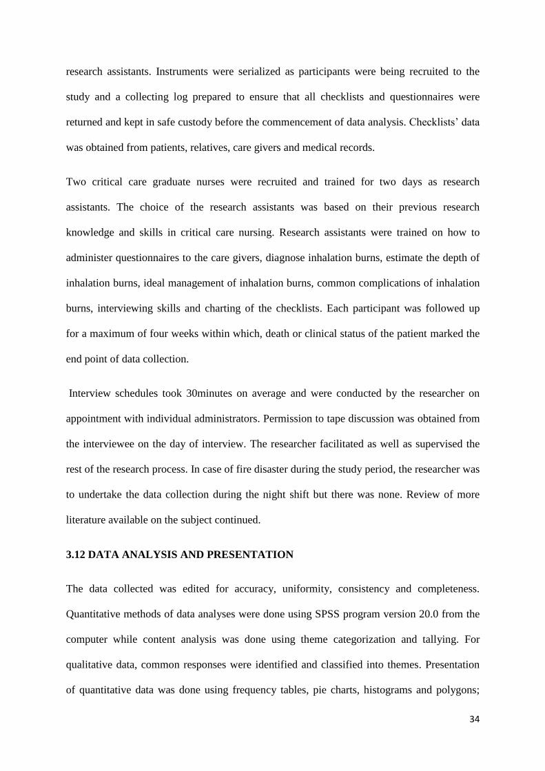
Patient handling of a multidose dry powder inhalation device for albuterol
Upload
khangminh22Category
view
0download
0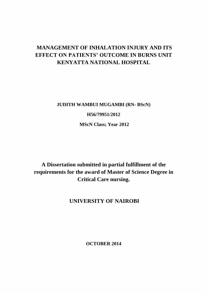
MANAGEMENT OF INHALATION INJURY AND ITS
EFFECT ON PATIENTS’ OUTCOME IN BURNS UNIT
KENYATTA NATIONAL HOSPITAL
JUDITH WAMBUI MUGAMBI (RN- BScN)
H56/79951/2012
MScN Class; Year 2012
A Dissertation submitted in partial fulfillment of the
requirements for the award of Master of Science Degree in
Critical Care nursing.
UNIVERSITY OF NAIROBI
OCTOBER 2014
ii
DECLARATION
This research is my original work, and has never been submitted for approval in any other
institution and should not be duplicated for the purpose of fulfillment of similar awards in
any college or university.
--------------------------------------- -----------------------------------
Judith W. Mugambi
BScN, RN, CCN Date
(MScN H56/79951/2012)
It has been submitted to the University of Nairobi for examination under the approval of
internal supervisors:
--------------------------------------- ------------------------------------
1st Supervisor Date
Theresa M. A. Odero
Msc, BScN, RN, RM, CCN
Lecturer School of Nursing Sciences
UNIVERSITY OF NAIROBI
--------------------------------------- ------------------------------------
2nd Supervisor Date
Angeline C. Kirui
Msc Medical microbiology, BScN
Lecturer School of Nursing Sciences
UNIVERSITY OF NAIROBI
iii
ACKNOWLEDGEMENTS
It is my pleasure to highly appreciate the effort of all who tirelessly assisted me in making
this dissertation a success.
I am greatly indebted to Mrs Theresa Odero and Mrs Angeline Kirui, my research
supervisors for their continued moral and instructional support throughout the research
process. Time accorded to me for verifications, editing and intellectual input is worth noting.
I further extend gratitude to Mrs Lucy Bitok for her ethical advice and the academic input on
quality literature search and research instruments formulation. Thank you for the crucial
inputs you made in my study.
Pleased to acknowledge all nurses and doctors of burns unit who participated in providing
relevant information thus making this study a reality.
Warm regards to Mrs Odero for her devotion in assisting me meet deadlines. She was a
source of encouragement and always available.
Special thanks to Mrs Kirui for her intellectual input and personalized couching. Her
research language is rich and without reservation; a constant inspiration she was.
Highly appreciated is Mr Cheserem for not only assisting in data analysis but also tutoring on
inferential statistics.
In addition, I appreciate my daughter Bilhah for typing and aligning the references.
Finally, I give glory to God and thank him for the accomplishment of this study.
iv
DEDICATION
This study is dedicated to the nurses and doctors working in burns unit Kenyatta National
Hospital for their tireless commitment in caring for the victims of burns despite limited
resources; and to all patients who lost their fight to burns due to inhalation injury.
I further dedicate this study to my children Charles, Michael and Bilhah as a testimonial of
their support during my study time.
In loving memory of my dear husband Thomas who would be more than excited to see me
graduate.
Further to my sister Eunice who always stood by my side to assist me and wondered what I
will do next.
Finally to my nephew Morris who always accompanied me every morning and helped me
revise statistics.

v
TABLE OF CONTENT
CONTENT PAGE
Declaration…………………..…………………………………………………………..……ii
Acknowledgement………………………………………………………………………..…..iii
Dedication………………………………………………………………………………..…. .iv
Table of contents………………………..……………………………………………………..v
List of tables...………………………….………………………………………………...….viii
List of figures………………...……………………………………………………….………ix
Abreviations……...……...……………………………………………………………........….x
Operation definitions…………………………..…………………………………...…..……xii
Abstract………………………….………………………………………….……….......…..xiii
1.0 CHAPTER ONE: INTRODUCTION……………….…………………..………..…….1
1.1 Background of study………………………………..……………………..…………..…..1
1.2 Statement of problem………………………………………………………..…….………2
1.3 Justification of study…………………………………………………………………..…..3
1.4 Research questions……………………………………………………………………..….4
1.5 Hypothesis……………………………………………………………………………..…..4
1.6 Objectives of study………………………………………………………………..……….4
1.7 Theoretical framework………………………………………………………………..…..5
1.8 Conceptual framework………………………………………………………………..…...8
2.0 CHAPTER TWO: LITERATURE REVIEW……………………………………..…..9
2.1 Burn definition and pathology………………………………………………………….....9
2.2 Epidemiology of burns and inhalation injury………………………………………..……9
2.3 Inhalation injury……………………………………………………………………….…10
2.4 Pathophysiology of inhalation injury……………………………………………….……12
2.5 Chest wall burn and inhalation injury……………………………………………….…...18
2.6 Steam inhalation injury………………………………………………………………..…19
2.7 Complications of inhalation injury………………………………………………………20
2.8 Diagnosis and treatment of inhalation injury………………………………………….…21
2.9 Air way edema treatment…………………………………………………………….…..24
vi
2.10 Indications for intubation………………………………………………………….....24
2.11 Restoring hemodynamics…………………………………………………………….25
3.0 CHAPTER THREE: RESEARCH METHODOLOGY…………………………..….27
3.1 Study design…………………………………………………………………………..….27
3.2 Study area……………………………………………………………………………..….27
3.3 Study population…………………………………………………………….………..…..28
3.4 Sampling procedure………………………………………………………………….…...28
3.5 Sample size………………………………………………………………………….……29
3.6 Study variables……………………………………………………………………...……30
3.7 Inclusion and exclusion criteria……………………………………….………………….31
3.8 Research instruments……………………………………………………………………..31
3.9 Pilot study…………..………………………………………………………………….…32
3.10 Ethical considerations…………………………………………………………….…..32
3.11 Data collection method…………………………………………………………….…33
3.12 Data analysis and presentation……………………………………………………….34
3.13 Limitations of study……………………………………………………………….….35
4.0 CHAPTER FOUR: RESULTS OF THE STUDY..………………………………...…36
4.1 Social demographics………………………………………………………………..…….36
4.2 Premorbid conditions….………………………………………………………...……….38
4.3 Other injuries sustained during burns process ….…………………………….…………39
4.4 Duration to hospital post burns..……………..…………………………………..………40
4.5 Duration to clerking post arrival………………..……………………………….……….41
4.6 Intubation…………………………...………………..…………………………..………41
4.7 Inhalation injury assessment………………………………………………….………….42
4.8 Ventilatory interventions……………………………………….………………..….……45
4.9 Oxygenation……………………………………………………………………...………46
4.10 Lung treatment………………………………………………………………………….49
4.11 Systemic therapy………………………………………………………………..………50
4.12 Respiratory distress occurrence……………………………….………………….……..52
4.13 Outcome of patients……………………………………………………………..………53
4.14 Attributes to mortality…………………………………………………………………..56
4.15 Professional expertise………………………………………………………………...…57
4.16 Complications…………………………………………………………………..……….57
4.17 Inferential statistics………………………………………………………………..…….59

vii
5.0 CHAPTER FIVE: DISCUSSION……………………………………………………...61
5.1 Discussion………………………….…………………………………………………….61
6.0 CHAPTER SIX: CONCLUSION AND RECOMMENDATION…………………....68
6.1 Conclusion……………………………….…………………………………….……..…..68
6.2 Recommendations…………………………………………….……..…………….…..…69
6.3 Further research……………………………………………………………….……..…...70
REFERENCES……………………………………………………………………….……..71
APPENDICES……………………………………………………………………………....74
Appendix A: Checklist for patients……………………………………………………….….74
Appendix B: Questionnaire for care givers……..………………………………………....…80
Appendix C: Interview guide for key informants…………………………………...……….85
Appendix D: Consent form……………………………………………………… ……….…86
Appendix E: Grading guide…………………………………………………………………..88
Appendix I: Workplan………………………………………………….…………………….89
Appendix J: Budget…………………………………………………………….…..………..90
Appendix F: KNH/UON ERC approval letter……………………………………………….91
Appendix G: KNH Research Certificate for data collection...…………………...…………..92
Appendix H: Departmental recommendation letter …………………………………………93
viii
LIST OF TABLES
Table name Page no.
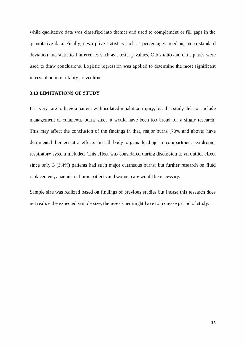
1. Patients age in years…………………………………………………………………37
2. Facial burns distribution……………………………………………………….…….37
3. Premorbid conditions………………………………………………….….…………38
4. Hours arrived to hospital……………………………………………………….……40
5. Hours clerking done post arrival………………………………………….…………41
6. Hours intubation done post clerking…………………………………………………41
7. Inhalation diagnosis……………………………………………………………….…42
8. Age versus cause……………………………………………………………..………44
9. Respiratory distress intervention………………………………………………..……45
10. Oxygen commencement……………………………………………………..……….47
11. Duration of oxygenation………………………………………………………..……48
12. Factors influencing oxygenation…………………………………………………….48
13. Lung treatment commencement……………………………………………………..50
14. Duration of lung treatment…………………………………………………………..50
15. Medications used……………………………………………………….……………51
16. Causes of respiratory distress……….…………………………………………...…..52
17. Outcome of patients…………………………………………………….……………54
18. Specific day of death…………………………………………………….……….…..55
19. Specific day of discharge…………………………………………………………….56
20. Complications………………………………………………………………….…….58
21. Cross tabulations……………………………………………………………………..59
ix
LIST OF FIGURES
Figure name Page no.
1. Age distribution…………………………………………………..................……37
2. Facial burns distribution…………………………...……………………………..38
3. Injuries sustained………...……………………………………………………….39
4. Hours arrived to hospital……………………………………………………..…..40
5. Correlation Clerking vs Intubation…….……………………………………..…..42
6. Causes of burns………………………………………………………….……….44
7. Ventilatory intervention…………………………………………………….……45
8. Accidental extubation versus re-intubation………………………………………46
9. Oxygenation duration……………………………………………………….……47
10. Smoke removal……………………………………………………………..…….49
11. Systemic therapy…………………………………………………………………50
12. Respiratory distress occurrence……………………………………….….………52
13. Mortality by days…………………………………….………………..…..……..54
14. Mortality by cause……………………….…………………………..……...……55
15. Attributes of mortality……………………………………………………...…….56
16. Professional expertise……………………………………………………….……57
17. Complications related to inhalation injury…………………………………..…...58

x
ABBREVIATIONS
ABGA: Arterial blood gas analysis
ACLS: Advanced cardiac life support
ACN: Assistant Chief Nurse
AD: Assistant Director Specialized surgery
ARDS: Adult respiratory distress syndrome
ATP: Adenosine Triphosphate
CDC: Center of Disease Control
CNS: Central nervous system
CO: Carbon monoxide
CPAP: Continuous positive airway pressure
CVP: Central venous pressure
DNA: Deoxyribonucleic acid
ERC: Ethical and Research Committee
ETT: Endotracheal tube
EVB: Evidence based
HCN: Hydrogen Cyanide
HOD: Head of Department
ICU: Intensive care unit
ISO: Organization of international standards
IV: Intravenous
Kgs: Kilograms
KNH: Kenyatta National Hospital
Mls: Milliliters
NAC: N- Acetyl cysteine, used for nebulization.
NAD: Nicotinamide adenine dinucleotide
NDMT: National disaster management team
NO: Nitric Oxide
ONOO-: Peroxynitrite
PEEP: Positive End Expiratory pressure
RD: Respiratory distress
RR: Respiratory rate
SAD: Senior Assistant Director Surgery Department
SIMV: Spontaneous intermittent mechanical ventilation
xi
SPSS: Statistical package of social studies
TBSA: Total body surface area
U.O.N.: University of Nairobi
V/Q: Ventilation/ perfusion mismatch
xii
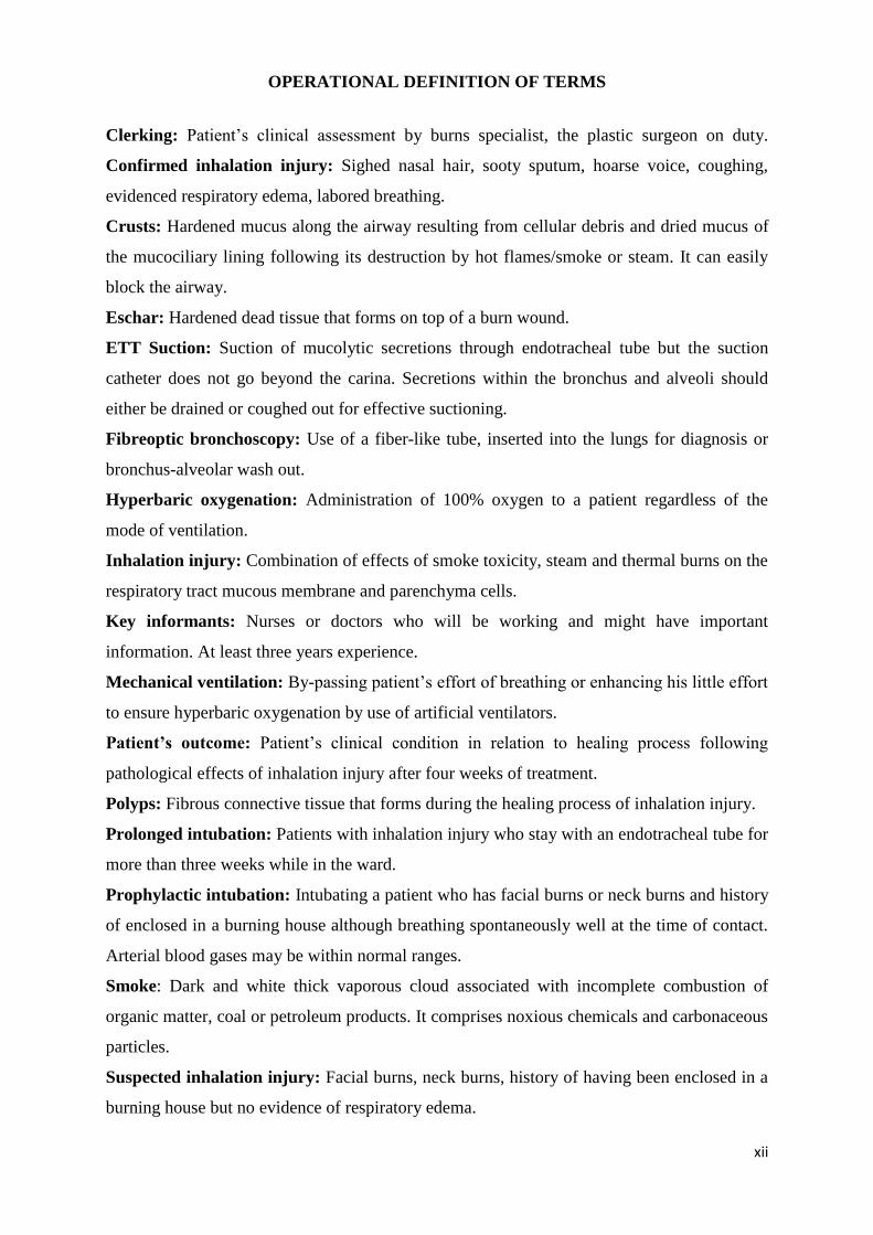
OPERATIONAL DEFINITION OF TERMS
Clerking: Patient’s clinical assessment by burns specialist, the plastic surgeon on duty.
Confirmed inhalation injury: Sighed nasal hair, sooty sputum, hoarse voice, coughing,
evidenced respiratory edema, labored breathing.
Crusts: Hardened mucus along the airway resulting from cellular debris and dried mucus of
the mucociliary lining following its destruction by hot flames/smoke or steam. It can easily
block the airway.
Eschar: Hardened dead tissue that forms on top of a burn wound.
ETT Suction: Suction of mucolytic secretions through endotracheal tube but the suction
catheter does not go beyond the carina. Secretions within the bronchus and alveoli should
either be drained or coughed out for effective suctioning.
Fibreoptic bronchoscopy: Use of a fiber-like tube, inserted into the lungs for diagnosis or
bronchus-alveolar wash out.
Hyperbaric oxygenation: Administration of 100% oxygen to a patient regardless of the
mode of ventilation.
Inhalation injury: Combination of effects of smoke toxicity, steam and thermal burns on the
respiratory tract mucous membrane and parenchyma cells.
Key informants: Nurses or doctors who will be working and might have important
information. At least three years experience.
Mechanical ventilation: By-passing patient’s effort of breathing or enhancing his little effort
to ensure hyperbaric oxygenation by use of artificial ventilators.
Patient’s outcome: Patient’s clinical condition in relation to healing process following
pathological effects of inhalation injury after four weeks of treatment.
Polyps: Fibrous connective tissue that forms during the healing process of inhalation injury.
Prolonged intubation: Patients with inhalation injury who stay with an endotracheal tube for
more than three weeks while in the ward.
Prophylactic intubation: Intubating a patient who has facial burns or neck burns and history
of enclosed in a burning house although breathing spontaneously well at the time of contact.
Arterial blood gases may be within normal ranges.
Smoke: Dark and white thick vaporous cloud associated with incomplete combustion of
organic matter, coal or petroleum products. It comprises noxious chemicals and carbonaceous
particles.
Suspected inhalation injury: Facial burns, neck burns, history of having been enclosed in a
burning house but no evidence of respiratory edema.
xiii
ABSTRACT
Background: Smoke inhalation is responsible for pulmonary injury common in burn victims
and is a major contributing factor to the morbidity and mortality of burn victims both in the
hospital and at incident sites. Inhalation burn injury predisposes burn victims to a major risk
for permanent pulmonary dysfunction and however small, should be central to the
management of burns. Cleaning up of the patients’ lungs after smoke exposure is not a
priority yet it may be of significant value in preventing progress of inhalation injury and
mortality following the rising incidents of fire disasters and high mortality recently reported
in Kenya. Much of the care given to burns patients in Kenya has overlooked the inhalation
injury and concentrated on airway maintenance as in general critical care patients.
Endotracheal intubation traumatizes the airway of patients with inhalation injury more easily
than it would to an intact airway. This calls for attention even after extubation since it might
be a contributing factor to high mortality among burns patients. Occurrence of tracheal
stenosis post extubation was reported 3(7.9%; n=38) and its prevention requires attention.
Main objective: The aim of this study was to determine the relationship between the
management of inhalation injury and the outcome of patients in Burns Unit, KNH.
Study design: This was a longitudinal descriptive study with both quantitative and
qualitative components. A sample size of 84 patients with inhalation injury was purposively
selected from Burns unit, KNH and study duration was three months. Key informants were
purposively selected and interviewed for in-depth information on management of inhalation
injury. A checklist of variables, a questionnaire and an interview guide were used. Data was
managed using SPSS soft ware version 20.0 while statistical inferences were based on p-
values and ODDS ratio.
Results: Diagnosis of inhalation injury was mainly clinical, based on history of the incidence
and presenting signs and symptoms. Other parameters like chest X-ray were primarily used to
confirm position of central lines and only 7 (8.3%) patients had this done. Grading inhalation
injury and determining levels of toxicity were not part of the diagnosis.
Purpose of intubation was to secure the airway and tracheal lavage for removing excess
secretions. However,12 (64.7%) nurses reported using tracheal lavage to remove smoke from
the lungs while 5 (29.4%) reported smoke is never removed. Literature recommends broncho
alveolar toileting for smoke removal and as such tracheal lavage (dry or wet) is not effective
to remove soot and carbonaceous particles from the base of lungs. This might explain the
deranged arterial blood gas results reported in majority 23 (39.4%) of the patients who died
during research period.Majority 27 (69.2%) of deaths occurred during the first week and
arterial blood gas analysis showed 13 (15.5%; n=84) of patients with hypoxemia. Intubation
was found to be significant in relation to mortality with (p-value 0.0001) but in relation with
other significant interventional parameters, it was not significant (p-value 0.63). Use of
steroids had no significant relationship with mortality (p-value 0.322). Assessment of carbon
monoxide blood levels was recommended as a guide to oxygenation of individual patients in
managing inhalation injury. Also, a documented standardized protocol of managing
inhalation injury was recommended to enhance uniformity in decision making and reduce
personal discretions.
1
1.0 CHAPTER ONE: INTRODUCTION
1.1 BACKGROUND OF STUDY
Different approaches in the care of patients with major burns have progressively reduced the
rate of mortality by a specific cause and changed the cause of death. According to Herndon
(2007), burn shock accounted for 20% of burn deaths in 1940s, but due to early and vigorous
fluid resuscitation of burn patients, it is no longer a problem. Second was burn wound sepsis
after burn shock, but this has also been controlled by use of topical antibiotics and timely
surgical debridement. Woodson (2009) reports inhalation injury as the commonest cause of
death in burn patients today, with smoke inhalation alone causing up to 11% deaths.
Combined with cutaneous burns, smoke inhalation led to (30- 90) % deaths of burn patients
(Woodson, 2009).
The aim of this study is to determine the relationship between management of inhalation
injury and the outcome of burns patients. Inhalation burn injury predisposes a patient to a
considerable risk for permanent pulmonary dysfunction and however small, should be central
to the management of burn victims. Managing a burnt airway and its consequences is a
challenge to anaesthetists, nurses and doctors who play a central role in stabilizing the patient
clinically. Inhalation injury results from thermal or chemical irritation after inspiration of
smoke, steam, toxic fumes or mists (Maybaurer, 2009). Its damage can result from direct
cytotoxic effects of the aspirated materials or the tissue inflammatory response. In addition to
damage of the airways and pulmonary parenchyma by heat, inhalation of carbon monoxide or
cyanide also produces toxic systemic effects.
In his study in Germany, Toon et al (2010), reports that 22% of all burn patients and 60% of
those with central facial burns have inhalation injury. In comparison, 30% of burn patients
who had smoke inhalation injury died compared to 2% of those without smoke inhalation. In
conclusion, (80–90) % of all fire-related deaths was attributed to smoke inhalation.
2
Study done in Turkey between 2009 and 2011 compared a burn of 50% of the total body
surface area (TBSA) with smoke inhalation injury to a burn of 73% TBSA without inhalation
injury; in that they both carry a 10% mortality risk ( Kabalak & Yasti,2012). Inhalation
injury predisposes patients to severe clinical consequences such as respiratory failure, acute
respiratory distress syndrome, pulmonary infections or prolonged ventilatory support.
Study done in KNH year 2010/2011 showed open flame burns as the major cause of burns
(49%) followed by hot water (26%); while the major cause of death was inhalation injury
(68.9 %) (Mugambi et al, 2012). The Kenya guideline on management of inhalation burns
only specifies intubation and airway suction. This implies that much of other interventions
will be decided by individual care givers. Standard treatment guidelines for Gertrudes
hospital also has very little about inhalation injury except early intubation (Gertrudes, 2010).
Early decontamination of the lungs after smoke exposure is not stressed upon, yet may be
very significant in preventing progress of inhalation injury and mortality.
1.2 STATEMENT OF THE PROBLEM
Massive fire incidents have been on rising trend in Kenya since the time of tribal crashes in
2007 with increased risk for smoke inhalation and massive deaths. Just to mention is the
Sachangwani and Molo petrol explosion fires, Sinai pipeline leak fire, students burning in
dormitories, domestic accidents / fights, mob burning suspects etc. Sachangwani realized
139 deaths; Molo realized 133 deaths and Sinai realized 100 deaths (Kenya Red Cross,
2011).Since majority died at the scene, the cause of death can be attributed more to
inhalation injury than to burn wound. Open flame burns seem to take toll compared to steam
inhalation, scalds or electricity. Burns unit admits an average of 39 patients per month, 50%
having inhalation injury out of which majority die. Study done in KNH year 2010/2011
showed open flame burns as the major cause of burns (49%) followed by hot water (26%);
while the major cause of death was inhalation injury (68.9 %) (Mugambi et al, 2012).

3
Inhalation burns is estimated at 10% of the total body surface area (TBSA) but it is the major
cause of death among patients with burn injuries, 80%–90% by smoke inhalation (Toon et
al, 2010). This implies that if inhalation burns are effectively managed, mortality rate in
burns patients would reduce significantly. Guidelines of managing inhalation injury in
Kenya are not specific leaving a lot of room for personal discretions and thus, inhalation
injury has not received adequate attention to reduce mortality risk among burn patients. It
would be appropriate to have a well organized protocol driven approach of managing
inhalation burn injury so as to reduce morbidity and mortality associated with inhalation
injury. Tom Lewis (2012) from John Hopkins hospital reviewed their burn management
protocol inspired by the mass casualty event that killed over 100 people here in Kenya. He
comments that the hospital staffs were overwhelmed and majority of patients mismanaged.
1.3 JUSTIFICATION OF THE STUDY
Being the only referral burns center east and central Africa, burns unit (KNH) attends to
majority of patients with inhalation injury from both public and private health facilities. Since
many patients die of inhalation injury and there is no previous study on this subject in Kenya,
this research intends to evaluate the care given against evidenced research recommendations,
determine patients’ outcome and identify other outcome influences hoping to come up with
recommendations on mortality reduction. Smoke inhalation is responsible for pulmonary
injury and significantly contributes to the morbidity and mortality of fire-related injuries in
burn victims. In Kenya, open flame burns are very common especially due to stove explosion
and usually accompanied by smoke toxicity. Apart from home accidents and domestic
violence resulting to burns, disasters involving open flame burns and carbon toxicity have
become common in Kenya and many patients die due to inhalation injury even while in the
hospital. Example is the Molo fire victims where, out of the 22 patients admitted in burns unit
KNH, 13 (59%) died of inhalation injury (Red Cross Kenya, 2011). Fire disaster can affect
4
anybody and therefore requires highly skilled burns personnel, ideal equipments and clear
protocols of inhalation burns management. Findings of this research will be useful as the
baseline of care given to inhalation injury patients in KNH. The research findings will benefit
the patients through mortality reduction and provide guidance to the burns management team
on formulating management protocol for inhalation injury. Care given to the participants was
closely monitored unlike other patients thus benefiting the participating patients. The
researcher hoped to gain more insight on burns management approaches and add knowledge
to the management of inhalation injury and mortality reduction. National disaster
management team (NDMT) can also utilize the findings of this research to design a policy of
care specific for inhalation injury aimed at mortality reduction in burns patients.
1.4 RESEARCH QUESTIONS
1. How does intubation influence patient’s outcome?
2. How is inhalation injury diagnosed?
3. What medications are used to combat respiratory inflammatory process?
4. What treatment protocols are applied to enhance lung healing following inhalation
injury?
5. What factors influence the management of inhalation injury in burns unit, KNH?
1.5 STUDY HYPOTHESIS
Outcome of patients with inhalation injury is not dependent on intubation.
1.6 STUDY OBJECTIVES
1.6.1 MAIN OBJECTIVE
The main objective of this study is to determine the relationship between the management of
inhalation injury and the outcome of patients in Burns Unit, Kenyatta National Hospital.

5
1.6.2 SPECIFIC OBJECTIVES
1. To ascertain the current management of inhalation injury in burns unit, KNH
2. To identify factors influencing the management of inhalation injury in burns unit, KNH
3. To determine the outcome of patients with inhalation injury in relation to the care given in
burns unit, KNH
1.7 THEORETICAL FRAME WORK: Betty Neuman Systems Model
1.7.1 Overview
In her theory of nursing, Betty Neuman as cited in Julia (2002) addresses stress and reaction
to stress. She viewed client as an open system in which cycles of input, process, output and
feedback constitute a dynamic organizational pattern. The aim of her theory was to achieve
optimal system stability and maintain balance among the various stressors. Reactions to the
stressors may be identifiable responses and symptoms. The usual level of health was
identified as the normal line of defense that is protected by a flexible line of defense. She
labeled stressors as intra, inter, and extra-personal in nature as they arise from the internal,
external, and created environments. She reasoned that, when stressors by pass the normal
lines and break through the flexible lines of defense, the system is invaded and the lines of
resistance are activated. At this point, system is described as moving into illness on a
wellness-illness continuum. According to Neuman, the system will be reconstituted and
normal lines of defense restored if adequate energy is available.
1.7.2 Application of Neuman’s theory to inhalation injury
Patient is a system and burn is a stressor. Inability to maintain stability balance follows the
pathologic effect of burns and inhalation injury. Betty Neuman identifies system stability or
6
homeostasis as occurring when energy available exceeds that being utilized by the system;
but in a patient with burns, more energy is required to cope with burn shock hence the
homeostatic imbalance.
The physiological variable explains how burns alter the physiological structure and functions
of the person burnt as detailed out in pathophysiology of inhalation burns. Psychological
variable explains the anxiety due to respiratory distress, alteration of body image, discomfort
of intubation, unusual feeding mode etc. Social cultural variable refers to sudden stoppage of
social role expectations since all burn incidents are accidents and emergencies. The
dependence role sets in and patient feels helpless. Developmental variable is better explained
by the homeostatic processes that become altered due to fluid and electrolyte loss, shunting of
blood to vital organs, tissue edema and tissue injury. It is at this point when believers wonder
why God allowed them to burn and non believers get closer to God. Effect of the stressor
influences individual spiritual beliefs either positively or negatively.
Betty Neuman’s four variables are well applicable to the management of inhalation injury
and burns as they form a basis for nursing diagnosis. Burns is an environmental stressor
which invades the normal lines of defense of a patient, making the lines of resistance to
respond by first shunting blood to protect vital organs while compromising the gastro
intestinal functions. Another response involving lines of resistance is the activation of
immune system leading to swelling of tissues beneath the burn. With this understanding,
prophylactic intubation is indicated in patients with inhalation, facial or neck burns. If the
lines of resistance are not effective, patient may suffer kidney failure. However, if no
provision respiratory support, effect of lines of resistance may cause respiratory failure
leading to death of the victim. In this case, flexible lines of defense would mean taking
precaution to avoid the fire source or reducing the time of direct contact with the source and
applying appropriate and timely first aid.
7
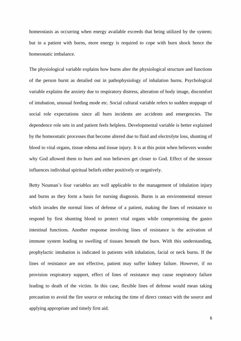
According to Betty Neuman as cited in Julia (2002), interventions can occur before or after
the resistance lines; and in burns patients, intervention occur during and after resistance lines.
Interventions are based on degree of injury, goals and anticipated sequential outcome. The
diagram below illustrates this theoretical framework.
Source: Mugambi (2013)
STRESSOR’S EFFECT
Physiological influences
1. Mucociliary lining dysfunction
2. Parenchyma cells injury
3. Edema of the airway
4. Hypoxia related to smoke toxicity
5. Blood shunting to CNS& heart
6. Effect of medications
Psychological influences
7. Intubation
8. Lung treatment
9. Oxygenation anxiety
Sequential Outcome
1. Normal lung compliance ( lung
volumes).
2. Spontaneous breathing of the
patient (Resp. rate)
3. Non-toxic chemical levels in
the blood
4. Adequate tissue perfusion
5. Adequate gaseous exchange
(ABGA)
Lines of defense
1. Lung injuries heal without
complications
2. Patient’s homeostatic state
restored
3. Patient’s body organs function
normally
Stressor (Intervening variables)
1.Cause of inhalation injury and duration of
contact
2.Pathophysiological effects of inhalation
injury
3. Degree of inhalation injury
4. Premorbid conditions prior to the burns
5.Other injuries sustained during burns process
8
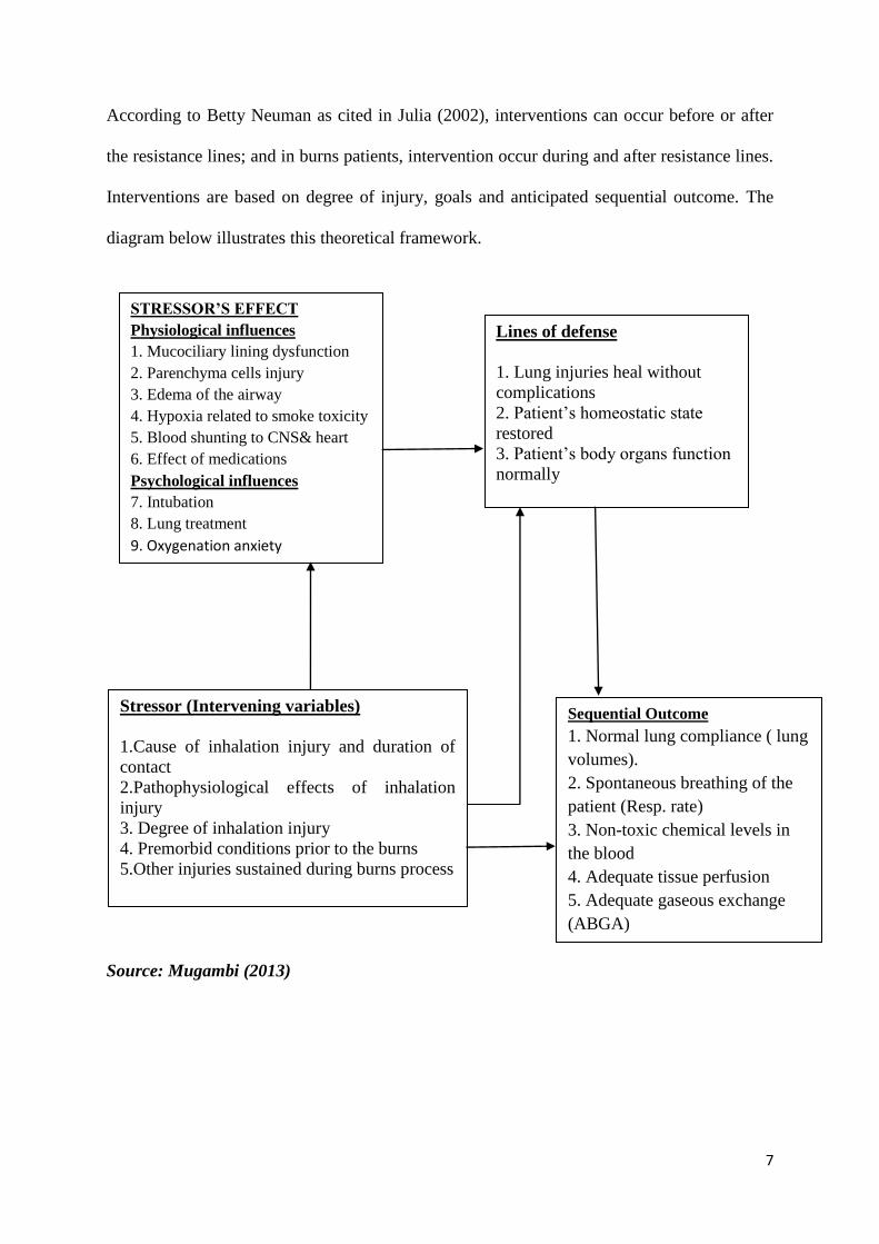
1.8 INTERVENTIONAL CONCEPTUAL FRAMEWORK
Independent variables Dependent variables Outcome
variables
Source: Mugambi (2013)
INTUBATION
Prophylactic
Therapeutic
Re-intubation
Early extubation-7days
LUNG TREATMENT
Bronchial toilet
Tracheal lavage
Chest physiotherapy
Fluid resuscitation/
replacement
MEDICATIONS
Anti-inflammatory
Analgesia/ sedative
Hydrocobalamin
Sodium nitrate/ Thiosulfate
N- Acetylcysteine
Nebulizing agent
OXYGENATION
Mechanical ventilation
(SIMV &PEEP)
Hyperbaric oxygenation
Non re-breather mask -
100%O2
Patent airway
Adequate ventilation
Emergency intubation avoided
Less risk for respiratory arrest
Smoke particles removed from lungs
Improved oxygen delivery to tissues
No fibrin/ mucous crusting
No airway obstruction
Cough reflex enhanced
Dilute smoke toxins in the blood
Enhanced parenchyma healing
Less risk for chest infection
Tissue edema relieved faster
Reduced risk for self extubation
Reduced risk for alveolar necrosis
Less re-intubation
Less tracheal trauma
Less reactive asthma
Cyanide toxins deactivated
Enhanced lung compliance
Improved Hb oxygen carrying
capacity
Improved tissue oxygenation
Reversed carbon monoxide
poisoning
Reduced risk for atelectasis
Enhanced parenchymal healing
Normal gaseous
exchange
Functional mucocilliary
lining
Effective lung
compliance
Parenchyma cells
regenerate and heal
normally
Blood shunting is
reversed and tissue
perfusion restored
Chemical toxin levels
in the blood are
nontoxic (very low)
Normal spontaneous
adequate ventilation
9
2.0 CHAPTER TWO: LITERATURE REVIEW
2.1 BURN DEFINITION AND PATHOLOGY
Herndon (2007) viewed burns as the most common and devastating form of trauma; caused
by heat, friction, electricity, radiation or chemicals. Burn injury is a result of heat transfer
from its source to body tissues leading to tissue destruction. Tissue destruction result from
coagulation, protein denaturation or ionization of cellular contents. The skin and the mucosa
of the upper airway are sites of tissue destruction while deep tissues including the viscera and
bone can be damaged by electrical burns or prolonged contact with heat source. Skin
disruption may lead to increased fluid loss, infection, hypothermia, scarring, compromised
immunity and physiologic functional changes (Brunner & Saddarth, 2010). Depth of injury
depends on the temperature of the burning agent and the duration of contact. Physiologic
responses usually involve fluid and electrolytes, cardiovascular, renal, gastrointestinal, and
pulmonary alterations.
Pathophysiologic changes resulting from major burns during initial burn shock period include
tissue hypoperfusion, and organ hypofunction secondary to decreased cardiac output
following blood shunting to vital organs. Hemodynamic instability results from loss of
capillary integrity and a subsequent shift of fluid, sodium and protein from intravascular to
interstitial spaces. (Polaski & Suzanne, 2010).
2.2 EPIDEMIOLOGY OF BURNS AND INHALATION INJURY
Burn injury can affect people of all age groups, in all social economic levels. In China, it is
estimated that 50,000 people are treated for minor burns annually (Pitts et al, 2008). Patients
hospitalized each year are more than 40,000 with 25,000 requiring specialized burn care. Just
like Kenya, China reports an increase in patients requiring specialized burn care. Of all the
burns admitted to burn centers, 40% are open flame burns, 30% are scalds, 4% electrical and
10
3% chemical burns (Miller et al, 2008). This implies that 40% have smoke inhalation and
30% might have steam inhalation.
Data from the National Center for Injury Prevention and Control in the United States reports
approximately 2 million fires each year which result in 1.2 million people with burn injuries
(Brunner & Saddarth, 2010). Moderate to severe burn injuries requiring hospitalization are
about 100,000 while about 5,000 patients die each year from burn-related complications
(Brunner & Saddarth, 2010). For patients with over 40% of the total body surface area
(TBSA), 75% of all deaths are currently related to either sepsis from burn wound infection,
other infection complications or inhalation injury.
2.3 INHALATION BURN INJURY
Inhalation injury results from thermal or chemical irritation following inspiration of smoke,
burning embers, steam, chemical fumes, cytotoxic fumes or mists (Toon et al, 2010). Damage
to the airway parenchyma cells result from direct heat damage and toxic effects of the
aspirated materials plus the consequence of inflammatory response. Further to the damage,
inhalation of carbon monoxide or cyanide also produces toxic systemic effects.
Inhalation injury is very common in patients who sustain burns and it has high morbidity and
mortality rates (Traber et al, 2007). Isolated inhalation injury can as well pose a significant
risk of mortality or permanent pulmonary dysfunction. When combined with cutaneous
burns, inhalation injury increases fluid requirements for resuscitation, risk for pulmonary
complications and mortality.
2.3.1 Assessment of inhalation injury
According to Palmieri (2007), there is insufficient data to support one treatment standard or
any treatment guideline for the diagnosis of inhalation injury but it should be suspected if
there is evidence in:
• Exposure in an enclosed space • Death of persons at scene
11
• Decreased level of consciousness; Confusion • Steam burns
• Soot in mouth, nares, burnt nasal hairs • Facial burns
• Carbonaceous sputum
• Swelling, ulceration of oral mucosa or tongue (deeper examination may compromise the
airway of the distressed child)
• Dyspnoea • Hoarseness
• Drooling • Stridor, wheeze, crepitations
• Increased work of breathing
• Oxygen saturations <90% in arterial blood (normal saturations do not exclude the diagnosis
as carboxyhaemoglobin is recognized as oxyhaemoglobin by oxygen saturation monitors)
• Carboxyhaemoglobin >5% on CO-oximetry
2.3.2 Upper airway injury
Direct thermal injury to the mouth, nasopharynx, pharynx and larynx are common and
generally appear erythematous and edematous with mucosal blisters or ulcerations. The
mucosal edema can lead to upper airway obstruction particularly during the first 48 hours
post burns. All clients with facial and neck burns are anticipates of upper airway obstruction
and should have prophylactic intubation. Thermal burns to lower airways are rare.
2.3.3 Lower airway injury
No factors accurately and consistently predict the need for intubation. It is a clinical decision
which is not based on laboratory data. Signs like drooling, stridor, hoarseness, facial or neck
burn or increased work of breathing are indications for intubation (Ignatavicious & Linda
(2013).
Patients with lower airway inhalation burns require intubation and should be managed in
ICU. Patients tend to deteriorate as lower airway injury progresses. Sometimes consequences
of inhalation burns may not manifest until after 48 hours when laryngeal edema peaks.
12
Toxins produce bronchospasm, mucosal oedema, increased vascular permeability, obstructive
airway casts and surfactant dysfunction.
Depressed epithelial integrity, loss of mucociliary clearance mechanism, accumulation of
secretions in the lower airway and immune-compromise predispose patient to bacterial
colonization. Lower airway injury may progress to acute respiratory distress syndrome and
strategies of management aim at minimizing iatrogenic ventilator induced lung injury.
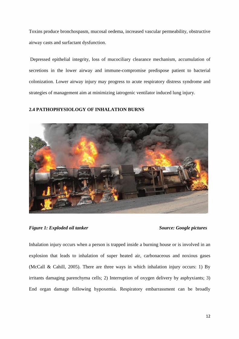
2.4 PATHOPHYSIOLOGY OF INHALATION BURNS
Figure 1: Exploded oil tanker Source: Google pictures
Inhalation injury occurs when a person is trapped inside a burning house or is involved in an
explosion that leads to inhalation of super heated air, carbonaceous and noxious gases
(McCall & Cahill, 2005). There are three ways in which inhalation injury occurs: 1) By
irritants damaging parenchyma cells; 2) Interruption of oxygen delivery by asphyxiants; 3)
End organ damage following hypoxemia. Respiratory embarrassment can be broadly
13
categorized as the result of thermal or chemical damage to epithelial surfaces of both the
intrathoracic and extrathoracic airways.
Deterioration of patients with inhalation burns occur due to broncho-constriction following
release of histamine, serotonin and thromboxane or chest constriction secondary to
circumferential full thickness chest burns (Traber et al, 2007). Catecholamine release in
response to stress of burn injury and hypermetabolism leads to increased oxygen
consumption by the body tissues which can lead to hypoxia. For this reason, supplemental
oxygen may be required.
Pulmonary burn injuries are categorized as above glottis or below glottis. The upper air way
injury results from inhalation of greater than 150oC to the epithelium. Result is severe upper
airway edema which can cause upper airway obstruction up to the larynx (Palmieri, 2007).
Due to the cooling effect of rapid vaporization in the pulmonary tract, direct heat injury does
not occur below bronchus. Upper airway is treated by endo-tracheal intubation.
Maybaurer et al (2009) reports that injury below glottis results from inhalation of noxious
gases, steam or incomplete combustion. These products include carbon monoxide, cyanide,
ammonia, aldehydes, acrolein, sulfur dioxide and isocyanides. Once inhaled, they trigger a
cascade of events, resulting to pulmonary oedema and ventilation/perfusion (V/Q) mismatch.
Intrapulmonary leukocyte aggregation following activation of the classic complement
cascade releases even more chemokines and cytokines, leading to production of oxygen free
radicals.
Edelman et al (2006) explains how Nitric Oxide synthase is produced by respiratory
epithelial cells and alveolar macrophages for the production of Nitric Oxide (NO), a powerful
vasodilator. Nitric Oxide increases bronchial blood flow, decreases hypoxic pulmonary
vasoconstriction in poorly ventilated areas of lung and results in ventilation/ perfusion (V/Q)
14
mismatch. Activated neutrophils produce superoxide (O2-) which combines with NO to form
peroxynitrite (ONOO-). This reactive nitrogen species lead to DNA damage. Repair of the
DNA by polymerase enzyme requires a lot of chemical energy in the form of ATP and NAD,
depletion of which causes necrotic cell death to the tissues involved. Combination of these
effects contributes to tissue injury and increased pulmonary vascular permeability, leading to
decreased diffusion, oedema and V/Q mismatch.
Neutrophil infiltration and fibrinogen activation by inflammatory mediators causes airway
crust formation and widespread plugging. Pathological lung specimens, after inhalation of
smoke, demonstrate the presence of obstructive casts in the airways. Cox and Burke (2003)
studied burnt sheep and discovered that crusts form at bronchial, bronchiolar and terminal
bronchiolar levels. Obstructive changes were maximal at 24 hours in large airways, and rose
continually up to 72 hours at the bronchiolar level. Crusts are composed of epithelial cells,
neutrophils, mucus and fibrin. These crusts obstruct the airway and subsequent efforts to
ventilate the lung mechanically can induce ventilator-induced barotrauma when the patient
lung becomes overstretched. Much of the study of smoke inhalation injuries in animal models
has focused on aspects of this pathophysiological sequence (Suman et al, 2007).
Inhalation injuries below glottis cause loss of cilliary function, hypersecretion, severe
mucosal edema and bronchospasm. Impaired cilliary function leads to accumulation of
airway debris. Mucosal edema in the smaller airways lead to audible wheezing or heard on
auscultation. Pulmonary safurctant is reduced leading to atelectasis. Expectoration of sooty
sputum is the obvious sign of lower airway injury. Early intubation and mechanical
ventilation with 100% oxygen reduces the half life of carboxyhemoglobin from 4hours to
45minutes (Kealey, 2009). Macrophages within the alveoli are destroyed, allowing bacteria
to proliferate enhanced by lack of an intact epithelial barrier leading to pneumonia.
15
Restrictive pulmonary excursion may occur with full thickness circumferential burns of neck
and chest. This is a confounding factor because it causes decreased tidal volume (Rehbergs et
al, 2009). Hypoxemia results from a decrease in inspired oxygen concentration at the scene of
injury, a mechanical inability to exchange gases due to airway obstruction or parenchyma
pulmonary disease. Inhibition of oxygen delivery and tissue use by toxins also causes
hypoxemia. More than 50% of patients with inhalation burns do not initially demonstrate
pulmonary signs and symptoms and as such, any patient with suspected or possible inhalation
injury should be observed for at least 24 hours for respiratory complications (Kabalak &
Yasti, 2012). With the advent of sophisticated intensive care support, patients who survive
the acute injury should have less mortality. However, presence of multi-organ dysfunction is
a common sequel of hypoxia and substantially raises morbidity and mortality of burns
patients (Mc Call &Cahill, 2005).
Carbon monoxide is a colourless odorless tasteless gas released from burning wood or coal. It
displaces oxygen from hemoglobin binding sites thereby decreasing the oxygen carrying
capacity of the blood. It not only has 250- fold increased affinity for hemoglobin but also
shifts the oxyhemoglobin dissociation curve to the left (Kealey, 2009). The left shift results in
increased tissue hypoxia because hemoglobin is less able to unload the little oxygen it’s
carrying. Carbon monoxide reacts with myoglobin to further impair oxygen uptake by
decreasing facilitated diffusion of oxygen to muscles. It interacts with several heme-
containing enzymes of the electron transport chain and so impairs tissue oxygen availability
(Kealey, 2009).
Hydrogen cyanide (HCN) represents the gaseous form of cyanide, which is a colorless gas
with the odor of bitter almonds. It is found in smoke especially from burning polyurethane
and causes tissue asphyxiation by inhibiting intracellular cytochrome oxidase. It blocks the
16
final step in oxidative phosphorylation and prevents mitochondrial oxygen use. Affected cells
convert to anaerobic metabolism and the lactic acid formed presents as metabolic acidosis.
The organs most sensitive to cellular hypoxia i.e. CNS and the heart react to low oxygen
concentrations through hyperventilation thereby increasing exposure to intoxication. Airway
obstruction may occur very rapidly especially during fluid resuscitation. Decreased lung
compliance, decreased arterial oxygen levels and respiratory acidosis occur gradually over
the first five days post burn.
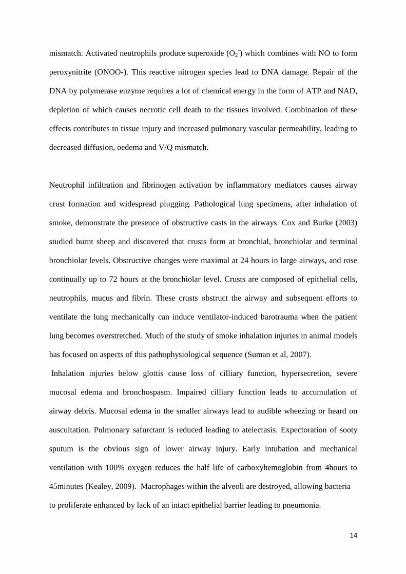
Facial burns &upper airway edema Facial burns & inhalation injury
Source: Google pictures
Complications include sloughing of the airway, increased secretions and inflammation,
atelectasis, airway obstruction, pulmonary edema, tissue hypoxia, and ulceration. As a result,
respiratory failure, acute respiratory distress syndrome and pneumonia can develop (Edelman
et al, 2006). Sloughing of trachea-bronchial epithelium may lead to hemorrhagic trachea-
bronchitis and if the disease process continues, ARDS ensues.
17
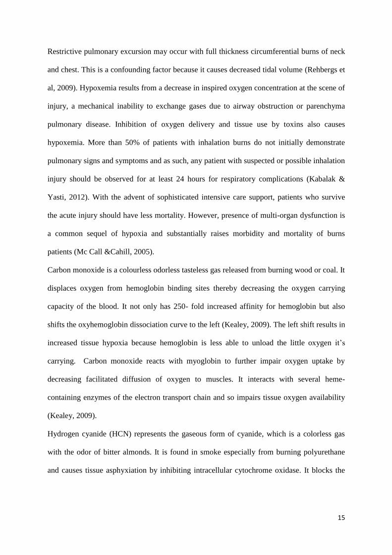
Tracheal burns & inhalation injury
Source: Google pictures
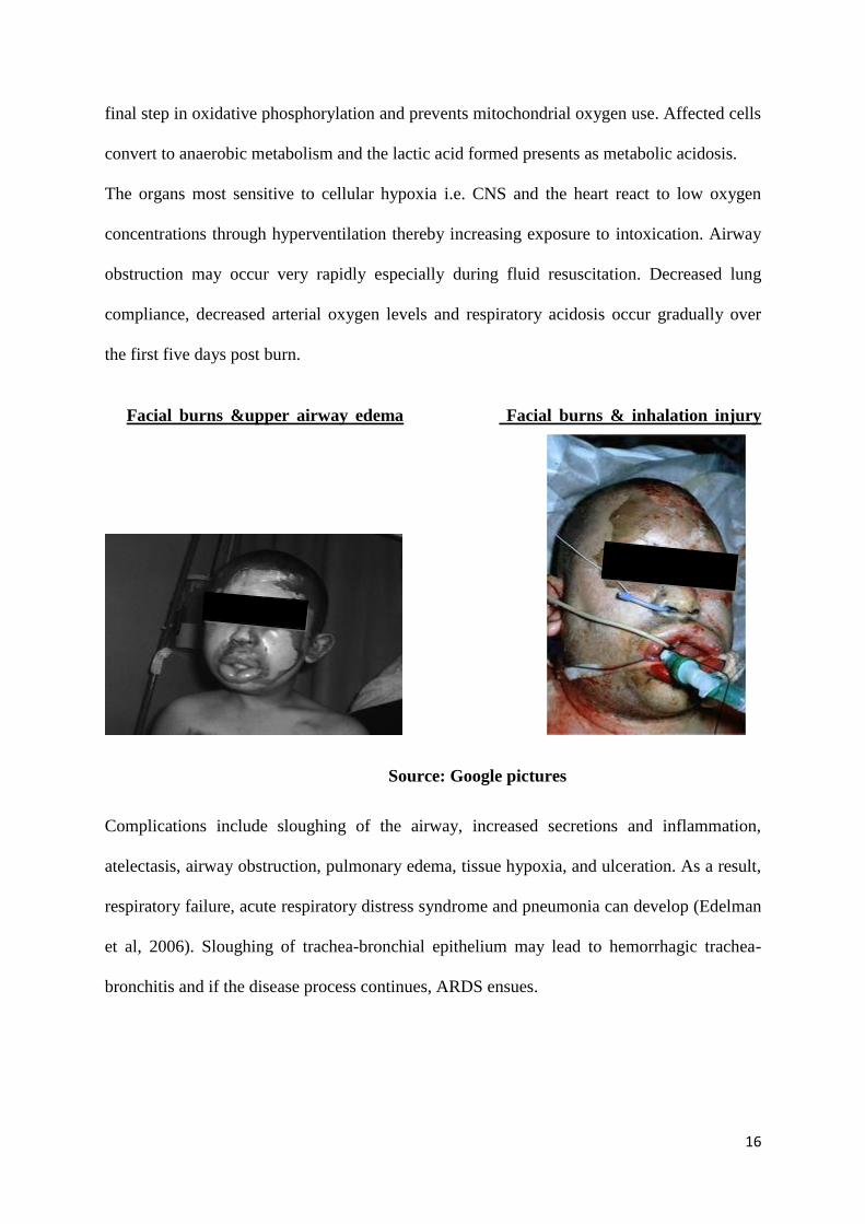
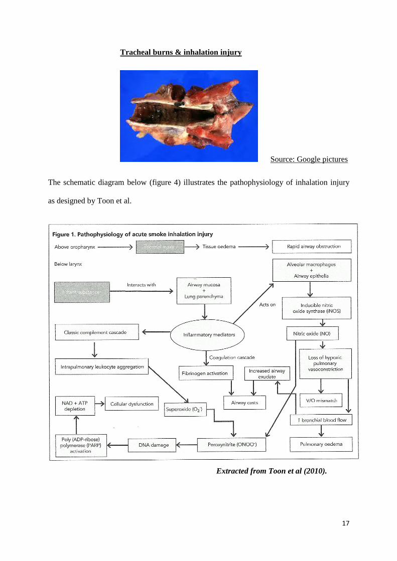
The schematic diagram below (figure 4) illustrates the pathophysiology of inhalation injury
as designed by Toon et al.
Extracted from Toon et al (2010).
18
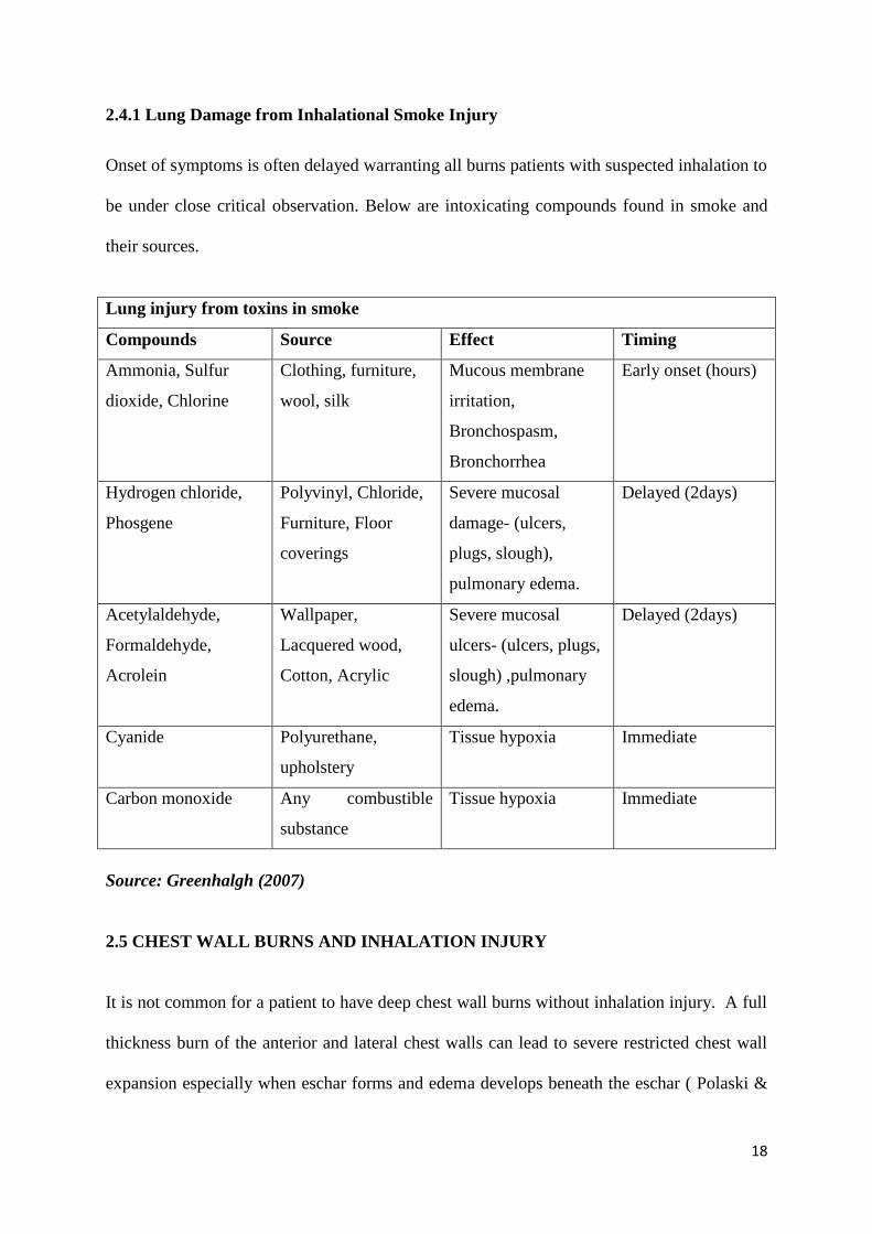
2.4.1 Lung Damage from Inhalational Smoke Injury
Onset of symptoms is often delayed warranting all burns patients with suspected inhalation to
be under close critical observation. Below are intoxicating compounds found in smoke and
their sources.
Lung injury from toxins in smoke
Compounds Source Effect Timing
Ammonia, Sulfur
dioxide, Chlorine
Clothing, furniture,
wool, silk
Mucous membrane
irritation,
Bronchospasm,
Bronchorrhea
Early onset (hours)
Hydrogen chloride,
Phosgene
Polyvinyl, Chloride,
Furniture, Floor
coverings
Severe mucosal
damage- (ulcers,
plugs, slough),
pulmonary edema.
Delayed (2days)
Acetylaldehyde,
Formaldehyde,
Acrolein
Wallpaper,
Lacquered wood,
Cotton, Acrylic
Severe mucosal
ulcers- (ulcers, plugs,
slough) ,pulmonary
edema.
Delayed (2days)
Cyanide Polyurethane,
upholstery
Tissue hypoxia Immediate
Carbon monoxide Any combustible
substance
Tissue hypoxia Immediate
Source: Greenhalgh (2007)
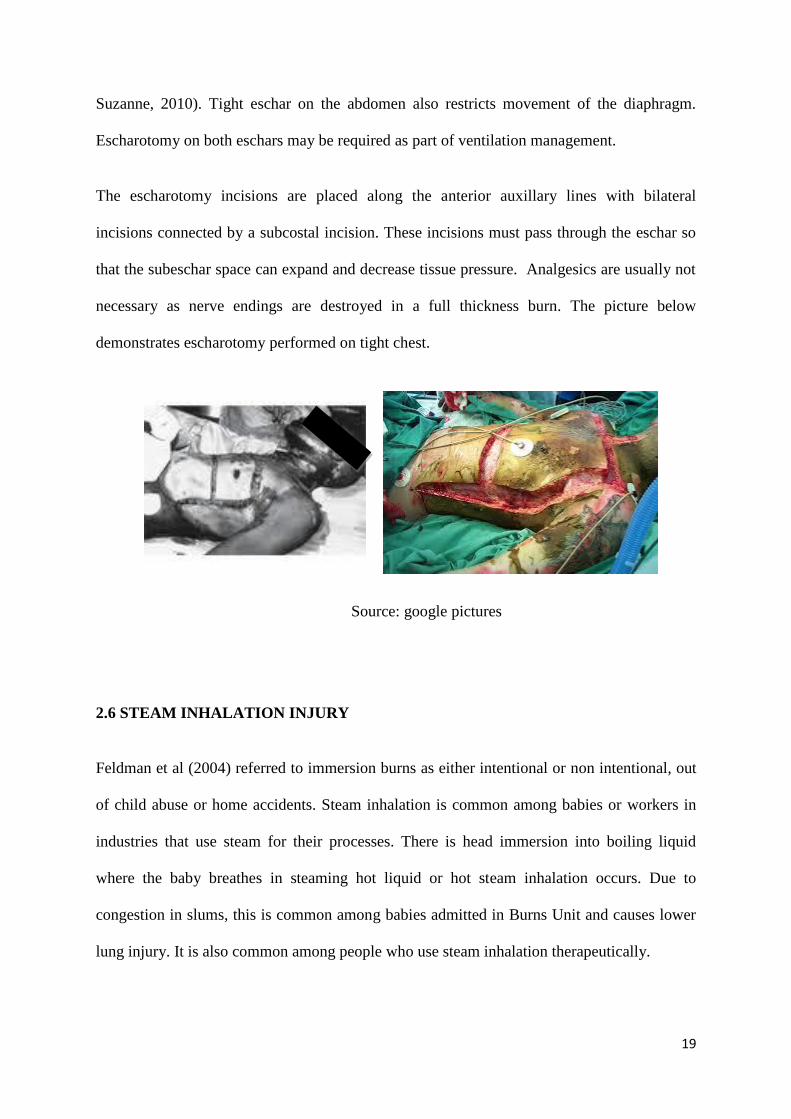
2.5 CHEST WALL BURNS AND INHALATION INJURY
It is not common for a patient to have deep chest wall burns without inhalation injury. A full
thickness burn of the anterior and lateral chest walls can lead to severe restricted chest wall
expansion especially when eschar forms and edema develops beneath the eschar ( Polaski &
19
Suzanne, 2010). Tight eschar on the abdomen also restricts movement of the diaphragm.
Escharotomy on both eschars may be required as part of ventilation management.
The escharotomy incisions are placed along the anterior auxillary lines with bilateral
incisions connected by a subcostal incision. These incisions must pass through the eschar so
that the subeschar space can expand and decrease tissue pressure. Analgesics are usually not
necessary as nerve endings are destroyed in a full thickness burn. The picture below
demonstrates escharotomy performed on tight chest.
Source: google pictures
2.6 STEAM INHALATION INJURY
Feldman et al (2004) referred to immersion burns as either intentional or non intentional, out
of child abuse or home accidents. Steam inhalation is common among babies or workers in
industries that use steam for their processes. There is head immersion into boiling liquid
where the baby breathes in steaming hot liquid or hot steam inhalation occurs. Due to
congestion in slums, this is common among babies admitted in Burns Unit and causes lower
lung injury. It is also common among people who use steam inhalation therapeutically.
20
2.7 COMPLICATIONS OF INHALATION INJURY
2.7.1 Short term complications
The most common short-term complications of inhalation injuries are those caused by
microbial infection. Most common is mechanical ventilation associated pneumonia, with
infection rates of up to 40% in those on artificial ventilation. It can be quite difficult to
diagnose pulmonary infections due to the similarity of symptoms between infections and
symptoms of the inhalation injuries (Kabalak &Yasti, 2012). The key to diagnosing an
infection is by noting unexpected worsening or changing of symptoms. Gram stains can also
be used to identify the bacteria responsible (Edelman et al, 2006).
The treatment of infection resulting from inhalation injury is accomplished with antibiotics
specific to the pathogen. Prophylactic use of antibiotics has not proofed effective, except
enhancing rapid development of antibiotic resistant strains (Klastersky as cited in Polaski &
Suzanne, 2010).
Endotracheal intubation is necessary in about 80% of patients with inhalation injuries because
of respiratory difficulties. However, prolonged intubation (over 3 weeks) can greatly increase
the risk of pulmonary infection (Palmieri, 2009). Bacteria can colonize the plastic tubing and
then cause infection. Long-term intubation is also believed to exacerbate laryngeal damage
by occasionally causing ulcers or adhering to the tissue.
Tracheal stenosis is rare but can occur where airway heals with adhesions especially
following traumatic intubation. Treatment is tracheostomy and early signs may be confused
with asthma. Edema proximal to the endotracheal tube tends to push the tube backwards
hence the many re-intubations especially in babies (Demling, 2005).
21
2.7.2 Long term complications
Polyps can be formed when there is excessive granulation (fibrous connective tissue that
forms after the fibrin clot) during the healing process. Polyps typically heal within 6 months
after injury but use of corticosteroids enhances the healing process ( Maybaurer et al, 2009).
However, in severe cases where polyps form in small airways, they can lead to a syndrome
similar to bronchiolitis obliterans. This leads to scaring and inflammation and can decrease
lung function to around 20% leading to respiratory failure and death. A rare long-term
complication of inhalation injury is reactive airway dysfunction, a form of asthma that is
irritant-induced (Palmieri, 2009).
2.8 DIAGNOSIS AND TREATMENT OF INHALATION INJURY
Indicators for possible airway injury include: 1) Burns occurring in an enclosed space; 2)
Burns of the face or neck; 3) Singed nasal hairs; 4) Hoarseness, cough, stridor, high pitched
voice; 5) Sooty or bloody sputum; 6) Labored breathing; 7) Hypoxemia; 8) Erythema and
blistering of oral or pharyngeal mucosa ( Nugen & Herndon, 2008).
Diagnosis of upper airway inhalation injury includes oral burns, swollen tongue and mucosa,
edematous supraglottis, infraglottis and cord. Erythema is demonstrated through
laryngoscopy and hoarseness of voice is the first sign.
Diagnosis of lower airway inhalation injury includes monitoring of arterial blood gases,
carboxyhemoglobin levels and fibreoptic bronchoscopy. Bronchoscopy findings include
visible airway edema, inflammation, necrosis, or soot. Fiberoptic bronchoscopy provides
direct information about the entire respiratory system (Nugen & Herndon, 2008). In addition
to its diagnostic functions, bronchoscopy is useful in lung therapy determining the severity of
22
inhalation injury. Woodson (2009) used bronchoscopy to grade the inhalation injury: The
indicators listed above are present in all grades but;
Grade I: Has no laryngeal oedema
Grade II: Minimal laryngeal oedema and erythema
Grade III: Slight tracheal mucosal oedema and erythema
Grade IV: Moderate tracheal mucosal oedema and erythema
Grade V: Severe tracheal oedema and erythema
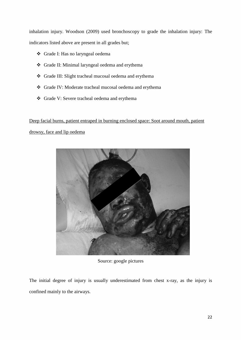
Deep facial burns, patient entraped in burning enclosed space: Soot around mouth, patient
drowsy, face and lip oedema
Source: google pictures
The initial degree of injury is usually underestimated from chest x-ray, as the injury is
confined mainly to the airways.
23
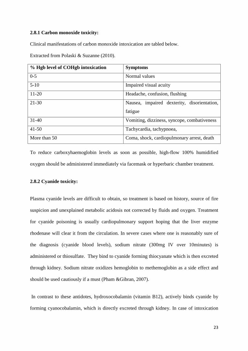
2.8.1 Carbon monoxide toxicity:
Clinical manifestations of carbon monoxide intoxication are tabled below.
Extracted from Polaski & Suzanne (2010).
% Hgb level of COHgb intoxication Symptoms
0-5 Normal values
5-10 Impaired visual acuity
11-20 Headache, confusion, flushing
21-30 Nausea, impaired dexterity, disorientation,
fatigue
31-40 Vomiting, dizziness, syncope, combativeness
41-50 Tachycardia, tachypnoea,
More than 50 Coma, shock, cardiopulmonary arrest, death
To reduce carboxyhaemoglobin levels as soon as possible, high-flow 100% humidified
oxygen should be administered immediately via facemask or hyperbaric chamber treatment.
2.8.2 Cyanide toxicity:
Plasma cyanide levels are difficult to obtain, so treatment is based on history, source of fire
suspicion and unexplained metabolic acidosis not corrected by fluids and oxygen. Treatment
for cyanide poisoning is usually cardiopulmonary support hoping that the liver enzyme
rhodenase will clear it from the circulation. In severe cases where one is reasonably sure of
the diagnosis (cyanide blood levels), sodium nitrate (300mg IV over 10minutes) is
administered or thiosulfate. They bind to cyanide forming thiocyanate which is then excreted
through kidney. Sodium nitrate oxidizes hemoglobin to methemoglobin as a side effect and
should be used cautiously if a must (Pham &Gibran, 2007).
In contrast to these antidotes, hydroxocobalamin (vitamin B12), actively binds cyanide by
forming cyanocobalamin, which is directly excreted through kidney. In case of intoxication
24
with 1mg cyanide, hydroxocobalamin (50mg/kg) is recommended (Polaski & Suzanne,
2010). It averts methemoglobin production and can be used even in the preclinical setting.
2.9 AIRWAY EDEMA TREATMENT
The patient’s head should be elevated to minimize facial and airway oedema (Pitts et al,
2008). Aerosolized adrenaline or corticosteroids may be beneficial to reduce upper airway
oedema, but there is no conclusive evidence of their efficacy.
In the case of bronchospasm, nebulized beta 2-agonists, improves respiratory efficiency by
decreasing airflow resistance and peak airway pressures. In addition, beta2-agonists have
anti-inflammatory properties and help decrease inflammatory mediators such as histamine,
leukotrienes and tumour necrosis factor (Demling, 2005). Finally, beta2-agonists improve
airspace fluid clearance and stimulate mucosal repair.
According to Toon et al (2010), nebulizing children with massive burns and inhalation injury
using heparin and N-acetylcysteine for the first 7 days decreases incidences of re-intubation,
progressive pulmonary failure, atelectasis, and hence mortality.
Miller et al (2009) in his study also confirmed reduction in lung-injury after smoke inhalat
ion when he administered nebulized heparin and the mucolytic N-acetylcysteine to a group of
patients.
Many drugs have proven effective in reducing the injury to the lung parenchyma in animal
models, but only a few are in clinical use. These include heparin, N-acetylcysteine and
inhaled albuterol.
2.10 INDICATIONS FOR INTUBATION IN INHALATION INJURY
Early intubation may be required if stridor, hoarse voice, chest retraction or respiratory
distress is present; but risk for rapid development of airway oedema should be considered for
prophylaxis intubation. However, endotracheal intubation at the injury scene risks patients to
25
oesophageal intubation, aspiration, barotrauma and even laryngeal trauma. It should be
avoided unless by professional experts (Pitts et al, 2008).
Deep burns to the face and neck call for early intubation due to anticipated upper airway
obstruction. Aim is to ensure airway patency when edema of the tongue and glottis sets in.
Mechanical ventilation assisted mode is indicated for all patients with lower airway
inhalation injury. Purpose is to enhance gaseous exchange while maintaining adequate
ventilation without much patient’s effort. Management of lung injury due to smoke inhalation
is mainly supportive, using mechanical ventilation, humidification and aggressive airway
toileting (Traber et al, 2007). Low tidal volume ventilation with associated permissive
hypercapnia has effectively reduced ventilator-induced lung injury and PEEP is a choice
application in patients with ARDS (Toon et al, 2010).
2.11 RESTORING HEMODYNAMICS AND INHALATION INJURY
Inhalation injury adds 10 % to the burnt total body surface area and should be included in the
calculation of fluid for replacement therapy. Loss of plasma volume is rapid after a burn
injury as fluid collects in the burn tissue. Patients with very severe deep burns develop
massive systemic edema and re-absorption is dependent on the depth of injury (Greenhalgh
as cited in Brunner & Saddarth, 2010). Partial thickness injury resolves more quickly due to a
more functional lymphatic system and increased perfusion compared to deep burns. Early
fluid resuscitation is required for burns exceeding 15% TBSA in adult and 10% TBSA in
children because low extra-cellular fluid volume enhances plugging of secretions along the
airway thus increasing risk of chest infections and obstruction (Brunner & Suddarth, 2010). .
Patient should have at least one large bore intravenous catheter or CVP catheter for
intravenous fluids and possible cardiopulmonary resuscitation. Body weight (Kgs) should be
estimated and Parklands formula used to estimate amount of fluid to be replaced: (4ml x
26
weight x TBSA %). Half the amount is given within eight hours from the time of burn and
the remaining half within the next sixteen hours (Williams, 2008).
Children receive maintenance fluid in addition, at an hourly rate of 4ml/kg for the first 10kg
of body weight plus; 2ml/kg for the second 10kg of body weight plus; 1ml/kg for >20kg of
body weight ( Williams, 2008).
Excessive fluid administration increases edema formation in both burned and unburned
tissues. As a result, pressure on small blood vessels and nerves in the distal extremities cause
obstruction to blood flow and consequent ischemia. This complication is similar to
compartment syndrome and may also cause pulmonary edema (Brunner & Saddarth, 2010).
During burn shock, hyponatremia is present as water shifts from intravascular to interstitial
spaces despite sodium reabsorption by the kidney. Hyperkaleamia results from the direct cell
injury which releases large amounts of cellular potassium. Hypokaleamia may occur later
following fluid shifts and potassium moving back into the cells (Ignatavicious & Linda,
2013).
27
3.0 CHAPTER THREE: RESEARCH METHODOLOGY
3.1 STUDY DESIGN
This research was a descriptive longitudinal study with both qualitative and quantitative
components. Data was collected prospectively assessing clinical management and patients’
response to the interventions on inhalation injury. Each patient was followed up for as long as
he lived up to four weeks and data collection took three months.
3.2 STUDY AREA
This research was conducted in Burns Unit, Kenyatta National Hospital (KNH). KNH is a
government hospital situated in Nairobi, and the only prime referral center for both private
and other government hospitals. KNH was established in 1901 and offers training and
research ground to Kenya medical training college, Nairobi University, CDC Kemri and also
participates in national health planning. KNH serves a population of three million and it is
situated between Ngong road, Mbagathi road and the Hospital road. To explain the common
causes of burns, KNH is surrounded by four slums namely, Kibera, Mukuru kwa Njenga,
Mathare and Lungalunga. Poverty forces people to encroach on oil pipelines as is the case of
Sinai fire disaster, while congestion in housing increase risk of babies falling into hot water
or tea. Out of the 45 wards, KNH houses the only referral burns critical care unit for severe or
major burns including inhalation injuries. Burns unit serves an average of 490 such patients a
year but during fire disasters, an emergency ward is opened to help cope with high numbers
of victims (Mugambi, 2012). Burns unit has a maximum bed capacity of 22 patients and
mainly receives severely burnt patients from other hospitals or disaster sites. Burns unit
admits an average of 39 new patients per month and it has an inbuilt surgical theater to
facilitate timely debridement, escharotomies and skin grafting. Burns unit has previously
been used by other hospitals like Moi referral, Gertrude’s and Nakuru hospitals to bench
28
mark for quality burns management thus qualifying findings in this research to be
generalizable. Being a referral and ISO certified hospital, KNH is expected to comply with
quality international standards including management of inhalation burns and the researcher
hoped to identify any non- conformities through this research as areas of improvement.
3.3 STUDY POPULATION
The study population incorporated both male and female patients of all ages admitted with
inhalation injury in burns unit, among patients with major burns. Key informants included
care givers, both doctors and nurses.
3.4 SAMPLING PROCEDURE
Sampling is a process of obtaining information about the entire population by examining only
a part of it (Kothari, 2004). From December 2012 to November 2013, Burns unit admitted
473 new patients and the ward has a capacity of 22 patients. On average of 20 inpatients, 493
patients pass through burns unit annually and according to Herndon (2007), 60% of all burns
cases have inhalation injury. This implies that 296.4 patients had inhalation injury during that
year. Since this study takes three months, the average estimated population attended to in
burns unit is 137 patients.
Non probability or convenient sampling technique was applied in selecting participants since
population is small. All patients with inhalation injury, confirmed or suspected diagnosis
qualified to be included in this study. This included patients with facial burns, neck burns and
full thickness anterior chest wall burns. Patients found in the ward at the time of study and
those admitted later for three months were recruited to participate in this research.
For key informants, purposeful sampling incorporated both doctors and nurses,
29
who have been working in burns unit for at least three years and happened to be in the ward
during data collection. This is mainly because their experience is worth valuable input to this
study. 17(89.5%; n=19) nurses and 3 (100%) doctors participated. Ward administrators
included Ward in-charge, ACN, HOU, AD Specialized surgery and SAD General Surgery;
based on their responsibilities and roles.
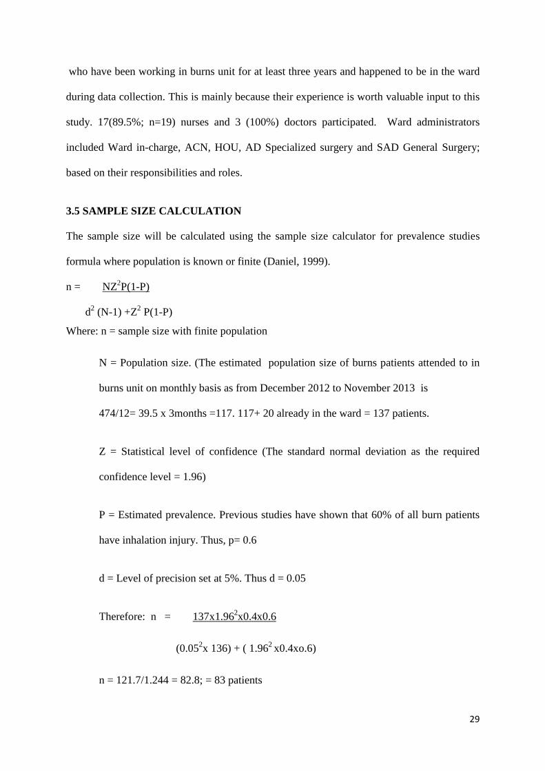
3.5 SAMPLE SIZE CALCULATION
The sample size will be calculated using the sample size calculator for prevalence studies
formula where population is known or finite (Daniel, 1999).
n = NZ2P(1-P)
d2 (N-1) +Z
2 P(1-P)
Where: n = sample size with finite population
N = Population size. (The estimated population size of burns patients attended to in
burns unit on monthly basis as from December 2012 to November 2013 is
474/12= 39.5 x 3months =117. 117+ 20 already in the ward = 137 patients.
Z = Statistical level of confidence (The standard normal deviation as the required
confidence level = 1.96)
P = Estimated prevalence. Previous studies have shown that 60% of all burn patients
have inhalation injury. Thus, p= 0.6
d = Level of precision set at 5%. Thus d = 0.05
Therefore: n = 137x1.962x0.4x0.6
(0.052x 136) + ( 1.96
2 x0.4xo.6)
n = 121.7/1.244 = 82.8; = 83 patients
30
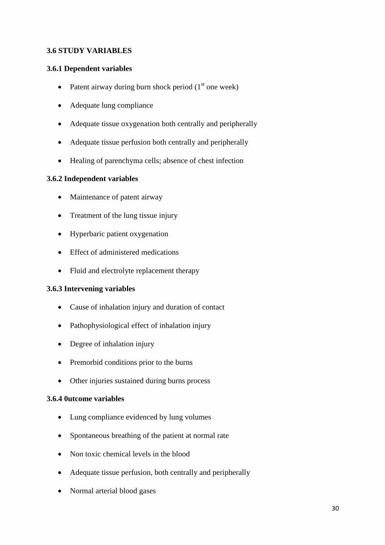
3.6 STUDY VARIABLES
3.6.1 Dependent variables
Patent airway during burn shock period (1st one week)
Adequate lung compliance
Adequate tissue oxygenation both centrally and peripherally
Adequate tissue perfusion both centrally and peripherally
Healing of parenchyma cells; absence of chest infection
3.6.2 Independent variables
Maintenance of patent airway
Treatment of the lung tissue injury
Hyperbaric patient oxygenation
Effect of administered medications
Fluid and electrolyte replacement therapy
3.6.3 Intervening variables
Cause of inhalation injury and duration of contact
Pathophysiological effect of inhalation injury
Degree of inhalation injury
Premorbid conditions prior to the burns
Other injuries sustained during burns process
3.6.4 0utcome variables
Lung compliance evidenced by lung volumes
Spontaneous breathing of the patient at normal rate
Non toxic chemical levels in the blood
Adequate tissue perfusion, both centrally and peripherally
Normal arterial blood gases
31
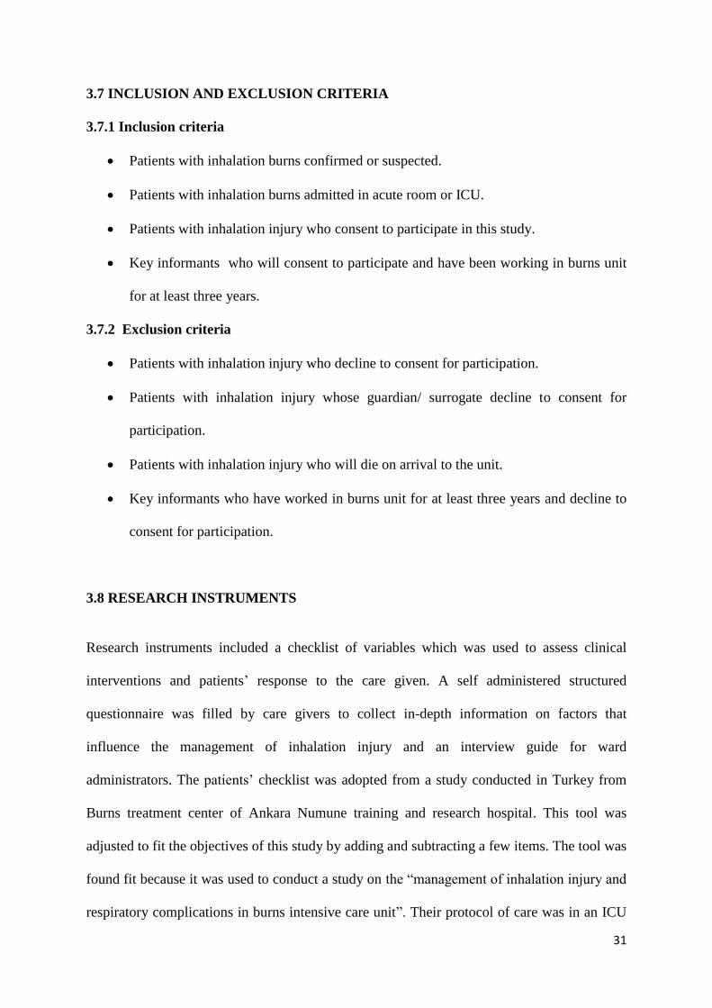
3.7 INCLUSION AND EXCLUSION CRITERIA
3.7.1 Inclusion criteria
Patients with inhalation burns confirmed or suspected.
Patients with inhalation burns admitted in acute room or ICU.
Patients with inhalation injury who consent to participate in this study.
Key informants who will consent to participate and have been working in burns unit
for at least three years.
3.7.2 Exclusion criteria
Patients with inhalation injury who decline to consent for participation.
Patients with inhalation injury whose guardian/ surrogate decline to consent for
participation.
Patients with inhalation injury who will die on arrival to the unit.
Key informants who have worked in burns unit for at least three years and decline to
consent for participation.
3.8 RESEARCH INSTRUMENTS
Research instruments included a checklist of variables which was used to assess clinical
interventions and patients’ response to the care given. A self administered structured
questionnaire was filled by care givers to collect in-depth information on factors that
influence the management of inhalation injury and an interview guide for ward
administrators. The patients’ checklist was adopted from a study conducted in Turkey from
Burns treatment center of Ankara Numune training and research hospital. This tool was
adjusted to fit the objectives of this study by adding and subtracting a few items. The tool was
found fit because it was used to conduct a study on the “management of inhalation injury and
respiratory complications in burns intensive care unit”. Their protocol of care was in an ICU
32
set up; but since there are no studies on inhalation injury conducted in developing countries,
this tool was found useful.
The questionnaire for care givers was formatted so as to capture both positive and negative
influencing factors in the management of inhalation injury, plus preferred areas of
improvement. The check list for patients’ care included some of the confounders which may
also contribute to high mortality in burns patients despite interventions for inhalation injury.
3.9 PILOT STUDY
Piloting of the research checklists and the key informants questionnaire was conducted at
Aga Khan University hospital since they also manage patients with severe and inhalation
burns and the conditions of care are almost similar to that of burns unit KNH. Likewise the
same research tools were presented to my research supervisors for validity evaluation and
approval. The purpose was to reduce errors, biases and ambiguity. After piloting, any
question needing revision, modification or even found unnecessary was appropriately
amended before going to the field. This helped the researcher to evaluate the instrument and
facilitate data accuracy. Piloting also increased reliability of the instruments and research
findings.
3.10 ETHICAL CONSIDERATION
Before conducting the study, the researcher presented three copies of the research proposal to
the KNH/U.O.N Ethical and Research committee seeking approval for the study. A copy of
the same proposal will be presented to the Ministry of higher education for research approval.
Once approved, the researcher proceeded to collecting data. Patient had to be stabilized and
pain control measures effected. The researcher then reassured relatives for quality care; and
after self introduction, the study topic was introduced to the participants, including guardians
or surrogates for intubated and unconscious patients. The researcher elaborated the aim of
33
study, its process and participants expectations. Respondents were assured of confidentiality
and anonymity maintenance in all research instruments. The researcher clarified that there
will be no payments for participation as a measure to minimize bias. Option to withdraw was
availed to those who would wish to discontinue for whatever reasons. The researcher then
requested the participants to sign the consent form for voluntary participation. Appointed
guardians or surrogates for intubated and unconscious patients responded on behalf of
individual patients. Instruments were serialized using numbers and all data collected
including signed consent forms were kept by the researcher for safe custodian and
confidentiality. Research checklist data was mainly monitory, to assess the care, thus posing
minimal risks to the participants. No harm or pain or exploitive investigations was realized on
participants in relation to this research. Patients and surrogates were allowed to share views
concerning their care as study progressed. In conclusion, findings of this study will be shared
with the relevant stakeholders through organized forums for utilization but will also be
published in a recognized journal for reference.
3.11 DATA COLLECTION METHODS
The researcher went physically to the area sampled for research. Self introduction was done,
letter of research approval presented and the purpose of study explained. Since data was to be
obtained through assessment of clinical interventions, details on expected data was not
elaborated hoping to capture practice as it was. Patients were recruited to this study on arrival
to the ward after they had been stabilized and pain control measures effected. After
recruitment, evaluation of care from time arrived in the hospital was done. Consecutive data
intervals were conducted daily for the first one week, then on fourteenth day, twenty first day
and twenty eighth day for the next three weeks. Assurance for confidentiality was done to all
participants in order to encourage them participate without fear (Mugenda and Mugenda,
2003).Researcher obtained informed consent from the participants and introduced her
34
research assistants. Instruments were serialized as participants were being recruited to the
study and a collecting log prepared to ensure that all checklists and questionnaires were
returned and kept in safe custody before the commencement of data analysis. Checklists’ data
was obtained from patients, relatives, care givers and medical records.
Two critical care graduate nurses were recruited and trained for two days as research
assistants. The choice of the research assistants was based on their previous research
knowledge and skills in critical care nursing. Research assistants were trained on how to
administer questionnaires to the care givers, diagnose inhalation burns, estimate the depth of
inhalation burns, ideal management of inhalation burns, common complications of inhalation
burns, interviewing skills and charting of the checklists. Each participant was followed up
for a maximum of four weeks within which, death or clinical status of the patient marked the
end point of data collection.
Interview schedules took 30minutes on average and were conducted by the researcher on
appointment with individual administrators. Permission to tape discussion was obtained from
the interviewee on the day of interview. The researcher facilitated as well as supervised the
rest of the research process. In case of fire disaster during the study period, the researcher was
to undertake the data collection during the night shift but there was none. Review of more
literature available on the subject continued.
3.12 DATA ANALYSIS AND PRESENTATION
The data collected was edited for accuracy, uniformity, consistency and completeness.
Quantitative methods of data analyses were done using SPSS program version 20.0 from the
computer while content analysis was done using theme categorization and tallying. For
qualitative data, common responses were identified and classified into themes. Presentation
of quantitative data was done using frequency tables, pie charts, histograms and polygons;
35
while qualitative data was classified into themes and used to complement or fill gaps in the
quantitative data. Finally, descriptive statistics such as percentages, median, mean standard
deviation and statistical inferences such as t-tests, p-values, Odds ratio and chi squares were
used to draw conclusions. Logistic regression was applied to determine the most significant
intervention in mortality prevention.
3.13 LIMITATIONS OF STUDY
It is very rare to have a patient with isolated inhalation injury, but this study did not include
management of cutaneous burns since it would have been too broad for a single research.
This may affect the conclusion of the findings in that, major burns (70% and above) have
detrimental homeostatic effects on all body organs leading to compartment syndrome;
respiratory system included. This effect was considered during discussion as an outlier effect
since only 3 (3.4%) patients had such major cutaneous burns; but further research on fluid
replacement, anaemia in burns patients and wound care would be necessary.
Sample size was realized based on findings of previous studies but incase this research does
not realize the expected sample size; the researcher might have to increase period of study.

36
CHAPTER FOUR: RESULTS OF THE STUDY
4.0 INTRODUCTION
The study involved males 46 (54.8%) and females 38(45.2%) aged between one month and
99 years. Majority 29 (34.5%) of the respondents were aged less than 10 years.22 (26.2%)
were aged from 21 to 30 years with mean age SD of 20.98 (19.3) years.
More than half 49 (58.3%) the population had neck burns with 34 (69.4%) indicating 1%
neck burns and 15 (30.6%) 2% neck.
Median (IQR) of hours arrived at the hospital post-burns was reported to be 6(7) hours. The
mean (SD) cutaneous burns percentage was 32.5(20.1) with minimum of 4% and maximum
of 88% reported. The median (IQR) clerking hours done post arrival was found to be 1(1.5)
hours.
4.1 SOCIAL DEMOGRAPHICS
4.1.1 Gender
Characteristics Number Percentage
Male 46 54.8
Female 38 45.2
4.1.2 Age of participants
Majority of the patients who sustained inhalation injury aged within 1 - 10 years 29 (34.5%)
followed by those within 21 and 30 years 22 (26.2%) where population was 84. Within 31 to
40 years was average 12 (14.3%) and beyond there the numbers decline. Only 2 (2.4%)
elderly patients were admitted with inhalation bur. The missing 5 (6.0%) were children
below one year of age but none was weeks or days old.
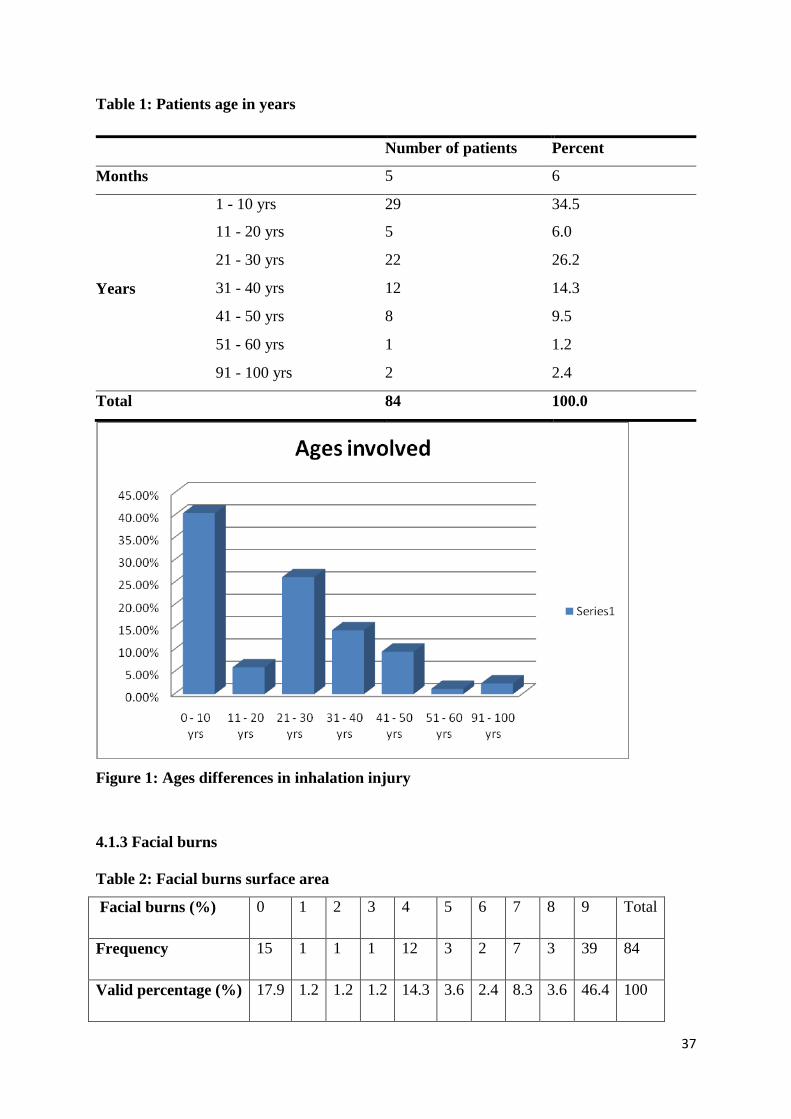
37
Table 1: Patients age in years
Number of patients Percent
Months 5 6
Years
1 - 10 yrs 29 34.5
11 - 20 yrs 5 6.0
21 - 30 yrs 22 26.2
31 - 40 yrs 12 14.3
41 - 50 yrs 8 9.5
51 - 60 yrs 1 1.2
91 - 100 yrs 2 2.4
Total 84 100.0
Figure 1: Ages differences in inhalation injury
4.1.3 Facial burns
Table 2: Facial burns surface area
Facial burns (%) 0 1 2 3 4 5 6 7 8 9 Total
Frequency 15 1 1 1 12 3 2 7 3 39 84
Valid percentage (%) 17.9 1.2 1.2 1.2 14.3 3.6 2.4 8.3 3.6 46.4 100
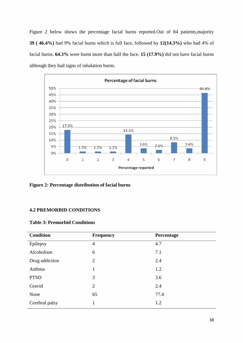
38
Figure 2 below shows the percentage facial burns reported.Out of 84 patients,majority
39 ( 46.4%) had 9% facial burns which is full face, followed by 12(14.3%) who had 4% of
facial burns. 64.3% were burnt more than half the face. 15 (17.9%) did not have facial burns
although they had signs of inhalation burns.
Figure 2: Percentage distribution of facial burns
4.2 PREMORBID CONDITIONS
Table 3: Premorbid Conditions
Condition Frequency Percentage
Epilepsy 4 4.7
Alcoholism 6 7.1
Drug addiction 2 2.4
Asthma 1 1.2
PTSD 3 3.6
Gravid 2 2.4
None 65 77.4
Cerebral palsy 1 1.2
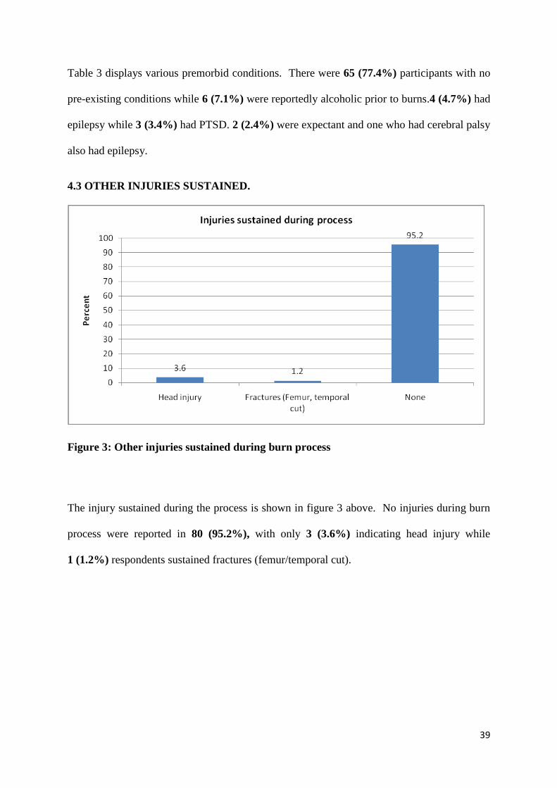
39
Table 3 displays various premorbid conditions. There were 65 (77.4%) participants with no
pre-existing conditions while 6 (7.1%) were reportedly alcoholic prior to burns.4 (4.7%) had
epilepsy while 3 (3.4%) had PTSD. 2 (2.4%) were expectant and one who had cerebral palsy
also had epilepsy.
4.3 OTHER INJURIES SUSTAINED.
Figure 3: Other injuries sustained during burn process
The injury sustained during the process is shown in figure 3 above. No injuries during burn
process were reported in 80 (95.2%), with only 3 (3.6%) indicating head injury while
1 (1.2%) respondents sustained fractures (femur/temporal cut).
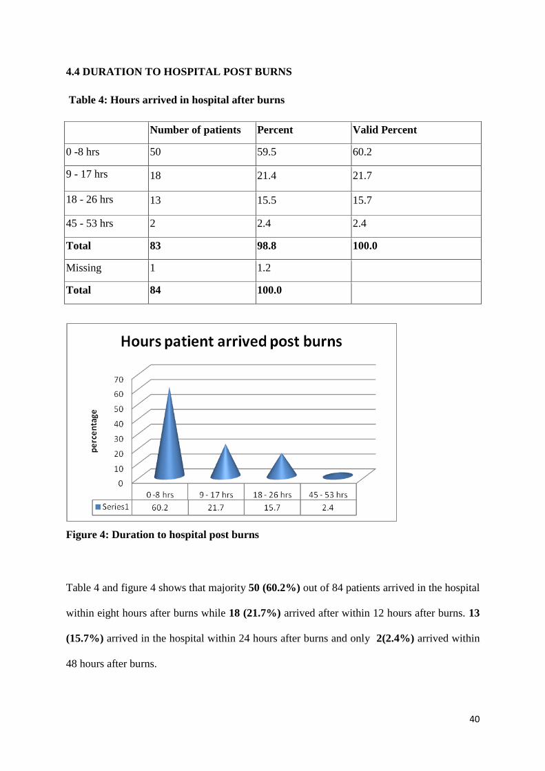
40
4.4 DURATION TO HOSPITAL POST BURNS
Table 4: Hours arrived in hospital after burns
Number of patients Percent Valid Percent
0 -8 hrs 50 59.5 60.2
9 - 17 hrs 18 21.4 21.7
18 - 26 hrs 13 15.5 15.7
45 - 53 hrs 2 2.4 2.4
Total 83 98.8 100.0
Missing 1 1.2
Total 84 100.0
Figure 4: Duration to hospital post burns
Table 4 and figure 4 shows that majority 50 (60.2%) out of 84 patients arrived in the hospital
within eight hours after burns while 18 (21.7%) arrived after within 12 hours after burns. 13
(15.7%) arrived in the hospital within 24 hours after burns and only 2(2.4%) arrived within
48 hours after burns.
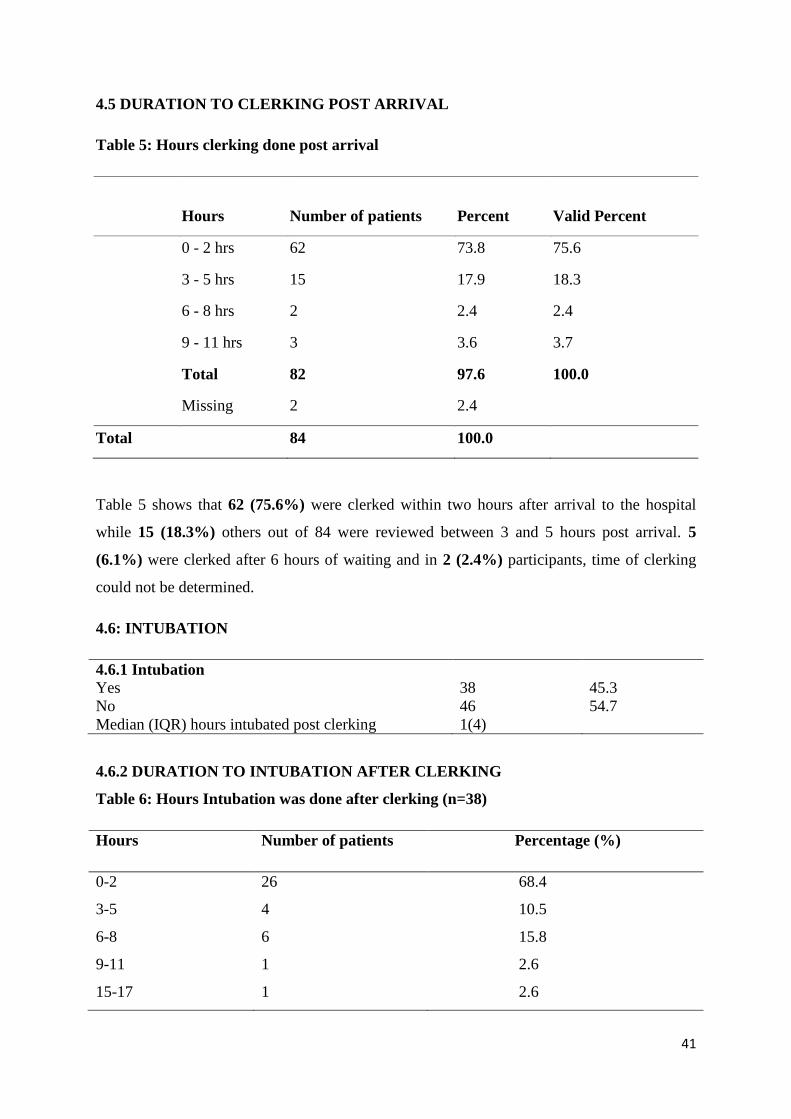
41
4.5 DURATION TO CLERKING POST ARRIVAL
Table 5: Hours clerking done post arrival
Hours Number of patients Percent Valid Percent
0 - 2 hrs 62 73.8 75.6
3 - 5 hrs 15 17.9 18.3
6 - 8 hrs 2 2.4 2.4
9 - 11 hrs 3 3.6 3.7
Total 82 97.6 100.0
Missing 2 2.4
Total 84 100.0
Table 5 shows that 62 (75.6%) were clerked within two hours after arrival to the hospital
while 15 (18.3%) others out of 84 were reviewed between 3 and 5 hours post arrival. 5
(6.1%) were clerked after 6 hours of waiting and in 2 (2.4%) participants, time of clerking
could not be determined.
4.6: INTUBATION
4.6.1 Intubation
Yes 38 45.3
No 46 54.7
Median (IQR) hours intubated post clerking 1(4)
4.6.2 DURATION TO INTUBATION AFTER CLERKING
Table 6: Hours Intubation was done after clerking (n=38)
Hours Number of patients Percentage (%)
0-2
3-5
6-8
9-11
15-17
26
4
6
1
1
68.4
10.5
15.8
2.6
2.6
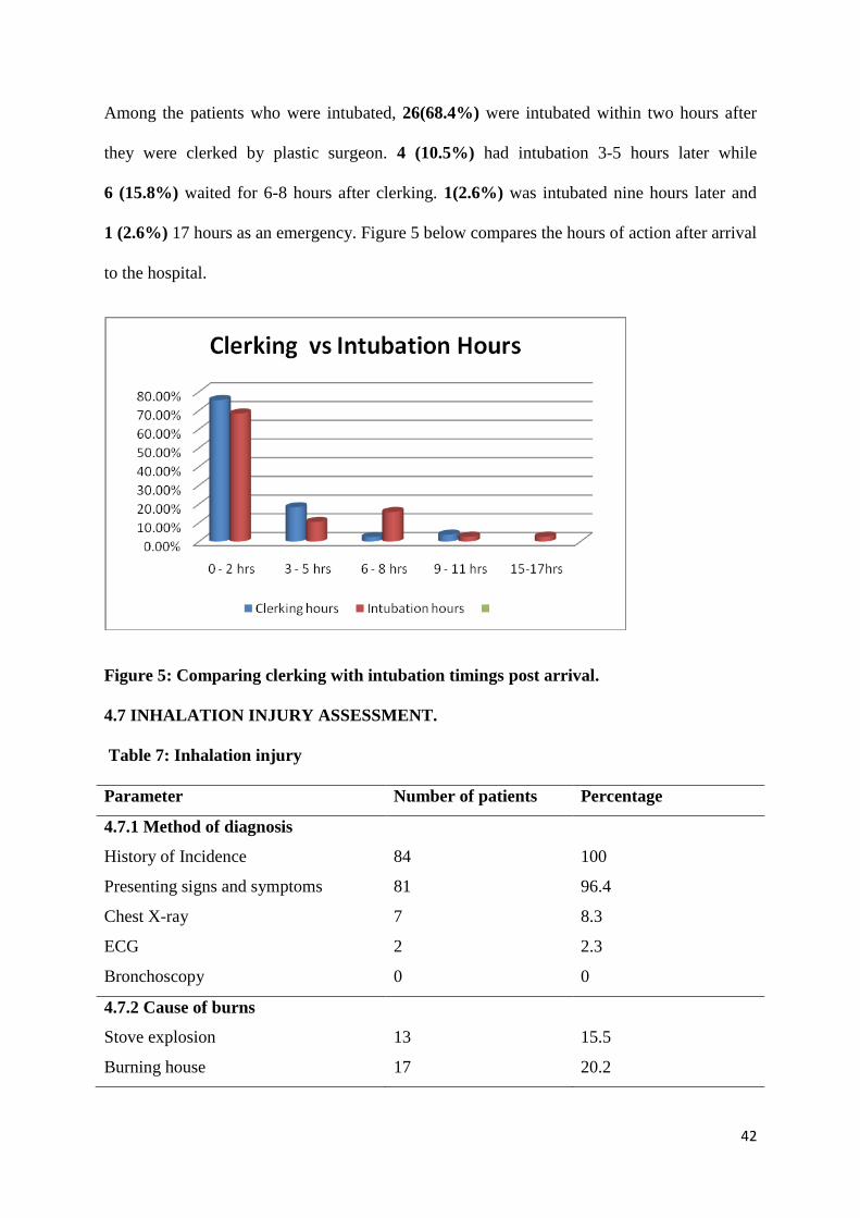
42
Among the patients who were intubated, 26(68.4%) were intubated within two hours after
they were clerked by plastic surgeon. 4 (10.5%) had intubation 3-5 hours later while
6 (15.8%) waited for 6-8 hours after clerking. 1(2.6%) was intubated nine hours later and
1 (2.6%) 17 hours as an emergency. Figure 5 below compares the hours of action after arrival
to the hospital.
Figure 5: Comparing clerking with intubation timings post arrival.
4.7 INHALATION INJURY ASSESSMENT.
Table 7: Inhalation injury
Parameter Number of patients Percentage
4.7.1 Method of diagnosis
History of Incidence 84 100
Presenting signs and symptoms 81 96.4
Chest X-ray 7 8.3
ECG
Bronchoscopy
2
0
2.3
0
4.7.2 Cause of burns
Stove explosion 13 15.5
Burning house 17 20.2
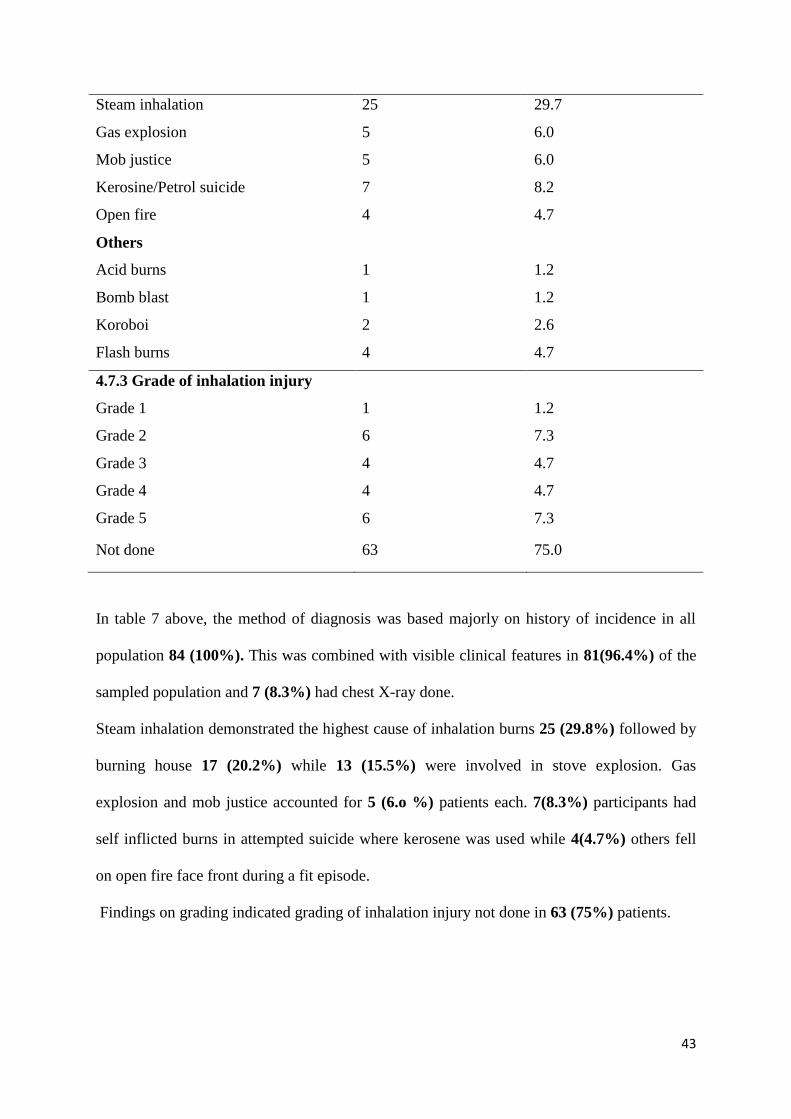
43
Steam inhalation 25 29.7
Gas explosion 5 6.0
Mob justice
Kerosine/Petrol suicide
Open fire
5
7
4
6.0
8.2
4.7
Others
Acid burns
Bomb blast
Koroboi
Flash burns
1
1
2
4
1.2
1.2
2.6
4.7
4.7.3 Grade of inhalation injury
Grade 1 1 1.2
Grade 2 6 7.3
Grade 3 4 4.7
Grade 4 4 4.7
Grade 5 6 7.3
Not done 63 75.0
In table 7 above, the method of diagnosis was based majorly on history of incidence in all
population 84 (100%). This was combined with visible clinical features in 81(96.4%) of the
sampled population and 7 (8.3%) had chest X-ray done.
Steam inhalation demonstrated the highest cause of inhalation burns 25 (29.8%) followed by
burning house 17 (20.2%) while 13 (15.5%) were involved in stove explosion. Gas
explosion and mob justice accounted for 5 (6.o %) patients each. 7(8.3%) participants had
self inflicted burns in attempted suicide where kerosene was used while 4(4.7%) others fell
on open fire face front during a fit episode.
Findings on grading indicated grading of inhalation injury not done in 63 (75%) patients.
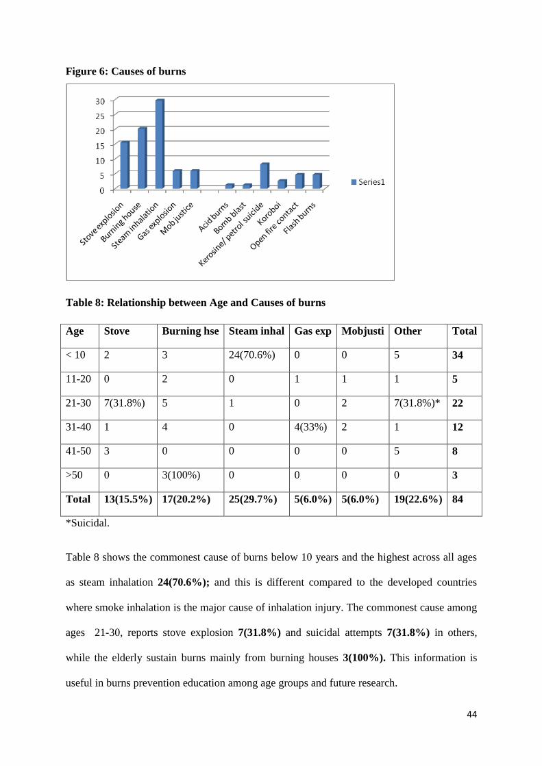
44
Figure 6: Causes of burns
Table 8: Relationship between Age and Causes of burns
Age Stove Burning hse Steam inhal Gas exp Mobjusti Other Total
< 10 2 3 24(70.6%) 0 0 5 34
11-20 0 2 0 1 1 1 5
21-30 7(31.8%) 5 1 0 2 7(31.8%)* 22
31-40 1 4 0 4(33%) 2 1 12
41-50 3 0 0 0 0 5 8
>50 0 3(100%) 0 0 0 0 3
Total 13(15.5%) 17(20.2%) 25(29.7%) 5(6.0%) 5(6.0%) 19(22.6%) 84
*Suicidal.
Table 8 shows the commonest cause of burns below 10 years and the highest across all ages
as steam inhalation 24(70.6%); and this is different compared to the developed countries
where smoke inhalation is the major cause of inhalation injury. The commonest cause among
ages 21-30, reports stove explosion 7(31.8%) and suicidal attempts 7(31.8%) in others,
while the elderly sustain burns mainly from burning houses 3(100%). This information is
useful in burns prevention education among age groups and future research.
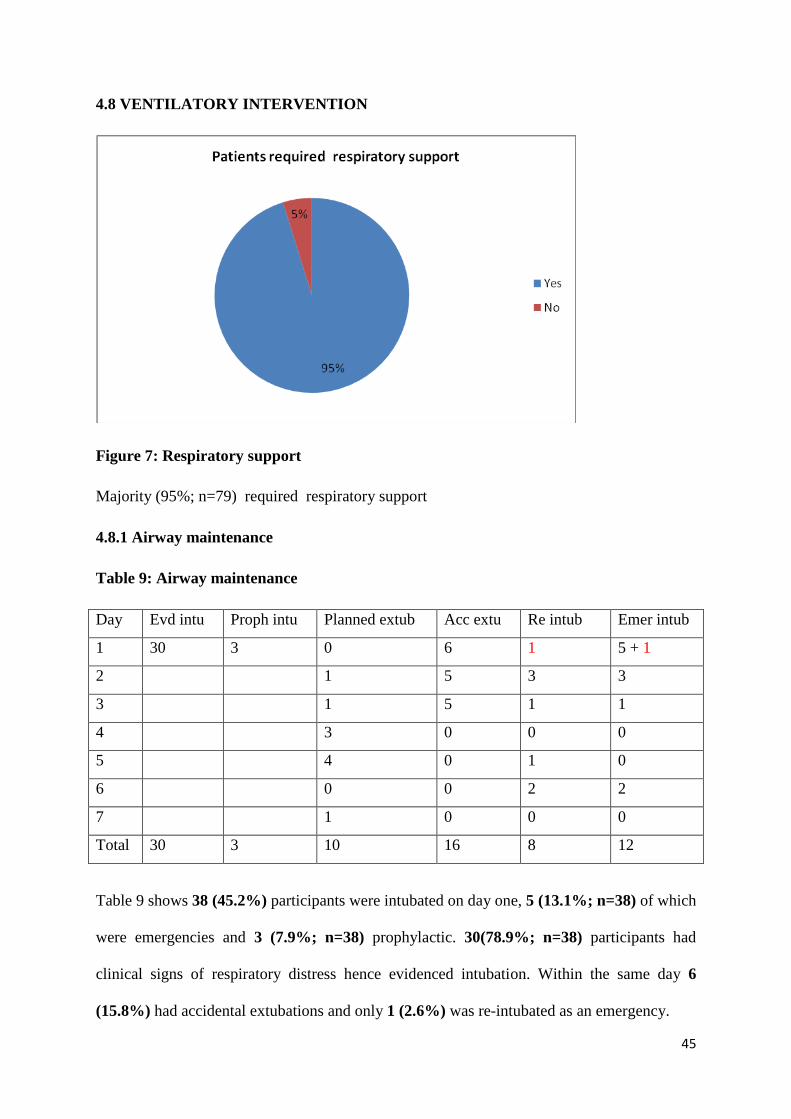
45
4.8 VENTILATORY INTERVENTION
Figure 7: Respiratory support
Majority (95%; n=79) required respiratory support
4.8.1 Airway maintenance
Table 9: Airway maintenance
Day Evd intu Proph intu Planned extub Acc extu Re intub Emer intub
1 30 3 0 6 1 5 + 1
2 1 5 3 3
3 1 5 1 1
4 3 0 0 0
5 4 0 1 0
6 0 0 2 2
7 1 0 0 0
Total 30 3 10 16 8 12
Table 9 shows 38 (45.2%) participants were intubated on day one, 5 (13.1%; n=38) of which
were emergencies and 3 (7.9%; n=38) prophylactic. 30(78.9%; n=38) participants had
clinical signs of respiratory distress hence evidenced intubation. Within the same day 6
(15.8%) had accidental extubations and only 1 (2.6%) was re-intubated as an emergency.
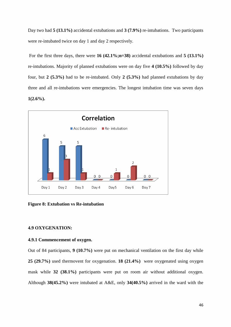
46
Day two had 5 (13.1%) accidental extubations and 3 (7.9%) re-intubations. Two participants
were re-intubated twice on day 1 and day 2 respectively.
For the first three days, there were 16 (42.1%;n=38) accidental extubations and 5 (13.1%)
re-intubations. Majority of planned extubations were on day five 4 (10.5%) followed by day
four, but 2 (5.3%) had to be re-intubated. Only 2 (5.3%) had planned extubations by day
three and all re-intubations were emergencies. The longest intubation time was seven days
1(2.6%).
Figure 8: Extubation vs Re-intubation
4.9 OXYGENATION:
4.9.1 Commencement of oxygen.
Out of 84 participants, 9 (10.7%) were put on mechanical ventilation on the first day while
25 (29.7%) used thermovent for oxygenation. 18 (21.4%) were oxygenated using oxygen
mask while 32 (38.1%) participants were put on room air without additional oxygen.
Although 38(45.2%) were intubated at A&E, only 34(40.5%) arrived in the ward with the
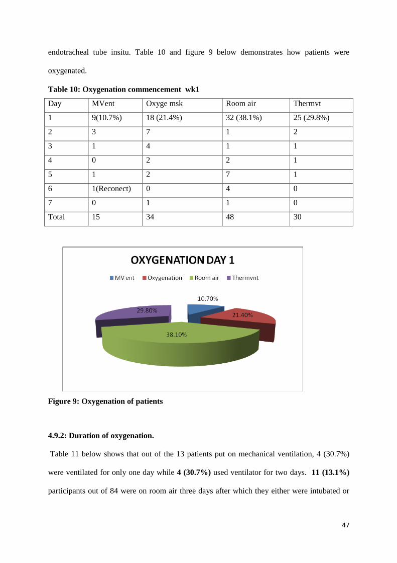
47
endotracheal tube insitu. Table 10 and figure 9 below demonstrates how patients were
oxygenated.
Table 10: Oxygenation commencement wk1
Day MVent Oxyge msk Room air Thermvt
1 9(10.7%) 18 (21.4%) 32 (38.1%) 25 (29.8%)
2 3 7 1 2
3 1 4 1 1
4 0 2 2 1
5 1 2 7 1
6 1(Reconect) 0 4 0
7 0 1 1 0
Total 15 34 48 30
Figure 9: Oxygenation of patients
4.9.2: Duration of oxygenation.
Table 11 below shows that out of the 13 patients put on mechanical ventilation, 4 (30.7%)
were ventilated for only one day while 4 (30.7%) used ventilator for two days. 11 (13.1%)
participants out of 84 were on room air three days after which they either were intubated or
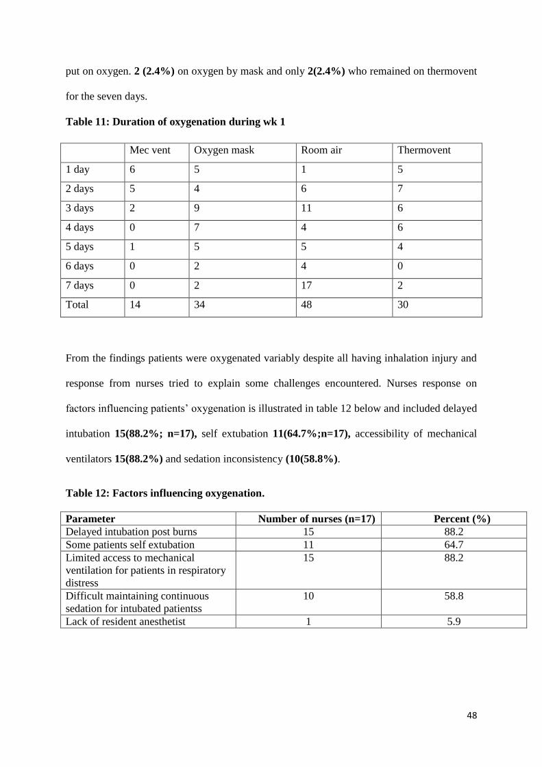
48
put on oxygen. 2 (2.4%) on oxygen by mask and only 2(2.4%) who remained on thermovent
for the seven days.
Table 11: Duration of oxygenation during wk 1
Mec vent Oxygen mask Room air Thermovent
1 day 6 5 1 5
2 days 5 4 6 7
3 days 2 9 11 6
4 days 0 7 4 6
5 days 1 5 5 4
6 days 0 2 4 0
7 days 0 2 17 2
Total 14 34 48 30
From the findings patients were oxygenated variably despite all having inhalation injury and
response from nurses tried to explain some challenges encountered. Nurses response on
factors influencing patients’ oxygenation is illustrated in table 12 below and included delayed
intubation 15(88.2%; n=17), self extubation 11(64.7%;n=17), accessibility of mechanical
ventilators 15(88.2%) and sedation inconsistency (10(58.8%).
Table 12: Factors influencing oxygenation.
Parameter Number of nurses (n=17) Percent (%)
Delayed intubation post burns 15 88.2
Some patients self extubation 11 64.7
Limited access to mechanical
ventilation for patients in respiratory
distress
15 88.2
Difficult maintaining continuous
sedation for intubated patientss
10 58.8
Lack of resident anesthetist 1 5.9
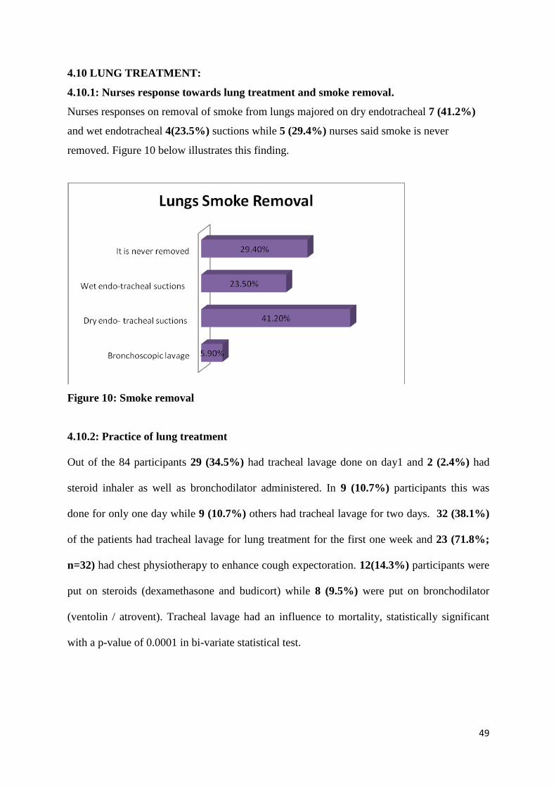
49
4.10 LUNG TREATMENT:
4.10.1: Nurses response towards lung treatment and smoke removal.
Nurses responses on removal of smoke from lungs majored on dry endotracheal 7 (41.2%)
and wet endotracheal 4(23.5%) suctions while 5 (29.4%) nurses said smoke is never
removed. Figure 10 below illustrates this finding.
Figure 10: Smoke removal
4.10.2: Practice of lung treatment
Out of the 84 participants 29 (34.5%) had tracheal lavage done on day1 and 2 (2.4%) had
steroid inhaler as well as bronchodilator administered. In 9 (10.7%) participants this was
done for only one day while 9 (10.7%) others had tracheal lavage for two days. 32 (38.1%)
of the patients had tracheal lavage for lung treatment for the first one week and 23 (71.8%;
n=32) had chest physiotherapy to enhance cough expectoration. 12(14.3%) participants were
put on steroids (dexamethasone and budicort) while 8 (9.5%) were put on bronchodilator
(ventolin / atrovent). Tracheal lavage had an influence to mortality, statistically significant
with a p-value of 0.0001 in bi-variate statistical test.
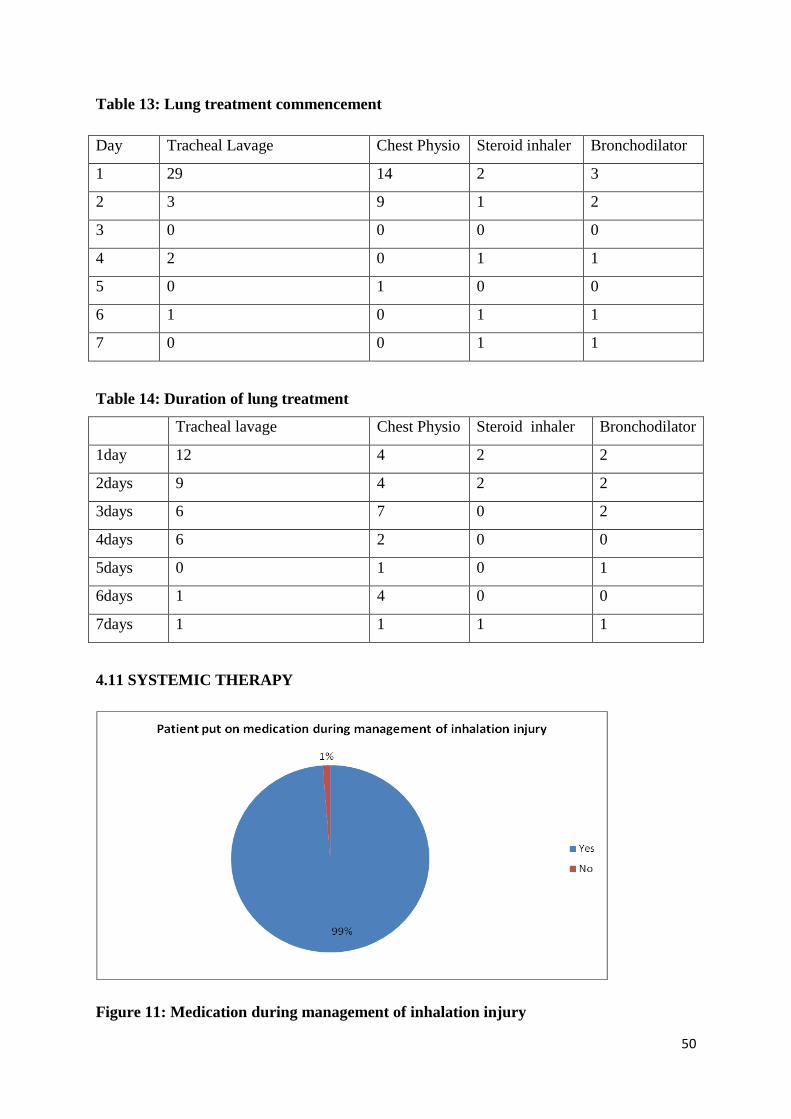
50
Table 13: Lung treatment commencement
Day Tracheal Lavage Chest Physio Steroid inhaler Bronchodilator
1 29 14 2 3
2 3 9 1 2
3 0 0 0 0
4 2 0 1 1
5 0 1 0 0
6 1 0 1 1
7 0 0 1 1
Table 14: Duration of lung treatment
Tracheal lavage Chest Physio Steroid inhaler Bronchodilator
1day 12 4 2 2
2days 9 4 2 2
3days 6 7 0 2
4days 6 2 0 0
5days 0 1 0 1
6days 1 4 0 0
7days 1 1 1 1
4.11 SYSTEMIC THERAPY
Figure 11: Medication during management of inhalation injury

51
It was confirmed that 99% were put on systemic medication during the management of
inhalation injury.
Table 15: Medication used
Medication Number of participants Percentage (%)
Antibiotics ( Prophylaxis) 51 60.7
Antibiotic ( EVB) 1 1.2
Steroids 7 8.3
Nsaids 77 91.6
Opiods 62 73.8
Sedatives 23 27.4
Antipyretics 22 26.2
Anti epileptics 2 2.3
Eye antibiotics 1 1.2
Haloperidol 1 1.2
Table 14 above shows the type of medication used by the respondents. 77( 91.6%) reported
using Nsaids, 62(73.8%) used Opioids while 51(60.7%) indicated using prophylactic
antibiotics. 23 (27.4%) patients had sedative prescribed and 22 (26.2%) used antipyretics.
Few patients 7 (8.3%) used steroids during their care.
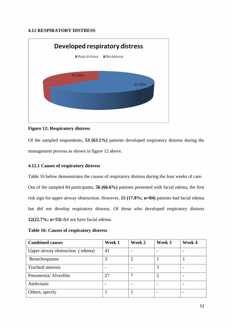
52
4.12 RESPIRATORY DISTRESS
Figure 12: Respiratory distress
Of the sampled respondents, 53 (63.1%) patients developed respiratory distress during the
management process as shown in figure 12 above.
4.12.1 Causes of respiratory distress
Table 16 below demonstrates the causes of respiratory distress during the four weeks of care.
Out of the sampled 84 participants, 56 (66.6%) patients presented with facial edema, the first
risk sign for upper airway obstruction. However, 15 (17.8%; n=84) patients had facial edema
but did not develop respiratory distress. Of those who developed respiratory distress
12(22.7%; n=53) did not have facial edema.
Table 16: Causes of respiratory distress
Combined causes Week 1 Week 2 Week 3 Week 4
Upper airway obstruction ( edema) 41 - - -
Bronchospasms 3 2 1 1
Tracheal stenosis - - 3 -
Pneumonia/ Alveolitis 27 7 2 -
Atelectasis - - - -
Others, specify 1 1 - -
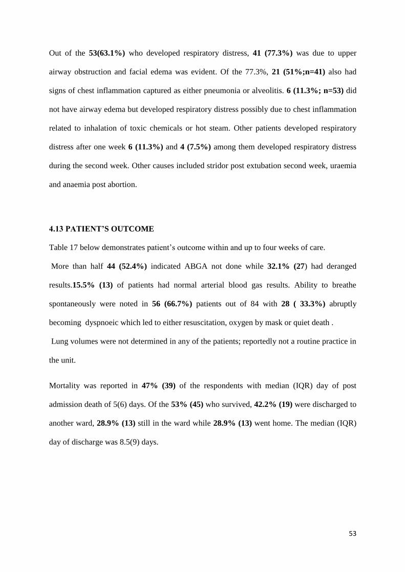
53
Out of the 53(63.1%) who developed respiratory distress, 41 (77.3%) was due to upper
airway obstruction and facial edema was evident. Of the 77.3%, 21 (51%;n=41) also had
signs of chest inflammation captured as either pneumonia or alveolitis. 6 (11.3%; n=53) did
not have airway edema but developed respiratory distress possibly due to chest inflammation
related to inhalation of toxic chemicals or hot steam. Other patients developed respiratory
distress after one week 6 (11.3%) and 4 (7.5%) among them developed respiratory distress
during the second week. Other causes included stridor post extubation second week, uraemia
and anaemia post abortion.
4.13 PATIENT’S OUTCOME
Table 17 below demonstrates patient’s outcome within and up to four weeks of care.
More than half 44 (52.4%) indicated ABGA not done while 32.1% (27) had deranged
results.15.5% (13) of patients had normal arterial blood gas results. Ability to breathe
spontaneously were noted in 56 (66.7%) patients out of 84 with 28 ( 33.3%) abruptly
becoming dyspnoeic which led to either resuscitation, oxygen by mask or quiet death .
Lung volumes were not determined in any of the patients; reportedly not a routine practice in
the unit.
Mortality was reported in 47% (39) of the respondents with median (IQR) day of post
admission death of 5(6) days. Of the 53% (45) who survived, 42.2% (19) were discharged to
another ward, 28.9% (13) still in the ward while 28.9% (13) went home. The median (IQR)
day of discharge was 8.5(9) days.
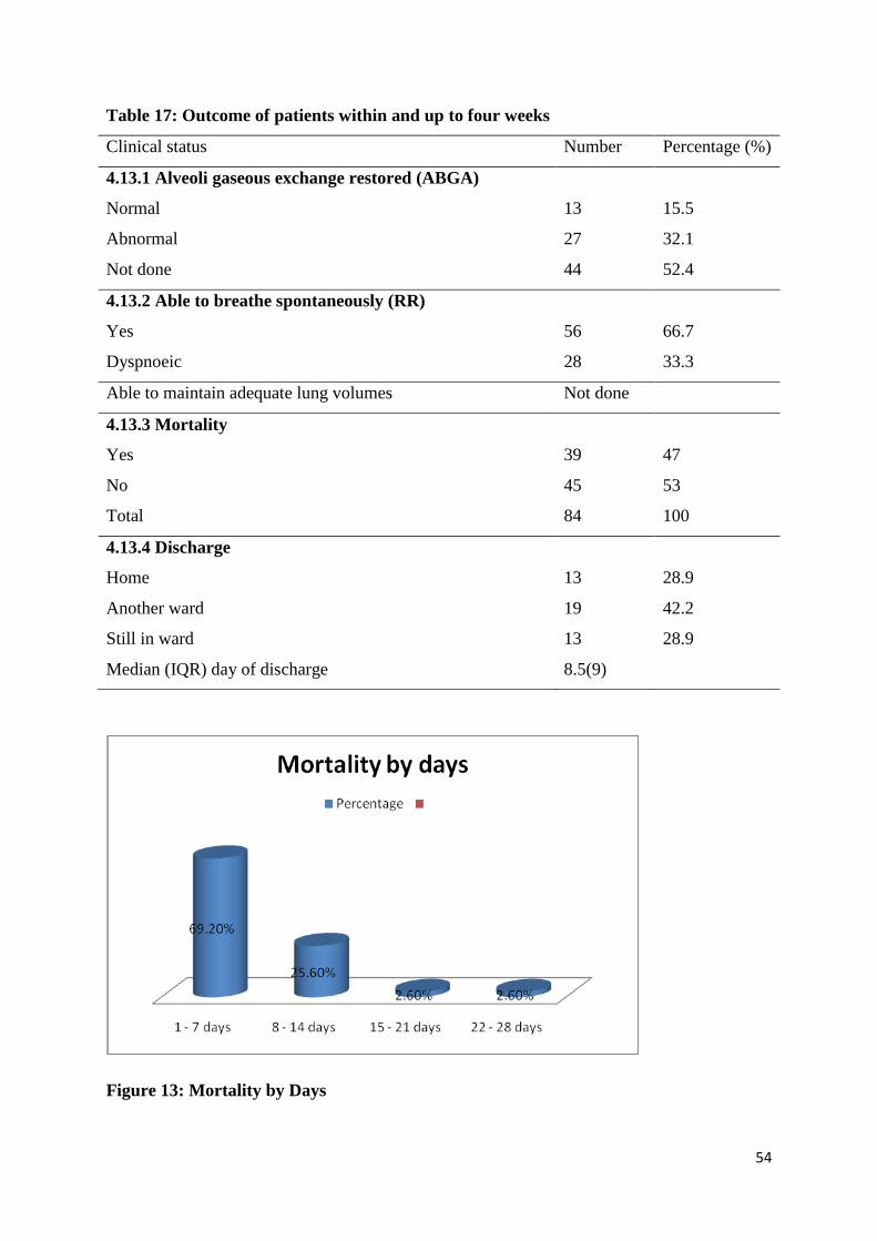
54
Table 17: Outcome of patients within and up to four weeks
Clinical status Number Percentage (%)
4.13.1 Alveoli gaseous exchange restored (ABGA)
Normal 13 15.5
Abnormal 27 32.1
Not done 44 52.4
4.13.2 Able to breathe spontaneously (RR)
Yes 56 66.7
Dyspnoeic 28 33.3
Able to maintain adequate lung volumes Not done
4.13.3 Mortality
Yes 39 47
No 45 53
Total 84 100
4.13.4 Discharge
Home 13 28.9
Another ward 19 42.2
Still in ward 13 28.9
Median (IQR) day of discharge 8.5(9)
Figure 13: Mortality by Days
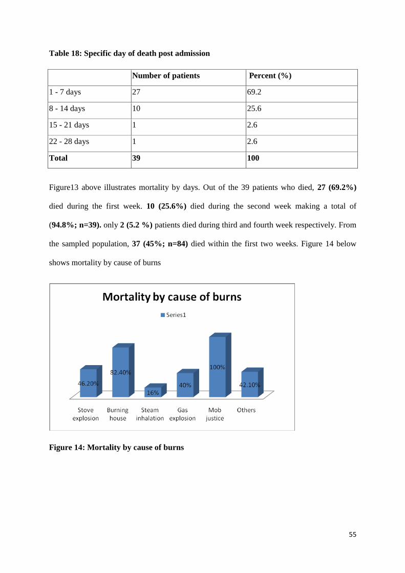
55
Table 18: Specific day of death post admission
Number of patients Percent (%)
1 - 7 days 27 69.2
8 - 14 days 10 25.6
15 - 21 days 1 2.6
22 - 28 days 1 2.6
Total 39 100
Figure13 above illustrates mortality by days. Out of the 39 patients who died, 27 (69.2%)
died during the first week. 10 (25.6%) died during the second week making a total of
(94.8%; n=39). only 2 (5.2 %) patients died during third and fourth week respectively. From
the sampled population, 37 (45%; n=84) died within the first two weeks. Figure 14 below
shows mortality by cause of burns
Figure 14: Mortality by cause of burns
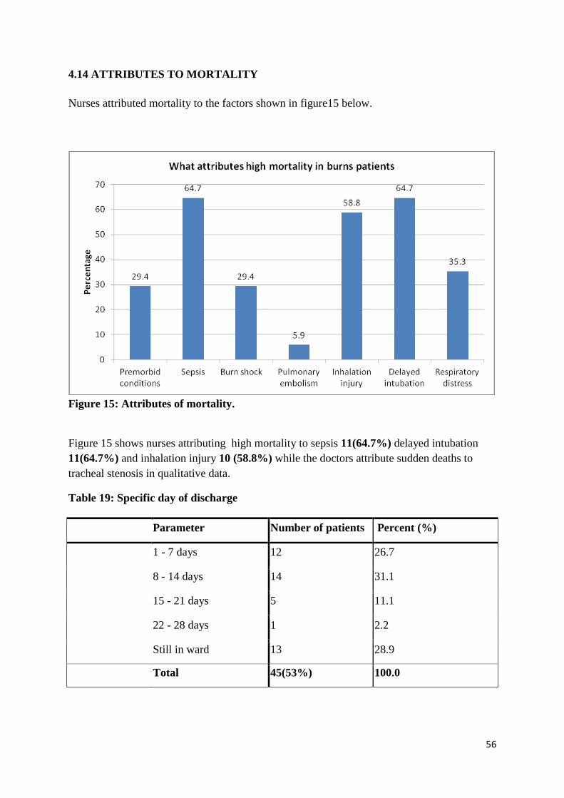
56
4.14 ATTRIBUTES TO MORTALITY
Nurses attributed mortality to the factors shown in figure15 below.
Figure 15: Attributes of mortality.
Figure 15 shows nurses attributing high mortality to sepsis 11(64.7%) delayed intubation
11(64.7%) and inhalation injury 10 (58.8%) while the doctors attribute sudden deaths to
tracheal stenosis in qualitative data.
Table 19: Specific day of discharge
Parameter Number of patients Percent (%)
1 - 7 days 12 26.7
8 - 14 days 14 31.1
15 - 21 days 5 11.1
22 - 28 days 1 2.2
Still in ward 13 28.9
Total 45(53%) 100.0
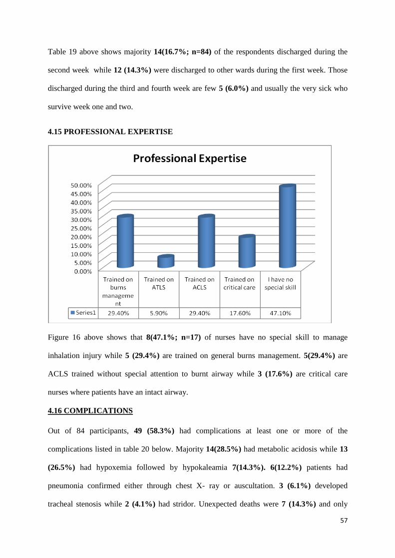
57
Table 19 above shows majority 14(16.7%; n=84) of the respondents discharged during the
second week while 12 (14.3%) were discharged to other wards during the first week. Those
discharged during the third and fourth week are few 5 (6.0%) and usually the very sick who
survive week one and two.
4.15 PROFESSIONAL EXPERTISE
Figure 16 above shows that 8(47.1%; n=17) of nurses have no special skill to manage
inhalation injury while 5 (29.4%) are trained on general burns management. 5(29.4%) are
ACLS trained without special attention to burnt airway while 3 (17.6%) are critical care
nurses where patients have an intact airway.
4.16 COMPLICATIONS
Out of 84 participants, 49 (58.3%) had complications at least one or more of the
complications listed in table 20 below. Majority 14(28.5%) had metabolic acidosis while 13
(26.5%) had hypoxemia followed by hypokaleamia 7(14.3%). 6(12.2%) patients had
pneumonia confirmed either through chest X- ray or auscultation. 3 (6.1%) developed
tracheal stenosis while 2 (4.1%) had stridor. Unexpected deaths were 7 (14.3%) and only
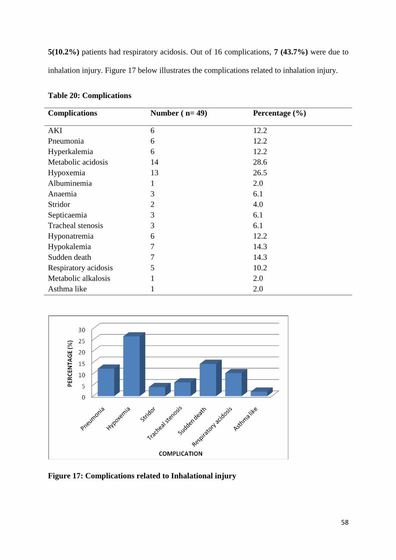
58
5(10.2%) patients had respiratory acidosis. Out of 16 complications, 7 (43.7%) were due to
inhalation injury. Figure 17 below illustrates the complications related to inhalation injury.
Table 20: Complications
Complications Number ( n= 49) Percentage (%)
AKI 6 12.2
Pneumonia 6 12.2
Hyperkalemia 6 12.2
Metabolic acidosis 14 28.6
Hypoxemia 13 26.5
Albuminemia 1 2.0
Anaemia 3 6.1
Stridor 2 4.0
Septicaemia 3 6.1
Tracheal stenosis 3 6.1
Hyponatremia 6 12.2
Hypokalemia 7 14.3
Sudden death 7 14.3
Respiratory acidosis 5 10.2
Metabolic alkalosis 1 2.0
Asthma like 1 2.0
Figure 17: Complications related to Inhalational injury
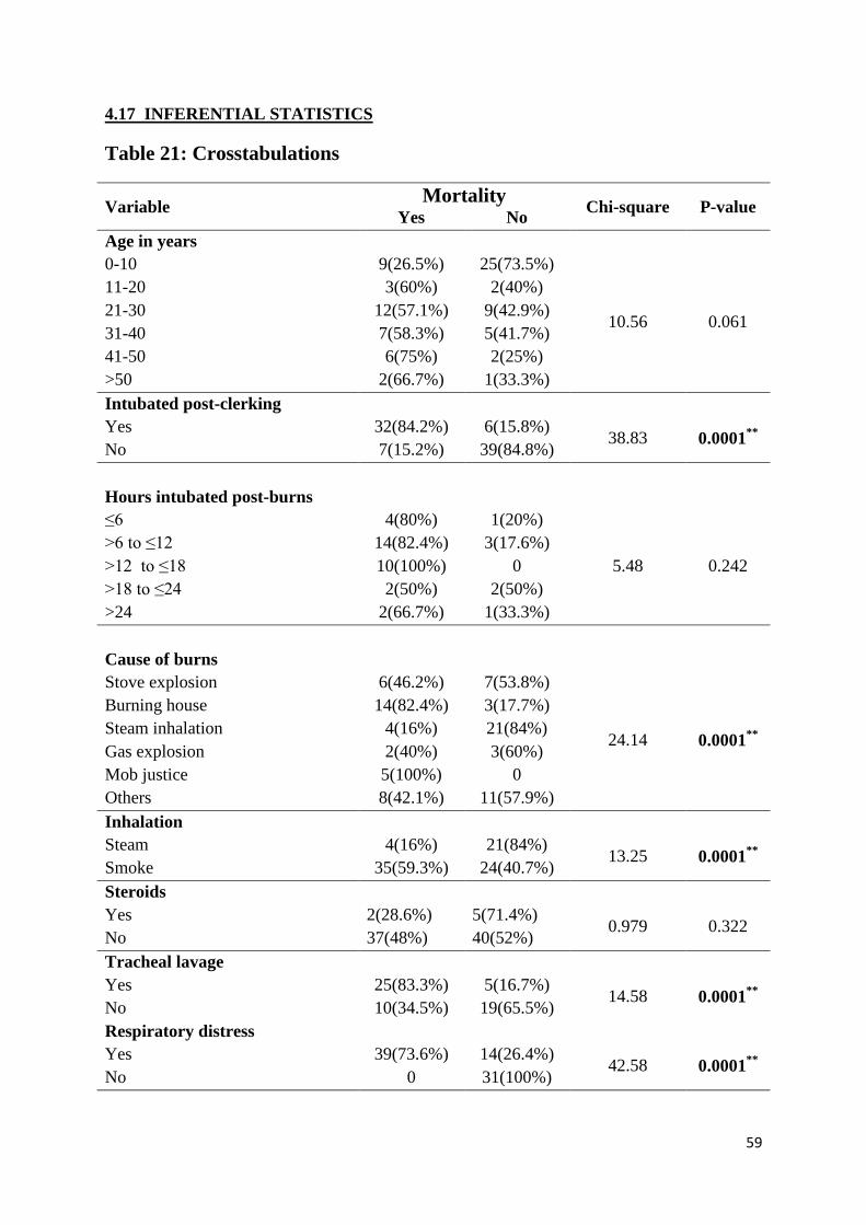
59
4.17 INFERENTIAL STATISTICS
Table 21: Crosstabulations
Variable Mortality
Chi-square P-value Yes No
Age in years
0-10 9(26.5%) 25(73.5%)
10.56 0.061
11-20 3(60%) 2(40%)
21-30 12(57.1%) 9(42.9%)
31-40 7(58.3%) 5(41.7%)
41-50 6(75%) 2(25%)
>50 2(66.7%) 1(33.3%)
Intubated post-clerking
Yes 32(84.2%) 6(15.8%) 38.83 0.0001
**
No 7(15.2%) 39(84.8%)
Hours intubated post-burns
≤6 4(80%) 1(20%)
5.48 0.242
>6 to ≤12 14(82.4%) 3(17.6%)
>12 to ≤18 10(100%) 0
>18 to ≤24 2(50%) 2(50%)
>24 2(66.7%) 1(33.3%)
Cause of burns
Stove explosion 6(46.2%) 7(53.8%)
24.14 0.0001**
Burning house 14(82.4%) 3(17.7%)
Steam inhalation 4(16%) 21(84%)
Gas explosion 2(40%) 3(60%)
Mob justice 5(100%) 0
Others 8(42.1%) 11(57.9%)
Inhalation
Steam 4(16%) 21(84%) 13.25 0.0001
**
Smoke 35(59.3%) 24(40.7%)
Steroids
Yes 2(28.6%) 5(71.4%) 0.979 0.322
No 37(48%) 40(52%)
Tracheal lavage
Yes 25(83.3%) 5(16.7%) 14.58 0.0001
**
No 10(34.5%) 19(65.5%)
Respiratory distress
Yes 39(73.6%) 14(26.4%) 42.58 0.0001
**
No 0 31(100%)
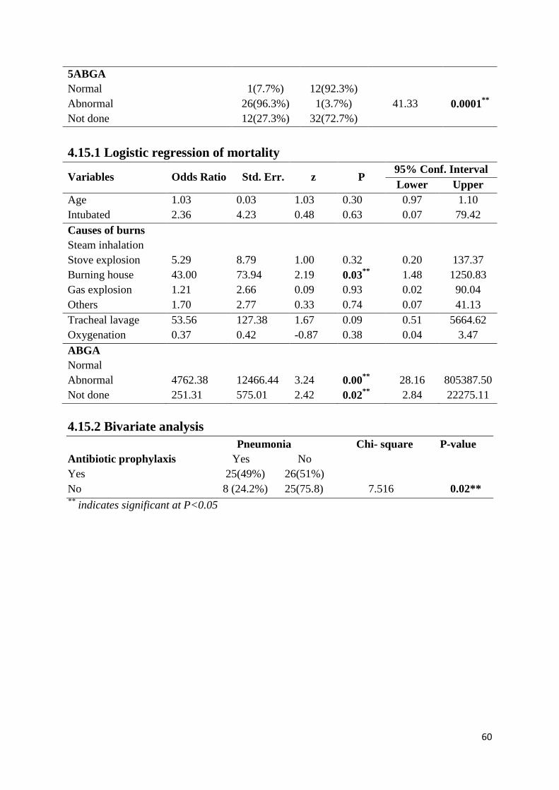
60
5ABGA
Normal 1(7.7%) 12(92.3%)
41.33 0.0001** Abnormal 26(96.3%) 1(3.7%)
Not done 12(27.3%) 32(72.7%)
4.15.1 Logistic regression of mortality
Variables Odds Ratio Std. Err. z P 95% Conf. Interval
Lower Upper
Age 1.03 0.03 1.03 0.30
0.97 1.10
Intubated 2.36 4.23 0.48 0.63 0.07 79.42
Causes of burns
Steam inhalation
Stove explosion 5.29 8.79 1.00 0.32 0.20 137.37
Burning house 43.00 73.94 2.19 0.03** 1.48 1250.83
Gas explosion 1.21 2.66 0.09 0.93 0.02 90.04
Others 1.70 2.77 0.33 0.74 0.07 41.13
Tracheal lavage 53.56 127.38 1.67 0.09
0.51 5664.62
Oxygenation 0.37 0.42 -0.87 0.38 0.04 3.47
ABGA
Normal
Abnormal 4762.38 12466.44 3.24 0.00** 28.16 805387.50
Not done 251.31 575.01 2.42 0.02** 2.84 22275.11
4.15.2 Bivariate analysis
Pneumonia Chi- square P-value
Antibiotic prophylaxis Yes No
Yes 25(49%) 26(51%)
No 8 (24.2%) 25(75.8) 7.516 0.02** **
indicates significant at P<0.05
61
CHAPTER FIVE: DISCUSSION
5.1 DISCUSSION
This study involved 84 participants who were admitted in burns unit, KNH due to inhalation
injury during the months of April, May and June 2014. Open flame burns had the highest
prevalence of (64.3%) while steam inhalation had (29.7%) Others included chemical burns
and flash burns. Smoke inhalation was responsible for (89.7%) mortality consistent with
Toon et al,(2010) finding. In comparison, (64.8%;n=54) of burn patients who had smoke
inhalation died compared to(13.3%;n=30) of those without smoke inhalation.
All age groups and gender are at risk of inhalation injury and this is consistent with Pitts et al
(2008) report that burns can affect all ages, gender and social economic levels. Burning house
was the most frequent cause of inhalation injury across all ages while steam inhalation had
higher frequency among those aged 10years and below. Being prevalent cause of burns
within this age bracket, steam inhalation ( head immersion or spill) reflects home accidents
and is relevant in burns prevention. Stove explosion and kerosine suicide featured more
among those aged 21-30 years with equal occurrence of 7(8.3%) mainly due to mismanaging
stove, domestic quarrels or love affair conflicts.Open flame burns accounted for 64.3% of
inhalation injury which is higher above Miller et al (2008) report which showed 40%
prevalence.
The study findings showed that majority 50 (59.5%) of the patients arrived in the hospital
within eight hours post burns. Eight hours is adequate duration for any chemical to destroy
body cells; but if appropriate resuscitation was done at the scene of injury, these patients are
likely to survive depending on time clerking and intubation is done. Oxygen by mask can be
administered to reduce effect of carbon monoxide and hydroxocobalamin to combat cyanide
effect. Arrival to the hospital is confounded by many factors.
62
After arrival to hospital, majority (73.8%) were clerked within two hours of waiting time and
this is a bit too long for an intoxicated patient. However (6.1 %) participants waited for more
than 6 hours before clerking and this can be a contributing factor to high mortality.
Qualitative data attributed this to inefficiencies in mobilizing resources and poor attitude
among clinicians.
Quite often, intubation follows clinical observations and patient may require intubation
immediately except where patient comes in already intubated. However, some patients wait
even longer. In this study, a few participants had to wait for 6 more hours after clerking
before intubation. Findings revealed majority 26( 68.4%; n=38) were intubated within 2
hours post clerking although time lapse was different considering the time of burn which
might be significant in determining patient’s outcome. Intubation alone was not statistically
significant [p-value > 0.05(0.63)] thus supporting hypothesis. Among the patients 53 (63.1%)
who developed respiratory distress, 41 (77.3%) was due to upper airway obstruction and
severity depended on the extent of obstruction. This might explain why some patients with
facial edema survived even without intubation. Since grading of inhalation injury is not
routinely done, these patients would require very close monitoring to enable timely
intubations where prophylactic intubation was not done.
Despite intubation, other parameters seemed to influence the outcome of patients with
inhalation injury, and premorbid conditions were presumed confounders. However, this study
findings did not show evidenced effect of premorbid conditions although some conditions
predisposed participants to sustaining burns as is the case of epilepsy 4(4.7%) and
alcoholism/drugs 8(9.5%). This implies that effective management of epilepsy and
alcoholism would help reduce incidents of burns.
63
Diagnosis of inhalation injury was mainly clinical 82(97.6%) based on history of the
incidence and presenting signs and symptoms. Lower airway inhalation injury was not
assessed except in some patients who presented with respiratory distress (Nugen & Herndon,
2008). Almost all patients had facial burns except 15 (17.9%) and this implies that patient
can have inhalation injury without necessarily having burns on the face. Since diagnosis was
based on clinical features, it is therefore possible to miss diagnosis unless further
investigation is conducted. Other parameters like chest X-ray were primarily used to confirm
position of central lines and only 7 (8.3%) patients had this done.Although literature does not
support chest X-ray to confirm inhalation injury, it can be useful incase of chest
inflammation. Inhalation injuries were not routinely graded and adopting Woodson’s criteria
of grading inhalation injury would assist in guiding on interventions. Grading inhalation
injury and determining levels of toxicity were not part of the diagnosis despite being key
factors in planning interventions (Woodson,2009).
Tracheal lavage is a common practice but literature does not support its use as the ultimate
intervention for lung treatment. More than half nurses reported use of saline tracheal lavage
to remove smoke from the lungs while a few said smoke is never removed.This discrepancy
may be due to lack of documented standardized operating procedure. In absence of
bronchoscopic lavage, tracheal lavage would be useful, but only small proportion of nurses
reported practicing the same. Tracheal lavage on its own was found to be significant to
mortality but was not significant when subjected to logistical regression; (p-value 0.09).
Other confounders seemed to influence the outcome. Toon et al (2010) and Miller et al
(2009) recommends broncho alveolar toileting using heparin and N- acetylcysteine for smoke
removal and as such tracheal lavage (dry or wet) is not effective to decrease lung toxicity,
incidences of re-intubation, atelectasis and progressive pulmonary failure. This might explain
the derraged arterial blood gas results reported in majority 23 (58.9%;n=39) of the
64
participants who died during the study period. Qualitative data reported lack of
bronchoscopic theater as the reason for not performing broncho-alveolar lavage otherwise the
doctors were willing to learn and perform.
Accidental extubations came out as common phenomena but few patients benefitted from re-
intubation. For the first three days, 16(19.0%) participants had accidental extubations and
those re-intubated were less than half. All re-intubations done were emergencies meaning
severe respiratory distress mainly due to laryngeal edema. From qualitative data, difficulty
convincing anaesthetists to intubate burns patients was commonly repeated. From the nurses’
responses, protocol of care is not standardized 13 (76.5%;n=17) and this might influence
decision making including whether to intubate, re-intubate or not. Best example is where
Doctor 1 confessed trying to convince an anaesthetist to intubate a patient who had inhalation
injury yet anaesthetist felt there was no indication for intubation. Responses from care givers
showed variation of ideas although majority reported that intubated patients should be
sedated as long as they needed intubation to prevent accidental extubations. Study findings
showed less number of participants put on sedatives (mainly dormicum administered as bolus
doses) compared to those intubated; attributing accidental extubations to in-effective
sedation. Qualitative data reported lack of syringe pumps for drug titration as one of the
challenges in managing inhalation injury and combined with other confounders like high
nurse: patient ratio might explain the inconsistent sedation.
Mechanical ventilation practice 13(24.5%; n=53) was found inconsistent with Traber et al
(2007) indications which include all patients with lower airway inhalation injury to enhance
gaseous exchange while maintaining adequate ventilation. On observation, those on
mechanical ventilation had settings of normal tidal volume and PEEP while Toon et al (2010)
recommends low tidal volume and PEEP to reduce ventilator associated lung injury in
inhalation injury and ARDS. This may be because airway management is borrowed from
65
critical care where airway is intact and risk for baro trauma is less. Knowledge on
management of a burnt airway is necessary to the clinicians managing burns patients.
Qualitative data reported lack of bronchoscopic theater as the reason for not assessing lower
airway to grade inhalation injury and as such, depend on arterial blood gases to determine
respiratory efficiency. On the other hand, arterial blood gas analysis is not routinely done and
as a result, some patients die before they are mechanically ventilated.
Carbon monoxide poisoning is a common feature in smoke inhalation and most nurses 14
(82.3%) demonstrated knowledge on use of hyberbaric oxygen to counter effect of carbon
monoxide. However, a discrepancy between knowledge and practice was observed where
almost half of the participants (40.5%) were being maintained on room air first day of
admission. This may be attributed to lack of a standardized operating procedure as was
reported by majority key informants. Lack of a standardized protocol may contribute
significantly to omissions and assumptions in decision making ( Raman& Edwin,2014).
Despite use of prophylactic antibiotics on 60.7% participants, it was observed that 49%
among them developed clinical signs of pneumonia / alveolitis and almost all cases were
noted during the first week. Bacterial chest infections were reportedly common during the
second week which suggests that the observed pneumonia must be related to inhaled smoke
or steam. Attributing the pneumonic / alveolitis process to the chemical toxicity within the
alveoli would therefore disqualify use of antibiotics to prevent pneumonia in patients with
inhalation injury. However, bivariate analysis showed significant relationship between
antibiotic prophylaxis use and pneumonia development with a (p- value of 0.02).
Among the intubated patients 32 (60.4%; n=53)) died and only 6 (11.3%) survived compared
to those with respiratory distress and were not intubated 7 (13.2%; n=53) died and 8 (15.1%)
66
survived. This shows that risk of death is higher to those intubated and a different approach
may be required to reduce the risk. If patent airway was the only problem, these patients
would not die once intubated but it appears like either intubation aggrevates the problem or
cause of death cannot be treated by intubation. Qualitative data hypothetically attributed
cause of death to smoke related chemical toxicity, alveolar atelectasis, tracheal stenosis and
alveolar steam burns which require more than intubation to treat.
Mortality was reported in 39 (47%) patients and 35 (89.7%) of them had smoke inhalation
while 4 (10.3%) had steam inhalation. 35 (41.6%) of the total population died of smoke
inhalation while 4 (4.7%) died of steam inhalation. Mortality was found to be the same
regardless of the time of intubation and this implies that the cause of death is the same in all
patients and it is not taken care of by intubation. The amount of smoke patient is exposed to
determine whether he/she will survive or die and any intervention to reduce this exposure
might increase chances of survival. Cyanide toxicity requires antidote (Polaski & Suzanne,
2010) but no intervention was identified in this study. Majority 27 (69.2%) of deaths occurred
during the first week and arterial blood gas analysis showed 13 (15.5%; n=84) of patients
with hypoxemia while at the same time 56 (66.7%) were breathing well spontaneously. This
indicates a problem with either oxygen transport or exchange and may be, prolonged
mechanical ventilation with alveolar wash out would have enhanced survival. Burning house
as a cause of inhalation injury is statistically significant towards mortality with a p-value of
0.03 (p<0.05).
A number of complications often follow inhalation injury and in this study, 58.3% of the
participants developed complications. Those related to inhalation injury included Pneumonia
due to steam inhalation and crust formation, hypoxemia due to impaired gaseous exchange
following chemical related alveolitis, respiratory acidosis due to retention of CO2 following
impaired gaseous exchange while asthma-like symptoms follow spasms as trachea contracts
67
during healing process. Some patients are prone to development of tracheal stenosis and in
this study two patients developed stridor post extubation and three had tracheal stenosis
which warranted immediate re-intubations. However, this information is not supported in
literature and requires more evaluation.
68
CHAPTER SIX: CONCLUSION AND RECOMMENDATIONS
6.1 CONCLUSION
Diagnosis and management of inhalation injury is very critical in determining the outcome of
patients with burns and early diagnosis is paramount. Clear guideline on management is very
key and there is need for grading the injury. However, the practiced management of
inhalation injury in burns unit is ineffective due to lack of clear protocol, delay in decision
making, inconsistent sedation and that has resulted in higher mortality than internationally
reported. From the finding of this study, the intubation being done is not well defined; does
not tell who should be intubated and who should not. Patients are treated differently despite
inhalation injury and this is not based on any written guideline. Intubation is mainly done for
airway maintenance but sustaining it is a challenge. Although intubation helps to keep the
airway patent, lower lung tissue injury due to smoke and related chemicals toxicity is not
managed and seems responsible for many deaths regardless of the age of patient.
There is no documented protocol of managing inhalation injury thus allowing room for
variable use of knowledge based on personalized reasoning. Qualitative data showed that
doctors and nurses know what should be done but inadequate resources and care facilitation
is a challenge.
Most of inhalation injuries were not graded and management of inhalation injury without
broncho-alveolar lavage might impact negatively on the outcome of care. Current approach
of care is subjective in that some nurses do saline tracheal instillations and others do not.
Some doctors recommend intubations while others disregard opinions without an objective
scale. This was featured in qualitative data where plastic surgery Registrars and nurses
working in burns unit commonly reported trying to convince anaesthetists to intubate burn
patients. Anaesthetists feel patients should be intubated incase of obvious respiratory distress
69
or derranged arterial blood gases. Oxygenation of patients is not based on toxic chemical
levels in the lungs or blood but on visible respiratory distress symptoms which might be
evident when chemical effects cannot be reversed. Whether on oxygen or not, death and life
probability is the same (p-value 0.38) thus implying a gap in the care. Putting patients on
oxygen alone could not change the outcome implying that, chemical removal or its
neutralization is very key in managing inhalation injury.
Critical care of inhalation injury should comprise broncho-alveolar lavage, high level
oxygenation and respiratory support for those with respiratory difficulties guided by arterial
blood gas levels and toxic chemical levels. Intubation is more traumatic to burnt airways than
it is to intact airways and this could be the reason for stridor and tracheal stenosis post
extubation, including majority deaths among the intubated patients.
Other related factors that influence the care of inhalation injury include lack of ventilators,
drug titrating pumps, cardiac monitors, Lack of burns acute care skilled nurses,
bronchoscopic theater and standardized protocol.
6.2 RECOMMENDATIONS
1. There is need for a standard operating procedure or policy on managing inhalation
injury for the entire team to guide on decision making concerning patient care.
2. Further training required on the following:
i. Nurses on pathophysiology of inhalation injury to appreciate need for
oxygenating all patients with inhalation injury guided by carbon monoxide
blood levels.
ii. All clinical care providers on critical care of a burnt airway as compared to
intact airway; which is the main reason for referral of burn patients from other
health facilities.
70
3. Carbon monoxide blood level should be determined on admission for all burn patients
exposed to smoke inhalation as a guide on oxygen concentration requirement on
individual patients.
4. Clinicians should practice routine arterial blood gas analysis for all burns patients
with respiratory distress to promote early diagnosis and early respiratory intervention.
5. All patients with smoke inhalation should be given vitamin B12 (hydroxycobalamin)
to combat cyanide toxic effect and this can be done even at the scene of injury.
6. To promote critical care of patients with inhalation injury, hospital management
should equip Burns unit with the following:
i. Syringe pumps for titrating sedatives to enhance continuous sedation during
intubated period to prevent accidental extubations, and improve on pain
management.
ii. Mechanical ventilators and cardiac monitors for effective respiratory support of
patients with inhalation injury.
iii. Equip Burns Theater with a fibre- optic bronchoscope to enhance grading of
inhalation injury and adoption of bronchoscopic-alveolar lavage, supported in
literature as the ultimate treatment for lower lung inhalation injury.
6.3 RECOMMENDATION FOR FURTHER RESEARCH
1. Challenges of managing a burnt airway.
2. Comparative study between use of bronchoscopic alveolar lavage versus tracheal
lavage to compare effectiveness in reducing mortality.
3. Case- control study on use of hydroxycobalamin versus non –use to determine its
effectiveness in reducing mortality.
4. Comparative study using tracheal instillations of heparin versus normal saline.
71
REFERENCES
Brunner & Saddath (2010). Textbook of Medical Surgical Nursing. 12th
edition. China:
Wolters Kluwer Health. Pages 1718-1751.
Cox,R. & Burke, A. (2003). Airway obstruction in sheep with burn and smoke inhalation
injuries. Journal of respiratory cell molecular biology 29 (3) 295-302 Available at
www.ncbi.nlm.gov/pubmed
Demling, R.H (2005). The Burn Edema Process: Current Concepts. Journal of Burn Care and
Rehabilitation, 26(3): 207-208. Available at http://www.researchgate.net/publication
Feldman, K. Daria, S. & Betom, S. (2004). Distribution of immersion inhalation burns: Head
first into hot water. Pediatrics journal 115(5):1419-21. Available at
http://www.ncbi.nlm.nih.gov/pubmed
Edelman, D.A., White, M.T. & Tyburski J.G (2006). Factors Affecting Prognosis of
Inhalation Injury. Journal of burn care and research. 27(6) 848-853. Available at
http://www.ncbi.nlm.nih.gov/pubmed
Gertrudes (2010). Standard treatment guidelines. Available at www.infocollecting.org/who
Greenhalgh, D.G. (2007). Burn resuscitation. Journal of burns and research 28 (4): 555-
565.Available at http://journals.lww.com/burncareresearch/2007/07000
Herndon, D.N (2007). Inhalation Injury in Burned Patients: Effects & Treatment of Burns.
14(5) 349-356.Available at http://www.worldburn.org/documents/respiratorycare.pdf
Ignatavicious, D. & Linda, W. (2013). Medical Surgical Nursing: Patient Centered
Collaborative Care. 7th
edition. U.S.A.: Saunders co.
Julia G.B.( 2002 ). Nursing Theories: The base for professional nursing practice. 5th
edition.
U.S.A.: Pearson education inc.
Kabalak, A. & Yasti A. (2012). Management of inhalation injury and respiratory
complications in Burns intensive care unit. Available at
www.totalburncare.com/inhalationinjury
72
Kealey, G.P (2009). Carbon Monoxide Toxicity. Journal of Burn Care & Research 32(1)
146-147. Available at www.sciencedaily.com/smokeinhalation
Kenya guidelines on management of inhalation injury. Available at
www.infocollecting.org/who
Kenya Red Cross (2011). DREF operation. Kenya: Fires 2011. Available at
www.ifrc.org/docs/appeals
Kothari, C. R. (2006). Research Methodology: Methods and techniques. 2nd
edition. India:
New age International publishers.
Maybaurer, M. O., Rehbergs, S. & Traber, D. (2009). Pathophysiology of acute lung injury in
severe burn and smoke inhalation injury. Anesthetists journal 58(1): 805-12; Germany.
Available at www.sjtrem.com/inhalationinjury
McCall, J. & Cahill, T. (2005). Respiratory Care of the Burn Patient. Journal of Burn Care
and Rehabilitation. 2226(3) 200-206. Available at www.worldburn.org/documents/respiratory
Miller, A.C., Rivero, A. & Lentz, C.W (2009). National Burn Repository Report. Influence of
nebulized unfractionated heparin and N- acetylcysteine in acute lung injury after smoke
inhalation. Burns Journal 29(6) 862-870. Available at
http://journals.lww.com/burncareresearch
Mugambi,j., Ndeto,F., Katama, J. & Ngure, R. (2012). Correlation between burn causes and
mortality among patients in Burns Unit, KNH. (Unpublished research).
Mugenda, O. & Mugenda, A. (2003). Research methods. 2nd edition. Nairobi : Laba
Graphics Services Ltd.
Nugen, N. & Herndon D.(2008). Diagnosis and treatment of inhalation injury: Total burn
care. 2nd
edition. London: WB Saunders.
Palmieri, T.L (2007). Inhalation Injuries: Research Progress and Needs. Journal of Burn Care
& Research 28(4) 549-554. Available at http://journals.lww.com/burncareresearch
73
Palmieri, T.L (2009). Long-term Outcomes after Inhalation Injury. Journal of Burn Care &
Research 30(1) 201-203.Available at http://journals.lww.com/burncareresearch/
Phan, T.N & Gibran, N.S (2007). Thermal and Electrical Injuries. Surgical Choice of North
America, 87(1) 1-11. Available at http://emedicine.medscape.com/ article/1278244
Pitts, Niska & Xu (2008). National Hospital Ambulatory Medical Survey. Hyattsville:
National Center for Health Statistics. Available at http://www.cdc.gov/nchs/data/pdf
Polaski, A.L. & Suzanne E.T (2010). Core Principles and Practice of Medical Surgical
Nursing: South Asia Edition, India: Elsevier
Raman, K. & Edwin,R. (2014). An overview of ITS Standards and Protocols. Institute of
transportation engineers. Available at www.ite.org/standards
Rehbergs,S. Maybaurer, M. Enkhbaatar, P. et al (2009).Pathophysiology, Managenent and
Treatment of Smoke inhalation injury. Expert Rev Respir med journal 3(3) 283-297.
Available at www.ncbi.nlm.nih.gov/pubmed/20161170
Suman, E., Herndon, D. & Ronald, M. (2007). Respiratory management of inhalation injury.
Journal of burn care 33(2007) 2-13. Available at worldburn.org/documents
Traber, D., Herndon, N. & Enkharbaatar, P. (2007). The pathophysiology of inhalation
injury: Total burn care. 3rd
edition. London: Saunders publishers.
Tom, L. (2012). John Hopkins hospital: Eight hour treatment for inhalation injury. Available
at www.imedcalapps.com
Toon, M., Maybauer, M., Greenwood, E. & Fraser, J.(2010).Management of acute smoke
inhalation injury.. Journal of Critical care and Resuscitation 12(1) 53. Available at
http://www.ncbi.nlm.nih.gov/pubmed
Williams, C. (2008). Fluid Resuscitation in Burn Patients: Nursing care. Nursing times
104(15) 24-25. Available at www.nursingtimes.net.
Woodson, L.C (2009). Diagnosis and Grading of Inhalation Injury, Journal of Burn Care &
Research 28(4), 143-145. Available at http://www.ncbi.nlm.nih.gov/pubmed/1906073
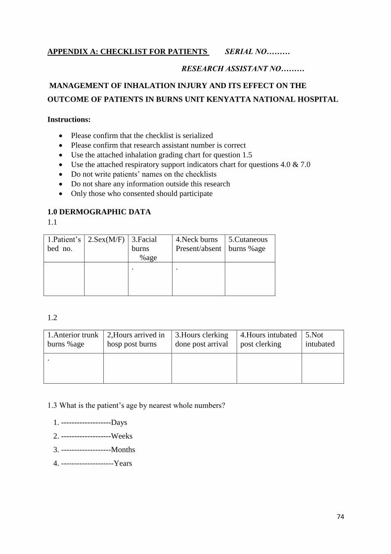
74
APPENDIX A: CHECKLIST FOR PATIENTS SERIAL NO………
RESEARCH ASSISTANT NO………
MANAGEMENT OF INHALATION INJURY AND ITS EFFECT ON THE
OUTCOME OF PATIENTS IN BURNS UNIT KENYATTA NATIONAL HOSPITAL
Instructions:
Please confirm that the checklist is serialized
Please confirm that research assistant number is correct
Use the attached inhalation grading chart for question 1.5
Use the attached respiratory support indicators chart for questions 4.0 & 7.0
Do not write patients’ names on the checklists
Do not share any information outside this research
Only those who consented should participate
1.0 DERMOGRAPHIC DATA
1.1
1.Patient’s
bed no.
2.Sex(M/F) 3.Facial
burns
%age
4.Neck burns
Present/absent
5.Cutaneous
burns %age
. .
1.2
1.Anterior trunk
burns %age
2,Hours arrived in
hosp post burns
3.Hours clerking
done post arrival
4.Hours intubated
post clerking
5.Not
intubated
.
1.3 What is the patient’s age by nearest whole numbers?
1. -------------------Days
2. -------------------Weeks
3. -------------------Months
4. --------------------Years
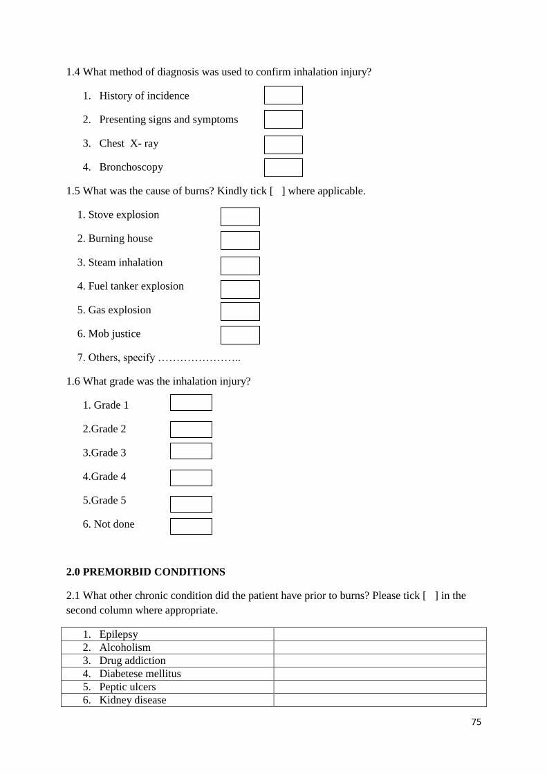
75
1.4 What method of diagnosis was used to confirm inhalation injury?
1. History of incidence
2. Presenting signs and symptoms
3. Chest X- ray
4. Bronchoscopy
1.5 What was the cause of burns? Kindly tick [ ] where applicable.
1. Stove explosion
2. Burning house
3. Steam inhalation
4. Fuel tanker explosion
5. Gas explosion
6. Mob justice
7. Others, specify …………………..
1.6 What grade was the inhalation injury?
1. Grade 1
2.Grade 2
3.Grade 3
4.Grade 4
5.Grade 5
6. Not done
2.0 PREMORBID CONDITIONS
2.1 What other chronic condition did the patient have prior to burns? Please tick [ ] in the
second column where appropriate.
1. Epilepsy
2. Alcoholism
3. Drug addiction
4. Diabetese mellitus
5. Peptic ulcers
6. Kidney disease
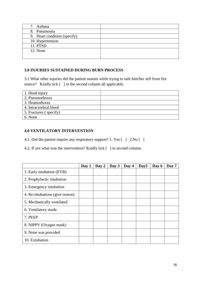
76
7. Asthma
8. Pneumonia
9. Heart condition (specify)
10. Hypertension
11. PTSD
12. None
3.0 INJURIES SUSTAINED DURING BURN PROCESS
3.1 What other injuries did the patient sustain while trying to safe him/her self from fire
source? Kindly tick [ ] in the second column all applicable.
1. Head injury
2. Pneumothorax
3. Heamothorax
4. Intracerebral bleed
5. Fractures ( specify)
6. None
4.0 VENTILATORY INTERVENTION
4.1. Did the patient require any respiratory support? 1. Yes [ ] 2.No [ ]
4.2. If yes what was the intervention? Kindly tick [ ] in second column.
Day 1 Day 2 Day 3 Day 4 Day5 Day 6 Day 7
1. Early intubation (EVB)
2. Prophylactic intubation
3. Emergency intubation
4. Re-intubations (give reason)
5. Mechanically ventilated
6. Ventilatory mode
7. PEEP
8. NIPPV (Oxygen mask)
9. None was provided
10. Extubation
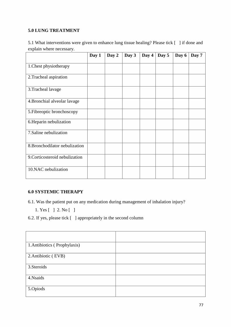
77
5.0 LUNG TREATMENT
5.1 What interventions were given to enhance lung tissue healing? Please tick [ ] if done and
explain where necessary.
Day 1 Day 2 Day 3 Day 4 Day 5 Day 6 Day 7
1.Chest physiotherapy
2.Tracheal aspiration
3.Tracheal lavage
4.Bronchial alveolar lavage
5.Fibreoptic bronchoscopy
6.Heparin nebulization
7.Saline nebulization
8.Bronchodilator nebulization
9.Corticosteroid nebulization
10.NAC nebulization
6.0 SYSTEMIC THERAPY
6.1. Was the patient put on any medication during management of inhalation injury?
1. Yes [ ] 2. No [ ]
6.2. If yes, please tick [ ] appropriately in the second column
1.Antibiotics ( Prophylaxis)
2.Antibiotic ( EVB)
3.Steroids
4.Nsaids
5.Opiods
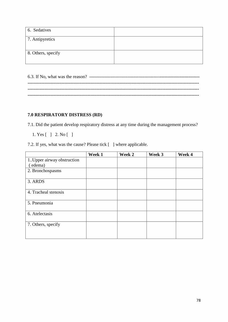
78
6. Sedatives
7. Antipyretics
8. Others, specify
6.3. If No, what was the reason? ------------------------------------------------------------------------
----------------------------------------------------------------------------------------------------------------
----------------------------------------------------------------------------------------------------------------
----------------------------------------------------------------------------------------------------------------
7.0 RESPIRATORY DISTRESS (RD)
7.1. Did the patient develop respiratory distress at any time during the management process?
1. Yes [ ] 2. No [ ]
7.2. If yes, what was the cause? Please tick [ ] where applicable.
Week 1 Week 2 Week 3 Week 4
1..Upper airway obstruction
( edema)
2. Bronchospasms
3. ARDS
4. Tracheal stenosis
5. Pneumonia
6. Atelectasis
7. Others, specify
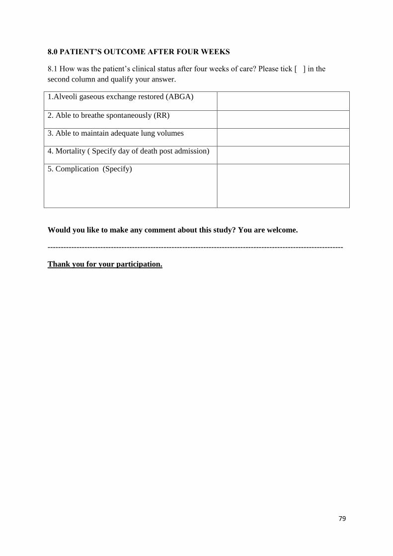
79
8.0 PATIENT’S OUTCOME AFTER FOUR WEEKS
8.1 How was the patient’s clinical status after four weeks of care? Please tick [ ] in the
second column and qualify your answer.
1.Alveoli gaseous exchange restored (ABGA)
2. Able to breathe spontaneously (RR)
3. Able to maintain adequate lung volumes
4. Mortality ( Specify day of death post admission)
5. Complication (Specify)
Would you like to make any comment about this study? You are welcome.
----------------------------------------------------------------------------------------------------------------
Thank you for your participation.
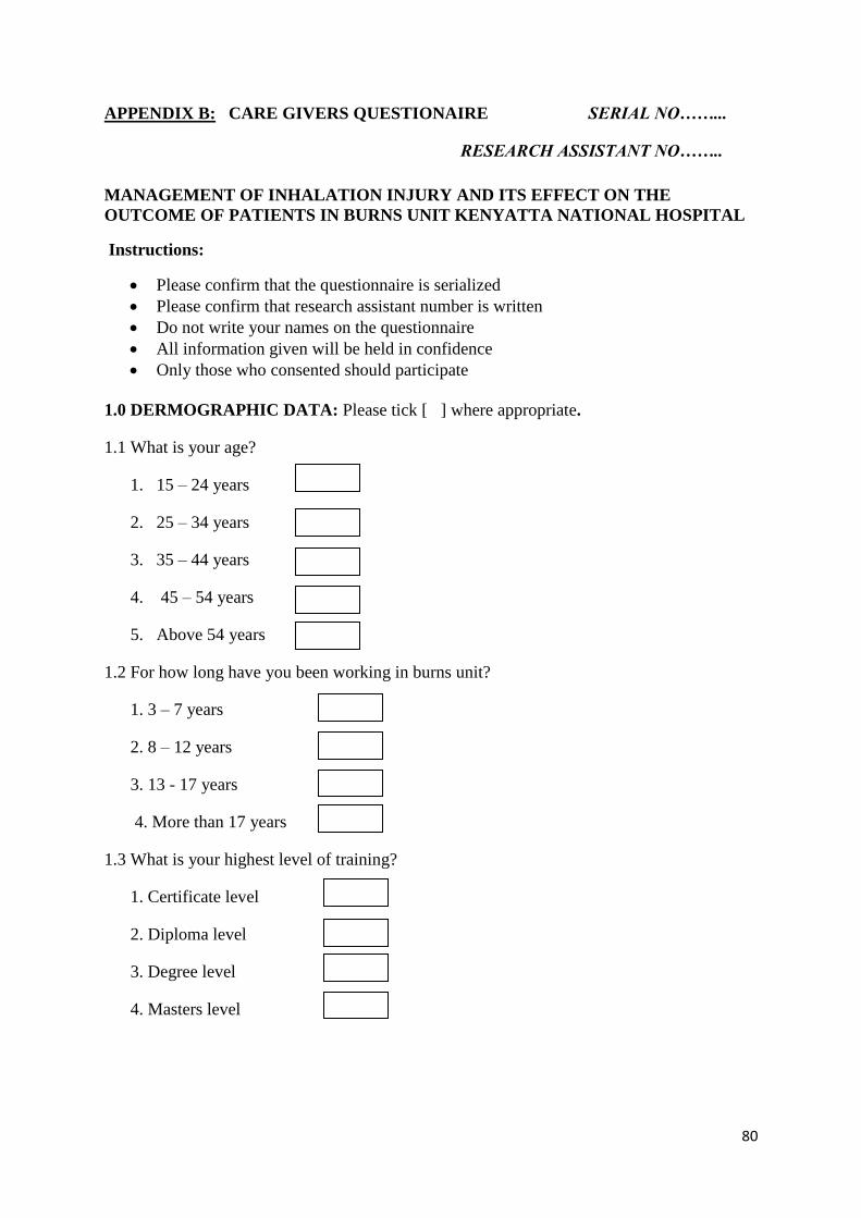
80
APPENDIX B: CARE GIVERS QUESTIONAIRE SERIAL NO……...
RESEARCH ASSISTANT NO……..
MANAGEMENT OF INHALATION INJURY AND ITS EFFECT ON THE
OUTCOME OF PATIENTS IN BURNS UNIT KENYATTA NATIONAL HOSPITAL
Instructions:
Please confirm that the questionnaire is serialized
Please confirm that research assistant number is written
Do not write your names on the questionnaire
All information given will be held in confidence
Only those who consented should participate
1.0 DERMOGRAPHIC DATA: Please tick [ ] where appropriate.
1.1 What is your age?
1. 15 – 24 years
2. 25 – 34 years
3. 35 – 44 years
4. 45 – 54 years
5. Above 54 years
1.2 For how long have you been working in burns unit?
1. 3 – 7 years
2. 8 – 12 years
3. 13 - 17 years
4. More than 17 years
1.3 What is your highest level of training?
1. Certificate level
2. Diploma level
3. Degree level
4. Masters level
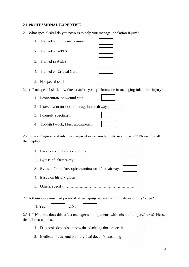
81
2.0 PROFESSIONAL EXPERTISE
2.1 What special skill do you possess to help you manage inhalation injury?
1. Trained on burns management
2. Trained on ATLS
3. Trained in ACLS
4. Trained on Critical Care
5. No special skill
2.1.1 If no special skill, how does it affect your performance in managing inhalation injury?
1. I concentrate on wound care
2. I have learnt on job to manage burnt airways
3. I consult specialists
4. Though I work, I feel incompetent
2.2 How is diagnosis of inhalation injury/burns usually made in your ward? Please tick all
that applies.
1. Based on signs and symptoms
2. By use of chest x-ray
3. By use of bronchoscopic examination of the airways
4. Based on history given
5. Others, specify………………………………………………….
2.3 Is there a documented protocol of managing patients with inhalation injury/burns?
1. Yes 2.No
2.3.1 If No, how does this affect management of patients with inhalation injury/burns? Please
tick all that applies.
1. Diagnosis depends on how the admitting doctor sees it
2. Medications depend on individual doctor’s reasoning
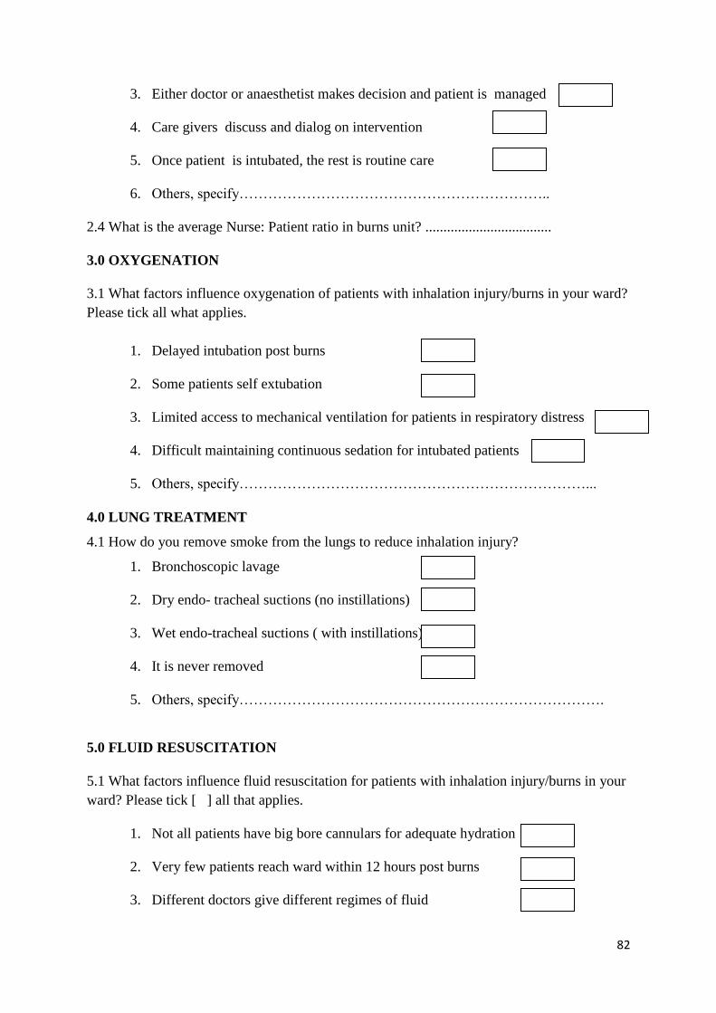
82
3. Either doctor or anaesthetist makes decision and patient is managed
4. Care givers discuss and dialog on intervention
5. Once patient is intubated, the rest is routine care
6. Others, specify………………………………………………………..
2.4 What is the average Nurse: Patient ratio in burns unit? ...................................
3.0 OXYGENATION
3.1 What factors influence oxygenation of patients with inhalation injury/burns in your ward?
Please tick all what applies.
1. Delayed intubation post burns
2. Some patients self extubation
3. Limited access to mechanical ventilation for patients in respiratory distress
4. Difficult maintaining continuous sedation for intubated patients
5. Others, specify………………………………………………………………...
4.0 LUNG TREATMENT
4.1 How do you remove smoke from the lungs to reduce inhalation injury?
1. Bronchoscopic lavage
2. Dry endo- tracheal suctions (no instillations)
3. Wet endo-tracheal suctions ( with instillations)
4. It is never removed
5. Others, specify………………………………………………………………….
5.0 FLUID RESUSCITATION
5.1 What factors influence fluid resuscitation for patients with inhalation injury/burns in your
ward? Please tick [ ] all that applies.
1. Not all patients have big bore cannulars for adequate hydration
2. Very few patients reach ward within 12 hours post burns
3. Different doctors give different regimes of fluid
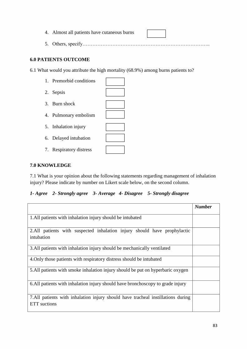
83
4. Almost all patients have cutaneous burns
5. Others, specify…………………………………………………………………..
6.0 PATIENTS OUTCOME
6.1 What would you attribute the high mortality (68.9%) among burns patients to?
1. Premorbid conditions
2. Sepsis
3. Burn shock
4. Pulmonary embolism
5. Inhalation injury
6. Delayed intubation
7. Respiratory distress
7.0 KNOWLEDGE
7.1 What is your opinion about the following statements regarding management of inhalation
injury? Please indicate by number on Likert scale below, on the second column.
1- Agree 2- Strongly agree 3- Average 4- Disagree 5- Strongly disagree
Number
1.All patients with inhalation injury should be intubated
2.All patients with suspected inhalation injury should have prophylactic
intubation
3.All patients with inhalation injury should be mechanically ventilated
4.Only those patients with respiratory distress should be intubated
5.All patients with smoke inhalation injury should be put on hyperbaric oxygen
6.All patients with inhalation injury should have bronchoscopy to grade injury
7.All patients with inhalation injury should have tracheal instillations during
ETT suctions
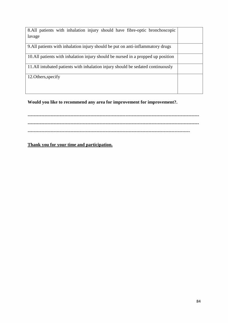
84
8.All patients with inhalation injury should have fibre-optic bronchoscopic
lavage
9.All patients with inhalation injury should be put on anti-inflammatory drugs
10.All patients with inhalation injury should be nursed in a propped up position
11.All intubated patients with inhalation injury should be sedated continuously
12.Others,specify
Would you like to recommend any area for improvement for improvement?.
----------------------------------------------------------------------------------------------------------------
----------------------------------------------------------------------------------------------------------------
----------------------------------------------------------------------------------------------------------
Thank you for your time and participation.
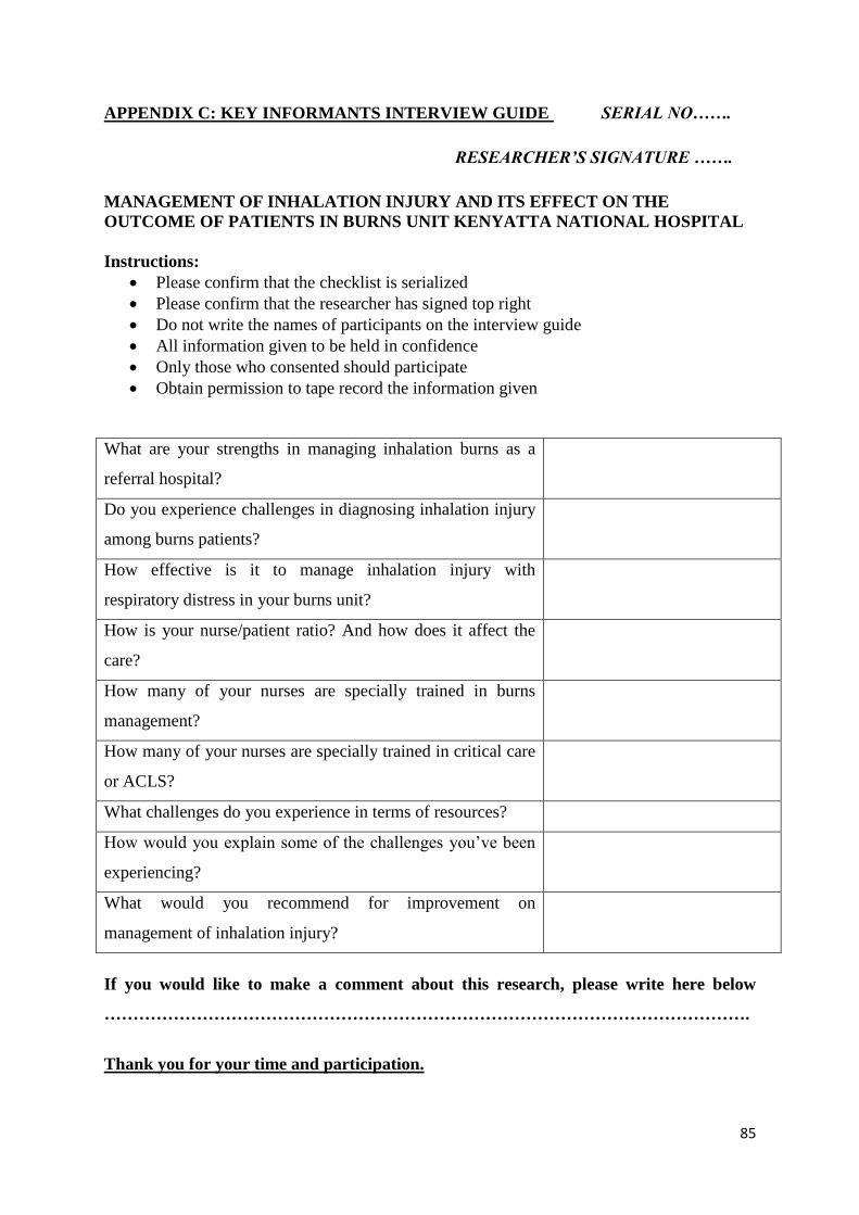
85
APPENDIX C: KEY INFORMANTS INTERVIEW GUIDE SERIAL NO…….
RESEARCHER’S SIGNATURE …….
MANAGEMENT OF INHALATION INJURY AND ITS EFFECT ON THE
OUTCOME OF PATIENTS IN BURNS UNIT KENYATTA NATIONAL HOSPITAL
Instructions:
Please confirm that the checklist is serialized
Please confirm that the researcher has signed top right
Do not write the names of participants on the interview guide
All information given to be held in confidence
Only those who consented should participate
Obtain permission to tape record the information given
What are your strengths in managing inhalation burns as a
referral hospital?
Do you experience challenges in diagnosing inhalation injury
among burns patients?
How effective is it to manage inhalation injury with
respiratory distress in your burns unit?
How is your nurse/patient ratio? And how does it affect the
care?
How many of your nurses are specially trained in burns
management?
How many of your nurses are specially trained in critical care
or ACLS?
What challenges do you experience in terms of resources?
How would you explain some of the challenges you’ve been
experiencing?
What would you recommend for improvement on
management of inhalation injury?
If you would like to make a comment about this research, please write here below
………………………………………………………………………………………………….
Thank you for your time and participation.
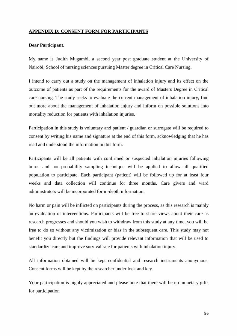
86
APPENDIX D: CONSENT FORM FOR PARTICIPANTS
Dear Participant.
My name is Judith Mugambi, a second year post graduate student at the University of
Nairobi; School of nursing sciences pursuing Master degree in Critical Care Nursing.
I intend to carry out a study on the management of inhalation injury and its effect on the
outcome of patients as part of the requirements for the award of Masters Degree in Critical
care nursing. The study seeks to evaluate the current management of inhalation injury, find
out more about the management of inhalation injury and inform on possible solutions into
mortality reduction for patients with inhalation injuries.
Participation in this study is voluntary and patient / guardian or surrogate will be required to
consent by writing his name and signature at the end of this form, acknowledging that he has
read and understood the information in this form.
Participants will be all patients with confirmed or suspected inhalation injuries following
burns and non-probability sampling technique will be applied to allow all qualified
population to participate. Each participant (patient) will be followed up for at least four
weeks and data collection will continue for three months. Care givers and ward
administrators will be incorporated for in-depth information.
No harm or pain will be inflicted on participants during the process, as this research is mainly
an evaluation of interventions. Participants will be free to share views about their care as
research progresses and should you wish to withdraw from this study at any time, you will be
free to do so without any victimization or bias in the subsequent care. This study may not
benefit you directly but the findings will provide relevant information that will be used to
standardize care and improve survival rate for patients with inhalation injury.
All information obtained will be kept confidential and research instruments anonymous.
Consent forms will be kept by the researcher under lock and key.
Your participation is highly appreciated and please note that there will be no monetary gifts
for participation
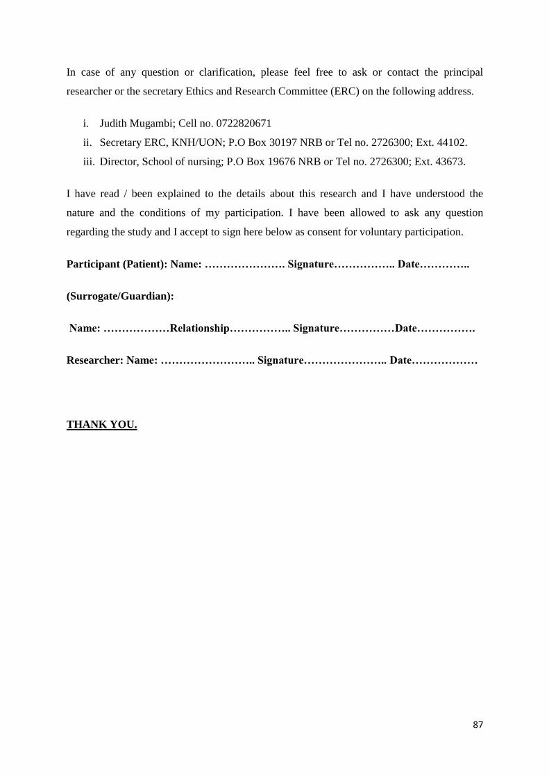
87
In case of any question or clarification, please feel free to ask or contact the principal
researcher or the secretary Ethics and Research Committee (ERC) on the following address.
i. Judith Mugambi; Cell no. 0722820671
ii. Secretary ERC, KNH/UON; P.O Box 30197 NRB or Tel no. 2726300; Ext. 44102.
iii. Director, School of nursing; P.O Box 19676 NRB or Tel no. 2726300; Ext. 43673.
I have read / been explained to the details about this research and I have understood the
nature and the conditions of my participation. I have been allowed to ask any question
regarding the study and I accept to sign here below as consent for voluntary participation.
Participant (Patient): Name: …………………. Signature…………….. Date…………..
(Surrogate/Guardian):
Name: ………………Relationship…………….. Signature……………Date…………….
Researcher: Name: …………………….. Signature………………….. Date………………
THANK YOU.

88
APPENDIX E:
1. GRADING INHALATION INJURY
First verify that the patient has at least two or three signs suggestive of inhalation
injury. i.e. sighed nasal hairs, sooty sputum, facial burns, swollen tongue, swollen
lips, cough, hoarse voice, history of enclosed in burning house or hot liquid head
immersion.
Then confirm:
Grade I: No laryngeal oedema
Grade II: Minimal laryngeal oedema and erythema
Grade III: Slight tracheal mucosal oedema and erythema
Grade IV: Moderate tracheal mucosal oedema and erythema
Grade V: Severe tracheal oedema and erythema
2. EVIDENCE OF RESPIRATORY DISTRESS; INDICATORS FOR
RESPIRATORY SUPPORT
Horseness of voice
Laboured breathing
Obvious upper airway edema
Respiratory acidosis
Respiratory alkalosis
Hypoxemia
Carbon monoxide levels above 20%Hgb
Dyspnoea
Stridor
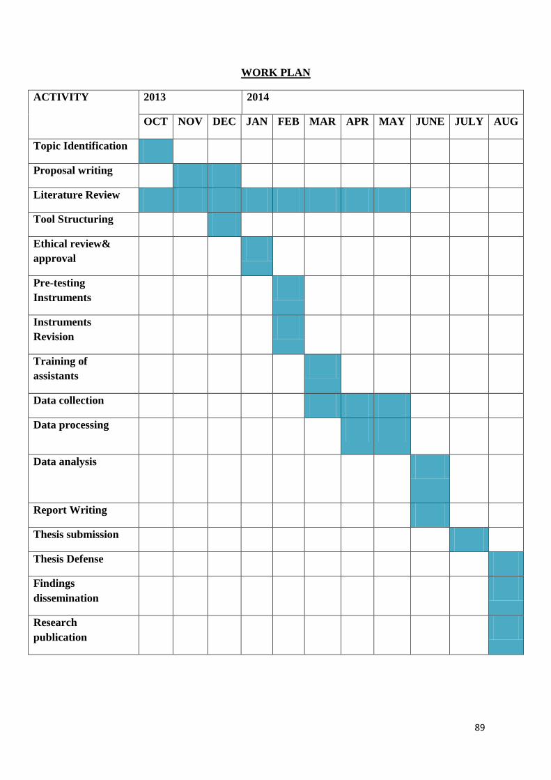
89
WORK PLAN
ACTIVITY 2013 2014
OCT NOV DEC JAN FEB MAR APR MAY JUNE JULY AUG
Topic Identification
Proposal writing
Literature Review
Tool Structuring
Ethical review&
approval
Pre-testing
Instruments
Instruments
Revision
Training of
assistants
Data collection
Data processing
Data analysis
Report Writing
Thesis submission
Thesis Defense
Findings
dissemination
Research
publication

90
BUDGET
ITEM ESTIMATED COST
PER UNIT
NUMBER OF
UNITS
TOTAL
COST
STATIONERY: Pens, Printing
papers.
Cartilages, airtime, Note books
500.00
4800.00
10
10
5000.00
48000.00
BROWSING 10.00 500 5000.00
ETHICAL APPROVAL:
KNH/UON, MOHE
- - 2000.00
TYPING : Questionnaires, Final
report
10.00 200 Pages 2000.00
PHOTOCOPYING:
Questionnaires, Drafts, Thesis
2.00 4000 pages 8000.00
ALLOWANCES FOR
ASSISTANTS (2): Training,
Field work, Transport, Lunch
1000 per day 90days 90,000.00
DATA PROCESSING &
ANALYSIS: Data analyst
5000.00 1 5000.00
PROPOSAL & DRAFT
THESIS BINDING
150.00 10 1500.00
THESIS FINAL BINDING 250.00 12 3000.00
SUBTOTAL 169,500.00
CONTIGENCY EXPENSES
(10%)
16,950.00
TOTAL 186,450.00
Copyright © 2022 FDOKUMEN