RESVERATROL SUPPLEMENTATION DURING IN VITRO ...
150
ADVERTIMENT. Lʼaccés als continguts dʼaquesta tesi queda condicionat a lʼacceptació de les condicions dʼús establertes per la següent llicència Creative Commons: http://cat.creativecommons.org/?page_id=184 ADVERTENCIA. El acceso a los contenidos de esta tesis queda condicionado a la aceptación de las condiciones de uso establecidas por la siguiente licencia Creative Commons: http://es.creativecommons.org/blog/licencias/ WARNING. The access to the contents of this doctoral thesis it is limited to the acceptance of the use conditions set by the following Creative Commons license: https://creativecommons.org/licenses/?lang=en
-
Upload
khangminh22 -
Category
Documents
-
view
2 -
download
0
Transcript of RESVERATROL SUPPLEMENTATION DURING IN VITRO ...

ADVERTIMENT. Lʼaccés als continguts dʼaquesta tesi queda condicionat a lʼacceptació de les condicions dʼúsestablertes per la següent llicència Creative Commons: http://cat.creativecommons.org/?page_id=184
ADVERTENCIA. El acceso a los contenidos de esta tesis queda condicionado a la aceptación de las condiciones de usoestablecidas por la siguiente licencia Creative Commons: http://es.creativecommons.org/blog/licencias/
WARNING. The access to the contents of this doctoral thesis it is limited to the acceptance of the use conditions setby the following Creative Commons license: https://creativecommons.org/licenses/?lang=en

DOCTORAL THESIS
RESVERATROL SUPPLEMENTATION DURING IN VITRO MATURATION:
EFFECT ON THE QUALITY OF OOCYTES IN SPECIES OF VETERINARY INTEREST
Anna Rita Piras
2019
Under the direction of:
Luisa Bogliolo
UNIVERSITÀ DEGLI STUDI DI SASSARI DIPARTIMENTI DI MEDICINA VETERINARIA
Scuola di Dottorato in: Riproduzione, Patologia, Allevamento e Benessere Animale
(XXXI CICLO)
María Teresa Paramio Nieto
UNIVERSITAT AUTÒNOMA DE BARCELONA FACULTAT DE VETERINÀRIA
Departament de Ciència Animal i dels Aliments Producciòn Animal
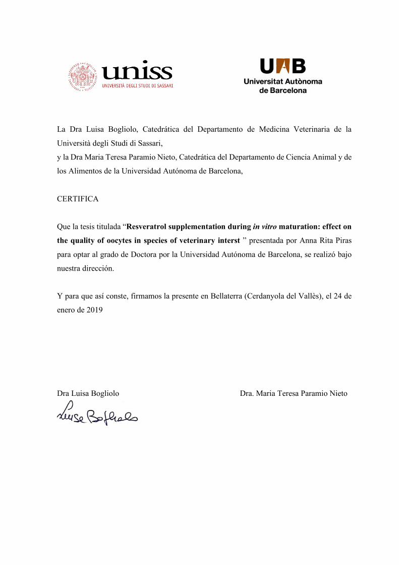
La Dra Luisa Bogliolo, Catedrática del Departamento de Medicina Veterinaria de la
Università degli Studi di Sassari,
y la Dra Maria Teresa Paramio Nieto, Catedrática del Departamento de Ciencia Animal y de
los Alimentos de la Universidad Autónoma de Barcelona,
CERTIFICA
Que la tesis titulada “Resveratrol supplementation during in vitro maturation: effect on
the quality of oocytes in species of veterinary interst ” presentada por Anna Rita Piras
para optar al grado de Doctora por la Universidad Autónoma de Barcelona, se realizó bajo
nuestra dirección.
Y para que así conste, firmamos la presente en Bellaterra (Cerdanyola del Vallès), el 24 de
enero de 2019
Dra Luisa Bogliolo Dra. Maria Teresa Paramio Nieto

Anna Rita Piras, Resveratrol supplementation during in vitro maturation: Effect on the quality of oocytes in species of veterinary interest, tesi di Dottorato in cotutela in: Riproduzione, Patologia, Allevamento e Benessere Animale, Università degli Studi di Sassari; Producción Animal, Universitat Autònoma de Barcelona
3
Abstract
The quality of oocytes plays a pivotal role in determining in vitro embryo production (IVEP)
outcomes. Different intrinsic and extrinsic factors can impair the quality of mammalian
oocytes, affecting their developmental competence.
The aim of the study was to evaluate the effect of supplementation of resveratrol, a natural
antioxidant, during in vitro maturation (IVM) on the quality of oocytes in species of
veterinary interest (domestic cat, prepubertal goats and sheep).
Three studies were performed to test the potential beneficial influence of resveratrol to
improve the in vitro developmental competence of poor quality oocytes such as under sub-
optimal condition or with low developmental competence.
Specifically, the effect of resveratrol addition to the IVM medium was tested on:
-STUDY I: IVEP from domestic cat oocytes retrieved from ovaries stored at 4°C for 24 and
48h.
Oocytes retrieved from stored ovaries for 24 and 48h were IVM with or without 5µM
resveratrol. Meiotic competence, intracellular levels of reactive oxygen species (ROS) and
glutathione (GSH), blastocyst yield and the blastocyst cell number were evaluated. The
results showed that resveratrol treatment had not effect on the meiotic competence of the
oocytes. Resveratrol groups had lower (P<0.05) intracellular ROS levels and higher
(P<0.05) GSH content compared to untreated oocytes, both at 24 and 48h. Moreover,
resveratrol supplementation significantly increased blastocyst yield in 48h group and
improves blastocyst cells number in both groups.

Anna Rita Piras, Resveratrol supplementation during in vitro maturation: Effect on the quality of oocytes in species of veterinary interest, tesi di Dottorato in cotutela in: Riproduzione, Patologia, Allevamento e Benessere Animale, Università degli Studi di Sassari; Producción Animal, Universitat Autònoma de Barcelona
4
-STUDY II: IVEP from prepubertal goat oocytes selected by brilliant cresyl blue (BCB) staining.
Oocytes were classified by BCB staining as BCB+ (fully grown oocytes ) or BCB- (growing
oocytes) and IVM with or without 1µM resveratrol. ROS and GSH levels, mitochondrial
activity and distribution and ATP content were analyzed in metaphase II oocytes (MII). After
in vitro fertilization (IVF), the blastocyst rate and the blastocyst quality were assessed. No
differences were found in ROS levels, ATP content and mitochondrial activity among
groups. GSH levels were significantly higher in both BCB groups treated with resveratrol
than their respective controls. Oocytes treated with resveratrol showed a higher proportion
of clustered active mitochondria than control groups. The development to blastocyst stage
was significantly higher in BCB+ oocytes matured with resveratrol compared with the other
groups. No differences were observed in blastocyst quality among groups.
STUDY III: In vitro fertilization outcome of prepubertal sheep oocytes under cadmium
exposure.
Oocytes were expose to 2µM cadmium (Cd) and IVM in the presence of different
concentration of resveratrol: 0µM (Cd), 1 µM (Cd-Resv1µM) and 2 µM (Cd-Resv2µM).
Oocytes matured in absence of Cd were used as control. Fertilization outcomes, cortical
granules (CGs) and mitochondria distribution, mitochondria activity and ROS level were
evaluated. Oocytes of control, Cd-Resv1µM and Cd-Resv2µM groups had higher normal
fertilization compared to Cd group (P<0.05). The percentage of MII oocytes with CGs
distributed in the cortex of the oocytes was higher (P<0.05) in control and Cd-Resv1µM
groups than Cd group. The percentage of MII oocytes that exhibited a homogeneous
mitochondria distribution throughout the cytoplasm was higher in control, Cd-Resv 1µM
and Cd-Resv2µM groups than Cd group (P<0.05). Lower activity (P<0.05) of mitochondria
was recorded in control and Cd-Resv 1µM oocytes compared to Cd oocytes. The
intracellular ROS levels were lower in control, Cd-Resv1µM and Cd-Resv2µM groups than
Cd group.

Anna Rita Piras, Resveratrol supplementation during in vitro maturation: Effect on the quality of oocytes in species of veterinary interest, tesi di Dottorato in cotutela in: Riproduzione, Patologia, Allevamento e Benessere Animale, Università degli Studi di Sassari; Producción Animal, Universitat Autònoma de Barcelona
5
Taking all these results into account, we conclude that resveratrol supplementation during
IVM constitutes a useful strategy to improve oocyte quality and IVEP outcome in species of
veterinary interest. The mechanism underlying resveratrol effects included the regulation of
bioenergetic/redox status of the oocytes by the modulation of ROS and GSH levels and
mitochondria function and the distribution of cytoplasmic organelles.

Anna Rita Piras, Resveratrol supplementation during in vitro maturation: Effect on the quality of oocytes in species of veterinary interest, tesi di Dottorato in cotutela in: Riproduzione, Patologia, Allevamento e Benessere Animale, Università degli Studi di Sassari; Producción Animal, Universitat Autònoma de Barcelona
6
Resumen La calidad de los oocitos juega un papel importante en la producción in vitro de embriones (PIVE).
Factores intrínsecos y extrínsicos pueden alterar la calidad de los oocitos, afectando su competencia
para el desarrollo. El objetivo del estudio fue evaluar el efecto de la suplementación con resveratrol
durante la maduración in vitro (MIV) sobre la calidad de los oocitos en especies de interés
veterinario.
Tres estudios fueron llevados a cabo para testar el posible efecto beneficioso del resveratrol para la
mejora de la competencia de desarrollo in vitro de los oocitos de mala calidad (oocitos en condiciones
sub-óptimas o con baja competencia de desarrollo).
Específicamente, el efecto del tratamiento con resveratrol fue probado en:
Estudio I: PIVE de gatos domésticos obtenidos de ovarios refrigerados (4ºC) durante 24 y 48 horas.
Los oocitos obtenidos de ovarios refrigerados fueron MIV con o sin 5µM de resveratrol. La
competencia meiótica, niveles de especies reactivas de oxigeno (ROS) y glutatión (GSH), la tasa de
blastocistos obtenidos y su calidad fueron evaluadas. Los resultados mostraron que el tratamiento
con resveratrol no tenía efecto en la competencia meiótica de los oocitos. Los grupos tratados con
resveratrol tenían menores niveles de ROS y mayores niveles de GSH comparado con los respectivos
controles. Además, la suplementación con resveratrol incrementó significativamente la producción
de blastocistos en el grupo refrigerado 48 horas y mejoró también la calidad de blastocistos en ambos
grupos (24 y 48h).
Estudio II: PIVE de oocitos de cabras prepúberes seleccionados mediante la tinción de brilliant cresyl
blue (BCB).
Después ser clasificados en: BCB+ (oocitos con crecimiento finalizado) y BCB- (oocitos en
crecimiento) los oocitos fueron MIV con y sin 1µM de resveratrol. Los niveles de ROS, GSH, y
ATP, la actividad y la distribución mitocondrial, la tasa de blastocistos obtenidos y su calidad fueron
evaluadas. No se encontraron diferencias en los niveles de ROS, de ATP y la actividad mitocondrial
entre grupos. Los niveles de GSH fueron significativamente mayores en ambos grupos tratados con
Anna Rita Piras, Resveratrol supplementation during in vitro maturation: Effect on the quality of oocytes in species of veterinary interest, tesi di Dottorato in cotutela in: Riproduzione, Patologia, Allevamento e Benessere Animale, Università degli Studi di Sassari; Producción Animal, Universitat Autònoma de Barcelona
7
resveratrol comparado con sus controles. Los oocitos tratados con resveratrol mostraron una
proporción mayor de mitocondrias activas agrupadas comparado con los grupos control. La tasa de blastocistos fue significativamente mayor en los oocitos BCB+ madurados con resveratrol
comparado con los demás grupos. Ninguna diferencia fue observada en la calidad de blastocistos
entre grupos.
Estudio III: Resultados de la fecundación in vitro (FIV) de oocitos de ovejas prepúberes expuestos a
cadmio (Cd).
Los oocitos fueron MIV en presencia de 2µM de cadmio y de diferentes concentraciones de
resveratrol: 0µM (Cd), 1µM (Cd-Resv1µM) y 2µM (Cd-Resv2µM). Oocitos madurados sin Cd
fueron usados como control. Los resultados de la FIV, la distribución de los gránulos corticales
(CGs), los niveles de ROS y la distribución y actividad mitocondrial, fueron evaluados. Los grupos
control, Cd-Resv1µM y Cd-Resv2µM tuvieron una mayor tasa de fertilización monospermal en
comparación con el grupo Cd (P<0.05). El porcentaje de oocitos en MII con los GCs distribuidos en
el córtex fue mayor (P<0.05) en los grupos control y Cd-Resv1µM que en el grupo Cd. El porcentaje
de oocitos MII que exhibían una distribución homogénea de las mitocondrias en el citoplasma fue
mayor en los grupos control, Cd-Resv1µM y Cd-Resv2µM que en el grupo Cd (P<0.05). Menor
actividad mitocondrial fue obtenida en los grupos control y Cd-Resv1µM comparado con el grupo
Cd. Los niveles de ROS fueron menores en el control, Cd-Resv1µM y Cd-Resv2µM que en el grupo
Cd.
Concluyendo, la suplementación con resveratrol durante la MIV permite mejorar la calidad oocitaria
y la PIVE en especies de interés veterinario, mediante la regulación del estado bioenergético/redox
de los oocitos y la distribución citoplasmática de organelas.
Anna Rita Piras, Resveratrol supplementation during in vitro maturation: Effect on the quality of oocytes in species of veterinary interest, tesi di Dottorato in cotutela in: Riproduzione, Patologia, Allevamento e Benessere Animale, Università degli Studi di Sassari; Producción Animal, Universitat Autònoma de Barcelona
8
Index Abstract……………………………………………………………………………………………...3 Resumen………………………………………………………………………………………….…6
Chapter1:General background ................................................................................................... 10
Chapter2:Bibliographical revision ............................................................................................. 13 2.1 Acquisition of oocyte development competence ............................................................... 14
2.1.1 The final phase of oocyte maturation ........................................................................ 15 2.2 Methods of oocyte quality assessment .............................................................................. 18
2.2.1 Morphological assessment of oocytes ........................................................................ 18 2.2.2 Brilliant cresyl blue selection .................................................................................... 20 2.2.3 Cumulus cells transcriptome analysis ....................................................................... 21 2.2.4 Follicular fluid analysis ............................................................................................ 21
2.3 Factors affecting oocyte quality ....................................................................................... 23 2.3.1 Donor Age ................................................................................................................. 23 2.3.2 Environmental contaminant ...................................................................................... 25 2.3.3 ARTs procedures ....................................................................................................... 26
2.4 Oxidative stress and oocyte quality ................................................................................... 29 2.5 Utility of antioxidants during ARTs .................................................................................. 33
2.5.1 Resveratrol ................................................................................................................ 35 2.5.1.1 Resveratrol effect on oocyte quality and in vitro embryo production ...................... 41 2.5.1.2 Protective effect of Resveratrol on oocyte quality under sub-optimal conditions .... 42
Chapter 3:Objectives .................................................................................................................. 44
Chapter 4:Resveratrol supplementation during in vitro maturation: effect on developmental competence of oocytes retrived from cat ovaries stored at 4°C for 24 and48h………………......47 References:…………………………………………………………………………………………70
Chapter 5:Resveratrol supplementation during in vitro maturation: effect on developmental competence of prepubertal goat oocytes selected by brilliant cresyl blue staining ...................... 76
Chapter 6:Resveratrol supplementation during in vitro maturation of oocytes under cadmium exposure: effect on fertilization outcomes in the ovine model .................................................... 77 References: ………………………………………………………………………………………...98
Chapter 7:General discussion .................................................................................................. 103
Chapter 8:General conclusion.................................................................................................. 110 Chapter9:Bibliography…………………………………………………………………………...112 Acknowledgements…………………………………………………………………………….....147

Anna Rita Piras, Resveratrol supplementation during in vitro maturation: Effect on the quality of oocytes in species of veterinary interest, tesi di Dottorato in cotutela in: Riproduzione, Patologia, Allevamento e Benessere Animale, Università degli Studi di Sassari; Producción Animal, Universitat Autònoma de Barcelona
9
Resveratrol supplementation during in vitro maturation: Effect on the quality of oocytes in species of veterinary interest.
Anna Rita Piras, Resveratrol supplementation during in vitro maturation: Effect on the quality of oocytes in species of veterinary interest, tesi di Dottorato in cotutela in: Riproduzione, Patologia, Allevamento e Benessere Animale, Università degli Studi di Sassari; Producción Animal, Universitat Autònoma de Barcelona
10
Chapter1:
General background

Anna Rita Piras, Resveratrol supplementation during in vitro maturation: Effect on the quality of oocytes in species of veterinary interest, tesi di Dottorato in cotutela in: Riproduzione, Patologia, Allevamento e Benessere Animale, Università degli Studi di Sassari; Producción Animal, Universitat Autònoma de Barcelona
11
1- General background
The application of Assisted Reproductive Technologies (ARTs) have experienced growing
interest in human to treat infertility and in animal to increase the rate of selective breeding
and in safeguard of endangered species. In the livestock industry, the use of methods such
as artificial insemination (AI), in vitro production of embryos (IVP), embryo transfer (ET)
and cryopreservation allow to increase the productivity of the animals and to obtain offspring
from sub-fertile subjects of high genetic value (Wu and Zan, 2012). In vitro embryo
production using oocytes derived from prepubertal donors in conjunction with in vitro
embryo transfer, termed as juvenile in vitro embryo transfer (JIVET), is also applied with
the aim of increasing the rate of genetic gain through a reduction of the generation gap
(Morton, 2008; Paramio and Izquierdo, 2014). In the general interest of biodiversity
conservation, ARTs represent an important tool for management and preservation of
endangered species (Pukazhenthi et al., 2006; Andrabi and Maxwell, 2007; Cocchia et al.,
2015). The possibility of producing embryos in vitro from germplasm of endangered species
recovered even after death allows the propagation of genotypes that would otherwise be lost.
However, despite the great potential application, the efficiency of ARTs and in particular
the in vitro production of embryos is still low. The quality of oocytes plays a pivotal role in
determining ARTs outcomes. The oocyte quality is unanimously defined as the oocyte
ability to mature, to be fertilized and to develop to the blastocyst stage and give rise to
healthy offspring (Duranthonn and Renard, 2001). The availability of good quality oocytes
is a prerequisite to ensure satisfactory blastocyst yields in in vitro embryo production
programs. Selection of oocytes with the best developmental potential has been the objective
of intense research in the last years (Goovaerts et al., 2010; Labrecque and Sirard, 2014;
Paramio and Izquierdo, 2016; Melo et al., 2017). The most widely used selection criterion
is the morphological evaluation of the oocytes (size, homogeneity and regularity of the
cytoplasm, number of cumulus cells). However, the still low efficiency of the IVP and the
variability of the results suggest the need to identify other non-invasive and more reliable
selection methods.

Anna Rita Piras, Resveratrol supplementation during in vitro maturation: Effect on the quality of oocytes in species of veterinary interest, tesi di Dottorato in cotutela in: Riproduzione, Patologia, Allevamento e Benessere Animale, Università degli Studi di Sassari; Producción Animal, Universitat Autònoma de Barcelona
12
In this regard, the selection with the brilliant cresyl blue (BCB), a vital dye that exploits the
action of the enzyme G6PDH to separate growing oocytes from fully grown oocytes, has
allowed to improve the in vitro production of embryos in different species (Opiela and
Kątska-Książkiewicz, 2013a). The potential developmental competence of oocytes can be
greatly affected by a large number of intrinsic and external factors including follicle size
(Lonergan et al., 1994; Crozet et al., 1995; Bagg et al., 2007; Töpfer et al., 2016), donor’s
age (Armstrong, 2001; Grupen et al., 2003; Tatone et al., 2008; Iwata et al., 2011;
Mohammadzadeh et al., 2018), nutrition (Moussa et al., 2015), season (Al-Katanani et al.,
2002; Comizzoli et al., 2003; Di Francesco et al., 2011; Mara et al., 2014), exposition to
environmental contaminants (Gandolfi et al., 2002).
Moreover, the in vitro conditions themselves for their inadequacy to recreate the
physiological environment where the oocyte matures and develops can contribute to affect
the oocyte quality, thus decreasing the success of IVP protocols (Agarwal et al., 2014).
All these factors act at multiple levels and may modify the bioenergetic/redox status of
oocytes leading to oxidative stress. This condition can determine structural and molecular
damages of the physiological arrangement of nuclear and cytoplasmic compartments of the
female gamete impairing its quality (Combelles et al., 2009).
Major efforts have been made to ameliorate the quality of oocytes and to protect against
oxidative damage (Zavareh et al., 2015). Among these approaches, the addition of
antioxidants to the maturation medium represents a therapeutic strategy of research interest.

Anna Rita Piras, Resveratrol supplementation during in vitro maturation: Effect on the quality of oocytes in species of veterinary interest, tesi di Dottorato in cotutela in: Riproduzione, Patologia, Allevamento e Benessere Animale, Università degli Studi di Sassari; Producción Animal, Universitat Autònoma de Barcelona
13
Chapter2:
Bibliographical revision

Anna Rita Piras, Resveratrol supplementation during in vitro maturation: Effect on the quality of oocytes in species of veterinary interest, tesi di Dottorato in cotutela in: Riproduzione, Patologia, Allevamento e Benessere Animale, Università degli Studi di Sassari; Producción Animal, Universitat Autònoma de Barcelona
14
2- Bibliographical revision
2.1 ACQUISITION OF OOCYTE DEVELOPMENT COMPETENCE
The acquisition of developmental competence of the oocyte is orchestrated by a complex
series of events, which include molecular and morphological changes to both the oocyte and
the surrounding follicle. Mammalian oocyte development begins during foetal life through to
a diploid primordial germ cell (PGC) which differentiates into a specialized haploid cell,
called oocyte (Picton, 2001).
The reservoir of female gametes is limited and defined in the foetal life in some species (e.g.
primates, ruminants) or in early neonatal period in others ones (e.g. rodents, rabbits) (Fortune,
1994). The oocytes enclosed in primordial follicles are arrested at the prophase of the first
meiotic division until puberty, when cyclically, few follicles are recruited to resume growth
(Fortune, 1994). A series of autocrine and paracrine stimuli promote the exit of primordial
follicles from the pool of follicles not growing towards the transition to primary follicles
(Kawashima and Kawamura, 2018). In this phase the oocyte undergoes a series of
morphological and structural changes: it increases in size, the cytoplasmic organelles begin to
take on more mature forms, the granulosa cells proliferate and the zona pellucida appears
(Paulini et al., 2014). Non-gonadotropic signals promote oocyte and follicle growth to the
secondary follicle stage (Kawashima and Kawamura, 2018). The cytoplasm of the oocyte is
enriched with organelles such as cortical granules, polyribosomes and a larger amount of lipid
droplets. Mitochondria elongated active form becomes more frequent and a well-developed
endoplasmic reticulum and Golgi cisternae become aggregated forming a complex (Paulini et
al., 2014). At this stage the oocyte is metabolically active and the mRNA synthesis begins
(Fair et al., 1997). Also the somatic compartment undergoes changes, the granulosa cells
become sensitive to gonadotropins thanks to the expression of the receptors for follicle-
stimulating hormone (FSH) (Yamoto et al., 1992), the layer of the theca cell is formed
accompanied by blood vessel network establishment (Orisaka et al., 2009). Follicular cells
begin to secrete follicular fluid that fills the spaces between granulosa cells, the accumulation
of follicular fluid leads to the formation of the antrum (Paulini et al., 2014).

Anna Rita Piras, Resveratrol supplementation during in vitro maturation: Effect on the quality of oocytes in species of veterinary interest, tesi di Dottorato in cotutela in: Riproduzione, Patologia, Allevamento e Benessere Animale, Università degli Studi di Sassari; Producción Animal, Universitat Autònoma de Barcelona
15
When antrum is formed, the oocytes become able to resume meiosis. Although, the
competence to complete meiosis and support embryo development after fertilization is
acquired in dominant follicle. In the antral follicle, the oocyte is immersed in the follicular
fluid which provides important regulatory substances produced by follicular cells or deriving
from the blood, such as hormones, growth factors, lipoproteins and proteoglycans necessary
for oocyte growth (Van Den Hurk and Zhao, 2005).The pre-antral and early-antral phases of
folliculogenesis are mainly regulated by autocrine/paracrine factors while progression through
antral and pre-ovulatory phase is gonadotropins dependent (Hutt and Albertini, 2007). Only
follicles expressing luteinizing hormone (LH) receptors and that are sensitive to low FSH
levels will become dominant follicles. These follicles produce large amount of estradiol and
inhibin and are potentially able to ovulate, while the remaining follicles undergo atresia.
In the dominant follicle (pre-ovulatory) the oocyte is arrested at the prophase I stage by the
action of the cyclic adenosine monophosphate (cAMP). The pre-ovulatory LH peak induces
the reduction of cAMP in the oocyte triggering a series of reactions that lead to the recovery
of meiosis by the oocyte.
2.1.1 The final phase of oocyte maturation
The final phase of oocyte maturation is a complex process involving both the progression of
the meiotic cycle and the reprogramming of cytoplasmic events of the fully-grown oocytes.
Although nuclear and cytoplasmic maturation are distinct processes, they are interlinked and
both events are required for appropriate acquisition of oocyte developmental competence.
• Nuclear maturation
In mammals, oocyte is arrested in prophase of the first meiotic division until the pre-ovulatory
period, when LH-surge induce meiosis resumption of fully-grown oocyte. The ability to
resume meiosis is acquired stepwise during folliculogenesis. Studies in animal model have
showed that oocytes become able to resume meiosis at the time of antrum formation.
Anna Rita Piras, Resveratrol supplementation during in vitro maturation: Effect on the quality of oocytes in species of veterinary interest, tesi di Dottorato in cotutela in: Riproduzione, Patologia, Allevamento e Benessere Animale, Università degli Studi di Sassari; Producción Animal, Universitat Autònoma de Barcelona
16
However, only oocytes derived from large antral follicles are competent to advance beyond
metaphase I and reach metaphase II (Wickramasinghe et al., 1991; De Smedt et al., 1994;
Handel and Eppig, 1998; Marchal et al., 2002).
Instead, cow oocytes seem to acquire both competence at the same time, but the two events
need different activators (Sirard et al., 1997). Once the oocyte becomes meiotically competent
its arrest in prophase I is imposed by the action of granulosa cells and additional time is
required to acquire complete developmental competence (Conti and Franciosi, 2018). Pincus
and Enzmann (1935) first proved the involvement of granulosa cells in suppression of meiosis
in oocytes derived from large antral follicles. In fact, when cumulus-oocyte complex (COC)
is isolated from an antral follicle and cultured, it spontaneously resumes meiosis without
needing hormonal stimulation (Pincus and Enzmann, 1935). This observation led to the
hypothesis, later confirmed, that some factors produced by granulosa wall cells are necessary
to block the meiotic progression of fully-grown mammalian oocytes.
Cyclic guanosine monophosphate (cGMP) produced by granulosa cells has been identified as
key factor in meiosis arrest. Indeed, cGMP diffuses into the oocyte thought gap junctions and
inhibits phosphodiesterase 3 (PDE3A) action. This enzyme is responsible for cyclic adenosine
3’, 5’ monophosphate (cAMP) degradation. Cyclic AMP is the main molecule responsible for
oocyte arrest in prophase I. High concentration of cAMP due to inhibition of its degradation
maintains a high protein kinase A (PKA) activity. In turn, PKA phosphorylates activating key
cell cycle factors prevent maturation promoting factor (MPF) activation causing meiotic arrest
(Conti and Franciosi, 2018).
In vivo, this arrest is maintained until the oocyte into pre-ovulatory follicle acquires complete
developmental competence. LH surge induces rapid modification in mural granulosa cells
leading to the shut off cGMP production. The drop-in cGMP levels actives PDE3A that
degrades cAMP. The loose of the cAMP- induced arrest permits the oocyte to proceed through
meiotic cycle until reaching metaphase II stage (Conti and Franciosi, 2018).

Anna Rita Piras, Resveratrol supplementation during in vitro maturation: Effect on the quality of oocytes in species of veterinary interest, tesi di Dottorato in cotutela in: Riproduzione, Patologia, Allevamento e Benessere Animale, Università degli Studi di Sassari; Producción Animal, Universitat Autònoma de Barcelona
17
• Cytoplasmic maturation
Cytoplasmic maturation gives the oocyte the ability to be activated, fertilized and support
embryonic development (Eppig, 1996).
The mains events associated with the acquisition of oocyte cytoplasm competence are the
intracellular reorganization of organelles, storage of mRNAs, proteins and transcription
factors (Gosden et al., 1997; Zuccotti et al., 2011; Reader et al., 2017). During the growth,
the oocyte transcripts, and accumulates mRNAs necessary not only for its maturation but also
for the early embryo development (Zuccotti et al., 2011). It is well established that oocyte
undergo ultrastructural changes during its growth and maturation. Cytoplasmic organelles
redistribution take a place under the action of cytoskeletal microfilaments and microtubules
in order to locate the organelles in the areas where their action is required. In particular,
mitochondria distribution and activity is necessary for cytoplasmic maturation and subsequent
embryo development (Stojkovic et al. 2001).
Mitochondria are the mainly producer of ATP in the cell and they are also involved in calcium
control and redox homeostasis (Dumollard et al., 2007). Mitochondria number increases
during oocyte growth from 10 units in the primordial germ cells to more 100000 units in the
mature oocyte (Poulton and Marchington, 2002).Mitochondria replication ends during
embryo cleavage. Therefore, an adequate number of mitochondria in the mature oocyte is
crucial for embryo development. These organelles are distributed into each blastomere of the
embryo in order to supply the energy necessary for the early embryo development (Dumollard
et al., 2007). Moreover, cytoplasm maturation is associated with mitochondria re-distribution.
Mitochondria move from peripheral localization in the immature oocyte to diffuse distribution
throughout the cytoplasm in the mature oocyte (Reader et al., 2017). This reorganization is
necessary not only to support oocyte function during maturation but also to ensure that each
blastomere contains an adequate number of mitochondria (Dumollard et al., 2007). The
endoplasmic reticulum (ER) is another component of the cytoplasm that undergoes
redistribution during oocyte maturation. This organelle is involved in several functions likely
protein folding and degradation, lipid metabolism, membrane synthesis, nucleus
compartmentalization and regulation of calcium signaling pathway.

Anna Rita Piras, Resveratrol supplementation during in vitro maturation: Effect on the quality of oocytes in species of veterinary interest, tesi di Dottorato in cotutela in: Riproduzione, Patologia, Allevamento e Benessere Animale, Università degli Studi di Sassari; Producción Animal, Universitat Autònoma de Barcelona
18
In the oocyte at the GV stage, ER is located in cortical region. As the oocyte progress through
meiosis up to MII stage, ER forms small clusters and diffuse throughout the cytoplasm
(Mehlmann et al., 1995).
Fertilization triggers a marked release of Ca2+ from ER leading to cortical granules exocytosis,
hardening of the zona pellucida and the beginning of embryonic development. Cortical
granules migration and localization close to the plasma membrane is crucial for blocking
polyspermy and for normal embryo development (Wessel et al., 2001).
2.2 METHODS OF OOCYTE QUALITY ASSESSMENT
Assessment of oocyte quality is an important matter of investigation in assisted reproduction
technology (Goovaerts et al., 2010; Labrecque and Sirard, 2014; Melo et al., 2017). Current
methods for assessing oocyte quality as morphological classification or molecular
techniques have significant limitations. Morphological classification of oocytes is highly
subjective and not always reflected the oocyte ability to develop (Wang and Sun, 2007).
Molecular biology methods enable to identify markers of oocyte developmental potential,
but they lead to destruction of the cell (Coticchio et al., 2004).
2.2.1 Morphological assessment of oocytes
Several studies have shown a relationship between the morphology of the cumulus-oocyte
complex and oocyte developmental competence. Morphological characteristics as structure
and number of cumulus cell layers, oocyte diameter and cytoplasm homogeneity allow to
select oocytes with different grade of quality (Lasienė et al., 2009). Oocytes with several
layers of compact cumulus cells and homogeneous cytoplasm are classified as good quality,
grade A, oocytes and developed to blastocyst stage at higher rate than other classes of
oocytes (Blondin and Sirard, 1995; Wood and Wildt, 1997; Warriach and Chohan, 2004;
Nagano et al., 2006).

Anna Rita Piras, Resveratrol supplementation during in vitro maturation: Effect on the quality of oocytes in species of veterinary interest, tesi di Dottorato in cotutela in: Riproduzione, Patologia, Allevamento e Benessere Animale, Università degli Studi di Sassari; Producción Animal, Universitat Autònoma de Barcelona
19
The morphology of the first polar body (PBI) has been also proposed as a marker for the
evaluation of the quality of the human oocyte in the ICSI protocols (Coticchio et al., 2004;
Borini et al., 2005). Ebner et al. (2000) showed that oocytes with the intact polar body had
higher fertilization rates after ICSI, and gave rise to higher quality embryos (Ebner et al.,
2000).
The correlation between PBI fragmentation and post-ovulatory aging of human oocytes has
been suggested by Eichenlaub-Ritter et al. (1995). These Authors reported an increase of the
implantation and the pregnancy rates, following the transfer of embryos derived from
oocytes selected on the basis of the PBI morphology (Eichenlaub-ritter et al., 1995).
However, results by other authors demonstrated no relationship between the morphological
characteristics of the PBI and the rate of fertilization, the development of blastocysts and the
quality of the embryo (Verlinsky et al., 2003; Ciotti et al., 2004).
Another marker of the oocyte quality is the morphology of the meiotic spindle of the oocyte
arrested in the second metaphase stage. The meiotic spindle plays a pivotal role in the correct
segregation of chromosomes in metaphase I and II, as well as in the fertilization process.
Polarized light microscopy (Polscope) allows to analyze the macromolecular structures of
the cell, such as spindle microtubules, on the basis of their birefringence (Liu et al., 2000).
Several authors observed a positive correlation between the visualization of the
birefringence of the meiotic spindle and the quality of the human oocyte. It has been reported
that oocytes with a birefringent spindle have greater development competence after in vitro
fertilization or ICSI compared to those without spindle birefringence (Wang et al., 2001 a,
b; Shen et al., 2006; Fang et al., 2007). The morphological selection of oocytes is an easy
and inexpensive practice applies routinely in the IVP laboratories. However, the still low
efficiency of IVP programs suggests the need to find alternative methods.

Anna Rita Piras, Resveratrol supplementation during in vitro maturation: Effect on the quality of oocytes in species of veterinary interest, tesi di Dottorato in cotutela in: Riproduzione, Patologia, Allevamento e Benessere Animale, Università degli Studi di Sassari; Producción Animal, Universitat Autònoma de Barcelona
20
2.2.2 Brilliant cresyl blue selection
The brilliant creasyl blue (BCB), a vital dye, has been proposed as good predictor of oocyte
quality. BCB is a blue compound which is reduced by the action of glucose-6-phosphate-
dehidrogenase (G6PDH) in a colorless substance. Studies in mouse (De Schepper et al.,
1985), rat (Tsutsumi et al., 1992) and cattle (Ferrandi et al., 1993) have been demonstrated
that G6PDH activity decrease during oocyte growth. Indeed, growing oocytes having high
G6PDH activity reduce BCB and present a colorless cytoplasm (BCB-), while grown
oocytes with low G6PDH activity are unable to metabolize the stain and exhibit a blue
cytoplasm (BCB+) (Ericsson et al., 1993). It has been demonstrated that BCB+ oocytes have
larger diameter than BCB- oocyte (Rodrı ́guez-González et al., 2002; Pujol et al., 2004;
Catalá et al., 2011).
Furthermore, after IVM more BCB+ oocyte reach metaphase II than BCB- oocyte in goat
(Rodrı ́guez-González et al., 2002), sheep (Mohammadi-Sangcheshmeh et al., 2012), horse
(Mohammadi-Sangcheshmeh et al., 2011), cattle (Alm et al., 2005), mouse (Wu et al., 2007)
and human (Duarte Alcoba et al., 2018).
Several authors evaluated the percentage of oocytes selected by morphological assessment
that were positive to BCB test. This percentage varies depending on the species and the
laboratory from 50% to 79% (Roca et al., 1998; Rodrı ́guez-González et al., 2002; Pujol et
al., 2004; Alm et al., 2005; Ishizaki et al., 2009). These data suggest that morphological
selection is not satisfactory to identify the most competent oocytes.
An increasing number of studies indicated that BCB test is a useful tool to select high quality
oocytes for in vitro embryo production. Indeed, it has been widely demonstrated that
fertilization and blastocyst production were significantly improved in BCB+ oocyte
compared to BCB-.oocytes in cattle (Pujol et al., 2004; Silva et al., 2013) buffalo
(Manjunatha et al., 2007) sheep (Catal et al., 2012; Mohammadi-Sangcheshmeh et al., 2012)
mouse (Wu et al., 2007) and goat (Rodrı ́guez-González et al., 2002).

Anna Rita Piras, Resveratrol supplementation during in vitro maturation: Effect on the quality of oocytes in species of veterinary interest, tesi di Dottorato in cotutela in: Riproduzione, Patologia, Allevamento e Benessere Animale, Università degli Studi di Sassari; Producción Animal, Universitat Autònoma de Barcelona
21
However, the efficacy of BCB test as non-invasive method to assess oocytes quality is still
a subject of debate. Wongsrikeao et al. (2006) showed that double exposure to BCB impaired
fertilization and embryonic development in pig oocytes (Wongsrikeao et al., 2006).
Moreover, an increased in chromosome abnormalities has been observe in the porcine
oocytes exposed to BCB test (Pawlak et al., 2011). Furthermore, blastostocysts derived from
bovine oocytes selected with BCB had higher caspase-3 activity than blastocysts of the
control group, suggesting a possible negative effect of BCB staining (Opiela et al., 2010).
Therefore, although the usefulness of BCB test as a method for selecting high-quality
oocytes has been widely described, further studies are needed to evaluate a possible adverse
effect.
2.2.3 Cumulus cells transcriptome analysis
It has well recognized the importance of thighs communication between oocyte and cumulus
cells on oocyte quality (Albertini et al., 2003; Gilchrist et al., 2008). Transcriptome analysis
of cumulus cells has been proposed as non-invasive approach to assess oocyte competence.
Several studies, overall in human, have been aimed to find differences in gene expression of
cumulus cells between high or low quality oocytes (Patrizio et al., 2007; Li et al., 2008).
Although numerous genes have been candidates as biomarkers, no agreement has yet been
reached on which are really predictors of oocyte competence (Dumesic et al., 2015).
Currently, despite the great potential, the analysis of cumulus cells transcriptome is not yet
routinely used in the human field (Goovaerts et al., 2010). Moreover, the need to separately
analyze cumulus cells of each oocyte makes this technique laborious, time-consuming and
expensive for the application in animal field (Goovaerts et al., 2010).
2.2.4 Follicular fluid analysis
The analysis of follicular fluid (FF) is another non-invasive technique to evaluate oocyte
quality (Revelli et al., 2009). The follicular fluid is easily available at the time of the oocyte
retrieval from the ovary and may provide important information on the state of the oocyte.
Anna Rita Piras, Resveratrol supplementation during in vitro maturation: Effect on the quality of oocytes in species of veterinary interest, tesi di Dottorato in cotutela in: Riproduzione, Patologia, Allevamento e Benessere Animale, Università degli Studi di Sassari; Producción Animal, Universitat Autònoma de Barcelona
22
In recent years, the application of molecular, proteomic and metabolomic analyzes enabled
to better characterize the complex composition of follicular fluid (Dumesic et al., 2015).
Hormones, growth factors, proteins, peptides, amino acids, reactive oxygen species,
antioxidants sugars and prostanoids are the main constituents of FF (Revelli et al., 2009).
Several studies have been performed to identify any components differently expressed in the
FF that can be associated with high or low oocyte development competence. Mendoza et al.
(2002) reported high levels of LH and growth hormone (GH) in the FF of follicles from
which the oocytes resulting in transferable embryos were derived (Mendoza et al., 2002). In
cattle, FF analysis of individual follicles showed that lower content of palmitic acid and total
fatty acids and higher levels of linoleic acid were present in FF of competent oocytes than
that of incompetent oocytes (Matoba et al., 2014).
Furthermore, L-alanine, glycine and L-glutamate were positively correlated and urea was
negatively correlated to blastocyst formation (Matoba et al., 2014). The concentration of D-
Asp in human follicular fluid has been indicated as a marker for oocyte quality in patients
undergoing IVF programs (D’Aniello et al., 2007). D'Aniello et al. (2007) reported a
relationship between the age of patients and D-Asp levels in follicle fluid. Nicholas et al.
(2005) described different levels of Insulin-like growth factor-blinding protein (IGFBP) in
FF of bovine follicles in various development stages and demonstrated that correlation
between the expression profiles of IGFBP in FF and oocyte developmental competence
(Nicholas et al., 2005). The concentration of Insulin-like growth factor-1 (IGF-1) and
IGFBP-1 in human FF has been associated with oocyte quality and pregnancy rate
(Oosterhuis et al., 1998; Fried et al., 2003). The concentration of anti and pro-oxidized
agents in FF has also been proposed as a possible predictive parameter of oocyte quality.
Hight total antioxidant capacity (TAC) and low reactive oxygen species (ROS) levels in the
FF has been positively related to the success rate of IVF and ICSI procedures and the quality
of the embryos (Oyawoye et al., 2003; Nuñez-Calonge et al., 2016).

Anna Rita Piras, Resveratrol supplementation during in vitro maturation: Effect on the quality of oocytes in species of veterinary interest, tesi di Dottorato in cotutela in: Riproduzione, Patologia, Allevamento e Benessere Animale, Università degli Studi di Sassari; Producción Animal, Universitat Autònoma de Barcelona
23
2.3 FACTORS AFFECTING OOCYTE QUALITY
Mounting evidence highlights the negative impact of multiple factors on the quality of the
oocyte. In vivo influences as lifestyle factors, nutrition, age, exposition to environmental
contaminants may compromise the developmental competence of oocytes. Similarly, the in
vitro external environment that is associated with ART technique (e.g. in vitro culture
condition, handling of gamete/embryo, exposition to sub optimal temperatures during
cryopreservation) may determine a deterioration of oocyte quality.
2.3.1 Donor Age
Maternal age is one of the most important factors influencing the success of assisted
reproductive technology both in human and in animal. It is well documented that advanced
maternal age is linked to a decline of physiological fertility, IVF outcome and achievement
of a normal pregnancy (Tatone et al., 2008; Cimadomo et al., 2018).
In animal field, the possibility of using juvenile subjects as a source of oocytes for the in
vitro embryo production (JIVEP) is of great interest in breeding programmes. This
technology offers the advantage of reducing the generational interval and increasing the rate
of genetic gain. However, the low competence of oocytes derived from prepubertal animals
makes the efficiency of this technique very low (O’Brien et al., 1997; S. Ledda, et al., 1997;
Marchal et al., 2001; Palma et al., 2001; Leoni GG, et al., 2009). Several studies have been
aimed to investigate the causes of sub-optimal developmental competence, after IVF, of
oocytes derived from juvenile animals in comparison with adult ones.
Compared to the oocytes of adult donors, oocytes from prepubertal animals are smaller in
diameter and, although have similar in vitro meiotic maturation ability, show a higher
percentage of abnormal fertilization and a lower embryo developmental competence
(Morton, 2008). The impaired development competence is mainly attributed to incomplete
or perturbed cytoplasm maturity (Morton, 2008).

Anna Rita Piras, Resveratrol supplementation during in vitro maturation: Effect on the quality of oocytes in species of veterinary interest, tesi di Dottorato in cotutela in: Riproduzione, Patologia, Allevamento e Benessere Animale, Università degli Studi di Sassari; Producción Animal, Universitat Autònoma de Barcelona
24
It has been demonstrated that oocytes from prepubertal animals exhibit a precocious decline
of protein synthesis and a reduced store of mRNA and proteins required to support normal
fertilization and embryonic development (Amstrong, 2001; Morton, 2008). Jiao et al. (2013)
demonstrated that oocytes from prepubertal animals have a decreased ability to synthesize
glutathione that is reflect in their impaired ability to decondense sperm head, form male
pronucleus and develop until blastocyst stage (Jiao et al., 2013). Moreover, difference in the
quantity and distribution of cytoplasmic organelles between adult and prepubertal oocytes
have been identified. O’Brien et al. (1996) reported that oocytes from juvenile ewes showed
a reduction in the volume fraction and size of cortical granules and mitochondria in
comparison with those of adult animals after in vitro maturation (O’ Brien et al. 1996).
Cortical granules exocytosis is necessary for zona hardening reaction and polyspermy block.
Higher frequency of polyspermy has been observed in oocytes from prepubertal than adult
animals in goat ( Mogas et al., 1997), sheep (O’ Brien et al. 1996), pig (Marchal et al., 2001)
and mouse (Jiao et al., 2013). Damiani et al. (1996) explained that high frequency of
polyspermy in calf oocytes is due to an altered Ca2+ oscillations which in turn altered
exocytosis of cortical granules (Damiani et al., 1996). Mitochondria represent important
organelles linked to oocyte quality and embryo development (Reader et al., 2017).
Mitochondria provide the oocytes with energy in form of ATP, control of redox potential in
the cytoplasm as well as intracellular Ca2+ level (Dumollard et al., 2007). In ovine (O’ Brien
et al., 1996) and bovine (Paz et al., 2001) models a lower volume density of mitochondria
has been described in oocytes from prepubertal animals than adult ones. It has been observed
that during maturation mitochondria are redistributed in the cytoplasm in bovine (Stojkovic
et al.,2001), dog (Valentini et al., 2010), goat (Velilla et al., 2006), horse (Torner et al.,
2007), human (Dell’Aquila et al., 2009) mice (Calarco, 1995) and pig (Torner et al.) oocytes.
Leoni et al. (2015) for the first times observed differences of active mitochondria
organization between sheep and lamb oocytes. At the GV stage both types of oocytes showed
a fine or granular homogeneous distribution. After maturation most of the sheep oocytes
presented a cluster mitochondrial organization while in the lamb oocytes persisted a fine
configuration (Leoni et al., 2015).

Anna Rita Piras, Resveratrol supplementation during in vitro maturation: Effect on the quality of oocytes in species of veterinary interest, tesi di Dottorato in cotutela in: Riproduzione, Patologia, Allevamento e Benessere Animale, Università degli Studi di Sassari; Producción Animal, Universitat Autònoma de Barcelona
25
2.3.2 Environmental contaminant
Exposure to environmental contaminants are also implicated in the reduction of reproductive
performances. Currently, a large number of chemical pollutants as polychlorinated biphenyls
(PCBs), organochlorine pesticides as 1,1,1-trichloro-2,2-bis (p-chlorophenyl)-ethane
(DDT), polychlorinated dibenzodioxins (PCDDs), bisphenol A, phthalates, metals including
cadmium, mercury and lead, have been identified as endocrine disruptors (EDs) (Schantz
and Widholm, 2001; Brevini et al., 2005).
EDs are defined as exogenous agents that interfere with the synthesis, secretion, and activity
of hormones involved in homeostasis, reproduction and developmental processes (Colborn
et al., 1993). The exposure to EDs occurs as result of ingestion of contaminated water and
food or breathing contaminated air (Brevini et al., 2005). The environmental concentration
of these compounds is constantly increasing due to their long half-live. In addition, the
stability and lipid solubility of EDs lead to their bioaccumulation in fat tissues, which could
compromise human and animal health and reproduction processes. Environmental pollutants
have been found in ovary and in the follicular fluid of human and animals (Kamarianos et
al., 2003; Henson MC, 2004; Brevini et al., 2005 a; Petro et al., 2012).
Environmental contaminants may exert a negatively effect on primordial and primary
follicles in the ovary compromising their healthy development to antral follicles (Hooser et
al., 1994). During follicular growth, the finely regulated endocrine/paracrine interaction
among oocytes, cumulus and follicular cells may be disrupted by the exposition to EDs
hindering oocyte developmental ability (Brevini et al., 2005 a; Petro et al., 2012). In humans,
high EDs levels in the follicular fluid have been associated with a decreased oocytes retrieval
, in vitro fertilization and embryo development rate (Petro et al., 2012; AL-Hussaini et al.,
2018)
The effects of metal and chemical contaminants have been studied in animal both in vitro
and in vivo.
Anna Rita Piras, Resveratrol supplementation during in vitro maturation: Effect on the quality of oocytes in species of veterinary interest, tesi di Dottorato in cotutela in: Riproduzione, Patologia, Allevamento e Benessere Animale, Università degli Studi di Sassari; Producción Animal, Universitat Autònoma de Barcelona
26
Alterations of the oocytes nuclear maturation associated to spindle abnormalities have been
documented after in vitro exposition to these chemicals (Alm et al., 1998; Leoni et al., 2002;
Picard et al., 2003; Rossi et al., 2006; Nandi et al., 2010; Liu et al., 2014; Machtinger and
Orvieto, 2014; Aker et al., 2018). Further recent findings also described the deleterious
impact of these substances on oocyte cytoplasmic maturation. EDs exposure during in vitro
maturation impaired transcript abundance as well as, mitochondria and cortical granules re-
organization in bovine and swine oocyte (Pocar et al., 2006; Kalo and Roth, 2015, 2017).
Furthermore, alteration of oxidative status and mitochondria activity of oocytes has been
reported as consequence of pollutants exposition in bovine and ovine (Kalo and Roth,
2015; Martino et al., 2017).
2.3.3 ARTs procedures
The procedures and conditions to which the oocyte is exposed during ARTs may also
contribute to reduce its developmental competence. In vivo, the oocyte is enclosed within
the follicle, where an intense bi-directional communication between the oocyte and the
surrounding environment modulates its growth and maturation. The follicular fluid derived
from the bloodstream and from the secretion of cumulus and granulosa cells provides the
oocytes with nutrients, growth factors, hormones, antioxidants and other undefined factors
necessary for its development (Krisher, 2013). The in vitro culture condition cannot
exhaustively simulate the follicular environment both for the formulation of maturation
media and for the multitude of physicochemical factors that are present in the ex-vivo
environment. During ART setting, oocytes are exposed to variation of pH and temperature,
high oxygen tension, visible light and handling which can compromise the oocyte quality
and the outcome of fertilization (Combelles et al., 2009; Agarwal et al., 2014). Exposition
to different environmental conditions reflected in the lower fertilization and blastocyst rate
of in vitro matured oocytes compared to in vivo matured oocytes, observed in different
mammalian species (Wang et al., 1998; van de Leemput et al., 1999; Zeng et al., 2009;
Sanfins et al., 2014; Arias-Álvarez et al., 2017).
Anna Rita Piras, Resveratrol supplementation during in vitro maturation: Effect on the quality of oocytes in species of veterinary interest, tesi di Dottorato in cotutela in: Riproduzione, Patologia, Allevamento e Benessere Animale, Università degli Studi di Sassari; Producción Animal, Universitat Autònoma de Barcelona
27
Despite the oocyte resumes, the meiotic progression once removed from the follicle and
cultured in vitro, the synchronous achievement of cytoplasmic maturation is not achieved.
Comparative studies aimed to identify possible changes induced by in vitro maturation,
revealed molecular and structural differences between in vitro and in vivo matured oocyte.
Mouse oocytes matured in vitro showed a delayed and lower progression of development up
to the blastocyst stage. This reduction in development competence has been associated with
an alteration of cytoskeletal patterns of the oocyte and embryo, probably due to an
asynchrony between nuclear and cytoplasmic maturation (Sanfins et al., 2014). The analysis
of the global transcriptome of in vivo and in vitro matured bovine oocytes, identified
alterations in the expression of the genes involved in cell cycle regulation, glucose
metabolism, in the production of ATP, osmoregulation and stress-induced apoptosis (Katz-
Jaffe et al., 2009). In addition, differences in the expression of redox-related gene in pig
(Yuan et al., 2012) and rabbit (Arias-Álvarez et al., 2017) oocytes have been identified
following in vitro maturation. Consistent with the results of the genetic analysis, other
studies reported that in vitro matured oocytes had a lower activity of the two major proteins
involved in the regulation of meiotic process as maturation promoting factor (MPF) and
mitogen-activated protein kinase (MAPK) (Bogliolo et al., 2004), as well as a reduced
content of ATP (Combelles and Albertini, 2003; Nishi et al., 2003) and GSH (Brad et al.,
2003) compared to oocytes matured in vivo. The organization, number and activity of
mitochondria are others important markers of cytoplasmic quality. Zeng et al. (2009)
reported that rat oocyte matured in vitro exhibited a reduction in the mitochondrial DNA
copy number and the intracytoplasmic ATP content (Zeng et al., 2009). Furthermore, the
alteration in the mitochondria distribution has been described in rat (Zeng et al., 2009), rabbit
(Arias-Álvarez et al., 2017) and swine (Sun et al., 2001) oocyte after in vitro maturation. All
together, these results suggest a modification of oocyte quality in response to the surrounding
in vitro environment, which could affect the in vitro embryo production efficiency.
Moreover, techniques such as cryopreservation of the oocyte can may lead to an even more
serious deterioration in the quality of the animal and human oocyte.

Anna Rita Piras, Resveratrol supplementation during in vitro maturation: Effect on the quality of oocytes in species of veterinary interest, tesi di Dottorato in cotutela in: Riproduzione, Patologia, Allevamento e Benessere Animale, Università degli Studi di Sassari; Producción Animal, Universitat Autònoma de Barcelona
28
The oocyte exposure to low temperature, the osmotic stress and the high concentration of
cryoprotectancts may cause structural and molecular damage impairing oocyte
developmental competence. A reduction in the ability to form blastocysts after in vitro
fertilization of vitrified/thawed oocytes with respect to non-vitrified oocytes has been
observed in various species such as porcine (Shi et al., 2007), bovine (Zhao et al., 2011),
ovine (Shirazi et al., 2016), feline (Merlo et al., 2008). The cytoskeleton and in particular
the meiotic spindle are sensitive to the low temperatures that can cause de-polymerization
of microtubules and microfilament, with consequent scattering of the chromosomes and
aneuploidies (Saunders and Parks, 1999; Bogliolo et al., 2007; Shi et al., 2007; Gomes et
al., 2008; Luciano et al., 2009; Mikołajewska et al., 2012). Another factor that may impair
the competence of vitrified/thawed oocytes is the oxidative stress generated during the
cryopreservation process (Tatone et al., 2010). In fact, an increase in reactive oxygen species
(Somfai et al., 2007; Tatone et al., 2011; Zhao et al., 2011; Dai et al., 2015; Succu et al.,
2018) and a reduction of antioxidant system (Somfai et al., 2007; Dai et al., 2015; Cao et
al., 2017; Succu et al., 2018) has been observed in vitrified oocytes, suggesting an alteration
of the mechanisms involved in the regulation of the redox status of the oocyte. Other
evidences indicated damage to the bioenergetic system of the oocyte.
Mitochondria are the main producers of oocyte energy, through oxidative phosphorylation.
The negative effect of vitrification of mitochondria function, organization and their
membrane potential has been described in human (Jones et al., 2004), fox (Cao et al., 2017),
swine (Dai et al., 2015), ovine (Succu et al., 2018) and bovine (Rho et al., 2002) oocytes.
Mitochondria damage has been associated with drop of ATP content, alteration in Ca2+
release, ROS production and decreased of oocytes developmental competence (Jones et al.,
2004; Zhao et al., 2011; Dai et al., 2015; Succu et al., 2018).

Anna Rita Piras, Resveratrol supplementation during in vitro maturation: Effect on the quality of oocytes in species of veterinary interest, tesi di Dottorato in cotutela in: Riproduzione, Patologia, Allevamento e Benessere Animale, Università degli Studi di Sassari; Producción Animal, Universitat Autònoma de Barcelona
29
2.4 OXIDATIVE STRESS AND OOCYTE QUALITY
Intrinsic and extrinsic factors can affect the quality of the oocyte by influencing its oxidative
status. In physiological condition, the equilibrium between reactive oxygen species (ROS)
production and degradation is maintained by the antioxidant defence system of the in vivo
environment.
The reactive oxygen species such as superoxide anion (O2 -), hydroxyl radical (.OH), and
hydrogen peroxide (H2O2) are normally produced as by-products of cellular metabolism
(Kohen and Nyska, 2002; Zavareh et al., 2015). The process of oxidative phosphorylation
by which the cell synthetizes adenosine triphosphate (ATP) is one of the main sources of
ROS (Murphy, 2009; Zavareh et al., 2015). Approximately, 1 to 3% of electrons crossing
the mitochondrial electron transport chain leak from the system at the level of the complex
I and III and creates superoxide anion (O2 -) (Kowaltowski and Vercesi, 1988; Rhoads et al.
2006; Murphy, 2009; Birben et al., 2012). Several other organelles such as peroxisomes and
endoplasmic reticulum, and enzymes, as xanthine oxidase, lipoxygenase, nicotinamide
adenine dinucleotide phosphate (NADPH) oxidase and cytochrome P450 enzyme,
contribute to ROS generation (Lewis, 2002; Schrader and Fahimi, 2004; Santos et al., 2009;
Manea, 2010).
At low concentration ROS regulate cellular functions and act as signaling molecules
(Hancock et al., 2001), whereas their over-production is detrimental for the oocyte. The
maintenance of redox homeostasis within the oocyte is given by the collaboration of
numerous enzymatic and non-enzymatic antioxidants. The main enzymatic antioxidants are
superoxide dismutase (SOD), catalase and glutathione peroxidase (GSH-Px). The SOD and
its cytoplasmic (Cu-SOD and Zn-SOD) and mitochondrial (Mn-SOD) variants by the dis-
mutation reaction of O2- to H2O2, represent the main defence system against superoxide
anion (Khazaei and Aghaz, 2017; Gajalakshmi et al., 2016). The hydrogen peroxide
produced by SOD or generated by the action of oxidases such as xanthine oxidase is reduced
to H2O by catalase and GSH-Px activity (Brigeliu-Flohè, 1999; Chelikani et al., 2004).

Anna Rita Piras, Resveratrol supplementation during in vitro maturation: Effect on the quality of oocytes in species of veterinary interest, tesi di Dottorato in cotutela in: Riproduzione, Patologia, Allevamento e Benessere Animale, Università degli Studi di Sassari; Producción Animal, Universitat Autònoma de Barcelona
30
The GSH-Px performs its detoxifying activity thanks to the action of glutathione (GSH) that
acts as a cofactor of different enzymes. In fact, the GSH gives an electron oxidizing to GSSG
to convert H2O2 to H2O and scavenge other free radical.
GSH together with other non-enzymatic antioxidants such as vitamin E and carotenoids
protect the cell membrane from lipid peroxidation (Birben et al., 2012) . The presence of a
micro-environment in which the pro and anti-oxidant agents are in equilibrium is important
to support the correct growth of the oocyte and the subsequent embryonic development.
Tonic levels of ROS are required for modulate key cellular functions as cell cycle or
apoptosis (Hancock et al., 2001). In the oocyte, a certain threshold level of ROS is required
for resume meiosis from the diplotene-arrested stage (Pandey and Chaube, 2014). Pandey
and Chaube (2014) reported that the reduction of intra-oocyte cAMP is associated with a
moderate increase in H2O2 which, in turn, through phosphorylation and dephosphorization
events, could induce the inactivation of the Maturation Promoting Factor (MPF) and restarts
the meiosis (Pandey and Chaube, 2014). Conversely, the excessive increased of H2O2 levels
inhibits oocyte maturation (Tamura et al., 2008), first polar body extrusion and promotes
apoptosis in rat oocytes (Chaube et al., 2005). Tarín et al. (1996) hypothesized that the
increase in the frequency of anomalies such as aneuploidy, the inhibition of first polar body
extrusion and fragmentation in the aged mammalian oocytes was attributable to the negative
effect of oxidative stress on cytoskeleton (Tarín, 1996). A recent study (Mihalas et al., 2017)
underlined the contribution of the electrophilic aldehyde 4-hydroxynonenal (4HNE)
produced by lipid peroxidation in the reduction of tubulin polymerization and in the
destabilization of the meiotic spindle in mouse oocytes. In addition, oxidative stress induced
mitochondrial damage, reduction of ATP levels, increase in cytosolic Ca2+ and fall in the
GSH/GSSG ratio (Tarín, 1996; Zhang et al., 2006). All these effects acting separately or
synergistically can interfere with the assembly/disassembly processes of the microtubules
and/or microfilaments inducing cytoskeletal alterations (Tarín, 1996; Zhang et al., 2006).
The integrity of the meiotic spindle is necessary for the correct alignment and segregation
of chromosomes during meiosis.

Anna Rita Piras, Resveratrol supplementation during in vitro maturation: Effect on the quality of oocytes in species of veterinary interest, tesi di Dottorato in cotutela in: Riproduzione, Patologia, Allevamento e Benessere Animale, Università degli Studi di Sassari; Producción Animal, Universitat Autònoma de Barcelona
31
Oxidative stress may also lead to alterations of fertilization and embryo development process
by affecting adequate cytoplasmic maturation. Mitochondrial dysfunction, low ATP levels
(Zhang et al., 2006) and the alteration of Ca2+ oscillations (Takahashi et al., 2003) induced
by oxidative stress can interfere with key processes of fertilization such as migration of
cortical granules and their exocitosis.
Appropriate glutathione content is crucial during fertilization and initial embryonic
development (Luberda, 2005; Adeoye et al., 2017). The depletion of GSH levels prevented
the decondensation of the sperm head and the formation of the male pronucleus in bovine
oocyte (Sutovsky and Schatten, 1997), as well as, gave rise to abnormalities in female
pronucleus in hamster oocytes (Zuelke et al., 1997a). Embryo development could be
compromised by oxidative stress proportionally to the stress intensity (Gardiner and Reed,
1994; Fujitani et al., 1997; Sakatani et al., 2008; Bain et al., 2011).
The exposure of mouse oocytes 6 hours after fertilization to a moderate increasing of ROS
levels induced a reduction in mitochondrial membrane potential and an increase in DNA
damage with a consequent negative effect on blastocyst formation (Qian et al., 2016).
Oxidative stress could also cause arrest, fragmentation and apoptosis in human (Yang et al.,
1998), bovine (Velez-Pardo et al., 2007) and mouse (Noda et al., 1991) embryos (Bain et
al., 2011).
Favetta et al. (2007) showed that bovine embryos cultured under 20% oxygen exhibited a 2-
4 cell arrest 2-fold higher than those cultured under 5% oxygen. The increase of embryo
arrest is associated with a 20-fold rise of ROS levels and to an increase in mRNA and protein
levels of p66 shc but not p53 (Favetta et al., 2007).
In contrast, the results of Velez-Pardo et al. (2007) demonstrated that oxidative stress
damaged the mitochondria membrane potential by inducing the activation of the
protease/caspase, thereby leading to the fragmentation and arrest of the bovine embryo
development (Velez-Pardo et al., 2007).

Anna Rita Piras, Resveratrol supplementation during in vitro maturation: Effect on the quality of oocytes in species of veterinary interest, tesi di Dottorato in cotutela in: Riproduzione, Patologia, Allevamento e Benessere Animale, Università degli Studi di Sassari; Producción Animal, Universitat Autònoma de Barcelona
32
In order to prevent oocyte oxidative damage during ARTs procedures therapeutic
approaches should be established. In this sense, the oral antioxidant therapy and the
supplementation of medium for culture during ARTs is an ongoing area of research interest
(Agarwal et al., 2014).
Fig.1. Schematic representation of the detrimental effects on oocyte quality induced by oxidative stress exposure.

Anna Rita Piras, Resveratrol supplementation during in vitro maturation: Effect on the quality of oocytes in species of veterinary interest, tesi di Dottorato in cotutela in: Riproduzione, Patologia, Allevamento e Benessere Animale, Università degli Studi di Sassari; Producción Animal, Universitat Autònoma de Barcelona
33
2.5 UTILITY OF ANTIOXIDANTS DURING ARTs
The oxidative stress due to intrinsic and external factor may affect oocyte quality and ARTs
outcome. In vivo, the oocyte is protected by endogen and exogenous antioxidant system.
Indeed, the enzymatic (such as SOD, glutathione peroxidase, and catalase) and non-
enzymatic (including taurine, hypotaurine, vitamin C, and glutathione) antioxidants present
in the oocyte, in the ovary, follicles, follicular, tubal and peritoneal fluid and endometrial
epithelium (Rakhit et al., 2013), preserve the oocyte to ROS injuries during all steps of its
developmental process. These defence systems are loose during in vitro culture condition.
The supplementation of environmental medium with antioxidant compounds represent a
useful therapeutic approach to protect oocytes against oxidative damage. In the last years,
the effect of several antioxidants has been tested in order to improve the efficiency of IVP
system. The beneficial effect of adding cysteamine to the maturation medium has been
demonstrated in several species (Deleuze and Goudet, 2010). Cysteamine promotes GSH
synthesis by increasing the oocyte cysteine content. An increase of GSH levels in
cysteamine-treated oocytes has been detected in buffalo (Gasparrini et al., 2003), dog
(Hossein et al., 2007), goat (Zhou et al., 2008), mouse (De Matos et al., 2003), sheep (De
Matos et al., 2002) and pig (Kobayashi et al., 2007). Furthermore, cysteamine promoted
male pronuclear formation and embryo development in goat (Urdaneta et al., 2003; Zhou et
al., 2008), buffalo (Gasparrini et al., 2003; Anand et al., 2008), pig (Bing et al., 2001;
Kobayashi et al., 2007) and sheep (De Matos et al., 2002). Hu et al. (2012) reported the
beneficial effect of Vitamin C supplementation to the maturation medium on the competence
of pig oocytes (Hu et al., 2012). The rate of blastocyst formation after parthenogenetic
activation and the total cell number/blastocyst were higher in oocytes treated with vitamin
C compared to control. Similar findings have been described by Sovernigo et al. (2017) in
bovine after in vitro fertilization (Sovernigo et al., 2017). The improved developmental
competence of the oocyte induced by vitamin C has been associated with its antioxidant
activity, as indicated by the reduction of ROS levels observed in pig embryos (Hu et al.,
2012) and the increase of GSH content in bovine oocytes (Sovernigo et al., 2017). Melatonin
is another antioxidant widely studied in various animal species (Cruz et al., 2014).

Anna Rita Piras, Resveratrol supplementation during in vitro maturation: Effect on the quality of oocytes in species of veterinary interest, tesi di Dottorato in cotutela in: Riproduzione, Patologia, Allevamento e Benessere Animale, Università degli Studi di Sassari; Producción Animal, Universitat Autònoma de Barcelona
34
In mice, treatment with melatonin at various stages of the in vitro embryo production process
helped to improve oocyte maturation (Bahadori et al., 2013) and embryonic development
(Ishizuka et al., 2000; Bahadori et al., 2013; He et al., 2016). Similarly, the addition of
melatonin to the maturation medium increased embryonic development and improved the
quality of embryos produced also from oocytes of prepubertal goats (Soto-Heras et al.,
2018), bovine (Tian et al., 2014) and pig (Li et al., 2016). Melatonin protected the oocyte
from oxidative stress by reducing the intracytoplasmic levels of ROS and increasing GSH
levels (He et al., 2016; Li et al., 2016; Soto-Heras et al., 2018). He et al. (2016) reported
that the use of melatonin during in vitro maturation regulated mitochondria activity,
increased ATP production, promoted the assembly of the meiotic spindle and protected DNA
from oxidative damage.
Interesting results have also been reported for L-carnitine. Recent studies in cattle (Knitlova
et al., 2017) and sheep (Reader et al., 2015) described the positive effect of L-carnitine on
the embryonic development of low-quality oocytes. In cattle, the use of L-carnitine during
the in vitro maturation of oocytes deriving from small follicles (2-5 mm) improved
fertilization and embryo development compared to control (Knitlova et al., 2017). Treatment
with L-carnitine positively affected embryonic development compared to control and
induced an increase in cytoplasmic volume of the lamb oocyte (Reader et al., 2015).
Furthermore, Wu et al. (2011) observed that L-carnitine improved pig oocytes
developmental competence following parthenogenetic activation, promoting redox
homeostasis of the oocyte and embryo (Wu et al., 2011). Several other antioxidants such as
quercetin (Kang et al., 2013; Banihosseini et al., 2017; Sovernigo et al., 2017), superoxide
dismutase (Ochota et al., 2016), and Coenzyme Q10 (Abdulhasan et al., 2017; Liang et al.,
2017) protected the oocyte against oxidative stress and improved embryo development.

Anna Rita Piras, Resveratrol supplementation during in vitro maturation: Effect on the quality of oocytes in species of veterinary interest, tesi di Dottorato in cotutela in: Riproduzione, Patologia, Allevamento e Benessere Animale, Università degli Studi di Sassari; Producción Animal, Universitat Autònoma de Barcelona
35
2.5.1 Resveratrol
In 1940, resveratrol (3,5,4’-trihydroxystilbene) was isolated for the first time from the root
of white hellebore (Takaoka, 1940). Two decades later, the growing interest in the
biomedical properties of chinese herbs led to the identification of resveratrol in the medicinal
plant Polygonum Cuspidatum (Nonomura et al., 1963; Timmers et al., 2012). However, the
real interest in resveratrol began in the 90s when its presence was discovered in large
quantities in red wine (Siemann and Creasy, 1992; Timmers et al., 2012). In fact, the
involvement of resveratrol in the cardio-protective action of red wine known as French
paradox was soon suggested (Meishiang Jang et al., 1997; Timmers et al., 2012). Further
studies led to the discovery of the multiple biological effects of resveratrol such as anti-
inflammatory, anti-cancer, blood glucose-lowering and antioxidant (Kuršvietienė et al.,
2016; Timmers et al., 2012). The identification in 2003 of resveratrol as a powerful activator
of SIRT1 (Howitz et al., 2003; Timmers et al., 2012), a NAD+ - dependent deacetylases
belonging Sirtuin family, led to an increased interest on this stilbene. In fact, Sirt1 acts,
inside the cell as the master controller of metabolism, gene silencing, energy homeostasis,
genomic stability, and cell survival, thanks to the multiplicity of targets on which it acts
(Canto and Auwerx, 2012). To date, numerous research groups are involved in the study of
the effects and mechanisms of action of resveratrol.
• Chemistry
Resveratrol is a phytoalexin produced by plants in response to biotic stresses such as fungal
and bacterial infections or UV radiation (Murakami et al., 2013) . Resveratrol is mainly
produced by the shikimic pathway in which the p-coumaroyl CoA, its precursor, is produced
starting from phenylalanine (Donnez et al., 2009). The p-coumaroyl CoA and 3 molecules
of malonyl CoA are condensed by the enzyme stilbene synthase to form resveratrol (Fig.2)
(Donnez et al., 2009). Resveratrol is present both in cis and trans- configuration; however,
the trans isoform is more stable and bioactive as well as the predominant one (Fig.3)
(Camont et al., 2009). Indeed, trans isoform is stable for a month when it is protected from
light and high pH (Soleas et al., 1997). The structure of resveratrol is stilbenic, and consists
of two phenolic rings linked by a methylene bridge on which 3 OH groups are inserted in
positions 3, 5 and 4 '.

Anna Rita Piras, Resveratrol supplementation during in vitro maturation: Effect on the quality of oocytes in species of veterinary interest, tesi di Dottorato in cotutela in: Riproduzione, Patologia, Allevamento e Benessere Animale, Università degli Studi di Sassari; Producción Animal, Universitat Autònoma de Barcelona
36
Fig.2. Representative image of biosynthetic pathway from phenylalanine to resveratrol. Adapted from (Soleas et al., 1997).
Fig.3. Chemical structures of (a) trans-resveratrol and (b) cis- resveratrol, from (Camont et al., 2009).
Anna Rita Piras, Resveratrol supplementation during in vitro maturation: Effect on the quality of oocytes in species of veterinary interest, tesi di Dottorato in cotutela in: Riproduzione, Patologia, Allevamento e Benessere Animale, Università degli Studi di Sassari; Producción Animal, Universitat Autònoma de Barcelona
37
• Antioxidant property of resveratrol
One of the multiplicity biological activities attributed to resveratrol is represented by its
antioxidant activity. In vitro studies have shown that resveratrol exerts its antioxidant action
mainly through three mechanisms:
1) free radical scavenger
2) regulation of enzymatic and non-enzymatic antioxidants
3) reduction of mitochondrial ROS production (Xia et al., 2017; Truong et al., 2018).
Thanks to its polyphenolic nature, resveratrol is able to exert a free radical scavenger
function against a variety of oxidant such as hydroxyl radical (●OH), superoxide anion
(O2●¯), singlet oxygen (1O2) hydrogen peroxide (H2O2), nitrogen oxide (NO●), and
peroxynitrite (Truong et al., 2018).
Antioxidant properties is conferred to the presence of phenolic rings with three hydroxyl
groups in positions 3, 4, and 5, and with conjugated double bonds, as well as potential for
electron delocalization in the structural molecule (Truong et al., 2018; Papuc et al., 2017).
Indeed, it has been demonstrated that hydrogen donation (or atom hydrogen abstraction) is
one of the mechanisms that explain the antioxidant activity of trans-resveratrol.
Two ways of hydrogen abstraction are possible: 1) from para-OH group located in position
4’ and 2) from a meta-OH located in position 3 or 5 (Fig 4) (Papuc et al., 2017). In the first
case, the delocalization of the unpaired electron occurs on the whole molecule generating a
more stable semiquinone than the one produced when the unpaired electron is delocalized
in a single ring. Thus, it is believed that the para- OH path is the most favorable mechanism
to resveratrol scavenger action. Another important mechanism by which resveratrol exerts
its antioxidant action is the regulation of endogenous antioxidant defense system (Papuc et
al., 2017). Resveratrol works by activating molecular targets involved in the regulation of
the expression and activity of cellular antioxidants (Xia et al., 2017). Currently, the best-
known pathway of resveratrol is mediated by SIRT1. It has been demonstrated that
resveratrol, by the activation of SIRT1 induces the up-regulation of SOD enzymes (SOD 1
and SOD2), GSH peroxidase 1(GPX1) and catalase (CAT) (Xia et al., 2017).

Anna Rita Piras, Resveratrol supplementation during in vitro maturation: Effect on the quality of oocytes in species of veterinary interest, tesi di Dottorato in cotutela in: Riproduzione, Patologia, Allevamento e Benessere Animale, Università degli Studi di Sassari; Producción Animal, Universitat Autònoma de Barcelona
38
Recent findings have shown that SIRT1 up-regulated SOD2 through the activation of the
transcription factor FOXO1 (Xia et al., 2017). Instead, the activation of FOXO3a causes the
up-regulation of CAT. Resveratrol treatment was also associated with the up-regulation of
NAD(P)H: quinone oxidoreductase 1 (NQO1), and ɣ-glutamylcysteine synthetase (Xia et
al., 2017). Although the direct target of resveratrol in this process has not yet been identified,
the involvement of Nrf2 has been demonstrated. Furthermore, resveratrol increased
intracellular content of glutathione, the major non-enzymatic antioxidant of the cell (Xia et
al., 2017). As well known, within the cell one of the main ROS-producing system is
constituted to the electron transport chain of mitochondria (mtETC) (Murphy, 2009; Zavareh
et al., 2015). There is a growing evidence that most of the O2●¯ generated by mitochondria
occurs at the levels of NADH dehydrogenase and coenzyme Q, respectively, in the
complexes I and III of electron transport chain (Kowaltowski and Vercesi, 1988; Rhoads et
al. 2006; Murphy, 2009; Birben et al., 2012, Xia et al., 2017). To counteract the continuous
production of O2●¯ that occurs physiologically at the mitochondrial level, this organelle is
equipped with an efficient antioxidant system (Xia et al., 2017). However, when the rate of
electrons leaving the mtETC through terminal oxidases is slowed down or the rate of electron
passing through the chain exceeds its capacity, an overproduction of ROS occurs. The
beneficial effect of resveratrol on mitochondria function has been described in different cell
types. Resveratrol through SIRT1 activates PGC-1α by stimulating mitochondrial
biosynthesis (Ungvari et al., 2011). The increase in mitochondria content reduces the flow
of electrons per unit of mitochondria by decreasing the production of ROS.

Anna Rita Piras, Resveratrol supplementation during in vitro maturation: Effect on the quality of oocytes in species of veterinary interest, tesi di Dottorato in cotutela in: Riproduzione, Patologia, Allevamento e Benessere Animale, Università degli Studi di Sassari; Producción Animal, Universitat Autònoma de Barcelona
39
Fig.4. Representative image of two possible mechanism for scavenging action of trans-resveratrol from (Papuc et al., 2017). A) hydrogen abstraction from the para-OH group and unpaired electron delocalization over the entire molecule. B) hydrogen abstraction from the meta-OH group (position 3 or 5) and unpaired electron delocalization in a single ring.

Anna Rita Piras, Resveratrol supplementation during in vitro maturation: Effect on the quality of oocytes in species of veterinary interest, tesi di Dottorato in cotutela in: Riproduzione, Patologia, Allevamento e Benessere Animale, Università degli Studi di Sassari; Producción Animal, Universitat Autònoma de Barcelona
40
Fig.5. Schematic representation of the mechanisms through which resveratrol up-regulates endogenous antioxidant systems (Xia et al., 2017).

Anna Rita Piras, Resveratrol supplementation during in vitro maturation: Effect on the quality of oocytes in species of veterinary interest, tesi di Dottorato in cotutela in: Riproduzione, Patologia, Allevamento e Benessere Animale, Università degli Studi di Sassari; Producción Animal, Universitat Autònoma de Barcelona
41
2.5.1.1 Resveratrol effect on oocyte quality and in vitro embryo production
The antioxidant properties of resveratrol and its ability to regulate numerous cellular
processes have led numerous researchers to investigate its effect on in vitro maturation of
oocyte and embryonic development. In vitro culture conditions may affect oocyte quality
leading to poor embryo development compared with in vivo produced embryos.
An increasing number of evidence indicate that resveratrol is a useful agent for oocyte
maturation and subsequent embryonic development. Supplementation of culture medium
with resveratrol had beneficial effect on in vitro embryo production in different domestic
species (Galeati and Spinaci, 2015,) as demonstrated by improved blastocysts rate, hatching
blastocyst rate, and number of blastocyst cells after in vitro maturation and fertilization
(Galeati and Spinaci, 2015).
Resveratrol promoted nuclear maturation of bovine and goat oocytes (Mukherjee et al.,
2014; Wang et al., 2014) by inducing progesterone secretion from cumulus cells that, in turn,
enhanced the expression of the Mos/ MEK/p42 MAP kinase cascade genes (Wang et al.,
2014).
Beneficial effect of resveratrol on cytoplasm maturation of oocytes and subsequent embryo
development has been also described. Downregulation of apoptosis-related genes such as
Bax/Bcl-2, Bak, and Caspase-3 expressions in matured oocytes resulted in the significant
improvement of the blastocyst formation rate and blastocyst quality after parthenogenetic
activation or in vitro fertilization (Lee et al., 2010; Kwak et al., 2012; Sugiyama et al.,
2015).
Recently, Takeo et al. (2014) observed that resveratrol improved the distribution of cortical
granules in vitro matured bovine oocytes enhancing fertilization outcome via reinforcement
of the mechanisms responsible for the blockage of polyspermic fertilization.

Anna Rita Piras, Resveratrol supplementation during in vitro maturation: Effect on the quality of oocytes in species of veterinary interest, tesi di Dottorato in cotutela in: Riproduzione, Patologia, Allevamento e Benessere Animale, Università degli Studi di Sassari; Producción Animal, Universitat Autònoma de Barcelona
42
Furthermore, the increase of the mitochondria number, mitochondria activity and ATP
content of oocytes under resveratrol treatment underlined its positive effect on oocyte quality
(Takeo et al., 2014).
The molecular mechanisms of resveratrol's effects on oocyte quality might be mediated by
SIRT1 activation. Sato et al. (2014) demonstrated that activation of SIRT1 by resveratrol
enhanced the biosynthesis and degradation of mitochondria in porcine oocytes (Sato et al.,
2014). Resveratrol also increased ATP content and mitochondrial membrane potential,
thereby improving mitochondrial function and the developmental ability of oocytes (Sato et
al., 2014).
In addition to its positive influence on bioenergetic status of the oocyte, resveratrol is also
an antioxidant. Resveratrol addition to the IVM medium has been associated with a reduction
of intracellular ROS levels and an increase of GSH content in cattle (Wang et al., 2014),
goat (Mukherjee et al., 2014) and porcine (Kwak et al., 2012) oocytes. Moreover, the up-
regulation of the expression of other antioxidant enzymes as CAT, GPx4 and SOD1 has been
demonstrated in porcine oocytes (Kwak et al., 2012).
Taken together, all these data clearly indicated that the use of resveratrol during in vitro
maturation may be an effective treatment to improve the oocyte developmental potential and
the in vitro embryo production outcome in different mammalian species.
2.5.1.2 Protective effect of Resveratrol on oocyte quality under sub-optimal conditions
Recent studies provide new evidences that resveratrol may be considered a useful candidate
to protect the oocytes from the deterioration of their quality induced by sub-optimal
conditions.
Using a murine model Jia et al. (2018) proved that the negative impact of diet-induced
obesity on the quality of oocytes can be alleviated by the addition of resveratrol.

Anna Rita Piras, Resveratrol supplementation during in vitro maturation: Effect on the quality of oocytes in species of veterinary interest, tesi di Dottorato in cotutela in: Riproduzione, Patologia, Allevamento e Benessere Animale, Università degli Studi di Sassari; Producción Animal, Universitat Autònoma de Barcelona
43
Specifically, resveratrol attenuated the obesity-induced oxidative stress, mitochondrial
misdistribution and abnormal spindle and chromosome arrangement of mouse oocytes (Jia
et al., 2018).
Administration of resveratrol can also be a useful strategy to protect oocytes against
postovulatory aging in vivo and in vitro as reported in the mouse (Liang et al., 2018) and the
porcine model (Ma et al., 2015). Such effect was mediated by the prevention of ROS
production (Liang et al., 2018) and the activation of SIRT1 expression which counteract the
decline of the oocyte quality.
Moreover, both in vivo and in vitro experiments, showed that resveratrol can protect rat and
mouse oocytes against toxic substances such as methylglyoxal, 2-bromopropane, and
ethanol through preventing oxidative damage and apoptosis (Huang et al., 2007, 2011;
Chan, 2011; Liu et al., 2013). Promising results have also been reported with regard to
resveratrol ability to alleviate the negative impact of cryopreservation on the female gamete.
Giaretta et al. (2013) showed that resveratrol supplementation to the IVM medium improved
the resistance of MII porcine oocytes to damage induced by vitrification by modulating the
apoptotic process (Giaretta et al., 2013).
Beneficial effect of resveratrol has been also reported in vitrified/warmed prepubertal bovine
oocytes, (Sprícigo et al., 2017) and immature vitrified/warmed porcine oocytes (Santos et
al., 2018).
Resveratrol can also protect the oocytes against the harmful effects induced by hyperthermia.
The oxidative stress generated by heat stress compromised the quality of the oocyte and
determined a reduction of embryo development (Nabenishi et al., 2012). Li et al. (2016),
demonstrated that resveratrol treatment during in vitro maturation improved nuclear
maturation and embryo development of pig oocytes under heat stress condition through its
antioxidant activity (Li et al., 2016).

Anna Rita Piras, Resveratrol supplementation during in vitro maturation: Effect on the quality of oocytes in species of veterinary interest, tesi di Dottorato in cotutela in: Riproduzione, Patologia, Allevamento e Benessere Animale, Università degli Studi di Sassari; Producción Animal, Universitat Autònoma de Barcelona
44
Chapter 3:
Objectives

Anna Rita Piras, Resveratrol supplementation during in vitro maturation: Effect on the quality of oocytes in species of veterinary interest, tesi di Dottorato in cotutela in: Riproduzione, Patologia, Allevamento e Benessere Animale, Università degli Studi di Sassari; Producción Animal, Universitat Autònoma de Barcelona
45
3- Objectives
The objective of the study was to evaluate the effect of resveratrol supplementation during
in vitro maturation on the quality of oocytes in species of veterinary interest (domestic cat,
prepubertal goats and sheep).
The research includes three different experimental studies which were performed to test the
potential beneficial influence of resveratrol to improve the in vitro developmental
competence of poor quality oocytes such as oocytes with low developmental competence
(oocytes from prepubertal animals) or oocytes under sub-optimal condition (oocytes
retrieved from cold stored ovaries and oocytes exposed to environmental contaminants).
SPECIFIC OBJECTIVES
To evaluate the effect of resveratrol addition to the in vitro maturation medium on:
STUDY I: in vitro embryo production from domestic cat oocytes retrieved from ovaries
stored at 4°C for 24 and 48h
STUDY II: in vitro embryo production from prepubertal goat oocytes selected by brilliant
cresyl blue (BCB) staining
STUDY III: In vitro fertilization outcome of prepubertal sheep oocytes under cadmium
exposure

Anna Rita Piras, Resveratrol supplementation during in vitro maturation: Effect on the quality of oocytes in species of veterinary interest, tesi di Dottorato in cotutela in: Riproduzione, Patologia, Allevamento e Benessere Animale, Università degli Studi di Sassari; Producción Animal, Universitat Autònoma de Barcelona
46

Anna Rita Piras, Resveratrol supplementation during in vitro maturation: Effect on the quality of oocytes in species of veterinary interest, tesi di Dottorato in cotutela in: Riproduzione, Patologia, Allevamento e Benessere Animale, Università degli Studi di Sassari; Producción Animal, Universitat Autònoma de Barcelona
47
Chapter 4:
STUDY I
Resveratrol supplementation during in vitro maturation: Effect on
developmental competence of oocytes retrieved from domestic cat
ovaries stored at 4°C for 24 and 48h
Anna Rita Piras, Resveratrol supplementation during in vitro maturation: Effect on the quality of oocytes in species of veterinary interest, tesi di Dottorato in cotutela in: Riproduzione, Patologia, Allevamento e Benessere Animale, Università degli Studi di Sassari; Producción Animal, Universitat Autònoma de Barcelona
48
ABSTRACT
Background: Feline oocytes have a unique tolerance to cold storage at 4°C. Domestic cat
oocytes collected from ovaries stored at 4°C up to 72h maintain the ability to mature in vitro.
However, the extension of the storage period beyond 24h decreases the oocyte ability to
produce blastocysts after in vitro fertilization. Oxidative stress of in vitro matured oocytes
is one of the factors affecting their potential developmental competence following ovary
storage for more than 24h.
Aim: To evaluate whether supplementation of maturation medium with resveratrol would
improve the in vitro embryo production after cold storage of cat ovaries up to 48h.
Methods: Ovaries were recovered from domestic queens during ovariectomy and stored at
4°C for 0 (control), 24 and 48h. After recovery cumulus-oocyte complexes (COCs) were
cultured in maturation medium supplemented with 0 (24h-; 48h- groups) and 5 µM
resveratrol (24h+; 48h+ groups) for 24h. COCs collected from fresh ovaries were matured
in vitro (IVM) in standard condition as control. The meiotic competence and intracellular
levels of reactive oxygen species (ROS) and glutathione (GSH) of oocytes were evaluated.
After IVM, oocytes were fertilized in vitro and presumptive zygote culture for 7 days. The
percentages of embryos cleaved and developing to the blastocyst stage and the blastocyst
cell number were determined.
Results
There were no significant differences in the maturation rates of oocytes among the groups,
irrespective of resveratrol supplementation.
The levels of GSH were similar between 24h+ and 24h- groups and higher (P<0.05) in 48h+
group respect to its counterpart 48h-. Significantly increased (P<0.05) GSH levels were
recorded both in 24h+ and 48h+ oocytes compared to those of fresh control ones. Resveratrol
supplementation decreased significantly (P<0.05) intracellular ROS levels in both 24h+ and
48h+ stored groups compared with their respective counterparts (24h-; 48h-).

Anna Rita Piras, Resveratrol supplementation during in vitro maturation: Effect on the quality of oocytes in species of veterinary interest, tesi di Dottorato in cotutela in: Riproduzione, Patologia, Allevamento e Benessere Animale, Università degli Studi di Sassari; Producción Animal, Universitat Autònoma de Barcelona
49
The rate of blastocyst formation from oocytes of ovaries stored for 48h and matured with
resveratrol was higher (P<0.05) than that of oocytes matured without resveratrol and similar to
that of fresh control oocytes. Resveratrol did not affect blastocyst rate of 24h+ stored group
compared to the respective counterpart 24-. Resveratrol treatment significantly (P<0.05)
increased blastocyst cell number in both 24h and 48h groups compared to their respective
counterparts
Conclusion: The antioxidant effect of resveratrol ameliorated the in vitro embryo production
of cat oocytes recovered from ovaries stored at 4°C for 48h. These results provide a basis for
improving culture conditions and extent the storage of cat ovarian tissue up to 48h ensuring the
in vitro embryo production outcome.

Anna Rita Piras, Resveratrol supplementation during in vitro maturation: Effect on the quality of oocytes in species of veterinary interest, tesi di Dottorato in cotutela in: Riproduzione, Patologia, Allevamento e Benessere Animale, Università degli Studi di Sassari; Producción Animal, Universitat Autònoma de Barcelona
50
INTRODUCTION
The accelerating decline in wild animal species through the word is an alarming problem.
Destruction or fragmentation of habitat (Brooks et al., 2002), climate change (Parmesan,
2006; Root et al., 2003), invasive species (McGeoch et al., 2010), diseases (Smith et al.,
2009) and poaching (Oldfield, 1988) are some of the causes which have led to an increase
of the number of species threatened, vulnerable or endangered of extinction.
As regard to feline species, most of the 36-living species of wild cat are included in the Red
List of Endangered Species of the IUNC (IUCN, 2018).
Assisted reproductive techniques (ARTs), such as in vitro embryo production and embryo
transfer, cryopreservation of gametes and embryos, represent a valuable tools for
conservation breading of endangered species (Pope, 2000;2006; Cocchia et al, 2015).
Recent progress has been made in the successful application of these technologies in
domestic cat which represent a model for development of ARTs in non-domestic Felidae
species (Prochowska et al., 2017; Veraguas et al., 2017). However, the efficiency of
reproductive technologies applied to non-domestic and especially to endangered species
remains low. Temporal storage of explanted gonads is a critical step in the setting of fertility
preservation programs in wild felids. Indeed, the possibility of rescuing germplasm from
feline females after death in the field may require the transport of ovaries, even for long
distance, from the retrieval site to a specialized laboratory.
In this context, studies were addressed to establish the appropriate transport conditions
(temperature, duration, medium) (Otoi et al., 2001; Naoi et al., 2007; Evecen et al., 2009;
Luu et al., 2013, 2014; Cocchia et al., 2015b). Naoi et colleagues (2007) investigated the
effect of different temperatures (4°C, 23-25°C, 38°C) during ovaries storage on the meiotic
and developmental competence of cat oocytes and concluded that only oocytes derived from
ovaries maintained at 4°C maintained the ability to develop in vitro up to blastocyst stage.

Anna Rita Piras, Resveratrol supplementation during in vitro maturation: Effect on the quality of oocytes in species of veterinary interest, tesi di Dottorato in cotutela in: Riproduzione, Patologia, Allevamento e Benessere Animale, Università degli Studi di Sassari; Producción Animal, Universitat Autònoma de Barcelona
51
Other authors have further confirmed the temperature of 4 °C as optimal for the temporary
storage of the ovaries of both the domestic cat and the wild felines (Johnston et al., 1991;
Wolfe and Wildt, 1996; Otoi et al., 2001; Evecen et al., 2009; Luu et al., 2014).
The storage of the ovaries at 4°C for 24 and 48h did not affect the ability of the oocyte to
reach the metaphase II stage in vitro (Wolfe and Wildt, 1996; Otoi et al., 2001; Luu et al.,
2014; Piras et al., 2018), but decreased significantly when ovarian tissue was stored for
longer periods (Wolfe and Wildt, 1996; Piras et al., 2018) and is closely related to oocytes
diameter (Otoi et al., 2001). In fact, it has been observed that small oocytes are more
sensitive to low temperatures. However, only oocytes retrieved from ovaries stored at 4°C
for 24h could produce blastocysts following in vitro fertilization (Wolfe and Wildt, 1996;
Piras et al., 2018) and developed into live offspring after transfer into recipients (Pope et al.,
2003).
The addition of relaxin, a member of the insulin-like family of hormones, to the maturation
medium improved the blastocyst formation after in vitro fertilization of cat oocytes stored
within the ovaries at 4°C for 24h (Luu et al., 2013). Recently, Cocchia et al. (2015) reported
that superoxide dismutase (SOD) supplementation in the transport media of domestic cat
ovaries stored at 4°C for 72h reduced cellular apoptosis, enhanced oocyte survival and in
vitro embryo production (Cocchia et al., 2015 ).
In a previous study, we explored the effect of ovaries storage a 4°C up to 96h on redox state
of in vitro matured oocytes (Piras et al., 2018). We observed that preservation of the ovarian
tissue for more than 24h induced a progressive increase of reactive oxygen species (ROS)
associated with elevated lipid peroxidation of in vitro matured oocytes and reduced their
ability to develop to the blastocyst stage. Increased levels of ROS beyond the physiological
range may lead to oxidative stress and can cause a wide range of damages at the structural
and molecular components of the oocyte, resulting in deterioration of its quality and
reduction of developmental competence (Combelles et al., 2009).
In order to counteract the negative effect of oxidative stress, the present study was addressed
to explore whether treatment with resveratrol during in vitro maturation of domestic cat

Anna Rita Piras, Resveratrol supplementation during in vitro maturation: Effect on the quality of oocytes in species of veterinary interest, tesi di Dottorato in cotutela in: Riproduzione, Patologia, Allevamento e Benessere Animale, Università degli Studi di Sassari; Producción Animal, Universitat Autònoma de Barcelona
52
oocytes could ameliorate the in vitro production after storage of the ovaries at 4°C for 24
and 48h.

Anna Rita Piras, Resveratrol supplementation during in vitro maturation: Effect on the quality of oocytes in species of veterinary interest, tesi di Dottorato in cotutela in: Riproduzione, Patologia, Allevamento e Benessere Animale, Università degli Studi di Sassari; Producción Animal, Universitat Autònoma de Barcelona
53
MATERIALS AND METHOD
All chemicals in this study were purchased from Sigma Chemical Company (St. Louis.
MO, USA) unless stated otherwise.
Ovary collection and storage
Ovaries were harvested from domestic queens (Felis catus, 8 months - 2 years of age) at
random stage of oestrus cycle during routine ovariectomy at the Veterinary Teaching
Hospital of the University of Sassari (Italy). Ovaries were placed in sterile 15-ml tubes
containing PBS (Dulbecco’s Phosphate Buffered Saline) with penicillin (100 IU/mL) and
streptomycin (100mg/mL) and immediately transported to the laboratory. Ovaries were
randomly divided in two groups:
-fresh ovaries (0 h, control group);
-ovaries to be stored at 4°C (stored groups).
For storage, each ovary was transferred under sterile conditions to a tube containing 4 ml of
fresh pre-cooled PBS and held at 4°C in a refrigerator for 24 and 48h.
Oocyte in vitro maturation.
Ovaries from fresh and stored groups were sliced with a scalpel blade to release the
cumulus-oocyte complexes (COCs). COCs were collected in sterile Petri dishes in
dissection medium (DM; 25 mM Hepes-buffered TCM 199) supplemented with 0.1%
(wt/vol) polyvinyl alcohol (PVA) and antibiotics (100 µg/ml penicillin and streptomycin).
Only COCs with darkly pigmented ooplasm and completely surrounded by at least one
layer of cumulus cells were selected for in vitro maturation (IVM).

Anna Rita Piras, Resveratrol supplementation during in vitro maturation: Effect on the quality of oocytes in species of veterinary interest, tesi di Dottorato in cotutela in: Riproduzione, Patologia, Allevamento e Benessere Animale, Università degli Studi di Sassari; Producción Animal, Universitat Autònoma de Barcelona
54
COCs were matured in groups of 25-35 in 650 µL of IVM medium (TCM 199
supplemented 0.36 mM pyruvate, 2 mM glutamine, 2.2 mM calcium lactate, 1.2 mM
cysteine, 4 mg/mL BSA and FSH 1 IU ⁄ mL and LH 1 IU ⁄ mL), in four-well Petri dishes
in a humidified atmosphere of 5% CO2, at 38.5°C for 24h.
Assessment of oocyte nuclear maturation
At the end of IVM, groups of oocytes were completely denuded of granulosa cells via gentle
pipetting with a fine bore glass pipette in DM, stained with Hoechst 33342 (1µg/mL) in 1:1
(v/v) glycerol/PBS solution, placed on a slide and overlaid with a coverslip supported by
four droplets of Vaseline. The nuclear configuration was classified under an epifluorescent
microscopy (Olympus IX 70, Italy) as germinal vesicle (GV), germinal vesicle breakdown
(GVBD), metaphase I (MI), or metaphase II (MII).
Measurement of intracellular ROS and GSH levels
Groups of matured oocytes (MII) of all experimental groups were selected on the basis
of the presence of the first polar body and sampled for intracellular reactive oxygen
species (ROS) and glutathione (GSH) level measurement. Briefly, 2′7′-
dichlorodihydrofluorescein diacetate (H2DCFDA; Molecular Probes Inc., Eugene, OR,
USA) and 4-chloromethyl-6,8-difluoro-7-hydroxycoumarin (CellTracker Blue
CMF2HC, Molecular Probes) (Sovernigo et al., 2017) were used to detect intracellular
ROS as green fluorescence and GSH levels as blue fluorescence. A total of 30-35 oocytes
from each treatment group were incubated in the dark for 30 minutes in DPBS-PVA
containing 10µM H2DCFDA and 10µM CellTracker Blue. After incubation, the oocytes
were washed with DPBS-PVA; each oocyte was placed in a single 50 µL droplet of DPBS
and observed using an epifluorescence microscope (Olympus IX 70) with UV filters (460
nm for ROS and 370 nm for GSH). Oocytes were positioned in the plane of focus, and
the area of measurement was adapted to the size of the oocyte. Microscope adjustments
and photomultiplier settings were kept constant for all experiments. The data of emission
intensity/oocyte were reduced by compensation for the background fluorescence.

Anna Rita Piras, Resveratrol supplementation during in vitro maturation: Effect on the quality of oocytes in species of veterinary interest, tesi di Dottorato in cotutela in: Riproduzione, Patologia, Allevamento e Benessere Animale, Università degli Studi di Sassari; Producción Animal, Universitat Autònoma de Barcelona
55
The fluorescent images were saved as graphic files in TIFF format. Intensities of
fluorescence were analyzed using Image J software (version 1.40; National Institute of
health, Bethesda, MD) and normalized to that of control oocytes.
In vitro fertilization and embryo culture
In vitro matured oocytes were fertilized in synthetic oviductal fluid (SOF) containing
4mg/mL BSA, 100 IU/mL penicillin, 50µg/mL gentamicin, at 38.5 °C and under a 5% CO2,
in air for approximately 22h (Sananmuanga et al., 2011) in four-well Petri dishes with
frozen-thawed spermatozoa (1×10⁶ motile spermatozoa/mL) selected by the swim-up
technique. Semen was collected from cauda epididymis of adult cats following routine
orchiectomy (Bogliolo et al., 2001; Pope et al, 2003) and frozen according to the procedure
described by Tsutsui et al. (2003).
Presumptive zygotes were washed and cultured in 650 µl of SOF containing 4 mg/mL
BSA, and 100 IU/mL penicillin. On day 3 after IVF (day=0) the embryos were transferred
to SOF supplemented with 10% fetal calf serum (FCS) and 2% MEM essential amino
acids, and cultured at 38.5°C in a humidified atmosphere of 5% CO2 in air for additional
4 days (Sananmuanga et al., 2011). The embryos that cleaved and reached the blastocyst
stage were evaluated morphologically on day 2 and on day7 after IVF, respectively.
Assessment of blastocyst cell number
Analysis of blastocyst cell number was performed by differential staining of the inner cell
mass (ICM) and trophectoderm (TE) cell compartments (Bogliolo et al., 2011). To
differentially stain ICM and TE nuclei, blastocysts derived from control and stored groups
were exposed to 1% Triton X-100 in 20 mM HEPES-buffered TCM 199 containing 30
mg/mL propidium iodide (PI) for 35 to 40 second.

Anna Rita Piras, Resveratrol supplementation during in vitro maturation: Effect on the quality of oocytes in species of veterinary interest, tesi di Dottorato in cotutela in: Riproduzione, Patologia, Allevamento e Benessere Animale, Università degli Studi di Sassari; Producción Animal, Universitat Autònoma de Barcelona
56
The blastocysts were then transferred into ice-cold ethanol for 2 to 5 second. Finally,
blastocysts were incubated in medium with 50% (v/v) glycerol and ethanol containing
0.1 mg/mL bis-benzimide (Hoechst 33342) for 5 min. The blastocysts were directly
mounted into a small droplet of glycerol on a glass slide and examined under
epifluorescent microscope (Olym-pus IX70, Italy). A digital image of each embryo was
taken, and the numbers of TE (red) and ICM (blue) nuclei were counted.

Anna Rita Piras, Resveratrol supplementation during in vitro maturation: Effect on the quality of oocytes in species of veterinary interest, tesi di Dottorato in cotutela in: Riproduzione, Patologia, Allevamento e Benessere Animale, Università degli Studi di Sassari; Producción Animal, Universitat Autònoma de Barcelona
57
Experimental design
Cumulus-oocyte complexes recovered from ovaries stored at 4°C for 24 and 48h were
matured in vitro in presence (+) or absence (-) of 5µM Resveratrol (Resv). Oocytes from
fresh ovaries (0h stored) were matured in vitro in standard condition.
Experiment 1 was performed to evaluate the effect of Resv on oocyte survival, nuclear
maturation, and intracellular levels of glutathione and reactive oxygen species.
In Experiment 2, we analyzed the effect of Resv on oocyte developmental competence.
After in vitro fertilization, cleavage rate, blastocyst formation and cell number were
assessed.

Anna Rita Piras, Resveratrol supplementation during in vitro maturation: Effect on the quality of oocytes in species of veterinary interest, tesi di Dottorato in cotutela in: Riproduzione, Patologia, Allevamento e Benessere Animale, Università degli Studi di Sassari; Producción Animal, Universitat Autònoma de Barcelona
58
EXPERIMENTAL DESIGN

Anna Rita Piras, Resveratrol supplementation during in vitro maturation: Effect on the quality of oocytes in species of veterinary interest, tesi di Dottorato in cotutela in: Riproduzione, Patologia, Allevamento e Benessere Animale, Università degli Studi di Sassari; Producción Animal, Universitat Autònoma de Barcelona
59
Statistical analysis
All statistical analyses were performed using Stata/IC 11.2 (StataCorp LP, USA). Categorical data
of survival, maturation, cleavage and development to blastocyst stage were analyzed by chi-square
test. The data of blastocyst cell number, intracellular ROS and GSH levels are expressed as mean ±
standard error (SEM) and analyzed using a one-way univariate analysis of variance (ANOVA)
followed by Bonferroni’s as post hoc test. P <0.05 was considered statistically significant. All the
experiments were replicated at least for three times.

Anna Rita Piras, Resveratrol supplementation during in vitro maturation: Effect on the quality of oocytes in species of veterinary interest, tesi di Dottorato in cotutela in: Riproduzione, Patologia, Allevamento e Benessere Animale, Università degli Studi di Sassari; Producción Animal, Universitat Autònoma de Barcelona
60
RESULTS
Effect of resveratrol on oocyte survival and nuclear maturation
After in vitro maturation, the survival rate (Table 1) was significantly lower (P<0.05) in
oocytes retrieved from ovaries stored at 4°C for 24h- and 48h- compared to control group.
The percentage of viable oocytes of 24h+ group was similar to that of control oocytes.
Treatment with resveratrol did not significantly affect the survival rate of the oocytes of
24h+ and 48h+ stored groups compared to their respective counterpart (24h-, 48h-). The
number of oocytes reaching MII stage did not differ among groups (Table 1).
Storage time
(h)
Resv
N° oocytes Survival (%) MII (%)
0
(control)
85
80 (94.12) a
52 (61.18)
24
-
63
47 (74.60) bc
38 (60.32)
+
66
54 (81.81) ab
41 (62.12)
48
-
69
41 (59.42) c
41 (59.42)
+
77
58 (75.32) bc
48 (62.34)
Tab.1 Effect of resveratrol supplementation during IVM on survival and nuclear maturation of cat oocytes retrieved from ovaries stored at 4°C for 24h and 48h. Different superscript letters in the same column (a, b, c) indicate a significant difference P<0.05. MII: metaphase II stage.

Anna Rita Piras, Resveratrol supplementation during in vitro maturation: Effect on the quality of oocytes in species of veterinary interest, tesi di Dottorato in cotutela in: Riproduzione, Patologia, Allevamento e Benessere Animale, Università degli Studi di Sassari; Producción Animal, Universitat Autònoma de Barcelona
61
Fig.1. Representative images of in vitro matured cat oocytes under phase contrast microscopy (a) and under epifluorescent microscopy after staining with Hoechst 33342 (b).

Anna Rita Piras, Resveratrol supplementation during in vitro maturation: Effect on the quality of oocytes in species of veterinary interest, tesi di Dottorato in cotutela in: Riproduzione, Patologia, Allevamento e Benessere Animale, Università degli Studi di Sassari; Producción Animal, Universitat Autònoma de Barcelona
62
Effect of resveratrol on intracellular levels of GSH and ROS of in vitro matured
oocytes
The levels of GSH of in vitro matured oocytes (Figure 2 a, a’) were similar among control,
24h-, 48h- stored groups. Treatment with resveratrol significantly (P< 0.05) increased GSH
level of oocytes from ovaries stored for 24h+ and 48h+ compared to control group.
Significantly higher levels of GSH were recorded in 48h+ stored group respect to its
counterpart 48h-.
ROS levels were higher (P< 0.05) in the oocytes of 48h- stored group, compared to those
found in 24h- stored and control groups (Fig.2 b, b’). Resveratrol supplementation decreased
significantly (P<0.05) intracellular ROS levels in both 24h+ and 48h+ stored groups
compared with their respective counterparts (24h-; 48h-).

Anna Rita Piras, Resveratrol supplementation during in vitro maturation: Effect on the quality of oocytes in species of veterinary interest, tesi di Dottorato in cotutela in: Riproduzione, Patologia, Allevamento e Benessere Animale, Università degli Studi di Sassari; Producción Animal, Universitat Autònoma de Barcelona
63
Fig.2. Effect of resveratrol on intracellular GSH (a) and ROS (b) levels of in vitro matured oocytes retrieved from ovaries stored at 4°C for 24h and 48h. Epifluorescence photomicrographs of MII oocytes that were stained with CellTracker Blue to determine the level of GSH (a’) and with 2′7′-dichlorodihydrofluorescein diacetate (H2DCFDA) to detect ROS (b’). Different superscripts among storage times indicate P<0.05.

Anna Rita Piras, Resveratrol supplementation during in vitro maturation: Effect on the quality of oocytes in species of veterinary interest, tesi di Dottorato in cotutela in: Riproduzione, Patologia, Allevamento e Benessere Animale, Università degli Studi di Sassari; Producción Animal, Universitat Autònoma de Barcelona
64
Effect of resveratrol on oocyte developmental competence and blastocyst cell
number
The developmental stages of embryos across treatments are shown in Table 2. The
proportion of oocytes that progressed to the first cleavage stage was similar among groups.
The percentages of blastocysts relative to the number of cleaved embryos (developmental
competence) and to the total number of oocytes (blastocyst yield) was lower in 48h- group
compared to those of control, 24h- and 24h+ groups. Resveratrol treatment significantly
(P<0.05) increased blastocyst development of the 48h+ group at rates comparable to those
of control, 24h- and 24h+ groups and higher than those of the 48h- respective counterpart.
The mean number of total cell/blastocyst, ICM and TE (84.8±16.7; 16.3±1.5; 68.5±3.5,
respectively) were significantly (P<0.05) lower after ovary storage for 48h compared to
control (186.4±7.8; 43.1±2.5; 143.3±6.8, respectively) and 24h (170.54±10.8; 39.4±4;
131.09±8.3, respectively) stored groups. Resveratrol treatment significantly (P<0.05)
increased blastocyst cell number in both 24h (234.15±15.2; 57.7±3.2; 173.4±11.2,
respectively) and 48h (147.6±10.1; 36.3±3.7; 110.5±8.2, respectively) groups compared to
their respective counterparts. These values are similar to those of fresh control group (Figure
4).

Anna Rita Piras, Resveratrol supplementation during in vitro maturation: Effect on the quality of oocytes in species of veterinary interest, tesi di Dottorato in cotutela in: Riproduzione, Patologia, Allevamento e Benessere Animale, Università degli Studi di Sassari; Producción Animal, Universitat Autònoma de Barcelona
65
Tab.2 Effect of resveratrol supplementation to IVM medium on embryo development of cat oocytes retrieved from ovaries stored at 4°C for 24h and 48h. Different superscript letters in the same column (a, b) indicate a significant difference P<0.05.
Fig.3. Representative images of cat in vitro produced embryos at different development stages, a) 2-cell stage; b) morulae; c) early blastocyst; d) blastocyst; e) hatched blastocyst.
Storage time
(h)
Resv N°
oocytes
fertilized
N° cleaved
embryos
(%)
N° blastocysts/
cleaved embryos
(%)
N° blastocysts/
total oocytes
(%)
0 (Control)
82
36 (43.90)
22 (61.11) a
22 (26.83) a
24
-
79
32 (40.50)
18 (56.25) a
18 (22.78) a
+
77
32 (41.56)
19 (59.37) a
19 (24.67) a
48
-
84
31 (36.90)
4 (12.90) b
4 (4.76) b
+
74
28 (37.84)
14 (50.00) a
14 (18.92) a
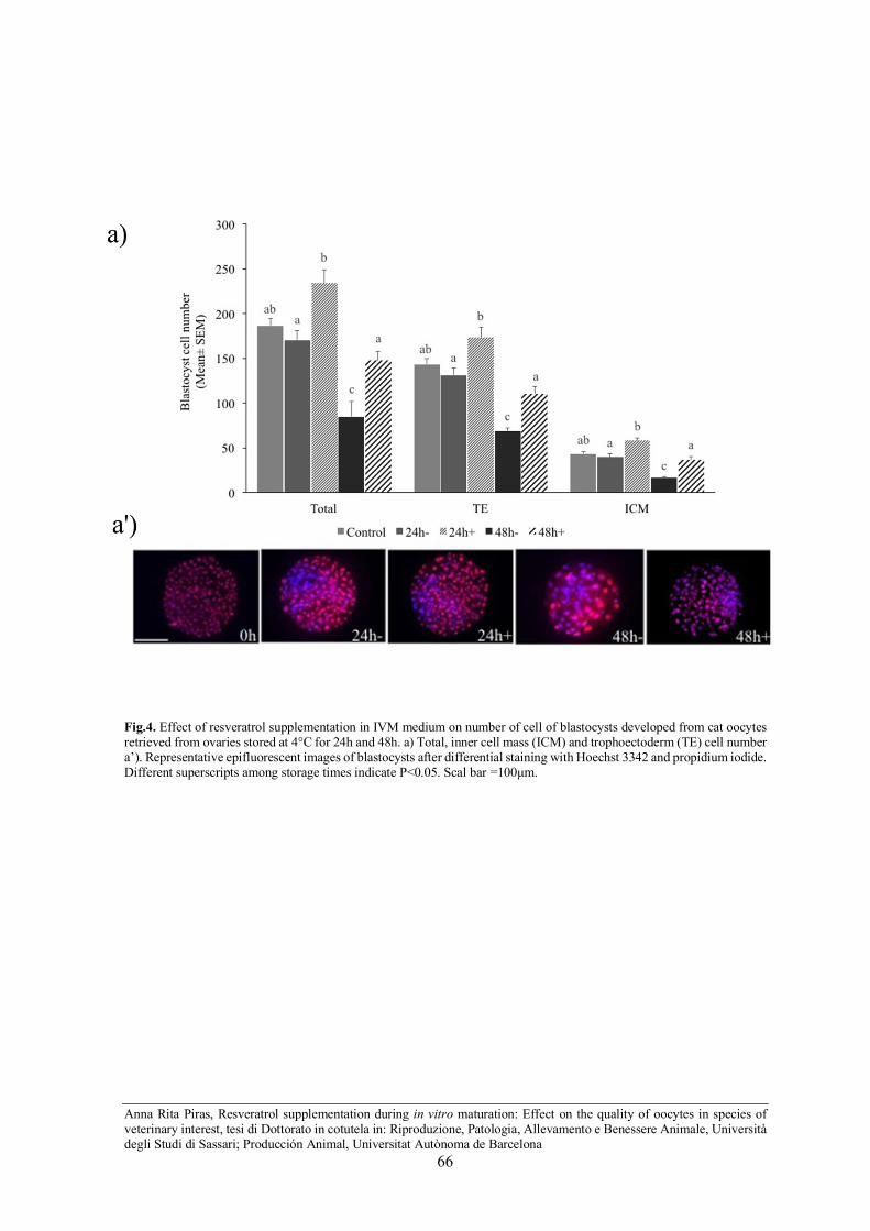
Anna Rita Piras, Resveratrol supplementation during in vitro maturation: Effect on the quality of oocytes in species of veterinary interest, tesi di Dottorato in cotutela in: Riproduzione, Patologia, Allevamento e Benessere Animale, Università degli Studi di Sassari; Producción Animal, Universitat Autònoma de Barcelona
66
Fig.4. Effect of resveratrol supplementation in IVM medium on number of cell of blastocysts developed from cat oocytes retrieved from ovaries stored at 4°C for 24h and 48h. a) Total, inner cell mass (ICM) and trophoectoderm (TE) cell number a’). Representative epifluorescent images of blastocysts after differential staining with Hoechst 3342 and propidium iodide. Different superscripts among storage times indicate P<0.05. Scal bar =100µm.

Anna Rita Piras, Resveratrol supplementation during in vitro maturation: Effect on the quality of oocytes in species of veterinary interest, tesi di Dottorato in cotutela in: Riproduzione, Patologia, Allevamento e Benessere Animale, Università degli Studi di Sassari; Producción Animal, Universitat Autònoma de Barcelona
67
DISCUSSION
In the present study, we explored the potential beneficial effect of resveratrol addition to the
in vitro maturation medium on embryonic development, after IVF, of cat oocytes recovered
from ovaries stored at 4°C for 24 and 48 h.
The main findings of the addition of resveratrol to maturation medium of cat oocytes were:
a) increased the GSH content and reduced the ROS levels of in vitro matured oocytes after
24h and 48h of ovary storage b) improved blastocyst rate after IVF of oocytes retrieved from
ovaries stored for 48h and c) increased the blastocyst cell numbers in both 24 and 48 h
groups.
Low temperature (4°C) is considered the most suitable for temporary storage of feline
ovarian tissue (Naoi et al., 2007; Luu et al., 2014). However, when the ovaries refrigeration
exceeded 24h embryonic development markedly declined (Wolfe and Wildt, 1996; Cocchia
et al., 2015; Piras et al., 2018). In the current study, the presence of resveratrol during
maturation not only had a positive effect on the in vitro embryo production after 48h of
ovaries storage, but also supported oocyte developmental competence with percentages of
blastocyst formation comparable to that obtained from fresh control oocytes.
However, we found that the supplementation of IVM with Resv did not affect oocyte meiotic
competence; this finding is consistent with the results of other authors in different species
reporting the absence of positive effect of resveratrol on in vitro nuclear maturation (Kwak
et al., 2012; Mukherjee et al., 2014; Sprícigo et al., 2017).
To the best of our knowledge, only one study explored the effect of resveratrol in the
domestic cat. In particular, the Authors reported that percentages of cleaved embryos and
ability to form more advanced embryo stages (48.7±6.4 %; 6.9 %, respectively) were
improved (p < 0.05) when immature cat oocytes were exposed to Resv before vitrification
compared to not exposed oocytes (17.7± 2.5%; 0%, respectively) (Comizzoli et al., 2009).

Anna Rita Piras, Resveratrol supplementation during in vitro maturation: Effect on the quality of oocytes in species of veterinary interest, tesi di Dottorato in cotutela in: Riproduzione, Patologia, Allevamento e Benessere Animale, Università degli Studi di Sassari; Producción Animal, Universitat Autònoma de Barcelona
68
The rationale for the resveratrol treatment derived from the results of our previous research
which highlighted the involvement of the oxidative stress in the loss of oocyte
developmental competence after ovaries storage for more than 24h (Piras et al., 2018).
Indeed, a low in vitro embryo production after 48h ovary storage was associated with a high
level of ROS and lipid peroxidation. of IVM oocytes (Piras et al., 2018).
Resveratrol is a strong antioxidant which maintains the levels of antioxidant enzymes such
as glutathione peroxidase (GPx), superoxide dismutase (SOD), and catalase (CAT) (Robb et
al., 2008; Wang et al., 2014) and improves the distribution and function of mitochondria
(Gerhart-Hines et al., 2007; Price et al., 2013).
Different studies in other species underlined the beneficial impact of resveratrol addition
during IVM on the developmental potential of oocytes after fertilization or artificial
activation, by increasing their intracellular GSH and decreasing ROS content (Galeati and
Spinaci, 2015).
GSH is the main non-protein sulphydryl compound in oocytes and embryos protecting them
from oxidative damage induced by ROS attack. The presence of high GSH content in
matured oocytes is a marker of cytoplasm maturity and it is associated with high in vitro
blastocyst production in several species ( De Matos and Fumus, 2000; De Matos et al., 2002,
2003; Li et al., 2014).
Recent studies have been addressed to improve transport and culture medium conditions
with the aim to enhance in vitro embryo production after cold storage of cat ovaries. The
study of Luu et al. (2013) found that the addition of relaxin to the in vitro maturation medium
ameliorated the blastocyst yield after cat ovaries storage at 4°C for 24h (Luu et al., 2013).
In fact, the rate of blastocyst formation from oocytes matured with relaxin (16.0%) was
higher than that from oocytes matured without relaxin (5.9%).
Moreover, superoxide dismutase (SOD) supplementation to the transport medium of cat
ovaries positively affected oocyte quality and reduced cellular apoptosis (Cocchia et al.,
2015).
Anna Rita Piras, Resveratrol supplementation during in vitro maturation: Effect on the quality of oocytes in species of veterinary interest, tesi di Dottorato in cotutela in: Riproduzione, Patologia, Allevamento e Benessere Animale, Università degli Studi di Sassari; Producción Animal, Universitat Autònoma de Barcelona
69
The presence of SOD during 48h and 72h storage supported the development to blastocyst
stage (7% and 4% blastocysts/total oocytes respectively) whereas in the SOD-free groups
embryos did not developed to blastocyst.
In the current study, the percentage of blastocyst yield obtained after addition of resveratrol
after 24h (24%) and 48h (18%) was higher than those of the above-mentioned studies (Luu
et al., 2014; Cocchia et al., 2015).
In conclusion, resveratrol treatment during in vitro maturation of cat oocytes recovered from
ovaries stored at 4°C for 48h enhanced in vitro embryo production. These data provide a
basis for extending the possibility of ovary collection and storage, especially for feline wild
species, which die in geographic areas far from specialized laboratories.

Anna Rita Piras, Resveratrol supplementation during in vitro maturation: Effect on the quality of oocytes in species of veterinary interest, tesi di Dottorato in cotutela in: Riproduzione, Patologia, Allevamento e Benessere Animale, Università degli Studi di Sassari; Producción Animal, Universitat Autònoma de Barcelona
70
REFERENCES
Bogliolo L, Ariu F, Leoni G, Uccheddu S, Bebbere D. 2011.High hydrostatic pressure
treatment improves the quality of in vitro produced ovine blastocysts. Reproduction, Fertility
and Development 23: 809–817.
Bogliolo L, Leoni GG, Ledda S, Naitana S, Zedda M, Carluccio A, Pau S. 2001.
Intracytoplasmic sperm injection of in vitro matured oocytes of domestic cats with frozen-
thawed epididymal spermatozoa. Theriogenology 56: 955-967.
Brooks TM, Mittermeier RA, Mittermeier CG, Da Fonseca GAB, Rylands AB, Konstant
WR, Flick P, Pilgrim J, Oldfield S, Magin G, Hilton-Taylor C. 2002. Habitat Loss and
Extinction in the Hotspots of Biodiversity. Conservation Biology 16: 909–923.
Cocchia N, Corteggio A, Altamura G, Tafuri S, Rea S, Rosapane I, Sica A, Landolfi F, Ciani
F. 2015. The effects of superoxide dismutase addition to the transport medium on cumulus-
oocyte complex apoptosis and IVF outcome in cats (Felis catus). Reproductive Biology 15:
56–64.
Cocchia N, Tafuri S, Abbondante L, Meomartino L, Esposito L, Ciani F. 2015. Assisted
Reproductive Technologies in Safeguard of Feline Endangered Species. Intech Open.
Combelles CMH, Gupta S, Agarwal. 2009. A Could oxidative stress influence the in vitro
maturation of oocytes? Reprod Biomed Online 18: 864–880.
Comizzoli P, Wildt DE, Pukazhenthi BS. 2009. In vitro compaction of germinal vesicle
chromatin is beneficial to survival of vitrified cat oocytes. Reproduction in Domestic
Animals 44: 269–274.

Anna Rita Piras, Resveratrol supplementation during in vitro maturation: Effect on the quality of oocytes in species of veterinary interest, tesi di Dottorato in cotutela in: Riproduzione, Patologia, Allevamento e Benessere Animale, Università degli Studi di Sassari; Producción Animal, Universitat Autònoma de Barcelona
71
De Matos DG and Fumus CC. The importance of having high glutathione (GSH) level after
bovine in vitro maturation on embryo development: effect of ß-mercaptoethanol, cysteine
and cystine. 2000. Theriogenology 53: 761–771.
De Matos DG, Gasparrini B, Pasqualini SR, Thompson JG. 2002. Effect of glutathione
synthesis stimulation during in vitro maturation of ovine oocytes on embryo development
and intracellular peroxide content. Theriogenology 57: 1443–1451.
De Matos DG, Nogueira D, Cortvrindt R, Herrera C, Adriaenssens T, Pasqualini RS, Smitz
J. 2003. Capacity of adult and prepubertal mouse oocytes to undergo embryo development
in the presence of cysteamine. Molecular Reproduction and Development 64: 214–218.
De Oliveira MR, Nabavi SF, Manayi A, Daglia M, Hajheydari Z, Nabavi SM. 2016.
Resveratrol and the mitochondria: From triggering the intrinsic apoptotic pathway to
inducing mitochondrial biogenesis, a mechanistic view. Biochimica et Biophysica Acta
1860: 727–745.
Evecen M, Cirit Ü, Demir K, Karaman E, Hamzaoǧlu AI, Bakirer G. 2009. Developmental
competence of domestic cat oocytes from ovaries stored at various durations at 4 °C
temperature. Animal Reproduction Science 116: 169–172.
Galeati G and Spinaci M. Resveratrol from red grapes: an useful agent for oocyte maturation
and subsequent embryonic development. 2015. Austin Journal of In vitro Fertilization 2: 1–
3.
Gerhart-Hines Z, Rodgers JT, Bare O, Lerin C, Kim SH, Mostoslavsky R, Alt FW, Wu Z,
Puigserver P. 2007. Metabolic control of muscle mitochondrial function and fatty acid
oxidation through SIRT1/PGC-1α. The EMBO Journal 26: 1913–1923.
IUCN 2018. The IUCN Red List of Threatened Species.Version 2018, http://
www.iucnredlist.org.

Anna Rita Piras, Resveratrol supplementation during in vitro maturation: Effect on the quality of oocytes in species of veterinary interest, tesi di Dottorato in cotutela in: Riproduzione, Patologia, Allevamento e Benessere Animale, Università degli Studi di Sassari; Producción Animal, Universitat Autònoma de Barcelona
72
Johnston LA, Donoghue AM, O’Brien SJ, Wildt DE. 1991. Rescue and maturation in vitro
of follicular oocytes collected from nondomestic Felid Species. Biol Reprod 45: 898–906.
Kwak SS, Cheong SA, Jeon Y, Lee E, Choi KC, Jeung EB, Hyun SH. 2012. The effects of
resveratrol on porcine oocyte in vitro maturation and subsequent embryonic development
after parthenogenetic activation and in vitro fertilization. Theriogenology 78: 86–101.
Li XX, Lee KB, Lee JH, Kim KJ, Kim EY, Han KW, Park KS, Yu J, Kim MK. 2014.
Glutathione and cysteine enhance porcine preimplantation embryo development in vitro
after intracytoplasmic sperm injection. Theriogenology 81: 309–314.
Luu VV, Hanatate K, Tanihara F, Sato Y, Do LTK, Taniguchi M, Otoi T. 2013. The effect
of relaxin supplementation of in vitro maturation medium on the development of cat oocytes
obtained from ovaries stored at 4°C. Reproductive Biology 13: 122–126.
Luu VV, Namula Z, Do LTK, Sato Y, Taniguchi M, Karja NWK, Otoi T. 2014. Nuclear
status and DNA fragmentation of oocytes from porcine, bovine and feline ovaries stored at
4°C for 5 days. CryoLetters 35: 48–53.
McGeoch MA, Butchart SHM, Spear D, Marais E, Kleynhans EJ, Symes A, Chanson J and
Hoffmann M. 2010. Global indicators of biological invasion: Species numbers, biodiversity
impact and policy responses. Diversity and Distributions 16: 95–108.
Mukherjee A, Malik H, Saha AP, Dubey A, Singhal DK, Boateng S, Saugandhika S, Kumar
S, De S, Guha SK, D Malakar. 2014. Resveratrol treatment during goat oocytes maturation
enhances developmental competence of parthenogenetic and hand-made cloned blastocysts
by modulating intracellular glutathione level and embryonic gene expression. Journal of
Assisted Reproduction and Genetics 31: 229–239.
Naoi H, Otoi T, Shimamura T, Karja NWK, Agung B, Shimizu R, Taniguchi M and Nagai
T. 2007. Developmental competence of cat oocytes from ovaries stored at various
temperature for 24 h. The Journal of Reproduction and Development 53: 271–277.

Anna Rita Piras, Resveratrol supplementation during in vitro maturation: Effect on the quality of oocytes in species of veterinary interest, tesi di Dottorato in cotutela in: Riproduzione, Patologia, Allevamento e Benessere Animale, Università degli Studi di Sassari; Producción Animal, Universitat Autònoma de Barcelona
73
Oldfield ML. 1988. Threatened mammals affected by human exploitation of the female-
offspring bond. Conservation Biology 2: 260–274.
Otoi T, Murakami M, Ooka A, Karja NWK, Suzuki T. 2001. Effects of size and storage
temperature on meiotic competence of domestic cat oocytes. Veterinary Record 148: 116–
118.
Parmesan C. Ecological and evolutionary responses to recent climate change. 2006. Annual
Review of Ecology, Evolution, and Systematics 37: 637–669.
Piras AR, Burrai G Pietro, Ariu F, Falchi L, Zedda MT, Pau S, Gadau SD, Antuofermo E,
Bebbere D, Ledda S, Bogliolo L. 2018. Structure of preantral follicles, oxidative status and
developmental competence of in vitro matured oocytes after ovary storage at 4 °C in the
domestic cat model. Reproductive Biology and Endocrinology 16: 1–14.
Pope CE, Gomez MC, Dresser LD. 2006. In vitro embryo production and embryo transfer
in domestic and non-domestic cats. Theriogenology 66: 1518–1524.
Pope CE, Gomez MC, King AL, Harris RF, Dresser BL. 2003. Embryos produced in vitro
after recovery of oocytes from cat ovaries stored at 4 °C for 24–28 h retain the competence
to develop into live kittens after transfer to recipients. Theriogenology 59: 308.
Pope CE. Embryo technology in conservation efforts for endangered felids. 2000.
Theriogenology 53: 163–174.
Price NL, Gomes AP, Ling AJY, Duarte F V, Martin-Montalvo A, North BJ, Agarwal B, Ye
L, Ramadori G, Teodoro JS, Hubbard BP, Varela AT, Davis JG, Varamini B, Hafner A,
Moaddel R, Rolo AP, Coppari R, Palmeira CM, de Cabo R, Baur JA, Sinclair DA. 2013.
SIRT1 is required for AMPK activation and the beneficial effects of resveratrol on
mitochondrial function. Cell Metab. 15: 675–690.
Anna Rita Piras, Resveratrol supplementation during in vitro maturation: Effect on the quality of oocytes in species of veterinary interest, tesi di Dottorato in cotutela in: Riproduzione, Patologia, Allevamento e Benessere Animale, Università degli Studi di Sassari; Producción Animal, Universitat Autònoma de Barcelona
74
Prochowska S, Niżański W, Partyka A, Kochan J, Młodawska W, Nowak A, Migdał A,
Skotnicki J, Grega T, Pałys M. 2017. Selected methods of in vitro embryo production in
felids - A review. Animal Science Papers and Reports 35: 361–377.
Robb EL, Page MM, Wiens BE and Stuart JA .2008. Molecular mechanisms of oxidative
stress resistance induced by resveratrol: Specific and progressive induction of MnSOD.
Biochemical and Biophysical Research Communications 367: 406–412.
Root TL, Price JT, Hall KR, Schneider CR, Pounds JA. 2003. Fingerprints of global
warming on wild animals and plants. Nature 421: 57–60.
Sananmuanga T, Tharasanit T, Nguyen C, Phutikanit N, Techakumphu M. 2011. Culture
medium and embryo density influence on developmental competence and gene expression
of cat embryos. Theriogenology 75: 1708–1719.
Smith KF, Acevedo-Whitehouse K, Pedersen AB. 2009. The role of infectious diseases in
biological conservation. Animal Conservation 12: 1–12.
Sovernigo TC, Adona PR, Monzani PS, Guemra S, Barros FDA, Lopes FG, Leal CLV. 2017.
Effects of supplementation of medium with different antioxidants during in vitro maturation
of bovine oocytes on subsequent embryo production. Reproduction in Domestic Animals
52: 561–569.
Sprícigo JF, Morató R, Arcarons N, Yeste M, Dode MA, López-Bejar M, Mogas T. 2017.
Assessment of the effect of adding L-carnitine and/or resveratrol to maturation medium
before vitrification on in vitro matured calf oocytes. Theriogenology 89: 47–57.
Takeo S, Sato D, Kimura K, Monji Y, Kuwayama T, Kawahara-Miki R, Iwata H. 2014.
Resveratrol improves the mitochondrial function and fertilization outcome of bovine
oocytes. Journal of Reproduction and Development 60: 92–99.

Anna Rita Piras, Resveratrol supplementation during in vitro maturation: Effect on the quality of oocytes in species of veterinary interest, tesi di Dottorato in cotutela in: Riproduzione, Patologia, Allevamento e Benessere Animale, Università degli Studi di Sassari; Producción Animal, Universitat Autònoma de Barcelona
75
Tsutsui T, Wada M, Anzai M, Hori T. 2003. Artificial insemination with frozen epididymal
sperm in cats. Theriogenology 65: 397–399.
Veraguas D, Echeverry D, Castro FO, Rodriguez-Alvarez L. 2017. Applied Biotechnologies
in the Conservation of Wild Felids: In Vitro Embryo Production and Cellular Regenerative
Therapies. Big Cats.
Wang F, Tian X, Zhang L, He C, Ji P, Li Y, Tan D and Liu G. 2014. Beneficial effect of
resveratrol on bovine oocyte maturation and subsequent embryonic development after in
vitro fertilization. Fertility and Sterility 101: 577–586.
Wolfe BA, Wildt DE. 1996. Development to blastocysts of domestic cat oocytes matured
and fertilized in vitro after prolonged cold storage. Journal of Reproduction and Fertility
106: 135-141.

Anna Rita Piras, Resveratrol supplementation during in vitro maturation: Effect on the quality of oocytes in species of veterinary interest, tesi di Dottorato in cotutela in: Riproduzione, Patologia, Allevamento e Benessere Animale, Università degli Studi di Sassari; Producción Animal, Universitat Autònoma de Barcelona
76
Chapter 5:
STUDY II
Resveratrol supplementation during in vitro maturation: Effect on
developmental competence of prepubertal goat oocytes selected by brilliant
cresyl blue staining
Accepted article
Journal of Reproduction and Development J Reprod Dev. 2018 Dec 29. doi: 10.1262/jrd.2018-077

Anna Rita Piras, Resveratrol supplementation during in vitro maturation: Effect on the quality of oocytes in species of veterinary interest, tesi di Dottorato in cotutela in: Riproduzione, Patologia, Allevamento e Benessere Animale, Università degli Studi di Sassari; Producción Animal, Universitat Autònoma de Barcelona
77
Chapter 6:
STUDY III
Resveratrol supplementation during in vitro maturation of oocytes under
cadmium exposure: Effect on fertilization outcome in the ovine model.

Anna Rita Piras, Resveratrol supplementation during in vitro maturation: Effect on the quality of oocytes in species of veterinary interest, tesi di Dottorato in cotutela in: Riproduzione, Patologia, Allevamento e Benessere Animale, Università degli Studi di Sassari; Producción Animal, Universitat Autònoma de Barcelona
78
ABSTRACT
Background: Cadmium (Cd) is a highly toxic heavy metal with adverse effect on female
reproduction. In vitro studies demonstrated that Cd exposure during maturation impaired
oocyte fertilization by inducing mitochondria hyper-activation and oxidative stress.
Aim: to investigate the effect of resveratrol (Resv) supplementation during in vitro
maturation on the fertilization outcome of prepubertal sheep oocytes under Cd exposure.
Methods: Ovaries collected from slaughterhouse-derived ovaries of prepubertal sheep were
exposed to 2 µM Cd and in vitro matured in the presence of 0 µM (Cd), 1 µM (Cd-Resv 1
µM) and 2 µM (Cd-Resv 2 µM) resveratrol. Oocytes matured in absence of Cd were used as
control (CTR). Fertilization outcomes, cortical granules (CGS) and mitochondria (mt)
distribution, mt activity and reactive oxygen species (ROS) level were evaluated.
Results: Oocytes of CTR, Cd-Resv 1 µM and Cd-Resv 2 µM groups had higher normal
fertilization and lower polyspermic fertilization rates compared to Cd group (P<0.05). The
percentage of metaphase II (MII) oocytes with CGs distributed in the cortex of the oocytes
was higher (P<0.05) in control and Cd-Resv 1µM groups than Cd group. The percentage of
MII oocytes that exhibit a homogeneous mitochondria distribution (normal distribution)
throughout the cytoplasm was higher in CTR, Cd-Resv 1 µM and Cd-Resv 2 µM groups
than Cd group (P<0.05). Lower activity (P<0.05) of mitochondria was recorded in CTR and
Cd-Resv 1 µM oocytes compared to oocytes matured under Cd exposure in absence of Resv.
The intracellular ROS levels were lower in CTR, Cd-Resv 1 µM and Cd-Resv 2 µM groups
than Cd group.
Conclusion: Resveratrol addition to the IVM medium at both 1 µM and 2 µM concentrations
protected oocytes against the toxic effect of Cd on in vitro fertilization of oocytes by
preventing inadequate distribution of cortical granules and mitochondria, dysfunction of
mitochondria and oxidative stress.

Anna Rita Piras, Resveratrol supplementation during in vitro maturation: Effect on the quality of oocytes in species of veterinary interest, tesi di Dottorato in cotutela in: Riproduzione, Patologia, Allevamento e Benessere Animale, Università degli Studi di Sassari; Producción Animal, Universitat Autònoma de Barcelona
79
INTRODUCTION
Anthropogenic activities have increased environmental toxicants pollutants levels in
terrestrial and aquatic ecosystems. Among inorganic pollutants, heavy metals (HMs), plays
a major role as they can affects reproductive performances and are associated with infertility
in human and animals (Ma et al., 2018).
Cadmium (Cd) is a highly toxic heavy metal of particular concern for human and animal
health due to its ubiquitous occurrence in air, water, soil, vegetables, food and feed.
Anthropogenic sources of Cd include combustion of fossil fuels, waste incineration, leachate
from landfill sites mining, smelting, rubber processing, galvanization process, nickel-Cd
batteries, plastic stabilizer and pigment (Flora and Agrawal, 2017). Cd is also a constituent
of pesticides and fertilizer (Järup, 2003) and is one of the major components of cigarette
smoke (Flora and Agrawal, 2017). Its long biological half-life (15-30 years) and its low rate
of excretion from the body cause its accumulation over time in organs and tissue.
Several studies underlined the negative effect of this metal on female reproduction
(Thompson and Bannigan, 2008). Long-term exposure to Cd damages reproductive system
and affects fertility (Thompson and Bannigan, 2008; Flora and Agrawal, 2017).
Bioaccumulation of Cd has been found in the ovaries and follicular fluid of both humans
and animals (Zenzes et al., 1995; Piasek et al., 2001; Martino et al., 2017). It has been shown
that Cd had a direct effect on the ovaries of hamster, resulting in the reduction of the ovarian
weight, and necrosis (Thompson and Bannigan, 2008). Furthermore, ovarian steroidogenesis
and ovulation was greatly altered by Cd exposure in rat (Zhang and Jia, 2007; Thompson
and Bannigan, 2008; Zhang et al., 2008). Cadmium influenced follicular development by
promoting follicular atresia in mouse (Wang et al., 2015) and inducing apoptosis of
granulosa cells of chicken ovarian follicles (Jia et al., 2011). The suppression of hyaluronic
acid synthesis and the reduction of FSH-induced cumulus cells expansion has been also
reported in porcine oocytes (Vršanská et al., 2003; Mlynarcikova et al., 2005) following Cd
exposure.

Anna Rita Piras, Resveratrol supplementation during in vitro maturation: Effect on the quality of oocytes in species of veterinary interest, tesi di Dottorato in cotutela in: Riproduzione, Patologia, Allevamento e Benessere Animale, Università degli Studi di Sassari; Producción Animal, Universitat Autònoma de Barcelona
80
By using in vitro models, previous studies investigated the influence of Cd on the female
gamete. Cd exposure have been proved to modify the morphology of buffalo oocytes (Nandi
et al., 2010) and to affect oocyte and cumulus cell viability in both buffalo and ovine oocytes
(Leoni et al., 2002; Nandi et al., 2010). Other evidences indicated a deleterious effect of Cd
exposure on the in vitro nuclear maturation of oocytes in sheep (Leoni et al., 2002), buffalo
(Nandi et al., 2010) and bovine (Ahmad and Khal, 2018 b). The disruption of periodic
changes of the Maturation Promoting Factor (MPF) activity by increasing mRNA and
protein expressions of MPF-related genes has been suggested as one of the mechanism
involved in the inhibition of meiotic progression of metaphase I mouse oocyte exposed to
Cd (Liu et al., 2018).
The exposure of ovine and bovine oocytes to Cd during in vitro maturation impaired in vitro
fertilization and embryo development (Leoni et al., 2002; Nandi et al., 2010; Martino et al.,
2017; Ahmad and Khal, 2018 b). In a recent study in juvenile and adult sheep, Martino et al.
(2017) reported age-related bioaccumulation of Cd, with detectable harmful levels even in
ovarian tissues of juvenile sheep. These Authors found that exposure to nanomolar
concentration of Cd during in vitro maturation reduced fertilization of oocyte retrieved from
both juvenile and adult sheep with a slight increase in the number of abnormally fertilized
oocytes (Martino et al., 2017). The deleterious effect of Cd on fertilization has been related
to mitochondria hyper-activation associated with two specific indicators of oxidative stress,
such as reactive oxygen species (ROS) levels and liporeroxidation (Martino et al., 2017).
We hypothesized that resveratrol could counteract oxidative stress and investigated the
effect of resveratrol supplementation during in vitro maturation of prepubertal sheep oocytes
under Cd exposure on the fertilization outcome, cortical granules distribution and
bioenergetic/redox status of oocytes.

Anna Rita Piras, Resveratrol supplementation during in vitro maturation: Effect on the quality of oocytes in species of veterinary interest, tesi di Dottorato in cotutela in: Riproduzione, Patologia, Allevamento e Benessere Animale, Università degli Studi di Sassari; Producción Animal, Universitat Autònoma de Barcelona
81
MATERIAL AND METHODS
Chemicals
All chemicals, in this study, were purchased from Sigma Chemical CO. (St. Louis, MO,
USA) unless stated otherwise.
Oocyte collection
Ovaries from prepubertal sheep (Ovis Aries, 30-40 days of age) were recovered from local
abattoirs and transported within 3h to the laboratory in Phosphate Buffered Saline (PBS)
with penicillin (100mg/mL) and streptomycin (100mg/mL) at 34-37°C. Ovaries were
washed three times in PBS and processed by the slicing procedure for cumulus oocytes
complexes (COCs) retrieval in dissection medium (DM; 25µM Hepes-buffered TCM199)
supplemented with 0.1% (w/v) polyvinyl alcohol (PVA) and antibiotics (100 µg/mL of
penicillin and streptomycin). Only COCs with several intact cumulus cells layers and
homogenous cytoplasm were selected for in vitro maturation (IVM).
In vitro maturation (IVM)
Cumulus oocytes complexes were matured in vitro in conventional maturation medium
(CMM) consisting of TCM 199 supplemented with 10% heat-treated estrous sheep serum
(ESS), 0.36 mM pyruvate, FSH 1 IU / ml and LH 1 IU / ml (Pluset; Bio98, Milan, Italy)
under mineral oil, in 4-well dishes (Nunc Cell Culture, Thermo Fisher Scientific, Waltham,
Massachusetts, USA) in a humidified atmosphere of 5% CO2, at 38.5°C.

Anna Rita Piras, Resveratrol supplementation during in vitro maturation: Effect on the quality of oocytes in species of veterinary interest, tesi di Dottorato in cotutela in: Riproduzione, Patologia, Allevamento e Benessere Animale, Università degli Studi di Sassari; Producción Animal, Universitat Autònoma de Barcelona
82
In vitro fertilization (IVF)
After maturation, COCs were partially stripped of the granulosa cells and co-incubated with
fresh ram semen (1 x 10⁶ sperm cells/ml) in SOF medium (synthetic oviductal fluid, Tervit
et al., 1972) + 2% ESS + 1 𝜇 g/mL heparin + 1 𝜇 g/mL hypotaurine, under mineral oil in a
humidified atmosphere of 5% CO2, at 38.5 °C for 16h.
All experimental procedures were performed with the semen of one single ram of proven
fertility.
Fertilization assessment
After IVF, the presumptive zygotes were denuded from cumulus cells by aspiration in and
out of finely-drawn glass pipettes, fixed with ethanol and stained with 1 µM Hoechst 33342
(Comizzoli et al., 2001). They were thereafter, placed on a slide and classified (Martino et
al., 2017) under an inverted epifluorescent microscope (Olympus IX70, Italy).
The presence of 2 pronuclei (2PN) and two polar bodies (2PB) indicated a normally fertilized
oocyte, the presence of more than two pronuclei (>2PN) and two polar bodies (2PB) was
designated polyspermic fertilization, one PN and a sperm head (SH) that failed chromatin
decondensation and 2PB or one PN and MII plate and 2PB were considered as abnormal
fertilization. Oocyte with the Metaphase II (MII) plate with the first PB indicated unfertilized
matured oocytes (representative image in Fig.1).
Assessment of cortical granules distribution
Analysis of cortical granules (CGs) was performed according the methodology of Hosseini
et al. (Hosseini et al., 2012). After IVM, MII oocytes from each experimental group (n=20-
25) were incubated with 0.5% pronase on a warm plate (38.5°C) to digest zona pellucida
and fixed with 3.7% paraformaldehyde in PBS at RT for 30 minute. After fixation, oocytes
were washed three times in PBS containing 0.3%BSA and 100-mM glycine.

Anna Rita Piras, Resveratrol supplementation during in vitro maturation: Effect on the quality of oocytes in species of veterinary interest, tesi di Dottorato in cotutela in: Riproduzione, Patologia, Allevamento e Benessere Animale, Università degli Studi di Sassari; Producción Animal, Universitat Autònoma de Barcelona
83
Oocytes were permeabilized in 0.1% Triton X-100 in PBS for 5 minutes, and incubated in
100 µg/mL Alexa Fluor 488-conjugated Lectin PNA (Molecular probes, Invitrogen) in PBS
for 30 minutes in the dark. The unbounded dye was removed by 3 washes in PBS containing
0.3% BSA, 100-mM glycine and 0.01% Triton X-100. Finally, oocytes were mounted on
glass slides with 2.5 mg/mL Hoechst 33342 in PBS and glycerol solution (3:1 v/v), overlaid
with a coverslip supported by four droplets of vaseline and sealed with nail varnish.
The localization of CGs (excitation 490 nm) and chromatin configuration (excitation 361nm)
were evaluated with Leica TCS SP5 CLSM with LAS lite 170. Image software equipped
with a 405-nm diode laser and a multiphoton laser. The CGs configuration was classified as
previously reported (Takeo et al., 2014), with some modifications in: Pattern A: cortical
granules distributed in the oocyte cortical region; Pattern B: cortical granules diffused
throughout the oocyte cytoplasm (representative images Fig.2).
Evaluation of bioenergetic/oxidative status of oocytes
In order to elucidate the effect of resveratrol on bioenergetic/oxidative status of oocytes
matured under Cd exposure, oocytes underwent laser scanning confocal microscopy
(LSCM) analysis of mitochondria (mt) distribution and activity and intracellular ROS levels.
After IVM, MII oocytes from each experimental group (n=25-30) were subjected to a triple
staining with MitoTracker Orange CMTMRos (Molecular Probes, Inc., Eugene, OR, USA),
2′7′-dichlorodihydrofluorescein diacetate (H2DCF-DA, Cat n° D 6883) and Hoechst 33342
for detection of mt distribution and activity, intracellular reactive oxygen species levels and
chromatin configuration respectively (Succu et al., 2018). MitoTracker Orange CMTMRos
passively diffuses across the plasma membrane and is readily sequestered only by active mt
depending on their membrane potential. H2DCF-DA is a cell-permeable non-fluorescent
probe. Inside to the oocyte this probe is de-esterified and turns to highly fluorescent 2′,7′-
dichlorofluorescein due to oxidation by H2O2 (Succu et al., 2018).

Anna Rita Piras, Resveratrol supplementation during in vitro maturation: Effect on the quality of oocytes in species of veterinary interest, tesi di Dottorato in cotutela in: Riproduzione, Patologia, Allevamento e Benessere Animale, Università degli Studi di Sassari; Producción Animal, Universitat Autònoma de Barcelona
84
Briefly, the oocytes were washed three times in PBS with 0.1% bovine serum albumin (BSA)
and were incubated for 30 minutes in PBS with 3% BSA containing 200 nM MitoTracker
Orange CMTMRos, at 38.5°C under 5% CO2. After the first incubation, oocytes were wash
again in PBS with 0.1% BSA and incubated for 30 minutes in PBS with 0.1% BSA and 10
µM H2DCFDA. After washing in in PBS with 0.1% BSA oocytes were fixed for 60 minutes
at 38°C in 3% paraformaldehyde and stained for 5minutes with 1 µM Hoechst 33342
solution. Particular attention was paid to avoid sample exposure to the light during staining
to reduce photo-bleaching. Oocytes were stored at 4°C in the dark until their analysis.
The oocytes were placed on a slide and covered with a drop of a solution of PBS and glycerol
(3:1v/v), overlaid with a coverslip supported by four droplets of vaseline and sealed with
nail varnish.
The mitochondria distribution patterns classified as: normal: homogeneous mt distribution,
with small granulation spread throughout the cytoplasm; abnormal: mt distribution, with
large granulations located in specific cytoplasmic area (representative images in Fig.3).
Mitochondria and intracellular ROS level quantification were performed by a confocal laser-
scanning fluorescence microscope (Leica TCS SP5 CLSM), equipped with 543 nm He/Ne,
488 nm Argon and 405 nm diode lasers using an oil immersion 40x objective and recorded
on a host computer.
A helium/neon laser ray at 543 nm and an argon-ion laser ray at 488 nm were used to detect
the MitoTracker Orange CMTM and DCF fluorescence signals respectively.
In each individual oocyte MitoTracker Orange CMTMRos (excitation 554nm) and
H2DCFDA (excitation 460 nm) fluorescence intensities were measured at the equatorial
plane as described previously (Dell’Aquila et al., 2014) and microscope objective, pinhole,
filters, offset, gain, and laser potency were kept constant throughout the experiment.
Acquisition, storage and image analysis were made with the LAS lite 170 Image software.
The fluorescence intensity of the cytoplasm area of the oocyte was measured and reduced
by compensation for the background fluorescence.

Anna Rita Piras, Resveratrol supplementation during in vitro maturation: Effect on the quality of oocytes in species of veterinary interest, tesi di Dottorato in cotutela in: Riproduzione, Patologia, Allevamento e Benessere Animale, Università degli Studi di Sassari; Producción Animal, Universitat Autònoma de Barcelona
85
Experimental design
Cumulus oocytes complexes were randomly divided in four groups for IVM:
-control (CTR) group: oocytes were matured in CMM
-Cd- group: oocytes were matured in CMM medium supplemented with 2 µM CdCl2
-Cd-Resv 1 µM and Cd-Resv 2 µM groups: oocytes were matured in CMM medium
supplemented with 2 µM CdCl2 and 1 µM or 2 µM resveratrol.
The concentration of cadmium used in the present study was selected by referring to earlier
in vitro study in ovine specie (Leoni et al., 2002).
The Experiment 1 studied the effect of Resv supplementation (1 µM and 2 µM) to the
IVM medium on fertilization rates of oocytes exposed to Cd during maturation.
The Experiment 2, assessed: the CGs distribution, bioenergetic/oxidative status of
oocytes (mitochondria distribution and activity, and ROS levels) of in vitro matured
oocytes exposed to Cd
.

Anna Rita Piras, Resveratrol supplementation during in vitro maturation: Effect on the quality of oocytes in species of veterinary interest, tesi di Dottorato in cotutela in: Riproduzione, Patologia, Allevamento e Benessere Animale, Università degli Studi di Sassari; Producción Animal, Universitat Autònoma de Barcelona
86
EXPERIMENTAL DESIGN

Anna Rita Piras, Resveratrol supplementation during in vitro maturation: Effect on the quality of oocytes in species of veterinary interest, tesi di Dottorato in cotutela in: Riproduzione, Patologia, Allevamento e Benessere Animale, Università degli Studi di Sassari; Producción Animal, Universitat Autònoma de Barcelona
87
Statistical analysis
All statistical analyses were performed using Stata/IC 11.2 (StataCorp LP, USA).
Categorical data of fertilization, mitochondria and cortical granules distribution were
analyzed by chi-square test. Data of intracellular ROS levels and mitochondria activity were
not normally distributed and were analyzed by non-parametric Kruskal-Walli’s test.
P <0.05 was considered statistically significant. All the experiments were replicated at least
for two times.

Anna Rita Piras, Resveratrol supplementation during in vitro maturation: Effect on the quality of oocytes in species of veterinary interest, tesi di Dottorato in cotutela in: Riproduzione, Patologia, Allevamento e Benessere Animale, Università degli Studi di Sassari; Producción Animal, Universitat Autònoma de Barcelona
88
RESULTS
Experiment 1
Effect of resveratrol on fertilization of oocytes under Cd exposure
Results about in vitro fertilization of oocytes in the different experimental groups are
reported in Table 1. Representative images of normal, polyspermic and abnormal
fertilization are shown in Fig.1. Cadmium exposure during in vitro maturation of the oocytes
significantly (P<0.05) reduced the percentage of normally fertilized oocytes and increased
the polyspermic fertilization compared to control group. Resveratrol addition to the
maturation medium, at both 1 µM and 2 µM concentrations, enhanced normal fertilization
and reduced polyspermy (P<0.05) compared to Cd group. Normal and polyspermic
fertilization rates of oocytes treated with 1 µM and 2 µM Resv were similar to those of
control oocytes. No difference was found in the percentages of abnormal fertilized and
unfertilized oocytes among groups.
Table 1. Effect of resveratrol supplementation to IVM medium on fertilization of
prepubertal sheep oocytes under cadmiun-exposure
Groups N° of
oocytes
Fertilization N° (%)
Normal
(%)
Polyspermy
(%)
Abnormal
(%)
Unfertilized
(%)
Control 149 92 (61.7) a 36 (24.2) a 10 (6.7) 11 (7.4)
Cd 147 53 (36.0) b 66 (44.9) b 10 (6.8) 18 (12.3)
Cd-Resv1
µM 141 82 (58.2) a 37 (26.2) a
4 (2.8) 18 (12.8)
Cd-Resv2
µM 99 58 (58.6) a 23 (23.2) a
8(8.1) 10 (10.1) Different superscripts in the same column (a, b) indicate a statistical significant difference P<0.05 a 3 replicate trials were performed

Anna Rita Piras, Resveratrol supplementation during in vitro maturation: Effect on the quality of oocytes in species of veterinary interest, tesi di Dottorato in cotutela in: Riproduzione, Patologia, Allevamento e Benessere Animale, Università degli Studi di Sassari; Producción Animal, Universitat Autònoma de Barcelona
89
Fig.1. Representative image of nuclear configuration of prepubertal sheep oocytes 16h post-in vitro fertilization showing A) 2PN (normal fertilization), B) > 2PN (polispermy) C), 1PN and a sperm head (SH) with no signs of nuclear decondensation (abnormal fertilization). Scale bar= 25 µm.

Anna Rita Piras, Resveratrol supplementation during in vitro maturation: Effect on the quality of oocytes in species of veterinary interest, tesi di Dottorato in cotutela in: Riproduzione, Patologia, Allevamento e Benessere Animale, Università degli Studi di Sassari; Producción Animal, Universitat Autònoma de Barcelona
90
Experiment 2
To understand cadmium and resveratrol mechanism of action on oocyte fertilization, we
analyzed Cortical Granules (CG)s distribution, mitochondria distribution and activity, and
intracellular ROS levels in in vitro matured oocytes of prepubertal sheep.
Effect of resveratrol on cortical granules distribution of oocytes under Cd-exposure
Figure 2 shows representative images of the two-different patterns of CGs distribution.
Most of control oocytes (n=23/24, 95.8%) showed a distribution of the CGs under the oocyte
cortex (pattern A). Cd-exposure induced a significant reduction (P<0.05) of this pattern of
distribution (n=15/23, 65.2 %). The ratio of oocytes exposed to Cd with CGs distributed in
the peripheral region was higher (P<0.05) for oocytes matured with Resv 1 µM (n=21/23;
91.3%) and similar for oocytes matured with Resv 2 µM oocytes (n=17/21, 80.95%)
compared to those matured without Resv (Cd-group).
No differences were observed among resveratrol (Cd-Resv1 µM: n=21/23, 91.3%; Cd-Resv2
µM: n=17/21, 80.95%) and control groups (n=23/24, 95.8%).

Anna Rita Piras, Resveratrol supplementation during in vitro maturation: Effect on the quality of oocytes in species of veterinary interest, tesi di Dottorato in cotutela in: Riproduzione, Patologia, Allevamento e Benessere Animale, Università degli Studi di Sassari; Producción Animal, Universitat Autònoma de Barcelona
91
Fig.2. Effect of resveratrol supplementation to IVM medium on CGs distribution of prepubertal sheep oocytes under Cd-exposure. I) Representative images of CGs distribution patterns showing A) CGs distributed in the cortical region (pattern A); B) CGs diffused throughout the cytoplasm (pattern B). II) Percentage of MII oocytes with different pattern of CGs distribution. Numbers of examined oocytes /group are indicated at the bottom of each histogram column. Different superscript letters (a vs b) indicate a significant difference (P<0.05). Scale bar= 25 µm.

Anna Rita Piras, Resveratrol supplementation during in vitro maturation: Effect on the quality of oocytes in species of veterinary interest, tesi di Dottorato in cotutela in: Riproduzione, Patologia, Allevamento e Benessere Animale, Università degli Studi di Sassari; Producción Animal, Universitat Autònoma de Barcelona
92
Effect of resveratrol on bioenergetic/oxidative status of oocytes under Cd exposure
Figure 3 shows representative images of the mitochondria distribution in the oocyte
cytoplasm. After in vitro maturation 73.1% of CTR oocytes (n=19/26) showed a
homogeneous distribution of the mitochondria with small granulation spread throughout the
cytoplasm (normal distribution). Cd-exposure during maturation induced a decrease
(P<0.05) of normal mt distribution (n=14/32, 43.7%) compared to control. Cd-Resv1 µM
(n=27/32, 84.4%) and Cd- Resv2 µM (n=21/27, 77.8%) groups displayed a percentage of
normal mt distribution higher (P<0.05) than Cd-group and similar to CTR oocytes.
Mitochondria activity (103.1 ± 1.4 ADU) and ROS levels (122.4 ± 1.5 pixel/oocyte) were
significantly higher (P <0.05) in Cd-exposed oocytes compared to the CTR (67±0.6 ADU;
71.1±2.3 pixel/oocyte, respectively). Mt activity (79.3±1.1 ADU) and ROS level (74.4±1.3
pixel/oocyte) of oocytes exposed to Cd and treated with Resv 1 µM were lower (P<0.05)
than those without Resv and similar to CTR. Similar rate of mt activity was recorded
between Cd-Resv1 µM and Cd-Resv2 µM groups. In the oocytes treated with resveratrol 2
µM mt activity (83±0.9 ADU) was higher than CTR and similar to that of oocytes of Cd and
resveratrol groups. Cadmium exposure during maturation increased ROS level (122.4 ± 1.5
pixel/oocyte) compared to CTR oocytes. The ROS levels of the oocytes treated with
resveratrol 2 µM (96.6± 2.4 pixel/oocyte) were lower than Cd group and similar to CTR
(Fig.4).

Anna Rita Piras, Resveratrol supplementation during in vitro maturation: Effect on the quality of oocytes in species of veterinary interest, tesi di Dottorato in cotutela in: Riproduzione, Patologia, Allevamento e Benessere Animale, Università degli Studi di Sassari; Producción Animal, Universitat Autònoma de Barcelona
93
Fig.3. Effect of resveratrol supplementation to IVM medium on mt distribution of prepubertal sheep oocytes under Cd exposure. I) Representative images of mt distribution patterns after staining of oocytes showing homogeneous mitochondria distribution, with small granulation spread throughout the cytoplasm (normal distribution) and b) large mt granulations located in specific cytoplasmic area abnormal distribution). II) Percentage of oocytes with normal and abnormal mitochondria distribution. Numbers of examined oocytes/group are indicated at the bottom of each histogram column Different superscript letters (a vs b) indicate a significant difference (P<0.05). Scale bar=50 µm.
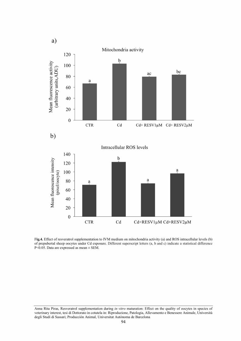
Anna Rita Piras, Resveratrol supplementation during in vitro maturation: Effect on the quality of oocytes in species of veterinary interest, tesi di Dottorato in cotutela in: Riproduzione, Patologia, Allevamento e Benessere Animale, Università degli Studi di Sassari; Producción Animal, Universitat Autònoma de Barcelona
94
Fig.4. Effect of resveratrol supplementation to IVM medium on mitochondria activity (a) and ROS intracellular levels (b) of prepubertal sheep oocytes under Cd exposure. Different superscript letters (a, b and c) indicate a statistical difference P<0.05. Data are expressed as mean ± SEM.
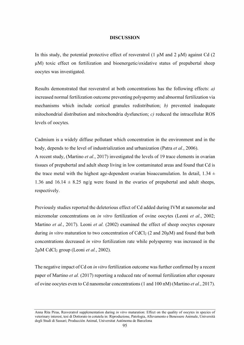
Anna Rita Piras, Resveratrol supplementation during in vitro maturation: Effect on the quality of oocytes in species of veterinary interest, tesi di Dottorato in cotutela in: Riproduzione, Patologia, Allevamento e Benessere Animale, Università degli Studi di Sassari; Producción Animal, Universitat Autònoma de Barcelona
95
DISCUSSION
In this study, the potential protective effect of resveratrol (1 µM and 2 µM) against Cd (2
µM) toxic effect on fertilization and bioenergetic/oxidative status of prepubertal sheep
oocytes was investigated.
Results demonstrated that resveratrol at both concentrations has the following effects: a)
increased normal fertilization outcome preventing polyspermy and abnormal fertilization via
mechanisms which include cortical granules redistribution; b) prevented inadequate
mitochondrial distribution and mitochondria dysfunction; c) reduced the intracellular ROS
levels of oocytes.
Cadmium is a widely diffuse pollutant which concentration in the environment and in the
body, depends to the level of industrialization and urbanization (Patra et al., 2006).
A recent study, (Martino et al., 2017) investigated the levels of 19 trace elements in ovarian
tissues of prepubertal and adult sheep living in low contaminated areas and found that Cd is
the trace metal with the highest age-dependent ovarian bioaccumulation. In detail, 1.34 ±
1.36 and 16.14 ± 8.25 ng/g were found in the ovaries of prepubertal and adult sheeps,
respectively.
Previously studies reported the deleterious effect of Cd added during IVM at nanomolar and
micromolar concentrations on in vitro fertilization of ovine oocytes (Leoni et al., 2002;
Martino et al., 2017). Leoni et al. (2002) examined the effect of sheep oocytes exposure
during in vitro maturation to two concentration of CdCl2 (2 and 20µM) and found that both
concentrations decreased in vitro fertilization rate while polyspermy was increased in the
2µM CdCl2 group (Leoni et al., 2002).
The negative impact of Cd on in vitro fertilization outcome was further confirmed by a recent
paper of Martino et al. (2017) reporting a reduced rate of normal fertilization after exposure
of ovine oocytes even to Cd nanomolar concentrations (1 and 100 nM) (Martino et al., 2017).
Anna Rita Piras, Resveratrol supplementation during in vitro maturation: Effect on the quality of oocytes in species of veterinary interest, tesi di Dottorato in cotutela in: Riproduzione, Patologia, Allevamento e Benessere Animale, Università degli Studi di Sassari; Producción Animal, Universitat Autònoma de Barcelona
96
In contrast to the findings of Leoni et al. (2002) these authors did not observe an increase in
polyspermic fertilization, possibly due to the lower concentrations of CdCl2 used. In buffalo
oocytes, the effect of different Cd concentrations (ranging from 27.3 nM to 54.60 µM) was
also assessed and a decline of fertilization and cleavage rates were observed starting from
the concentration of 1.0 µg/ml (5.5 µM) (Nandi et al., 2010).
Successful fertilization requires the correct function and distribution of cytoplasmic
organelles. Specifically, calcium release from the endoplasmic reticulum, CGs distribution
under oocyte oolema and proper exocytosis are crucial for inhibit polyspermic fertilization
and support normal embryonic development (Blerkom and Davis, 2007; Dumollard et al.,
2008; Calì et al., 2013). It has been demonstrated that Cd competes with calcium for protein-
binding sites disturbing intracellular calcium activity (Biagioli et al., 2008). Moreover, Cd
has adverse effects on microfilaments of oocyte cytoskeleton which drive CGs migration
(Prozialeck and Niewenhuis, 1991; Abbott and Ducibella, 2001).
Our results showed that the exposure to Cd during IVM disturbed CGs redistribution in the
cortex of the oocyte and that resveratrol was able to reverse this effect therefore avoiding
polyspermic fertilization. Confirming our findings, resveratrol improved the correct CG
distribution before in vitro fertilization of bovine oocytes (Takeo et al., 2014) and during in
vitro aging of porcine oocytes (Ma et al., 2015).
The protective effect of resveratrol on CGs migration and fertilization may be mediated by
the maintenance of the proper distribution of mitochondria during oocyte maturation (Ma et
al., 2015). It is well known that mitochondria distribution, function and interaction with
endoplasmic reticulum are involved in the process of CGs migration and exocytosis
(Blerkom and Davis, 2007; Dumollard et al., 2008; Calì et al., 2013).
In this respect, our results demonstrated that resveratrol addition in the culture medium
counteracted the adverse effect of Cd on mitochondria distribution and organization in the
oocyte cytoplasm.

Anna Rita Piras, Resveratrol supplementation during in vitro maturation: Effect on the quality of oocytes in species of veterinary interest, tesi di Dottorato in cotutela in: Riproduzione, Patologia, Allevamento e Benessere Animale, Università degli Studi di Sassari; Producción Animal, Universitat Autònoma de Barcelona
97
A prevention effect of resveratrol to the inadequate translocation of mitochondria has been
described in mouse oocyte exposed to methylglyoxal (MG), during in vitro maturation (Liu
et al., 2013). Oxidative stress may be another factor underlying the alteration of fertilization
induced by the Cd exposure (Martino et al., 2017). In this regard, Martino et al. (2017)
demonstrated that cumulus-oocyte mitochondria over-activity associated with an increase of
ROS levels and membrane lipid peroxidation were related to impaired in vitro fertilization
of ovine oocytes exposed to nanomolar Cd concentrations during maturation.
The findings of our study corroborated the negative impact of Cd on bionergetic/oxidative
status of oocytes as showed by the increased mitochondria activity and cytosolic ROS levels,
which occurred in oocytes under Cd exposure. We demonstrated the beneficial action of
resveratrol to alleviate oxidative stress induced by Cd and, even more important, this effect
was proved using micromolar concentration of Cd using a model of low competence
consisting of oocytes derived from prepubertal donors. This highlights the powerful
antioxidant properties of resveratrol and its potential use to improve the quality of oocytes.
The beneficial effect of resveratrol on oocytes redox status has been described in several
species (Galeati and Spinaci, 2015) in standard in vitro embryo production protocols or after
oocyte exposition to toxic substances (Huang et al., 2007; Chan, 2011; Liu et al., 2013). The
mechanism of resveratrol action included the reduction of ROS levels by up-regulation of
Sirtuin 1, which in turn modulated the activity of antioxidant enzymes, the increasing of
GSH ( Wang et al., 2014; Xia et al., 2017), the regulation of biosynthesis, degradation and
activity of mitochondria (Sato et al., 2014).
In conclusion, resveratrol is a useful therapeutic agent to counteract the toxic effect of Cd
on in vitro fertilization of oocytes by preventing inadequate distribution of cortical granules,
dysfunction of mitochondria and oxidative stress.
Further experiments are requiring to better understand the exact mechanisms underlying the
resveratrol effect on fertilization outcome.

Anna Rita Piras, Resveratrol supplementation during in vitro maturation: Effect on the quality of oocytes in species of veterinary interest, tesi di Dottorato in cotutela in: Riproduzione, Patologia, Allevamento e Benessere Animale, Università degli Studi di Sassari; Producción Animal, Universitat Autònoma de Barcelona
98
REFERENCES
Abbott AL and Ducibella T. 2001 Calcium and the control of mammalian cortical granule
exocytosis. Frontiers in Bioscience 6: 792–806.
Akar Y, Ahmad N and Khalid M. 2018. The effect of cadmium on the bovine in vitro oocyte
maturation and early embryo development. International Journal of Veterinary Science and
Medicine 6 3–7.
Biagioli M, Pifferi S, Ragghianti M, Bucci S, Rizzuto R and Pinton P. 2008. Endoplasmic
reticulum stress and alteration in calcium homeostasis are involved in cadmium-induced
apoptosis. Cell Calcium 43 184–195.
Van Blerkom J and Davis P. 2007. Mitochondrial signaling and fertilization. Molecular
Human Reproduction 13: 759–770.
Calì T, Ottolini D, Negro A and Brini M. 2013. Enhanced parkin levels favor ER-
mitochondria crosstalk and guarantee Ca 2 + transfer to Biochimica et Biophysica Acta
sustain cell bioenergetics 1832 :495–508.
Chan WH. 2011. Resveratrol protects against 2-bromopropane-induced apoptosis and
disruption of embryonic development in blastocysts 4991–5010.
Comizzoli P, Mermillod P, Congie Y, Chai N, Legendre X and Mauget R. 2001. Successful
in vitro production of embryos in the Red Deer (Cervus elaphus) and the Sika Deer (Cervus
nippon). Theriogenology 55; 649–659.
Dell’Aquila ME, Bogliolo L, Russo R, Martino NA, Filioli Uranio M, Ariu F, Amati F,
Sardanelli AM, Linsalata V, Ferruzzi MG, Cardinali A. Minervini F. 2014. Prooxidant
effects of verbascoside, a bioactive compound from olive oil mill wastewater, on in vitro
developmental potential of ovine prepubertal oocytes and bioenergetic/oxidative stress
parameters of fresh and vitrified oocytes. BioMed Research International 2014.

Anna Rita Piras, Resveratrol supplementation during in vitro maturation: Effect on the quality of oocytes in species of veterinary interest, tesi di Dottorato in cotutela in: Riproduzione, Patologia, Allevamento e Benessere Animale, Università degli Studi di Sassari; Producción Animal, Universitat Autònoma de Barcelona
99
Dumollard R, Campbell K, Halet G, Carroll J and Swann K. 2008. Regulation of cytosolic
and mitochondrial ATP levels in mouse eggs and zygotes. Developmental Biology 316:
431–440.
Flora SJS and Agrawal S. 2017. Arsenic, Cadmium, and Lead. Reproduction and
Developmental Toxicology 537-566.
Galeati G and Spinaci M. 2015. Resveratrol from Red Grapes: an useful agent for oocyte
maturation and subsequent embryonic development. Austin Journal of In vitro Fertilization
2 :1–3.
Hosseini SM, Asgari V, Ostadhosseini S, Hajian M, Piryaei A, Najarasl M and Nasr-
Esfahani MH. 2012. Potential applications of sheep oocytes as affected by vitrification and
in vitro aging. Theriogenology 77 :1741–1753.
Huang LH, Shiao NH, Hsuuw YD, Chan WH. 2007. Protective effects of resveratrol on
ethanol-induced apoptosis in embryonic stem cells and disruption of embryonic
development in mouse blastocysts. Toxicology 242 :109–122.
Järup L. 2003. Hazards of heavy metal contamination. British Medical Bulletin 68: 167–
182.
Jia Y, Lin J, Mi Y, Zhang C. 2011. Quercetin attenuates cadmium-induced oxidative damage
and apoptosis in granulosa cells from chicken ovarian follicles. Reproductive Toxicology
31: 477–485.
Leoni G, Bogliolo L, Deiana G, Berlinguer F, Rosati I, Pintus PP, Ledda S, Naitana S. 2002.
Influence of cadmium exposure on in vitro ovine gamete dysfunction. Reproductive
Toxicology 16: 371–377.

Anna Rita Piras, Resveratrol supplementation during in vitro maturation: Effect on the quality of oocytes in species of veterinary interest, tesi di Dottorato in cotutela in: Riproduzione, Patologia, Allevamento e Benessere Animale, Università degli Studi di Sassari; Producción Animal, Universitat Autònoma de Barcelona
100
Liu Y, He XQ, Huang X, Ding L, Xu L, Shen YT, Zhang F, Zhu MB, Xu BH, Qi ZQ, Wang
HL. 2013. Resveratrol protects mouse oocytes from methylglyoxal-induced oxidative
damage. PLoS ONE 8: e77960.
Liu J, Lu X, Wang W, Zhu J, Li Y, Luo L, Zhang W. 2018. Activity of MPF and expression
of its related genes in mouse MI oocytes exposed to cadmium. Food and Chemical
Toxicology 112: 332–341.
Ma R, Zhang Y, Zhang L, Han J and Rui R. 2015. Sirt1 protects pig oocyte against in vitro
aging. Animal Science Journal 86: 826–832.
Ma Y, He X, Qi K, Wang T, Qi Y, Cui L, Wang F, Song M. 2018. Effects of environmental
contaminants on fertility and reproductive health. Journal of Environmental Sciences 1–8.
Martino NA, Marzano G, Mangiacotti M, Miedico O, Sardanelli AM, Gnoni A, Lacalandra
GM, Chiaravalle AE, Ciani E, Bogliolo L, Minervini F, Pizzi F, Dell’Aquila ME. 2017.
Exposure to cadmium during in vitro maturation at environmental nanomolar levels impairs
oocyte fertilization through oxidative damage: A large animal model study. Reproductive
Toxicology 69: 132–145.
Mlynarcikova A, Fickova M, Scsukova S. 2005. Ovarian intrafollicular processes as a target
for cigarette smoke components and selected environmental reproductive disruptors.
Endocrine Regulations 39: 21–32.
Nandi S, Gupta PSP, Selvaraju S, Roy SC, Ravindra JP. 2010. Effects of exposure to heavy
metals on viability, maturation, fertilization, and embryonic development of buffalo
(Bubalus bubalis) oocytes in vitro. Archives of Environmental Contamination and
Toxicology 58: 194–204.
Patra RC, Swarup D, Sharma MC, Naresh R. 2006. Trace mineral profile in blood and hair
from cattle environmentally exposed to lead and cadmium around different industrial units.
Journal of Veterinary Medicine 53: 511–517.

Anna Rita Piras, Resveratrol supplementation during in vitro maturation: Effect on the quality of oocytes in species of veterinary interest, tesi di Dottorato in cotutela in: Riproduzione, Patologia, Allevamento e Benessere Animale, Università degli Studi di Sassari; Producción Animal, Universitat Autònoma de Barcelona
101
Piasek M, Blanuša M, Kostial K, Laskey JW. 2001. Placental cadmium and progesterone
concentrations in cigarette smokers. Reproductive Toxicology 15: 673–681.
Prozialeck WC and Niewenhuis RJ. 1991. Cadmium (Cd2+) disrupts intercellular junctions
and actin filaments in LLC-PK1cells. Toxicology and Applied Pharmacology 107: 81–97.
Reader KL, Stanton JAL and Juengel JL. 2017. The role of oocyte organelles in determining
developmental competence. Biology 6: 35.
Sato D, Itami N, Tasaki H, Takeo S, Kuwayama T, Iwata H. 2014. Relationship between
mitochondrial DNA copy number and SIRT1 expression in porcine oocytes. PLoS ONE 9:
e94488.
Succu S, Gadau SD, Serra E, Zinellu A, Carru C, Porcu C, Naitana S, Berlinguer F, Leoni
GG. 2018. A recovery time after warming restores mitochondrial function and improves
developmental competence of vitrified ovine oocytes. Theriogenology 110: 18-26.
Takeo S, Sato D, Kimura K, Monji Y, Kuwayama T, Kawahara-Miki R, Iwata H. 2014.
Resveratrol improves the mitochondrial function and fertilization outcome of bovine
oocytes. Journal of Reproduction and Development 60: 92–99.
Tervit HR, Whittingham DG, Rowson LEA. 1972. Successful culture in vitro of sheep and
cattle ova. J. Reprod. Fert 30: 493–497.
Thompson J and Bannigan J. 2008. Cadmium: Toxic effects on the reproductive system and
the embryo. Reproductive Toxicology 25: 304–315.
Vršanská S, Nagyová E, Mlynarčíková A, Ficková M, Kolena J. 2003. Components of
cigarette smoke inhibit expansion of oocyte-cumulus complexes from porcine follicles.
Physiological Research 52: 383–387.

Anna Rita Piras, Resveratrol supplementation during in vitro maturation: Effect on the quality of oocytes in species of veterinary interest, tesi di Dottorato in cotutela in: Riproduzione, Patologia, Allevamento e Benessere Animale, Università degli Studi di Sassari; Producción Animal, Universitat Autònoma de Barcelona
102
Wang F, Tian X, Zhang L, He C, Ji PY, Li Y, Tan D, Liu G. 2014. Beneficial effect of
resveratrol on bovine oocyte maturation and subsequent embryonic development after in
vitro fertilization. Fertility and Sterility 101: 577–586.
Wang Y, Wang X, Wang Y, Fan R, Qiu C, Zhong S, Wei L, Luo D. 2015. Effect of cadmium
on cellular ultrastructure in mouse ovary. Ultrastructural Pathology 1-5.
Xia N, Daiber A, Förstermann U, Li H. 2017. Antioxidant effects of resveratrol in the
cardiovascular system. British Journal of Pharmacology 174: 1633–1646.
Zenzes MT, Krishnan S, Krishnan B, Zhang H and Casper RF. 1995. Cadmium
accumulation in follicular fluid of women in in vitro fertilization-embryo transfer is higher
in smokers. Fertility and Sterility 64: 599–603.
Zhang W and Jia H. 2007. Effect and mechanism of cadmium on the progesterone synthesis
of ovaries. Toxicology 239: 204–212.
Zhang W, Pang F, Huang Y, Yan P and Lin W (2008) Cadmium exerts toxic effects on
ovarian steroid hormone release in rats. Toxicology Letters 182 18–23.

Anna Rita Piras, Resveratrol supplementation during in vitro maturation: Effect on the quality of oocytes in species of veterinary interest, tesi di Dottorato in cotutela in: Riproduzione, Patologia, Allevamento e Benessere Animale, Università degli Studi di Sassari; Producción Animal, Universitat Autònoma de Barcelona
103
Chapter 7:
General discussion

Anna Rita Piras, Resveratrol supplementation during in vitro maturation: Effect on the quality of oocytes in species of veterinary interest, tesi di Dottorato in cotutela in: Riproduzione, Patologia, Allevamento e Benessere Animale, Università degli Studi di Sassari; Producción Animal, Universitat Autònoma de Barcelona
104
7- General discussion
In recent years, the interest of the scientific community is focused on the developmental of
new strategies to improve the outcome of ARTs techniques. The techniques of in vitro
maturation (IVM), fertilization (IVF) and culture (IVC) are routinely used for in vitro
production of embryos in several animal species, but the success in terms of births is still
sub-optimal.
The oocyte quality certainly represents the most important determinant of in vitro embryo
production. Many attempts have been made to optimize the in vitro environment by
formulation of appropriate culture conditions ( Bermejo-Alvarez et al., 2010) including the
formulation of culture media which can minimize stress imposed upon gametes and
enhances oocyte developmental competence (Sovernigo et al., 2017).
In the present work, we have aimed to improve the oocyte competence by using a potent
antioxidant as is resveratrol, in different veterinary animal species. Thus, we have designed
the following studies:
Study I: Resveratrol supplementation during in vitro maturation: effect on embryo
developmental competence of oocytes retrieved from domestic cat ovaries stored at 4°C for 24
and 48h.
Study II: Resveratrol supplementation during in vitro maturation: effect on embryo
developmental competence of prepubertal goat oocytes selected by brilliant cresyl blue
staining.
Study III: Resveratrol supplementation during in vitro maturation of oocytes under cadmium
exposure: effect on fertilization outcome in the ovine model.

Anna Rita Piras, Resveratrol supplementation during in vitro maturation: Effect on the quality of oocytes in species of veterinary interest, tesi di Dottorato in cotutela in: Riproduzione, Patologia, Allevamento e Benessere Animale, Università degli Studi di Sassari; Producción Animal, Universitat Autònoma de Barcelona
105
As detailed in the introduction, an increasing number of studies have recently indicated
resveratrol as a useful agent to ameliorate oocyte quality during in vitro maturation and to
protect the gamete against stressful events. Resveratrol improved in vitro maturation of
oocytes and in vitro embryo production (Galeati and Spinaci, 2015) and enhanced the oocyte
resistance to chemical reagents (Huang et al., 2007; Liu et al., 2013), heat stress (Li et al.,
2016) and cryopreservation-induced damage (Comizzoli et al., 2009; Santos et al., 2018) in
various species.
The results of the present study further confirmed the utility of resveratrol supplementation
to the in vitro maturation media, expanding the knowledge on its beneficial effect even on
oocytes of animal species not yet been investigated and in specific sub-optimal conditions
which may affect the oocyte quality. In particular, the effect of resveratrol has been studied
in association with ovary cold storage in the domestic cat, oocytes from prepubertal goats
and exposure to cadmium in prepubertal sheep.
Resveratrol has been widely studied for its many beneficial properties, including antioxidant
properties. Like any other antioxidant, resveratrol is considered a redox agent, which can
produce a pro- or anti-oxidant effect depending on the administered dose. With regard to the
oocyte the overall results demonstrate that supplementation of in vitro maturation medium
with low concentration of resveratrol improves oocyte maturation and blastocysts formation
(Galeati and Spinaci, 2015). However, species-specific differences have been reported with
different resveratrol concentrations.
Kwak et al. (2012) investigated the effect of supplementation of five different concentrations
(0, 0.1, 0.5, 2.0 and 10 µM) of resveratrol during in vitro maturation of pig oocytes and
reported the positive effect of 2 µM resveratrol on blastocyst formation and total cell number
both after IVF and after parthenogenetic activation. On the contrary when the concentration
was increased to 10 µM, despite not having a toxic effect, a reduction of nuclear maturation
and no effect on blastocyst production has been recorded (Kwak et al., 2012).

Anna Rita Piras, Resveratrol supplementation during in vitro maturation: Effect on the quality of oocytes in species of veterinary interest, tesi di Dottorato in cotutela in: Riproduzione, Patologia, Allevamento e Benessere Animale, Università degli Studi di Sassari; Producción Animal, Universitat Autònoma de Barcelona
106
In bovine was observed that among all the concentrations tested (0.1, 1.0, 10 µM), that 1 µM
was the best to improve nuclear maturation, embryo development and blastocyst quality
(Wang et al., 2014). As demonstrated for pig (Kwak et al., 2012), the beneficial effect of
resveratrol is lost at a concentration of 10 µM.
In the goat, even lower resveratrol concentrations (0.25 and 0.5 µM) positively affected
embryo production following parthenogenetic activation and somatic cell nuclear transfer,
while negative effects have been found at a concentration of 5 µM (Mukherjee et al., 2014).
In contrast with most results, some other studies reported that high resveratrol concentration
(20 µM) may exert a positive influence on fertilization and embryonic development in cattle
and pig oocytes (Sato et al., 2014; Takeo et al., 2014). These differences within the same
species could depend on several factors including different culture conditions existing
among laboratories.
In the present research, appropriate resveratrol concentrations have been identified
according to each experimental study.
In the domestic cat, (Study I), the supplementation of 5 µM resveratrol to the IVM medium
of oocytes retrieved from ovaries stored at 4°C for 24 and 48 h improved blastocyst yield.
To the best of our knowledge, this is the first study evaluating the effect of resveratrol on in
vitro embryo production in the domestic cat. A previous study (Comizzoli et al., 2009) was
designed to examine the influence of in vitro compaction of GV chromatin using various
resveratrol exposures (0, 0.5, 1.0 or 1.5 mM) for 1.5 h before in vitro maturation, on
preservation of oocyte meiotic and developmental competence during vitrification
(Comizzoli et al., 2009). Results of this study showed that, in fresh oocytes, treatment with
0, 0.5, 1.0 mM resveratrol had no effect on maturation and embryonic development, and the
concentration of 1.5 mM was found to be detrimental. In vitrified oocytes, exposure to
resveratrol 1 mM before vitrification allowed to improve the rates of survival, maturation
and development after thawing (Comizzoli et al., 2009). In pig, Santos et al. (2018)
described a positive effect of 2 µM resveratrol on the blastocyst rate of vitrified/thawed
oocytes, while no effect was observed on fresh oocytes (Santos et al., 2018).

Anna Rita Piras, Resveratrol supplementation during in vitro maturation: Effect on the quality of oocytes in species of veterinary interest, tesi di Dottorato in cotutela in: Riproduzione, Patologia, Allevamento e Benessere Animale, Università degli Studi di Sassari; Producción Animal, Universitat Autònoma de Barcelona
107
In our study, the exposition of cat oocytes to 5µM resveratrol during in vitro maturation
allowed to improve the embryonic development only after storage of the ovaries for 48h but
had no effect on the 24h group.
These results suggest that, in addition to the species-to-species variation to the dose effect
of resveratrol, the sensitivity of oocytes to resveratrol concentration should be evaluated
specifically according to each specific stress conditions, which may impair oocyte quality
differently. Moreover, the optimal concentration of resveratrol may vary according to the
intrinsic competence of the oocytes.
In the caprine species, 0.5 µM resveratrol addition to the IVM medium increased the
embryonic development of oocytes retrieved from adult animals (more competent)
(Mukherjee et al., 2014). Consistently, in our second study (Study II), resveratrol treatment
at the concentration of 0.5 µM had no effect in prepubertal goat (less competent). Our
findings revealed that prepubertal goat oocytes required a higher dose of resveratrol than
adult ones. Indeed, resveratrol at the concentration of 1µM significant increased both
cleavage and embryo development.
Based on the results obtained in the prepubertal goat the concentration of 1 µM was also
selected for the prepubertal sheep. In fact, for the best of our knowledge, there is not study
on the effects of resveratrol in the ovine specie. Taking into account to both the low
competence of oocytes from prepubertal donors and also the sub-optimal condition induced
by cadmium exposure during maturation, it was decided to test also a higher concentration
of resveratrol (2µM). The two concentrations tested positively affected the competence of
prepubertal sheep oocytes under Cd exposure, favouring the normal fertilization and
preventing polyspermy (Study III).
The antioxidant action is one of the main mechanisms underlying resveratrol effect. In our
studies, resveratrol supplementation during maturation reduced the intracytoplasmic levels
of ROS both in cat oocytes collected from cold stored ovaries and in prepubertal sheep
oocytes exposed to cadmium.

Anna Rita Piras, Resveratrol supplementation during in vitro maturation: Effect on the quality of oocytes in species of veterinary interest, tesi di Dottorato in cotutela in: Riproduzione, Patologia, Allevamento e Benessere Animale, Università degli Studi di Sassari; Producción Animal, Universitat Autònoma de Barcelona
108
It has been shown that resveratrol can directly scavenge free radicals or can stimulate targets
molecular involved in the regulation of the endogenous antioxidant system (Xia et al., 2017).
Sirtuin 1 (SIRT1), a key regulator of the redox state in the oocytes and granulosa
cells(Tatone et al.,2015 ), represent the main target of resveratrol. SIRT1 activation by
resveratrol induces the up-regulation of the antioxidant enzymes such as superoxide
dismutase, GSH peroxidase and catalase (Xia et al., 2017). Wang et al. (2014) reported that
in the cumulus cells of pig oocytes the SIRT 1 levels and the expression of antioxidant genes
CAT, GPx4 and SOD1 were significantly increased after maturation in presence of
resveratrol. Furthermore, an increase in GSH content has been reported in cattle (Wang et
al., 2014) in pig (Kwak et al., 2012) and goat oocytes (Mukherjee et al., 2014).
In agreement with these findings, we found higher GSH content in resveratrol treated groups
both in cat and goat oocyte. GSH is the major non-protein sulphydryl compound in
mammalian cells and protects cells from oxidative damage. The intra-oocytes level of GSH
can be considered a marker of cytoplasmic maturity due to the close correlation with
embryonic development (Luberda, 2005). In our studies, the increased of GSH content in
cat and goat oocytes treated with resveratrol was associated to the enhancement of in vitro
blastocyst formation.
Numerous studies demonstrated the involvement of resveratrol in mitochondrial regulation
(Ungvari et al., 2011). Resveratrol up-regulate SIRT1 which modulatating mitochondrial
biogenesis and degradation promotes mitochondrial turnover and enhanced mitochondrial
activity in pig oocytes (Sato et al. 2014). Parameters such as mitochondrial activity, number
and distribution are considered good markers of the quality of the oocyte.
The data of this study underlined the influence of resveratrol in the regulation of active
mitochondria organization in the oocyte. In prepubertal goat, resveratrol addition to the
maturation medium induced a modification of active mitochondria organization in the
cytoplasm of BCB+ and BCB- oocytes from a fine homogeneous pattern to a clustered
phenotype.

Anna Rita Piras, Resveratrol supplementation during in vitro maturation: Effect on the quality of oocytes in species of veterinary interest, tesi di Dottorato in cotutela in: Riproduzione, Patologia, Allevamento e Benessere Animale, Università degli Studi di Sassari; Producción Animal, Universitat Autònoma de Barcelona
109
A clustered active mitochondria organization was associated with oocytes maturation and
high developmental competence in several species (Reyes et al., 2011; Leoni et al., 2015).
This phenotype is related to a higher development competence also in the prepubertal goat,
as demonstrated in Study II by the increase in the in vitro embryo production.
In the third study (Study III) resveratrol protected prepubertal sheep oocytes against the Cd
induced toxicity at the mitochondria level. Indeed, resveratrol prevented inadequate
translocation of mitochondria and modulate their activity by restoring a condition similar to
that of control. A prevention effect of resveratrol to the inadequate translocation of
mitochondria has been described in mouse oocyte exposed to methylglyoxal (MG), during
in vitro maturation (Liu et al., 2013).
Taking all these results into account, we conclude that resveratrol supplementation during
in vitro maturation constitutes a useful strategy to improve oocyte quality and in vitro
embryo production outcome in species of veterinary interest. Furthermore, resveratrol may
represent an effective therapeutic agent to protect oocytes against external injuries, which
can deteriorate oocyte quality. The mechanism underlying resveratrol effects included the
regulation of bioenergetic/redox status of the oocytes by the modulation of ROS and GSH
levels and mitochondria function and the distribution of cytoplasmic organelles.

Anna Rita Piras, Resveratrol supplementation during in vitro maturation: Effect on the quality of oocytes in species of veterinary interest, tesi di Dottorato in cotutela in: Riproduzione, Patologia, Allevamento e Benessere Animale, Università degli Studi di Sassari; Producción Animal, Universitat Autònoma de Barcelona
110
Chapter 8:
General conclusion

Anna Rita Piras, Resveratrol supplementation during in vitro maturation: Effect on the quality of oocytes in species of veterinary interest, tesi di Dottorato in cotutela in: Riproduzione, Patologia, Allevamento e Benessere Animale, Università degli Studi di Sassari; Producción Animal, Universitat Autònoma de Barcelona
111
8- General conclusion
Study I. Resveratrol supplementation during in vitro maturation of domestic cat oocytes
recovered from ovaries stored at 4°C for 24 and 48h:
• Increased the GSH content and reduced the ROS levels of in vitro matured
oocytes after 24h and 48h of ovary storage;
• Improved blastocyst rate after IVF of oocytes retrieved from ovaries stored
for 48;
• Increased the mean number of cells/blastocyst both after 24h and 48h of ovary
storage.
Study II. Resveratrol supplementation during in vitro maturation of prepubertal goat oocytes
selected by BCB test:
• Increased GSH content of both BCB+ and BCB- selected oocytes;
• Modified active mitochondria organization;
• Improved embryo development to blastocyst stage with more significance in
BCB+ than in BCB- oocytes.
Study III. Resveratrol supplementation during in vitro maturation of prepubertal sheep
oocytes under cadmium exposure:
• Increased normal fertilization rates preventing polyspermy fertilization via
mechanisms which include cortical granules redistribution;
• Prevented inadequate mitochondrial distribution and mitochondria
dysfunction;
• Reduced the intracellular ROS levels of oocytes.

Anna Rita Piras, Resveratrol supplementation during in vitro maturation: Effect on the quality of oocytes in species of veterinary interest, tesi di Dottorato in cotutela in: Riproduzione, Patologia, Allevamento e Benessere Animale, Università degli Studi di Sassari; Producción Animal, Universitat Autònoma de Barcelona
112
Chapter9:
Bibliography

Anna Rita Piras, Resveratrol supplementation during in vitro maturation: Effect on the quality of oocytes in species of veterinary interest, tesi di Dottorato in cotutela in: Riproduzione, Patologia, Allevamento e Benessere Animale, Università degli Studi di Sassari; Producción Animal, Universitat Autònoma de Barcelona
113
9- Bibliography
Abdulhasan MK, Li Q, Dai J, Abu-Soud HM, Puscheck EE, Rappolee DA. 2017. CoQ10 increases mitochondrial mass and polarization, ATP and Oct4 potency levels, and bovine oocyte MII during IVM while decreasing AMPK activity and oocyte death. Journal of Assisted Reproduction and Genetics 34: 1595–1607. Adeoye O, Olawumi J, Opeyemi A, Christiania O. 2017. Review on the role of glutathione on oxidative stress and infertility. JBRA Assisted Reproduction 00: 1–6. Agarwal A, Durairajanayagam D, Du Plessis SS. 2014. Utility of antioxidants during assisted reproductive techniques: an evidence based review. Reprodective Biology and Endocrinology 12: 112. Aker Y, Ahmad N, Khal M. 2018. The effect of cadmium on the bovine in vitro oocyte maturation and early embryo development. International Journal of Veterinary Science and Medicine 6: s7 3–77. Al-Hussaini TK, Abdelaleem AA, Elnashar I, Shabaan OM, Mostafa R, El-Baz MAH, El-Deek SEM, Farghaly TA. 2018. The effect of follicullar fluid pesticides and polychlorinated biphenyls concentrations on intracytoplasmic sperm injection (ICSI) embryological and clinical outcome. European Journal of Obstetrics Gynecology and Reproductive Biology 220: 39–43. Al-Katanani YM, Paula-Lopes FF, Hansen PJ. 2002. Effect of season and exposure to heat stress on oocyte competence in Holstein cows. Journal of Dairy Science 85: 390–396. Albertini DF, Sanfins A and Combelles CM. 2003. Origins and manifestations of oocyte maturation competencies. Reprod Biomed Online 6: 410–415.

Anna Rita Piras, Resveratrol supplementation during in vitro maturation: Effect on the quality of oocytes in species of veterinary interest, tesi di Dottorato in cotutela in: Riproduzione, Patologia, Allevamento e Benessere Animale, Università degli Studi di Sassari; Producción Animal, Universitat Autònoma de Barcelona
114
Alm H, Torner H, Löhrke B, Viergutz T, Ghoneim IM, Kanitz W. 2005. Bovine blastocyst development rate in vitro is influenced by selection of oocytes by brillant cresyl blue staining before IVM as indicator for glucose-6-phosphate dehydrogenase activity. Theriogenology 63: 2194–2205. Alm H, Torner H, Tiemann U, Kanitz W. 1998. Influence of organochlorine pesticides on maturation and postfertilization development of bovine oocytes in vitro. Reproductive Toxicology 12: 559-563. Amstrong DT. 2001. Effects of Maternal Age on Oocytes Developmental Competence. Theriogenology 55: 1303–1322. Anand T, Kumar D, Chauhan MS, Manik RS and Palta P. 2008. Cysteamine supplementation of in vitro maturation medium, in vitro culture medium or both media promotes in vitro development of buffalo (Bubalus bubalis) embryos. Reproduction, Fertility and Development 20: 253–257. Andrabi SMH and Maxwell WMC. 2007. A review on reproductive biotechnologies for conservation of endangered mammalian species. Animal Reproduction Science 99: 223–243. Arias-Álvarez M, García-García RM, López-Tello J, Rebollar PG, Gutiérrez-Adán A, Lorenzo PL. 2017. In vivo and in vitro maturation of rabbit oocytes differently affects the gene expression profile, mitochondrial distribution, apoptosis and early embryo development. Reproduction, Fertility and Development 29: 1667–1679. Bagg MA, Nottle MB, Armstrong DT and Grupen CG. 2007. Relationship between follicle size and oocyte developmental competence in prepubertal and adult pigs. Reproduction, Fertility and Development 19: 797–803.
Anna Rita Piras, Resveratrol supplementation during in vitro maturation: Effect on the quality of oocytes in species of veterinary interest, tesi di Dottorato in cotutela in: Riproduzione, Patologia, Allevamento e Benessere Animale, Università degli Studi di Sassari; Producción Animal, Universitat Autònoma de Barcelona
115
Bahadori MH, Ghasemian F, Ramezani M, Asgari Z. 2013. Melatonin effect during different maturation stages of oocyte and subsequent embryo development in mice. Iranian Journal of Reproductive Medicine 11: 11–18. Bain NT, Madan P, Betts DH. 2011. The early embryo response to intracellular reactive oxygen species is developmentally regulated. Reproduction, Fertility and Development 23: 561–575. Banihosseini SZ, Novin MG, Nazarian H, Piryaei A, Parvardeh S and Eini F. 2017. Quercetin improves developmental competence of mice oocytes by reducing oxidative stress during in vitro maturation. Annals of Animal Science 18: 87–98. Bermejo-Alvarez P, Lonergan P, Rizos D, Gutierrez-Adan A .2010. Low oxyen tension during IVM improves bovine oocyte competence and enhances anaerobic glycolysis. Reprod Biomed Online 20: 341–349. Bing YZ, Nagai T, H. Rodriguez-Martinez. 2001. Effects of cysteamine, FSH and estradiol -17 beta on in vitro maturation of porcine oocytes. Theriogenology 55: 867–876. Birben E, Sahiner UM, Sackesen C, Erzurum S, Kalayci O. 2012. Oxidative stress and antioxidant defense. World Allergy Organization Journal 5: 9–19. Blondin P and Sirard M -A. 1995. Oocyte and follicular morphology as determining characteristics for developmental competence in bovine oocytes. Molecular Reproduction and Development 41: 54–62. Bogliolo L, Ariu F, Fois S, Rosati I, Zedda MT, Leoni G, Succu S, Pau S, Ledda S. 2007. Morphological and biochemical analysis of immature ovine oocytes vitrified with or without cumulus cells. Theriogenology 68: 1138–1149.

Anna Rita Piras, Resveratrol supplementation during in vitro maturation: Effect on the quality of oocytes in species of veterinary interest, tesi di Dottorato in cotutela in: Riproduzione, Patologia, Allevamento e Benessere Animale, Università degli Studi di Sassari; Producción Animal, Universitat Autònoma de Barcelona
116
Bogliolo L, Leoni G, Ledda S, Zedda MT, Bonelli P, Madau L, Santucciu C, Naitana S, Pau S. 2004. M-phase promoting factor (MPF) and mitogen activated protein kinases (MAPK) activities of domestic cat oocytes matured in vitro and in vivo. Cloning Stem Cells 6: 15–23. Borini A, Lagalla C, Cattoli M, Sereni E, Sciajno R, Flamigni C, Coticchio G. 2005. Predictive factors for embryo implantation potential. Reproductive BioMedicine Online 10: 653–668. Brad AM, Bormann CL, Swain JE, Durkin RE, Johnson AE, Clifford AL and Krisher RL. 2003. Glutathione and adenosine triphosphate content of in vivo and in vitro matured porcine oocytes. Molecular Reproduction and Development 64: 492–498. Brevini TAL, Cillo F, Antonini S, Gandolfi F. 2005. Effects of endocrine disrupters on the oocytes and embryos of farm animals. Reproduction in Domestic Animals 40: 291–299. Brigeliu-Flohè R. 1999. Tissue-specific functions of individual glutathione peroxidases. Free Radical Biology & Medicine 27: 951–965. Calarco PG. 1995. Polarization of mitochondria in the unfertilized mouse oocyte. Developmental Genetics 16: 36–43. Camont L, Cottart CH, Rhayem Y, Nivet-Antoine V, Djelidi R, Collin F, Beaudeux JL and Bonnefont-Rousselot D. 2009. Simple spectrophotometric assessment of the trans-/cis-resveratrol ratio in aqueous solutions. Analytica Chimica Acta 634: 121–128. Canto C and Auwerx J. 2012. Targeting Sirtuin 1 to improve metabolism: all you need is NAD+? Pharmacological Reviews 64: 166–187. Cao X, Li J, Xue H, Wang S, Zhao W, Du Z, Yang Y, Yue Z. 2017. Effect of vitrification on meiotic maturation, mitochondrial distribution and glutathione synthesis in immature silver fox cumulus oocyte complexes. Theriogenology 91: 104–111.

Anna Rita Piras, Resveratrol supplementation during in vitro maturation: Effect on the quality of oocytes in species of veterinary interest, tesi di Dottorato in cotutela in: Riproduzione, Patologia, Allevamento e Benessere Animale, Università degli Studi di Sassari; Producción Animal, Universitat Autònoma de Barcelona
117
Catalá MG, Izquierdo D, Rodríguez-Prado M, Hammami S, Paramio MT. 2012. Effect of oocyte quality on blastocyst development after in vitro fertilization (IVF) and intracytoplasmic sperm injection (ICSI) in a sheep model. Fertility and Sterility 97: 1004–1008. Catalá MG, Izquierdo D, Uzbekova S, Morató R, Roura M, Romaguera R, Papillier P, Paramio MT. 2011. Brilliant Cresyl Blue stain selects largest oocytes with highest mitochondrial activity, maturation-promoting factor activity and embryo developmental competence in prepubertal sheep. Reproduction 142: 517–527. Chan W. 2011. Resveratrol protects against 2-Bromopropane-induced apoptosis and disruption of embryonic development in blastocysts. Int. J. Mol. Sci. 12: 4991–5010. Chaube SK, Prasad P V., Thakur SC and Shrivastav TG. 2005. Hydrogen peroxide modulates meiotic cell cycle and induces morphological features characteristic of apoptosis in rat oocytes cultured in vitro. Apoptosis 10: 863–874. Chelikani P, Fita I and Loewen PC. 2004. Diversity of structures and properties among catalases. Cellular and Molecular Life Sciences 61: 192–208. Cimadomo D, Fabozzi G, Vaiarelli A, Ubaldi N, Ubaldi FM and Rienzi L. 2018. Impact of maternal age on oocyte and embryo competence. Frontiers in Endocrinology 9. Ciotti PM, Notarangelo L, Morselli-Labate AM, Felletti V, Porcu E, Venturoli S. 2004. First polar body morphology before ICSI is not related to embryo quality or pregnancy rate. Human Reproduction 19: 2334–2339. Cocchia N, Tarufi S, Abbondante L, Meomartino L, Esposito L, Ciani F. 2015. Assisted Reproductive Technologies in Safeguard of Feline Endangered Species. Intech Open 2: 64. Colbon T, von Saal F, Soto A. 1993. Developmental effects of endocrine-disrupting chemicels in wildlife and humans. Environmental Health Perspectives 101: 378- 384.

Anna Rita Piras, Resveratrol supplementation during in vitro maturation: Effect on the quality of oocytes in species of veterinary interest, tesi di Dottorato in cotutela in: Riproduzione, Patologia, Allevamento e Benessere Animale, Università degli Studi di Sassari; Producción Animal, Universitat Autònoma de Barcelona
118
Combelles CMH and Albertini DF. 2003. Assessment of Oocyte Quality Following Repeated Gonadotropin Stimulation in the Mouse. Biology of Reproduction 68: 812–821. Combelles CMH, Gupta S, Agarwal A. 2009. Could oxidative stress influence the in vitro maturation of oocytes? Reprod Biomed Online 18: 864–880. Comizzoli P, Wildt DE, Pukazhenthi BS. 2003. Overcoming poor in vitro nuclear maturation and developmental competence of domestic cat oocytes during the non-breeding season. Reproduction: 126 809–816. Comizzoli P, Wildt DE, Pukazhenthi BS. 2009. In vitro compaction of germinal vesicle chromatin is beneficial to survival of vitrified cat oocytes. Reproduction in Domestic Animals 44 269–274. Conti M and Franciosi F. 2018. Acquisition of oocyte competence to develop as an embryo: integrated nuclear and cytoplasmic events. Human Reproduction Update 1–22. Coticchio G, Sereni E, Serrao L, Mazzone S, Iadarola I and Borini A. 2004. What criteria for the definition of oocyte quality? Annals of the New York Academy of Sciences 1034: 132–144. Crozet N, Ahmed-Ali M, Dubos MP. 1995. Developmental competence of goat oocytes from follicles of different size categories following maturation, fertilization and culture in vitro. Journal of Reproduction and Fertility 103 293–298. Cruz MHC, Leal CLV, da Cruz JF, Tan DX, Reiter RJ. 2014. Role of melatonin on production and preservation of gametes and embryos: A brief review. Animal Reproduction Science 145: 150–160. D’Aniello G, Grieco N, Di Filippo MA, Cappiello F, Topo E, D’Aniello E, Ronsini S. 2007. Reproductive implication of D-aspartic acid in human pre-ovulatory follicular fluid. Human Reproduction 22: 3178–3183.

Anna Rita Piras, Resveratrol supplementation during in vitro maturation: Effect on the quality of oocytes in species of veterinary interest, tesi di Dottorato in cotutela in: Riproduzione, Patologia, Allevamento e Benessere Animale, Università degli Studi di Sassari; Producción Animal, Universitat Autònoma de Barcelona
119
Dai J, Wu C, Muneri CW, Niu Y, Zhang S, Rui R, Zhang D. 2015. Changes in mitochondrial function in porcine vitrified MII-stage oocytes and their impacts on apoptosis and developmental ability. Cryobiology 71: 291–298. Damiani P, Fissore RA, Cibelli JB, Long CR, Balise JJ, Robl JM, Duby RT. 1996. Evaluation of developmental competence, nuclear and ooplasmic maturation of calf oocytes. Molecular Reproduction and Development 45: 521–534. De los Reyes M, Palomino J, Parraguez VH, Hidalgo M, Saffie P. 2011. Mitochondrial distribution and meiotic progression in canine oocytes during in vivo and in vitro maturation. Theriogenology 75: 346–353. De Matos DG, Gasparrini B, Pasqualini SR, Thompson JG. 2002. Effect of glutathione synthesis stimulation during in vitro maturation of ovine oocytes on embryo development and intracellular peroxide content. Theriogenology 57: 1443–1451. De Matos DG, Nogueira D, Cortvrindt R, Herrera C, Adriaenssens T, Pasqualini RS, Smitz J. 2003. Capacity of adult and prepubertal mouse oocytes to undergo embryo development in the presence of cysteamine. Molecular Reproduction and Development 64: 214–218. de Paz P., Sànchez AJ, De la Fuente J, Chamorro CA, Alvarez M, Anel E, Anel L. 2001. Ultrastructural and cytochemical comparison between calf and cow oocytes. Theriogenology 55: 1107–1116. De Schepper GG, Van Noorden CJF, Koperdraad F. 1985. A cytochemical method for measuring enzyme activity in individual preovulatory mouse oocytes. Journal of Reproduction and Fertility 74: 709–716. De Smedt V, Crozet N, Gall L. 1994. Morphological and functional changes accompanying the acquisition of meiotic competence in ovarian goat oocyte. Journal of Experimental Zoology 269: 128–139.

Anna Rita Piras, Resveratrol supplementation during in vitro maturation: Effect on the quality of oocytes in species of veterinary interest, tesi di Dottorato in cotutela in: Riproduzione, Patologia, Allevamento e Benessere Animale, Università degli Studi di Sassari; Producción Animal, Universitat Autònoma de Barcelona
120
Deleuze S and Goudet G. 2010. Cysteamine supplementation of in vitro maturation media: A review. Reproduction in Domestic Animals 45: 476–482. Dell’Aquila ME, Ambruosi B, De Santis T, Cho YS. 2009. Mitochondrial distribution and activity in human mature oocytes: gonadotropin-releasing hormone agonist versus antagonist for pituitary down-regulation. Fertility and Sterility 91 249–255. Di Francesco S, Boccia L, Campanile G, Di Palo R, Vecchio D, Neglia G, Zicarelli L, Gasparrini B. 2011. The effect of season on oocyte quality and developmental competence in Italian Mediterranean buffaloes (Bubalus bubalis). Animal Reproduction Science 123: 48–53. Donnez D, Jeandet P, Clément C, Courot E. 2009. Bioproduction of resveratrol and stilbene derivatives by plant cells and microorganisms. Trends in Biotechnology 27: 706–713. Duarte Alcoba D, Gonsales Valério E, Conzatti M, Schneider J, Capp E, von Eye Corleta H and Brum IS. 2018. Selection of developmentally competent human oocytes aspirated during cesarean section. Journal of Maternal-Fetal and Neonatal Medicine 31: 735–739. Dumesic DA, Meldrum DR, Katz-Jaffe MG, Krisher RL, Schoolcraft WB. 2015. Oocyte environment: Follicular fluid and cumulus cells are critical for oocyte health. Fertility and Sterility 103 303–316. Dumollard R, Duchen M, Carroll J. 2007. The Role of mitochondrial function in the oocyte and embryo. Current Topics in Developmental Biology 77: 21–49. Duranthonn V and Renard JP. 2001. The developmental competence of mammalian oocytes: a convenient but biologically fuzzy concept. Theriogenology 55: 1277–1289.

Anna Rita Piras, Resveratrol supplementation during in vitro maturation: Effect on the quality of oocytes in species of veterinary interest, tesi di Dottorato in cotutela in: Riproduzione, Patologia, Allevamento e Benessere Animale, Università degli Studi di Sassari; Producción Animal, Universitat Autònoma de Barcelona
121
Ebner T, Moser M, Sommergruber M, Yaman C, Pfleger U, Tews G. 2002. First polar body morphology and blastocyst formation rate ICSI patients. Human Reproduction 17: 2415-2418. Ebner T, Moser M, Yaman C, Feichtinger O, Hartl J, Tews G. 1999. Elective transfert of embryo selected on the basis of first polar body morphology is associated with increased rate of implantation and pregnancy. Fertility and Sterility 72: 599-603. Ebner T, Yaman C, Moser M, Sommergruber M, Feichtinger O, Tews G. 2000. Prognostic value of first polar body morphology on fertilization rate and embryo quality in intracytoplasmic sperm injection. Human Reproduction 15: 427–430. Eichenlaub-ritter U, Schmiady H, Kentenich H, Soewarto D. 1995. Recurrent failure in polar body formation and premature chromosome condensation in oocytes from a human patient: Indicators of asynchrony in nuclear and cytoplasmic maturation. Human Reproduction 10: 2343–2349. Eppig JJ. 1996. Coordination of nuclear and cytoplasmic oocyte maturation in eutherian mammals. Reproduction Fertility and Development 8: 485–489. Ericsson SA, Boice ML, Funahashi H, Day BN. 1993. Assessment of porcine oocytes using brilliant cbesyl blue. Theriogenology 39: 214. Fair T, Hulshof SCJ, Hyttel P, Greve T, Boland M. 1997. Nucleus ultrastructure and transcriptional activity of bovine oocytes in preantral and early antral follicles. Molecular Reproduction and Development 46: 208–215. Fang C, Tang M, Li T, Peng WL, Zhou CQ, Zhuang GL, Leong M. 2007. Visualization of meiotic spindle and subsequent embryonic development in in vitro and in vivo matured human oocytes. Journal of Assisted Reproduction and Genetics 24: 547–551.
Anna Rita Piras, Resveratrol supplementation during in vitro maturation: Effect on the quality of oocytes in species of veterinary interest, tesi di Dottorato in cotutela in: Riproduzione, Patologia, Allevamento e Benessere Animale, Università degli Studi di Sassari; Producción Animal, Universitat Autònoma de Barcelona
122
Favetta LA, St. John EJ, King WA, Betts DH. 2007. High levels of p66shcand intracellular ROS in permanently arrested early embryos. Free Radical Biology and Medicine 42: 1201–1210. Ferrandi B, Cremonesti F, Geiger R, Consiglio AL, Carnevali A, Porcelli F. 1993. Quantitative cytochemical study of some enzymatic activities in preovulatory bovine oocytes after in vitro maturation. Acta Histochemica 95: 89–96. Fortune JE. 1994. Ovarian follicular growth and development in mammals. BIOLOGY OF REPRODUCTION 50: 225–232. Fried G, Remaeus K, Harlin J, Krog E, Csemiczky G, Aanesen A, Tally M. 2003. Inhibin B predicts oocyte number and the ratio IGF-I/IGFBP-1 may indicate oocyte quality during ovarian hyperstimulation for in vitro fertilization. Journal of Assisted Reproduction and Genetics 20: 167–176. Fujitani Y, Kasai K, Ohtani S, Nishimura K, Yamada M, Utsumi K. 1997. Effect of oxygen concentration and free radicals on in vitro development of in vitro produced bovine embryos. Journal of Animal Science 75: 483–489. Galeati G and Spinaci M. 2015. Resveratrol from Red Grapes: an useful agent for oocyte maturation and subsequent embryonic development. Austin Journal of In vitro Fertilization 2: 1–3. Gandolfi F, Pocar P, Brevini TAL, Fischer B. 2002. Impact of endocrine disrupters on ovarian function and embryonic development. Domestic Animal Endocrinology 23: 189-201. Gardiner CS and Reed DJ. 1994. Status of glutathione during oxidant-induced oxidative stress in the preimplantation mouse embryo. Biology of Reproduction 51: 1307–1314.

Anna Rita Piras, Resveratrol supplementation during in vitro maturation: Effect on the quality of oocytes in species of veterinary interest, tesi di Dottorato in cotutela in: Riproduzione, Patologia, Allevamento e Benessere Animale, Università degli Studi di Sassari; Producción Animal, Universitat Autònoma de Barcelona
123
Gasparrini B, Sayoud H, Neglia G, De Matos DG, Donnay I and Zicarelli L. 2003. Glutathione synthesis during in vitro maturation of buffalo (Bubalus bubalis) oocytes: Effects of cysteamine on embryo development. Theriogenology 60: 943–952. Giaretta E, Spinaci M, Bucci D, Tamanini C, Galeati G. 2013. Effects of resveratrol on vitrified porcine oocytes. Oxidative Medicine and Cellular Longevity 2013. Gilchrist RB, Lane M, Thompson JG. 2008. Oocyte-secreted factors: regulators of cumulus cell function and oocyte quality. Human Reproduction Update 14: 159–177. Gomes CM, Silva CASE, Acevedo N, Baracat E, Serafini P, Smith GD. 2008. Influence of vitrification on mouse metaphase II oocyte spindle dynamics and chromatin alignment. Fertility and Sterility 90: 1396–1404. Goovaerts IGF, Leroy JLMR, Jorssen EPA, Bols PEJ. 2010. Noninvasive bovine oocyte quality assessment: Possibilities of a single oocyte culture. Theriogenology 74: 1509–1520. Gosden R, Krapez J, Briggs D. 1997. Growth and development of the mammalian oocyte. BioEssays 19: 875–882. Grupen CG, McIlfatrick SM, Ashman RJ, Boquest AC, Armstrong DT, Nottle MB. 2003. Relationship between donor animal age, follicular fluid steroid content and oocyte developmental competence in the pig. Reproduction, Fertility and Development 15: 81–87. Hancock JT, Desikan R, Neil SJ. 2001. Role of reactive oxygen species in cell signalling pathways. Biochemical Society Transactions 29: 345–350. Handel MA and Eppig JJ. 1998. Sexual dimorphism in the regulation of mammalian meiosis. Curr Top Dev Biol 37 333–358.

Anna Rita Piras, Resveratrol supplementation during in vitro maturation: Effect on the quality of oocytes in species of veterinary interest, tesi di Dottorato in cotutela in: Riproduzione, Patologia, Allevamento e Benessere Animale, Università degli Studi di Sassari; Producción Animal, Universitat Autònoma de Barcelona
124
He C, Wang J, Zhang Z, Yang M, Li Y, Tian X, Ma T, Tao J, Zhu K, Song Y, ji P, Liu G. 2016. Mitochondria synthesize melatonin to ameliorate its function and improve mice oocyte’s quality under in vitro conditions. International Journal of Molecular Sciences 17:1–16. Henson MC and Chedreset PJ. 2004. Endocrine disruption by cadmiun, a common environmental toxicant with paradoxical effcts on reproduction. Society for Experimental Biology and Medicine 229: 383–392. Hooser SB, Douds DP, DeMerell DG, Hoyer PB, Sipes IG. 1994. Long-term ovarian and gonadotropin changes in mice exposed to 4-vinylcyclohexene. Reproductive Toxicology 8: 315–323. Hossein MS, Kim MK, Jang G, Oh HJ, Koo O, Kim JJ, Kang SK, Lee BC, Hwang WS. 2007. Effects of thiol compounds on in vitro maturation of canine oocytes collected from different reproductive stages. Molecular Reproduction and Development 74: 1213–1220. Howitz KT, Bitterman KJ, Cohen HY, Lamming DW, Lavu S, Wood JG, Zipkin RE, Chung P, Kisielewski A, Zhang LL, Scherer B, Sinclair DA. 2003. Small molecule activators of sirtuins extend Saccharomyces cerevisiae lifespan. Nature 425: 191-6. Hu J, Cheng D, Gao X, Bao J, Ma X, Wang H. 2012. Vitamin C enhances the in vitro development of porcine pre-implantation embryos by reducing oxidative stress. Reproduction in Domestic Animals 47: 873–879. Huang FJ, Chin TY and Chan WH. 2011. Resveratrol protects against methylglyoxal-induced apoptosis and disruption of embryonic development in mouse blastocyst. Environmental Toxicology 431–441.

Anna Rita Piras, Resveratrol supplementation during in vitro maturation: Effect on the quality of oocytes in species of veterinary interest, tesi di Dottorato in cotutela in: Riproduzione, Patologia, Allevamento e Benessere Animale, Università degli Studi di Sassari; Producción Animal, Universitat Autònoma de Barcelona
125
Huang LH, Shiao NH, Hsuuw YD, Chan WH. 2007. Protective effects of resveratrol on ethanol-induced apoptosis in embryonic stem cells and disruption of embryonic development in mouse blastocysts. Toxicology 242: 109–122. Hutt KJ and Albertini DF. 2007. An oocentric view of folliculogenesis and embryogenesis. Reproductive BioMedicine Online 14: 758–764. Ishizaki C, Watanabe H, Bhuiyan MMU, Fukui Y. 2009. Developmental competence of porcine oocytes selected by brilliant cresyl blue and matured individually in a chemically defined culture medium. Theriogenology 72: 72–80. Ishizuka B, Kuribayashi Y, Murai K, Amemiya A, Itoh MT. 2000. The effect of melatonin on in vitro fertilization and embryo development in mice. Journal of Pineal Research 28: 48–51. Iwata H, Goto H, Tanaka H, Sakaguchi Y, Kimura K, Kuwayama T and Monji Y. 2011. Effect of maternal age on mitochondrial DNA copy number, ATP content and IVF outcome of bovine oocytes. Reproduction, Fertility and Development 23: 424–432. Jang M, Cai L, Udeani GO, Slowing KV, Thomas CF, Beecher CWW, Fong HHS, Farnsworth NR, Kinghorn AD, Mehta RG, Moon RC, Pezzuto JM. 1997. Cancer chemopreventive activity of resveratrol, a natural product derived from grapes. Science 275: 218–220. Jia Z, Feng Z, Wang L, Li H, Wang H, Xu D, Zhao X, Feng D, Feng X. 2018. Resveratrol reverses the adverse effects of a diet-induced obese murine model on oocyte quality and zona pellucida softening. Food and Function 9: 2623–2633. Jiao GZ, Cao XY, Cui W, Lian HY, Miao YL, Wu XF, Han D and Tan JH. 2013. Developmental potential of prepubertal mouse oocytes is compromised due mainly to their impaired synthesis of glutathione. PLoS ONE 8: e58018.
Anna Rita Piras, Resveratrol supplementation during in vitro maturation: Effect on the quality of oocytes in species of veterinary interest, tesi di Dottorato in cotutela in: Riproduzione, Patologia, Allevamento e Benessere Animale, Università degli Studi di Sassari; Producción Animal, Universitat Autònoma de Barcelona
126
Johnson F and Giulivi C. 2005. Superoxide dismutases and their impact upon human health. Molecular Aspects of Medicine 26: 340-352. Jones A, Van Blerkom J, Davis P, Toledo AA. 2004. Cryopreservation of metaphase II human oocytes effects mitochondrial membrane potential: Implications for developmental competence. Human Reproduction 19: 1861–1866. Kalo D and Roth Z. 2015. Effects of mono(2-ethylhexyl)phthalate on cytoplasmic maturation of oocytes - The bovine model. Reproductive Toxicology 53: 141–151. Kalo D and Roth Z. 2017. Low level of mono(2-ethylhexyl) phthalate reduces oocyte developmental competence in association with impaired gene expression. Toxicology 377: 38–48. Kamarianos A, Karamanlis X, Goulas P, Theodosiadou E and Smokovitis A. 2003. The presence of environmental pollutants in the follicular fluid of farm animals (Cattle, Sheep, Goats, and Pigs). In Reproductive Toxicology 17: 185-190. Kang JT, Kwon DK, Park SJ, Kim SJ, Moon JH, Koo OJ, Jang G and Lee BC. 2013. Quercetin improves the in vitro development of porcine oocytes by decreasing reactive oxygen species levels. Journal of Veterinary Science 14: 15–20. Kawashima I and Kawamura K. 2018. Regulation of follicle growth through hormonal factors and mechanical cues mediated by Hippo signaling pathway. Systems Biology in Reproductive Medicine 64 3–11. Khazaei M and Aghaz F. 2017. Reactive Oxygen Species Generation and Use of Antioxidants during in vitro maturation of Oocytes. Int J Fertil Steril 11: 63-70.

Anna Rita Piras, Resveratrol supplementation during in vitro maturation: Effect on the quality of oocytes in species of veterinary interest, tesi di Dottorato in cotutela in: Riproduzione, Patologia, Allevamento e Benessere Animale, Università degli Studi di Sassari; Producción Animal, Universitat Autònoma de Barcelona
127
Knitlova D, Hulinska P, Jeseta M, Hanzalova K, Kempisty B, Machatkova M. 2017. Supplementation of L-carnitine during in vitro maturation improves embryo development from less competent bovine oocytes. Theriogenology 102: 16–22. Kobayashi M, Asakuma S, Fukui Y. 2007. Blastocyst production by in vitro maturation and development of porcine oocytes in defined media following intracytoplasmic sperm injection. Zygote 15: 93–102. Kohen R, Nyska A. 2002. Oxidation of biological systems: oxidative stress phenomena, antioxidants, redox reactions, and methods for their quantification. Toxicologic pathology 30 (6): 620-50. Kowaltowski AJ and Vercesi AE. 1999. Mitochondrial damage induced by conditions of oxidative stress. Free Radical Biology & Medicine 26: 463-471. Krisher RL. 2013. In Vivo and In Vitro Environmental Effects on Mammalian Oocyte Quality. Annual Review of Animal Biosciences 1: 393–417. Kuršvietienė L, Stanevičienė I, Mongirdienė A, Bernatonienė J. 2016. Multiplicity of effects and health benefits of resveratrol. Medicina 52: 148–155. Kwak SS, Cheong SA, Jeon Y, Lee E, Choi KC, Jeung EB, Hyun SH. 2012. The effects of resveratrol on porcine oocyte in vitro maturation and subsequent embryonic development after parthenogenetic activation and in vitro fertilization. Theriogenology 78: 86–101. Labrecque R and Sirard MA. 2014. The study of mammalian oocyte competence by transcriptome analysis: progress and challenges. Molecular Human Reproduction 20: 103–116. Lasienė K, Vitkus A, Valančiūtė A, Lasys V. 2009. Morphological criteria of oocyte quality. Medicina 45: 509–515.

Anna Rita Piras, Resveratrol supplementation during in vitro maturation: Effect on the quality of oocytes in species of veterinary interest, tesi di Dottorato in cotutela in: Riproduzione, Patologia, Allevamento e Benessere Animale, Università degli Studi di Sassari; Producción Animal, Universitat Autònoma de Barcelona
128
Ledda S, Bogliolo L, Calvia P, Leoni G, Naitana S. 1997. Meiotic progression and developmental competence of oocytes collected from juvenile and adult ewes. Journal of Reproduction and Fertility 109: 73–78. Lee K, Wang C, Chaille JM, Machaty Z. 2010. Effect of resveratrol on the development of porcine embryos produced in vitro. Journal of Reproduction and Development 56: 9–174. Leoni G, Bogliolo L, Deiana G, Berlinguer F, Rosati I, Pintus PP, Ledda S and Naitana S. 2002. Influence of cadmium exposure on in vitro ovine gamete dysfunction. Reproductive Toxicology 16: 371–377. Leoni GG, Palmerini MG, Satta V, Succu S, Pasciu V, Zinellu A, Carru C, Macchiarelli G, Nottola SA, Naitana S, Berlinguer F. 2015. Differences in the kinetic of the first meiotic division and in active mitochondrial distribution between prepubertal and adult oocytes mirror differences in their developmental competence in a sheep model. PLoS ONE 10: 1–25. Leoni GG, Succu S, Satta V, Mereu P, Bogliolo L, Bebbere D, Spezzigu A, Madeddu M, Berlinguer F, Ledda S, Naitana S. 2009. In vitro production and cryotolerance of prepubertal and adult goat blastocysts obtained from oocytes collected by laparoscopic oocyte-pick-up (LOPU) after FSH treatment. Reproduction, Fertility and Development 21: 901–908. Lewis DFV. 2002. Oxidative stress: The role of cytochromes P450 in oxygen activation. Journal of Chemical Technology and Biotechnology 77: 1095–1100. Li Q, Mckenzie LJ, Matzuk MM. 2008. Revisiting oocyte-somatic cell interactions: in search of novel intrafollicular predictors and regulators of oocyte developmental competence. Molecular Human Reproduction 14: 673–678.

Anna Rita Piras, Resveratrol supplementation during in vitro maturation: Effect on the quality of oocytes in species of veterinary interest, tesi di Dottorato in cotutela in: Riproduzione, Patologia, Allevamento e Benessere Animale, Università degli Studi di Sassari; Producción Animal, Universitat Autònoma de Barcelona
129
Li Y, Wang J, Zhang Z, Yi J, He C, Wang F, Tian X, Yang M, Song Y, He P, Liu G. 2016. Resveratrol compares with melatonin in improving in vitro porcine oocyte maturation under heat stress. Journal of Animal Science and Biotechnology 7: 1–10. Liang Q, Lin YH, Zhang CH, Sun HM, Zhou L, Schatten H, Sun QY, Qian WP. 2018. Resveratrol increases resistance of mouse oocytes to postovulatory aging in vivo. Aging 10. Liang S, Niu YJ, Shin KT and Cui XS. 2017. Protective Effects of Coenzyme Q10 on developmental competence of porcine early embryos. Microscopy and Microanalysis 23: 849–858. Liu L, Oldenbourg R, Trimarchi JR, Keefe DL. 2000. A reliable, noninvasive technique for spindle imaging and enucleation of mammalian oocytes. Nature Biotechnology 18: 223–225. Liu SZ, Jiang LG, Meng XQ, Han XY, Cheng D, Zhang TL, Miao YL. 2014. Effects of Aroclor 1254 on in vivo oocyte maturation in the mouse. PLoS ONE 9: 3–10. Liu Y, He XQ, Huang X, Ding L, Xu L, Shen YT, Zhang F, Zhu MB, Xu BH, Qi ZQ, Wang HL. 2013. Resveratrol Protects Mouse Oocytes from Methylglyoxal-Induced Oxidative Damage. PLoS ONE 8: e77960. Lonergan P, Monaghan P, Rizos D, Boland MP and Gordon I (1994) Effect of follicle size on bovine oocyte quality and developmental competence following maturation, fertilization, and culture in vitro. Molecular Reproduction and Development 37 48–53. Luberda Z. 2005. The role of glutathione in mammalian gametes. Reproductive Biology 5: 5–17. Luciano AM, Chigioni S, Lodde V, Franciosi F, Luvoni GC, Modina SC. 2009. Effect of different cryopreservation protocols on cytoskeleton and gap junction mediated communication integrity in feline germinal vesicle stage oocytes. Cryobiology 59: 90–95.
Anna Rita Piras, Resveratrol supplementation during in vitro maturation: Effect on the quality of oocytes in species of veterinary interest, tesi di Dottorato in cotutela in: Riproduzione, Patologia, Allevamento e Benessere Animale, Università degli Studi di Sassari; Producción Animal, Universitat Autònoma de Barcelona
130
Ma R, Zhang Y, Zhang L, Han J, Rui R. 2015. Sirt1 protects pig oocyte against in vitro aging. Animal Science Journal 86: 826–832. Machtinger R and Orvieto R. 2014. Bisphenol A, oocyte maturation, implantation, and IVF outcome: Review of animal and human data. Reproductive BioMedicine Online 29: 404–410. Manea A. 2010. NADPH oxidase-derived reactive oxygen species: Involvement in vascular physiology and pathology. Cell and Tissue Research 342: 325–339. Manjunatha BM, Gupta PSP, Devaraj M, Ravindra JP, Nandi S. 2007. Selection of developmentally competent buffalo oocytes by brilliant cresyl blue staining before IVM. Theriogenology 68: 1299–1304. Mara L, Sanna D, Casu S, Dattena M, Muñoz IMM. 2014. Blastocyst rate of in vitro embryo production in sheep is affected by season. Zygote 22: 366–371. Marchal R, Feugang JM, Perreau C, Venturi E, Terqui M, Mermillod P. 2001. Meiotic and developmental competence of prepubertal and adult swine oocytes. Theriogenology 56: 17–29. Marchal R, Vigneron C, Perreau C, Bali-Papp A, Mermillod P. 2002. Effect of follicular size on meiotic and developmental competence of porcine oocytes. Theriogenology 57: 1523–1532. Martino NA, Marzano G, Mangiacotti M, Miedico O, Sardanelli AM, Gnoni A, Lacalandra GM, Chiaravalle AE, Ciani E, Bogliolo L, Minervini F, Pizzi F, Dell’Aquila ME. 2017. Exposure to cadmium during in vitro maturation at environmental nanomolar levels impairs oocyte fertilization through oxidative damage: A large animal model study. Reproductive Toxicology 69: 132–145.

Anna Rita Piras, Resveratrol supplementation during in vitro maturation: Effect on the quality of oocytes in species of veterinary interest, tesi di Dottorato in cotutela in: Riproduzione, Patologia, Allevamento e Benessere Animale, Università degli Studi di Sassari; Producción Animal, Universitat Autònoma de Barcelona
131
Matoba S, Bender K, Fahey AG, Mamo S, Brennan L, Lonergan P, Fair T. 2014. Predictive value of bovine follicular components as markers of oocyte developmental potential. Reproduction, Fertility and Development 26: 337–345. Mehlmann LM, Terasaki M, Jaffe LA, Kline D. 1995. Reorganization of the endoplasmic reticulum during meiotic maturation of the mouse oocytes. Dev. Biol. 170: 607–615. Melo EO, Cordeiro DM, Pellegrino R, Wei Z, Daye ZJ, Nishimura RC, Dode MAN. 2017. Identification of molecular markers for oocyte competence in bovine cumulus cells. Animal Genetics 48: 19–29. Mendoza C, Ruiz-Requena E, Ortega E, Cremades N, Martinez F, Berabeu R, Geco E, Tesarik J. 2002. Follicular fluid markers of oocyte developmental potential. Human Reproduction 17: 1017–1022. Merlo B, Iacono E, Regazzini M, Zambelli D. 2008. Cat blastocysts produced in vitro from oocytes vitrified using the cryoloop technique and cryopreserved electroejaculated semen. Theriogenology 70: 126–130. Mihalas BP, Redgrove KA, McLaughlin EA, Nixon B. 2017. Molecular mechanisms responsible for increased vulnerability of the ageing oocyte to oxidative damage. Oxidative Medicine and Cellular Longevity 2017. Mikołajewska N, Müller K, Nizański W, Jewgenow K. 2012. Vitrification of domestic cat oocytes - effect on viability and integrity of subcellular structures. Reproduction in Domestic Animals 47: 295–299. Mogas T, Palomo MJ, Izquierdo MD, Paramio MT. 1997. Morphological events during in vitro fertilization of prepubertal goat oocytes matured in vitro. Theriogenology 48: 815–829.

Anna Rita Piras, Resveratrol supplementation during in vitro maturation: Effect on the quality of oocytes in species of veterinary interest, tesi di Dottorato in cotutela in: Riproduzione, Patologia, Allevamento e Benessere Animale, Università degli Studi di Sassari; Producción Animal, Universitat Autònoma de Barcelona
132
Mohammadi-Sangcheshmeh A, Held E, Ghanem N, Rings F, Salilew-Wondim D, Tesfaye D, Sieme H, Schellander K, Hoelker M. 2011. G6PDH-activity in equine oocytes correlates with morphology, expression of candidate genes for viability, and preimplantative in vitro development. Theriogenology 76: 1215–1226. Mohammadi-Sangcheshmeh A, Soleimani M, Deldar H, Salehi M, Soudi S, Hashemi SM, Schellander K, Hoelker M. 2012. Prediction of oocyte developmental competence in ovine using glucose-6-phosphate dehydrogenase (G6PDH) activity determined at retrieval time. Journal of Assisted Reproduction and Genetics 29: 153–158. Mohammadzadeh M, Fesahat F, Khoradmehr A, Khalili MA. 2018. Influential effect of age on oocyte morphometry, fertilization rate and embryo development following IVF in mice. Middle East Fertility Society Journal 23: 117–120. Morton KM. 2008. Developmental Capabilities of embryos produced in vitro from prepubertal lamb oocytes. Reproduction in Domestic Animals 43: 137–143. Moussa M, Shu J, Zhang XH, Zeng F. 2015. Maternal control of oocyte quality in cattle ‘a review’. Animal Reproduction Science 155: 11–27. Mukherjee A, Malik H, Saha AP, Dubey A, Singhal DK, Boateng S, Saugandhika S, Kumar S, De S, Guha SK, Malakar D. 2014. Resveratrol treatment during goat oocytes maturation enhances developmental competence of parthenogenetic and hand-made cloned blastocysts by modulating intracellular glutathione level and embryonic gene expression. Journal of Assisted Reproduction and Genetics 31: 229–239. Murakami A. 2013. Modulation of protein quality control system by food phytochemicals. J. Clin. Biochem Nutr. 52, 215-227.

Anna Rita Piras, Resveratrol supplementation during in vitro maturation: Effect on the quality of oocytes in species of veterinary interest, tesi di Dottorato in cotutela in: Riproduzione, Patologia, Allevamento e Benessere Animale, Università degli Studi di Sassari; Producción Animal, Universitat Autònoma de Barcelona
133
Murphy MP. 2009. How mitochondria produce reactive oxygen species. Biochemical Journal 417: 1–13. Nabenishi H, Ohta H, Nishimoto T, Morita T, Ashizawa K, Tsuzuki Y. 2012. The effects of cysteine addition during in vitro maturation on the developmental competence, ROS, GSH and apoptosis level of bovine oocytes exposed to heat stress. Zygote 20: 249–259. Nagano M, Katagiri S, Takahashi Y. 2006. Relationship between bovine oocyte morphology and in vitro developmental potential. Zygote 14: 53–61. Nandi S, Gupta PSP, Selvaraju S, Roy SC, Ravindra JP. 2010. Effects of exposure to heavy metals on viability, maturation, fertilization, and embryonic development of buffalo (Bubalus bubalis) oocytes in vitro. Archives of Environmental Contamination and Toxicology 58: 194–204. Nicholas B, Alberio R, Fouladi-Nashta AA Webb R. 2005. Relationship Between Low-Molecular-Weight Insulin-Like Growth Factor-Binding Proteins, Caspase-3 Activity, and Oocyte Quality1. Biology of Reproduction 72: 796–804. Nishi Y, Takeshita T, Sato K, Araki T. 2003. Change of the mitochondrial distribution in mouse ooplasm during in vitro maturation. J Nippon Med Sch 70: 408–415. Noda Y, Matsumoto H, Umaoka Y, Tatsumi K, Kishi J, Mori T. 1991. Involvement of superoxide radicals in the mouse two-cell block. Molecular Reproduction and Development 28: 356–360. Nonomura S, Kanagawa H, Makimoto A. 1963. Chemical costituents of polygonaceous plant.I. studies on the components of KO-J O-KON. (Polygonum cuspidatumsieb. et zucc). Yakugaku Zasshi: Journal of the Pharmaceutical Society of Japan 83: 988—990.

Anna Rita Piras, Resveratrol supplementation during in vitro maturation: Effect on the quality of oocytes in species of veterinary interest, tesi di Dottorato in cotutela in: Riproduzione, Patologia, Allevamento e Benessere Animale, Università degli Studi di Sassari; Producción Animal, Universitat Autònoma de Barcelona
134
Nuñez-Calonge R, Cortés S, Gutierrez Gonzalez LM, Kireev R, Vara E, Ortega L, Caballero P, Rancan L, Tresguerres J. 2016. Oxidative stress in follicular fluid of young women with low response compared with fertile oocyte donors. Reproductive BioMedicine Online 32: 446–456. O’ Brien JK, Dwarte D, Ryan JP, Maxwell WMC, Evans G. 1996. Developmental capacity, energy metabolisn and ultrastructure of mature oocytes from prepubertal and adult sheep. Reprod. Fertil. Dev. 8: 1029–1037. O’Brien JK, Catt SL, Ireland KA, Maxwell WMC, Evans G. 1997. In vitro and in vivo developmental capacity of oocytes from prepubertal and adult sheep. Theriogenology 47: 1433–1443. Ochota M, Pasieka A, Nizański W. 2016. Superoxide dismutase and taurine supplementation improves in vitro blastocyst yield from poor-quality feline oocytes. Theriogenology 85: 922–927. Oosterhuis GJ, Vermes I, Lambalk CB, Michgelsen HWB, Schoemaker J. 1998. Insulin-like growth factor (IGF)-I and IGF binding protein-3 concentrations in fluid from human stimulated follicles. Human Reproduction 13: 285–289. Opiela J and Kątska-Książkiewicz L. 2013. The utility of Brilliant Cresyl Blue (BCB) staining of mammalian oocytes used for in vitro embryo production (IVP). Reproductive Biology 13: 177–183. Opiela J, Ryńska B, Katska-Ksiazkiewicz L. 2010. BCB test utility evaluated by caspase-3 activity in blastocysts developed from bovine oocytes with different G6PDH level. Annals of Animal Science 10: 27–38. Orisaka M, Tajima K, Tsang BK, Kotsuji F. 2009. Oocyte-granulosa-theca cell interactions during preantral follicular development. Journal of Ovarian Research 2: 9.

Anna Rita Piras, Resveratrol supplementation during in vitro maturation: Effect on the quality of oocytes in species of veterinary interest, tesi di Dottorato in cotutela in: Riproduzione, Patologia, Allevamento e Benessere Animale, Università degli Studi di Sassari; Producción Animal, Universitat Autònoma de Barcelona
135
Oyawoye O, Gadir AA, Garner A, Constantinovici N, Perrett C, Hardiman P. 2003. Antioxidants and reactive oxygen species in follicular fluid of women undergoing IVF: Relationship to outcome. Human Reproduction 18: 2270–2274. Palma GA, Tortonese DJ, Sinowatz F. 2001. Developmental capacity in vitro of prepubertal oocytes. Anatomia, Histologia, Embryologia 30: 295–300. Pandey AN and Chaube SK. 2014. A moderate increase of hydrogen peroxide level is beneficial for spontaneous resumption of meiosis from diplotene arrest in rat oocytes cultured in vitro. BioResearch Open Access 3: 183–191. Papuc C, Goran GV, Predescu CN, Nicorescu V, Stefan G. 2017. Plant polyphenols as antioxidant and antibacterial agents for shelf-life extension of meat and meat products: classification, structures, sources, and action mechanisms. Comprehensive Reviews in Food Science and Food Safety 16: 1243–1268. Paramio MT and Izquierdo D. 2014. Current status of in vitro embryo production in sheep and goats. Reproduction in Domestic Animals 49: 37–48. Paramio MT and Izquierdo D. 2016. Recent advances in in vitro embryo production in small ruminants. Theriogenology 86: 152–159. Patrizio P, Fragouli E, Bianchi V, Borini A, Wells D. 2007. Molecular methods for selection of the ideal oocyte. Reproductive BioMedicine Online 15: 346–353. Paulini F, Silva RC, de Paula Rolo JLJ, Lucci CM. 2014. Ultrastructural changes in oocytes during folliculogenesis in domestic mammals. Journal of Ovarian Research 7: 102–114. Pawlak P, Pers-Kamczyc E, Renska N, Kubickova S, Lechniak D. 2011. Disturbances of nuclear maturation in BCB positive oocytes collected from peri-pubertal gilts. Theriogenology 75: 832–840.

Anna Rita Piras, Resveratrol supplementation during in vitro maturation: Effect on the quality of oocytes in species of veterinary interest, tesi di Dottorato in cotutela in: Riproduzione, Patologia, Allevamento e Benessere Animale, Università degli Studi di Sassari; Producción Animal, Universitat Autònoma de Barcelona
136
Petro EML, Leroy JLMR, Covaci A, Fransen E, De Neubourg D, Dirtu AC, De Pauw I, Bols PEJ. 2012. Endocrine-disrupting chemicals in human follicular fluid impair in vitro oocyte developmental competence. Human Reproduction 27: 1025–1033. Picard A, Palavan G, Robert S, Pesando D, Ciapa B. 2003. Effect of organochlorine pesticides on maturation of starfish and mouse oocytes. Toxicological Science 73: 141-148. Picton HM. 2001. Activation of follicle development: the primordial follicle. Theriogenology 55: 1193–1210. Pincus G and Enzmann EV. 1935. The comparative behavior of mammalian eggs in vivo and in vitro: I. The activation of ovarian eggs. J Exp Med 62: 665–675. Pocar P, Brevini TAL, Antonini S, Gandlfi F. 2006. Cellular and molecular mechanisms mediating the effect of polychlorinated biphenyls on oocyte in vitro maturation. Reproductive Toxicology 22: 242–249. Poulton J and Marchington DR. 2002. Segregation of mitochondrial DNA (mtDNA) in human oocytes and in animal models of mtDNA disease: clinical implications. Reproduction 123: 751–755. Pujol M, López-Béjar M, Paramio MT. 2004. Developmental competence of heifer oocytes selected using the brilliant cresyl blue (BCB) test. Theriogenology 61: 735–744. Pukazhenthi B, Comizzoli P, Travis AJ, Wildt DE. 2006. Applications of emerging technologies to the study and conservation of threatened and endangered species. Reproduction, Fertility and Development 18: 77–90.

Anna Rita Piras, Resveratrol supplementation during in vitro maturation: Effect on the quality of oocytes in species of veterinary interest, tesi di Dottorato in cotutela in: Riproduzione, Patologia, Allevamento e Benessere Animale, Università degli Studi di Sassari; Producción Animal, Universitat Autònoma de Barcelona
137
Qian D, Li Z, Zhang Y, Huang Y, Wu Q, Ru G, Chen M, Wang B. 2016. Response of mouse zygotes treated with mild hydrogen peroxide as a model to reveal novel mechanisms of oxidative stress-induced injury in early embryos. Oxidative Medicine and Cellular Longevity 2016. Rakhit M, Gokul SR, Agarwal A, du Plessis SS. 2013. Antioxidant strategies to overcome OS in IVF-embryo transfer. In: Agarwal A., Aziz N., Rizk B. (eds) Studies on women’s Health. Oxidative Stress in Applied basic research and clinical practice. Humana press, Totowa, NJ. Reader KL, Cox NR, Stanton JAL, Juengel JL. 2015. Effects of acetyl-L-carnitine on lamb oocyte blastocyst rate, ultrastructure, and mitochondrial DNA copy number. Theriogenology 83: 1484–1492. Reader KL, Stanton JA, Juengel JL. 2017. The role of oocyte organelles in determining developmental competence. Biology 6: 35. Revelli A, Piane LD, Casano S, Molinari E, Massobrio M, Rinaudo P. 2009. Follicular fluid content and oocyte quality: From single biochemical markers to metabolomics. Reproductive Biology and Endocrinology 7: 1–13. Rho GJ, Kim S, Yoo JG, Balasubramanian S, Lee HJ, Choe SY. 2002. Microtubulin configuration and mitochondrial distribution after ultra-rapid cooling of bovine oocytes. Molecular Reproduction and Development 63: 464–470. Rhoads DM, Umbach AL, Subbaiah CC, Siedow JN. 2006. Mitochondrial reactive oxygen species. Contribution to oxidative stress and interorganellar signaling. Plant Physiology 141: 357-366. Roca J, Martinez E, Vazquez JM, Lucas X. 1998. Selection of immature pig oocytes for homologous in vitro penetration assays with the brilliant cresyl blue test. Reproduction, Fertility and Development 10: 479-485.

Anna Rita Piras, Resveratrol supplementation during in vitro maturation: Effect on the quality of oocytes in species of veterinary interest, tesi di Dottorato in cotutela in: Riproduzione, Patologia, Allevamento e Benessere Animale, Università degli Studi di Sassari; Producción Animal, Universitat Autònoma de Barcelona
138
Rodrı ́guez-González E, López-Béjar M, Velilla E, Paramio MT. 2002. Selection of prepubertal goat oocytes using the brilliant cresyl blue test. Theriogenology 57: 1397–1409. Rossi G, Palmerini MG, Macchiarelli G, Buccione R, Cecconi S. 2006. Mancozeb adversely affects meiotic spindle organization and fertilization in mouse oocytes. Reproductive Toxicology 22: 51–55. Sakatani M, Yamanaka K, Kobayashi S, Takahashi M. 2008. Heat shock-derived reactive oxygen species induce embryonic mortality in in vitro early stage bovine embryos. The Journal of Reproduction and Development 54: 496–501. Sanfins A, Plancha CE, Albertini DF. 2014. Pre-implantation developmental potential from in vivo and in vitro matured mouse oocytes: a cytoskeletal perspective on oocyte quality. Journal of Assisted Reproduction and Genetics 32: 127–136. Santos CXC, Tanaka LY, Wosniak J, Laurindo FRM. 2009. Mechanisms and implications of reactive oxygen species generation during the unfolded protein response: roles of endoplasmic reticulum oxidoreductases, mitochondrial electron transport, and NADPH Oxidase. Antioxidants & Redox Signaling 11: 2409–2427. Santos ECS, Appeltant R, Dang-Nguyen TQ, Noguchi J, Kaneko H, Kikuchi K, Somfai T. 2018. The effect of resveratrol on the developmental competence of porcine oocytes vitrified at germinal vesicle stage. Reproduction in Domestic Animals 53 304–312. Sato D, Itami N, Tasaki H, Takeo S, Kuwayama T, Iwata H. 2014. Relationship between mitochondrial DNA copy number and SIRT1 expression in porcine oocytes. PLoS ONE 9: e94488. Saunders KM and Parks JE. 1999. Effects of cryopreservation procedures on the cytology and fertilization rate of in vitro matured bovine oocytes. Biology of Reproduction 61: 178–187.

Anna Rita Piras, Resveratrol supplementation during in vitro maturation: Effect on the quality of oocytes in species of veterinary interest, tesi di Dottorato in cotutela in: Riproduzione, Patologia, Allevamento e Benessere Animale, Università degli Studi di Sassari; Producción Animal, Universitat Autònoma de Barcelona
139
Schantz SL and Widholm JJ. 2001. Cognitive effects of endocrine-disrupting chemicals in animals. In Environmental Health Perspectives 109: 1197-1206. Schrader M and Fahimi HD (2004) Mammalian peroxisomes and reactive oxygen species. Histochem Cell Biol 122 383–393. Shen Y, Stalf T, Mehnert C, De Santis L, Cino I, Tinneberg HR, Eichenlaub-Ritter U. 2006. Light retardance by human oocyte spindle is positively related to pronuclear score after ICSI. Reproductive BioMedicine Online 12: 737–751. Shi LY, Jin HF, Kim JG, Mohana Kumar B, Balasubramanian S, Choe SY, Rho GJ. 2007. Ultra-structural changes and developmental potential of porcine oocytes following vitrification. Animal Reproduction Science 100: 128–140. Shirazi A, Naderi MM, Hassanpour H, Heidari M, Borjian S, Sarvari A, Akhondi MM. 2016. The effect of ovine oocyte vitrification on expression of subset of genes involved in epigenetic modifications during oocyte maturation and early embryo development. Theriogenology 86: 2136–2146. Siemann EH and Creasy LL. 1992. Concentration of the phytoalexin Resveratrol in wine. Am. J. Enol. Vitic. 43: 49–52. Silva DS, Rodriguez P, Galuppo A, Arruda NS, Rodrigues JL. 2013. Selection of bovine oocytes by brilliant cresyl blue staining: effect on meiosis progression, organelle distribution and embryo development. Zygote 21: 250–255. Sirard MA, Richard F, Mayes M. 1997. Controlling meiotic resumption in bovine oocytes: a review. Theriogenology 49: 483–497. Soleas GJ, Diamandis EP, Goldberg DM. 1997.Resveratrol: A molecule whose time has come? And gone? Clinical Biochemistry 30: 91–113.
Anna Rita Piras, Resveratrol supplementation during in vitro maturation: Effect on the quality of oocytes in species of veterinary interest, tesi di Dottorato in cotutela in: Riproduzione, Patologia, Allevamento e Benessere Animale, Università degli Studi di Sassari; Producción Animal, Universitat Autònoma de Barcelona
140
Somfai T, Ozawa M, Noguchi J, Kaneko H, Kuriani Karja NW, Farhudin M, Dinnyés A, Nagai T, Kikuchi K. 2007. Developmental competence of in vitro-fertilized porcine oocytes after in vitro maturation and solid surface vitrification: Effect of cryopreservation on oocyte antioxidative system and cell cycle stage. Cryobiology 55: 115–126. Soto-Heras S, Roura M, Catalá MG, Menéndez-Blanco I, Izquierdo D, Fouladi-Nashta AA, Paramio MT. 2018. Beneficial effects of melatonin on in vitro embryo production from juvenile goat oocytes. Reproduction, Fertility and Development 30: 253–261. Sovernigo TC, Adona PR, Monzani PS, Guemra S, Barros FDA, Lopes FG, Leal CLV. 2017. Effects of supplementation of medium with different antioxidants during in vitro maturation of bovine oocytes on subsequent embryo production. Reproduction in Domestic Animals 52: 561–569. Sprícigo JF, Morató R, Arcarons N, Yeste M, Dode MA, López-Bejar M, Mogas T. 2017. Assessment of the effect of adding L-carnitine and/or resveratrol to maturation medium before vitrification on in vitro matured calf oocytes. Theriogenology 89: 47–57. Stojkovic M, Machado SA, Stojkovic P, Zakhartcheko V, Hutzler P, Goncalves PB, Wolf E. 2001. Mitochondrial distribution and adenosine triphosphate content of bovine oocytes before and after in vitro maturation: correlation with morphological criteria and developmental capacity after in vitro fertilization and culture. Biology of Reproduction 64: 904–909. Succu S, Gadau SD, Serra E, Zinellu A, Carru C, Porcu C, Naitana S, Berlinguer F, Leoni GG. 2018. A recovery time after warming restores mitochondrial function and improves developmental competence of vitrified ovine oocytes. Theriogrnology 110: 18-26. Sugiyama M, Kawahara-Miki R, Kawana H, Shirasuna K, Kuwayama T, Iwata H. 2015. Resveratrol-induced mitochondrial synthesis and autophagy in oocytes derived from early antral follicles of aged cows. Journal of Reproduction and Development 61: 251–259.

Anna Rita Piras, Resveratrol supplementation during in vitro maturation: Effect on the quality of oocytes in species of veterinary interest, tesi di Dottorato in cotutela in: Riproduzione, Patologia, Allevamento e Benessere Animale, Università degli Studi di Sassari; Producción Animal, Universitat Autònoma de Barcelona
141
Sun QY, Wu GM, Lai L, Park KW, Cabot R, Cheong HT, Day BN, Prather RS, Schatten H. 2001. Translocation of active mitochondria during pig oocyte maturation, fertilization and early embryo development in vitro. Reproduction 122:155–163. Sutovsky P and Schatten G. 1997. Depletion of glutathione during bovine oocyte maturation reversibly blocks the decondensation of the male pronucleus and pronuclear apposition during fertilization. Biology of Reproduction 56: 1503–1512. Takahashi T, Takahashi E, Igarashi H, Tezuka N, Kurachi H. 2003. Impact of oxidative stress in aged mouse oocytes on calcium oscillations at fertilization. Molecular Reproduction and Development 66: 143–152. Takaoka MJ. 1940. Of phenolic substances of white hellebore (veratrum grandiflrum Loes. fil.). Faculty Sci. Hokkaido Imperial University. 3: 1–16. Takeo S, Sato D, Kimura K, Monji Y, Kuwayama T, Kawahara-Miki R, Iwata H. 2014. Resveratrol improves the mitochondrial function and fertilization outcome of bovine oocytes. Journal of Reproduction and Development 60: 92–99. Tamura H, Takasaki A, Miwa I, Taniguchi K, Maekawa R, Asada H, Taketani T, Matsuoka A, Yamagata Y, Shimamura K, Morioka H, Ishikawa H, Reiter RJ, Sugino N. 2008. Oxidative stress impairs oocyte quality and melatonin protects oocytes from free radical damage and improves fertilization rate. Journal of Pineal Research 44: 280–287. Tarín JJ. 1996. Potential effects of age-associated oxidative stress on mammalian oocytes/embryos. Molecular Human Reproduction 2: 717–724. Tatone C, Amicarelli F, Carbone MC, Monteleone P, Caserta D, Marci R, Artini PG, Piomboni P, Focarelli R. 2008. Cellular and molecular aspects of ovarian follicle ageing. Human Reproduction Update 14: 131–142.
Anna Rita Piras, Resveratrol supplementation during in vitro maturation: Effect on the quality of oocytes in species of veterinary interest, tesi di Dottorato in cotutela in: Riproduzione, Patologia, Allevamento e Benessere Animale, Università degli Studi di Sassari; Producción Animal, Universitat Autònoma de Barcelona
142
Tatone C, Di Emidio G, Barbaro R, Vento M, Ciriminna R, Artini PG. 2011. Effects of reproductive aging and postovulatory aging on the maintenance of biological competence after oocyte vitrification: Insights from the mouse model. Theriogenology 76: 864–873. Tatone C, Di Emidio G, Vento M, Ciriminna R, Artini PG. 2010. Cryopreservation and oxidative stress in reproductive cells. Gynecological Endocrinology 26: 563–567. Tatone C, Di Emidio G, Vitti M, Di Carlo M, Santini Jr. S, D’Alessandro AM, Falone S, Amicarelli F. 2015. Sirtuin functions in female fertility: Possible role in oxidative stress and aging. Oxidative medicine and cellular longevity 2015. Tian XZ, Wang F, He CJ, Zhang L, Tan DX, Reiter RJ, Xu J, Ji PY, Liu GS. 2014. Beneficial effects of melatonin on bovine oocytes maturation: A mechanistic approach. Journal of Pineal Research 57: 239–247. Timmers S, Auwerx J, Schrauwen P. 2012. The journey of resveratrol from yeast to human. AGING 4: 146-158. Töpfer D, Ebeling S, Weitzel JM, Spannbrucker AC. 2016. Effect of follicle size on in vitro maturation of prepubertal porcine cumulus oocyte complexes. Reproduction in Domestic Animals 51: 370–377. Torner H, Alm H, Kanitz W, Goellnitz K, Becker F, Poehland R, Bruessow KP, Tuchscherer A. 2007. Effect of initial cumulus morphology on meiotic dynamic and status of mitochondria in horse oocytes during IVM. Reproduction in Domestic Animals 42: 176–183. Torner H, Brüssow K-P, Alm H, Ratky J, Pöhland R, Tuchscherer A, Kanitz W. 2004. Mitochondrial aggregation patterns and activity in porcine oocytes and apoptosis in surrounding cumulus cells depends on the stage of pre-ovulatory maturation. Theriogenology 61: 1675-1689.

Anna Rita Piras, Resveratrol supplementation during in vitro maturation: Effect on the quality of oocytes in species of veterinary interest, tesi di Dottorato in cotutela in: Riproduzione, Patologia, Allevamento e Benessere Animale, Università degli Studi di Sassari; Producción Animal, Universitat Autònoma de Barcelona
143
Truong VL, Jun M, Jeong WS. 2018. Role of resveratrol in regulation of cellular defense systems against oxidative stress. BioFactors 44: 36–49. Tsutsumi O, Satoh K, Taketani Y, Kato T. 1992. Determination of enzyme activities of energy metabolism in the maturing rat oocyte. Molecular Reproduction and Development 33: 333–337. Ungvari Z, Sonntag WE, Cabo R de, Baur JA, Csiszar A. 2011. Mitochondria protection by resveratrol. Exerc Sport Sci Resv 39: 128–132. Urdaneta A, Jiménez-Macedo AR, Izquierdo D, Paramio MT. 2003. Supplementation with cysteamine during maturation and embryo culture on embryo development of prepubertal goat oocytes selected by the brilliant cresyl blue test. Zygote 11: 347–354. Valentini L, Iorga AI, De Santis T, Ambruosi B, Reynaud K, Chastant-Maillard S, Guaricci AC, Caira M, Dell’Aquila ME. 2010. Mitochondrial distribution patterns in canine oocytes as related to the reproductive cycle stage. Animal Reproduction Science 117: 166–177. van de Leemput EE, Vos PLAM, Zeinstra EC, Bevers MM, van der Weijden GC, Dieleman SJ. 1999. Improved in vitro embryo development using in vivo matured oocytes from heifers superovulated with a controlled preovulatory LH surge. Theriogenology 52: 335–349. Van Den Hurk R and Zhao J. 2005. Formation of mammalian oocytes and their growth, differentiation and maturation within ovarian follicles. Theriogenology 63: 1717–1751. Velez-Pardo C, Morales AT, Rio MJ Del, Olivera-Angel M. 2007. Endogenously generated hydrogen peroxide induces apoptosis via mitochondrial damage independent of NF-κB and p53 activation in bovine embryos. Theriogenology 67: 1285–1296.

Anna Rita Piras, Resveratrol supplementation during in vitro maturation: Effect on the quality of oocytes in species of veterinary interest, tesi di Dottorato in cotutela in: Riproduzione, Patologia, Allevamento e Benessere Animale, Università degli Studi di Sassari; Producción Animal, Universitat Autònoma de Barcelona
144
Velilla E, Rodríguez-Gonzalez E, Vidal F, Izquierdo D, Paramio MT. 2006. Mitochondrial organization in prepubertal goat oocytes during in vitro maturation and fertilization. Molecular Reproduction and Development 73: 617–626. Verlinsky Y, Lerner S, Illkevitch N, Kuznetsov V, Kuznetsov I, Cieslak J, Kuliev A. 2003. Is there any predictive value of first polar body morphology for embryo genotype or developmental potential? Reproductive BioMedicine Online 7: 336–341. Wang F, Tian X, Zhang L, He C, Ji P, Li Y, Tan D, Liu G. 2014. Beneficial effect of resveratrol on bovine oocyte maturation and subsequent embryonic development after in vitro fertilization. Fertility and Sterility 101: 577–586. Wang Q and Sun QY. 2007. Evaluation of oocyte quality: morphological, cellular and molecular predictors. Reproduction, Fertility and Development 19:1-12. Wang WH, Abeydeera LR, Prather RS, Day BN. 1998. Morphologic comparison of ovulated and in vitro matured porcine oocytes, with particular reference to polyspermy after in vitro fertilization. Molecular Reproduction and Development 49: 308–316. Wang WH, Meng L, Hackett RJ, Keefe DL. 2001b. Developmental ability of human oocytes with or without birefringent spindles imaged by Polscope before insemination. Hum Reprod 16:1464–1468. Wang WH, Meng L, Hackett RJ, Odenbourg R, Keefe DL. 2001a. The spindle observation and its relationship with fertilization after intracytoplasmic sperm injection in living human oocytes. Fertility and Sterility 75: 348–353. Warriach HM and Chohan KR. 2004. Thickness of cumulus cell layer is a significant factor in meiotic competence of buffalo oocytes. Journal of Veterinary Science 5: 247–251.

Anna Rita Piras, Resveratrol supplementation during in vitro maturation: Effect on the quality of oocytes in species of veterinary interest, tesi di Dottorato in cotutela in: Riproduzione, Patologia, Allevamento e Benessere Animale, Università degli Studi di Sassari; Producción Animal, Universitat Autònoma de Barcelona
145
Wessel GM, Brooks JM, Green E, Haley S, Voronina E, Wong J, Zaydfudim V, Conner S. 2001. The biology of cortical granules. International Review of Cytology 209: 117–206. Wickramasinghe D, Ebert KM, Albertini DF. 1991. Meiotic competence acquisition is associated with the appearance of m-phase characteristics in growing mouse oocytes. DEVELOPMENTAL BIOLOGY 143: 162-172. Wongsrikeao P, Otoi T, Yamasaki H, Agung B, Taniguchi M, Naoi H, Shimizu R, Nagai T. 2006. Effects of single and double exposure to brilliant cresyl blue on the selection of porcine oocytes for in vitro production of embryos. Theriogenology 66: 366–372. Wood TC and Wildt DE. 1997. Effect of the quality of the cumulus oocyte complex in the domestic cat on the ability of oocytes to mature, fertilize and develop into blastocysts in vitro. Journal of Reproduction and Fertility 110: 355–360. Wu B and Zan L. 2012. Enhance beef cattle improvement by embryo biotechnologies. Reproduction in Domestic Animals 47: 865–871. Wu GQ, Jia BY, Li JJ, Fu XW, Zhou GB, Hou YP, Zhu SE. 2011. L-carnitine enhances oocyte maturation and development of parthenogenetic embryos in pigs. Theriogenology 76: 785–793. Wu YG, Liu Y, Zhou P, Lan GC, Han D, Miao DQ, Tan JH. 2007. Selection of oocytes for in vitro maturation by brilliant cresyl blue staining: a study using the mouse model. Cell Research 17: 722–731. Xia N, Daiber A, Förstermann U, Li H. 2017. Antioxidant effects of resveratrol in the cardiovascular system. British Journal of Pharmacology 174: 1633–1646. Yamoto M, Shima K, Nakano R. 1992. Gonadotropin receptors in human ovarian follicles and corpora lutea throughout the menstrual cycle. Hormone Research 37: 5–11.

Anna Rita Piras, Resveratrol supplementation during in vitro maturation: Effect on the quality of oocytes in species of veterinary interest, tesi di Dottorato in cotutela in: Riproduzione, Patologia, Allevamento e Benessere Animale, Università degli Studi di Sassari; Producción Animal, Universitat Autònoma de Barcelona
146
Yang HW, Hwang KJ, Kwon HC, Kim HS, Choi KW, Oh KS. 1998. Detection of reactive oxygen species (ROS) and apoptosis in human fragmented embryos. Human Reproduction 13: 998–1002. Yuan Y, Wheeler MB, Krisher RL. 2012. Disrupted Redox Homeostasis and Aberrant Redox Gene Expression in Porcine Oocytes Contribute to Decreased Developmental Competence1. Biology of Reproduction 87: 1–10. Zavareh S, Talebi A, Hasanzadeh H. 2015. Amending in vitro culture condition to overcome oxidative stress in assisted reproduction techniques (ART). Journal of Paramedical Sciences 6:135–148. Zeng HT, Yeung WSB, Cheung MPL, Ho PC, Lee CKF, Zhuang G lun, Liang XY, Wai-Sum O. 2009. In vitro-matured rat oocytes have low mitochondrial deoxyribonucleic acid and adenosine triphosphate contents and have abnormal mitochondrial redistribution. Fertility and Sterility 91: 900–907. Zhang X, Wu XQ, Lu S, Guo YL, Ma X. 2006. Deficit of mitochondria-derived ATP during oxidative stress impairs mouse MII oocyte spindles. Cell Research 16:841–850. Zhao XM, Du WH, Wang D, Hao HS, Liu Y, Qin T, Zhu HB. 2011. Recovery of mitochondrial function and endogenous antioxidant systems in vitrified bovine oocytes during extended in vitro culture. Molecular Reproduction and Development 78: 942–950. Zhou P, Wu YG, Li Q, Lan GC, Wang G, Gao D, Tan JH. 2008. The interactions between cysteamine, cystine and cumulus cells increase the intracellular glutathione level and developmental capacity of goat cumulus-denuded oocytes. Reproduction 135: 605–611. Zuccotti M, Merico V, Cecconi S, Redi CA, Garagna S. 2011. What does it take to make a developmentally competent mammalian egg? Human Reproduction Update 17: 525–540. Zuelke KA, Jones DP, Perreaults SD. 1997. Glutathione oxidation is associated with altered microtubule function and disrupted fertilization in mature hamster oocytes. Biology of Reproduction 57: 1413–1419.

Anna Rita Piras, Resveratrol supplementation during in vitro maturation: Effect on the quality of oocytes in species of veterinary interest, tesi di Dottorato in cotutela in: Riproduzione, Patologia, Allevamento e Benessere Animale, Università degli Studi di Sassari; Producción Animal, Universitat Autònoma de Barcelona
147
Acknowledgements…

Anna Rita Piras, Resveratrol supplementation during in vitro maturation: Effect on the quality of oocytes in species of veterinary interest, tesi di Dottorato in cotutela in: Riproduzione, Patologia, Allevamento e Benessere Animale, Università degli Studi di Sassari; Producción Animal, Universitat Autònoma de Barcelona
148
Desidero ringraziare tutte le persone che hanno condiviso con me questo percorso, dandomi
sostegno e aiuto.
In primis le mie direttrici di tesi, la prof.ssa Bogliolo, la quale fin da subito, mi ha trasmesso
attraverso le sue lezioni la passione per le biotecnologie della riproduzione, il suo amore per
la ricerca è ancora oggi per me fonte d’ispirazione. Nel corso degli anni è stata un punto di
riferimento, non solo ha saputo insegnare con pazienza, ma ha anche saputo sostenermi.
Quiero agradecer a la dra Paramio, para todo el mundo Maite, que me ha acogido en su
laboratorio en su equipo desde el primer Erasmus y que me ha ofrecido la grande oportunidad
de hacer un doctorado en co-tutela. Gracias por haber confiado en mi, por los consejos, los
enseñamientos, la paciencia y la disponibilidad que me has siempre demostrado! Gracias!
Un ringraziamento doveroso va alla dott.ssa Ariu, (per me Fedi), sono stata la tua ombra in
laboratorio fino a quando ho imparato ad lavorare in autonomia e anche allora tu sei sempre
stata pronta a consigliarmi, aiutarmi, insegnarmi e soprattutto a spronarmi quando pensavo
di non potercela fare.
Ringrazio tutta la sezione di Ginecologia ed Ostetricia del Dipartimento di Medicina
Veterinaria dell’Università degli Studi di Sassari, professor Pau, la professoressa Zedda,
professor Ledda, la professoressa Mossa, la dottoressa Bebbere, la dottoressa, Falchi, la
dottoressa Idda, e la dottoressa Succu.
Ringrazio ancora Laura Falchi, la nostra maga della statistica, per l’aiuto nell’analisi dei dati,
il mio compagno di dottorato e di ufficio Stefano Nieddu, con il quale ho condiviso gioie e
dolori e soprattutto, mi ha ceduto la stufa, questo non lo dimenticherò mai!!!!!!! Un grazie
va a Eleonora Schianchi, Andrea Bacciu, Sara Pinna, dottorandi allo sbaraglio come me, per
l’amicizia e le risate insieme.
Ahora pasemos a mi familia de la UAB ... Irene, Sandra, Montse, María Rodríguez, María
Català, mi hermana paraguaya Lichi, Anouar, Rokia, Ignacio, Rubén, Marcelo, gracias a
todos por ser mucho más que compañeros de oficina o laboratorio! ¡Por todas las risas, las
cenas, las comidas típicas (no tú, Anouar) y las actividades realizadas con vosotros, por
hacerme hecho sentir pronto parte del grupo y por no hacerme hecho sentir sola! ¡He pasado
una temporada maravillosa con vosotros! Dejé parte de mi corazón en Barcelona y espero
volver! Todos tienen su propio camino y tuve la suerte de coicidir con vosotros en la UAB,
no sé a dónde nos llevará nuestro camino y si nos volvemos a encontrar todos juntos, asi que
¡les deseo a todos lo mejor!

Anna Rita Piras, Resveratrol supplementation during in vitro maturation: Effect on the quality of oocytes in species of veterinary interest, tesi di Dottorato in cotutela in: Riproduzione, Patologia, Allevamento e Benessere Animale, Università degli Studi di Sassari; Producción Animal, Universitat Autònoma de Barcelona
149
¡Quiero agradecer a mi amiga / profesora de español / compañera de reggaeton Irene e
Isaaaaaaaaaaaaac por abrirme su casa y siempre ayudarme de mil maneras!
Gracias a Dolors, Julia y todas las personas de la UAB por su ayuda y disponibilidad.
Ringrazio la colonia UNISS a Barcellona, Veronica, Luca, Nina e Milena per il supporto,
l’amicizia, le risate e le innumerevoli avventure insieme. Ringrazio Veronica e Grazia per il
supporto morale offerto quotidianamente e per la sincera amicizia.
Ringrazio la mia cara amica Betta, insieme abbiamo dato inizio a questo percorso e insieme
l’abbiamo concluso, incoraggiandoci anche quando il morale era basso e c’erano difficoltà
da affrontare! Grazie a Roberto per il supporto tecnico! A Corrado per la sua capacità di
farmi ridere e ridimensionare le situazioni!
Un immenso grazie a tutta la mia famiglia, cugini e zii compresi, che ha sempre creduto in
me anche quando ero io a non crederci! A mio padre, per aver contribuito a farmi diventare
la persona che sono oggi, a mia sorella che con affetto mi ha sempre aiutato e spronato ad
andare avanti, alla mia nonna/coinquilina per tutti i pranzetti che mi ha preparato e per il
calore con il quale mi accoglieva ogni sera, sarà orgogliosa di questo traguardo! Grazie
soprattutto a mia madre che è la mia roccia, con amore mi ha sempre incoraggiato a seguire
i miei sogni, facendo di tutto per darmi i mezzi per realizzarli, per questo e molto altro grazie!