Does omega-3 fatty acid supplementation enhance neural efficiency? A review of the literature
Upload
khangminh22Category
view
1download
0
Omega-3 Fatty Acid Supplementation PreventsHepatic Steatosis in a Murine Model of
Nonalcoholic Fatty Liver DiseaseIAN P.J. ALWAYN, KATHLEEN GURA, VANIA NOSÉ, BLANCA ZAUSCHE, PATRICK JAVID,
JENNIFER GARZA, JENNIFER VERBESEY, STEPHAN VOSS, MARIO OLLERO,CHARLOTTE ANDERSSON, BRUCE BISTRIAN, JUDAH FOLKMAN, AND MARK PUDER
Departments of Surgery and the Vascular Biology Program [I.P.J.A., P.J., J.G., J.V., J.F., M.P.], ClinicalPharmacy [K.G.], Pathology [V.N.], and Radiology [B.Z.S.V.], The Children’s Hospital, and Department
of Medicine [M.O., C.A.] and Laboratory of Nutrition and Infection [B.B.], Beth Israel DeaconessMedical Center, Harvard Medical School, Boston, MA 02115
Prolonged use of total parenteral nutrition can lead to nonal-coholic fatty liver disease, ranging from hepatic steatosis tocirrhosis and liver failure. It has been demonstrated that omega-3fatty acids are negative regulators of hepatic lipogenesis and thatthey can also modulate the inflammatory response in mice.Furthermore, they may attenuate hepatic steatosis even in leptin-deficient ob/ob mice. We hypothesized that omega-3 fatty acidsupplementation may protect the liver against hepatic steatosis ina murine model of parenteral nutrition in which all animalsdevelop steatosis and liver enzyme disturbances. For testing thishypothesis, groups of mice received a fat-free, high-carbohydrateliquid diet ad libitum for 19 d with enteral or i.v. supplementationof an omega-3 fatty acid emulsion or a standard i.v. lipidemulsion. Control mice received food alone or the fat-free,high-carbohydrate diet without lipid supplementation. Mice thatreceived the fat-free, high-carbohydrate diet only or supple-
mented with a standard i.v. lipid emulsion developed severe liverdamage as determined by histology and magnetic resonancespectroscopy as well as elevation of serum liver function tests.Animals that received an i.v. omega-3 fatty acid emulsion,however, showed only mild deposits of fat in the liver, whereasenteral omega-3 fatty acids prevented hepatic pathology and ledto normalization of liver function tests. In conclusion, whereasstandard i.v. lipid emulsions fail to improve dietary-inducedsteatotic injury to the liver, i.v. supplementation of omega-3 fattyacids partially and enteral supplementation completely protectsthe liver against such injury. (Pediatr Res 57: 445–452, 2005)
AbbreviationsHCD, fat free, high carbohydrate dietMRS, magnetic resonance spectroscopyO3FA, omega-3 fatty acid
Nonalcoholic fatty liver disease consists of a variety ofpathologic states ranging from the simple buildup of fat in theliver (hepatic steatosis) to nonalcoholic steatohepatitis, cirrho-sis, and ultimately liver failure (1–4). Nonalcoholic fatty liverdisease is being increasingly appreciated as a major cause ofliver-related morbidity and mortality. A recent survey indi-cated that nonalcoholic fatty liver disease may account for~80% of individuals with elevated serum liver enzymes (5) andfurther that up to 30% of the Western population may havenonalcoholic fatty liver disease (6). Furthermore, nonalcoholicfatty liver disease is associated with metabolic disorders such
as obesity (7–9) and diabetes (10), as well as with prolongedchemotherapy (11,12) and total parenteral nutrition (13–17). Infact, liver injury related to total parenteral nutrition is one ofthe major causes of liver injury in premature children (18,19).
Several hypotheses have been proposed to explain the patho-genesis of nonalcoholic fatty liver disease, although none havebeen conclusive. The most accepted theory is the “two hit”hypothesis, in which the first hit involves the development ofhepatic steatosis, rendering the liver more susceptible to asecond, as yet undefined, hit, resulting in more severe liverdamage. The development of hepatic steatosis results from animbalance in the rates of entry, synthesis, or clearance of fatfrom the liver (20). More specific, the hepatic uptake of fattyacids and triglycerides and their rates of synthesis, the secre-tion of these compounds via plasma or bile, or the hydrolysisof triglycerides or oxidation of fatty acids may be altered (21).These mechanisms may also be important in the developmentof hepatic steatosis as a result of the prolonged use of total
Received May 4, 2004; accepted August 16, 2004.Correspondence: Mark Puder, M.D., Ph.D., Department of Surgery, New Research
Building 11.120, Children’s Hospital Boston, 300 Longwood Avenue, Boston, MA02115; e-mail: [email protected].
IPJA was supported by the Dutch Cancer Society. MP was supported by the GarrettSmith Foundation and CHMC Surgical Foundation.
DOI: 10.1203/01.PDR.0000153672.43030.75
0031-3998/05/5703-0445PEDIATRIC RESEARCH Vol. 57, No. 3, 2005Copyright © 2005 International Pediatric Research Foundation, Inc. Printed in U.S.A.
ABSTRACT
445
parenteral nutrition, as some investigators have proposed thatdeficiencies or toxicities of the parenteral nutrition solutionmay lead to hepatic injury (22–24).
Since the mid-1980s, a renewed interest in omega-3 fattyacids (O3FAs) led to consideration of their role in the causeand management of diverse diseases such as psychiatric (25–27) and cardiovascular disorders (28,29), inflammatory boweldisease (30), and cystic fibrosis (31). The O3FAs, present infish oil, interfere with the arachidonic acid pathway of inflam-mation (32) and can also modulate the response of macro-phages to endotoxin by inhibition of TNF-� production in vitro(33,34). Dietary O3FAs can decrease IL-6 and other proinflam-matory cytokine levels as well, both in vitro and in vivo(35–37). Furthermore, O3FAs have been shown to acceleratethe clearance of chylomicron triglycerides in humans, thuseffectively reducing triacylglycerol concentrations in serum invivo (38). Moreover, it has been demonstrated that i.v. admin-istration of fish oil reduces parenteral nutrition–induced cho-lestasis in newborn piglets (39) and rats (40,41) and thatdietary omega-3 and omega-6 polyunsaturated fatty acids canregulate hepatic lipogenesis by reducing sterol regulatory ele-ment-binding protein-1 in the liver (42). Finally, a recentclinical study that compared 19 patients who had nonalcoholicfatty liver disease with 11 normal control subjects revealed thatpatients with nonalcoholic fatty liver disease had a higher ratioof long-chain polyunsaturated fatty acids omega-6/omega-3 intheir livers than did control subjects (43).
These findings provide the rationale for undertaking theexperiments to test the hypothesis that supplementation ofO3FAs could attenuate fatty liver changes in a novel murinemodel of hepatic steatosis that mimics the effects of totalparenteral nutrition. Furthermore, we were interested in com-paring O3FA supplementation with a standard lipid emulsionthat is commonly administered with parenteral nutrition toprovide additional calories and prevent essential fatty aciddeficiency. Other investigators have shown that the lipid emul-sions used in parenteral nutrition may in fact be associated withthe development of hepatic steatosis (44). In this model, micethat have exclusive ad libitum access to a fat-free liquid dietthat contains 20% dextrose develop hepatic steatosis within19 d in conjunction with essential fatty acid deficiency (45).Hepatic changes are characterized by the development ofdiffuse macro- and microvesicular steatosis seen predomi-nantly in the periportal and midzone hepatocytes by histologyand elevated serum alanine aminotransferase and aspartateaminotransferase levels. In this study, we demonstrate that i.v.and enteral supplementation of O3FAs can indeed preventdietary-induced hepatic steatosis in mice, whereas i.v. supple-mentation of a conventional lipid emulsion that is commonlyused in total parenteral nutrition fails to improve hepaticsteatosis.
METHODS
Animal model. Experiments were performed on 6- to 8-wk-old C57BL6mice (Taconic, Germantown, NY). The animals, in groups of five, were housedin a barrier room and were acclimated to their environment for at least 72 hbefore the initiation of each experiment. Animal protocols complied with theNational Institutes of Health Animal Research Advisory Committee guidelines
and were approved by the Children’s Hospital Boston Animal Care and UseCommittee. The animals were weighed every third day, and at the time whenthe animals were killed, each group of mice consisted of five animals.
Diet and experimental groups. Experimental mice had exclusive ad libitumaccess to a liquid fat-free, high-carbohydrate diet (HCD) identical to theparenteral nutrition solution used at Children’s Hospital, Boston. This solutioncontains 20% dextrose, a commercial mixture of 2% essential and nonessentialamino acids (TrophAmine; B. Braun Medical, Irvine, CA), 0.2% pediatrictrace elements (American Regent, Shirley, NY), 0.5% pediatric multivitamins(MVI Pediatric; aaiPharma, Wilmington, NC), 30 mEq of sodium, 20 mEq ofpotassium, 15 mEq of calcium (as gluconate), 10 mEq of magnesium, 10 mMof phosphate, 5 mEq of acetate, and 30 mEq of chloride per liter.
The control animals received standard rodent diet and water ad libitum. Allanimals in the experimental groups were fed the experimental HCD ad libitumplaced in one bottle per cage. No additional sources of nutrition or hydrationwere provided for these animals. The bottles with the HCD were replaced dailyto minimize bacterial contamination.
One group of animals received HCD without other supplements (HCD-only) for 19 d. A second group of animals additionally received O3FAs as acommercial lipid emulsion (Omegaven; Fresenius Kabi Deutschland GmbH,Neufahrn, Germany) via orogastric gavage at 600 �L every other day(HCD�O3FA-oral). This dose (2.4 g of fat/kg body weight of fish oil) contains7.5–16.9 mg of omega-3 eicosapentaenoic acid and 8.6–18.5 mg of omega-3docosahexaenoic acid. A third group of animals were similar to HCD�O3FA-oral but received the same dose of O3FA i.v. (HCD�O3FA-iv). A final groupof animals (HCD�LIP-iv) received HCD for 19 d supplemented by 600 �Levery other day (4.8 g of fat/kg body weight) of a standard i.v. lipid emulsion(20% Intralipid; Baxter Healthcare/Fresenius Kabi Clayton LP, Clayton, NC).
Specimen collection. At 19 d, mice were anesthetized with 300 �L of 2.5%Avertin (Tribromoethanol; Sigma Chemical Co.-Aldrich Corp., St. Louis, MO)by i.p. injection. Approximately 400 �L of blood was collected from eachmouse via retro-orbital puncture. The specimens then were placed into serumseparator tubes (Becton Dickinson, Franklin Lakes, NJ) and centrifuged at 4°Cat 8000 rpm for 10 min to collect serum. Serum was frozen at �80°C anddelivered to the Clinical Laboratory at Children’s Hospital for measurement ofaspartate aminotransferase, alanine aminotransferase, alkaline phosphatase,and total and direct bilirubin levels. A fatty acid profile was also obtained fromthese samples as described below.
Animals underwent a midline laparotomy to observe, excise, and weigh theliver. Approximately one half of the liver was fixed in 10% formalin overnight,washed with PBS, and then embedded in paraffin. After 5-�m sections werecut, slides were stained at the Harvard Rodent Pathology Facility and theDepartment of Pathology, Children’s Hospital Boston with hematoxylin andeosin to examine cellular architecture and lipid accumulation and with periodicacid-Schiff (PAS) to identify the presence of glycogen.
Another portion of the liver was collected as frozen sections, placed inembedding medium (Optimal Cutting Temperature OCT; Sakura Fenetek,Torrance, CA), and promptly immersed in liquid nitrogen. A last portion wasimmediately snap-frozen in liquid nitrogen and placed on dry ice for futurefatty acid analysis. The samples were stored at �80°C. Sections were stainedat the Harvard Rodent Pathology Facility and the Department of Pathology,Children’s Hospital Boston with Oil Red “O” to visualize hepatic fat.
Magnetic resonance imaging. The remaining liver was snap-frozen andstored at �80°C for evaluation by magnetic resonance spectroscopy (MRS) todetermine percentage of liver fat content. MR imaging and MRS were per-formed on a Bruker 8.5 T magnet. The liver samples were thawed at roomtemperature 1 h before the MR analyses. Spin-lattice relaxation time T1measurements were made with the saturation recovery approach using spinecho images with a TE of 6.4 ms and 8 TRs ranging from 0.05 to 4000 ms.Three 2-mm-thick slices were imaged for each sample, and the saturationrecovery curves were generated from signal intensities measured in identicallysized regions of interest within a given slice. Care was taken to excludemacroscopic fat from the selected region of interest. Free induction decayswith 1024 time points and a 5-kHz bandwidth were also acquired from eachsample using a hard 90° radiofrequency pulse with 16 signal averages, a 10-sTR, and a flip angle of 90°. Spectra were obtained following Fourier transfor-mation and phasing, and percentage of fat content was determined relative towater by numerical integration of the areas under the lipid and water peaks byan independent, blinded reviewer.
Fatty acid analysis. Each serum sample (45 �L) was diluted to 0.5 mL inPBS before lipids were extracted with 6 volumes of chloroform-methanol (2:1,vol/vol) and centrifuged at 800 � g for 3 min, and the resulting lower phasewas aspirated. Heptadecanoic acid was added to all samples as an internalstandard in the form of triheptadecanoyl glycerol and diheptadecanoyl phos-phatidylcholine [30 �g of each, from chloroform:methanol (1:1, vol/vol) stocksolutions; Nu-Chek Prep, Elysian, MN] before extraction. Lipid extracts fromthe different sample preparations were fractionated into triglycerides and
446 ALWAYN ET AL.
phospholipids by solid-phase chromatography using an aminopropyl column,as described elsewhere (46). The resulting fractions were evaporated to drynessunder nitrogen. Fatty acids were transmethylated by alkaline methanolysisusing the BF3 reagent kit (Supelco, Bellefonte, PA). Dry fractions wereresuspended in 0.5 mL of methanolic-base and incubated at 100°C for 3 min,followed by addition of boron trifluoride-methanol (0.5 mL), incubation at100°C for 1 min, addition of hexane (0.5 mL), incubation at 100°C for 1 min,and addition of 6.5 mL of saturated NaCl. Samples were centrifuged at 800 �g for 4 min. The hexane upper layer was transferred to a new glass tube, andan aliquot was injected in a Hewlett Packard 5890A gas chromatograph. ASupelcowax column of 30-m length and 0.5-mm internal diameter was used.Initial temperature was 150°C, and final temperature was 260°C. FID temper-ature was 300°C, and the total running time was 27 min. Fatty acid methylester peaks were identified by comparison of retention times of standardmixtures (Nu-Chek-Prep) and quantified in comparison with the internalstandard (methylheptadecanoate) detector response. Mouse livers were homog-enized in PBS by sonication, and the lipid fraction was extracted by additionof 6 volumes of chloroform-methanol (2:1, vol/vol). The subsequent process-ing for fatty acid profiling was performed as for blood serum.
Statistical analysis. Comparisons of means between two experimentalgroups were made using two-tailed, independent t tests. Comparisons betweenmultiple experimental groups were performed using a one-tailed, ANOVA test.p � 0.05 was considered statistically significant. All statistical tests wereperformed using SigmaStat (SPSS, Chicago, IL). Values are listed as mean �SEM.
RESULTS
Animal and macroscopic liver findings. All animals sur-vived the protocol, and no animal had any signs of morbidity.All experimental groups gained weight on the HCD after 19 d(data not shown). There were no statistically significant differ-ences in animal weights among experimental groups.
When the animals were killed, livers from the groups thatwere supplemented with O3FAs (i.v. and oral) had a similarmacroscopic appearance as those of the control animals. Liversfrom HCD-only and HCD�LIP-iv groups, however, were paleyellow, suggesting fatty liver changes. There were no signifi-cant differences in liver weights between groups (data notshown).Histology. A pathologist who was blinded to the groups
performed histologic evaluation of the livers. There were nosigns of steatohepatitis, as we did not observe any acuteinflammatory changes in the experimental groups on hematox-ylin and eosin sections.
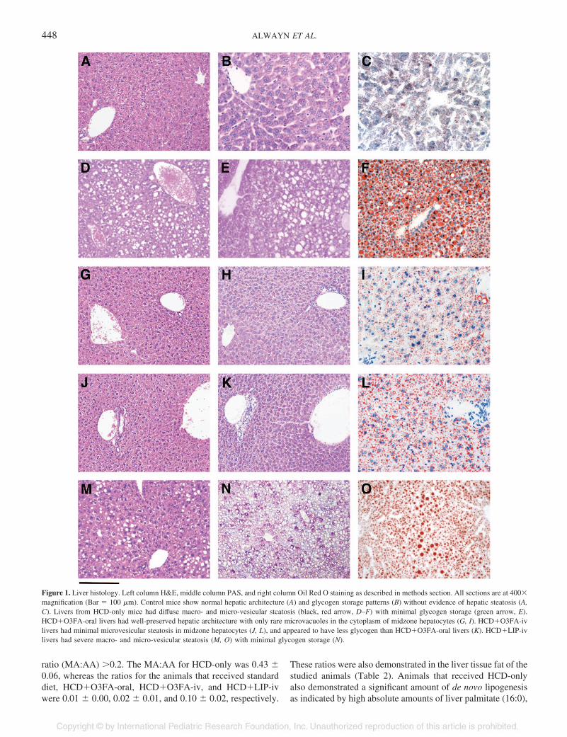
Figure 1 presents histologic results from all groups. Controlmice (standard diet fed) showed normal hepatic architecture(Fig. 1A) and glycogen storage patterns (Fig. 1B), withoutevidence of hepatic steatosis (Fig. 1A–C). In contrast, liversfrom HCD-only mice had diffuse macro- and microvesicularsteatosis. These changes were most marked (black arrow) inthe periportal and midzone hepatocytes (Fig. 1D–F). Centralvein hepatocytes were spared of steatosis for a two- to three-cell layer; cells outside this perimeter, however, showed anabrupt change to steatosis (red arrow). HCD-only livers re-vealed minimal PAS positivity in these sections, indicatingminor glycogen storage (Fig. 1E). Most of the PAS-positivecells were present in a two- to three-cell layer around thecentral vein (green arrow). HCD�O3FA-oral livers had well-preserved hepatic architecture with only rare microvacuoles inthe cytoplasm of midzone hepatocytes (Fig. 1G and I). PASstaining was strongly positive, finely granular, and diffuse butmost prominent within hepatocytes around the portal andcentral vein regions and less prominent in the midzone (Fig.
1H). HCD�O3FA-iv livers had minimal microvesicular ste-atosis in midzone hepatocytes (Fig. 1J–L), and there waspositive PAS staining (Fig. 1K), albeit to a lesser extent thanHCD�O3FA-oral livers. HCD�LIP-iv livers had severe mac-ro- and microvesicular steatosis (Fig. 1M–O) that, in contrast toHCD-only livers, included the central vein hepatocytes withminimal glycogen storage.Serum liver function tests. Serum liver function tests were
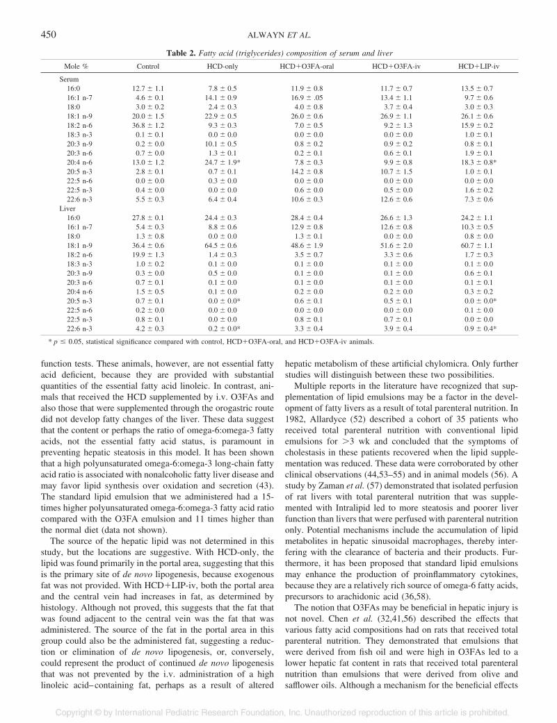
obtained as an additional marker of liver injury. The results forthese tests are summarized in Table 1. Values obtained fromcontrol standard diet–fed mice were considered to be within thenormal range. The levels of aspartate aminotransferase, alanineaminotransferase, and total bilirubin were significantly higherin the HCD-only animals (109.0 � 6.4 U/L, 71.6 � 8.2 U/L,and 0.20 � 0.00 mg/dL, respectively) compared with controls[72.2 � 5.5 U/L (p � 0.01); 48.2 � 3.6 U/L (p � 0.05); and0.10 � 0.02 mg/dL (p � 0.05), respectively]. It is interestingthat both aspartate aminotransferase (53.0 � 5.8 U/L) andalanine aminotransferase (23.0 � 2.6 U/L) values in theHCD�O3FA-oral–treated animals were significantly lowerthan in control animals (p � 0.05 and p � 0.01, respectively)and HCD-only animals (p � 0.01). In addition, alkaline phos-phatase levels were significantly decreased in theHCD�O3FA-oral (81.8 � 7.2 U/L) mice compared withcontrol animals (113.2 � 4.2 U/L; p � 0.01) and HCD-onlyanimals (126.0 � 5.9 U/L; p � 0.01). The serum liver functiontests of the HCD�O3FA-iv or HCD�LIP-iv animals were notdifferent from control animals or HCD-only animals except fortotal bilirubin levels of HCD�O3FA-iv animals (0.12 � 0.02mg/dL). These were significantly lower than in HCD-onlymice (p � 0.05).Radiologic fat measurements. To quantify changes in he-
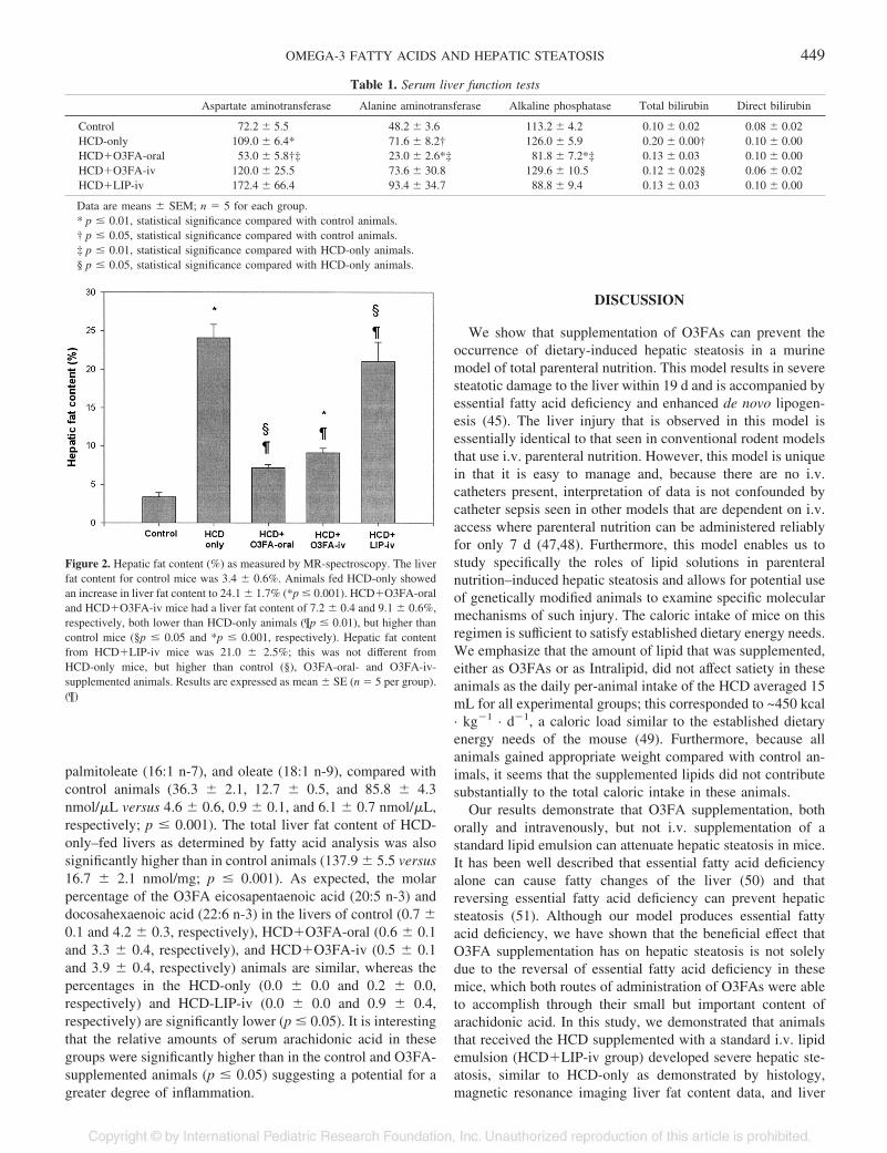
patic fat content, we studied livers with MRS. Livers of controlmice that received only standard diet were used as a baselinewith which all groups were compared. There was a closecorrelation between the percentage of fat calculated from theMRS data and degree of T1 shortening determined from the T1relaxation curves (data not shown). Liver fat content values forall experimental groups are demonstrated in Fig. 2.
The liver fat content for control mice was 3.4 � 0.6%.Animals that were fed HCD-only showed a significant increasein liver fat content to 24.1 � 1.7% (p � 0.001). HCD�O3FA-oral and HCD�O3FA-iv mice had a liver fat content of 7.2 �0.4 and 9.2 � 0.6%, respectively. These values all are signif-icantly lower than in HCD-only animals (p � 0.01) but sig-nificantly higher than in control mice (p � 0.05 and p � 0.001,respectively). Livers from HCD�LIP-iv mice had a fat contentof 21.0 � 2.5%; this was not different from HCD-only micebut was significantly higher than in control animals (p � 0.05)and O3FA-oral– and O3FA-iv–supplemented animals (p �0.01).Fatty acid analysis. A comprehensive analysis of fatty acid
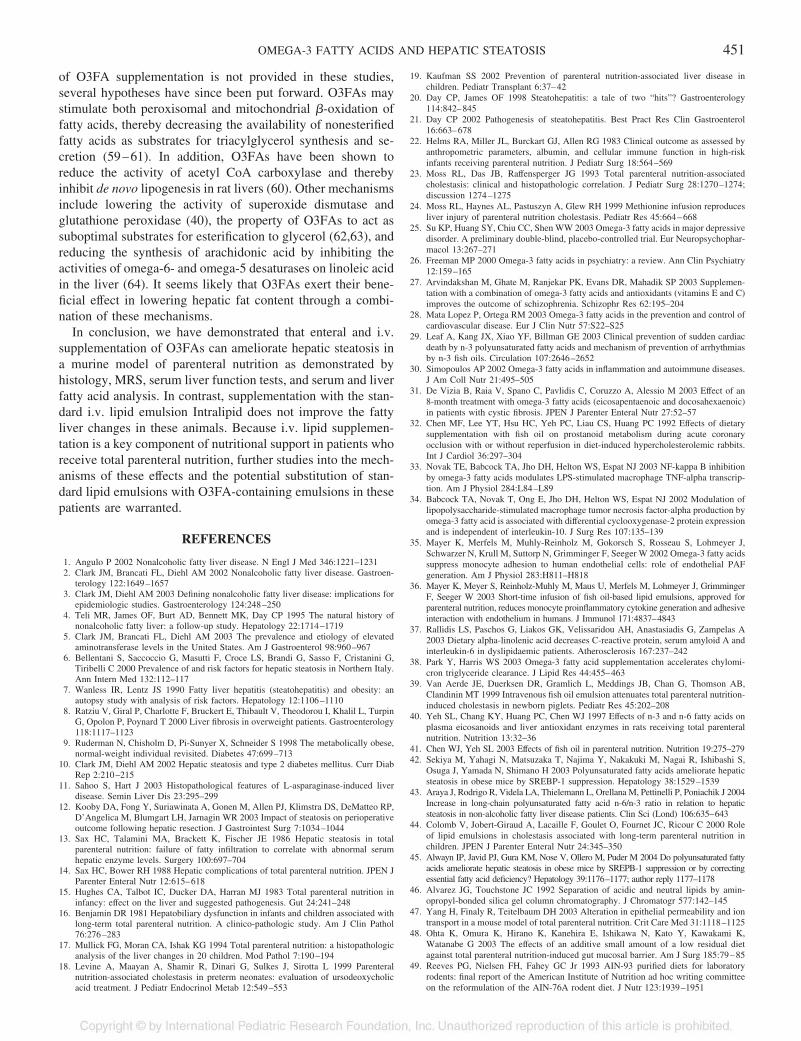
composition of serum and livers was performed; the results aresummarized in Table 2. As expected, animals that were fedHCD-only were essential fatty acid deficient, whereas all otheranimals, including HCD�LIP-iv, were not. Essential fatty aciddeficiency is generally characterized as a serum Mead acid(20:3 n-9) to arachidonic acid (20:4 n-6) serum triglyceride
447OMEGA-3 FATTY ACIDS AND HEPATIC STEATOSIS
ratio (MA:AA) �0.2. The MA:AA for HCD-only was 0.43 �0.06, whereas the ratios for the animals that received standarddiet, HCD�O3FA-oral, HCD�O3FA-iv, and HCD�LIP-ivwere 0.01 � 0.00, 0.02 � 0.01, and 0.10 � 0.02, respectively.
These ratios were also demonstrated in the liver tissue fat of thestudied animals (Table 2). Animals that received HCD-onlyalso demonstrated a significant amount of de novo lipogenesisas indicated by high absolute amounts of liver palmitate (16:0),
Figure 1. Liver histology. Left column H&E, middle column PAS, and right column Oil Red O staining as described in methods section. All sections are at 400�magnification (Bar � 100 �m). Control mice show normal hepatic architecture (A) and glycogen storage patterns (B) without evidence of hepatic steatosis (A,C). Livers from HCD-only mice had diffuse macro- and micro-vesicular stcatosis (black, red arrow, D–F) with minimal glycogen storage (green arrow, E).HCD�O3FA-oral livers had well-preserved hepatic architecture with only rare microvacuoles in the cytoplasm of midzone hepatocytes (G, I). HCD�O3FA-ivlivers had minimal microvesicular steatosis in midzone hepatocytes (J, L), and appeared to have less glycogen than HCD�O3FA-oral livers (K). HCD�LIP-ivlivers had severe macro- and micro-vesicular steatosis (M, O) with minimal glycogen storage (N).
448 ALWAYN ET AL.
palmitoleate (16:1 n-7), and oleate (18:1 n-9), compared withcontrol animals (36.3 � 2.1, 12.7 � 0.5, and 85.8 � 4.3nmol/�L versus 4.6 � 0.6, 0.9 � 0.1, and 6.1 � 0.7 nmol/�L,respectively; p � 0.001). The total liver fat content of HCD-only–fed livers as determined by fatty acid analysis was alsosignificantly higher than in control animals (137.9 � 5.5 versus16.7 � 2.1 nmol/mg; p � 0.001). As expected, the molarpercentage of the O3FA eicosapentaenoic acid (20:5 n-3) anddocosahexaenoic acid (22:6 n-3) in the livers of control (0.7 �0.1 and 4.2 � 0.3, respectively), HCD�O3FA-oral (0.6 � 0.1and 3.3 � 0.4, respectively), and HCD�O3FA-iv (0.5 � 0.1and 3.9 � 0.4, respectively) animals are similar, whereas thepercentages in the HCD-only (0.0 � 0.0 and 0.2 � 0.0,respectively) and HCD-LIP-iv (0.0 � 0.0 and 0.9 � 0.4,respectively) are significantly lower (p � 0.05). It is interestingthat the relative amounts of serum arachidonic acid in thesegroups were significantly higher than in the control and O3FA-supplemented animals (p � 0.05) suggesting a potential for agreater degree of inflammation.
DISCUSSION
We show that supplementation of O3FAs can prevent theoccurrence of dietary-induced hepatic steatosis in a murinemodel of total parenteral nutrition. This model results in severesteatotic damage to the liver within 19 d and is accompanied byessential fatty acid deficiency and enhanced de novo lipogen-esis (45). The liver injury that is observed in this model isessentially identical to that seen in conventional rodent modelsthat use i.v. parenteral nutrition. However, this model is uniquein that it is easy to manage and, because there are no i.v.catheters present, interpretation of data is not confounded bycatheter sepsis seen in other models that are dependent on i.v.access where parenteral nutrition can be administered reliablyfor only 7 d (47,48). Furthermore, this model enables us tostudy specifically the roles of lipid solutions in parenteralnutrition–induced hepatic steatosis and allows for potential useof genetically modified animals to examine specific molecularmechanisms of such injury. The caloric intake of mice on thisregimen is sufficient to satisfy established dietary energy needs.We emphasize that the amount of lipid that was supplemented,either as O3FAs or as Intralipid, did not affect satiety in theseanimals as the daily per-animal intake of the HCD averaged 15mL for all experimental groups; this corresponded to ~450 kcal· kg�1 · d�1, a caloric load similar to the established dietaryenergy needs of the mouse (49). Furthermore, because allanimals gained appropriate weight compared with control an-imals, it seems that the supplemented lipids did not contributesubstantially to the total caloric intake in these animals.
Our results demonstrate that O3FA supplementation, bothorally and intravenously, but not i.v. supplementation of astandard lipid emulsion can attenuate hepatic steatosis in mice.It has been well described that essential fatty acid deficiencyalone can cause fatty changes of the liver (50) and thatreversing essential fatty acid deficiency can prevent hepaticsteatosis (51). Although our model produces essential fattyacid deficiency, we have shown that the beneficial effect thatO3FA supplementation has on hepatic steatosis is not solelydue to the reversal of essential fatty acid deficiency in thesemice, which both routes of administration of O3FAs were ableto accomplish through their small but important content ofarachidonic acid. In this study, we demonstrated that animalsthat received the HCD supplemented with a standard i.v. lipidemulsion (HCD�LIP-iv group) developed severe hepatic ste-atosis, similar to HCD-only as demonstrated by histology,magnetic resonance imaging liver fat content data, and liver
Table 1. Serum liver function tests
Aspartate aminotransferase Alanine aminotransferase Alkaline phosphatase Total bilirubin Direct bilirubin
Control 72.2 � 5.5 48.2 � 3.6 113.2 � 4.2 0.10 � 0.02 0.08 � 0.02HCD-only 109.0 � 6.4* 71.6 � 8.2† 126.0 � 5.9 0.20 � 0.00† 0.10 � 0.00HCD�O3FA-oral 53.0 � 5.8†‡ 23.0 � 2.6*‡ 81.8 � 7.2*‡ 0.13 � 0.03 0.10 � 0.00HCD�O3FA-iv 120.0 � 25.5 73.6 � 30.8 129.6 � 10.5 0.12 � 0.02§ 0.06 � 0.02HCD�LIP-iv 172.4 � 66.4 93.4 � 34.7 88.8 � 9.4 0.13 � 0.03 0.10 � 0.00
Data are means � SEM; n � 5 for each group.* p � 0.01, statistical significance compared with control animals.† p � 0.05, statistical significance compared with control animals.‡ p � 0.01, statistical significance compared with HCD-only animals.§ p � 0.05, statistical significance compared with HCD-only animals.
Figure 2. Hepatic fat content (%) as measured by MR-spectroscopy. The liverfat content for control mice was 3.4 � 0.6%. Animals fed HCD-only showedan increase in liver fat content to 24.1 � 1.7% (*p � 0.001). HCD�O3FA-oraland HCD�O3FA-iv mice had a liver fat content of 7.2 � 0.4 and 9.1 � 0.6%,respectively, both lower than HCD-only animals (¶p � 0.01), but higher thancontrol mice (§p � 0.05 and *p � 0.001, respectively). Hepatic fat contentfrom HCD�LIP-iv mice was 21.0 � 2.5%; this was not different fromHCD-only mice, but higher than control (§), O3FA-oral- and O3FA-iv-supplemented animals. Results are expressed as mean � SE (n � 5 per group).(¶)
449OMEGA-3 FATTY ACIDS AND HEPATIC STEATOSIS
function tests. These animals, however, are not essential fattyacid deficient, because they are provided with substantialquantities of the essential fatty acid linoleic. In contrast, ani-mals that received the HCD supplemented by i.v. O3FAs andalso those that were supplemented through the orogastric routedid not develop fatty changes of the liver. These data suggestthat the content or perhaps the ratio of omega-6:omega-3 fattyacids, not the essential fatty acid status, is paramount inpreventing hepatic steatosis in this model. It has been shownthat a high polyunsaturated omega-6:omega-3 long-chain fattyacid ratio is associated with nonalcoholic fatty liver disease andmay favor lipid synthesis over oxidation and secretion (43).The standard lipid emulsion that we administered had a 15-times higher polyunsaturated omega-6:omega-3 fatty acid ratiocompared with the O3FA emulsion and 11 times higher thanthe normal diet (data not shown).
The source of the hepatic lipid was not determined in thisstudy, but the locations are suggestive. With HCD-only, thelipid was found primarily in the portal area, suggesting that thisis the primary site of de novo lipogenesis, because exogenousfat was not provided. With HCD�LIP-iv, both the portal areaand the central vein had increases in fat, as determined byhistology. Although not proved, this suggests that the fat thatwas found adjacent to the central vein was the fat that wasadministered. The source of the fat in the portal area in thisgroup could also be the administered fat, suggesting a reduc-tion or elimination of de novo lipogenesis, or, conversely,could represent the product of continued de novo lipogenesisthat was not prevented by the i.v. administration of a highlinoleic acid–containing fat, perhaps as a result of altered
hepatic metabolism of these artificial chylomicra. Only furtherstudies will distinguish between these two possibilities.
Multiple reports in the literature have recognized that sup-plementation of lipid emulsions may be a factor in the devel-opment of fatty livers as a result of total parenteral nutrition. In1982, Allardyce (52) described a cohort of 35 patients whoreceived total parenteral nutrition with conventional lipidemulsions for �3 wk and concluded that the symptoms ofcholestasis in these patients recovered when the lipid supple-mentation was reduced. These data were corroborated by otherclinical observations (44,53–55) and in animal models (56). Astudy by Zaman et al. (57) demonstrated that isolated perfusionof rat livers with total parenteral nutrition that was supple-mented with Intralipid led to more steatosis and poorer liverfunction than livers that were perfused with parenteral nutritiononly. Potential mechanisms include the accumulation of lipidmetabolites in hepatic sinusoidal macrophages, thereby inter-fering with the clearance of bacteria and their products. Fur-thermore, it has been proposed that standard lipid emulsionsmay enhance the production of proinflammatory cytokines,because they are a relatively rich source of omega-6 fatty acids,precursors to arachidonic acid (36,58).
The notion that O3FAs may be beneficial in hepatic injury isnot novel. Chen et al. (32,41,56) described the effects thatvarious fatty acid compositions had on rats that received totalparenteral nutrition. They demonstrated that emulsions thatwere derived from fish oil and were high in O3FAs led to alower hepatic fat content in rats that received total parenteralnutrition than emulsions that were derived from olive andsafflower oils. Although a mechanism for the beneficial effects
Table 2. Fatty acid (triglycerides) composition of serum and liver
Mole % Control HCD-only HCD�O3FA-oral HCD�O3FA-iv HCD�LIP-iv
Serum16:0 12.7 � 1.1 7.8 � 0.5 11.9 � 0.8 11.7 � 0.7 13.5 � 0.716:1 n-7 4.6 � 0.1 14.1 � 0.9 16.9 � .05 13.4 � 1.1 9.7 � 0.618:0 3.0 � 0.2 2.4 � 0.3 4.0 � 0.8 3.7 � 0.4 3.0 � 0.318:1 n-9 20.0 � 1.5 22.9 � 0.5 26.0 � 0.6 26.9 � 1.1 26.1 � 0.618:2 n-6 36.8 � 1.2 9.3 � 0.3 7.0 � 0.5 9.2 � 1.3 15.9 � 0.218:3 n-3 0.1 � 0.1 0.0 � 0.0 0.0 � 0.0 0.0 � 0.0 1.0 � 0.120:3 n-9 0.2 � 0.0 10.1 � 0.5 0.8 � 0.2 0.9 � 0.2 0.8 � 0.120:3 n-6 0.7 � 0.0 1.3 � 0.1 0.2 � 0.1 0.6 � 0.1 1.9 � 0.120:4 n-6 13.0 � 1.2 24.7 � 1.9* 7.8 � 0.3 9.9 � 0.8 18.3 � 0.8*20:5 n-3 2.8 � 0.1 0.7 � 0.1 14.2 � 0.8 10.7 � 1.5 1.0 � 0.122:5 n-6 0.0 � 0.0 0.3 � 0.0 0.0 � 0.0 0.0 � 0.0 0.0 � 0.022:5 n-3 0.4 � 0.0 0.0 � 0.0 0.6 � 0.0 0.5 � 0.0 1.6 � 0.222:6 n-3 5.5 � 0.3 6.4 � 0.4 10.6 � 0.3 12.6 � 0.6 7.3 � 0.6
Liver16:0 27.8 � 0.1 24.4 � 0.3 28.4 � 0.4 26.6 � 1.3 24.2 � 1.116:1 n-7 5.4 � 0.3 8.8 � 0.6 12.9 � 0.8 12.6 � 0.8 10.3 � 0.518:0 1.3 � 0.8 0.0 � 0.0 1.3 � 0.1 0.0 � 0.0 0.8 � 0.018:1 n-9 36.4 � 0.6 64.5 � 0.6 48.6 � 1.9 51.6 � 2.0 60.7 � 1.118:2 n-6 19.9 � 1.3 1.4 � 0.3 3.5 � 0.7 3.3 � 0.6 1.7 � 0.318:3 n-3 1.0 � 0.2 0.1 � 0.0 0.1 � 0.0 0.1 � 0.0 0.1 � 0.020:3 n-9 0.3 � 0.0 0.5 � 0.0 0.1 � 0.0 0.1 � 0.0 0.6 � 0.120:3 n-6 0.7 � 0.1 0.1 � 0.0 0.1 � 0.0 0.1 � 0.0 0.1 � 0.120:4 n-6 1.5 � 0.5 0.1 � 0.0 0.2 � 0.0 0.2 � 0.0 0.3 � 0.220:5 n-3 0.7 � 0.1 0.0 � 0.0* 0.6 � 0.1 0.5 � 0.1 0.0 � 0.0*22:5 n-6 0.2 � 0.0 0.0 � 0.0 0.0 � 0.0 0.0 � 0.0 0.1 � 0.022:5 n-3 0.8 � 0.1 0.0 � 0.0 0.8 � 0.1 0.7 � 0.1 0.0 � 0.022:6 n-3 4.2 � 0.3 0.2 � 0.0* 3.3 � 0.4 3.9 � 0.4 0.9 � 0.4*
* p � 0.05, statistical significance compared with control, HCD�O3FA-oral, and HCD�O3FA-iv animals.
450 ALWAYN ET AL.
of O3FA supplementation is not provided in these studies,several hypotheses have since been put forward. O3FAs maystimulate both peroxisomal and mitochondrial �-oxidation offatty acids, thereby decreasing the availability of nonesterifiedfatty acids as substrates for triacylglycerol synthesis and se-cretion (59–61). In addition, O3FAs have been shown toreduce the activity of acetyl CoA carboxylase and therebyinhibit de novo lipogenesis in rat livers (60). Other mechanismsinclude lowering the activity of superoxide dismutase andglutathione peroxidase (40), the property of O3FAs to act assuboptimal substrates for esterification to glycerol (62,63), andreducing the synthesis of arachidonic acid by inhibiting theactivities of omega-6- and omega-5 desaturases on linoleic acidin the liver (64). It seems likely that O3FAs exert their bene-ficial effect in lowering hepatic fat content through a combi-nation of these mechanisms.
In conclusion, we have demonstrated that enteral and i.v.supplementation of O3FAs can ameliorate hepatic steatosis ina murine model of parenteral nutrition as demonstrated byhistology, MRS, serum liver function tests, and serum and liverfatty acid analysis. In contrast, supplementation with the stan-dard i.v. lipid emulsion Intralipid does not improve the fattyliver changes in these animals. Because i.v. lipid supplemen-tation is a key component of nutritional support in patients whoreceive total parenteral nutrition, further studies into the mech-anisms of these effects and the potential substitution of stan-dard lipid emulsions with O3FA-containing emulsions in thesepatients are warranted.
REFERENCES
1. Angulo P 2002 Nonalcoholic fatty liver disease. N Engl J Med 346:1221–12312. Clark JM, Brancati FL, Diehl AM 2002 Nonalcoholic fatty liver disease. Gastroen-
terology 122:1649–16573. Clark JM, Diehl AM 2003 Defining nonalcoholic fatty liver disease: implications for
epidemiologic studies. Gastroenterology 124:248–2504. Teli MR, James OF, Burt AD, Bennett MK, Day CP 1995 The natural history of
nonalcoholic fatty liver: a follow-up study. Hepatology 22:1714–17195. Clark JM, Brancati FL, Diehl AM 2003 The prevalence and etiology of elevated
aminotransferase levels in the United States. Am J Gastroenterol 98:960–9676. Bellentani S, Saccoccio G, Masutti F, Croce LS, Brandi G, Sasso F, Cristanini G,
Tiribelli C 2000 Prevalence of and risk factors for hepatic steatosis in Northern Italy.Ann Intern Med 132:112–117
7. Wanless IR, Lentz JS 1990 Fatty liver hepatitis (steatohepatitis) and obesity: anautopsy study with analysis of risk factors. Hepatology 12:1106–1110
8. Ratziu V, Giral P, Charlotte F, Bruckert E, Thibault V, Theodorou I, Khalil L, TurpinG, Opolon P, Poynard T 2000 Liver fibrosis in overweight patients. Gastroenterology118:1117–1123
9. Ruderman N, Chisholm D, Pi-Sunyer X, Schneider S 1998 The metabolically obese,normal-weight individual revisited. Diabetes 47:699–713
10. Clark JM, Diehl AM 2002 Hepatic steatosis and type 2 diabetes mellitus. Curr DiabRep 2:210–215
11. Sahoo S, Hart J 2003 Histopathological features of L-asparaginase-induced liverdisease. Semin Liver Dis 23:295–299
12. Kooby DA, Fong Y, Suriawinata A, Gonen M, Allen PJ, Klimstra DS, DeMatteo RP,D’Angelica M, Blumgart LH, Jarnagin WR 2003 Impact of steatosis on perioperativeoutcome following hepatic resection. J Gastrointest Surg 7:1034–1044
13. Sax HC, Talamini MA, Brackett K, Fischer JE 1986 Hepatic steatosis in totalparenteral nutrition: failure of fatty infiltration to correlate with abnormal serumhepatic enzyme levels. Surgery 100:697–704
14. Sax HC, Bower RH 1988 Hepatic complications of total parenteral nutrition. JPEN JParenter Enteral Nutr 12:615–618
15. Hughes CA, Talbot IC, Ducker DA, Harran MJ 1983 Total parenteral nutrition ininfancy: effect on the liver and suggested pathogenesis. Gut 24:241–248
16. Benjamin DR 1981 Hepatobiliary dysfunction in infants and children associated withlong-term total parenteral nutrition. A clinico-pathologic study. Am J Clin Pathol76:276–283
17. Mullick FG, Moran CA, Ishak KG 1994 Total parenteral nutrition: a histopathologicanalysis of the liver changes in 20 children. Mod Pathol 7:190–194
18. Levine A, Maayan A, Shamir R, Dinari G, Sulkes J, Sirotta L 1999 Parenteralnutrition-associated cholestasis in preterm neonates: evaluation of ursodeoxycholicacid treatment. J Pediatr Endocrinol Metab 12:549–553
19. Kaufman SS 2002 Prevention of parenteral nutrition-associated liver disease inchildren. Pediatr Transplant 6:37–42
20. Day CP, James OF 1998 Steatohepatitis: a tale of two “hits”? Gastroenterology114:842–845
21. Day CP 2002 Pathogenesis of steatohepatitis. Best Pract Res Clin Gastroenterol16:663–678
22. Helms RA, Miller JL, Burckart GJ, Allen RG 1983 Clinical outcome as assessed byanthropometric parameters, albumin, and cellular immune function in high-riskinfants receiving parenteral nutrition. J Pediatr Surg 18:564–569
23. Moss RL, Das JB, Raffensperger JG 1993 Total parenteral nutrition-associatedcholestasis: clinical and histopathologic correlation. J Pediatr Surg 28:1270–1274;discussion 1274–1275
24. Moss RL, Haynes AL, Pastuszyn A, Glew RH 1999 Methionine infusion reproducesliver injury of parenteral nutrition cholestasis. Pediatr Res 45:664–668
25. Su KP, Huang SY, Chiu CC, Shen WW 2003 Omega-3 fatty acids in major depressivedisorder. A preliminary double-blind, placebo-controlled trial. Eur Neuropsychophar-macol 13:267–271
26. Freeman MP 2000 Omega-3 fatty acids in psychiatry: a review. Ann Clin Psychiatry12:159–165
27. Arvindakshan M, Ghate M, Ranjekar PK, Evans DR, Mahadik SP 2003 Supplemen-tation with a combination of omega-3 fatty acids and antioxidants (vitamins E and C)improves the outcome of schizophrenia. Schizophr Res 62:195–204
28. Mata Lopez P, Ortega RM 2003 Omega-3 fatty acids in the prevention and control ofcardiovascular disease. Eur J Clin Nutr 57:S22–S25
29. Leaf A, Kang JX, Xiao YF, Billman GE 2003 Clinical prevention of sudden cardiacdeath by n-3 polyunsaturated fatty acids and mechanism of prevention of arrhythmiasby n-3 fish oils. Circulation 107:2646–2652
30. Simopoulos AP 2002 Omega-3 fatty acids in inflammation and autoimmune diseases.J Am Coll Nutr 21:495–505
31. De Vizia B, Raia V, Spano C, Pavlidis C, Coruzzo A, Alessio M 2003 Effect of an8-month treatment with omega-3 fatty acids (eicosapentaenoic and docosahexaenoic)in patients with cystic fibrosis. JPEN J Parenter Enteral Nutr 27:52–57
32. Chen MF, Lee YT, Hsu HC, Yeh PC, Liau CS, Huang PC 1992 Effects of dietarysupplementation with fish oil on prostanoid metabolism during acute coronaryocclusion with or without reperfusion in diet-induced hypercholesterolemic rabbits.Int J Cardiol 36:297–304
33. Novak TE, Babcock TA, Jho DH, Helton WS, Espat NJ 2003 NF-kappa B inhibitionby omega-3 fatty acids modulates LPS-stimulated macrophage TNF-alpha transcrip-tion. Am J Physiol 284:L84–L89
34. Babcock TA, Novak T, Ong E, Jho DH, Helton WS, Espat NJ 2002 Modulation oflipopolysaccharide-stimulated macrophage tumor necrosis factor-alpha production byomega-3 fatty acid is associated with differential cyclooxygenase-2 protein expressionand is independent of interleukin-10. J Surg Res 107:135–139
35. Mayer K, Merfels M, Muhly-Reinholz M, Gokorsch S, Rosseau S, Lohmeyer J,Schwarzer N, Krull M, Suttorp N, Grimminger F, Seeger W 2002 Omega-3 fatty acidssuppress monocyte adhesion to human endothelial cells: role of endothelial PAFgeneration. Am J Physiol 283:H811–H818
36. Mayer K, Meyer S, Reinholz-Muhly M, Maus U, Merfels M, Lohmeyer J, GrimmingerF, Seeger W 2003 Short-time infusion of fish oil-based lipid emulsions, approved forparenteral nutrition, reduces monocyte proinflammatory cytokine generation and adhesiveinteraction with endothelium in humans. J Immunol 171:4837–4843
37. Rallidis LS, Paschos G, Liakos GK, Velissaridou AH, Anastasiadis G, Zampelas A2003 Dietary alpha-linolenic acid decreases C-reactive protein, serum amyloid A andinterleukin-6 in dyslipidaemic patients. Atherosclerosis 167:237–242
38. Park Y, Harris WS 2003 Omega-3 fatty acid supplementation accelerates chylomi-cron triglyceride clearance. J Lipid Res 44:455–463
39. Van Aerde JE, Duerksen DR, Gramlich L, Meddings JB, Chan G, Thomson AB,Clandinin MT 1999 Intravenous fish oil emulsion attenuates total parenteral nutrition-induced cholestasis in newborn piglets. Pediatr Res 45:202–208
40. Yeh SL, Chang KY, Huang PC, Chen WJ 1997 Effects of n-3 and n-6 fatty acids onplasma eicosanoids and liver antioxidant enzymes in rats receiving total parenteralnutrition. Nutrition 13:32–36
41. Chen WJ, Yeh SL 2003 Effects of fish oil in parenteral nutrition. Nutrition 19:275–27942. Sekiya M, Yahagi N, Matsuzaka T, Najima Y, Nakakuki M, Nagai R, Ishibashi S,
Osuga J, Yamada N, Shimano H 2003 Polyunsaturated fatty acids ameliorate hepaticsteatosis in obese mice by SREBP-1 suppression. Hepatology 38:1529–1539
43. Araya J, Rodrigo R, Videla LA, Thielemann L, Orellana M, Pettinelli P, Poniachik J 2004Increase in long-chain polyunsaturated fatty acid n-6/n-3 ratio in relation to hepaticsteatosis in non-alcoholic fatty liver disease patients. Clin Sci (Lond) 106:635–643
44. Colomb V, Jobert-Giraud A, Lacaille F, Goulet O, Fournet JC, Ricour C 2000 Roleof lipid emulsions in cholestasis associated with long-term parenteral nutrition inchildren. JPEN J Parenter Enteral Nutr 24:345–350
45. Alwayn IP, Javid PJ, Gura KM, Nose V, Ollero M, Puder M 2004 Do polyunsaturated fattyacids ameliorate hepatic steatosis in obese mice by SREPB-1 suppression or by correctingessential fatty acid deficiency? Hepatology 39:1176–1177; author reply 1177–1178
46. Alvarez JG, Touchstone JC 1992 Separation of acidic and neutral lipids by amin-opropyl-bonded silica gel column chromatography. J Chromatogr 577:142–145
47. Yang H, Finaly R, Teitelbaum DH 2003 Alteration in epithelial permeability and iontransport in a mouse model of total parenteral nutrition. Crit Care Med 31:1118–1125
48. Ohta K, Omura K, Hirano K, Kanehira E, Ishikawa N, Kato Y, Kawakami K,Watanabe G 2003 The effects of an additive small amount of a low residual dietagainst total parenteral nutrition-induced gut mucosal barrier. Am J Surg 185:79–85
49. Reeves PG, Nielsen FH, Fahey GC Jr 1993 AIN-93 purified diets for laboratoryrodents: final report of the American Institute of Nutrition ad hoc writing committeeon the reformulation of the AIN-76A rodent diet. J Nutr 123:1939–1951
451OMEGA-3 FATTY ACIDS AND HEPATIC STEATOSIS
50. Keim NL, Mares-Perlman JA 1984 Development of hepatic steatosis and essentialfatty acid deficiency in rats with hypercaloric, fat-free parenteral nutrition. J Nutr114:1807–1815
51. Goheen SC, Larkin EC, Rao GA 1983 Severe fatty liver in rats fed a fat-free ethanoldiet, and its prevention by small amounts of dietary arachidonate. Lipids 18:285–290
52. Allardyce DB 1982 Cholestasis caused by lipid emulsions. Surg Gynecol Obstet154:641–647
53. Cavicchi M, Crenn P, Beau P, Degott C, Boutron MC, Messing B 1998 Severe livercomplications associated with long-term parenteral nutrition are dependent on lipidparenteral input. Transplant Proc 30:2547
54. Cavicchi M, Beau P, Crenn P, Degott C, Messing B 2000 Prevalence of liver diseaseand contributing factors in patients receiving home parenteral nutrition for permanentintestinal failure. Ann Intern Med 132:525–532
55. Clayton PT, Whitfield P, Iyer K 1998 The role of phytosterols in the pathogenesis ofliver complications of pediatric parenteral nutrition. Nutrition 14:158–164
56. Chen WJ, Yeh SL, Huang PC 1996 Effects of fat emulsions with different fatty acidcomposition on plasma and hepatic lipids in rats receiving total parenteral nutrition.Clin Nutr 15:24–28
57. Zaman N, Tam YK, Jewell LD, Coutts RT 1997 Effects of intravenous lipid as asource of energy in parenteral nutrition associated hepatic dysfunction and lidocaineelimination: a study using isolated rat liver perfusion. Biopharm Drug Dispos18:803–819
58. Fernandes G 1994 Dietary lipids and risk of autoimmune disease. Clin ImmunolImmunopathol 72:193–197
59. Burdge GC, Finnegan YE, Minihane AM, Williams CM, Wootton SA 2003 Effect ofaltered dietary n-3 fatty acid intake upon plasma lipid fatty acid composition,conversion of [13C]alpha-linolenic acid to longer-chain fatty acids and partitioningtowards beta-oxidation in older men. Br J Nutr 90:311–321
60. Neschen S, Moore I, Regittnig W, Yu CL, Wang Y, Pypaert M, Petersen KF,Shulman GI 2002 Contrasting effects of fish oil and safflower oil on hepaticperoxisomal and tissue lipid content. Am J Physiol 282:E395–E401
61. Willumsen N, Hexeberg S, Skorve J, Lundquist M, Berge RK 1993 Docosahexaenoicacid shows no triglyceride-lowering effects but increases the peroxisomal fatty acidoxidation in liver of rats. J Lipid Res 34:13–22
62. Rustan AC, Nossen JO, Christiansen EN, Drevon CA 1988 Eicosapentaenoic acidreduces hepatic synthesis and secretion of triacylglycerol by decreasing the activity ofacyl-coenzyme A:1,2-diacylglycerol acyltransferase. J Lipid Res 29:1417–1426
63. Nossen JO, Rustan AC, Gloppestad SH, Malbakken S, Drevon CA 1986 Eicosapen-taenoic acid inhibits synthesis and secretion of triacylglycerols by cultured rathepatocytes. Biochim Biophys Acta 879:56–65
64. Bellenger J, Bellenger S, Clement L, Mandard S, Diot C, Poisson JP, Narce M 2004A new hypotensive polyunsaturated fatty acid dietary combination regulates oleicacid accumulation by suppression of stearoyl CoA desaturase 1 gene expression in theSHR model of genetic hypertension. FASEB J 18:773–775
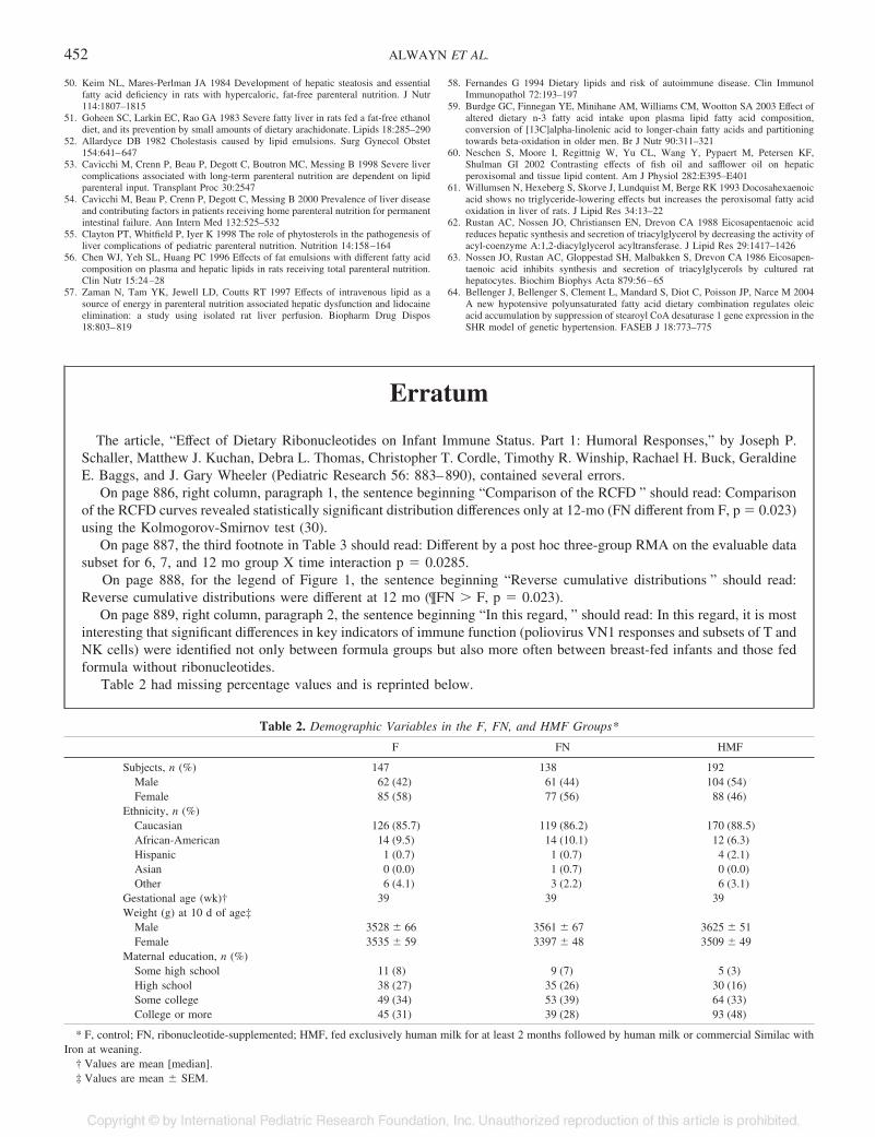
Table 2. Demographic Variables in the F, FN, and HMF Groups*
F FN HMF
Subjects, n (%) 147 138 192Male 62 (42) 61 (44) 104 (54)Female 85 (58) 77 (56) 88 (46)
Ethnicity, n (%)Caucasian 126 (85.7) 119 (86.2) 170 (88.5)African-American 14 (9.5) 14 (10.1) 12 (6.3)Hispanic 1 (0.7) 1 (0.7) 4 (2.1)Asian 0 (0.0) 1 (0.7) 0 (0.0)Other 6 (4.1) 3 (2.2) 6 (3.1)
Gestational age (wk)† 39 39 39Weight (g) at 10 d of age‡
Male 3528 � 66 3561 � 67 3625 � 51Female 3535 � 59 3397 � 48 3509 � 49
Maternal education, n (%)Some high school 11 (8) 9 (7) 5 (3)High school 38 (27) 35 (26) 30 (16)Some college 49 (34) 53 (39) 64 (33)College or more 45 (31) 39 (28) 93 (48)
* F, control; FN, ribonucleotide-supplemented; HMF, fed exclusively human milk for at least 2 months followed by human milk or commercial Similac withIron at weaning.
† Values are mean [median].‡ Values are mean � SEM.
Erratum
The article, “Effect of Dietary Ribonucleotides on Infant Immune Status. Part 1: Humoral Responses,” by Joseph P.Schaller, Matthew J. Kuchan, Debra L. Thomas, Christopher T. Cordle, Timothy R. Winship, Rachael H. Buck, GeraldineE. Baggs, and J. Gary Wheeler (Pediatric Research 56: 883–890), contained several errors.
On page 886, right column, paragraph 1, the sentence beginning “Comparison of the RCFD ” should read: Comparisonof the RCFD curves revealed statistically significant distribution differences only at 12-mo (FN different from F, p � 0.023)using the Kolmogorov-Smirnov test (30).
On page 887, the third footnote in Table 3 should read: Different by a post hoc three-group RMA on the evaluable datasubset for 6, 7, and 12 mo group X time interaction p � 0.0285.
On page 888, for the legend of Figure 1, the sentence beginning “Reverse cumulative distributions ” should read:Reverse cumulative distributions were different at 12 mo (¶FN � F, p � 0.023).
On page 889, right column, paragraph 2, the sentence beginning “In this regard, ” should read: In this regard, it is mostinteresting that significant differences in key indicators of immune function (poliovirus VN1 responses and subsets of T andNK cells) were identified not only between formula groups but also more often between breast-fed infants and those fedformula without ribonucleotides.
Table 2 had missing percentage values and is reprinted below.
452 ALWAYN ET AL.
Copyright © 2022 FDOKUMEN