Lipoic acid supplementation prevents angiotensin II–induced renal injury
8
Kidney International, Vol. 64 (2003), pp. 501–508 Lipoic acid supplementation prevents angiotensin II–induced renal injury EERO MERVAALA,PIET FINCKENBERG,RISTO LAPATTO,DOMINIK N. MU ¨ LLER,JOON-KEUN PARK, RALF DECHEND,DETLEV GANTEN,HEIKKI VAPAATALO, and FRIEDRICH C. LUFT Institute of Biomedicine, Pharmacology, University of Helsinki, Finland; Hospital for Children and Adolescents, University of Helsinki, Finland; HELIOS-Klinikum Berlin, Franz Volhard Clinic, Medical Faculty of the Charite ´, Humboldt University of Berlin, Germany; Department of Internal Medicine, Hannover Medical School, Hannover, Germany; Max Delbru ¨ ck Center for Molecular Medicine, Berlin, Germany Lipoic acid supplementation prevents angiotensin II–induced renal injury. Background. Angiotensin II (Ang II)–induced renal injury is associated with perivascular inflammation, cell proliferation, and increased superoxide production in the vascular wall. We tested whether lipoic acid, an endogenous antioxidant, protects against the Ang II–induced inflammatory response and end- organ damage. Methods. Light microscopy, immunohistochemistry, electro- phoretic mobility shift assay, Northern blots, and high-pressure liquid chromatography (HPLC) were used in kidneys from double transgenic rats (dTGR) harboring human renin and angiotensinogen genes and normotensive Sprague Dawley (SD) rats. The effects of lipoic acid supplementation for three weeks were examined in dTGR and SD rats. Results. Lipoic acid effectively prevented Ang II-induced glomerular and vascular damage in the kidneys and completely prevented the development of albuminuria. Ang II–induced leukocyte infiltration and cell proliferation in the kidney were attenuated. The redox-sensitive transcription factors nuclear factor (kappa) B (NF-B) and activator protein-1 (AP-1) in the kidneys were increased in dTGR compared with SD, and were effectively reduced. Renal glutathione levels were much higher in dTGR than in SD, while the opposite was true for cysteine levels. These results suggested increased renal gluta- thione oxidation in dTGR, leading to cysteine shortage. Lipoic acid partly prevented renal cysteine depletion and increased hepatic cysteine and glutathione concentrations. This effect was accompanied by increased hepatic gamma-glutamylcys- teine synthetase mRNA expression. Conclusion. Our in vivo results suggest that lipoic acid pro- tects against Ang II–induced renal injury through anti-inflam- matory/antioxidative mechanisms. The effects are associated with decreased NF-B and AP-1 activation, as well as improved thiol homeostasis. Key words: oxidative stress, glutathione, cysteine, inflammation, tran- scription factor NF-B, transcription factor AP-1. Received for publication August 10, 2002 and in revised form February 13, 2003 Accepted for publication March 21, 2003 2003 by the International Society of Nephrology 501 Reactive oxygen species (ROS) have been implicated in the pathogenesis of cardiovascular diseases [1]. ROS influence signal transduction pathways, cellular growth, and posttranslational protein modifications [2, 3]. Sev- eral hormone-sensitive enzymes, such as nicotinamide adenine dinucleotide phosphate [NAD(P)H] oxidase, xan- thine oxidase, cyclooxygenases, nitric oxide (NO) syn- thases, heme oxygenases, and peroxidases generate ROS [2]. ROS are important in the pathogenesis of angioten- sin (Ang) II–induced hypertension and vascular damage [4]. Furthermore, Ang II induces oxidative stress and superoxide formation primarily through activation of NAD(P)H oxidase in the vascular smooth muscle cells and myocytes [4, 5]. Intra- and extracellular defense systems protect the organism from deleterious effects of ROS. Glutathione sulfhydryl (GSH) is the key molecule in one of the most important antioxidant systems and helps to maintain the intracellular redox state by providing a thiol group [6]. GSH is made in two consecutive adenosine triphosphate (ATP)-requiring enzymatic reactions in which glutamate and cysteine are first ligated and glycine is added. Cys- teine supply is often rate limiting; however, transcriptional and posttranscriptional regulation also occur [7]. Boosting GSH levels has been attempted with various compounds [8]. Lipoic acid (6,8-thioctic acid) is promising in this regard [9], as it is a sulfur-containing coenzyme required for the mitochondrial dehydrogenase reactions leading to ATP formation [10–12]. Morikawa et al [13] recently identified a mouse cDNA encoding a lipoic acid synthase located in the mitochondria. Lipoic acid and the reduced form, dihydrolipoate, are potent natural antioxidants both in fat- and water-soluble media [10, 12, 14]. Lipoic acid reacts with superoxide, hydroxyl radicals, hypochlorous acid, peroxyl radicals, and singlet oxygen. Lipoic acid also recycles vitamin C, vitamin E, and increases intracel- lular glutathione concentrations. The antioxidative effect
Transcript of Lipoic acid supplementation prevents angiotensin II–induced renal injury

Kidney International, Vol. 64 (2003), pp. 501–508
Lipoic acid supplementation prevents angiotensin II–inducedrenal injury
EERO MERVAALA, PIET FINCKENBERG, RISTO LAPATTO, DOMINIK N. MULLER, JOON-KEUN PARK,RALF DECHEND, DETLEV GANTEN, HEIKKI VAPAATALO, and FRIEDRICH C. LUFT
Institute of Biomedicine, Pharmacology, University of Helsinki, Finland; Hospital for Children and Adolescents, Universityof Helsinki, Finland; HELIOS-Klinikum Berlin, Franz Volhard Clinic, Medical Faculty of the Charite, Humboldt University ofBerlin, Germany; Department of Internal Medicine, Hannover Medical School, Hannover, Germany; Max Delbruck Center forMolecular Medicine, Berlin, Germany
Lipoic acid supplementation prevents angiotensin II–inducedrenal injury.
Background. Angiotensin II (Ang II)–induced renal injuryis associated with perivascular inflammation, cell proliferation,and increased superoxide production in the vascular wall. Wetested whether lipoic acid, an endogenous antioxidant, protectsagainst the Ang II–induced inflammatory response and end-organ damage.
Methods. Light microscopy, immunohistochemistry, electro-phoretic mobility shift assay, Northern blots, and high-pressureliquid chromatography (HPLC) were used in kidneys fromdouble transgenic rats (dTGR) harboring human renin andangiotensinogen genes and normotensive Sprague Dawley (SD)rats. The effects of lipoic acid supplementation for three weekswere examined in dTGR and SD rats.
Results. Lipoic acid effectively prevented Ang II-inducedglomerular and vascular damage in the kidneys and completelyprevented the development of albuminuria. Ang II–inducedleukocyte infiltration and cell proliferation in the kidney wereattenuated. The redox-sensitive transcription factors nuclearfactor (kappa) B (NF-�B) and activator protein-1 (AP-1) inthe kidneys were increased in dTGR compared with SD, andwere effectively reduced. Renal glutathione levels were muchhigher in dTGR than in SD, while the opposite was true forcysteine levels. These results suggested increased renal gluta-thione oxidation in dTGR, leading to cysteine shortage. Lipoicacid partly prevented renal cysteine depletion and increasedhepatic cysteine and glutathione concentrations. This effectwas accompanied by increased hepatic gamma-glutamylcys-teine synthetase mRNA expression.
Conclusion. Our in vivo results suggest that lipoic acid pro-tects against Ang II–induced renal injury through anti-inflam-matory/antioxidative mechanisms. The effects are associatedwith decreased NF-�B and AP-1 activation, as well as improvedthiol homeostasis.
Key words: oxidative stress, glutathione, cysteine, inflammation, tran-scription factor NF-�B, transcription factor AP-1.
Received for publication August 10, 2002and in revised form February 13, 2003Accepted for publication March 21, 2003
2003 by the International Society of Nephrology
501
Reactive oxygen species (ROS) have been implicatedin the pathogenesis of cardiovascular diseases [1]. ROSinfluence signal transduction pathways, cellular growth,and posttranslational protein modifications [2, 3]. Sev-eral hormone-sensitive enzymes, such as nicotinamideadenine dinucleotide phosphate [NAD(P)H] oxidase, xan-thine oxidase, cyclooxygenases, nitric oxide (NO) syn-thases, heme oxygenases, and peroxidases generate ROS[2]. ROS are important in the pathogenesis of angioten-sin (Ang) II–induced hypertension and vascular damage[4]. Furthermore, Ang II induces oxidative stress andsuperoxide formation primarily through activation ofNAD(P)H oxidase in the vascular smooth muscle cellsand myocytes [4, 5].
Intra- and extracellular defense systems protect theorganism from deleterious effects of ROS. Glutathionesulfhydryl (GSH) is the key molecule in one of the mostimportant antioxidant systems and helps to maintain theintracellular redox state by providing a thiol group [6].GSH is made in two consecutive adenosine triphosphate(ATP)-requiring enzymatic reactions in which glutamateand cysteine are first ligated and glycine is added. Cys-teine supply is often rate limiting; however, transcriptionaland posttranscriptional regulation also occur [7]. BoostingGSH levels has been attempted with various compounds[8]. Lipoic acid (6,8-thioctic acid) is promising in thisregard [9], as it is a sulfur-containing coenzyme requiredfor the mitochondrial dehydrogenase reactions leadingto ATP formation [10–12]. Morikawa et al [13] recentlyidentified a mouse cDNA encoding a lipoic acid synthaselocated in the mitochondria. Lipoic acid and the reducedform, dihydrolipoate, are potent natural antioxidants bothin fat- and water-soluble media [10, 12, 14]. Lipoic acidreacts with superoxide, hydroxyl radicals, hypochlorousacid, peroxyl radicals, and singlet oxygen. Lipoic acidalso recycles vitamin C, vitamin E, and increases intracel-lular glutathione concentrations. The antioxidative effect

Mervaala et al: Lipoic acid and angiotensin II502
of lipoic acid may also be partially medixnated by transi-tion metal chelation, modification of the redox status ofthiol-containing proteins, and by the inhibition of redox-sensitive nuclear transcription factors [10–17]. Hagen et al[18, 19] reported that dietary lipoic acid supplementationimproves mitochondrial function, decreases oxidativestress, and increases metabolic rate in aging rats. Lipoicacid supplementation ameliorated hypertension, insulinresistance, and diabetes-associated renal damage [20–22].Protection against deoxycorticosterone acetate (DOCA)-salt–induced renal and vascular injuries has also beenreported [23]. We investigated the effectiveness of di-etary lipoic acid supplementation in the prevention ofcomplications associated with Ang II production in adouble transgenic rat (dTGR) model that featured oxi-dative stress [24–28].
METHODS
Experimental animals, drug treatments, andsample preparation
We used 25, 4-week-old male dTGR and 10 age-matched normotensive Sprague Dawley (SD) rats, de-scribed elsewhere [29, 30]. The protocols were approvedby the Animal Experimentation Committee of the Uni-versity of Helsinki, Finland, whose standards correspondto those of the American Physiological Society. The ratshad free access to chow (NaCl 0.8%) and drinking water.dTGR and normotensive SD control rats were dividedinto three groups: (1) dTGR control group (N � 15);(2) dTGR � lipoic acid group (N � 10); and (3) SDcontrol group (N � 10). The cardiovascular and renaleffects of lipoic acid were also examined in normotensiveSD rats (lipoic acid–treated SD rats, N � 6; control SDrats, N � 6). (R)-�-lipoic acid (Sigma Aldrich Co., Cam-bridgeshire, UK) was mixed in the food (0.5% weight/weight) to produce an approximate daily dose of 350mg/kg and given for three weeks. This dietary lipoic acidconcentration has produced several beneficial effects inprevious rat experiments [18, 31]. Systolic blood pressurewas measured by using a tail cuff blood pressure analyzer(Apollo-2AB Blood Pressure Analyzer, Model 179-2AB, IITC Life Science, Woodland Hills, CA, USA).At 7 weeks of age, rat urines were collected over 24hours in metabolic cages. Rats were then decapitated;the heart and kidneys were removed, washed with ice-cold saline, blotted dry, and weighed. Tissue sampleswere snap-frozen in liquid nitrogen and samples for im-munohistochemistry in isopentane (�35�C). All sampleswere stored at �80�C until assayed. Urinary albuminwas measured by enzyme-linked immunosorbent assay(ELISA) using rat albumin as a standard (Celltrend,Luckenwalde, Germany).
Renal and cardiac morphology
Our histologic techniques are described elsewhere [32].We relied on a quantitative grading scale for kidneys,viewed by a pathologist unaware of the regimens, as fol-lows: (0) normal arteriologlomerular unit with open capil-lary lumens and a normal afferent arteriole. Arcuate andinterlobular vessels normal. No tubular atrophy, intersti-tial inflammation, or interstitial fibrosis; (1) slight thick-ening of the media of the afferent arteriole. Some mesan-gial thickening. Open capillary lumens in the glomerulus.Patchy atrophy of tubular epithelium. Small interstitialfibrotic scars. Minimal medial thickening in larger ves-sels. No signs of inflammation; (2) marked mesangialthickening partly collapsed capillaries in the glomerulus.Occasional arteriolar thickening. Diffuse tubular atro-phy and small interstitial fibrotic scars. Clear medialthickening in larger vessels. Occasional inflammatory in-filtrate in the interstitium; (3) necrotic arterioglomerularunits with medial and intimal thickening of the arteriolarwall. Diffuse tubular atrophy with proteinaceous casts,patches of interstitial fibrosis, sometimes fibrinoid necro-sis of larger vessels. Inflammatory infiltrate in the inter-stitium; (4) necrotic arterioglomerular units with markedcellular crescents. Concentric hypertrophy and necrosisof afferent arteries. Fibrinoid necrosis and occlusion ofthe interlobular and arcuate arteries with thromboticmaterial. Atrophic and necrotic tubules. Evident intersti-tial fibrosis. Strong interstitial inflammation.
Furthermore, the hearts, arteries, and ventricular fi-brous tissue formation were also evaluated in a blindedfashion [33]. Each sample was scored from 0 to 3 accordingto morphologic changes as follows: (0) epicardial vesselsnormal. No excessive ventricular connective tissue forma-tion; (1) normal intima. Media slightly thickened or nor-mal. Slight increase of connective tissue around the epi-cardial and intramuscular arteries; (2) normal intima.Clear medial thickening and adventitial scarring in epi-cardial and intramuscular arteries. Patchy increase ofslender connective tissue bundles; (3) normal intima. Clearmedial thickening and adventitial scarring in epicardialand intramuscular arteries. Evident myocardial scars.
ImmunohistochemistryFor immunohistochemistry, frozen kidneys were pro-
cessed and semiquantitative scoring of inflammatorycells was performed as previously described in detail[25, 26]. The relative amount of positive labels per sam-ple was determined with computerized densitometry(Leica IM500 and Leica QWIN software; Leica Micro-systems AG, Heerbrugg, Switzerland). Primary mono-clonal antibodies against rat ED-1, CD-4, and CD-8 (Sero-tec, Ltd., Oslo, Norway) and peroxidase-conjugated rabbitanti-mouse secondary antibodies (DAKO A/S, Glostrup,Denmark) were used. Proliferating cell nuclear antigen(PCNA) was detected by using a polyclonal mouse anti-
Mervaala et al: Lipoic acid and angiotensin II 503
PCNA antibody (Stressgen Biotechnologies Corp., Vic-toria, British Columbia, Canada).
Electrophoretic mobility shift assayTissue preparation for electrophoretic mobility shift
assay (EMSA) was performed as described earlier [34].Nuclear extracts (5 �g) were incubated in binding re-action medium with 0.5 ng of 32P-dATP end-labeled oligo-nucleotide containing the NF-�B binding site from themajor histocompatability complex (MHC)-enhancer (H2K,5�-GAT CCA GGG CTG GGG ATT CCC CAT CTCCAC AGG). Oligonucleotides were synthesized by Bio-Tez (BioTez, Berlin, Germany). For AP-1, double-stranded oligonucleotides containing the consensus se-quence for AP-1 (Santa Cruz, CA, USA) 5�-GAT CGAACT GAC CGC CCG CCG CCC GT-3�) were radio-labeled with -32P with the use of T4 polynucleotidekinase by standard methods and purified on a column.The DNA-protein complexes were analyzed on a 5%polyacrylamide gel 0.5% Tris buffer, dried, and auto-radiographed. In competition assays, 50 ng unlabeled H2Kor AP-1 oligonucleotides were used.
Determination of tissue glutathione homeostasisThe concentration of thiol compounds glutathione and
cysteine in the liver and kidney was determined fluoro-metrically after labeling with monobromobimane andseparating various thiols on an HPLC column [35]. Thegene expression of gamma-glutamylcysteine synthetase(gammaGCS), the rate-limiting enzyme for glutathionesynthesis, was determined by Northern blot as describedin detail elsewhere [36]. Briefly, total RNA was isolatedfrom kidney samples using Rneasy mini kit (QiagenGmbH, Hilden, Germany) and fractionated on agarose/formaldehyde gel. Following transfer onto nylon filters(Hybond-N, Amersham Pharmacia Biotech, Uppsala,Sweden), the blots were hybridized using standard meth-ods with 32P-labeled complementary RNA probe forgamma-GCS heavy and light chain. Probes for ribosomalS18 were used for normalization.
StatisticsData are presented as mean standard error of mean
(SEM). Statistically significant differences in mean val-ues were tested by analysis of variance (ANOVA) andFisher’s test. The differences were considered significantwhen P � 0.05. The data were analyzed using SYSTATstatistical software (SYSTAT, Inc., Evanston, IL, USA).
RESULTSEffects of lipoic acid on albuminuria, blood pressure,and cardiac hypertrophy
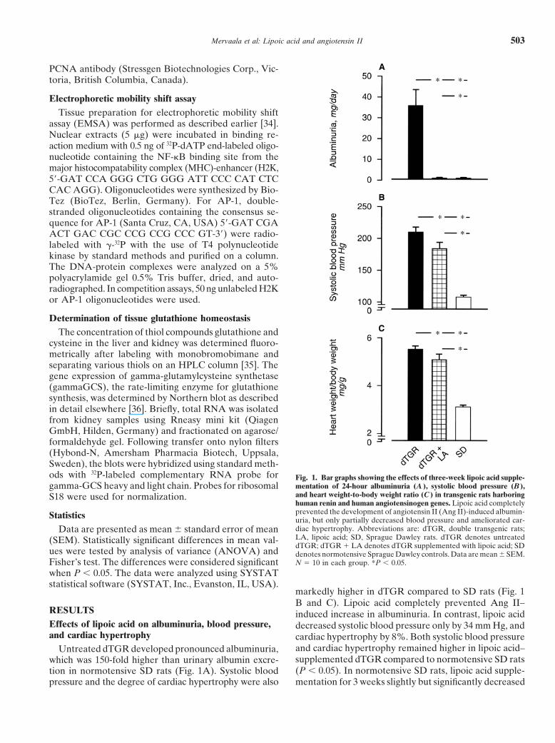
Untreated dTGR developed pronounced albuminuria,which was 150-fold higher than urinary albumin excre-tion in normotensive SD rats (Fig. 1A). Systolic bloodpressure and the degree of cardiac hypertrophy were also
Fig. 1. Bar graphs showing the effects of three-week lipoic acid supple-mentation of 24-hour albuminuria (A ), systolic blood pressure (B ),and heart weight-to-body weight ratio (C ) in transgenic rats harboringhuman renin and human angiotensinogen genes. Lipoic acid completelyprevented the development of angiotensin II (Ang II)-induced albumin-uria, but only partially decreased blood pressure and ameliorated car-diac hypertrophy. Abbreviations are: dTGR, double transgenic rats;LA, lipoic acid; SD, Sprague Dawley rats. dTGR denotes untreateddTGR; dTGR � LA denotes dTGR supplemented with lipoic acid; SDdenotes normotensive Sprague Dawley controls. Data are mean SEM.N � 10 in each group. *P � 0.05.
markedly higher in dTGR compared to SD rats (Fig. 1B and C). Lipoic acid completely prevented Ang II–induced increase in albuminuria. In contrast, lipoic aciddecreased systolic blood pressure only by 34 mm Hg, andcardiac hypertrophy by 8%. Both systolic blood pressureand cardiac hypertrophy remained higher in lipoic acid–supplemented dTGR compared to normotensive SD rats(P � 0.05). In normotensive SD rats, lipoic acid supple-mentation for 3 weeks slightly but significantly decreased

Mervaala et al: Lipoic acid and angiotensin II504
Fig. 2. Representative photomicrographs fromthe kidneys and heart of untreated dTGR (Aand C ) and dTGR treated with lipoic acid forthree weeks (B and D ). Untreated dTGR kid-neys show severe glomerular and vasculardamage with mesangial proliferation, glomeru-lar sclerosis and necrosis, increased intimal andmedia thickness, and deposition of matrix. Pro-found vascular damage with inflammation wasdetected in the heart of dTGR. Lipoic acidprevented angiotensin II (Ang II)–inducedvascular lesions both in the kidney and heart.Kidney and cardiac damage scores are de-picted in (E ) and (F ), respectively. dTGR isdouble transgenic rat. Data are mean SEM.N � 9 to 10 in each group. *P � 0.05. Originalmagnification, �200.
blood pressure (114 4 mm Hg vs. 128 3 mm Hg;P � 0.05), but did not influence cardiac weight-to-bodyweight ratio (3.522 0.045 mg/g vs. 3.493 0.05 mg/g;P 0.05) or albuminuria (253 19 �g/day vs. 190 55 �g/day; P 0.05).
Effects of lipoic acid on kidney and heart morphologyKidneys of untreated dTGR showed severe glomeru-
lar and vascular damage with mesangial proliferation,glomerular sclerosis and necrosis, increased intimal andmedia thickness, and deposition of matrix (Fig. 2, original

Mervaala et al: Lipoic acid and angiotensin II 505
Table 1. Immunohistochemical markers of leukocyte infiltration(ED1, CD4, CD8) and cell proliferation in dTGR harboring humanrenin and angiotensinogen genes, dTGR treated with LA for 3 weeks,
and normotensive SD rats
ANOVAVariable dTGR dTGR � LA SD P value
ED-1 1.070.22a,b 0.260.04 0.150.05 �0.001CD-4 0.390.07a,b 0.130.02 0.110.01 �0.001CD-8 0.240.08a,b 0.170.02 0.070.02 �0.001PCNA 0.160.04a,b 0.030.01 0.010.01 �0.001
Abbreviations are: dTGR, double transgenic rats; LA, lipoic acid; SD, SpragueDawley; ANOVA, analysis of variance; PCNA, proliferating cell nuclear antigen.Values are expressed as the percentage of the immunopositive label per totalsurface area. Mean SEM is given; N � 5 to 6 for each group.
aP � 0.05 compared to SD; bP � 0.05 compared to LA-treated dTGR.
magnification, �100). Profound vascular damage with fib-rinoid necrosis and accumulation of fibronectin (datanot shown) was also observed in the coronary arteries ofdTGR (Fig. 2). Lipoic acid effectively prevented Ang II-induced vascular lesions both in the kidney (Fig. 2) andheart (Fig. 2). Renal damage score (Fig. 2) and myocar-dial damage score (Fig. 2) were markedly decreased bylipoic acid in dTGR. Lipoic acid supplementation didnot influence either the kidneys or hearts in SD rats(data not shown).
Effects of lipoic acid on leukocyte infiltration andcell proliferation
Untreated dTGR showed pronounced leukocyte infil-tration in the kidneys measured as ED-1, CD-4, and CD-8immunopositive cells (Table 1). Infiltrated cells werelocated predominantly around the damaged vessels. Leu-kocyte infiltration was less prominent in the heart com-pared to the kidney (data not shown). Lipoic acid veryeffectively prevented inflammatory response in the kid-neys. In untreated dTGR, the number of PCNA-immu-nopositive cells in the kidneys was increased by 16-fold.Lipoic acid attenuated the Ang II–induced cell prolifera-tion in dTGR.
Effects of lipoic acid on NF-�B and AP-1 activation
Nuclear extracts of untreated dTGR showed increasednuclear factor kappa B (NF-�B) and activator protein-1(AP-1) DNA binding activities in kidney compared toSD controls (Fig. 3). Lipoic acid inhibited both NF-�Band AP-1 DNA binding activities. Binding specificityfor NF-�B was demonstrated by competition of excessunlabeled oligonucleotides containing the �B site fromthe MHC-enhancer (H2K). Nuclear extracts were super-shifted with antibodies against the NF-�B subunits p50and p65. Binding specificity for AP-1 was demonstratedby competition with excess of unlabeled oligonucleotidescontaining the AP-1 site. Complexes were supershiftedwith antibodies against c-fos and c-jun. In Figure 3, eachlane represents a separate animal (N � 3 in each group).NF-�B and AP-1 DNA binding activities were measured
using separate gels from 6 rats per group. The calculatedmean SEM are depicted as bar graphs in Figure 3.
Effects of lipoic acid on tissue glutathione homeostasis
Transgenic animals had an increased concentration ofglutathione in the kidneys (Fig. 4). On the other hand,cysteine levels were reduced. Lipoic acid partly pre-vented renal cysteine depletion. In the liver, lipoic acidincreased both cysteine and glutathione levels. This wasaccompanied with an increase in the gammaGCS heavysubunit mRNA (Fig. 5). Renal gammaGCS mRNA ex-pression was reduced in dTGR with or without lipoicacid, suggesting possible control function for oxidizedglutathione.
DISCUSSION
The important finding of the present study is that di-etary supplementation with lipoic acid protected againstthe development of Ang II–induced renal injury. Lipoicacid prevented Ang II–induced monocyte, macrophage,and T-cell infiltrations, and also attenuated cell prolifera-tion in the kidneys. The beneficial effects were associatedwith inhibition of transcription factors NF-�B and AP-1.Lipoic acid also improved cellular cysteine supply andthereby improved tissue thiol homeostasis. Since lipoicacid partially attenuated the Ang II–induced hyperten-sion, the renoprotective effects of lipoic acid may alsohave been mediated in part by decreased renal hemody-namic load.
Lipoic acid is a natural antioxidant that scavengesROS and regenerates/recycles endogenous antioxidants.Its metal chelating capacity and its ability to repair oxida-tive damage contribute to the protective effects [10, 14,37]. Moini et al [12] have postulated that lipoic acid mayalso exert prooxidant properties in vitro. Lipoic acid anddihydrolipoic acid were shown to promote the mitochon-drial permeability transition in permeabilized hepato-cytes and isolated rat liver mitochondria, and to stimulatesuperoxide anion production in rat liver mitochondriaand submitochondrial particles. Whether such prooxida-tive properties of lipoic acid contribute to its versatileeffects observed in vivo remains to be determined. Incell culture studies lipoic acid effectively inhibits theDNA binding activity of the redox-sensitive transcrip-tion factors NF-�B and AP-1 [10, 14, 38, 39].
Clark et al [16] used a transient focal central nervoussystem ischemic model and showed that pretreatmentwith lipoic acid reduced stroke infarct volume and im-proved neurologic function. Coombes et al [17] demon-strated that lipoic acid combined with vitamin E pro-tected the aged rat heart from ischemia/reperfusioninjury and improved cardiac performance. Lipoic acidproduced a modest decrease in blood pressure in ourstudy. Therefore, the renoprotective benefit found in

Mervaala et al: Lipoic acid and angiotensin II506
Fig. 3. Activation of DNA binding nuclear factors by Ang II in thekidney. EMSA for the detection of NF-�B and AP-1 showed increasedactivities in dTGR kidney compared to SD rats. Lipoic acid inhibitedboth NF-�B and AP-1. Binding specificity for NF-�B was demonstratedby competition of excess unlabeled oligonucleotides containing the �Bsite from the MHC-enhancer (H2K). Nuclear extracts were supershiftedwith antibodies against the NF-�B subunits p50 and p65. Binding speci-fity for AP-1 was demonstrated by competition with excess unlabeled
Fig. 4. Bar graphs showing the effects of three-week lipoic acid supple-mentation on liver and kidney cysteine and total glutathione. dTGRshows increased glutathione (A ) and decreased cysteine concentrations(B ) in the kidney, but not in the liver (C and D ). Lipoic acid increasedglutathione and cysteine concentrations only in the liver. DTGR isdouble transgenic rat. Mean SEM is given. N � 6 in each group.*P � 0.05.
dTGR could have been mediated, at least in part, byblood pressure–dependent mechanisms. However, weshowed previously in dTGR that hydralazine decreasedblood pressure but did not provide any protection againstAng II–induced renal injury [40]. Furthermore, Ang IIinduced leukocyte infiltration and endothelial adhesionmolecule overexpression in the kidneys of dTGR evenwhen blood pressure was decreased to control levels bya combination of hydralazine, hydrochlorothiazide, andreserpine [26]. Vasdev et al [41] showed that lipoic acidsupplementation normalized intracellular calcium con-
oligonucleotides containing the AP-1 site. Complexes were supershiftedwith antibodies against c-fos and c-jun. Each lane represents a separateanimal (N � 3 in each group). NF-�B and AP-1 DNA binding activitieswere measured using separate gels from 6 rats per group. The calculatedmean SEM is depicted as a bar graph. Abbreviations are: Ang II,angiotensin II; EMSA, electrophoretic mobility shift assay; NF-�B,nuclear factor kappa B; AP-1, activator protein-1; dTGR, double trans-genic rat; SD, Sprague Dawley rat; MHC, major histocompatibilitycomplex.

Mervaala et al: Lipoic acid and angiotensin II 507
Fig. 5. Bar graphs showing the effects of three-week lipoic acid supple-mentation on liver and kidney gammaGCS mRNA expressions. Lipoicacid increased gammaGCS mRNA expression in the liver, but not in thekidney. dTGR showed decreased renal gammaGCS mRNA expression.Abbreviations are: GCS, glutamylcysteine synthetase; dTGR, doubletransgenic rat. Data shown are mean SEM. N � 6 in each group.*P � 0.05.
centration, vascular resistance, and blood pressure inspontaneously hypertensive rats. Since the kidneys playa pivotal role in blood pressure regulation, the modestantihypertensive effect of lipoic acid may have been dueto preserved renal morphology and function. The antihy-pertensive effect may also be related to scavenging ofsuperoxide anions. Previous studies have demonstrated amarked antihypertensive effect by superoxide dismutasetreatment in Ang II–induced hypertension [42]. Thepresent study also demonstrated that the effects of lipoicacid on cardiac hypertrophy are likely to be mediatedthrough decreased pressure overload. We found a closecorrelation between blood pressure and the degree ofcardiac hypertrophy in dTGR earlier [43].
NF-�B activation can be inhibited by several thiol
antioxidants [38, 39]. Packer et al [10] have previouslyshown that lipoic acid suppresses NF-�B activation in-duced by either hydrogen peroxide, phorbol ester, ortumor necrosis factor-� (TNF-�) in Wurzburg T cells.Our study provided in vivo evidence that the beneficialeffects of chronic lipoic acid supplementation are associ-ated with NF-�B and AP-1 inhibition. They support thenotion that activation of these transcription factors wasrelated to Ang II–induced reactive oxygen species gener-ation [44].
Lipoic acid is taken up and rapidly reduced to dihy-drolipoate by lipoamide dehydrogenase, thioredoxin re-ductase, and glutathione reductase in the presence ofnicotinamide adenine dinucleotide (NADH) or nicotin-amide adenine dinucleotide phosphate (NADPH) [10, 14].Dihydrolipoate is then released extracellularly, reducescystine to cysteine, and thus promotes glutathione syn-thesis [39]. We found that dTGR decreased renal cyste-ine concentrations, whereas the glutathione concentra-tions were increased. We suggest that Ang II leads eitherto increased glutathione production or to decreased con-sumption. The expression of gammaGCS heavy subunitwas not increased, but this finding does not rule outchanges in posttranscriptional regulation. Further stud-ies are also needed to determine what happens to GSH.Lipoic acid increased renal cysteine concentrations, buthad little effect on glutathione. This finding suggeststhat lipoic acid cannot compensate for glutathione, butinstead improves the cysteine supply.
CONCLUSION
SD and dTGR rat liver thiol concentrations did notdiffer. However, lipoic acid increased cysteine and gluta-thione levels in the liver. Lipoic acid–induced increasesin hepatic thiol concentrations were accompanied witha marked up-regulation of gammaGCS gene expression.Our study agrees with previous reports demonstratingthat glutathione tissue concentrations in the heart andkidney usually remain unaltered by lipoic acid supple-mentation [10, 14]. Hence, our findings suggest that thebeneficial effects of lipoic acid against Ang II–inducedend-organ damage is more closely linked to scavengedROS and NF-�B and AP-1 inhibition rather than directchanges in the intracellular glutathione concentrations.Nevertheless, lipoic acid does have a significant effecton cysteine availability. We were unable to separate theeffects of cysteine, lipoate, glutathione, or other thiolson oxygen radical scavenging, thiols status, gene expres-sion, or protein function. We believe that our resultshave clinical implications. Lipoic acid may have greaterutility than antioxidant strategies that rely solely on vita-min E.

Mervaala et al: Lipoic acid and angiotensin II508
ACKNOWLEDGMENTS
This study was supported by grants from the Academy of Finlandand the Sigrid Juselius Foundation. We are grateful to Ms. SaaraMerasto and Ms. Sari Laakkonen for expert technical assistance. E.M.and P.F. contributed equally to this work. The Deutsche Forschungs-gemeinschaft and the Klinisch-Pharmakologischer Verbund, Berlin-Brandenburg, supported Dominik N. Muller.
Reprint requests to Eero Mervaala, M.D., Ph.D., Assistant Professor,Institute of Biomedicine, Pharmacology, Biomedicum Helsinki, Univer-sity of Helsinki (Haartmaninkatu 8), FIN-00014, Finland.E-mail: [email protected]
REFERENCES
1. Kunsch C, Medford RM: Oxidative stress as a regulator of geneexpression in the vasculature. Circ Res 85:753–766, 1999
2. Griendling KK, Sorescu D, Lassegue B, Ushio-Fukai M: Modu-lation of protein kinase activity and gene expression by reactiveoxygen species and their role in vascular physiology and pathophys-iology. Arterioscler Thromb Vasc Biol 20:2175–2183, 2000
3. Irani K: Oxidant signalling in vascular cell growth, death, andsurvival. A review of the roles of reactive oxygen species in smoothmuscle and endothelial cell mitogenic and apoptotic signalling.Circ Res 87:179–183, 2000
4. Romero JC, Reckelhoff JF: Role of angiotensin and oxidativestress in essential hypertension. Hypertension 34:943–949, 1999
5. Griendling KK, Sorescu D, Ushio-Fukai M: NAD(P)H oxidase.Role in cardiovascular biology and disease. Circ Res 86:494–501,2000
6. Meister A, Anderson ME: Glutathione. Annu Rev Biochem52:711–760, 1983
7. Meister A: Glutathione metabolism. Methods Enzymol 251:3–7,1995
8. Anderson ME: Glutathione and glutathione delivery compounds.Adv Pharmacol 38:65–78, 1997
9. Packer L, Roy S, Sen CK: Alpha-lipoic acid: A metabolic antioxi-dant and potential redox modulator of transcription. Adv Pharma-col 38:79–101, 1997
10. Packer L, Witt EC, Tritschler HJ: Alpha-lipoic acid as a biologi-cal antioxidant. Free Rad Biol Med 19:227–250, 1995
11. Carreau JP: Biosynthesis of lipoic acid via unsaturated fatty acids.Methods Enzymol 62:152–158, 1979
12. Moini H, Packer L, Saris NE: Antioxidant and prooxidant activi-ties of alpha-lipoic acid and dihydrolipoic acid. Toxicol Appl Phar-macol 182:84–90, 2002
13. Morikawa T, Yasuno R, Wada H: Do mammalian cells synthesizelipoic acid? Identification of a mouse cDNA encoding a lipoic acidsynthase located in the mitochondria. FEBS Lett 498:16–21, 2001
14. Sen CK, Packer L: Thiol homeostasis and supplements in physicalexercise. Am J Clin Nutr 72(Suppl 2):653S–659S, 2000
15. Sen CK, Packer L: Antioxidant and redox regulation of genetranscription. FASEB J 10:709–720, 1996
16. Clark WM, Rinker LG, Lessov NS, et al: Efficacy of antioxidanttherapies in transient focal ischemia in mice. Stroke 32:1000–1004,2001
17. Coombes JS, Powers SK, Hamilton KL, et al: Improved cardiacperformance after ischemia in aged rats supplemented with vitaminE and �-lipoic acid. Am J Physiol Regul Integr Comp Physiol279:R2149–R2155, 2000
18. Hagen TM, Ingersoll RT, Lykkesfeldt J, et al: (R)-�-Lipoicacid-supplemented old rats have improved mitochondrial function,decreased oxidative damage, and increased metabolic rate. FASEBJ 13:411–418, 1999
19. Suh JH, Shigeno ET, Morrow JD, et al: Oxidative stress in theaging rat heart is reversed by dietary supplementation with (R)-�-lipoic acid. FASEB J 15:700–706, 2001
20. Kocak G, Aktan F, Canbolat O, et al: Alpha-lipoic acid treatmentameliorates metabolic parameters, blood pressure, vascular reac-tivity and morphology of vessels already damaged by streptozo-tocin-diabetes. Diabetes Nutr Metab 13:308–318, 2000
21. Melhem MF, Craven PA, Liachenko J, DeRubertis FR: Alpha-lipoic acid attenuates hyperglycemia and prevents glomerular mes-angial matrix expansion in diabetes. J Am Soc Nephrol 13:108–116,2000
22. El Midaoui A, de Champlain J: Prevention of hypertension, insu-lin resistance, and oxidative stress by alpha-lipoic acid. Hyperten-sion 39:303–307, 2002
23. Takaoka M, Kobayashi Y, Yuba M, et al: Effects of alpha-lipoicacid on deoxycorticosterone acetate-salt-induced hypertension inrats. Eur J Pharmacol 424:121–129, 2001
24. Luft FC, Mervaala EMA, Muller DN, et al: Hypertension-induced end-organ damage. A new transgenic approach to an oldproblem. Hypertension 33:212–218, 1999
25. Mervaala EMA, Muller DN, Park J-K, et al: Monocyte infiltra-tion and adhesion molecules in a rat model of high human reninhypertension. Hypertension 33:389–395, 1999
26. Mervaala EMA, Muller DN, Schmidt F, et al: Blood pressure-independent effects in rats with human renin and angiotensinogengenes. Hypertension 35:587–594, 2000
27. Muller DN, Dechend R, Mervaala EMA, et al: NF-kappaBinhibition ameliorates angiotensin II-induced inflammatory dam-age in rats. Hypertension 35:193–201, 2000
28. Mervaala EMA, Cheng ZJ, Tikkanen I, et al: Endothelial dys-function and xanthine oxidoreductase activity in rats with humanrenin and angiotensinogen genes. Hypertension 37:414–418, 2001
29. Ganten D, Wagner J, Zeh K, et al: Species specificity of reninkinetics in transgenic rats harboring human renin and angiotensino-gen genes. Proc Natl Acad Sci USA 89:7806–7810, 1992
30. Bohlender J, Fukamizu A, Lippoldt A, et al: High human reninhypertension in transgenic rats. Hypertension 29:428–434, 1997
31. Lykkesfeldt J, Hagen TM, Vinarsky V, Ames BN: Age-associ-ated decline in ascorbic acid concentration, recycling, and biosyn-thesis in rat hepatocytes—Reversal with (R)-�-lipoic acid supple-mentation. FASEB J 12:1183–1189, 1998
32. Pere A-K, Lindgren L, Tuomainen P, et al: Dietary potassiumand magnesium supplementations in cyclosporine-induced hyper-tension and nephrotoxicity. Kidney Int 58:2462–2472, 2000
33. Finckenberg P, Lassila M, Inkinen K, et al: Cyclosporine Ainduces myocardial connective tissue growth factor in spontane-ously hypertensive rats on high sodium diet. Transplantation 71:951–958, 2001
34. Muller DN, Dechend R, Mervaala EMA, et al: NF-kappaBinhibition ameliorates angiotensin II-induced inflammatory dam-age in rats. Hypertension 35:193–201, 2000
35. Levonen AL, Laakso J, Vaskonen T, et al: Down-regulation ofrenal glutathione synthesis by systemic nitric oxide synthesis inhibi-tion in spontaneously hypertensive rats. Biochem Pharmacol 59:441–443, 2000
36. Levonen AL, Lapatto R, Saksela M, Raivio KO: Expression ofgamma-glutamylcysteine synthetase during development. PediatrRes 47:266–270, 2000
37. Biewenga GP, Haenen GR, Bast A: The pharmacology of theantioxidant lipoic acid. Gen Pharmacol 29:315–331, 1997
38. Sen CK, Packer L: Antioxidant and redox regulation of genetranscription. FASEB J 10:709–720, 1996
39. Sen CK: Redox signaling and the emerging therapeutic potentialof thiol antioxidants. Biochem Pharmacol 55:1747–1758, 1998
40. Muller DN, Mervaala EMA, Schmidt F, et al: Effect of bosentanon NF-kB, inflammation, and tissue factor in angiotensin II-induced end-organ damage. Hypertension 36:282–290, 2000
41. Vasdev S, Ford CA, Parai S, et al: Dietary �-lipoic acid supplemen-tation lowers blood pressure in spontaneously hypertensive rats.J Hypertens 18:567–573, 2000
42. Laursen JB, Rajagopalan S, Galis Z, et al: Role of superoxidein angiotensin II-induced but not catecholamine-induced hyperten-sion. Circulation 95:588–593, 1997
43. Mervaala EMA, Dehmel B, Gross V, et al: ACE inhibition andAT1 receptor blockade modify pressure-natriuresis by differentmechanisms in rats with human renin and angiotensinogen genes.J Am Soc Nephrol 10:1669–1680, 1999
44. Dzau V: Tissue angiotensin and pathobiology of vascular disease.A unifying hypothesis. Hypertension 37:1047–1052, 2001