Neuroprotective effects of alpha-lipoic acid in experimental spinal cord injury in rats

A p r i l 2 0 1 0 , v o l . 6 8 . N o . 4 , I s s n 0 3 0 0 - 2 9 7 7
p u b l i s h e d i n c o l l a b o r a t i o n w i t h t h e n e t h e r l a n d s a s s o c i a t i o n o f i n t e r n a l m e d i c i n e
Pathogenesis of bacterial sepsis•
Impact of dialysis modality on cognitive function•
Alpha-lipoic acid for diabetic neuropathy•
Incidence of diabetes mellitus in patients with dyslipidaemia•
Kimura’s disease of parotid glands and lymph nodes•
Hypothyroid Graves’ disease•
Dyslipidaemia despite treatment with cholesterol-lowering agents
A patient with hepatitis B, liver and kidney dysfunction and neuropathy:
what is your diagnosis?

a p r i l 2 0 1 0 , v o l . 6 8 , n o 4
M i s s i o n s t a t e M e n t
The mission of the journal is to serve the need of the internist to practise up-to-date medicine and to keep track with important issues in health care. With this purpose we publish editorials, original articles, reviews, controversies, consensus reports, papers on speciality training and medical education, book reviews and correspondence.
e d i t o r i a l i n f o r M a t i o n
C i t e d i n
Biosis database; embase/excerpta medica; index medicus (medline) science citation index, science citation index expanded, isi alerting services, medical documentation services, current contents/clinical medicine, PubMed.
editor in chiefMarcel Levi, Department of Medicine, Academic Medical Centre, University of Amsterdam, the Netherlands
associate editorsIneke J. ten BergeUlrich H. BeuersHarry R. BüllerEric FliersTon HagenbeekJoost B. HoekstraEvert de JongeJohn J. KasteleinRay T. KredietJoep LangeRien H. van OersTom van der PollPeter ReissDick J. RichelMarcus J. SchultzPeter SpeelmanPaul Peter Tak
Junior associate editorsGoda ChoiMichiel CoppensMette D. HazenbergKees HovinghJoppe W. HoviusPaul T. Krediet
Gabor E. LinthorstMax NieuwdorpRoos RenckensLeen de RijckeJoris RotmansMaarten R. SoetersSander W. TasTitia M. VriesendorpDavid van WesterlooJoost WiersingaSanne van Wissen
editorial boardG. Agnelli, Perugia, ItalyJ.V. Bonventre, Massachusetts, USAJ.T. van Dissel, Leiden, the NetherlandsR.O.B. Gans, Groningen, the NetherlandsA.R.J. Girbes, Amsterdam, the NetherlandsD.E. Grobbee, Utrecht, the NetherlandsD.L. Kastner, Bethesda, USAM.H. Kramer, Amsterdam, the NetherlandsE.J. Kuipers, Rotterdam, the NetherlandsPh. Mackowiak, Baltimore, USAJ.W.M. van der Meer, Nijmegen, the Netherlands
B. Lipsky, Seattle, USAB. Lowenberg, Rotterdam, the NetherlandsG. Parati, Milan, ItalyA.J. Rabelink, Leiden, the NetherlandsD.J. Rader, Philadelphia, USAJ.A. Romijn, Leiden, the NetherlandsJ.L.C.M. van Saase, Rotterdam, the NetherlandsY. Smulders, Amsterdam, the NetherlandsC.D.A. Stehouwer, Maastricht, the NetherlandsJ.L. Vincent, Brussels, BelgiumE. van der Wall, Utrecht, the NetherlandsR.G.J. Westendorp, Leiden, the Netherlands
editorial officeAcademic Medical Centre,Department of Medicine (F-4) Meibergdreef 9 1105 AZ Amsterdam The NetherlandsTel.: +31 (0)20-566 21 71 Fax: +31 (0)20-691 96 58E-mail: [email protected]://mc.manuscriptcentral.com/nethjmed


a p r i l 2 0 1 0 , v o l . 6 8 , n o 4
ISSN: 0300-2977
Copyright© 2010 Van Zuiden Communications B.V. All rights reserved. Except as outlined below, no part of this publication may be reproduced, stored in a retrieval system or transmitted in any form or by any means, electronic, mechanical, photocopying, recording or otherwise, without prior written permission of the publisher. Permission may be sought directly from Van Zuiden Communications B.V.
PhotocopyingSingle photocopies of single articles may be made for personal use as allowed by national copyright laws. Permission of the publisher and payment of a fee is required for all other photocopying, including multiple or systematic copying, copying for advertising or promotional purposes, resale, and all forms of document delivery. Special rates are available for educational institutions that wish to make photocopies for non-profit educational classroom use.
Derivative worksSubscribers may reproduce tables of contents or prepare lists of articles including abstracts for internal circulation within their institutions. Permission of the publisher is required for resale or distribution outside the institution. Permission of the publisher is also required for all other derivative works, including compilations and translations.
Electronic storagePermission of the publisher is required to store or use electronically any material contained in this journal, including any article or part of an article.
ResponsibilityNo responsibility is assumed by the publisher for any injury and/or damage to persons or property as a matter of product liability, negligence or otherwise, or from any use or operation of any methods, products, instructions or ideas contained in the material herein. Because of the rapid advances in the medical sciences, independent verification of diagnoses and drug dosages is advised.Although all advertising material is expected to conform to ethical (medical) standards, inclusion in this publication does not constitute a guarantee or endorsement of the quality or value of such product or of the claims made of it by its manufacturer.
SubscriptionsGeneral informationAn annual subscription to The Netherlands Journal of Medicine consists of 11 issues. Issues within Europe are sent by standard mail and outside Europe by air delivery. Cancellations should be made, in writing, at least two months before the end of the year.
Subscription feeThe annual subscription fee within Europe is 1 705, for the USA 1 735 and for the rest of the world 1 845. Subscriptions are accepted on a prepaid basis only and are entered on a calendar year basis.
Payment methodPlease make your cheque payable to Van Zuiden Communications B.V., PO Box 2122, 2400 CC Alphen aan den Rijn, the Netherlands or you can transfer the fee to ING Bank, account number 67.89.1 0.872, Castellumstraat 1, Alphen aan den Rijn, the Netherlands, swift-code: ING BNL 2A. Do not forget to mention the complete address for delivery of the Journal.
ClaimsClaims for missing issues should be made within two months of the date of dispatch. Missing issues will be mailed without charge. Issues claimed beyond the two-month limit must be prepaid at back copy rates.
Orders, preprints, advertising, changes in address, author or general enquiriesPlease contact the publisher.
Van Zuiden Communications B.V.PO Box 2122 2400 CC Alphen aan den RijnThe NetherlandsTel.: +31 (0)172-47 61 91Fax: +31 (0)172-47 18 82E-mail: [email protected]: www.njm-online.nl
Contentsedi tor i a l
The unhealthy fruits of insulin resistance 146F. Holleman
r e v ie ws
Recent insights into the pathogenesis of bacterial sepsis 147A.A. Anas, W.J. Wiersinga, A.F. de Vos, T. van der Poll
The possible impact of dialysis modality on cognitive function in chronic dialysis patients
153
J. Radic, D. Ljutic, M. Radic, V. Kovacic, M. Sain, K. Dodig Curkovic
Alpha lipoic acid: a new treatment for neuropathic pain in patients with diabetes?
158
G.S. Mijnhout, A. Alkhalaf, N. Kleefstra, H.J.G. Bilo
or igina l a rt iCl e
Five-year incidence of type 2 diabetes mellitus in patients with familial combined hyperlipidaemia
163
M.C.G.J. Brouwers, C.J.H. van der Kallen, N.C. Schaper, M.M.J. van Greevenbroek, C.D.A. Stehouwer
speCi a l a rt iCl e
Prevalence of dyslipidaemia in patients treated with lipid-modifying drugs in the Netherlands.Part of the Dyslipidaemia International Survey
168
A.C. Strang, H.A.H. Kaasjager, D.C.G. Basart, E.S.G. Stroes
C ase r eports
Kimura’s disease of the parotid glands and multiple cervical lymph nodes 175V.K. Dik, B.A. van de Wiel, W.L.E. Vasmel
Four patients with hypothyroid Graves’ disease 178A.J. Starrenburg-Razenberg, M. Castro Cabezas, I.M. Gan, T.L. Njo, A.P. Rietveld, J.W.F. Elte
photo quizzes
A patient with hepatitis B, liver and kidney dysfunction and polyneuropathy
181
L. Klieverik, M. Mallant, M. Sekkat, J. Branger
A patient with a long history of nicotine addiction presenting with haemoptysis
182
J.M. van Rooijen, L.Brokkaar, B.A.A.M. van Hasselt, P.H.P. Groeneveld
An 86-year-old man with a unilateral pectoral swelling 183Hung-Bin Tsai, Chin-Chi Kuo
l et t er to t he edi tor
Popping pneumothorax 187H.H.F. Remmelts, J.D. Banga
© Van Zuiden Communications B.V. All rights reserved.

146
a p r i l 2 0 1 0 , v o l . 6 8 , n o 4
© Van Zuiden Communications B.V. All rights reserved.
Extracting energy and substrates from the environment and excreting useless or even toxic by-products is key to cellular survival. However, in the evolution from a single-cell organism to a highly complex multi-organ multi-cellular species, cells have differentiated in function, needs and in their access to the environment. Therefore, it is no surprise that the elaborate cooperative cellular system that is called the human species has concomitantly developed a highly integrative and complex metabolic system which tries to ensure that the needs of individual organs and cells are appropriately met.This also explains why a seemingly simple disease entity called ‘type 2 diabetes mellitus’ (DM2) in fact encompasses a huge spectrum of differing underlying metabolic disturbances, all hallmarked by a high glucose. Moreover, as the paper by Brouwers et al. on familial combined hyperlipidaemia (FCHL) and subsequent risk of diabetes in this issue of our Journal nicely illustrates,1 DM2 itself is just one part of a far wider group of metabolic disturbances that may share some features but differ in others.For clinicians it is appealing and sometimes useful to try and classify these diseases in distinct groups based on certain clinical features. However, this approach is fraught with problems because of the clinical heterogeneity in (the presence of) symptoms, and because a simple clustering of symptoms such as the ‘metabolic syndrome’ does not correlate with one single and uniform pathophysiological explanation.2
This is also clear from the data of Brouwers et al. Within the syndromal diagnosis of ‘FCHL’ two types of dyslipidaemia may occur, either alone or in combination. However, these same dyslipidaemias may also occur in the context of other abnormalities, most notably those associated with obesity and insulin resistance. And, as the authors demonstrate, against this background of obesity and insulin resistance, DM2 is more likely to develop.
Clearly the distribution of BMI was not equal between cases and controls, with about 60% of spouses in the lower BMI quartiles and 60% of cases in the higher BMI quartiles. Accordingly, the authors corrected for confounders associated with insulin resistance such as BMI, and the association they found is persistent. From their data the authors subsequently conclude that ‘FCHL is a dynamic entity that may progress to DM2’.However, what is most noteworthy in their data is that of all variables, only correcting for baseline insulin levels negated the association they found between FCHL and subsequent diabetes. Thus, it would be more appropriate to say that FCHL and DM2 are fruits of a tree firmly rooted in the myriad of metabolic effects of insulin and insulin resistance. Some (genetic) factors may eventually prove to be root causes of both diseases and one candidate is the upstream transcription factor 1 (USF1).3 However, it is the genetic and environmental factors that are superimposed on this basis, e.g. increased apolipoprotein B production or beta-cell vulnerability, which determine whether the primary phenotype will be FCHL or DM2, and in which order these abnormalities will develop. Unfortunately, no healthy fruit grows from the tree of insulin resistance, and some branches will end up carrying both fruits at the same time.
r e f e r e n C e s
1. Brouwers MCGJ, van der Kallen CJH, Schaper NC, van Greevenbroek MMJ, Stehouwer CDA. Five-year incidence of type 2 diabetes mellitus in patients with familial combined hyperlipidaemia. Neth J Med. 2010;68(4):162-6.
2. Ferrannini E. Metabolic Syndrome: A solution in search of a problem. J Clin Endocrinol Metab. 2007;92(2):396-8.
3. Pajukanta P, Lilja HE, Sinsheimer JS, et al. Familial combined hyperlipidemia is associated with upstream transcription factor 1 (USF1). Nat Genet. 2004;36:371-6.
e d i t o r i a l
the unhealthy fruits of insulin resistance
F. Holleman
Department of Medicine/Division Clinical Diabetology, Academic Medical Centre, Amsterdam, the Netherlands

147
a p r i l 2 0 1 0 , v o l . 6 8 , n o 4
© Van Zuiden Communications B.V. All rights reserved.
a b s t r a C t
sepsis is a very heterogeneous clinical syndrome broadly defined as the systemic host response to an infection. until very recently, the prevailing concept of the pathogenesis of sepsis was that mortality is the consequence of an uncontrolled hyperinf lammatory response of the host. the disappointing results of nearly 40 years of anti-inflammatory strategies and the development of animal models that more closely mimic clinical sepsis have led to the reconsideration of the pathophysiology of sepsis. sepsis is now considered a misbalance between proinflammatory reactions (designed to kill invading pathogens but at the same time responsible for tissue damage) and anti-inflammatory responses (designed to limit excessive inflammation, but at the same time making the host more vulnerable for secondary infections). this review discusses key components of the pro- and anti-inflammatory response to sepsis, listing potential novel interventional strategies along the way.
K e y w o r d s
Cytokines, coagulation, innate immunity, sepsis
h i s t o r i C a l p e r s p e C t i v e
The original theory that sepsis mortality is caused by an excessive stimulation of the immune system by high bacterial loads was based on studies in animals that were infused with large doses of bacteria or bacterial products, in particular lipopolysaccharide (LPS), the toxic component of the Gram-negative bacterial cell wall. Such infusions result in a strong activation of different proinflammatory protein cascades which, although designed to protect the host against invading pathogens, can cause damage to
tissues when produced in high amounts. In a hallmark article published in 1985, Beutler and colleagues reported that neutralisation of a single proinflammatory cytokine – tumour necrosis factor (TNF)a – secreted after intravenous injection of an otherwise lethal dose of LPS prevented death in mice.1 Two years later, these results were confirmed by Tracey and colleagues, who showed that a monoclonal anti-TNFa antibody protected baboons against lethal Gram-negative sepsis elicited by intravenous infusion of high quantities of viable Escherichia coli.2 Since then anti-TNFa interventions have been reported to protect against lethality in a number of sepsis models in which high doses of bacteria or bacterial products were administered systemically.3 In addition, elimination of another proinflammatory cytokine, interleukin (IL)-1, also reduced lethality induced by LPS or living bacteria in animals.4,5 These early experimental sepsis studies resulted in the design and performance of many clinical trials seeking to inhibit either TNFa or IL-1 activity in patients with severe sepsis. Unfortunately, virtually all clinical sepsis trials with anti-TNFa strategies and recombinant IL-1 receptor antagonist failed, and many other anti-inflammatory strategies were also not successful in altering the outcome of patients with sepsis. As such, the hypothesis that excessive inflammation is the main or sole underlying cause for an adverse outcome of a septic patient is not correct.
i n d u C t i o n o f a n i n n a t e i M M u n e r e s p o n s e t o b a C t e r i a
The innate immune system is able to detect pathogens via a limited number of pattern-recognition receptors (PRRs).6,7 PRRs recognise conserved motifs expressed by pathogens, known as pathogen-associated molecular patterns (PAMPs).
r e v i e w
recent insights into the pathogenesis of bacterial sepsis
A.A. Anas, W.J. Wiersinga, A.F. de Vos, T. van der Poll*
Center of Infection and Immunity Amsterdam (CINIMA) and the Center for Experimental and Molecular Medicine, Academic Medical Center, University of Amsterdam, Amsterdam, the Netherlands, *corresponding author: tel.: +31 (0)20-566 59 10, fax: +31 (0)20-697 71 92,
e-mail: [email protected]

148
a p r i l 2 0 1 0 , v o l . 6 8 , n o 4
Examples of bacterial PAMPs are LPS, expressed by all virulent Gram-negative bacteria, peptidoglycan, lipopeptides (constituents of many pathogens), lipoteichoic acid (a cell wall component of Gram-positive bacteria), flagellin (factor in the mobility of bacteria) and bacterial DNA.6,7 Additionally, PRRs can also recognise endogenous mediators released upon injury, thereby warning the host for imminent danger. Such endogenous danger signals have been named ‘alarmins’ or ‘danger-associated molecular patterns’ (DAMPs). Heat shock proteins, fibrinogen, hyaluronic acid and high-mobility group box-1 protein (HMGB-1) are examples of DAMPs that cause further amplification of the proinflammatory response through Toll-like receptor (TLR) 4 (see below).A specific family of PRRs named Toll-like receptors (TLRs) play a pivotal role in the initiation of cellular innate immune responses. Thirteen TLRs (TLRs 1 to 13) have been identified in mammals. Bacterial ligands for most TLRs have been described; table 1 summarises TLR specificity for several bacterial PAMPs with probable relevance for sepsis. The entire TLR family signals via four adapter proteins (myeloid differentiation primary-response protein 88 [MyD88], TIR domain-containing adaptor protein [TIRAP], TIR domain-containing adaptor protein-inducing IFNb [TRIF] and TRIF-related adaptor molecule [TRAM]), which together with a number of protein kinases take care of the recognition and response to microbial molecules. With regard to the role of TLRs in sepsis, it should be noted that TLRs are on the one hand essential for the early detection of pathogens, but on the other hand may also cause excessive inflammation after
uncontrolled stimulation.7 As an example, TLR4 deficient mice are fully protected against LPS-induced lethality, but these animals display an enhanced susceptibility to several Gram-negative infections.8 The clinical relevance of TLR signalling is reflected by the recent description in children with a genetic deficiency for MyD88 or IL-1 receptor-associated kinase 4 (IRAK4), a kinase acting directly downstream from MyD88, who are especially vulnerable to purulent infections.9,10 In addition, several single nucleotide polymorphisms in genes encoding TLRs have been associated with an altered susceptibility to bacterial infections.11
Several other innate immune receptors have been implicated in the recognition of bacteria and induction of a host inflammatory response after infection. Whereas TLRs detect pathogens at either the cell surface or in lysosomes/endosomes, microorganisms that invade the cytosol can be recognised by cytoplasmatic PRRs, among which nucleotide-binding oligomerisation domain (NOD)-like receptors (NLRs).12 Several members of the NLR family can assemble multimolecular complexes termed ‘inflammasomes’ in response to various activators, leading to the activation of inflammatory caspases. Activation of the NLRP3 inflammasome by PAMPs or DAMPs induces activation of caspase-1, which causes the processing of the proinflammatory cytokines interleukin (IL)-1b and IL-18.12 Although it has become clear that NLRs are of utmost importance for the recognition of bacteria by the innate immune system, their exact role in sepsis pathophysiology is far from clear.
Anas, et al. Pathogenesis of sepsis.
table 1. Pathogen and danger associated molecular patterns and their recognition by Toll-like receptors
species tlr
pathogen-associated molecular patterns
Bacteria
Lipopolysaccharide Gram-negative bacteria TLR4
Lipoteichoic acid Gram-positive bacteria TLR2*
Peptidoglycan Most bacteria TLR2
Triacyl lipopeptides Most bacteria TLR1/TLR2
Diacyl lipopeptides Mycoplasma spp TLR2/ TLR6
Porins Neisseria TLR2
Flagellin Flagellated bacteria TLR5
CpG DNA Bacteria TLR9
Unknown Uropathogenic bacteria TLR11‡
danger-associated molecular patterns**
Heat shock proteins Host TLR4
Fibrinogen, fibronectin Host TLR4
Hyaluronan Host TLR4
Biglycans Host TLR4
HMGB1 Host TLR4, TLR2
the table shows paMps and daMps with likely relevance for bacterial sepsis (paMps expressed by fungi, viruses and parasites are not shown). *for detection of lta from some pathogens tlr6 functions as a coreceptor for tlr2. ‡tlr11 is not functional in humans. **recent studies describe a role for tlrs in acute injury using rodent models of haemorrhagic shock, ischaemia and reperfusion, tissue trauma and wound repair, and various toxic exposures; these studies have implicated tlr4 as a major factor in the initial injury response. the table shows endogenous mediators identified as tlr4 ligands.

149
a p r i l 2 0 1 0 , v o l . 6 8 , n o 4
Triggering receptor expressed on myeloid cells-1 (TREM-1) amplifies the TLR- and NLR-mediated inflammatory response to microbial products.13,14 TREM-1 is strongly and specifically expressed on monocytes and neutrophils from patients with sepsis. Blockade of TREM-1 protected mice against LPS-induced shock, as well as microbial sepsis caused by live E. coli or coecal ligation and puncture. In addition, a synthetic peptide mimicking a short highly conserved domain of soluble TREM-1 protected septic animals from hyper-responsiveness and death.13
The exponentially increasing knowledge of PRRs involved in the activation of the innate immune system will likely lead to new sepsis interventions. At present, a phase III clinical sepsis trial with Eritoran, a TLR4 antagonist, is under way.7
h M g b 1 a n d r a g e
HMGB1, a nuclear protein that stabilises nucleosome formation, has been implicated in the pathogenesis of sepsis.15 Patients with sepsis demonstrate elevated circulating levels of HMGB1.16,17 LPS-induced shock in mice was associated with a relatively late release of HMGB1 into the circulation; importantly, an anti-HMGB1 antibody protected against LPS-induced lethality even when the administration was postponed until after the peak levels of TNFa and IL-1 had been reached.16 Delayed administration of anti-HMGB1 also improved survival in a model of abdominal sepsis.18 Considering that the therapeutic window for anti-HMGB1 therapies is much wider than for TNF-neutralising strategies, inhibitors of HMGB1 may be valuable as an adjunctive therapy for severe sepsis.It is uncertain whether highly purified HMGB1 can directly activate cells. It has been suggested that other molecules bound by HMGB1 are at least in part responsible for this. Nonetheless, several receptors have been implicated in mediating the cellular effects of HMGB1, including TLR2 and TLR 4, and the receptor for advanced glycation end products (RAGE).15,19 RAGE is a promiscuous receptor that interacts with diverse ligands such as advanced glycation end products, S100/calgranulins, amyloid A, leucocyte adhesion receptors, Escherichia coli curli operons and HMGB1. The potential role of RAGE signalling in sepsis pathophysiology has been documented in mice with abdominal sepsis: both RAGE-deficient mice and wild-type mice treated with soluble RAGE were partially protected against lethality in this model of severe sepsis.20,21 In addition, RAGE-deficient mice demonstrated an improved host defence during pneumococcal pneumonia.22 Further research is warranted to address the therapeutic potential of RAGE (ligand) inhibitors in sepsis.
M a C r o p h a g e M i g r a t i o n i n h i b i t o r y f a C t o r
Macrophage migration inhibitory factor (MIF) is a cytokine that can be produced by many different cell types. Serum MIF levels are elevated in patients with sepsis.23,24 MIF regulates innate immune responses through modulation of TLR4: when MIF-deficient mice were challenged with LPS they showed a defective response as a direct result of decreased TLR4 expression.25 Inhibition of MIF activity with neutralising anti-MIF antibodies protected mice from septic shock.23 These data suggest that MIF-directed therapies offer a new treatment opportunity for sepsis. Intriguingly, however, polymorphisms associated with higher MIF expression were recently shown to be associated with a reduced 90-day mortality in patients with community-acquired pneumonia.26 These new data prompt caution in the clinical application of anti-MIF strategies in infectious diseases.
M y e l o i d - r e l a t e d p r o t e i n ( M r p ) 8 a n d M r p 1 4
Myeloid-related protein 8 (Mrp8 also called S100A8) and Mrp14 (also called S100A9) are members of the S100 protein family.27 Mrp8 and Mrp14 can form heterodimers that elicit a variety of inflammatory responses. Mrp8/14 complexes can activate TLR4 and amplify the LPS-triggered inflammatory responses of phagocytes.28 In patients with sepsis and in healthy humans injected with LPS elevated Mrp8/14 plasma levels have been observed.29 Mice lacking Mrp8-Mrp14 complexes had an increased survival during LPS-induced lethal shock and bacterial sepsis,28 and displayed a reduced bacterial dissemination after intraperitoneal infection with E. coli.29 It remains to be established whether inhibition of Mrp8/14 could be a useful adjunctive therapy for clinical sepsis.
C 5 a a n d C 5 a r e C e p t o r
Although the complement system has traditionally been considered a central part of host defence against invading pathogens, complement activation may also contribute to an adverse outcome of sepsis.30 Indeed, infusion of anti-C5a antibodies improved haemodynamic parameters in pigs infused with LPS or live E. coli and reduced mortality in primates with E. coli sepsis and rats subjected to coecal ligation and puncture.30 As such, interventions seeking to block C5a signalling represent promising targets for sepsis treatment, although as with other anti-inflammatory strategies, an important goal of complement inhibition would be to avoid disrupting the role of complement in host defence.
Anas, et al. Pathogenesis of sepsis.

150
a p r i l 2 0 1 0 , v o l . 6 8 , n o 4
C o a g u l a t i o n a n d a n t i C o a g u l a t i o n
Patients with sepsis almost invariably show evidence for activation of the coagulation system.31,32 Tissue factor (TF) is regarded as the primary initiator of coagulation in sepsis. The pivotal role of TF in activation of coagulation during endotoxaemia and sepsis has been established by many different experiments. In particular, a number of different strategies that prevent the activation of the TF pathway in endotoxaemic humans and chimpanzees, and in bacteraemic baboons abrogated the activation of the common pathway of coagulation, which in septic baboons was accompanied by a reduced mortality.31,32
Procoagulant events are controlled by three major anticoagulant proteins: tissue factor pathway inhibitor (TFPI), antithrombin and activated protein C (APC).31,32 During severe sepsis the activities of TFPI, antithrombin and the protein C-APC system are impaired, which together with enhanced TF-dependent coagulation results in a shift toward a net procoagulant state. In septic primates the administration of either TFPI, antithrombin or APC attenuated consumptive coagulopathy.31,32 Large phase III clinical trials in sepsis patients have been completed with these three anticoagulants.33-36 Only recombinant human APC was found to reduce 28-day mortality in patients with severe sepsis;33 importantly, APC was not effective in patients with severe sepsis and a low risk of death.36 Recently, the European licensing authorities have requested Eli Lilly (the manufacturer of recombinant human APC) to perform another placebo-controlled trial with APC in adult patients with severe sepsis; this trial (PROWESS-SHOCK) was recently initiated.In recent years, much attention has been given to the role of protease-activated cell receptors (PARs) in linking coagulation and inflammation.37 The PAR family consists of four members, PAR-1 to PAR-4, which are localised in the vasculature on endothelial cells, mononuclear cells, platelets, fibroblasts and smooth muscle cells. Recently, cell penetrating peptides (so-called pepducins) were used to delineate the roles of PAR-1 and PAR-2 in LPS shock and abdominal sepsis.38 Evidence was provided that activation of PAR-1 is harmful during the early phases of endotoxaemia and sepsis, facilitating pulmonary leak and disseminated intravascular coagulation, but becomes beneficial at later stages.38 Remarkably, PAR-1 deficiency was reported to protect mice against LPS-induced lethality in an LD80 model of endotoxaemia at least in part through interruption of the PAR-1 mediated amplification of systemic inflammation.39 More studies are warranted to determine the potential value of PAR signalling inhibitors for the treatment of sepsis.
i M M u n e s u p p r e s s i o n a n d a p o p t o s i s
Although severe infection may be associated with an early phase of hyperinflammation, in most if not all patients who survive the acute phase of sepsis, a prolonged state of immune suppression evolves, a condition referred to as immunoparalysis.7,40 Indeed, the greater part of patients who are enrolled in sepsis trials display evidence of this state of reduced immune responsiveness: their blood leucocytes are less capable of releasing proinflammatory cytokines upon stimulation with bacteria or bacterial products. Although immunoparalysis has been regarded as beneficial in the sense that it counteracts a potential devastating pro-inflammatory response, it can also lead to an inability to clear infection and a subsequent predisposition to nosocomial infection. Experimental data have provided firm evidence for a causal role for enhanced apoptosis in the pathogenesis of sepsis, i.e. prevention of apoptosis of lymphocytes or the intestinal epithelium improved survival in experimental sepsis.40
t h e C h o l i n e r g i C a n t i - i n f l a M M a t o r y p a t h w a y
The cholinergic nervous system, and in particular the vagus nerve, represents another host response pathway designed to limit inflammatory responses.41 In the so-called cholinergic anti-inflammatory pathway enhanced efferent activity of parasympathetic nerve endings results in the release of acetylcholine, which by a specific action on a7 cholinergic receptors on macrophages suppresses proinflammatory cytokine production.41 Disruption of this neural-based system by vagotomy renders animals more vulnerable to LPS toxicity. Conversely, electrical stimulation of the efferent vagus nerve prevented the development of shock and attenuated the release of TNFa, whereas stimulation of a7 cholinergic receptors by specific agonists, such as nicotine, attenuated systemic inflammation and improved the outcome of mice with polymicrobial abdominal sepsis.41 Together, these preclinical data suggest that stimulation of the vagus nerve and/or pharmacological a7 cholinergic receptor agonists may be a useful strategy in the treatment of the severe inflammation accompanying sepsis.
C o n C l u s i o n
Sepsis can be defined as the host response to infection. For many years this response was considered to be dictated by an overwhelming inflammatory reaction to invading bacteria. Although some septic patients may succumb from the initial exacerbated hyperinflammatory
Anas, et al. Pathogenesis of sepsis.

151
a p r i l 2 0 1 0 , v o l . 6 8 , n o 4
response, the majority of patients die during the following extended period of immunodepression. A careful balance between the inf lammatory and anti-inflammatory response is vital for a successful host response to sepsis. Intervening in this delicate balance in order to improve sepsis outcome will be a major challenge for the years to come.
a C K n o w l e d g M e n t
W.J. Wiersinga is supported by a VENI grant from the Netherlands Organisation of Scientific Research.
r e f e r e n C e s
1. Beutler B, Milsark IW, Cerami AC. Passive immunization against cachectin/tumor necrosis factor protects mice from lethal effect of endotoxin. Science. 1985;229(4716):869-71.
2. Tracey KJ, Fong Y, Hesse DG, et al. Anti-cachectin/TNF monoclonal antibodies prevent septic shock during lethal bacteraemia. Nature. 1987;330(6149):662-4.
3. Lorente JA, Marshall JC. Neutralization of tumor necrosis factor in preclinical models of sepsis. Shock. 2005;24(Suppl 1):107-19.
4. Ohlsson K, Bjork P, Bergenfeldt M, Hageman R, Thompson RC. Interleukin-1 receptor antagonist reduces mortality from endotoxin shock. Nature. 1990;348(6301):550-2.
5. Fischer E, Marano MA, Van Zee KJ, et al. Interleukin-1 receptor blockade improves survival and hemodynamic performance in Escherichia coli septic shock, but fails to alter host responses to sublethal endotoxemia. J Clin Invest. 1992;89(5):1551-7.
6. Ishii KJ, Koyama S, Nakagawa A, Coban C, Akira S. Host innate immune receptors and beyond: making sense of microbial infections. Cell Host Microbe. 2008;3(6):352-63.
7. van der Poll T, Opal SM. Host-pathogen interactions in sepsis. Lancet Infect Dis. 2008;8(1):32-43.
8. Beutler B, Rietschel ET. Innate immune sensing and its roots: the story of endotoxin. Nat Rev Immunol. 2003;3(2):169-76.
9. von Bernuth H, Picard C, Jin Z, et al. Pyogenic bacterial infections in humans with MyD88 deficiency. Science. 2008;321(5889):691-6.
10. Ku CL, von Bernuth H, Picard C, et al. Selective predisposition to bacterial infections in IRAK-4-deficient children: IRAK-4-dependent TLRs are otherwise redundant in protective immunity. J Exp Med. 2007;204(10):2407-22.
11. Schroder NW, Schumann RR. Single nucleotide polymorphisms of Toll-like receptors and susceptibility to infectious disease. Lancet Infect Dis. 2005;5(3):156-64.
12. Stutz A, Golenbock DT, Latz E. Inflammasomes: too big to miss. J Clin Invest. 2009;119(12):3502-11.
13. Klesney-Tait J, Turnbull IR, Colonna M. The TREM receptor family and signal integration. Nat Immunol. 2006;7(12):1266-73.
14. Netea MG, Azam T, Ferwerda G, Girardin SE, Kim SH, Dinarello CA. Triggering receptor expressed on myeloid cells-1 (TREM-1) amplifies the signals induced by the NACHT-LRR (NLR) pattern recognition receptors. J Leukoc Biol. 2006;80(6):1454-61.
15. Lotze MT, Tracey KJ. High-mobility group box 1 protein (HMGB1): nuclear weapon in the immune arsenal. Nat Rev Immunol. 2005;5(4):331-42.
16. Wang H, Bloom O, Zhang M, et al. HMG-1 as a late mediator of endotoxin lethality in mice. Science. 1999;285(5425):248-51.
Anas, et al. Pathogenesis of sepsis.
figure 1. Important components of the host response to sepsis
Pathogen
PAMPs
TRLs
DAMPs
Balanced responsePathogen elimination
Tissue recovery
Full recovery
Host cells‘Innate immune response’
Unbalanced response
Immune suppressionApoptosis of immune cells
HyperinflammationCytokine mediated pathology
Coagulation activationComplement activation
Early mortality with acute organ dysfunction
Late mortality and/or development of secondary
infections
the interaction between pathogens and the host is mediated initially via an interaction between paMps (pathogen associated molecular pathogens) and tlrs (toll-like receptors). this interaction can result in the release of alarmins or daMps (danger associated molecular patterns) which have the ability to further amplify the inflammatory response at least in part via tlrs. the resulting innate response of immune cells can result in a balanced reaction leading to pathogen elimination and tissue recovery or an unbalanced reaction that on the one hand can lead to exaggerated inflammation and tissue injury and on the other hand to immune suppression caused by immune cell apoptosis.

152
a p r i l 2 0 1 0 , v o l . 6 8 , n o 4
17. van Zoelen MA, Laterre PF, van Veen SQ, et al. Systemic and local high mobility group box 1 concentrations during severe infection. Crit Care Med. 2007;35(12):2799-804.
18. Yang H, Ochani M, Li J, et al. Reversing established sepsis with antagonists of endogenous high-mobility group box 1. Proc Natl Acad Sci USA. 2004;101(1):296-301.
19. van Zoelen MA, Yang H, Florquin S, et al. Role of toll-like receptors 2 and 4, and the receptor for advanced glycation end products in high-mobility group box 1-induced inflammation in vivo. Shock. 2009;31(3):280-4.
20. Liliensiek B, Weigand MA, Bierhaus A, et al. Receptor for advanced glycation end products (RAGE) regulates sepsis but not the adaptive immune response. J Clin Invest. 2004;113(11):1641-50.
21. Lutterloh EC, Opal SM, Pittman DD, et al. Inhibition of the RAGE products increases survival in experimental models of severe sepsis and systemic infection. Crit Care. 2007;11(6):R122.
22. van Zoelen MA, Schouten M, de Vos AF, et al. The receptor for advanced glycation end products impairs host defense in pneumococcal pneumonia. J Immunol. 2009;182(7):4349-56.
23. Calandra T, Echtenacher B, Roy DL, et al. Protection from septic shock by neutralization of macrophage migration inhibitory factor. Nat Med. 2000;6(2):164-70.
24. Emonts M, Sweep FC, Grebenchtchikov N, et al. Association between high levels of blood macrophage migration inhibitory factor, inappropriate adrenal response, and early death in patients with severe sepsis. Clin Infect Dis. 2007;44(10):1321-8.
25. Roger T, David J, Glauser MP, Calandra T. MIF regulates innate immune responses through modulation of Toll-like receptor 4. Nature. 2001;414(6866):920-4.
26. Yende S, Angus DC, Kong L, et al. The influence of macrophage migration inhibitory factor gene polymorphisms on outcome from community-acquired pneumonia. Faseb J. 2009;23(8):2403-11.
27. Ehrchen JM, Sunderkotter C, Foell D, Vogl T, Roth J. The endogenous Toll-like receptor 4 agonist S100A8/S100A9 (calprotectin) as innate amplifier of infection, autoimmunity, and cancer. J Leukoc Biol. 2009;86(3):557-66.
Anas, et al. Pathogenesis of sepsis.
28. Vogl T, Tenbrock K, Ludwig S, et al. Mrp8 and Mrp14 are endogenous activators of Toll-like receptor 4, promoting lethal, endotoxin-induced shock. Nat Med. 2007;13(9):1042-9.
29. van Zoelen MA, Vogl T, Foell D, et al. Expression and role of myeloid-related protein-14 in clinical and experimental sepsis. Am J Respir Crit Care Med. 2009;180(11):1098-106.
30. Guo RF, Ward PA. Role of C5a in inflammatory responses. Annu Rev Immunol. 2005;23:821-52.
31. Schouten M, Wiersinga WJ, Levi M, van der Poll T. Inflammation, endothelium, and coagulation in sepsis. J Leukoc Biol. 2008;83(3):536-45.
32. Levi M, van der Poll T. Inflammation and coagulation. Crit Care Med. 2010;38(2 Suppl):S26-34.
33. Bernard GR, Vincent JL, Laterre PF, , et al. Efficacy and safety of recombinant human activated protein C for severe sepsis. N Engl J Med. 2001;344(10):699-709.
34. Warren BL, Eid A, Singer P, et al. Caring for the critically ill patient. High-dose antithrombin III in severe sepsis: a randomized controlled trial. Jama. 2001;286(15):1869-78.
35. Abraham E, Reinhart K, Opal S, et al. Efficacy and safety of tifacogin (recombinant tissue factor pathway inhibitor) in severe sepsis: a randomized controlled trial. JAMA. 2003;290(2):238-47.
36. Abraham E, Laterre PF, Garg R, et al. Drotrecogin alfa (activated) for adults with severe sepsis and a low risk of death. N Engl J Med. 2005;353(13):1332-41.
37. Shpacovitch V, Feld M, Bunnett NW, Steinhoff M. Protease-activated receptors: novel PARtners in innate immunity. Trends Immunol. 2007;28(12):541-50.
38. Kaneider NC, Leger AJ, Agarwal A, et al. ‘Role reversal’ for the receptor PAR1 in sepsis-induced vascular damage. Nat Immunol. 2007;8(12):1303-12.
39. Niessen F, Schaffner F, Furlan-Freguia C, et al. Dendritic cell PAR1-S1P3 signalling couples coagulation and inflammation. Nature. 2008;452(7187):654-8.
40. Hotchkiss RS, Nicholson DW. Apoptosis and caspases regulate death and inflammation in sepsis. Nat Rev Immunol. 2006;6(11):813-22.
41. Tracey KJ. Reflex control of immunity. Nat Rev Immunol. 2009;9(6):418-28.

153
a p r i l 2 0 1 0 , v o l . 6 8 , n o 4
a b s t r a C t
Chronic kidney disease (CKd) is a growing public health problem. individuals in all stages of CKd are at higher risk for development of cognitive impairment and this may be a major determinant in their quality of life (qol). the prevalence of cognitive deficits is particularly high in subjects with end-stage renal disease (esrd). while it is sufficiently well documented that esrd is linked with a change in cognitive function, little is known about the influence of different dialysis modalities on cognitive function. the effect of dialysis modality on risk of cognitive impairment is unclear. some data suggest that patients with esrd treated with chronic ambulatory peritoneal dialysis (Capd) had consistently better cognitive function than patients treated with haemodialysis (hd). we concluded that the previously observed apparent difference between two modalities of dialysis treatments resulted either from very low dialysis delivery or comparison with poorly matched controls. regarding these data from previous studies we hypothesised that well-dialysed, well-nourished and medically stable hd patients had no cognitive dysfunction in comparison with well-dialysed, well-nourished, medically stable and demographically matched Capd patients. also, future studies are needed to differentiate between modality as a risk factor from the factors contributing to selection bias among patients choosing Capd over hd.
K e y w o r d s
Chronic kidney disease, cognitive function, dialysis modality
Chronic kidney disease (CKD) is the permanent loss of kidney function and it is a rapidly growing global health problem, with a prevalence of 15% in developed
r e v i e w
the possible impact of dialysis modality on cognitive function in chronic dialysis patients
J. Radic1*, D. Ljutic1, M. Radic2, V. Kovacic1, M. Sain1, K. Dodig Curkovic3
Department of 1Nephrology, 2Rheumatology and Clinical Immunology, University Hospital, Split, Croatia, 3Department of Psychiatry, University Hospital, Osijek, Croatia, *corresponding author:
e-mail: [email protected]
nations.1,2 The final stage of chronic kidney disease is end-stage renal disease (ESRD). ESRD is a progressive, debilitating, chronic illness that requires nursing and medical interventions that include dialysis, education on lifestyle alterations, and dietary and fluid restrictions.In ESRD, kidney function can be replaced by three main medical treatment modalities: haemodialysis (HD), chronic ambulatory peritoneal dialysis (CAPD) or by kidney transplantation. The best treatment for a patient who is very close to ESRD is pre-emptive transplantation, but transplantation generally does not happen because of an insufficient number of donors. The two major dialysis types, HD and PD, are not only different from one another technically, but also with regard to the expectations of patients pertaining to the effort involved. Each dialysis type has its advantages and disadvantages and has a different level of impact on patients’ physical, psychological and social health, and each places its own limitations on lifestyle.3
Results from single centre and multicentre studies with CAPD and HD patients show conflicting results with respect to the survival benefits of one form of therapy over the other.4-9 Based on review of recent publications and additional analyses of US Medicare data, patient survival is similar for CAPD and HD but important differences do exist within select subgroups of patients, particularly those subgroups defined by age and the presence or absence of diabetes.8
ESRD can have an impact on patients’ quality of life (QOL), potentially affecting their physical and mental health, functional status, independence, general well-being, personal relationships and social functioning. Awareness of patient satisfaction and QOL has been increasing and health-related QOL issues are now recognised as important outcome measures in health care, cost-effective analyses of the efficacy of medical care and clinical trials,
© Van Zuiden Communications B.V. All rights reserved.

154
a p r i l 2 0 1 0 , v o l . 6 8 , n o 4
and therapeutic interventions for chronic conditions, including end-stage renal disease (ESRD). QOL is also a factor in the decision-making process for dialysis treatment selection.11 Defining QOL is complex as it can encompass a wide range of factors including psychological, cognitive, social, economic, political, cultural, spiritual, and physical factors.12
Comparative studies suggest that health-related QOL differs within dialysis patients, such as CAPD vs HD. However, evidence to suggest one mode of dialysis modality is better than the other in impacting on/improving health-related QOL is still inconclusive.13 Lately, awareness of patient satisfaction and QOL has been increasing. Some recent studies have evaluated patients’ treatment satisfaction level. In most of these studies, CAPD patients seem to be much happier than HD patients are.14-16 These results do not change after adjustment for age, ethnicity, education level, marital status, employment status, distance from the treatment centre, and treatment duration.15 Patients in the HD treatment modality, particularly those with many years of treatment, experienced a more compromised QOL in comparison with CAPD patients.17 In a meta-analysis of 61 studies, CAPD patients were characterised by a better well-being and less distress than HD patients.18 Some studies suggested that both HD and CAPD patients had similar health-related QOL.19,20 Regarding psychological dimensions in ESRD, it seems that CAPD patients are better adjusted than HD patients. This may be because the peritoneal treatment modality offers increased autonomy and control, flexibility in everyday life and the dietary regime, as well as fewer social restrictions.18,21-23 CAPD patients have been found to report better QOL ratings in specific areas such as ‘perceived ability to travel’, ‘financial concerns’, ‘restriction in eating and drinking’ and ‘dialysis access problems’.24 Furthermore, PD patients have indicated more positive ratings in several disease QOL domains, e.g. less kidney disease burden, and being more encouraged and satisfied with care.25 Compared with HD patients, CAPD patients experienced more personal control and had a better understanding of the illness.26 The only randomised trial investigating health-related QOL of CAPD and HD patients found a small difference favouring HD patients after two years follow-up.27
Change in cognitive function is one of the well-known consequences of ESRD28,29 and this may be a major determinant in patients’ QOL. Cognitive impairment is defined as a new deficit in at least two areas of cognitive functioning. These may include disturbances in memory, executive functioning, attention or speed of information processing, perceptual motor abilities or language.30 It has been shown that cognitive impairment is associated with the severity of kidney disease31,32 and
that prevalence of cognitive deficits is particularly high in subjects with ESRD.33 Cognitive impairment is likely to become an increasingly important public health issue in dialysis patients as the ESRD population ages, and the prevalence of diabetes and vascular disease increases in incident dialysis patients.30 The accumulation of toxic substances resulting from significantly reduced metabolic rates, as well as chronic dialysis, has been shown to impair functions of the central nervous system in this population. The patients with CKD have been demonstrated to develop uraemic or dialysis-related encephalopathy accompanied by frontal and basal ganglia abnormalities on neuroimaging.34-37 The impairment of cognitive functioning is also attributed to the effect of uraemic toxins on neurons. However, the persistence of cognitive impairment despite clinically adequate dialysis dose delivery indicates that other factors also contribute to the brain dysfunction.38 Cerebrovascular disease is a powerful risk factor for the development of cognitive impairment39,40 in the general population and as eluded to above vascular disease is a more likely cause of cognitive impairment than Alzheimer’s disease in patients with CKD. Traditional vascular risk factors linked to development of cerebrovascular disease include hypertension,41 diabetes, hyper cholesterolaemia,42 cardiovascular disease and cigarette smoking.43 Other nontraditional vascular risk factors that may be associated with cognitive impairment include hyperhomo-cysteinaemia, haemostatic abnormalities, hypercoaguable states, inflammation, and oxidative stress.44-46 A more recent focus relates to the potential roles of secondary hyperparathyroidism as risk factor for cognitive impairment in the CKD population. Animal studies have identified parathyroid hormone as neurotoxic, and the increased brain calcium content, driven by elevated parathyroid hormone levels, in patients with CKD has been postulated to interfere with neurotransmission in the central nervous system.47,48 Anaemia in patients with ESRD has been associated with cognitive impairment and neuropsychological and neurophysiological tests have shown improvement with the treatment of anaemia with CKD.38,49,50 It is well known that as patients progress through the stages of CKD nutritional requirements are altered and the metabolism of protein is affected.51 Serum albumin is the most extensively studied nutritional marker in these patients due to its easy viability and strong association with hospitalisation and risk of death.52 According to these studies serum albumin is strongly associated with cognitive performance in patients with ESRD. In a study with HD patients,53 Mini Mental State Examination score was associated with serum albumin, protein catabolic rate and interdialytic weight gain. Pliskin et al. observed no clear neuropsychological deficit in well-nourished, well-dialysed and medically stable HD
Radic, et al. Impact of dialysis modality on cognitive function.

155
a p r i l 2 0 1 0 , v o l . 6 8 , n o 4
patients when compared with age- and education-matched medical controls with no ESRD.54 In a study performed by Umans et al. attention and mental processing speed were also not different in ten stable HD patients compared with age- and education-matched controls.55 Also hypervolaemia may be one of the causal and potentially modifiable factors of cognitive dysfunction. Strict volume control may have beneficial effects on cognitive functions in HD patients.56
Earlier studies have shown that cognitive impairment is a complication of advanced pre-ESRD and ESRD patients on maintenance dialysis.29-31,50,57-59 Cognitive function is reduced even in patients with only moderate reduction in glomerular filtration rate. Thus awareness and treatment of cognitive deficit should begin early in the progression of kidney disease.60
The diagnosis of cognitive impairment is important because this is associated with an increased risk of death in dialysis patients and with a decreased QOL in this population.61,62 Furthermore, cognitive impairment may impact decision-making as well as the ability to adhere to dialysis recommendations, such as dietary modification and medication compliance. Cognitive impairment is also associated with increased staff time in caring for the patients, greater utilisation of healthcare resources, more frequent hospitalisations and an increased number of days spent in the hospital.63
Cognitive impairment is often not detected by clinicians and cognitive assessment should be included in the routine evaluation of elderly patients with renal failure, with potential implications for the treatment and quality of treatment for these patients.64
While it is sufficiently well documented that ESRD has been linked with change in cognitive function, little is known about influence of different dialysis modalities on cognitive function. Although a considerable number of articles on ERSD have been published, there are a limited number of studies comparing cognitive function in HD and CAPD patients. Some data suggest that the prevalence of cognitive impairment may be different in patients treated with HD compared with patients treated with CAPD.65-67 They also demonstrated that CAPD patients had consistently better cognitive function than HD patients. The results from these studies may not reflect the dialysis procedure itself but selection bias as to who is receiving which modality of dialysis treatment. A selected group of patients was not matched for important demographic variables, including age or level of education. The differences in cognitive functions between the two dialysis modalities could also be due to differences in cognitive functions prior to the start of dialysis, which makes a comparison between the modalities difficult. Also, patients with medical comorbidities such as unstable coronary or cerebrovascular disease, neurological deficits, refractory anaemia, malnutrition, autoimmune diseases,
malignancies, liver disease or other metabolic disease leading to encephalopathy were not excluded from these studies.
C o n C l u s i o n
We presumed that the previously observed apparent difference in cognitive functions between the two modalities of dialysis treatment resulted either from very low dialysis delivery or comparison with poorly matched controls. Regarding these data drawn from the literature, we hypothesised that well-dialysed, well-nourished and medically stable HD patients had no cognitive dysfunction in comparison with well-dialysed, well-nourished, medically stable and demographically matched CAPD patients. Future investigations on cognitive function in uraemic patients treated with HD and CAPD are needed, with a larger number of participants in a prospective research model. Those studies are needed to differentiate between modality as a risk factor from the factors contributing to selection bias among patients choosing peritoneal dialysis over haemodialysis. Increased awareness of cognitive impairment effects on daily function, quality of life, medication, fluid and dietary compliance is needed.
r e f e r e n C e s
1. Fox CS, Larson MG, Vasan RS, et al. Cross-sectional association of kidney function with valvular and annular calcification: the Framingham heart study. J Am Soc Nephrol. 2006;17:521-7.
2. Nitsch D, Felber Dietrich D, von Eckardstein A, et al. Prevalence of renal impairment and its association with cardiovascular risk factors in a general population: results of the Swiss SAPALDIA study. Nephrol Dial Transplant. 2006;21:935-44.
3. Lindqvist R, Carlsson M, Sjödén PO. Coping strategies and health-related quality of life among spouses of continuous ambulatory peritoneal dialysis, haemodialysis, and transplant patients. J Adv Nurs. 2000;31:1398-408.
4. Maiorca R, Vonesh E, Cancarini GC, et al. A six-year comparison of patient and technique survivals in CAPD and HD. Kidney Int. 1988;34:518-24.
5. Serkes KD, Blagg CR, Nolph KD, Vonesh EF, Shapiro F. Comparison of patient and technique survival in continuous ambulatory peritoneal dialysis (CAPD) and hemodialysis: a multicenter study. Perit Dial Int. 1990;10:15-9.
6. Maiorca R, Vonesh EF, Cavalli P, et al. A multicenter selection-adjusted comparison of patient and technique survivals on CAPD and hemodialysis. Perit Dial Int. 1991;11:118-27.
7. Nelson CB, Port FK, Wolfe RA, Guire KE. Comparison of continuous ambulatory peritoneal dialysis and hemodialysis patient survival with evaluation of trends during the 1980s. J Am Soc Nephrol. 1992;3:1147-55.
8. Held PJ, Port FK, Turenne MN, et al. Continuous ambulatory peritoneal dialysis and hemodialysis: comparison of patient mortality and adjustment for co-morbid conditions. Kidney Int. 1994;45:1163-9.
9. Foley RN, Parfrey PS, Harnett JD, et al. Mode of dialysis therapy and mortality in end-stage renal disease. J Am Soc Nephrol. 1998;9:267-76.
10. Vonesh EF, Snyder JJ, Foley RN, Collins AJ. Mortality studies comparing peritoneal dialysis and hemodialysis: what do they tell us? Kidney Int Suppl. 2006;103:S3-11.
Radic, et al. Impact of dialysis modality on cognitive function.

156
a p r i l 2 0 1 0 , v o l . 6 8 , n o 4
11. Unruh ML, Weisbord SD, Kimmel PL. Health-related quality of life in nephrology research and clinical practice. Semin Dial. 2005;18:82-90.
12. Schipper H, Clinch JJ, Olweny CLM. Quality of life studies: definitions and conceptual issues. In: B Spilker., editor. Quality of life and pharmacoeconomics in clinical trials. 2nd edition. Philadelphia: Lippincott-Raven Publishers; 1996. p. 11-24.
13. Thong MS, Kaptein AA. Quality of life in patients on peritoneal dialysis. In: Khanna R, Krediet RT, editors. Nolph and Gokal’s Textbook of Peritoneal Dialysis. 3rd edition. USA: Springer; 2009. p. 21-50.
14. Barendse SM, Speight J, Bradley C. The Renal Treatment Satisfaction Questionnaire (RTSQ): a measure of satisfaction with treatment for chronic kidney failure. Am J Kidney Dis. 2005;45:572-9.
15. Rubin HR, Fink NE, Plantinga LC, Sadler JH, Kliger AS, Powe NR. Patient ratings of dialysis care with peritoneal dialysis vs hemodialysis. JAMA. 2004;291:697-703.
16. Juergensen E, Wuerth D, Finkelstein SH, Juergensen PH, Bekui A, Finkelstein FO. Hemodialysis and peritoneal dialysis: patients’ assessment of their satisfaction with therapy and the impact of the therapy on their lives. Clin J Am Soc Nephrol. 2006;1:1191-6.
17. Ginieri-Coccossis M, Theofilou P, Synodinou C, Tomaras V, Soldatos C. Quality of life, mental health and health beliefs in haemodialysis and peritoneal dialysis patients: investigating differences in early and later years of current treatment. BMC Nephrol. 2008;14:9-14.
18. Cameron JL, Whiteside C, Katz J, Devins GM. Differences in quality of life across renal replacement therapies: A meta-analytic comparison. Am J Kidney Dis. 2000;35:29-37.
19. Evans RW, Manninen DL, Garrison LP Jr, et al. The quality of life of patients with end-stage renal disease. N Engl J Med. 1985;312:553-9.
20. Wasserfallen JB, Halabi G, Saudan P, et al. Quality of life on chronic dialysis: comparison between haemodialysis and peritoneal dialysis. Nephrol Dial Transplant. 2004;19:1594-9.
21. Merkus MP, Jager KJ, Dekker FW, Boeschoten EW, Stevens P, Krediet RT, (The NECOSAD Study Group). Quality of life in patients on chronic dialysis: Self-assessment 3 months after the start of treatment. Am J Kidney Dis. 1997;29:584-92.
22. Wuerth DB, Finkelstein SH, Schwetz O, Kliger AS, Finkelstein FO. Patients’ descriptions of specific factors leading to modality selection of chronic peritoneal dialysis or haemodilysis. Perit Dial Int. 2002;22:184-90.
23. Wight JP, Edwards L, Brazier J, Walters S, Payne JN, Brown CB. The SF-36 as an outcome measure of services for end-stage renal failure. Qual Health Care. 1998;7:209-21.
24. Wu AW, Fink NE, Marsh-Manzi JV, et al. Changes in quality of life during haemodialysis and peritoneal dialysis treatment: Generic and disease specific measures. J Am Soc Nephrol. 2004;15:743-53.
25. Kutner NG, Zhang R, Barnhart H, Collins AJ. Health status and quality of life reported by incident patients after 1 year on haemodialysis or peritoneal dialysis. Nephrol Dial Transplant. 2005;20:2159-67.
26. Timmers L, Thong M, Dekker FW, et al. Illness perceptions in dialysis patients and their association with quality of life. Psychol Health. 2008;23:679-90.
27. Korevaar JC, Feith GW, Dekker FW, NECOSAD Study Group. Effect of starting with hemodialysis compared with peritoneal dialysis in patients new on dialysis treatment: a randomized controlled trial. Kidney Int. 2003;64:2222-8.
28. Souheaver GT, Ryan JJ, DeWolfe AS. Neuropsychological patterns in uremia. J Clin Psychol. 1982;38:490-6.
29. Hart RP, Pederson JA, Czerwinski AW, et al. Chronic renal failure, dialysis, and neuropsychological function. J Clin Neuropsychol. 1983;5:301-12.
30. Madero M, Gul A, Sarnak MJ. Cognitive function in chronic kidney disease. Semin Dial. 2008;21:29-37.
31. Kurella M, Chertow GM, Luan J, Yaffe K. Cognitive impairment in chronic kidney disease. J Am Geriatr Soc. 2004;52:1863-9.
32. Madan P, Kalra OP, Agarwal S, Tandon OP. Cognitive impairment in chronic kidney disease. Nephrol Dial Transplant. 2007;22:440-4.
33. Seghal AR, Grey SF, DeOreo PB, et al. Prevalence, recognition, and implications of mental impairment among hemodialysis patients. Am J Kidney Dis. 1997;41-49.
34. Burn DJ, Bates D. Neurology and the kidney. J Neurol Neurosurg Psychiatry. 1998;65:810-21.
35. Fazekas G, Fazekas F, Schmidt R, Kapeller P, Offenbacher H, Krejs GJ. Brain MRI findings and cognitive impairment in patients undergoing chronic hemodialysis treatment. J Neurol Sci. 1995;134:83-8.
36. Kamata T, Hishida A, Takita T, et al. Morphologic abnormalities in the brain of chronically hemodialyzed patients without cerebrovascular disease. Am J Nephrol. 2000;20:27-31.
37. Okada J, Yoshikawa K, Matsuo H, Kanno K, Oouchi M. Reversible MRI and CT findings in uremic encephalopathy. Neuroradiology. 1991;33:524-6.
38. Marsh JT, Brown WS, Wolcott D, et al. rHuEPO treatment improves brain and cognitive function of anemic dialysis patients. Kidney Int. 1991;39:155-63.
39. Johnston SC, O’Meara ES, Manolio TA, et al. Cognitive impairment and decline are associated with carotid artery disease in patients without clinically evident cerebrovascular disease. Ann Intern Med. 2004;140:237-47.
40. Burton EJ, Kenny RA, O’Brien J, et al. White matter hyperintensities are associated with impairment of memory, attention, and global cognitive performance in older stroke patients. Stroke. 2004;35:1270-5.
41. Yoshitake T, Kiyohara Y, Kato I, et al. Incidence and risk factors of vascular dementia and Alzheimer’s disease in a defined elderly Japanese population: the Hisayama Study. Neurology. 1995;45:1161-8.
42. Desmond DW, Tatemichi TK, Paik M, Stern Y. Risk factors for cerebrovascular disease as correlates of cognitive function in a stroke-free cohort. Arch Neurol. 1993;50:162-6.
43. Gorelick PB, Brody J, Cohen D, et al. Risk factors for dementia associated with multiple cerebral infarcts. A case-control analysis in predominantly African-American hospital-based patients. Arch Neurol. 1993;50:714-20.
44. Morris MC, Evans DA, Bienias JL, Tangney CC, Wilson RS. Vitamin E and cognitive decline in older persons. Arch Neurol. 2002;59:1125-32.
45. Ikizler TA, Morrow JD, Roberts LJ, et al. Plasma F2-isoprostane levels are elevated in chronic hemodialysis patients. Clin Nephrol. 2002;58:190-7.
46. Wright CB, Lee HS, Paik MC, Stabler SP, Allen RH, Sacco RL. Total homocysteine and cognition in a tri-ethnic cohort: the Northern Manhattan Study. Neurology. 2004;63:254-60.
47. Mahoney CA, Arieff AI. Central and peripheral nervous system effects of chronic renal failure. Kidney Int. 1983;24:170-7.
48. Pereira AA, Weiner DE, Scott T, Sarnak MJ. Cognitive function in dialysis patients. Am J Kidney Dis. 2005;45:448-62.
49. Stivelman JC. Benefits of anaemia treatment on cognitive function. Nephrol Dial Transplant. 2000;15:29-35.
50. Murray AM. Cognitive impairment in the aging dialysis and chronic kidney disease populations: an occult burden. Adv Chronic Kidney Dis. 2008;15:123-32.
51. Appel GB, Blum CB, Chien S, et al. The hyperlipidemia of the nephrotic syndrome. Relation to plasma albumin concentration, oncotic pressure, and viscosity. New Engl J Med. 1985;312:1544-8.
52. Herrmann FR, Safran C, Levkoff SE, Minaker KL. Serum albumin level on admission as a predictor of death, length of stay, and readmission. Arch Intern Med. 1992;152:125-30.
53. Kutlay S, Nergizoglu G, Duman N, et al. Recognition of neurocognitive dysfunction in chronic hemodialysis patients. Ren Fail. 2001;23:781-7.
54. Pliskin NH, Yurk HM, Ho T, Umans JG. Neurocognitive function in chronic hemodialysis patients. Kidney Int. 1996;49:1435-40.
55. Umans JG, Pliskin NH. Attention and mental processing speed in hemodialysis patients. Am J Kidney Dis. 1998;32:749-51.
56. Dogukan A, Guler M, Yavuzkir MF, et al. The effect of strict volume control on cognitive functions in chronic hemodialysis patients. Ren Fail. 2009;31:641-6.
57. Kurella Tamura M, Wadley V, Yaffe K, et al. Kidney function and cognitive impairment in US adults: the Reasons for Geographic and Racial Differences in Stroke (REGARDS) Study. Am J Kidney Dis. 2008;52:227-34.
58. Murray AM, Tupper DE, Knopman DS, et al.Cognitive impairment in hemodialysis patients is common. Neurology. 2006;67:216-23.
Radic, et al. Impact of dialysis modality on cognitive function.

157
a p r i l 2 0 1 0 , v o l . 6 8 , n o 4
59. Thornton WL, Shapiro RJ, Deria S, Gelb S, Hill A. Differential impact of age on verbal memory and executive functioning in chronic kidney disease. J Int Neuropsychol Soc. 2007;13:344-53.
60. Elias MF, Elias PK, Seliger SL, Narsipur SS, Dore GA, Robbins MA. Chronic kidney disease, creatinine and cognitive functioning. Nephrol Dial Transplant. 2009;24:2446-52.
61. Kurella M, Mapes DL, Port FK, Chertow GM. Correlates and outcomes of dementia among dialysis patients: the Dialysis Outcomes and Practice Patterns Study. Nephrol Dial Transplant. 2006;21:2543-8.
62. Gokal R. Quality of life in patients undergoing renal replacement therapy. Kidney Int Suppl. 1993;40:S23-S27.
63. Sehgal AR, Grey SF, DeOreo PB, Whitehouse PJ. Prevalence, recognition and implications of mental impairment among hemodialysis patients. Am J Kidney Dis. 1997;30:41-9.
64. Saele K, Sønnesyn H, Svarstad E, Aarsland D. Cognitive failure in terminal kidney disease. Tidsskr Nor Laegeforen. 2009;129:296-9.
65. Wolcott DL, Wellisch DK, Marsh JT, Schaeffer J, Landsverk J, Nissenson AR. Relationship of dialysis modality and other factors to cognitive function in chronic dialysis patients. Am J Kidney Dis. 1988;12:275-84.
66. Buoncristiani U, Alberti A, Gubbiotti G, et al. Better preservation of cognitive faculty in continuous ambulatory peritoneal dialysis. Perit Dial Int. 1993;13:S202-S5.
67. Tilki HE, Akpolat T, Tunali G, Kara A, Onar MK. Effects of haemodialysis and continuous ambulatory peritoneal dialysis on P300 cognitive potentials in uraemic patients. Ups J Med Sci. 2004;109:43-8.
Radic, et al. Impact of dialysis modality on cognitive function.

158
a p r i l 2 0 1 0 , v o l . 6 8 , n o 4
a b s t r a C t
background: neuropathic pain is difficult to treat. we identified those studies in the literature in which the effectiveness of alpha lipoic acid as a treatment for neuropathic pain was evaluated.Methods: systematic literature review. the databases Medline and eMbase were searched using the keywords “lipoic acid”, “thioctic acid”, “diabet*”, and the medical subject headings (Mesh) “thioctic acid” and “diabetes mellitus”. randomised placebo-controlled trials (rCts) and meta-analyses were selected and assessed for their methodological quality.results: five rCts and one meta-analysis were found. the total symptom score (tss) was used as the primary outcome measure. a significant improvement in the tss was reported in four of the rCts. an oral or intravenous alpha lipoic dose of at least 600 mg per day resulted in a 50% reduction in the tss. however, compared with the control group, the tss reduction in most groups was less than 30%, which is the threshold presumed to be clinically relevant. four rCts were of good quality (level of evidence 1b), one rCt had methodological limitations (level 2b), and the methodological quality of the meta-analysis was insufficient for the purposes of this review.Conclusion: based on the currently available evidence, when given intravenously at a dosage of 600 mg once daily over a period of three weeks, alpha lipoic acid leads to a significant and clinically relevant reduction in neuropathic pain (grade of recommendation a). it is unclear if the significant improvements seen after three to five weeks of oral administration at a dosage of ≥600 mg daily are clinically relevant.
K e y w o r d s
Alpha lipoic acid, diabetes mellitus, neuropathic pain
i n t r o d u C t i o n
Neuropathy is a microvascular complication of diabetes mellitus which leads to considerable morbidity and a decreased quality of life. Peripheral neuropathy starts with the toes and spreads to the feet and the lower legs.1 Besides decreased sensation, which is a risk factor for the development of neuropathic foot ulcers, neuropathic pain can also be a sign of polyneuropathy. Neuropathic pain can present as tingling, burning, pain, and cramps. There is overwhelming evidence that the likelihood of developing microvascular complications is related to the level of glucose dysregulation over an extended period of time.2 Hyperglycaemia induces an increased production of free oxygen radicals in the mitochondria (oxidative stress), which leads to the activation of the four known pathways to hyperglycaemic damage: the polyol, hexosamine, protein kinase C, and AGE pathways.3 These lead to damage of endothelial and neuronal cells. Antioxidants, such as alpha lipoic acid, could theoretically be effective in treating diabetic neuropathy.Neuropathic pain is difficult to treat, and does not usually respond to standard analgesics.4 The medications currently used to treat neuropathic pain in patients with diabetes mainly include antidepressants, antiepileptics, and opioids. These medications are limited in their effectiveness, have considerable side effects, and they have no effect on the processes by which hyperglycaemia leads to cell damage.5 In 1951, alpha lipoic acid was identified as a coenzyme in the tricarboxylic acid cycle (Krebs cycle).6 Alpha lipoic acid
r e v i e w
alpha lipoic acid: a new treatment for neuropathic pain in patients with diabetes?
G.S. Mijnhout1*, A. Alkhalaf2,3, N. Kleefstra3,4, H.J.G. Bilo1,2,3
1Department of Internal Medicine, Isala Clinics, Zwolle, the Netherlands, 2Department of Internal Medicine, University Medical Centre Groningen and University of Groningen, the Netherlands,
3Diabetes Centre, Zwolle, the Netherlands, 4Langerhans Medical Research Group, Zwolle, the Netherlands, *corresponding author: tel.: +31 (0)38-424 42 54, fax: +31 (0)38-424 33 67,
e-mail: [email protected]
© Van Zuiden Communications B.V. All rights reserved.

159
a p r i l 2 0 1 0 , v o l . 6 8 , n o 4
is also a potent antioxidant, reported to reduce diabetic microvascular and macrovascular complications in animal models.7,8 A recent study in humans with type 1 diabetes mellitus showed a normalisation of the increased AGE formation and a reduction of the hexosamine pathway.9 By preventing the damage caused by hyperglycaemia, alpha lipoic acid may not only be an analgesic treatment but may also improve nerve function. Besides, compared with the medications currently in use, alpha lipoic acid has few side effects.10
M a t e r i a l s a n d M e t h o d s
On 11 May 2009, three of the authors (GSM, AA, and NK) conducted a search for relevant publications in the electronic database MEDLINE, using the search engine PubMed, and EMBASE. The search strategy used in MEDLINE used the terms “lipoic acid”, “thioctic acid”, “diabet*”, and the MeSH terms “thioctic acid” and “diabetes mellitus” (table 1A). A similar search strategy was used in EMBASE (table 1B). The search results were combined in PubMed with a sensitive filter for randomised controlled trials (RCTs) and systematic reviews. In EMBASE, the filter “evidence based medicine” was applied which searched for Cochrane Reviews, Controlled Clinical Trials, Meta Analyses, Randomised Controlled Trials, and Systematic Reviews. The Cochrane Library was also searched for systematic reviews. All the authors obtained the same results. For study selection, the following inclusion criteria were used: 1) randomised controlled trials or systematic reviews on alpha lipoic acid, 2) a study population consisting of patients with diabetes mellitus and peripheral neuropathic pain, and 3) use of the total symptom score (TSS) as the primary outcome measure. The following exclusion criteria were used: 1) animal studies and 2) articles not written in English. GSM, AA, and NK independently selected
which studies were to be included in the review by checking the titles and abstracts downloaded from the databases. A consensus meeting was then held to resolve any disagreements. The final decision to include or exclude any study was based on the article’s full text. The reference lists of the identified studies were reviewed to discover additional potentially eligible studies. Unpublished data and conference proceedings were excluded from this review. The aforementioned authors proceeded to independently evaluate the quality of each study using the standardised evaluation form for RCTs and systematic reviews developed by the Dutch Cochrane Centre (www.cochrane.nl) (table 4). The levels of evidence and recommendation grades were applied according to the Oxford Centre of Evidence-based Medicine, version 2001 (http://www.cebm.net/index.aspx?o=1025).
r e s u l t s
identification and selection of studiesThe search yielded 215 publications in PubMed and 98 in EMBASE. After reviewing the titles and the abstracts, ten randomised placebo-controlled trials on alpha lipoic acid in patients with diabetic neuropathic pain were selected. These studies were identified in both MEDLINE and EMBASE. After reading the complete articles, two studies were excluded,11,12 because they dealt with the effects of alpha lipoic acid on autonomic instead of diabetic neuropathy. Two additional studies were excluded because the articles were not written in English.13,14 One study15 was excluded because the TSS was not used as the outcome measure. One systematic review16 was found in both MEDLINE and EMBASE and included. No systematic reviews were found in the Cochrane Library. A protocol for a proposed systematic review was found in the Cochrane Library.17 There was no disagreement among the reviewers regarding the studies selected for inclusion.
randomised controlled trialsThe study populations in the five selected RCTs were all made up of patients with peripheral diabetic neuropathy.18-22 The age range was from 18 to 74 years, and most of the patients included had type 2 diabetes mellitus. The effects of orally administered alpha lipoic acid were investigated in three studies, intravenous administration in two studies, and a combination of oral and IV administration was investigated in one study (table 2). The dosage of alpha lipoic acid ranged from 100 to 1800 mg per day. Intravenous alpha lipoic acid was given for three weeks, and oral administration varied between three weeks and six months. The primary outcome measure was the total symptom score (TSS). The TSS is a questionnaire in which the patient is asked to assess the intensity (absent, mild, moderate, severe)
Mijnhout, et al. Alpha lipoic acid for neuropathic pain in diabetes.
table 1a. Search strategy used in PubMed to identify randomised controlled trials investigating the effect of alpha lipoic acid on diabetic neuropathy
(((lipoic acid OR thioctic acid OR thioctic acid[MeSH]) AND (diabete* OR diabeti* OR diabeto* OR diabetes mellitus[MeSH])) AND ((clinical[Title/Abstract] AND trial[Title/Abstract]) OR clinical trials[MeSH Terms] OR clinical trial[Publication Type] OR random*[Title/Abstract] OR random allocation[MeSH Terms] OR therapeutic use[MeSH Subheading]))
table 1b. Search strategy used in Embase
((lipoic acid OR thioctic acid) AND (diabetes mellitus OR diabetic*) AND ([cochrane review]/lim OR [controlled clinical trial]/lim OR [meta analysis]/lim OR [randomized controlled trial]/lim OR [systematic review]/lim))

160
a p r i l 2 0 1 0 , v o l . 6 8 , n o 4
Mijnhout, et al. Alpha lipoic acid for neuropathic pain in diabetes.
table 2. Overview of randomised, placebo-controlled studies with alpha lipoic acid in persons with symptomatic peripheral diabetic neuropathy
study1st author, year;study name
research group length of study
alpha lipoic acid dosage
admini-stration route
primary outcome measure
findings difference interven-tion vs control* (signifi-cance)
level of evi dence
patient type
number of patients (inter-vention/control)
inter vention Control
Ziegler 1995ALADIN
DM2;18-70 yr
328 (65/63/ 66/66)
3 weeks a) 100 mg dailyb) 600 mg dailyc) 1200 mg daily
IV TSS a) 7.6→4.3b) 7.8→2.8c) 7.6→3.1
6.8→4.2 -0.7 (ns)-2.4 (p<0.001)-1.9 (p=0.003)
1b
Ruhnau 1999ORPIL
DM2;18-70 yr
24 (12/12)
3 weeks 600 mg tid Oral TSS 7.99→4.24 8.18→6.24 -1.81 (p=0.021)
1b
Ziegler 1999ALADIN III
DM2;18-65 yr
509 (167/174 / 168)
3 weeks + 6 months
a) 600 mg iv daily for 3 wks, then 600 mg tid orally for 6 monthsb) 600 mg iv daily for 3 wks then placebo tid orally for 6 months
IV & oral
TSS After 3 weeks:a+b) 8.2→4.5
After 7 months:a) 8.1→4.1b) 8.3→4.3
After 3 weeks:8.4→5.4
After 7 months:8.4→4.4
-0.7 (ns)
0 (ns)0 (ns)
2b
Ametov 2003SYDNEY
DM1+ DM2; 18-74 yr
120 (60/60)
3 weeks 600 mg daily for 14 days
IV TSS -5.72 -1.83 -3.89 (p<0.001)
1b
Ziegler 2006SYDNEY 2
DM1+ DM2;18-74 yr
181 (45/47/ 46/43)
5 weeks a) 600 mg dailyb) 1200 mg dailyc) 1800 mg daily
Oral TSS a) 9.44→4.59b) 9.40→4.90c) 9.02→4.32
9.27 → 6.35
-1.93 (p<0.05)-1.58 (p<0.05)-1.78 (p<0.05)
1b
*Calculated differences between intervention and control groups - not controlled. dM = diabetes mellitus ; ns = non significant; iv=intravenous; tss = total symptom score; tid = 3 times a day.
and the frequency (now and then, often, continuous) of four symptoms (pain, burning, paresthesia, numbness) resulting in a scaled score in which 0 means no symptoms and 14.64 means that all four symptoms are severe and more or less continuously present. A 30% change on this scale is considered to be clinically relevant (or ≥2 points in patients with a starting score ≤4 points).18 A significant improvement in the TSS scores was reported in four of the five studies. In these studies an average 50% reduction was seen in the TSS with the oral or intravenous administration of a minimum of 600 mg per day. However, when compared with the subjects in the control groups, the reduction in TSS was actually less than the clinically relevant threshold of 30%,18 as the TSS in the control group also decreased. This was particularly evident in the studies where the alpha lipoic acid was administered orally. In one study, in which the alpha lipoic acid was administered intravenously, the intervention group did show a more than 30% reduction in TSS when compared with the control group.19 Dosages higher than 600 mg per day did not result in a further improvement in the TSS, and resulted in a greater incidence of side effects
such as nausea, vomiting, and dizziness. The side effects seen with dosages ≤600 mg per day were no different than was seen with placebo.
Methodological quality of the randomised controlled trialsA survey of the methodological quality assessment is shown in table 4. Four of the RCTs18-21 were of good methodological quality (level 1b). One RCT22 had methodological limitations (level 2b), with too many patients dropping out of the study, carrying with it the risk of selective loss to follow-up with influence on the results through exclusion bias. When we leave this study out of our assessment, we are left with four level 1b RCTs: two investigating oral20,21 and two investigating intravenous18,19 administration of alpha lipoic acid.
systematic reviews / meta-analysesWe found one meta-analysis of four RCTs in which it was concluded that three weeks of treatment with intravenous alpha lipoic acid (600 mg/day) led to a significant decrease in reported neuropathic pain.16 No

161
a p r i l 2 0 1 0 , v o l . 6 8 , n o 4
Mijnhout, et al. Alpha lipoic acid for neuropathic pain in diabetes.
table 3. Total Symptom Score (TSS): scoring system for neuropathic symptoms (pain, burning, paresthesiae and numbness)
symptom frequency symptom intensity
absent slight Moderate severe
Occasional 0 1.00 2.00 3.00
Frequent 0 1.33 2.33 3.33
(Almost) continuous 0 1.66 2.66 3.66
the score can range from 0 (no symptoms) to maximally 14.64 (all symptoms present, severe, continuous).
table 4. Methodological quality assessment of the intervention studies
ziegler 1995aladin
ruhnau 1999orpil
reljanovic 1999aladin ii
ziegler 1999aladin iii
ametov 2003sydney
ziegler 2006sydney 2
1 Randomisation? Yes Yes Yes Yes Yes Yes
2 Concealment of allocation?
Yes Yes Yes Yes Yes Yes
3 Patients blinded? Yes Yes Yes Yes Yes Yes
4 Doctors blinded? Yes Yes Yes Yes Yes Yes
5 Investigators blinded? No No Yes No No No
6 Groups comparable at baseline?
Yes Yes Yes Yes Yes Yes
If not, correction for this in analysis?
7 Follow-up complete of >80% of patients?
Yes Yes No No Yes Yes
8 Intention-to-treat analysis?
No* Yes Yes No Yes Yes
Level of evidence 1b 1b 2b 2b 1b 1b
* per protocol analysis, but did not differ from intention-to-treat analysis.
studies investigating the effect of oral administration were included. The meta-analysis did not fulfil the requirements of the Cochrane Collaboration. No search strategies were reported, the search was not conducted using MEDLINE, the publications were not selected by two reviewers independently, and the quality of the studies to be included was not evaluated. The results for clinically heterogeneous studies were combined without the creation of any subgroups for the different dosages of alpha lipoic acid used in each study. We concluded that the methodological quality of this meta-analysis did not satisfy our requirements, and we did not include the results in our review.
d i s C u s s i o n
Based on the four level 1b randomised, placebo-controlled studies included here, there is evidence to support that alpha lipoic acid causes a significant and clinically relevant decrease in neuropathic pain when administered for a period of three weeks at a dosage of 600 mg/day (grade of recommendation A). We can not conclude
that the significant improvements seen after the oral administration of alpha lipoic acid over a period of three to five weeks at a dosage of >600 mg per day are clinically relevant. More research will be required before definitive conclusions about the oral administration of alpha lipoic acid can be drawn. There are, at present, no publications in which the effects of long-term treatment with intravenous or oral lipoic acid are presented.The RCTs and the meta-analysis addressing this subject matter come primarily from a single German group of researchers. A number of these studies were multicentre studies which included German as well as Russian, Israeli, and Croatian patients. There is not likely to be any overlap between these patient populations. All of the studies were sponsored by a pharmaceutical company which manufactures alpha lipoic acid. A number of the authors received salaries from this company, besides which, the pharmaceutical company also had representatives sitting on the advisory body for several of these studies. In Germany, alpha lipoic acid is registered as an accepted medication for the treatment of diabetic neuropathic pain and is covered by health insurance companies.
162
a p r i l 2 0 1 0 , v o l . 6 8 , n o 4
Mijnhout, et al. Alpha lipoic acid for neuropathic pain in diabetes.
It is striking that clinically relevant effects on neuropathic pain are seen after only three to five weeks of alpha lipoic acid administration. This is unexpectedly rapid for an antioxidising diet supplement. The explanation for this is not clear from the evidence.To investigate the long-term effects of alpha lipoic acid, long-term studies will be required. The continued, long-term effectiveness of any treatment is of the utmost importance for chronic conditions such as diabetic neuropathy. The possibility of a mechanism of action by which alpha lipoic acid may act to prevent neuropathic pain in high-risk patients is also worth further investigation.Alpha lipoic acid is not covered by health insurance companies in the Netherlands, although it may be prescribed and ordered by pharmacies. The maximum dosage per capsule or tablet is 300 mg in the Netherlands. The cost of using alpha lipoic acid at a dosage of 600 mg/day varies between 17.15 and 75.00 euros per month, depending on the manufacturer. In comparison, the costs of amitryptiline, carbamazepine, duloxetine, gabapentine, and pregabalin are 3.41, 9.38, 35.80, 53.75, and 71.71, respectively, per month (based on the Z-index tax, August 2009).23 Nothing has been published concerning qualitative manufacturer-dependent differences in alpha lipoic acid preparations. With the demonstrated efficacy of alpha lipoic acid, those patients with diabetic neuropathic pain who would benefit should be identified. Suitable compensation structures would then still need to be developed and applied for.
C o n C l u s i o n
The intravenous administration of alpha lipoic acid leads to clinically relevant improvement of painful diabetic neuropathy in the short term. Unfortunately, there are not yet any results on its administration over a longer time period. The results we have seen are encouraging enough to recommend intravenous alpha lipoic acid for the treatment of diabetic neuropathy. The improvements seen with the oral administration of alpha lipoic acid are much less clearly described, and additional research will be necessary to investigate its effects. We do not recommend the use of orally administered alpha lipoic acid for the treatment of diabetic neuropathy at this time.
r e f e r e n C e s
1. Ziegler D. Treatment of diabetic neuropathy and neuropathic pain: how far have we come? Diabetes Care. 2008;31(Suppl):S255-61.
2. Holman RR, Paul SK, Bethel MA, et al. 10-year follow-up of intensive glucose control in type 2 diabetes. N Engl J Med. 2008;359:1577-89.
3. Brownlee M. The pathobiology of diabetic complications. A unifying mechanism. Diabetes. 2005;54:1615-25.
4. Bansal V, Kalita J, Misra UK. Diabetic neuropathy. Postgrad Med J. 2006;82:95-100.
5. Rutkove SB. A 52-year old woman with disabling peripheral neuropathy: review of diabetic polyneuropathy. JAMA. 2009;302:1451-8.
6. Reed LJ, DeBusk BG, Gunsalus IC, et al. Crystalline alpha-lipoic acid; a catalytic agent associated with pyruvate dehydrogenase. Science. 1951;114:93-4.
7. Lin J, Bierhaus A, Burgert P, et al. Effect of R-(+)-alpha-lipoic acid on experimental diabetic retinopathy. Diabetologia. 2006;49:1089-96.
8. Yi X, Maeda N. Alpha-lipoic acid prevents the increase in atherosclerosis induced by diabetes in apolipoprotein E-deficient mice fed high-fat/low-cholesterol diet. Diabetes. 2006;55:2238-44.
9. Du X, Edelstein D, Brownlee M. Oral benfothiamine plus a-lipoic acid normalises complication-causing pathways in type 1 diabetes. Diabetologia. 2008;51:1930-2.
10. Singh U, Jialal I. Alpha-lipoic acid supplementation and diabetes. Nutr Rev. 2008;66:646-57.
11. Ziegler D, Schatz H, Conrad F, et al. Effects of treatment with the antioxidant a-lipoic acid on cardiac autonomic neuropathy in NIDDM patients. Diabetes Care. 1997;20:369-73.
12. Tankova T, Koev D, Dakovska L. Alpha-lipoic acid in the treatment of autonomic diabetic neuropathy (controlled, randomized, open-label study). Rom J Intern Med. 2004;42:457-64.
13. Liu F, Zhang Y, Yang M, et al. Curative effect of a-lipoic acid on peripheral neuropathy in type 2 diabetes: A clinical study. Nat Med J China. 2007;87:2706-9.
14. Strokov IA, Kozlova NA, Mozolevskii IV, et al. The efficacy of the intravenous administration of the trometamol salt of thioctic (alpha-lipoic) acid in diabetic neuropathy. Vserossiiskoe Obshchestvo Psikhiatrov. 1999;99:18-22.
15. Reljanovic M, Reichel G, Rett K, et al. Treatment of diabetic polyneuropathy with the antioxidant thioctic acid (a-lipoic acid): a two year multicenter randomized double-blind placebo-controlled trial (ALADIN II). Free Rad Res. 1999;31:171-9.
16. Ziegler D, Nowak H, Kempler P, et al. Treatment of symptomatic diabetic polyneuropathy with the antioxidant a-lipoic acid: a meta-analysis. Diab Med. 2004;21:114-21.
17. Mirza N, Cornblath DR, Hasan S, et al. Alpha-lipoic acid for diabetic peripheral neuropathy (Protocol). Cochrane Database of Systematic Reviews 2005, Issue 4. Art. No.: CD005492. DOI: 10.1002/14651858.CD005492.
18. Ziegler D, Hanefeld M, Ruhnau KJ, et al. Treatment of symptomatic diabetic peripheral neuropathy with the anti-oxidant a-lipoic acid. Diabetologia. 1995;38:1425-33.
19. Ametov AS, Barinov A, Dyck PJ, et al. The sensory symptoms of diabetic polyneuropathy are improved with a-lipoic acid. Diabetes Care. 2003;26:770-6.
20. Ziegler D, Ametov A, Barinov A, et al. Oral treatment with a-lipoic acid improves symptomatic diabetic polyneuropathy. Diabetes Care. 2006;29:2365-70.
21. Ruhnau KJ, Meissnert HP, Finn JR, et al. Effects of 3-week oral treatment with the antioxidant thioctic acid (a-lipoic acid) in symptomatic diabetic polyneuropathy. Diab Med. 1999;16:1040-3.
22. Ziegler D, Hanefeld M, Ruhnau KJ, et al. Treatment of symptomatic diabetic polyneuropathy with the antioxidant a-lipoic acid. Diabetes Care. 1999;22:1296-1301.
23. Farmacotherapeutisch Kompas 2009: http://www.fk.cvz.nl/.

163
a p r i l 2 0 1 0 , v o l . 6 8 , n o 4
a b s t r a C t
background: the current study was conducted to investigate whether patients with familial combined hyperlipidaemia (fChl) are predisposed to the development of type 2 diabetes mellitus (t2dM).Methods: a cohort of 56 fChl patients and 54 spouses was followed over time with a five-year interval. diagnosis of t2dM was based on fasting glucose levels or use of antidiabetic medication. baseline body mass index, waist circumference, blood pressure, use of antihypertensive and lipid-lowering medication, plasma cholesterol, triglycerides, apolipoprotein b, glucose, insulin and alanine aminontransferase (alat) levels were determined as potential predictors of new onset t2dM.results: baseline prevalence of t2dM was 2% in spouses and 9% in fChl patients, and 4 and 20%, respectively, after five-year follow-up. the incidence of t2dM was significantly higher in fChl patients (2 vs 14%; or 9.1; 95% Ci 1.0 to 81.4; p=0.04; age and sex adjusted). of all baseline variables, only plasma insulin levels (not glucose) significantly predicted the development of t2dM (p=0.04).Conclusion: the present study is the first to present incidence numbers of t2dM in fChl and demonstrates that fChl patients, as compared with healthy controls, are predisposed to the development of t2dM. this is – at least in part – accounted for by an increased insulin resistance.
K e y w o r d s
Fatty liver, hepatic steatosis, insulin resistance, VLDL
i n t r o d u C t i o n
Familial combined hyperlipidaemia (FCHL) is a highly prevalent genetic dyslipidaemia (estimated prevalence 1:100) that is associated with an increased risk to develop premature myocardial disease.1 It is characterised by different types of hyperlipidaemia within one family, i.e. hypercholesterolaemia, hypertriglyceridaemia or the combination of both, which is the consequence of both hepatic very-low-density lipoprotein (VLDL) overproduction and an impaired clearance of remnant particles.2
There is ample evidence that FCHL patients – similar to patients with type 2 diabetes mellitus (T2DM) – display many features of the metabolic syndrome, such as insulin resistance,3,4 visceral obesity, hepatic steatosis,5 low HDL cholesterol,2 low-grade inflammation, endothelial dysfunction and hypertension.6 Since the metabolic syndrome predisposes to the development of T2DM,7 it might seem reasonable to assume that FCHL patients may also be predisposed to T2DM.However, despite this substantial metabolic overlap between FCHL and T2DM, it is also clear that they actually differ in their primary phenotype i.e. increased plasma lipid levels or disturbed glucose metabolism. This might therefore imply that FCHL and T2DM are two distinct entities.8
Arguments in favour of this assumption can be derived from the original description of FCHL, in which the presence of T2DM has been an exclusion criterion of FCHL.1 This could have led to an increase of T2DM-protective genes in the FCHL gene pool. An illustrative example of such a protective gene in the general population is glucokinase regulatory protein, which predisposes to high plasma triglycerides, but simultaneously protects from hyperglycaemia.9,10
o r i g i n a l a r t i C l e
five-year incidence of type 2 diabetes mellitus in patients with familial combined
hyperlipidaemia
M.C.G.J. Brouwers*, C.J.H. van der Kallen, N.C. Schaper, M.M.J. van Greevenbroek, C.D.A. Stehouwer
Departments of Medicine and Endocrinology, Laboratory of Vascular Medicine and Metabolism, Cardiovascular Research Institute Maastricht, Maastricht University Medical Centre,
the Netherlands, *corresponding author: tel.: +31 (0)43-388 21 29, fax: +31 (0)43-367 09 16, e-mail: [email protected]
© Van Zuiden Communications B.V. All rights reserved.

164
a p r i l 2 0 1 0 , v o l . 6 8 , n o 4
Prospective studies regarding the incidence of T2DM in FCHL have not been conducted. Although cross-sectional studies have revealed normal hepatic glucose production and undisturbed glucose tolerance when T2DM was used as an exclusion criterion of FCHL,3,4 they were not able to address the question whether FCHL patients are predisposed to the development of T2DM or not. Therefore, in the present study we investigated the incidence of T2DM in a cohort of FCHL patients and their spouses who were followed over a five-year period.
M a t e r i a l s a n d M e t h o d s
subjectsThe incidence of T2DM was determined in our well-defined and documented FCHL cohort which was followed over time with a five-year interval, as described in detail previously. The FCHL cohort consists of index patients (an index patient is the first identified patient in a FCHL family) and their hyperlipidaemic relatives.11 At follow-up, subjects were re-recruited in the same order as during the baseline measurement, thereby preventing a difference in follow-up period for different subgroups (mean follow-up duration: 4.8 ± 0.5 years). FCHL was diagnosed when at least two different lipid phenotypes (hypercholesterolaemia, hypertriglyceridaemia or combined hyperlipidaemia) and premature myocardial disease, i.e. before the age of 60 years, were present in one family (traditional criteria). Secondary causes of hyperlipidaemia, i.e. obesity (BMI >30 kg/m2), T2DM, hypothyroidism and kidney or liver disease were exclusion criteria in the index patient.1,11
The in-married spouses of the FCHL patients were used as controls. The advantage of spouses as a reference group is that these subjects are exposed to a similar environment as the affected group under investigation.At baseline and in follow-up, diagnosis of T2DM was established in all subjects by fasting venous whole blood glucose levels ≥6.1 mmol/l,12 or by the use of glucose-lowering medication.The study protocol was approved by the Human Investigations Review Committee at Maastricht University/Academic Hospital Maastricht. All subjects gave written informed consent.
MeasurementsAt both visits, subjects filled in questionnaires concerning current use of medication and history of coronary artery disease (CAD) and cardiovascular disease (CVD). Use of b-blockers is specifically provided in the results section, given their recently observed association with incident T2DM.13
CAD was defined as a self-reported history of angina pectoris, myocardial infarction, percutaneous coronary intervention or coronary artery bypass graft surgery. CVD was defined as a self-reported history of CAD, ischaemic cerebrovascular attack, transient ischaemic attack or interventions with regard to peripheral artery disease.Height, weight and waist circumference measurements, and plasma cholesterol, triglycerides, insulin, glucose, alanine aminotransferase (ALAT) and apolipoprotein B determinations were all done as described previously.11 HOMA-IR (homeostasis model assessment insulin resistance) was calculated as (glucose * insulin)/22.5.14
Blood pressure was measured twice in sitting position after ten minutes of rest (Omron 705CP, OMRON Health Care, GmbH, Hamburg, Germany). Hypertension was defined as systolic blood pressure ≥140 mmHg, and/or diastolic blood pressure ≥90 mmHg and/or current use of antihypertensive medication.
statistical analysesDifferences between FCHL patients and their spouses were analysed with a Student’s T-test, after log transformation in case of non-normal distribution, or with a c2 test in case of dichotomous variables. Differences between continuous traits during five-year follow-up were compared with a paired samples T-test, and with a McNemar test for paired samples in case of dichotomous traits.Logistic regression models were constructed to compare the age- and sex-adjusted incidence and prevalence of T2DM between FCHL patients and spouses. For this purpose, baseline age, sex (male = 0 , female = 1) and FCHL status (spouse = 0, FCHL = 1) were simultaneously entered in the logistic model as independent variables. Subsequently, we determined which baseline variable of interest, i.e. BMI, waist circumference, blood pressure, use of lipid-lowering medication, use of antihypertensive medication (and more specifically use of b-blockers), plasma cholesterol, triglycerides, apolipoprotein B, glucose, insulin, HOMA-IR and ALAT levels, predicted incident T2DM, independent of FCHL status. Given the small sample size, only one variable of interest was entered in each logistic regression model together with age, sex and FCHL status. Therefore, for each variable of interest a new model was constructed.SPSS 13.0 statistical package was used for all analyses (SPSS Inc, Chicago, Ill, USA).
r e s u l t s
prevalence of t2dM in fChl patients and their spousesBaseline and five-year follow-up characteristics of FCHL patients and their spouses are presented in table 1. At baseline, all variables under investigation,
Brouwers, et al. Five-year incidence of T2DM in FCHL.

165
a p r i l 2 0 1 0 , v o l . 6 8 , n o 4
except for age, fasting whole blood glucose levels and prevalence of antihypertensive drugs, CAD and CVD were significantly different between both groups. At follow-up, a significant increase in waist circumference was observed in both FCHL patients and their spouses. Of interest, as a significant rise in plasma triglycerides was demonstrated for spouses, an increment in plasma glucose levels was only observed in FCHL patients. Furthermore, use of antihypertensive medication, and more specifically the use of b-blockers, almost doubled in FCHL patients after five-year follow-up (table 1). Of note, exclusion of FCHL patients with T2DM at baseline hardly affected plasma insulin levels at baseline and in follow-up.At baseline, the prevalence of T2DM was not statistically significant between FCHL patients and their spouses (9 vs 2%, respectively; p = 0.11, figure 1). After five-year follow-up, the prevalence of T2DM increased nonsignificantly in both spouses and FCHL patients, but the absolute increase was more pronounced in FCHL patients (from 2% to 4% in spouses vs 9 to 20% in FCHL patients, figure 1). Correspondingly, the prevalence of T2DM after five-year follow-up was significantly higher in FCHL patients than their spouses (p = 0.02, figure 1).Of note, of the 11 FCHL patients who were diagnosed with T2DM after five-year follow-up, only two subjects were
related. Therefore, familial relationships do not account for the significantly higher prevalence of T2DM after follow-up.
incidence of t2dM in fChl patients and their spousesThe number of new cases of T2DM after five-year follow-up, i.e. the incidence, was significantly higher in
Brouwers, et al. Five-year incidence of T2DM in FCHL.
table 1. Baseline and five-year follow-up characteristics of familial combined hyperlipidaemia (FCHL) patients and their spouses
baseline five-year follow-up
spouses fChl spouses fChl
Male / Female 27/27 27/29 27/27 27/29
Age, years 47±10 50±13 52±10 55±13
BMI, kg/m2 25.5±3.7 28.1±3.9* 25.9±4.0 28.3±3.8*
Waist circumference, cm 90.2±10.5 96.5±10.5* 91.9±11.7‡ 98.9±10.1*‡
Lipid-lowering medication, % 2 36† 9 50†§
Cholesterol, mmol/l 5.2±0.8 6.7±1.2* 5.4±0.9 6.7±2.0*
Triglycerides, mmol/l 1.1 (0.7-1.6) 2.0 (1.4-2.6)* 1.2 (0.8-1.8)‡ 2.0 (1.4-2.9)*
Apolipoprotein B, g/l 1.0±0.2 1.4±0.3* 1.0±0.2 1.3±0.3*‡
Glucose, mmol/l 5.0±0.6 5.1±0.7 5.0±0.5 5.5±1.8*‡
Insulin, mU/l 4.9 (2.0-9.1) 8.2 (5.2-13.5)* 5.6 (2.0-9.3) 9.6 (6.1-14.2)*
HOMA-IR 1.2 (0.5-1.9) 1.7 (1.0-3.0)* 1.3 (1.0-2.1) 2.2 (1.3-4.0)*
ALAT, U/l 14.8 (11.9-19.4) 24.4 (18.3-31.1)* 15.9 (12.9-18.3) 20.0 (16.2-27.9)*‡
Antihypertensive medication, % 11 25 15 46†§
Beta-blockers, % 6 14 9 27†§
Systolic BP, mmHg 132±20 145±19* 132±20 145±19*
Diastolic BP, mmHg 85±12 91±11* 84±11 90±9*
Hypertension, % 52 93† 52 90†
CAD, % 7 14 9 21
CVD, % 9 20 11 27†
data are expressed as mean ± sd or as medians with interquartile range between parentheses. * p <0.05, fChl patients vs spouses, student’s t-test; † p <0.05, fChl patients vs spouses, c2 test; ‡ p <0.05, 2004 vs 1999, paired samples t-test; § p <0.05, 2004 vs 1999, Mcnemar test for paired samples. bMi = body mass index; hoMa-ir = homeostasis model of insulin resistance; alat = alanine aminotransferase; Cad = coronary artery disease; Cvd = cardiovascular disease.
figure 1. Prevalence of type 2 diabetes mellitus in FCHL patients and spouses at baseline and after five-year follow-up
0
5
10
15
20
25
30
35
Baseline
Spouse FCHL
5-years
p=0.11
p=0.02
Baseline 5-years
Prev
alen
ce o
f T2D
M (
%)
error bars represent 95% confidence intervals. analysed with logistic regression, with correction for age and sex.

166
a p r i l 2 0 1 0 , v o l . 6 8 , n o 4
FCHL compared with spouses (14 vs 2%, odds ratio [OR] 9.1; 95% CI 1.0 to 81.4; p = 0.04; adjusted for sex and age at baseline). Similar results were obtained when the index patients were omitted from analyses (data not shown).Although there was a substantial difference in BMI between FCHL patients and their spouses, it did not appear to affect the difference in incidence between the groups of interest. The highest incidence numbers were consistently observed in FCHL patients when standardised for BMI, as shown in table 2. Similar results were obtained when (sex specific) quartiles for waist circumference were used (data not shown).Indeed, of all variables presented in table 1, including BMI and waist circumference, only baseline plasma insulin levels were independently associated with the onset of T2DM after correction for sex, age and FCHL status (p = 0.04). FCHL status was no longer significant after adjustment for insulin (OR 6.2; 95% CI 0.6 to 64.7; p = 0.11).When the change in BMI during five-year follow-up entered the model together with age, sex and FCHL status, the odds ratio for FCHL status was hardly affected, although it was not significant anymore (OR 8.6; 95% CI 1.0 to 74.3; p = 0.05).
degree of obesity between the two groups, this factor did not confound the observed difference in T2DM incidence, since stratification and statistical correction for BMI did not materially alter the results.Of all baseline variables that were determined, only plasma insulin levels significantly predicted incident T2DM. Since FCHL status was no longer significant in these analyses, these data imply that the increased susceptibility for FCHL patients to develop T2DM is, at least in part, accounted for by an increased insulin resistant state. Of interest, stable isotope studies and large longitudinal cohort studies have demonstrated that (hepatic) insulin resistance has also been associated with the overproduction of VLDL particles and the hypertriglyceridaemic phenotype, respectively.15,16 Furthermore, Bredie and others have demonstrated that insulin resistance is commonly observed in FCHL patients, independent of the degree of obesity.4,17 Finally, Pihlajamäki et al. have reported that an increased insulin resistant state is a heritable trait of FCHL.18 These findings, together with our observations, demonstrate that insulin resistance is an integral feature of FCHL that drives not only the development of hypertriglyceridaemia but also the progression of T2DM. This underlines the importance to unravel its metabolic and genetic background of this complex disease.The small sample size probably explains that only baseline insulin levels were a significant predictor of incident T2DM. Further studies in larger FCHL cohorts are required to study the contribution of other candidates, which have already been confirmed in the general population, such as the degree of obesity and plasma ALAT levels.19,20
The present study was not originally designed to address the incidence of T2DM in FCHL. For this reason, oral glucose tolerance tests (OGTT) were not performed, which should be regarded as a limitation. Of note, previous studies have shown that the prevalence of T2DM diagnosed by fasting glucose levels does not substantially differ from an OGTT.21 Furthermore, the incidence number of T2DM in the spouses is in concordance with a previous large-scaled Dutch cohort.21
The present data demonstrate that FCHL and T2DM are not distinct entities, as was suggested previously.8 Instead, FCHL appears to be a dynamic entity that may progress into T2DM as insulin resistance progresses and - most likely - also beta cell insufficiency develops ( figure 2). Therefore, our observations may have marked implications on how this genetic dyslipidaemia should be viewed in relation to T2DM. It emphasises that clinicians should be alert on the development of T2DM in this highly prevalent entity and underlines the necessity to unravel the genetic and metabolic background of insulin resistance.
Brouwers, et al. Five-year incidence of T2DM in FCHL.
table 2. Five-year incidence of type 2 diabetes mellitus (T2DM) standardised by baseline body mass index (BMI) in spouses and FCHL patients
bMi quartile spouse fChl
BMI <23.5 kg/m2 0/20 (0) 0/5 (0)
BMI 23.5 to 26.4 kg/m2 0/12 (0) 1/14 (7.1)
BMI 26.4 to 29.4 kg/m2 1/11 (9.1) 2/14 (14.2)
BMI >29.4 kg/m2 0/7 (0) 4/17 (23.5)
data are expressed as new cases of total; percentages are presented between parentheses.
d i s C u s s i o n
From the original description of FCHL almost four decades ago, T2DM has been used as an exclusion criterion.1,11 Nevertheless, many metabolic syndrome-related features that have been observed in T2DM, such as insulin resistance, abdominal obesity, fatty liver and hypertension, are also present in FCHL.3-6 In the present study we have demonstrated that FCHL patients have a greater risk to develop T2DM when compared with their spouses. The advantage of spouses as a reference group is that these subjects are exposed to a similar environment as the affected group under investigation. Although there was a substantial difference in the

167
a p r i l 2 0 1 0 , v o l . 6 8 , n o 4
r e f e r e n C e s
1. Goldstein JL, Schrott HG, Hazzard WR, Bierman EL, Motulsky AG. Hyperlipidemia in coronary heart disease. II. Genetic analysis of lipid levels in 176 families and delineation of a new inherited disorder, combined hyperlipidemia. J Clin Invest. 1973;52(7):1544-68.
2. Ayyobi AF, Brunzell JD. Lipoprotein distribution in the metabolic syndrome, type 2 diabetes mellitus, and familial combined hyperlipidemia. Am J Cardiol. 200318;92(4B):27-33.
3. Aitman TJ, Godsland IF, Farren B, Crook D, Wong HJ, Scott J. Defects of insulin action on fatty acid and carbohydrate metabolism in familial combined hyperlipidemia. Arterioscler Thromb Vasc Biol. 1997;17(4):748-54.
4. Bredie SJ, Tack CJ, Smits P, Stalenhoef AF. Nonobese patients with familial combined hyperlipidemia are insulin resistant compared with their nonaffected relatives. Arterioscler Thromb Vasc Biol. 1997;17(7):1465-71.
5. Brouwers MC, Cantor RM, Kono N, et al. Heritability and genetic loci of fatty liver in familial combined hyperlipidemia. J Lipid Res. 2006;47(12):2799-807.
6. Brouwers MC, Govers-Riemslag J, Schalkwijk CG, et al. Plasma PAI-1 levels are independently related to fatty liver and hypertriglyceridemia in familial combined hyperlipidemia, involvement of apolipoprotein E. Thromb Res. 2008;122(4):466-72.
Brouwers, et al. Five-year incidence of T2DM in FCHL.
7. Sattar N, McConnachie A, Shaper AG, et al. Can metabolic syndrome usefully predict cardiovascular disease and diabetes? Outcome data from two prospective studies. Lancet. 2008;371(9628):1927-35.
8. Carr MC, Brunzell JD. Abdominal obesity and dyslipidemia in the metabolic syndrome: importance of type 2 diabetes and familial combined hyperlipidemia in coronary artery disease risk. J Clin Endocrinol Metab. 2004;89(6):2601-7.
9. Orho-Melander M, Melander O, Guiducci C, et al. Common missense variant in the glucokinase regulatory protein gene is associated with increased plasma triglyceride and C-reactive protein but lower fasting glucose concentrations. Diabetes. 2008;57(11):3112-21.
10. Vaxillaire M, Cavalcanti-Proenca C, Dechaume A, et al. The common P446L polymorphism in GCKR inversely modulates fasting glucose and triglyceride levels and reduces type 2 diabetes risk in the DESIR prospective general French population. Diabetes. 2008;57(8):2253-7.
11. Brouwers MC, van Greevenbroek MM, Vermeulen VM, van Lin JM, van der Kallen CJ, de Bruin TW. Five-year follow-up of waist circumference, insulin and ALT levels in familial combined hyperlipidaemia. Clin Sci (Lond). 2007;113(9):375-81.
12. Definition, diagnosis and classification of Diabetes Mellitus and its complications. Report of a WHO consultation. Part 1: Diagnosis and classification of diabetes mellitus. Geneva; 1999.
13. Bangalore S, Parkar S, Grossman E, Messerli FH. A meta-analysis of 94,492 patients with hypertension treated with beta blockers to determine the risk of new-onset diabetes mellitus. Am J Cardiol. 2007;100(8):1254-62.
14. Matthews DR, Hosker JP, Rudenski AS, Naylor BA, Treacher DF, Turner RC. Homeostasis model assessment: insulin resistance and beta-cell function from fasting plasma glucose and insulin concentrations in man. Diabetologia. 1985;28(7):412-9.
15. Malmstrom R, Packard CJ, Caslake M, et al. Defective regulation of triglyceride metabolism by insulin in the liver in NIDDM. Diabetologia. 1997;40(4):454-62.
16. Veerkamp MJ, de Graaf J, Stalenhoef AF. Role of insulin resistance in familial combined hyperlipidemia. Arterioscler Thromb Vasc Biol. 2005;25(5):1026-31.
17. de Graaf J, Veerkamp MJ, Stalenhoef AF. Metabolic pathogenesis of familial combined hyperlipidaemia with emphasis on insulin resistance, adipose tissue metabolism and free fatty acids. J R Soc Med. 2002;95(Suppl 42):46-53.
18. Pihlajamaki J, Austin M, Edwards K, Laakso M. A major gene effect on fasting insulin and insulin sensitivity in familial combined hyperlipidemia. Diabetes. 2001;50(10):2396-401.
19. Bonora E, Kiechl S, Willeit J, et al. Population-based incidence rates and risk factors for type 2 diabetes in white individuals: the Bruneck study. Diabetes. 2004;53(7):1782-9.
20. Fraser A, Harris R, Sattar N, Ebrahim S, Davey Smith G, Lawlor DA. Alanine aminotransferase, gamma-glutamyltransferase, and incident diabetes: the British Women’s Heart and Health Study and meta-analysis. Diabetes Care. 2009;32(4):741-50.
21. de Vegt F, Dekker JM, Jager A, et al. Relation of impaired fasting and postload glucose with incident type 2 diabetes in a Dutch population: The Hoorn Study. JAMA. 2001;285(16):2109-13.
figure 2. Familial combined hyperlipidaemia (FCHL) and type 2 diabetes mellitus (T2DM) are the two major entities within the metabolic syndrome (MS).8 The present study suggests that at least some of the FCHL patients migrate towards T2DM, as insulin resistance progresses and, most likely, beta cell insufficiency develops
T2DMInsulin resistance
Beta cell insufficiency
FCHLMS
T2DM
FCHL
FCHL
MS
T2DM
MS

168
a p r i l 2 0 1 0 , v o l . 6 8 , n o 4
a b s t r a C t
background: patients at risk for cardiovascular disease require medical treatment to optimise their lipid profile. failure to reach optimal lipid levels contributes significantly to the residual cardiovascular risk in treated patients. in the dysis-netherlands study, residual lipid profile abnormalities in patients on stable statin therapy in the netherlands were assessed.Methods: as part of a multinational cross-sectional cohort study conducted in Canada and europe, 1212 patients were included in the netherlands. patients aged 45 years and older were included if they had taken statins for at least three months. data on demographics, cardiovascular history and cardiovascular risk profile were recorded, and compared using european society of Cardiology (esC) risk classification.results: in 1139 patients, total lipid profile was measured. in this population esC ldl-cholesterol normal levels were not achieved in 33.3% (n=379), whereas 71.7% (n=817) did not reach the three-normal level: for ldl cholesterol, plus they had low hdl cholesterol and/or elevated triglycerides. in the high-risk group (n=1036), ldl-cholesterol levels were not at goal in 33.3% (n=345). in the entire cohort, only 28.3% (n=322) of patients receiving statin therapy reached normal levels for all lipid parameters.Conclusion: the majority of patients receiving statin therapy fail to reach normal levels for lipid parameters. although the final results of ongoing outcome trials using combinations of lipid-altering treatments should be awaited, optimisation of lipid management is still amenable to improvement in the netherlands.
K e y w o r d s
Cardiovascular disease prevention, DYSIS, HDL cholesterol, LDL cholesterol, triglycerides
i n t r o d u C t i o n
Cardiovascular disease (CVD) was a leading cause of mortality in the Netherlands in 2008 with a total of 40,000 CVD deaths.1 The incidence of CVD is expected to increase in the Netherlands as well as in other countries due to the rising prevalence of obesity and diabetes mellitus. Dyslipidaemia secondary to obesity and diabetes mellitus plays a central role in the development of CVD. During the last decades, the use of cholesterol-lowering drugs has increased substantially due to their beneficial impact on cardiovascular risk. Particularly statins (HMG-CoA reductase inhibitors) have been associated with a profound reduction in CVD risk. As a consequence, more than 1,500,000 patients in the Netherlands were taking statins in 2007.2 Statin use is expected to increase even further due to improved identification of patients at risk as well as the lower thresholds used for initiating statin therapy. Statins reduce CVD risk by approximately 23% per every 1 mmol/l (~39 mg/dl) low-density lipoprotein (LDL) cholesterol lowering. This proportional reduction is largely independent of the LDL cholesterol prior to statin initiation.3 Besides LDL cholesterol, low high-density lipoprotein (HDL) cholesterol levels and high triglyceride (TG) levels also are predictors of CVD risk.4,5 Even if LDL cholesterol is lowered to levels below 2 mmol/l (~77 mg/dl), HDL cholesterol and TG levels remain independent predictors of CVD risk.6,7 In men and women, a TG
s p e C i a l a r t i C l e
prevalence of dyslipidaemia in patients treated with lipid-modifying drugs in the netherlands
Part of the Dyslipidaemia International Study
A.C. Strang1, H.A.H. Kaasjager2, D.C.G. Basart3, E.S.G. Stroes1*
1Department of Vascular Medicine, Academic Medical Centre, Amsterdam, the Netherlands, 2Department of Internal Medicine, Rijnstate Hospital, Arnhem, the Netherlands, 3Department
of Internal Medicine, Westfriesgasthuis, Hoorn, the Netherlands, *corresponding author: tel.: +31 (0)20-566 66 12, fax: +31 (0)20-566 94 17, e-mail: [email protected]
© Van Zuiden Communications B.V. All rights reserved.

169
a p r i l 2 0 1 0 , v o l . 6 8 , n o 4
increase of 1 mmol/l (~89 mg/dl) is associated with a 12 and 37% increase in risk of coronary heart disease (CHD) respectively.8 For HDL cholesterol, an increase of 0.03 mmol/l (~1 mg/dl) is associated with a 1.9 to 2.3% decrease in risk of CHD for men and 3.2% risk reduction in women.9
In an attempt to fight the CVD risk arising from dyslipidaemia, the European Society of Cardiology (ESC) recently published their fourth, adapted guidelines on cardiovascular prevention in clinical practice.10 These guidelines were produced to promote higher quality of care to prevent CVD in Europe. In order to achieve this goal, objectives were formulated for individuals after attributing them to a risk class based on their demographic characters using the ESC Systematic Coronary Risk Evaluation (SCORE).The present study, conducted in Canada and Europe, is called the Dyslipidemia International Study (DYSIS).11 Data gathered in the Netherlands were extracted from the international database in order to assess the lipid profile abnormalities in patients on stable statin therapy in the Netherlands. The ESC guidelines accompanied with the SCORE was used to illustrate discrepancy between the initiated therapy and the pursued goal of that therapy.
M e t h o d s
study design and patient populationThe DYSIS-Netherlands study is an epidemiological multicentre cross-sectional cohort study and is part of a multinational study conducted in Canada and Europe, where approximately 21,000 patients have participated. In the Netherlands, 1208 patients were included if aged 45 or older and taking statins for at least three months. Patients were recruited randomly from primary and secondary care centres. All patient data were collected from clinical examination and medical charts from single consecutive visits over a two-month recruitment period between April 2008 and February 2009. Data on demographics, cardiovascular history, cardiovascular risk profile and lipid-modifying therapy were recorded. For 69 (5.7%) of the included patients, lipid parameters were inappropriate or missing. These patients were not included in the lipid analyses.
risk classificationIn order to define treatment goals for the included patients, the ten-year risk of fatal CVD was assessed using the Systemic Coronary Risk Evaluation (SCORE) risk chart ( figure 1). In this chart, risk estimation is based
Strang, et al. Prevalence of dyslipidaemia in patients treated with lipid-modifying drugs.
figure 1. SCORE chart, ten-year risk of fatal CVD in the Netherlands by gender, age, systolic pressure, total cholesterol and smoking status (adapted from the SCORE chart of the European Society of Cardiology)
Women Men
Non-smoker Smoker Age Non-smoker Smoker
Syst
olic
blo
od p
ress
ure
(mm
HG
)
18 7 8 9 10 12 13 15 17 19 22
65
14 16 19 22 26 26 30 35 41 47
16 5 5 6 7 8 9 10 12 13 16 9 11 13 15 16 18 21 25 29 34
14 3 3 4 5 6 6 7 8 9 11 6 8 9 11 13 13 15 17 20 24
12 2 2 3 3 4 4 5 5 6 7 4 5 6 7 9 9 10 12 14 17
18 4 4 5 6 7 8 9 10 11 13
60
9 11 15 15 18 18 21 24 28 33
16 3 3 3 4 5 5 6 7 8 9 6 7 9 10 12 12 14 17 20 24
14 2 2 2 3 3 3 4 5 5 6 4 5 6 7 9 8 10 12 14 17
12 1 1 2 2 2 2 3 3 4 4 3 3 4 5 6 6 7 8 10 12
18 2 2 3 3 4 4 5 5 6 7
55
6 7 8 10 12 12 13 16 19 22
16 1 2 2 2 3 3 3 4 4 5 4 5 6 7 8 8 9 11 13 16
14 1 1 1 1 1 2 2 2 3 3 3 3 4 5 6 5 6 8 9 11
12 1 1 1 1 1 1 1 2 2 2 2 2 3 3 4 4 4 5 6 8
18 1 1 1 2 2 2 2 3 3 4
50
4 4 5 6 7 7 8 10 12 14
16 1 1 1 1 1 1 2 2 2 3 2 3 3 4 5 5 6 7 8 10
14 1 1 1 1 1 1 1 1 1 2 2 2 2 3 3 3 4 5 6 7
12 0 0 1 1 1 1 1 1 1 1 1 5 2 2 2 2 3 3 4 5
18 0 0 0 0 0 0 0 0 1 1
40
1 1 1 2 2 2 2 3 3 4
16 0 0 0 0 0 0 0 0 0 0 1 1 1 1 1 1 2 2 2 3
14 0 0 0 0 0 0 0 0 0 0 0 1 1 1 1 1 1 1 2 2
12 0 0 0 0 0 0 0 0 0 0 0 0 1 1 1 1 1 1 1 1
4 5 6 7 8 4 5 6 7 8 4 5 6 7 8 4 5 6 7 8
Total cholesterol (mmol/l)
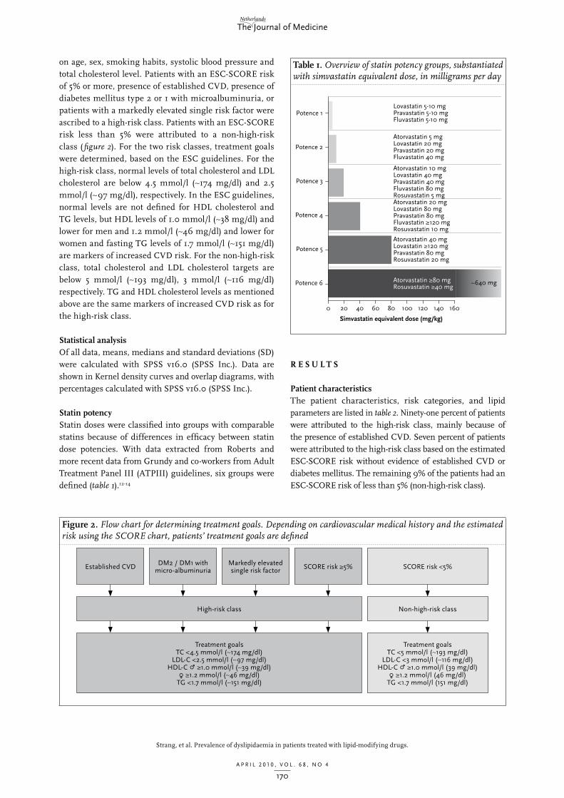
170
a p r i l 2 0 1 0 , v o l . 6 8 , n o 4
on age, sex, smoking habits, systolic blood pressure and total cholesterol level. Patients with an ESC-SCORE risk of 5% or more, presence of established CVD, presence of diabetes mellitus type 2 or 1 with microalbuminuria, or patients with a markedly elevated single risk factor were ascribed to a high-risk class. Patients with an ESC-SCORE risk less than 5% were attributed to a non-high-risk class ( figure 2). For the two risk classes, treatment goals were determined, based on the ESC guidelines. For the high-risk class, normal levels of total cholesterol and LDL cholesterol are below 4.5 mmol/l (~174 mg/dl) and 2.5 mmol/l (~97 mg/dl), respectively. In the ESC guidelines, normal levels are not defined for HDL cholesterol and TG levels, but HDL levels of 1.0 mmol/l (~38 mg/dl) and lower for men and 1.2 mmol/l (~46 mg/dl) and lower for women and fasting TG levels of 1.7 mmol/l (~151 mg/dl) are markers of increased CVD risk. For the non-high-risk class, total cholesterol and LDL cholesterol targets are below 5 mmol/l (~193 mg/dl), 3 mmol/l (~116 mg/dl) respectively. TG and HDL cholesterol levels as mentioned above are the same markers of increased CVD risk as for the high-risk class.
statistical analysisOf all data, means, medians and standard deviations (SD) were calculated with SPSS v16.0 (SPSS Inc.). Data are shown in Kernel density curves and overlap diagrams, with percentages calculated with SPSS v16.0 (SPSS Inc.).
statin potencyStatin doses were classified into groups with comparable statins because of differences in efficacy between statin dose potencies. With data extracted from Roberts and more recent data from Grundy and co-workers from Adult Treatment Panel III (ATPIII) guidelines, six groups were defined (table 1).12-14
r e s u l t s
patient characteristicsThe patient characteristics, risk categories, and lipid parameters are listed in table 2. Ninety-one percent of patients were attributed to the high-risk class, mainly because of the presence of established CVD. Seven percent of patients were attributed to the high-risk class based on the estimated ESC-SCORE risk without evidence of established CVD or diabetes mellitus. The remaining 9% of the patients had an ESC-SCORE risk of less than 5% (non-high-risk class).
Strang, et al. Prevalence of dyslipidaemia in patients treated with lipid-modifying drugs.
figure 2. Flow chart for determining treatment goals. Depending on cardiovascular medical history and the estimated risk using the SCORE chart, patients’ treatment goals are defined
Established CVD
High-risk class
Treatment goalsTC <4.5 mmol/l (~174 mg/dl)
LDL-C <2.5 mmol/l (~97 mg/dl)HDL-C P ≥1.0 mmol/l (~39 mg/dl)O ≥1.2 mmol/l (~46 mg/dl)
TG <1.7 mmol/l (~151 mg/dl)
DM2 / DM1 with micro-albuminuria
Markedly elevated single risk factor SCORE risk ≥5% SCORE risk <5%
Non-high-risk class
Treatment goalsTC <5 mmol/l (~193 mg/dl)
LDL-C <3 mmol/l (~116 mg/dl)HDL-C P ≥1.0 mmol/l (39 mg/dl)O ≥1.2 mmol/l (46 mg/dl)
TG <1.7 mmol/l (151 mg/dl)
table 1. Overview of statin potency groups, substantiated with simvastatin equivalent dose, in milligrams per day
0 20
Potence 6
Potence 5
Potence 4
Potence 3
Potence 2
Potence 1
40 60 80
Simvastatin equivalent dose (mg/kg)
100 120 140 160
Atorvastatin 5 mgLovastatin 20 mgPravastatin 20 mgFluvastatin 40 mg
Lovastatin 5-10 mgPravastatin 5-10 mgFluvastatin 5-10 mg
Atorvastatin 10 mgLovastatin 40 mgPravastatin 40 mgFluvastatin 80 mgRosuvastatin 5 mgAtorvastatin 20 mgLovastatin 80 mgPravastatin 80 mgFluvastatin ≥120 mgRosuvastatin 10 mg
Atorvastatin 40 mgLovastatin ≥120 mgPravastatin 80 mgRosuvastatin 20 mg
~640 mgAtorvastatin ≥80 mgRosuvastatin ≥40 mg

171
a p r i l 2 0 1 0 , v o l . 6 8 , n o 4
lipid-modifying treatment and statin dose potencyAtorvastatin was the most commonly used statin (34.8%), followed by simvastatin (32.7%) and rosuvastatin (21.4%). Other statins used by the patients were pravastatin (8.8%) and fluvastatin (2.3%). Non-statin lipid-lowering treatment on top of statins was used by 13.6% of the patients, including ezetimibe (11.7%), fibrates (2.0%), nicotinic acid (0.7%) and bile acid sequestrants (0.1%).In figure 3, patients’ statin regimen classified according to potency for the high-risk patients and non-high-risk
patients is shown. The majority of patients used a regimen with potency 3 and 4, which is equivalent to simvastatin 20 mg/day and 40 mg/day respectively. Interestingly, in the non-high-risk class a higher percentage of the patients used a more potent (5 and more) statin regimen compared with the high-risk class, whereas the less potent regimens (4 and below) were more frequent in the high-risk groups. Of patients from both risk categories, 6.1% use statin regimens with a potency of 2 and less, which is equivalent to a maximum dosage of 10 mg simvastatin and less.
achievement of normal lipid levelsOne third of all patients (33.3%) had an LDL cholesterol not at goal (≥2.5 mmol/l; 79 mg/dl in high-risk patients and ≥3.0 mmol/l; 116 mg/dl in non-high risk patients) according to ESC guidelines. In 35% total cholesterol was not at goal (≥4.5 mmol/l; 174 mg/dl in high-risk patients and ≥5.0 mmol/l; 193 mg/dl in non-high-risk patients). Low HDL cholesterol (<1.0 mmol/l; 39 mg/dl in men and <1.2 mmol/l; 46 mg/dl in women) and elevated triglycerides (>1.7 mmol/l; 151 mg/dl) were seen in 38.1 and 40.8% of patients (figure 4). Strikingly, the same proportion of patients at high risk (defined as presence of CVD, diabetes and/or ESC-SCORE ≥5%) fail to reach goals for total and LDL cholesterol, had low HDL cholesterol and elevated triglycerides as patients at non-high risk (ESC-SCORE <5%, see figures 5 and 6).The most frequent single lipid abnormality in all patients was elevated triglycerides. Regarding combined lipid abnormalities, elevated triglycerides combined with low HDL cholesterol was most frequent (22.6%), followed by elevated triglycerides with LDL cholesterol not at goal (14.0%). Combined lipid abnormalities were approximately 3% more common in patients with a high risk, compared with patients without high risk, which counts for all lipid abnormality
Strang, et al. Prevalence of dyslipidaemia in patients treated with lipid-modifying drugs.
table 2. Patient characteristics, risk categories and lipid parameters
all patients(n=1208)
patient characteristics
Age (years) [mean±SD] 65.6±9.7
Caucasian (%) 95.2
Family history of premature CHD (%) 39.4
Current smokers (%) 20.0
Hypertension (%) 69.0
Systolic BP (mmHg) [mean±SD] 138.9±18.7
Diastolic BP (mmHg) [mean±SD] 79.0±9.7
Waist circumference (cm) [mean±SD] 101.9±13.0
BMI (kg/m2) [mean±SD] 28.5±4.7
BMI >30 kg/m2 (%) 30.9
esC risk parameters
High risk (CVD, diabetes and/or SCORE risk ≥5%) (%)
91.0
CVD (%) 59.8
Diabetes mellitus (%) 46.4
SCORE risk ≥5% (%) without CVD and diabetes 7.0
SCORE risk <5% (%) without CVD and diabetes 9.0
lipid parameters
Total cholesterol (mmol/l) [mean±SD] 4.4±1.0
LDL-C (mmol/l) [mean±SD] 2.4±0.9
HDL-C (mmol/l) [mean±SD] 1.2±0.4
Triglycerides (mmol/l) [median (IQR)] 1.4 (1.0-2.1)
statin treatment
Simvastatin (%) 32.7
Atorvastatin (%) 34.8
Rosuvastatin (%) 21.4
Pravastatin (%) 8.8
Fluvastatin (%) 2.3
Lovastatin (%) 0.0
other lipid-lowering treatment (%) 13.5
Ezetimibe (%) 11.7
Fibrate (%) 2.0
Nicotinic acid (%) 0.7
Bile acid sequestrant (%) 0.1
Chd = coronary heart disease; bp = blood pressure; bMi = body mass index; Cvd = cardiovascular disease, dM = diabetes mellitus.
figure 3. Statin dose potency according to patients’ risk status
1
0.9 1.44.6 4.9
31.5
39.9 35.238.6
20.4
16.1
7.4
2.1
2 3 4 5 60.0
5.0
10.0
15.0
20.0
25.0
30.0
35.0
40.0
45.0
Statin dose potency
Non-high-risk patientsHigh-risk patients
Perc
enta
ge o
f pat
ient
s

172
a p r i l 2 0 1 0 , v o l . 6 8 , n o 4
high-risk class, a larger proportion of patients with non-high risk had elevated triglycerides as the sole abnormality (10.1 and 18.4% respectively). More appropriately, isolated low HDL cholesterol is found in a larger proportion of high-risk patients than in non-high-risk patients (12.2 and 5.8%, respectively).
d i s C u s s i o n
The results of DYSIS-Netherlands, a study of lipid levels in patients receiving stable statin therapy, reveal the failure to achieve normal levels in patients at risk for CVD. To our knowledge, this is the first study using a large dataset focused solely on statin users in both primary and secondary care settings in the Netherlands. This study was conducted after the release of the 2007 ESC guidelines for the prevention of CVD.10
Despite stable statin therapy, one third of the patients had LDL-cholesterol levels above normal levels. Similarly, elevated triglycerides and low HDL cholesterol remained despite therapy in approximately 40% per moiety.
previous studiesA number of cross-sectional studies have investigated the prevalence of lipid abnormalities and statin use.15-23 These studies, however, were limited to specific populations such as patients with pre-existing CVD, focused on specific lipid parameters such as LDL cholesterol or HDL cholesterol, or had mixed patients with or without lipid-lowering therapies. DYSIS-Netherlands provides an up-to-date view of statin therapy across primary care and specialist treatment centres. Consistent with published data, the majority of patients in this study (71.7%) showed lipid abnormalities despite lipid-modifying therapy.
statin potency and risk classFigure 3 shows a lower use of highly potent statins in the high-risk class, compared with the non-high-risk class. The apparent discrepancy may be due to the fact that patients at high risk are treated with a combination of statins with lipid-lowering drugs from other classes. Thus, doctors may deliberately use lower doses and less potent statins for instance upon combination therapy with fibrates because of the increased risk of myotoxicity.24,25 In addition, the non-high-risk group includes young people with inherited dyslipidaemias such as familial hypercholesterolaemia, who are classified in this category using the ESC-risk engine based on their age. However, a diagnosis of primary dyslipidaemia is automatically an indication for the use of aggressive lipid-lowering therapy.
ldl cholesterol, hdl cholesterol, and triglyceridesAlthough LDL cholesterol is the best characterised risk factor, one third of the patients taking statins have
Strang, et al. Prevalence of dyslipidaemia in patients treated with lipid-modifying drugs.
figure 4. Distribution of single and multiple combined lipid abnormalities in patients with total lipid profile
28.3%(322)
No lipid abnormalities
LDL-C not at goal (≥2.5 mmol/l (high risk) / ≥3 mmol/l (non-high risk))
Low HDL-C(<1.0 (P) / 1.2 (O) mmol/l)
Elevated TG (≥1.7 mmol/l)
15.4%(175)
11.6%(132)
10.9%(124)
4.0%(45)
6.6%(75)
16.0%(182)
7.4%(84)
figure 5. Distribution of single and multiple combined lipid abnormalities in high-risk patients (CVD, diabetes and/or ESC SCORE ≥5%)
28.2%(292)
No lipid abnormalities
LDL-C not at goal (≥2.5 mmol/l)
Low HDL-C(<1.0 (P) / 1.2 (O) mmol/l)
Elevated TG(≥1.7 mmol/l)
15.2%(157)
12.2%(126)
10.1%(105)
4.0%(41)
6.9%(71)
16.2%(168)
7.3%(76)
figure 6. Distribution of single and multiple combined lipid abnormalities in non-high-risk patients (ESC-SCORE <5%)
29.1%(30)
No lipid abnormalities
LDL-C not at goal (≥3 mmol/l)
Low HDL-C(<1.0 (P) / 1.2 (O) mmol/l)
Elevated TG(≥1.7 mmol/l)
17.5%(18)
5.8%(6)
18.4%(19)
3.9%(4)
3.9%(4)
13.6%(14)
7.8%(8)
combinations. As a consequence, single lipid abnormalities were more frequent in non-high-risk patients, with HDL cholesterol excluded. Remarkable is the fact that, despite the inclusion of people with diabetes and established CVD in the

173
a p r i l 2 0 1 0 , v o l . 6 8 , n o 4
LDL-cholesterol levels above their normal level. Failure to achieve normal levels in the majority of patients may not be due to an absolute inability to do so, but rather reflects the use of less potent statins. Since the proportion of patients reaching LDL-C normal levels is very modest, there is a clear need to increase awareness of the relevance of achieving normal levels in order to optimally lower CVD risk.10
In parallel, 38.1% of patients presented with HDL-cholesterol levels below the normal level. In this case, it most likely reflects the lack of therapeutic options to selectively increase HDL cholesterol.26 The effect of statins on HDL cholesterol is fairly modest (2 to 10%), and the clinical relevance of statin-induced HDL-cholesterol changes has never been established.27 Novel approaches to increase HDL cholesterol have gained a lot of attention. The recent withdrawal of the CETP-inhibitor torcetrapib, which increased HDL cholesterol significantly with a concomitant increase in CVD, has once again fuelled the concept that increasing HDL cholesterol may turn out to be more complicated compared with LDL cholesterol-lowering interventions.28
Surprisingly, with a prevalence of 40.8%, elevated triglycerides was the most frequent lipid abnormality. A potential explanation is the inclusion of patients with secondary dyslipidaemias, largely due to metabolic syndrome treated with statins only. Although potent statins such as atorvastatin and rosuvastatin can reduce fasting triglyceride levels by 20 to 40%, less potent statins have little impact on triglyceride levels.29,30 At the same time, whereas fibrates are valuable in lowering triglyceride levels, the additional impact of fibrates on top of statins on CVD event rate has not been convincingly demonstrated.31,32 The final verdict awaits the results of the ongoing Action to Control Cardiovascular Risk in Diabetes (ACCORD) study.33
study limitationsOur study has some limitations that deserve closer attention. First, DYSIS was a cross-sectional (single point) observational study which did not evaluate long-term outcomes. Therefore SCORE risk was calculated based on current or retrospective data, rather than prospectively observed. Second, lipid parameters were those taken from patients’ case notes. There was no blood sample collection or core laboratory analysis of the lipid parameters. Notwithstanding this observation, our results are a true reflection of clinical practice. Thirdly, the treatment centres included were those prescribing statins to their patients, given that current statin use was a patient eligibility criterion. This may have introduced a self-selection bias. This implies that, in fact, the present data may over-estimate the use of statins across the CVD population. Finally, the present study did not collect
Strang, et al. Prevalence of dyslipidaemia in patients treated with lipid-modifying drugs.
data on patient lifestyle, genetic predisposition to CVD (although family history was assessed) or treatment compliance.
Clinical implicationsIn more than 70% of patients in the DYSIS-Netherlands study, lipids were not at normal levels despite stable statin treatment. Side effects and lack of selective drugs were probably the cause of undertreatment. These data indicate that the awareness of the need to achieve appropriate or normal lipid levels needs close attention. For LDL cholesterol, since the Treating to New Targets (TNT) and Incremental Decrease in End Points through Aggressive Lipid Lowering (IDEAL) trials, it has been demonstrated that lower LDL cholesterol results in lower CVD risk.34,35
For TG levels, in a recent post hoc analysis of the TNT and IDEAL trial, risk of a cardiovascular event increased as a function of increasing TG levels.36 This is presumably because TG levels cause an increase of small dense LDL cholesterol and lower HDL cholesterol, thereby causing higher CVD risk. For HDL cholesterol, no evidence has been found that therapeutically increased levels lead to a decreased CVD risk, because of the lack of selective therapeutics.26 Because of the residual risk, in future, combination therapy of lipid-modifying agents will presumably be used to reach normal levels for all moieties. In the next decade, statins combined with a second drug will be the best regimen in high-risk patients. New agents have to be developed to reach lipid normal levels.
a C K n o w l e d g e M e n t s
This work was supported by Merck & Co., Inc. Special gratitude to Monica Hackett and Sjoerd Visser (Merck & Co., Inc), the study coordinators. We also thank Dr Steffen Schneider and Dr Claus Jünger, from the Stiftung Institut für Herzinfarktforschung, Ludwigshafen, Germany, who provided statistical support in the analysis of the study results.
dysis scientific Committee:Anselm K. Gitt, Heinz Drexel, John Feely, Jean Ferrières, José-Ramon González-Juanetey, Kristian Korsgaard Thomsen, Lawrence A. Leiter, Pia Lundman, Pedro Marques da Silva, Terje Pedersen, David Wood, John J. P. Kastelein
dysis-netherlands investigators:Apperloo, Dr A.J.; Basart, D.C.G.; Bax, M.; Beysens, Dr P.A.J.; Breed, Dr J.G.S.; Derks, A.; Eijgenraam J.W.; Herrmans, Dr J.P.R.; Janus, C.L.; Kaasjager,Dr H.A.H.; Klomps, H.C.; Koole, M.A.C.; Koster, Dr T.; Kroon,Dr C.; Lieverse, Dr A.G.; Massaar-Hagen, B.E.M.; Moghaddam, F.; Oldenburg-Ligtenberg, Dr P.C.; Potter van Loon, B.J.; Stroes, Dr E.S.G.; Twickler, Dr Th.B.; Van Asperdt, F.G.M.H.; Van Asseldonk,

174
a p r i l 2 0 1 0 , v o l . 6 8 , n o 4
J.P.M.; Van der Loos, T.L.J.M.; Van der Velde, R.Y.; Van der Vring, J.A.F.; Van Dorp, Dr W.T.; Van Essen, Dr G.G.; Van Kalmthout, P.M.; Van Liebergen, Dr R.A.M.; Van Wissem, Dr S.; Waanders, H.; Withagen, A.J.A.M.
f i n a n C i a l d i s C l o s u r e
The authors report no potential conflicts of interest.
r e f e r e n C e s
1. Cause of death; month and year of death [in Dutch]. https://statline.cbs.nl. Date accessed: 1-7-2009.
2. Top 100 drugs on the basis of the number of users in 2007 [in Dutch]. http://www.GIPdatabank.nl. Date accessed: 23-1-2009.
3. Baigent C, Keech A, Kearney PM, et al. Efficacy and safety of cholesterol-lowering treatment: prospective meta-analysis of data from 90,056 participants in 14 randomised trials of statins. Lancet. 2005;366(9493):1267-78.
4. Gordon T, Castelli WP, Hjortland MC, Kannel WB, Dawber TR. High density lipoprotein as a protective factor against coronary heart disease. The Framingham Study. Am J Med. 1977;62(5):707-14.
5. Patsch JR, Miesenbock G, Hopferwieser T, et al. Relation of triglyceride metabolism and coronary artery disease. Studies in the postprandial state. Arterioscler Thromb. 1992;12(11):1336-45.
6. Barter P, Gotto AM, LaRosa JC, et al. HDL cholesterol, very low levels of LDL cholesterol, and cardiovascular events. N Engl J Med. 2007;357(13):1301-10.
7. Nordestgaard BG, Benn M, Schnohr P, Tybjaerg-Hansen A. Nonfasting triglycerides and risk of myocardial infarction, ischemic heart disease, and death in men and women. JAMA. 2007;298(3):299-308.
8. Abdel-Maksoud MF, Hokanson JE. The complex role of triglycerides in cardiovascular disease. Semin Vasc Med. 2002;2(3):325-33.
9. Gordon DJ, Probstfield JL, Garrison RJ, et al. High-density lipoprotein cholesterol and cardiovascular disease. Four prospective American studies. Circulation. 1989;79(1):8-15.
10. Graham I, Atar D, Borch-Johnsen K, et al. European guidelines on cardiovascular disease prevention in clinical practice: executive summary. Eur Heart J. 2007;28(19):2375-414.
11. Gitt AK, Drexel J, Ferrieres J, et al. High prevalence of dyslipidemia in 18,574 patients treated with statins in Europe and Canada: Results of the dyslipidemia international study. Eur Heart J. 2009;30(suppl 1):303.
12. National Heart Lung and Blood Institute. Third Report of the Expert Panel on Detection, Evaluation, and Treatment of the High Blood Cholesterol in Adults (Adult Treatment Panel III): Final Report. http://www.nhlbi.nih.gov/guidelines/cholesterol/. Date accessed: 26-1-2009.
13. Grundy SM, Cleeman JI, Merz CN, et al. Implications of recent clinical trials for the National Cholesterol Education Program Adult Treatment Panel III guidelines. Circulation. 2004;110(2):227-39.
14. Roberts WC. The rule of 5 and the rule of 7 in lipid-lowering by statin drugs. Am J Cardiol. 1997;80(1):106-7.
15. Kotseva K, Wood D, De Backer G, De Bacquer D, Pyorala K, Keil U. Cardiovascular prevention guidelines in daily practice: a comparison of EUROASPIRE I, II, and III surveys in eight European countries. Lancet. 2009;373(9667):929-40.
16. Luc G, Bard JM, Ferrieres J, et al. Value of HDL cholesterol, apolipoprotein A-I, lipoprotein A-I, and lipoprotein A-I/A-II in prediction of coronary heart disease: the PRIME Study. Prospective Epidemiological Study of Myocardial Infarction. Arterioscler Thromb Vasc Biol. 2002;22(7):1155-61.
17. Bourgault C, Davignon J, Fodor G, et al. Statin therapy in Canadian patients with hypercholesterolemia: the Canadian Lipid Study -- Observational (CALIPSO). Can J Cardiol. 2005;21(13):1187-93.
18. Bruckert E, Baccara-Dinet M, McCoy F, Chapman J. High prevalence of low HDL-cholesterol in a pan-European survey of 8545 dyslipidaemic patients. Curr Med Res Opin. 2005;21(12):1927-34.
19. Bruckert E, Baccara-Dinet M, Eschwege E. Low HDL-cholesterol is common in European Type 2 diabetic patients receiving treatment for dyslipidaemia: data from a pan-European survey. Diabet Med. 2007;24(4):388-91.
20. Assmann G, Benecke H, Neiss A, Cullen P, Schulte H, Bestehorn K. Gap between guidelines and practice: attainment of treatment targets in patients with primary hypercholesterolemia starting statin therapy. Results of the 4E-Registry (Efficacy Calculation and Measurement of Cardiovascular and Cerebrovascular Events Including Physicians’ Experience and Evaluation). Eur J Cardiovasc Prev Rehabil. 2006;13(5):776-83.
21. Goff DC, Jr., Bertoni AG, Kramer H, et al. Dyslipidemia prevalence, treatment, and control in the Multi-Ethnic Study of Atherosclerosis (MESA): gender, ethnicity, and coronary artery calcium. Circulation. 2006;113(5):647-56.
22. Yan AT, Yan RT, Tan M, et al. Contemporary management of dyslipidemia in high-risk patients: targets still not met. Am J Med. 2006;119(8):676-83.
23. Grant RW, Meigs JB. Prevalence and treatment of low HDL cholesterol among primary care patients with type 2 diabetes: an unmet challenge for cardiovascular risk reduction. Diabetes Care. 2007;30(3):479-84.
24. Alsheikh-Ali AA, Kuvin JT, Karas RH. Risk of adverse events with fibrates. Am J Cardiol. 2004;94(7):935-8.
25. Ballantyne CM, Corsini A, Davidson MH, et al. Risk for myopathy with statin therapy in high-risk patients. Arch Intern Med. 2003;163(5):553-64.
26. Singh IM, Shishehbor MH, Ansell BJ. High-density lipoprotein as a therapeutic target: a systematic review. JAMA. 2007;298(7):786-98.
27. Nicholls SJ, Tuzcu EM, Sipahi I, et al. Statins, high-density lipoprotein cholesterol, and regression of coronary atherosclerosis. JAMA. 2007;297(5):499-508.
28. Barter PJ, Caulfield M, Eriksson M, et al. Effects of torcetrapib in patients at high risk for coronary events. N Engl J Med. 2007;357(21):2109-22.
29. Hunninghake DB. Postdischarge lipid management of coronary artery disease patients according to the new National Cholesterol Education Program guidelines. Am J Cardiol. 2001;88(8A):37K-41K.
30. Bakker-Arkema RG, Davidson MH, Goldstein RJ, et al. Efficacy and safety of a new HMG-CoA reductase inhibitor, atorvastatin, in patients with hypertriglyceridemia. JAMA. 1996;275(2):128-33.
31. Rosenson RS, Pitt B. Triglycerides and cardiovascular events in ACS: the need for combined lipid-altering therapies. Nat Clin Pract Cardiovasc Med. 2009;6(2):98-100.
32. Effect of fenofibrate on progression of coronary-artery disease in type 2 diabetes: the Diabetes Atherosclerosis Intervention Study, a randomised study. Lancet. 2001;357(9260):905-10.
33. Clinical trial register: Action to Control Cardiovascular Risk in Diabetes (ACCORD). http://clinicaltrials.gov/ct2/show/NCT00000620?term=accord. Date accessed: 20-11-2009.
34. LaRosa JC, Grundy SM, Waters DD, et al. Intensive lipid lowering with atorvastatin in patients with stable coronary disease. N Engl J Med. 2005;352(14):1425-35.
35. Pedersen TR, Faergeman O, Kastelein JJ, et al. High-dose atorvastatin vs usual-dose simvastatin for secondary prevention after myocardial infarction: the IDEAL study: a randomized controlled trial. JAMA. 2005;294(19):2437-45.
36. Faergeman O, Holme I, Fayyad R, et al. Plasma triglycerides and cardiovascular events in the Treating to New Targets and Incremental Decrease in End-Points through Aggressive Lipid Lowering trials of statins in patients with coronary artery disease. Am J Cardiol. 2009;104(4):459-63.
Strang, et al. Prevalence of dyslipidaemia in patients treated with lipid-modifying drugs.

175
a p r i l 2 0 1 0 , v o l . 6 8 , n o 4
K e y w o r d s
Kimura disease, lymphadenopathy, eosinophilia
s u M M a r y
A Pakistani male patient presented with a parotid and retroauricular mass on both sides, which was diagnosed as Kimura’s disease. Kimura’s disease is a chronic disorder of unknown origin, which presents as subcutaneous mass, predominantly in the cervical and head region, with regional lymphadenopathy, blood eosinophilia and elevated serum IgE levels. Steroid therapy usually induces remission of the disease.
C a s e
A 47-year-old male of Pakistani origin presented to our outpatients’ clinic with a painless mass on both sides in the parotid region, extending to the neck and retroauricular regions (figure 1). The swellings had already existed for six years and had varied slightly in size over time. The patient did not have any complaints of pain, dry mouth, weight loss or night sweating and he felt in good condition. For years he had been suffering from itchiness over his whole body, without a known cause. He had not had any unsafe sexual contacts in the past and had lived in the Netherlands for the past nine years. Physical examination showed a bilateral swelling in the region of the parotid gland of approximately 8 x 3 cm in size on the left side, at the right of 6 x 3 cm. In addition, multiple lymph nodes were palpated in the head and neck region of 1-2 cm in diameter. There was no axillary or inguinal lymphadenopathy present and the liver and spleen were not enlarged. Retroauricular, the skin showed some erythema with excoriations.
© Van Zuiden Communications B.V. All rights reserved.
Laboratory findings were as follows: erythrocyte sedimentation rate 6 mm/h, haemoglobin 9.2 mmol/l, platelets 350 x 109/l, leucocytes 19.8 x 10⁹/l, neutrophils 3.0 x 10⁹/l, eosinophils 13.3 x 10⁹/l, aspartate transaminase 26 U/l, alanine aminotransferase (ALAT) 32 U/l, lactate dehydrogenase 250 U/l, gamma glutamyltransferase 45 U/l, and alkaline phosphatase 84 U/l; the immunoglobulin E (IgE) serum level was not determined.Chest radiography and abdominal echography were both normal. An incisional biopsy of one of the regional lymph nodes was performed. Histological examination showed a preserved nodal architecture with follicular hyperplasia, eosinophilic infiltrates with microabscesses in the interfollicular areas and proliferation of postcapillary venules (figure 2). There were several small clusters of epithelioid histiocytes without necrosis. Immunohistochemistry showed normal B and T cells
C a s e r e p o r t
Kimura’s disease of the parotid glands and multiple cervical lymph nodes
V.K. Dik1, B.A. van der Wiel2, W.L.E. Vasmel1*
1Department of Internal Medicine, Sint Lucas Andreas Hospital, Amsterdam, the Netherlands, 2Department of Pathology, The Netherlands Cancer Institute – Antoni van Leeuwenhoek Hospital,
Amsterdam, the Netherlands, *corresponding author: tel.: +31 (0)20-510 87 70, e-mail: [email protected]
figure 1. Bilateral swelling of the neck and parotid gland regions.

176
a p r i l 2 0 1 0 , v o l . 6 8 , n o 4
compartments with Bcl-2 negative germinal centres. Histochemical staining with Ziehl-Nielssen, Grocott Methamine silver and periodic acid Schiff did not show any infectious agents. Both polymerase chain reaction and culture for Mycobacterium tuberculosis were negative.Hence, based on the combination of these clinical data and biopsy results the diagnosis of Kimura’s disease was made six months after first presentation.Surgery was considered during this period. However, this was not performed because of the known high risk of complications and the risk of recurrent disease after surgery.1 We followed a careful wait and see policy.Initially the lesions did not grow, but two years after first presentation, both left and right parotid glands had grown to a size of 12 x 5 cm and 8 x 4 cm, respectively. The patient was complaining of progressive pruritus and erythematous lesions spread out over all parts of the body. During this period the leucocyte and eosinophil count were 18.0 x 10⁹/l and 10.4 x 10⁹/l, respectively. Therapy with an H2 blocking agent, cetirizine 10 mg/day, was started without effect. Subsequently we started steroid therapy (prednisolone 30 mg/day orally), combined with osteoporosis prophylaxis. The effects of prednisolone were spectacular within two weeks. Both the swelling and his pruritus diminished significantly.
d i s C u s s i o n
Kimura’s disease was first described in 1937 as eosinophilic hyperplastic lymphogranuloma, but later became known as Kimura’s disease after Kimura et al. reported their findings in similar cases.2
Kimura’s disease is most commonly seen in men, with an average age of 30 years, from Asian countries such as Japan, Taiwan and China, although it has also been reported in Caucasians. The symptoms can exist for several months to years before patients present, because the size of the lesion
progresses slowly. Patients mainly present with a mass in the neck and retroauricular region, but other locations such as temporal, inguinal and axillary regions have been reported. Parotid gland involvement is not very common, but nevertheless, may occur. In most cases the size of the mass is between 1 and 7 cm. In addition, multiple regional lymph nodes are involved presenting as a multifocal mass of 1 to 2 cm. As demonstrated in our patient, Kimura’s disease can be accompanied by pruritis surrounding the tumour area, but also spreading out over the whole body.3,4 Histologically, the lymph nodes are characterised by eosinophilic microabscesses, eosinophilic folliculolysis, (perivenular) sclerosis and eosinophilic infiltrates in the germinal centre. Vascularisation of the germinal centre is common, but also germinal centre necrosis can be present. Besides hyperplasia, the lymph node architecture remains preserved and B and T cells are found in their normal nodal compartments. Laboratory findings that support the diagnosis of Kimura’s disease are blood eosinophilia and elevated serum IgE levels. The level of blood eosinophilia seems to be closely related to the size of the mass and might be used as a parameter of disease activity.5,6 The aetiology of Kimura’s disease still remains unclear. An abnormal T cell stimulation seems possible when considering the presence of eosinophilia and elevated IgE and IL-5 levels.4,7 Infectious agents as well as autoantibodies have not yet been identified. However, it is thought that an unusual autoimmune response or a parasitic infection is responsible for the onset of the disease.4
When considering the diagnosis, other common causes of lymphadenopathy such as metastatic lymphadenopathy, lymphoma and infectious diseases should be ruled out (table 1). In the differential diagnosis angiolymphoid hyperplasia with eosinophilia and Kikuchi’s disease should be considered as well.3 Both angiolymphoid hyperplasia with eosinophilia and Kimura’s disease present with soft tissue masses usually in the head and neck region with microscopically eosinophilic infiltrates. Histologically, in angiolymphoid hyperplasia with eosinophilia, vascular proliferation is more significant and regional lymphadenopathy, serum eosinophilia and elevated serum IgE levels are rare. In contrast with Kimura’s disease, Kikuchi’s disease is characterised by necrosis and large numbers of different histiocytes surrounding the necrotic areas.3,8
A standard therapy has not yet been established. Multiple treatment options have been used with varying results, including surgery, radiotherapy, cetirizine, steroid and cyclosporine therapy.4,9 Recently, Sun et al. proposed treatment with imatinib, because of its inhibitory effect on the protein-tyrosine kinases PDGFR and c-Kit reported in the hypereosinophilic syndrome. Sun et al. found a positive expression of c-Kit and PDGFRa patients with Kimura’s disease as well and imatinib could therefore have
Dik, et al. Kimura’s disease of the parotid glands and multiple cervical lymph nodes.
figure 2. Detail of an eosinophilic micro-abcess. H&E, 200x magnification

177
a p r i l 2 0 1 0 , v o l . 6 8 , n o 4
a beneficial effect on Kimura’s disease.4,10 As yet, no studies concerning imatinib therapy in Kimura’s disease have been published. In our patient immunosuppressive therapy with steroids promptly resulted in a good clinical response.In conclusion, Kimura’s disease is a chronic disorder of unknown origin. The subcutaneous masses are predominantly seen in the cervical and head region with regional lymphadenopathy, blood eosinophilia and elevated
Dik, et al. Kimura’s disease of the parotid glands and multiple cervical lymph nodes.
table 1. Differential diagnosis of Kimura disease
disease Clinical and histological characteristics
Kimura’s disease
Predominantly young Asian men, neck and retro auricular mass, blood eosinophilia, elevated serum IgE levels, histologically preserved lymph node architecture, eosino philic microabscesses and folliculolysis, eosino philic infiltrates in the germinal centre
Angio-lymphoid hyper-plasia with eosinophilia
Usually in the head and neck region, regional lymphadenopathy, blood eosinophilia and elevated serum IgE levels are rare, histologically microscopically eosinophilic infiltrates and vascular proliferation
Kikuchi-Fujimoto disease
Cervical lymphadenopathy, fever, elevated ESR, histologically necrosis and large numbers of different histiocytes surrounding the necrotic areas, associated with SLE
Kawasaki’s disease
Predominantly young children, skin involve-ment, histologically geographic necrosis, fibrinoid thrombosis and neutrophilic infiltrates
Hodgkin’s lymphoma
Young adults, systemic B symptoms, cervical and supraclavicular lymphadenopathy, presence of Reed-Sternberg cells
Non-Hodgkin’s lymphoma
Children and young adults, systemic B symptoms, anaemia, tenderless peripheral lymphadenopathy, no striking polymorphous histio cytic infiltrate
Tuberculosis Young adult (immigrants) from endemic countries, family history of tuberculosis, mostly cervical lymphadenopathy, mass fixed to sur-rounding structures, histologically caseating granulomas
serum IgE levels. Preliminary reports on the use of imatinib in Kimura’s disease suggest a beneficial effect. However, in our patient steroid therapy was successfully initiated.
a C K n o w l e d g e M e n t
We thank Professor Dr. G.B. Snow for his careful reading of the manuscript.
r e f e r e n C e s
1. Urabe A, Tsuneyoshi M, Enjoji M. Epithelioid hemangioma versus Kimura’s disease. A comparative clinicopathologic study. Am J Surg Pathol. 1987;11(10):758-66.
2. Kung IT, Chan JK. Kimura’s disease or Kimm’s disease? Am J Surg Pathol. 1988;12(10):804-5.
3. Chen H, Thompson LDR, Aguilera NSI, Abbondanzo SL. Kimura disease: a clinicopathologic study of 21 cases. Am J Surg Pathol. 2004;28(4):505-13.
4. Sun QF, Xu DZ, Pan SH, Ding JG, Xue ZQ, Miao CS, et al. Kimura disease: review of the literature. Intern Med J. 2008;38(8):668-72.
5. Ohta N, Okazaki S, Fukase S, Akatsuka N, Aoyagi M, Yamakawa M. Serum concentrations of eosinophil cationic protein and eosinophils of patients with Kimura’s disease. Allergol Int. 2007;56(1):45-9.
6. Sakamoto M, Komura A, Nishimura S. Hematoserological analysis of Kimura’s disease for optimal treatment. Otolaryngol Head Neck Surg. 2005;132(1):159-60.
7. Kimura Y, Pawankar R, Aoki M, Niimi Y, Kawana S. Mast cells and T cells in Kimura’s disease express increased levels of interleukin-4, interleukin-5, eotaxin and RANTES. Clin Exp Allergy. 2002;32(12):1787-93.
8. Bosch X, Guilabert A, Miquel R, Campo E. Enigmatic Kikuchi-Fujimoto disease: a comprehensive review. Am J Clin Pathol 2004;122(1):141-52.
9. Takeishi M, Makino Y, Nishioka H, Miyawaki T, Kurihara K. Kimura disease: diagnostic imaging findings and surgical treatment. J Craniofac Surg. 2007;18(5):1062-7.
10. Buchdunger E, Cioffi CL, Law N, et al. Abl protein-tyrosine kinase inhibitor STI571 inhibits in vitro signal transduction mediated by c-kit and platelet-derived growth factor receptors. J Pharmacol Exp Ther. 2000;295(1):139-45.

178
a p r i l 2 0 1 0 , v o l . 6 8 , n o 4
© Van Zuiden Communications B.V. All rights reserved.
a b s t r a C t
in autoimmune hypothyroidism (hashimoto’s disease), tpo (thyroid peroxidase) antibodies may be detected, while in autoimmune hyperthyroidism (graves’ disease) thyroid-stimulating hormone (tsh) receptor antibodies (tsh-r-abs) are frequently present. less well known is the fact that autoimmune hypothyroidism can present with tsh-r-abs and ophthalmic graves’ disease (ogd). this condition is also known as hypothyroid graves’ disease. in this report we describe four patients with this uncommon phenomenon.these four cases demonstrate that differences between hashimoto and graves’ disease are less clear than expected. hypothetically the thyroid cell might be ‘attacked’ by blocking and stimulating antibodies. dependent on the relative concentrations, hypothyroidism or hyperthyroidism may occur. so the differences between hashimoto’s disease and graves’ disease, at least in these cases, may be gradual and small.
K e y w o r d s
Hypothyroidism, Graves disease
i n t r o d u C t i o n
Functional disorders of the thyroid gland often result from autoimmune processes that either cause overproduction of thyroid hormones (Graves’ disease) or glandular destruction and hormone deficiency (Hashimoto’s disease). In Hashimoto’s disease thyroid peroxidase antibodies (TPO-ABs) may be detected, while in Graves’ disease thyroid-stimulating hormone (TSH) receptor antibodies (TSH-R-ABs) and TPO-ABs are frequently present.1,2 Less well known is the fact that autoimmune hypothyroidism can present with ophthalmic Graves’ disease (OGD). This
condition is also known as hypothyroid Graves’ disease and was already reported by Wyse et al. in 1968.3 In this report we describe four patients with this rather rare disorder.
C a s e r e p o r t s
Case 1. A 60-year-old woman was referred to us by her ophthalmologist because of fatigue and her OGD, expressing in an unilateral lid retraction. Ten years before she was seen because of hypertension and Graves’ disease including OGD for which she had been successfully treated with suppression and suppletion therapy. Ophthalmic treatment only consisted of topical lubricants for discomfort. At presentation laboratory tests showed elevated TSH (144 mU/l), low FT4 (5.2 pmol/l), strongly positive TPO-ABs (185 IE/ml) and weakly positive TSH-R-ABs (1.0 IE/l). She received 75 mg of levothyroxine (L-T4) daily. With that dosage she stayed euthyroid.
Case 2. A 44-year-old man was referred to our department by the ophthalmologist because of fatigue and weight gain. Few years earlier he underwent surgery on fibrotic extraocular muscles treating troublesome diplopia due to clinical OGD. However, he presented with new ophthalmic findings: mild periorbital oedema, unilateral lid retraction and exophthalmos of the right eye ( figure 1). His medical history only revealed tonsillectomy. Furthermore he smoked ten cigarettes per day, which had previously been 25 cigarettes daily. Laboratory results were compatible with hypothyroidism (FT4 11 pmol/l, TSH 58.2 mU/l). Further investigations showed elevated TSH-R-ABs (56 E/l) and strongly positive TPO-ABs (399 IU/l). He started L-T4 suppletion, 150 mg daily and remained under supervision of his general practitioner.
C a s e r e p o r t
four patients with hypothyroid graves’ disease
A.J. Starrenburg-Razenberg1*, M. Castro Cabezas1, I.M. Gan2, T.L. Njo3, A.P. Rietveld1, J.W.F. Elte1
Departments of 1Internal Medicine and Endocrinology, 2Ophthalmology, 3Clinical Laboratory, Sint Franciscus Gasthuis, Rotterdam, the Netherlands, *corresponding author: tel.: +31 (0)10-461 61 61,
fax: +31 (0)10-461 26 92, e-mail: [email protected]

179
a p r i l 2 0 1 0 , v o l . 6 8 , n o 4
Starrenburg-Razenberg, et al. Hypothyroid Graves’ disease.
Case 3. A 69-year-old woman was referred by her ophthalmologist because of severe congestive Graves’ ophthalmopathy and signs of optic nerve involvement. Her history revealed hypothyroidism for which she was under supervision by her general practitioner and received 100 mg of L-T4 daily. At presentation she was euthyroid. Laboratory results showed normal TSH (3.8 mU/l), normal FT4 (19 pmol/l), positive TSH-R-ABs 8.5 IE/l and TPO-ABs 89 IU/ml. She was treated with high-dose intravenous steroids and followed by an oral taper regimen. Eventually orbital decompression surgery was performed.
Case 4. A 44-year-old woman presented to the ophthalmologist with exophthalmos and subjective pressure sensations behind her eyes. The diagnosis of OGD was made and she was referred to our internal department for further clinical assessment. For years she was known with hypothyroidism for which she received 75 mg of L-T4 daily. She smoked six cigarettes per day. Laboratory results showed TSH 0.023 mU/l, FT4 18 pmol/l, positive TSH-R-ABs 15.4 IE/l and positive TPO-ABs 354 U/ml. For her OGD she received methylprednisolone 1000 mg intravenously once daily for three days, followed by prednisolone 40 mg daily. The clinical features improved, but she developed herpes zoster at her left nates, which was treated with valaciclovir.
The laboratory results of all four patients are shown in table 1.
M e t h o d s
Serum levels of free T4 and human TSH were measured using the Immulite 2000 free T4 and Immulite 2000 third-generation TSH assay (Siemens Medical Solutions Diagnostica B.V., Breda, the Netherlands). Thyroid peroxidase antibodies were measured with the Immunocap 250 (Phadia B.V., Woerden, the Netherlands).In cases 1, 3 and 4 TSH receptor antibodies in serum were measured with a highly sensitive radioreceptor assay using the human recombinant TSH receptor (DYNOtest TRAK human from B.R.A.H.M.S. Diagnostica, Berlin, Germany). In case 2 the same test was used but with animal (pig) TSH receptor.
d i s C u s s i o n
Our four cases had OGD, hypothyroidism and positive TSH-R-ABs, leading to the diagnosis of hypothyroid Graves’ disease. This phenomenon might be more common than is generally recognised. As mentioned in the introduction, this phenomenon was already reported by Wyse et al. in 1968.3 Christy et al. described three cases with nonthyrotoxic Graves’ disease and concomitant hypothyroidism in 1977. They refer to Werner, who reported ten euthyroid patients with classical OGD in 1955. After this description, the concept of hyperthyroidism being an essential component of Graves’ disease has been modified.4 McDermott et
al. also described two cases and a review of 21 cases in 1968 in which primary autoimmune or idiopathic hypothyroidism was followed by the development of thyreotoxicosis.5 They were intrigued by the phenomenon of hypothyroidism progressing to hyperthyroidism. One of their explanations was that Graves’ and Hashimoto’s disease might occur in one patient, but at different times pointing to the possibility of spontaneous transition. Another explanation could be that blocking antibodies change into stimulating antibodies which may then be responsible for the change from hypothyroidism to hyperthyroidism.5
Another example of hypothyroid Graves’ disease was reported by Elte et al. in 1983. The article is about patients with pretibial myxoedema. Usually patients with pretibial myxoedema have eye signs of Graves’ disease and are euthyroid or hyperthyroid. However, from the 17 patients, one appeared to be hypothyroid.6
The pathogenesis is unknown, but hypotheses centre on the TSH receptor and its antibodies. The initial hypothyroid phase would be due to autoimmune damage to the thyroid gland rather than resulting from inhibitory antibodies. Thyroxine treatment has been
figure 1. Ophthalmic Graves’ disease of case 2
table 1. Laboratory results from the four patients
ft4 (pmol/l)
tsh (mu/l)
tsh-r-ab (ie/l)
tpo (ie/ml)
Reference values
10-24 0.16-4.6 <1 <26
Patient 1 5.2 144 1 185
Patient 2 11 58.2 56 399
Patient 3 19 3.8 8.5 89
Patient 4 18 0.023 15.4 354

180
a p r i l 2 0 1 0 , v o l . 6 8 , n o 4
postulated to be the trigger for this change by causing decreased immunological surveillance.7 There have been descriptions and reviews about hypothyroidism preceding hyperthyroidism.8-11
Our hypothesis is that the thyroid cell might be ‘attacked’ by blocking and stimulating antibodies. Dependent on the relative concentrations, hypothyroidism or hyperthyroidism may occur. So the difference between Hashimoto’s disease and Graves’ disease may be gradual and small.During the last 50 years several case reports have been published in literature about hypothyroid Graves’ disease. However, in medical textbooks this phenomenon is hardly ever mentioned. Hypothyroid Graves’ disease thus is not very well known, which might have led to an underestimation considering the reports published and our own experience, as described in this article.
Starrenburg-Razenberg, et al. Hypothyroid Graves’ disease.
r e f e r e n C e s
1. Jameson JL, Weetman AP. Disorders of the thyroid gland. In: Jameson JL, editor. Harrison’s Endocrinology. 16th ed. McGraw Hill; 2006. p.71-111.
2. Davies TF, Larsen PR. Adrenocortical diseases. In: Larsen PR, Kronenburg HM, Melmed S, Polonsky KS, editors. Williams Textbook of Endocrinology, 10th ed. Elsevier; 2003. p.378-401.
3. Wyse EP, McConahey WM, Woolner LB, Scholz DA, Kearns TP. Ophthalmopathy without hyperthyroidism in patients with histologic Hashimoto’s thyroiditis. J Clin Endocrinol Metab. 1968;28(11):1623-9.
4. Christy JH, Morse SM. Hypothyroid Graves’ disease. Am J Med. 1977;62:291-6.
5. McDermott MT, Kidd GS, Dodson LE, Hofeldt FD. Case report: Hyperthyroidism following hypothyroidism. Am J Med Sci. 1986;291(3):194-8.
6. Elte JWF, Hensen EJ. Pretibial myoedema. Report of 17 cases and review of the literature. Neth J Med. 1983;26(8):220-7.
7. Kang BH. Hyperthyroidism following hypothyroidism - a case report. Med J Malaysia. 1991;46(3):287-9.
8. Irvine WJ, Lamberg BA, Cullen DR, Gordin R. Primary hypothyroidism preceding throtoxicosis: a report of 2 cases and a review of the literature. J Clin Immunol. 1979;2:349-52.
9. Skare S, Frey HMM, Konow- Thorsen R. Primary hypothyroidism followed by hyperthyroidism. Five case reports. Acta Endocrinol. 1984;105:179-83.
10. Cho BY, Shong YK, Lee HK, Koh CS, Min HK. Graves’ hyperthyroidism following primary hypothroidism: sequential changes in various activities of thyroptropin receptor antibodies. Acta Endocrinol. 1989;120:447-50.
11. Yamasaki H, Takeda K, Nakauchi Y, Suehiro T, Hashimoto K. Hypothyroidism preceding hyperthyroidism in a patient with continuously positive thyroid stimulating antibody. Intern Med. 1995;34(4):247-50.

181
a p r i l 2 0 1 0 , v o l . 6 8 , n o 4
© Van Zuiden Communications B.V. All rights reserved.
C a s e r e p o r t
A 65-year-old male was admitted to our hospital because of postprandial nausea and vomiting, weight loss (approximately 19 kg) and night sweats in the last two months. He also reported loss of sensibility in his feet and difficulty walking for approximately two weeks. Recently, he was diagnosed with an HBe-antigen positive hepatitis B (HBV) infection.Physical examination revealed hypertension. Furthermore, there was bilateral sensibility loss of the lower extremities and peroneal paresis, compatible with acute polyneuropathy.Laboratory investigations showed mild normocytic anaemia with an elevated erythrocyte sedimentation rate, leucocytosis (19.8 x10⁹/l; 86% granulocytes), impaired kidney function (eGFR 63 ml/min) and elevated liver enzymes, predominantly cholestatic. Based on symptomatology and biochemical alterations suggestive of multi-system involvement, computed tomography was performed early on in the diagnostic process (figures 1 and 2).
w h a t i s y o u r d i a g n o s i s ?
See page 184 for the answer to this photo quiz.
p h o t o q u i z
a patient with hepatitis b, liver and kidney dysfunction and polyneuropathy
L. Klieverik1, M. Mallant2, M. Sekkat1, J. Branger1*
Department of 1Internal Medicine and 2Radiology, Flevoziekenhuis, Almere, the Netherlands, *corresponding author: tel.: +31 (0)36-868 87 17, fax: +31 (0)36-539 88 98,
e-mail: [email protected]
figure 1. CT angiography showing maximum intensity projection (Mip) in transversal plane of liver and coeliac trunk. Note the multiple aneurysm of the medium-sized hepatic artery branches
figure 2. Arterial phased CTA showing Mip in the coronal plane of kidneys and renal arteries. Note the multiple small aneurysms of the medium-sized branches of the renal artery and several small cortical perfusion defects

182
a p r i l 2 0 1 0 , v o l . 6 8 , n o 4
© Van Zuiden Communications B.V. All rights reserved.
C a s e r e p o r t
A 54-year-old man, who has been smoking two packs a week for more than 40 years, presented to the outpatients department with haemoptysis. He has a history of a untreated familial hypercholesterolaemia. He denied having suffered a major physical traumatic event.The patient had coughed up one cup of clear blood one hour before presentation and complains of a nonproductive cough for more than three months. His body weight is stable, his appetite has not decreased and the patient does not complain about fever or flu-like symptoms.Physical examination is unremarkable and did not reveal any signs of haemodynamic instability. Laboratory investigation showed a haemoglobin level of 8.4 mmol/l; this was 9.4 mmol/l one year before presentation. There were no signs of inflammation. Total cholesterol levels had been 7.6 mmol/l for more than two years. A chest X-ray was performed (figure 1A and B).
p h o t o q u i z
a patient with a long history of nicotine addiction presenting with haemoptysis
J.M. van Rooijen1*, L.Brokkaar2, B.A.A.M. van Hasselt3, P.H.P. Groeneveld1
1Department of Internal Medicine, 2Department of Pulmonology, 3Department of Radiology, Isala Clinics Zwolle, the Netherlands, *corresponding author: tel.: +31 (0)38-424 40 31, fax: +31 (0)38-424 55 55,
e-mail: [email protected]
figure 1. X-ray of the thorax in posteroanterior (A) an lateral (B) view
a b
w h a t i s y o u r d i a g n o s i s ?
See page 185 for the answer to this photo quiz.

183
a p r i l 2 0 1 0 , v o l . 6 8 , n o 4
© Van Zuiden Communications B.V. All rights reserved.
C a s e r e p o r t
An 86-year-old bedridden man with the history of ischaemic stroke and chronic kidney disease (CKD stage 5, creatinine 327.9 mmol/l, normal <91.5 mmol/l) was admitted for a suspected non-ST-elevation myocardial infarction (NSTEMI) with initial presentation of a two-day history of dyspnoea. His blood pressure was 133/85 mmHg, pulse rate 99 beats/min, and respiratory rate 25 breaths/min. An electrocardiogram revealed diffuse T-wave inversion accompanied by elevated cardiac enzymes (creatine phosphate 603 U/l, normal 39 to 308; creatine kinase-myocardial band 38 U/l, normal 7 to 25; troponin I 1.5 mg/l, normal <0.5). The patient was immediately treated with clopidogrel (300 mg loading dose, followed by 75 mg once daily) and aspirin (300 mg loading dose, followed by 100 mg per day) with continuous heparinisation (5000 units loading dose, followed by 600 units every hour) with target aPTT 1.5~2.5 times higher than control. Meanwhile, inhalation bronchodilator therapy was delivered to relieve the dyspnoea and wheezing, but provoked several bouts of violent coughing. Sixteen hours after heparinisation, an 8 x 7 cm2 bulging mass with mild bruising at the left upper chest wall developed (figure 1).
p h o t o q u i z
an 86-year-old man with a unilateral pectoral swelling
Hunag-Bin Tasai1,3, Chin-Chi Kuo2,4, Jeng-Wen Huang5, Kuan-Yu Hung5*
1Departments of Internal Medicine and 2Medical Imaging, Buddhist Dalin Tzu Chi General Hospital, Chiayi County, Taiwan, 2,4Departments of Internal Medicine, National Taiwan University Hospital Yun-Lin Branch, Yun-Lin, Taiwan, 5Department of Internal Medicine, National Taiwan University
Hospital, Taipei, Taiwan, *corresponding author: tel.: +886-2-23562082, fax: +886-2-23934176, e-mail: [email protected]
figure 1. A large pectoral bulging mass
w h a t i s y o u r d i a g n o s i s ?
See page 186 for the answer to this photo quiz.

184
a p r i l 2 0 1 0 , v o l . 6 8 , n o 4
© Van Zuiden Communications B.V. All rights reserved.
d i a g n o s i s
The microaneurysms found on CT scan in combination with a recently diagnosed HBV infection raised the suspicion of polyarteritis nodosa (PAN). This diagnosis was further supported by autoimmune serology, showing negative ANCA, anti-dsDNA and low to normal complement titres.PAN is a systemic necrotising vasculitis with typical involvement of medium-sized arteries in various organ systems. The incidence varies between 2.4/106 per year in West-European countries, and 77/106 per year in HBV hyperendemic areas.1 PAN is associated with several infectious diseases, of which HBV is the most common. Among patients with PAN, approximately 7% have an active HBV infection. Of patients with HBV infection, roughly 1% develop PAN.2 PAN typically occurs as a subacute complication of HBV infection within six months after infection. The pathogenesis of HBV-PAN is not fully understood. It is hypothesised that deposition of (HBe-anti HBe) immune complexes in vessel walls induces focal inflammation resulting in stenosis, thrombosis, formation of microaneurysms and/or rupture.2 Symptoms are often constitutional. In addition, symptoms are related to the organ system involved. Hypertension, renal insufficiency, orchitis, livedo reticulairis and peripheral neuropathy are most frequently reported.1 When PAN is suspected, the diagnosis is confirmed by arterial biopsy or angiography showing aneurysms or occlusions of visceral
a n s w e r t o p h o t o q u i z ( p a g e 1 8 1 )
a p a t i e n t w i t h h e p a t i t i s b , l i v e r a n d K i d n e y d y s f u n C t i o n a n d p o l y n e u r o p a t h y
arteries. In our patient, the CT images clearly showed microaneurysms in the liver and kidneys. We therefore refrained from any further invasive diagnostic tests. Differential diagnosis includes microscopic polyangiitis, Wegener’s granulomatosis, Churge-Strauss vasculitis and SLE. Therapy consists of immunosuppression (corticosteroids, cyclophosphamide) in combination with antiviral therapy.3 Without therapy, outcome is poor. With therapy, the five-year survival is approximately 75%. HBe-antigen seroconversion is highly associated with the absence of relapses and a good prognosis.4 In our patient, the described therapy resulted in a clear clinical and biochemical improvement within several weeks.
r e f e r e n C e s
1. Ebert EC, Hagspiel KD, Nagar M, Schlesinger N. Gastrointestinal involvement in polyarteritis nodosa. Clin Gastroenterol Hepatol. 2008;6(9):960-6.
2. Trepo C, Guillevin L. Polyarteritis nodosa and extrahepatic manifestations of HBV infection: the case against autoimmune intervention in pathogenesis. J Autoimmun. 2001;16(3):269-74.
3. Segelmark M, Selga D. The challenge of managing patients with polyarteritis nodosa. Curr Opin Rheumatol. 2007;19(1):33-8.
4. Guillevin L, Mahr A, Callard P, Godmer P, Pagnoux C, Leray E, et al. Hepatitis B virus-associated polyarteritis nodosa: clinical characteristics, outcome, and impact of treatment in 115 patients. Medicine. (Baltimore) 2005;84(5):313-22.

185
a p r i l 2 0 1 0 , v o l . 6 8 , n o 4
© Van Zuiden Communications B.V. All rights reserved.
d i a g n o s i s
The chest X-ray shows an infiltrate in the left lower lobe. On suspicion of a pneumonic infiltrate, a bronchus carcinoma or bleeding from an arteriovenous malformation, a CT scan was performed. The CT scan showed a saccular aneurysm of the thoracic descending aorta with a thrombus at the inner and outer side of the circular calcified plaque, indicating recent bleeding. A continuous layer of blood clots was seen from the aneurysm ending centrally in the pulmonary tissue (figures 2A and B).Additional bronchoscopy revealed many blood clots in the left lower lobe. The patient remained haemodynamically stable and was scheduled for an urgent endovascular intervention.Hypercholesterolaemia and long-term nicotine addiction are established risk factors for atherosclerosis, which
a n s w e r t o p h o t o q u i z ( p a g e 1 8 2 )
a p a t i e n t w i t h a l o n g h i s t o r y o f n i C o t i n e a d d i C t i o n p r e s e n t i n g w i t h
h a e M o p t y s i s
is the cause of the vast majority of descending thoracic aneurysms. Other causes are connective tissue disorders such as the Marfan or Ehlers-Danlos syndromes and inflammatory disorders such as giant cell arteritis, syphilitic aortitis or ankylosing spondylitis.Chronic mechanical pressure from the aneurysm on the pleural layer probably lead to a disruption in the pleural layer, which explains the localisation of blood in the pulmonary tissue.Overwhelming haemoptysis has been reported as the first presenting symptom of a bleed from a thoracic aortic aneurysm resulting in hypovolaemic shock.1 However, as presented in this case and once before, moderate haemoptysis can also be the result of a more subacute bleeding. Implementing an adequate diagnostic approach, including a CT scan, is essential and could detect a subacute bleed from a thoracic aneurysm before a life-threatening bleeding occurs.2
r e f e r e n C e s
1. Fujii A, Sawamoto R, Misumi Y, Hidaka D, Koshizuka H, Hiyama J, et al. An autopsy case of massive hemoptysis due to a rupture of the thoracic aortic aneurysm into the left lung. Nihon Kokyuki Gakkai Zasshi. 2009;47(1):57-60.
2. Sun D, Mehta S. Hemoptysis caused by erosion of thoracic aortic aneurysm. CMAJ. 2010 [in press].
figure 2. Sagittal view (A) and axial view (B) of the CT scan
a b

186
a p r i l 2 0 1 0 , v o l . 6 8 , n o 4
d i a g n o s i s
The differential diagnoses of a bulging pectoral swelling include subpectoral abscess, pectoral major tendon rupture, post-pectoral implant procedures, post-pacemaker implantation, and pectoral muscle rupture with haematoma. In our patient, transverse ultrasound of the left upper chest wall showed a non-organised haematoma measuring approximately 7.2 x 6.2 x 4.2 cm in the left pectoralis major muscle with a fluid-fluid level. Axial contrast-enhanced computed tomography (CT) revealed a left pectoralis major haematoma with an extravasation of contrast medium (figure 2, arrow) which limited the use of antiplatelet agents and heparinisation. Desmopressin (4 mg, subcutaneously every 12 hours), two units of packed red blood cells, and three units of fresh frozen plasma were then given and extension of the haematoma gradually
ceased. He was discharged in a stable condition one week later. Triple-vessel disease was confirmed on the following coronary stenting.Pectoralis muscle haematoma (PMH) is rare during heparinisation and has only been reported in two cases previously.1,2 In our case, an acquired bleeding tendency from uraemia, antiplatelet agents, and anticoagulation predisposed to spontaneous bleeding. Besides, violent coughing induced vigorous muscular contractions and the haematoma secondarily resulted from partial pectoralis major rupture, which was also related to old age and uraemic status.3 Treatment for the PMH is typically non-surgical, as in our patient. Another point highlighted here is the safety concerns about the use of guideline-recommended interventions for non-ST-segment elevation acute coronary syndrome in an advanced CKD population.4
r e f e r e n C e s
1. Armstrong PA, McNaughton GW. Case of the month: Intrathoracic rupture of a pectoral muscle haematoma. Emerg Med J. 2006;23:657-8.
2. Axmann K, Jr. [Sonographic picture of a hematoma in the pectoral muscle arising during anticoagulation therapy]. Ceskoslov Radiol. 1987;41:334-6.
3. Ho LC, Chiang CK, Huang JW, Hung KY, Wu KD. Rupture of pectoralis major muscle in an elderly patient receiving long-term hemodialysis: case report and literature review. Clin Nephrol. 2009;71:451-3.
4. Han JH, Chandra A, Mulgund J, et al. 2006 Chronic kidney disease in patients with non-ST-segment elevation acute coronary syndromes. Am J Med. 2006;119:248-54.
a n s w e r t o p h o t o q u i z ( p a g e 1 8 3 )
a n 8 6 - y e a r - o l d M a n w i t h a u n i l a t e r a l p e C t o r a l s w e l l i n g
figure 2. Axial contrast-enhanced computed tomography (CT) showed a left pectoralis major haematoma with an extravasation of contrast medium indicating active bleeding

187
a p r i l 2 0 1 0 , v o l . 6 8 , n o 4
© Van Zuiden Communications B.V. All rights reserved.
Dear Editor,
We observed a typical popping sound on auscultation, leading to the diagnosis of a small pneumothorax in a 25-year old man.He presented with a three-week history of left-sided chest pain, with acute onset at rest. He also felt a friction rub under the left costal arch. No dyspnoea was present.Four years earlier he twice experienced spontaneous pneumothorax, both in the apex of the left lung, eventually treated with thoracoscopic pleurodesis by talkage. He was a heavy smoker with asthma and he took inhalations of fluticasone and salmeterol. Physical examination revealed a normal breathing pattern and symmetrical chest movements, a 100% oxygen saturation and no cardiac abnormalities. A remarkable cyclical popping sound was audible in the basal area of the left lateral chest wall when the patient was examined in sitting position.Blood tests were normal. Chest radiography in the upright position revealed an air-fluid level at the base of the left pleural space, indicating a small left-sided pneumothorax. Fibrotic abnormalities in the left apical lung were as observed on earlier chest films. This subtle pneumothorax was expected to resolve spontaneously, and the patient was dismissed with paracetamol (acetaminophen).At two-week follow-up he was well without chest pain. The popping sound on auscultation had disappeared.The popping sound accompanying a left-sided pneumothorax, as in this case, is called ‘Hamman’s sign’, after Louis Hamman, who described it in 1937.1 Hamman’s sign is described as a crunching, bubbling, popping, crackling or clicking sound synchronous with the heartbeat.2,3 Traditionally, Hamman’s sign was associated primarily with pneumomediastinum.1 After 1937 an association of Hamman’s sign with left-sided pneumothorax was reported incidentally.2-4 It appears to be associated with a small pneumothorax only, and varies in intensity with changes in posture.2,4 The sound may arise from mediastinal air abutting the heart, or from pleural
l e t t e r t o t h e e d i t o r
popping pneumothorax
H.H.F. Remmelts*, J.D. Banga
Department of Internal Medicine and Gastroenterology, Gelderse Vallei Hospital, Ede, the Netherlands, *corresponding author: tel.: +31 (0)318-43 43 43, fax: +31 (0)318-61 39 44, e-mail:
air collections being pulsed by the beating heart cyclically through pleural pockets and against the chest wall.3
The popping sound on auscultation was indicative of a left-sided pneumothorax or pneumomediastinum. On initial examination of the chest radiograph, the small pneumothorax was overlooked. The horizontal air-fluid level at the base of the left pleural space indirectly suggested the presence of pneumothorax. No mediastinal air was detected. This case illustrates how small pneumothoraces may initially be missed on chest films and how careful physical examination may assist in the diagnostic process.To our best knowledge, Hamman’s sign has not been described since 1992.3 This remarkable physical sign should be kept ‘alive’ as it may provide a clue to the diagnosis of a left-sided pneumothorax or pneumo- mediastinum in patients presenting with left-sided chest pain, even when radiological examination is not supportive.
a C K n o w l e d g e M e n t s
The authors wish to thank Dr. R.H.A.M. Kneepkens, radiologist, Department of Radiology, Gelderse Vallei Hospital Ede, the Netherlands, for his comment on the chest radiograph.No conflicts of interest exist.
r e f e r e n C e s
1. Hamman L. Spontaneous interstitial emphysema of the lungs. Tr Assoc Am Physicians. 1937;52:311-19.
2. Scadding JG, Wood P. Systolic clicks due to left-sided pneumothorax. Lancet. 1939;2:1208-11.
3. Baumann MH, Sahn SA. Hamman’s sign revisited. Pneumothorax or pneumomediastinum? Chest. 1992;102:1281-82.
4. Scott JT. Mediastinal emphysema and left pneumothorax. Dis Chest. 1957;32:421-34.


a p r i l 2 0 1 0 , v o l . 6 8 , n o 4
i n f o r M a t i o n f o r a u t h o r s
aims and scopeThe Netherlands Journal of Medicine publishes papers in all relevant fields of internal medicine. In addition to reports of original clinical and experimental studies, reviews on topics of interest or importance, case reports, book reviews and letters to the editor are welcomed.
ManuscriptsManuscripts submitted to the Journal should report original research not previously published or being considered for publication elsewhere. Submission of a manuscript to this Journal gives the publisher the right to publish the paper if it is accepted. Manuscripts may be edited to improve clarity and expression.
languageThe language of the Journal is English. English idiom and spelling is used in accordance with the Oxford dictionary. Thus: Centre and not Center, Tumour and not Tumor, Haematology and not Hematology.
submissionAll submissions to the Netherlands Journal of Medicine should be submitted online through Manuscript Central at http://mc.manuscriptcentral.com/nethjmed. Authors should create an account and follow the instructions. If you are unable to submit through Manuscript Central contact the editorial office at [email protected], tel.: +31 (0)20-566 21 71, fax: +31 (0)20-691 96 58.
preparation of manuscriptsType all pages with double spacing and wide margins on one side of the paper. To facilitate the reviewing process, number the lines in the margin and the pages.Subheadings should not exceed 55 characters, including spaces.Abbreviations: Measurements should be abbreviated according to SI units. All other abbreviations or acronyms should be defined on the first appearance in the text. Use a capital letter for generic names of substances and materials.A Covering letter should accompany the manuscript, identifying the corresponding person (with the address, telephone number, fax number and e-mail address). Conflicts of interest, commercial affiliations, consultations, stock or equity interests should be specified. In the letter one to three sentences should be dedicated to what this study adds. The letter should make it clear that the final manuscript has been seen and approved by all authors. All authors should sign the letter. The letter should either be submitted through http://mc.manuscriptcentral.com/nethjmed or faxed to the editorial office (+31 (0)20-691 96 58).
Divide the manuscript into the following sections: Title page, Abstract, Keywords, Introduction, Materials and Methods, Results, Discussion, Acknowledgements, References, Tables and Figures with Legends.
The Title page should include authors’ names, degrees, academic addresses, correspondence address, including telephone number, fax number, e-mail address and grant support. Also the contribution of each author should be specified.The title should be informative and not exceed 90 characters, including spaces. Avoid use of extraneous words such as ‘study’, ‘investigation’ as well as priority claims (new, novel, first). Give a running title of less than 50 characters. If data from the manuscript have been presented at a meeting, list the name, date and location of the meeting and reference and previously published abstracts in the bibliography. Give a word count (including references, excluding tables and legends) at the bottom of this page.
The Abstract, not exceeding 250 words, should be written in a structured manner and with particular care. In original articles, the Abstract should consist of the following paragraphs: Background, Methods, Results and Conclusion. They should briefly describe the problem being addressed in the study, how the study was performed and which measurements were carried out, the most relevant results, and what the authors conclude from the results.
Keywords: Include three to five keywords in alphabetical order.
The Introduction should be brief and set out the purposes for which the study has been performed.
The Materials and methods should be sufficiently detailed so that readers and reviewers can understand precisely what has been done without studying the references directly. The description may be abbreviated when well-accepted techniques are used.
The Results should be presented precisely, without discussion.
The Discussion should directly relate to the study being reported. Do not include a general review of the topic, but discuss the pertinent literature.
Acknowledgement: All funding sources should be credited here. Also a statement of conflicts of interest should be mentioned.
a p r i l 2 0 1 0 , v o l . 6 8 , n o 4
References should be numbered consecutively as they appear in the text (after the punctuation and in square brackets). Type the reference list with double spacing on a separate page. References should be in the language they are published in, conform the ‘Vancouver’ style for biomedical journals (N Engl J Med. 1991;324:424-8).
Journal abbreviations should conform to the style used in the Cumulated Index Medicus. Examples:1. Smilde TJ, van Wissen S, Wollersheim H, Kastelein
JJP, Stalenhoef AFH. Genetic and metabolic factors predicting risk of cardiovascular disease in familial hypercholesterolemia. Neth J Med. 2001;59:184-95.
2. Kaplan NM. Clinical Hypertension. 7th ed. Baltimore: Williams & Wilkins; 1998.
3. Powell LW, Isselbacher KJ. Hemochromatosis. In: Braunwald E, Fauci AS, Kasper DL, et al., editors. Harrison’s Principles of Internal Medicine. 15th edition. New York: McGraw-Hill; 2001. p. 2257-61.
Please note that all authors should be listed when six or less; when seven or more, list only the first three and add et al. Do not include references to personal communications, unpublished data or manuscripts either ‘in preparation’ or ‘submitted for publication’. If essential, such material may be incorporated into the appropriate place in the text. Recheck references in the text against the reference list after your manuscript has been revised.The use of bibliographic software programmes that are designed to generate reference lists such as Reference Manager© or Endnote© is highly encouraged. Authors can use the predefined output ‘Vancouver’ style from these programmes.
Tables should be typed with double spacing each on a separate page, numbered consecutively with Arabic numerals, and should contain only horizontal lines. Provide a short descriptive heading above each table with footnotes and/or explanation underneath.
Figures must be suitable for high-quality reproduction (>300 DPI). Submit line drawings made in Word or other computer programmes but not in a PowerPoint file. Colour figures are occasionally possible and will be charged to the authors. Legends for figures should be typed, with double spacing, on a separate page.
Case reportsCase reports containing concise reports on original work will be considered for publication. Case reports which are relevant for understanding the pathophysiology or clinical presentation of disease may also be accepted under this heading. Selection of case reports will be based on criteria as outlined in a special report by the editors (Drenth et al. The case for case reports in the Netherlands Journal of Medicine. Neth J Med. 2006;64(7):262-4). We advise potential authors to take notice of the instructions in this report. Articles published in this
section should be no longer than 1000 words, and supplied with a summary of about 60 words, preferably no more than two figures and/or tables, and no more than 15 references. In addition, we require that authors of case reports answer the following two questions (Neth J Med. 2008;66(7):289-90): 1) What was known on this topic? and 2) What does this add? The answers will appear in a separate box in the text.
Mini reviewsMini reviews are concise notes that bring the reader up to date with the recent developments in the field under discussion. The review article should mention any previous important reviews in the field and contain a comprehensive discussion starting with the general background of the field. It should then go on to discuss the salient features of recent developments. The authors should avoid presenting material which has already been published in a previous review. The manuscript should be divided as follows: title page, abstract and main text. The text may be subdivided further according to the areas to be discussed. The text should not exceed 2500 words.
letters to the editor (correspondence)Letters to the editor will be considered by the editorial board. Letters should be no more than 400 words. Please use SI units for measurements and provide the references conform the Vancouver style (N Engl J Med. 1991;324:424-8). No more than one figure is allowed. For letters referring to articles previously published in the Journal, the referred article should be quoted in the list of references.
photo quizA photo quiz should not exceed 500 words and include no more than two figures and four references conform the Vancouver style. Abbreviations of measurements should be quoted in SI units.
book reviewsThe editorial board will consider articles reviewing books.
reviewing processAfter external and editorial review of the manuscript the authors will be informed about acceptance, rejection or revision. We require revision as stated in our letter.
proofsProofs will be sent to the authors to be carefully checked for printer’s errors. Changes or additions to the edited manuscript cannot be allowed at this stage. Corrected proofs should be returned to the editorial office within two days of receipt.
offprintsThese are not available. The first author receives a sample copy of the Journal with the published article.
Copyright © 2022 FDOKUMEN