The Renin-Angiotensin-Aldosterone System in Vascular Inflammation and Remodeling
Upload
khangminh22Category
view
4download
0
CD1d-dependent natural killer T cells attenuate angiotensin II-induced cardiac remodeling
via IL-10 signaling in mice
Hong-Xia Wang1*, Wen-Jun Li1, Cui-Liu Hou1, Song Lai2, Yun-Long Zhang2, Cui Tian1, Hui Yang1,
Jie Du2, and Hui-Hua Li3*
1From the Department of Physiology and Pathophysiology, School of Basic Medical Sciences,
Capital Medical University, Beijing 100069, China; 2Beijing AnZhen Hospital the Key Laboratory of
Remodeling-Related Cardiovascular Diseases, Capital Medical University and Beijing Institute of
Heart Lung and Blood Vessel Diseases, Beijing 100029, China; 3Department of Cardiology,
Institute of Cardiovascular Diseases, First Affiliated Hospital of Dalian Medical University, Dalian
116011, China
*Correspondence author. Hui-Hua Li, Tel: +86-411-88135660, Fax: +86-411-88135660, E-mail:
[email protected]; Hong-Xia Wang, Tel: +86-10-83950092, Fax: 86-10-83950092, E-mail:
Published on behalf of the European Society of Cardiology. All rights reserved. © The Author(s) 2018. For permissions please email:
Downloaded from https://academic.oup.com/cardiovascres/advance-article-abstract/doi/10.1093/cvr/cvy164/5043284by Capital Medical University useron 09 July 2018

1
Aims Cd1d is a member of the cluster of differentiation 1 (CD1) family of glycoproteins expressed
on the surface of various antigen-presenting cells, which is recognized by natural killer T (NKT)
cells. CD1d-dependent NKT cells play an important role in immune-mediated diseases; but the
role of these cells in regulating cardiac remodeling remains unknown. Methods and Results
Cardiac remodeling was induced by angiotensin (Ang) II infusion for 2 weeks. Ang II-induced
increase in hypertension, cardiac performance, hypertrophy and fibrosis, inflammatory response
and activation of the NF-kB and TGF-β1/Smad2/3 pathways was significantly aggravated in CD1d
knockout (CD1dko) mice compared with wild type (WT) mice, but these effects were markedly
abrogated in WT mice treated with α-galactosylceramide (αGC), a specific activator of NKT cells.
Adoptive transfer of CD1dko bone marrow cells to WT mice further confirmed the deleterious
effect of CD1d knockout. Moreover, IL-10 expression was significantly decreased in CD1dko
hearts but increased in αGC-treated mice. Co-culture experiments revealed that CD1dko dentritic
cells (DCs) significantly reduced IL-10 mRNA expression from NKT cells. Administration of
recombinant murine IL-10 to CD1dko mice improved hypertension, cardiac performance and
adverse cardiac remodeling induced by Ang II, and its cardioprotective effect was possibly
associated with activation of STAT3, and inhibition of the TGF-β1 and NF-kB pathways.
Conclusion These findings revealed a previously undefined role for CD1d-dependent NKT cells
in Ang II-induced cardiac remodeling, hence activation of NKT cells may be a novel therapeutic
target for hypertensive cardiac disease.
Keywords: CD1d ● natural killer T cells ● angiotensin II ● cardiac remodeling ● IL-10
Downloaded from https://academic.oup.com/cardiovascres/advance-article-abstract/doi/10.1093/cvr/cvy164/5043284by Capital Medical University useron 09 July 2018
2
1. Introduction
Cardiac remodeling is the main ways by which the heart responds to mechanical and
neurohormonal stimuli such as angiotensin II (Ang II)1. There are many common features during
this process, including increased cardiomyocyte size, extracellular matrix deposition, and immune
cell infiltration and activation1. Cardiac hypertrophy initially represents an adaptive response of the
myocardium, ultimately leading to left ventricular dilatation and heart failure1. It has been well
demonstrated that immune cells respond much earlier to cardiac injury, and activation of immune
cells, including T lymphocytes and macrophages, leads to the secretion of many cytokines and
growth factors, which play a critical role in the development of Ang II- and pressure
overload-induced cardiac hypertrophy, fibrosis and heart failure2-5.
Natural killer T (NKT) cells are a unique T lymphocyte sublineage, which is characterized by
co-expression of NK receptors and invariant T cell receptors (TCRs)6. NKT cells are reactive to
the class I antigen-presenting molecule, CD1d, which is predominantly expressed on
antigen-presenting cells6, 7. Based on differences in TCR characteristics, CD1d-dependent NKT
cells are mainly divided into type I and type II NKT cells. The majority of NKT cells in mice
comprise type I cells (invariant NKT cells, iNKT), which recognize the synthetic glycolipid,
α-galactosylceramide (α-GC)6. Importantly, upon TCR activation, NKT cells are capable of quickly
producing a wide array of growth factors, cytokines and chemokines, including pro-inflammatory
[interteron γ (IFN-γ), interleukin-2 (IL-2), tumor necrosis factor (TNF)-α] and/or anti-inflammatory
cytokines (IL-4, IL-5, IL-10, IL-13)8. Previous studies have reported that CD1-deficient mice lack
NKT cells and show reduced IL-4, IL-12, IFN-γ level and IgE production9 10, 11. CD1d-deficient
mice are more susceptible to some viruses, bacteria and protozoa, and have a lower survival rate
after influenza virus infection12. Recently, a study showed that mice lacking CD1d-expressing B
cells have reduced iNKT cell activation and IFN-γ production, and develop exacerbated arthritis
compared with wild-type (WT) mice13. Increasing evidence demonstrated that NKT cells play a
bidirectional role in the regulation of the immune response associated with a broad range of
diseases. Activation of NKT cells promotes the development of inflammatory bowel disease,
atherosclerosis and kidney injury through the Th1 immune response14-17. Conversely, NKT cells
have a protective role against type 1 diabetes, allergic encephalomyelitis, rheumatoid arthritis and
ischemic heart diseases 18-22. Interestingly, the infiltration of iNKT cells was increased during the
Downloaded from https://academic.oup.com/cardiovascres/advance-article-abstract/doi/10.1093/cvr/cvy164/5043284by Capital Medical University useron 09 July 2018

3
early phase in the noninfarcted left ventricule after ischemia/reperfusion (I/R) or myocardial
infarction (MI), and α-GalCer further enhanced them20, 21. Ang II treatment also stimulates splenic
NKT cells activation in low-density lipoprotein receptor (LDLr)- knockout mice23. However, no
previous studies have examined the change in NKT cells and their role in Ang II-induced cardiac
remodeling.
In this study, we examined cardiac remodeling in CD1d knockout (CD1dko) mice,
αGC-treated mice and bone marrow (BM)-transplanted CD1dko chimeric mice after Ang II infusion.
Our results showed that Ang II-induced hypertension, cardiac remodeling and inflammation were
aggravated in CD1dko and chimeric mice, whereas this effect was attenuated by αGC-treated
mice. Co-culture experiments revealed that CD1dko dentritic cells (DCs) significantly reduced
IL-10 mRNA expression from NKT cells. Moreover, administration of IL-10 to Cd1dko mice
markedly reversed Ang II-induced hypertension and adverse cardiac remodeling. This effect was
associated with activation of STAT3 and inhibition of the TGF-β1 and NF-κB pathways. Thus, our
results provide novel evidence supporting that activation of CD1d-dependent NKT cells have a
protective role against Ang II-induced cardiac remodeling.
2. Methods
Detailed description of the Methods is available in the online-only Data Supplement.
2.1 Animals
The Cd1dko mice [B6(C)-Cd1d1tm1.2Aben/J] carrying a targeted knockout of the Cd1d1 (CD1d1
antigen) gene (Exons 2-6) were purchased from the Jackson Laboratory. Cardiac remodeling was
induced in 10- to 12-week-old male Cd1dko mice and in matched C57BL/6 WT mice by
subcutaneous infusion of 1000 ng/kg/min Ang II (Sigma-Aldrich, St Louis, MO, USA) for 2 weeks
using osmotic mini-pumps (Alzet MODEL 1007D; DURECT, Cupertino, CA, USA) as described
previously24. Saline infusion was used as the control. WT mice were intraperitoneally injected with
0.1μg/g αGC (Funakoshi, Bunkyo-ku, Japan) on day 0, 1, 3, 5, 7, 9, 11 and 13. For IL-10
administration, Cd1dko mice were subcutaneously injected with 50 μg/kg mouse recombinant
IL-10 (PeproTech, Rocky Hill, USA) on day 0, 1, 3, 5, 7, 9, 11 and 1321. All mice were
intraperitoneally anaesthetized with 0.25 mg/g tribromoethanol. All animal experiments performed
in this study adhered to the protocols approved by the Institutional Animal Care and Use
Downloaded from https://academic.oup.com/cardiovascres/advance-article-abstract/doi/10.1093/cvr/cvy164/5043284by Capital Medical University useron 09 July 2018

4
Committee of Capital Medical University. All animal studies were conducted in accordance with
the National Institutes of Health (NIH) Guide for the Care and Use of Laboratory Animals.
2.2 Generation of Chimeric Mice
Chimeric mice were generated as described previously25. BM cells were collected from
C57BL/6 WT and Cd1dko mice by flushing femurs and tibiae with RPMI-1640 medium. After
recipient mice (8-week-old) were lethally irradiated (8.5 Gy), they received 5×10⁶ BM cells from
C57BL/6 WT or Cd1dko mice. To verify the successful reconstitution of the BM in transplanted
mice, we genotyped WT or CD1dko mice transplanted with CD1dko BM cells (CD1dko→WT) or
WT BM cells (WT→CD1dko), respectively. Using the protocols provided by the The Jackson
Laboratory, PCR analysis showed that the product in the BM and blood was 391 bp (WT), and the
product in the heart and liver was ~330 bp (mutant) after WT→CD1dko. In contrast, the product in
the BM and blood was ~330 bp (mutant), and the product in the heart and liver was 391bp (WT)
after CD1dko→WT. These mice were kept in individually ventilated cages and were given acidified,
antibiotic water and sterilized food. After 4 weeks of transplantation, these mice were infused with
Ang II for 2 weeks. Levofloxacin (5%) and fluconazol (0.5%) were used in the BM transfer
experiment in all groups.
2.3 Co-culture experiments
Dentritic cells (DCs) (5×10⁶) isolated from WT and CD1dko mice were co-cultured with NKT cells
(1×10⁵) from WT mice. After 24 hours of co-culturing with or without Ang II stimulation, the
supernatant was collected and centrifuged, and then RNA was extracted from NKT cells and the
mRNA level of IL-10 was examined by qPCR analysis. Isolation of primary cardiac fibroblasts
(CFs) was performed as described previously4. CFs (5×10⁶) were co-cultured with NKT cells
(1×10⁵) from WT or CD1dko mice with or without Ang II stimulation for 24 hours. Protein was
extracted from CFs to examine the protein levels of α-SMA, TGF-β1, p-Smad2/3 and Smad2/3 by
western blot analysis.
2.4 Statistical analysis
All values in the text and figures are expressed as mean ± SEM. Comparisons between groups of
Downloaded from https://academic.oup.com/cardiovascres/advance-article-abstract/doi/10.1093/cvr/cvy164/5043284by Capital Medical University useron 09 July 2018
5
mice or treatments were conducted using one-way ANOVA or two-way ANOVA followed by either
the post-hoc test (Student-Newman-Keuls) or Bonferroni tests when the ANOVA F values were
less than 0.05.
3. Results
3.1 CD1d deficiency accelerates Ang II-Induced cardiac remodeling in mice
To determine the role of the CD1d-dependent NKT cells in modulating Ang II-induced remodeling,
we first assessed the effect of depleting these cells in WT and CD1dko mice. Flow cytometry
analysis revealed that Ang II infusion for 2 weeks significantly increased the number of NKT cells
in WT hearts, which was fully attenuated in CD1dko mice (Supplementary material, Figure 1A).
The systolic blood pressure (SBP) was elevated in the Ang II-treated WT and CD1dko mice
compared with the saline-treated groups, and the SBP was higher in CD1dko mice than in WT
mice (Supplementary material, Figure 2A). Moreover, cardiac dysfunction as reflected by
increased left ventricular (LV) ejection fraction (EF%), fractional shortening (FS%) and reduced
E/A ratio, cardiac hypertrophy as indicated by increased LV wall thickness, heart weight/body
weight (HW/BW) and HW/tibia length (TL) ratios, cross-sectional area of myocytes and the mRNA
levels of atrial natriuretic factor (ANF) and brain natriuretic peptide (BNP) in the heart were
observed in Ang II-treated WT and CD1dcKo mice compared with saline groups, and these effects
were further enhanced in CD1dko mice after Ang II infusion (Figure 1A-C, Table S1).
We next examined the effect of Cd1d on cardiac fibrosis and TGF-β1 signaling. Ang II
infusion markedly increased the interstitial and perivascular fibrosis, the mRNA expression of
collagen I and collagen III, and the protein levels of TGF-β1 and phosphorylated Smad2/3 in the
hearts of WT and CD1dko mice, which was further exacerbated in the CD1dko mice (Figure 1D-E).
There was no significant difference in the parameters of cardiac remodeling and signaling
mediators between the two groups after saline infusion (Figure 1A-E). Overall, these data clearly
suggest that CD1dko mice are more susceptible to Ang II-induced hypertension and cardiac
remodeling.
3.2 Administration of an NKT cell activator attenuates Ang II-induced cardiac remodeling in
mice
Downloaded from https://academic.oup.com/cardiovascres/advance-article-abstract/doi/10.1093/cvr/cvy164/5043284by Capital Medical University useron 09 July 2018

6
To assess the causative role of CD1d-restricted NKT cells in the development of cardiac
remodeling, we systemically co-treated WT mice with aGC (0.1 μg/g), a specific activator of NKT
cells, and Ang II for 14 days. Administration of aGC to WT mice significantly increased the number
of NKT cells in the heart tissue, but this increase was totally attenuated in CD1dko mice
(Supplementary material, Figure 1B). Moreover, the SBP, cardiac performance (EF% and FS%)
and hypertrophy (increased LV wall thickness, the HW/BW and HW/TL ratios, the cross-sectional
area of myocytes and the mRNA levels of ANF and BNP in the heart) were increased in Ang
II-treated WT mice compared with saline-treated control groups, and these effects were reduced
in WT mice co-treated with Ang II- and aGC (Supplementary material, Figure 2B and Figure 2A-C).
In addition, aGC-treated WT mice exhibited a marked reduction of cardiac fibrosis, collagen I and
collagen III expression and activation of TGF-β1 and Smad2/3 in the hearts compared with the
vehicle control after Ang II infusion (Figure 2D-E). There was no statistical difference in cardiac
performance and remodeling between the saline and aGC-treated mice at baseline (Figure 2A-E).
3.3 CD1d deficiency promotes Ang II-induced myocardial inflammation
We next examined whether CD1d deletion influences myocardial inflammatory cell infiltration.
Flow cytometry showed that Ang II infusion significantly increased infiltration of CD45+ cells,
CD11b+F4/80+ macrophages, CD11b+Gr1+ neutrophils and CD3+ T cells in both WT and CD1dko
hearts compared with the saline-treated groups (Figure 3A). However, compared with the WT
mice, the number of CD45+ cells was lower and the number of CD11b+F4/80+ macrophages was
higher in CD1dko mice after Ang II treatment (Figure 3A). Although the Ang II-induced infiltration
of CD11b+Gr1+ neutrophils and CD3+ T cells was lower in CD1dko hearts than in WT, there was
no significant difference between two groups (Figure 3A). The number of these cells was similar
between WT and CD1dko hearts after saline infusion (Figure 3A). Moreover, the accumulation of
the inflammatory cells, Mac-2-positive macrophages, the mRNA levels of IL-1β and TNF-α and
p65 phosphorylation in the hearts were increased in WT and CD1dko mice, and this increase was
more obvious in CD1dko mice (Figure 3B-D), however, it was markedly attenuated in aGC-treated
mice compared with controls (Supplementary material, Figure 3A-C).
3.4 Selective deletion of CD1d in bone marrow-derived cells aggravates Ang II-induced
Downloaded from https://academic.oup.com/cardiovascres/advance-article-abstract/doi/10.1093/cvr/cvy164/5043284by Capital Medical University useron 09 July 2018

7
cardiac remodeling and inflammation
To specifically determine the role of myeloid cell CD1d expression in the development of cardiac
remodeling, we generated chimeric mice by transplanting bone marrow (BM)-derived cells from
CD1dko and WT mice into lethally irradiated CD1dko and WT mice. After 4 weeks of
transplantation, these mice were infused with Ang II for additional 2 weeks. Compared with WT
mice transplanted with WT BM (WT→WT), WT mice transplanted with CD1dko BM (CD1dko→WT)
exhibited increased cardiac performance (EF% and FS%), hypertrophy (HW/BW and TL/BW
ratios, myocyte cross-sectional area and expression of ANP and BNP in the heart), fibrosis
(collagen deposition and expression), infiltration of Mac-2-positive macrophages and expression
of IL-1β and TNF-α in the heart (Figure 4A-E). Similar effects were observed in CD1dko mice
transplanted with CD1dko BM (CD1dko→CD1dko) (Figure 4A-E). In contrast, CD1dko mice
transplanted with WT BM (WT→CD1dko) reversed these pathological features of cardiac
functional alterations and remodeling compared with the CD1dko→WT and CD1dko→CD1dko
groups (Figure 4A-E). Together, these results demonstrate that myeloid cell-specific CD1d
deletion augments Ang II-induced cardiac remodeling.
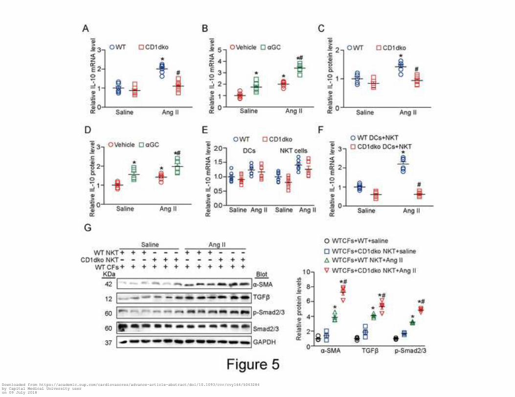
3.5 CD1d deficiency reduces IL-10 expression from NKT cells causing fibroblast
differentiation
The results in Figure 3C showed that CD1d deficiency increased Th1-type cytokines (IL-1β and
TNF-α) in Ang II-treated hearts, we then tested whether CD1d deficiency reduced Th2-type
cytokines (IL-10 and IL-4) in this model. qPCR analysis revealed that Ang II infusion upregulated
the expression of IL-10 and IL-4 in the hearts of WT mice compared with saline-treated control.
This effect was significantly reduced in CD1dko mice (Figure 5A and Supplementary material,
Figure 4A), but was enhanced in αGC-treated hearts after Ang II infusion (Figure 5B and
Supplementary material, Figure 4B). The change of IL-10 protein level in CD1dko hearts or
αGC-treated WT hearts was further verified by ELISA assay (Figure 5C and 5D).
To determine which cells could produce IL-10, we measured IL-10 mRNA level in DCs or
NKT cells isolated from WT and Cd1dko mice, respectively. qPCR analysis showed that IL-10
mRNA expression was slightly increased in Ang II-treated DCs or NKT cells, and there was no
significant difference between WT and Cd1dko mice (Figure 5E). We next test whether CD1d+
Downloaded from https://academic.oup.com/cardiovascres/advance-article-abstract/doi/10.1093/cvr/cvy164/5043284by Capital Medical University useron 09 July 2018

8
DCs mediated the mRNA expression of IL-10 from NKT cells. Ang II treatment reduced the Cd1d
mRNA level in DCs level compared with saline control (Supplementary material, Figure 4C).
Furthermore, co-culture of NKT cells with CD1dko DCs significantly reduced IL-10 mRNA level in
NKT cells compared with NKT cells co-cultured with WT DCs after Ang II treatment (Figure 5F),
indicating that IL-10 is mainly produced from NKT cells via CD1d+ DCs, and may be critical in this
model.
We next examined the regulatory properties of NKT cells in activation of cardiac fibroblasts
(CFs) in vitro. NKT cells isolated from WT or CD1dko mice were co-cultured with WT CFs.
Immunoblotting analysis indicated that the protein levels of a-SMA (a marker for myofibroblast
differentiation), TGF-β1 and p-Smad2/3 were markedly upregulated in CFs co-cultured with WT or
CD1dko NKT cells compared with saline controls, and this increase was enhanced in CFs
co-cultured CD1dko NKT cells after Ang II stimulation (Figure 5G). These results indicate that
CD1dko NKT cells are capable of promoting myofibroblast activation.
3.6 Administration of recombinant IL-10 improves cardiac remodeling in CD1dko mice
We next tested whether IL-10 can prevent Ang II-induced hypertension and cardiac remodeling in
WT and CD1dko mice. After Ang II treatment, IgG-treated CD1dko mice showed increased SBP,
cardiac performance (EF% and FS%), hypertrophy (HW/BW and HW/TL ratios, myocyte
cross-sectional area) and fibrosis (collagen deposition), infiltration of Mac-2-positive macrophages
and expression of ANF, BNP, β-myosin heavy chain (β-MHC), collagen I and collagen III
compared with IgG-treated WT mice (Figure 6A-E and Supplementary material, Figure 5 and
Figure 6A-B). However, systemic administration of mouse recombinant IL-10 (50 μg/kg)
significantly reversed these pathological changes in WT and CD1dko mice compared with the IgG
controls (Figure 6A-E and Supplementary material, Figure 5 and Figure 6A-B). Moreover,
administration of IL-10 markedly increased the p-STAT3 level, but reduced the levep-p65, TGF-β1
and α-SMA protein levels in both WT and CD1dko mice compared with the IgG controls after Ang
II infusion (Figure 6F). Together, these results suggest that IL-10 improves cardiac remodeling in
Cd1d-deficient mice, which was partially associated with activation of STAT3 and inhibition of the
NF-kB and TGF-β1 pathways.
Downloaded from https://academic.oup.com/cardiovascres/advance-article-abstract/doi/10.1093/cvr/cvy164/5043284by Capital Medical University useron 09 July 2018

9
4. Discussion
In this study, we evaluated the regulatory role of CD1d-dependent NKT cells in Ang II-induced
cardiac remodeling. We showed that Cd1d deficiency significantly accelerated Ang II-induced
hypertension, cardiac remodeling and inflammatory response, which were markedly improved by
αGC. Importantly, Cd1d-deficient DCs reduced IL-10 production by NKT cells. Administration of
IL-10 to Cd1dko mice significantly reversed the Ang II-induced hypertension and cardiac
remodeling, possibly through activation of STAT3 and inhibition of the TGF-β1 and NF-kB
signaling pathways.
Cardiac hypertrophy and fibrosis are pathological features of many cardiac diseases,
including hypertension, cardiomyopathy and myocardial infarction. However, the mechanisms that
regulate these diseases have not been fully elucidated. Recently, immune cells including
monocytes, macrophages, dendritic cells (DCs) and T Iymphocytes have been implicated in the
development of hypertension, cardiac remodeling and heart failure5. Among them, T cells have
been regarded as critical modulators in the onset of the cardiac proinflammatory response and
remodeling. CD8+ T cells promote macrophage activation, leading to cardiac injury upon Ang II
infusion2. Deletion of IL-12p35 increases the CD4+T cell-dependent differentiation of M2
macrophages and the production of TGF-β1 to aggravate Ang II-induced cardiac fibrosis3.
γδT-cell-derived IL-17 also promotes Ang II-induced cardiac injury and fibrosis26. Moreover, our
recent findings suggest that CXCL1-CXCR2 axis mediates monocyte activation, which induces
hypertension thereby leading to cardiac remodeling25, 27, 28. In this study, we extended previous
findings and clearly showed that CD1d-dependent NKT cells also exhibited a cardioprotective role.
CD1d-deficiency exacerbates Ang II-induced elevation of SBP together with cardiac hypertrophy
and fibrosis, whereas aGC blunts this response (Figure 1 and 2, Figure S2), suggesting that Ang
II-induced cardiac remodeling was partially secondary to BP after Cd1d deletion.
Proinflammatory cells-derived cytokines including IL-1 β, IL-6, TNF-α and TGF-β1 have
been implicated in the development of cardiac remodeling, most likely via downstream activation
of many signaling mediators and transcription factors, such as NF-kB and Smad2/327. Activation
of NF-kB signaling significantly induces cardiac inflammation and hypertrophy29. Conversely,
inhibition of NF-kB by ablation or various inhibitors markedly attenuated left ventricular
hypertrophy induced by Ang II and chronic pressure overload30-32. TGF-β-Smad signaling pathway
Downloaded from https://academic.oup.com/cardiovascres/advance-article-abstract/doi/10.1093/cvr/cvy164/5043284by Capital Medical University useron 09 July 2018

10
is considered a major contributor of cardiac fibrosis 27. Dendritic cells (DCs) are highly specialized
antigen-presenting cells with a unique ability to activate naive T lymphocytes. Our previous study
suggested that Ang II can enhance the maturation and activation of DCs through activation of
NF-kB, ERK and STAT1 signaling pathways 33. Further, antigen-presenting molecule CD1d on
DCs can activate NKT cells through their semi-invariant αβ T cell receptors (TCRs)34. The present
study suggested that CD1d-expressing DCs regulated activation of NKT cells to produce multiple
cytokines such as IL-1β, TNF-α, IL-4 and IL-10 after Ang II stimulation. Deletion of Cd1d
upregulated IL-1β and TNF-α but reduced IL-4 and IL-10 accompanied with activation of the
NF-kB pathways, and these effects were markedly reversed by αGC (Figure 1 through 3, 5, Figure
S3 and 4). Thus, our findings indicate that CD1d-dependent NKT cells exert a cardioprotective
effect partially through reduction of proinflammatory cytokines and inhibition of the NF-kB and
TGF-β1/Smad2/3 signaling cascades.
Increasing evidence suggests that NKT cells have opposite roles in various
immune-based diseases through either Th1-cytokines or Th2-cytokines14-16,19-22, 35. A recent study
has demonstrated that the balance of Th1/Th2 cytokines plays a critical role in cardiac remodeling
and inflammation36. Consistent with the role of NKT cells in inducing Th2 cytokines in diabetes,
rheumatoid arthritis and ischemic heart diseases19-22, 35, our results showed that NKT cell
deficiency can cause a shift from Th2 to Th1 in Ang II-treated heart, while activation of NKT cells
by αGC had the opposite effect (Figure 5 and Figure S4). IL-10 is an anti-inflammatory cytokine
that exerts different effects on hypertension through regulating multiple signaling pathways. IL-10
preserves endothelium-dependent vasorelaxation perhaps by reducing production of superoxide
during diabetes37. Moreover, IL-10 limits Ang II-induced hypertension by inhibiting
RhoA/Rho-kinase signaling in angiotensin II-infused mice. In contrast, IL-10 increases
salt-sensitive hypertension and renal injury induced by Ang II through reduction of nitric oxide38.
Here our results showed that IL-10 treatment markedly reduced Ang II-induced hypertension in
both WT and CD1dko mice (Figure S5), which may contribute to attenuation of Ang II-cardiac
remodeling (Figure 6). In addition to hypertension, IL-10 also has opposite role in regulation of
cardiac remodeling via STAT3 and NF-kB. Administration of IL-10 significantly attenuates
isoproterenol- or pressure overload-induced hypertrophic remodeling and improves heart
function39. Moreover, IL-10 mediates the protection of αGC against myocardial remodeling after
Downloaded from https://academic.oup.com/cardiovascres/advance-article-abstract/doi/10.1093/cvr/cvy164/5043284by Capital Medical University useron 09 July 2018

11
ischemia/reperfusion and post-infarction20, 21, 40. However, a recent study in mice and human
showed that IL-10 exerts profibrotic deleterious actions in humans and mice. Cardiac
macrophages produce IL-10, activate fibroblasts to collagen deposition, leading to impaired
myocardial relaxation. Deletion of IL-10 in macrophages improves diastolic function41. In contrast
to this study, our in vitro results revealed that CD1d-deficient DCs reduced IL-10 expression by
NKT cells (Figure 5F), and CD1dko NKT cells significantly promoted myofibroblast differentiation
and TGF-β1/Smad2/3 signaling activation (Figure 5G), suggesting that IL-10 produced by NKT
cells may be crucial in this model (Figure 5). Consistent with the results for the cardioprotective
role of IL-10, the present data confirmed that IL-10 treatment markedly reduced Ang II-induced
cardiac remodeling in both WT and CD1dko mice, and this beneficial effect was partially
associated with activation of STAT3 and inhibition of the TGF-β1 and NF-κB pathways (Figure 6).
Overall, these data support the idea that activation of Cd1d-dependent NKT cells prevented Ang
II-induced cardiac injury possibly through increased IL-10 production.
In conclusion, we demonstrated for the first time that activation of CD1d-dependent NKT
cells plays a protective role in Ang II-induced cardiac remodeling and inflammation. NKT cells
release IL-10, which inhibited cardiomyocyte hypertrophy and fibroblast differentiation through
activation of STAT3 and inhibition of the TGF-β1 and NF-κB pathways. Thus, selective activation
of NKT cells may represent a promising therapeutic approach for the treatment of hypertensive
cardiac diseases. Further investigations in other animal models of cardiac hypertrophy and
remodeling are needed to determine the clinical use of NKT cell activation as a pharmacological
therapy.
Funding
This work was supported by grants from the National Natural Science Foundation of China
(81630009, 81570207 and 81330003), Beijing Natural Science Foundation (7162018) and Chang
Jiang Scholar Program of China (T2011160, H.H.L).
Downloaded from https://academic.oup.com/cardiovascres/advance-article-abstract/doi/10.1093/cvr/cvy164/5043284by Capital Medical University useron 09 July 2018

12
Conflict of interest
None declared.
References
1. Cohn JN, Ferrari R, Sharpe N. Cardiac remodeling--concepts and clinical implications: a
consensus paper from an international forum on cardiac remodeling. Behalf of an International
Forum on Cardiac Remodeling. J Am Coll Cardiol 2000;35:569-582.
2. Ma F, Feng J, Zhang C, Li Y, Qi G, Li H, Wu Y, Fu Y, Zhao Y, Chen H, Du J, Tang H. The
requirement of CD8+ T cells to initiate and augment acute cardiac inflammatory response to
high blood pressure. J Immunol 2014;192:3365-3373.
3. Li Y, Zhang C, Wu Y, Han Y, Cui W, Jia L, Cai L, Cheng J, Li H, Du J. Interleukin-12p35 deletion
promotes CD4 T-cell-dependent macrophage differentiation and enhances angiotensin
II-Induced cardiac fibrosis. Arterioscler Thromb Vasc Biol 2012;32:1662-1674.
4. Wang L, Li YL, Zhang CC, Cui W, Wang X, Xia Y, Du J, Li HH. Inhibition of Toll-like receptor 2
reduces cardiac fibrosis by attenuating macrophage-mediated inflammation. Cardiovasc Res
2014;101:383-392.
5. Frieler RA, Mortensen RM. Immune cell and other noncardiomyocyte regulation of cardiac
hypertrophy and remodeling. Circulation 2015;131:1019-1030.
6. Skold M, Behar SM. Role of CD1d-restricted NKT cells in microbial immunity. Infect Immun
2003;71:5447-5455.
7. Godfrey DI, Kronenberg M. Going both ways: immune regulation via CD1d-dependent NKT
cells. The Journal of clinical investigation 2004;114:1379-1388.
8. Wu L, Gabriel CL, Parekh VV, Van Kaer L. Invariant natural killer T cells: innate-like T cells with
potent immunomodulatory activities. Tissue Antigens 2009;73:535-545.
9. Smiley ST, Kaplan MH, Grusby MJ. Immunoglobulin E production in the absence of
interleukin-4-secreting CD1-dependent cells. Science 1997;275:977-979.
10. Exley MA, Bigley NJ, Cheng O, Shaulov A, Tahir SM, Carter QL, Garcia J, Wang C, Patten K,
Stills HF, Alt FW, Snapper SB, Balk SP. Innate immune response to encephalomyocarditis
virus infection mediated by CD1d. Immunology 2003;110:519-526.
11. Mendiratta SK, Martin WD, Hong S, Boesteanu A, Joyce S, Van Kaer L. CD1d1 mutant mice
are deficient in natural T cells that promptly produce IL-4. Immunity 1997;6:469-477.
12. Ishikawa H, Tanaka K, Kutsukake E, Fukui T, Sasaki H, Hata A, Noda S, Matsumoto T.
IFN-gamma production downstream of NKT cell activation in mice infected with influenza virus
enhances the cytolytic activities of both NK cells and viral antigen-specific CD8+ T cells.
Virology 2010;407:325-332.
13. Oleinika K, Rosser EC, Matei DE, Nistala K, Bosma A, Drozdov I, Mauri C. CD1d-dependent
immune suppression mediated by regulatory B cells through modulations of iNKT cells. Nat
Commun 2018;9:684.
14. Olszak T, Neves JF, Dowds CM, Baker K, Glickman J, Davidson NO, Lin CS, Jobin C, Brand S,
Sotlar K, Wada K, Katayama K, Nakajima A, Mizuguchi H, Kawasaki K, Nagata K, Muller W,
Snapper SB, Schreiber S, Kaser A, Zeissig S, Blumberg RS. Protective mucosal immunity
mediated by epithelial CD1d and IL-10. Nature 2014;509:497-502.
Downloaded from https://academic.oup.com/cardiovascres/advance-article-abstract/doi/10.1093/cvr/cvy164/5043284by Capital Medical University useron 09 July 2018

13
15. Nakai Y, Iwabuchi K, Fujii S, Ishimori N, Dashtsoodol N, Watano K, Mishima T, Iwabuchi C,
Tanaka S, Bezbradica JS, Nakayama T, Taniguchi M, Miyake S, Yamamura T, Kitabatake A,
Joyce S, Van Kaer L, Onoe K. Natural killer T cells accelerate atherogenesis in mice. Blood
2004;104:2051-2059.
16. Rabb H. The promise of immune cell therapy for acute kidney injury. The Journal of clinical
investigation 2012;122:3852-3854.
17. Getz GS, Reardon CA. Natural killer T cells in atherosclerosis. Nat Rev Cardiol
2017;14:304-314.
18. Van Kaer L, Wu L, Parekh VV. Natural killer T cells in multiple sclerosis and its animal model,
experimental autoimmune encephalomyelitis. Immunology 2015;146:1-10.
19. Miellot A, Zhu R, Diem S, Boissier MC, Herbelin A, Bessis N. Activation of invariant NK T cells
protects against experimental rheumatoid arthritis by an IL-10-dependent pathway. Eur J
Immunol 2005;35:3704-3713.
20. Homma T, Kinugawa S, Takahashi M, Sobirin MA, Saito A, Fukushima A, Suga T, Takada S,
Kadoguchi T, Masaki Y, Furihata T, Taniguchi M, Nakayama T, Ishimori N, Iwabuchi K, Tsutsui
H. Activation of invariant natural killer T cells by alpha-galactosylceramide ameliorates
myocardial ischemia/reperfusion injury in mice. J Mol Cell Cardiol 2013;62:179-188.
21. Sobirin MA, Kinugawa S, Takahashi M, Fukushima A, Homma T, Ono T, Hirabayashi K, Suga T,
Azalia P, Takada S, Taniguchi M, Nakayama T, Ishimori N, Iwabuchi K, Tsutsui H. Activation of
natural killer T cells ameliorates postinfarct cardiac remodeling and failure in mice. Circ Res
2012;111:1037-1047.
22. Hong S, Wilson MT, Serizawa I, Wu L, Singh N, Naidenko OV, Miura T, Haba T, Scherer DC,
Wei J, Kronenberg M, Koezuka Y, Van Kaer L. The natural killer T-cell ligand
alpha-galactosylceramide prevents autoimmune diabetes in non-obese diabetic mice. Nat Med
2001;7:1052-1056.
23. van Puijvelde GHM, Foks AC, van Bochove RE, Bot I, Habets KLL, de Jager SC, Ter Borg
MND, van Osch P, Boon L, Vos M, de Waard V, Kuiper J. CD1d deficiency inhibits the
development of abdominal aortic aneurysms in LDL receptor deficient mice. PLoS One
2018;13:e0190962.
24. Wang X, Wang HX, Li YL, Zhang CC, Zhou CY, Wang L, Xia YL, Du J, Li HH. MicroRNA Let-7i
negatively regulates cardiac inflammation and fibrosis. Hypertension 2015;66:776-785.
25. Wang L, Zhao XC, Cui W, Ma YQ, Ren HL, Zhou X, Fassett J, Yang YZ, Chen Y, Xia YL, Du J,
Li HH. Genetic and Pharmacologic Inhibition of the Chemokine Receptor CXCR2 Prevents
Experimental Hypertension and Vascular Dysfunction. Circulation 2016;134:1353-1368.
26. Li Y, Wu Y, Zhang C, Li P, Cui W, Hao J, Ma X, Yin Z, Du J. gammadeltaT Cell-derived
interleukin-17A via an interleukin-1beta-dependent mechanism mediates cardiac injury and
fibrosis in hypertension. Hypertension 2014;64:305-314.
27. Wang L, Zhang YL, Lin QY, Liu Y, Guan XM, Ma XL, Cao HJ, Liu Y, Bai J, Xia YL, Du J, Li HH.
CXCL1-CXCR2 axis mediates angiotensin II-induced cardiac hypertrophy and remodelling
through regulation of monocyte infiltration. European heart journal 2018;39:1818-1831.
28. Paradis P, Schiffrin EL. CXCL1-CXCR2 lead monocytes to the heart of the matter. European
heart journal 2018;39:1832-1834.
29. Freund C, Schmidt-Ullrich R, Baurand A, Dunger S, Schneider W, Loser P, El-Jamali A, Dietz R,
Scheidereit C, Bergmann MW. Requirement of nuclear factor-kappaB in angiotensin II- and
Downloaded from https://academic.oup.com/cardiovascres/advance-article-abstract/doi/10.1093/cvr/cvy164/5043284by Capital Medical University useron 09 July 2018

14
isoproterenol-induced cardiac hypertrophy in vivo. Circulation 2005;111:2319-2325.
30. Zelarayan L, Renger A, Noack C, Zafiriou MP, Gehrke C, van der Nagel R, Dietz R, de Windt L,
Bergmann MW. NF-kappaB activation is required for adaptive cardiac hypertrophy.
Cardiovascular research 2009;84:416-424.
31. Wang C, Li L, Zhang ZG, Fan D, Zhu Y, Wu LL. Globular adiponectin inhibits angiotensin
II-induced nuclear factor kappaB activation through AMP-activated protein kinase in cardiac
hypertrophy. Journal of cellular physiology 2010;222:149-155.
32. Esposito G, Rapacciuolo A, Naga Prasad SV, Takaoka H, Thomas SA, Koch WJ, Rockman HA.
Genetic alterations that inhibit in vivo pressure-overload hypertrophy prevent cardiac
dysfunction despite increased wall stress. Circulation 2002;105:85-92.
33. Chen C, Meng Y, Wang L, Wang HX, Tian C, Pang GD, Li HH, Du J. Ubiquitin-activating
enzyme E1 inhibitor PYR41 attenuates angiotensin II-induced activation of dendritic cells via
the IkappaBa/NF-kappaB and MKP1/ERK/STAT1 pathways. Immunology 2014;142:307-319.
34. Alvarez D, Vollmann EH, von Andrian UH. Mechanisms and consequences of dendritic cell
migration. Immunity 2008;29:325-342.
35. Singh AK, Wilson MT, Hong S, Olivares-Villagomez D, Du C, Stanic AK, Joyce S, Sriram S,
Koezuka Y, Van Kaer L. Natural killer T cell activation protects mice against experimental
autoimmune encephalomyelitis. The Journal of experimental medicine 2001;194:1801-1811.
36. Fujiu K, Wang J, Nagai R. Cardioprotective function of cardiac macrophages. Cardiovascular
research 2014;102:232-239.
37. Gunnett CA, Heistad DD, Faraci FM. Interleukin-10 protects nitric oxide-dependent relaxation
during diabetes: role of superoxide. Diabetes 2002;51:1931-1937.
38. Singh P, Castillo A, Islam MT, Majid DSA. Evidence for Prohypertensive, Proinflammatory
Effect of Interleukin-10 During Chronic High Salt Intake in the Condition of Elevated
Angiotensin II Level. Hypertension 2017;70:839-845.
39. Verma SK, Krishnamurthy P, Barefield D, Singh N, Gupta R, Lambers E, Thal M, Mackie A,
Hoxha E, Ramirez V, Qin G, Sadayappan S, Ghosh AK, Kishore R. Interleukin-10 treatment
attenuates pressure overload-induced hypertrophic remodeling and improves heart function
via signal transducers and activators of transcription 3-dependent inhibition of nuclear
factor-kappaB. Circulation 2012;126:418-429.
40. Jung M, Ma Y, Iyer RP, DeLeon-Pennell KY, Yabluchanskiy A, Garrett MR, Lindsey ML. IL-10
improves cardiac remodeling after myocardial infarction by stimulating M2 macrophage
polarization and fibroblast activation. Basic Res Cardiol 2017;112:33.
41. Hulsmans M, Sager HB, Roh JD, Valero-Munoz M, Houstis NE, Iwamoto Y, Sun Y, Wilson RM,
Wojtkiewicz G, Tricot B, Osborne MT, Hung J, Vinegoni C, Naxerova K, Sosnovik DE, Zile MR,
Bradshaw AD, Liao R, Tawakol A, Weissleder R, Rosenzweig A, Swirski FK, Sam F,
Nahrendorf M. Cardiac macrophages promote diastolic dysfunction. The Journal of
experimental medicine 2018;215:423-440.
Downloaded from https://academic.oup.com/cardiovascres/advance-article-abstract/doi/10.1093/cvr/cvy164/5043284by Capital Medical University useron 09 July 2018

15
Figure legends:
Figure 1. CD1d deficiency accelerates Ang II-induced cardiac remodeling. (A) Echocardiography
was performed on CD1dko or WT mice after 14 days of Ang II infusion (1000 ng/kg/min) (top).
Quantification of EF% and FS% (bottom; n=10 per group). (B) Representative H&E staining of
heart sections (top). Scale bar: 2 mm. The ratios of the heart weight/body weight (HW/BW) and
heart weight/tibia length (HW/TL) (bottom; n=10 per group). (C) Representative wheat germ
agglutinin (WGA) staining of heart sections (left) and quantification of the cross-sectional area of
myocytes (middle; n=6 per group). Scale bar: 50 μm. qPCR analysis of the hypertrophic markers
ANF and BNP mRNA expression in wild-type (WT) and CD1dko hearts after saline or Ang II
infusion (right; n=6 per group). (D) Representative Masson’s trichrome staining in heart sections of
each group (left) and quantification of collagen deposition (middle; n=6 per group). Scale bar: 100
μm. qPCR analysis of collagen I and collagen III mRNA expression in hearts (right; n=6 per group).
(E) Western blot analysis for the protein expression of TGF-β1, p-Smad2/3 and Smad2/3 (left) and
quantification of the protein bands (right; n=5 per group). Data are expressed as mean ± SEM.
*P<0.05, **P<0.01 vs saline group; #P<0.05, ##P<0.01 vs Ang II group.
Figure 2. aGC alleviates Ang II-induced cardiac remodeling. (A) Echocardiography was performed
in wild type (WT) mice after 14 days of aGC administration (top). Quantification of EF% and FS%
(bottom; n=10 per group). (B) Representative H&E staining of heart sections (top), Scale bar: 2
mm, and the ratios of heart weight/body weight (HW/BW) and heart weight/tibial length (HW/TL)
(bottom; n=10 per group). (C) Representative wheat germ agglutinin (WGA) staining of heart
sections (left) and quantification of the cross-sectional area of myocytes (middle; n=6 per group).
Scale bar: 50 μm. qPCR analysis of hypertrophic markers ANF and BNP mRNA expression in
hearts (right; n=6 per group). (D) Representative Masson’s trichrome staining of the heart sections
of each group (left) and quantification of collagen deposition (middle; n=6 per group). Scale bar:
100 μm. qPCR analysis of collagen I and collagen III mRNA expression in the hearts (right; n=6
per group). (E) Western blot analysis for the protein expression of TGFβ, p-Smad2/3 and Smad2/3
(left). Quantification of the protein bands (right; n=5 per group). Data are expressed as mean ±
SEM. *P<0.05, **P<0.01, ***P<0.001 vs vehicle groups. #P<0.05, ##P<0.01 vs Ang II group.
Downloaded from https://academic.oup.com/cardiovascres/advance-article-abstract/doi/10.1093/cvr/cvy164/5043284by Capital Medical University useron 09 July 2018

16
Figure 3. CD1d deficiency aggravates Ang II-induced cardiac inflammation and activation of
NF-κB in the heart tissues. (A) Flow cytometry analysis of CD45+ leukocytes,
CD45+CD11b+F4/80+ macrophages, CD45+CD11b+Gr-1+ neutrophils and CD45+CD3+ T
lymphocytes in the hearts (n=4 per group). (B) Representative H&E and immunohistochemical
staining of Mac-2 in heart sections from wild-type (WT) and CD1dko mice (left). Scale bar: 20 μm;
Quantification of Mac-2-positive cells (right; n=6 per group). (C) qPCR analysis of IL-1β and
TNF-α mRNA expression in the hearts (n=6 per group). (D) Immunoblotting analysis for the
protein expression of p-p65 and p65 (top). Quantification of protein bands (bottom; n=5 per group).
Data are expressed as mean ± SEM. *P < 0.05, **P < 0.01 vs saline or vehicle group. #P < 0.05,
##P < 0.01 vs Ang II group.
Figure 4. Bone marrow-derived CD1dko cells exacerbate cardiac remodeling induced by Ang II
infusion. (A) Echocardiography was performed on wild-type (WT) mice after bone marrow (BM)
transplantation (top). Quantification of EF% and FS% (bottom; n=10 per group). (B)
Representative H&E staining of heart sections (top). Scale bar: 2 mm. The ratios of heart
weight/body weight (HW/BW) and heart weight/tibial length (HW/TL) were calculated after BM
transplantation (bottom; n=10 per group). (C) Representative wheat germ agglutinin (WGA)
staining of the heart sections (left) and quantification of cross-sectional area of myocytes (middle).
Scale bar: 50 μm. qPCR analysis of hypertrophic markers ANF and BNP mRNA expression in the
hearts (right; n=6 mice per group). (D) Representative Masson’s trichrome staining (left) and
quantification of collagen deposition in the heart sections of each group (middle; n=6 per group).
Scale bar: 100 μm. qPCR analysis of collagen I and collagen III mRNA expression in the hearts
(right; n=6 per group). (E) Representative H&E and immunohistochemical staining of
Mac-2-positive cells (left) and quantification of these cells (middle; n=6 per group). Scale bar: 20
μm; qPCR analysis of IL-1β and TNF-α mRNA expression in the hearts (right; n=6 per group).
Data are expressed as mean ± SEM. *P <0.05 vs WT BM to WT.
Figure 5. CD1d deficiency reduces IL-10 expression by NKT cells resulting in fibroblast activation.
(A) qPCR analysis of the mRNA level of IL-10 in the hearts of wild type (WT) and CD1dko mice
after Ang II infusion (n=6 per group). (B) qPCR analysis of IL-10 mRNA level in vehicle- or
aGC-treated WT hearts after Ang II infusion (n=6 per group). (C) ELISA of the protein level of
Downloaded from https://academic.oup.com/cardiovascres/advance-article-abstract/doi/10.1093/cvr/cvy164/5043284by Capital Medical University useron 09 July 2018

17
IL-10 in WT and CD1dko mice after Ang II infusion (n=6 per group). (D) ELISA of the protein level
of IL-10 in WT mice treated with vehicle or aGC after Ang II infusion (n=6 per group). (E) qPCR
analysis of IL-10 mRNA level in DCs or NKT cells from WT mice after saline or Ang II treatment. (F)
qPCR analysis of IL-10 mRNA level in NKT cells co-cultured with DCs from WT or CD1dko mice
after Ang II treatment (n=6 per group). (G) Immunoblotting analysis of the protein levels of α-SMA,
TGF-β1, p-Smad2/3 and Smad2/3 in cardiac fibroblasts (CFs) co-cultured with NKT cells from WT
or CD1dko mice after Ang II-infusion (left) and quantification (right; n=5 per group). Data are
expressed as mean ± SEM. *P <0.05 vs saline groups; #P<0.05 vs Ang II group.
Figure 6. IL-10 administration reverses cardiac remodeling in CD1d-deficient mice. (A)
Echocardiography was performed in wild type (WT) and CD1dko mice 14 days after Ang II
infusion and IL-10 administration (50 μg/kg) (top). Quantification of EF% and FS% (bottom; n=10
per group). (B) Representative H&E staining of heart sections (top). Scale bar: 2 mm. The ratios of
heart weight/body weight (HW/BW) and heart weight/tibial length (HW/TL) (bottom; n=10 per
group). (C) Representative wheat germ agglutinin (WGA, Scale bar: 50 μm) and Masson’s
trichrome staining in heart sections (Scale bar: 100 μm). (D) Quantification of the cross-sectional
area of myocytes and collagen deposition (n=6 per group). (E) Representative
immunohistochemical staining of the Mac-2 on the heart sections (left). Scale bar: 50 μm.
Quantification of Mac-2-positive area (right; n=6 per group). (F) Immunoblotting analysis of the
protein expression of p-STA3, STAT3, p-p65, p65, TGF-β1 and α-SMA (left). Quantification of the
protein bands (right; n=5 per group). Data are expressed as mean ± SEM. *P<0.05 vs
WT+IgG+Ang II; #P< vs CD1dko+IgG+Ang II; $ P< vs CD1dko+IL-10+Ang II.
Downloaded from https://academic.oup.com/cardiovascres/advance-article-abstract/doi/10.1093/cvr/cvy164/5043284by Capital Medical University useron 09 July 2018

Downloaded from https://academic.oup.com/cardiovascres/advance-article-abstract/doi/10.1093/cvr/cvy164/5043284by Capital Medical University useron 09 July 2018

Downloaded from https://academic.oup.com/cardiovascres/advance-article-abstract/doi/10.1093/cvr/cvy164/5043284by Capital Medical University useron 09 July 2018

Downloaded from https://academic.oup.com/cardiovascres/advance-article-abstract/doi/10.1093/cvr/cvy164/5043284by Capital Medical University useron 09 July 2018

Downloaded from https://academic.oup.com/cardiovascres/advance-article-abstract/doi/10.1093/cvr/cvy164/5043284by Capital Medical University useron 09 July 2018
Downloaded from https://academic.oup.com/cardiovascres/advance-article-abstract/doi/10.1093/cvr/cvy164/5043284by Capital Medical University useron 09 July 2018

Downloaded from https://academic.oup.com/cardiovascres/advance-article-abstract/doi/10.1093/cvr/cvy164/5043284by Capital Medical University useron 09 July 2018
Copyright © 2022 FDOKUMEN