A Novel Isoform of Acetylcholinesterase Exacerbates Photoreceptors Death after Photic Stress
Upload
khangminh22Category
view
6download
0
RESEARCH ARTICLE
Angiotensin II receptor blocker losartan
exacerbates muscle damage and exhibits
weak blood pressure-lowering activity in a
dysferlin-null model of Limb-Girdle muscular
dystrophy type 2B
Zoe WhiteID1,2☯*, Nadia MiladID
1,2☯, Arash Y. Tehrani1,2, William Wei-Han ChenID1,2,
Graham Donen1,2, Stephanie L. Sellers1,2, Pascal Bernatchez1,2*
1 University of British Columbia (UBC) Department of Anesthesiology, Pharmacology & Therapeutics,
Vancouver, Canada, 2 UBC Centre for Heart Lung Innovation & St. Paul’s Hospital, Vancouver, Canada
☯ These authors contributed equally to this work.
* [email protected] (ZW); [email protected] (PB)
Abstract
There is no cure or beneficial management option for Limb-Girdle muscular dystrophy (MD)
type 2B (LGMD2B). Losartan, a blood pressure (BP) lowering angiotensin II (AngII) receptor
type 1 (ATR1) blocker (ARB) with unique anti-transforming growth factor-β (TGF-β) proper-
ties, can protect muscles in various types of MD such as Duchenne MD, suggesting a poten-
tial benefit for LGMD2B patients. Herein, we show in a mild, dysferlin-null mouse model of
LGMD2B that losartan increased quadriceps muscle fibrosis (142%; P<0.0001). In a severe,
atherogenic diet-fed model of LGMD2B recently described by our group, losartan further
exacerbated dysferlin-null mouse muscle wasting in quadriceps and triceps brachii, two
muscles typically affected by LGMD2B, by 40% and 51%, respectively (P<0.05). Lower
TGF-β signalling was not observed with losartan, therefore plasma levels of atherogenic lip-
ids known to aggravate LGMD2B severity were investigated. We report that losartan
increased both plasma triglycerides and cholesterol concentrations in dysferlin-null mice.
Other protective properties of losartan, such as increased nitric oxide release and BP lower-
ing, were also reduced in the absence of dysferlin expression. Our data suggest that
LGMD2B patients may show some resistance to the primary BP-lowering effects of losartan
along with accelerated muscle wasting and dyslipidemia. Hence, we urge caution on the
use of ARBs in this population as their ATR1 pathway may be dysfunctional.
Introduction
Limb-girdle muscular dystrophy (MD) type 2B (LGMD2B) and Miyoshi myopathy are forms
of MD caused by mutations in the dysferlin gene[1–3], a calcium-dependent sarcolemma
repair and vesicle trafficking protein [[4,5] and reviewed in [6]]. In their late teens, patients
PLOS ONE | https://doi.org/10.1371/journal.pone.0220903 August 12, 2019 1 / 18
a1111111111
a1111111111
a1111111111
a1111111111
a1111111111
OPEN ACCESS
Citation: White Z, Milad N, Tehrani AY, Chen WW-
H, Donen G, Sellers SL, et al. (2019) Angiotensin II
receptor blocker losartan exacerbates muscle
damage and exhibits weak blood pressure-
lowering activity in a dysferlin-null model of Limb-
Girdle muscular dystrophy type 2B. PLoS ONE 14
(8): e0220903. https://doi.org/10.1371/journal.
pone.0220903
Editor: Atsushi Asakura, University of Minnesota
Medical School, UNITED STATES
Received: February 13, 2019
Accepted: July 25, 2019
Published: August 12, 2019
Copyright: © 2019 White et al. This is an open
access article distributed under the terms of the
Creative Commons Attribution License, which
permits unrestricted use, distribution, and
reproduction in any medium, provided the original
author and source are credited.
Data Availability Statement: All relevant data are
within the manuscript and its Supporting
Information files.
Funding: This work was supported by grants from
the Jain Foundation, the Canadian Institutes of
Health Research, the Canadian Foundation For
Innovation, the British Columbia Knowledge
Development Fund, the Heart & Stroke Foundation
(PNB), MITACS Canada and the Pacific Airways

with dysferlinopathies present with proximal and/or distal muscle weakness, which eventually
results in a complete loss of ambulation. To facilitate the testing of therapeutics, a number of
mouse strains have been identified (i.e. A/J, SJ/LJ [7]) or engineered (dysferlin-null [5]) to
express abnormally low levels of dysferlin. These typically show slightly elevated plasma crea-
tine kinase (CK) levels, a marker of muscle damage, and skeletal muscle biopsies reveal early
muscle regeneration followed by moderate fibrofatty remodelling, accumulation of macro-
phages and T cells as well as muscle wasting, but without the dramatic loss of ambulation typi-
cally observed in patients [5,8]. A recent report from our group has shown that the mild
phenotype of dysferlin-null mice can be exacerbated by increasing atherogenic, or non-HDL
(high-density lipoprotein) levels of plasma cholesterol, which results in accelerated fibrofatty
remodeling and gradual loss of ambulatory function similar to affected patients [9]. Currently,
there is no cure for dysferlinopathies nor any effective management options to delay the inevi-
table loss of ambulation observed in this patient population.
Losartan is an angiotensin II (AngII) receptor type 1 (ATR1) blocker (ARB) routinely used
to safely reduce blood pressure. In addition, others have reported that losartan has the unique
ability to attenuate transforming growth factor beta (TGF-β) signaling, a key regulator of mus-
cle homeostasis and regeneration [10–12]. In Duchenne MD (DMD), the cardiac and skeletal
muscle wasting cascade believed to be partly regulated by TGF-β can be attenuated by losartan
treatment, resulting in reduced serum CK, fibrosis, and calcification in the gastrocnemius, dia-
phragm and myocardium of mdx mouse model of DMD [13,14]. Other studies in the same
model reported reduced diaphragm fibrosis, improved grip strength and improved ex vivoforce frequency in the extensor digitorum longus (EDL) 6 months (mo) post-treatment [10].
Acute losartan treatment also protects against disuse atrophy following 21 days of hind-limb
immobilisation and against cardiotoxin injury, where losartan improved muscle regeneration,
time to heal as well as a number of in vivo functional readouts in the tibialis anterior (TA) of
sarcopenic mice [15]. Furthermore, therapeutic benefits following muscle injury include
improved muscle regeneration (4 and 18 days post-injury) and reduced fibrotic tissue forma-
tion in adult skeletal muscles [10]. In another model of MD, α2 laminin-deficient congenital
MD type 1A (MDC1A) was improved with losartan treatment [16] and this tissue protection
was also observed in patients [17]. Since LGMD2B patients also suffer from chronic muscle
wasting and have no management options, we hypothesized that losartan could protect muscle
tissues in dysferlin-deficient animals. For this purpose, the effect of chronic losartan treatment
(9 mo) on muscle wasting was evaluated in two mouse models of diet-accelerated dysferlinopa-
thy; 1) dysferlin-null mice fed a control diet, and 2) dysferlin-null mice fed a high-fat Western
diet (HFD), previously shown to exacerbate LGMD2B severity [9,18]. We report that losartan
treatment unexpectedly aggravated muscle wasting and fibrofatty infiltration in mild and
severely dysferlinopathic mice, which correlated with a losartan-dependent atherogenic shift
in their plasma lipid profile. In addition, dysferlin-null mice were resistant to the primary
blood pressure (BP)-lowering effects of losartan, which was in stark contrast to WT controls.
Hence our data suggest that use of losartan, and perhaps ARBs in general, should be avoided
in dysferlinopathic patients and further highlights the unique pathogenesis of dysferlinopa-
thies compared to other types of MD.
Materials & methods
Animal models
Mice were housed in a 12-hour/12-hour light/dark cycle, temperature-regulated facility. All
animal procedures were prospectively approved by the UBC Animal Care Committee (proto-
col A18-0172). Experimental mice were bred using dysferlin-null mice (C57BL/6 background:
Losartan exacerbates dysferlinopathy
PLOS ONE | https://doi.org/10.1371/journal.pone.0220903 August 12, 2019 2 / 18
Centre. The funders had no role in study design,
data collection and analysis, decision to publish, or
preparation of the manuscript.
Competing interests: The authors have declared
that no competing interests exist.
Abbreviations: Akt, protein kinase B; AngII,
angiotensin II; ANOVA, analysis of variance; ApoE,
apolipoprotein E; ARB, angiotensin II type 1
receptor blocker; A.U., arbitrary units; C/EBPδ,CCAAT-enhancer binding protein δ; CK, creatine
kinase; DMD, Duchenne muscular dystrophy; EDL,
extensor digitorum longus; 4E-BP1, eukaryotic
translation initiation factor 4E-binding protein 1;
GAPDH, glyceraldehyde 3-phosphate
dehydrogenase; HDL, high-density lipoprotein;
HDLc, HDL-associated cholesterol; HFD, high-fat
high-cholesterol diet; LDL, low-density lipoprotein;
LGMD2B, limb-girdle muscular dystrophy 2B;
MAPK, mitogen-activated protein kinase; MD,
muscular dystrophy; MDC1A, α2 laminin-deficient
congenital MD type 1A; MM, Myoshi myopathy;
Mo, months of age; mTORC1, mammalian target
of rapamycin complex 1; NADH, nicotinamide
adenine dinucleotide; NO, nitric oxide; NOS, nitric
oxide synthase; NT, nitro-tyrosine; PECAM, platelet
endothelial cell adhesion molecule-1; PPAR-γ,peroxisome proliferator-activated receptor gamma;
rpS6, ribosomal protein S6; S6K1, ribosomal
protein S6 kinase beta-1; SEM, standard error of
the mean; Ser, serine; SMAD, Mothers against
decapentaplegic homolog; TA, tibialis anterior; TC,
total cholesterol; TG, triglyceride; TGF-β,
transforming growth factor β.
Dysftm1Kcam) provided by the Dr. Kevin Campbell lab [19]. Ear-clip DNA was extracted using
DNeasy extraction kit (Qiagen, #69506) following manufacturer’s instructions. Mice were gen-
otyped using a previously described dysferlin PCR protocol [20]. Euthanasia was performed
under anaesthesia (3.5% v/v isoflurane, 2L O2) and either cervical dislocation or cardiac punc-
ture for perfusion with warm Krebs solution as described [9,18].
Losartan treatment
Mice were fed either a high-fat (HFD) (Harlan, TD88137; 42% kcal from fat and 34% sucrose
by weight + 0.2% total cholesterol) or normal control diet (Chow; LabDiet #5001); 13% kcal
from fat; 3.7% sucrose by weight) from 2 to 11 mo of age. For chronic treatment, Losartan
(0.6g/L) was administered in drinking water ad libitum for the same duration at a dose previ-
ously shown to have biological availability and therapeutic efficacy in mouse models of DMD
and connective tissue disease [10,15,21]. For acute treatment, WT and dysferlin-null mice
were supplemented with the same dose of losartan (0.6g/L) ad libitum from 6 to 12 weeks of
age. Mice provided with standard drinking water served as controls. For dose response experi-
ments, both WT and dysferlin-null mice were treated with 6 increasing dosages of losartan
spanning 6 days (0, 3.6, 7.1, 10.7, 17.8, 21.4 and 100mg/kg/d).
Blood pressure (BP) measurements
Systemic BP was noninvasively measured using the tail cuff system (Kent Scientific CODA2).
Briefly, mice were lightly anesthetised (0.75% v/v isoflurane, 1.5L O2) and placed on a warming
tray with the tail inserted into an inflatable cuff where systolic BP (SBP) and diastolic BP
(DBP) were measured. Mean arterial pressure (MAP) was calculated as follows: 1/3 x SBP + 2/
3 x DBP.
Analysis of plasma cholesterol, triglycerides and creatine kinase
Plasma was collected in heparinized tubes via cardiac puncture of mice at 11mo, centrifuged at
4,000 RPM for 10 min at 4˚C and stored at -80˚C. The Siemans Advia 1800 system was used to
quantify plasma concentrations of creatine kinase (CK), total cholesterol (TC), high density
lipoprotein (HDL-C), low density lipoprotein (LDL-C) and triglyceride (TGs) levels (assays all
from Siemens) were performed according to instructions from the manufacturer and as previ-
ously published [9,18].
Tissue processing
Muscles were fixed in 10% formalin for 24h then transferred to 70% EtOH, paraffin-embed-
ded, sectioned to 8μm and stained with Masson’s trichrome as previously reported [9]), as fro-
zen sectioning causes fat smearing when numerous adipocytes are present. Fat was quantified
by manually tracing adipocyte containing regions (previously confirmed by perilipin staining
[9]) using Aperio ImageScope. Muscle damage, which included areas of bulk inflammation
and necrotic muscle fibres were also quantified as previously described [9]. Area values for
each parameter were divided by total muscle area (μm2) to obtain percentage values. Collagen
content/fibrosis was measured using a positive pixel count algorithm in Aperio ImageScope
software using the following parameters: hue value of 0.66 and hue width of 0.25 and standard-
ized to the total area of outlined sections (μm2). For cross-sectional area (CSA) measures,
quadriceps (rectus femoris) and triceps brachii muscles stained with Masson’s were portioned
into nine equal quadrants and the CSA of 100 myofibers were measured in each quadrant,
totalling 900 myofibers for each muscle section. Myofiber number and the percentage of total
Losartan exacerbates dysferlinopathy
PLOS ONE | https://doi.org/10.1371/journal.pone.0220903 August 12, 2019 3 / 18

myofibres with a displaced and/or central nuclei were counted on entire muscle cross sections
using Aperio ImageScope.
Phospho-SMAD2 immunofluorescence
As described in [22], 8μm frozen sections were placed in ice-cold EtOH for 10 min. Sections
were blocked in 10% FBS, 0.03% Triton-X 100 in PBS before being incubated overnight at 4˚C
in 1% FBS, 0.03% Triton-X 100 in PBS (1:200; Phospho-SMAD2(Ser465/467); Invitrogen;
#44–244). Sections were washed 3 x 15min in 0.03% Triton-X 100 in PBS on ice and incubated
for 3h at 4˚C in 1% FBS, 0.03% Triton-X 100 in PBS (AlexaFluor594 goat anti-rabbit #A11037
Invitrogen). Samples were again washed 3 x 15min in 0.03% Triton-X 100 in PBS on ice and
mounted with DABCO mounting media with DAPI. Phospho-SMAD2 (pSMAD2) positive
nuclei were manually quantified and divided by the total number of nuclei present (DAPI)
across four images (each 20x magnification) using Aperio ImageScope taken across whole
quadriceps muscles. No discrimination between myonuclei and nuclei from other cells types
were made.
Immunoblotting
Briefly, remaining frozen quadriceps were ground in liquid nitrogen, homogenized in ice-cold
PBS, 1% NP40, 1mM EDTA buffer, with complete EDTA-free protease inhibitor and Phos-
STOP phosphatase inhibitor tablets (Roche, Manheim, Germany), and centrifuged at 13,000g
for 20 min at 4˚C [23]. Protein was quantified with the DCA protein Assay (Bio-Rad). Samples
were resolved on 4–15% SDS-PAGE TGX gels (Bio-Rad) and transferred onto nitrocellulose
membranes (Bio-Rad) using the Trans-Blot Turbo Transfer System (Bio-Rad; mixed molecu-
lar weight program; 2.5A-25V-7min). Following transfer, membranes were blocked in 1%
casein in TBS (Li-COR) for 1 hour at RT. Primary antibodies were diluted 1:1000 in 1% casein
in TBST (0.1% Tween 20); p-Akt(Ser473) (#9271), t-Akt (#9272), p-ribosomal protein S6
(Ser235/236) (#4858), p-ribosomal protein S6(Ser240/244) (#5364) t-ribosomal protein S6
(#2217), p-p44/42 MAP Kinase (Thr202/Tyr204) (ERK; #9101), p44/42 MAP Kinase (ERK;
#9102), LC3B (#2775) and incubated overnight at 4 degrees. Membranes were then washed
3x5mins in TBST, incubated with goat anti-rabbit AlexaFluor700 (Invitrogen; #A-21038;
1:5000; 1% casein in TBST) for 45 mins at RT and rewashed 3x5mins in TBST before imaging
with the Li-COR Odyssey scanner. The loading control GAPDH (#2118; Cell Signaling;
1:2000) was probed from all membranes following stripping in 2% SDS, 62.5mM Tris-HCL
(pH 6.7), 100mM ß-mercaptoethanol at 50˚C for 30 min and washed 5 x 5 min in TBST before
re-blotting. Total and phosphorylated protein forms were standardized to individual GAPDH
values before ratios of phosphorylated/total protein intensity was calculated. A common sam-
ple was loaded onto each gel to normalize for detection efficiencies across membranes. The
prefixes “P” and “P” signify “phosphorylated” and “total” forms respectively.
Measurement of isometric force
The descending thoracic aorta (ThA) was dissected from the thoracic cage and cleaned of fat
and connective tissue in ice-cold Krebs solution [118mmol/L NaCl, 22.5mmol/L NaHCO3,
4mmol/L KCl, 1.2mmol/L NaH2PO4, 2mmol/L CaCl2, 2mmol/L MgCl2, 11mmol/L dextrose,
0.01mmol/L Ibuprofen]. Segments of the ThA (2mm) were mounted isometrically in a small
vessel myograph (AS Danish Myotechnology, Aarhus N, Denmark), left to equilibrate for
30min at 37˚C in Krebs solution aerated continuously with 95% O2-5% CO2, followed by opti-
mal tension stretching (6.0mN) for 30min as previously described [24]. KCl (30mmol/L) and
concentration-response with phenylephrine (PE) (3nM to 100μM) were performed.
Losartan exacerbates dysferlinopathy
PLOS ONE | https://doi.org/10.1371/journal.pone.0220903 August 12, 2019 4 / 18

Contraction was calculated as the % increase or decrease in force with respect to untreated
WT mice, where the maximum recorded response was set to 100%. Nω-nitro-L-arginine
methyl ester (L-NAME, 200mM) was used to block NO release.
PECAM immunohistochemistry
PECAM (Cell Signaling #77699; 1:100) was stained on paraffin sections cut at 4μm. Sections
were deparaffinized in 2 x 10 min xylenes; 2 x 10 min 100% EtOH; 5 min 95% EtOH; 5 min
70% EtOH and dH2O, then washed for 3 x 5 min minutes in PBS at RT. Antigen retrieval was
performed in 10mM citrate buffer until boiling for 8–15 min in the microwave and cooled at
RT for 20–30 min. Sections were washed 3 x 5 min in PBS, quenched in 3% H2O2 –MeOH for
15 min at RT before being rewashed in PBS 3 x 5 min. Sections were then blocked in 3% BSA
in PBS for 1h at RT. Primary antibody was diluted in 3% BSA in PBS and slides incubated
overnight at 4˚C. Sections were rinsed again 3 x 5 min in PBS at RT, goat anti rabbit secondary
antibody (Vector Laboratories; #BA-1000; 1:350) diluted in 3% BSA in PBS and applied for 30
min at RT before washing again for 3 x 5 min in PBS. VECTASTAIN ABC reagents (Vector
Laboratories; #SK6100) were pre-complexed 30 min prior to application as specified by the
manufacturer and applied to sections for 30 min at RT, before rinsing sections 3 x 5 min in
PBS. ImmPACT DAB reagents (Vector Laboratories; #SK-4105) was added for 1–2 min,
rinsed in dH2O, counterstained with Haematoxylin and cover-slipped. Average vessel density
was quantified on PECAM stained sections by averaging the number of blood vessels across
4–6 randomised images (dependent on muscle size) taken at 20x magnification using Aperio
ImageScope software.
Sudan IV and Van Geisson staining
Whole ThA aortas fixed in 10% formalin were cleaned and stained with Sudan IV. Briefly,
cleaned aortas were rinsed in 70% EtOH and placed into Sudan IV (5g Sudan IV in 500mL
70% EtOH and 500mL acetone) for 20 min. Aortas were rinsed briefly in 80% EtOH, soaked in
another exchange of 80% EtOH for 20 min washed under running tap water for 60 min and
then stored in 10% formalin until imaging. An apolipoprotein E deficient mouse aorta (a com-
mon model of atherosclerosis) served as a positive control. Images were taken with a through a
Zeiss KL2500-LCD dissecting microscope (Diagnostic Instruments) with a Samsung A8 cam-
era. Remnant ascending aortas were embedded cross-sectionally, stained with Van Geisson
using standard method, and representative images taken at 20x magnification using Aperio
ImageScope software.
Statistical analyses and data availability
Statistical analyses were performed using GraphPad Prism 6. One-way analysis of variance
(ANOVA) was used to compare the means of each group and Fisher’s post-hoc tests of least
significant difference used to analyse direct mean comparisons unless stated otherwise. A p-
value of less than 0.05 was considered statistically significant. Figures show data as mean plus
standard error of the mean (SEM).
Results
Losartan treatment exacerbates muscle wasting in dysferlin-null mice
To test the therapeutic potential of losartan in LGMD2B, dysferlin-null mice were treated with
0.6g/L of losartan in drinking water for 9 mo. Staining of both quadriceps (rectus femoris) and
triceps brachii muscle groups with Masson’s trichrome revealed that losartan exacerbated
Losartan exacerbates dysferlinopathy
PLOS ONE | https://doi.org/10.1371/journal.pone.0220903 August 12, 2019 5 / 18

muscle pathology in a diet and drug-specific manner (Figs 1 and S1). Losartan exerted minor
detrimental effects in chow-fed mice, whereby heightened fibrosis was observed in rectus
femoris (142%; P<0.001; Fig 1A and 1D), but not triceps brachii muscle groups (S1A and
S1D Fig). Conversely, in HFD-fed mice losartan treatment resulted in a profound 32%
decrease in rectus femoris and triceps brachii size (Figs 1A and 1B and S1A and S1B;
P<0.05), as well as increased fat infiltration (129% and 202%; P<0.05) and collagen deposition
(81% and 54%; P<0.05) in each muscle, respectively, compared to chow-treated mice (Figs
1A, 1C and 1D and S1A, S1C and S1D). Consistent with profound muscle atrophy, losartan
caused a 40% decrease (P<0.05) in total rectus femoris myofibre number in HFD-fed mice,
irrespective of changes to myofibre cross-sectional area (Fig 1E and 1F) and the percentage of
centrally nucleated myofibers (S2A Fig). In triceps brachii however, muscle atrophy was asso-
ciated with reduced myofibre number, an increased frequency of smaller myofibres (500–
1000μm2) (S1E and S1F Fig), and in HFD-fed muscles increased rates of central nucleation
(S2A Fig). Active sites of muscle necrosis and bulk inflammation in both muscle groups was
minor (<10%; S2B Fig), yet reduced in rectus femoris muscles of HFD-fed mice. Despite obvi-
ous histological changes, plasma CK levels (a bi-phasic marker of muscle damage) were unaf-
fected (S2C Fig), as published by others in dysferlin-null animals [25]. Together, these data
show an exacerbation of muscle wasting by losartan in both the triceps brachii and quadriceps
(rectus femoris), two muscle groups severely affected by LGMD2B [8,9].
Losartan does not attenuate TGF-β-related signalling and shows limited
effects on muscle protein synthesis (AKT/rpS6) or autophagy (LC3B)
pathways
Since losartan is often linked to attenuated TGF-β signalling [10,16], downstream SMAD2 and
ERK1/2 activation were investigated by immunohistochemistry and Western blotting (Fig
2A–2D). While robustly expressed, the percentage of pSMAD2 positive nuclei in whole quad-
riceps muscles were similar across all experimental groups (Fig 2A and 2B), as was the average
number of DAPI positive nuclei counted across all images: Chow, 280 ± 36; Chow + Losartan;
393 ± 50; HFD, 390 ± 74; and HFD + Losartan, 410 ± 64). Protein lysates from whole quadri-
ceps were separated by SDS-PAGE and immunoblotting revealed that p-ERK(Thr202/Tyr204)
standardised to t-ERK (which reflects activation of this protein) was also unaffected by diet or
losartan treatment (Fig 2C and 2D). Finally, a major regulator of muscle protein homeostasis,
the IGF-1/insulin signalling pathway, was also studied. The activation of mTORC1 by protein
kinase B (PKB)/AKT or directly by nutrients can promote protein synthesis by phosphorylat-
ing two major targets, ribosomal protein S6 kinase beta-1 (S6K1) and eukaryotic translation
initiation factor 4E-binding protein 1 (4E-BP1), which can be assessed by rpS6(Ser235/236)
[26]. Immunoblotting of phosphorylated AKT (Ser473; p-AKT) standardized to total AKT (t-
AKT) (S3A and S3B Fig), although unaffected in control diet-fed muscle, was significantly
reduced in HFD-fed conditions, an effect abolished by losartan treatment (S3A and S3B Fig).
P-rpS6 standardized to t-rpS6 was less robust, and overall, unaffected by diet or losartan (S3A
and S3C Fig). Given that mTORC1 activation is also shown to negatively regulate autophagy
in skeletal muscle [27], the ratio of LC3BII/I (a marker of autophagy) was also evaluated. Con-
sistent with a lack of rpS6 phosphorylation, ratios of LC3BII/I were also similar across all
experimental groups (S3A and S3D Fig). Combined, these data provide evidence that losartan
does not modulate TGF-β activation in mild or severe models of dysferlinopathy and that clas-
sical pathways responsible for muscle homeostasis (protein synthesis and degradation) were
not impacted by losartan despite significant muscle atrophy.
Losartan exacerbates dysferlinopathy
PLOS ONE | https://doi.org/10.1371/journal.pone.0220903 August 12, 2019 6 / 18
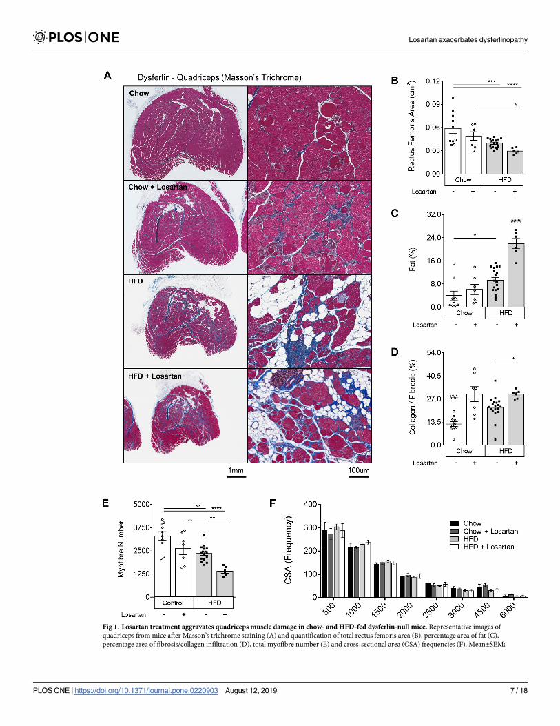
Fig 1. Losartan treatment aggravates quadriceps muscle damage in chow- and HFD-fed dysferlin-null mice. Representative images of
quadriceps from mice after Masson’s trichrome staining (A) and quantification of total rectus femoris area (B), percentage area of fat (C),
percentage area of fibrosis/collagen infiltration (D), total myofibre number (E) and cross-sectional area (CSA) frequencies (F). Mean±SEM;
Losartan exacerbates dysferlinopathy
PLOS ONE | https://doi.org/10.1371/journal.pone.0220903 August 12, 2019 7 / 18

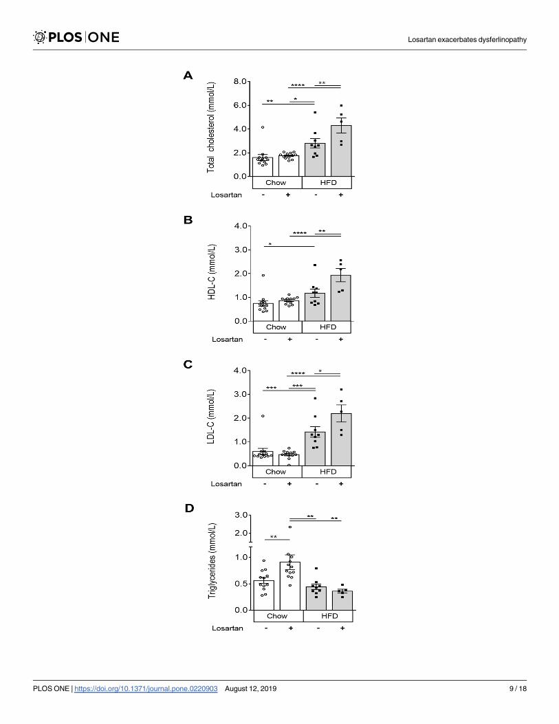
Losartan induces an atherogenic shift in plasma cholesterol and TG levels
In other types of MD, losartan can protect against muscle damage while reducing plasma TGs
and improving the “good” (HDL) to “bad" (LDL) cholesterol ratio [28,29]. Hence, plasma lipo-
protein levels were examined. We observed that under normal chow conditions, losartan
increased plasma TG levels by 62.5%, leaving TC, HDL-C and LDL-C unaffected (P<0.01; Fig
3A–3D). Conversely, in HFD-fed animals, losartan elevated TC, HDL-C and LDL-C (P<0.05;
P<0.05 (�), P<0.01 (��), P<0.001 (���), P<0.0001 (����), One-way ANOVA with Fisher’s post-hoc tests of least significant differences. Hash (#)
indicates significantly different from all other groups, P<0.001 (###), P<0.0001 (####); Scale bars for 2x and 20x images are 1mm and 100μm,
respectively. Muscle tissue (pink); fibrosis (blue); fat/adipocytes (white). Chow (N = 10); Chow losartan (N = 7); HFD (N = 18); HFD losartan
(N = 6).
https://doi.org/10.1371/journal.pone.0220903.g001
Fig 2. Canonical (pSMAD2) and non-canonical (p44/42 MAP Kinase; ERK-1/2) TGF-β signalling in quadriceps muscle lysates are unaffected by diet or
losartan treatment. Percentage of total nuclei positive for p-SMAD2(Ser465/467) quantified on whole quadriceps muscle sections (A) with representative
pSMAD positive images (B), and quantitation of p-ERK(Thr202/Tyr204) standardised to t-ERK (C,D). DAPI (Red); pSMAD2 (Green); Merge (yellow). Mean
±SEM; One-way ANOVA with Fisher’s post-hoc tests of least significant differences. Y-axes represent arbitrary units (A.U.) unless stated. Scale bar for 20x
images is 100μm. p-ERK and t-ERK were blotted on separate gels, and GAPDH blotted on each to control for loading. Immunoblots share the same loading
order, sample concentration and loading control. Full blots were imaged separately and thus have differing exposures. Chow (N = 5–6); Chow losartan (N = 5);
HFD (N = 5–6); HFD losartan (N = 5).
https://doi.org/10.1371/journal.pone.0220903.g002
Losartan exacerbates dysferlinopathy
PLOS ONE | https://doi.org/10.1371/journal.pone.0220903 August 12, 2019 8 / 18
Losartan exacerbates dysferlinopathy
PLOS ONE | https://doi.org/10.1371/journal.pone.0220903 August 12, 2019 9 / 18

Fig 3A–3C), independent of changes to circulating TG (Fig 3D). These data suggest that the
losartan-mediated exacerbation of muscle damage may be related to a shift in atherogenic,
LDL-C or TG lipoprotein components.
Dysferlinopathic mice are non-responsive to the endothelial function-
activating and BP-lowering effects of losartan
The effect of losartan on dysferlin-null muscle prompted us to test its primary effect on BP in
two additional cohorts of dysferlin-null mice (Fig 4). Six weeks of losartan treatment failed to
lower systolic, diastolic and mean arterial blood pressure (MABP) in dysferlin-null mice, com-
pared to untreated controls (Fig 4A). Dose-response analyses revealed a right shift in MAP
sensitivity in response to losartan as quantified by EC50 in dysferlin-null compared to WT
mice (Fig 4B; P<0.05). Having recently shown that losartan can improve endothelial function
via release of the vasodilatory mediator, nitric oxide (NO) [21], ex vivo myography was used to
test the effect of acute losartan treatment on vascular NO release. While aortic rings from WT
mice displayed reduced contractility in response to losartan (an effect fully reversed using the
NO synthase inhibitor L-NAME) (Fig 5A and 5B), PE-induced contractility was unaffected in
Fig 3. Plasma cholesterol and triglyceride levels in chow and HFD fed dysferlin-null mice with or without losartan
treatment. Total cholesterol (A), high-density lipoprotein (HDL-C) (B), low-density lipoprotein (LDL-C) (C) and
triglycerides (TG) (D). Mean±SEM; P<0.05 (�), P<0.01 (��), P<0.001 (���), P<0.0001 (����), One-way ANOVA with
Fisher’s post-hoc tests of least significant differences. Chow (N = 12); Chow losartan (N = 12); HFD (N = 9); HFD
losartan (N = 5).
https://doi.org/10.1371/journal.pone.0220903.g003
Fig 4. Dysferlin-null mice exhibit reduced sensitivity to the blood pressure (BP)-lowering effects of losartan.
Systolic, diastolic and mean arterial blood pressure measurements (MABP) in dysferlin-null mice treated acutely (6
weeks) with a 0.6g/L dose of losartan (A), and EC50 of losartan dose response curves in both WT and dysferlin-null
cohorts (B). Mean+SEM; P<0.05 (�), unpaired student t-test (two-tailed). N = 5–7 mice per group.
https://doi.org/10.1371/journal.pone.0220903.g004
Losartan exacerbates dysferlinopathy
PLOS ONE | https://doi.org/10.1371/journal.pone.0220903 August 12, 2019 10 / 18
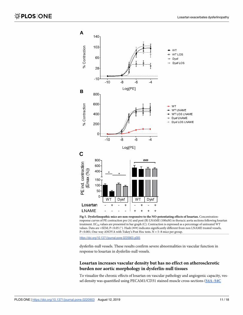
dysferlin-null vessels. These results confirm severe abnormalities in vascular function in
response to losartan in dysferlin-null vessels.
Losartan increases vascular density but has no effect on atherosclerotic
burden nor aortic morphology in dysferlin-null tissues
To visualize the chronic effects of losartan on vascular pathology and angiogenic capacity, ves-
sel density was quantified using PECAM1/CD31 stained muscle cross-sections (S4A–S4C
Fig 5. Dysferlinopathic mice are non-responsive to the NO-potentiating effects of losartan. Concentration-
response curves of PE contraction pre (A) and post (B) LNAME (100uM) in thoracic aorta sections following losartan
treatment. EC50 values are presented in bar graph (C). Contraction is expressed as a percentage of untreated WT
values. Data are +SEM; P<0.05 (�). Hash (###) indicates significantly different from non LNAME treated vessels,
P<0.001; One-way ANOVA with Tukey’s Post Hoc tests. N = 5–8 mice per group.
https://doi.org/10.1371/journal.pone.0220903.g005
Losartan exacerbates dysferlinopathy
PLOS ONE | https://doi.org/10.1371/journal.pone.0220903 August 12, 2019 11 / 18

Fig). In settings of increased muscle damage and fibrosis we observed increased vascular den-
sity in triceps brachii (S4A and S4B Fig) and rectus femoris (S4C Fig; quantification only) fol-
lowing HFD and/or Losartan treatment. Despite the presence of elevated lipids,
HFD-Losartan treated aortas displayed no sign of atherosclerosis as measured by Sudan IV
staining (S4D Fig) and no overt structural changes to vessel morphology were observed on
representative Van Geisson stained ascending aortic segments (S4E Fig).
Discussion
The current study provides evidence in a preclinical model of dysferlinopathy that losartan
does not prevent, but rather exacerbates muscle wasting and damage, which is in stark contrast
to its protective effects in other types of MD and muscle disease. Previous work by us and
other groups have shown that losartan can ameliorate the manifestation of muscle disease in
mouse models of DMD, MDC1A, Marfan syndrome and sarcopenia, likely by attenuating the
pro-fibrotic action of TGF-β signalling [10,13,16,30,31]. This is in addition to studies that have
documented prophylactic effects when targeting the same pathways using angiotensin convert-
ing enzyme inhibitors [17,32]. Rationalization of these unexpected observations may be found
in a report that glucocorticosteroids used to treat other forms of MD are not effective in dysfer-
linopathic patients [33], indicating that loss of dysferlin causes unique and distinctive changes
in muscle signalling and homeostasis, compared to other MDs.
Losartan and TGF-β signalling in MD
The unexpected results observed in the current study argue against a therapeutic, TGF-β-
attenuating role for losartan in dysferlinopathies. Elevated TGF-β signalling has been docu-
mented in DMD patients, congenital MDC1A, as well as both mdx mouse and golden retriever
dog models of DMD [10,16,34,35]. Since TGF-β overexpression can inhibit satellite cell activa-
tion, and in injured muscle promote the differentiation of myogenic progenitors into fibrotic
cells [12,16,36], TGF-β elevation in DMD likely plays an active part in the promotion of profi-
brotic remodelling [16]. In a recent study, L-158809 (an analogue of losartan) reduced muscle
fibrosis and inflammation in MDC1A dyw/dyw mice and improved ambulation via reduced
TGF-β and pSMAD2/3 signalling [16]. In sarcopenic muscle, losartan was also able to rescue
necessary Pax7 and myogenic (MyoD and MyoG) signalling interactions essential for muscle
regeneration, in a TGF-β-driven, Smad2/3 and MAPK-dependent manner [15,37]. Similar
therapeutic properties also restored regenerative capacity in both DMD and MDC1A mouse
models following toxin-induced muscle damage [10,16]. In the current study, despite docu-
mented TGF-β upregulation in dysferlin-deficient tissues [38,39], we did not detect inhibition
of SMAD2 or ERK phosphorylation following chronic losartan treatment, which suggests that
loss of dysferlin may impair losartan signalling (see below). If this assessment is correct, the
contrasting effects of losartan in relatively similar genetic models of muscle disease (DMD and
LGMD2B) suggests that caution must be taken when testing experimental therapies in animal
models harbouring mutations of the dystrophin-associated glycoprotein complex. For
instance, the common use of mdx/utrophin double mutant mice as a more severe alternative
to the mild mdx model of DMD may result in misleading outcomes if used to test therapies, as
the probability of patients exhibiting double DGC mutations is extremely low. Instead, our
team has shown that modulating atherogenic plasma lipoprotein levels, particularly LDL-C
and TG, exacerbates muscle wasting to levels closer to what is generally seen in patients [9]
which could be beneficial for testing of advanced therapies.
Losartan exacerbates dysferlinopathy
PLOS ONE | https://doi.org/10.1371/journal.pone.0220903 August 12, 2019 12 / 18

Losartan aggravates plasma lipid abnormalities but not CK in
dysferlinopathies
We have recently demonstrated that both dysferlin-null and mdx mice exhibit drastically wors-
ened muscle pathology and intramuscular fat accretion when plasma lipoprotein levels are ele-
vated to an atherogenic, LDL and TG-rich state [9,18], thus emphasising a strong relationship
between lipids and muscle homeostasis in multiple forms of MD. In particular, abnormal
plasma TG, phospholipids, free cholesterol, cholesterol esters and total cholesterol concentra-
tions in DMD patients [40], as well as significant lipid accumulation in dysferlin-deficient myo-
fibers, can be observed prior to the replacement of muscle area with adipocytes [8]. This
suggests either an inherent lipid handling defect or inefficient membrane repair in response to
lipotoxicity. Indeed, changes influencing phospholipid composition and oxidation can signifi-
cantly alter sarcolemmal stability and calcium signalling, as observed in a number of other mus-
cle wasting [41–44]. In general, ARBs (including losartan) as well as other anti-hypertensives
are shown to improve plasma lipid profiles and/or lipoprotein composition in a number of clin-
ical and pre-clinical disease models [13,28,45–49]. Moreover, losartan is known to reduce mus-
cle fibrosis concurrent with increased HDL-C concentration, and reduced TG and TC in mdxmice [13]. Very few studies have explored the mechanisms behind losartan’s lipid-lowering
effects, however other ARBs including Telmisartan and Irbesartan can modulate peroxisome
proliferator-activated receptor gamma (PPAR-γ) agonist activity, a key regulator of lipid metab-
olism, while others have suggested high lipophilicity as a possible mediator [50]. Whether these
effects and those reported herein are due to losartan’s anti-ATR1 properties or its significant
off-target effects [21] is unknown. The extent to which these pathways are modified in dysfer-
lin-null tissue warrants further investigation, although studies have reported aberrant expres-
sion of very-low-density lipoprotein receptor, reduced LDL receptor-associated protein and
reduced uptake of cholesterol containing particles in dysferlin-null muscle [38]. Elevated
expression of the lipogenic marker CCAAT/enhancer binding protein- δ, has also been reported
in dysferlin-null A/J and BLAJ skeletal muscles prior to disease onset (3 mo), suggesting an
early induction of signalling pathways that promote an adipogenic lineage [8].
Based on the dramatic changes in muscle size in losartan-treated animals, one can antici-
pate changes in muscle weights; however plasma CK (a commonly used marker of muscle
damage in both clinical and preclinical muscle disease) was not affected in the present study,
as shown by others [25]. In mdx mice, reduced plasma CK and improved lipid profiles
(reduced TG, increased HDL-C) were correlated with reduced muscle damage and pathology
following chronic losartan treatment [13]. Interestingly, in our study plasma CK concentra-
tions did not correlate with muscle damage in dysferlin-null mice; a phenomenon that has
been noted in late stage disease in both mdx [51] and DMD patients [reviewed in [52]], where
CK levels can vary dramatically over time, and with the loss of viable myofiber area [51–53].
Anti-hypertensive treatments in LGMD2B
How blood pressure lowering medications impact both vascular and muscle tissue homeostasis
in the LGMD2B patient population is poorly understood. Although, previous work has shown
that Diltiazem, a Ca2+ channel blocker with blood pressure lowering capabilities, can elicit par-
tial protection in dysferlin-null tissues using acute damage models [54]. Our group has recently
shown that losartan mediates its off-target, NO-protective effects on vascular tissues in a
VEGFR-dependent manner [21]; a function shown to be ablated in dysferlin-deficient cells
[55]. Moreover, losartan has been shown to block AngII induced, Ca2+-regulated lysosome
fusion and lipid raft formation [56], which may have implications in dysferlin-deficient cells,
given their already inherent loss of Ca2+-regulated membrane repair [5]. While a growing
Losartan exacerbates dysferlinopathy
PLOS ONE | https://doi.org/10.1371/journal.pone.0220903 August 12, 2019 13 / 18

number of studies report profound vascular abnormalities in MD [9,18,55,57–59], why dysfer-
lin-null tissues also display aberrant signalling in response to the blood pressure-lowering effects
of ATR-1 blockade is unknown. Significant downregulation of ATR-1 and ATR-1 associated
proteins has however been documented in dysferlin-null heart tissues [60], which supports the
notion that the loss of dysferlin may have an impact on losartan’s biological function. Whether
other blood pressure-lowering drugs which do not target the AngII type 1 receptor, such as
angiotensin 1 converting enzyme inhibitors or β-blockers, would be a preferable pharmacologi-
cal approach to treat hypertension in this patient population should be explored.
In summary, despite the efficacy of losartan in ameliorating muscle pathology in other forms
of MD, the current study demonstrates that in the dysferlin-null mouse model of LGMD2B,
losartan exacerbates muscle wasting concurrent with an atherogenic shift in plasma lipid pro-
files. When combined with earlier studies detailing lipid abnormalities in many forms of MD,
this report provides an early indication of plasma lipid handling defects in dysferlinopathies,
which may be a primary contributor to muscle disease pathogenesis. More importantly, our
data are the first to document the reduced efficacy of losartan to lower blood pressure and pro-
mote endothelial function in dysferlin null tissues. Together, these data highlight important het-
erogeneities between dysferlinopathies and other types of MD, and stresses the need to assess
the safety and efficacy of AngII-dependent therapies in the LGMD2B patient population.
Supporting information
S1 Fig. Losartan treatment aggravates triceps brachii muscle damage in chow- and HFD-
fed dysferlin-null mice. Representative images of triceps brachii from mice after Masson’s tri-
chrome staining (A) and quantification of total triceps brachii area (B), percentage area of fat
(C), percentage area of fibrosis/collagen infiltration (D), total myofibre number (E) and cross-
sectional area (CSA) frequencies (F). Mean±SEM; P<0.05 (�), P<0.01 (��), P<0.001 (���),
P<0.0001 (����), One-way ANOVA with Fisher’s post-hoc tests of least significant differences.
Scale bars for 2x and 8x images are 1mm and 300μm, respectively. Muscle tissue (pink); fibro-
sis (blue); fat/adipocytes (white). Chow (N = 8); Chow losartan (N = 7); HFD (N = 8); HFD
losartan (N = 6).
(TIF)
S2 Fig. Muscle regeneration and plasma CK levels in chow- and HFD-fed dysferlin-null
mice with or without losartan treatment. Percentage of centrally nucleated myofibres (A)
and the percentage of muscle damage (B) in rectus femoris and triceps brachii muscle groups,
and levels of plasma CK (C). Mean±SEM; P<0.05 (�), P<0.01 (��), P<0.001 (���), P<0.0001
(����), One-way ANOVA with Fisher’s post-hoc tests of least significant differences. Chow
(N = 12); Chow losartan (N = 12); HFD (N = 9); HFD losartan (N = 5).
(TIF)
S3 Fig. Anabolic (AKT/rpS6) and autophagy (LC3B) signaling in quadriceps muscles of
chow- and HFD-fed dysferlin-null mice with or without losartan treatment. Quantitation
of p-AKT(Ser473) standardised to t-AKT (A,B), p-rpS6(Ser235/236) to t-rpS6 (A,C) and ratio
of LC3BII/I (A,D). Mean+SEM; P<0.05 (�), P<0.01 (��), P<0.001 (���). One-way ANOVA
with Fisher’s post-hoc tests of least significant differences. Y-axes represent arbitrary units (A.
U). p-AKT, p-rpS6 and LC3B were cut from the same gel and blotted with respective antibod-
ies, as were t-AKT and t-rpS6. Both total and phosphorylated strips for rpS6 were stripped and
blotted for GAPDH to check for loading efficiencies. Full blots were imaged separately and
thus have differing exposures. Chow (N = 6); Chow losartan (N = 5); HFD (N = 6); HFD
Losartan exacerbates dysferlinopathy
PLOS ONE | https://doi.org/10.1371/journal.pone.0220903 August 12, 2019 14 / 18

losartan (N = 5).
(TIF)
S4 Fig. Vessel density, atherosclerotic burden and aortic morphology in chow- and HFD-
fed dysferlin-null mice with or without losartan treatment. Representative images (triceps
brachii only; A) and quantitation of vessel density for triceps brachii rectus femoris (B) and
rectus femoris (C); Scale bar is 100μm. Representative images of Sudan IV stained thoracic
aorta segments in ApoE (Control) and HFD Losartan treated dysferlin-null mice (D); Scale
bar is 0.5cm. Representative images of Van Geisson stained ascending aortic segments (E);
Scale bar is 100μm. N = 3–6 mice per group. Mean+SEM; P<0.05 (�), P<0.01 (��), P<0.001
(���), P<0.0001 (����), One-way ANOVA with Fisher’s post-hoc tests of least significant differ-
ences.
(TIF)
Acknowledgments
The authors wish to thank Tatjana Ponomarev and Lubos Bohunek (St. Paul’s Hospital, UBC)
for their help with the animal experiments and Ingrid Barta (Biomedical Research Centre,
UBC) for the processing of histology.
Author Contributions
Conceptualization: Zoe White, Nadia Milad, Pascal Bernatchez.
Data curation: Zoe White, Nadia Milad, Arash Y. Tehrani, William Wei-Han Chen, Graham
Donen, Stephanie L. Sellers, Pascal Bernatchez.
Formal analysis: Zoe White, Nadia Milad.
Funding acquisition: Pascal Bernatchez.
Investigation: Zoe White, Nadia Milad.
Methodology: Zoe White, Nadia Milad, Pascal Bernatchez.
Project administration: Pascal Bernatchez.
Visualization: Zoe White.
Writing – original draft: Zoe White, Nadia Milad, Pascal Bernatchez.
Writing – review & editing: Zoe White, Nadia Milad, Pascal Bernatchez.
References1. Bashir R, Britton S, Strachan T, Keers S, Vafiadaki E, Lako M, et al. A gene related to Caenorhabditis
elegans spermatogenesis factor fer-1 is mutated in limb-girdle muscular dystrophy type 2B. Nat Genet.
United States; 1998; 20: 37–42. https://doi.org/10.1038/1689 PMID: 9731527
2. Bejaoui K, Hirabayashi K, Hentati F, Haines JL, Ben Hamida C, Belal S, et al. Linkage of Miyoshi myop-
athy (distal autosomal recessive muscular dystrophy) locus to chromosome 2p12-14. Neurology. United
States; 1995; 45: 768–772. https://doi.org/10.1212/wnl.45.4.768 PMID: 7723968
3. Liu J, Aoki M, Illa I, Wu C, Fardeau M, Angelini C, et al. Dysferlin, a novel skeletal muscle gene, is
mutated in Miyoshi myopathy and limb girdle muscular dystrophy. Nat Genet. United States; 1998; 20:
31–36. https://doi.org/10.1038/1682 PMID: 9731526
4. Leung C, Utokaparch S, Sharma A, Yu C, Abraham T, Borchers C, et al. Proteomic identification of dys-
ferlin-interacting protein complexes in human vascular endothelium. Biochem Biophys Res Commun.
United States; 2011; 415: 263–269. https://doi.org/10.1016/j.bbrc.2011.10.031 PMID: 22037454
Losartan exacerbates dysferlinopathy
PLOS ONE | https://doi.org/10.1371/journal.pone.0220903 August 12, 2019 15 / 18

5. Bansal D, Miyake K, Vogel SS, Groh S, Chen C-C, Williamson R, et al. Defective membrane repair in
dysferlin-deficient muscular dystrophy. Nature. England; 2003; 423: 168–172. https://doi.org/10.1038/
nature01573 PMID: 12736685
6. Cardenas AM, Gonzalez-Jamett AM, Cea LA, Bevilacqua JA, Caviedes P. Dysferlin function in skeletal
muscle: Possible pathological mechanisms and therapeutical targets in dysferlinopathies. Exp Neurol.
United States; 2016; 283: 246–254. https://doi.org/10.1016/j.expneurol.2016.06.026 PMID: 27349407
7. Mueller AL, Desmond PF, Hsia R-C, Roche JA. Improved immunoblotting methods provide critical
insights into phenotypic differences between two murine dysferlinopathy models. Muscle Nerve. United
States; 2014; 50: 286–289. https://doi.org/10.1002/mus.24220 PMID: 24639380
8. Grounds MD, Terrill JR, Radley-Crabb HG, Robertson T, Papadimitriou J, Spuler S, et al. Lipid accumu-
lation in dysferlin-deficient muscles. Am J Pathol. United States; 2014; 184: 1668–1676. https://doi.org/
10.1016/j.ajpath.2014.02.005 PMID: 24685690
9. Sellers SL, Milad N, White Z, Pascoe C, Chan R, Payne GW, et al. Increased nonHDL cholesterol levels
cause muscle wasting and ambulatory dysfunction in the mouse model of LGMD2B. J Lipid Res. United
States; 2018; 59: 261–272. https://doi.org/10.1194/jlr.M079459 PMID: 29175948
10. Cohn RD, van Erp C, Habashi JP, Soleimani AA, Klein EC, Lisi MT, et al. Angiotensin II type 1 receptor
blockade attenuates TGF-beta-induced failure of muscle regeneration in multiple myopathic states. Nat
Med. United States; 2007; 13: 204–210.
11. Allen RE, Boxhorn LK. Inhibition of skeletal muscle satellite cell differentiation by transforming growth
factor-beta. J Cell Physiol. United States; 1987; 133: 567–572. https://doi.org/10.1002/jcp.1041330319
PMID: 3480289
12. Li Y, Foster W, Deasy BM, Chan Y, Prisk V, Tang Y, et al. Transforming growth factor-beta1 induces
the differentiation of myogenic cells into fibrotic cells in injured skeletal muscle: a key event in muscle
fibrogenesis. Am J Pathol. United States; 2004; 164: 1007–1019. https://doi.org/10.1016/s0002-9440
(10)63188-4 PMID: 14982854
13. Lee E-M, Kim D-Y, Kim A-Y, Lee E-J, Kim S-H, Lee M-M, et al. Chronic effects of losartan on the mus-
cles and the serologic profiles of mdx mice. Life Sci. Netherlands; 2015; 143: 35–42. https://doi.org/10.
1016/j.lfs.2015.10.023 PMID: 26497927
14. Ceco E, McNally EM. Modifying muscular dystrophy through transforming growth factor-beta. FEBS J.
England; 2013; 280: 4198–4209. https://doi.org/10.1111/febs.12266 PMID: 23551962
15. Burks TN, Andres-Mateos E, Marx R, Mejias R, Van Erp C, Simmers JL, et al. Losartan restores skele-
tal muscle remodeling and protects against disuse atrophy in sarcopenia. Sci Transl Med. United
States; 2011; 3: 82ra37.
16. Meinen S, Lin S, Ruegg MA. Angiotensin II type 1 receptor antagonists alleviate muscle pathology in
the mouse model for laminin-alpha2-deficient congenital muscular dystrophy (MDC1A). Skelet Muscle.
England; 2012; 2: 18. https://doi.org/10.1186/2044-5040-2-18 PMID: 22943509
17. Allen HD, Flanigan KM, Thrush PT, Dvorchik I, Yin H, Canter C, et al. A randomized, double-blind trial
of lisinopril and losartan for the treatment of cardiomyopathy in duchenne muscular dystrophy. PLoS
Curr. United States; 2013; 5.
18. Milad N, White Z, Tehrani AY, Sellers S, Rossi FM V, Bernatchez P. Increased plasma lipid levels exac-
erbate muscle pathology in the mdx mouse model of Duchenne muscular dystrophy. Skelet Muscle.
England; 2017; 7: 19. https://doi.org/10.1186/s13395-017-0135-9 PMID: 28899419
19. Wiktorowicz T, Kinter J, Kobuke K, Campbell KP, Sinnreich M. Genetic characterization and improved
genotyping of the dysferlin-deficient mouse strain Dysf (tm1Kcam). Skelet Muscle. England; 2015; 5:
32. https://doi.org/10.1186/s13395-015-0057-3 PMID: 26464793
20. Han R, Kobuke K, Anderson M, Beltran-Valero de Bernabe D, Kobayashi Y, Yang B, et al. Improved
genotyping of the dysferlin null mouse. Protoc Exch. 2011;
21. Sellers SL, Milad N, Chan R, Mielnik M, Jermilova U, Huang PL, et al. Inhibition of Marfan Syndrome
Aortic Root Dilation by Losartan: Role of Angiotensin II Receptor Type 1-Independent Activation of
Endothelial Function. Am J Pathol. United States; 2018; 188: 574–585. https://doi.org/10.1016/j.ajpath.
2017.11.006 PMID: 29433732
22. Goldstein JA, Bogdanovich S, Beiriger A, Wren LM, Rossi AE, Gao QQ, et al. Excess SMAD signaling
contributes to heart and muscle dysfunction in muscular dystrophy. Hum Mol Genet. England; 2014; 23:
6722–6731. https://doi.org/10.1093/hmg/ddu390 PMID: 25070948
23. White Z, Terrill J, White RB, McMahon C, Sheard P, Grounds MD, et al. Voluntary resistance wheel
exercise from mid-life prevents sarcopenia and increases markers of mitochondrial function and autop-
hagy in muscles of old male and female C57BL/6J mice. Skelet Muscle. England; 2016; 6: 45. https://
doi.org/10.1186/s13395-016-0117-3 PMID: 27964759
Losartan exacerbates dysferlinopathy
PLOS ONE | https://doi.org/10.1371/journal.pone.0220903 August 12, 2019 16 / 18

24. Chung AWY, Au Yeung K, Cortes SF, Sandor GGS, Judge DP, Dietz HC, et al. Endothelial dysfunction
and compromised eNOS/Akt signaling in the thoracic aorta during the progression of Marfan syndrome.
Br J Pharmacol. England; 2007; 150: 1075–1083. https://doi.org/10.1038/sj.bjp.0707181 PMID:
17339838
25. Collier AF, Gumerson J, Lehtimaki K, Puolivali J, Jones JW, Kane MA, et al. Effect of Ibuprofen on Skel-
etal Muscle of Dysferlin-Null Mice. J Pharmacol Exp Ther. United States; 2018; 364: 409–419. https://
doi.org/10.1124/jpet.117.244244 PMID: 29284661
26. White Z, White RB, McMahon C, Grounds MD, Shavlakadze T. High mTORC1 signaling is maintained,
while protein degradation pathways are perturbed in old murine skeletal muscles in the fasted state. Int
J Biochem Cell Biol. Netherlands; 2016; 78: 10–21. https://doi.org/10.1016/j.biocel.2016.06.012 PMID:
27343428
27. Castets P, Ruegg MA. MTORC1 determines autophagy through ULK1 regulation in skeletal muscle.
Autophagy. United States; 2013; 9: 1435–1437. https://doi.org/10.4161/auto.25722 PMID: 23896646
28. Srivastava A, Adams-Huet B, Vega GL, Toto RD. Effect of losartan and spironolactone on triglyceride-
rich lipoproteins in diabetic nephropathy. J Investig Med. England; 2016; 64: 1102–1108. https://doi.org/
10.1136/jim-2016-000102 PMID: 27388615
29. Rodriguez-Iturbe B, Quiroz Y, Shahkarami A, Li Z, Vaziri ND. Mycophenolate mofetil ameliorates
nephropathy in the obese Zucker rat. Kidney Int. United States; 2005; 68: 1041–1047. https://doi.org/
10.1111/j.1523-1755.2005.00496.x PMID: 16105034
30. Spurney CF, Sali A, Guerron AD, Iantorno M, Yu Q, Gordish-Dressman H, et al. Losartan decreases
cardiac muscle fibrosis and improves cardiac function in dystrophin-deficient mdx mice. J Cardiovasc
Pharmacol Ther. United States; 2011; 16: 87–95. https://doi.org/10.1177/1074248410381757 PMID:
21304057
31. Bish LT, Yarchoan M, Sleeper MM, Gazzara JA, Morine KJ, Acosta P, et al. Chronic losartan adminis-
tration reduces mortality and preserves cardiac but not skeletal muscle function in dystrophic mice.
PLoS One. United States; 2011; 6: e20856. https://doi.org/10.1371/journal.pone.0020856 PMID:
21731628
32. Duboc D, Meune C, Lerebours G, Devaux J-Y, Vaksmann G, Becane H-M. Effect of perindopril on the
onset and progression of left ventricular dysfunction in Duchenne muscular dystrophy. J Am Coll Car-
diol. United States; 2005; 45: 855–857. https://doi.org/10.1016/j.jacc.2004.09.078 PMID: 15766818
33. Walter MC, Reilich P, Thiele S, Schessl J, Schreiber H, Reiners K, et al. Treatment of dysferlinopathy
with deflazacort: a double-blind, placebo-controlled clinical trial. Orphanet J Rare Dis. England; 2013; 8:
26. https://doi.org/10.1186/1750-1172-8-26 PMID: 23406536
34. Araujo KPC, Bonuccelli G, Duarte CN, Gaiad TP, Moreira DF, Feder D, et al. Bortezomib (PS-341)
treatment decreases inflammation and partially rescues the expression of the dystrophin-glycoprotein
complex in GRMD dogs. PLoS One. United States; 2013; 8: e61367. https://doi.org/10.1371/journal.
pone.0061367 PMID: 23579193
35. Sun G, Haginoya K, Wu Y, Chiba Y, Nakanishi T, Onuma A, et al. Connective tissue growth factor is
overexpressed in muscles of human muscular dystrophy. J Neurol Sci. Netherlands; 2008; 267: 48–56.
https://doi.org/10.1016/j.jns.2007.09.043 PMID: 17996907
36. Carlson ME, Hsu M, Conboy IM. Imbalance between pSmad3 and Notch induces CDK inhibitors in old
muscle stem cells. Nature. England; 2008; 454: 528–532. https://doi.org/10.1038/nature07034 PMID:
18552838
37. Burks TN, Cohn RD. Role of TGF-beta signaling in inherited and acquired myopathies. Skelet Muscle.
England; 2011; 1: 19. https://doi.org/10.1186/2044-5040-1-19 PMID: 21798096
38. Suzuki N, Aoki M, Hinuma Y, Takahashi T, Onodera Y, Ishigaki A, et al. Expression profiling with pro-
gression of dystrophic change in dysferlin-deficient mice (SJL). Neurosci Res. Ireland; 2005; 52: 47–60.
https://doi.org/10.1016/j.neures.2005.01.006 PMID: 15811552
39. Onofre-Oliveira PCG, Santos ALF, Martins PM, Ayub-Guerrieri D, Vainzof M. Differential expression of
genes involved in the degeneration and regeneration pathways in mouse models for muscular dystro-
phies. Neuromolecular Med. United States; 2012; 14: 74–83. https://doi.org/10.1007/s12017-012-8172-
3 PMID: 22362587
40. Srivastava NK, Pradhan S, Mittal B, Gowda GAN. High resolution NMR based analysis of serum lipids
in Duchenne muscular dystrophy patients and its possible diagnostic significance. NMR Biomed.
England; 2010; 23: 13–22. https://doi.org/10.1002/nbm.1419 PMID: 19787747
41. Rando TA, Disatnik MH, Yu Y, Franco A. Muscle cells from mdx mice have an increased susceptibility
to oxidative stress. Neuromuscul Disord. England; 1998; 8: 14–21. PMID: 9565986
42. Tidball JG, Wehling-Henricks M. The role of free radicals in the pathophysiology of muscular dystrophy.
J Appl Physiol. United States; 2007; 102: 1677–1686. https://doi.org/10.1152/japplphysiol.01145.2006
PMID: 17095633
Losartan exacerbates dysferlinopathy
PLOS ONE | https://doi.org/10.1371/journal.pone.0220903 August 12, 2019 17 / 18

43. Whitehead NP, Yeung EW, Allen DG. Muscle damage in mdx (dystrophic) mice: role of calcium and
reactive oxygen species. Clin Exp Pharmacol Physiol. Australia; 2006; 33: 657–662. https://doi.org/10.
1111/j.1440-1681.2006.04394.x PMID: 16789936
44. Terrill JR, Radley-Crabb HG, Iwasaki T, Lemckert FA, Arthur PG, Grounds MD. Oxidative stress and
pathology in muscular dystrophies: focus on protein thiol oxidation and dysferlinopathies. FEBS J.
England; 2013; 280: 4149–4164. https://doi.org/10.1111/febs.12142 PMID: 23332128
45. Derosa G, Cicero AFG, Bertone G, Piccinni MN, Fogari E, Ciccarelli L, et al. Comparison of the effects
of telmisartan and nifedipine gastrointestinal therapeutic system on blood pressure control, glucose
metabolism, and the lipid profile in patients with type 2 diabetes mellitus and mild hypertension: a 12-
month, randomized, doubl. Clin Ther. United States; 2004; 26: 1228–1236. PMID: 15476904
46. Kalikar M, Nivangune KS, Dakhale GN, Bajait CS, Sontakke SD, Motghare VM, et al. Efficacy and Tol-
erability of Olmesartan, Telmisartan, and Losartan in Patients of Stage I Hypertension: A Randomized,
Open-label Study. J Pharmacol Pharmacother. India; 2017; 8: 106–111. https://doi.org/10.4103/jpp.
JPP_39_17 PMID: 29081617
47. Tershakovec AM, Keane WF, Zhang Z, Lyle PA, Appel GB, McGill JB, et al. Effect of LDL cholesterol
and treatment with losartan on end-stage renal disease in the RENAAL study. Diabetes Care. United
States; 2008; 31: 445–447. https://doi.org/10.2337/dc07-0196 PMID: 18070995
48. Kyvelou S-MG, Vyssoulis GP, Karpanou EA, Adamopoulos DN, Zervoudaki AI, Pietri PG, et al. Effects
of antihypertensive treatment with angiotensin II receptor blockers on lipid profile: an open multi-drug
comparison trial. Hellenic J Cardiol. Netherlands; 2006; 47: 21–28. PMID: 16532712
49. Lerch M, Teuscher AU, Beissner P, Schneider M, Shaw SG, Weidmann P. Effects of angiotensin II-
receptor blockade with losartan on insulin sensitivity, lipid profile, and endothelin in normotensive off-
spring of hypertensive parents. J Cardiovasc Pharmacol. United States; 1998; 31: 576–580. https://doi.
org/10.1097/00005344-199804000-00016 PMID: 9554807
50. Miura S, Karnik SS, Saku K. Review: angiotensin II type 1 receptor blockers: class effects versus molec-
ular effects. J Renin Angiotensin Aldosterone Syst. England; 2011; 12: 1–7. https://doi.org/10.1177/
1470320310370852 PMID: 20603272
51. Coulton GR, Morgan JE, Partridge TA, Sloper JC. The mdx mouse skeletal muscle myopathy: I. A histo-
logical, morphometric and biochemical investigation. Neuropathol Appl Neurobiol. England; 1988; 14:
53–70. PMID: 2967442
52. Pennington RJ. Clinical biochemistry of muscular dystrophy. Br Med Bull. England; 1980; 36: 123–126.
https://doi.org/10.1093/oxfordjournals.bmb.a071625 PMID: 7020837
53. Zatz M, Rapaport D, Vainzof M, Passos-Bueno MR, Bortolini ER, Pavanello R de C, et al. Serum crea-
tine-kinase (CK) and pyruvate-kinase (PK) activities in Duchenne (DMD) as compared with Becker
(BMD) muscular dystrophy. J Neurol Sci. Netherlands; 1991; 102: 190–196. https://doi.org/10.1016/
0022-510x(91)90068-i PMID: 2072118
54. Begam M, Collier AF, Mueller AL, Roche R, Galen SS, Roche JA. Diltiazem improves contractile prop-
erties of skeletal muscle in dysferlin-deficient BLAJ mice, but does not reduce contraction-induced mus-
cle damage. Physiol Rep. United States; 2018; 6: e13727. https://doi.org/10.14814/phy2.13727 PMID:
29890050
55. Sharma A, Sellers S, Stefanovic N, Leung C, Tan SM, Huet O, et al. Direct Endothelial Nitric Oxide
Synthase Activation Provides Atheroprotection in Diabetes-Accelerated Atherosclerosis. Diabetes.
United States; 2015; 64: 3937–3950. https://doi.org/10.2337/db15-0472 PMID: 26116699
56. Han W-Q, Chen W-D, Zhang K, Liu J-J, Wu Y-J, Gao P-J. Ca2+ -regulated lysosome fusion mediates
angiotensin II-induced lipid raft clustering in mesenteric endothelial cells. Hypertens Res. England;
2016; 39: 227–236. https://doi.org/10.1038/hr.2015.144 PMID: 26763850
57. Palladino M, Gatto I, Neri V, Straino S, Smith RC, Silver M, et al. Angiogenic impairment of the vascular
endothelium: a novel mechanism and potential therapeutic target in muscular dystrophy. Arterioscler
Thromb Vasc Biol. United States; 2013; 33: 2867–2876. https://doi.org/10.1161/ATVBAHA.112.301172
PMID: 24072696
58. Loufrani L, Matrougui K, Gorny D, Duriez M, Blanc I, Levy BI, et al. Flow (shear stress)-induced endo-
thelium-dependent dilation is altered in mice lacking the gene encoding for dystrophin. Circulation.
United States; 2001; 103: 864–870. https://doi.org/10.1161/01.cir.103.6.864 PMID: 11171796
59. Dabire H, Barthelemy I, Blanchard-Gutton N, Sambin L, Sampedrano CC, Gouni V, et al. Vascular
endothelial dysfunction in Duchenne muscular dystrophy is restored by bradykinin through upregulation
of eNOS and nNOS. Basic Res Cardiol. Germany; 2012; 107: 240. https://doi.org/10.1007/s00395-011-
0240-6 PMID: 22193759
60. Wenzel K, Geier C, Qadri F, Hubner N, Schulz H, Erdmann B, et al. Dysfunction of dysferlin-deficient
hearts. J Mol Med (Berl). Germany; 2007; 85: 1203–1214.
Losartan exacerbates dysferlinopathy
PLOS ONE | https://doi.org/10.1371/journal.pone.0220903 August 12, 2019 18 / 18
Copyright © 2022 FDOKUMEN