The effect of inhaled nitric oxide on the carrageenan-induced paw edema
Reduction of CuZn-Superoxide Dismutase Activity ExacerbatesNeuronal Cell Injury and Edema Formation after Transient FocalCerebral Ischemia
Takeo Kondo,1,2 Andrew G. Reaume,4 Ting-Ting Huang,3 Elaine Carlson,3 Kensuke Murakami,1,2
Sylvia F. Chen,1,2 Eric K. Hoffman,4 Richard W. Scott,4 Charles J. Epstein,3 and Pak H. Chan1,2
Departments of 1Neurological Surgery, 2Neurology, and 3Pediatrics, University of California School of Medicine, SanFrancisco, California 94143, and 4Department of Molecular Biology, Cephalon, Incorporated, West Chester,Pennsylvania 19380
Apoptotic neuronal cell death has recently been associatedwith the development of infarction after cerebral ischemia. In avariety of studies, CuZn-superoxide dismutase (CuZn-SOD)has been shown to protect the brain from ischemic injury. Apossible role for CuZn-SOD-related modulation of neuronalviability is suggested by the finding that CuZn-SOD inhibitsapoptotic neuronal cell death in response to some forms ofcellular damage. We evaluated this possibility in the model oftransient focal cerebral ischemia in mice bearing a disruption ofthe CuZn-SOD gene (Sod1). Homozygous mutant (Sod1 2/2)mice had no detectable CuZn-SOD activity, and heterozygousmutants (Sod1 1/2) showed a 50% decrease compared withwild-type mice. Sod1 2/2 mice showed a high level of blood–brain barrier disruption soon after 1 hr of middle cerebral arteryocclusion and 100% mortality at 24 hr after ischemia. Sod1
1/2 mice showed 30% mortality at 24 hr after ischemia, andneurological deficits were exacerbated compared with wild-type controls. The Sod1 1/2 animals also had increased infarctvolume and brain swelling, accompanied by increased apopto-tic neuronal cell death as indicated by the in situ nick-endlabeling technique to detect DNA fragmentation and morpho-logical criteria. These results suggest that oxygen-free radicals,especially superoxide anions, are an important factor forthe development of infarction by brain edema formation andapoptotic neuronal cell death after focal cerebral ischemia andreperfusion.
Key words: CuZn-superoxide dismutase; focal cerebral isch-emia; blood–brain barrier; Evans blue extravasation; neuronalapoptosis; TUNEL; oxidative stress
Oxygen-free radicals are believed to be involved in the pathogenesisafter cerebral ischemia and reperfusion. During cerebral ischemia, anumber of events that predispose the brain to the formation ofoxygen-free radicals may occur (Siesjo, 1984; McCord, 1985). Afterreperfusion, these events can set off a cascade of other biochemicaland molecular sequelae such as the xanthine–xanthine oxidase reac-tion and phospholipase activation, leading to free-radical production(Gaudet and Levine, 1979; Chan et al., 1984; Siesjo, 1984; McCord,1985). Among these oxygen-free radicals, superoxide anion (O2
2),being directly toxic to neurons (Fridovich, 1986; Patel et al., 1996),may initiate a free radical-mediated chain reaction causing additionalCNS damage (Saugstad and Aasen, 1980; Chan, 1994).
One of the manifestations of CNS damage after cerebral isch-emia is the formation of brain edema caused by the breakdown ofthe blood–brain barrier (BBB). CuZn-superoxide dismutase(CuZn-SOD), a cytosolic protein, prevents vasogenic brain edemaafter several kinds of injuries (Chan et al., 1991; Kinouchi et al.,1991; Shukla et al., 1993), suggesting that O2
2 is an important
factor for BBB disruption. Another manifestation of CNS damageis the direct injury of neuronal cells, including excitatory eventsthat are induced by glutamate release after cerebral ischemia.Glutamate elevates cytosolic free calcium (Ca21) (Choi, 1988),which activates Ca21-dependent enzymes and leads to free radicalproduction (Orrenius et al., 1992; Pazdernik et al., 1992). Recentstudies suggest that excitotoxic injury causes apoptotic neuronalcell death in some neuronal subpopulations (Kure et al., 1991;Ankarcrona et al., 1995). Both CuZn-SOD (Greenlund et al.,1995) and BCL-2, which recently has been shown to have anantioxidant property (Kane et al., 1993), inhibit apoptotic neuro-nal cell death, suggesting the possibility that oxygen-free radicalsmay modulate neuronal apoptosis. Apoptotic neuronal cell deathmay also play an important role in focal ischemic brain injury(Linnik et al., 1993; Tominaga et al., 1993; MacManus et al., 1994;Li et al., 1995b).
The development of mice deficient in the mouse CuZn-SODgene (Sod1) has provided a model for assessing the role ofCuZn-SOD in nervous system injuries (Reaume et al., 1996) (T.Huang, M. Yasunami, E. Carlson, A. Gillespie, A. Reaume, E.Hoffman, P. Chan, R. Scott, C. Epstein, unpublished data). Thesemutant mice with a decrease in or complete absence of endoge-nous CuZn-SOD activity do not show any phenotypic abnormal-ities under normal physiological conditions. However, we hypoth-esized that the mutant mice might be vulnerable to oxidativestress because of additional oxygen-free radical formation afterischemia and reperfusion. Using an in vivo model of focal cerebralischemia and reperfusion, we now show early BBB disruption and
Received Jan. 30, 1997; revised March 17, 1997; accepted March 21, 1997.This work was supported by National Institutes of Health Grants NS14543 and
NS25372 (P.H.C.) and AG08938 (C.J.E., P.H.C.), and by the Lucille Markey Foun-dation to the University of California, San Francisco, Program in Biological Sciences.We thank L. F. Reola and B. E. Calagui for their expert technical assistance and C.Christensen for her editorial assistance. We particularly thank F. R. Sharp, J.Honkaniemi, K. Lamborn, and H. Kinouchi for their suggestions.
Correspondence should be addressed to Dr. P.H. Chan, Departments of Neuro-logical Surgery and Neurology, University of California, Box 0651, San Francisco, CA94143.Copyright © 1997 Society for Neuroscience 0270-6474/97/174180-10$05.00/0
The Journal of Neuroscience, June 1, 1997, 17(11):4180–4189
high mortality in the mutant mice with reduced CuZn-SOD ac-tivity. In addition, using an in situ technique to detect the DNAfree 39-OH ends, we have found that apoptotic neuronal celldeath is increased in the mutant mice after ischemia and reper-fusion. These data indicate that oxygen-free radicals, especiallyO2
2, are important mediators of brain edema and apoptoticneuronal cell death in focal ischemic brain injury.
MATERIALS AND METHODSSod1-deficient mice. In the present study, we used two independentsources of mutant mice. One strain of mutant mice designated 129/CD1-Sod1,tm1 Cep. was produced at Cephalon (West Chester, PA)(Reaume et al., 1996). This mutant was made by targeted deletion of theSod1 gene in embryonic stem (ES) cells using a positive-negative selec-tion scheme that replaced all Sod1 coding sequences (7.6 kb) with theneomycin resistance gene. The mutant ES cells were in turn used tocreate mice that were either heterozygous (Sod1 1/2 cep) or homozygous(Sod1 2/2 cep) mutants. Another strain of mutant mice designatedCD1-Sod1,tm1 Cje. was produced in the Department of Pediatrics,University of California, San Francisco (T.-T. Huang et al., unpublisheddata). This mutant was made by targeted deletion of the Sod1 gene in EScells using an alternative positive-negative selection scheme that replaceda portion of exon 3 and the entire exon 4 of the Sod1 gene (1.9 kb) withthe neomycin resistance gene. The mutant ES cells were in turn used tocreate mice that were heterozygous (Sod1 1/2 uc) mutants. All thesemutant mice were bred with the CD1 strain of mice. In the present study,age-matched wild-type (Wt) 129/CD1 mice were used as controls.
Two methods were used for assessing of CuZn-SOD activity in themutant mice. For the gel electrophoresis assay, previously describedmethods (Epstein et al., 1987) were used. For the measurement ofCuZn-SOD-specific activity, supernatant fluids from red blood cell lysatesor brain homogenates were assayed by a previously described methodbased on cytochrome C reduction (McCord and Fridovich, 1969). CuZn-SOD activity, which reduces the cytochrome C deoxidative capacity byhalf, was taken as one unit. Tissue protein was quantified using BCAprotein assay reagent (Pierce, Rockford, IL), and CuZn-SOD activity wascalculated in relation to the mass of protein.
Focal cerebral ischemia and neurological assessment. Focal cerebralischemia was achieved with suture monofilament blockade of the middlecerebral artery (MCA) using the previously described method of Yang etal. (1994), with some modification. Male 2- to 3-month-old Wt or mutantmice (35–40 gm) were anesthetized with 1.5% isoflurane in a 30%O2/70% N2O mixture under spontaneous breathing. The rectal temper-ature was controlled at 37.0 6 0.5°C during surgery with a feedbackregulated heating pad. After exposing the left carotid artery, a 5–0Dermalon suture (1756-41, Devis and Geck, Manati, Puerto Rico) wasadvanced into the internal carotid artery 12 mm from the lumen of theexternal carotid artery. The ipsilateral common carotid artery was oc-cluded with a small surgical clip immediately after suture blockade. Theanimals underwent MCA occlusion for 1 hr and were then subjected toreperfusion by removing the suture and the clip. Mean arterial bloodpressure and cerebral blood flow (CBF) were monitored as physiologicalparameters during ischemia, and arterial blood gas was analyzed afterreperfusion. After recovering from anesthesia, the animals were main-tained in an air conditioned room at 20°C during the reperfusion periods(2–96 hr).
At 24 hr after 1 hr of MCA occlusion, surviving mice were evaluated bya blinded observer for their neurological deficits. The neurological deficitscore assignment of 0 to 4 was based on the methods described previouslyby Yang et al. (1994). After evaluating the scores, the brains wereremoved and rapidly frozen and then were analyzed histologically withcresyl violet staining. Animals that had massive hematomas in the brainor no infarction in the brain were omitted from the neurological analysis.
Measurement of Evans blue extravasation. A quantitative assay of Evansblue was based on the method described previously by Chan et al. (1991).Just after reperfusion, 0.1 ml of 4% Evans blue (Sigma, St. Louis, MO) in0.9% saline was intravenously injected. At 2, 8, or 24 hr after theinjection, the animals were killed by transcardiac perfusion using 200 mlof heparinized saline (10 U/ml heparin in 0.9% of saline), and the brainswere removed. After removing the cerebellum and brainstem, the cere-bral hemispheres were separated into the ischemic hemispheres andnonischemic hemispheres. Each hemisphere was well homogenized in 1ml of 0.1 M PBS. After 1000 3 g spin for 5 min, 0.7 ml of supernatant was
taken, and 0.7 ml of 100% (w/ v) trichloroacetic acid was added. Themixture was incubated at 4°C for 18 hr, then centrifuged at 1000 3 g for30 min. The Evans blue concentration of the supernatant was measuredat 610 nm by a spectrophotometer. Results are presented as milligrams ofEvans blue/hemisphere by comparing it with the standard solution.
Histological analysis. At 8, 24, 48, or 96 hr after 1 hr of MCAocclusion, the brains were removed and frozen rapidly. The brains weresectioned with a cryostat into a 20 mm thickness from the anterior sideto the posterior side at 500 mm intervals. The sections were stained withcresyl violet using standard histological criteria. The staining image wasscanned by Color One Scanner 600/27 (Apple Computer, Cupertino,CA) at 1200 dpi resolution. The infarct area and the bilateral hemi-spheric area were measured by the National Institutes of Health systemin 14 sections from the anterior tip of the brain. The total infarctvolume and total hemispheric volume were calculated according to themethod of Liu et al. (1989).
In situ labeling of DNA fragmentation. At 8, 24, 48, or 96 hr after 1 hrof MCA occlusion, the brain sections at the level of the caudate putamenthat show typical infarction were stained using an in situ technique todetect the DNA free 39-OH ends (TUNEL) reaction (Gavrieli et al.,1992). Frozen brain sections were fixed for 30 min in 4% formaldehyde in0.1 M PBS, pH 7.4. The slides were placed in 1 3 terminal deoxynucleo-tidyl transferase buffer (Life Technologies, Gaithersburg, MD) for 15min, followed by 10 ml/ml of terminal deoxynucleotidyl transferase en-zyme (Life Technologies) with 40 ml/ml biotinylated 16-dUTP (Boehr-inger Mannheim, Indianapolis, IN) at 37°C for 60 min. The slides werethen washed in 23 SSC (150 mM sodium chloride, 15 mM sodium citrate,pH 7.4) for 15 min, followed by PBS two times for 15 min. Avidin–biotinhorseradish peroxidase solution (ABC kit, Vector Laboratories, Burlin-game, CA) was applied to the sections for 30 min, then washed with PBStwo times for 15 min. Staining was visualized using 0.025% diaminoben-zidine and 0.075% H2O2 in PBS. After staining, the slides were rinsedwith water. Slides were then stained with methyl green for 10 min anddehydrated and mounted. Slides were observed with either a bright-fieldor a dark-field phase-contrast microscopy (Diaphot-TMD, Nicon, Tokyo,Japan). In the latter, a yellow filter, which is used for detection offluorescent isothiocyanate, was used without excitation. Consecutive sec-tions were stained with hematoxylin and eosin (H & E) to observestandard morphological criteria.
Cell counts. A blinded observer counted the number of TUNEL-positive cells in 625 mm2 pixels in 4003 magnification of light-fieldmicrogram. Positive cells were determined carefully by the criteria de-scribed by Charriaut-Marlangue and Ben-Ari (1995) and counted in 200pixels (0.125 mm2) with random movement under blinded conditions toavoid intentional selection. Individual regions were determined as fol-lows: inner boundary of the caudate putamen from the medial edge of theinfarction to 200 mm toward infarct area; center of the caudate putamenfrom external capsule to 500 mm toward midline; cortical penumbra fromthe edge of the infarction to 1 mm toward infarct area; the piriform cortexinside of the piriform cortex neuronal cell layer. The total positive cellnumber in 200 pixels was expressed as cells/mm2.
Statistical analysis. All measurements, except the neurological deficitscore, were statistically analyzed using one-way ANOVA followed byFisher’s protected least-significant difference (PLSD) test. The neurolog-ical deficit score was analyzed statistically using a nonparametric test(Kruskal–Wallis test for overall effects, followed by the Mann–Whitney Utest for individual group comparisons). Spearman rank correlation fol-lowed by linear regression analysis was used for correlation study.
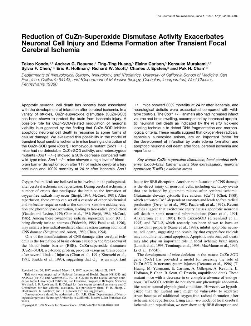
RESULTSCuZn-SOD activity and physiological parameters inmutant miceNondenatured gel electrophoresis followed by nitroblue tetrazo-lium staining confirms that CuZn-SOD activity is reduced in bothSod1 1/2uc and Sod1 1/2cep and completely absent in Sod12/2cep (Fig. 1). A quantitative assay of CuZn-SOD activity shows;50% reduction in both Sod1 1/2uc and Sod1 1/2cep and only atrace of activity in Sod1 2/2cep (Table 1). Although the gene-targeted locus is different in the two independent lines of het-erozygous mutant mice (Sod1 1/2uc and Sod1 1/2cep), thesemice show the same level of CuZn-SOD activity. The minor levelof activity in Sod1 2/2cep likely represents background from the
Kondo et al. • Focal Cerebral Ischemia in Sod1 Mutant Mice J. Neurosci., June 1, 1997, 17(11):4180–4189 4181
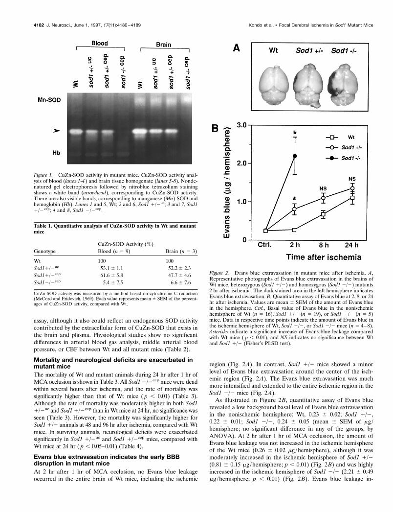
assay, although it also could reflect an endogenous SOD activitycontributed by the extracellular form of CuZn-SOD that exists inthe brain and plasma. Physiological studies show no significantdifferences in arterial blood gas analysis, middle arterial bloodpressure, or CBF between Wt and all mutant mice (Table 2).
Mortality and neurological deficits are exacerbated inmutant miceThe mortality of Wt and mutant animals during 24 hr after 1 hr ofMCA occlusion is shown in Table 3. All Sod1 2/2cep mice were deadwithin several hours after ischemia, and the rate of mortality wassignificantly higher than that of Wt mice ( p , 0.01) (Table 3).Although the rate of mortality was moderately higher in both Sod11/2uc and Sod1 1/2cep than in Wt mice at 24 hr, no significance wasseen (Table 3). However, the mortality was significantly higher forSod1 1/2 animals at 48 and 96 hr after ischemia, compared with Wtmice. In surviving animals, neurological deficits were exacerbatedsignificantly in Sod1 1/2uc and Sod1 1/2cep mice, compared withWt mice at 24 hr ( p , 0.05–0.01) (Table 4).
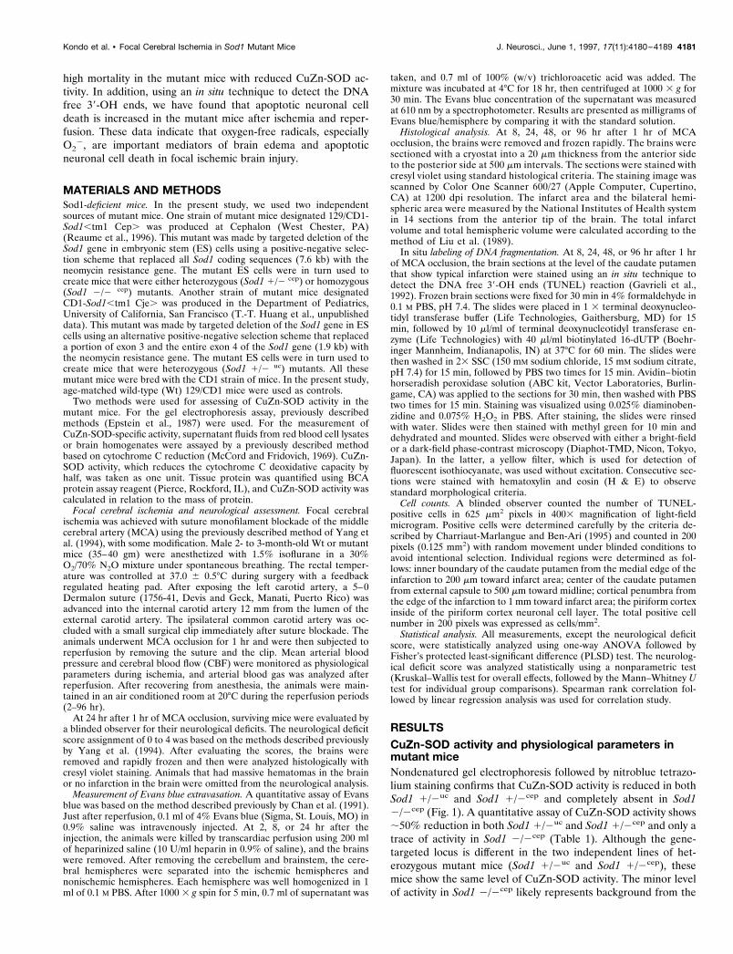
Evans blue extravasation indicates the early BBBdisruption in mutant miceAt 2 hr after 1 hr of MCA occlusion, no Evans blue leakageoccurred in the entire brain of Wt mice, including the ischemic
region (Fig. 2A). In contrast, Sod1 1/2 mice showed a minorlevel of Evans blue extravasation around the center of the isch-emic region (Fig. 2A). The Evans blue extravasation was muchmore intensified and extended to the entire ischemic region in theSod1 2/2 mice (Fig. 2A).
As illustrated in Figure 2B, quantitative assay of Evans bluerevealed a low background basal level of Evans blue extravasationin the nonischemic hemisphere: Wt, 0.23 6 0.02; Sod1 1/2,0.22 6 0.01; Sod1 2/2, 0.24 6 0.05 (mean 6 SEM of mg /hemisphere; no significant difference in any of the groups, byANOVA). At 2 hr after 1 hr of MCA occlusion, the amount ofEvans blue leakage was not increased in the ischemic hemisphereof the Wt mice (0.26 6 0.02 mg /hemisphere), although it wasmoderately increased in the ischemic hemisphere of Sod1 1/2(0.81 6 0.15 mg /hemisphere; p , 0.01) (Fig. 2B) and was highlyincreased in the ischemic hemisphere of Sod1 2/2 (2.21 6 0.49mg /hemisphere; p , 0.01) (Fig. 2B). Evans blue leakage in-
Figure 1. CuZn-SOD activity in mutant mice. CuZn-SOD activity anal-ysis of blood (lanes 1-4 ) and brain tissue homogenate (lanes 5-8). Nonde-natured gel electrophoresis followed by nitroblue tetrazolium stainingshows a white band (arrowhead), corresponding to CuZn-SOD activity.There are also visible bands, corresponding to manganese (Mn)-SOD andhemoglobin (Hb). Lanes 1 and 5, Wt; 2 and 6, Sod1 1/2uc; 3 and 7, Sod11/2cep; 4 and 8, Sod1 2/2cep.
Table 1. Quantitative analysis of CuZn-SOD activity in Wt and mutantmice
GenotypeCuZn-SOD Activity (%)Blood (n 5 9) Brain (n 5 3)
Wt 100 100Sod11/2uc 53.1 6 1.1 52.2 6 2.3Sod11/2cep 61.6 6 5.8 47.7 6 4.6Sod12/2cep 5.4 6 7.5 6.6 6 7.6
CuZn-SOD activity was measured by a method based on cytochrome C reduction(McCord and Fridovich, 1969). Each value represents mean 6 SEM of the percent-ages of CuZn-SOD activity, compared with Wt.
Figure 2. Evans blue extravasation in mutant mice after ischemia. A,Representative photographs of Evans blue extravasation in the brains ofWt mice, heterozygous (Sod1 1/2) and homozygous (Sod1 2/2) mutants2 hr after ischemia. The dark stained area in the left hemisphere indicatesEvans blue extravasation. B, Quantitative assay of Evans blue at 2, 8, or 24hr after ischemia. Values are mean 6 SEM of the amount of Evans bluein the hemisphere. Ctrl., Basal value of Evans blue in the nonischemichemisphere of Wt (n 5 16), Sod1 1/2 (n 5 19), or Sod1 2/2 (n 5 5)mice. Data in respective time points indicate the amount of Evans blue inthe ischemic hemisphere of Wt, Sod1 1/2, or Sod1 2/2 mice (n 5 4–8).Asterisks indicate a significant increase of Evans blue leakage comparedwith Wt mice ( p , 0.01), and NS indicates no significance between Wtand Sod1 1/2 (Fisher’s PLSD test).
4182 J. Neurosci., June 1, 1997, 17(11):4180–4189 Kondo et al. • Focal Cerebral Ischemia in Sod1 Mutant Mice
creased in Wt mice at 8 hr after ischemia and gradually increasedup to 24 hr (Fig. 2B). Also, Sod1 1/2 mice showed an increasingamount of Evans blue extravasation up to 24 hr, although nosignificant difference was seen, compared with Wt at 8 and 24 hrafter ischemia (Fig. 2B). No Evans blue leakage studies wereperformed in Sod 1 2/2 mice at 8 and 24 hr because of 100%animal mortality. These results suggest that a decreased level ofCuZn-SOD activity in the mutant mice mediates early BBBbreakdown after cerebral ischemia.
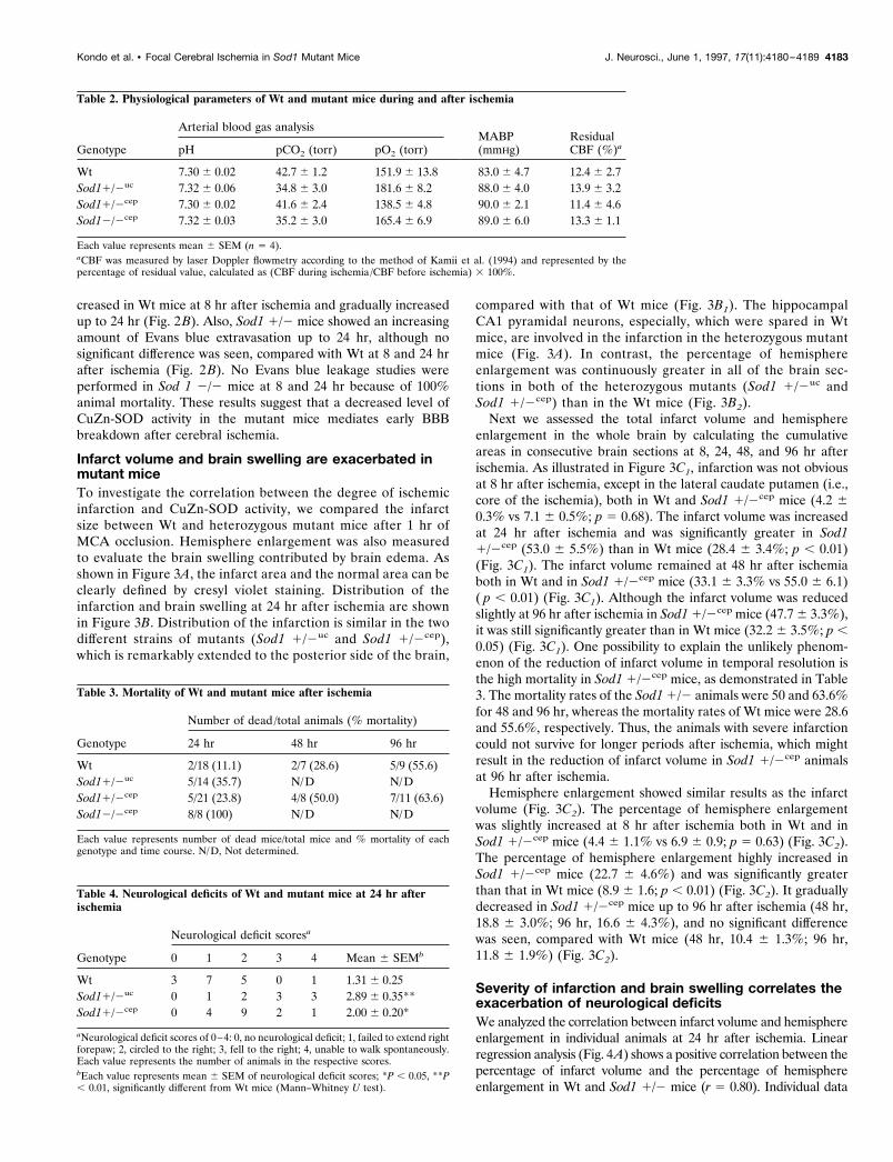
Infarct volume and brain swelling are exacerbated inmutant miceTo investigate the correlation between the degree of ischemicinfarction and CuZn-SOD activity, we compared the infarctsize between Wt and heterozygous mutant mice after 1 hr ofMCA occlusion. Hemisphere enlargement was also measuredto evaluate the brain swelling contributed by brain edema. Asshown in Figure 3A, the infarct area and the normal area can beclearly defined by cresyl violet staining. Distribution of theinfarction and brain swelling at 24 hr after ischemia are shownin Figure 3B. Distribution of the infarction is similar in the twodifferent strains of mutants (Sod1 1/2uc and Sod1 1/2cep),which is remarkably extended to the posterior side of the brain,
compared with that of Wt mice (Fig. 3B1). The hippocampalCA1 pyramidal neurons, especially, which were spared in Wtmice, are involved in the infarction in the heterozygous mutantmice (Fig. 3A). In contrast, the percentage of hemisphereenlargement was continuously greater in all of the brain sec-tions in both of the heterozygous mutants (Sod1 1/2uc andSod1 1/2cep) than in the Wt mice (Fig. 3B2).
Next we assessed the total infarct volume and hemisphereenlargement in the whole brain by calculating the cumulativeareas in consecutive brain sections at 8, 24, 48, and 96 hr afterischemia. As illustrated in Figure 3C1, infarction was not obviousat 8 hr after ischemia, except in the lateral caudate putamen (i.e.,core of the ischemia), both in Wt and Sod1 1/2cep mice (4.2 60.3% vs 7.1 6 0.5%; p 5 0.68). The infarct volume was increasedat 24 hr after ischemia and was significantly greater in Sod11/2cep (53.0 6 5.5%) than in Wt mice (28.4 6 3.4%; p , 0.01)(Fig. 3C1). The infarct volume remained at 48 hr after ischemiaboth in Wt and in Sod1 1/2cep mice (33.1 6 3.3% vs 55.0 6 6.1)( p , 0.01) (Fig. 3C1). Although the infarct volume was reducedslightly at 96 hr after ischemia in Sod1 1/2cep mice (47.7 6 3.3%),it was still significantly greater than in Wt mice (32.2 6 3.5%; p ,0.05) (Fig. 3C1). One possibility to explain the unlikely phenom-enon of the reduction of infarct volume in temporal resolution isthe high mortality in Sod1 1/2cep mice, as demonstrated in Table3. The mortality rates of the Sod1 1/2 animals were 50 and 63.6%for 48 and 96 hr, whereas the mortality rates of Wt mice were 28.6and 55.6%, respectively. Thus, the animals with severe infarctioncould not survive for longer periods after ischemia, which mightresult in the reduction of infarct volume in Sod1 1/2cep animalsat 96 hr after ischemia.
Hemisphere enlargement showed similar results as the infarctvolume (Fig. 3C2). The percentage of hemisphere enlargementwas slightly increased at 8 hr after ischemia both in Wt and inSod1 1/2cep mice (4.4 6 1.1% vs 6.9 6 0.9; p 5 0.63) (Fig. 3C2).The percentage of hemisphere enlargement highly increased inSod1 1/2cep mice (22.7 6 4.6%) and was significantly greaterthan that in Wt mice (8.9 6 1.6; p , 0.01) (Fig. 3C2). It graduallydecreased in Sod1 1/2cep mice up to 96 hr after ischemia (48 hr,18.8 6 3.0%; 96 hr, 16.6 6 4.3%), and no significant differencewas seen, compared with Wt mice (48 hr, 10.4 6 1.3%; 96 hr,11.8 6 1.9%) (Fig. 3C2).
Severity of infarction and brain swelling correlates theexacerbation of neurological deficitsWe analyzed the correlation between infarct volume and hemisphereenlargement in individual animals at 24 hr after ischemia. Linearregression analysis (Fig. 4A) shows a positive correlation between thepercentage of infarct volume and the percentage of hemisphereenlargement in Wt and Sod1 1/2 mice (r 5 0.80). Individual data
Table 2. Physiological parameters of Wt and mutant mice during and after ischemia
Genotype
Arterial blood gas analysisMABP(mmHg)
ResidualCBF (%)apH pCO2 (torr) pO2 (torr)
Wt 7.30 6 0.02 42.7 6 1.2 151.9 6 13.8 83.0 6 4.7 12.4 6 2.7Sod11/2uc 7.32 6 0.06 34.8 6 3.0 181.6 6 8.2 88.0 6 4.0 13.9 6 3.2Sod11/2cep 7.30 6 0.02 41.6 6 2.4 138.5 6 4.8 90.0 6 2.1 11.4 6 4.6Sod12/2cep 7.32 6 0.03 35.2 6 3.0 165.4 6 6.9 89.0 6 6.0 13.3 6 1.1
Each value represents mean 6 SEM (n 5 4).aCBF was measured by laser Doppler flowmetry according to the method of Kamii et al. (1994) and represented by thepercentage of residual value, calculated as (CBF during ischemia /CBF before ischemia) 3 100%.
Table 3. Mortality of Wt and mutant mice after ischemia
Genotype
Number of dead /total animals (% mortality)
24 hr 48 hr 96 hr
Wt 2/18 (11.1) 2/7 (28.6) 5/9 (55.6)Sod11/2uc 5/14 (35.7) N/D N/DSod11/2cep 5/21 (23.8) 4/8 (50.0) 7/11 (63.6)Sod12/2cep 8/8 (100) N/D N/D
Each value represents number of dead mice/total mice and % mortality of eachgenotype and time course. N/D, Not determined.
Table 4. Neurological deficits of Wt and mutant mice at 24 hr afterischemia
Genotype
Neurological deficit scoresa
0 1 2 3 4 Mean 6 SEMb
Wt 3 7 5 0 1 1.31 6 0.25Sod11/2uc 0 1 2 3 3 2.89 6 0.35**Sod11/2cep 0 4 9 2 1 2.00 6 0.20*
aNeurological deficit scores of 0–4: 0, no neurological deficit; 1, failed to extend rightforepaw; 2, circled to the right; 3, fell to the right; 4, unable to walk spontaneously.Each value represents the number of animals in the respective scores.bEach value represents mean 6 SEM of neurological deficit scores; *P , 0.05, **P, 0.01, significantly different from Wt mice (Mann–Whitney U test).
Kondo et al. • Focal Cerebral Ischemia in Sod1 Mutant Mice J. Neurosci., June 1, 1997, 17(11):4180–4189 4183
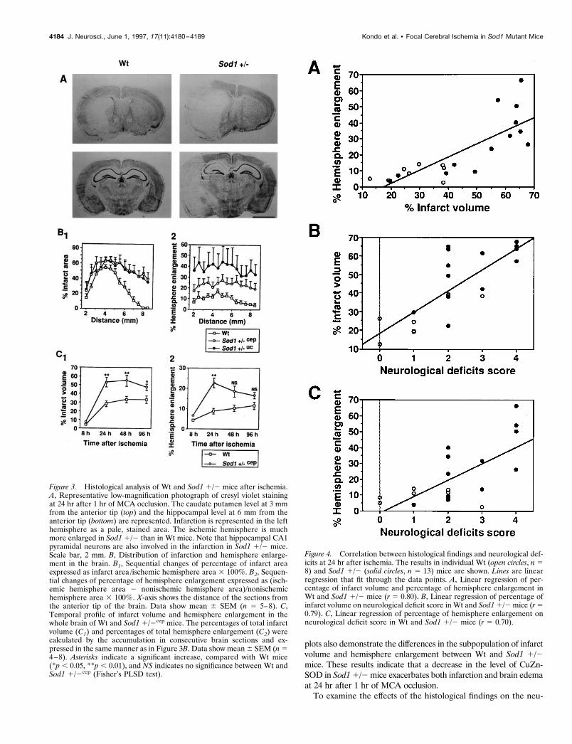
plots also demonstrate the differences in the subpopulation of infarctvolume and hemisphere enlargement between Wt and Sod1 1/2mice. These results indicate that a decrease in the level of CuZn-SOD in Sod1 1/2 mice exacerbates both infarction and brain edemaat 24 hr after 1 hr of MCA occlusion.
To examine the effects of the histological findings on the neu-
Figure 3. Histological analysis of Wt and Sod1 1/2 mice after ischemia.A, Representative low-magnification photograph of cresyl violet stainingat 24 hr after 1 hr of MCA occlusion. The caudate putamen level at 3 mmfrom the anterior tip (top) and the hippocampal level at 6 mm from theanterior tip (bottom) are represented. Infarction is represented in the lefthemisphere as a pale, stained area. The ischemic hemisphere is muchmore enlarged in Sod1 1/2 than in Wt mice. Note that hippocampal CA1pyramidal neurons are also involved in the infarction in Sod1 1/2 mice.Scale bar, 2 mm. B, Distribution of infarction and hemisphere enlarge-ment in the brain. B1, Sequential changes of percentage of infarct areaexpressed as infarct area /ischemic hemisphere area 3 100%. B2, Sequen-tial changes of percentage of hemisphere enlargement expressed as (isch-emic hemisphere area 2 nonischemic hemisphere area)/nonischemichemisphere area 3 100%. X-axis shows the distance of the sections fromthe anterior tip of the brain. Data show mean 6 SEM (n 5 5–8). C,Temporal profile of infarct volume and hemisphere enlargement in thewhole brain of Wt and Sod1 1/2cep mice. The percentages of total infarctvolume (C1) and percentages of total hemisphere enlargement (C2) werecalculated by the accumulation in consecutive brain sections and ex-pressed in the same manner as in Figure 3B. Data show mean 6 SEM (n 54–8). Asterisks indicate a significant increase, compared with Wt mice(*p , 0.05, **p , 0.01), and NS indicates no significance between Wt andSod1 1/2cep (Fisher’s PLSD test).
Figure 4. Correlation between histological findings and neurological def-icits at 24 hr after ischemia. The results in individual Wt (open circles, n 58) and Sod1 1/2 (solid circles, n 5 13) mice are shown. Lines are linearregression that fit through the data points. A, Linear regression of per-centage of infarct volume and percentage of hemisphere enlargement inWt and Sod1 1/2 mice (r 5 0.80). B, Linear regression of percentage ofinfarct volume on neurological deficit score in Wt and Sod1 1/2 mice (r 50.79). C, Linear regression of percentage of hemisphere enlargement onneurological deficit score in Wt and Sod1 1/2 mice (r 5 0.70).
4184 J. Neurosci., June 1, 1997, 17(11):4180–4189 Kondo et al. • Focal Cerebral Ischemia in Sod1 Mutant Mice
rological deficits, next we analyzed them by plotting variations ofthe percentage of infarct volume (Fig. 4B) or the percentage ofhemisphere enlargement (Fig. 4C) versus neurological deficitscores in individual animals at 24 hr after ischemia. Linear regres-sion analysis indicates that neurological deficits in individual an-imals were positively correlated with the percentage of infarctvolume (r 5 0.79) and the percentage of hemisphere enlargement(r 5 0.70). These findings indicate that increased formation ofinfarction and brain edema causes exacerbation of neurologicaldeficits in Sod1 1/2 mice.
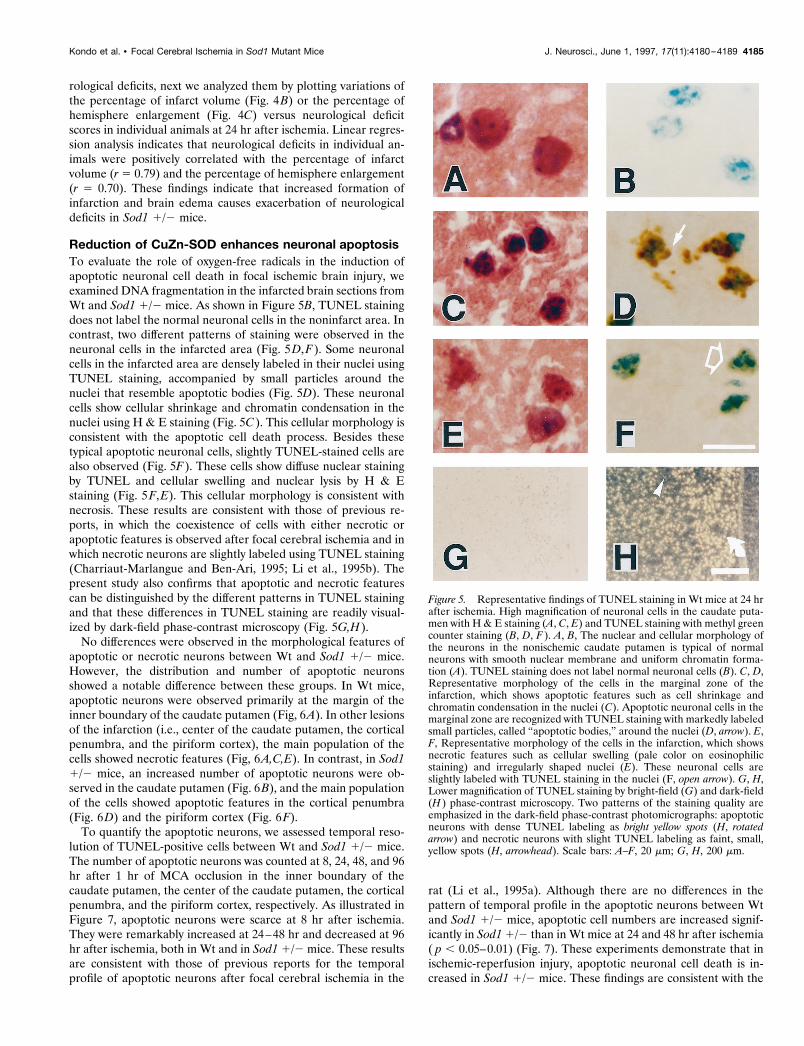
Reduction of CuZn-SOD enhances neuronal apoptosisTo evaluate the role of oxygen-free radicals in the induction ofapoptotic neuronal cell death in focal ischemic brain injury, weexamined DNA fragmentation in the infarcted brain sections fromWt and Sod1 1/2 mice. As shown in Figure 5B, TUNEL stainingdoes not label the normal neuronal cells in the noninfarct area. Incontrast, two different patterns of staining were observed in theneuronal cells in the infarcted area (Fig. 5D,F). Some neuronalcells in the infarcted area are densely labeled in their nuclei usingTUNEL staining, accompanied by small particles around thenuclei that resemble apoptotic bodies (Fig. 5D). These neuronalcells show cellular shrinkage and chromatin condensation in thenuclei using H & E staining (Fig. 5C). This cellular morphology isconsistent with the apoptotic cell death process. Besides thesetypical apoptotic neuronal cells, slightly TUNEL-stained cells arealso observed (Fig. 5F). These cells show diffuse nuclear stainingby TUNEL and cellular swelling and nuclear lysis by H & Estaining (Fig. 5F,E). This cellular morphology is consistent withnecrosis. These results are consistent with those of previous re-ports, in which the coexistence of cells with either necrotic orapoptotic features is observed after focal cerebral ischemia and inwhich necrotic neurons are slightly labeled using TUNEL staining(Charriaut-Marlangue and Ben-Ari, 1995; Li et al., 1995b). Thepresent study also confirms that apoptotic and necrotic featurescan be distinguished by the different patterns in TUNEL stainingand that these differences in TUNEL staining are readily visual-ized by dark-field phase-contrast microscopy (Fig. 5G,H).
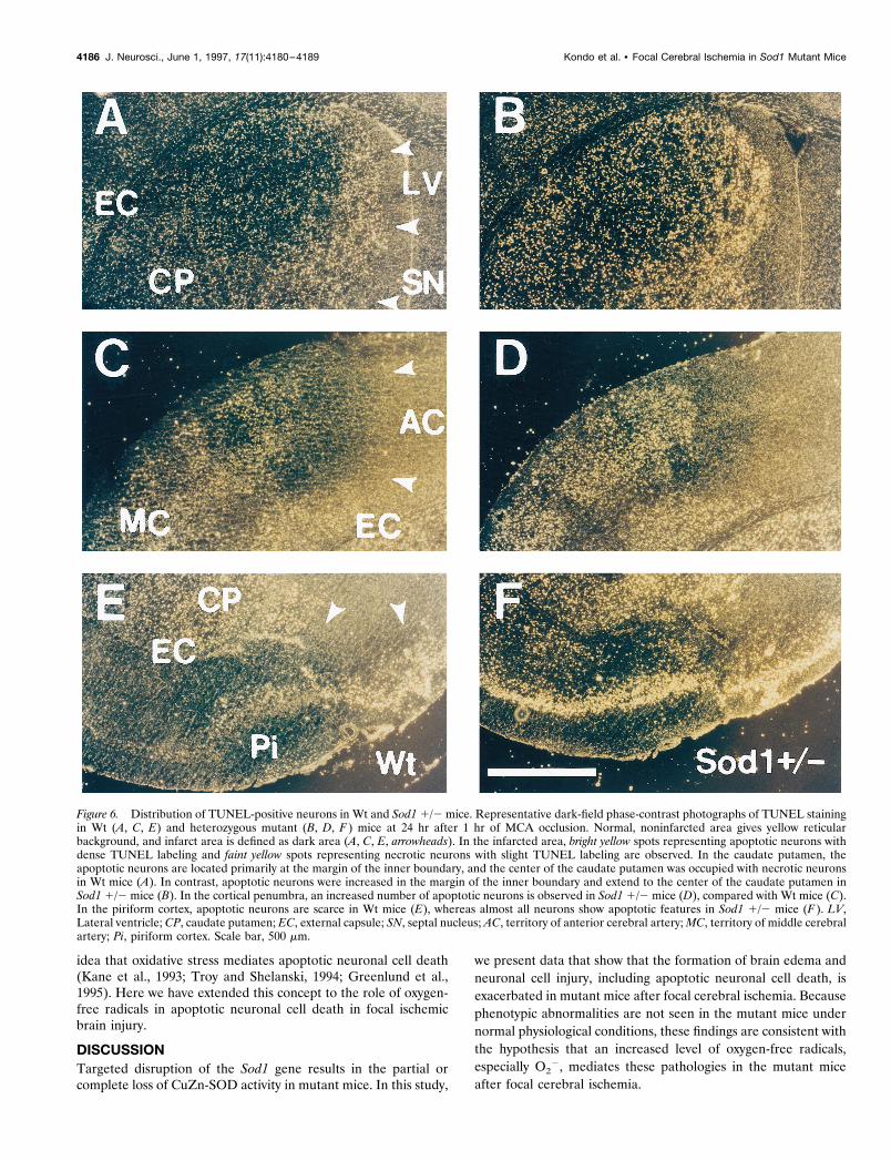
No differences were observed in the morphological features ofapoptotic or necrotic neurons between Wt and Sod1 1/2 mice.However, the distribution and number of apoptotic neuronsshowed a notable difference between these groups. In Wt mice,apoptotic neurons were observed primarily at the margin of theinner boundary of the caudate putamen (Fig, 6A). In other lesionsof the infarction (i.e., center of the caudate putamen, the corticalpenumbra, and the piriform cortex), the main population of thecells showed necrotic features (Fig, 6A,C,E). In contrast, in Sod11/2 mice, an increased number of apoptotic neurons were ob-served in the caudate putamen (Fig. 6B), and the main populationof the cells showed apoptotic features in the cortical penumbra(Fig. 6D) and the piriform cortex (Fig. 6F).
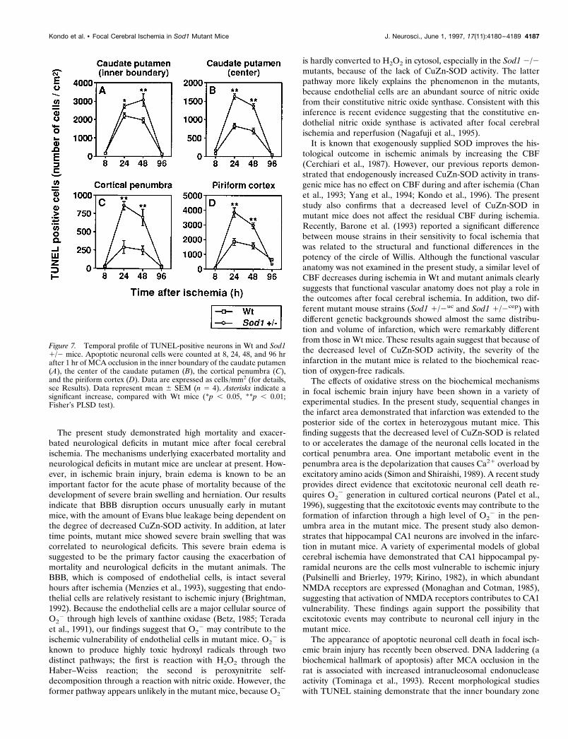
To quantify the apoptotic neurons, we assessed temporal reso-lution of TUNEL-positive cells between Wt and Sod1 1/2 mice.The number of apoptotic neurons was counted at 8, 24, 48, and 96hr after 1 hr of MCA occlusion in the inner boundary of thecaudate putamen, the center of the caudate putamen, the corticalpenumbra, and the piriform cortex, respectively. As illustrated inFigure 7, apoptotic neurons were scarce at 8 hr after ischemia.They were remarkably increased at 24–48 hr and decreased at 96hr after ischemia, both in Wt and in Sod1 1/2 mice. These resultsare consistent with those of previous reports for the temporalprofile of apoptotic neurons after focal cerebral ischemia in the
rat (Li et al., 1995a). Although there are no differences in thepattern of temporal profile in the apoptotic neurons between Wtand Sod1 1/2 mice, apoptotic cell numbers are increased signif-icantly in Sod1 1/2 than in Wt mice at 24 and 48 hr after ischemia( p , 0.05–0.01) (Fig. 7). These experiments demonstrate that inischemic-reperfusion injury, apoptotic neuronal cell death is in-creased in Sod1 1/2 mice. These findings are consistent with the
Figure 5. Representative findings of TUNEL staining in Wt mice at 24 hrafter ischemia. High magnification of neuronal cells in the caudate puta-men with H & E staining (A, C, E) and TUNEL staining with methyl greencounter staining (B, D, F ). A, B, The nuclear and cellular morphology ofthe neurons in the nonischemic caudate putamen is typical of normalneurons with smooth nuclear membrane and uniform chromatin forma-tion (A). TUNEL staining does not label normal neuronal cells (B). C, D,Representative morphology of the cells in the marginal zone of theinfarction, which shows apoptotic features such as cell shrinkage andchromatin condensation in the nuclei (C). Apoptotic neuronal cells in themarginal zone are recognized with TUNEL staining with markedly labeledsmall particles, called “apoptotic bodies,” around the nuclei (D, arrow). E,F, Representative morphology of the cells in the infarction, which showsnecrotic features such as cellular swelling (pale color on eosinophilicstaining) and irregularly shaped nuclei (E). These neuronal cells areslightly labeled with TUNEL staining in the nuclei (F, open arrow). G, H,Lower magnification of TUNEL staining by bright-field (G) and dark-field(H ) phase-contrast microscopy. Two patterns of the staining quality areemphasized in the dark-field phase-contrast photomicrographs: apoptoticneurons with dense TUNEL labeling as bright yellow spots (H, rotatedarrow) and necrotic neurons with slight TUNEL labeling as faint, small,yellow spots (H, arrowhead). Scale bars: A–F, 20 mm; G, H, 200 mm.
Kondo et al. • Focal Cerebral Ischemia in Sod1 Mutant Mice J. Neurosci., June 1, 1997, 17(11):4180–4189 4185
idea that oxidative stress mediates apoptotic neuronal cell death(Kane et al., 1993; Troy and Shelanski, 1994; Greenlund et al.,1995). Here we have extended this concept to the role of oxygen-free radicals in apoptotic neuronal cell death in focal ischemicbrain injury.
DISCUSSIONTargeted disruption of the Sod1 gene results in the partial orcomplete loss of CuZn-SOD activity in mutant mice. In this study,
we present data that show that the formation of brain edema andneuronal cell injury, including apoptotic neuronal cell death, isexacerbated in mutant mice after focal cerebral ischemia. Becausephenotypic abnormalities are not seen in the mutant mice undernormal physiological conditions, these findings are consistent withthe hypothesis that an increased level of oxygen-free radicals,especially O2
2, mediates these pathologies in the mutant miceafter focal cerebral ischemia.
Figure 6. Distribution of TUNEL-positive neurons in Wt and Sod1 1/2 mice. Representative dark-field phase-contrast photographs of TUNEL stainingin Wt (A, C, E) and heterozygous mutant (B, D, F ) mice at 24 hr after 1 hr of MCA occlusion. Normal, noninfarcted area gives yellow reticularbackground, and infarct area is defined as dark area (A, C, E, arrowheads). In the infarcted area, bright yellow spots representing apoptotic neurons withdense TUNEL labeling and faint yellow spots representing necrotic neurons with slight TUNEL labeling are observed. In the caudate putamen, theapoptotic neurons are located primarily at the margin of the inner boundary, and the center of the caudate putamen was occupied with necrotic neuronsin Wt mice (A). In contrast, apoptotic neurons were increased in the margin of the inner boundary and extend to the center of the caudate putamen inSod1 1/2 mice (B). In the cortical penumbra, an increased number of apoptotic neurons is observed in Sod1 1/2 mice (D), compared with Wt mice (C).In the piriform cortex, apoptotic neurons are scarce in Wt mice (E), whereas almost all neurons show apoptotic features in Sod1 1/2 mice (F ). LV,Lateral ventricle; CP, caudate putamen; EC, external capsule; SN, septal nucleus; AC, territory of anterior cerebral artery; MC, territory of middle cerebralartery; Pi, piriform cortex. Scale bar, 500 mm.
4186 J. Neurosci., June 1, 1997, 17(11):4180–4189 Kondo et al. • Focal Cerebral Ischemia in Sod1 Mutant Mice
The present study demonstrated high mortality and exacer-bated neurological deficits in mutant mice after focal cerebralischemia. The mechanisms underlying exacerbated mortality andneurological deficits in mutant mice are unclear at present. How-ever, in ischemic brain injury, brain edema is known to be animportant factor for the acute phase of mortality because of thedevelopment of severe brain swelling and herniation. Our resultsindicate that BBB disruption occurs unusually early in mutantmice, with the amount of Evans blue leakage being dependent onthe degree of decreased CuZn-SOD activity. In addition, at latertime points, mutant mice showed severe brain swelling that wascorrelated to neurological deficits. This severe brain edema issuggested to be the primary factor causing the exacerbation ofmortality and neurological deficits in the mutant animals. TheBBB, which is composed of endothelial cells, is intact severalhours after ischemia (Menzies et al., 1993), suggesting that endo-thelial cells are relatively resistant to ischemic injury (Brightman,1992). Because the endothelial cells are a major cellular source ofO2
2 through high levels of xanthine oxidase (Betz, 1985; Teradaet al., 1991), our findings suggest that O2
2 may contribute to theischemic vulnerability of endothelial cells in mutant mice. O2
2 isknown to produce highly toxic hydroxyl radicals through twodistinct pathways; the first is reaction with H2O2 through theHaber–Weiss reaction; the second is peroxynitrite self-decomposition through a reaction with nitric oxide. However, theformer pathway appears unlikely in the mutant mice, because O2
2
is hardly converted to H2O2 in cytosol, especially in the Sod1 2/2mutants, because of the lack of CuZn-SOD activity. The latterpathway more likely explains the phenomenon in the mutants,because endothelial cells are an abundant source of nitric oxidefrom their constitutive nitric oxide synthase. Consistent with thisinference is recent evidence suggesting that the constitutive en-dothelial nitric oxide synthase is activated after focal cerebralischemia and reperfusion (Nagafuji et al., 1995).
It is known that exogenously supplied SOD improves the his-tological outcome in ischemic animals by increasing the CBF(Cerchiari et al., 1987). However, our previous reports demon-strated that endogenously increased CuZn-SOD activity in trans-genic mice has no effect on CBF during and after ischemia (Chanet al., 1993; Yang et al., 1994; Kondo et al., 1996). The presentstudy also confirms that a decreased level of CuZn-SOD inmutant mice does not affect the residual CBF during ischemia.Recently, Barone et al. (1993) reported a significant differencebetween mouse strains in their sensitivity to focal ischemia thatwas related to the structural and functional differences in thepotency of the circle of Willis. Although the functional vascularanatomy was not examined in the present study, a similar level ofCBF decreases during ischemia in Wt and mutant animals clearlysuggests that functional vascular anatomy does not play a role inthe outcomes after focal cerebral ischemia. In addition, two dif-ferent mutant mouse strains (Sod1 1/2uc and Sod1 1/2cep) withdifferent genetic backgrounds showed almost the same distribu-tion and volume of infarction, which were remarkably differentfrom those in Wt mice. These results again suggest that because ofthe decreased level of CuZn-SOD activity, the severity of theinfarction in the mutant mice is related to the biochemical reac-tion of oxygen-free radicals.
The effects of oxidative stress on the biochemical mechanismsin focal ischemic brain injury have been shown in a variety ofexperimental studies. In the present study, sequential changes inthe infarct area demonstrated that infarction was extended to theposterior side of the cortex in heterozygous mutant mice. Thisfinding suggests that the decreased level of CuZn-SOD is relatedto or accelerates the damage of the neuronal cells located in thecortical penumbra area. One important metabolic event in thepenumbra area is the depolarization that causes Ca21 overload byexcitatory amino acids (Simon and Shiraishi, 1989). A recent studyprovides direct evidence that excitotoxic neuronal cell death re-quires O2
2 generation in cultured cortical neurons (Patel et al.,1996), suggesting that the excitotoxic events may contribute to theformation of infarction through a high level of O2
2 in the pen-umbra area in the mutant mice. The present study also demon-strates that hippocampal CA1 neurons are involved in the infarc-tion in mutant mice. A variety of experimental models of globalcerebral ischemia have demonstrated that CA1 hippocampal py-ramidal neurons are the cells most vulnerable to ischemic injury(Pulsinelli and Brierley, 1979; Kirino, 1982), in which abundantNMDA receptors are expressed (Monaghan and Cotman, 1985),suggesting that activation of NMDA receptors contributes to CA1vulnerability. These findings again support the possibility thatexcitotoxic events may contribute to neuronal cell injury in themutant mice.
The appearance of apoptotic neuronal cell death in focal isch-emic brain injury has recently been observed. DNA laddering (abiochemical hallmark of apoptosis) after MCA occlusion in therat is associated with increased intranucleosomal endonucleaseactivity (Tominaga et al., 1993). Recent morphological studieswith TUNEL staining demonstrate that the inner boundary zone
Figure 7. Temporal profile of TUNEL-positive neurons in Wt and Sod11/2 mice. Apoptotic neuronal cells were counted at 8, 24, 48, and 96 hrafter 1 hr of MCA occlusion in the inner boundary of the caudate putamen(A), the center of the caudate putamen (B), the cortical penumbra (C),and the piriform cortex (D). Data are expressed as cells /mm2 (for details,see Results). Data represent mean 6 SEM (n 5 4). Asterisks indicate asignificant increase, compared with Wt mice (*p , 0.05, **p , 0.01;Fisher’s PLSD test).
Kondo et al. • Focal Cerebral Ischemia in Sod1 Mutant Mice J. Neurosci., June 1, 1997, 17(11):4180–4189 4187
of the caudate putamen is vulnerable to apoptotic neuronal celldeath after focal cerebral ischemia (Li et al., 1995b; Charriaut-Marlangue et al., 1996), and the number of apoptotic neurons wasmaximized at 24–48 hr after ischemia (Li et al., 1995a). Ourresults in Wt mice are consistent with these previous reports. Thepresent study provides a new perspective that whereas a smallnumber of neurons in the core of the ischemia show apoptoticneuronal cell death in Wt mice, the number is increased signifi-cantly in the mutant mice. Because neurons in the core of theischemic region rapidly die, it is difficult to explain this phenom-enon. Recent studies have demonstrated that a neurotoxic dose ofglutamate induced acute necrosis in a subpopulation of cerebellargranule cells during and immediately after exposure, whereas theremaining neurons died from delayed-onset apoptosis (Ankarc-rona et al., 1995). Furthermore, excitotoxic animal models suggestthat apoptotic and necrotic mechanisms of neuronal death mayoccur simultaneously within individual dying cells in the injuredbrain (Portera-Cailliau et al., 1995). These findings are likely toexplain tissue injury such as in focal ischemic brain injury, in whichcells with either necrotic or apoptotic features coexist. Thus, evenin the core of the infarction, some neuronal subpopulations dierapidly via the necrotic process, and the remaining neuronal cellsmay die gradually by the apoptotic process, which could be en-hanced by a decreased level of CuZn-SOD in the mutant mice.
More importantly, heterozygous mutant mice demonstratesignificantly increased apoptotic neuronal cell death in thecortical penumbra and piriform cortex, suggesting that oxygen-free radicals, in particular O2
2, are important factors forinducing apoptotic neuronal cell death in focal ischemic braininjury. These results are consistent with recent studies in whichCuZn-SOD, as well as BCL-2 (recently shown to have antiox-idant properties), has been found to delay or prevent neuronalapoptosis by growth factor deprivation in culture (Hockenberyet al., 1993; Kane et al., 1993). In addition, Araki et al. (1992)demonstrated that SOD attenuated increases in intracellularCa21 concentration after MCA occlusion, suggesting that adecreased level of CuZn-SOD in the mutant mice induces Ca21
overload, subsequently leading to apoptosis. A recent study hasdemonstrated that mild focal ischemia causes delayed infarc-tion, related to the appearance of apoptotic neurons (Du et al.,1996), suggesting the important role of apoptotic neuronal celldeath in the development of ischemic brain injury. This findingsupports our hypothesis that CuZn-SOD-related modulation ofneuronal viability is related to the inhibition of apoptoticneuronal cell death after focal cerebral ischemia andreperfusion.
We have presented data showing that mutant mice exhibitexacerbation of ischemic brain injury through early, severe BBBdisruption and subsequent infarction with increased apoptoticneuronal cell death. The early BBB breakdown suggests that thevulnerability of the endothelial cells makes them a target for earlyoxygen-free radical damage in the mutant mice. Differences incellular distribution of necrosis and apoptosis in heterozygousmutants suggest that excitotoxic events play a critical role in braininjury induced by oxidative stress after focal cerebral ischemia.Current data are consistent with the hypothesis that a series ofbiochemical events involving the generation of O2
2, perhapsperoxynitrite formation and Ca21 overload, is involved. Thisschema, although it is still incomplete, provides a framework foradditional mechanistic studies.
REFERENCESAnkarcrona M, Dypbukt JM, Bonfoco E, Zhivotovsky B, Orrenius S,
Lipton SA, Nicotera P (1995) Glutamate-induced neuronal death: asuccession of necrosis or apoptosis depending on mitochondrial func-tion. Neuron 15:961–973.
Araki N, Greenberg JH, Uematsu D, Sladky JT, Reivich M (1992) Effectof superoxide dismutase on intracellular calcium in stroke. J CerebBlood Flow Metab 12:43–52.
Barone FC, Knudsen DJ, Nelson AH, Feuerstein GZ, Willette RN (1993)Mouse strain differences in susceptibility to cerebral ischemia are re-lated to cerebral vascular anatomy. J Cereb Blood Flow Metab13:683–692.
Betz AL (1985) Identification of hypoxanthine transport and xanthineoxidase activity in brain capillaries. J Neurochem 44:574–579.
Brightman MW (1992) Ultrastructure of brain endothelium. In: Physiol-ogy and pharmacology of the blood–brain barrier (Bradbury MWB, ed),pp 1–19. Berlin: Springer.
Cerchiari EL, Hoel TM, Safar P, Sclabassi RJ (1987) Protective effects ofcombined superoxide dismutase and deferoxamine on recovery of ce-rebral blood flow and function after cardiac arrest in dogs. Stroke18:869–878.
Chan PH (1994) Oxygen radicals in focal cerebral ischemia. Brain Pathol4:59–65.
Chan PH, Fishman RA, Schmidley JW, Chen SF (1984) Release ofpolyunsaturated fatty acids from phospholipids and alteration of brainmembrane integrity by oxygen-derived free radicals. J Neurosci Res12:595–605.
Chan PH, Yang GY, Chen SF, Carlson E, Epstein CJ (1991) Cold-induced brain edema and infarction are reduced in transgenic miceoverexpressing CuZn-superoxide dismutase. Ann Neurol 29:482–486.
Chan PH, Kamii H, Yang G, Gafni J, Epstein CJ, Carlson E, Reola L(1993) Brain infarction is not reduced in SOD-1 transgenic mice aftera permanent focal cerebral ischemia. NeuroReport 5:293–296.
Charriaut-Marlangue C, Ben-Ari Y (1995) A cautionary note on the useof the TUNEL stain to determine apoptosis. NeuroReport 7:61–64.
Charriaut-Marlangue C, Margaill I, Represa A, Popovici T, Plotkine M,Ben-Ari Y (1996) Apoptosis and necrosis after reversible focal isch-emia: an in situ DNA fragmentation analysis. J Cereb Blood FlowMetab 16:186–194.
Choi DW (1988) Glutamate neurotoxicity and diseases of the nervoussystem. Neuron 1:623–634.
Du C, Hu R, Csernansky CA, Hsu CY, Choi DW (1996) Very delayedinfarction after mild focal cerebral ischemia: a role for apoptosis?J Cereb Blood Flow Metab 16:195–201.
Epstein CJ, Avraham KB, Lovett M, Smith S, Elroy SO, Rotman G, BryC, Groner Y (1987) Transgenic mice with increased Cu/Zn-superoxidedismutase activity: animal model of dosage effects in Down syndrome.Proc Natl Acad Sci USA 84:8044–8048.
Fridovich I (1986) Biological effects of the superoxide radical. Arch Bio-chem Biophys 247:1–11.
Gaudet RJ, Levine L (1979) Transient cerebral ischemia and brain pros-taglandins. Biochem Biophys Res Commun 86:893–901.
Gavrieli Y, Sherman Y, Ben-Sasson SA (1992) Identification of pro-grammed cell death in situ via specific labeling of nuclear DNA frag-mentation. J Cell Biol 119:493–501.
Greenlund LJS, Deckwerth TL, Johnson Jr EM (1995) Superoxide dis-mutase delays neuronal apoptosis: a role for reactive oxygen species inprogrammed neuronal death. Neuron 14:303–315.
Hockenbery DM, Oltvai ZN, Yin XM, Milliman CL, Korsmeyer SJ(1993) Bcl-2 functions in an antioxidant pathway to prevent apoptosis.Cell 75:241–251.
Kamii H, Kinouchi H, Sharp FR, Koistinaho J, Epstein CJ, Chan PH(1994) Prolonged expression of hsp70 mRNA following transient focalcerebral ischemia in transgenic mice overexpressing CuZn-superoxidedismutase. J Cereb Blood Flow Metab 14:478–486.
Kane DJ, Sarafian TA, Anton R, Hahn H, Butler Gralla E, SelverstoneValentine J, Ord T, Bredesen DE (1993) Bcl-2 inhibition of neuronaldeath: decreased generation of reactive oxygen species. Science262:1274–1277.
Kinouchi H, Epstein CJ, Mizui T, Carlson E, Chen SF, Chan PH (1991)Attenuation of focal cerebral ischemic injury in transgenic mice over-expressing CuZn superoxide dismutase. Proc Natl Acad Sci USA88:11158–11162.
Kirino T (1982) Delayed neuronal death in the gerbil hippocampus fol-lowing ischemia. Brain Res 239:57–69.
4188 J. Neurosci., June 1, 1997, 17(11):4180–4189 Kondo et al. • Focal Cerebral Ischemia in Sod1 Mutant Mice
Kondo T, Murakami K, Honkaniemi J, Sharp FR, Epstein CJ, Chan PH(1996) Expression of hsp70 mRNA is induced in the brain of transgenicmice overexpressing human CuZn-superoxide dismutase following tran-sient global cerebral ischemia. Brain Res 737:321–326.
Kure S, Tominaga T, Yoshimoto T, Tada K, Narisawa K (1991) Gluta-mate triggers internucleosomal DNA cleavage in neuronal cells. Bio-chem Biophys Res Commun 179:39–45.
Li Y, Chopp M, Jiang N, Yao F, Zaloga C (1995a) Temporal profile of insitu DNA fragmentation after transient middle cerebral artery occlusionin the rat. J Cereb Blood Flow Metab 15:389–397.
Li Y, Chopp M, Jiang N, Zaloga C (1995b) In situ detection of DNAfragmentation after focal cerebral ischemia in mice. Brain Res MolBrain Res 28:164–168.
Linnik MD, Zobrist RH, Hatfield MD (1993) Evidence supporting a rolefor programmed cell death in focal cerebral ischemia in rats. Stroke24:2002–2009.
Liu TH, Beckman JS, Freeman BA, Hogan EL, Hsu CY (1989) Polyeth-ylene glycol-conjugated superoxide dismutase and catalase reduce isch-emic brain injury. Am J Physiol 256:H589–H593.
MacManus JP, Hill IE, Huang ZG, Rasquinha I, Xue D, Buchan AM(1994) DNA damage consistent with apoptosis in transient focal isch-aemic neocortex. NeuroReport 5:493–496.
McCord JM (1985) Oxygen-derived free radicals in postischemic tissueinjury. N Engl J Med 312:159–163.
McCord JM, Fridovich I (1969) Superoxide dismutase: an enzymic func-tion for erythrocuprein (hemocuprein). J Biol Chem 244:6049–6055.
Menzies SA, Betz AL, Hoff JT (1993) Contributions of ions and albuminto the formation and resolution of ischemic brain edema. J Neurosurg78:257–266.
Monaghan DT, Cotman CW (1985) Distribution of N-methyl-D-aspartate-sensitive L-[3H]glutamate-binding sites in rat brain. J Neuro-sci 5:2909–2919.
Nagafuji T, Sugiyama M, Matsui T, Muto A, Naito S (1995) Nitric oxidesynthase in cerebral ischemia. Mol Chem Neuropathol 26:107–157.
Orrenius S, Burkitt MJ, Kass GE, Dypbukt JM, Nicotera P (1992) Cal-cium ions and oxidative cell injury. Ann Neurol 32[Suppl]:S33–S42.
Patel M, Day BJ, Crapo JD, Fridovich I, McNamara JO (1996) Require-ment for superoxide in excitotoxic cell death. Neuron 16:345–355.
Pazdernik TL, Layton M, Nelson SR, Samson FE (1992) The osmotic /calcium stress theory of brain damage: are free radicals involved?Neurochem Res 17:11–21.
Portera-Cailliau C, Hedreen JC, Price DL, Koliatsos VE (1995) Evi-dence for apoptotic cell death in Huntington disease and excitotoxicanimal models. J Neurosci 15:3775–3787.
Pulsinelli WA, Brierley JB (1979) A new model of bilateral hemisphericischemia in the unanesthetized rat. Stroke 10:267–272.
Reaume AG, Elliott JL, Hoffman EK, Kowall NW, Ferrante RJ, SiwekDF, Wilcox IM, Flood DG, Beal MF, Brown RHJ, Scott RW, SniderWD (1996) Motor neurons in Cu/Zn superoxide dismutase-deficientmice develop normally but exhibit enhanced cell death after axonalinjury. Nat Genet 13:43–47.
Saugstad OD, Aasen AO (1980) Plasma hypoxanthine concentration inpigs a prognostic aid in hypoxia. Eur Surg Res 12:123–129.
Shukla A, Shukla R, Dikshit M, Srimal RC (1993) Alterations in freeradical scavenging mechanisms following blood–brain barrier disrup-tion. Free Radic Biol Med 15:97–100.
Siesjo BK (1984) Cerebral circulation and metabolism. J Neurosurg60:883–908.
Simon R, Shiraishi K (1989) N-methyl-D-aspartate antagonist reducesstroke size and regional glucose metabolism. Ann Neurol27:606 – 611.
Terada LS, Willingham IR, Rosandich ME, Leff JA, Kindt GW, RepineJE (1991) Generation of superoxide anion by brain endothelial cellxanthine oxidase. J Cell Physiol 148:191–196.
Tominaga T, Kure S, Narisawa K, Yoshimoto T (1993) Endonucleaseactivation following focal ischemic injury in the rat brain. Brain Res608:21–26.
Troy CM, Shelanski ML (1994) Down-regulation of copper/zinc super-oxide dismutase causes apoptotic death in PC12 neuronal cells. ProcNatl Acad Sci USA 91:6384–6387.
Yang G, Chan PH, Chen J, Carlson E, Chen SF, Weinstein P, Epstein CJ,Kamii H (1994) Human copper-zinc superoxide dismutase transgenicmice are highly resistant to reperfusion injury after focal cerebralischemia. Stroke 25:165–170.
Kondo et al. • Focal Cerebral Ischemia in Sod1 Mutant Mice J. Neurosci., June 1, 1997, 17(11):4180–4189 4189
Copyright © 2022 FDOKUMEN