Metabolomic characterization of myocardial ischemia ... - Nature
Upload
independentCategory
view
0download
0
BI
AM
DTT
AhaamsepoiBemac2rt7pbaihsetojr
Ksp
BsqoUba
*3EAao2
Neuroscience 144 (2007) 217–222
0d
ILOBALIDE PREVENTS ISCHEMIA-INDUCED EDEMA FORMATION
N VITRO AND IN VIVOndi2elhisatbs
udDlcbee2ppatwr(pF
teEwpcaa1of1G2tt
. MDZINARISHVILI, C. KIEWERT, V. KUMAR,. HILLERT AND J. KLEIN*
epartment of Pharmaceutical Sciences, School of Pharmacy, Texasech University Health Science Center, 1300 Coulter Drive, Amarillo,X 79106, USA
bstract—EGb761, a standardized extract of Ginkgo biloba,as neuroprotective properties in animal models of ischemia,n activity that is partially attributed to its constituent, bilob-lide. EGb761 has also been reported to inhibit edema for-ation induced by toxins such as triethyltin. The goal of this
tudy was to test the activity of pure bilobalide to preventdema formation in models of ischemia. Oxygen-glucose de-rivation (OGD) in rat hippocampal slices served as a modelf in vitro-ischemia. OGD caused cellular edema formation as
ndicated by an increase of slice water contents in 30 min.ilobalide (1–10 �M) reduced slice water contents in isch-mic slices in a concentration-dependent manner. As aodel of in vivo-ischemia, we performed middle cerebral
rtery occlusion (MCAO) in mice. Permanent MCAO causedell death and swelling of the ischemic hemisphere within4 h. Pretreatment of the mice with bilobalide (10 mg/kg i.p.)educed infarct area by 43% (as judged by 2,3,5-triphenyl-etrazolium chloride (TTC) staining) and edema formation by0% (as judged by hemispheric enlargement). In parallel ex-eriments, pretreatment with bilobalide also reduced fore-rain water contents in the ischemic hemisphere by 57%. Asn alternative model of brain edema formation, we used waterntoxication to increase brain water content; bilobalide, was,owever, inactive in this model. We conclude that bilobalidetrongly and specifically attenuates edema formation in mod-ls of brain ischemia in vitro and in vivo. Bilobalide may beherapeutically effective in brain edema which occurs sec-ndarily to large hemispheric stroke and traumatic brain in-
ury in humans. © 2006 IBRO. Published by Elsevier Ltd. Allights reserved.
ey words: brain water content, Ginkgo biloba, hippocampallices, middle cerebral artery occlusion, oxygen-glucose de-rivation, water intoxication.
rain edema formation, as observed after large hemi-pheric stroke, is one of the most dangerous conse-uences of ischemic brain injury and a major determinantf survival after traumatic brain injury (Steiner et al., 2001;nterberg et al., 2004). While water contents in healthyrains are homeostatically regulated by a variety of mech-nisms (Kimelberg, 2004), breakdown of these mecha-
Corresponding author. Tel: �1-806-356-4015x252; fax: �1-806-56-4034.-mail address: [email protected] (J. Klein).bbreviations: ANOVA, analysis of variance; ECA, external carotidrtery; MCA, middle cerebral artery; MCAO, middle cerebral artery
pcclusion; OGD, oxygen-glucose deprivation; TET, triethyltin; TTC,,3,5-triphenyl-tetrazolium chloride.
306-4522/07$30.00�0.00 © 2006 IBRO. Published by Elsevier Ltd. All rights reseroi:10.1016/j.neuroscience.2006.08.037
217
isms in ischemia, including severe dysregulations of ionicistributions, causes swelling of the brain and increased
ntracranial pressure (Steiner et al., 2001; Unterberg et al.,004). Early brain swelling is known to be due to cytotoxicdema while vasogenic edema develops in a more de-
ayed fashion (over hours). Among the brain regions, theippocampus has been found to be highly susceptible to
schemia; pyramidal cells of the CA1 region are most sen-itive (Schmidt-Kastner and Freund, 1991). In addition,strocytes are well known to undergo swelling when exci-otoxic concentrations of glutamate are present (Kimel-erg, 2005; Seifert et al., 2006), and they may contributetrongly to ensuing changes of intracranial pressure.
Extracts of Ginkgo biloba such as EGb 761 are widelysed for the treatment of progressive neurodegenerativeisorders such as Alzheimer’s disease (Oken et al., 1998;eFeudis and Drieu, 2000). Experimental work during the
ast 20 years has also shown that ginkgo extracts and theironstituents, such as ginkgolides and bilobalide, exerteneficial effects in animal models of acute neurodegen-ration, e.g. in cerebral hypoxia and ischemia (Krieglsteint al., 1995; Pierre et al., 1999; Chandrasekaran et al.,001). In an experimental model of hypoxia-induced phos-holipid breakdown, we found that bilobalide, a sesquiter-ene lactone which constitutes ca. 3% of EGb761, was thective constituent of the extract (Klein et al., 1997) acting in
he submicromolar range (IC50�0.38 �M). In further work,e described antagonistic effects of bilobalide on NMDA
eceptor-induced choline release from hippocampal slicesIC50�2.3 �M) (Weichel et al., 1999). The neuroprotectiveroperties of bilobalide have recently been reviewed (De-eudis, 2002; Ahlemeyer and Krieglstein, 2003).
One aspect of ginkgo’s potential therapeutic effectshat has not been widely investigated is the anti-edemaffect of the extracts. As early as 1986, ginkgo extractGb761 was reported to inhibit toxic edema formation inhite matter induced by triethyltin (TET). EGb761 bothrevented TET-induced edema as well as accelerated re-overy from prior exposure (Otani et al., 1986; Sancesariond Kreutzberg, 1986). Bilobalide was tentatively identifieds the active constituent of the extract (Chatterjee et al.,986; Sancesario and Kreutzberg, 1986). Later work dem-nstrated beneficial effects of EGb761 on cerebral edemaormation induced by bromethalin, a toxin (Dorman et al.,992), and by hyperthermia (Westman et al., 2000).inkgolide B was implicated in one study (Westman et al.,000), but other active ingredients were not identified inhese studies. As these previous studies suggested a po-ential anti-edema effect of bilobalide that may be thera-
eutically useful, we decided to probe its effects on theved.

ciad
A
MttaaIef
E
Rapsf1gsCtgocbfsftat
Io
Ip(ia(tadstgoplbclTt1ift
qswaIccd
dswdww
W
Awew(tacwcbrtrw
S
Dc3d
I
AimcoscigwE3waOicT8
A. Mdzinarishvili et al. / Neuroscience 144 (2007) 217–222218
linically important edema formation that is induced byschemia. Our results establish bilobalide as a potentialnti-edema drug and as a lead structure for further drugevelopment.
EXPERIMENTAL PROCEDURES
nimals
ale Sprague-Dawley rats (250–350 g; Charles River, Wilming-on, MA, USA) were kept under standardized 12-h light/dark,emperature (22 °C) and humidity (70%) conditions, with rat chownd water available ad libitum. All animal procedures were inccordance with NIH regulations and were registered with thenstitutional Animal Care and Use Committee of TTUHSC. Allfforts were made to minimize animal numbers and animal suf-ering.
dema formation in hippocampal slices
ats were briefly anesthetized in an isoflurane induction chambernd decapitated. Hippocampal slices (400 �m) were prepared asreviously described (Weichel et al., 1999; Klein et al., 2003) anduperfused (0.7 ml/min) at 35 °C with Tyrode solution of theollowing composition: NaCl, 137 mM; KCl, 2.7 mM; CaCl2,.8 mM; MgCl2, 1.2 mM; NaH2PO4, 0.2 mM; NaHCO3, 11.9 mM;lucose, 5.6 mM. During the equilibration period, all superfusionolutions were continuously gassed with carbogen (95% O2, 5%O2). For in vitro-ischemia (OGD, oxygen-glucose deprivation),
he slices were superfused with solutions which did not containlucose and which had been gassed with nitrogen instead ofxygen (95% N2, 5% CO2). Bilobalide was added in DMSO;ontrol solutions contained the same amount of DMSO (0.1%) asilobalide-containing solutions. Four lanes of slices were super-used in parallel for 30 min. At the end of the superfusion period,lices from each lane were collected, superficially dried, trans-erred to aluminum foil, and weighed (“wet weight”). They werehen dried overnight at 105 °C in a desiccating oven and weighedgain (“dry weight”). Slice water content was calculated accordingo [(wet weight�dry weight)/wet weight]�100.
schemic stroke by middle cerebral arterycclusion (MCAO)
n vivo-ischemia in the brain was induced as described in detailreviously (Mdzinarishvili et al., 2005). Briefly, female CD-1 mice26–32 g; Charles River) were anesthetized with 1.5% isofluranen 30% O2/70% N2O. The skin was incised, and the left occipitalnd superior thyroid artery, branches of the external carotid arteryECA), as well as the pterygopalatine artery were exposed, elec-rocoagulated, and cut. After occlusion of the common carotidrtery by microclip, the left ECA was ligated, coagulated and cutistally to the cranial thyroid artery. A 21 mm monofilament nylonuture (5–0, Harvard Apparatus, Holliston, MA, USA; diameter ofhe heat-rounded tip: 0.2–0.3 mm) was inserted into the ECA andently advanced through the internal carotid artery until its tipccluded the origin of the middle cerebral artery (MCA). Correctlacement of the suture was monitored by a sudden drop of the
ocal cortical blood flow in the left MCA territory to 10–15% ofasal flow as monitored by laser-Doppler flowmetry. After suc-essful occlusion, the monofilament was secured in place withigature, and the skin incision was closed by surgical clips.hroughout surgery, temperature was maintained at 37 °C by a
hermostatic blanket (rectal thermometer). Bilobalide (or vehicle,0% DMSO in saline) was injected intraperitoneally 60 min before
nduction of ischemia at a dose of 10 mg/kg. MCAO was sustainedor a period of 24 h, after which the animals were deeply anes-
hetized with isoflurane and killed by decapitation. The brains were duickly removed, sectioned coronally into 1 mm slices, andtained with 2,3,5-triphenyl-tetrazolium chloride (TTC). Imagesere acquired by digital camera, and areas of both hemispheresnd the infarcted regions were quantified for each slice usingmage J 1.30. Brain edema (brain swelling) were quantified byomparing the area of the ipsilateral (ischemic) hemisphere to theontralateral (non-ischemic) hemisphere (“hemispheric ratio”), asescribed previously (Kinouchi et al., 1991; Sydserff et al., 1996).
Brain water contents were determined 24 h after MCAO byifferential weighing. For this purpose, brain hemispheres wereuperficially dried, transferred to aluminum foil, weighed (“weteight”) and dried overnight at 105 °C in a desiccating oven. Theried slices were weighed again (“dry weight”), and total brainater was calculated according to [(wet weight�dry weight)/weteight]�100.
ater intoxication
s an alternative in vivo-model of brain edema formation, we usedater intoxication in mice (Manley et al., 2000; Amiry-Moghaddamt al., 2004). Briefly, male CD-1 mice (N�34) were given distilledater (20% of body weight) by rapid i.p. infusion. Desmopressin
DDVP) was added in a dose of 3 �g/kg to prevent renal elimina-ion of excess fluid. Following water intoxication, the mice showed
decrease of spontaneous motility after 15 min which was ac-ompanied by uncoordinated movements. After 30 min, the miceere rapidly anesthetized with isoflurane (4%) in an inductionhamber, and decapitated. Brain water contents were determinedy the differential weighing procedure described above. Miceeceived either bilobalide (10 mg/kg i.p.) or vehicle (saline con-aining 10% DMSO) 30 min before water infusion. Controls did noteceive water but were killed 1 h after injection with saline (0.1 ml,ith 10% DMSO) or bilobalide (10 mg/kg, i.p.).
tatistics
ata are shown as means�S.D. of N experiments. Statisticalalculations were performed by GraphPad InStat 3.0 or by Prism.0, using analysis of variance (ANOVA) of paired or unpairedata as indicated in text and figure legends.
RESULTS
n vitro model of brain edema formation
nti-edema effects of bilobalide were first tested in ann vitro-paradigm using rat hippocampal slices; edema for-
ation in slices was quantified by measuring slice waterontents using a differential weighing method as previ-usly described (MacGregor et al., 2003). After 30 min ofuperfusion with control buffer (Tyrode solution), the waterontent of the slices was 80.7�1.0% (Fig. 1A). To mimicschemic conditions, the slices were exposed to OGD, i.e.lucose was omitted from the superfusion buffer whichas also gassed with nitrogen (MacGregor et al., 2003).xposure of the slices to OGD increased water content by.05�0.43% (P�0.01). Bilobalide slightly reduced sliceater content under control conditions and, importantly,lmost completely prevented edema formation induced byGD; in the presence of 10 �M bilobalide, the OGD-
nduced increase of water content was 0.55�0.22% whenompared with incubations with bilobalide alone (Fig. 1A).his corresponds to an inhibition of edema formation of2%. The effect of bilobalide was concentration-depen-
ent; as shown in Fig. 1B, bilobalide also induced smaller
bt
Bs
NtiwoamwMwtsiwb24
masaMsFh1bbsw
B
Amdnbstw7c(pr
Fmcwk
FbCpweaec(
A. Mdzinarishvili et al. / Neuroscience 144 (2007) 217–222 219
ut significant decreases of edema formation at concen-rations of 1 and 3 �M.
rain edema formation induced by ischemictroke in vivo
europrotective and anti-edema effects of bilobalide wereested in vivo using MCAO in mice to induce focal cerebralschemia. Fig. 2 shows a representative experiment inhich infarct areas were stained 24 h after permanentcclusion with TTC (see Experimental Procedures). Thereas of non-viable tissue, as indicated by pale color, wereuch smaller in the infarcted hemisphere when animalsere pretreated with bilobalide (10 mg/kg i.p.) 1 h beforeCAO (Fig. 2, right panel), compared with controls whichere treated with vehicle (Fig. 2, left panel). Fig. 3A shows
he averaged infarct areas as found in consecutive coronallices. When infarct areas were calculated by averagingndividual slices, the infarct area after stroke in control miceas 32.9�4.5% of the total brain area. Pretreatment withilobalide was found to decrease infarct area to 17.0�.8% which corresponds to a reduction of infarct volume by
ig. 1. Tissue water content in hippocampal slices. (A) Effects ofilobalide (“Bilo,” 10 �M) on tissue edema induced by OGD. (B)oncentration–response relationship of bilobalide. Slices were ex-osed to OGD for 30 min. When bilobalide (1–10 �M) was present, itas added 5 min before OGD. Water contents were determined at thend of the superfusion procedure by differential weighing before andfter drying the slices. Data in (A) are absolute values, data in (B) werexpressed as differences from control incubations. Statistical signifi-ance was evaluated by paired ANOVA. ** P�0.01 vs. controls (Ctr)N�6 for each series of experiments).
3�9% (P�0.01) (all data mean�S.D., N�6).ca
In addition to infarct area, we also calculated the for-ation of edema in vivo by comparing the size of infarctednd contralateral hemisphere as described before (“hemi-pheric enlargement”; Elliott and Jaspar, 1949; Kinouchi etl., 1991; Sydserff et al., 1996). Twenty-four hours afterCAO, the ischemic side of the brain showed significant
welling compared with the control side (Fig. 3B; see alsoig. 2). The average increase of the ipsilateral (stroked)emispheric area over the contralateral area was9.9�6.5% (Fig. 3B). Slices from mice pretreated withilobalide (10 mg/kg i.p.) had a much reduced swelling ofrain tissue; the measured value (�6.0�2.2%) corre-ponds to a reduction of brain edema by 70�5% (P�0.01)hen compared with untreated mice.
rain water contents after stroke
s an alternative procedure to measure brain edema for-ation, we quantified brain water contents in vivo using aifferential weighing procedure (Kinouchi et al., 1991; Gue-iau and Oberlander, 1997). Water contents in the fore-rain were compared in the stroked and non-stroked hemi-pheres of the same animals thereby increasing the sta-istical power of the method. As shown in Fig. 4, brainater contents in non-stroked hemispheres were8.0�0.4% (N�6). MCAO induced an increase of waterontent to 81.5�0.8% in the ipsilateral hemisphereP�0.01, ANOVA) indicating significant edema formationoststroke. Pretreatment with bilobalide (10 mg/kg i.p.)educed edema formation in the stroked hemisphere but
ig. 2. Effects of bilobalide on cell death induced by MCAO in theouse. Left panel: Mouse treated with vehicle (0.3 ml saline i.p.
ontaining 10% DMSO) 1 h prior to MCAO. Right panel: Mouse treatedith bilobalide (10 mg/kg i.p.) 1 h prior to MCAO. The animals wereilled 24 h past stroke induction, five slices of 1 mm thickness were cut
oronally and stained by TTC. Areas with insufficient mitochondrialctivity to reduce TTC are indicated by pale white color.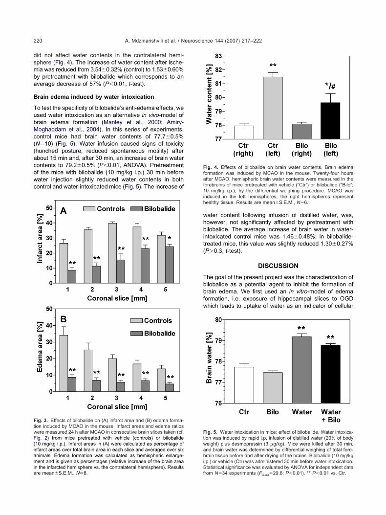
dsmba
B
TubMc((acowc
whbit(
Tbbfw
FtwF(iamia
Ffaf1ih
Ftwabi
A. Mdzinarishvili et al. / Neuroscience 144 (2007) 217–222220
id not affect water contents in the contralateral hemi-phere (Fig. 4). The increase of water content after ische-ia was reduced from 3.54�0.32% (control) to 1.53�0.60%y pretreatment with bilobalide which corresponds to anverage decrease of 57% (P�0.01, t-test).
rain edema induced by water intoxication
o test the specificity of bilobalide’s anti-edema effects, wesed water intoxication as an alternative in vivo-model ofrain edema formation (Manley et al., 2000; Amiry-oghaddam et al., 2004). In this series of experiments,
ontrol mice had brain water contents of 77.7�0.5%N�10) (Fig. 5). Water infusion caused signs of toxicityhunched posture, reduced spontaneous motility) afterbout 15 min and, after 30 min, an increase of brain waterontents to 79.2�0.5% (P�0.01, ANOVA). Pretreatmentf the mice with bilobalide (10 mg/kg i.p.) 30 min beforeater injection slightly reduced water contents in bothontrol and water-intoxicated mice (Fig. 5). The increase of
ig. 3. Effects of bilobalide on (A) infarct area and (B) edema forma-ion induced by MCAO in the mouse. Infarct areas and edema ratiosere measured 24 h after MCAO in consecutive brain slices taken (cf.ig. 2) from mice pretreated with vehicle (controls) or bilobalide10 mg/kg i.p.). Infarct areas in (A) were calculated as percentage ofnfarct areas over total brain area in each slice and averaged over sixnimals. Edema formation was calculated as hemispheric enlarge-ent and is given as percentages (relative increase of the brain area
n the infarcted hemisphere vs. the contralateral hemisphere). Resultsre mean�S.E.M., N�6.
Sf
ater content following infusion of distilled water, was,owever, not significantly affected by pretreatment withilobalide. The average increase of brain water in water-
ntoxicated control mice was 1.46�0.48%; in bilobalide-reated mice, this value was slightly reduced 1.30�0.27%P�0.3, t-test).
DISCUSSION
he goal of the present project was the characterization ofilobalide as a potential agent to inhibit the formation ofrain edema. We first used an in vitro-model of edemaormation, i.e. exposure of hippocampal slices to OGDhich leads to uptake of water as an indicator of cellular
ig. 4. Effects of bilobalide on brain water contents. Brain edemaormation was induced by MCAO in the mouse. Twenty-four hoursfter MCAO, hemispheric brain water contents were measured in the
orebrains of mice pretreated with vehicle (“Ctr”) or bilobalide (“Bilo”;0 mg/kg i.p.), by the differential weighing procedure. MCAO was
nduced in the left hemispheres; the right hemispheres representealthy tissue. Results are mean�S.E.M., N�6.
ig. 5. Water intoxication in mice: effect of bilobalide. Water intoxica-ion was induced by rapid i.p. infusion of distilled water (20% of bodyeight) plus desmopressin (3 �g/kg). Mice were killed after 30 min,nd brain water was determined by differential weighing of total fore-rain tissue before and after drying of the brains. Bilobalide (10 mg/kg
.p.) or vehicle (Ctr) was administered 30 min before water intoxication.
tatistical significance was evaluated by ANOVA for independent datarom N�34 experiments (F3,33�29.6; P�0.01). ** P�0.01 vs. Ctr.

etcpBdmbawmuhbest(ai3
MifsnitRhi1ivecs(ectm
mb1riqplswmtw
p
aidi2Neowcebtebbitgnn
ae5plodbakdoc01t5egbt
ACftS
A
A
A. Mdzinarishvili et al. / Neuroscience 144 (2007) 217–222 221
dema formation. According to previous studies, prepara-ion and superfusion of hippocampal slices per se alreadyause a swelling of slices over 30–60 min which is accom-anied by sodium and calcium uptake (Siklos et al., 1997).ilobalide slightly reduced water contents under basal con-itions (Fig. 1A), probably by interfering with ionic move-ents, although its mechanism of action is unknown (seeelow). During ischemia (OGD), an increase of sodiumnd calcium uptake was observed in earlier studies tohich voltage-operated cation channels as well as gluta-ate receptors of the AMPA and NMDA subtypes contrib-ted (LoPachin et al., 2001; MacGregor et al., 2003). In ourands, bilobalide (1–10 �M), a constituent of Ginkgoiloba, was very effective in preventing OGD-induceddema formation by more than 80%; thus, its efficacy wasimilar to antioxidants, sodium channel blockers and glu-amate receptor antagonists tested in previous studiesLoPachin et al., 2001; MacGregor et al., 2003). Bilobalidected in the low micromolar range, with a half-maximum
nhibition of edema formation occurring at approximately�M (Fig. 1B).
To test in vivo-activity of bilobalide, we employedCAO as an experimental model to induce focal brain
schemia. When used in the mouse, MCAO causes theormation of a large infarct area in cortex and striatum, withome damage also observed in the hippocampus (Mdzi-arishvili et al., 2005). A variety of pharmacological agents
s known to suppress neuronal cell death after experimen-al ischemia and/or traumatic brain injury (Lees, 2000;oyo et al., 2003) but anti-edema effects of compoundsave been less frequently reported. In the present exper-
ments, we confirm an earlier report (Krieglstein et al.,995) that bilobalide, at a dose of 10 mg/kg, reduces the
nfarct area after MCAO as judged by TTC staining ofiable cells (Fig. 2). More importantly, we report thatdema formation in the brain is strongly inhibited by theompound. When judged from measurements of hemi-pheric enlargement, brain edema was inhibited by 70%Fig. 3B). When brain water content was used as param-ter of edema formation, pretreatment of bilobalide de-reased edema formation by 57% (Fig. 4). The similarity ofhese results attests to the validity of the two methods toeasure edema formation.
Water intoxication causes hyponatremia and hypos-olarity in plasma and is followed by the development ofrain edema (Olson et al., 1990; Gullans and Verbalis,993). Brain water uptake following water intoxication hasecently been shown to depend on aquaporin-4, and swell-ng of astrocytic endfeet is the most likely cellular conse-uence of water intoxication (Manley et al., 2000). In theresent study, we used water intoxication to test the se-
ectivity of bilobalide’s anti-edema action. Bilobalide, whilelightly reducing brain water contents in both control andater-treated mice, was not significantly active in thisodel. Thus, bilobalide does not seem to block water
ransport under hyposmolar conditions, and its effectsere selective for edema formation induced by ischemia.
An important question that was not addressed in the
resent study is the molecular mechanism of bilobalide’snti-edema action. Previous work has shown that inhib-tors of sodium and calcium influx, as well as antioxi-ants, protect against edema formation, at least in
n vitro-models (LoPachin et al., 2001; MacGregor et al.,003). Bilobalide can protect mitochondrial energetics anda,K-ATPase activity under conditions of ischemia (Pierret al., 1999; Chandrasekaran et al., 2001), and we andthers have provided evidence that bilobalide interferesith chloride fluxes through ligand-operated receptorhannels (Chatterjee et al., 2003; Klein et al., 2003). Theseffects may contribute to an improved maintenance of ionicalances in ischemic areas. As water enters the cellsogether with ions, improved maintenance of ionic gradi-nts of sodium and chloride would explain bilobalide’seneficial effects. Astrocytes may be potential targets ofilobalide because they contribute strongly to brain swell-
ng (see introduction), and bilobalide was recently showno increase the expression of glial growth factors in astro-lial cultures (Zheng et al., 2000). However, more work iseeded to identify the molecular target(s) of this importantatural compound.
Summarizing, the present results identify bilobalide asrather potent neuroprotective agent which reduces brain
dema formation under ischemic conditions by more than0%. This effect is of sufficient magnitude to be of thera-eutic relevance. Bilobalide’s potency in the low micromo-
ar range would make the compound a potential drug in itswn right, or it may serve as a lead compound for theevelopment of more potent derivatives. Bilobalide proba-ly reaches therapeutic levels in the brain, as shown in thisnd previous studies, but as yet, there are no pharmaco-inetic studies with pure bilobalide that would allow theefinition of an active brain level of the drug. Rats dosedrally with 30–100 mg/kg ginkgo extract EGb761 (whichontains 3% bilobalide) had plasma levels of bilobalide of.5–1.3 �M (Biber, 2003). With linear pharmacokinetics,0 mg/kg of bilobalide (as used in the present study) wouldherefore be expected to produce plasma levels of ca.
�M, a concentration that was active in the in vitro-xperiments (Fig. 1). It remains an open question whetherinkgo extracts would yield anti-edema effects in humans,ut the current study would seem to encourage investiga-ions into this important issue of human health.
cknowledgments—The authors are grateful to Drs. Shyam S.hatterjee and M. Nöldner, Schwabe Co., Karlsruhe, Germany,
or helpful discussions and for supplying pure bilobalide, and tohe Alzheimer Association and to Texas Tech University Healthcience Center (Cardiovascular Seed Grant) for financial support.
REFERENCES
hlemeyer B, Krieglstein J (2003) Neuroprotective effects of Ginkgobiloba extract. Cell Mol Life Sci 60:1779–1792.
miry-Moghaddam M, Xue R, Haug FM, Neely JD, Bhardwaj A, AgreP, Adams ME, Froehner SC, Mori S, Ottersen OP (2004) Alpha-syntrophin deletion removes the perivascular but not endothelialpool of aquaporin-4 at the blood-brain barrier and delays thedevelopment of brain edema in an experimental model of acute
hyponatremia. FASEB J 18:542–544.
B
C
C
C
D
D
D
E
G
G
K
K
K
K
K
K
LL
M
M
M
O
O
O
P
R
S
S
S
S
S
S
U
W
W
Z
A. Mdzinarishvili et al. / Neuroscience 144 (2007) 217–222222
iber A (2003) Pharmacokinetics of Ginkgo biloba extracts. Pharma-copsychiatry 36 (Suppl 1):S32–S37.
handrasekaran K, Mehrabian Z, Spinnewyn B, Drieu K, Fiskum G(2001) Neuroprotective effects of bilobalide, a component of theGinkgo biloba extract (EGb761), in gerbil global brain ischemia.Brain Res 922:282–292.
hatterjee SS, Gabard BL, Jaggy HE (1986) Pharmaceutical compo-sitions containing bilobalide for the treatment of neuropathies. U.S.Patent No. 4,571,407 (18 February 1986).
hatterjee SS, Kondratskaya EL, Krishtal OA (2003) Structure-activitystudies with Ginkgo biloba extract constituents as receptor-gatedchloride channel blockers and modulators. Pharmacopsychiatry 36(Suppl 1):S68–S77.
eFeudis FV (2002) Bilobalide and neuroprotection. Pharmacol Res46:565–568.
eFeudis FV, Drieu K (2000) Ginkgo biloba extract (EGb761) andCNS functions: basic studies and clinical applications. Curr DrugTargets 1:25–58.
orman DC, Cote LM, Buck WB (1992) Effects of an extract of Gingkobiloba on bromethalin-induced cerebral lipid peroxidation andedema in rats. Am J Vet Res 53:138–142.
lliott KC, Jaspar H (1949) Measurement of experimentally inducedbrain swelling and shrinkage. Am J Physiol 157:122–129.
ueniau C, Oberlander C (1997) The kappa opioid agonist niravolinedecreases brain edema in the mouse middle cerebral artery occlu-sion model of stroke. J Pharmacol Exp Ther 282:1–6.
ullans SR, Verbalis JG (1993) Control of brain volume during hyper-osmolar and hypoosmolar conditions. Annu Rev Med 44:289–301.
imelberg HK (2004) Water homeostasis in the brain: basic concepts.Neuroscience 129:851–860.
imelberg HK (2005) Astrocytic swelling in cerebral ischemia as apossible cause of injury and target for therapy. Glia 50:389–397.
inouchi H, Epstein CJ, Mizui T, Carlson E, Chen SF, Chan PH (1991)Attenuation of focal cerebral ischemic injury in transgenic miceoverexpressing CuZn superoxide dismutase. Proc Natl Acad SciU S A 88:11158–11162.
lein J, Chatterjee SS, Löffelholz K (1997) Phospholipid breakdownand choline release under hypoxic conditions: inhibition by bilob-alide, a constituent of Ginkgo biloba. Brain Res 755:347–350.
lein J, Weichel O, Hilgert M, Rupp J, Chatterjee SS, Nawrath H(2003) Excitotoxic hippocampal membrane breakdown and its in-hibition by bilobalide: role of chloride fluxes. Pharmacopsychiatry36 (Suppl 1):S78–S83.
rieglstein J, Ausmeier F, El-Abhar H, Lippert K, Welsch M, RuppallaK, Henrich-Noack P (1995) Neuroprotective effects of Ginkgo bi-loba constituents. Eur J Pharmacol Sci 3:39–48.
ees KR (2000) Neuroprotection. BMJ 56:400–412.oPachin RM, Gaughan CL, Lehning EJ, Weber ML, Taylor CP (2001)
Effects of ion channel blockade on the distribution of Na, K, Ca andother elements in oxygen-glucose deprived CA1 hippocampal neu-rons. Neuroscience 103:971–983.
acGregor DG, Avshalumov MV, Rice ME (2003) Brain edema in-duced by in vitro ischemia: causal factors and neuroprotection.
J Neurochem 85:1402–1411.anley GT, Fujimura M, Ma T, Noshita N, Filiz F, Bollen AW, Chan P,Verkman AS (2000) Aquaporin-4 deletion in mice reduces brainedema after acute water intoxication and ischemic stroke. Nat Med6:159–163.
dzinarishvili A, Geldenhuys WJ, Abbruscato TJ, Bickel U, Klein J,Van der Schyf CJ (2005) NGP1-01, a lipophilic polycyclic cageamine, is neuroprotective in focal ischemia. Neurosci Lett 383:49–53.
ken BS, Storzbach DM, Kaye JA (1998) The efficacy of Ginkgobiloba on cognitive function in Alzheimer disease. Arch Neurol55:1409–1415.
lson JE, Mishler L, Dimlich RV (1990) Brain water content, brainblood volume, blood chemistry, and pathology in a model of cere-bral edema. Ann Emerg Med 19:1113–1121.
tani M, Chatterjee SS, Gabard B, Kreutzberg GW (1986) Effect of anextract of Ginkgo biloba on triethyltin-induced cerebral edema.Acta Neuropathol 69:54–65.
ierre S, Jamme I, Droy-Lefaix MT, Nouvelot A, Maixent JM (1999)Ginkgo biloba extract (EGb 761) protects Na,K-ATPase activityduring cerebral ischemia in mice. Neuroreport 10:47–51.
oyo NC, Shimizu S, Schouten JW, Stover JF, McIntosh TK (2003)Pharmacology of traumatic brain injury. Curr Opin Pharmacol3:27–32.
ancesario G, Kreutzberg GW (1986) Stimulation of astrocytes affectscytotoxic edema. Acta Neuropathol 72:3–14.
chmidt-Kastner R, Freund TF (1991) Selective vulnerability of thehippocampus in brain ischemia. Neuroscience 40:599–636.
eifert G, Schilling K, Steinhäuser C (2006) Astrocyte dysfunction inneurological disorders: a molecular perspective. Nat Rev Neurosci7:194–206.
iklos L, Kuhnt U, Parducz A, Szerdahelyi P (1997) Intracellular cal-cium redistribution accompanies changes in Na�, K� and waterduring the first two hours of in vitro incubation of hippocampalslices. Neuroscience 79:1013–1022.
teiner T, Ringleb P, Hacke W (2001) Treatment options for largehemispheric stroke. Neurology 57 (Suppl 1):S61–S68.
ydserff SG, Green AR, Cross AJ (1996) The effect of oedema andtissue swelling on the measurement of neuroprotection: a studyusing chlormethiazole and permanent middle cerebral artery oc-clusion in rats. Neurodegeneration 5:81–85.
nterberg AW, Stover J, Kress B, Kiening KL (2004) Edema and braintrauma. Neuroscience 129:1019–1027.
eichel O, Hilgert M, Chatterjee SS, Lehr M, Klein J (1999) Bilobalide,a constituent of Ginkgo biloba, inhibits NMDA-induced phospho-lipase A2 activation and phospholipid breakdown in rat hippocam-pus. Naunyn Schmiedebergs Arch Pharmacol 360:609–615.
estman J, Drieu K, Sharma HS (2000) Antioxidant compounds EGB-761 and BN-52021 attenuate heat shock protein (HSP 72 kD)response, edema and cell changes following hyperthermic braininjury. An experimental study using immunohistochemistry in therat. Amino Acids 19:339–350.
heng SX, Zhou LJ, Chen ZL, Yin ML, Zhu XZ (2000) Bilobalidepromotes expression of glial cell line-derived neurotrophic factorand vascular endothelial growth factor in rat astrocytes. Acta Phar-
macol Sin 21:151–155.(Accepted 20 August 2006)(Available online 2 October 2006)
Copyright © 2022 FDOKUMEN