CARBON DISTRIBUTION IN PROTEIN LOCAL STRUCTURE DIRECT SUPEROXIDE DISMUTASE TO DISEASE WAY
6
Corresponding Author: E. Rajasekaran E-mail: [email protected] Received: November 21, 2012 Accepted: November 25, 2012 Published: November 30, 2012 J P P JOURNAL OF PROTEINS AND PROTEOMICS 3(2), July-December 2012, pp. 99-104 COMMUNICATION CARBON DISTRIBUTION IN PROTEIN LOCAL STRUCTURE DIRECT SUPEROXIDE DISMUTASE TO DISEASE WAY E. Rajasekaran*, Sneha Nirmala John and J. Jannet Vennila School of Biotechnology and Health Sciences, Karunya University, Coimbatore 641114, Tamil Nadu, India. Abstract: During the reaction involving superoxide dismutase (SOD), the oxidized Cu(II) form converts one superoxide molecule into oxygen while itself getting reduced to Cu(I) form. This reduced form of Cu(I) again converts another superoxide molecule into hydrogen peroxide and returns to oxidized Cu(II) form. Amyotrophic sclerosis is associated to instability in SOD and oxidative damage. Amyotrophic sclerosis mutants of human SOD form aggregates due to destabilization. Mostly these aggregates are due to hydrophobic interaction which comes from carbon present in the SOD. Some of these mutations found in amyotrophic sclerosis are studied here. Results reveal that most of these mutations alter local hydrophobic structures that are responsible for disorder. Carbon distribution analysis program is effective in identification and modification of mutational sites in biological sequences. This can be better exploited for gene therapy. Keywords: Superoxide dismutase; carbon distribution study; CARd; mutation; metalloprotein; amyotrophic lateral sclerosis; FALS. Introduction The copper containing superoxide dismutase (SOD) catalyses the reaction that convert superoxide into oxygen and hydrogen peroxide and prevent oxidative damage to cell. During the reaction, oxidized Cu(II) form converts one superoxide molecule into oxygen and becomes reduced to Cu(I) form in the enzyme. This reduced form of Cu(I) again converts another superoxide molecule into hydrogen peroxide and becomes oxidized to Cu(II) form back again (Rotilio et al., 1972; Fielden et al., 1974). Changes in Cu binding, instability in SOD and oxidative damage are associated with familial amyotrophic lateral sclerosis (FALS) disease (Furukawa and O’Halloran et al., 2006). Demetallated SOD is pathogenic (Rakhit et al., 2004). Why does it cause disease? Many point mutations in SOD reportedly cause amyotrophic sclerosis (Deng et al., 1993). These mutations understandably change the binding capability of Cu by altering the SOD character. ALS mutants of human superoxide dismutase form fibrous aggregates via framework destabilization (DiDonato et al., 2003). Mostly these aggregates are due to hydrophobic interaction. Hydrophobic interaction is a force that comes from carbon atoms present in the SOD. Any point mutations in SOD, certainly varies the number of carbon. How significant are these changes in the overall function of the SOD? Carbon distribution analysis on several numbers of proteins show that single mutation can alter the protein function based on carbon distribution profile (Zelko et al., 2002). Is it the same with point mutation reported for SOD? To address these issues, some of the SOD mutations found in amyotrophic sclerosis were studied here. Point mutations A4V, G37R, H43R, G85R, G93A and I113T in human SOD are analysed for carbon distribution using CARd program. Methodology The protein structure and sequences were downloaded from PDB [www.rcsb.org]. The PDB
Transcript of CARBON DISTRIBUTION IN PROTEIN LOCAL STRUCTURE DIRECT SUPEROXIDE DISMUTASE TO DISEASE WAY
Corresponding Author: E. RajasekaranE-mail: [email protected]
Received: November 21, 2012Accepted: November 25, 2012Published: November 30, 2012
J P P JOURNAL OF PROTEINS AND PROTEOMICS3(2), July-December 2012, pp. 99-104
COMMUNICATION
CARBON DISTRIBUTION IN PROTEIN LOCAL STRUCTURE DIRECTSUPEROXIDE DISMUTASE TO DISEASE WAY
E. Rajasekaran*, Sneha Nirmala John and J. Jannet Vennila
School of Biotechnology and Health Sciences, Karunya University, Coimbatore 641114, Tamil Nadu, India.
Abstract: During the reaction involving superoxide dismutase (SOD), the oxidized Cu(II) form converts onesuperoxide molecule into oxygen while itself getting reduced to Cu(I) form. This reduced form of Cu(I) againconverts another superoxide molecule into hydrogen peroxide and returns to oxidized Cu(II) form. Amyotrophicsclerosis is associated to instability in SOD and oxidative damage. Amyotrophic sclerosis mutants of humanSOD form aggregates due to destabilization. Mostly these aggregates are due to hydrophobic interaction whichcomes from carbon present in the SOD. Some of these mutations found in amyotrophic sclerosis are studiedhere. Results reveal that most of these mutations alter local hydrophobic structures that are responsible fordisorder. Carbon distribution analysis program is effective in identification and modification of mutational sitesin biological sequences. This can be better exploited for gene therapy.
Keywords: Superoxide dismutase; carbon distribution study; CARd; mutation; metalloprotein; amyotrophic lateralsclerosis; FALS.
Introduction
The copper containing superoxide dismutase(SOD) catalyses the reaction that convertsuperoxide into oxygen and hydrogen peroxideand prevent oxidative damage to cell. During thereaction, oxidized Cu(II) form converts onesuperoxide molecule into oxygen and becomesreduced to Cu(I) form in the enzyme. This reducedform of Cu(I) again converts another superoxidemolecule into hydrogen peroxide and becomesoxidized to Cu(II) form back again (Rotilio et al.,1972; Fielden et al., 1974). Changes in Cu binding,instability in SOD and oxidative damage areassociated with familial amyotrophic lateralsclerosis (FALS) disease (Furukawa andO’Halloran et al., 2006). Demetallated SOD ispathogenic (Rakhit et al., 2004). Why does it causedisease? Many point mutations in SOD reportedlycause amyotrophic sclerosis (Deng et al., 1993).
These mutations understandably change thebinding capability of Cu by altering the SODcharacter. ALS mutants of human superoxidedismutase form fibrous aggregates via frameworkdestabilization (DiDonato et al., 2003). Mostly theseaggregates are due to hydrophobic interaction.Hydrophobic interaction is a force that comes fromcarbon atoms present in the SOD. Any pointmutations in SOD, certainly varies the number ofcarbon. How significant are these changes in theoverall function of the SOD? Carbon distributionanalysis on several numbers of proteins show thatsingle mutation can alter the protein function basedon carbon distribution profile (Zelko et al., 2002).Is it the same with point mutation reported forSOD? To address these issues, some of the SODmutations found in amyotrophic sclerosis werestudied here. Point mutations A4V, G37R, H43R,G85R, G93A and I113T in human SOD are analysedfor carbon distribution using CARd program.
Methodology
The protein structure and sequences weredownloaded from PDB [www.rcsb.org]. The PDB

100 Journal of Proteins and Proteomics
identifier is 1SPD. The carbon distributioninvestigations were carried out using CARdprogram (Rajasekaran, 2012). First the entiresequence was scanned for carbon density using35 inner and 700 outer lengths. The outer length(700) corresponds to 45 amino acid long stretch.So the carbon density is computed by these manyamino acids at a time. This gives thehydrophobic and hydrophilic regions along thesequence in a broader term. Then the mutationalsites are analysed for local carbon density. Forthis, different outer lengths were used. Lengthsof 120, 210 and 255 were taken. These lengthscorrespond to 8, 14 and 17 amino acid longstretch.
Results and Discussion
Carbon content along the SOD protein sequencewas computed as shown in Figure 1. It is astatistical mean carbon content at different aminoacid location. The outer window length of 700atoms are chosen with mid point of referenceamino acid. Carbon content in inner lengths areaveraged statistically. The least amino acidnumber that can appear in the graph is 22 since itis the mid point of 45 amino acid long stretchtaken for statistics. So the disease mutation A4Vwill not be visible with large outer length. Othermutational sites are visible and labelled in thegraph. There appears hydrophilic region after 88th
amino acid. Mostly these mutations occur at edgesof carbon rich or carbon less stretch, similar toedges of secondary structures. Most of the timeany mutations that occur at the edge of thesecondary structure unit that alter function andleads to disease. These edge mutations totallychange the adjoining secondary structure thatleads to different structure and function. From thegraph it is noted that the FALS mutations occurboth in hydrophilic or hydrophobic regions. TheSOD protein show a long stretch (88-154) ofhydrophilic region. This region require anadditional amount of carbon content. This needto be computed carefully before incorporation ofamino acid in these region that will remain stablein the mRNA. If someone looks for binding sitesfor lead compounds (Antonyuk et al., 2010),amino acid around 40 is ideal, as there is carbonrich site. From the crystal structure, this siteappears to be on the surface and available for
binding of aromatic compounds. Now lets lookat the individual mutational site.
The carbon distribution profile for native anddisease mutant (A4V) is given Figure 2. This siteis in interface of protein-protein interaction. Anyincrease in hydrophobicity (Alanine by Valine)strengthens the dimerisation. Carbon distributionprofile is calculated with outer lengths 120 and135. Beyond this, the outer length is not possibleto compute for higher lengths as the residueappear at 4th position. Looking at the distributiongraph, the native protein shows a less normaldistribution compared to mutant. Mutant seemsstable compared to native. Cardoso 2002, arguethat a instability in protein due to A4V mutation.But here we observe that stability on mutation.In this mutation, the cytosine in the second placeof codon is substituted with thymine. Thyminecontent and distribution is important to produceadequate carbon in the protein. Amino acidchange alone need not be the reason for disease.The mRNA expression and stability are alsofactors to be considered for disease.
Carbon distribution profile for native andmutant G37R is given Figure 3. The profiles arecomputed for outer lengths of 120, 210 and 255which correspond to 8, 14 and 17 amino acid. Atlength 8, both native and mutant are away fromnormal distribution. Instability is observedlocally. But at higher lengths, these profiles arenormal. At these lengths the carbon distributionis well maintained. That is the amino acids areorganized in such way that it give stability fromcarbon distribution point of view. In fact themutant shows normal distribution over the
Figure 1: Carbon content along the native superoxidedismutase protein. The diseased mutational sites are labelled.

Protein Local Structure and Disease 101
Figure 2: Carbon distribution profile for native and diseasemutant (A4V) in different outer lengths.
Figure 3: Carbon distribution profile for native and diseasemutant (G37R) in different outer lengths.

102 Journal of Proteins and Proteomics
Figure 4: Carbon distribution profile for native and diseasemutant (H43R) in different outer lengths.
native. The mutant increases the stability. Ofcourse the charge needs to be taken into accounthere as neutral glycine was substituted withpositively charged arginine. Since it is a metalbinding protein, the charge is a matter, though itcan be neutralized with ions and polar solvent.
Figure 4 shows the profile for native anddisease mutant H43R. Surprisingly, there is nochange in the carbon distribution profile in nativeand mutant and in all three lengths. Hydrophobicinteraction is not perturbed. Considering thecharge, both native and mutant residues arepositively charged. So charge is not going toaffect. Then what causes disease? One possibleexplanation may be the adenine (secondnucleotide in codon) in native mRNA issubstituted with guanine in the mutant.Competition between thymine less and rich is amatter. Finding cause at mRNA level may beappropriate.
The profile for native and mutant G85R (Caoet al. , 2008) is given in Figure 5. There isconsiderable change in carbon distributionbetween native and mutant in different lengthsof calculations. Though normal distribution isobserved in both forms, the native shows anarrow tall distribution curve compared mutant.The native shows stable over the mutant. Thisinstability in hydrophobic interaction seemlycauses disease.
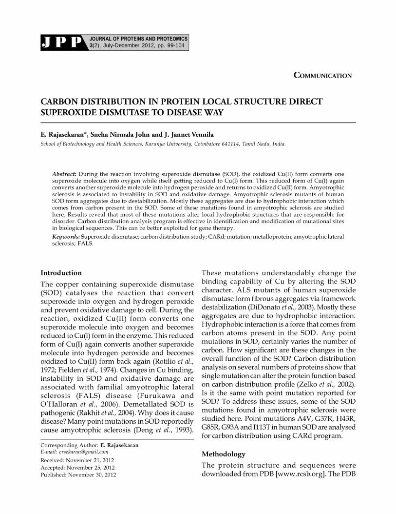
Disease causing mutation G93A is given infigure 6. No change in carbon distribution profileexcept minor difference at lower length (8 aminoacid long). At lower length, the mutant showmore hydrophobic over the native one. From
Figure 5: Carbon distribution profile for native and diseasemutant (G85R) in different outer lengths.
Protein Local Structure and Disease 103
Figure 1, one can observe that G93A occurs inhydrophilic region. Possibly the mutation cantake it to hydrophobic stretch that can aggregatethe protein causing unusual function.
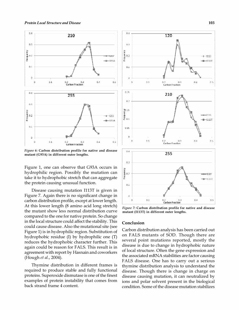
Disease causing mutation I113T is given inFigure 7. Again there is no significant change incarbon distribution profile, except at lower length.At this lower length (8 amino acid long stretch)the mutant show less normal distribution curvecompared to the one for native protein. So changein the local structure could affect the stability. Thiscould cause disease. Also the mutational site (seeFigure 1) is in hydrophilic region. Substitution ofhydrophobic residue (I) by hydrophilic one (T)reduces the hydrophobic character further. Thisagain could be reason for FALS. This result is inagreement with report by Hasnain and coworkers(Hough et al., 2004).
Thymine distribution in different frames isrequired to produce stable and fully functionalproteins. Superoxide dismutase is one of the finestexamples of protein instability that comes fromback strand frame 4 content.
Conclusion
Carbon distribution analysis has been carried outon FALS mutants of SOD. Though there areseveral point mutations reported, mostly thedisease is due to change in hydrophobic natureof local structure. Often the gene expression andthe associated mRNA stabilities are factor causingFALS disease. One has to carry out a seriousthymine distribution analysis to understand thedisease. Though there is change in charge ondisease causing mutation, it can neutralized byions and polar solvent present in the biologicalcondition. Some of the disease mutation stabilizes
Figure 6: Carbon distribution profile for native and diseasemutant (G93A) in different outer lengths.
Figure 7: Carbon distribution profile for native and diseasemutant (I113T) in different outer lengths.

104 Journal of Proteins and Proteomics
the proteins while some do not. From carbondistribution point of view, destabilization occursat local structures. Carbon distribution analysisprogram can be better exploited for this kind ofanalysis and modification of protein for genetherapy.
Many mutations in superoxide dismutase(SOD) cause FALS. It is mainly due to changes inthe local architectural integrity. These changespromote the formation of filamentous aggregates.This study provides molecular levelunderstanding for FALS disease. Further thyminedistribution in SOD mRNAs may be anothercause of disease which requires deeperunderstanding of frame shift and translation.
AbbreviationsSOD, Superoxide Dismutase; CARd, Carbon DistributionAnalysis Program; FALS, Familial Amyotrophic LateralSclerosis; PDB, Protein Data Bank.
ReferencesAntonyuk, S.V., Strange, R.W., and Hasnain, S.S. (2010)
Structural discovery of small molecule binding sitesin Cu-Zn human superoxide dismutase familialamyotrophic lateral sclerosis mutants provides insightsfor lead optimization. J.Med.Chem. 53, 1402-1406.
Cao, X., Antonyuk, S. V., Seetharaman, S. V., Whitson, L. J.,Taylor, A. B., Holloway, S. P., Strange, R. W., Doucette,P. A., Valentine, J. S., Tiwari, A., Hayward, L. J., Padua,S., Cohlberg, J. A., Hasnain, S. S., and Hart, P. J. (2008)Structures of the G85R variant of SOD1 in familialamyotrophic lateral sclerosis. J. Biol. Chem. 283, 16169-16177.
Didonato, M, Craig, L, Huff, M.E., Thayer, M.M., Cardoso,R.M., Kassmann, C.J., Lo, T.P., Bruns, C.K., Powers,
E.T., Kelly, J.W., Getzoff, E.D., and Tainer, J.A. (2003)ALS mutants of human superoxide dismutase formfibrous aggregates via framework destabilization. MolBiol. 332, 601-615.
Fielden, E.M., Roberts, P.B., Bray, R.C., Lowe, D.J., Mautner,G.N., Rotilio, G., and Calabrese, L. (1974) Mechanismof action of superoxide dismutase from pulse radiolysisand electron paramagnetic resonance. Evidence thatonly half the active sites function in catalysis. BiochemJ. 139, 49-60.
Gagliardi, S, Cova, E, Davin, A, Guareschi, S, Abel, K, Alvisi,E, Laforenza, U, Ghidoni, R, Cashman, JR, Ceroni, M.,and Cereda, C. (2010) SOD1 mRNA expression insporadic amyotrophic lateral sclerosis. Neurobiol Dis.39, 198-203.
Hough M.A., Grossmann J.G., Antonyuk S.V., Strange R.W.,Doucette P.A., Rodriguez J.A., Whitson L.J., Hart P.J.,Hayward L.J., Valentine J.S., and Hasnain S.S. (2004)Dimer destabilization in superoxide dismutase mayresult in disease-causing properties: Structures ofmotor neuron disease mutants, Proc. Natl. Acad. Sci.USA, 101, 5976-5981.
Rajasekaran, E. (2012) CARd: Carbon distribution analysisprogram for protein sequences. Bioinformation, 8, 508-512.
Rakhit, R., Crow, J.P., Lepock, J.R., Kondejewski, L.H.,Cashman, N.R., and Chakrabartty, A. (2004)Monomeric Cu,Zn-superoxide dismutase is a commonmisfolding intermediate in the oxidation models ofsporadic and familial amyotrophic lateral sclerosis. JBiol Chem. 279, 15499-504.
Rotilio, G., Morpurgo, L., Giovagnoli, C., Calabrese, L., andMondovì, B. (1972) Studies of the metal sites of copperproteins. Symmetry of copper in bovine superoxidedismutase and its functional significance.Biochemistry, 11, 2187–2192.
Zelko, I.N, Mariani, T.J., and Folz, R.J. (2002) Superoxidedismutase multigene family: a comparison of theCuZn-SOD (SOD1), Mn-SOD (SOD2), and EC-SOD(SOD3) gene structures, evolution and expression. FreeRadic Biol Med. 33, 337-49.