the influence of viral vector delivery of superoxide dismutase
Upload
independentCategory
view
0download
0
eScholarship provides open access, scholarly publishingservices to the University of California and delivers a dynamicresearch platform to scholars worldwide.
University of California
Peer Reviewed
Title:Liposome-mediated extracellular superoxide dismutase gene delivery protects against acute liverinjury in mice
Author:Wu, Jian, University of California, DavisLiu, LYen, R DCatana, ANantz, M HZern, M A
Publication Date:07-01-2004
Publication Info:Postprints, UC Davis
Permalink:http://escholarship.org/uc/item/7h75n0cw
Additional Info:This is a preprint of an article published in Hepatology.
Keywords:Liposome, oxidative liver injury, gene therapy
Abstract:Our previous study demonstrated that polycationic liposomes are highly stable in the bloodstreamand represent an effective agent for liver gene delivery. We report here that liposome-mediatedextracellular superoxide dismutase (EC-SOD) gene delivery successfully prevented acute liverinjury in mice. The therapeutic efficacy of EC-SOD gene delivery by polycationic liposomes wasdetermined against the toxicity of superoxide anions and hydroxyethyl radicals in HepG2 cellsand in a mouse model of acute liver injury caused by D-galactosamine and lipopolysaccharideintoxication. Transfection of HepG2 cells with an EC-SOD plasmid led to a striking increase insuperoxide dismutase activity in the medium. The transfected cells had much less cell death afterreactive oxygen species exposure compared with untransfected or control plasmid-transfectedcells. In a model of acute liver injury, serum alanine aminotransferase levels in mice receivingportal vein injections of EC-SOD lipoplexes were much lower than in those receiving normal saline,liposomes alone, or control lipoplexes. Liver histology confirmed that there was less cell death inthe EC-SOD lipoplex-treated group. Quantitative reverse transcriptase polymerase chain reactionshowed a 55-fold increase in human EC-SOD gene expression in the liver of mice injected withEC-SOD lipoplexes. Serum superoxide dismutase activity in EC-SOD lipoplex-treated mice washigher than in the control groups; this was associated with higher liver glutathione levels andreduced lipid peroxidation. In conclusion, polycationic liposome-mediated EC-SOD gene deliveryprotects against reactive oxygen species toxicity in vitro and against lipopolysaccharide-inducedacute liver injury in D-galactosamine-sensitized mice.

HepatologyCopy of e-mail Notification zhe0316
Your article ( 03-1504 ) from Hepatology is available for download=====Hepatology Published by John Wiley & Sons, Inc.
Dear Author,
Your article page proofs for Hepatology are ready for review. John Wiley & Sons has made this article available to you online for faster, more efficient editing. Please follow the instructions below and you will be able to access a PDF version of your article as well as relevant accompanying paperwork.
First, make sure you have a copy of Adobe Acrobat Reader software to read these files. This is free software and is available for user downloading at http://www.adobe.com/products/acrobat/readstep.html.
Open your web browser, and enter the following web address:http://rapidproof.cadmus.com/RapidProof/retrieval/index.jsp
You will be prompted to log in, and asked for a password. Your login name will be your email address, and your password will be ----
Example:
Login: your e-mail addressPassword: ----
The site contains one file, containing:
- Author Instructions Checklist- Adobe Acrobat Users - NOTES tool sheet- Reprint Order form- A copy of your page proofs for your article
Print out this file, and fill out the forms by hand. (If you do not wish to order reprints, please mark a "0" on the reprint order form.) Read your page proofs carefully and:
- indicate changes or corrections in the margin of the page proofs- answer all queries (footnotes A,B,C, etc.) on the last page of the PDF proof- proofread any tables and equations carefully- check your figure legends for accuracy
Within 48 hours, please return via fax or express mail all materials to the address given below. This will include:
1) Page proofs with corrections2) Reprint Order form
Return to:
Paula Vetrovec Production Editor John Wiley & Sons, Inc. 111 River Street, 8th Floor

HepatologyCopy of e-mail Notification zhe0316
Hoboken, NJ 07030 TEL: (201) 748-5762 FAX: (201) 748-6281/6182 E-mail: [email protected]
Technical problems? If you experience technical problems downloading your file or any other problem with the website listed above, please contact Melissa Lutchkus (e-mail: [email protected], phone: 800-238-3814 x662)or 717-721-2662.
Questions regarding your article? Please don’t hesitate to contact me with any questions about the article itself, or if you have trouble interpreting any of the questions listed at the end of your file. REMEMBER TO INCLUDE YOUR ARTICLE NO. ( 03-1504 ) WITH ALL CORRESPONDENCE. This will help both of us address your query most efficiently.
As this e-proofing system was designed to make the publishing process easier for everyone, we welcome any and all feedback. Thanks for participating in our e-proofing system!
This e-proof is to be used only for the purpose of returning corrections to the publisher.
Sincerely,
Paula VetrovecProduction EditorJohn Wiley & Sons, Inc.111 River Street, 8th FloorHoboken, NJ 07030TEL: (201) 748-5762FAX: (201) 748-6281/6182E-mail: [email protected]

111 RI V E R S T R E E T , H O B O K E N , NJ 07030
HEPATOLOGY PRODUCTION
***IMMEDIATE RESPONSE REQUIRED***
Please follow these instructions to avoid delay of publication.
READ PROOFS CAREFULLYThis will be your only chance to review these proofs. Please note that once your corrected article is postedonline, it is considered legally published, and cannot be removed from the Web site for further corrections. Please note that the volume and page numbers shown on the proofs are for position only.
ANSWER ALL QUERIES ON PROOFS (Queries for you to answer are attached as the last page of your proof.)Mark all corrections directly on the proofs. Note that excessive author alterations may ultimately result in delay of publication and extra costs may be charged to you.
CHECK FIGURES AND TABLES CAREFULLY (Color figure proofs will be sent under separate cover.) Check size, numbering, and orientation of figures.All images in the PDF are downsampled (reduced to lower resolution and file size) to facilitate Internet delivery.These images will appear at higher resolution and sharpness in the printed article. Review figure legends to ensure that they are complete. Check all tables. Review layout, title, and footnotes.
COMPLETE REPRINT ORDER FORM Fill out the attached reprint order form. It is important to return the form even if you are not ordering reprints. You may, if you wish, pay for the reprints with a credit card. Reprints will be mailed only after your article appears in print. This is the most opportune time to order reprints. If you wait until after your article comes off press, the reprints will be considerably more expensive.
RETURN PROOFSREPRINT ORDER FORM
PLEASE RETURN PROOFS (VIA FAX, E-MAIL, OR EXPRESS MAIL) WITHIN 48 HOURS OF RECEIPT TO:
QUESTIONS? Paula Vetrovec, Production Editor John Wiley & Sons, Inc. 111 River St., Mail Stop 8-02 Hoboken, NJ 07030 Phone: 201-748-5762
FAX: 201-748-6281/6182 E-mail: [email protected] to journal acronym and article production number(i.e., HEP 00-001 for HEPATOLOGY ms 00-001).


HEP/LT
REPRINT BILLING DEPARTMENT • 111 RIVER STREET • HOBOKEN, NJ 07030PHONE: (201) 748-8789; FAX: (201) 748-6326
E-MAIL: [email protected] REPRINT ORDER FORM
Please complete this form even if you are not ordering reprints. This form must be returned with your corrected proofs.Your reprints will be shipped approximately 4 weeks after publication. Reprints ordered after printing will be substantially more expensive.
JOURNAL HEPATOLOGY (HEP) VOLUME ISSUE
TITLE OF MANUSCRIPT
MS. NO. NO. OF PAGES AUTHOR(S)
Pages 100 200 300 400 500
$ $ $ $ $
1-4pp 253 322 391 460 529
5-8pp 357 443 529 615 684
9-12pp 437 558 679 799 868
13-16pp 541 684 834 978 1047
17-20pp 788 955 1121 1288 1357
21-24pp 828 1035 1242 1449 1518
25-28pp 943 1012 1081 1150 1219
29-32pp 1063 1132 1201 1270 1339
33-36pp 1080 1149 1218 1287 1356
37-40pp 1316 1385 1454 1523 1592
*Reprints are only available in lots of 100. If you wish to order more than 500 reprints, please contact our Reprints Department at (201) 748-8789for a price quote.
Please send me _____________________ reprints of the above article at $
Please add appropriate State and Local Tax (Tax Exempt No.____________________) $
for United States orders only.Please add 5% Postage and Handling $
TOTAL AMOUNT OF ORDER** $
**International orders must be paid in currency and drawn on a U.S. bankPlease check one: Check enclosed Bill me Credit CardIf credit card order, charge to: American Express Visa MasterCard
Credit Card No Signature Exp. Date
BILL TO: SHIP TO: (Please, no P.O. Box numbers)Name Name
Institution Institution
Address Address
Purchase Order No. Phone Fax
03-1504
HEPATOLOGY111 RI V E R ST R E E T, MA I L ST O P 8-02 , HO B O K E N, NJ 07030
Telephone Number: 201-748-5762 Facsimile Number: 201-748-6281/6182
To: Paula Vetrovec, Production Editor Fax: 201-748-6281 / 6182
From:
Date:
Re: HEPATOLOGY, ms #
Pages (including cover sheet)
Message:
03-1504

Liposome-Mediated Extracellular SuperoxideDismutase Gene Delivery Protects Against
Acute Liver Injury in MiceJian Wu,1 Li Liu,1 Roy D. Yen,1 Andreea Catana,1 Michael H. Nantz,2 and Mark A. Zern1
Our previous study demonstrated that polycationic liposomes are highly stable in the blood-stream and represent an effective agent for liver gene delivery. We report here that liposome-mediated extracellular superoxide dismutase (EC-SOD) gene delivery successfully preventedacute liver injury in mice. The therapeutic efficacy of EC-SOD gene delivery by polycationicliposomes was determined against the toxicity of superoxide anions and hydroxyethyl radicals inHepG2 cells and in a mouse model of acute liver injury caused by D-galactosamine and lipo-polysaccharide intoxication. Transfection of HepG2 cells with an EC-SOD plasmid led to astriking increase in superoxide dismutase activity in the medium. The transfected cells had muchless cell death after reactive oxygen species exposure compared with untransfected or controlplasmid-transfected cells. In a model of acute liver injury, serum alanine aminotransferase levelsin mice receiving portal vein injections of EC-SOD lipoplexes were much lower than in thosereceiving normal saline, liposomes alone, or control lipoplexes. Liver histology confirmed thatthere was less cell death in the EC-SOD lipoplex-treated group. Quantitative reverse transcrip-tase polymerase chain reaction showed a 55-fold increase in human EC-SOD gene expression inthe liver of mice injected with EC-SOD lipoplexes. Serum superoxide dismutase activity inEC-SOD lipoplex-treated mice was higher than in the control groups; this was associated withhigher liver glutathione levels and reduced lipid peroxidation. In conclusion, polycationic lipo-some-mediated EC-SOD gene delivery protects against reactive oxygen species toxicity in vitroand against lipopolysaccharide-induced acute liver injury in D-galactosamine–sensitized mice.(HEPATOLOGY 2004;40:000–000.)
Formation of reactive oxygen species (ROS) occursin a variety of forms of liver injury and fibrogenesis.Common free radicals, such as superoxide anions
(O2●�), hydroxyl radicals (HO●�), hydrogen peroxide
(H2O2) or hydroxyethyl radicals (HERs), are generatedduring drug toxicity, ischemia/reperfusion, and alcoholmetabolism, in addition to reactive intermediate metab-olites of hepatotoxins or drugs.1 These ROS are responsi-ble for necrosis and/or apoptosis of hepatocytes andsinusoidal endothelial cells, the activation of Kupffer cells(thus causing the second phase of liver inflammation),and the activation of hepatic stellate cells and subsequenthepatic fibrogenesis; therefore, antioxidative treatmentappears to be an effective means of attenuating liver injuryand fibrosis in liver diseases.2 Our previous studies haveshown that using antioxidants such as vitamins E and C orfree radical scavengers such as catalase or superoxide dis-mutase (SOD) prevented carbon tetrachloride, D-galac-tosamine (GalN), or bromobenzene-induced acutehepatocellular damage.3–5 Therefore, our speculation isthat treatment with antioxidants or free radical scavengers
Abbreviations: EC-SOD, extracellular superoxide dismutase; ROS, reactive oxygenspecies; HER, hydroxyethyl radical; SOD, superoxide dismutase; GalN, D-galac-tosamine; PCL, polycationic lipid; Chol, cholesterol; G418, geneticin; BSO, buthioninesulfoximine; HX, hypoxanthine; XO, xanthine oxidase; T3, triiodothyronine; LPS, li-popolysaccharide; ALT, alanine aminotransferase; GFP, green fluorescence protein;GSH, glutathione; MDA, malondialdehyde; HAE, 4-hydroxyalkenal; RT-PCR, re-verse-transcriptase polymerase chain reaction.
From the 1Transplant Research Institute, University of California–Davis Med-ical Center, Sacramento, CA; and the 2Department of Chemistry, University ofCalifornia–Davis, Davis, CA.
Received November 14, 2003; accepted April 4, 2004.This study was supported in part by the University of California–Davis Health
System, the Transplant Hope at the University of California–Davis Medical Center,the American Liver Foundation (J. W.), and a grant from the National Institutes ofHealth (AA06386, to M. A. Z).
This work was presented, in part, at the 6th Annual Meeting of the AmericanSociety of Gene Therapy, June 4–8, 2003, Washington, DC, and the 54th AnnualMeeting of the American Association for the Study of Liver Disease, October 24–28,2003, Boston, MA.
Address reprint requests to: Jian Wu, M.D., Ph.D., University of California–Davis Medical Center, Transplant Research Institute, 4635 2nd Ave., Suite 1001,Sacramento, CA 95817. E-mail: [email protected]; fax: 916-734-8097.
Copyright © 2004 by the American Association for the Study of Liver Diseases.Published online in Wiley InterScience (www.interscience.wiley.com).DOI 10.1002/hep.20288
1
AQ: 1
AQ: 2
AQ: 3
AQ: 4
tapraid5/zhe-hepa/zhe-hepa/zhe00704/zhe0316d04a royerl S�8 5/20/04 12:48 Art: 03-1504 Input-DCT-msh

will prevent or attenuate acute liver injury caused by hepa-totoxins, hepatotoxic drugs, Fe2� or Cu2� overload, alco-hol abuse, or ischemia/reperfusion.6
There are three isozymes of SOD. The copper–zinc-containing form of SOD is localized in the cytosol andnucleus of all cell types and plays a major role in theintracellular antioxidative system. Manganese SOD is amanganese-containing enzyme localized in the matrix ofmitochondria. The third type is extracellular SOD (EC-SOD), which is a secretory glycoprotein and is composedof four 30-kd subunits, each containing a Cu and a Znatom.7 EC-SOD is localized primarily in the interstitialmatrix of tissue and is characterized by its high affinity forheparan sulfate binding.8 The importance of EC-SOD innormal liver and pathological conditions has not beenfully elucidated. It may play a role in regulating O2
• levelsin the extracellular space, because O2
• poorly penetratesthe cell membrane when it can be cleared by intracellularcopper–zinc SOD.7,9 Thus EC-SOD may be an impor-tant factor in the extracellular space to degrade O2
• gener-ated during pathological processes and to protect tissuesfrom ROS toxicity.
ROS that exist in the extracellular space appear to me-diate the interactions among different cell types. There-fore, treatments that reduce the production of ROS,inhibit their release, or inactivate their toxic action shouldattenuate ROS-associated liver injury. Emerging strate-gies include the administration of antioxidants, SOD en-zymes, or mimics10–12 and the gene delivery of free radicalscavengers, such as copper–zinc SOD via adenoviral vec-tors.13 Adenoviral EC-SOD vectors were also used to pro-tect rabbits from myocardial infarction damage14 andfrom ischemia/reperfusion-associated liver injury inmice.15 Gene delivery will overcome the short half-life ofthe enzyme in the body. However, obvious drawbacksexist with adenoviral vectors, such as liver toxicity, immu-nogenicity of viral products, and so forth.
Many formulations of liposomes as gene transferagents are used for in vitro gene transfection, and their invivo applications were considered to be limited due totheir interaction with plasma proteins, transient genetransfer, and relative lower levels of transgene expressioncompared with some viral vectors.16–17 With modifica-tions in lipid structure, formulations, and targeting ap-proaches, significant improvements in gene transferefficacy have been achieved.18–22 A series of successfulattempts in genetic correction,19,22 cancer gene therapy,23
and targeting liver gene delivery24 have been reported. Inan attempt to establish a more effective liposomal formu-lation, we have developed a polycationic lipid (PCL) andformulated polycationic liposomes with cholesterol(Chol).25 The PCL-Chol formulation we developed is
nontoxic, binds the least to plasma proteins, and displayshigh gene transfer efficacy to the liver when compared withthe commonly used 1,2-bis(dioleoyloxy)-3-(trimethylamo-nio)propane–Chol or 1,2-bis(dioleoyloxy)-3-(trimethyla-monio)propane–L-� dioleoyl phosphatidylethanolamineformulations.26 We also developed a noninvasive approachto promote hepatocyte proliferation by subcutaneously ad-ministering thyroid hormone, which led to profound re-porter gene expression in mouse liver.26 In this study wereport that our PCL-Chol liposome-mediated EC-SODgene delivery protected against superoxide anion- or hy-droxyethyl radical–induced HepG2 cell death as well as li-popolysaccharide-induced acute liver injury in GalN-sensitized mice.
Materials and Methods
Subcloning EC-SOD Expression Plasmid. PlasmidpUC18-ECSOD (kindly provided by Dr. Stefan Mark-lund, Department of Medical Biosciences, Umeå Univer-sity, Umeå, Sweden27) was digested by EcoRI, and theEC-SOD gene was ligated into pEGFP-C1 or pIRES2-EGFP from Clontech Laboratories, Inc. (Palo Alto, CA)at the multiple cloning site to form new plasmids,pEGFP-C1-ECSOD and pIRES2-EGFP-ECSOD. Theorientation of the ligated fragment of the EC-SOD genein the plasmids was verified by restriction enzymes(BamHI) and sequencing, using a specific forwardprimer. The resulting plasmid DNA was transformed intocompetent Escherichia coli cells from Gibco Life ScienceTechnologies (Grand Island, NY). The plasmid DNAwas extracted from overnight cultures of the competentcells and purified by affinity chromatography with anEndofree plasmid extraction kit from Qiagen, Inc. (Va-lencia, CA). The quality of the DNA was determined byUV spectroscopy and agarose gel electrophoresis aftercleavage by specific restriction endonucleases. The DNAconcentration was quantitated spectrophotometrically.The plasmid DNA was frozen at �20°C and diluted to 1�g/�L in water for in vitro transfection, or in normalsaline for in vivo gene delivery, prior to use.25
Transfection of EC-SOD Plasmids in HepG2 Cellsand Measurement of SOD Activity in Culture Me-dium. HepG2 cells were cultured in minimum essentialmedium plus 10% fetal bovine serum and antibiotics andwere transiently transfected with either the control plas-mid (pEGFP-C1) or EC-SOD plasmid (pEGFP-C1-EC-SOD) using Fugene 6 (Roche Molecular Biochemicals,Indianapolis, IN), when they were 50%–70% confluent.One day after the transfection, the culture medium waschanged to serum-free medium. Culture medium was col-lected 48 hours after the medium change for the spectro-
2 WU ET AL. HEPATOLOGY, July 2004
tapraid5/zhe-hepa/zhe-hepa/zhe00704/zhe0316d04a royerl S�8 5/20/04 12:48 Art: 03-1504 Input-DCT-msh

photometric determination of SOD activity with acommercially available kit from Calbiochem, Inc. (SanDiego, CA). For stable transfection, Hep3B cells werecultured with minimum essential medium plus 10% fetalbovine serum and antibodies, and were transfected witheither pEGFP-C1-ECSOD or control plasmids by aPCL-Chol formulation as we reported previously.25 Thetransfected cells were subjected to geneticin (G418) selec-tion (350 �g/mL medium) over 1 week. G418-resistantcells were grown in the medium containing G418 until itwas changed for serum-free medium. One to three daysafter the cells were cultured in the serum-free medium,SOD activity in cell culture medium and cell lysates wasdetermined by the kit mentioned above.
Necrosis and/or Apoptosis in HepG2 Cells Inducedby Superoxide Anions or HERs. One day after HepG2cells were transiently transfected with either the controlplasmid or EC-SOD plasmid, the cells were subjected topretreatment with the glutathione-depleting agent buthi-onine sulfoximine (BSO, 0.3 mM) for 18 hours and asubsequent exposure to the superoxide anion–generatingsystem hypoxanthine (HX, 1 mM) and xanthine oxidase(XO, 2 mU) for 4–7 hours.28 In separate experiments, thetransfected cells were subsequently exposed to a HER-generating system, which consists of H
2O2 (0.1 mM), ferrous
ammonium sulfate (20 �M), and ethanol (200 mM) for 8 hours.29
After the cells were exposed to either O2●� or HER,
culture medium was collected for the determination oflactate dehydrogenase leakage (Roche Molecular Bio-chemicals, Indianapolis, IN), which is employed as anindicator of cell necrosis.25 The percentage of cell deathwas calculated according to a formula provided by themanufacturer. After the ROS exposure, the cells culturedon LabTech chamber slides (Fisher Scientific, Inc., SantaClara, CA) were fixed with 1% paraformaldehyde andstained with an in situ apoptosis detecting kit, Apoptag
(Intergen Co., Purchase, NY), which uses rhodamine-conjugated antidigoxigenin for final images.
Liposome Generation and Size Measurements.PCL was synthesized and validated as previously de-scribed25 and the PCL-Chol liposome formulation wasgenerated in a molar ratio of 3:1 as described in detailpreviously.26 The liposome size was measured after soni-cation by a laser-based, submicron particle size analyzerfrom Beckman Coulter, Inc., as described previously.5,25
The size of liposomes at different times of generation isbetween 200–250 nm before use. For animal experi-ments, PCL-Chol liposomes were complexed with plas-mid DNA at a charge ratio of 5:1.26 For each mouse, 200�L of liposome suspension containing 0.3 �mol PCL and0.1 �mol cholesterol was injected via the portal vein.
GalN/Lipopolysaccharide-Induced Acute Liver In-jury and Its Prevention by Polycationic Liposome-Mediated EC-SOD Gene Delivery. Polycationicliposome-mediated EC-SOD gene delivery to the liverwas conducted according to our previous description.26
The animal experimental protocol was approved by theAnimal Care and Use Administrative Advisory Commit-tee of the University of California–Davis, according toguidelines of the National Institutes of Health. C57BL/6mice (Charles River Laboratory, Wilmington, MA) werefed a pellet diet and water ad libitum and kept on a 12hour-light/dark cycle. Animals were randomly dividedinto 4 groups, and all received 1 injection of thyroid hor-mone (triiodothyronine [T3], 4 mg/kg subcutaneously) tostimulate hepatocyte proliferation. Two days after T3 in-jection, the animals were treated following a protocol out-lined in Table 1. Animals were anesthetized withpentobarbital for the operation (60 mg/kg intraperitone-ally). Polycationic liposomes alone (200 �L), or com-plexes with plasmid DNA (lipoplexes) were injected viathe portal vein (100 �g control plasmid pEGFP-C1 or
Table 1. Design of Animal Experiment
GroupT3 (4 mg/kg
Subcutaneously)Treatments
(Portal Vein Injection of Lipoplexes)Ga1N/LPS
(500 mg � 25 �g/kg Intraperitoneally)
Saline � Saline (300 �L) �Liposome � PCL-Chol (200 �L) �Liposome � control
Plasmid DNA �PCL-Chol � pEGFP-C1 (100 �gDNA) �
Liposome � EC-SODPlasmid DNA �
PCL-Chol � pEGFP-C1-EC-SOD (100�g DNA) �
Time line of the experimentT3 Lipoplexes Ga1N/LPS Sacrifice2 2 2 2
� � � � �Day 0 1 2 3 4
HEPATOLOGY, Vol. 40, No. 1, 2004 WU ET AL. 3
T1
tapraid5/zhe-hepa/zhe-hepa/zhe00704/zhe0316d04a royerl S�8 5/20/04 12:48 Art: 03-1504 Input-DCT-msh
pEGFP-C1-ECSOD per mouse). One day after the lipo-plex injection, animals were exposed to GalN (500 mg/kgintraperitoneally) plus lipopolysaccharide (LPS, 25 �g/kgintraperitoneally). The animals were sacrificed 24 hoursafter the GalN/LPS intoxication, and the blood was col-lected for measurement of alanine aminotransferase (ALT)levels (Sigma Chemical Co., St. Louis, MO) and serumSOD activity as described previously. Part of the liver tissuewas fixed in 10% buffered formalin for routine paraffin em-bedding and hematoxylin-eosin staining. Some liver tissuewas snap-frozen for sectioning. The frozen sections were sub-sequently fixed in 10% buffered formalin and examined forgreen fluorescence protein (GFP) expression under a fluores-cent microscope. The images were recorded with a digitalvideocamera.25 The reduced form of glutathione (GSH),malondialdehyde (MDA) and 4-hydroxyalkenal (HAE) lev-els in the liver tissue were determined spectrophotometricallyby commercially available kits (OXISResearch, Portland,OR) according to the manufacture’s instructions and ex-pressed as nmol/mg protein in tissue.
Determination of Human EC-SOD GeneExpression in Mouse Liver Tissue via Real-TimeQuantitative Reverse-Transcriptase PolymeraseChain Reaction. Messenger RNA levels of human EC-SOD in mouse liver tissue were determined by quantita-tive reverse-transcriptase polymerase chain reaction (RT-PCR) using mouse �-actin as a housekeeping genecontrol. RNA was extracted from the mouse liver tissue of3 independent experiments using TRIzol (Invitrogen,Carlsbad, CA) and quantitated via absorbance of a260-nm wavelength. Complementary DNA was gener-ated via reverse transcription of DNase I–digested RNAemploying ThermoScript RT-PCR Systems (Invitrogen).Amplification was performed with Platinum PCR SuperMix (Invitrogen) and a primer pair of the human EC-SOD gene in ABI Prism 7700 Thermal Cycler (AppliedBioSystems, Foster City, CA).30 The sequences of theprimer pair are: forward primer, 5�-AACTGCCC-CGCGTCTTC-3�; reverse primer, 5�-GCCAAACATTC-CCCCAAAG-3�; and fluorescent probe, 6-carboxyfluores-cein-5�-TGTTTCGCATCCACCGCCACC-3�. Theconcentrations for forward and reverse primers were 900nM and 50 nM, respectively, both of which were optimizedfor an annealing temperature of 60°C. Following 40 cycles ofamplification in the above condition, semi–log amplificationcurves were evaluated using comparative quantification(��CT).30 Expression levels were normalized to mouse �-ac-tin housekeeping gene control. The relative gene expressionin different groups was calculated based on the average levelof the saline control group.
Statistical Analysis. Most data were expressed asmean � SEM and evaluated using ANOVA and Newman-
Keuls test for multiple comparisons among groups. Stu-dent’s unpaired t test was used for the comparison betweentwo groups. Wilcoxon Signed Rank test was employed forevaluating quantitative RT-PCR data, followed by q tests formultiple comparisons among groups. A P value less than .05was considered statistically significant.
Chemicals and Reagents. Dulbecco’s Modified EagleMedium, cell culture supplements, and fetal bovine se-rum were purchased from Invitrogen, Inc. (Gaithersburg,MD). GalN, LPS, HX, and XO were the products ofSigma Chemical Co. Most other chemicals used werecommercially available reagents of analytical grade.
Results
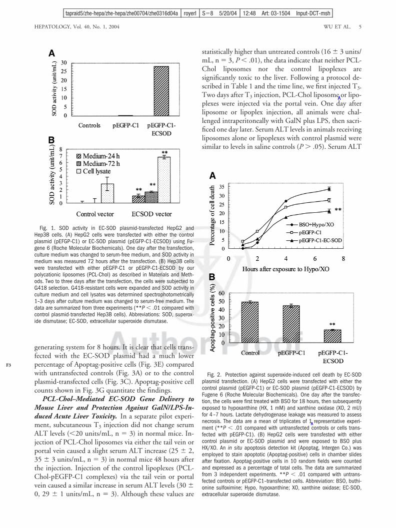
SOD Activity in Culture Medium After Transientor Stable Transfection of an EC-SOD Plasmid inHepatoma Cells. After verification with restriction en-zyme cleavage and sequencing, the new subcloned EC-SOD plasmid, pEGFP-C1-ECSOD, was used totransfect human hepatoma cell lines HepG2 and Hep3B,either transiently or stably. After transient transfection inHepG2 cells with pEGFP-C1-ECSOD plasmid DNA,SOD levels in the culture medium were elevated from thebackground to 28 units/mL at 72 hours (Fig. 1A). Un-transfected cells or cells transfected with the control plas-mid, pEGFP-C1, did not show any increase in SODactivity in medium. Hep3B cells were transfected witheither the control plasmid or EC-SOD plasmid using ourPCL-Chol liposome formulation, and the transfectedcells were selected by the addition of G418. SOD activityin culture medium and cell lysates from EC-SOD plas-mid-transfected Hep3B cells was markedly increased 24and 72 hours after the medium change compared with thecontrol plasmid-transfected cells (P � .01) (Fig. 1B). TheSOD activity measured reflects total activity of all threeSOD isoforms in the cell lysates.
Protection Against Toxicity of Superoxide Anionsand HERs by EC-SOD Overexpression in HepG2Cells. One day after the transfection of HepG2 cells witheither control plasmid or EC-SOD plasmid, the cells werefirst exposed to BSO for 18 hours, and subsequently toHX and OX for 7 hours. As shown in Fig. 2A, lactatedehydrogenase leakage from the cells transfected withpEGFP-C1-ECSOD at a late time point (7 hours afterexposure to HX/OX) was significantly lower than un-transfected cells or cells transfected with the control plas-mid, pEGFP-C1 (P � .01). EC-SOD plasmidtransfection also markedly diminished the number of Ap-optag-positive cells after HX/XO exposure (Fig. 2B).
In separate experiments, after HepG2 cells were treatedwith BSO, they were subsequently exposed to a HER-
4 WU ET AL. HEPATOLOGY, July 2004
AQ: 5
AQ: 6
F1
F2
tapraid5/zhe-hepa/zhe-hepa/zhe00704/zhe0316d04a royerl S�8 5/20/04 12:48 Art: 03-1504 Input-DCT-msh
generating system for 8 hours. It is clear that cells trans-fected with the EC-SOD plasmid had a much lowerpercentage of Apoptag-positive cells (Fig. 3E) comparedwith untransfected controls (Fig. 3A) or to the controlplasmid-transfected cells (Fig. 3C). Apoptag-positive cellcounts shown in Fig. 3G quantitate the findings.
PCL-Chol–Mediated EC-SOD Gene Delivery toMouse Liver and Protection Against GalN/LPS-In-duced Acute Liver Toxicity. In a separate pilot experi-ment, subcutaneous T3 injection did not change serumALT levels (�20 units/mL, n � 3) in normal mice. In-jection of PCL-Chol liposomes via either the tail vein orportal vein caused a slight serum ALT increase (25 � 2,35 � 3 units/mL, n � 3) in normal mice 48 hours afterthe injection. Injection of the control lipoplexes (PCL-Chol-pEGFP-C1 complexes) via the tail vein or portalvein caused a similar increase in serum ALT levels (30 �0, 29 � 1 units/mL, n � 3). Although these values are
statistically higher than untreated controls (16 � 3 units/mL, n � 3, P � .01), the data indicate that neither PCL-Chol liposomes nor the control lipoplexes aresignificantly toxic to the liver. Following a protocol de-scribed in Table 1 and the time line, we first injected T3.Two days after T3 injection, PCL-Chol liposome or lipo-plexes were injected via the portal vein. One day afterliposome or lipoplex injection, all animals were chal-lenged intraperitoneally with GalN plus LPS, then sacri-ficed one day later. Serum ALT levels in animals receivingliposomes alone or lipoplexes with control plasmid weresimilar to levels in saline controls (P � .05). Serum ALT
Fig. 1. SOD activity in EC-SOD plasmid-transfected HepG2 andHep3B cells. (A) HepG2 cells were transfected with either the controlplasmid (pEFGP-C1) or EC-SOD plasmid (pEGFP-C1-ECSOD) using Fu-gene 6 (Roche Molecular Biochemicals). One day after the transfection,culture medium was changed to serum-free medium, and SOD activity inmedium was measured 72 hours after the transfection. (B) Hep3B cellswere transfected with either pEGFP-C1 or pEGFP-C1-ECSOD by ourpolycationic liposomes (PCL-Chol) as described in Materials and Meth-ods. Two to three days after the transfection, the cells were subjected toG418 selection. G418-resistant cells were expanded and SOD activity inculture medium and cell lysates was determined spectrophotometrically1–3 days after culture medium was changed to serum-free medium. Thedata are summarized from three experiments (**P � .01 compared withcontrol plasmid-transfected Hep3B cells). Abbreviations: SOD, superox-ide dismutase; EC-SOD, extracellular superoxide dismutase.
Fig. 2. Protection against superoxide-induced cell death by EC-SODplasmid transfection. (A) HepG2 cells were transfected with either thecontrol plasmid (pEGFP-C1) or EC-SOD plasmid (pEGFP-C1-ECSOD) byFugene 6 (Roche Molecular Biochemicals). One day after the transfec-tion, the cells were first treated with BSO for 18 hours, then subsequentlyexposed to hypoxanthine (HX, 1 mM) and xanthine oxidase (XO, 2 mU)for 4–7 hours. Lactate dehydrogenase leakage was measured to assessnecrosis. The data are a mean of triplicates of 1 representative experi-ment (**P � .01 compared with untransfected controls or cells trans-fected with pEGFP-C1). (B) HepG2 cells were transfested with eithercontrol plasmid or EC-SOD plasmid and were exposed to BSO plusHX/XO. An in situ apoptosis detection kit (Apoptag, Intergen Co.) wasemployed to stain apoptotic (Apoptag-positive) cells in chamber slidesafter fixation. Apoptag-positive cells in 10 random fields were countedand expressed as a percentage of total cells. The data are summarizedfrom 3 independent experiments. **P � .01 compared with untrans-fected controls or pEGFP-C1–transfected cells. Abbreviation: BSO, buthi-onine sulfoximine; Hypo, hypoxanthine; XO, xanthine oxidase; EC-SOD,extracellular superoxide dismutase.
HEPATOLOGY, Vol. 40, No. 1, 2004 WU ET AL. 5
F3
tapraid5/zhe-hepa/zhe-hepa/zhe00704/zhe0316d04a royerl S�8 5/20/04 12:48 Art: 03-1504 Input-DCT-msh

levels in animals receiving EC-SOD lipoplexes (PCL-Chol-pEGFP-C1-ECSOD) were markedly lower thanthe other three groups (P � .01) (Fig. 4). Liver histologyrevealed a similar degree of massive cell death and inflam-matory infiltration in GalN/LPS-intoxicated animals plusportal vein injection of saline, liposomes (PCL-Chol)or control lipoplexes (PCL-Chol-pEGFP-C1) (Fig. 5A–5C). Much less cell death was found in the liver of animalsreceiving EC-SOD lipoplexes (Fig. 5D) before the GalN/LPS challenge, compared with the other three groups.Therefore, the liver histology findings are consistent withserum ALT level alterations, and liver injury in mice re-ceiving EC-SOD lipoplexes was markedly attenuatedcompared with those treated with saline control, PCL-Chol liposomes only, or control lipoplexes.
Liver frozen sections were examined for GFP expres-sion. Intensive GFP expression was found in the liversections from animals receiving control lipoplexes or EC-
SOD lipoplexes (Fig. 6C and 6D), but not in the sectionsfrom saline control mice or from liposome-injected mice(Fig. 6A and 6B).
Human EC-SOD gene expression in the livers of micereceiving injections of either PCL-Chol liposomes or con-trol lipoplexes did not significantly differ from the salinecontrol group, as evaluated by real-time quantitative RT-PCR. Mouse EC-SOD does not cross-hybridize with thehuman gene. The liver human EC-SOD gene expressionlevels in mice receiving EC-SOD lipoplex injection wereapproximately 55-fold higher than the other 3 groups(Fig. 7A). Serum SOD activity was measured in all GalN/LPS-intoxicated mice, and the data showed that the totalSOD activity in the serum of animals receiving portal veininjection of EC-SOD lipoplexes was higher than in thosereceiving the injection of liposomes alone or control lipo-plexes (P � .01) (Fig. 7B). The SOD activity measuredreflects the 3 human and mouse isoforms of SOD in se-rum. These results indicate that the gene was successfullydelivered to the mouse liver with our polycationic lipo-somes and that the gene was highly expressed in themouse liver.
GSH Preservation and Reduced Lipid Peroxida-tion by EC-SOD Gene Delivery in GalN/LPS-Intox-icated Mice. As shown in Fig. 8, LPS challenge in GalN-sensitized mice led to enhanced lipid peroxidation asindicated by MDA/HAE levels and decreased levels of thereduced form of glutathione in the liver (P � 0.01) com-pared with untreated controls. Animals receiving EC-SOD gene delivery had preserved GSH levels (P � .05)and reduced MDA/HAE levels (P � .05) in their livers incomparison with liposome controls or control plasmid-transfected animals (Fig. 8A and 8B). Both liver GSH and
Fig. 4. Serum ALT levels in mice exposed to GalN/LPS with or withoutPCL-Chol–mediated EC-SOD gene delivery. The experimental protocolwas described in detail in Materials and Methods. Data are summarizedfrom 3 independent experiments. **P � .01 compared with salinecontrols, liposomes (PCL-Chol) alone, or pEGFP-C1–transfected cells.Abbreviations: ALT, alanine aminotransferase; PCL, polycationic lipid;Chol, cholesterol.
Fig. 3. Representative images of Apoptag-positive cells after exposureto HERs. An in situ apoptosis detection kit was employed to stainapoptotic (Apoptag-positive) cells in chamber slides after fixation. Theexperimental protocol is the same as in Fig. 2B. (A, C, E) Apoptag-positive cells shown in redby staining with an in situ apoptosis detectionkit (Apoptag, Intergen Co.); (B, D, F) plain images of the same field. (A,B) untransfected images; (C, D) control plasmid-transfected images; (E,F) EC-SOD plasmid-transfected images. (G) Apoptag-positive cells in 10random fields were counted and expressed as a percentage of total cells.**P � .01 compared with untransfected controls or pEGFP-C1–trans-fected cells.
6 WU ET AL. HEPATOLOGY, July 2004
F4
F5
COLOR
F6
F7
F8
tapraid5/zhe-hepa/zhe-hepa/zhe00704/zhe0316d04a royerl S�8 5/20/04 12:48 Art: 03-1504 Input-DCT-msh

MDA/HAE levels in animals receiving EC-SOD genedelivery were close to those in untreated controls withoutGalN/LPS intoxication (P � 0.05).
DiscussionIn the present study, we first tested whether the newly
subcloned EC-SOD plasmid works in hepatoma cell linesafter transfection. Elevated SOD activity was seen in cul-ture medium from HepG2 cells transiently transfectedand from Hep3B cells stably transfected with the newrecombinant EC-SOD plasmid, pEGFP-C1-ECSOD.Increased SOD levels in culture medium demonstrate thecharacteristic extracellular location of the enzyme. Wethen treated control plasmid-transfected or EC-SODplasmid-transfected HepG2 cells initially with BSO, aGSH-depleting agent,31 and subsequently with either O2
•
generated from HX and XO32 or HER using a HER-generating system.29 The cells transfected with pEGFP-C1-ECSOD showed less lactate dehydrogenase leakageand a lower percentage of Apoptag-positive cells com-pared with those transfected with the control plasmidor untransfected cells. The results indicate that theoverexpression of EC-SOD makes cells more resistantto O2
• or other O2•-derived ROS, such as HO• or per-
oxynitrite anions (ONOO•).1,33 A similar finding wasachieved in HepG2 cells when they were treated withHER, which occurs in ethanol metabolism and proba-bly contributes to the pathogenesis of alcohol-associ-ated liver injury.34
In our previous studies,25–26 we reported that our poly-cationic liposomes are serum-resistant in vitro and inter-act the least with plasma proteins in the bloodstreamwhen compared with two other commonly used cationicliposome formulations.18,35 In the present study, we em-ployed the same approach as reported in our previousstudy26 to deliver the EC-SOD gene with our poly-cationic liposome formulation to the liver. The markedlyenhanced human EC-SOD gene expression in the mouseliver tissue and elevated serum SOD activity in the ani-mals receiving EC-SOD lipoplex injection via the portalvein confirmed that the hepatocytes were transfected, ex-pressed the functional gene, and released the active en-zyme into the bloodstream. Moreover, due to the bindingproperties to glycosaminoglycans in the extracellularspace, the measurement of SOD or EC-SOD activity mayunderestimate the actual EC-SOD activity in the blood-stream.36 Finally, both the control plasmid- and EC-SODplasmid-transfected liver cells showed positive GFP ex-
Fig. 5. Representative micrographs of liver histology. Liver sections were fixed in 10% buffered formalin, embedded in paraffin, and stained withhematoxylin-eosin. (A) GalN/LPS treatment with saline control. (B) GalN/LPS treatment with liposomes (PCL-Chol) alone. (C) GalN/LPS treatmentplus PCL-Chol–mediated control plasmid (pEGFP-C1) transfer. (D) GalN/LPS treatment plus PCL-Chol–mediated EC-SOD gene (pEGFP-C1-ECSOD)transfer (original magnification 100).
HEPATOLOGY, Vol. 40, No. 1, 2004 WU ET AL. 7
COLOR
tapraid5/zhe-hepa/zhe-hepa/zhe00704/zhe0316d04a royerl S�8 5/20/04 12:48 Art: 03-1504 Input-DCT-msh
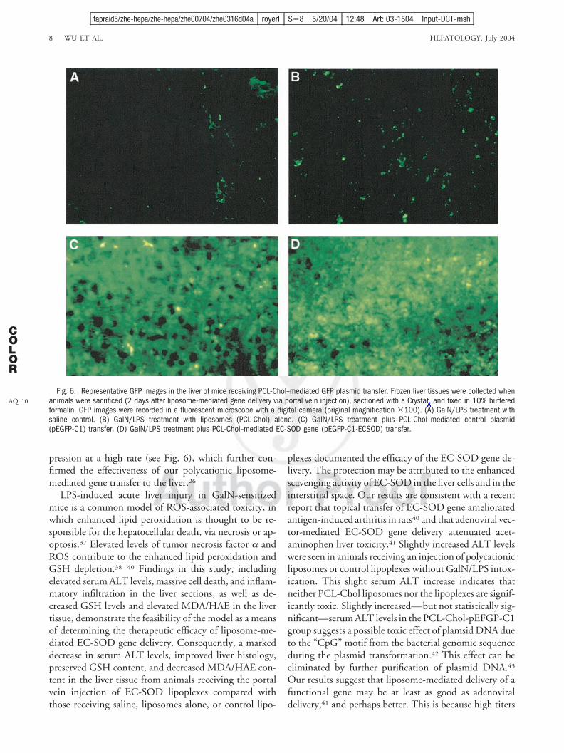
pression at a high rate (see Fig. 6), which further con-firmed the effectiveness of our polycationic liposome-mediated gene transfer to the liver.26
LPS-induced acute liver injury in GalN-sensitizedmice is a common model of ROS-associated toxicity, inwhich enhanced lipid peroxidation is thought to be re-sponsible for the hepatocellular death, via necrosis or ap-optosis.37 Elevated levels of tumor necrosis factor � andROS contribute to the enhanced lipid peroxidation andGSH depletion.38–40 Findings in this study, includingelevated serum ALT levels, massive cell death, and inflam-matory infiltration in the liver sections, as well as de-creased GSH levels and elevated MDA/HAE in the livertissue, demonstrate the feasibility of the model as a meansof determining the therapeutic efficacy of liposome-me-diated EC-SOD gene delivery. Consequently, a markeddecrease in serum ALT levels, improved liver histology,preserved GSH content, and decreased MDA/HAE con-tent in the liver tissue from animals receiving the portalvein injection of EC-SOD lipoplexes compared withthose receiving saline, liposomes alone, or control lipo-
plexes documented the efficacy of the EC-SOD gene de-livery. The protection may be attributed to the enhancedscavenging activity of EC-SOD in the liver cells and in theinterstitial space. Our results are consistent with a recentreport that topical transfer of EC-SOD gene amelioratedantigen-induced arthritis in rats40 and that adenoviral vec-tor-mediated EC-SOD gene delivery attenuated acet-aminophen liver toxicity.41 Slightly increased ALT levelswere seen in animals receiving an injection of polycationicliposomes or control lipoplexes without GalN/LPS intox-ication. This slight serum ALT increase indicates thatneither PCL-Chol liposomes nor the lipoplexes are signif-icantly toxic. Slightly increased—but not statistically sig-nificant—serum ALT levels in the PCL-Chol-pEFGP-C1group suggests a possible toxic effect of plamsid DNA dueto the “CpG” motif from the bacterial genomic sequenceduring the plasmid transformation.42 This effect can beeliminated by further purification of plasmid DNA.43
Our results suggest that liposome-mediated delivery of afunctional gene may be at least as good as adenoviraldelivery,41 and perhaps better. This is because high titers
Fig. 6. Representative GFP images in the liver of mice receiving PCL-Chol–mediated GFP plasmid transfer. Frozen liver tissues were collected whenanimals were sacrificed (2 days after liposome-mediated gene delivery via portal vein injection), sectioned with a Crystat, and fixed in 10% bufferedformalin. GFP images were recorded in a fluorescent microscope with a digital camera (original magnification 100). (A) GalN/LPS treatment withsaline control. (B) GalN/LPS treatment with liposomes (PCL-Chol) alone. (C) GalN/LPS treatment plus PCL-Chol–mediated control plasmid(pEGFP-C1) transfer. (D) GalN/LPS treatment plus PCL-Chol–mediated EC-SOD gene (pEGFP-C1-ECSOD) transfer.
8 WU ET AL. HEPATOLOGY, July 2004
AQ: 10
COLOR
tapraid5/zhe-hepa/zhe-hepa/zhe00704/zhe0316d04a royerl S�8 5/20/04 12:48 Art: 03-1504 Input-DCT-msh

of adenoviruses (i.e., titers high enough to be therapeuti-cally significant) may cause liver injury through a directtoxic effect or through immunomediated damage.44
The fact that liposomes are nonimmunogenic en-hances their role as a nonviral vector for gene delivery.When targeting approaches are desirable, liposomescan be employed to selectively deliver therapeutic genesto a preferential organ or cell type.21,22,24 One disad-vantage associated with nonviral gene delivery ap-proaches and retroviral vectors is that a high level oftransgene expression can be achieved only when tar-geted cells are in a proliferative state.26 We employed anoninvasive approach to promote hepatocyte prolifer-
ation through the injection of thyroid hormone beforeadministering the lipoplexes. This approach is nottoxic and resulted in a level of hepatocyte proliferationsimilar to partial hepatectomy,26 and a high level ofEC-SOD gene expression was detected in the mouseliver, leading to a functional consequence in thepresent study: protection against oxidative injury.
In conclusion, the findings in the present study dem-onstrate that the overexpression of extracellular SOD pro-tects against either superoxide anion– or HER-induced
Fig. 7. Human EC-SOD gene expression in mouse liver and serumSOD activity in mice receiving liposome-mediated EC-SOD gene delivery.(A) Human EC-SOD messenger RNA levels in mouse liver tissue weredetermined with real-time quantitative RT-PCR using mouse �-actin as ahousekeeping gene control. The relative human EC-SOD gene expressionlevels in three other groups were calculated based on an average levelin the control group. (B) Serum SOD activity after PCL-Chol–mediatedEC-SOD gene delivery and subsequent GalN/LPS toxicity. Serum totalSOD activity was measured spectrophotometrically 2 days after portalvein injection of liposomes or lipoplexes and subsequent GalN/LPStoxicity. *P � .05, **P � .01 compared with the 3 other groups.Abbreviations: EC-SOD, extracellular superoxide dismutase; PCL, poly-cationic lipid; Chol, cholesterol; SOD, superoxide dismutase.
Fig. 8. Liver GSH content and lipid peroxidation after EC-SOD geneoverexpression and GalN/LPS toxicity. (A) GSH content in normal controlliver and in the liver after portal vein injection of liposomes or lipoplexesand subsequent GalN/LPS toxic challenge. Liver GSH content was de-termined spectrophotometrically and expressed as nmol/mg protein ofthe tissue. The data are summarized from 3 experiments. �P � .05,**P � .01 compared with controls. �P � .5 compared with eitherPCL-Chol liposomes or the liposome–pEGFP-C1 group. (B) MDA and HAEin normal controls or in the mouse liver after liposome-mediated EC-SODgene delivery and subsequent GalN/LPS toxic challenge. Liver MDA andHAE levels were measured spectrophotometrically and expressed asnmol/mg protein of the tissue. Data are summarized from 3 independentexperiments. �P � .05, ** P � .01 compared with controls. �P � 0.5compared to either PCL-Chol liposomes or the liposome–pEGFP-C1group. Abbreviations: GSH, glutathione; PCL, polycationic lipid; Chol,cholesterol; MDA, malondialdehyde; HAE, 4-hydroxyalkenal. ABI Prism7700 Thermal Cycler.
HEPATOLOGY, Vol. 40, No. 1, 2004 WU ET AL. 9
tapraid5/zhe-hepa/zhe-hepa/zhe00704/zhe0316d04a royerl S�8 5/20/04 12:48 Art: 03-1504 Input-DCT-msh

necrosis/apoptosis in HepG2 cells, and that polycationicliposome-mediated EC-SOD gene delivery markedly at-tenuated GalN/LPS-induced acute liver injury in mice.To our knowledge, this is a first report, which demon-strates that polycationic liposome-mediated EC-SODgene delivery to the liver represents a potential therapy forROS-associated liver injury.
Acknowledgment: The authors are particularly grate-ful to Professor Stefan L. Marklund, Department of Med-ical Biosciences, Umeå University, Umeå, Sweden, forproviding the human EC-SOD plasmid, and for invalu-able comments on the study.
References1. Wu J, Danielsson Å, Zern MA. Toxicity of hepatotoxins: new insights into
mechanisms and therapy. Exp Opin Invest Drugs 1999;8:585–607.2. Wu J, Zern MA. Hepatic stellate cells, a target for the treatment of liver
fibrosis. J Gastroenterol 2000;35:665–672.3. Wu J, Soderbergh H, Karlsson K, Danielsson Å. Protective effect of S-
adenosyl-L-methionine on bromobenzene- or D-galactosamine toxicity toisolated rat hepatocytes. HEPATOLOGY 1996;23:359–365.
4. Wu J, Karlsson K, Danielsson Å. Effects of vitamin E, ascorbic acid andcatalase on bromobenzene- and hydrogen peroxide-induced intracellularoxidation and single strand DNA breakage in Hep G2 cells. J Hepatol1997;26:669–677.
5. Wu J, Liu P, Zhu JL, Maddukuri S, Zern MA. Increased liver up-take ofliposomes and improved targeting efficacy by labeling with asialofetuin inrodents. HEPATOLOGY 1998;27:772–778.
6. Gujral JS, Bucci TJ, Frahood A, Jeaschke H. Mechanism of cell deathduring warm hepatic ischemic-reperfusion in rats: apoptosis or necrosis.HEPATOLOGY 2001;33:397–405.
7. Marklund SL. Extracellular superoxide dismutase. Methods Enzymol.2002;349:74–80.
8. Marklund SL. Extracellular superoxide dismutase in human tissues andhuman cell lines. J Clin Invest 1984;74:1398–1403.
9. Winterbourn CC, Stern A. Human red cells scavenge extracellular hydro-gen peroxide and inhibit formation of hypochlorous acid and hydroxylradical. J Clin Invest 1987;80:1486–1491.
10. Yao T, Esposti SD, Huang L, Arnon R, Spangenberger A, Zern MA.Inhibition of carbon tetrachloride-induced liver injury by liposomes con-taining vitamin E. Am J Physiol 1994;267:G476–G484.
11. Ferret PJ, Hammoud R, Tulliez M, Tran A, Trebeden H, Jaffray P, et al.Detoxification of reactive oxygen species by a nonpeptidyl mimic of super-oxide dismutase cures acetaminophen-induced acute liver failure in themouse. HEPATOLOGY 2001;33:1173–1180.
12. Malassagne B, Ferret PJ, Hammoud R, Tulliez M, Bedda S, Trebeden H,et al. The superoxide dismutase mimetic MnTBAP prevents Fas-inducedacute liver failure in the mouse. Gastroenterology 2001;121:1451–1459.
13. Wheeler MD, Kono H, Yin M, Rusyn I, Froh M, Connor HD, et al.Delivery of the Cu/Zn-superoxide dismutase gene with adenovirus reducesearly alcohol-induced liver injury in rats. Gastroenterology 2001;-120:1241–1250.
14. Li Q-H, Bolli R, Qiu Y-M, Tang X-L, Guo Y-R, French BA. Gene therapywith extracellular superoxide dismutase protects conscious rabbits againstmyocardial infarction. Circulation 2001;103:1893–1898.
15. Wheeler MD, Katuna M, Smutney OM, Froh M, Dikalova A, Mason RP,et al. Comparison of the effect of adenoviral delivery of three superoxidedismutase genes against hepatic ischemia-reperfusion injury. Human GeneTher 2001;12:2167–2177.
16. Wu J, Wu GY, Zern MA. The prospects of hepatic drug delivery and genetherapy. Exp Opin Invest Drugs 1998;7:1795–1817.
17. Niidome T, Huang L. Gene therapy progress and prospects: nonviralvectors. Gene Ther 2002;9:1647–1652.
18. Templeton NS, Lasic DD, Frederik PM, Strey HH, Roberts DD, PavlakisGN. Improved DNA:liposome complexes for increased systemic deliveryand gene expression. Nature Biotechnol 1997;15:647–652.
19. Dasi F, Benet M, Crespo J, Crespo A, Alino SF. Asialofetuin liposome-mediated human �1-antitrypsin gene transfer in vivo results in stationarylong-term gene expression. J Mol Med 2001;79:205–212.
20. Liu F, Qi H, Huang L, Liu D. Factors controlling the efficiency of cationiclipid-mediated transfection in vivo via intravenous administration. GeneTher 1997;4:517–523.
21. Wu J, Nantz MH, Zern MA. Targeting hepatocytes for drug and genedelivery: emerging novel approaches and applications. Front Biosci 2002;7:d717–d725.
22. Kren BT, Praetor B, Bandyopadhyay P, Chowdhury NR, Chowdhury JR,Steer CJ. Correction of the UDP-glucuronosyltransferase gene defect inthe Gunn rat model of Crigler-Najjar syndrome type I with chimericoligonucleotide. Proc Natl Acad Sci U S A 1999;96:10349–10354.
23. Mohr L, Yoon S, Eastman SJ, Chu Q, Scheule RK, Scaglioni PP, et al.Cationic liposome-mediated gene delivery to the liver and to hepatocellu-lar carcinoma in mice. Hum Gene Ther 2001;12:799–809.
24. Kawakami S, Fumoto S, Nishikawa M, Yamashita F, Nishida M. In vivogene delivery to the liver using novel galactosylated cationic liposomes.Pharm Res 2000;17:306–313.
25. Wu J, Lizarzaburu ME, Kurth MJ, Liu L, Wege H, Zern MA, et al.Cationic lipid polymerization as a novel approach for constructing newDNA delivery agents. Bioconjugate Chem 2001;12:251–257.
26. Liu L, Zern MA, Lizarzburu ME, Nantz MH, Wu J. Poly(cationic lipid)-mediated in vivo gene delivery to mouse liver. Gene Ther 2003;10:180–187.
27. Hjalmarsson K, Marklund SL, Engstrom Å, Edlund T. Isolation and se-quence of complementary DNA encoding human extracellular superoxidedismutase. Proc Natl Acad Sci U S A 1987;84:6340–6344.
28. Gabriel A, Kuddus RH, Rao AS, Watkins WD, Gandhi CR. Superoxide-induced changes in endothelin receptors in hepatic stellate cells. J Hepatol1998;29;614–627.
29. Sakurai K, Stoyanovsky DA, Fujimoto Y, Cederbaum AI. Mitochondrialpermeability transition induced by 1-hydroxyethyl radical. Free Rad BiolMed 2000;28:2:273–280.
30. Wege H, He HT, Chui MS, Liu L, Wu J, Giri R, et al. Telomerasereconstitution immortalizes human fetal hepatocytes without disruptingtheir differentiation potential. Gastroenterology 2003;124:432–444.
31. Wu D-F, Cederbaum AI. Removal of glutathione produces apoptosis andnecrosis in Hep G2 cells overexpressing CYP2E1. Alcohol Clin Exp Res2001;25:619–628.
32. Halliwell B, Gutteridge JMC. Protection against oxidants in biologicalsystems: the superoxide theory of oxgen toxicity. In: Halliwell B, Gut-teridge JMC, eds. Free Radicals in Biology and Medicine. 2nd edition.Oxford (UK): Clarendon Press, 1989:86–187.
33. Jaeschke H, Gores GJ, Cederbaum, AI, Hinson JA, Pessayre D, LemastersJJ. Mechanisms of hepatoxicity. Toxicol Sci 2002;65:166–176.
34. Cederbaum AI, Wu D-F, Mari M, Bai J-X. CYP2E1-dependent toxicity andoxidative stress in Hep G2 cells. Free Rad Biol Med 2001;31:1539–1543.
35. Ramesh R, Saeki T, Templeton NS, Ji L, Stephens LC, Ito I, et al. Suc-cessful treatment of primary and disseminated human lung cancers bysystemic delivery of tumor suppressor genes using an improved liposomevector. Mol Ther 2001;4:337–350.
36. Adachi T, Marklund SL. Interactions between human extracellular super-oxide dismutase C and sulfated polysaccharides. J Biol Chem 1989;264:8537–8541.
37. Nakama T, Hirono S, Moriuchi A, Hasuike S, Nagata K, Hori T, et al.Etoposide prevents apoptosis in mouse liver with D-galactosamine/ li-popolysaccharide-induced fulminant hepatic failure resulting in reductionof lethality. HEPATOLOGY 2001;33:1441–1450.
10 WU ET AL. HEPATOLOGY, July 2004
tapraid5/zhe-hepa/zhe-hepa/zhe00704/zhe0316d04a royerl S�8 5/20/04 12:48 Art: 03-1504 Input-DCT-msh

38. Bang R, Sass G, Kiemer AK, Vollmar AM, Neuhuber VL, Tiegs G. Neu-rokinin-1 receptor antagonists CP-96,345 and L-733,060 protect micefrom cytokine-mediated liver injury. J Pharmacol Exp Ther 2003;305:31–39.
39. Garcia-Ruiz C, Colell A, Mari M, Morales A, Calvo M, Enrich C, et al.Defective TNF-�-mediated hepatocellular apoptosis and liver damagein acidic sphingomyelinase knockout mice. J Clin Invest 2003;111:197–208.
40. Dai L, Claxson A, Marklund SL, Feakins R, Yousaf N, Chernajovsky Y, et al.Amelioration of antigen-induced arthritis in rats by transfer of extracellularsuperoxide dismutase and catalase genes. Gene Ther 2003;10:550–558.
41. Laukkanen MO, Leppanen P, Turanen P, Tuomisto T, Naaarala J, Yla-Herttuala S. EC-SOD gene therapy reduces paracetamol induced liverdamage in mice. J Gene Med 2001;3:321–325.
42. Loisel S, Le Gall C, Doucet L, Ferec C, Foch V. Contribution of plasmidDNA to hepatotoxicity after systemic administration of lipoplexes. HumGene Ther 2001;12:685–606.
43. Yew NS, Zhao HM, Przybylska M, Wu I-H, Tousignant JD, Scheule RK,et al. CpG-depleted plasmid DNA vectors with enhanced safety and long-term gene expression in vivo. Mol Ther 2002;5:731–738.
44. Liu Q, Muruve MA. Molecular basis of the inflammatory response toadenovirus vectors. Gene Ther 2003;10:935–940.
HEPATOLOGY, Vol. 40, No. 1, 2004 WU ET AL. 11
tapraid5/zhe-hepa/zhe-hepa/zhe00704/zhe0316d04a royerl S�8 5/20/04 12:48 Art: 03-1504 Input-DCT-msh

AQ1: Please confirm spelling of Andreea with two e’s.
AQ2: Davis OK as added for location of UC–Davis in affiliatons?
AQ3: Should this be the Transplant Research Center (instead of the Transplant Hope) per the affiliatons?
AQ4: Please confirm e-mail address and fax number for correspondence.
AQ5: Please provide reference citation for previously described methods.
AQ6: Please verify/provide manufacturer name and location for TRIzol, ThermoScript RT-PCR Systems,Platinum PCR Super Mix, and
AQ10: Please provide manufacturer name and location for Crystat.
Orig. Op. OPERATOR: Session PROOF: PE’s: AA’s: COMMENTS ARTNO:
1st kll, 2nd add(add) royerl 8 03-1504
Copyright © 2022 FDOKUMEN