AT1 blockade during lactation as a model of chronic nephropathy: mechanisms of renal injury
b i o c h e m i c a l p h a r m a c o l o g y 7 3 ( 2 0 0 7 ) 1 5 8 2 – 1 5 9 2
AT1 receptor blocker-insensitive mutant AT1A angiotensinreceptors reveal the presence of G protein-independentsignaling in C9 cells
Laszlo Szidonya a, Katinka Supeki a, Eszter Karip a, Gabor Turu a,b, Peter Varnai a,Adrian J.L. Clark c, Laszlo Hunyady a,b,*aDepartment of Physiology, Semmelweis University, P.O. Box 259, 1444 Budapest, Hungaryb Laboratory of Neurobiochemistry and Molecular Physiology, Hungarian Academy of Sciences, Budapest, HungarycDepartment of Endocrinology, Barts & The London, Queen Mary School of Medicine, London EC1A 7BE, UK
a r t i c l e i n f o
Article history:
Received 17 November 2006
Accepted 3 January 2007
Keywords:
Angiotensin receptor
BRET
Calcium signal
Candesartan
G protein
Receptor internalization
a b s t r a c t
Although mutant receptors are highly useful to dissect the signal transduction pathways of
receptors, they are difficult to study in physiological target tissues, due to the presence of
endogenous receptors. To study AT1 angiotensin receptors in their physiological environ-
ment, we constructed a mutant receptor, which differs only from the AT1A receptor in its
reduced affinity for candesartan, a biphenylimidazole antagonist. We have determined that
the conserved S109Y substitution of the rat AT1A receptor eliminates its candesartan
binding, without exerting any major effect on its angiotensin II and peptide angiotensin
receptor antagonist binding, internalization kinetics, b-arrestin binding, and potency or
efficacy of the inositol phosphate response. To demonstrate the usefulness of this mutant
receptor in signal transduction studies, we combined it with substitution of the highly
conserved DRY sequence with AAY, which abolishes G protein activation. In rat C9 hepa-
tocytes the S109Y receptor caused ERK activation with the same mechanism as the
endogenous AT1 receptor. After combination with the DRY/AAY mutation G protein-inde-
pendent ERK activation was detected demonstrating that this approach can be used to study
the angiotensin II-stimulated signaling pathways in cells endogenously expressing AT1
receptors.
# 2007 Elsevier Inc. All rights reserved.
avai lable at www.sc iencedi rec t .com
journal homepage: www.e lsev ier .com/ locate /b iochempharm
1. Introduction
Most of the physiological and pathophysiological actions of
the octapeptide hormone angiotensin II (Ang II) are mediated
* Corresponding author at: Department of Physiology, Semmelweis Unfax: +36 1 266 6504.
E-mail address: [email protected] (L. Hunyady).
Abbreviations: Ang II, angiotensin II; AT1-R, type I angiotensin rangiotensin receptor; MAP kinase, mitogen-activated protein kinasedomain; DMEM, Dulbecco’s modified Eagle’s medium; BRET, biolufluorescent protein; eGFP, enhanced green fluorescent protein; SI-AnRluc, Renilla luciferase; EGF-R, epidermal growth factor receptor; PKC0006-2952/$ – see front matter # 2007 Elsevier Inc. All rights reserveddoi:10.1016/j.bcp.2007.01.012
by the type I angiotensin receptor (AT1-R). This typical seven
transmembrane domain, G protein-coupled receptor (GPCR)
activates a wide variety of signaling pathways, including the
Gq-mediated, phospholipase C-dependent phosphoinositide
iversity, P.O. Box 259, 1444 Budapest, Hungary. Tel.: +36 1 266 9180;
eceptor; AT1A-R, type 1A angiotensin receptor; AT1B-R, type 1B; ERK, extracellular signal-regulated kinase; TM, transmembraneminescence resonance energy transfer; eYFP, enhanced yellowg II, [Sar1,Ile8]Ang II; Rhod-Ang II, rhodamine-conjugated Ang II;, protein kinase C; PI3K, phosphoinositide 3-kinase.
b i o c h e m i c a l p h a r m a c o l o g y 7 3 ( 2 0 0 7 ) 1 5 8 2 – 1 5 9 2 1583
hydrolysis and subsequent Ca2+ signaling, and G protein-
independent signaling mechanisms. Ang II also induces
activation of different MAP kinases via several mechanisms
including transactivation of various growth factor receptors
[1,2], and the more recently described b-arrestin-mediated
ERK activation pathway [3]. It has been demonstrated that the
latter pathway is G protein-independent, similar to the
interaction of the receptor with different AT1-R-associated
proteins [2,4]. Since these different mechanisms are often cell
specific, the physiological relevance of these mechanisms
needs to be established in physiological target tissues, which
express AT1-Rs.
Creation of receptor mutants is a powerful tool for studying
the different signal transduction pathways [5], but the use of
this technique is limited in cells endogenously expressing the
studied receptor, as it is extremely difficult to differentiate
between the responses of wild-type and expressed receptors.
As the significance of performing such studies in these kinds
of cells, which are physiological targets of Ang II, would be
high, our aim was to develop angiotensin receptors, which can
be studied in physiological target tissues. We have used a
pharmacological approach, by constructing a mutant receptor,
which differs only from the wild-type receptor in its reduced
affinity for biphenylimidazole antagonists. A series of muta-
tions in the hemagglutinin epitope-tagged rat Ang II receptor
type 1A (AT1A-R) was created, and their ligand binding and
signal transduction properties were determined. We have
identified a single amino acid mutation in transmembrane
domain (TM) III, which seems to interfere only with the
binding of candesartan to the receptor, but leaves the other
ligand binding and signaling properties intact. To demonstrate
the usefulness of this mutant receptor in signal transduction
studies, we combined this mutation with substitution of the
highly conserved DRY sequence with AAY sequence, which
we have shown to abolish G protein activation [6], however, it
can activate G protein-independent, b-arrestin-mediated ERK
activation [4]. This construct was used to study G protein-
independent ERK activation in rat C9 cells, which endogen-
ously express the AT1-R.
2. Materials and methods
2.1. Materials
The cDNA of the rat vascular smooth muscle AT1A-R was
provided by Dr. K.E. Bernstein (Emory University, Atlanta, GA),
and a HindIII/NotI fragment of this receptor was subcloned into
pcDNATM 3.1 vector and influenza hemagglutinin epitope was
inserted as previously described [7,8]. The presence of the
epitope tag had no effect on ligand binding or inositol
phosphate signaling and internalization properties of the
receptor, as described earlier [8]. b-Arrestin2-eGFP was kindly
provided by Dr. Marc G. Caron (Duke University, Durham, NC).
b-Arrestin2-Rluc and AT1AR-eYFP was generated as previously
described [9]. 125I-Ang II and 125I-[Sar1,Ile8]Ang II (SI-Ang II) was
provided by Dr. R.C. Speth (Univ. Mississippi, University, MS),
[3H]inositol was from Amersham Pharmacia Biotech (Piscat-
away, NJ). Rhodamine-conjugated Ang II (Rhod-Ang II) was
obtained from NEN Life Science Products (Boston, MA).
Candesartan was a gift from AstraZeneca (Molndal, Sweden).
Anti-phospho-ERK1/2 (Thr202/Tyr204) and ERK1/2 antibodies
were from Cell Signaling Technology1 Inc. (Beverly, MA).
DMEM, Opti-MEM1 I, fetal bovine serum, LipofectamineTM,
LipofectamineTM 2000, OptifectTM, Fura-2/AM and coelenter-
azine h were from InvitrogenTM Life Technologies (Carlsbad,
CA). Clone 9 rat liver cells were obtained from ATCC
(Manassas, VA). Unless otherwise stated, all other chemicals
and reagents were from Sigma.
2.2. Site-directed mutagenesis
Mutations in the rat AT1A-R were performed with the
QuikChange1 Site-Directed Mutagenesis Kit (Stratagene1,
La Jolla, CA) according to manufacturer’s suggestions, and
verified using automated sequencing.
2.3. Transfections and cell culture
C9 rat liver epithelial cells were grown in 5% CO2 in NaHCO3-
buffered F-12K nutrient mixture (Kaighn’s modification)
supplemented with 10% fetal calf serum (FCS), 100 mg/ml
streptomycin, 100 IU/ml penicillin. For all studies, C9 cells
between passages 3 and 10 were used because these cells
exhibit maximum expression of their endogenous AT1
receptors. COS-7 cells were grown in 5% CO2 in DMEM
containing glucose, glutamine, sodium bicarbonate, and
supplemented with 10% FCS, 100 mg/ml streptomycin,
100 IU/ml penicillin. COS-7 cells were plated on 24 well plates.
Seventy-two hours later the cells were transfected overnight
with 0.5 mg receptor cDNA using LipofectamineTM in Opti-
MEM1 I. Ligand binding, internalization and inositol phos-
phate measurements were performed 48 h after transfection.
C9 cells were plated on 6 and 24 well plates 24 h before
transfection in antibiotic-free F-12K medium. After transfec-
tion with OptifectTM in Opti-MEM1 I for 6 h, cells were allowed
to recover overnight in complete medium.
2.4. Binding assay and receptor endocytosis
Cell surface angiotensin receptor binding was measured in
cold displacement studies using 125I-SI-Ang II. The radioligand
(2.5 kBq/ml,�0.03 nM) and various concentrations of the non-
labeled ligand were added in 0.5 ml of HEPES-buffered DMEM,
and the cells were incubated overnight at 4 8C. Following
incubation, cells were washed twice with PBS, and radio-
activities were measured by g-spectrometry after solubiliza-
tion with 0.5 M NaOH/0.05% SDS. For data analysis non-linear
regression (curve fit) with one site competition or homologous
competitive binding was performed using GraphPad Prism
4.03 for Windows (GraphPad Software, San Diego, CA, USA,
www.graphpad.com).
To determine the internalization kinetics of the AT1-R
mutants, 125I-Ang II (2.5 kBq/ml, �0.03 nM) was added in
0.25 ml of HEPES-buffered DMEM, and the cells were incubated
at 37 8C for the indicated times. Incubations were stopped by
placing the cells on ice and rapidly washing them twice with
ice-cold PBS. Acid-released and acid-resistant radioactivities
were separated and measured by g-spectrometry as described
previously [10]. The percentage of internalized ligand at each

b i o c h e m i c a l p h a r m a c o l o g y 7 3 ( 2 0 0 7 ) 1 5 8 2 – 1 5 9 21584
time point was calculated from the ratio of the acid-resistant
specific binding to the total (acid-resistant + acid-released)
specific binding.
2.5. Inositol phosphate measurements
The culture medium was replaced 24 h after transfection with
0.5 ml inositol-free DMEM containing 10–20 mCi/ml [3H]inosi-
tol, 2.5% fetal bovine serum, 100 IU/ml penicillin, and 100 mg/
ml streptomycin as described earlier [11]. Twenty-four hours
later the cells were washed twice, incubated in inositol-free
DMEM containing 25 mM HEPES (pH 7.4) and 10 mM LiCl for
30 min at 37 8C, and stimulated with Ang II for 20 min. Inositol
phosphates were extracted with the addition of 10 mM formic
acid as described earlier [12]. The samples were applied to Bio-
Rad AG1 X 8 columns (Bio-Rad Laboratories Inc., Hercules, CA),
and washed three times with 3 ml water and twice with 3 ml
0.2 M ammonium-formate in 0.1 M formic acid to remove
inositol and inositol monophosphates. After these washing
steps, the combined InsP2 (inositol bisphosphate) + InsP3
(inositol trisphosphate) fractions were eluted with two 3-ml
aliquots of 1 M ammonium-formate in 0.1 M formic acid, and
radioactivities were determined by liquid scintillation count-
ing.
2.6. Bioluminescence Resonance Energy Transfer (BRET)measurements
BRET was measured between b-arrestin2-Rluc and eYFP-
tagged receptors [13]. For the BRET measurements, COS-7 cells
were plated and transfected with LipofectamineTM 2000 at the
same time on white 96 well plates in Opti-MEM1 I. After 48 h,
medium was changed to HEPES-buffered F-12, and coelenter-
azine h was added for a final concentration of 5 mM. Sequential
measurements at 485 and 530 nm were taken with a Mithras
LB 940 Multilabel Reader (Berthold Technologies, Bad Wildbad,
Germany). Normalized ratios were calculated by subtracting
the 530 nm/485 nm ratio of the luciferase-tagged molecule co-
expressed with an untagged receptor from the ratio obtained
with the YFP-tagged partner.
2.7. Western blots
Twenty-four hours after transfection, C9 cells were serum-
starved for 6 h, then preincubated or not with 100 nM
candesartan and/or 1 mM AG1478 for 30 min and stimulated
for various times with 10 nM of Ang II. Cells were scraped into
SDS sample buffer containing protease and phosphatase
inhibitors, briefly sonicated, boiled, and separated on SDS-
polyacrilamide gels. The proteins were transferred to PVDF
membranes and incubated with the appropriate primary and
secondary antibodies. The antibodies were visualized by ECL,
using SuperSignal1 West Pico or Dura reagents (Pierce
Biotechnology Inc., Rockford, IL). The films were scanned
and quantified by densitometry.
2.8. Confocal microscopy
For confocal microscopy, C9 cells were grown on glass
coverslips and transiently transfected using OptifectTM
reagent following the manufacturer’s recommendations.
Images were detected with a Zeiss LSM 510 confocal laser-
scanning microscope 48 h later. GFP and rhodamine were
excited with argon (488 nm) and helium/neon (543 nm) lasers,
respectively, and emitted fluorescence was detected in
multitrack mode with 500–530 nm bandpass and 560 nm
longpass filters [9].
2.9. Cytoplasmic [Ca2+] measurements
C9 cells were grown on 10 cm dishes and transiently
transfected using OptifectTM reagent following the manufac-
turer’s recommendations. The cells were detached by trypsi-
nization, allowed to recover for 1 h in HEPES-buffered F12K
medium, and loaded with Fura-2/AM (2 mM, 45 min). The
calcium measurement in populations of C9 cells (106 cells/ml)
was performed in a fluorescence spectrophotometer (DeltaS-
can, PTI, Lawrenceville, NJ), in the absence or presence of
candesartan as indicated.
2.10. Statistical analysis
All data are presented as means � S.E.M. Differences between
groups were analyzed by ANOVA combined with Holm–Sidak
test or Kruskal–Wallis ANOVA on ranks combined with Dunn’s
test using the software SigmaStat for Windows 3.5 (Systat
Software Inc., Richmond, CA). The value of P less than 0.05 was
considered significant.
3. Results
3.1. Creation and characterization of the ligand binding ofmutant AT1A-Rs
Mammalian AT1-Rs show high affinity for the biphenylimida-
zole antagonists, such as losartan or candesartan, whereas the
avian and amphibian angiotensin receptors have similar
functional properties, but do not bind these ligands [14].
Previous studies have shown several amino acids responsible
for losartan binding, located in transmembrane domains III, IV,
V, VI and VII of the rat Ang II receptor type 1B (AT1B-R), with TM
III being the most important [15–17]. Based on these data, we
have created a series of mutant AT1A-Rs, by introducing
substitutions of the amino acids in TM III, which eliminate
losartan binding of the amphibian angiotensin receptor
(Table 1). To determine the binding of different ligands to the
receptors, theconstructswere expressed inCOS-7 cells and cold
displacement experiments were performed, in which a con-
stant amount of 125I-SI-Ang II was displaced with different
concentrations of unlabelled Ang II, SI-Ang II and candesartan.
The first mutant we tested was the V108I/S109T double mutant,
which was described earlier by Ji et al. [16] to cause a large
reduction in losartan affinity for the AT1B-R without interfering
with the binding of SI-Ang II to the receptor. As can be seen in
Fig. 1A and Table 1, this mutation, as expected, greatly impaired
the binding of candesartan to the AT1A-R without affecting the
SI-Ang II binding, but it increased the affinity of the receptor for
thephysiological agonist,Ang II. Since increasedagonist affinity
is characteristic of constitutively active GPCRs [18–20], and our
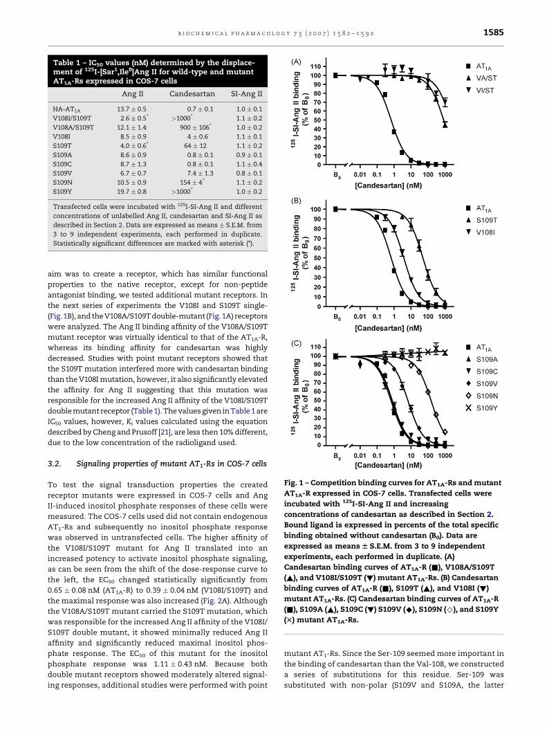
Table 1 – IC50 values (nM) determined by the displace-ment of 125I-[Sar1,Ile8]Ang II for wild-type and mutantAT1A-Rs expressed in COS-7 cells
Ang II Candesartan SI-Ang II
HA-AT1A 13.7 � 0.5 0.7 � 0.1 1.0 � 0.1
V108I/S109T 2.6 � 0.5* >1000* 1.1 � 0.2
V108A/S109T 12.1 � 1.4 900 � 106* 1.0 � 0.2
V108I 8.5 � 0.9 4 � 0.6 1.1 � 0.1
S109T 4.0 � 0.6* 64 � 12 1.1 � 0.2
S109A 8.6 � 0.9 0.8 � 0.1 0.9 � 0.1
S109C 8.7 � 1.3 0.8 � 0.1 1.1 � 0.4
S109V 6.7 � 0.7 7.4 � 1.3 0.8 � 0.1
S109N 10.5 � 0.9 154 � 4* 1.1 � 0.2
S109Y 19.7 � 0.8 >1000* 1.0 � 0.2
Transfected cells were incubated with 125I-SI-Ang II and different
concentrations of unlabelled Ang II, candesartan and SI-Ang II as
described in Section 2. Data are expressed as means � S.E.M. from
3 to 9 independent experiments, each performed in duplicate.
Statistically significant differences are marked with asterisk (*).
Fig. 1 – Competition binding curves for AT1A-Rs and mutant
AT1A-R expressed in COS-7 cells. Transfected cells were
incubated with 125I-SI-Ang II and increasing
concentrations of candesartan as described in Section 2.
Bound ligand is expressed in percents of the total specific
binding obtained without candesartan (B0). Data are
expressed as means W S.E.M. from 3 to 9 independent
experiments, each performed in duplicate. (A)
Candesartan binding curves of AT1A-R (&), V108A/S109T
(~), and V108I/S109T (!) mutant AT1A-Rs. (B) Candesartan
binding curves of AT1A-R (&), S109T (~), and V108I (!)
mutant AT1A-Rs. (C) Candesartan binding curves of AT1A-R
(&), S109A (~), S109C (!) S109V (^), S109N (^), and S109Y
(T) mutant AT1A-Rs.
b i o c h e m i c a l p h a r m a c o l o g y 7 3 ( 2 0 0 7 ) 1 5 8 2 – 1 5 9 2 1585
aim was to create a receptor, which has similar functional
properties to the native receptor, except for non-peptide
antagonist binding, we tested additional mutant receptors. In
the next series of experiments the V108I and S109T single-
(Fig. 1B), and the V108A/S109T double-mutant (Fig. 1A) receptors
were analyzed. The Ang II binding affinity of the V108A/S109T
mutant receptor was virtually identical to that of the AT1A-R,
whereas its binding affinity for candesartan was highly
decreased. Studies with point mutant receptors showed that
the S109T mutation interfered more with candesartan binding
than the V108I mutation, however, it also significantly elevated
the affinity for Ang II suggesting that this mutation was
responsible for the increased Ang II affinity of the V108I/S109T
double mutant receptor (Table 1). The values given in Table 1 are
IC50 values, however, Ki values calculated using the equation
described by Cheng and Prusoff [21], are less then 10% different,
due to the low concentration of the radioligand used.
3.2. Signaling properties of mutant AT1-Rs in COS-7 cells
To test the signal transduction properties the created
receptor mutants were expressed in COS-7 cells and Ang
II-induced inositol phosphate responses of these cells were
measured. The COS-7 cells used did not contain endogenous
AT1-Rs and subsequently no inositol phosphate response
was observed in untransfected cells. The higher affinity of
the V108I/S109T mutant for Ang II translated into an
increased potency to activate inositol phosphate signaling,
as can be seen from the shift of the dose-response curve to
the left, the EC50 changed statistically significantly from
0.65 � 0.08 nM (AT1A-R) to 0.39 � 0.04 nM (V108I/S109T) and
the maximal response was also increased (Fig. 2A). Although
the V108A/S109T mutant carried the S109T mutation, which
was responsible for the increased Ang II affinity of the V108I/
S109T double mutant, it showed minimally reduced Ang II
affinity and significantly reduced maximal inositol phos-
phate response. The EC50 of this mutant for the inositol
phosphate response was 1.11 � 0.43 nM. Because both
double mutant receptors showed moderately altered signal-
ing responses, additional studies were performed with point
mutant AT1-Rs. Since the Ser-109 seemed more important in
the binding of candesartan than the Val-108, we constructed
a series of substitutions for this residue. Ser-109 was
substituted with non-polar (S109V and S109A, the latter

Fig. 2 – Inositol phosphate responses of AT1A-Rs and
mutant AT1A-R. Transfected COS-7 cells were prelabeled
with [3H]inositol. The cells were pretreated with LiCl and
incubated in the presence of various concentrations of Ang
II as described in Section 2. Data are shown as fold
increase over the unstimulated cells. All data are shown as
means W S.E.M. from three independent experiments,
each performed in duplicate. (A) Inositol phosphate
response of AT1A-R (&), V108I/S109T (~), and V108A/
S109T (!) mutant AT1A-Rs. (B) Inositol phosphate
response of AT1A-R (&) and S109Y (!) mutant AT1A-Rs.
Fig. 3 – Inositol phosphate responses of AT1A-Rs and
mutant AT1A-R in the presence of candesartan.
Transfected COS-7 cells were prelabeled with [3H]inositol.
The cells were pretreated with LiCl and candesartan for
30 min and incubated in the presence of 10 nM Ang II (AII)
as described in Section 2. Data are shown as fold increase
over the unstimulated cells. Candesartan (CS) (300 nM) on
its own did not cause any significant change in the inositol
phosphate response. All data are shown as
means W S.E.M. from three independent experiments,
each performed in duplicate.
b i o c h e m i c a l p h a r m a c o l o g y 7 3 ( 2 0 0 7 ) 1 5 8 2 – 1 5 9 21586
mutant was previously published [16]) and polar (S109N,
S109C and S109Y) residues. Fig. 1C and Table 1 shows the
binding characteristics of these constructs. The S109A,
S109V and S109C mutants did not differ considerably form
the AT1A-R in their candesartan affinities, the S109N mutant
showed a marked decrease in binding this non-peptide
antagonist, and S109Y mutants displayed a remarkable
resistance to the compound, showing no displacement of
labeled SI-Ang II even at micromolar concentration of
candesartan. Binding of the peptide antagonist/partial
agonist SI-Ang II was not affected in any of the mutant
receptors. When tested for inositol phosphate generation,
the S109Y (Fig. 2B) and S109N (data not shown) mutants
did not differ significantly from the AT1A-R in their maximal
response and EC50 values, the latter being 0.67 � 0.1 nM
for the S109Y and 0.75 � 0.06 nM for the S109N mutant
receptor.
The effect of candesartan on the inositol phosphate
response of the AT1A-R, and the V108I/S109T, V108A/S109T,
S109N and S109Y mutant receptors is shown in Fig. 3. While
the response of the AT1A-R to 10 nM of Ang II is completely
abolished after preincubation with 30 nM of candesartan,
only the S109N mutant receptor showed reduced inositol
phosphate responses, whereas no major impairment of the
signaling of V108I/S109T and V108A/S109T mutant receptors
were detected, and the S109Y mutant completely retained
its signaling ability even in the presence of 300 nM
candesartan.
Even single amino acid mutations can alter the whole
conformation of a GPCR, creating functionally inactive or,
conversely, constitutively active receptor mutants, such as
the N111G mutant [18,22,23]. A very sensitive test for the
detection of constitutive activity in mutant receptors is their
sensitivity for stimulation with peptide antagonists, such as
SI-Ang II, which act in several test system as weak partial
agonists of the wild-type receptor, but can act as full agonists
of constitutively active receptors. We tested this possibility by
incubating the mutants as seen in Fig. 3 with 1000 nM of SI-
Ang II, which was used to reveal constitutive active properties
of the D125A receptor mutant [6]. However, SI-Ang II caused
no detectable activation of the S109Y mutant receptor (data
not shown), which, in agreement with the normal agonist
affinity of the receptor, shows that S109Y is not constitutively
active.
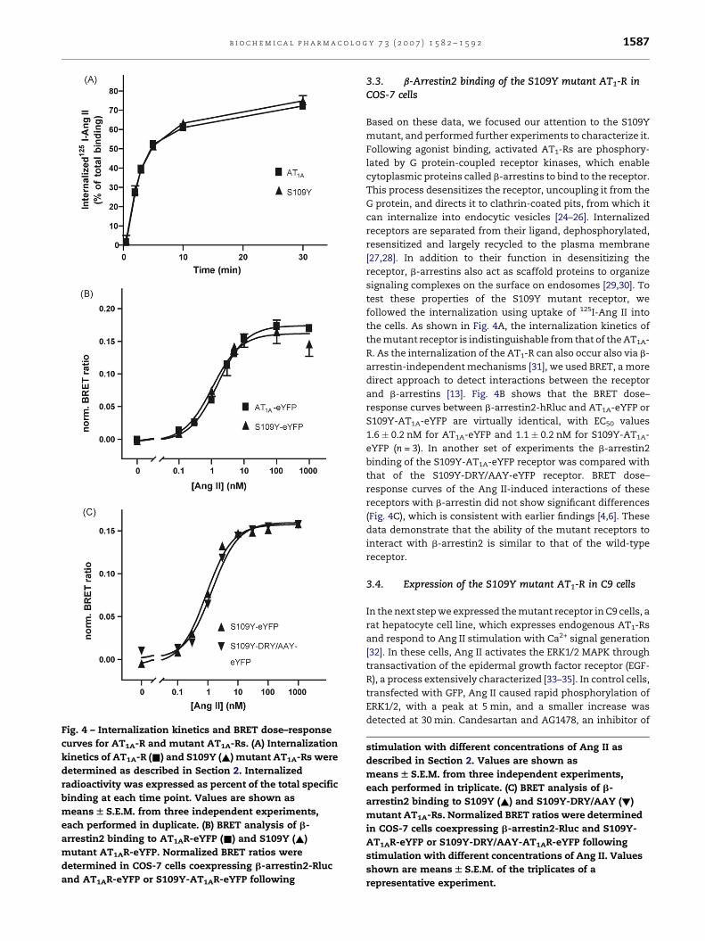
Fig. 4 – Internalization kinetics and BRET dose–response
curves for AT1A-R and mutant AT1A-Rs. (A) Internalization
kinetics of AT1A-R (&) and S109Y (~) mutant AT1A-Rs were
determined as described in Section 2. Internalized
radioactivity was expressed as percent of the total specific
binding at each time point. Values are shown as
means W S.E.M. from three independent experiments,
each performed in duplicate. (B) BRET analysis of b-
arrestin2 binding to AT1AR-eYFP (&) and S109Y (~)
mutant AT1AR-eYFP. Normalized BRET ratios were
determined in COS-7 cells coexpressing b-arrestin2-Rluc
and AT1AR-eYFP or S109Y-AT1AR-eYFP following
b i o c h e m i c a l p h a r m a c o l o g y 7 3 ( 2 0 0 7 ) 1 5 8 2 – 1 5 9 2 1587
3.3. b-Arrestin2 binding of the S109Y mutant AT1-R inCOS-7 cells
Based on these data, we focused our attention to the S109Y
mutant, and performed further experiments to characterize it.
Following agonist binding, activated AT1-Rs are phosphory-
lated by G protein-coupled receptor kinases, which enable
cytoplasmic proteins called b-arrestins to bind to the receptor.
This process desensitizes the receptor, uncoupling it from the
G protein, and directs it to clathrin-coated pits, from which it
can internalize into endocytic vesicles [24–26]. Internalized
receptors are separated from their ligand, dephosphorylated,
resensitized and largely recycled to the plasma membrane
[27,28]. In addition to their function in desensitizing the
receptor, b-arrestins also act as scaffold proteins to organize
signaling complexes on the surface on endosomes [29,30]. To
test these properties of the S109Y mutant receptor, we
followed the internalization using uptake of 125I-Ang II into
the cells. As shown in Fig. 4A, the internalization kinetics of
the mutant receptor is indistinguishable from that of the AT1A-
R. As the internalization of the AT1-R can also occur also via b-
arrestin-independent mechanisms [31], we used BRET, a more
direct approach to detect interactions between the receptor
and b-arrestins [13]. Fig. 4B shows that the BRET dose–
response curves between b-arrestin2-hRluc and AT1A-eYFP or
S109Y-AT1A-eYFP are virtually identical, with EC50 values
1.6 � 0.2 nM for AT1A-eYFP and 1.1 � 0.2 nM for S109Y-AT1A-
eYFP (n = 3). In another set of experiments the b-arrestin2
binding of the S109Y-AT1A-eYFP receptor was compared with
that of the S109Y-DRY/AAY-eYFP receptor. BRET dose–
response curves of the Ang II-induced interactions of these
receptors with b-arrestin did not show significant differences
(Fig. 4C), which is consistent with earlier findings [4,6]. These
data demonstrate that the ability of the mutant receptors to
interact with b-arrestin2 is similar to that of the wild-type
receptor.
3.4. Expression of the S109Y mutant AT1-R in C9 cells
In the next step we expressed the mutant receptor in C9 cells, a
rat hepatocyte cell line, which expresses endogenous AT1-Rs
and respond to Ang II stimulation with Ca2+ signal generation
[32]. In these cells, Ang II activates the ERK1/2 MAPK through
transactivation of the epidermal growth factor receptor (EGF-
R), a process extensively characterized [33–35]. In control cells,
transfected with GFP, Ang II caused rapid phosphorylation of
ERK1/2, with a peak at 5 min, and a smaller increase was
detected at 30 min. Candesartan and AG1478, an inhibitor of
stimulation with different concentrations of Ang II as
described in Section 2. Values are shown as
means W S.E.M. from three independent experiments,
each performed in triplicate. (C) BRET analysis of b-
arrestin2 binding to S109Y (~) and S109Y-DRY/AAY (!)
mutant AT1A-Rs. Normalized BRET ratios were determined
in COS-7 cells coexpressing b-arrestin2-Rluc and S109Y-
AT1AR-eYFP or S109Y-DRY/AAY-AT1AR-eYFP following
stimulation with different concentrations of Ang II. Values
shown are means W S.E.M. of the triplicates of a
representative experiment.

Fig. 5 – ERK1/2 activation transfected C9 cells. (A) Ang II
activates ERK1/2 through an AT1-R and EGF-R dependent
way in C9 cells. In C9 cells transfected with GFP as a
control Ang II (10 nM) caused a rapid phosphorylation of
ERK1/2 with a peak at 5 min and decreasing at 30 min,
which could be prevented by preincubation with either
candesartan (100 nM) or AG1478 (1 mM). (B) The S109Y
mutant AT1A-R expressed in C9 cells activates ERK1/2 in
the same EGF-R dependent manner as the endogenous
receptor. In C9 cells transfected with the S109Y mutant
AT1A-R Ang II (10 nM) activated ERK1/2 in the presence of
the AT1-R blocker candesartan (100 nM). Preincubation
with AG1478 (1 mM) prevented this activation. These
pictures are representative of at least 10 independent
experiments.
b i o c h e m i c a l p h a r m a c o l o g y 7 3 ( 2 0 0 7 ) 1 5 8 2 – 1 5 9 21588
EGF-R, completely eliminated the ERK activation mediated by
endogenous receptors (Fig. 5A). When transfected with the
S109Y mutant receptor, ERK activation occurred even in the
presence of candesartan with similar kinetics to the wild-type
receptor and it could be blocked by AG1478, which suggests
that it activated the same mechanism (Fig. 5B).
AT1-R mutants incapable of activating G proteins have been
shown to activate ERK1/2 through a b-arrestin2-dependent
pathway [36,4], but these experiments were performed in cells
with no endogenous AT1-R. To test for the existence of G
protein-independent ERK1/2 activation in C9 cells, the D125A/
R126A mutation, which disrupts the highly conserved DRY
motif in TM III proximal to the 2nd intracellular loop required
for G protein activation, was combined with the S109Y
mutation. These constructs were expressed in C9 cells. The
surface expression levels of the S109Y and S109Y-DRY/AAY
receptors in C9 cells (measured in the presence of 300 nM
candesartan to block endogenous receptors) were
287 � 26 fmol/mg protein and 306 � 29 fmol/mg protein,
whereas the expression of the endogenous receptors was
409 � 35 fmol/mg protein in the same set of experiments
(n = 3).
To demonstrate that the S109Y-DRY/AAY mutant receptor
does not activate G proteins in C9 cells, we measured
intracellular calcium levels in response to Ang II in the
presence or absence of candesartan in C9 cells. Our data shows
that while in non-transfected cells the response to Ang II
(Fig. 6A) is abolished in the presence of candesartan (Fig. 6B),
cells transfected with the S109Y mutant receptor retain their
ability to elicit a Ca2+-signal (Fig. 6C), whereas the cells
expressing the S109Y-DRY/AAY mutant receptor does not
activate this signaling pathway (Fig. 6D). The smaller Ca2+
signal of the expressed receptor (Fig. 6C) versus the endogen-
ous receptor (Fig. 6A) is due to the incomplete transfection of
the cells. Visualization of GFP-tagged b-arrestin2 in trans-
fected C9 cells demonstrated that this S109Y-DRY/AAY
mutant AT1-R was able to mediate translocation of b-arrestin2
to the S109Y-DRY/AAY mutant receptor, similar to the S109Y
mutant receptor (Fig. 7B). In candesartan-treated cells, the
addition of Rhod-Ang II caused the previously cytoplasmic b-
arrestin2-eGFP (Fig. 7A) to accumulate on the plasma
membrane, and following that both molecules internalized
into and colocalized in endocytic vesicles. The S109Y-DRY/
AAY mutant receptor was also able to induce ERK1/2
phosphorylation (Fig. 8A), with different kinetic properties
from the wild-type receptor. The ERK activation elicited by this
mutant was lesser in magnitude, and showed a steady
elevation even at 30 min (Fig. 8B), consistent with the earlier
observations on the kinetics of the G protein-independent ERK
activation [36].
4. Discussion
Structure–function studies in eukaryotic expression systems
contributed greatly to our current understanding of the signal
transduction pathways of angiotensin receptors and other
GPCRs. However, signaling mechanisms of GPCRs, such as
activation of MAP kinases, are highly cell specific. For example
in many tissues, including vascular smooth muscle cells,
stimulation by Ang II leads to the transactivation of growth
factor receptors such as EGF, PDGF and IGF receptors [1,37,2]
and the subsequent activation of MAP kinases; whereas in
other cell types (e.g. HEK-293 or H295R cells), protein kinase C
(PKC) activation directly leads to the activation of ERK [2,35].
While these mechanisms both require intact coupling of the
receptor to G proteins, a more recently described pathway
shows that just as the internalization of the receptor is
independent of G protein activation [11,38], b-arrestins can
mediate ERK activation independently from G protein cou-
pling [30], and in some cell types, this ERK activation also
involves EGF receptor transactivation [38,39]. However, the
physiologically relevant signaling mechanisms in the target
tissues of hormones and other mediators are difficult to
analyze using expression of mutant receptors, due to the
presence of endogenous receptors. This is a particularly
interesting problem in case of the AT1-R, since its signaling
involves G protein-dependent and -independent pathways,
including signal transduction pathways characteristic of
GPCRs, growth factor and cytokine receptors [2].
To study the mechanism of Ang II-induced MAP kinase
activation in cells expressing native angiotensin receptors, we
have developed a mutant AT1-R, which is deficient in
candesartan binding, whereas completely normal in its other
pharmacological properties and signal transduction charac-
teristics. Positions 108 and 109 were selected to eliminate
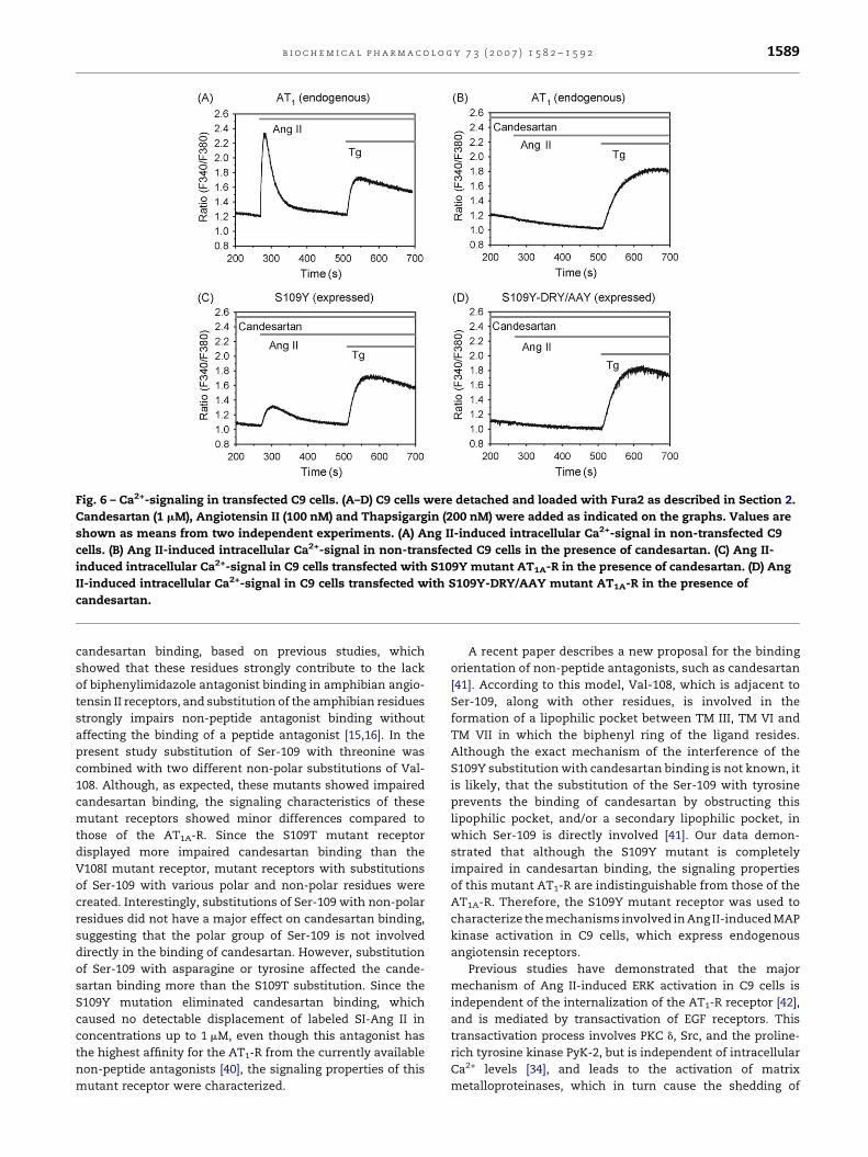
Fig. 6 – Ca2+-signaling in transfected C9 cells. (A–D) C9 cells were detached and loaded with Fura2 as described in Section 2.
Candesartan (1 mM), Angiotensin II (100 nM) and Thapsigargin (200 nM) were added as indicated on the graphs. Values are
shown as means from two independent experiments. (A) Ang II-induced intracellular Ca2+-signal in non-transfected C9
cells. (B) Ang II-induced intracellular Ca2+-signal in non-transfected C9 cells in the presence of candesartan. (C) Ang II-
induced intracellular Ca2+-signal in C9 cells transfected with S109Y mutant AT1A-R in the presence of candesartan. (D) Ang
II-induced intracellular Ca2+-signal in C9 cells transfected with S109Y-DRY/AAY mutant AT1A-R in the presence of
candesartan.
b i o c h e m i c a l p h a r m a c o l o g y 7 3 ( 2 0 0 7 ) 1 5 8 2 – 1 5 9 2 1589
candesartan binding, based on previous studies, which
showed that these residues strongly contribute to the lack
of biphenylimidazole antagonist binding in amphibian angio-
tensin II receptors, and substitution of the amphibian residues
strongly impairs non-peptide antagonist binding without
affecting the binding of a peptide antagonist [15,16]. In the
present study substitution of Ser-109 with threonine was
combined with two different non-polar substitutions of Val-
108. Although, as expected, these mutants showed impaired
candesartan binding, the signaling characteristics of these
mutant receptors showed minor differences compared to
those of the AT1A-R. Since the S109T mutant receptor
displayed more impaired candesartan binding than the
V108I mutant receptor, mutant receptors with substitutions
of Ser-109 with various polar and non-polar residues were
created. Interestingly, substitutions of Ser-109 with non-polar
residues did not have a major effect on candesartan binding,
suggesting that the polar group of Ser-109 is not involved
directly in the binding of candesartan. However, substitution
of Ser-109 with asparagine or tyrosine affected the cande-
sartan binding more than the S109T substitution. Since the
S109Y mutation eliminated candesartan binding, which
caused no detectable displacement of labeled SI-Ang II in
concentrations up to 1 mM, even though this antagonist has
the highest affinity for the AT1-R from the currently available
non-peptide antagonists [40], the signaling properties of this
mutant receptor were characterized.
A recent paper describes a new proposal for the binding
orientation of non-peptide antagonists, such as candesartan
[41]. According to this model, Val-108, which is adjacent to
Ser-109, along with other residues, is involved in the
formation of a lipophilic pocket between TM III, TM VI and
TM VII in which the biphenyl ring of the ligand resides.
Although the exact mechanism of the interference of the
S109Y substitution with candesartan binding is not known, it
is likely, that the substitution of the Ser-109 with tyrosine
prevents the binding of candesartan by obstructing this
lipophilic pocket, and/or a secondary lipophilic pocket, in
which Ser-109 is directly involved [41]. Our data demon-
strated that although the S109Y mutant is completely
impaired in candesartan binding, the signaling properties
of this mutant AT1-R are indistinguishable from those of the
AT1A-R. Therefore, the S109Y mutant receptor was used to
characterize the mechanisms involved in Ang II-induced MAP
kinase activation in C9 cells, which express endogenous
angiotensin receptors.
Previous studies have demonstrated that the major
mechanism of Ang II-induced ERK activation in C9 cells is
independent of the internalization of the AT1-R receptor [42],
and is mediated by transactivation of EGF receptors. This
transactivation process involves PKC d, Src, and the proline-
rich tyrosine kinase PyK-2, but is independent of intracellular
Ca2+ levels [34], and leads to the activation of matrix
metalloproteinases, which in turn cause the shedding of
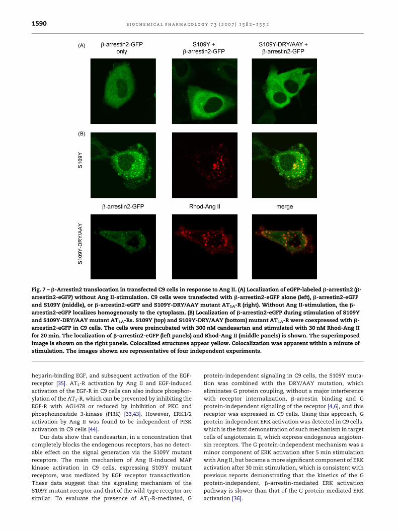
Fig. 7 – b-Arrestin2 translocation in transfected C9 cells in response to Ang II. (A) Localization of eGFP-labeled b-arrestin2 (b-
arrestin2-eGFP) without Ang II-stimulation. C9 cells were transfected with b-arrestin2-eGFP alone (left), b-arrestin2-eGFP
and S109Y (middle), or b-arrestin2-eGFP and S109Y-DRY/AAY mutant AT1A-R (right). Without Ang II-stimulation, the b-
arrestin2-eGFP localizes homogenously to the cytoplasm. (B) Localization of b-arrestin2-eGFP during stimulation of S109Y
and S109Y-DRY/AAY mutant AT1A-Rs. S109Y (top) and S109Y-DRY/AAY (bottom) mutant AT1A-R were coexpressed with b-
arrestin2-eGFP in C9 cells. The cells were preincubated with 300 nM candesartan and stimulated with 30 nM Rhod-Ang II
for 20 min. The localization of b-arrestin2-eGFP (left panels) and Rhod-Ang II (middle panels) is shown. The superimposed
image is shown on the right panels. Colocalized structures appear yellow. Colocalization was apparent within a minute of
stimulation. The images shown are representative of four independent experiments.
b i o c h e m i c a l p h a r m a c o l o g y 7 3 ( 2 0 0 7 ) 1 5 8 2 – 1 5 9 21590
heparin-binding EGF, and subsequent activation of the EGF-
receptor [35]. AT1-R activation by Ang II and EGF-induced
activation of the EGF-R in C9 cells can also induce phosphor-
ylation of the AT1-R, which can be prevented by inhibiting the
EGF-R with AG1478 or reduced by inhibition of PKC and
phosphoinositide 3-kinase (PI3K) [33,43]. However, ERK1/2
activation by Ang II was found to be independent of PI3K
activation in C9 cells [44].
Our data show that candesartan, in a concentration that
completely blocks the endogenous receptors, has no detect-
able effect on the signal generation via the S109Y mutant
receptors. The main mechanism of Ang II-induced MAP
kinase activation in C9 cells, expressing S109Y mutant
receptors, was mediated by EGF receptor transactivation.
These data suggest that the signaling mechanism of the
S109Y mutant receptor and that of the wild-type receptor are
similar. To evaluate the presence of AT1-R-mediated, G
protein-independent signaling in C9 cells, the S109Y muta-
tion was combined with the DRY/AAY mutation, which
eliminates G protein coupling, without a major interference
with receptor internalization, b-arrestin binding and G
protein-independent signaling of the receptor [4,6], and this
receptor was expressed in C9 cells. Using this approach, G
protein-independent ERK activation was detected in C9 cells,
which is the first demonstration of such mechanism in target
cells of angiotensin II, which express endogenous angioten-
sin receptors. The G protein-independent mechanism was a
minor component of ERK activation after 5 min stimulation
with Ang II, but became a more significant component of ERK
activation after 30 min stimulation, which is consistent with
previous reports demonstrating that the kinetics of the G
protein-independent, b-arrestin-mediated ERK activation
pathway is slower than that of the G protein-mediated ERK
activation [36].
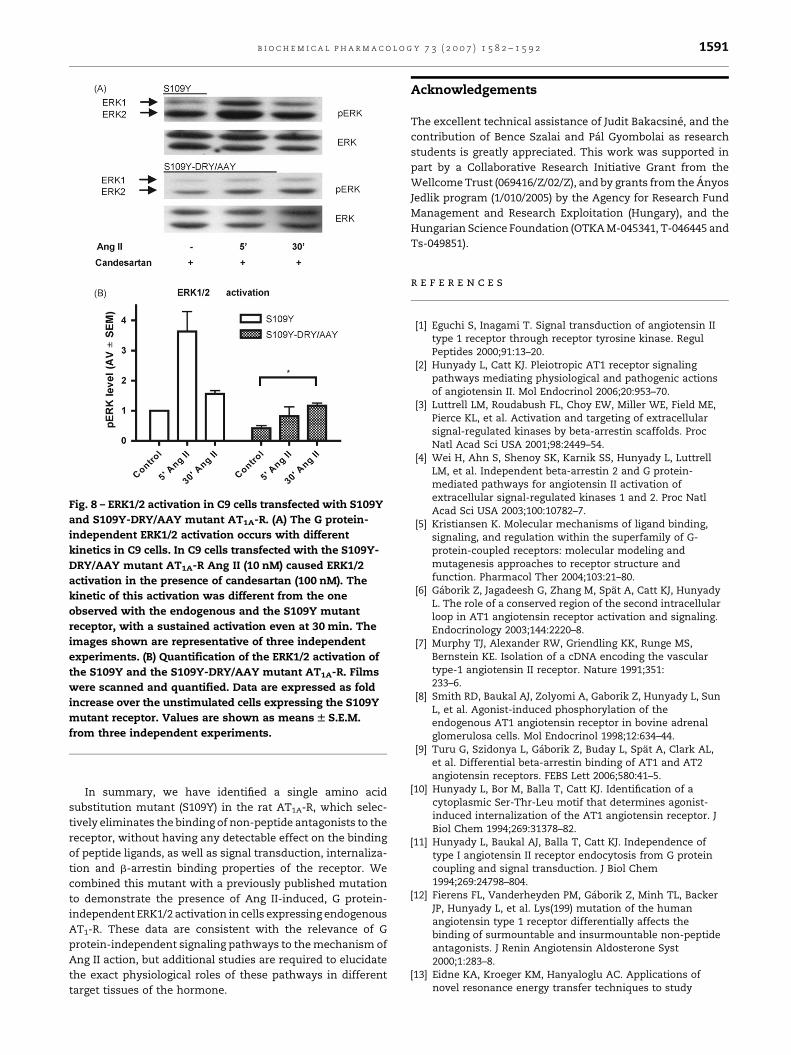
Fig. 8 – ERK1/2 activation in C9 cells transfected with S109Y
and S109Y-DRY/AAY mutant AT1A-R. (A) The G protein-
independent ERK1/2 activation occurs with different
kinetics in C9 cells. In C9 cells transfected with the S109Y-
DRY/AAY mutant AT1A-R Ang II (10 nM) caused ERK1/2
activation in the presence of candesartan (100 nM). The
kinetic of this activation was different from the one
observed with the endogenous and the S109Y mutant
receptor, with a sustained activation even at 30 min. The
images shown are representative of three independent
experiments. (B) Quantification of the ERK1/2 activation of
the S109Y and the S109Y-DRY/AAY mutant AT1A-R. Films
were scanned and quantified. Data are expressed as fold
increase over the unstimulated cells expressing the S109Y
mutant receptor. Values are shown as means W S.E.M.
from three independent experiments.
b i o c h e m i c a l p h a r m a c o l o g y 7 3 ( 2 0 0 7 ) 1 5 8 2 – 1 5 9 2 1591
In summary, we have identified a single amino acid
substitution mutant (S109Y) in the rat AT1A-R, which selec-
tively eliminates the binding of non-peptide antagonists to the
receptor, without having any detectable effect on the binding
of peptide ligands, as well as signal transduction, internaliza-
tion and b-arrestin binding properties of the receptor. We
combined this mutant with a previously published mutation
to demonstrate the presence of Ang II-induced, G protein-
independent ERK1/2 activation in cells expressing endogenous
AT1-R. These data are consistent with the relevance of G
protein-independent signaling pathways to the mechanism of
Ang II action, but additional studies are required to elucidate
the exact physiological roles of these pathways in different
target tissues of the hormone.
Acknowledgements
The excellent technical assistance of Judit Bakacsine, and the
contribution of Bence Szalai and Pal Gyombolai as research
students is greatly appreciated. This work was supported in
part by a Collaborative Research Initiative Grant from the
Wellcome Trust (069416/Z/02/Z), and by grants from the Anyos
Jedlik program (1/010/2005) by the Agency for Research Fund
Management and Research Exploitation (Hungary), and the
Hungarian Science Foundation (OTKA M-045341, T-046445 and
Ts-049851).
r e f e r e n c e s
[1] Eguchi S, Inagami T. Signal transduction of angiotensin IItype 1 receptor through receptor tyrosine kinase. RegulPeptides 2000;91:13–20.
[2] Hunyady L, Catt KJ. Pleiotropic AT1 receptor signalingpathways mediating physiological and pathogenic actionsof angiotensin II. Mol Endocrinol 2006;20:953–70.
[3] Luttrell LM, Roudabush FL, Choy EW, Miller WE, Field ME,Pierce KL, et al. Activation and targeting of extracellularsignal-regulated kinases by beta-arrestin scaffolds. ProcNatl Acad Sci USA 2001;98:2449–54.
[4] Wei H, Ahn S, Shenoy SK, Karnik SS, Hunyady L, LuttrellLM, et al. Independent beta-arrestin 2 and G protein-mediated pathways for angiotensin II activation ofextracellular signal-regulated kinases 1 and 2. Proc NatlAcad Sci USA 2003;100:10782–7.
[5] Kristiansen K. Molecular mechanisms of ligand binding,signaling, and regulation within the superfamily of G-protein-coupled receptors: molecular modeling andmutagenesis approaches to receptor structure andfunction. Pharmacol Ther 2004;103:21–80.
[6] Gaborik Z, Jagadeesh G, Zhang M, Spat A, Catt KJ, HunyadyL. The role of a conserved region of the second intracellularloop in AT1 angiotensin receptor activation and signaling.Endocrinology 2003;144:2220–8.
[7] Murphy TJ, Alexander RW, Griendling KK, Runge MS,Bernstein KE. Isolation of a cDNA encoding the vasculartype-1 angiotensin II receptor. Nature 1991;351:233–6.
[8] Smith RD, Baukal AJ, Zolyomi A, Gaborik Z, Hunyady L, SunL, et al. Agonist-induced phosphorylation of theendogenous AT1 angiotensin receptor in bovine adrenalglomerulosa cells. Mol Endocrinol 1998;12:634–44.
[9] Turu G, Szidonya L, Gaborik Z, Buday L, Spat A, Clark AL,et al. Differential beta-arrestin binding of AT1 and AT2angiotensin receptors. FEBS Lett 2006;580:41–5.
[10] Hunyady L, Bor M, Balla T, Catt KJ. Identification of acytoplasmic Ser-Thr-Leu motif that determines agonist-induced internalization of the AT1 angiotensin receptor. JBiol Chem 1994;269:31378–82.
[11] Hunyady L, Baukal AJ, Balla T, Catt KJ. Independence oftype I angiotensin II receptor endocytosis from G proteincoupling and signal transduction. J Biol Chem1994;269:24798–804.
[12] Fierens FL, Vanderheyden PM, Gaborik Z, Minh TL, BackerJP, Hunyady L, et al. Lys(199) mutation of the humanangiotensin type 1 receptor differentially affects thebinding of surmountable and insurmountable non-peptideantagonists. J Renin Angiotensin Aldosterone Syst2000;1:283–8.
[13] Eidne KA, Kroeger KM, Hanyaloglu AC. Applications ofnovel resonance energy transfer techniques to study

b i o c h e m i c a l p h a r m a c o l o g y 7 3 ( 2 0 0 7 ) 1 5 8 2 – 1 5 9 21592
dynamic hormone receptor interactions in living cells.Trends Endocrinol Metab 2002;13:415–21.
[14] de Gasparo M, Catt KJ, Inagami T, Wright JW, Unger T.International union of pharmacology. XXIII. Theangiotensin II receptors. Pharmacol Rev 2000;52:415–72.
[15] Ji H, Leung M, Zhang Y, Catt KJ, Sandberg K. Differentialstructural requirements for specific binding of non-peptideand peptide antagonists to the AT1 angiotensin receptor.Identification of amino acid residues that determinebinding of the antihypertensive drug losartan. J Biol Chem1994;269:16533–6.
[16] Ji H, Zheng W, Zhang Y, Catt KJ, Sandberg K. Genetictransfer of a non-peptide antagonist binding site to apreviously unresponsive angiotensin receptor. Proc NatlAcad Sci USA 1995;92:9240–4.
[17] Hunyady L, Balla T, Catt KJ. The ligand binding site of theangiotensin AT1 receptor. Trends Pharmacol Sci1996;17:135–40.
[18] Auger-Messier M, Clement M, Lanctot PM, Leclerc PC, LeducR, Escher E, et al. The constitutively active N111G-AT1receptor for angiotensin II maintains a high affinityconformation despite being uncoupled from its cognate Gprotein Gq/11alpha. Endocrinology 2003;144:5277–84.
[19] Miura S, Feng YH, Husain A, Karnik SS. Role of aromaticityof agonist switches of angiotensin II in the activation of theAT1 receptor. J Biol Chem 1999;274:7103–10.
[20] Parnot C, Bardin S, Miserey-Lenkei S, Guedin D, Corvol P,Clauser E. Systematic identification of mutations thatconstitutively activate the angiotensin II type 1A receptorby screening a randomly mutated cDNA library with anoriginal pharmacological bioassay. Proc Natl Acad Sci USA2000;97:7615–20.
[21] Cheng Y, Prusoff WH. Relationship between the inhibitionconstant (K1) and the concentration of inhibitor whichcauses 50 percent inhibition (I50) of an enzymatic reaction.Biochem Pharmacol 1973;22:3099–108.
[22] Groblewski T, Maigret B, Larguier R, Lombard C, BonnafousJC, Marie J. Mutation of Asn111 in the third transmembranedomain of the AT1A angiotensin II receptor induces itsconstitutive activation. J Biol Chem 1997;272:1822–6.
[23] Noda K, Feng YH, Liu XP, Saad Y, Husain A, Karnik SS. Theactive state of the AT1 angiotensin receptor is generated byangiotensin II induction. Biochemistry 1996;35:16435–42.
[24] Kohout TA, Lin FS, Perry SJ, Conner DA, Lefkowitz RJ. beta-Arrestin 1 and 2 differentially regulate heptahelicalreceptor signaling and trafficking. Proc Natl Acad Sci USA2001;98:1601–6.
[25] Lefkowitz RJ. G protein-coupled receptors. III. New roles forreceptor kinases and beta-arrestins in receptor signalingand desensitisation. J Biol Chem 1998;273:18677–80.
[26] Gaborik Z, Szaszak M, Szidonya L, Balla B, Paku S, Catt KJ,et al. Beta-arrestin- and dynamin-dependent endocytosisof the AT1 angiotensin receptor. Mol Pharmacol2001;59:239–47.
[27] Ferguson SS. Evolving concepts in G protein-coupledreceptor endocytosis: the role in receptor desensitizationand signaling. Pharmacol Rev 2001;53:1–24.
[28] Gaborik Z, Hunyady L. Intracellular trafficking of hormonereceptors. Trends Endocrinol Metab 2004;15:286–93.
[29] Luttrell LM, Lefkowitz RJ. The role of beta-arrestins in thetermination and transduction of G-protein-coupledreceptor signals. J Cell Sci 2002;115:455–65.
[30] Lefkowitz RJ, Shenoy SK. Transduction of receptor signalsby beta-arrestins. Science 2005;308:512–7.
[31] Zhang J, Ferguson SS, Barak LS, Menard L, Caron MG.Dynamin and beta-arrestin reveal distinct mechanisms forG protein-coupled receptor internalization. J Biol Chem1996;271:18302–5.
[32] Garcia-Sainz JA, Garcia-Caballero A, Gonzalez-Espinosa C.Angiotensin AT1 receptors in Clone 9 rat liver cells: Ca2+
signaling and c-fos expression. Eur J Pharmacol1998;362:235–43.
[33] Olivares-Reyes JA, Shah BH, Hernandez-Aranda J, Garcia-Caballero A, Farshori MP, Garcia-Sainz JA, et al. Agonist-induced interactions between angiotensin AT1 andepidermal growth factor receptors. Mol Pharmacol2005;68:356–64.
[34] Shah BH, Catt KJ. Calcium-independent activation ofextracellularly regulated kinases 1 and 2 by angiotensin IIin hepatic C9 cells: roles of protein kinase Cdelta, Src/proline-rich tyrosine kinase 2, and epidermal growthreceptor trans-activation. Mol Pharmacol 2002;61:343–51.
[35] Shah BH, Yesilkaya A, Olivares-Reyes JA, Chen HD,Hunyady L, Catt KJ. Differential pathways of angiotensin II-induced extracellularly regulated kinase 1/2phosphorylation in specific cell types: role of heparin-binding epidermal growth factor. Mol Endocrinol2004;18:2035–48.
[36] Ahn S, Shenoy SK, Wei H, Lefkowitz RJ. Differential kineticand spatial patterns of beta-arrestin and G protein-mediated ERK activation by the angiotensin II receptor. JBiol Chem 2004;279:35518–25.
[37] Natarajan K, Berk BC. Crosstalk coregulation mechanismsof G protein-coupled receptors and receptor tyrosinekinases. Methods Mol Biol 2006;332:51–77.
[38] Feng YH, Ding Y, Ren S, Zhou L, Xu C, Karnik SS.Unconventional homologous internalization of theangiotensin II type-1 receptor induced by G-protein-independent signals. Hypertension 2005;46:419–25.
[39] Miura S, Zhang J, Matsuo Y, Saku K, Karnik SS. Activation ofextracellular signal-activated kinase by angiotensin II-induced Gq-independent epidermal growth factor receptortransactivation. Hypertens Res 2004;27:765–70.
[40] Unger T. Pharmacology of AT1-receptor blockers. BloodPressure 2001;(Suppl.):5–10.
[41] Tuccinardi T, Calderone V, Rapposelli S, Martinelli A.Proposal of a new binding orientation for non-peptide AT1antagonists: homology modeling, docking and three-dimensional quantitative structure–activity relationshipanalysis. J Med Chem 2006;49:4305–16.
[42] Shah BH, Olivares-Reyes JA, Yesilkaya A, Catt KJ.Independence of angiotensin II-induced MAP kinaseactivation from angiotensin type 1 receptor internalizationin clone 9 hepatocytes. Mol Endocrinol 2002;16:610–20.
[43] Garcia-Caballero A, Olivares-Reyes JA, Catt KJ, Garcia-SainzJA. Angiotensin AT(1) receptor phosphorylation anddesensitization in a hepatic cell line. Roles of protein kinasec and phosphoinositide 3-kinase. Mol Pharmacol2001;59:576–85.
[44] Shah BH, Neithardt A, Chu DB, Shah FB, Catt KJ. Role of EGFreceptor transactivation in phosphoinositide 3-kinase-dependent activation of MAP kinase by GPCRs. J Cell Physiol2006;206:47–57.
Copyright © 2022 FDOKUMEN