Pre-exposure to Staphylococcal enterotoxin A exacerbates the pulmonary allergic eosinophil...
7
Pre-exposure to Staphylococcal enterotoxin A exacerbates the pulmonary allergic eosinophil recruitment in rats Nadia S. Mariano a , Glaucia C. de Mello a , Tatiane Ferreira a , André Schenka a , Enilton A. Camargo a , Gilberto de Nucci a , Ivani A. DeSouza a,b , Edson Antunes a, ⁎ a Department of Pharmacology, Faculty of Medical Sciences, State University of Campinas (UNICAMP), Campinas (Sao Paulo), Brazil b Department of Biology and Physiology, Faculty of Medicine of Jundiái (FMJ), Jundiaí (São Paulo), Brazil abstract article info Article history: Received 1 July 2009 Received in revised form 25 August 2009 Accepted 24 September 2009 Keywords: Staphylococcal enterotoxins Lung inflammation Eosinophils Eotaxin Bone marrow Gram-positive Staphylococcus aureus releases classical enterotoxins which aggravates allergic airway diseases. However, little is known about the mechanisms underlying the cell influx exacerbation in asthmatic individuals under exposure to Staphylococcal enterotoxins. We therefore aimed to investigate the effects of airways exposure to Staphylococcal enterotoxin A (SEA) to pulmonary leukocyte recruitment in rats sensitized and challenged with ovalbumin (OVA). Rats were exposed to SEA at 4 h prior to OVA challenge or at 4 h post-OVA challenge. Bronchoalveolar lavage (BAL) fluid, bone marrow and lung tissue were obtained at 24 h after OVA challenge. Pre-exposure to SEA markedly enhanced the eosinophil counts in both BAL fluid and pulmonary tissue in OVA-challenged rats, whereas neutrophil and mononuclear cell counts remained unchanged. In bone marrow, pre-exposure to SEA alone significantly increased the number of eosinophils, and that was further increased in OVA-challenged rats. Exposure to SEA post-OVA challenge did not affect the number of eosinophils, neutrophils and mononuclear cells in BAL fluid. Pre-exposure to the endotoxin lipopolyssacharide (LPS) in OVA-challenged animals rather enhanced the neutrophil number in BAL fluid. In rats pre-exposed to SEA and OVA-challenged, a marked elevation in the levels of TNF-α and eotaxin (but not of IL-10) in BAL fluid was observed. The eotaxin levels increased by about of 3-fold in alveolar macrophages treated with SEA in vitro. In conclusion, airways pre- exposure to SEA causes a selective increase in eosinophil number in BAL fluid and bone marrow of OVA- challenged rats by mechanisms involving enhancement of TNF-α and eotaxin synthesis. © 2009 Elsevier B.V. All rights reserved. 1. Introduction Staphylococcus aureus is a gram-positive bacterium that produces and secretes the so-called staphylococcal enterotoxins, which are a family of structurally related heat-stable 25 to 30 kDa proteins. These enterotoxins comprise several serological types, namely the classical types A to E, and the newly characterized types G to Q [1]. Two novel genes coding for enterotoxins types S and T have been recently described [2]. Among the Staphylococcal enterotoxins, toxins A and B can be easily obtained in relatively large amounts and purity [3], and therefore has been used to a great extent in experimental investiga- tions. In addition to the well-known ability to cause food poisoning [4], Staphylococcal enterotoxins often lead to multiorgan dysfunction [5] that may be a consequence of the release of several inflammatory mediators such as cytokines (IL-1, IL-2, IL-6, IL-8, IFN-γ, TNF-α), mast cell-derived amines (histamine and 5-hydroxytriptamine), neuropep- tides (substance P), lipid mediators (PGE 2 and PAF), amongst others [6–12]. Moreover, Staphylococcal enterotoxins are considered proto- typical superantigens, which interacts with the outside of the MHC molecule and V-beta chain of T-cell-receptor without the need for internalization, processing, or presentation within the confines of a major histocompatibility complex [13]. S. aureus, which often asymptomatically colonize the upper respiratory tract, has been associated with allergic diseases. A correlation between levels of IgE antibodies to staphylococcal enter- otoxins and the severity of eosinophilic inflammation in upper airway disease has been found [14–16]. Polyvalent sensitization with Staphy- lococcal enterotoxins A and B has been associated with increased IgE reactivity leading to exacerbation of allergic responses in humans [17]. However, little is known about the mechanisms underlying such pulmonary inflammatory exacerbation in asthmatic individuals under exposure to Staphylococcal enterotoxins. An experimental study showed that mice cutaneous sensitization with Staphylococcal entero- toxin B cause allergic skin inflammation and increased serum IgE levels [18]. Later, nasal and bronchial exposure to Staphylococcal enterotoxin B was shown to enhance the allergic pulmonary eosinophilic inflamma- tion in ovalbumin (OVA)-sensitized mice, which was associated with increased mRNA expression of IL-5, IL-4, IFN-γ, IL-12 p40, eotaxin-1 and TGF-β as well as IgE titers in the serum [19]. However, no studies exist International Immunopharmacology 10 (2010) 43–49 ⁎ Corresponding author. Department of Pharmacology, Faculty of Medical Sciences, P.O. Box 6111, UNICAMP, 13084-971, Campinas, SP, Brazil. Tel.: +55 19 35219555; fax: +55 19 32892968. E-mail addresses: [email protected], [email protected] (E. Antunes). 1567-5769/$ – see front matter © 2009 Elsevier B.V. All rights reserved. doi:10.1016/j.intimp.2009.09.017 Contents lists available at ScienceDirect International Immunopharmacology journal homepage: www.elsevier.com/locate/intimp
Transcript of Pre-exposure to Staphylococcal enterotoxin A exacerbates the pulmonary allergic eosinophil...

International Immunopharmacology 10 (2010) 43–49
Contents lists available at ScienceDirect
International Immunopharmacology
j ourna l homepage: www.e lsev ie r.com/ locate / in t imp
Pre-exposure to Staphylococcal enterotoxin A exacerbates the pulmonary allergiceosinophil recruitment in rats
Nadia S. Mariano a, Glaucia C. de Mello a, Tatiane Ferreira a, André Schenka a, Enilton A. Camargo a,Gilberto de Nucci a, Ivani A. DeSouza a,b, Edson Antunes a,⁎a Department of Pharmacology, Faculty of Medical Sciences, State University of Campinas (UNICAMP), Campinas (Sao Paulo), Brazilb Department of Biology and Physiology, Faculty of Medicine of Jundiái (FMJ), Jundiaí (São Paulo), Brazil
⁎ Corresponding author. Department of PharmacologP.O. Box 6111, UNICAMP, 13084-971, Campinas, SP, Bfax: +55 19 32892968.
E-mail addresses: [email protected], antunes
1567-5769/$ – see front matter © 2009 Elsevier B.V. Aldoi:10.1016/j.intimp.2009.09.017
a b s t r a c t
a r t i c l e i n f oArticle history:Received 1 July 2009Received in revised form 25 August 2009Accepted 24 September 2009
Keywords:Staphylococcal enterotoxinsLung inflammationEosinophilsEotaxinBone marrow
Gram-positive Staphylococcus aureus releases classical enterotoxins which aggravates allergic airway diseases.However, little is known about the mechanisms underlying the cell influx exacerbation in asthmatic individualsunderexposure to Staphylococcal enterotoxins.We therefore aimed to investigate theeffects of airways exposureto Staphylococcal enterotoxinA (SEA) topulmonary leukocyte recruitment in rats sensitized and challengedwithovalbumin (OVA). Rats were exposed to SEA at 4 h prior to OVA challenge or at 4 h post-OVA challenge.Bronchoalveolar lavage (BAL) fluid, bone marrow and lung tissue were obtained at 24 h after OVA challenge.Pre-exposure to SEA markedly enhanced the eosinophil counts in both BAL fluid and pulmonary tissue inOVA-challenged rats, whereas neutrophil and mononuclear cell counts remained unchanged. In bone marrow,pre-exposure to SEA alone significantly increased the number of eosinophils, and that was further increased inOVA-challenged rats. Exposure to SEA post-OVA challenge did not affect the number of eosinophils, neutrophilsand mononuclear cells in BAL fluid. Pre-exposure to the endotoxin lipopolyssacharide (LPS) in OVA-challengedanimals rather enhanced the neutrophil number in BAL fluid. In rats pre-exposed to SEA and OVA-challenged, amarked elevation in the levels of TNF-α and eotaxin (but not of IL-10) in BAL fluid was observed. The eotaxinlevels increased by about of 3-fold in alveolar macrophages treated with SEA in vitro. In conclusion, airways pre-exposure to SEA causes a selective increase in eosinophil number in BAL fluid and bone marrow of OVA-challenged rats by mechanisms involving enhancement of TNF-α and eotaxin synthesis.
y, Faculty of Medical Sciences,razil. Tel.: +55 19 35219555;
@fcm.unicamp.br (E. Antunes).
l rights reserved.
© 2009 Elsevier B.V. All rights reserved.
1. Introduction
Staphylococcus aureus is a gram-positive bacterium that producesand secretes the so-called staphylococcal enterotoxins, which are afamily of structurally related heat-stable 25 to 30 kDa proteins. Theseenterotoxins comprise several serological types, namely the classicaltypes A to E, and the newly characterized types G to Q [1]. Two novelgenes coding for enterotoxins types S and T have been recentlydescribed [2]. Among the Staphylococcal enterotoxins, toxins A and Bcan be easily obtained in relatively large amounts and purity [3], andtherefore has been used to a great extent in experimental investiga-tions. In addition to the well-known ability to cause food poisoning[4], Staphylococcal enterotoxins often lead to multiorgan dysfunction[5] that may be a consequence of the release of several inflammatorymediators such as cytokines (IL-1, IL-2, IL-6, IL-8, IFN-γ, TNF-α), mastcell-derived amines (histamine and 5-hydroxytriptamine), neuropep-tides (substance P), lipid mediators (PGE2 and PAF), amongst others
[6–12]. Moreover, Staphylococcal enterotoxins are considered proto-typical superantigens, which interacts with the outside of the MHCmolecule and V-beta chain of T-cell-receptor without the need forinternalization, processing, or presentation within the confines of amajor histocompatibility complex [13].
S. aureus, which often asymptomatically colonize the upperrespiratory tract, has been associated with allergic diseases. Acorrelation between levels of IgE antibodies to staphylococcal enter-otoxins and the severity of eosinophilic inflammation in upper airwaydisease has been found [14–16]. Polyvalent sensitization with Staphy-lococcal enterotoxins A and B has been associated with increased IgEreactivity leading to exacerbation of allergic responses in humans [17].However, little is known about the mechanisms underlying suchpulmonary inflammatory exacerbation in asthmatic individuals underexposure to Staphylococcal enterotoxins. An experimental studyshowed that mice cutaneous sensitization with Staphylococcal entero-toxin B cause allergic skin inflammation and increased serum IgE levels[18]. Later, nasal andbronchial exposure toStaphylococcal enterotoxinBwas shown to enhance the allergic pulmonary eosinophilic inflamma-tion in ovalbumin (OVA)-sensitized mice, which was associated withincreasedmRNA expression of IL-5, IL-4, IFN-γ, IL-12 p40, eotaxin-1 andTGF-β as well as IgE titers in the serum [19]. However, no studies exist

44 N.S. Mariano et al. / International Immunopharmacology 10 (2010) 43–49
investigating the potential interactions between airways exposure toStaphylococcal enterotoxin A (SEA) and pulmonary allergic inflamma-tion in experimental animals to help in the comprehension of themechanisms underlying the exacerbation of allergic diseases byStaphylococcal enterotoxin exposure. Therefore, this study initiallytested the hypothesis that exposure to SEA enhances the airways cellrecruitment (with emphasis in eosinophil influx) in rats activelysensitized and challengedwith ovalbumin (OVA), and next investigatedthe contribution of Th-cytokines in the resulting cell infiltration. Theeffects of airways exposure to the endotoxin lipopolyssacharide (LPS),the main structural component of the outer membrane of Gram-negative bacteria, have been compared with those of SEA.
2. Material and methods
2.1. Animals
The experimental protocols were approved by the EthicalPrinciples in Animal Research adopted by the Brazilian College forAnimal Experimentation (COBEA). All experimental procedures wereapproved by the Animal Care and Use Committee of the StateUniversity of Campinas (UNICAMP). Male Wistar rats (200–250 g)were housed in temperature-controlled rooms and received waterand food ad libitum until used.
2.2. Sensitization and challenge with ovalbumin (OVA)
We actively sensitized the animals by subcutaneous injections of200 µgOVA dissolved in 0.15 ml of aluminumhydroxide (200 mg/ml in0.9% NaCl), according to our previous experience [20]. Non-sensitizedanimals were injected with aluminum hydroxide alone. Fourteen dayslater, sensitized (and non-sensitized) rats were intranasally challengedwith 1 mg of OVA (0.2 ml) or instilled with phosphate-buffered saline(PBS; 0.2 ml). Bronchoalveolar lavage (BAL) fluid, bone marrow andlung tissuewere obtained at selected times thereafter, and processed asdetailed below.
2.3. Experimental protocols
In the first series of experiments, at 4 h prior to OVA challenge (orPBS instillation), rats were pre-exposed (intranasal administration) to3 ngof SEA (SigmaChem. Co., St Louis,MO,USA) or PBS (0.2 ml). At 24 hthereafter, rats were anaesthetized, and BAL fluid obtained. In someprotocols, bonemarrowwas also obtained. The exposure time (4 h) anddose of SEA (3 ng) was based on our previous experience in rats [21].
In the second series of experiments, at 4 h after OVA challenge (or PBSinstillation), rats were intranasally exposed to SEA (3 ng) or PBS (0.2 ml).At 24 h thereafter, rats were anaesthetized, and BAL fluid obtained.
In the third series of experiments, at 4 h prior to OVA challenge (orPBS instillation), rats were pre-exposed (intranasal administration) toeither Salmonella abortus equi LPS (0.1 and 1.0 µg/rat; Sigma ChemicalCo., St Louis, MO, USA) or PBS (0.2 ml). At 24 h thereafter, rats wereanaesthetized with halothane inhalation, and BAL fluid obtained.
2.4. Bronchoalveolar lavage (BAL) fluid and leukocyte counts
The BAL fluidwas obtained at 12, 24 or 48 h after OVA challenge. Thelungs were washed by flushing with PBS solution containing heparin(20 UI/ml). Briefly, the trachea was cannulated with a polyethylene(1 mm in diameter) connected a syringe. The lungs were washed byflushing with PBS solution containing heparin (20 UI/ml). The PBSbuffer was instilled through a tracheal cannula as one 10-ml aliquotfollowed by three 5-ml aliquots. The first 10 ml aliquot was centrifuged(450×g for 10 min at 20 °C), and supernatant recovered and stored at−80 °C for measurement of inflammatory mediators. The three 5-mlaliquots were pooled and centrifuged (450×g for 10 min at 20 °C). The
cell pellet obtained from all aliquots (the 10-ml together with those ofthe 5-ml aliquots) were combined and resuspended in 2 ml of PBSsolution. Total leukocyte counts were performed in Neubauer chamber,while differential counts were carried out on a minimum of 200 cellsusing cytospinpreparation stainedwithDiff-Quick. The leukocyteswereclassified as neutrophils, eosinophils and mononuclear cells based onnormal morphological criteria.
2.5. Leukocyte counts in bone marrow
After OVA (or PBS) intranasal instillation, femurs were removedimmediately after killing. The epiphyseswere cut transversely and bonemarrow cells were flushed out with PBS containing heparin (20 IU/ml).Total leukocyte counts were performed in Newbauer chamber, whiledifferential counts were carried out on a minimum of 200 cells usingcytospin preparation stained with Diff-Quick. The leukocytes wereclassified as immature and mature neutrophils or eosinophils based onnormal morphological criteria. Results are expressed as the number ofleukocytes per femur.
2.6. Histological analysis
Rats were anesthetized with overdose of inhaled halothane andtheir abdominal cavities exposed by a midline incision. By cutting theabdominal aorta, the animals were allowed to exsanguinate. Their chestcavities were then opened bymeans of a standard Y-shaped incision, sothat the anterior chest plate could be removed. With the lungs in situ,the trachea was cannulated and perfused with 10% buffered formalin ata constant pressure of 20 cmH2O until both lungs were completelyinflated. The trachea was then suture ligated, and the lungs and heartwere removed en bloc and post-fixed by immersion for at least 24 h inthe same fixative solution. Next, both lungs were macroscopicallyexamined and cut transversally into slices of approximately 3 mm. Onlythe middle third of the caudal aspects of both lungs were sent toembedding in paraffin. Sections of these portions, 4 to 5 µm thick, werestainedwith hematoxylin-eosin and evaluated for bronchiolitis, under aNikon Eclipse E200 microscope adapted to a Nikon Coolpix 995 camera(3 Mpixel). For each animal, using the 40 × objective, three randomdigital images were taken within areas of overt bronchiolar inflamma-tion. Total and differential inflammatory cell counts were determinedfrom these images, using the Imagelab Analysis software (version 2.4).
2.7. Measurement of eotaxin, TNF-α and IL-10 levels in BAL fluid
Levels of eotaxin, TNF-α, and IL-10 weremeasured in the first 10-mlaliquot of BALfluid supernatant obtained at 24 h afterOVAchallenge (orPBS instillation), using commercially available enzyme linked immu-nosorbent assay (ELISA) according to the manufactured instructions(R&D Systems, MN, USA).
2.8. Alveolar macrophage isolation
Alveolar macrophages (2×10⁶/ml) were isolated from BAL fluid ofnaïve rats (n=8, each sample done in quadruplicate), after whichwere harvestedwith Krebsmedium (pH 7.4) and allowed to adhere toplastic tissue culture dishes for 2 h at 37 °C in atmosphere of aircontaining 5% CO2. Non-adherent cells were removed by washing thedishes three times with sterile PBS. The adherent population wasincubated for 2 h with 1 ml of Krebs (control) or SEA (1 µg/ml).Subsequently, the supernatants were recovered and stored at −80 °Cfor further measurement of eotaxin levels.
2.9. Statistical analysis
Data were presented as the mean values±SEM and were analyzedby analysis of variance (ANOVA) for multiple comparisons followed by

Fig. 1. Effect of rat airways exposure to Staphylococcal enterotoxin A (SEA) in theeosinophil recruitment in bronchoalveolar lavage (BAL) fluid of ovalbumin (OVA)-challenged rats. Animals were exposed to SEA (3 ng) at 4 h prior to OVA challenge (pre-exposure protocols; panel A) or 4 h post-OVA challenge (post-exposure protocols; panelB). Control animals were instilled with phosphate-buffered saline (PBS; 0.2 ml) insteadof SEA. The BALfluidwas obtained 24 h thereafter. Results aremean values±S.E.M. from5 to 14 rats for each group. *p<0.05, **p<0.05 comparedwith respective control group;δp<0.05 compared with OVA group in PBS-instilled rats.
45N.S. Mariano et al. / International Immunopharmacology 10 (2010) 43–49
Bonferroni post-test, using a program package for statistical analysis(GraphPad software, version 3.00; San Diego, USA). Data obtained fromhistological analysis are expressed as median and average±SD pergroup, andwere analyzed by the Kruskal–Wallis test, using the softwareSPSS for Windows (release 8.0). Significance was accepted at p<0.05.
3. Results
3.1. Leukocyte counts in BAL fluid after OVA challenge
In non-sensitized rats instilled with PBS (0.2 ml; n=7), leukocytesconsisted mainly of mononuclear cells in BAL fluid (>95%), with fewneutrophils (2.2 to 3.2%) andeosinophils nearly absent. Similarly, in non-sensitized rats instilled with OVA (1mg/0.2ml; n=7), leukocytesconsisted mainly of mononuclear cells in BAL fluid (91%), with fewneutrophils (9%), and eosinophils nearly absent. However, instillation ofOVA in previously sensitized animals caused a marked eosinophil influxin BAL fluid at 12, 24 and 48 h post-OVA challenge, with maximalresponses obtained at 24 h (Table 1). A significant increase in theneutrophil counts inBALfluidwasobserved at 12 and24 h (butnot 48 h)post-OVAchallenge comparedwithPBS-instilled animals. Thenumberofmononuclear cells in BAL fluid was not significantly changed by OVAchallenge at 12 and 24 h, but a slight increase was observed at 48 h(Table 1). Since eosinophil peaked at 24 h after OVA challenge, furtherexperimental protocols were routinely carried out at this time-period.
3.2. Effects of pre-exposure to SEA on leukocyte counts in BAL fluid
In the first series of experiments, rats were exposed to SEA (3 ng/0.2ml) at 4 h prior to OVA challenge, and leukocyte number evaluatedin BAL fluid at 24 h after OVA challenge. Pre-exposure to SEA (3 ng) inOVA-challenged rats enhanced by 56% (p<0.05) the eosinophil countscompared with control animals (Fig. 1A). The number of neutrophilsand mononuclear cells in BAL fluid were not significantly affected bySEA pre-exposure in OVA-challenged rats (Table 2).
Fig. 2 shows histology sections of connective tissue surrounding thebronchial and bronchiolar segments in all groups. Histological examina-tion of the lungs from sensitized rats intranasally instilled with PBSshowed normal tissue, with no inflammatory cells throughout thepulmonary parenchyma (Panel A). Pulmonary inflammatory infiltratesof increased intensitywere observed in SEA-exposed rats alone (instilledwith PBS instead of OVA; Panel B), OVA-challenged rats under no SEAexposure groups (Panel C) and OVA-challenged rats pre-exposed to SEA(Panel D). In SEA-exposed rats alone, cell infiltrate was usually mild andlocatedpredominantlywithinperi-bronchiolar andperi-vascular regions(Panel B), whereas in OVA-challenged rats under no SEA exposure, peri-bronchiolar and peri-vascular infiltrates were moderate to severe, andoccasionally extensive to alveoli (Panel C). Finally, OVA-challenged ratspre-exposed to SEA (Panel D) presented multiple confluent inflamma-tory foci, consisting of profound peri-bronchiolar and peri-vascular
Table 1Leukocyte counts in bronchoalveolar lavage (BAL) fluid after intranasal challenge withovalbumin (OVA) in previously sensitized rats.
Groups OVAchallenge
Cell number (×106/ml)
Total cells Neutrophils Eosinophils Mononuclear cells
PBS 12 h 2.2±0.3 0.05±0.01 0.01±0.01 2.2±0.3OVA 3.7±0.08⁎ 0.5±0.1⁎ 0.2±0.06⁎ 2.9±0.2PBS 24 h 3.1±0.2 0.1±0.03 0.01±0.01 3.0±0.2OVA 6.1±0.7⁎ 1.9±0.6⁎ 0.6±0.1⁎ 3.5±0.5PBS 48 h 1.6±0.06 0.04±0.02 0.00±0.00 1.6±0.04OVA 2.6±0.1⁎ 0.05±0.01 0.3±0.07⁎ 2.2±0.2⁎
In control groups, phosphate-buffered saline (PBS) was instilled instead of OVA.Bronchoalveolar fluid was collected at the indicated time-periods after OVA challenge.Results are mean values±S.E.M. from 7 rats for each group.⁎ p<0.05 compared with respective PBS group.
infiltrates often accompanied by exuberant intra-alveolar and intra-bronchiolar exudates. In all of these groups, the inflammatory processwas composed by a mixed cell population, including lymphocytes,macrophages, neutrophils, and specially eosinophils. With regard toeosinophils in particular, the number increased from 4.8±0.8 in SEA-exposed rats alone to 12.5±2.9 cells/high power field in SEA-exposedrats challenged with OVA (p<0.001, n=7 each group).
3.3. Effects of post-exposure to SEA on leukocyte counts in BAL fluid
In a second series of experiments, rats were exposed to SEA(3 ng/0.2 ml; n=5) at 4 h after OVA challenge, and leukocytenumber evaluated in BAL fluid at 24 h thereafter. Post-exposure toSEA did not significantly affect the number of eosinophils in BAL fluidof OVA-challenged rats compared with control group (Fig. 1B). Thenumber of neutrophils and mononuclear cells in BAL fluid of OVA-challenged rats were also unchanged by the SEA post-exposure(Table 2).
Table 2Lack of effect of Staphylococcal enterotoxin A (SEA) exposure in the number ofneutrophils and mononuclear cells in bronchoalveolar lavage (BAL) fluid afterovalbumin (OVA) challenge in rats.
Groups Neutrophils(106/ml)
Mononuclear(106/ml)
Control PBS+PBS 0.12±0.02 3.2±0.4Control PBS+OVA 1.87±0.36⁎ 3.7±0.3Pre-exposure SEA+PBS 0.18±0.02 3.1±0.3Pre-exposure SEA+OVA 1.86±0.32⁎ 4.5±0.7Post-exposure PBS+SEA 0.23±0.03 5.2±0.6Post-exposure OVA+SEA 2.07±0.42⁎ 5.2±0.4
In pre-exposure protocols, animals were exposed to SEA (or instilled with PBS) at 4prior OVA challenge, whereas in post-exposure protocols, animals were exposed to SEA(or instilled with PBS) at 4 h post-OVA challenge. The BAL fluid was obtained at 24 hthereafter. The data represent the mean values±S.E.M. of 5–7 rats for each group.⁎ p<0.05 compared with the group which received PBS instillation instead of OVA.

Fig. 2. Representative images from PBS/PBS (A), PBS/SEA (B), PBS/OVA (C) and OVA/SEA (D) animals. Note the marked peri-bronchiolar infiltrate in OVA-challenged rats pre-exposed to SEA (panel D). Hematoxylin and eosin; scale bars=50 µm. PBS, phosphate-buffered saline; SEA, Staphylococcal enterotoxin; OVA, ovalbumin.
Fig. 3. Effect of rat airways pre-exposure to lipopolyssacharide (LPS) in the neutrophiland eosinophil recruitment in bronchoalveolar lavage (BAL) fluid of ovalbumin (OVA)-challenged rats. Animals were exposed to LPS (0.1 and 1.0 µg) at 4 h prior OVAchallenge, and BAL fluid was obtained 24 h thereafter. Control animals were instilledwith phosphate-buffered saline (PBS; 0.2 ml) instead of LPS. Panels A and B showsrespectively neutrophil and eosinophil counts. Results are mean values ± S.E.M. from 5to 11 rats for each group. *p<0.05 compared with respective control group.
46 N.S. Mariano et al. / International Immunopharmacology 10 (2010) 43–49
3.4. Effects of pre-exposure to LPS on leukocyte counts in BAL fluid
In a third set of experiments, rats were exposed to LPS (0.1 and1 µg) at 4 h prior to OVA challenge, and leukocyte number evaluatedin BAL fluid at 24 h after OVA challenge. Pre-exposure to LPS in PBS-instilled rats caused a dose-dependent increase in the neutrophilcounts, without affecting the eosinophil and mononuclear cell countscompared with control group (Fig. 3). Pre-exposure to LPS in OVA-challenged animals further enhanced the neutrophil number in anadditivemanner (Fig. 3). The number of eosinophils andmononuclearcells in OVA-challenged rats was not changed in LPS-exposed rats (notshown, n=5–11).
3.5. Effects of pre-exposure to SEA on leukocyte counts in bone marrow
As showed above, pre-exposure of rats to SEA (but not to LPS)enhances the eosinophil influx in BAL fluid. Therefore, we examinedthe leukocyte number in bonemarrow of SEA pre-exposed rats at 24 hpost-OVA challenge. A significant increase in eosinophil counts wasobserved in OVA-challenged rats. Pre-exposure to SEA alone alsosignificantly increased the number of eosinophils comparedwith non-exposed rats (n=7; Fig. 4). A further increase in eosinophil counts inbone marrow was seen in SEA-exposed and OVA-challenged rats. Thenumber of neutrophils in bone marrow was neither significantlychanged by OVA challenge nor by SEA pre-exposure (Fig. 4).
3.6. Levels of eotaxin, TNF-α and IL-10 in BAL fluid
Rats were pre-exposed to SEA at 4 h prior to OVA challenge, andmeasurements of cytokines carried out in BAL fluid at 24 h post-OVAchallenge, as showed in Fig. 5.
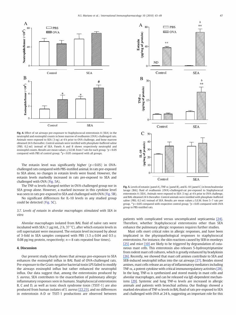
Fig. 4. Effect of rat airways pre-exposure to Staphylococcal enterotoxin A (SEA) in theneutrophil and eosinophil counts in bone marrow of ovalbumin (OVA)-challenged rats.Animals were exposed to SEA (3 ng) at 4 h prior to OVA challenge, and bone marrowobtained 24 h thereafter. Control animals were instilled with phosphate-buffered saline(PBS; 0.2 ml) instead of SEA. Panels A and B shows respectively neutrophil andeosinophil counts. Results are mean values±S.E.M. from 7 rats for each group. *p<0.05compared with PBS of control group; #p<0.05 compared with all groups.
Fig. 5. Levels of eotaxin (panel A), TNF-α (panel B), and IL-10 (panel C) in bronchoalveolarlavage (BAL) fluid of ovalbumin (OVA)-challenged-rat pre-exposed to Staphylococcalenterotoxin A (SEA). Animals were exposed to SEA (3 ng) at 4 h prior to OVA challenge,and BAL obtained 24 h thereafter. Control animals were instilledwith phosphate-bufferedsaline (PBS; 0.2 ml) instead of SEA. Results are mean values±S.E.M. from 5–7 rats pergroup. **p<0.05 compared with respective control group; δp<0.05 compared with OVAgroup in PBS-instilled rats.
47N.S. Mariano et al. / International Immunopharmacology 10 (2010) 43–49
The eotaxin level was significantly higher (p<0.05) in OVA-challenged rats comparedwith PBS-instilled animal. In rats pre-exposedto SEA alone, no changes in eotaxin levels were found. However, theeotaxin levels markedly increased in rats pre-exposed to SEA andchallenged with OVA (Fig. 5A).
The TNF-α levels changed neither in OVA-challenged group nor inSEA group alone. However, a marked increase in this cytokine levelwas seen in rats pre-exposed to SEA and challengedwith OVA (Fig. 5B).
No significant differences for IL-10 levels in any studied groupcould be detected (Fig. 5C).
3.7. Levels of eotaxin in alveolar macrophages stimulated with SEA invitro
Alveolar macrophages isolated from BAL fluid of naïve rats wereincubated with SEA (1 µg/mL, 2 h, 37 °C), after which eotaxin levels incell supernatant were measured. The eotaxin level increased by aboutof 3-fold in SEA samples compared with PBS (1.5±0.04 and 0.5±0.08 pg/mg protein, respectively; n=8 rats repeated four times).
4. Discussion
Our present study clearly shows that airways pre-exposure to SEAenhances the eosinophil influx in BAL fluid of OVA-challenged rats.Pre-exposure to the Gram-negative bacterial toxin LPS failed tomodifythe airways eosinophil influx but rather enhanced the neutrophilinflux. Our data suggest that, among the enterotoxins produced byS. aureus, SEA contributes to the exacerbation of pulmonary allergicinflammatory responses seen in humans. Staphylococcal enterotoxinsB, C and D, as well as toxic shock syndrome toxin (TSST-1) are alsoproduced from human isolates of S. aureus [22,23], and no differencesin enterotoxin A-D or TSST-1 productions are observed between
patients with complicated versus uncomplicated septicaemia [24].Therefore, whether Staphylococcal enterotoxins other than SEAenhance the pulmonary allergic responses requires further studies.
Mast cells exert critical roles in allergic responses, and have beenimplicated in the physiopathological responses to staphylococcalenterotoxins. For instance, the skin reactions caused by SEB inmonkeys[25] and mice [10] are likely to be triggered by degranulation of cuta-neous mast cells. This enterotoxin also releases 5-hydroxytriptaminefrom rodentmast cell cultures, which is greatly enhanced by bradykinin[26]. Recently, we showed that mast cell amines contribute to SEA andSEB-induced neutrophil influx into the rat airways [27]. Besides storedamines,mast cells release an array of inflammatorymediators includingTNF-α, a potent cytokinewith critical immunoregulatory activities [28].In the lung, TNF-α is synthesized and stored mainly in mast cells andalveolar macrophages, and can be released via IgE-dependent mechan-isms [29]. Systemic and lung TNF-α levels are increased in allergicanimals and patients with bronchial asthma. Our findings showed amarked elevation of TNF-α levels in BALfluid of rats pre-exposed to SEAand challenged with OVA at 24 h, suggesting an important role for this

48 N.S. Mariano et al. / International Immunopharmacology 10 (2010) 43–49
pro-inflammatory Th1 cytokine. Of interest, TNF-α has been shown toinduce the synthesis of eotaxin in several cell types including dermalfibroblasts [30], human lung epithelial cells [31], monocytic cells [32],epithelial cells [33] and human eosinophils [34].
Eotaxin is a CC-chemokine responsible for selective eosinophilchemotaxis and transendothelial migration in airways of allergicsubjects [35–38]. Eotaxin and IL-5 are said to act synergistically topromote eosinophil recruitment into tissues [39–41]. Moreover, theincreased number of eosinophils in BAL fluid has been correlated withhigher eotaxin levels [42]. In our study, a significant elevation in eotaxinlevels in BAL fluid of OVA-challenged rats was found, as expected. Theeotaxin levels were further enhanced in rats pre-exposed to SEA.Furthermore, incubation of isolated alveolar macrophages with SEAcauses the release of marked amounts of TNF-α [21] and eotaxin (thisstudy). Thesefindings strongly suggest thatmacrophage-derivedTNF-αacts to release eotaxin,which further aggravates the eosinophil influxbySEA pre-exposure in allergic animals. The eotaxin levels in BAL fluid ofSEA group alone (non-challenged rats) did not change in comparisonwithPBSgroup,which is consistentwith the lackof TNF-α inBALfluid at24 h. Interestingly, the enhancement of airways eosinophil infiltrationby SEA is not seenwhen this enterotoxin was delivered at 4 h post-OVAchallenge. Our previous studies show that exposure of airways toSEA and SEB in non-allergic rats cause an acute lung injury at 4 h ascharacterized by a marked neutrophil influx, accompanied by theincrease of various inflammatory mediators including TNF-α [21,43].Therefore, a prior (but not a post) contact of airways with SEA maybe crucial to allow a pro-inflammatory state with increased TNF-αproduction that result in an enhanced response to antigen challenge.Several evidences also show that cytokine IL-10 reduces the levels andexpression of TNF-α by activated monocytes and/or macrophages,whichhas been associatedwith clinical protection and reduction of lungpathology [44]. However, our findings showed that elevations of TNF-αin BAL fluid of OVA-challenged rats pre-exposed to SEA were notaccompanied by concomitant elevations in the IL-10 levels, indicatingthis type of pulmonary allergic inflammation does not undergodownregulation by this cytokine.
It iswell-established that bonemarrowplays a pivotal role in allergicinflammatory responses [45]. Eosinophils are derived in the bonemarrow frommyeloid precursors in response to cytokineactivation, andfollowingappropriate stimulus they are released into the circulationandrecruited to tissues. Evidences indicate that several pathways have beenimplicated, including stimulation of resident bonemarrow cells, releaseof allergen-induced hematopoietic growth factors and cell trafficking[46]. The cytokines IL-5 and eotaxin, as well as adhesion molecules(VLA-4 and VCAM) play important roles in this process [47,48]. In ourstudy, a significant increase in eosinophil number in bone marrow ofOVA-challenged rats was observed at 24 h, confirming previous studiesin rats [49], mice [50] and asthmatic subjects after inhaled antigen[45]. In rats exposed to SEA alone, a significant increase in eosinophilnumber in bonemarrowwas also observed, and such effect was furtherenhanced in OVA-challenged animals. Whilst proliferation and differ-entiation of bone marrow progenitor cells after airway allergenexposure are well-established [51–53], our study is the first to showthat SEA itself, given intranasally, promotes eosinopoesis. Therefore,modifications in bone marrow eosinophil pattern may play animportant role in determining the exacerbation of airways eosinophilinflux by pre-exposure to SEA after OVA challenge in rats. Interestingly,an increasednumberof eosinophils in bonemarrowof SEA-exposed ratsalone (SEA+PBS group) was observed. The reasons why eosinophils insuch group do not achieve BAL fluid are uncertain. A fundamentalfeature of asthma associated with allergic sensitization is the abilityof the airways to recognize allergens and to generate the so-calledTh2 cytokines, which is related to IgE production, eosinopoiesis, andbronchial inflammation. Therefore, the failure of SEA alone to promoteeosinophil infiltration into the BAL fluid may reflect the absence of anallergic stimulus.
Epidemiological evidence also suggest that exposure to LPS caninfluence the development and severity of asthma [54]. Therefore, inour study, the effects of airways pre-exposure to LPS have also beeninvestigated in order to compare with those of SEA. Endotoxin hasthe potential to either suppress the development of asthma throughinduction of counter-regulatory Th1 cells or to exacerbate asthmaseverity, presumably through its pro-inflammatory activities [55]. In ourstudy, pre-exposure to LPS neither modified the eosinophil number inBAL fluid when instilled alone nor when instilled in OVA-challengedrats, which is in agreement with a previous study showing that airwaysLPS exposure, at low dose (0.3 µg), causes only minimal effects on therecruitment of airways eosinophils in mice; as opposed, at a muchhigher dose (15–25 µg), LPS markedly enhanced Th2 eosinophilicinflammation [56,57]. Endotoxin is reported to mobilize neutrophilsafter instillation into themouse and rat airways [58]. Accordingly, in ourstudy, LPS caused a dose-dependent pulmonary neutrophil influx,which was further enhanced in OVA-challenged rats. Lipopolyssachar-ide activates toll-like receptor 4 (TLR-4) causing the mobilization ofneutrophils after instillation into the airways [59], an effect largelyreduced in TLR4 mutant mice [60]. Whether potentiation by LPS ofneutrophil influx in both non-challenged and OVA-challenged ratsreflects activation of TLR4 signaling pathway requires further studies.
In conclusion, this study shows that airways pre-exposure to SEAin OVA-challenged rats causes a selective increase in eosinophilnumber in bone marrow and BAL fluid by mechanisms involvingenhancement of TNF-α and eotaxin synthesis.
Acknowledgments
Nadia S. Mariano thanks Fundação de Amparo à Pesquisa do Estadode São Paulo (FAPESP) for financial support.
References
[1] Balaban N, Rasooly A. Staphylococcal enterotoxins. Int J Food Microbiol 2000;61:1–10.
[2] Ono HK, Omoe K, Imanishi K, Iwakabe Y, Hu DL, Kato H, et al. Identification andcharacterization of two novel staphylococcal enterotoxins, types S and T. InfectImmun 2008;76:4999–5005.
[3] Bergdoll MS, Czop JK, Gould SS. Enterotoxin synthesis by the staphylococci. Ann NY Acad Sci 1974;236:307–16.
[4] Le Loir Y, Baron F, Gautier M. Staphylococcus aureus and food poisoning. Genet MolRes 2003;2:63–76.
[5] Müller-Alouf H, Carnoy C, Simonet M, Alouf JE. Superantigen bacterial toxins: stateof the art. Toxicon 2001;39:1691–701.
[6] Marrack P, Kappler J. The staphylococcal enterotoxins and their relatives. Science1990;248:705–11.
[7] Micusan VV, Thibodeau J. Superantigens of microbiol origin. Semin Immunol1993;5:3–11.
[8] Desouza IA, Ribeiro-DaSilva G. Neutrophil migration induced by Staphylococcalenterotoxin type A in mice: a pharmacological analysis. Eur J Pharmacol 1998;363:189–95.
[9] Tessier PA, Naccache PH, Diener KR, Gladue RP, Neote KA, Clark-Lewis I, et al.Induction of acute inflammation in vivo by staphylococcal superantigens: II.Critical role for chemokines, ICAM-1, and TNF-α. J Immunol 1998;161:1204–11.
[10] Linardi A, Costa SKP, Dasilva GR, Antunes E. Involvement of kinins, mast cells andsensory neurons on the plasma exudation and paw oedema induced byStaphylococcal enterotoxin B in the mouse. Eur J Pharmacol 2000;399:235–42.
[11] Franco-Penteado CF, Desouza IA, Teixeira SA, Ribeiro-DaSilva G, De Nucci G,Antunes E. Role of nitric oxide on the increased vascular permeability andneutrophil accumulation induced by Staphylococcal enterotoxin B into the mousepaw. Biochem Pharmacol 2001;61:1305–11.
[12] Desouza IA, Hyslop S, Franco-Penteado CF, Ribeiro-da-Silva G. Mouse macro-phages release a neutrophil chemotactic mediator following stimulation byStaphylococcal enterotoxin type A. Inflamm Res 2001;50:206–12.
[13] Baker MD, Acharya KR. Superantigens: structure–function relationships. Int J MedMicrobiol 2004;293:529–37.
[14] KraftM. The role of bacterial infections in asthma. Clin ChestMed 2000;21:301–13.[15] Bachert C, Gevaert P, van Cauwenberge P. Staphylococcus aureus superantigens
and airway disease. Curr Allergy Asthma Rep 2002;2:252–8.[16] Rossi RE, Monasterolo G. Prevalence of serum IgE antibodies to the Staphylococcal
aureus enterotoxins (SAE, SEB, SEC, SED, TSST-1) in patients with persistentallergic rhinitis. Int Arch Allergy Immunol 2004;133:261–6.
[17] Lee JH, Lin YT, Yang YH, Wang LC, Chiang BL. Increased levels of serum-specificimmunoglobulin e to staphylococcal enterotoxin A and B in patients with allergicrhinitis and bronchial asthma. Int Arch Allergy Immunol 2005;138:305–11.
49N.S. Mariano et al. / International Immunopharmacology 10 (2010) 43–49
[18] Laouini D, Kawamoto S, Yalcindag A, Bryce P, Mizoguchi E, Oettgen H, et al.Epicutaneous sensitization with superantigen induces allergic skin inflammation.J Allergy Clin Immunol 2003;112:981–7.
[19] Hellings PW, Hens G, Meyts I, Bullens D, Vanoirbeek J, Gevaert P, et al. Aggravationof bronchial eosinophilia in mice by nasal and bronchial exposure to Staphylo-coccus aureus enterotoxin B. Clin Exp Allergy 2006;36:1063–71.
[20] Franco-Penteado CF, De Souza IA, Camargo EA, Teixeira SA, MuscaraMN, De Nucci G,et al. Mechanisms involved in the enhancement of allergic airways neutrophil influxby permanent C-fiber degeneration in rats. J Pharmacol Exp Ther 2005;313:440–8.
[21] Desouza IA, Franco-Penteado CF, Camargo EA, Lima CSP, Teixeira SA, Muscará MN,et al. Inflammatory mechanisms underlying the rat pulmonary neutrophil influxinduced by airway exposure to staphylococcal enterotoxin type A. Br J Pharmacol2005;146:781–91.
[22] Humphreys H, Keane CT, Hone R, Pomeroy H, Russell RJ, Arbuthnott JP, et al.Enterotoxin production by Staphylococcus aureus isolates from cases of septicae-mia and from healthy carriers. J Med Microbiol 1989;28:163–72.
[23] Kenny K, Reiser RF, Bastida-Corcuera FD, Norcross NL. Production of enterotoxinsand toxic shock syndrome toxin by bovine mammary isolates of Staphylococcusaureus. J Clin Microbiol 1993;31:706–7.
[24] KanclerskiK, SöderquistB,KjellgrenM,HolmbergH,MöllbyR. Serumantibodyresponseto Staphylococcus aureus enterotoxins and TSST-1 in patients with septicaemia. J MedMicrobiol 1996;44:171–7.
[25] GottfriedA, ScheuberPH, BernhardR, Sailer-KramerB,HartmannA.Role of substanceP in immediate-type skin reactions induced by Staphylococcal enterotoxin Bunsensitized monkeys. J Allergy Clin Immunol 1989;84:880–5.
[26] Komisar J, Rivera J, Vega A, Tseng J. Effects of staphylococcal enterotoxin B onrodent mast cells. Infect Immun 1992;60:2969–75.
[27] Desouza IA, Camargo EA,Mariano NS, Optiz-Neto JB, Resende JS, Costa SKP, et al. Roleof sensory innervation in the rat pulmonary neutrophil recruitment induced byStaphylococcal enterotoxins type A (SEA) and B (SEB). Eur J Pharmacol 2009;613:128–234.
[28] Thomas PS. Tumour necrosis factor-α: the role of this multifunctional cytokine inasthma. Immunol Cell Biol 2001;79:132–40.
[29] Russo C, Polosa R. TNF-α as a promising therapeutic target in chronic asthma: alesson from rheumatoid arthritis. Clin Sci 2005;109:135–42.
[30] Bartels J, Schluter C, Richter E, Nosso N, Kulke R, Christophers E, et al. Human dermalfibroblasts express eotaxin: molecular cloning, mRNA expression, and identificationof eotaxin sequence variants. Biochem Biophys Res Commun 1996;225:1045–51.
[31] Lilly CM, Nakamura H, Kesselman H, Nagler-Anderson C, Asano K, Garcia-ZepedaA, et al. Expression of eotaxin by human lung epithelial cells: induction bycytokines and inhibition by glucocorticoids. J Clin Invest 1997;99:1767–73.
[32] Nakamura H, Haley KJ, Nakamura T, Luster AD, Lilly CM. Differential regulation ofeotaxin expression by TNF-α and PMA in human monocytic U-937 cells. Am JPhysiol 1998;275:L601–10.
[33] Matsukura S, Stellato C, Plitt JR, Bickel C, Miura K, Georas SN, et al. Activation ofeotaxin gene transcription by NF-kappa B and STAT6 in human airway epithelialcells. J Immunol 1999;163:6876–83.
[34] Wong CK, Zhang JP, IpWK, Lam CWK. Activation of p38 mitogen-activated proteinkinase and nuclear factor-kappa B in tumour necrosis factor-induced eotaxinrelease of human eosinophils. Clin Exp Immunol 2002;128:483–9.
[35] Lamkhioued B, Renzi PM, Abi-Younes S, Garcia-Zepada EA, Allakhverdi Z, Ghaffar O,et al. Increased expression of eotaxin in bronchoalveolar lavage and airways ofasthmatics contributes to the chemotaxis of eosinophils to the site of inflammation.Immunology 1997;159:4593–601.
[36] Minshall EM, Cameron L, Lavigne F, Leung DY, Hamilos D, Garcia-Zepada EA, et al.EotaxinmRNAandprotein expression in chronic sinusitis and allergen-inducednasalresponses in seasonal allergic rhinitis. Am J Respir Cell Mol Biol 1997;17:683–90.
[37] Tateno H, Nakamura H, Minematsu N, Nakajima T, Takahashi S, Nakamura M, et al.Plasma eotaxin level and severity of asthma treated with corticosteroid. RespirMed 2004;98:782–90.
[38] Haley KJ, Sunday ME, Porrata Y, Kelley C, Twomey A, Shahsafaei A, et al. Ontogenyof the eotaxins in human lung. Am J Physiol Lung Cell Mol Physiol 2008;294:L214–24.
[39] Smith N, Johnson FJ. Effects of inhaled eotaxin on airway function and inflammatorycell influx in sensitised and non-sensitised guinea pigs. Pulm Pharmacol Ther2006;19:391–6.
[40] Palframan RT, Collins PD, Williams TJ, Rankin SM. Eotaxin induces a rapid releaseof eosinophils and their progenitors from the bone marrow. Blood 1998;91:2240–8.
[41] Costa GG, Silva RM, Franco-Penteado CF, Antunes E, Ferreira HHA. Interactionsbetween eotaxin and interleukin-5 in the chemotaxis of primed and non-primedhuman eosinophils. Eur J Pharmacol 2007;566:200–5.
[42] Young SS, Ritacco G, Skeans S, Chapman RW. Eotaxin and nitric oxide productionas markers of inflammation in allergic cynomolgus monkeys. Int Arch AllergyImmunol 1999;120:209–17.
[43] Desouza IA, Franco-Penteado CF, Camargo EA, Lima CSP, Teixeira SA, Muscará MN,et al. Acute pulmonary inflammation induced by exposure of airways tostaphylococcal enterotoxin type B in rats. Toxicol Appl Pharmacol 2006;217:107–13.
[44] Wood LJ, Inman MD, Watson RM, Foley R, Denburg JA, O'Byrne PM. Changes inbone marrow inflammatory cell progenitors after inhaled allergen in asthmaticsubjects. Am J Respir Care Med 1998;157:99–105.
[45] Wood LJ, Sehmi R, Dorman S, Hamid Q, Tulic MK, Watson RM, et al. Allergen-induced increases in bone marrow T lymphocytes and interleukin-5 expression insubjects with asthma. Am J Respir Crit Care Med 2002;166:883–9.
[46] Mohler R, Salemi P, Moore MAS, Rafii S. Expression of interleukin-5 by humanbone marrow microvascular endothelial cells: implications for the regulation ofeosinophilopoiesis in vivo. Br J Haematol 1997;99:732–8.
[47] Papayannopoulou T, Priestley YGV, Nakamoto B. Anti-VLA4/VCAM-1-inducedmobilization requires cooperative signaling through the kit/mkit ligand pathway.Blood 1998;7:2231–9.
[48] Ferreira HHA, Lodo MLS, Martins AR, Kandratavicius L, Salaroli AF, Conran N, et al.Expression of nitric oxide synthases in vitro migration of eosinophils from allergicrhinitis subject. Eur J Pharmacol 2002;442:155–62.
[49] Chin JE, Winterrowd GE, Hatfield CA, Brashler JR, Griffin RL, Vonderfecht SL, et al.Involvement of intercellular adhesion molecule-1 in the antigen-inducedinfiltration of eosinophils and lymphocytes into the airways in a murine modelof pulmonary inflammation. Am J Respir Cell Mol Biol 1998;18:158–67.
[50] Gaspar-Elsas MI, Joseph D, Elsas PX, Vargaftig BB. Rapid increase in bone-marroweosinophil production and responses to eosinopoietic interleukins triggered byintranasal allergen challenge. Am J Respir Cell Mol Biol 1997;17:404–13.
[51] Ohkawara Y, Lei XF, Stämpfli MR, Marshall JS, Xing Z, Jordana M. Cytokine andeosinophil responses in the lung, peripheral blood, and bone marrow compart-ments in a murine model of allergen-induced airways inflammation. Am J RespirCell Mol Biol 1997;16:510–20.
[52] Tomaki M, Zhao LL, Lundahl J, Sjöstrand M, Jordana M, Lindén A, et al.Eosinophilopoiesis in amurine model of allergic airway eosinophilia: involvementof bone marrow IL-5 and IL-5 receptor alpha. J Immunol 2000;165:4040–50.
[53] Morrison DF, Foss DL, Murtaugh MP. Interleukin-10 gene therapy-mediatedamelioration of bacterial pneumonia. Infect Immun 2000;68:4752–8.
[54] Michel O. Role of lipopolysaccharide (LPS) in asthma and other pulmonaryconditions. J Endotoxin Res 2003;9:293–300.
[55] Liu AH. Endotoxin exposure in allergy and asthma: reconciling a paradox. J AllergyClin Immunol 2002;109:379–92.
[56] Stephens R, Chaplin DD. IgE cross-linking or lipopolysaccharide treatment inducesrecruitment of Th2 cells to the lung in the absence of specific antigen. J Immunol2002;169:5468–76.
[57] Lundy SK, Berlin AA, Lukacs NW. Interleukin-12-independent down-modulationof cockroach antigen-induced asthma in mice by intranasal exposure to bacteriallipopolysaccharide. Am J Pathol 2003;163:1961–8.
[58] Reutershan J, Basit A, Galkina EV, Ley K. Sequential recruitment of neutrophils intolung and bronchoalveolar lavage fluid in LPS-induced acute lung injury. Am JPhysiol Lung Cell Mol Physiol 2005;289:L807–15.
[59] Savov JD, Gavett SH, Brass DM, Costa DL, Schwartz DA. Neutrophils play a criticalrole in development of LPS-induced airway disease. Am J Physiol Lung Cell MolPhysiol 2002;283:L952–62.
[60] Jung YW, Schoeb TR, Weaver CT, Chaplin DD. Antigen and lipopolysaccharide playsynergistic roles in the effector phase of airway inflammation in mice. Am J Pathol2006;168:1425–34.

![[Care of staphylococcal skin infections by doctors and nurses deployed in Guyana]. Prise en charge des infections cutanées staphylococciques par les médecins et infirmiers militaires](https://static.fdokumen.com/doc/165x107/6331e74983bb92fe9804268f/care-of-staphylococcal-skin-infections-by-doctors-and-nurses-deployed-in-guyana.jpg)