Eotaxin-1 in exhaled breath condensate of stable and unstable asthma patients
Upload
independentCategory
view
0download
0
International Immunology, Vol. 11, No. 1, pp. 1–10 © 1999 Japanese Society for Immunology
Selective eosinophil transendothelialmigration triggered by eotaxin viamodulation of Mac-1/ICAM-1 andVLA-4/VCAM-1 interactions
Gui-Quan Jia 1,5, Jose-Angel Gonzalo 1,5, Andres Hidalgo 3, Denisa Wagner 2,Myron Cybulsky 4 and Jose C. Gutierrez-Ramos 1,5
1The Center for Blood Research, Inc. and 2The Department of Genetics and Pathology, Harvard MedicalSchool. Boston, MA 02115, USA3Centro de Investigaciones Biologicas, Madrid 28006, Spain4Toronto Hospital, 200 Elizabeth Street, CCRW 1–855, Toronto, Ontario, M5G 2C4, Canada5Millennium Pharmaceuticals, Inc. Cambridge, MA 02139, USA
Keywords: chemokine, eotaxin, eosinophil, integrin, transendothelial migration
Abstract
We have recently cloned eotaxin, a highly efficacious eosinophilic chemokine involved in thedevelopment of lung eosinophilia during allergic inflammatory reactions. To understand moreprecisely how eotaxin facilitates the specific migration of eosinophils, we have studied whichadhesion receptors are essential for eotaxin action both in vivo and in vitro . Experiments usingmice genetically deficient in adhesion receptors demonstrated that molecules previously reportedto be involved in both leukocyte tethering/rolling (P-selectin and E-selectin) and in sticking/transmigration (ICAM-1 and VCAM-1) are required for eotaxin action in vivo . To further elucidate themechanism(s) involved in this process, we have used an in vitro transendothelial chemotaxismodel. mAb neutralization studies performed in this system suggest that the integrins Mac-1(CD11b/18), VLA-4 (α 4β1) and LFA-1 (CD11a/18) are involved in the transendothelial chemotaxis ofeosinophils to eotaxin. Accordingly, the expression of these integrins on eosinophils is elevated bydirect action of this chemokine in a concentration-dependent manner. Taken together, our resultssuggest that eotaxin-induced eosinophil transendothelial migration in vivo and in vitro relies onMac-1/ICAM-1 and VLA-4/VCAM-1 interactions, the latter ones becoming more relevant at later timepoints of the eotaxin-induced recruitment process.
Introduction
Eosinophil accumulation is a prominent feature of allergicinflammatory disorders. However, eosinophils represent onlya small percentage of circulating leukocytes, suggesting theexistence of mechanisms for selective eosinophil recruit-ment in vivo. The mechanism(s) leading to the selectiveaccumulation of eosinophils in allergic inflamed tissue areat present unclear. The recruitment of subpopulations ofcirculating leukocytes to the sites of inflammation has beenproposed to be a complex process involving the coordinatedaction of various primary adhesion receptors that mediatetethering/rolling of leukocytes along the endothelial wall and
Correspondence to: G.-Q. Jia, Millennium Pharmaceutical Inc., 640 Memorial Drive, Cambridge, MA 02139, USA
Transmitting editor: C. Martinez-A Received 29 May 1998, accepted 17 September 1998
of activation-dependent adhesion receptors that mediatesticking/transmigration across the vascular endothelium.
However, it is unlikely that the differential expression ofadhesion receptors is sufficient to fully explain the selectiverecruitment of eosinophil into tissue during allergic inflamma-tion. Eosinophils express L-selectin (1,2), ligands for P-selectinand E-selectin (2) as well as the integrins LFA-1 (CD11a/18)and Mac-1 (CD11b/18) which interact with ICAM-1. However,these adhesion molecules are also expressed on a widevariety of other leukocyte subset(s). It is likely that duringthe development of the inflammatory response, additional
by guest on October 3, 2014
http://intimm
.oxfordjournals.org/D
ownloaded from

2 Eosinophil migration induced by eotaxin
factors are required to regulate the selective accumulation ofeosinophils.
Recently, chemokines have been suggested to play animportant role in the selective transmigration of leukocytesubsets (3). Eotaxin is a chemokine which is able to mediatechemotaxis of peripheral blood eosinophils. This chemokinediffers from most other chemokines because it is highlyspecific for eosinophils (4,5). Injection of eotaxin into guineapig skin or into mouse peritoneum induces a profoundselective eosinophilic infiltrate (4,5). The expression of eotaxinalso parallels the accumulation of eosinophils in the lungduring the course of a mouse model of lung allergic inflamma-tion (5). The importance of eotaxin in the development of lungeosinophilia has recently been demonstrated using selectiveanti-eotaxin mAb or eotaxin gene knockout mice (6,7). In viewof the critical role of this chemokine, the precise adhesivemechanism(s) by which eotaxin induces a selective eosinophilmigration become an important issue.
In the present study, we demonstrate, using both adhesionmolecule-deficient mice and an in vitro transendothelialchemotactic migration model, that eotaxin modulates thesurface expression/function of adhesion molecules to achieveselective eosinophil transendothelial migration. Mac-1/ICAM-1 interactions are dominant at early time points during eotaxin-induced eosinophil recruitment, whereas VLA-4/VCAM-1become predominant at later time points.
Methods
Reagents and mice
The mAb used for detection of surface adhesion moleculeswere purchased from PharMingen (San Diego, CA). For theblocking experiments, the following mAb were used: rat anti-LFA-1 mAb M17/4 (IgG2a) (8,9), rat anti-Mac-1 mAb M1/70(IgG2b) (10) and anti-VLA-4 mAb PS/2 (IgG2b) (11). Adhesionmolecule-deficient mice used in this study were: P-selectinmutant mice purchased from the Jackson laboratory (BarHarbor, ME), L-selectin mutant mice provided by Dr M. H.Siegelman (12), and E-selectin mutant mice, ICAM-1 mutantmice (13), VCAM-1-hypomorphic mutant mice (14) and micedeficient for both P-selectin and ICAM-1 or P-selectin andE-selectin (15). Since VCAM-1-null mutant mice are embryoniclethal (16), VCAM-1-hypomorphic mutant mice were used forthese studies (14). These mutant mice were generated by atargeted deletion in domain 4 of the VCAM-1 molecule (whicheliminates one α4 integrin binding site). Based on previouswork, a very low affinity of this truncated protein to α4 can bepredicted (17). The mutation resulted in 20-fold decrease ofexpression (hypomorphic ) of the domain 4-deficient (D4D)VCAM-1 gene, as found by Northern blot analysis andincreased survival rates of one-third (not shown and (14)).Mice designated as wild-type in the results are littermatesof these mutants that have a mixed genetic background129sv3C57BL/6J.
Endothelial cell culture
The mouse endothelial cell line b-End-2 cells (18) wascultured in IMDM with 10% FCS. Cells (23 104) from wellgrown passage were seeded on transwell inserts (6.5 mm
diameter polycarbonate membrane with 5 µm pores; Costar,Cambridge, MA) and cultured for a further 2 days. The mediawere added into the transwell inserts to prevent the formationof an endothelial bilayer (i.e. a monolayer both above andbelow the filter). After 2 days, the endothelial cell-coated wellswere used for transendothelial migration assays describedbelow.
Purification of eosinophils
For the in vitro studies, eosinophils were purified from theperitoneum of IL-5 transgenic mice as previously described(5). Eosinophil purity was 92 6 2% based on Giemsa stainingand viability was .95% based on Trypan blue exclusion forthe experiments in this article except some experiments forFig. 3, where unpurified population cells were used as inputfor the chemotaxis assay.
Adherence assay
The ability of eotaxin to induce adhesiveness on the integrinligands was determined as follows. Isolated eosinophils fromIL-5 transgenic mouse were labeled with BCECF-AM at roomtemperature for 30 min and then preincubated with or withoutneutralized antibodies to VLA-4, Mac-1 or LFA-1 (10 µg/ml)for 20 min at 4°C. After that, the cells were incubated with orwithout eotaxin (500 ng/ml) for varying times at 37°C andsubsequently subjected to adherence assay in 96-wellplates pre-coated with a recombinant fragment of fibronectin(5 µg/ml), containing the CS1 binding site specific for VLA-4(positive control), VCAM-1 (1.25 µg/ml), ICAM-1 (2.5 µg/ml)or BSA (5 µg/ml) (negative control) for 30 min at 4°C toreduce background binding. After incubation, the cells weregently washed with PBS and read by fluorescence intensity.
In vitro transendothelial migration assay
The migration of eosinophils to recombinant eotaxin throughthe endothelial cell layer was evaluated in duplicate aspreviously described (5) with the membrane coated withendothelial cells described as above. The endothelial cells intranswell inserts were washed once with sera-free mediumand 23105 eosinophils in 0.1 ml sera-free medium wereadded. After 2 h incubation, the transwells were removedand the number of cells per well counted in the FACScan bypassing each sample for a constant predetermined timeperiod. Contaminating endothelial cells were gated out anda constant gate was assigned for the eosinophil populationin the side/forward scatter window and used for every sample.In the blocking experiments, eosinophils were preincubatedwith anti-integrin antibodies or with control antibody at 37°Cfor 15 min before their addition to the transwell inserts.
Immunofluorescence and flow cytometry
Flow cytometry was performed to determine the expressionof cell surface adhesion molecules in selected experiments.The migratory cells from the assay were incubated with anti-Fc-receptor mAb (2.492; PharMingen, San Diego, CA) andthen stained with each one of the following antibodiesindividually: ICAM-1, Mac-1 (CD11b), LFA-1 (CD11a), VLA-4(CD49d), VLA-5 or CD44, conjugated with FITC or phyco-erythrin (PharMingen, San Diego, CA). Leukocyte cell popula-tions were identified by forward scatter and side scatter
by guest on October 3, 2014
http://intimm
.oxfordjournals.org/D
ownloaded from

Eosinophil migration induced by eotaxin 3
Fig. 1. Eotaxin-induced peritoneal eosinophilia. Peritoneal exudatewas collected at the time points indicated following injection of eotaxin(closed symbols) or PBS (empty symbols). Three different doses ofeotaxin were administered: 3 µg/mouse (squares), 1.5 µg/mouse(circles) or 0.5 µg/mouse (triangles). The number of eosinophils inthe peritoneal exudate was determined as described in Methods.The error bars represent differences between the values obtainedfrom five mice per time point.
gating. Dead cells were excluded by propidium iodide (Sigma,St Louis, MO) incorporation. Flow cytometry data wereacquired with a FACScan cytometer (Becton Dickinson) andanalyzed with CellQuest software. Fluorescence data werecollected on a log scale and the level of expression wasdetermined by the relative fluorescence intensity as the meanfluorescence channel for each individual surface adhesionmolecule.
Intraperitoneal injection of eotaxin
The migration of eosinophils in vivo in response to eotaxinwas determined by injection of recombinant murine eotaxinprotein (1 µg/200 µl PBS per mouse; Peprotech, Rocky Hill,NJ) into the peritoneum of the different adhesion receptor-deficient mice. Mice injected with the same volume of PBSwere used as controls. Peritoneal exudates were collected atdifferent time points as indicated or for 2 h (10 mice pergroup) or 6 h (five mice per group) after eotaxin administration.The number of eosinophils recovered was then determinedas described below.
Quantitation of eosinophils in peritoneal exudates.
Peritoneal cells (53105/slide) were applied to glass slidesby cytocentrifugation, stained with Wright–Giemsa (FisherDiagnostics, Pittsburgh, PA), rinsed with distilled water, airdried and mounted. The percentage of eosinophils wasdetermined by counting their number in eight high-powerfields (340 magnification; total area 0.5 mm2) per arearandomly selected and dividing this number by the totalnumber of cells per high power field. To obtain the absolutenumber of infiltrating peritoneal eosinophils, these percent-ages were multiplied by the total number of cells recoveredfrom the exudate of this organ.
Results
The accumulation of eosinophils in response to eotaxin isaffected in mutant mice lacking adhesion molecules
To characterize the peritoneal accumulation of eosinophilsin response to eotaxin, eosinophils were quantified in theperitoneal exudates of mice 0, 1, 2, 3 or 4 h after injectionwith different doses of eotaxin (Fig. 1). Eotaxin-inducedeosinophil accumulation happened in both a dose- and time-dependent manner with maximum peritoneal eosinophiliaoccurring at 2 h for all doses (Fig. 1). At this time point 3, 1.5and 0.5 µg/mouse eotaxin induced a 22-, 13- and 4-foldincrease respectively in eosinophils compared to PBS-treatedcontrols (Fig. 1).
To study which adhesion molecules are essential duringthe selective accumulation of eosinophils mediated by eotaxinin vivo, we used a panel of mice that had been madegenetically deficient in adhesion molecules. The deficientmice were injected i.p. with 1 µg/mouse of recombinantmurine eotaxin. The eosinophil accumulation detected in theperitoneal cavity 2 h after eotaxin injection of wild-type micewas ~1.53105 eosinophils and was considered 100%. Thepercentage of eosinophil accumulation in response toeotaxin in the different mutants are always referred to this100% in the wild-type strain. At the same time point, similarnumbers of eosinophils to the wild-type strain were found inthe peritoneum of L-selectin-deficient and E-selectin-deficientmice in response to eotaxin (Fig. 2). In contrast, eosinophilaccumulation was markedly reduced (29.7 6 4.5% of wild-type) in P-selection-deficient mice 2 h after eotaxin administra-tion when compared with wild-type mice (Fig. 2). Injectionof eotaxin in mice that were double mutants for P-selectin1 ICAM-1 and P-selectin 1 E-selectin resulted in 14.0 6 2.1and 26.9 6 7.8% of the peritoneal eosinophil accumulationobserved in wild-type mice respectively (Fig. 2). To evaluatewhether leak of the expression of these adhesion receptorsresults in a delay in the eosinophil migration process to theperitoneum rather than impairment, the numbers of eosino-phils were also determined in the peritoneum of these mutantmice 6 h after eotaxin administration. No differences wereobserved in peritoneal eosinophil recruitment between L-selectin-deficient mice and wild-type littermates 6 h afterinjection with eotaxin (Fig. 2). Interestingly, P-selectin-deficientmice that showed a significant reduction in peritoneal eosino-philia 2 h after eotaxin administration were comparable to wild-type mice 6 h after eotaxin injection (Fig. 2). This indicates thateotaxin-induced eosinophil accumulation in the peritoneumis only initially delayed in the P-selectin-deficient mice andthat alternative mechanisms exist for eosinophil recruitmentin the absence of P-selectin. Our data favors an importantrole for both ICAM-1 and P-selectin as the P-selectin 1 ICAM-1 double-deficient mice had a 21.1 6 3.3% of the peritonealaccumulation of eosinophils observed in the wild-type miceat any time point analyzed (Fig. 2). A similar phenomenonoccurs in the P-selectin 1 E-selectin double-deficient mice.Eotaxin-injected P-selectin 1 E-selectin double-deficientmice exhibited only 10.2 6 2.4% of the eosinophil migrationobserved in wild-type mice 6 h after injection (Fig. 2). Thiscorrelates with the low range of eosinophil migration (25.7 6
by guest on October 3, 2014
http://intimm
.oxfordjournals.org/D
ownloaded from

4 Eosinophil migration induced by eotaxin
Fig. 2. Eotaxin-induced peritoneal recruitment of eosinophils indifferent adhesion receptor-deficient mice. The total number ofperitoneal eosinophils (circles) and the percentage of eosinophilmigration to this organ (gray bars) were evaluated 2 and 6 h afterthe administration of eotaxin (1 µg/mouse, i.p.). Each circle representsa single mouse. Eotaxin-treated wild-type or test mice are representedby closed circles. PBS-treated wild-type and deficient mice arerepresented by open circles. Black short lines indicate the mean ofeach group. Errors bars indicate the SD of the average of thepercentage of eosinophil migration in the different groups of mice.Percentages of migration shown here are calculated as a percentageof total eosinophils detected in the peritoneum of wild-type mice 2 hafter eotaxin injection which was considered to be 100%. Thegenotype of wild-type, single or double adhesion receptor-deficientmice is represented by black squares in the matrix. Differencesbetween groups were tested for significance by the Mann–WhitneyWilcoxon test using Xlstat in Microsoft Excel 5.0.
Fig. 3. Eotaxin-induced eosinophil transendothelial migration. Purifiedeosinophils were placed in the upper chamber of the assay with (I)or without (II) endothelial cells and recombinant mouse eotaxin wasplaced in the lower chamber at the concentration indicated. After 2h incubation, the migration of eosinophils was analyzed as describedin Methods. Data represent one of four experiments performed.
Fig. 4. Eotaxin transiently increases VLA-4- and Mac-1-dependent binding of eosinophils to FN-H89, VCAM-1 and ICAM-1. Isolated eosinophils (purity . 90%) were preincubated witheotaxin (500 ng/ml) (bars) or without (circles) for varying times at37°C and subsequently subjected to adhesion assays in 96-well plates pre-coated with a recombinant fragment of fibronectin(5 µg/ml), containing the CS1 binding site specific for VLA-4,,VCAM-1 (1.25 µg/ml), ICAM-1 (2.5 µg/ml) or BSA (5 µg/ml) for30 min at 4°C to reduce background binding. Previously, cellswere labeled with BCECF-AM and preincubated without or withantibodies to VLA-4, Mac-1 or LFA-1 (10 µg/ml) for 20 min at 4°C.Data are mean 6 SEM of a representative experiment out of three,performed in triplicate.
2.6%) detected in the peritoneum of E-selectin-deficient miceat the same time point after eotaxin injection (Fig. 2).
ICAM-1-deficient mice and VCAM-1-deficient miceexhibited 62.7 6 6.1 and 75 6 2.7% respectively of theperitoneal eosinophil accumulation observed in wild-typelittermates 2 h after eotaxin injection (Fig. 2). More interest-ingly, similar numbers of eosinophils were found in theperitoneum of both ICAM-1-deficient mice and wild-typelittermates 6 h after eotaxin administration (Fig. 2). In contrast,peritoneal eosinophilia was reduced by half in the VCAM-1-mutant littermates when compared with that observed in wild-
by guest on October 3, 2014
http://intimm
.oxfordjournals.org/D
ownloaded from

Eosinophil migration induced by eotaxin 5
Fig. 5. Effects of anti-Mac-1, anti-LFA-1 and anti-VLA-4 on eotaxin-induced eosinophil transendothelial migration. Assays wereperformed as described in the legend to Fig. 3 with endothelial cellsexcept eosinophils were preincubated for 15 min at 37°C with theindicated anti-integrin mAb at a concentration of 5 µg/ml. Valuesrepresent mean 6 SEM from three independent experiments. Themigration of eosinophils in response to eotaxin with control mAb wasconsidered 100% and then used to normalize the experimental data.*P . 0.05 versus control antibody; **P . 0.01 versus control antibody.
type controls 6 h after eotaxin injection (Fig. 2). This suggeststhat these two adhesion molecules could exert a differentialcontribution to this process; VCAM-1 being more critical atlate stages (6 h) in the response to eotaxin.
Eotaxin induces selective eosinophil transendothelial migra-tion in vitro
Transendothelial chemotaxis in vitro has been established asan in vitro model of leukocyte migration across postcapillaryvenules (19,20). This assay allows quantitative evaluation ofthe mechanisms by which eotaxin induces selectiveeosinophil migration, as well as to study its effects on adhesionmolecules both on eosinophils and endothelial cells (19,20).Recombinant mouse eotaxin was able to induce significanteosinophil transendothelial migration in vitro, although theactual percentage of eosinophils that migrated to eotaxinwas lower in the assay with an endothelial cell layer (Fig. 3,I) than in that performed without endothelial cells (Fig. 3, II).The transendothelial migration of eosinophils responding toeotaxin occurred in a dose-dependent manner. Almost noeosinophil transendothelial migration was found in theabsence of eotaxin (Fig. 3). Eotaxin-induced transmigrationwas selective for eosinophils since in the migrated cellpopulation almost 100% were eosinophils [which increasedfrom 70% eosinophils in the input of an unpurified cellpopulation; data not shown and (5)]. These data together withour previously reported results showing that in vivo eotaxininduced the recruitment of eosinophils to the peritoneum(Fig. 1) (5) demonstrate that eotaxin alone can selectivelyinduce migration of eosinophils across endothelial beds in vivoand in vitro.
Integrins are required for the transmigration of eosinophilsin vitro
Based on our results in vivo, we studied if the integrin ligandsfor ICAM-1 and VCAM-1, Mac-1, LFA-1 and VLA-4 participated
in the transendothelial migration of eosinophils induced byeotaxin. Firstly, we determined if there is any effect of eotaxinon the functional activity of the integrins. We used the adher-ence assay as a tool to examine eotaxin action becauseadherence of eosinophils is an initial step for its transmigration.In these experiments, we found that eotaxin was able toincrease the eosinophil adherence to ICAM-1 and VCAM-1(Fig. 4). This specific binding was abrogated by specificantibodies to CD11b or CD49d, but not by control antibodies.The eotaxin-induced eosinophil adherence to ICAM-1 ismostly attributable to CD11b (Mac-1) (Fig. 4).
Secondly, the contribution of the integrins during the trans-migration was examined with the chemotaxis. Chemotaxisassays were performed in the presence of neutralizing anti-bodies against these three molecules. As shown in Fig. 5,mAb to CD11a (LFA-1), to CD11b (Mac-1) or to α4 (VLA-4) alone can significantly inhibit eosinophil transendothelialmigration induced by eotaxin to different extents. Blockadeof CD11b (Mac-1) had a more marked effect than the othertwo [CD11a (LFA-1) and α4 (VLA-4)] in inhibiting eosinophiltransendothelial migration (up to 60–70% reduction). Blockingof LFA-1 in these experiments resulted in a much lowerreduction than that after blocking of Mac-1 during eosinophiltransendothelial migration (only 15% reduction; Fig. 5). Theseresults demonstrated that eotaxin-induced eosinophil trans-endothelial migration relied more heavily on Mac-1 than onLFA-1 and VLA-4. β1 and β2 integrins belong to differentintegrin subfamilies and bind different ligands; however, thecombination of neutralizing antibodies to these three mole-cules did not further significantly inhibit eosinophil transmigra-tion (Fig. 5). It is still possible that both β1 and β2 integrins,as well as other molecules such as CD31, are needed foroptimal migration of eosinophils in vivo.
The expression of integrins is increased on eosinophils thattransmigrate to eotaxin across endothelial cells
The effect of eotaxin on the expression of adhesion moleculesboth on eosinophil and endothelial cells during the processof transendothelial migration was evaluated using immuno-fluorescence and flow cytometry (21). On eosinophils, amongthe adhesion molecules possibly involved in the trans-migration process (Mac-1, LFA-1, VLA-4, VLA-5, CD44, CD31and ICAM-1), only Mac-1, LFA-1 and VLA-4 were found toundergo significant changes in their surface expression levelson eotaxin-induced migratory eosinophils compared to theinput cell population (Fig. 6 and data not shown). All adhesionmolecules mentioned above were expressed on eosinophils(data not shown). The expression of ICAM-1 had minorchanges that were not statistically significant (Fig. 6). Theseresults suggested that eotaxin could induce the up-regulationof selected adhesion molecules on eosinophils during thetransmigration process. Since we did not find two populationsof eosinophil with different expression levels of integrins inthe input population used for chemotaxis, it ruled out thepossibility that only the eosinophils with higher expressionlevels of these integrins were selected to migrate to eotaxinthrough the endothelial cell layer during the transmigrationprocess (data not shown). Thus, eosinophil transendothelialmigration induced by eotaxin correlates with a selectivemodulation of Mac-1, LFA-1 and VLA-4 expression. The
by guest on October 3, 2014
http://intimm
.oxfordjournals.org/D
ownloaded from
6 Eosinophil migration induced by eotaxin
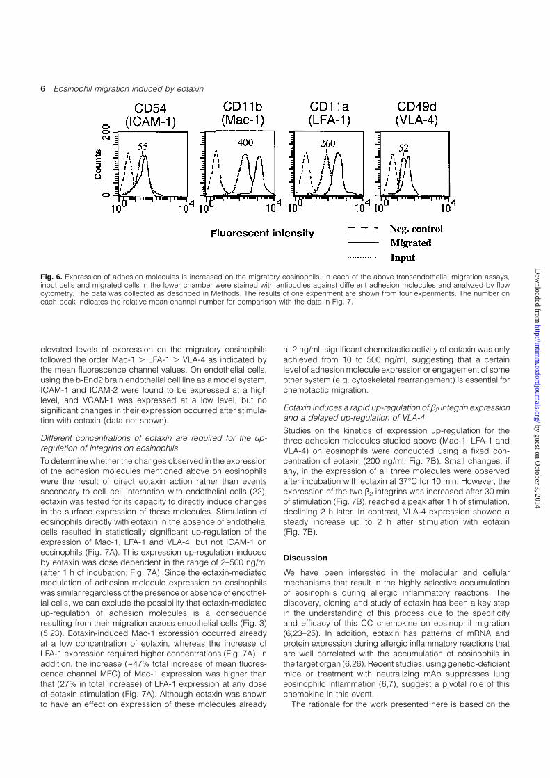
Fig. 6. Expression of adhesion molecules is increased on the migratory eosinophils. In each of the above transendothelial migration assays,input cells and migrated cells in the lower chamber were stained with antibodies against different adhesion molecules and analyzed by flowcytometry. The data was collected as described in Methods. The results of one experiment are shown from four experiments. The number oneach peak indicates the relative mean channel number for comparison with the data in Fig. 7.
elevated levels of expression on the migratory eosinophilsfollowed the order Mac-1 . LFA-1 . VLA-4 as indicated bythe mean fluorescence channel values. On endothelial cells,using the b-End2 brain endothelial cell line as a model system,ICAM-1 and ICAM-2 were found to be expressed at a highlevel, and VCAM-1 was expressed at a low level, but nosignificant changes in their expression occurred after stimula-tion with eotaxin (data not shown).
Different concentrations of eotaxin are required for the up-regulation of integrins on eosinophils
To determine whether the changes observed in the expressionof the adhesion molecules mentioned above on eosinophilswere the result of direct eotaxin action rather than eventssecondary to cell–cell interaction with endothelial cells (22),eotaxin was tested for its capacity to directly induce changesin the surface expression of these molecules. Stimulation ofeosinophils directly with eotaxin in the absence of endothelialcells resulted in statistically significant up-regulation of theexpression of Mac-1, LFA-1 and VLA-4, but not ICAM-1 oneosinophils (Fig. 7A). This expression up-regulation inducedby eotaxin was dose dependent in the range of 2–500 ng/ml(after 1 h of incubation; Fig. 7A). Since the eotaxin-mediatedmodulation of adhesion molecule expression on eosinophilswas similar regardless of the presence or absence of endothel-ial cells, we can exclude the possibility that eotaxin-mediatedup-regulation of adhesion molecules is a consequenceresulting from their migration across endothelial cells (Fig. 3)(5,23). Eotaxin-induced Mac-1 expression occurred alreadyat a low concentration of eotaxin, whereas the increase ofLFA-1 expression required higher concentrations (Fig. 7A). Inaddition, the increase (~47% total increase of mean fluores-cence channel MFC) of Mac-1 expression was higher thanthat (27% in total increase) of LFA-1 expression at any doseof eotaxin stimulation (Fig. 7A). Although eotaxin was shownto have an effect on expression of these molecules already
at 2 ng/ml, significant chemotactic activity of eotaxin was onlyachieved from 10 to 500 ng/ml, suggesting that a certainlevel of adhesion molecule expression or engagement of someother system (e.g. cytoskeletal rearrangement) is essential forchemotactic migration.
Eotaxin induces a rapid up-regulation of β2 integrin expressionand a delayed up-regulation of VLA-4
Studies on the kinetics of expression up-regulation for thethree adhesion molecules studied above (Mac-1, LFA-1 andVLA-4) on eosinophils were conducted using a fixed con-centration of eotaxin (200 ng/ml; Fig. 7B). Small changes, ifany, in the expression of all three molecules were observedafter incubation with eotaxin at 37°C for 10 min. However, theexpression of the two β2 integrins was increased after 30 minof stimulation (Fig. 7B), reached a peak after 1 h of stimulation,declining 2 h later. In contrast, VLA-4 expression showed asteady increase up to 2 h after stimulation with eotaxin(Fig. 7B).
Discussion
We have been interested in the molecular and cellularmechanisms that result in the highly selective accumulationof eosinophils during allergic inflammatory reactions. Thediscovery, cloning and study of eotaxin has been a key stepin the understanding of this process due to the specificityand efficacy of this CC chemokine on eosinophil migration(6,23–25). In addition, eotaxin has patterns of mRNA andprotein expression during allergic inflammatory reactions thatare well correlated with the accumulation of eosinophils inthe target organ (6,26). Recent studies, using genetic-deficientmice or treatment with neutralizing mAb suppresses lungeosinophilc inflammation (6,7), suggest a pivotal role of thischemokine in this event.
The rationale for the work presented here is based on the
by guest on October 3, 2014
http://intimm
.oxfordjournals.org/D
ownloaded from

Eosinophil migration induced by eotaxin 7
Fig. 7. Dose-dependent enhancement of eosinophil surface expression of adhesion molecules (A) and the time course of induction of adhesionmolecules (B) by eotaxin. (A) Eosinophils were incubated with eotaxin at different concentrations as indicated for 1 h and stained as describedin Methods. Results are expressed as the mean of duplicates from one of three experiments and the bars indicate the range for the duplicates.(B) Eosinophils were incubated with (squares) or without (triangles) eotaxin at a concentration of 200 ng/ml for various time periods asindicated. Then, the experiments followed the same procedure as described in (A). Data is representative of three similar experiments withthe range of duplicates indicated by bars.
assumption that eotaxin exerts its specific task of recruitingeosinophils to the inflammatory site by inducing the activation/up-regulation of certain combination(s) of adhesion receptorthat allow(s) eosinophils to go across an endothelial barrierallowing them to migrate preferentially at inflammation sites.This rationale has its origin in a hypothesis proposed byothers based mainly on elegant in vitro studies (27,28).Moreover, clinical studies have further supported this hypo-thesis in that eosinophils at inflammation sites expressconsiderably elevated levels of CD11b, ICAM-1 and HLA,whereas non-recruited blood eosinophils do not express thosemolecules (22,29).
It has been our intention to answer as many questions aspossible using in vivo models because we are well aware ofthe complexity of the leukocyte migration process and thedifficulty of reproducing it entirely in vitro (28,30). The use ofadhesion receptor-deficient mice has allowed us to examinemolecules and cellular processes as they occur in vivo duringacute inflammatory events. On the other hand, we havealso taken advantage of informative in vitro transendothelial
migration assays that allowed us to perform mechanisticexperiments in a more controlled assay system (19,20).
Eosinophils express L-selectin as well as ligands for P-selectin and E-selectin (1,2). We have found that the absenceof L-selectin does not affect eotaxin-induced eosinophilaccumulation in the peritoneum, whereas in the absence ofP-selectin eotaxin-induced eosinophils recruitment is delayedand suboptimal when compared to that observed in wild-type mice (Fig. 2). Although 2 h after eotaxin administrationeosinophil recruitment was basically absent in P-selectin-deficient mice, 6 h after the administration of this chemokinethere was a significant degree of eosinophil recruitment(Fig. 2), suggesting that other molecules compensatedpartially for the loss of P-selectin at later time points. Amongthe possible surface molecules, E-selectin and ICAM-1 seemto contribute to the delayed recruitment of eosinophils asboth P-selectin/E-selectin or P-selectin/ICAM-1 doublemutants examined 6 h after eotaxin administration exhibiteda marked reduction in eotaxin-induced accumulation ofeosinophil (Fig. 2). These molecules are likely to participate
by guest on October 3, 2014
http://intimm
.oxfordjournals.org/D
ownloaded from

8 Eosinophil migration induced by eotaxin
at different stages of the eosinophil extravasation process:P-selectin is dominant at early stages in the initial rollingprocess (tethering), whereas E-selectin participates later ineosinophil tethering/rolling. This is consistent with the observa-tion that E-selectin is only expressed several hours afterinduction (31). In this regard, others have shown an anti-E-selectin mAb only effectively inhibited the accumulation ofneutrophils in both the peritoneum and lung at time pointslater than 4 h after the induction of an inflammation (32). Thiscorrelates with the fact that accumulation of eosinophils inthe peritoneum of E-selectin-deficient mice in response toeotaxin is more impaired at late time points (6 h) than at earlytime points (2 h) after eotaxin administration (Fig. 2). ICAM-1-mediated interactions also took part in reinforcing eosinophilinteractions to the endothelium, since in the double-mutantP-selectin/ICAM-1 eosinophil accumulation was completelyabrogated at any time point (Fig. 2). These observationssupport our previous data that ICAM-1 and VCAM-1 wererequired in vivo for the accumulation of eosinophils in thelung in a murine model of lung allergic inflammation (6).
However, the kinetics of eosinophil accumulation is differentin these two mutants in that whereas in the ICAM-1-deficientmice injection of eotaxin results in impaired eosinophil accu-mulation in the peritoneum which is slightly more pronouncedat early time points (non-significant difference), in the VCAM-1 mutant it is at later time points when the genetic deficiencyresults in a more pronounced impairment (reduction in eosino-phil accumulation was ~25% of wild-type levels at 2 h and~50% of wild-type levels at 6 h after eotaxin injection, statisticalsignificance P , 0.05). Interestingly this observation correlateswith the kinetics of eotaxin-induced up-regulation of expres-sion for the ligands for these molecules. Thus, eotaxin-inducedup-regulation of CD11 on eosinophils was maximal at 1 h,whereas that of VLA-4 reached a peak after 2 h (Fig. 7B).Since eosinophils used in this study were purified from IL-5transgenic mice (33), it is possible that the expression ofsome adhesion molecules on eosinophils has been elevatedto some degree as a consequence of the exposure to IL-5.We found that eotaxin can further elevate the expression levelof adhesion molecules on IL-5 transgenic eosinophils, stronglysuggesting that a high level of adhesion molecule expressionon eosinophils might be required for the eotaxin-inducedtransmigration through endothelial cells, leading to selectiverecruitment.
The interpretation of these phenotypes does not allow usto determine if the impairment in eotaxin-induced peritonealeosinophilia in mutant lacking these adhesion moleculesreflects the participation of eotaxin in the rolling as well as inthe sticking of eosinophils to the endothelial walls (34).Alternatively, activating events mediated by eotaxin could beinvolved solely in the sticking process and the phenotypeobserved in some mutant mice reflects the need of previousinteractions (tethering, initial rolling) that are not eotaxindependent for the proper action of eotaxin in the stickingstep per se. Experiments are underway to determine furtherthe specific participation of each one of the adhesion recep-tors studied here in eotaxin-mediated rolling and sticking ofeosinophils.
These adhesion molecules are central for the action ofchemokines other than eotaxin. Results from a parallel study
have shown that the lack of, at least, L-selectin, P-selectinand/or ICAM-1 has a similar impact on the recruitment ofeosinophils to the peritoneum in response to MIP-1α injection(data not shown)
We also examined the adhesion receptors that areessential during the eotaxin-induced eosinophil transendo-thelial migration in vitro. The blockade of specific adhesionreceptors in the in vitro assay revealed which adhesionreceptors are essential for this process. We found that (i)eotaxin alone is able to support transendothelial migrationin vitro (Fig. 3), (ii) eotaxin up-regulates the expression andfunctional activity of the adhesion receptors indicated belowon eosinophils directly rather than acting indirectly throughendothelial cells (Figs 4 and 7A), and (iii) transendothelialmigration of eosinophils to eotaxin is mainly CD11b (Mac-1)dependent, and to a lesser extent CD11a (LFA-1) (Figs 4 and5) and VLA-4 (Fig. 5) dependent. The effect of eotaxin oneosinophils during the transmigration process started at theearly step of increasing adherence (Fig. 4). The increase inthe expression of integrins on eosinophils induced by eotaxinis still observed in the presence of an inhibitor of proteinsynthesis, cycloheximide (data not shown), but to a lesserextent, indicating that a certain amount of integrins increasedon the eosinophil surface transported from cell granules.The eotaxin-induced eosinophil transendothelial migrationcorrelated with an increase in the expression of CD11aand CD11b, and to a less extent of VLA-4, after eotaxinpretreatment in vitro (Fig. 7A) (21,35). This observation couldbe related to previous reports that showed that eosinophilsinfiltrating the bronchial submucosa of asthmatics with air flowlimitation have strongly up-regulated expression of CD11a,CD11b and VLA-4 (36). We have previously reported thatlung epithelial cells express high levels of eotaxin proteinduring the development of an allergic response (6,37). Thus,it is most likely that in vivo eosinophils are exposed to eotaxinand this could account for their up-regulation of adhesionmolecule expression. In this study, the elevated levels ofexpression of these three adhesion receptors on eosinophilswere dependent on the dose of eotaxin, notably that of CD11b(Fig. 7B). This is in contrast to RANTES, another CC chemokinethat supports chemotaxis of eosinophils in vitro (38), but doesnot induce the up-regulation of CD11/CD18 (20), suggestingthat the mechanism of RANTES-induced eosinophil migrationis different from that of eotaxin-induced eosinophil migration.
In summary, we have dissected the adhesion receptorsthat are involved in the recruitment of eosinophils mediatedby eotaxin in vivo and in vitro. Our work reveals that (i) eotaxinalone is able to induce eosinophil transendothelial migrationin vivo and in vitro; and (ii) eotaxin function in vivo and in vitrorelies mainly on Mac-1/ICAM-1 interactions and on VLA-4/VCAM-1 interactions at later time points of the recruitmentprocess. These results imply that Mac-1 and LFA-1 might playan important role in eotaxin-induced eosinophil transmigration,whereas VLA-4 could play a more important role at laterstages of this response.
Acknowledgements
This work has been funded by NIH grants HL 148675-02, DK1543,DK33506 and HL94-10-B, and by CiCyT PB93-0317 and the Aplastic
by guest on October 3, 2014
http://intimm
.oxfordjournals.org/D
ownloaded from

Eosinophil migration induced by eotaxin 9
Anemia Foundation of America grants. G.-Q. J. was from ShanghaiMedical University. J.-A. G. is a recipient of a postdoctoral fellowshipfrom the Spanish Ministry for Science. J.-C. G.-R. is the Amy C.Potter fellow.
References
1 Lewinsohn, D. M., Bargatze, R. F. and Butcher, E. C. 1987.Leukocyte–endothelial cell recognition: evidence of a commonmolecular mechanism shared by neutrophils, lymphocytes andother leukocytes. J. Immunol. 138:4313.
2 Resnick, M. B. and Weller, P. F. 1993. Mechanisms of eosinophilrecruitment. Am. J. Resp. Cell Mol. Biol. 8:349.
3 Schall, T. 1991. Biology of the RANTES/SIS cytokine family.Cytokine 3:165.
4 Jose, P. J., Griffiths-Johnson, D. A., Collins, P. D., Walsh, D. T.,Moqbel, R., Totty, N. F., Truong, O., Hsuan, J. J. and Williams, T.J. 1994. Eotaxin: a potent eosinophil chemoattractant cytokinedetected in a guinea pig model of allergic airways inflammation.J. Exp. Med. 179:881.
5 Gonzalo, J. A., Jia, G.-Q., Aguirre, V., Friend, D., Coyle, A. J.,Jenkins, N. A., Lin, G. S., Katz, H., Litchman, A., Copeland,N., Kopf, M. and Gutierrez-Ramos, J. C. 1996. Mouse eotaxinexpression parallels eosinophil accumulation during lung allergicinflammation but it is not restricted to a Th2-type response.Immunity 4:1.
6 Gonzalo, J. A., Lloyd, C. L., Kremer, L., Finger, E., Martinez-A,C., Siegelman, M. H., Cybulski, M. and Gutierrez-Ramos, J. C.1996. Eosinophil recruitment to the lung in a murine model ofallergic inflammation: the role of T cells, chemokines andendothelial adhesion receptors. J. Clin. Invest. 98(10):2332.
7 Rothenberg, M. E., MacLean, J. A., Pearlman, E., Luster, A. D.and Leder, P. 1997. Targeted disruption of the chemokine eotaxinpartially reduces antigen-induced tissue eosinophilia. J. Exp.Med. 185, no. 4:785.
8 Springer, T. A., Davignon, D., Ho, M., Kurzinger, K., Martz, E.and Sanchez-madrid, F. 1982. LFA-1 and Lyt-2,3, moleculesassociated with T lymphocyte-mediated killing; and Mac-1, anLFA-1 homologue associated with complement receptor function.Immunol. Rev. 68:171.
9 Wuthrich, R. P. 1992. Monoclonal antibodies targeting murineLFA-1 induce LFA-1/ICAM-1-independent homotypic lymphocyteaggregation. Cell. Immunol. 144:22.
10 Beller, D. I., Springer, T. A. and Schreiber, R. D. 1982. Anti-Mac-1 selectively inhibits the mouse and human type three complementreceptor. J. Exp. Med. 156:1000.
11 Miyake, K., Weissman, I. L., Greenberger, J. S. and Kincade,P. W. 1991. Evidence for a role of the integrin VLA-4 in lympho-hemopoiesis. J. Exp. Med. 173:599.
12 Catalina, M. D., Carroll, M. C., Arizpe, H., Takashima, A., Estess,P. and Siegelman, M. H. 1996. The route of antigen entrydetermines the requirement for L-selectin during immuneresponses. J. Exp. Med. 184:2341.
13 Xu, H., Gonzalo, J. A., St. Pierre, Y., Williams, I. R., Kupper, T. S.,Cotran, R. S., Springer, T. A. and Gutierrez-Ramos, J.-C. 1994.Leukocytosis and resistance to septic shock in intercellularadhesion molecule 1-deficient mice. J. Exp. Med. 180:95.
14 Friedried, C., Cybulsky, M. I. and Gutierrez-Ramos, J. C. 1996.Vascular cell adhesion molecule-1 expression by hematopoiesis-supporting stromal cells is not essential for lymphoid or myeloiddifferentiation in vivo or in vitro. Eur. J. Immunol. 26:2773.
15 Frenette, P. S., Mayadas, T. N., Rayburn, H., Hynes, R. O.and Wagner, D. D. 1996. Susceptibility to infection and alteredhematopoiesis in mice deficient in both P- and E-selectins.Cell 84:563.
16 Gurtner, G. C., Davis, V., Li, H., McCoy, M. J., Sharpe, A. andCybulsky, M. I. 1995. Targeted disruption of the murine VCAM1gene: essential role of VCAM-1 in chorioallantoic fusion andplacentation. Genes Dev. 9:1.
17 Chuluyan, H. E., Osborn, L., Lobb, R. and C., I. A. 1995. Domains1 and 4 of vascular cell adhesion molecule-1 (CD106) bothsupport very late activation antigen-4 (CD49d/CD29)-dependentmonocyte transendothelial migration. J. Immunol. 155:3135.
18 Hahne, M., Jager, U., Isenmann, S., Hallmann, R. and Vestweber,D. 1993. Five tumor necrosis factor-inducible cell adhesionmechanisms on the surface of mouse endothelioma cells mediatethe binding of leukocytes. J. Cell Biol. 121:655.
19 Moser, R., Schleiffenbaum, B., Groscurth, P. and Fehr, J. 1989.Interleukin 1 and tumor necrosis factor stimulate human vascularendothelial cells to promote transendothelial neutrophil passage.J. Clin. Invest. 83:444.
20 Ebisawa, M., Yamada, T., Bickel, C., Klunk, D. and Schleimer,R. P. 1994. Eosinophil transendothelial migration induced bycytokines. Effect of the chemokine RANTES1. J. Immunol.153:2153.
21 Palecek, S. P., Loftus, J. C., Ginsberg, M. H., Lauffenburger,D. A. and Horwitz, A. F. 1997. Integrin-ligand binding propertiesgovern cell migration speed through cell-substratumadhesiveness. Nature 385:537.
22 Walker, C., Rihs, S., Braun, R. K., Betz, S. and Bruijnzeel, P. L. B.1993. Increased expression of CD11b and functional changes ineosinophils after migration across endothelial cell monolayers.J. Immunol. 150:4061.
23 Rothenberg, M. E., Luster, A. D. and Leder, P. 1995. Murineeotaxin: an eosinophil chemoattractant inducible in endothelialcells and in interleukin 4-induced tumor suppression. Proc. NatlAcad. Sci. USA 92:8960.
24 Griffith-Johnson, D. A., Collins, P. D., Rossi, A. G., Jose, P. J. andWilliams, T. J. 1993. The chemokine, eotaxin, activates guinea-pig eosinophils in vitro and causes their accumulation into thelung in vivo. Biochem. Biophys. Res. Commun. 197:1167.
25 Ponath, P. D., Qin, S., Post, T. W., Wang, J., Wu, L., Gerard,N. P., Newman, W., Gerard, C. and Mackay, C. R. 1996. Molecularcloning and characterization of a human eotaxin receptorexpressed selectively on eosinophils. J. Exp. Med. 183:2437.
26 Rothenberg, M. E., Ownbey, R., Mehlhop, P. D., Loiselle, P. M.,van de Rijn, M., Bonventre, J. V., Oettgen, H. C., Leder, P.and Luster, A. D. 1996. Eotaxin triggers eosinophil-selectivechemotaxis and calcium flux via a distinct receptor and inducespulmonary eosinophilia in the presence of interleukin 5 in mice.Mol. Med. 2:334.
27 Springer, T. A. 1994. Traffic signals for lymphocyte recirculationand leukocyte emigration: The multistep paradigm. Cell 76:301.
28 Butcher, E. C. and Picker, L. J. 1996. Lymphocyte homing andhomeostasis. Science 272:60.
29 Hansel, T. T., Braunstein, J. B., Walker, C., Blaser, K., Bruijnzeel,J. C., Virchow, J., J. C. and Virchow, S., C. 1991. Sputumeosinophils from asthmatics express ICAM-1 and HLA-DR. Clin.Exp. Immunol. 86:271.
30 Girard, J.-P. and Springer, T. A. 1995. High endothelial venules(HEVs): specialized endothelium for lymphocyte migration.Immunol. Today 16:449.
31 Lawrence, M. B. and Springer, T. A. 1993. Neutrophils roll on E-selectin. J. Immunol. 151:6338.
32 Mulligan, M. S., Varani, J., Dame, M. K., Lane, C. L., Smith,C. W., Anderson, D. C. and Ward, P. A. 1991. Role of endothelial–leukocyte adhesion molecule 1 (ELAM-1) in neutrophil-mediatedlung injury in rats. J. Clin. Invest. 88:1396.
33 Tominaga, A., Takaki, S., Koyama, N., Katoh, S., Matsumoto, R.,Migita, M., Hitoshi, Y., Hosoya, Y., Yamauchi, S., Kanai, y., Miyazaki,J., Usuku, G., Yamamura, K. and Takatsu, K. 1991. Transgenicmice expressing a B cell growth and differentiation factor gene(interleukin 5) develop eosinophilia and autoantibody production.J. Exp. Med. 173:429.
34 Das, A. M., Flower, R. J. and Perretti, M. 1997. Eotaxin-Inducedeosinophil migration in the peritoneal cavity of ovalbumin-sensitized mice. J. Immunol. 159:1466.
35 Weber, C., Kitayama, J. and Springer, T. A. 1996. Differentialregulation of β1 and β2 integrin avidity by chemoattractants ineosinophil. Proc. Natl Acad. Sci. USA 93:10939.
36 Ohkawara, Y., Yamauchi, K., Maruyama, N., Hoshi, H., Ohno, I.,Shirato, K. and Ohtani, H. 1995. In situ expression of the celladhesion molecules in bronchial tissues from asthmatics with airflow limitation: in vivo evidence of VCAM-1/VLA-4 interaction inselective eosinophil infiltration. Am. J. Respir. Cell Mol. Biol. 12:4.
by guest on October 3, 2014
http://intimm
.oxfordjournals.org/D
ownloaded from

10 Eosinophil migration induced by eotaxin
37 Ponath, P. D., Qin, S., Ringler, D. J., Clark-Lewis, I., Wang, J.,Kassam, N., Smith, H., Shi, X., Gonzalo, J.-A., Newman, W.,Gutierrez-Ramos, J.-C. and Mackay, C. R. 1996. Cloning of thehuman eosinophil chemoattractant, eotaxin. J. Clin. Invest. 97:604.
38 Rot, A., Krieger, M., Brunner, T., Bischoff, S. C., Schall, T. J. andDahinden, C. A. 1992. RANTES and macrophage inflammatoryprotein 1 alpha induce the migration and activation of normalhuman eosinophil granulocytes. J. Exp. Med. 176:1489.
by guest on October 3, 2014
http://intimm
.oxfordjournals.org/D
ownloaded from
Copyright © 2022 FDOKUMEN