Heparin and structurally related polymers attenuate eotaxin-1 (CCL11) release from human airway...
10
RESEARCH PAPER Heparin and structurally related polymers attenuate eotaxin-1 (CCL11) release from human airway smooth muscle V Kanabar 1,2 , CP Page 2 , DE Simcock 1 , C Karner 1 , K Mahn 1 , BJ O’Connor 1 and SJ Hirst 1 1 King’s College London, Division of Asthma, Allergy & Lung Biology, MRC and Asthma UK Centre in Allergic Mechanisms of Asthma, London, UK and 2 Division of Pharmaceutical Sciences, King’s College London, Sackler Institute of Pulmonary Pharmacology, London, UK Background and purpose: The glycosaminoglycan heparin has anti-inflammatory activity and is exclusively found in mast cells, which are localized within airway smooth muscle (ASM) bundles of asthmatic airways. Interleukin (IL)-13 induces the production of multiple inflammatory mediators from ASM including the eosinophil chemoattractant chemokine, eotaxin-1. Heparin and related glycosaminoglycan polymers having structurally heterogeneous polysaccharide side chains that varied in molecular weight, sulphation and anionic charge were used to identify features of the heparin molecule linked to anti- inflammatory activity. Experimental approach: Cultured human ASM cells were stimulated with interleukin (IL)-13 in the absence or presence of heparin and related polymers. Eotaxin-1 was quantified using chemokine antibody arrays and ELISA. Key results: Unfractionated heparin attenuated IL-13-dependent eotaxin-1 production and this effect was reproduced with low molecular weight heparins (3 and 6 kDa), demonstrating a minimum activity fragment of at least 3 kDa. N-desulphated, 20% re-N-acetylated heparin (anticoagulant) was ineffective against IL-13-dependent eotaxin-1 production compared with 90% re-N-acetylated (anticoagulant) or O-desulphated (non-anticoagulant) heparin, suggesting a requirement for N-sulphation independent of anticoagulant activity. Other sulphated molecules with variable anionic charge and molecular weight exceeding 3 kDa (dextran sulphate, fucoidan, chondroitin sulphate B) inhibited IL-13-stimulated eotaxin-1 release to varying degrees. However, non-sulphated dextran had no effect. Conclusions: Inhibition of IL-13-dependent eotaxin-1 release by heparin involved but did not depend upon sulphation, though loss of N-sulphation reduced the attenuating activity, which could be restored by N-acetylation. This anti-inflammatory effect was also partially dependent on anionic charge, but independent of molecular size above 3 kDa and the anticoagulant action of heparin. British Journal of Pharmacology (2008) 154, 833–842; doi:10.1038/bjp.2008.109; published online 21 April 2008 Keywords: airway smooth muscle; asthma; eotaxin-1; glycosaminoglycan; heparin; inflammation Abbreviations: ASM, airway smooth muscle; CCL, CC chemokine ligand; CXCL, CXC chemokine ligand; CSA, chondroitin sulphate A; CSB, chondroitin sulphate B; D, dextran; DS, dextran sulphate; GAG, glycosaminoglycan; IL, interleukin; LMW heparin, low molecular weight heparin; O-de heparin, O-desulphated; N-de 20%ac heparin, N-desulphated, 20% re-N-acetylated heparin; N-de 90%ac heparin, N-desulphated, 90% re-acetylated heparin; PGA, poly-L-glutamic acid Introduction Airway smooth muscle (ASM) myositis involves the production of multiple pro-inflammatory cytokines and chemokines including eotaxin-1 (CC chemokine ligand 11; CCL11), polypeptide growth factors and extracellular matrix components, as well as expression of cell surface receptor molecules involved in lymphocyte adhesion. This supports the possibility that ASM participates directly, through recruitment and activation of eosinophils and other infil- trating inflammatory cells, to perpetuate asthmatic airway inflammation and airway hyperresponsiveness (reviewed by Panettieri, 2002; Hirst, 2003). The chemokine eotaxin-1 is a potent and specific eosino- phil chemoattractant (Jose et al., 1994) whose expression is Received 5 October 2007; revised 24 January 2008; accepted 20 February 2008; published online 21 April 2008 Correspondence: Dr SJ Hirst, Monash University, Department of Physiology, School of Biomedical Sciences, Building 13F, Wellington Road, Clayton, Victoria, 3800, Australia. E-mail: [email protected] British Journal of Pharmacology (2008) 154, 833–842 & 2008 Nature Publishing Group All rights reserved 0007 – 1188/08 $30.00 www.brjpharmacol.org
-
Upload
independent -
Category
Documents
-
view
5 -
download
0
Transcript of Heparin and structurally related polymers attenuate eotaxin-1 (CCL11) release from human airway...

RESEARCH PAPER
Heparin and structurally related polymers attenuateeotaxin-1 (CCL11) release from human airwaysmooth muscle
V Kanabar1,2, CP Page2, DE Simcock1, C Karner1, K Mahn1, BJ O’Connor1 and SJ Hirst1
1King’s College London, Division of Asthma, Allergy & Lung Biology, MRC and Asthma UK Centre in Allergic Mechanisms ofAsthma, London, UK and 2Division of Pharmaceutical Sciences, King’s College London, Sackler Institute of Pulmonary Pharmacology,London, UK
Background and purpose: The glycosaminoglycan heparin has anti-inflammatory activity and is exclusively found in mastcells, which are localized within airway smooth muscle (ASM) bundles of asthmatic airways. Interleukin (IL)-13 induces theproduction of multiple inflammatory mediators from ASM including the eosinophil chemoattractant chemokine, eotaxin-1.Heparin and related glycosaminoglycan polymers having structurally heterogeneous polysaccharide side chains that varied inmolecular weight, sulphation and anionic charge were used to identify features of the heparin molecule linked to anti-inflammatory activity.Experimental approach: Cultured human ASM cells were stimulated with interleukin (IL)-13 in the absence or presence ofheparin and related polymers. Eotaxin-1 was quantified using chemokine antibody arrays and ELISA.Key results: Unfractionated heparin attenuated IL-13-dependent eotaxin-1 production and this effect was reproduced withlow molecular weight heparins (3 and 6 kDa), demonstrating a minimum activity fragment of at least 3 kDa. N-desulphated,20% re-N-acetylated heparin (anticoagulant) was ineffective against IL-13-dependent eotaxin-1 production compared with90% re-N-acetylated (anticoagulant) or O-desulphated (non-anticoagulant) heparin, suggesting a requirement forN-sulphation independent of anticoagulant activity. Other sulphated molecules with variable anionic charge and molecularweight exceeding 3 kDa (dextran sulphate, fucoidan, chondroitin sulphate B) inhibited IL-13-stimulated eotaxin-1 release tovarying degrees. However, non-sulphated dextran had no effect.Conclusions: Inhibition of IL-13-dependent eotaxin-1 release by heparin involved but did not depend upon sulphation,though loss of N-sulphation reduced the attenuating activity, which could be restored by N-acetylation. This anti-inflammatoryeffect was also partially dependent on anionic charge, but independent of molecular size above 3 kDa and the anticoagulantaction of heparin.British Journal of Pharmacology (2008) 154, 833–842; doi:10.1038/bjp.2008.109; published online 21 April 2008
Keywords: airway smooth muscle; asthma; eotaxin-1; glycosaminoglycan; heparin; inflammation
Abbreviations: ASM, airway smooth muscle; CCL, CC chemokine ligand; CXCL, CXC chemokine ligand; CSA, chondroitinsulphate A; CSB, chondroitin sulphate B; D, dextran; DS, dextran sulphate; GAG, glycosaminoglycan; IL,interleukin; LMW heparin, low molecular weight heparin; O-de heparin, O-desulphated; N-de 20%ac heparin,N-desulphated, 20% re-N-acetylated heparin; N-de 90%ac heparin, N-desulphated, 90% re-acetylatedheparin; PGA, poly-L-glutamic acid
Introduction
Airway smooth muscle (ASM) myositis involves the
production of multiple pro-inflammatory cytokines and
chemokines including eotaxin-1 (CC chemokine ligand 11;
CCL11), polypeptide growth factors and extracellular matrix
components, as well as expression of cell surface receptor
molecules involved in lymphocyte adhesion. This supports
the possibility that ASM participates directly, through
recruitment and activation of eosinophils and other infil-
trating inflammatory cells, to perpetuate asthmatic airway
inflammation and airway hyperresponsiveness (reviewed by
Panettieri, 2002; Hirst, 2003).
The chemokine eotaxin-1 is a potent and specific eosino-
phil chemoattractant (Jose et al., 1994) whose expression isReceived 5 October 2007; revised 24 January 2008; accepted 20 February
2008; published online 21 April 2008
Correspondence: Dr SJ Hirst, Monash University, Department of Physiology,
School of Biomedical Sciences, Building 13F, Wellington Road, Clayton,
Victoria, 3800, Australia.
E-mail: [email protected]
British Journal of Pharmacology (2008) 154, 833–842& 2008 Nature Publishing Group All rights reserved 0007–1188/08 $30.00
www.brjpharmacol.org

markedly increased by human ASM cells obtained from
subjects with asthma both constitutively and upon cytokine
stimulation (Ghaffar et al., 1999; Chan et al., 2006). A key
stimulus eliciting eotaxin-1 release from ASM in vitro is the
Th2 (T helper-2) cytokine, interleukin (IL)-13 (Hirst et al.,
2002; Moore et al., 2002). Excessive IL-13 production in
atopic and non-atopic asthma is well documented (Huang
et al., 1995; Kotsimbos et al., 1996). Transgene pulmonary
overexpression of IL-13 in mice is associated with several key
pathological features of airways inflammation and remodel-
ling also observed in patients with chronic severe asthma,
including lymphocyte and eosinophil accumulation, mucus
cell metaplasia, subepithelial fibrosis and airway hyper-
responsiveness (Zhu et al., 1999). In addition, IL-13 is
released by primed sensitized human mast cells upon
exposure to eotaxin-1 (Grunig et al., 1998).
Airway wall mast cells are increased in number in patients
with atopy and asthma and are specifically localized within
the ASM bundles in asthma (Ammit et al., 1997; Brightling
et al., 2002). Emerging evidence suggests that a complex
bidirectional interplay exists between ASM and mast cells
involving the production of lipid mediators, chemokines,
cytokines and enzymes that may lead to airway hyper-
responsiveness (Page et al., 2001; Robinson, 2004; Kaur et al.,
2006). Mast cells are also the only endogenous source of
heparin in mammals. Heparin has been shown to inhibit
growth responses of ASM cultured from several species
including bovine, canine, guinea-pig and man (Johnson
et al., 1995; Kilfeather et al., 1995; Kanabar et al., 2005) and
has been found to exert anti-inflammatory effects in a wide
range of in vitro assays, animal models and in human disease
(Seeds et al., 1995; Rose and Page, 2004). The release of
heparin upon mast cell degranulation (Green et al., 1993)
may play a protective role by limiting inflammation and
airway remodelling (Rose and Page, 2004).
Heparin and related glycosaminoglycans (GAGs) comprise
a class of carbohydrates with alternating repeating disac-
charide units with an amino sugar (either glucosamine
or galactosamine) and uronic acid (either glucuronic
or iduronic acid) residue (Tyrrell et al., 1999). These
disaccharide units are of irregular chain length and variably
N-sulphated, O-sulphated (regions of high sulphation) and
N-acetylated (regions of low sulphation). Furthermore,
uronic acid residues are also carboxylated, which in
combination with variable sulphate groups provide GAGs
with a high net negative charge. Collectively, these physical
properties of heparin result in a molecule of varying
sulphation, charge and size. Heterogeneity such as this is
likely to underlie heparin’s broad range of biological effects
(Lever and Page, 2002). Previously, we and others have
shown that heparin inhibits human ASM proliferation
(Johnson et al., 1995; Kilfeather et al., 1995) and have
identified some of the structural requirements for this effect
including N-sulphation (Kanabar et al., 2005). However, the
critical physicochemical properties of the heparin molecule
underlying its anti-inflammatory effect in the lung have not
been investigated in detail.
The purpose of this study was to explore if specific
structural properties within the heparin molecule could
target the secretory phenotype of ASM by suppressing
production of pro-inflammatory cytokines and chemokines.
On the basis of its previously characterized antiproliferative
action on human ASM (Kanabar et al., 2005), we hypo-
thesized that the degree and position of sulphation of
heparin would be important determinants of anti-inflam-
matory activity on ASM and that identification of such
properties would facilitate development of polysaccharide
sequences that could target ASM specifically. Accordingly, we
evaluated, along with heparin, the ability of heparin
analogues and polysaccharides, varying in the degree and
position of sulphation, molecular weight, charge density and
anticoagulant activity, to regulate the release of eotaxin-1
induced by IL-13.
Methods
Isolation and culture of human airway smooth muscle cells
Human ASM cells were obtained in accordance with
procedures approved by the Guy’s and St Thomas’ Hospitals’
Research Ethics Committee from the lobar or main bronchus
of 13 non-asthmatic subjects (mean age 62±5 years, range
35–78 years; nine male and four female) undergoing lung
resection for carcinoma of the bronchus using methods
described previously (Hirst et al., 2000, 2002). Fluorescent
immunocytochemical and flow cytometric techniques con-
firmed that near-confluent, serum-starved early passage
human ASM cells stained (495%) for smooth muscle-
specific a-actin and calponin (Hirst et al., 2000). Cell passages
3–6 were used in all experiments.
Cell stimulation
Cells in multiwell plates were seeded at a density of
10 000 cm�2 in Dulbecco’s modified Eagle’s medium supple-
mented with 10% fetal bovine serum. Near-confluent cells
were growth-arrested for 72 h in fetal bovine serum-free
RPMI-1640 supplemented with 10 mg mL�1 insulin,
5.5 mg mL�1 transferrin and 6.7mg mL�1 sodium selenite
and BSA (0.1%). Cells were pretreated with or without
varying quantities (mg mL�1) of unfractionated heparin or
related polymers for 1 h and then incubated in the continued
presence of these compounds in the presence or absence of
stimulation for 24 h with maximally effective concentrations
of IL-13 (10 ng mL�1) or IL-4 (1 ng mL�1) (Hirst et al., 2002).
Cell-free, cell-conditioned media were collected and stored
at �80 1C until detection and quantification of eotaxin-1
levels by antibody arrays or by ELISA.
Chemokine antibody array and eotaxin-1 ELISA
The RayBio Human Chemokine Antibody Array 1 (RayBio-
tech, Norcross, GA, USA) was employed to assay multiple
chemokines in cell-conditioned media from unstimulated
and IL-13-stimulated ASM cells treated as above with and
without heparin (5 mg mL�1). Thirty-eight different chemo-
kines including eotaxin-1 were evaluated according to the
manufacturer’s instructions. A list and map of chemokines
detected can be found at http://www.raybiotech.com/map/
human_chemokine_map.pdf. To exclude interference by
Heparin attenuates airway smooth muscle secretionV Kanabar et al834
British Journal of Pharmacology (2008) 154 833–842

heparin with the detection of chemokines (including
eotaxin-1), cell-conditioned media collected from IL-13-
stimulated cells were ‘spiked’ with heparin (5 mg mL�1)
immediately after collection. Semiquantitative chemokine
levels were visualized by enhanced chemiluminescence
(Amersham-Pharmacia, Amersham, UK) and quantified
(ImageQuant; Molecular Dynamics, Sunnyvale, CA, USA)
on autoradiographs that depicted spots within a linear range
of exposure. The variation from membrane to membrane
between duplicate positive control spots ranged from 0 to
8%. Chemokine levels were quantified against internal
controls within each array and compared with other samples
as fold increases of values assigned to the same chemokine
under unstimulated conditions on a separate array.
Levels of eotaxin-1 in conditioned medium were also
determined in duplicate by specific sandwich ELISA as
described previously (Chan et al., 2006). To exclude inter-
ference by heparin with the eotaxin-1 ELISA, 1000 pg mL�1
of recombinant human (rh)-eotaxin-1 was prepared in
varying concentrations of unfractionated heparin (0.1, 1, 5
and 10 mg mL�1) in RPMI containing 0.1% BSA. In three
experiments, incubation of heparin (5 and 10 mg mL�1) for
24 h with 1000 pg mL�1 rh-eotaxin-1 inhibited the detection
of rh-eotaxin-1 by 23±3 and 34±2%, respectively, but had
no effect on rh-eotaxin-1 recovery at concentrations below
5 mg mL�1 heparin (data not shown). All subsequent samples
were routinely diluted at least 20-fold to ensure that the
maximum concentration of heparin in the ELISA did not
exceed 0.5 mg mL�1. Levels of eotaxin-1 were calculated
initially as ng per mL per 106 cells and then where
appropriate expressed as a percentage of the response to
IL-13 or IL-4 alone. The minimum ELISA detection limit for
eotaxin-1 was 20 pg mL�1.
Data and statistical analysis
Effective concentrations giving a 50% inhibition (IC50) and
extrapolated maximum responses were estimated for indivi-
dual concentration–response curves using nonlinear least-
squares regression (r2 values were 40.9 and the curve fit
described by the equation y¼ y0þ (ax/bþ x), SigmaPlot 10;
SPSS Inc., Chicago, IL, USA). IC50 values were converted to
negative logarithmic values for all statistical analysis,
although for ease of comprehension IC50 values (±95%
confidence interval range) are given in the text. All other
values are given as mean±s.e.mean from ASM cells cultured
from n patients. Raw data values were compared using
Student’s t-test or one-way ANOVA with a Bonferroni’s
post hoc test as appropriate (SigmaStat 3.5; SPSS Inc.). A
probability (P) value of o0.05 was considered significant.
The polymeric nature of heparin and related molecules
precluded use of molar concentrations, and, thus, IC50 values
could not be compared between molecules. Differences
in concentration–response relationships were therefore
compared by two-way ANOVA.
Compounds
All oligosaccharides were obtained from Sigma-Aldrich
Company Ltd (Poole, Dorset, UK) with the exception of
O-desulphated heparin (O-de heparin, o10 kDa), which was
a gift from Dr T Kennedy (Carolinas Medical Health Care
Foundation, Charlotte, NC, USA). Oligosaccharides investi-
gated were as follows: unfractionated heparin (17–19 kDa,
170–195 USP units mg�1), low molecular weight (LMW)
heparins (3 and 6 kDa), chondroitin sulphate A (CSA,
5–50 kDa), chondroitin sulphate B (CSB, 37.5 kDa),
N-desulphated, N-acetyl heparin (17–19 kDa), fucoidan
(193 kDa), dextran (D, 9.5 kDa), dextran sulphate (DS,
10 kDa) and poly-L-glutamic acid (PGA, 3–15 kDa and
50–100 kDa). rh-IL-4, rh-IL-13 and rh-eotaxin-1 (CCL11)
were purchased from R&D Systems (Abingdon, UK). All cell
culture reagents were from Invitrogen (Paisley, UK).
Results
Effect of unfractionated heparin upon constitutive eotaxin-1
release
Unfractionated heparin (comprising variable chain lengths,
17–19 kDa), at the lowest concentration examined (0.01
mg mL�1) significantly potentiated constitutive eotaxin-1
release (maximum increase of 61±8% at 0.01 mg mL�1,
n¼5, Po0.001, Figure 1a). Higher concentrations of
unfractionated heparin resulted in concentration-dependent
inhibition of constitutive eotaxin-1 release (IC50 heparin 1.8
(0.8–2.7) mg mL�1, maximum inhibition of 74±5% at
10 mg mL�1, Po0.001).
Effect of unfractionated heparin upon IL-13- and IL-4-induced
eotaxin-1 release
Given the finding that eotaxin-1 release by IL-13 is
dependent on the IL-4 receptor a-chain (Hirst et al., 2002),
the effect of unfractionated heparin upon both IL-13-
stimulated (10 ng mL�1) and IL-4-stimulated (1 ng mL�1)
eotaxin-1 release was investigated. The release of eotaxin-1
by IL-13 (119±24 ng per mL per 106 cells) and IL-4
(109±27 ng per mL per 106 cells) under these conditions
was similar (P40.05, n¼6).
Unfractionated heparin did not alter (maximum of
48±20% at 0.05 mg mL�1, n¼4, P40.05) IL-13-dependent
eotaxin-1 release at low concentrations (o0.1 mg mL�1)
(Figure 1b). In contrast, marked inhibition of eotaxin-1
release was observed at higher heparin concentrations
(maximum inhibition of 77±8% at 10 mg mL�1, n¼6,
Po0.001, Figure 1b). As with IL-13, eotaxin-1 release induced
by IL-4 was inhibited by unfractionated heparin (maximum
inhibition at 10 mg mL�1 of 95±4%, n¼6, Po0.05,
Figure 1c). No differences were detected in the potency of
the attenuating effect of heparin on either IL-13-dependent
(IC50 heparin 2.0 (0.7–3.3) mg mL�1) or IL-4-dependent (IC50
heparin 1.3 (1.0–1.7) mg mL�1) eotaxin-1 production
(P40.05, n¼6), suggesting a common mechanism of
attenuation by heparin.
RayBio Human Chemokine Antibody Arrays were used to
examine if heparin suppressed the release of multiple
chemokines present in cell-conditioned media from IL-13-
stimulated human ASM cells. The array identified upregula-
tion of six chemokines by IL-13 (at least Po0.05, n¼3).
Heparin attenuates airway smooth muscle secretionV Kanabar et al 835
British Journal of Pharmacology (2008) 154 833–842

These were eotaxin-1 (CCL11, 1.62-fold), growth-related
oncogene-a (GROa (CXC chemokine ligand 1; CXCL1),
1.87-fold) and monocyte chemotactic peptide-3 (MCP-3
(CCL7), 2.38-fold) (Figure 2) as well as I-309 (CCL1, 1.50-
fold), interferon-inducible I-TAC (T-cell-a chemoattractant
(CXCL11), 1.41-fold), and MCP-2 (CCL8, 2.89-fold) (not
shown). Heparin (5 mg mL�1) suppressed the induction of
these chemokines by IL-13 (Po0.05–0.01, n¼3), but did not
prevent IL-13-dependent MCP-3 production (Figure 2b).
Failure to suppress MCP-3 production while preventing
upregulation of multiple chemokines in the same samples
suggested that attenuating effects of heparin on susceptible
chemokines such as eotaxin-1 did not involve sequestration
of the stimulus, IL-13. To exclude any interference by
heparin with the detection of chemokines (including
eotaxin-1), cell-conditioned media collected from IL-13-
stimulated cells were ‘spiked’ with heparin (5 mg mL�1).
Under these conditions, the attenuation by heparin of IL-13-
induced upregulation of eotaxin-1, GROa or MCP-3 did not
occur (Figure 2b).
Role of sulphation
To examine any requirement for sulphation, the attenuating
effect of unfractionated heparin on IL-13-dependent eotaxin-1
release was compared with heparin-like (CSA and CSB) or
non-heparin-like (D and DS) sulphated compounds that
varied either in the degree of sulphation (DS4heparin4C-
SA¼CSBcD) or in the position of sulphation (N-sulphated
or O-sulphated) (Tyrrell et al., 1999; Rabenstein, 2002). As
heparin is variably N-acetylated, the effect of N-desulphated
and re-N-acetylated heparins was also examined.
Dextran sulphate was examined and compared with its
non-sulphated derivative, D. DS (0.1–10 mg mL�1) prevented
IL-13-induced eotaxin-1 release in a concentration-
dependent manner (maximum inhibition of 100±5% at
10 mg mL�1, n¼6, Po0.001), whereas D was without effect
over the concentration range investigated (Figure 3a). CSA
and CSB were also examined and IL-13-dependent eotaxin-1
release was increased by CSA (maximal increase of 57±19%
at 10 mg mL�1, n¼6, Po0.001, Figure 3b), but was attenu-
ated by CSB (maximum inhibition of 39±17% at
10 mg mL�1, n¼6, Po0.05).
Sulphation pattern and anticoagulant activity
We next examined if positional sulphation or anticoagulant
activity was factors in the attenuated secretory responses by
heparin. A molecule that was completely N-desulphated
with 20% re-N-acetylation (N-de 20%ac, anticoagulant) did
not prevent IL-13-dependent eotaxin-1 release. Increased
acetylation to 90% within this N-desulphated molecule
Figure 1 Attenuation by unfractionated heparin of constitutiveeotaxin-1 release and release induced by the Th2-like cytokines.Subconfluent growth-arrested human airway smooth muscle (ASM)cells in the absence or presence of unfractionated heparin were lefteither (a) unstimulated (Unstim) for 24 h or treated with (b)10 ng mL�1 interleukin (IL)-13 or (c) 1 ng mL�1 IL-4. Points aremean±s.e.mean of duplicate values from independent experimentsusing cells at passages 4–6 from five to six donors. *Po0.05,**Po0.01, ***Po0.001 compared with (a) Unstim or (b and c)cytokine-stimulated cells by one-way ANOVA followed by aBonferroni’s t-test.
Figure 2 (a) Human Chemokine Antibody Array for detection of proteins in conditioned medium from unstimulated (Unstim) or interleukin(IL)-13-stimulated human airway smooth muscle (ASM) cells treated with or without heparin (hep, 5 mg mL�1). In some experiments,conditioned medium from IL-13-stimulated cells was immediately spiked with heparin (hepSpk, 5 mg mL�1) to examine any potential directinterference by heparin on chemokine detection. (b) Examples of chemokines upregulated by IL-13 are shown for eotaxin-1, growth-relatedoncogene-a (GROa) and monocyte chemotactic peptide-3 (MCP-3). Chemokine levels were quantified against an internal control on the arrayand shown as mean±s.e.mean of duplicate values from independent experiments from three separate donors. (c) Chemokine array spot map.See http://www.raybiotech.com/map/human_chemokine_map.pdf for key to abbreviations for chemokines. Pos¼positive control;Neg¼negative control. *Po0.05, **Po0.01 compared with IL-13-stimulated cells by one-way ANOVA followed by a Bonferroni’s t-test.
Heparin attenuates airway smooth muscle secretionV Kanabar et al836
British Journal of Pharmacology (2008) 154 833–842

(N-de 90%ac, anticoagulant) fully restored the attenuating
activity (maximal inhibition 94±3% at 10 mg mL�1, n¼6,
Po0.001), which was similar to unfractionated heparin
(P40.05 by two-way ANOVA, n¼6) (Figure 4a). O-de heparin
unlike unfractionated heparin is non-anticoagulant and
lacks 2-O- and 3-O-sulphates but retains 6-O-sulphates (Fryer
Heparin attenuates airway smooth muscle secretionV Kanabar et al 837
British Journal of Pharmacology (2008) 154 833–842
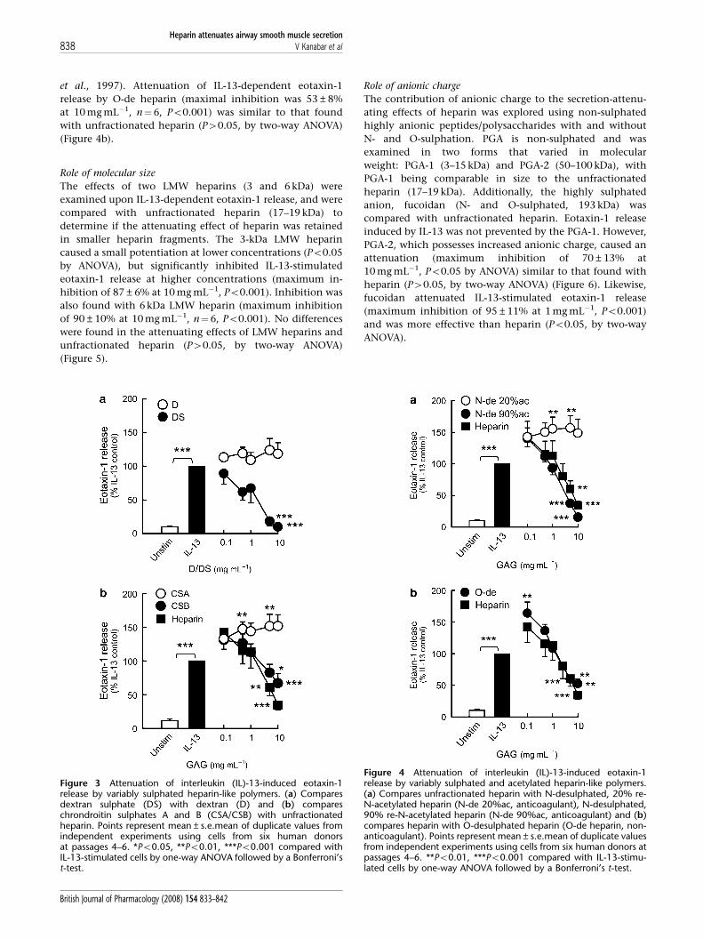
et al., 1997). Attenuation of IL-13-dependent eotaxin-1
release by O-de heparin (maximal inhibition was 53±8%
at 10 mg mL�1, n¼6, Po0.001) was similar to that found
with unfractionated heparin (P40.05, by two-way ANOVA)
(Figure 4b).
Role of molecular size
The effects of two LMW heparins (3 and 6 kDa) were
examined upon IL-13-dependent eotaxin-1 release, and were
compared with unfractionated heparin (17–19 kDa) to
determine if the attenuating effect of heparin was retained
in smaller heparin fragments. The 3-kDa LMW heparin
caused a small potentiation at lower concentrations (Po0.05
by ANOVA), but significantly inhibited IL-13-stimulated
eotaxin-1 release at higher concentrations (maximum in-
hibition of 87±6% at 10 mg mL�1, Po0.001). Inhibition was
also found with 6 kDa LMW heparin (maximum inhibition
of 90±10% at 10 mg mL�1, n¼6, Po0.001). No differences
were found in the attenuating effects of LMW heparins and
unfractionated heparin (P40.05, by two-way ANOVA)
(Figure 5).
Role of anionic charge
The contribution of anionic charge to the secretion-attenu-
ating effects of heparin was explored using non-sulphated
highly anionic peptides/polysaccharides with and without
N- and O-sulphation. PGA is non-sulphated and was
examined in two forms that varied in molecular
weight: PGA-1 (3–15 kDa) and PGA-2 (50–100 kDa), with
PGA-1 being comparable in size to the unfractionated
heparin (17–19 kDa). Additionally, the highly sulphated
anion, fucoidan (N- and O-sulphated, 193 kDa) was
compared with unfractionated heparin. Eotaxin-1 release
induced by IL-13 was not prevented by the PGA-1. However,
PGA-2, which possesses increased anionic charge, caused an
attenuation (maximum inhibition of 70±13% at
10 mg mL�1, Po0.05 by ANOVA) similar to that found with
heparin (P40.05, by two-way ANOVA) (Figure 6). Likewise,
fucoidan attenuated IL-13-stimulated eotaxin-1 release
(maximum inhibition of 95±11% at 1 mg mL�1, Po0.001)
and was more effective than heparin (Po0.05, by two-way
ANOVA).
Figure 3 Attenuation of interleukin (IL)-13-induced eotaxin-1release by variably sulphated heparin-like polymers. (a) Comparesdextran sulphate (DS) with dextran (D) and (b) compareschrondroitin sulphates A and B (CSA/CSB) with unfractionatedheparin. Points represent mean±s.e.mean of duplicate values fromindependent experiments using cells from six human donorsat passages 4–6. *Po0.05, **Po0.01, ***Po0.001 compared withIL-13-stimulated cells by one-way ANOVA followed by a Bonferroni’st-test.
Figure 4 Attenuation of interleukin (IL)-13-induced eotaxin-1release by variably sulphated and acetylated heparin-like polymers.(a) Compares unfractionated heparin with N-desulphated, 20% re-N-acetylated heparin (N-de 20%ac, anticoagulant), N-desulphated,90% re-N-acetylated heparin (N-de 90%ac, anticoagulant) and (b)compares heparin with O-desulphated heparin (O-de heparin, non-anticoagulant). Points represent mean±s.e.mean of duplicate valuesfrom independent experiments using cells from six human donors atpassages 4–6. **Po0.01, ***Po0.001 compared with IL-13-stimu-lated cells by one-way ANOVA followed by a Bonferroni’s t-test.
Heparin attenuates airway smooth muscle secretionV Kanabar et al838
British Journal of Pharmacology (2008) 154 833–842

Discussion
Heparin’s structural heterogeneity arises from polysaccharide
side chains comprising alternating residues of an amino
sugar (glucosamine) and uronic acid (either glucuronic or
iduronic acid), which result in regions of sulphated domains
(O-sulphated and N-sulphated) and less sulphated domains
(N-acetylated). Additionally, heparin comprises irregular
chain lengths and is highly anionic due to the sulphated
and carboxylated groups present on the polysaccharide side
chains (Tyrrell et al., 1999). These variables in chemical
structure are thought to account for its diverse biological
properties (Tyrrell et al., 1999; Lever and Page, 2002).
Although, it is well-established that heparin is antiprolifera-
tive for ASM (Johnson et al., 1995; Kilfeather et al., 1995;
Kanabar et al., 2005), its effects on ASM-derived chemokine
production is unknown. Here, we demonstrate that heparin
prevents IL-13-dependent eotaxin-1 production and identify
some of the key structural moieties on the heparin molecule
required for this activity. These include sulphation, particu-
larly N-sulphation and to a much lesser extent, 2-O- and 3-O-
sulphation. Furthermore, substitution of N-sulphation to
90% acetylation by re-N-acetylation restored activity com-
parable with unfractionated heparin. We also show that
heparin’s attenuating effect on eotaxin-1 secretion was
associated with its non-anticoagulant activity, and was
partially dependent on net anionic charge, but independent
of molecular size above 3 kDa.
Owing to the highly anionic nature of GAGs, electrostatic
interactions can form with other chemokines. Thus, a
confounder at the outset was that heparin could bind
eotaxin-1 and interfere with its detection or reduce the
bioavailability of IL-13 and thus limit eotaxin-1 production.
Alternatively, heparin could bind other factors in the culture
media that could in turn modulate the release of eotaxin. In
support of these possibilities, heparin is known to bind
multiple chemokines including IL-8 (CXCL8), RANTES
(regulated on activation, normal T-cell expressed and
secreted; CCL5), MCP-1 (CCL2) and MIP (macrophage
inflammatory peptide)-1b (CCL4) (Frevert et al., 2003;
Johnson et al., 2004; de Paz et al., 2007; Ellyard et al.,
2007), as well as several cytokines such as IL-1b, IL-2, IL-6, IL-
10, TNF (tumour-necrosis factor)-a and IFN (interferon)-g(Fernandez-Botran et al., 1999; Kuschert et al., 1999;
Mummery and Rider, 2000; Salek-Ardakani et al., 2000; Bode
et al., 2006). Heparin’s capacity to bind cytokines and
chemokines has been postulated to enhance the biological
effects of chemokines by increasing their binding affinities
for their respective receptors, although speculation exists as
to whether this occurs in vivo over physiological pH ranges
(Fernandez-Botran et al., 1999).
A recent report by Ellyard et al. (2007) demonstrates that
heparin binds eotaxin-1 to increase its chemotactic activity
in vivo. To establish if eotaxin-1 binding or IL-13 sequestra-
tion by heparin were important factors in explaining the
attenuating effects of heparin on eotaxin-1 production from
ASM, several strategies were employed. In the first, we
ensured that the concentration of heparin in the ELISA
detection step was below the level that could reduce the
recovery of a known amount of rh-eotaxin-1. Although this
limited direct interference with the assay, it did not exclude a
possible reduction in detection of eotaxin-1 already bound
to heparin. Control experiments indicated that recovery of
rh-eotaxin-1 was reduced by no more than 35% at
10 mg mL�1 heparin, which could not account for overall
attenuating effect of heparin on released eotaxin-1, espe-
cially at the lower concentrations of heparin. Additionally,
we examined the attenuating effect of heparin on multiple
chemokines induced by IL-13 using an antibody array. In
this, we confirmed that heparin prevented IL-13-dependent
upregulation of eotaxin-1 and also other chemokines
including GROa, I-309, I-TAC and MCP-2, but had no effect
on upregulation of MCP-3. Reasons for the lack of effect of
Figure 5 Attenuation of interleukin (IL)-13-stimulated eotaxin-1production by low molecular weight (LMW) heparins. Human airwaysmooth muscle (ASM) cells were stimulated with IL-13 for 24 h in thepresence or absence of LMW heparin fractions of 3 or 6 kDa andcompared with attenuation by unfractionated heparin. Data aremean±s.e.mean of six independent experiments using cells atpassages 4–6 cultured from individual donors. **Po0.01,***Po0.001 compared with IL-13 in the absence of LMW fractionsby ANOVA followed by a Bonferroni’s t-test.
Figure 6 Attenuation of interleukin (IL)-13-stimulated eotaxin-1production by polyanions. Human airway smooth muscle (ASM)cells were stimulated with IL-13 for 24 h in the presence or absenceof sulphated (fucoidan) or non-sulphated (poly-L-glutamic acid,PGA) polyanions. Data are mean±s.e.mean of six independentexperiments using cells at passages 4–6 cultured from individualdonors. **Po0.01, ***Po0.001 compared with IL-13 in the absenceof polyanions by ANOVA followed by a Bonferroni’s t-test.
Heparin attenuates airway smooth muscle secretionV Kanabar et al 839
British Journal of Pharmacology (2008) 154 833–842

heparin on MCP-3 release are unclear, but in the context of
the suppression of other chemokines by heparin, it
suggests that heparin was not acting via IL-13 sequestration.
Likewise, addition (at the time of collection) of heparin to
cell-conditioned media samples from IL-13-stimulated
cells did not reduce the detection of eotaxin-1 (or GROa)
by the array, further suggesting that heparin did not
act to mask eotaxin-1 detection. Significantly, whereas
Ellyard et al. (2007) reported that heparin can bind
rh-eotaxin-1, other GAGs including CSB were found not to
bind rh-eotaxin-1. In the present study, we found that both
heparin and CSB were equivalent in preventing IL-13-
dependent eotaxin-1 release. Thus, on balance, the attenuat-
ing effects of heparin on IL-13-stimulated eotaxin-1 release
from ASM are not explained by sequestration of IL-13, the
stimulus for eotaxin-1 production, or interference with
the direct detection of eotaxin-1 by either the ELISA or the
chemokine array.
The importance of sulphation for the attenuating effects of
heparin on IL-13-dependent eotaxin-1 production was
initially identified with the non-heparin-like polymer DS,
which unlike non-sulphated D inhibited IL-13-dependent
eotaxin-1 production by human ASM. Though structurally
unrelated, both heparin and DS are comparable in their
high level of sulphation, with DS having 3.3 sulphate
residues per disaccharide unit (Windholz et al., 1976)
and heparin having 2.7 sulphate residues per disaccharide
unit (Rabenstein, 2002; de Paz et al., 2007). The compound-
ing effect of DS polymers having varying molecular
weights and hence a differing overall sulphate content was
not addressed directly in this study. We have previously
shown, however, that several DS polymers with molecular
weights between 5 and 500 kDa were equally effective in
preventing fetal bovine serum-dependent proliferation of
bovine ASM cells (Kilfeather et al., 1995), suggesting that the
absolute presence of sulphation above a threshold is
required. This is also supported by the finding that non-
sulphated D had no effect on IL-13-dependent eotaxin-1
production.
Reports suggest that the number of sulphate groups in
polysaccharides directly correlates with the level of bio-
activity (Koyanagi et al., 2003). We further examined the
contribution of sulphation in experiments employing poly-
saccharides that exhibit less sulphation than heparin.
Consistent with the importance of sulphation, we found
that CSA was poorly effective against IL-13-dependent
eotaxin-1 production compared with heparin (average
number of sulphate groups per repeating disaccharide unit
for heparin is 2.7 compared with 1.0 for the chondroitins;
Varma and Varma, 1983). In contrast, CSB, though less
sulphated than heparin, was found to be as effective as
heparin in attenuating eotaxin-1 release. A key difference
between CSA and CSB is in the uronic acid content of the
chrondroitin backbone, which in the case of CSA is
glucuronate-based and iduronate in CSB (Casu et al., 1988).
The greater flexibility of the iduronate residue in the CSB
polysaccharide (compared with the glucuronate residue in
CSA) is commonly used to explain the propensity of
iduronate GAGs to interact with proteins and display a large
number of different biological activities (Kawashima et al.,
2002). This may have relevance in the context of large
chondroitins such as versican that are elevated in the airways
of patients with asthma (Huang et al., 1999). Moreover,
heparin comprises both glucuronate and iduronate residues
in its backbone, but it is unclear whether the iduronate
content explains either the attenuating effect of heparin or
CSB over CSA.
Heparin is variably N-sulphated, O-sulphated and N-
acetylated (Varma and Varma, 1983). Salek-Ardakani et al.
(2000) demonstrated that the ability of heparin to inhibit IL-
10-induced CD14 expression upon monocytes/macrophages
was dependent upon specific sulphation patterns. They
demonstrated that loss of N-sulphates resulted in diminished
inhibitory activity. Consistent with this, we found
N-desulphated, 20% re-N-acetylated heparin (N-de 20%ac)
was devoid of activity against IL-13-dependent eotaxin-1
production. In studies of vascular smooth muscle, loss of
N-sulphation results in complete loss of antiproliferative
activity (Wright et al., 1989). The finding that 90% re-N-
acetylation (N-de 90%ac) in the molecule restored attenuat-
ing activity similar to unfractionated heparin against IL-13-
stimulated eotaxin-1 release also agrees with studies in
vascular smooth muscle where N-acetylation restores anti-
proliferative activity (Castellot et al., 1985; Tiozzo et al.,
1993) and suggests that N-sulphation is required for
heparin’s eotaxin-1-attenuating activity. Although we did
not investigate a minimum level of N-sulphation required
for suppression of IL-13-dependent eotaxin-1 production, a
recent study suggests heparin must retain 24% N-sulphate
groups to retain comparable antiproliferative activity with
native heparin (Longas et al., 2003). Suppression of eotaxin-1
release occurred also with the O-de heparin fraction
(non-anticoagulant), suggesting that the pentasaccharide
sequence required for anticoagulant activity did not play a
role in the attenuation of IL-13-dependent eotaxin-1 pro-
duction, a finding supported in vascular smooth muscle
proliferation in vivo (Guyton et al., 1980) and in vitro
(Hoover et al., 1980). Collectively, as O-de heparin retains
N-sulphation and 6-O-sulphation, but not 2-O- or 3-O-
sulphation (Fryer et al., 1997), our findings suggest
attenuation of ASM cell eotaxin-1 production by heparin
involves N-sulphation and N-acetylation, but not 2-O- or
3-O-sulphation.
We also investigated whether the overall size of the
heparin polymer was a factor in the attenuation. Both the
LMW heparins (3 and 6 kDa) examined prevented IL-13-
dependent eotaxin-1 release, and were comparable in
activity with unfractionated heparin, suggesting that the
efficacy of heparin against IL-13-dependent eotaxin-1 release
resides in chains of 3–6 kDa but may be retained in fractions
o3 kDa. This agrees with previous findings for antiprolifera-
tive activity in human ASM (Kanabar et al., 2005), bovine
ASM (Kilfeather et al., 1995) and a report by Tiozzo et al.
(1991), who demonstrated a graded loss of antiproliferative
activity with decreasing molecular weight from 4.5 to 1 kDa
LMW heparin.
The importance of net anionic charge within the heparin
molecule for antisecretory activity has not been investigated.
PGA, a highly anionic, non-sulphated linear polysaccharide
was examined in two forms, PGA-1 and PGA-2. PGA-1 (3–
Heparin attenuates airway smooth muscle secretionV Kanabar et al840
British Journal of Pharmacology (2008) 154 833–842

15 kDa), although comparable in size to unfractionated
heparin (17–19 kDa) did not prevent IL-13-stimulated
eotaxin-1 release. However, when the larger polymer (PGA-
2, 50–100 kDa) was examined, attenuating activity was
recovered to a level similar to that with heparin, suggesting
that an overall net negative charge was required for this
activity. Previous studies examining the antiproliferative
effect of heparin report variable findings. For example,
Joseph et al. (1997) showed that the antiproliferative
activity of a lower charge density (less negative) heparin
fraction in vascular smooth muscle cells was similar to
the parent heparin, but high charge density fractions were
ineffective. This contrasts with the findings of Wright et al.
(1989), who showed that increased negative charge was
concomitant with antiproliferative activity. Our finding
that the highly anionic, non-sulphated linear polysaccharide
PGA-1 was less effective than the highly anionic
and sulphated polysaccharide fucoidan further suggests
that sulphation contributes to suppression of eotaxin-1
release.
We observed that low concentrations (0.1 mg mL�1) of
unfractionated heparin potentiated both constitutive and
cytokine-induced eotaxin-1 release. This has not been
reported previously and is a potentially undesirable side
effect of any heparin-based therapeutic compound (de Paz
et al., 2007). Potentiation was also observed with both LMW
heparins and with N-de 20%ac, N-de 90%ac and O-de
heparin, suggesting that neither the position of sulphation
nor size of the heparin molecule were determinants for this
effect. Furthermore, this profile contrasts with the observed
pattern of activity for the attenuating effects of heparin and
suggests that independent properties of the heparin mole-
cule account for the potentiating role of heparin on ASM
cells. Although GAGs are required for many chemokines to
function and are reported to enhance chemokine/cytokine
receptor binding affinity (Frevert et al., 2003; Johnson et al.,
2004; de Paz et al., 2007; Ellyard et al., 2007), this would seem
an unlikely explanation given that the potentiating
effect of heparin occurred with both constitutive and
cytokine-induced eotaxin-1 release. Further investigation is
required to ascertain the nature and significance of the
potentiation.
In conclusion, we provide new information for the anti-
inflammatory activity of unfractionated heparin upon either
constitutive release of eotaxin-1 from ASM or release induced
by the Th2-like cytokines, IL-13 and IL-4. Additionally, we
show that specific structural properties of the heparin
molecule are involved in its secretion-attenuating activity
against IL-13-dependent eotaxin-1 production. These in-
clude sulphation, particularly N-sulphation and to a much
lesser extent, 2-O- and 3-O-sulphation and anionic charge.
Attenuation was independent of anticoagulant activity
and molecular weight above 3 kDa. Understanding the
structural properties of the heparin molecule that underlie
its anti-inflammatory activity offers opportunities for the
design of ‘tailor-made’ sequences based on the heparin
template for specific therapeutic uses (Tyrrell et al., 1999;
Lever and Page, 2002) including suppression of eosinophilic
mediators produced by ASM in allergic airways diseases such
as asthma.
Acknowledgements
This work was supported by Asthma UK (no. 05/022 and no.
05/027). We thank the thoracic surgeons, operating theatre
staff and pathologists of Guy’s and St Thomas’ Hospitals
(London) for supply of human lung tissue for culture of
human airway smooth muscle cells.
Conflict of interest
The authors state no conflict of interest.
References
Ammit AJ, Bekir SS, Johnson PR, Hughes JM, Armour CL, Black JL(1997). Mast cell numbers are increased in the smooth muscle ofhuman sensitized isolated bronchi. Am J Respir Crit Care Med 155:1123–1129.
Bode L, Murch S, Freeze HH (2006). Heparan sulfate plays a centralrole in a dynamic in vitro model of protein-losing enteropathy.J Biol Chem 281: 7809–7815.
Brightling CE, Bradding P, Symon FA, Holgate ST, Wardlaw AJ, PavordID (2002). Mast-cell infiltration of airway smooth muscle inasthma. N Engl J Med 346: 1699–1705.
Castellot JJ, Wong K, Herman B, Hoover RL, Albertini DF, Wright TCet al. (1985). Binding and internalization of heparin by vascularsmooth muscle cells. J Cell Physiol 124: 13–20.
Casu B, Petitou M, Provasoli M, Sinay P (1988). Conformationalflexibility: a new concept for explaining binding and biologicalproperties of iduronic acid-containing glycosaminoglycans. TrendsBiochem Sci 13: 221–225.
Chan V, Burgess JK, Ratoff JC, O’Connor BJ, Greenough A, Lee THet al. (2006). Extracellular matrix regulates enhanced eotaxin-1expression in asthmatic airway smooth muscle cells. Am J RespirCrit Care Med 174: 379–385.
de Paz JL, Moseman EA, Noti C, Polito L, von Andrian UH, SeebergerPH (2007). Profiling heparin–chemokine interactions using syn-thetic tools. ACS Chem Biol 2: 735–744.
Ellyard JI, Simson L, Bezos A, Johnston K, Freeman C, Parish CR(2007). Eotaxin-1 selectively binds heparin: an interaction thatprotects eotaxin from proteolysis and potentiates chemotacticactivity in vivo. J Biol Chem 282: 15238–15247.
Fernandez-Botran R, Yan J, Justus DE (1999). Binding of interferongamma by glycosaminoglycans: a strategy for localization and/orinhibition of its activity. Cytokine 11: 313–325.
Frevert CW, Kinsella MG, Vathanaprida C, Goodman RB, Baskin DG,Proudfoot A et al. (2003). Binding of interleukin-8 to heparansulfate and chondroitin sulfate in lung tissue. Am J Respir Cell MolBiol 28: 464–472.
Fryer A, Huang YC, Rao G, Jacoby D, Mancilla E, Whorton R et al.(1997). Selective O-desulfation produces non-anticoagulantheparin that retains pharmacological activity in the lung.J Pharmacol Exp Ther 282: 208–219.
Ghaffar O, Hamid Q, Renzi PM, Allakhverdi Z, Molet S, Hogg JC et al.(1999). Constitutive and cytokine-stimulated expression of eotax-in by human airway smooth muscle cells. Am J Respir Crit Care Med159: 1933–1942.
Green WF, Konnaris K, Woolcock AJ (1993). Effect of salbutamol,fenoterol, and sodium cromoglycate on the release of heparinfrom sensitized human lung fragments challenged withDermatophagoides pteronyssinus allergen. Am J Respir Cell Mol Biol8: 518–521.
Grunig G, Warnock M, Wakil AE, Venkayya R, Brombacher F, RennickDM et al. (1998). Requirement for IL-13 independently of IL-4 inexperimental asthma. Science 282: 2261–2263.
Guyton JR, Rosenberg RD, Clowes AW, Karnovsky MJ (1980).Inhibition of rat arterial smooth muscle cell proliferation byheparin. In vivo studies with anticoagulant and nonanticoagulantheparin. Circ Res 46: 625–634.
Heparin attenuates airway smooth muscle secretionV Kanabar et al 841
British Journal of Pharmacology (2008) 154 833–842

Hirst SJ (2003). Regulation of airway smooth muscle cell immuno-modulatory function: role in asthma. Respir Physiol Neurobiol 137:309–326.
Hirst SJ, Hallsworth MP, Peng Q, Lee TH (2002). Selective inductionof eotaxin release by interleukin-13 or interleukin-4 in humanairway smooth muscle cells is synergistic with interleukin-1betaand is mediated by the interleukin-4 receptor alpha-chain. Am JRespir Crit Care Med 165: 1161–1171.
Hirst SJ, Twort CH, Lee TH (2000). Differential effects of extracellularmatrix proteins on human airway smooth muscle cell prolifera-tion and phenotype. Am J Respir Cell Mol Biol 23: 335–344.
Hoover RL, Rosenberg R, Haering W, Karnovsky MJ (1980). Inhibi-tion of rat arterial smooth muscle cell proliferation by heparin.In vitro studies. Circ Res 47: 578–583.
Huang J, Olivenstein R, Taha R, Hamid Q, Ludwig M (1999).Enhanced proteoglycan deposition in the airway wall of atopicasthmatics. Am J Respir Crit Care Med 160: 725–729.
Huang SK, Xiao HQ, Kleine-Tebbe J, Paciotti G, Marsh DG,Lichtenstein LM et al. (1995). IL-13 expression at the sites ofallergen challenge in patients with asthma. J Immunol 155: 2688–2694.
Johnson PR, Armour CL, Carey D, Black JL (1995). Heparin and PGE2
inhibit DNA synthesis in human airway smooth muscle cells inculture. Am J Physiol 269: L514–L519.
Johnson Z, Kosco-Vilbois MH, Herren S, Cirillo R, Muzio V, Zaratin Pet al. (2004). Interference with heparin binding and oligomeriza-tion creates a novel antiinflammatory strategy targeting thechemokine system. J Immunol 173: 5776–5785.
Jose PJ, Griffiths-Johnson DA, Collins PD, Walsh DT, Moqbel R, TottyNF et al. (1994). Eotaxin: a potent eosinophil chemoattractantcytokine detected in a guinea pig model of allergic airwaysinflammation. J Exp Med 179: 881–887.
Joseph PA, Garg HG, Thompson BT, Liu X, Hales CA (1997).Influence of molecular weight, protein core and charge of nativeheparin fractions on pulmonary artery smooth muscle cellproliferation. Biochem Biophys Res Commun 241: 18–23.
Kanabar V, Hirst SJ, O’Connor BJ, Page CP (2005). Some structuraldeterminants of the antiproliferative effect of heparin-like mole-cules on human airway smooth muscle. Br J Pharmacol 146:370–377.
Kaur D, Saunders R, Berger P, Siddiqui S, Woodman L, Wardlaw Aet al. (2006). Airway smooth muscle and mast cell-derived CCchemokine ligand 19 mediate airway smooth muscle migration inasthma. Am J Respir Crit Care Med 174: 1179–1188.
Kawashima H, Atarashi K, Hirose M, Hirose J, Yamada S, Sugahara Ket al. (2002). Oversulphated chondroitin/dermatan sulphatescontaining GlcAb1/IdoAa1–3GalNAc (4,6-O-disulphate) interactwith L- and P-selectin and chemokines. J Biol Chem 277: 12921–12930.
Kilfeather SA, Tagoe S, Perez AC, Okona-Mensa K, Matin R, Page CP(1995). Inhibition of serum-induced proliferation of bovinetracheal smooth muscle cells in culture by heparin and relatedglycosaminoglycans. Br J Pharmacol 114: 1442–1446.
Koyanagi S, Tanigawa N, Nakagawa H, Soeda S, Shimeno H (2003).Oversulphation of fucoidan enhances its anti-angiogenic andantitumor activities. Biochem Pharmacol 65: 173–179.
Kotsimbos TC, Ernst P, Hamid QA (1996). Interleukin-13 andinterleukin-4 are coexpressed in atopic asthma. Proc Assoc AmPhysicians 108: 368–373.
Kuschert GS, Coulin F, Power CA, Proudfoot AE, Hubbard RE,Hoogewerf AJ et al. (1999). Glycosaminoglycans interact selec-tively with chemokines and modulate receptor binding andcellular responses. Biochemistry 38: 12959–12968.
Lever R, Page CP (2002). Novel drug development opportunities forheparin. Nat Rev Drug Discov 1: 140–148.
Longas MO, Garg HG, Trinkle-Pereira JM, Hales CA (2003). Heparinantiproliferative activity on bovine pulmonary artery smoothmuscle cells requires both N-acetylation and N-sulfonation.Carbohydr Res 338: 251–256.
Moore PE, Church TL, Chism DD, Panettieri RA, Shore SA (2002).IL-13 and IL-4 cause eotaxin release in human airway smoothmuscle cells: a role for ERK. Am J Physiol Lung Cell Mol Physiol 282:L847–L853.
Mummery RS, Rider CC (2000). Characterization of the heparin-binding properties of IL-6. J Immunol 165: 5671–5679.
Page S, Ammit AJ, Black JL, Armour CL (2001). Human mast cell andairway smooth muscle cell interactions: implications for asthma.Am J Physiol Lung Cell Mol Physiol 281: L1313–L1323.
Panettieri RA (2002). Airway smooth muscle: an immunomodulatorycell. J Allergy Clin Immunol 110: S269–S274.
Rabenstein DL (2002). Heparin and heparan sulfate: structure andfunction. Nat Prod Rep 19: 312–331.
Robinson DS (2004). The role of the mast cell in asthma: induction ofairway hyperresponsiveness by interaction with smooth muscle?J Allergy Clin Immunol 114: 58–65.
Rose MJ, Page C (2004). Glycosaminoglycans and the regulationof allergic inflammation. Curr Drug Targets Inflamm Allergy 3:221–225.
Salek-Ardakani S, Arrand JR, Shaw D, Mackett M (2000). Heparin andheparan sulfate bind interleukin-10 and modulate its activity.Blood 96: 1879–1888.
Seeds EA, Horne AP, Tyrrell DJ, Page CP (1995). The effect of inhaledheparin and related glycosaminoglycans on allergen-inducedeosinophil infiltration in guinea-pigs. Pulm Pharmacol 8: 97–105.
Tiozzo R, Cingi MR, Reggiani D, Andreoli T, Calandra S, Milani MRet al. (1993). Effect of the desulfation of heparin on itsanticoagulant and antiproliferative activity. Thromb Res 70:99–106.
Tiozzo R, Reggiani D, Cingi MR, Bianchini P, Osima B, Calandra S(1991). Effect of heparin derived fractions on the proliferation andprotein synthesis of cells in culture. Thromb Res 62: 177–188.
Tyrrell DJ, Horne AP, Holme KR, Preuss JM, Page CP (1999). Heparinin inflammation: potential therapeutic applications beyond anti-coagulation. Adv Pharm 46: 151–208.
Varma R, Varma RS (eds) (1983). Chemistry and metabolism ofglycosaminoglycans and proteoglycans. In. Mucopolysaccharides.Glycosaminoglycans of Body Fluids in Health and Disease. Walter deGruyter: Berlin. pp 3–48.
Windholz M, Budaveri S, Strountsos L, Fertig M (1976). The MerckIndex, 9th edn. Merck Rahway: New Jersey, 387 pp.
Wright Jr TC, Castellot Jr JJ, Petitou M, Lormeau JC, Choay J,Karnovsky MJ (1989). Structural determinants of heparin’s growthinhibitory activity. Interdependence of oligosaccharide size andcharge. J Biol Chem 264: 1534–1542.
Zhu Z, Homer RJ, Wang Z, Chen Q, Geba GP, Wang J et al. (1999).Pulmonary expression of interleukin-13 causes inflammation,mucus hypersecretion, subepithelial fibrosis, physiologic abnorm-alities, and eotaxin production. J Clin Invest 103: 779–788.
Heparin attenuates airway smooth muscle secretionV Kanabar et al842
British Journal of Pharmacology (2008) 154 833–842