Myb-Related Schizosaccharomyces pombe cdc5p Is Structurally and Functionally Conserved in Eukaryotes
Staufen- and FMRP-Containing Neuronal RNPs Are Structurallyand Functionally Related to Somatic P Bodies
Scott A. Barbee1,2, Patricia S. Estes1,2, Anne-Marie Cziko1,2, Jens Hillebrand4, Rene A.Luedeman1,2, Jeff M. Coller1,3,11, Nick Johnson1,3, Iris C. Howlett1,2, Cuiyun Geng5, RyuUeda10, Andrea H. Brand6, Sarah F. Newbury7, James E. Wilhelm8, Richard B. Levine2, AkiraNakamura9,12, Roy Parker1,3,12,*, and Mani Ramaswami1,2,4,12,*
1Department of Molecular and Cellular Biology, University of Arizona Tucson, Arizona 85721 2ARL Divisionof Neurobiology, University of Arizona Tucson, Arizona 85721 3Howard Hughes Medical Institute, Universityof Arizona Tucson, Arizona 85721 4Smurfit Institute of Genetics and TCIN Lloyd Building, Trinity CollegeDublin, Dublin-2, Ireland 5Institute for Cellular and Molecular Biology Section of Molecular Cell andDevelopmental Biology, The University of Texas at Austin 1 University Station Austin, Texas 787126Wellcome/CRC Institute and Department of Genetics, University of Cambridge, Cambridge CB2 IQR, UnitedKingdom 7Institute of Cell and Molecular Biosciences, University of Newcastle, The Medical SchoolFramlington Place, Newcastle-upon-Tyne, United Kingdom 8Section of Cell and Developmental Biology,Division of Biological Sciences, University of California, San Diego La Jolla, California 92093 9Laboratoryfor Germline Development, RIKEN Center for Developmental Biology, 2-2-3 Minatojimaminamimachi Chuo-ku, Kobe 650-0047 Japan 10Invertebrate Genetics Lab, Genetic Strains Research Center, National Instituteof Genetics (NIG) 1111 Yata Mishima, Shizuoka 411-8540, Japan
SummaryLocal control of mRNA translation modulates neuronal development, synaptic plasticity, andmemory formation. A poorly understood aspect of this control is the role and composition ofribonucleoprotein (RNP) particles that mediate transport and translation of neuronal RNAs. Here,we show that staufen- and FMRPcontaining RNPs in Drosophila neurons contain proteins alsopresent in somatic “P bodies,” including the RNA-degradative enzymes Dcp1p and Xrn1p/Pacmanand crucial components of miRNA (argonaute), NMD (Upf1p), and general translational repression(Dhh1p/Me31B) pathways. Drosophila Me31B is shown to participate (1) with an FMRP-associated,P body protein (Scd6p/trailer hitch) in FMRP-driven, argonautedependent translational repressionin developing eye imaginal discs; (2) in dendritic elaboration of larval sensory neurons; and (3) inbantam miRNA-mediated translational repression in wing imaginal discs. These results argue for aconserved mechanism of translational control critical to neuronal function and open up newexperimental avenues for understanding the regulation of mRNA function within neurons.
IntroductionLocalized translation of mRNAs has emerged as a major mechanism for regulating dynamicintracellular processes such as those involved in early embryonic development and synapseplasticity (Johnstone and Lasko, 2001;Martin, 2004). In the specific cases of growthcone
*Correspondence: [email protected] (R.P.), [email protected]. edu (M.R.)11Present address: Center for RNA Molecular Biology, Case Western Reserve University, Cleveland, Ohio 44106.12Additional corresponding authors: Akira Nakamura (akiran@cdb. riken.jp), Roy Parker ([email protected]), and ManiRamaswami ([email protected]).Supplemental DataThe Supplemental Data for this article can be found online at http://www.neuron.org/cgi/content/full/52/6/997/DC1/.
NIH Public AccessAuthor ManuscriptNeuron. Author manuscript; available in PMC 2007 August 29.
Published in final edited form as:Neuron. 2006 December 21; 52(6): 997–1009.
NIH
-PA Author Manuscript
NIH
-PA Author Manuscript
NIH
-PA Author Manuscript

guidance and synapse plasticity, temporally and spatially restricted repression of mRNAtranslation allows subcellular locations within a single neuron to transiently achieve differentmolecular and functional properties. This allows growth-cone turning in specific directions or,potentially, synapse-specific alterations required during learning and memory (Martin,2004;Richter and Lorenz, 2002). Similarly, in dendrites, translationally repressed RNAsmobilized by synaptic stimulation are translated through control mechanisms that may includepolyadenylation of mRNAs at stimulated synapses (Richter and Lorenz, 2002). It is likely thatsuch locally translated mRNAs influence dendritic growth as well as maintain proteinsynthesis-dependent forms of synaptic plasticity (Ye et al., 2004;Martin, 2004).
Translational repression often occurs in cytoplasmic, ribonucleoprotein (RNP) particles. In themammalian nervous system, staufen-containingRNPsare thought to mediate translationalrepression and/or mRNA transport of dendritically localized mRNAs (Kiebler and Bassell,2006). These granules often contain the fragile X mental retardation protein (FMRP), atranslational repressor that negatively regulates dendritic growth (Nimchinsky et al., 2001), aswell as mRNAs translationally regulated at synapses (Knowles et al., 1996;Kohrmann et al.,1999;Krichevsky and Kosik, 2001;Mallardo et al., 2003;Kanai et al., 2004). However, thecompositional diversity, cellular functions, and underlying mechanisms of staufen-containingRNPs remain largely unknown.
The shared presence of staufen (Stau) and an associated protein, barentsz (Btz), on maternaland neuronal RNPs suggests a compositional similarity between at least two classes of RNAstorage/transport granules (Kiebler et al., 1999;Macchi et al., 2003;Mallardo et al., 2003). Thishypothesis is further supported by roles for Stau in both maternal and neuronal mRNA transport(St Johnston et al., 1991;Tang et al., 2001) and for FMRP (dFMR1 in Drosophila) intranslational repression during Drosophila oocyte development (Costa et al., 2005). Whileadditional shared components may soon be identified using biochemistry combined withproteomics (Elvira et al., 2006;Kanai et al., 2004), there is currently limited information onhow far biochemical and functional similarities between neuronal and maternal RNPs extend.
Recently, a third class of conserved somatic cytoplasmic RNPs, termed cytoplasmic RNAprocessing bodies (“P bodies”; also termed GW182 or DCP bodies), have been described inyeast, C. elegans, and mammalian cells. P bodies contain nontranslating mRNAs and multipleproteins involved in mRNA degradation and translational control (Kiebler and Bassell,2006). While first described as sites of mRNA decapping and 5′to 3′exonucleolytic degradation(Cougot et al., 2004;Sheth and Parker, 2003), P bodies have recently been shown to functionin conventional and miRNA-mediated translational control as well as mRNA storage(Brengues et al., 2005;Coller and Parker, 2005;Liu et al., 2005a;Pillai et al., 2005). Indeed,shared features of yeast mRNA turnover and translational pathways are indicated by theobservation that two proteins that accumulate with mRNA in P bodies, Dhh1p and Pat1p,promote both mRNA decapping and translational repression (Coller and Parker, 2005).Similarities between P bodies and maternal RNPs are further suggested by the known functionsof Dhh1p-orthologous, DEAD-box RNA helicases (Me31B, CGH-1, and Xp54) in maternalRNA granules of Drosophila, C. elegans, and Xenopus oocytes, respectively (Coller et al.,2001;Ladomery et al., 1997;Nakamura et al., 2001;Navarro et al., 2001). Together, theseobservations led us to hypothesize that many RNA granules will share a core composition andfunction.
In this work, we provide experimental support for a model in which neuronal staufen-containing RNPs (also referred to here as “staufen RNPs” or “staufen granules”) sharefundamental organization with maternal RNA granules and somatic P bodies. Staufen RNPsvisualized in Drosophila are shown to contain not only maternal translational control and RNA-transport molecules but also components of miRNA, nonsense-mediated decay (NMD), and
Barbee et al. Page 2
Neuron. Author manuscript; available in PMC 2007 August 29.
NIH
-PA Author Manuscript
NIH
-PA Author Manuscript
NIH
-PA Author Manuscript

RNA-turnover pathways present on somatic P bodies. Additionally, we present functional datashowing that Me31B/Dhh1p, a protein present in neuronal staufen granules, P bodies, andmaternal RNA granules, functions (1) together with another dFMR1- associated,Pbodyprotein(trailerhitch/Scd6p) indFMR1- driven, argonaute-dependent translational repression in thedeveloping eye disc; (2) dendritic elaboration in larval sensory neurons, a process previouslyshown to be regulated by translational repressor proteins pumilio (Pum), nanos (Nos), anddFMR1; and (3) in bantam miRNA-mediated translational repression in the developing wingimaginal disc. Thus, in addition to documenting broadly conserved composition and functionof RNA granules in neuronal, germline, and somatic cells, we identify Me31B as novelcomponent (to our knowledge) of thedFMR1 pathway, which acts as a critical regulator ofdendritic morphogenesis and microRNA function in vivo.
ResultsNeuronal Staufen Granules in Drosophila
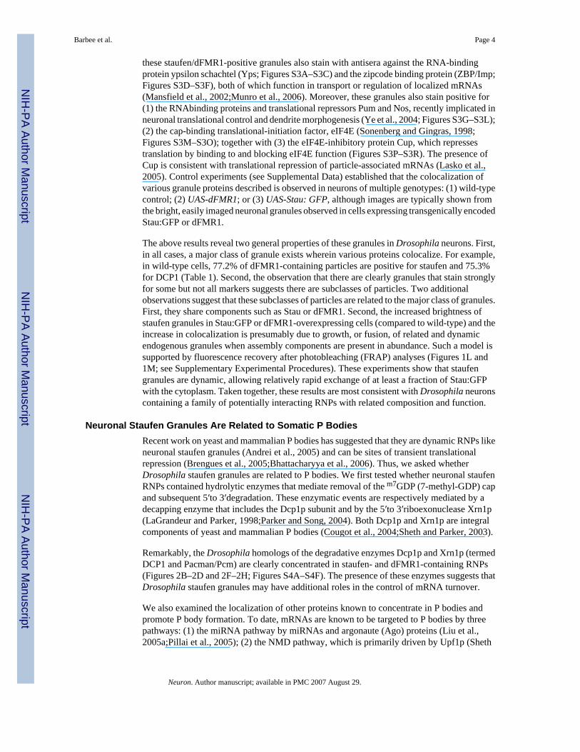
To identify and characterize Drosophila RNPs involved in neuronal translation control, wecombined a primary cell-culture system (Kraft et al., 1998) with microscopic localization oftransgenically expressed Stau, a highly conserved protein of maternal RNPs and mammalianneuronal granules (Ferrandon et al., 1994;Kiebler et al., 1999). A Stau:GFP fusion proteinexpressed in Drosophila ventral ganglion neurons is concentrated in puncta within neurites of3- to 4-day-old primary cultures of dissociated larval ventral ganglia, with large punctaobserved in the cell body (Figure 1A; see Figure S1 in the Supplemental Data available online).Of 292 granules analyzed in nine Stau:GFP-expressing cells, 56.5% of granules were within1 μm of branch points and 33.9% were away from branch points (Figure 1A and inset). Thisobserved localization of staufen granules is consistent with the previously proposed role fortranslational regulation in controlling dendritic branching in Drosophila (Ye et al., 2004). Invivo, panneuronally expressed Stau:GFP revealed similar particles within peripheral nervesexiting the larval central nervous system as well as in cell bodies within the ventral ganglion(Figure 1B).
To determine whether these Stau:GFP particles were similar to mammalian RNPs involved inneuronal mRNA regulation, we asked if they contained other established components ofmammalian neuronal RNPs. As shown in Figure 1, Stau:GFP-containing granules werestrongly labeled by antibodies against dFMR1 (Figures 1C–1E) or Btz (Figures 1F–1H).Stau:GFP and dFMR1 colocalized extensively but not completely in wild-type and Stau:GFP-or dFMR1-overexpressing neurons (Table 1; Figure 1; Figure S2). These results indicate thatdFMR1 and Stau exist substantially in the same granules but can also be observed in separateyet related particles (see Discussion).
For additional evidence that staufen granules could be involved in translational repression, wealso examined whether a known dendritically transported mRNA was present in these staufen/dFMR1-positive granules. Recent work has shown that Drosophila CaMKII mRNA istransported along dendrites through a process stimulated by neuronal activity (Ashraf et al.,2006). This phenomenon is analogous to activity-stimulated movement of mammalianCaMKII mRNAs in staufen-positive neuronal RNPs (Krichevsky and Kosik, 2001). Tovisualize CaMKII mRNA, we cultured neurons coexpressing a GFP-tagged, nuclearly targetedRNA virus capsid protein (GFP:MCP) and CaMKII mRNA, multiply tagged with binding sitesfor MCP (Ashraf et al., 2006). Figures 1I–1K show that CaMKII mRNA-containing punctaobserved in neurites overlap with protein markers of staufen granules.
The presence of Stau, dFMR1, Btz, and, in at least some cases, CaMKII mRNA in overlappingpuncta indicates that these foci represent Drosophila neuronal RNPs likely to function in thetransport and translational regulation of neuronal mRNAs. Consistent with this hypothesis,
Barbee et al. Page 3
Neuron. Author manuscript; available in PMC 2007 August 29.
NIH
-PA Author Manuscript
NIH
-PA Author Manuscript
NIH
-PA Author Manuscript
these staufen/dFMR1-positive granules also stain with antisera against the RNA-bindingprotein ypsilon schachtel (Yps; Figures S3A–S3C) and the zipcode binding protein (ZBP/Imp;Figures S3D–S3F), both of which function in transport or regulation of localized mRNAs(Mansfield et al., 2002;Munro et al., 2006). Moreover, these granules also stain positive for(1) the RNAbinding proteins and translational repressors Pum and Nos, recently implicated inneuronal translational control and dendrite morphogenesis (Ye et al., 2004; Figures S3G–S3L);(2) the cap-binding translational-initiation factor, eIF4E (Sonenberg and Gingras, 1998;Figures S3M–S3O); together with (3) the eIF4E-inhibitory protein Cup, which repressestranslation by binding to and blocking eIF4E function (Figures S3P–S3R). The presence ofCup is consistent with translational repression of particle-associated mRNAs (Lasko et al.,2005). Control experiments (see Supplemental Data) established that the colocalization ofvarious granule proteins described is observed in neurons of multiple genotypes: (1) wild-typecontrol; (2) UAS-dFMR1; or (3) UAS-Stau: GFP, although images are typically shown fromthe bright, easily imaged neuronal granules observed in cells expressing transgenically encodedStau:GFP or dFMR1.
The above results reveal two general properties of these granules in Drosophila neurons. First,in all cases, a major class of granule exists wherein various proteins colocalize. For example,in wild-type cells, 77.2% of dFMR1-containing particles are positive for staufen and 75.3%for DCP1 (Table 1). Second, the observation that there are clearly granules that stain stronglyfor some but not all markers suggests there are subclasses of particles. Two additionalobservations suggest that these subclasses of particles are related to the major class of granules.First, they share components such as Stau or dFMR1. Second, the increased brightness ofstaufen granules in Stau:GFP or dFMR1-overexpressing cells (compared to wild-type) and theincrease in colocalization is presumably due to growth, or fusion, of related and dynamicendogenous granules when assembly components are present in abundance. Such a model issupported by fluorescence recovery after photobleaching (FRAP) analyses (Figures 1L and1M; see Supplementary Experimental Procedures). These experiments show that staufengranules are dynamic, allowing relatively rapid exchange of at least a fraction of Stau:GFPwith the cytoplasm. Taken together, these results are most consistent with Drosophila neuronscontaining a family of potentially interacting RNPs with related composition and function.
Neuronal Staufen Granules Are Related to Somatic P BodiesRecent work on yeast and mammalian P bodies has suggested that they are dynamic RNPs likeneuronal staufen granules (Andrei et al., 2005) and can be sites of transient translationalrepression (Brengues et al., 2005;Bhattacharyya et al., 2006). Thus, we asked whetherDrosophila staufen granules are related to P bodies. We first tested whether neuronal staufenRNPs contained hydrolytic enzymes that mediate removal of the m7GDP (7-methyl-GDP) capand subsequent 5′to 3′degradation. These enzymatic events are respectively mediated by adecapping enzyme that includes the Dcp1p subunit and by the 5′to 3′riboexonuclease Xrn1p(LaGrandeur and Parker, 1998;Parker and Song, 2004). Both Dcp1p and Xrn1p are integralcomponents of yeast and mammalian P bodies (Cougot et al., 2004;Sheth and Parker, 2003).
Remarkably, the Drosophila homologs of the degradative enzymes Dcp1p and Xrn1p (termedDCP1 and Pacman/Pcm) are clearly concentrated in staufen- and dFMR1-containing RNPs(Figures 2B–2D and 2F–2H; Figures S4A–S4F). The presence of these enzymes suggests thatDrosophila staufen granules may have additional roles in the control of mRNA turnover.
We also examined the localization of other proteins known to concentrate in P bodies andpromote P body formation. To date, mRNAs are known to be targeted to P bodies by threepathways: (1) the miRNA pathway by miRNAs and argonaute (Ago) proteins (Liu et al.,2005a;Pillai et al., 2005); (2) the NMD pathway, which is primarily driven by Upf1p (Sheth
Barbee et al. Page 4
Neuron. Author manuscript; available in PMC 2007 August 29.
NIH
-PA Author Manuscript
NIH
-PA Author Manuscript
NIH
-PA Author Manuscript

and Parker, 2006); and (3) a general pathway that works on bulk mRNA and is mediated bythe Dhh1p and Pat1p proteins in yeast (Coller and Parker, 2005).
Association of Ago-1 and Ago-2 with dFMR1 has been argued by genetic and biochemicaltests in Drosophila (Ishizuka et al., 2002;Jin et al., 2004).Wetherefore tested whether theseproteins were present on staufen- and dFMR1-positive granules. While the generally poorquality of the Ago-1 antibody for immunohistochemistry (data not shown) did not allow us toeasily examine its presence in Drosophila neuronal granules, Ago-2 could be visualized withinthese particles (Figures 2Q–2S; Figures S4J–S4L). This is consistent with recent analysessuggesting that miRNAs may function in granules such as P bodies (Pillai et al., 2005;Liu etal., 2005a,2005b;Jakymiw et al., 2005;Chu and Rana, 2006). Thus, our observation that Ago-2is present in dFMR1-containing staufen granules is consistent with a previously proposed rolefor FMRP/dFMR1 in miRNA/RNAi-mediated gene silencing (Kosik and Krichevsky, 2005).
The critical protein for translation repression in NMD, UPF1, is also present on staufen granules(Figures 2J–2L; Figures S4G–S4H). Finally, we observed that Me31B, a highly conservedhomolog of yeast Dhh1p, is also present on these particles (Figures 2N–2P; Figures S5D–S5F).Thus, neuronal staufen- and dFMR1-positive RNPs contain critical components of threedifferent systems of translation repression suggesting that these RNPs, like P bodies, mediatediverse RNA regulatory events.
The presence of similar proteins in staufen granules and P bodies suggests that these neuronaland somatic RNPs share a similar core biochemical composition. These data also suggest thatshared proteins will be common to other types of RNA granules, including maternal RNAgranules. Consistent with this view, the decapping enzymes (Dcp1 and Dcp2) have beenrecently reported to be present on C. elegans P granules (Lall et al., 2005). Moreover, we findboth Pcm/Xrn1p and DCP1 colocalize with Me31B in maternal RNA granules inDrosophila nurse cells (Figures 3A–3F).
Trailer Hitch, a Me31B-Associated Maternal Protein, Is Present on P Bodies and NeuronalStaufen Granules
Me31B functions during oogenesis as a translational repressor of oskar mRNA in a well-studied eIF4E-Cup- Bru translational control complex (Lasko et al., 2005). This complex alsocontains a conserved Sm- and FDFdomain RNA-binding protein, trailer hitch (Tral). InDrosophila ovaries, Tral coimmunoprecipitates with Me31B and colocalizes with Me31B-containing maternal RNA granules (Boag et al., 2005).
Figure 4A shows that a Me31B/Tral/dFMR1 complex coimmunoprecipitates fromDrosophila adult head extracts, consistent with a model in which the three proteins functiontogether in neuronal translation control. Me31B, Tral, and dFMR1 all have a similar, ubiquitousexpression pattern in the central nervous system, showing a predominantly cytoplasmic,steady-state localization (Figures S5A–S5C). In cultured Drosophila neurons, Tral alsolocalizes to staufen- and dFMR1-containing granules (Figures 4B–4D and Figures S5G–S5I).Moreover, a GFP fusion to Scd6p, the S. cerevisiae homolog of Tral, colocalizes withDcp2p:RFP under high cell density or nutrient starvation, conditions that enlarge yeast P bodies(Teixeira et al., 2005; Figures 4E–4G). Together, these data indicate that (1) Tral is present onDrosophila neuronal RNPs in a biochemical complex that contains Me31B and dFMR1; and(2) Scd6p, the yeast homolog of Tral, is a component of P bodies. The latter observation furtherextends similarities between P bodies and staufen granules.
Barbee et al. Page 5
Neuron. Author manuscript; available in PMC 2007 August 29.
NIH
-PA Author Manuscript
NIH
-PA Author Manuscript
NIH
-PA Author Manuscript

Me31B and Tral Are Required for dFMR1-Mediated Translational RepressionThe compositional similarity of P bodies and staufen RNPs suggests that neuronal translationalcontrol is regulated through proteins and mechanisms associated with somatic P bodies. Totest this prediction, we focused on the highly conserved DEAD-box RNA helicase Me31B,which functions in translational repression of maternal mRNAs and in the targeting of mRNAsto P bodies (Coller and Parker, 2005;Nakamura et al., 2001). The presence of Me31B and Tralwith Ago-1 on dFMR1-containing complexes suggests that these proteins may function inneuronal translation control, potentially with dFMR1 in miRNA-mediated processes.
To test whether Me31B and Tral function in dFMR1- mediated translational repression, weasked if defects caused by dFMR1 overexpression in developing eyes were modified in geneticbackgrounds deficient for Me31B or Tral. Ectopic overexpression of dFMR1 in the compoundeye driven by the sevenless enhancer (sevdFMR1) results in a “rough-eye” phenotype througha pathway that requires dFMR1 domains essential for translational repression as well as Ago-1function (Figure 5A; Jin et al., 2004;Wan et al., 2000).
As shown in Figure 5B, loss of a single copy of me31B suppressed sev-dFMR1-induced rougheye phenotypes. Unambiguous suppression was observed with either me31BD1 or me31BD2allele (Figure 5B; data not shown). Internally, the disruption of ommatidia caused bydFMR1 overexpression was also suppressed, as observed in tangential sections (Figures 5Fand 5G). This suppression is a direct result of me31B deficiency, because a genomic me31B+ transgene, P[me31BAflII], which is capable of rescuing lethality of me31BΔmutants(Nakamura et al., 2001) rescues suppression of the sev-dFMR1 rough eye phenotype (Figures5C and 5H).
Results with tral mutations were similar. We isolated deletion alleles for tral (seeSupplementary Experimental Procedures) and found them to result in larval lethality. Bothtral deletions dominantly suppressed sev-dFMR1- induced rough eyes (Figures 5D and 5I; datanot shown). A tral+ genomic transgene (P[tral-10]) containing the entire tral locus wassufficient to rescue the lethality of tral mutants. This genomic transgene also “rescued”dominant suppression of the rough eye phenotype, thereby demonstrating that phenotypicsuppression of sev-dFMR1occurs specifically due to loss of tral (Figures 5E and 5J).
Given that Me31B, Tral, and dFMR1 form a physical complex, the above results suggest thatMe31B and Tral act, together with dFMR1, as translational regulators in neuronal cells. Analternative interpretation is that single-copy deletions of tral or me31B block apoptosis or otherdevelopmental errors induced by Sev-dFMR1. However, this is unlikely for three reasons: First,coimmunoprecipitation and colocalization of Me31B, Tral, and dFMR1 are more consistentwith a direct mechanism. Second, all three proteins have RNA-binding domains that predictroles in translational control. Finally, ectopic expression of Me31B in the eye causes rougheyes via a mechanism requiring amino acid residues necessary for translational repression(Figures S6A–S6C; see below).
Me31B and Tral Regulate Dendrite Morphogenesis in Sensory NeuronsThe observed effect of Me31B (and Tral) induction on dendritic development of sensoryneurons (Figure 6 and Figures S6D–S6F) provides further evidence for function in neuronaltranslation regulation. Previous studies have established that translational control of geneexpression regulates dendrite morphogenesis in vivo. For example, neurons of human fragileX patients and Drosophila dFMR1 mutants show an increase in dendritic spine number andlength (Nimchinsky et al., 2001;Lee et al., 2003). Conversely, induction of dFMR1, Pum, orNos in class IV Drosophila da sensory neurons greatly perturbs, and can dramatically reduce,higher-order dendritic branching (Lee et al., 2003;Ye et al., 2004). If Me31B and Tral act in
Barbee et al. Page 6
Neuron. Author manuscript; available in PMC 2007 August 29.
NIH
-PA Author Manuscript
NIH
-PA Author Manuscript
NIH
-PA Author Manuscript

dendritic translational control, we anticipated that their induction would also have specificeffects on higher-order dendritic branching.
Overexpression of Me31B in class IV neurons substantially reduced high-order dendriticcomplexity (Figures 6A and 6B). In neurons overexpressing Me31B, the number of higher-order dendrites was significantly reduced compared with the control, in which only the reportergene UAS-mCD8:GFP was overexpressed (p ≤0.001; Figures 6A, 6B, and 6F). To determineif this effect of Me31B induction reflected increased translational repression activity of Me31B,we asked whether similar effects would be shown by induction of an Me31B mutant protein(D207A, E208A) homologous to a yeast Dhh1p mutant incapable of translational repression(Coller and Parker, 2005). Expressed at comparable levels (data not shown), the mutanttransgene had no effect on dendritic complexity (Figures 6C and 6F), consistent with theobserved effect being dependent on Me31B-induced translational repression.
Overexpression of Tral in class IV neurons also substantially changed dendrite morphologycompared to the control (Figures S6D–S6F). Interestingly, closer examination revealed asignificant increase in the number of finer dendritic “tendrils” at terminal dendritic branchescompared to control neurons. Differences between effects of Tral and Me31B induction ondendritic arborization are consistent with a relatively specific role for CAR-1, the C. elegansortholog of Tral, in translational control compared to CGH-1 (the Me31B ortholog), suggestedby phenotypic differences following RNAi-mediated inhibition of respective proteins in theC. elegans germline (Audhya et al., 2005;Navarro et al., 2001).
In class IV sensory neurons, loss of nanos or pumilio causes abnormal dendritic growth (Yeet al., 2004).This aberrant growth, visible in about 20% of mutant neurons, is most easilyapparent as a loss of “tiling,” a term that refers to the complete, nonoverlapping coverage ofthe epidermis by dendrites of wild-type sensory neurons (Ye et al., 2004;Grueber et al.,2003). We therefore asked whether loss of me31B, achieved by expressing a transgenic RNAiconstruct that generates a hairpin Me31B RNA (UAS-Me31Bhpn) would cause similar defects.As shown in Figure 6E, UAS-Me31Bhpn sensory neurons showed frequent defects in terminaldendrite morphology and dendritic tiling highly reminiscent of nanos and pum phenotypes.Incomplete coverage of the epidermis was observed in at least 33% (n = 15 neurons) of neuronsanalyzed. Additionally, Me31Bhpn neurons show a modest increase (37%) in high-orderdendritic complexity similar to that observed in dFmr1 mutants (Figure 6F;Lee et al., 2003).Parallel analyses of a hairpin construct for Lk6, which encodes the Drosophila homolog of theeIF4E-kinase MNK, showed no effect on dendritic branching of class IV sensory neurons (datanot shown).
From these data, we conclude that Me31B (and Tral) regulates dendritic arborization of classIV da neurons. This observation, consistent with observations of other translational repressorssuch as dFMR1, Pum, and Nos provides a second line of evidence suggesting that Me31B andTral function as neuronal translational regulators.
Me31B Functions in MicroRNA-Mediated Translational RepressionTwo previous findings led us to the hypothesis that the dFMR1-associated Me31B protein maybe required for miRNA/RNAi function. First, FMRP/dFMR1, showing strong biochemical orgenetic interactions with Ago-1 and Ago-2, is strongly implicated in microRNA-mediatedtranslational repression (Kosik and Krichevsky, 2005). Second, miRNA-mediated repressionhas been proposed to occur in P bodies of somatic cells (Liu et al., 2005a;Pillai et al., 2005).Thus, we tested whether Me31B is required in vivo for the function of bantam, an endogenousmiRNA that represses hid mRNA translation in wing imaginal discs (Brennecke et al., 2003).
Barbee et al. Page 7
Neuron. Author manuscript; available in PMC 2007 August 29.
NIH
-PA Author Manuscript
NIH
-PA Author Manuscript
NIH
-PA Author Manuscript

We used two transgenically encoded GFP reporters to assay bantam-mediated translationalrepression (Brennecke et al., 2003). The “hid reporter,” which carries the 3′ UTR of hid fusedto the 3′end of EGFP-coding sequence, closely reports bantam repression of a native targetmRNA. This 3′UTR contains four repeats complementary to bantam target recognitionsequences, with several mismatches typically associated with miRNAmediated translationalrepression. The “bantam reporter,” in which four synthetic repeats 100% complementary tothe bantam target recognition element are fused 3′to EGFP coding module, also reportsbantam function.
We used the heat-shock FLP/FRT system to generate me31b−/− clones in the wing disc andidentified these clones by loss of β-galactosidase or Me31B staining with respective antibodies(Figure 7A–C). We then asked how a control protein (Dlg), hid reporter, or bantam reporterexpression was affected by loss of Me31B (Figures 7F and 7G). While cells lacking me31Bshowed no detectable increase in a control protein (Dlg) expression (Figure 7G), they showedclear increases in both hid reporter (Figures 7C–7E) and bantam reporter (Figures 7H–7J)expression, indicating that bantam-mediated silencing does not function in the absence ofme31B. These data, from in vivo analyses of an endogenous miRNA in cells carrying a nullmutation for me31B, support a recent study showing a role for RCK (the human homolog ofMe31B) in Let-7 miRNA-mediated translational repression in cultured mammalian cells (Chuand Rana, 2006). In addition, our observations extend this study by demonstrating a role forMe31B in repression mediated by perfectly base-paired miRNAs. It should be noted that therequirement for Me31B for efficient repression of the bantam reporter does not necessarilymean that Me31B is required for miRNA-mediated endonucleolytic cleavage, since it is likelythat repression by perfectly base-paired miRNAs can be a combination of translationrepression, decapping, and/or endonucleolytic cleavage of the mRNA (Valencia-Sanchez etal., 2006). Importantly, these data demonstrate that Me31B is required for repression mediatedby an endogenous Drosophila miRNA. An obvious corollary of our analysis in wing imaginaldiscs is that Me31B plays a similar role in mediating functions of neuronal miRNAs, althoughassays to directly test this issue are not immediately available in Drosophila.
DiscussionNeuronal Staufen RNPs Are Related to Somatic P Bodies
Several observations now indicate that P bodies, maternal granules, and a major subclass ofneuronal RNP are similar in underlying composition and represent a conserved system for theregulation of cytoplasmic mRNAs. As summarized in Table 2, known RNA transport andtranslational repressors shared between maternal and neuronal staufen granules now include,Stau, Btz, dFMR1, Pum, Nos, Yps, Me31B, Tral, Cup, eIF4E, Ago- 2, and Imp. Strikingly, inhuman cells, the Me31B homolog RCK/p54, the Tral homolog RAP55, the four humanargonaute proteins, eIF4E, and a eIF4E-binding protein analogous to Cup, 4E-T, are all foundin P bodies (Andrei et al., 2005;Cougot et al., 2004;Kedersha et al., 2005;Liu et al.,2005a;Pillai et al., 2005;Yang et al., 2006). In yeast, homologs of Me31B (Dhh1p) and Tral(Scd6p) are also known to be in P bodies (Sheth and Parker, 2003;Figures 4E–4G), and Dhh1pin particular plays a role in recruiting RNA-decapping proteins and exonucleases to these RNPs(Coller and Parker, 2005). Consistent with the above observations in yeast, the enzymesinvolved in mRNA hydrolysis including the 5′to 3′RNA exonuclease Xrn1p/Pcm and the RNA-decapping enzyme DCP1 are present on Drosophila neuronal staufen RNPs (Figure 2) andmaternal RNA granules (Figure 3). Our data unequivocally demonstrate tight spatial proximityof components mediating various RNA regulatory processes in Drosophila neurons.
The large collection of proteins and processes common to P bodies, staufen granules, and likelymaternal RNA granules suggests that they share an underlying corebiochemicalcompositionandfunction,which would then be elaborated in different biological
Barbee et al. Page 8
Neuron. Author manuscript; available in PMC 2007 August 29.
NIH
-PA Author Manuscript
NIH
-PA Author Manuscript
NIH
-PA Author Manuscript

contexts. For example, one anticipates that proteins involved inmRNA transport will be moreprevalent in maternal and neuronal RNPs, which need to be transported for their biologicalfunction.
An interesting aspect of neuronal staufen RNPs described here is the diversity of translationalrepression systems that are present within them. First, in Me31B, they contain a protein thatworks in general translation repression of a wide variety of mRNAs and can also affect miRNA-based repression (Figure 7; Coller and Parker, 2005;Chu and Rana, 2006). Second, in Ago-2,they contain a component specific to miRNA/RNAidependent repression. Third, neuronalstaufen granules also contain UPF1, which was originally thought to be solely involved inmRNA degradation. However, because UPF1 can act as a translation repressor (Muhlrad andParker, 1999;Sheth and Parker, 2006) and physically interacts with Stau (Kim et al., 2005), areasonable hypothesis is that UPF1 might work in neuronal granules, in conjunction with Stau,to repress the translation of a subset of mRNAs. The presence of multiple mechanisms fortranslation repression colocalizing in granules in Drosophila neurons may allow for differentialtranslation control of subclasses of mRNA in response to different stimuli.
Neuronal Granule Diversity and FunctionEvidence accumulating in the literature suggests that there is a potential diversity of RNAgranule types in neurons. Our observations in Drosophila neurons are most consistent with amodel in which a major subclass of neuronal RNP, in which various translational repressorand mRNA turnover proteins colocalize, is related to other compositionally distinct, diverseRNPs. A major subclass of staufen-containing RNP is indicated by our data showing substantialcolocalization among various proteins we have analyzed (Table 1; data not shown). Diversityis indicated by the lack of 100% colocalization: for instance, 55% of staufen-positive particlesin wildtype neurons do not contain detectable dFMR1.
Two types of observations suggest that the apparent subclasses of particles containing Stau ordFMR1, but not both, are related to the particles in which they colocalize. First, these two typesof RNPs are clearly compositionally related to particles that contain both proteins. Second, thisis supported by the observation that colocalization can be substantially increased undersomeconditions. Overexpression of either dFMR1 or Stau:GFP increases colocalizationbetween Stau and dFMR1 from 45% in wild-type neurons to more than 80%. Concurrent withincreased frequency of colocalization, Stau:GFP or dFMR1 induction increases apparentparticle size (or brightness) and reduces the total number of particles. The increase incolocalization and brightness, as well as reduction in particle number, is most easily explainedby growth and/or fusion of related RNPs. Significantly, similar effects on mammalian neuronalgranule size and number have been reported following overexpression of Stau or anothergranule protein, RNG105 (Kiebler et al., 1999;Shiina et al., 2005). Thus, the underlyingregulatory processes appear conserved between Drosophila and mammalian neurons.
While it remains unclear how FMRP, Stau, or RNG105 enhance granule growth or fusion, itis conceivable that individual mRNAs first form small RNPs whose compositions reflectspecific requirements for translational repression of the mRNAs they contain. These smallRNPs exist in dynamic equilibrium with larger RNPs in which multiple, diverse translationalrepression complexes are sequestered. Induction of factors that promote granule assemblycould push the equilibrium toward mRNP sequestration within large granules. A requirementof this dynamic model, which postulates interactions among different types of RNP, is that theRNPs themselves can change in composition during transport to synaptic domains. This issupported by FRAP analyses showing rapid exchange of Stau:GFP between cytosol andgranule (Figures 1L and 1M).
Barbee et al. Page 9
Neuron. Author manuscript; available in PMC 2007 August 29.
NIH
-PA Author Manuscript
NIH
-PA Author Manuscript
NIH
-PA Author Manuscript

Additional types of RNPs have also been described in neurons. For example, polysomesapparently arrested in translation have been observed near dendritic spines, and these RNPsshow no obvious similarity to large, ribosome-containing particles, termed neuronal RNAgranules (Knowles et al., 1996;Greenough et al., 2001;Ostroff et al., 2002). In addition, apotentially distinctRNP containing Stau, kinesin, and translationally repressed RNAs, but notribosomes, has been purified from the mammalian brain (Mallardo et al., 2003). More recently,it has been shown that RNPs containing stress-granule markers TIA-1 and TIA-R as well aspumilio2 are induced by arsenate-treatment of mammalian cultured neurons (Vessey et al.,2006). Interestingly, as previously shown for somatic cells, these large stress granules appeartightly apposed to domains containing DCP1 and Lsm1, markers of P bodies (Vessey et al.,2006). Determining the temporal and compositional relatedness of such varied RNPs, theirpathways of assembly as well as their functions, is a broad area of future research not only inneuroscience but also in cell biology.
These diverse types of biochemical compartments for individual mRNAs suggest that neuralactivity or other developmental signaling events would influence translation in two steps: first,by desequestering mRNPs held within large granules and, then, by derepressing quiescentmRNAs in individual mRNPs. Thus, RNPs we describe here could have a complex precursor-product relationship with other RNPs, including polysomes discovered by now-classicalstudies at dendritic spines.
Two Functionally Important Translational Repressors in dFMR1-Containing Neuronal RNPsDespite the complexity revealed by the diversity of neuronal RNPs, the importance andsignificance of the observed colocalization of Me31B, Tral, argonaute, and dFMR1 in staufen-positive neuronal RNPs is most clearly demonstrated by functional analyses revealingbiological pathways in which these proteins function together.
Several independent lines of evidence are consistent with a function for Me31B in neuronaltranslational repression as part of a biochemical complex that includes dFMR1. First,subcellular localization studies indicate that Me31B and Tral localize to dFMR1-containingRNPs especially prominent at neurite branch points in cultured Drosophila neurons (FiguresS5D–S5I; data not shown). Second, Me31B, Tral, and dFMR1 coimmunoprecipitate fromDrosophila head extract, thus confirming the physical association of three proteins (Figure4A). Third, lossof- function alleles of either Me31B or Tral suppress the rough eye phenotypeseen when dFMR1 is overexpressed in the sev-positive photoreceptors (Figure 5). Fourth,overexpression of Me31B in sensory neurons leads to altered branching of terminal dendrites,a phenotype also seen with overexpression analyses of Nos, Pum, and dFMR1 (Figures 6B and6F; Lee et al., 2003;Ye et al., 2004). Finally, reduction of Me31B expression in sensory neuronsby RNAi results in abnormal dendrite morphogenesis and tiling defects, phenotypes similar tothat observed following loss of nanos, pum, or dFmr1 function (Figures 6E and 6F;Ye et al.,2004;Lee et al., 2003). Significantly, the effect of Me31B on dendritic growth is correlatedwith its ability to function in translational repression (Figures 6C and 6F). These fiveindependent lines of evidence provide considerable support for Me31B (and Tral) function inneuronal translation control processes. While the site of functional interaction between dFMR1,Me31B, and Tral (soma or neuronal processes) is not identified here, the importance of thephysical interactions is clearly demonstrated.
Several observations also argue that Me31B acts, at least in part, within neurons to promotetranslation repression and/or mRNA degradation in response to miRNAs. This possibility wasfirst suggested by the physical and genetic interactions of Me31B with dFMR1 (discussedabove; Figure 5; Figures S5D–S5F), a protein that has previously been implicated in themiRNA-mediated repression (Ishizuka et al., 2002;Jin et al., 2004). Using direct assays formiRNA-mediated function in vivo (Brennecke et al., 2003), we show that Me31B is required
Barbee et al. Page 10
Neuron. Author manuscript; available in PMC 2007 August 29.
NIH
-PA Author Manuscript
NIH
-PA Author Manuscript
NIH
-PA Author Manuscript

for efficient repression by the bantam miRNA in developing wing imaginal discs (Figure 7).This identifies Me31B as a protein required for efficient miRNAbased repression.
Recently, miRNA-based regulation has been shown to be important for the control of spinegrowth in hippocampal neurons (Schratt et al., 2006) and to be a target of protein-degradativepathways involved in long-term memory formation in Drosophila (Ashraf et al., 2006). Thus,our data predict that Me31B will be important in modulating miRNA function pertinent todevelopment of functional neuronal plasticity. More generally, because Me31B homologs inyeast and mammals have been shown to function in P body formation in somatic cells (Andreiet al., 2005;Coller and Parker, 2005), the requirement for Me31B in miRNA function providesevidence to support a model in which formation of P bodies is required for efficient miRNA-based repression in varied cell types and biological contexts.
Implications for Translational Control in NeuronsOur conclusion that staufen- and dFMR1-containing neuronal RNPs are similar in organizationand function to P bodies has several implications for neuronal translational control. First, thepresence of diverse translational repression systems on these RNPs suggests that, like in Pbodies, different classes of mRNAs will be repressed by different mechanisms. This may allowspecific RNA classes to be released for new translation in response to different stimuli. Suchdiversity of control may allow synapses to remodel themselves differently,depending on thefrequency and strength of stimulation (e.g., LTD or LTP). Second, FRAP experiments indicatethat both P bodies and staufen granules are dynamic structures (Andrei et al., 2005;Kedershaet al., 2005;Figures 1K and 1L). This argues that, like P bodies, staufen granules are in a stateof dynamic flux, perhaps in activity- regulated equilibrium with the surrounding translationalpool. Third, the presence of mRNA-degradative enzymes on staufen granules suggestsregulation of mRNA turnover may play an important role in local synaptic events. For example,if synaptic signaling were to induce turnover of specific mRNAs at a synapse, then stimulatedsynapses could acquire properties different from unstimulated ones that retain a “naïve” poolof stored synaptic mRNAs. Finally, these observations imply that the proteins known tofunction in translation repression within P bodies will play important roles in modulatingtranslation in neurons. Thus, we anticipate that proteins of mammalian or yeast P bodies suchas Edc3p, Pat1p, the Lsm1-7p complex, GW182, and FAST will be present on and influenceassembly and function of neuronal granules (Cougot et al., 2004;Eystathioy et al.,2003;Kedersha et al., 2005;Sheth and Parker, 2003).
Experimental ProceduresDrosophila Stocks—Fly stocks were raised at 25°C on standard cornmeal and agar media.Wild-type (Oregon-R and w1118) were from Ramaswami lab stocks. Other strains wereobtained from D42-Gal4-chaGal80 (constructed by S. Sanyal with components from T.Kitamoto and G. Boulianne); C380 (V. Budnik); C155 (C. Goodman); UAS-dFMR1 (T.Jongens); sev-dFMR1 (P. Jin); UAS-Stau-GFP (A. Brand); UASMe31Bhpn (R. Ueda); and elav,UAS-GFP:MCP:nls and UAS-Cam- KII3′UTR - ms2 (S. Ashraf and S. Kunes). Gal4477;UASmCD8-GFP, UAS-flip Act < CD2 < Gal4 was constructed using strains from W. Grueberand S. Sanyal; hsFLP-1; FRT 40A, armadillo-lacZ (Bloomington); “hid-reporter” and “bantamreporter” lines are described in Brennecke et al. (2003). Me31B alleles were as previouslydescribed (Nakamura et al., 2001).
Drosophila Neuron Primary Cell Culture—Cells for culture were obtained from thethoracic-abdominal (ventral) region of the CNS of late third-instar larvae. Tissues weredissected and placed into a Liberase enzyme (combination of collagenase and dispase) solutionand incubated at room temperature for 1 hr. Tissues were then rinsed in culture medium(Schneider’s- or IL15- based medium) and subjected to two mechanical trituration steps. Cells
Barbee et al. Page 11
Neuron. Author manuscript; available in PMC 2007 August 29.
NIH
-PA Author Manuscript
NIH
-PA Author Manuscript
NIH
-PA Author Manuscript

were plated onto coverslips coated with concanavalin A and laminin in tissue culture dishesand allowed to grow at 25°C for 3–4 days prior to immunostaining. We used a composite Gal4/Gal80 system (D42-Gal4; chaGal80) to drive expression of a functional Stau:GFP fusionprotein (UAS-Stau:GFP) or dFMR1 (UASdFMR1) in a subset of motor neurons. Cells wereidentified using confocal microscopy by the presence of Stau:GFP (or dFMR1-positive)punctae allowing for the identification of a discrete population of neurons in an otherwiseheterogeneous neuronal culture.
Immunohistochemistry—Primary antibodies used for neuronal granule staining are listedin Table 1. Additional primary antibodies used were mouse anti-βgalactosidase (MolecularProbes), rabbit anti-GFP (Molecular Probes), mouse anti-DLG 4F3 (Developmental StudiesHybridoma Bank), and goat anti-HRP-TRITC (Sigma). Secondary antibodies used were FITC(Sigma), Alexa 488-, Alexa 555-, Alexa 568-, and Alexa 647-conjugated anti-rat, mouse and-rabbit IgG (Molecular Probes). Cultured cells were fixed and stained as follows. Briefly, 3-day old ventral ganglion cell cultures were rinsed in prewarmed PBS buffer (pH = 7.2) andfixed for 10 min in 3.5% paraformaldehyde in PBS. Cells were blocked for 30 min in Blocksolution (PBS containing 0.1% Triton X-100, 2% BSA, and 5% normal goat serum). Primaryand secondary antibodies were diluted in Block solution and incubated with cells for 2 hr and1 hr, respectively, at RT. After rinsing, preparations were mounted in Vectashield MountingMedium (Vector Labs) and imaged on a Nikon PCM2000 laser confocal microscope usingSimple PCI software. Further discussion of methods used to examine colocalization of neuronalgranule components can be found in the Supplemental Data.
For larval CNS preparations, wandering third-instar larvae were processed according to themethod of Sanyal et al. (2003), with the following modification. To permeabilize the sheathsurrounding the ventral ganglion, CNS preps were treated with 50 μg/ml collagenase dilutedin HL-3 saline (+Ca2+) for 3 min prior to fixation.
Immunostaining of Drosophila oocytes was done essentially as described in Wilhelm et al.(2003), with the following alterations. Ovaries were dissected in room temperature PBS + 0.1%Triton X-100 and fixed for 10 min in one part 3.7% paraformaldehyde in PBS to six partsheptane.
Immunoprecipitation of Me31B—Immunoprecipitation from head extracts with rat anti-Me31B was carried out essentially as described (Nakamura et al., 2001). Samples wereseparated by SDS-PAGE, transferred to PVDF membrane (Millipore), and analyzed byWestern blotting. Proteins were detected by ECL (Amersham).
Analysis of Drosophila Rough Eye Phenotypes—Drosophila genotypes used for SEManalysis and tangential eye sectioning were as follows: dFMR1 overexpression, +/SevdFMR1; Me31B suppression, Me31BΔ1FRT40A/SevdFMR1 and Me31BΔ2 FRT40A/SevdFMR1; Me31B “rescue”, Me31BΔ2FRT40A/SevdFMR1; +/P[w+MeAflII]; Tralsuppression, +/SevdFMR1; +/Tral Δ3- FRT2A and +/SevdFMR1; +/TralΔ4FRT2A; Tral“rescue”, P[Tral-10]/SevdFMR1; +/Tral Δ4FRT2A. All indicated stocks (above) were crossedto w1118 to generate heterozygotes for subsequent analysis.
Further analysis of the rough eye phenotype using scanning electron microscopy (SEM) andin tangential eye sections is described in the Supplemental Experimental Procedures.
Analysis of Dendritic Processes—Experiments were done in a Gal4477, UAS-mCD8-GFP; UAS-flp, Act < CD2 < Gal4 background in which flp-recombinase target sequences(“<”) flanking CD2 stuffer sequence is often excised thorough the activity of Gal4477-drivenFlp recombinase. Thus, in this background, individual Gal4477-positive da sensory neurons
Barbee et al. Page 12
Neuron. Author manuscript; available in PMC 2007 August 29.
NIH
-PA Author Manuscript
NIH
-PA Author Manuscript
NIH
-PA Author Manuscript

are occasionally very brightly labeled by Actin-Gal4 mCD8:GFP. In genetic backgroundscarrying a Gal4-responsive transgene, the transgene is also strongly expressed. Further analysisof sensory neuron dendritic complexity is described in the Supplemental ExperimentalProcedures.
Generation and Characterization of me31B-Mitotic Clones—Mitotic recombinationclones were induced 48±2 hr after egg laying (AEL) in staged larvae by heat shock at 37°C for90 min. Larval genotypes used were hs-FLP1; FRT 40A, arm-lacZ/Me31B Δ1, FRT 40A;bantam-reporter (or hid-reporter). Discs were dissected at 120 ± 2 hr AEL, fixed with 4%formaldehyde, and stained with different antibodies. The discs were mounted in Vectashield(Vector Labs) and analyzed by confocal microscopy (Zeiss LSM 510) with a 20×objective.Clone areas were measured and analyzed using Adobe Photoshop.
Supplementary MaterialRefer to Web version on PubMed Central for supplementary material.
Acknowledgements
We thank T. Kitamoto, T. Aigaki, W. Grueber, Y.N. Jan, S. Warren, P. Jin, V. Budnik, G. Boulianne, D. St. Johnston,S. Sanyal, J. Brennecke, S. Cohen, the National Institute of Genetics (Japan) fly stock center, and the BloomingtonDrosophila stock center for Drosophila stocks; W. Grueber and S. Sanyal for advice; J. Brennecke, E. Holohan, D.Zarnescu, P. Macchi, V. Rodrigues, and M. Kiebler for useful discussions; G. Hannon, T. Jongens, P. Lasko, D. St.Johnston, C. Temme, J. Raff, and the Developmental Studies Hybridoma Bank for antibodies; B. Suter for aDrosophila genomic library; S. Zusman (Genetic Services Inc.) for germline transformation services; C. Boswell ofthe MCB Imaging Facility for help with microscopy; D. Bentley, P. Jansma, N. Ingraham, and C. Hedgcock of theArizona Research Labs Divisions of Neurobiology and Biotechnology for access to and instruction in the SEM,Microscopy (tissue sectioning), Tissue Culture Core, and Photo/Graphics Facilities (respectively), as well as theirimaging and computing facilities; and G. Bosco for use of the Nikon Eclipse E800 microscope, camera, and software.This work was funded by grants from the NIH/NIDA (DA15495; DA17749) and the Science Foundation of Irelandto M.R., from the Howard Hughes Medical Institute to R.P., from the Ministry of Education, Culture, Sports, Scienceand Technology, Japan (to A.N.), the UK Biotechnology and Biological Sciences Research Council (S.F.N.), NIHgrant GM54409 to Paul MacDonald and an NIH grant to R.B.L.
ReferencesAndrei MA, Ingelfinger D, Heintzmann R, Achsel T, Rivera-Pomar R, Luhrmann R. A role for eIF4E
and eIF4Etransporter in targeting mRNPs to mammalian processing bodies. RNA 2005;11:717–727.[PubMed: 15840819]
Antar LN, Dictenberg JB, Plociniak M, Afroz R, Bassell GJ. Localization of FMRP-associated mRNAgranules and requirement of microtubules for activity-dependent trafficking in hippocampal neurons.Genes Brain Behav 2005;4:350–359. [PubMed: 16098134]
Ashraf SI, McLoon AL, Sclarsic SM, Kunes S. Synaptic protein synthesis associated with memory isregulated by the RISC pathway in Drosophila. Cell 2006;124:191–205. [PubMed: 16413491]
Audhya A, Hyndman F, McLeod IX, Maddox AS, Yates JR 3rd, Desai A, Oegema K. A complexcontaining the Sm protein CAR-1 and the RNA helicase CGH-1 is required for embryonic cytokinesisin Caenorhabditis elegans. J Cell Biol 2005;171:267–279. [PubMed: 16247027]
Bhattacharyya SN, Habermacher R, Martine U, Closs EI, Fillipowicz W. Relief of microRNA-mediatedtranslational repression in human cells subjected to stress. Cell 2006;125:1111–1124. [PubMed:16777601]
Boag PR, Nakamura A, Blackwell TK. A conserved RNA-protein complex component involved inphysiological germline apoptosis regulation in C. elegans. Development 2005;132:4975– 4986.[PubMed: 16221731]
Brengues M, Teixeira D, Parker R. Movement of eukaryotic mRNAs between polysomes and cytoplasmicprocessing bodies. Science 2005;310:486–489. [PubMed: 16141371]
Barbee et al. Page 13
Neuron. Author manuscript; available in PMC 2007 August 29.
NIH
-PA Author Manuscript
NIH
-PA Author Manuscript
NIH
-PA Author Manuscript

Brennecke J, Hipfner DR, Stark A, Russell RB, Cohen SM. bantam encodes a developmentally regulatedmicro- RNA that controls cell proliferation and regulates the proapoptotic gene hid in Drosophila. Cell2003;113:25–36. [PubMed: 12679032]
Chu CY, Rana TM. Translation repression in human cells by microRNA-induced gene silencing requiresRCK/p54. PLoS Biol 2006;4:e210.10.1371/journal.pbio.0040210 [PubMed: 16756390]
Coller J, Parker R. General translational repression by activators of mRNA decapping. Cell2005;122:875–886. [PubMed: 16179257]
Coller JM, Tucker M, Sheth U, Valencia-Sanchez MA, Parker R. The DEAD box helicase, Dhh1p,functions in mRNA decapping and interacts with both the decapping and deadenylase complexes.RNA 2001;7:1717–1727. [PubMed: 11780629]
Costa A, Wang Y, Dockendorff TC, Erdjument-Bromage H, Tempst P, Schedl P, Jongens TA. TheDrosophila fragile X protein functions as a negative regulator in the orb autoregulatory pathway. DevCell 2005;8:331–342. [PubMed: 15737929]
Cougot N, Babajko S, Seraphin B. Cytoplasmic foci are sites of mRNA decay in human cells. J Cell Biol2004;165:31–40. [PubMed: 15067023]
Elvira G, Wasiak S, Blandford V, Tong XK, Serrano A, Fan X, Del Rayo Sanchez-Carbente M, ServantF, Bell AW, Boismenu D, et al. Characterization of an RNA granule from developing brain. MolCell Proteomics 2006;5:635–651. [PubMed: 16352523]
Eystathioy T, Jakymiw A, Chan EK, Seraphin B, Cougot N, Fritzler MJ. The GW182 protein colocalizeswith mRNA degradation associated proteins hDcp1 and hLSm4 in cytoplasmic GW bodies. RNA2003;9:1171–1173. [PubMed: 13130130]
Ferrandon D, Elphick L, Nusslein-Volhard C, St Johnston D. Staufen protein associates with the 3′UTRof bicoid mRNA to form particles that move in a microtubule-dependent manner. Cell 1994;79:1221–1232. [PubMed: 8001156]
Forbes A, Lehmann R. Nanos and Pumilio have critical roles in the development and function ofDrosophila germline stem cells. Development 1998;125:679–690. [PubMed: 9435288]
Greenough WT, Klintsova AY, Irwin SA, Galvez R, Bates KE, Weiler IJ. Synaptic regulation of proteinsynthesis and the fragile X protein. Proc Natl Acad Sci USA 2001;98:7101–7106. [PubMed:11416194]
Grueber WB, Ye B, Moore AW, Jan LY, Jan YN. Dendrites of distinct classes of Drosophila sensoryneurons show different capacities for homotypic repulsion. Curr Biol 2003;13:618–626. [PubMed:12699617]
Ishizuka A, Siomi MC, Siomi H. A Drosophila fragile X protein interacts with components of RNAi andribosomal proteins. Genes Dev 2002;16:2497–2508. [PubMed: 12368261]
Jakymiw A, Lian S, Eystathioy T, Li S, Satoh M, Hamel JC, Fritzler MJ, Chan EK. DisruptionofGWbodies impairs mammalian RNA interference. Nat Cell Biol 2005;7:1267–1274. [PubMed:16284622]
Jin P, Alisch RS, Warren ST. RNA and microRNAs in fragile X mental retardation. Nat Cell Biol2004;6:1048–1053. [PubMed: 15516998]
Johnstone O, Lasko P. Translational regulation and RNA localization in Drosophila oocytes and embryos.Annu Rev Genet 2001;35:365–406. [PubMed: 11700288]
Kanai Y, Dohmae N, Hirokawa N. Kinesin transports RNA: isolation and characterization of an RNA-transporting granule. Neuron 2004;43:513–525. [PubMed: 15312650]
Kedersha N, Stoecklin G, Ayodele M, Yacono P, Lykke-Andersen J, Fitzler MJ, Scheuner D, KaufmanRJ, Golan DE, Anderson P. Stress granules and processing bodies are dynamically linked sites ofmRNP remodeling. J Cell Biol 2005;169:871–884. [PubMed: 15967811]
Kiebler MA, Bassell GJ. Neuronal RNA granules: movers and makers. Neuron 2006;51:685–690.[PubMed: 16982415]
Kiebler MA, Hemraj I, Verkade P, Kohrmann M, Fortes P, Marion RM, Ortin J, Dotti CG. Themammalian staufen protein localizes to the somatodendritic domain of cultured hippocampalneurons: implications for its involvement in mRNA transport. J Neurosci 1999;19:288–297.[PubMed: 9870958]
Kim YK, Furic L, Desgroseillers L, Maquat LE. Mammalian Staufen1 recruits Upf1 to specific mRNA3′UTRs so as to elicit mRNA decay. Cell 2005;120:195–208. [PubMed: 15680326]
Barbee et al. Page 14
Neuron. Author manuscript; available in PMC 2007 August 29.
NIH
-PA Author Manuscript
NIH
-PA Author Manuscript
NIH
-PA Author Manuscript

Knowles RB, Sabry JH, Martone ME, Deerinck TJ, Ellisman MH, Bassell GJ, Kosik KS. Translocationof RNA granules in living neurons. J Neurosci 1996;16:7812–7820. [PubMed: 8987809]
Kohrmann M, Luo M, Kaether C, DesGroseillers L, Dotti CG, Kiebler MA. Microtubule-dependentrecruitment of Staufen-green fluorescent protein into large RNA-containing granules and subsequentdendritic transport in living hippocampal neurons. Mol Biol Cell 1999;10:2945–2953. [PubMed:10473638]
Kosik KS, Krichevsky AM. The elegance of the micro- RNAs: a neuronal perspective. Neuron2005;47:779–782. [PubMed: 16157272]
Kraft R, Levine RB, Restifo LL. The steroid hormone 20-hydroxyecdysone enhances neurite growth ofDrosophila mushroom body neurons isolated during metamorphosis. J Neurosci 1998;18:8886–8899.[PubMed: 9786994]
Krichevsky AM, Kosik KS. Neuronal RNA granules: a link between RNA localization and stimulation-dependent translation. Neuron 2001;32:683–696. [PubMed: 11719208]
Ladomery M, Wade E, Sommerville J. Xp54, the Xenopus homologue of human RNA helicase p54, isan integral component of stored mRNP particles in oocytes. Nucleic Acids Res 1997;25:965–973.[PubMed: 9023105]
LaGrandeur TE, Parker R. Isolation and characterization of Dcp1p, the yeast mRNA decapping enzyme.EMBO J 1998;17:1487–1496. [PubMed: 9482745]
Lall S, Piano F, Davis RE. Caenorhabditis elegans decapping proteins: localization and functionalanalysis of Dcp1, Dcp2, and DcpS during embryogenesis. Mol Biol Cell 2005;16:5880– 5890.[PubMed: 16207815]
Lasko P, Cho P, Poulin F, Sonenberg N. Contrasting mechanisms of regulating translation of specificDrosophila germline mRNAs at the level of 5′-cap structure binding. Biochem Soc Trans2005;33:1544–1546. [PubMed: 16246166]
Lee A, Li W, Xu K, Bogert BA, Su K, Gao FB. Control of dendritic development by the Drosophilafragile X-related gene involves the small GTPase Rac1. Development 2003;130:5543– 5552.[PubMed: 14530299]
Liu J, Valencia-Sanchez MA, Hannon GJ, Parker R. MicroRNA-dependent localization of targetedmRNAs to mammalian P-bodies. Nat Cell Biol 2005a;7:719–723. [PubMed: 15937477]
Liu J, Rivas FV, Wohlschlegel J, Yates JR, Parker R, Hannon GJ. A role for the P-body componentGW182 in microRNA function. Nat Cell Biol 2005b;2005:1261–1266.
Macchi P, Kroening S, Palacios IM, Baldassa S, Grunewald B, Ambrosino C, Goetze B, Lupas A, StJohnston D, Kiebler M. Barentsz, a new component of the Staufen-containing ribonucleoproteinparticles in mammalian cells, interacts with Staufen in an RNA-dependent manner. J Neurosci2003;23:5778–5788. [PubMed: 12843282]
Mallardo M, Deitinghoff A, Muller J, Goetze B, Macchi P, Peters C, Kiebler MA. Isolation andcharacterization of Staufen-containing ribonucleoprotein particles from rat brain. Proc Natl Acad SciUSA 2003;100:2100–2105. [PubMed: 12592035]
Mansfield JH, Wilhelm JE, Hazelrigg T. Ypsilon Schachtel, a Drosophila Y-box protein, actsantagonistically to Orb in the oskar mRNA localization and translation pathway. Development2002;129:197–209. [PubMed: 11782413]
Martin KC. Local protein synthesis during axon guidance and synaptic plasticity. Curr Opin Neurobiol2004;14:305–310. [PubMed: 15194110]
Muhlrad D, Parker R. Recognition of yeast mRNAs as “nonsense containing” leads to both inhibition ofmRNA translation and mRNA degradation: implications for the control of mRNA decapping. MolBiol Cell 1999;10:3971–3978. [PubMed: 10564284]
Munro TP, Kwon S, Schnapp BJ, St Johnston D. A repeated IMP-binding motif controls oskar mRNAtranslation and anchoring independently of Drosophila melanogaster IMP. J Cell Biol 2006;172:577–588. [PubMed: 16476777]
Nakamura A, Amikura R, Hanyu K, Kobayashi S. Me31B silences translation of oocyte-localizing RNAsthrough the formation of cytoplasmic RNP complex during Drosophila oogenesis. Development2001;128:3233–3242. [PubMed: 11546740]
Barbee et al. Page 15
Neuron. Author manuscript; available in PMC 2007 August 29.
NIH
-PA Author Manuscript
NIH
-PA Author Manuscript
NIH
-PA Author Manuscript

Nakamura A, Sato K, Hanyu-Nakamura K. Drosophila cup is an eIF4E binding protein that associateswith Bruno and regulates oskar mRNA translation in oogenesis. Dev Cell 2004;6:69–78. [PubMed:14723848]
Navarro RE, Shim EY, Kohara Y, Singson A, Blackwell TK. cgh-1, a conserved predicted RNA helicaserequired for gametogenesis and protection from physiological germline apoptosis in C. elegans.Development 2001;128:3221–3232. [PubMed: 11546739]
Nimchinsky EA, Oberlander AM, Svoboda K. Abnormal development of dendritic spines in FMR1knock-out mice. J Neurosci 2001;21:5139–5146. [PubMed: 11438589]
Ostroff LE, Fiala JC, Allwardt B, Harris KM. Polyribosomes redistribute from dendritic shafts into spineswith enlarged synapses during LTP in developing rat hippocampal slices. Neuron 2002;35:535–545.[PubMed: 12165474]
Parker R, Song H. The enzymes and control of eukaryotic mRNA turnover. Nat Struct Mol Biol2004;11:121–127. [PubMed: 14749774]
Pillai RS, Bhattacharyya SN, Artus CG, Zoller T, Cougot N, Basyuk E, Bertrand E, Filipowicz W.Inhibition of translational initiation by Let-7 MicroRNA in human cells. Science 2005;309:1573–1576. [PubMed: 16081698]
Richter JD, Lorenz LJ. Selective translation of mRNAs at synapses. Curr Opin Neurobiol 2002;12:300–304. [PubMed: 12049937]
Sanyal S, Narayanan R, Consoulas C, Ramaswami M. Evidence for cell autonomous AP1 function inregulation of Drosophila motor-neuron plasticity. BMC Neurosci 2003;4:20. [PubMed: 12969508]
Schratt GM, Tuebing F, Nigh EA, Kane CG, Sabatini ME, Kiebler M, Greenberg ME. A brain-specificmicroRNA regulates dendritic spine development. Nature 2006;439:283–289. [PubMed: 16421561]
Sheth U, Parker R. Decapping and decay of messenger RNA occur in cytoplasmic processing bodies.Science 2003;300:805–808. [PubMed: 12730603]
Sheth U, Parker R. Nonsense-mediated decay in yeast involves targeting of aberrant mRNAs tocytoplasmic processing bodies. Cell 2006;125:1095–1109. [PubMed: 16777600]
Shiina N, Shinkura K, Tokunaga M. A novel RNA-binding protein in neuronal RNA granules: regulatorymachinery for local translation. J Neurosci 2005;25:4420–4434. [PubMed: 15858068]
Sigrist SJ, Thiel PR, Reiff DF, Lachance PE, Lasko P, Schuster CM. Postsynaptic translation affects theefficacy and morphology of neuromuscular junctions. Nature 2000;405:1062– 1065. [PubMed:10890448]
Sonenberg N, Gingras AC. The mRNA 5′cap-binding protein eIF4E and control of cell growth. CurrOpin Cell Biol 1998;10:268–275. [PubMed: 9561852]
St Johnston D, Beuchle D, Nusslein-Volhard C. Staufen, a gene required to localize maternal RNAs inthe Drosophila egg. Cell 1991;66:51–63. [PubMed: 1712672]
Tang SJ, Meulemans D, Vazquez L, Colaco N, Schuman E. A role for a rat homolog of staufen in thetransport of RNA to neuronal dendrites. Neuron 2001;32:463–475. [PubMed: 11709157]
Teixeira D, Sheth U, Valencia-Sanchez MA, Brengues M, Parker R. Processing bodies require RNA forassembly and contain nontranslating mRNAs. RNA 2005;11:371–382. [PubMed: 15703442]
Valencia-Sanchez MA, Liu J, Hannon GJ, Parker R. Control of translation and mRNA degradation bymiRNAs and siRNAs. Genes Dev 2006;20:515–524. [PubMed: 16510870]
van Eeden FJ, Palacios IM, Petronczki M, Weston MJ, St Johnston D. Barentsz is essential for theposterior localization of oskar mRNA and colocalizes with it to the posterior pole. J Cell Biol2001;154:511–523. [PubMed: 11481346]
Vessey JP, Vaccani A, Xie Y, Dahm R, Karra D, Kiebler M, Macchi P. Dendritic localization of thetranslational repressor Pumilio 2 and its contribution to dendritic stress granules. J Neurosci2006;26:6496–6508. [PubMed: 16775137]
Wan L, Dockendorff TC, Jongens TA, Dreyfuss G. Characterization of dFMR1, a Drosophilamelanogaster homolog of the fragile X mental retardation protein. Mol Cell Biol 2000;20:8536–8547.[PubMed: 11046149]
Wang C, Dickinson LK, Lehmann R. Genetics of nanos localization in Drosophila. Dev Dyn1994;199:103–115. [PubMed: 7515724]
Barbee et al. Page 16
Neuron. Author manuscript; available in PMC 2007 August 29.
NIH
-PA Author Manuscript
NIH
-PA Author Manuscript
NIH
-PA Author Manuscript

Wilhelm JE, Mansfield J, Hom-Booher N, Wang S, Turck CW, Hazelrigg T, Vale RD. Isolation of aribonucleoprotein complex involved in mRNA localization in Drosophila oocytes. J Cell Biol2000;148:427–440. [PubMed: 10662770]
Wilhelm JE, Hilton M, Amos Q, Henzel WJ. Cup is an eIF4E binding protein required for both thetranslational repression of oskar and the recruitment of Barentsz. J Cell Biol 2003;163:1197–1204.[PubMed: 14691132]
Yang WH, Yu JH, Gulick T, Bloch KD, Bloch DB. RNA-associated protein 55 (RAP55) localizes tomRNA processing bodies and stress granules. RNA 2006;12:547–554. [PubMed: 16484376]
Ye B, Petritsch C, Clark IE, Gavis ER, Jan LY, Jan YN. Nanos and Pumilio are essential for dendritemorphogenesis in Drosophila peripheral neurons. Curr Biol 2004;14:314–321. [PubMed: 14972682]
Zhang HL, Eom T, Oleynikov Y, Shenoy SM, Liebelt DA, Dictenberg JB, Singer RH, Bassell GJ.Neurotrophin- induced transport of a beta-actin mRNP complex increases beta-actin levels andstimulates growth cone motility. Neuron 2001;31:261–275. [PubMed: 11502257]
Barbee et al. Page 17
Neuron. Author manuscript; available in PMC 2007 August 29.
NIH
-PA Author Manuscript
NIH
-PA Author Manuscript
NIH
-PA Author Manuscript
Figure 1. Drosophila Neurons Have Ribonucleoprotein Particles Containing Stau, dFMR1, Btz,and a Dendritically Targeted mRNA(A) Stau:GFP (green) in cultured Drosophila motor neurons counterstained with a anti-HRPantibody (red). The inset shows Stau:GFP puncta at the base of small neurite branches (arrows).These puncta show occasional bidirectional movement within neurites (Movies S1 and S2).(B) View of a Drosophila larval ventral ganglion and emerging nerve from an animalexpressing Stau:GFP in the nervous system.(C–E) Confocal image pair and merged image of a cultured motor neuron labeled for Stau:GFP(C) and endogenous dFMR1 (D). Dashed boxes show regions optimized for displaying faintspots: the yellow arrowheads show that particles appearing red on the merged image (E) in factcontain Stau:GFP (green).(F–H) Cell double labeled with Stau:GFP (F) and endogenous Btz (G).(I–K) Drosophila CaMKII mRNA (I) visualized by ms2-tagged CamKII mRNA combinedwith MCP:GFP detection (Ashraf et al., 2006) is present on dFMR1-positive particles (J).(L) In FRAP experiments in live cultured motor neurons expressing Stau:GFP, images of astaufen granule were recorded “before,” immediately after bleaching (“0 sec”), and once every30 s during the course of recovery.(M) For each time point, fluorescence intensity within a small region of interest (ROI) wasmeasured and plotted on the graph after normalization to a paired “unbleached” spot. From thedata set (n = 6 cells; 11 spots), a fluorescence recovery curve was calculated using nonlinearregression. Rectangles frame the bleached particle; ROIs, not shown, were smaller and closerto spot dimensions. Scale Bar, 10 μm.
Barbee et al. Page 18
Neuron. Author manuscript; available in PMC 2007 August 29.
NIH
-PA Author Manuscript
NIH
-PA Author Manuscript
NIH
-PA Author Manuscript

Figure 2. Neuronal Staufen Granules Contain P Body Components Mediating TranslationRepression and RNA DecayYeast P-body proteins tagged with GFP in S. cerevisiae cells (left column) with theirDrosophila orthologs localized relative to Stau:GFP in cultured Stau:GFP-expressing motorneurons. (A–D) Dcp1p/DCP1; (E–H) Xrn1p/Pcm; (I–L) Upf1p/UFP1; (M–P) Dhh1p/Me31B;(Q–S) and Ago-2 are also present on neuronal staufen granules. The inset with a magnifiedview of small Me31B particles in a neurite show that these also contain Stau:GFP. Scale bar,10 μm for neurons.
Barbee et al. Page 19
Neuron. Author manuscript; available in PMC 2007 August 29.
NIH
-PA Author Manuscript
NIH
-PA Author Manuscript
NIH
-PA Author Manuscript

Figure 3. RNA Decapping and Degradative Enzymes Are Present on Maternal RNP Granules(A–C) DCP1 and (D–F) Pcm colocalize with Me31B in cytoplasmic foci in nurse cells (stage8 is shown). (C and F) Merged images. Scale bar, 10 μm.
Barbee et al. Page 20
Neuron. Author manuscript; available in PMC 2007 August 29.
NIH
-PA Author Manuscript
NIH
-PA Author Manuscript
NIH
-PA Author Manuscript

Figure 4. Tral Is an Me31B/dFMR1-Associated Protein Present on Staufen RNPs with a ConservedHomolog, Scd6p, in Yeast P Bodies(A) Western blot of Me31B coimmunoprecipitates probed with antibodies against Me31B,Tral, dFMR1, and dynamin.(B–D) Me31B (B) and Tral (C) colocalize in neuronal granules of dFMR1 expressing culturedmotor neurons (similar results in w1118 cells are shown in Figures S5D–S5I).(E–G) Yeast cells expressing Scd6p:GFP (E) and Dcp2p:RFP (F) showing colocalization ofScd6p:GFP to P bodies. Scale bar, 10 μm.
Barbee et al. Page 21
Neuron. Author manuscript; available in PMC 2007 August 29.
NIH
-PA Author Manuscript
NIH
-PA Author Manuscript
NIH
-PA Author Manuscript
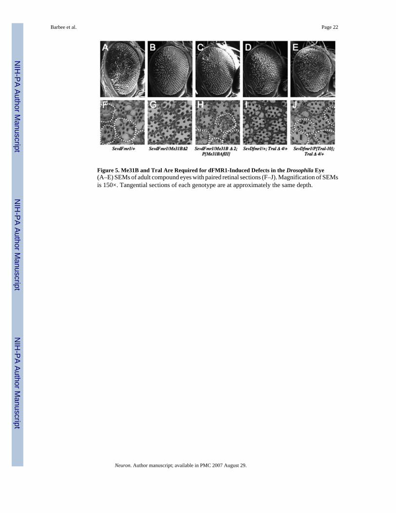
Figure 5. Me31B and Tral Are Required for dFMR1-Induced Defects in the Drosophila Eye(A–E) SEMs of adult compound eyes with paired retinal sections (F–J). Magnification of SEMsis 150×. Tangential sections of each genotype are at approximately the same depth.
Barbee et al. Page 22
Neuron. Author manuscript; available in PMC 2007 August 29.
NIH
-PA Author Manuscript
NIH
-PA Author Manuscript
NIH
-PA Author Manuscript

Figure 6. Me31B Regulates Dendritic Growth in Sensory Neurons(A) Control class IV ddaC neuron expressing UAS-mCD8:GFP alone.(B) Class IV ddaC neurons overexpressing Me31B and UAS-mCD8: GFP showing a reductionin higher-order dendrite arborization.(C) The same neurons overexpressing a mutant Me31B incapable of translational repression(Me31BD207A, E208A) show normal dendritic branching.(D) Transgenic RNAi dramatically reduces Me31B protein levels. Anti-Me31B staining ofthird-instar imaginal discs shows that UAS-Me31Bhpn expressed in the patched domain of wingimaginal discs reduces Me31B levels along the anterior-posterior border (top panel) comparedto control wing imaginal discs (lower panel).(E) Class IV ddaC neurons overexpressing a Me31B RNA hairpin (UAS-Me31Bhpn) exhibitabnormal dendrite morphology and increased high-order branching.(F) Numbers of dendritic branches in each order, as revealed by reversed Strahler analysis (seeSupplemental Data). Number of neurons analyzed for each genotype are: UAS-mCD8:GFPcontrol (n = 15), UAS-Me31B (n = 10), UAS-Me31BD207A, E208A (n = 11), and UAS-Me31Bhpn (n = 13). Values are mean ± standard error. A star (*) indicates a significant reductionin fifth-order dendrite branching following Me31B overexpression compared to the control (p< 0.001) and a significant increase in fifth-order dendrite branching following Me31B RNAi(p < 0.001). Scale bar, 20 μm.
Barbee et al. Page 23
Neuron. Author manuscript; available in PMC 2007 August 29.
NIH
-PA Author Manuscript
NIH
-PA Author Manuscript
NIH
-PA Author Manuscript

Figure 7. Me31B Is Required for the In Vivo Function of an Endogenous MicroRNAHeat-shock-induced clones in wing imaginal discs of hs-FLP1; FRT40A, arm-lacZ/FRT40A,me31BΔ1; reporter flies show dark me31B−/me31B− clones revealed either by staining for LacZ(B) or Me31B (A and C). Anti-GFP staining shows that the hid reporter (D and E) is upregulatedin cell clones lacking Me31B (C). Similar analyses (F and G) show that a control protein Dlg(G) is not upregulated in me31B−/me31B− clones (F). However, consistent with a Me31Brequirement in miRNA/RNAi, the bantam reporter expression is upregulated in me31B−/me31B−-lacking cells (H–J).
Barbee et al. Page 24
Neuron. Author manuscript; available in PMC 2007 August 29.
NIH
-PA Author Manuscript
NIH
-PA Author Manuscript
NIH
-PA Author Manuscript

NIH
-PA Author Manuscript
NIH
-PA Author Manuscript
NIH
-PA Author Manuscript
Barbee et al. Page 25
Table 1Percent Colocalization of P Body Components with Stau- and dFMR1-Containing Granulesa
Genotype Stau:GFP Expressing dFMR1 Overexpressing Wild-Type (w1118) Wild-Type (w1118)
Antibody usedas reference
v. Stau v. dFMR1 v. dFMR1 v. Stau
Stau — 80.5 (11; 462) 77.2 (8; 408) —dFMR1 80.3 (19; 524) — — 45.1 (17; 1223)Me31B 71.0 (12; 526) 85.0 (4; 207) 60.0 (14; 432) 50.2 (8; 440)Tral 100.0 (4; 52) 90.6 (7; 339) 56.8 (6; 491) NDbPcm 88.0 (7; 117) 72.3 (7; 343) 60.3 (8; 401) NDbDCP1 92.6 (5; 81) 85.8 (11; 318) 75.3 (3; 97) NDbUPF1 90.0 (6; 130) 80.5 (10; 394) 58.3 (3; 96) NDbAgo-2 74.3 (4; 109) 73.0 (8; 293) NDc NDb
aValues are expressed as percent colocalization (number of cells analyzed; number granules analyzed).
bAntibodies both rabbit polyclonal.
cPercent colocalization not determined.
Neuron. Author manuscript; available in PMC 2007 August 29.

NIH
-PA Author Manuscript
NIH
-PA Author Manuscript
NIH
-PA Author Manuscript
Barbee et al. Page 26
Table 2Conserved Components of P Bodies, Maternal Granules, and Neuronal Granules
Protein Class Mammalianand Yeast PBodiesa
Drosophila MaternalGranulesb
Drosophila NeuronalGranulesc
Mammalian Neuronal Granulesd
RNA transport ? Stau, Btz Stau, Btz Stau, BtzFragile X-like ? dFMR1 dFMR1 FMRP, FXR1, FXR2Zip-code binding ? ? Imp ZBP1Pum domain ? Pum? Pum PumlCCHC Zn-finger domain ? Nanos Nanosalso f ?DEAD-box RNA helicase RCK/Dhh1p Me31B Me31B ?Sm-like domain RAP55/
Scd6pjTral Tral ?
Cap-binding eIF4E,ek eIF4E eIF4Ealso g under some conditionsEnhancers of decapping Edc3p, Pat1pi ? ? ?eIF4E-binding eIF4E-Tk Cup Cup ?PABP PABPe ? PABPg ?Y-box ? Yps Yps ?5′ to 3′ RNA decaymachinery
Lsm, Dcp1p,Dcp2p,Xrn1p
DCP1, Pacman DCP1, Pacman(others not examined)
?
NMD machineryh Upf-1 Upf-1 Upf-1 ?miRNA, siRNA machinery mAGO1
mAGO2? Ago-2 ?
?Ortholog present but association with RNA granules has not been described.
aSheth and Parker, 2003;Andrei et al., 2005;Liu et al., 2005a;Pillai et al., 2005.
bSt Johnston et al., 1991;van Eeden et al., 2001;Nakamura et al., 2001;Boag et al., 2005;Nakamura et al., 2004;Wilhelm et al., 2000; this work; Wang et
al., 1994;Forbes and Lehmann, 1998.
cThis work (except PABP).
dKiebler et al., 1999;Macchi et al., 2003;Kanai et al., 2004;Antar et al., 2005;Zhang et al., 2001.
eeIF4E and PABP can associate with P bodies in yeast under conditions of translational arrest induced by glucose deprivation (Teixeira et al., 2005).
fYe et al., 2004 (describes particles likely to be, but not clearly established as, neuronal RNA granules).
gSigrist et al., 2000 (describes particles likely to be, but not clearly established as, neuronal RNA granules).
hUPF1 has also been shown to interact with Stau in a Stau-mediated NMD pathway (Kim et al., 2005).
iPat1p acts as a general repressor of translation in yeast (Coller and Parker, 2005).
jYang et al., 2006; this work.
kAndrei et al., 2005.
lVessey et al., 2006.
Neuron. Author manuscript; available in PMC 2007 August 29.
Copyright © 2022 FDOKUMEN