Direct reprogramming of somatic cells: an update
10
Biomedical Research and Therapy 2015, 2(3): 231-240 ISSN 2198-4093 www.bmrat.org Direct reprogramming of somatic cells 231 REVIEW Direct reprogramming of somatic cells: an update Phuc Van Pham * Laboratory of Stem Cell Research and Application, University of Science, Vietnam National University, Ho Chi Minh city, Vietnam Faculty of Biology, University of Science, Vietnam National University, Ho Chi Minh city, Vietnam *Corresponding author: [email protected] Received: 15 October 2014 / Accepted: 01 January 2015 / Published online: 13 March 2015 © The Author(s) 2015. This article is published with open access by BioMedPress (BMP). Abstract— Direct epigenetic reprogramming is a technique that converts a differentiated adult cell into another dif- ferentiated cell—such fibroblasts to cardiomyocytes—without passage through an undifferentiated pluripotent stage. This novel technology is opening doors in biological research and regenerative medicine. Some preliminary studies about direct reprogramming started in the 1980s when differentiated adult cells could be converted into other differ- entiated cells by overexpressing transcription-factor genes. These studies also showed that differentiated cells have plasticity. Direct reprogramming can be a powerful tool in biological research and regenerative medicine, especially the new frontier of personalized medicine. This review aims to summarize all direct reprogramming studies of somat- ic cells by master control genes as well as potential applications of these techniques in research and treatment of se- lected human diseases. Keywords— Direct reprogramming; Gene over-expression; Induced pluripotent stem cells; Stem cell technology; Trans-differentiation CELL FATE AND REPROGRAMMING The human body originates from a totipotent stem cell, the zygote. Development and growth of an organ- ism are due to proliferation and differentiation of these cells. Stem cell proliferation by self-renewal causes an increase in cell numbers, while stem cell differentiation causes an increase in cell types. Alt- hough all cells in the human body originate from a single cell, they play different roles. Their finalized specific functions are decided by mechanisms that are yet unclear, but it is considered that their functions are decided by their fates or programming (alterations in gene expression). From a single totipotent stem cell, generations of daughter cells are programmed into specific cell types that collaborate with each other to produce a completed body. In the traditional view, cell fates cannot be modified, and stem cell differentiation is unidirectional, in which only uncommitted or undifferentiated cells can differentiate into committed or specific cells. Howev- er, to date, many studies prove that fully differentiat- ed cells can reverse to pluripotent stem cells. This pro- cess is termed as “reprogramming” (Fig. 1). REPROGRAMMING The first attempt of the reprogramming technique was performed by Robert Briggs and Thomas King. In 1952, they injected an embryonic nucleus into an enu- cleated egg in the amphibian Rana pipiens (Briggs and King, 1952), advancing from an oocyte to the tadpole DOI 10.7603/s40730-015-0008-y
-
Upload
independent -
Category
Documents
-
view
1 -
download
0
Transcript of Direct reprogramming of somatic cells: an update
Biomedical Research and Therapy 2015, 2(3): 231-240 ISSN 2198-4093 www.bmrat.org
Direct reprogramming of somatic cells
231
REVIEW
Direct reprogramming of somatic cells: an update
Phuc Van Pham*
Laboratory of Stem Cell Research and Application, University of Science, Vietnam National University, Ho Chi Minh city, Vietnam Faculty of Biology, University of Science, Vietnam National University, Ho Chi Minh city, Vietnam
*Corresponding author: [email protected]
Received: 15 October 2014 / Accepted: 01 January 2015 / Published online: 13 March 2015 © The Author(s) 2015. This article is published with open access by BioMedPress (BMP).
Abstract— Direct epigenetic reprogramming is a technique that converts a differentiated adult cell into another dif-ferentiated cell—such fibroblasts to cardiomyocytes—without passage through an undifferentiated pluripotent stage. This novel technology is opening doors in biological research and regenerative medicine. Some preliminary studies about direct reprogramming started in the 1980s when differentiated adult cells could be converted into other differ-entiated cells by overexpressing transcription-factor genes. These studies also showed that differentiated cells have plasticity. Direct reprogramming can be a powerful tool in biological research and regenerative medicine, especially the new frontier of personalized medicine. This review aims to summarize all direct reprogramming studies of somat-ic cells by master control genes as well as potential applications of these techniques in research and treatment of se-lected human diseases. Keywords— Direct reprogramming; Gene over-expression; Induced pluripotent stem cells; Stem cell technology; Trans-differentiation
CELL FATE AND REPROGRAMMING
The human body originates from a totipotent stem
cell, the zygote. Development and growth of an organ-
ism are due to proliferation and differentiation of
these cells. Stem cell proliferation by self-renewal
causes an increase in cell numbers, while stem cell
differentiation causes an increase in cell types. Alt-
hough all cells in the human body originate from a
single cell, they play different roles. Their finalized
specific functions are decided by mechanisms that are
yet unclear, but it is considered that their functions are
decided by their fates or programming (alterations in
gene expression). From a single totipotent stem cell,
generations of daughter cells are programmed into
specific cell types that collaborate with each other to
produce a completed body.
In the traditional view, cell fates cannot be modified,
and stem cell differentiation is unidirectional, in
which only uncommitted or undifferentiated cells can
differentiate into committed or specific cells. Howev-
er, to date, many studies prove that fully differentiat-
ed cells can reverse to pluripotent stem cells. This pro-
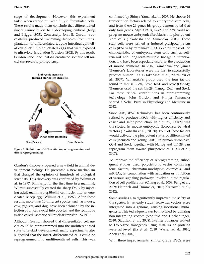
cess is termed as “reprogramming” (Fig. 1).
REPROGRAMMING
The first attempt of the reprogramming technique was
performed by Robert Briggs and Thomas King. In
1952, they injected an embryonic nucleus into an enu-
cleated egg in the amphibian Rana pipiens (Briggs and
King, 1952), advancing from an oocyte to the tadpole
DOI 10.7603/s40730-015-0008-y
Pham, 2015 Biomed Res Ther 2015, 2(3): 231-240
Direct reprogramming of somatic cells
232
stage of development. However, this experiment
failed when carried out with fully differentiated cells.
These results made them conclude that differentiated
nuclei cannot revert to a developing embryo (King
and Briggs, 1955). Conversely, John B. Gurdon suc-
cessfully produced swimming tadpoles from trans-
plantation of differentiated tadpole intestinal epitheli-
al cell nuclei into enucleated eggs that were exposed
to ultraviolet irradiation (Gurdon, 1962). By this result,
Gurdon concluded that differentiated somatic cell nu-
clei can revert to pluripotency.
Figure 1. Definitions of differentiation, reprogramming and direct reprogramming.
Gurdon’s discovery opened a new field in animal de-
velopment biology. He presented a new mechanism
that changed the opinion of hundreds of biological
scientists. This discovery was confirmed by Wilmut et
al. in 1997. Similarly, for the first time in a mammal,
Wilmut successfully created the sheep Dolly by inject-
ing adult mammary epithelial cell nuclei into an enu-
cleated sheep egg (Wilmut et al., 1997). After these
results, more than 10 different species, such as mouse,
cow, pig, cat, and dog, have been “cloned” by the in-
jection adult cell nuclei into oocytes, and the technique
is also called “somatic cell nuclear transfer—SCNT.”
Although Gurdon showed that differentiated cell nu-
clei could be reprogrammed into the undifferentiated
state to re-start development, many experiments also
suggested that the intact, differentiated cells could be
reprogrammed into undifferentiated cells. This was
confirmed by Shinya Yamanaka in 2007. He choose 24
transcription factors related to embryonic stem cells,
and from these 24 genes his group demonstrated that
only four genes, Myc, Oct3/4, Sox2, and Klf4 could re-
program mouse embryonic fibroblasts into pluripotent
stem cells (Takahashi and Yamanaka, 2006). These
stem cells were termed as induced pluripotent stem
cells (iPSCs) by Yamanaka. iPSCs exhibit most of the
characteristics of embryonic stem cells such as self-
renewal and long-term-multiple lineage differentia-
tion, and have been especially useful in the production
of mouse chimeras. In 2007, Yamanaka and James
Thomson’s laboratories were the first to successfully
produce human iPSCs (Takahashi et al., 2007a; Yu et
al., 2007). Yamanaka’s group used the four factors
found in mouse: Oct4, Sox2, Klf4, and Myc (OSKM);
Thomson used the set: Lin28, Nanog, Oct4, and Sox2.
For these critical contributions in reprogramming
technology, John Gurdon and Shinya Yamanaka
shared a Nobel Prize in Physiology and Medicine in
2012.
Since 2006, iPSC technology has been continuously
refined to produce iPSCs with higher efficiency and
easier and safer production. In a study, OSKM was
transfected in mouse embryonic fibroblasts by viral
vectors (Takahashi et al., 2007b). Four of these factors
would activate the pluripotent status of differentiated
cells (Jaenisch and Young, 2008). In human fibroblasts,
Oct4 and Sox2, together with Nanog and LIN28, can
reprogram them toward pluripotent cells (Yu et al.,
2007).
To improve the efficiency of reprogramming, subse-
quent studies used polycistronic vector containing
four factors, chromatin-modifying chemicals, and
mRNAs, in combination with activation or inhibition
of various signaling pathways involved in the regula-
tion of cell proliferation (Chang et al., 2009; Feng et al.,
2009; Heinrich and Dimmeler, 2012; Kretsovali et al.,
2012).
Some studies also significantly improved the safety of
transgenes. In an early study, retroviral vectors were
integrated into a genome, causing insertional muta-
genesis. This technique is can be modified by utilizing
non-integrating vectors (Stadtfeld and Hochedlinger,
2010; Stadtfeld et al., 2008). Further advances related
to DNA-free transgenes using mRNAs or proteins
were achieved (Jia et al., 2010; Warren et al., 2010;
Zhou et al., 2009).
With these improvements, clinical-grade iPSCs were
Pham, 2015 Biomed Res Ther 2015, 2(3): 231-240
Direct reprogramming of somatic cells
233
developed in the recent years. Clinical grade iPSCs
usually use donor cells such as fibroblasts, keratino-
cytes, and peripheral blood mononuclear cells
(PBMCs), which are preferable for inducing pluripo-
tency. Moreover, clinical-grade iPSCs need to be pro-
duced from safer techniques, reducing the likelihood
of accidently creating tumor-forming cells.
Some safer techniques in gene transfection are used to
produce vectors containing reprogramming genes.
The first effort used F-deficient Sendai virus particles
to induce pluripotency in somatic cells (Dowey et al.,
2012; Fusaki et al., 2009). iPSCs produced using this
method must be sub-cultured for 10–20 passages to
remove the excess virus particles and to make virus-
free iPSC lines. Later, an improvement in gene trans-
fection using temperature-sensitive Sendai virus parti-
cles made it is easier to remove the virus particles by
temperature shift (Ban et al., 2011).
Virus-free vectors carrying reprogramming factors
have been studied since 2010 to replace the viral vec-
tors. Episomal DNA can be used to transfect
transgenes into adult cells. These virus-free vectors
have important clinical applications because they are
safer in manipulations as well as in the patients. There
are two kinds of episomes: non-replicating episomal
vectors and replicating episomal vectors. The iPSC
production procedure using non-replicating episomal
vectors is of low-yield; therefore, multiple transfec-
tions are suggested as a solution to increase the iPSC
production efficacy (Jia et al., 2010; Okita et al., 2008).
Improvements such as the use of minicircle or codon-
optimized 4-in-1 minicircle (CoMiP) DNA vectors
were devised (Lu et al., 2013; Okita et al., 2008).
Although DNA-based episome is considered safe to
reprogram adult cells to iPSCs, in principle, foreign
DNA can integrate into the host genome. Therefore,
iPSCs must be screened to select free cells for further
applications (Gonzalez et al., 2009). To date, the safest
technique of iPSC production is induction of pluripo-
tency via mRNA (Warren et al., 2012; Yoshioka et al.,
2013) or protein (Kim et al., 2009; Lee et al., 2012).
These iPSCs are called “clean” iPSCs.
Together with improvement of iPSC production
methods, some approaches using iPSCs in treatment
were also developed. The most significant approach
for clinical applications of iPSCs relates to the combi-
nation of iPSC technology and targeting editing of the
iPSC genome. This combination helps to push iPSCs
into clinical treatment, particularly for patients with
genetic disorders. There are three ways to correct the
mutated genes in iPSCs: the zinc finger nuclease
(ZFN) system, the transcription activator-like effector
nuclease (TALEN) system, and the clustered regularly
interspaced short palindromic repeats (CRISPR) sys-
tem (Ding et al., 2013; Hockemeyer et al., 2009; Horii et
al., 2013). By using these techniques, patient-specific
iPSCs were successfully produced to treat epilepsy
(Parent and Anderson, 2015), myotonic dystrophy
type 1 (Xia et al., 2015), sickle erythrocytes (Huang et
al., 2015), retinal degenerative diseases (Wiley et al.,
2015), and recessive dystrophic epidermolysis bullosa
(Sebastiano et al., 2014).
DIRECT REPROGRAMMING
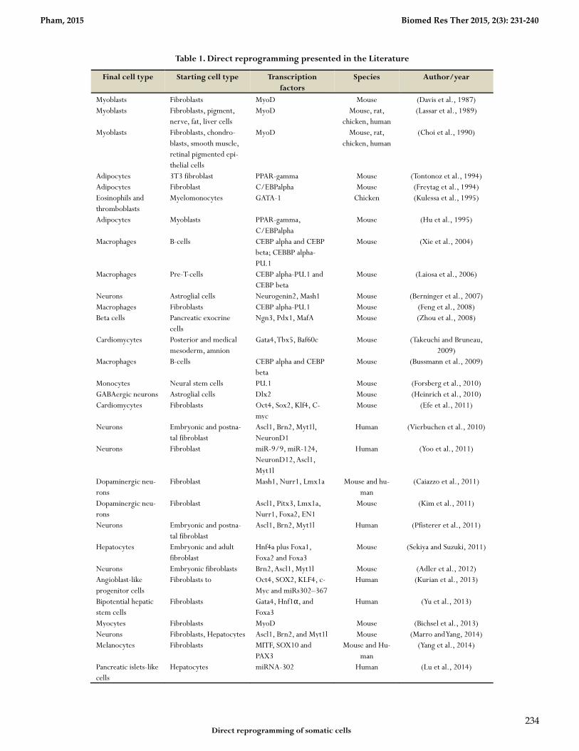
The direct reprogramming technique was discovered
in the 1980s (Table 1). In 1987, Davis et al. converted
embryonic mouse fibroblasts into muscle cells by
transfection of myogenic differentiation factor (MyoD)
(Davis et al., 1987). Similarly, MyoD was used to re-
program immature chondrocytes, smooth muscle
cells, and retinal cells into muscle cells (Choi et al.,
1990). In the 1990s, some other transcription factors
were discovered, particularly globin transcription fac-
tor 1 (Gata-1), that can reprogram avian monocyte
precursors into erythrocytes, eosinophils, and
megakaryocytes (Kulessa et al., 1995).
Since 2000, several transcription factors were discov-
ered and were successfully used to reprogram target
cells such as pancreatic islet cells (Zhou et al., 2008),
neurons (Fishman et al., 2015; Vierbuchen et al., 2010),
hepatocytes (Huang et al., 2011; Sekiya and Suzuki,
2011), endothelial cells (Ginsberg et al., 2012; Han et
al., 2014), smooth muscle cells (Karamariti et al., 2013),
and hepatocyte like cells (Simeonov and Uppal, 2014).
In recent years, in situ direct reprogramming as well as
in vivo direct reprogramming has become important,
as the ability to provide novel therapies is nearly in
clinical applications. In vivo direct reprogramming is
the usage of specific transcription factors to change
target cell fate in the body without the need to isolate
the target cells (Table 1).
Pham, 2015 Biomed Res Ther 2015, 2(3): 231-240
Direct reprogramming of somatic cells
234
Table 1. Direct reprogramming presented in the Literature
Final cell type Starting cell type Transcription factors
Species Author/year
Myoblasts Fibroblasts MyoD Mouse (Davis et al., 1987) Myoblasts Fibroblasts, pigment,
nerve, fat, liver cells MyoD Mouse, rat,
chicken, human (Lassar et al., 1989)
Myoblasts Fibroblasts, chondro-blasts, smooth muscle, retinal pigmented epi-thelial cells
MyoD Mouse, rat, chicken, human
(Choi et al., 1990)
Adipocytes 3T3 fibroblast PPAR-gamma Mouse (Tontonoz et al., 1994) Adipocytes Fibroblast C/EBPalpha Mouse (Freytag et al., 1994) Eosinophils and thromboblasts
Myelomonocytes GATA-1 Chicken (Kulessa et al., 1995)
Adipocytes Myoblasts PPAR-gamma, C/EBPalpha
Mouse (Hu et al., 1995)
Macrophages B-cells CEBP alpha and CEBP beta; CEBBP alpha-PU.1
Mouse (Xie et al., 2004)
Macrophages Pre-T-cells CEBP alpha-PU.1 and CEBP beta
Mouse (Laiosa et al., 2006)
Neurons Astroglial cells Neurogenin2, Mash1 Mouse (Berninger et al., 2007) Macrophages Fibroblasts CEBP alpha-PU.1 Mouse (Feng et al., 2008) Beta cells Pancreatic exocrine
cells Ngn3, Pdx1, MafA Mouse (Zhou et al., 2008)
Cardiomycytes Posterior and medical mesoderm, amnion
Gata4, Tbx5, Baf60c Mouse (Takeuchi and Bruneau, 2009)
Macrophages B-cells CEBP alpha and CEBP beta
Mouse (Bussmann et al., 2009)
Monocytes Neural stem cells PU.1 Mouse (Forsberg et al., 2010) GABAergic neurons Astroglial cells Dlx2 Mouse (Heinrich et al., 2010) Cardiomycytes Fibroblasts Oct4, Sox2, Klf4, C-
myc Mouse (Efe et al., 2011)
Neurons Embryonic and postna-tal fibroblast
Ascl1, Brn2, Myt1l, NeuronD1
Human (Vierbuchen et al., 2010)
Neurons Fibroblast miR-9/9, miR-124, NeuronD12, Ascl1, Myt1l
Human (Yoo et al., 2011)
Dopaminergic neu-rons
Fibroblast Mash1, Nurr1, Lmx1a Mouse and hu-man
(Caiazzo et al., 2011)
Dopaminergic neu-rons
Fibroblast Ascl1, Pitx3, Lmx1a, Nurr1, Foxa2, EN1
Mouse (Kim et al., 2011)
Neurons Embryonic and postna-tal fibroblast
Ascl1, Brn2, Myt1l Human (Pfisterer et al., 2011)
Hepatocytes Embryonic and adult fibroblast
Hnf4a plus Foxa1, Foxa2 and Foxa3
Mouse (Sekiya and Suzuki, 2011)
Neurons Embryonic fibroblasts Brn2, Ascl1, Myt1l Mouse (Adler et al., 2012) Angioblast-like progenitor cells
Fibroblasts to Oct4, SOX2, KLF4, c-Myc and miRs302–367
Human (Kurian et al., 2013)
Bipotential hepatic stem cells
Fibroblasts Gata4, Hnf1α, and Foxa3
Human (Yu et al., 2013)
Myocytes Fibroblasts MyoD Mouse (Bichsel et al., 2013) Neurons Fibroblasts, Hepatocytes Ascl1, Brn2, and Myt1l Mouse (Marro and Yang, 2014) Melanocytes Fibroblasts MITF, SOX10 and
PAX3 Mouse and Hu-
man (Yang et al., 2014)
Pancreatic islets-like cells
Hepatocytes miRNA-302 Human (Lu et al., 2014)
Pham, 2015 Biomed Res Ther 2015, 2(3): 231-240
Direct reprogramming of somatic cells
235
MECHANISMS OF DIRECT REPROGRAM-MING
In early studies, it was shown that transcription fac-
tors can directly affect reprogramming. Recent studies
indicated that there are at least five kinds of repro-
gramming factors that can directly reprogram adult
cells into other phenotypic cells: transcription factors,
epigenetic regulators, miRNAs, Small molecules, and
pluripotency factors for direct reprogramming.
Transcription factors
Different from reprogramming techniques make adult
cells pluripotent after receiving some key transcription
factors causing epigenetic modifications, direct repro-
gramming mechanisms are still elusive. The most im-
portant mechanism is the effect of transcription factors
that drive the phenotype changes in specific cells. By
using transcription factors, transfected cells can
change phenotype via activation of target genes. Inter-
estingly, these changes can occur some hours after
transfections (Ieda et al., 2010), do not require cell di-
vision (Heinrich et al., 2010; Vierbuchen et al., 2010),
and are stable after removal of reprogramming factors
(Huang et al., 2011; Sekiya and Suzuki, 2011). Some
authors have demonstrated that direct reprogram-
ming of fibroblasts to neurons was hierarchical, estab-
lished mechanisms dictate that fibroblasts gradually
change with multiple steps to become neurons
(Wapinski et al., 2013).
Epigenetic regulators
Differentiated status of cells seems depend on epige-
netic status of these cells. Transcription factors are
known as important factors effecting to expression of
lineage specific genes. However, gene expression also
is effected by epigenetic regulators. In fact, there are
three ways that epigenetic regulators effect gene ex-
pression. First, epigenetic regulators can decide the
reprogramming process by themselves. For example,
pancreatic beta cells can be reprogrammed into alpha
cells by DNA methyltransferase Dnmt1 deficiency
(Dhawan et al., 2011). Second, epigenetic regulators
can interact with exogenous factors to re-activate or
suppress related gene expression. In the study by
Takeuchi and Bruneau (2009), they showed that Baf60c
– cardiac specific subunit of BAF chromatin remodel-
ing complexes hold a particular role in the repro-
gramming from mouse mesoderm to cardiac myocytes
that is helped by Gata4 – a transcription factor to bind
to cardiac genes (Takeuchi and Bruneau, 2009). Third,
some epigenetic regulators act as epigenetic barriers
that can prevent reprogramming. In fact, the inhibi-
tion or removal of histone deacetylases and polycomb
repressor complex 2 (PRC2) can facilitate the repro-
gramming of germ cells into neurons (Patel et al.,
2012).
miRNAs
More and more studies proved that miRNAs play im-
portant roles in the reprogramming process. Some
specific miRNAs such as miR-124, miR-9/9, miR-1,
miR-133, miR-208, and miR-499 were demonstrated
with reprogramming effects in fibroblasts. Overex-
pression of miR-9/9 and miR-124 in human fibroblasts
can induce the expression of markers indicative of
neuron-like cells (Yoo et al., 2011). It seems that miR-
NAs can regulate some mechanisms relating to epige-
netic reprogramming. In fact, miRNAs can directly
stimulate or suppress target genes (Bartel, 2009) as
well as regulate epigenetic regulators (Neo et al.,
2014). However, in general, miRNAs are not as effi-
cient as transcription factors to induce epigenetic re-
programming.
Small molecules
Some small molecules were successfully used to pro-
duce iPSC (Li et al., 2013b). The main advantage of
small molecules is small structure, therefore they can
more easily move across cellular membranes. By this
advantage, small molecules are more richly investigat-
ed in recent studies. The biggest success in direct re-
programming by small molecules is the neural con-
version process (Kim et al., 2014; Sayed et al., 2015).
How the small molecules can reprogram the cell fate is
a question that needs to be answered. In some cases,
small molecules activate some pluripotency genes
(Hou et al., 2013) as well as transcription factors (Yuan
et al., 2013).
Pluripotency Factors for Indirect Repro-
gramming
Some pluripotency factors used to produce iPSC can
directly reprogram some cell types such as cardiomy-
ocytes (Efe et al., 2011), neural stem cells or progeni-
tors (Wang et al., 2013), angioblast-like progenitor cells
Pham, 2015 Biomed Res Ther 2015, 2(3): 231-240
Direct reprogramming of somatic cells
236
(Kurian et al., 2013), endothelial cells (Li et al., 2013a),
pancreatic lineages (Li et al., 2014), and hepatocytes
(Zhu et al., 2014). Ma et al. (2013) showed that pluripo-
tent factors can reprogram adult cells into pluripotent
cells with multiple steps and that at certain steps some
cells’ fates are formed as transition stages of epigenetic
reprogramming (Ma et al., 2013). Moreover, overex-
pression of pluripotent factors can also induce differ-
entiation (Loh and Lim, 2011).
Although direct reprogramming can produce the
functional cells that can be used in translational appli-
cations as well as therapy, the main limitation of this
technology is slow or non-proliferation of repro-
grammed cells. Therefore, direct reprogramming
should be improved in order to produce proliferating
cells such as tissue specific stem cells or progenitor
cells more than fully differentiated cells. In fact, some
kinds of stem cells as well as progenitor cells were
produced by direct reprogramming technology, in-
cluding neural stem cells or progenitors (Han et al.,
2012; Schindeler et al., 2015; Thier et al., 2012), oli-
godendrocyte precursor cells (Najm et al., 2013), he-
patic stem cells (Yu et al., 2013), HSCs (Riddell et al.,
2014), and hematopoietic multipotent progenitors
(Batta et al., 2014; Sandler et al., 2014).
INVIVO DIRECT REPRORAMMING
As direct reprogramming technology is gradually per-
fected, especially its efficiency in combination with the
tools of in situ gene therapy that were developed in
previous studies. In vivo direct reprogramming has
become more interesting as a novel therapy in regen-
erative medicine. Using in situ gene therapy strategies
with direct reprogramming factors, some preclinical
trials with a mouse model were successful in the con-
version of various cerebral cell types into neurons
(Heinrich and Rouaux, 2015). By enhanced expression
of Sox10 in Satellite Glial cells, Weider et al (2015) suc-
cessfully induced these cells in vivo into oligodendro-
cyte-like cells (Weider et al., 2015).
Particularly, reactive glial cells in the cortex of stab-
injured or Alzheimer's disease (AD) model mice can
be directly reprogrammed into functional neurons in
vivo using retroviral expression of a single neural tran-
scription factor, NeuroD1 (Guo et al., 2014). More im-
portantly, cardiac injury model mice can be treated by
in vivo direct reprogramming(Jayawardena et al.,
2015). miRNAs and lentiviral vectors were injected
into these mice. After 5-6 weeks, cardiac function was
improved, associated with existence of cardiac myo-
cyte-like cells in injected sites.
CONCLUSION Epigenetic reprogramming has seen rapid growth in
recent years. Supported by some modern molecular
biology techniques, reprogramming technology is be-
coming important and promising for wide use in basic
research to translational research, and clinical applica-
tion in the near future. Direct epigenetic reprogram-
ming is a combination of stem cell therapy and gene
therapy that can induce cell regeneration in an in situ
manner. Many non-viral vectors and some novel re-
programming factors have facilitated direct repro-
gramming applications in preclinical models. Direct
reprogramming, however, also faces with some chal-
lenges. Safety of vectors as well as technology must be
investigated and carefully evaluated, especially in vi-
ral vector transfections or DNA transfection. Another
challenge relates to control of reprogramming effi-
ciency as well as specificity of target cells in vivo.
ABBREVIATIONS
AD: Alzheimer's disease; PRC2: Polycomb repressor
complex 2; ZFN: zinc finger nuclease; HSCs: Hemato-
poietic stem cells; iPSC: Induced pluripotent stem
cells.
ACKNOWLEDGEMENT
This work is funded by Vietnam National Foundation
for Science and Technology Development
(NAFOSTED) under grant number 106-YS.06-2013.37.
Competing interests The authors declare that they have no competing interests.
Open Access
This article is distributed under the terms of the Creative
Commons Attribution License (CC-BY 4.0) which permits any
use, distribution, and reproduction in any medium, provided the
original author(s) and the source are credited.
Pham, 2015 Biomed Res Ther 2015, 2(3): 231-240
Direct reprogramming of somatic cells
237
References
Adler, A.F., Grigsby, C.L., Kulangara, K., Wang, H., Yasuda, R., and Leong, K.W. (2012). Nonviral direct conversion of primary mouse embryonic fibroblasts to neuronal cells. Molecular therapy Nucleic acids 1, e32.
Ban, H., Nishishita, N., Fusaki, N., Tabata, T., Saeki, K., Shikamura, M., Takada, N., Inoue, M., Hasegawa, M., Kawamata, S., et al. (2011). Efficient generation of transgene-free human induced pluripotent stem cells (iPSCs) by temperature-sensitive Sendai virus vectors. Proceedings of the National Academy of Sciences of the United States of America 108, 14234-14239.
Bartel, D.P. (2009). MicroRNAs: target recognition and regulatory functions. Cell 136, 215-233.
Batta, K., Florkowska, M., Kouskoff, V., and Lacaud, G. (2014). Direct reprogramming of murine fibroblasts to hematopoietic progenitor cells. Cell reports 9, 1871-1884.
Berninger, B., Costa, M.R., Koch, U., Schroeder, T., Sutor, B., Grothe, B., and Gotz, M. (2007). Functional properties of neurons derived from in vitro reprogrammed postnatal astroglia. The Journal of neuroscience : the official journal of the Society for Neuroscience 27, 8654-8664.
Bichsel, C., Neeld, D., Hamazaki, T., Chang, L.J., Yang, L.J., Terada, N., and Jin, S. (2013). Direct reprogramming of fibroblasts to myocytes via bacterial injection of MyoD protein. Cellular reprogramming 15, 117-125.
Briggs, R., and King, T.J. (1952). Transplantation of Living Nuclei From Blastula Cells into Enucleated Frogs' Eggs. Proceedings of the National Academy of Sciences of the United States of America 38, 455-463.
Bussmann, L.H., Schubert, A., Vu Manh, T.P., De Andres, L., Desbordes, S.C., Parra, M., Zimmermann, T., Rapino, F., Rodriguez-Ubreva, J., Ballestar, E., et al. (2009). A robust and highly efficient immune cell reprogramming system. Cell stem cell 5, 554-566.
Caiazzo, M., Dell'Anno, M.T., Dvoretskova, E., Lazarevic, D., Taverna, S., Leo, D., Sotnikova, T.D., Menegon, A., Roncaglia, P., Colciago, G., et al. (2011). Direct generation of functional dopaminergic neurons from mouse and human fibroblasts. Nature 476, 224-227.
Chang, C.W., Lai, Y.S., Pawlik, K.M., Liu, K., Sun, C.W., Li, C., Schoeb, T.R., and Townes, T.M. (2009). Polycistronic lentiviral vector for "hit and run" reprogramming of adult skin fibroblasts to induced pluripotent stem cells. Stem cells (Dayton, Ohio) 27, 1042-1049.
Choi, J., Costa, M.L., Mermelstein, C.S., Chagas, C., Holtzer, S., and Holtzer, H. (1990). MyoD converts primary dermal fibroblasts, chondroblasts, smooth muscle, and retinal pigmented epithelial cells into striated mononucleated myoblasts and multinucleated myotubes. Proceedings of the National Academy of Sciences of the United States of America 87, 7988-7992.
Davis, R.L., Weintraub, H., and Lassar, A.B. (1987). Expression of a single transfected cDNA converts fibroblasts to myoblasts. Cell 51, 987-1000.
Dhawan, S., Georgia, S., Tschen, S.I., Fan, G., and Bhushan, A. (2011). Pancreatic beta cell identity is maintained by DNA methylation-mediated repression of Arx. Developmental cell 20, 419-429.
Ding, Q., Lee, Y.K., Schaefer, E.A., Peters, D.T., Veres, A., Kim, K., Kuperwasser, N., Motola, D.L., Meissner, T.B., Hendriks, W.T., et al. (2013). A TALEN genome-editing system for generating human stem cell-based disease models. Cell stem cell 12, 238-251.
Dowey, S.N., Huang, X., Chou, B.K., Ye, Z., and Cheng, L. (2012). Generation of integration-free human induced pluripotent stem cells from
postnatal blood mononuclear cells by plasmid vector expression. Nature protocols 7, 2013-2021.
Efe, J.A., Hilcove, S., Kim, J., Zhou, H., Ouyang, K., Wang, G., Chen, J., and Ding, S. (2011). Conversion of mouse fibroblasts into cardiomyocytes using a direct reprogramming strategy. Nature cell biology 13, 215-222.
Feng, B., Ng, J.H., Heng, J.C., and Ng, H.H. (2009). Molecules that promote or enhance reprogramming of somatic cells to induced pluripotent stem cells. Cell stem cell 4, 301-312.
Feng, R., Desbordes, S.C., Xie, H., Tillo, E.S., Pixley, F., Stanley, E.R., and Graf, T. (2008). PU.1 and C/EBPalpha/beta convert fibroblasts into macrophage-like cells. Proceedings of the National Academy of Sciences of the United States of America 105, 6057-6062.
Fishman, V.S., Shnayder, T.A., Orishchenko, K.E., Bader, M., Alenina, N., and Serov, O.L. (2015). Cell Divisions are not Essential for the Direct Conversion of Fibroblasts into Neuronal Cells. Cell cycle (Georgetown, Tex), 0.
Forsberg, M., Carlen, M., Meletis, K., Yeung, M.S., Barnabe-Heider, F., Persson, M.A., Aarum, J., and Frisen, J. (2010). Efficient reprogramming of adult neural stem cells to monocytes by ectopic expression of a single gene. Proceedings of the National Academy of Sciences of the United States of America 107, 14657-14661.
Freytag, S.O., Paielli, D.L., and Gilbert, J.D. (1994). Ectopic expression of the CCAAT/enhancer-binding protein alpha promotes the adipogenic program in a variety of mouse fibroblastic cells. Genes & development 8, 1654-1663.
Fusaki, N., Ban, H., Nishiyama, A., Saeki, K., and Hasegawa, M. (2009). Efficient induction of transgene-free human pluripotent stem cells using a vector based on Sendai virus, an RNA virus that does not integrate into the host genome. Proceedings of the Japan Academy Series B, Physical and biological sciences 85, 348-362.
Ginsberg, M., James, D., Ding, B.S., Nolan, D., Geng, F., Butler, J.M., Schachterle, W., Pulijaal, V.R., Mathew, S., Chasen, S.T., et al. (2012). Efficient direct reprogramming of mature amniotic cells into endothelial cells by ETS factors and TGFbeta suppression. Cell 151, 559-575.
Gonzalez, F., Barragan Monasterio, M., Tiscornia, G., Montserrat Pulido, N., Vassena, R., Batlle Morera, L., Rodriguez Piza, I., and Izpisua Belmonte, J.C. (2009). Generation of mouse-induced pluripotent stem cells by transient expression of a single nonviral polycistronic vector. Proceedings of the National Academy of Sciences of the United States of America 106, 8918-8922.
Guo, Z., Zhang, L., Wu, Z., Chen, Y., Wang, F., and Chen, G. (2014). In vivo direct reprogramming of reactive glial cells into functional neurons after brain injury and in an Alzheimer's disease model. Cell stem cell 14, 188-202.
Gurdon, J.B. (1962). Adult frogs derived from the nuclei of single somatic cells. Developmental biology 4, 256-273.
Han, D.W., Tapia, N., Hermann, A., Hemmer, K., Hoing, S., Arauzo-Bravo, M.J., Zaehres, H., Wu, G., Frank, S., Moritz, S., et al. (2012). Direct reprogramming of fibroblasts into neural stem cells by defined factors. Cell stem cell 10, 465-472.
Han, J.K., Chang, S.H., Cho, H.J., Choi, S.B., Ahn, H.S., Lee, J., Jeong, H., Youn, S.W., Lee, H.J., Kwon, Y.W., et al. (2014). Direct conversion of adult skin fibroblasts to endothelial cells by defined factors. Circulation 130, 1168-1178.
Heinrich, C., Blum, R., Gascon, S., Masserdotti, G., Tripathi, P., Sanchez, R., Tiedt, S., Schroeder, T., Gotz, M., and Berninger, B. (2010). Directing astroglia from the cerebral cortex into subtype specific functional neurons. PLoS biology 8, e1000373.
Heinrich, C., and Rouaux, C. (2015). [Inducing brain regeneration from within: in vivo reprogramming of endogenous somatic cells into neurons]. Medecine sciences : M/S 31, 35-42.
Pham, 2015 Biomed Res Ther 2015, 2(3): 231-240
Direct reprogramming of somatic cells
238
Heinrich, E.M., and Dimmeler, S. (2012). MicroRNAs and stem cells: control of pluripotency, reprogramming, and lineage commitment. Circulation research 110, 1014-1022.
Hockemeyer, D., Soldner, F., Beard, C., Gao, Q., Mitalipova, M., DeKelver, R.C., Katibah, G.E., Amora, R., Boydston, E.A., Zeitler, B., et al. (2009). Efficient targeting of expressed and silent genes in human ESCs and iPSCs using zinc-finger nucleases. Nature biotechnology 27, 851-857.
Horii, T., Morita, S., Kimura, M., Kobayashi, R., Tamura, D., Takahashi, R.U., Kimura, H., Suetake, I., Ohata, H., Okamoto, K., et al. (2013). Genome engineering of mammalian haploid embryonic stem cells using the Cas9/RNA system. PeerJ 1, e230.
Hou, P., Li, Y., Zhang, X., Liu, C., Guan, J., Li, H., Zhao, T., Ye, J., Yang, W., Liu, K., et al. (2013). Pluripotent stem cells induced from mouse somatic cells by small-molecule compounds. Science (New York, NY) 341, 651-654.
Hu, E., Tontonoz, P., and Spiegelman, B.M. (1995). Transdifferentiation of myoblasts by the adipogenic transcription factors PPAR gamma and C/EBP alpha. Proceedings of the National Academy of Sciences of the United States of America 92, 9856-9860.
Huang, P., He, Z., Ji, S., Sun, H., Xiang, D., Liu, C., Hu, Y., Wang, X., and Hui, L. (2011). Induction of functional hepatocyte-like cells from mouse fibroblasts by defined factors. Nature 475, 386-389.
Huang, X., Wang, Y., Yan, W., Smith, C., Ye, Z., Wang, J., Gao, Y., Mendelsohn, L., and Cheng, L. (2015). Production of gene-corrected adult beta globin protein in human erythrocytes differentiated from patient iPSCs after genome editing of the sickle point mutation. Stem cells (Dayton, Ohio).
Ieda, M., Fu, J.D., Delgado-Olguin, P., Vedantham, V., Hayashi, Y., Bruneau, B.G., and Srivastava, D. (2010). Direct reprogramming of fibroblasts into functional cardiomyocytes by defined factors. Cell 142, 375-386.
Jaenisch, R., and Young, R. (2008). Stem cells, the molecular circuitry of pluripotency and nuclear reprogramming. Cell 132, 567-582.
Jayawardena, T.M., Finch, E.A., Zhang, L., Zhang, H., Hodgkinson, C.P., Pratt, R.E., Rosenberg, P.B., Mirotsou, M., and Dzau, V.J. (2015). MicroRNA Induced Cardiac Reprogramming In Vivo: Evidence for Mature Cardiac Myocytes and Improved Cardiac Function. Circulation research 116, 418-424.
Jia, F., Wilson, K.D., Sun, N., Gupta, D.M., Huang, M., Li, Z., Panetta, N.J., Chen, Z.Y., Robbins, R.C., Kay, M.A., et al. (2010). A nonviral minicircle vector for deriving human iPS cells. Nature methods 7, 197-199.
Karamariti, E., Margariti, A., Winkler, B., Wang, X., Hong, X., Baban, D., Ragoussis, J., Huang, Y., Han, J.D., Wong, M.M., et al. (2013). Smooth muscle cells differentiated from reprogrammed embryonic lung fibroblasts through DKK3 signaling are potent for tissue engineering of vascular grafts. Circulation research 112, 1433-1443.
Kim, D., Kim, C.H., Moon, J.I., Chung, Y.G., Chang, M.Y., Han, B.S., Ko, S., Yang, E., Cha, K.Y., Lanza, R., et al. (2009). Generation of human induced pluripotent stem cells by direct delivery of reprogramming proteins. Cell stem cell 4, 472-476.
Kim, J., Su, S.C., Wang, H., Cheng, A.W., Cassady, J.P., Lodato, M.A., Lengner, C.J., Chung, C.Y., Dawlaty, M.M., Tsai, L.H., et al. (2011). Functional integration of dopaminergic neurons directly converted from mouse fibroblasts. Cell stem cell 9, 413-419.
Kim, Y.J., Lim, H., Li, Z., Oh, Y., Kovlyagina, I., Choi, I.Y., Dong, X., and Lee, G. (2014). Generation of multipotent induced neural crest by direct reprogramming of human postnatal fibroblasts with a single transcription factor. Cell stem cell 15, 497-506.
King, T.J., and Briggs, R. (1955). Changes in the nuclei of differentiating gastrula cells, as demonstrated by nuclear transplantation. Proceedings of the National Academy of Sciences of the United States of America 41, 321-325.
Kretsovali, A., Hadjimichael, C., and Charmpilas, N. (2012). Histone deacetylase inhibitors in cell pluripotency, differentiation, and reprogramming. Stem cells international 2012, 184154.
Kulessa, H., Frampton, J., and Graf, T. (1995). GATA-1 reprograms avian myelomonocytic cell lines into eosinophils, thromboblasts, and erythroblasts. Genes & development 9, 1250-1262.
Kurian, L., Sancho-Martinez, I., Nivet, E., Aguirre, A., Moon, K., Pendaries, C., Volle-Challier, C., Bono, F., Herbert, J.M., Pulecio, J., et al. (2013). Conversion of human fibroblasts to angioblast-like progenitor cells. Nature methods 10, 77-83.
Laiosa, C.V., Stadtfeld, M., Xie, H., de Andres-Aguayo, L., and Graf, T. (2006). Reprogramming of committed T cell progenitors to macrophages and dendritic cells by C/EBP alpha and PU.1 transcription factors. Immunity 25, 731-744.
Lassar, A.B., Thayer, M.J., Overell, R.W., and Weintraub, H. (1989). Transformation by activated ras or fos prevents myogenesis by inhibiting expression of MyoD1. Cell 58, 659-667.
Lee, J., Sayed, N., Hunter, A., Au, K.F., Wong, W.H., Mocarski, E.S., Pera, R.R., Yakubov, E., and Cooke, J.P. (2012). Activation of innate immunity is required for efficient nuclear reprogramming. Cell 151, 547-558.
Li, J., Huang, N.F., Zou, J., Laurent, T.J., Lee, J.C., Okogbaa, J., Cooke, J.P., and Ding, S. (2013a). Conversion of human fibroblasts to functional endothelial cells by defined factors. Arteriosclerosis, thrombosis, and vascular biology 33, 1366-1375.
Li, K., Zhu, S., Russ, H.A., Xu, S., Xu, T., Zhang, Y., Ma, T., Hebrok, M., and Ding, S. (2014). Small molecules facilitate the reprogramming of mouse fibroblasts into pancreatic lineages. Cell stem cell 14, 228-236.
Li, W., Li, K., Wei, W., and Ding, S. (2013b). Chemical approaches to stem cell biology and therapeutics. Cell stem cell 13, 270-283.
Loh, K.M., and Lim, B. (2011). A precarious balance: pluripotency factors as lineage specifiers. Cell stem cell 8, 363-369.
Lu, J., Dong, H., Lin, L., Wang, Q., Huang, L., and Tan, J. (2014). miRNA-302 facilitates reprogramming of human adult hepatocytes into pancreatic islets-like cells in combination with a chemical defined media. Biochemical and biophysical research communications 453, 405-410.
Lu, J., Zhang, F., and Kay, M.A. (2013). A mini-intronic plasmid (MIP): a novel robust transgene expression vector in vivo and in vitro. Molecular therapy : the journal of the American Society of Gene Therapy 21, 954-963.
Ma, T., Xie, M., Laurent, T., and Ding, S. (2013). Progress in the reprogramming of somatic cells. Circulation research 112, 562-574.
Marro, S., and Yang, N. (2014). Transdifferentiation of mouse fibroblasts and hepatocytes to functional neurons. Methods in molecular biology (Clifton, NJ) 1150, 237-246.
Najm, F.J., Lager, A.M., Zaremba, A., Wyatt, K., Caprariello, A.V., Factor, D.C., Karl, R.T., Maeda, T., Miller, R.H., and Tesar, P.J. (2013). Transcription factor-mediated reprogramming of fibroblasts to expandable, myelinogenic oligodendrocyte progenitor cells. Nature biotechnology 31, 426-433.
Neo, W.H., Yap, K., Lee, S.H., Looi, L.S., Khandelia, P., Neo, S.X., Makeyev, E.V., and Su, I.H. (2014). MicroRNA miR-124 controls the choice between neuronal and astrocyte differentiation by fine-tuning Ezh2 expression. The Journal of biological chemistry 289, 20788-20801.
Okita, K., Nakagawa, M., Hyenjong, H., Ichisaka, T., and Yamanaka, S. (2008). Generation of mouse induced pluripotent stem cells without viral vectors. Science (New York, NY) 322, 949-953.
Parent, J.M., and Anderson, S.A. (2015). Reprogramming patient-derived cells to study the epilepsies. Nature neuroscience 18, 360-366.
Patel, T., Tursun, B., Rahe, D.P., and Hobert, O. (2012). Removal of Polycomb repressive complex 2 makes C. elegans germ cells susceptible
Pham, 2015 Biomed Res Ther 2015, 2(3): 231-240
Direct reprogramming of somatic cells
239
to direct conversion into specific somatic cell types. Cell reports 2, 1178-1186.
Pfisterer, U., Wood, J., Nihlberg, K., Hallgren, O., Bjermer, L., Westergren-Thorsson, G., Lindvall, O., and Parmar, M. (2011). Efficient induction of functional neurons from adult human fibroblasts. Cell cycle (Georgetown, Tex) 10, 3311-3316.
Riddell, J., Gazit, R., Garrison, B.S., Guo, G., Saadatpour, A., Mandal, P.K., Ebina, W., Volchkov, P., Yuan, G.C., Orkin, S.H., et al. (2014). Reprogramming committed murine blood cells to induced hematopoietic stem cells with defined factors. Cell 157, 549-564.
Sandler, V.M., Lis, R., Liu, Y., Kedem, A., James, D., Elemento, O., Butler, J.M., Scandura, J.M., and Rafii, S. (2014). Reprogramming human endothelial cells to haematopoietic cells requires vascular induction. Nature 511, 312-318.
Sayed, N., Wong, W.T., Ospino, F., Meng, S., Lee, J., Jha, A., Dexheimer, P., Aronow, B.J., and Cooke, J.P. (2015). Transdifferentiation of human fibroblasts to endothelial cells: role of innate immunity. Circulation 131, 300-309.
Schindeler, A., Yu, N.Y.C., Cheng, T.L., Sullivan, K., Mikulec, K., Peacock, L., Matthews, R., and Little, D.G. (2015). Local Delivery of the Cationic Steroid Antibiotic CSA-90 Enables Osseous Union in a Rat Open Fracture Model of Staphylococcus aureus Infection. J Bone Joint Surg Am 97, 302-309.
Sebastiano, V., Zhen, H.H., Haddad, B., Bashkirova, E., Melo, S.P., Wang, P., Leung, T.L., Siprashvili, Z., Tichy, A., Li, J., et al. (2014). Human COL7A1-corrected induced pluripotent stem cells for the treatment of recessive dystrophic epidermolysis bullosa. Science translational medicine 6, 264ra163.
Sekiya, S., and Suzuki, A. (2011). Direct conversion of mouse fibroblasts to hepatocyte-like cells by defined factors. Nature 475, 390-393.
Simeonov, K.P., and Uppal, H. (2014). Direct reprogramming of human fibroblasts to hepatocyte-like cells by synthetic modified mRNAs. PloS one 9, e100134.
Stadtfeld, M., and Hochedlinger, K. (2010). Induced pluripotency: history, mechanisms, and applications. Genes & development 24, 2239-2263.
Stadtfeld, M., Nagaya, M., Utikal, J., Weir, G., and Hochedlinger, K. (2008). Induced pluripotent stem cells generated without viral integration. Science (New York, NY) 322, 945-949.
Takahashi, K., Okita, K., Nakagawa, M., and Yamanaka, S. (2007a). Induction of pluripotent stem cells from fibroblast cultures. Nature protocols 2, 3081-3089.
Takahashi, K., Tanabe, K., Ohnuki, M., Narita, M., Ichisaka, T., Tomoda, K., and Yamanaka, S. (2007b). Induction of pluripotent stem cells from adult human fibroblasts by defined factors. Cell 131, 861-872.
Takahashi, K., and Yamanaka, S. (2006). Induction of pluripotent stem cells from mouse embryonic and adult fibroblast cultures by defined factors. Cell 126, 663-676.
Takeuchi, J.K., and Bruneau, B.G. (2009). Directed transdifferentiation of mouse mesoderm to heart tissue by defined factors. Nature 459, 708-711.
Thier, M., Worsdorfer, P., Lakes, Y.B., Gorris, R., Herms, S., Opitz, T., Seiferling, D., Quandel, T., Hoffmann, P., Nothen, M.M., et al. (2012). Direct conversion of fibroblasts into stably expandable neural stem cells. Cell stem cell 10, 473-479.
Tontonoz, P., Hu, E., and Spiegelman, B.M. (1994). Stimulation of adipogenesis in fibroblasts by PPAR gamma 2, a lipid-activated transcription factor. Cell 79, 1147-1156.
Vierbuchen, T., Ostermeier, A., Pang, Z.P., Kokubu, Y., Sudhof, T.C., and Wernig, M. (2010). Direct conversion of fibroblasts to functional neurons by defined factors. Nature 463, 1035-1041.
Wang, L., Wang, L., Huang, W., Su, H., Xue, Y., Su, Z., Liao, B., Wang, H., Bao, X., Qin, D., et al. (2013). Generation of integration-free neural progenitor cells from cells in human urine. Nature methods 10, 84-89.
Wapinski, O.L., Vierbuchen, T., Qu, K., Lee, Q.Y., Chanda, S., Fuentes, D.R., Giresi, P.G., Ng, Y.H., Marro, S., Neff, N.F., et al. (2013). Hierarchical mechanisms for direct reprogramming of fibroblasts to neurons. Cell 155, 621-635.
Warren, L., Manos, P.D., Ahfeldt, T., Loh, Y.H., Li, H., Lau, F., Ebina, W., Mandal, P.K., Smith, Z.D., Meissner, A., et al. (2010). Highly efficient reprogramming to pluripotency and directed differentiation of human cells with synthetic modified mRNA. Cell stem cell 7, 618-630.
Warren, L., Ni, Y., Wang, J., and Guo, X. (2012). Feeder-free derivation of human induced pluripotent stem cells with messenger RNA. Scientific reports 2, 657.
Weider, M., Wegener, A., Schmitt, C., Kuspert, M., Hillgartner, S., Bosl, M.R., Hermans-Borgmeyer, I., Nait-Oumesmar, B., and Wegner, M. (2015). Elevated In Vivo Levels of a Single Transcription Factor Directly Convert Satellite Glia into Oligodendrocyte-like Cells. PLoS genetics 11, e1005008.
Wiley, L.A., Burnight, E.R., Songstad, A.E., Drack, A.V., Mullins, R.F., Stone, E.M., and Tucker, B.A. (2015). Patient-specific induced pluripotent stem cells (iPSCs) for the study and treatment of retinal degenerative diseases. Progress in retinal and eye research 44, 15-35.
Wilmut, I., Schnieke, A.E., McWhir, J., Kind, A.J., and Campbell, K.H. (1997). Viable offspring derived from fetal and adult mammalian cells. Nature 385, 810-813.
Xia, G., Gao, Y., Jin, S., Subramony, S., Terada, N., Ranum, L.P., Swanson, M.S., and Ashizawa, T. (2015). Genome Modification Leads to Phenotype Reversal in Human Myotonic Dystrophy type 1 iPS-cell Derived Neural Stem Cells. Stem cells (Dayton, Ohio).
Xie, H., Ye, M., Feng, R., and Graf, T. (2004). Stepwise reprogramming of B cells into macrophages. Cell 117, 663-676.
Yang, R., Zheng, Y., Li, L., Liu, S., Burrows, M., Wei, Z., Nace, A., Herlyn, M., Cui, R., Guo, W., et al. (2014). Direct conversion of mouse and human fibroblasts to functional melanocytes by defined factors. Nature communications 5, 5807.
Yoo, A.S., Sun, A.X., Li, L., Shcheglovitov, A., Portmann, T., Li, Y., Lee-Messer, C., Dolmetsch, R.E., Tsien, R.W., and Crabtree, G.R. (2011). MicroRNA-mediated conversion of human fibroblasts to neurons. Nature 476, 228-231.
Yoshioka, N., Gros, E., Li, H.R., Kumar, S., Deacon, D.C., Maron, C., Muotri, A.R., Chi, N.C., Fu, X.D., Yu, B.D., et al. (2013). Efficient generation of human iPSCs by a synthetic self-replicative RNA. Cell stem cell 13, 246-254.
Yu, B., He, Z.Y., You, P., Han, Q.W., Xiang, D., Chen, F., Wang, M.J., Liu, C.C., Lin, X.W., Borjigin, U., et al. (2013). Reprogramming fibroblasts into bipotential hepatic stem cells by defined factors. Cell stem cell 13, 328-340.
Yu, J., Vodyanik, M.A., Smuga-Otto, K., Antosiewicz-Bourget, J., Frane, J.L., Tian, S., Nie, J., Jonsdottir, G.A., Ruotti, V., Stewart, R., et al. (2007). Induced pluripotent stem cell lines derived from human somatic cells. Science (New York, NY) 318, 1917-1920.
Yuan, Y., Hartland, K., Boskovic, Z., Wang, Y., Walpita, D., Lysy, P.A., Zhong, C., Young, D.W., Kim, Y.K., Tolliday, N.J., et al. (2013). A small-molecule inducer of PDX1 expression identified by high-throughput screening. Chemistry & biology 20, 1513-1522.
Zhou, H., Wu, S., Joo, J.Y., Zhu, S., Han, D.W., Lin, T., Trauger, S., Bien, G., Yao, S., Zhu, Y., et al. (2009). Generation of induced pluripotent stem cells using recombinant proteins. Cell stem cell 4, 381-384.

Pham, 2015 Biomed Res Ther 2015, 2(3): 231-240
Direct reprogramming of somatic cells
240
Zhou, Q., Brown, J., Kanarek, A., Rajagopal, J., and Melton, D.A. (2008). In vivo reprogramming of adult pancreatic exocrine cells to beta-cells. Nature 455, 627-632.
Zhu, S., Rezvani, M., Harbell, J., Mattis, A.N., Wolfe, A.R., Benet, L.Z., Willenbring, H., and Ding, S. (2014). Mouse liver repopulation with hepatocytes generated from human fibroblasts. Nature 508, 93-97.
Cite this article as:
Pham, P. (2015). Direct reprogramming of somatic
cells: an update. Biomedical Research And Therapy, 2(3):
231-240.