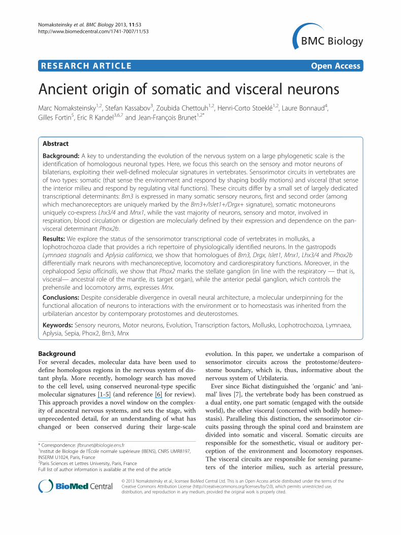
Ancient origin of somatic and visceral neurons
11
RESEARCH ARTICLE Open Access Ancient origin of somatic and visceral neurons Marc Nomaksteinsky 1,2 , Stefan Kassabov 3 , Zoubida Chettouh 1,2 , Henri-Corto Stoeklé 1,2 , Laure Bonnaud 4 , Gilles Fortin 5 , Eric R Kandel 3,6,7 and Jean-François Brunet 1,2* Abstract Background: A key to understanding the evolution of the nervous system on a large phylogenetic scale is the identification of homologous neuronal types. Here, we focus this search on the sensory and motor neurons of bilaterians, exploiting their well-defined molecular signatures in vertebrates. Sensorimotor circuits in vertebrates are of two types: somatic (that sense the environment and respond by shaping bodily motions) and visceral (that sense the interior milieu and respond by regulating vital functions). These circuits differ by a small set of largely dedicated transcriptional determinants: Brn3 is expressed in many somatic sensory neurons, first and second order (among which mechanoreceptors are uniquely marked by the Brn3+/Islet1+/Drgx+ signature), somatic motoneurons uniquely co-express Lhx3/4 and Mnx1, while the vast majority of neurons, sensory and motor, involved in respiration, blood circulation or digestion are molecularly defined by their expression and dependence on the pan- visceral determinant Phox2b. Results: We explore the status of the sensorimotor transcriptional code of vertebrates in mollusks, a lophotrochozoa clade that provides a rich repertoire of physiologically identified neurons. In the gastropods Lymnaea stagnalis and Aplysia californica, we show that homologues of Brn3, Drgx, Islet1, Mnx1, Lhx3/4 and Phox2b differentially mark neurons with mechanoreceptive, locomotory and cardiorespiratory functions. Moreover, in the cephalopod Sepia officinalis, we show that Phox2 marks the stellate ganglion (in line with the respiratory — that is, visceral— ancestral role of the mantle, its target organ), while the anterior pedal ganglion, which controls the prehensile and locomotory arms, expresses Mnx. Conclusions: Despite considerable divergence in overall neural architecture, a molecular underpinning for the functional allocation of neurons to interactions with the environment or to homeostasis was inherited from the urbilaterian ancestor by contemporary protostomes and deuterostomes. Keywords: Sensory neurons, Motor neurons, Evolution, Transcription factors, Mollusks, Lophotrochozoa, Lymnaea, Aplysia, Sepia, Phox2, Brn3, Mnx Background For several decades, molecular data have been used to define homologous regions in the nervous system of dis- tant phyla. More recently, homology search has moved to the cell level, using conserved neuronal-type specific molecular signatures [1-5] (and reference [6] for review). This approach provides a novel window on the complex- ity of ancestral nervous systems, and sets the stage, with unprecedented detail, for an understanding of what has changed or been conserved during their large-scale evolution. In this paper, we undertake a comparison of sensorimotor circuits across the protostome/deutero- stome boundary, which is, thus, informative about the nervous system of Urbilateria. Ever since Bichat distinguished the ‘organic’ and ‘ani- mal’ lives [7], the vertebrate body has been construed as a dual entity, one part somatic (engaged with the outside world), the other visceral (concerned with bodily homeo- stasis). Paralleling this distinction, the sensorimotor cir- cuits passing through the spinal cord and brainstem are divided into somatic and visceral. Somatic circuits are responsible for the somesthetic, visual or auditory per- ception of the environment and locomotory responses. The visceral circuits are responsible for sensing parame- ters of the interior milieu, such as arterial pressure, * Correspondence: [email protected] 1 Institut de Biologie de l’École normale supérieure (IBENS), CNRS UMR8197, INSERM U1024, Paris, France 2 Paris Sciences et Lettres University, Paris, France Full list of author information is available at the end of the article © 2013 Nomaksteinsky et al.; licensee BioMed Central Ltd. This is an Open Access article distributed under the terms of the Creative Commons Attribution License (http://creativecommons.org/licenses/by/2.0), which permits unrestricted use, distribution, and reproduction in any medium, provided the original work is properly cited. Nomaksteinsky et al. BMC Biology 2013, 11:53 http://www.biomedcentral.com/1741-7007/11/53
Transcript of Ancient origin of somatic and visceral neurons
Nomaksteinsky et al. BMC Biology 2013, 11:53http://www.biomedcentral.com/1741-7007/11/53
RESEARCH ARTICLE Open Access
Ancient origin of somatic and visceral neuronsMarc Nomaksteinsky1,2, Stefan Kassabov3, Zoubida Chettouh1,2, Henri-Corto Stoeklé1,2, Laure Bonnaud4,Gilles Fortin5, Eric R Kandel3,6,7 and Jean-François Brunet1,2*
Abstract
Background: A key to understanding the evolution of the nervous system on a large phylogenetic scale is theidentification of homologous neuronal types. Here, we focus this search on the sensory and motor neurons ofbilaterians, exploiting their well-defined molecular signatures in vertebrates. Sensorimotor circuits in vertebrates areof two types: somatic (that sense the environment and respond by shaping bodily motions) and visceral (that sensethe interior milieu and respond by regulating vital functions). These circuits differ by a small set of largely dedicatedtranscriptional determinants: Brn3 is expressed in many somatic sensory neurons, first and second order (amongwhich mechanoreceptors are uniquely marked by the Brn3+/Islet1+/Drgx+ signature), somatic motoneuronsuniquely co-express Lhx3/4 and Mnx1, while the vast majority of neurons, sensory and motor, involved inrespiration, blood circulation or digestion are molecularly defined by their expression and dependence on the pan-visceral determinant Phox2b.
Results: We explore the status of the sensorimotor transcriptional code of vertebrates in mollusks, alophotrochozoa clade that provides a rich repertoire of physiologically identified neurons. In the gastropodsLymnaea stagnalis and Aplysia californica, we show that homologues of Brn3, Drgx, Islet1, Mnx1, Lhx3/4 and Phox2bdifferentially mark neurons with mechanoreceptive, locomotory and cardiorespiratory functions. Moreover, in thecephalopod Sepia officinalis, we show that Phox2 marks the stellate ganglion (in line with the respiratory — that is,visceral— ancestral role of the mantle, its target organ), while the anterior pedal ganglion, which controls theprehensile and locomotory arms, expresses Mnx.
Conclusions: Despite considerable divergence in overall neural architecture, a molecular underpinning for thefunctional allocation of neurons to interactions with the environment or to homeostasis was inherited from theurbilaterian ancestor by contemporary protostomes and deuterostomes.
Keywords: Sensory neurons, Motor neurons, Evolution, Transcription factors, Mollusks, Lophotrochozoa, Lymnaea,Aplysia, Sepia, Phox2, Brn3, Mnx
BackgroundFor several decades, molecular data have been used todefine homologous regions in the nervous system of dis-tant phyla. More recently, homology search has movedto the cell level, using conserved neuronal-type specificmolecular signatures [1-5] (and reference [6] for review).This approach provides a novel window on the complex-ity of ancestral nervous systems, and sets the stage, withunprecedented detail, for an understanding of what haschanged or been conserved during their large-scale
* Correspondence: [email protected] de Biologie de l’École normale supérieure (IBENS), CNRS UMR8197,INSERM U1024, Paris, France2Paris Sciences et Lettres University, Paris, FranceFull list of author information is available at the end of the article
© 2013 Nomaksteinsky et al.; licensee BioMedCreative Commons Attribution License (http:/distribution, and reproduction in any medium
evolution. In this paper, we undertake a comparison ofsensorimotor circuits across the protostome/deutero-stome boundary, which is, thus, informative about thenervous system of Urbilateria.Ever since Bichat distinguished the ‘organic’ and ‘ani-
mal’ lives [7], the vertebrate body has been construed asa dual entity, one part somatic (engaged with the outsideworld), the other visceral (concerned with bodily homeo-stasis). Paralleling this distinction, the sensorimotor cir-cuits passing through the spinal cord and brainstem aredivided into somatic and visceral. Somatic circuits areresponsible for the somesthetic, visual or auditory per-ception of the environment and locomotory responses.The visceral circuits are responsible for sensing parame-ters of the interior milieu, such as arterial pressure,
Central Ltd. This is an Open Access article distributed under the terms of the/creativecommons.org/licenses/by/2.0), which permits unrestricted use,, provided the original work is properly cited.
Nomaksteinsky et al. BMC Biology 2013, 11:53 Page 2 of 11http://www.biomedcentral.com/1741-7007/11/53
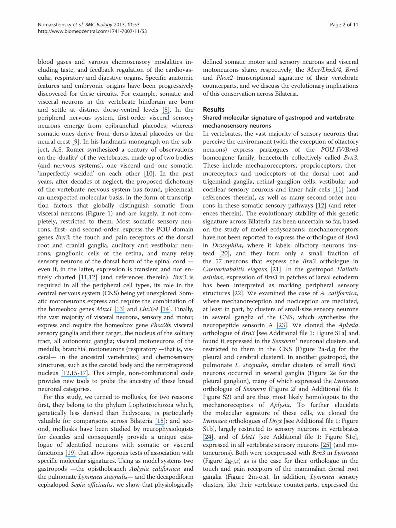
blood gases and various chemosensory modalities in-cluding taste, and feedback regulation of the cardiovas-cular, respiratory and digestive organs. Specific anatomicfeatures and embryonic origins have been progressivelydiscovered for these circuits. For example, somatic andvisceral neurons in the vertebrate hindbrain are bornand settle at distinct dorso-ventral levels [8]. In theperipheral nervous system, first-order visceral sensoryneurons emerge from epibranchial placodes, whereassomatic ones derive from dorso-lateral placodes or theneural crest [9]. In his landmark monograph on the sub-ject, A.S. Romer synthesized a century of observationson the ‘duality’ of the vertebrates, made up of two bodies(and nervous systems), one visceral and one somatic,‘imperfectly welded’ on each other [10]. In the pastyears, after decades of neglect, the proposed dichotomyof the vertebrate nervous system has found, piecemeal,an unexpected molecular basis, in the form of transcrip-tion factors that globally distinguish somatic fromvisceral neurons (Figure 1) and are largely, if not com-pletely, restricted to them. Most somatic sensory neu-rons, first- and second-order, express the POU domaingenes Brn3: the touch and pain receptors of the dorsalroot and cranial ganglia, auditory and vestibular neu-rons, ganglionic cells of the retina, and many relaysensory neurons of the dorsal horn of the spinal cord —even if, in the latter, expression is transient and not en-tirely charted [11,12] (and references therein). Brn3 isrequired in all the peripheral cell types, its role in thecentral nervous system (CNS) being yet unexplored. Som-atic motoneurons express and require the combination ofthe homeobox genes Mnx1 [13] and Lhx3/4 [14]. Finally,the vast majority of visceral neurons, sensory and motor,express and require the homeobox gene Phox2b: visceralsensory ganglia and their target, the nucleus of the solitarytract, all autonomic ganglia; visceral motoneurons of themedulla; branchial motoneurons (respiratory —that is, vis-ceral— in the ancestral vertebrates) and chemosensorystructures, such as the carotid body and the retrotrapezoidnucleus [12,15-17]. This simple, non-combinatorial codeprovides new tools to probe the ancestry of these broadneuronal categories.For this study, we turned to mollusks, for two reasons:
first, they belong to the phylum Lophotrochozoa which,genetically less derived than Ecdysozoa, is particularlyvaluable for comparisons across Bilateria [18]; and sec-ond, mollusks have been studied by neurophysiologistsfor decades and consequently provide a unique cata-logue of identified neurons with somatic or visceralfunctions [19] that allow rigorous tests of association withspecific molecular signatures. Using as model systems twogastropods —the opisthobranch Aplysia californica andthe pulmonate Lymnaea stagnalis— and the decapodiformcephalopod Sepia officinalis, we show that physiologically
defined somatic motor and sensory neurons and visceralmotoneurons share, respectively, the Mnx/Lhx3/4, Brn3and Phox2 transcriptional signature of their vertebratecounterparts, and we discuss the evolutionary implicationsof this conservation across Bilateria.
ResultsShared molecular signature of gastropod and vertebratemechanosensory neuronsIn vertebrates, the vast majority of sensory neurons thatperceive the environment (with the exception of olfactoryneurons) express paralogues of the POU-IV/Brn3homeogene family, henceforth collectively called Brn3.These include mechanoreceptors, proprioceptors, ther-moreceptors and nociceptors of the dorsal root andtrigeminal ganglia, retinal ganglion cells, vestibular andcochlear sensory neurons and inner hair cells [11] (andreferences therein), as well as many second-order neu-rons in these somatic sensory pathways [12] (and refer-ences therein). The evolutionary stability of this geneticsignature across Bilateria has been uncertain so far, basedon the study of model ecdysozoans: mechanoreceptorshave not been reported to express the orthologue of Brn3in Drosophila, where it labels olfactory neurons ins-tead [20], and they form only a small fraction ofthe 57 neurons that express the Brn3 orthologue inCaenorhabditis elegans [21]. In the gastropod Haliotisasinina, expression of Brn3 in patches of larval ectodermhas been interpreted as marking peripheral sensorystructures [22]. We examined the case of A. californica,where mechanoreception and nociception are mediated,at least in part, by clusters of small-size sensory neuronsin several ganglia of the CNS, which synthesize theneuropeptide sensorin A [23]. We cloned the Aplysiaorthologue of Brn3 [see Additional file 1: Figure S1a] andfound it expressed in the Sensorin+ neuronal clusters andrestricted to them in the CNS (Figure 2a-d,q for thepleural and cerebral clusters). In another gastropod, thepulmonate L. stagnalis, similar clusters of small Brn3+
neurons occurred in several ganglia (Figure 2e for thepleural ganglion), many of which expressed the Lymnaeaorthologue of Sensorin (Figure 2f and Additional file 1:Figure S2) and are thus most likely homologous to themechanoreceptors of Aplysia. To further elucidatethe molecular signature of these cells, we cloned theLymnaea orthologues of Drgx [see Additional file 1: FigureS1b], largely restricted to sensory neurons in vertebrates[24], and of Islet1 [see Additional file 1: Figure S1c],expressed in all vertebrate sensory neurons [25] (and mo-toneurons). Both were coexpressed with Brn3 in Lymnaea(Figure 2g-j,r) as is the case for their orthologue in thetouch and pain receptors of the mammalian dorsal rootganglia (Figure 2m-o,s). In addition, Lymnaea sensoryclusters, like their vertebrate counterparts, expressed the
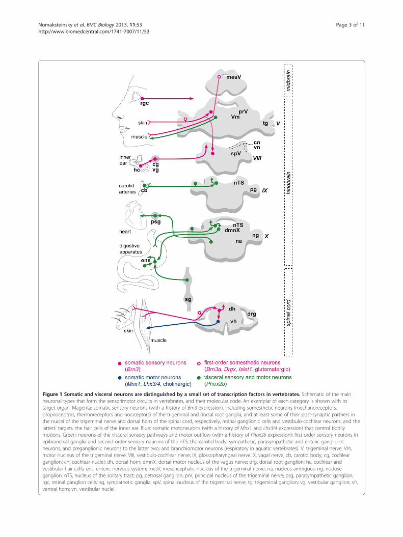
Figure 1 Somatic and visceral neurons are distinguished by a small set of transcription factors in vertebrates. Schematic of the mainneuronal types that form the sensorimotor circuits in vertebrates, and their molecular code. An exemplar of each category is shown with itstarget organ. Magenta: somatic sensory neurons (with a history of Brn3 expression), including somesthetic neurons (mechanoreceptors,proprioceptors, thermoreceptors and nociceptors) of the trigeminal and dorsal root ganglia, and at least some of their post-synaptic partners inthe nuclei of the trigeminal nerve and dorsal horn of the spinal cord, respectively, retinal ganglionic cells and vestibulo-cochlear neurons, and thelatters’ targets, the hair cells of the inner ear. Blue: somatic motoneurons (with a history of Mnx1 and Lhx3/4 expression) that control bodilymotions. Green: neurons of the visceral sensory pathways and motor outflow (with a history of Phox2b expression): first-order sensory neurons inepibranchial ganglia and second-order sensory neurons of the nTS; the carotid body; sympathetic, parasympathetic and enteric ganglionicneurons, and preganglionic neurons to the latter two; and branchiomotor neurons (respiratory in aquatic vertebrates). V, trigeminal nerve; Vm,motor nucleus of the trigeminal nerve; VIII, vestibulo-cochlear nerve; IX, glossopharyngeal nerve; X, vagal nerve; cb, carotid body; cg, cochlearganglion; cn, cochlear nuclei; dh, dorsal horn; dmnX, dorsal motor nucleus of the vagus nerve; drg, dorsal root ganglion; hc, cochlear andvestibular hair cells; ens, enteric nervous system; mesV, mesencephalic nucleus of the trigeminal nerve; na, nucleus ambiguus; ng, nodoseganglion; nTS, nucleus of the solitary tract; pg, petrosal ganglion; prV, principal nucleus of the trigeminal nerve; psg, parasympathetic ganglion;rgc, retinal ganglion cells; sg, sympathetic ganglia; spV, spinal nucleus of the trigeminal nerve; tg, trigeminal ganglion; vg, vestibular ganglion; vh,ventral horn; vn, vestibular nuclei.
Nomaksteinsky et al. BMC Biology 2013, 11:53 Page 3 of 11http://www.biomedcentral.com/1741-7007/11/53
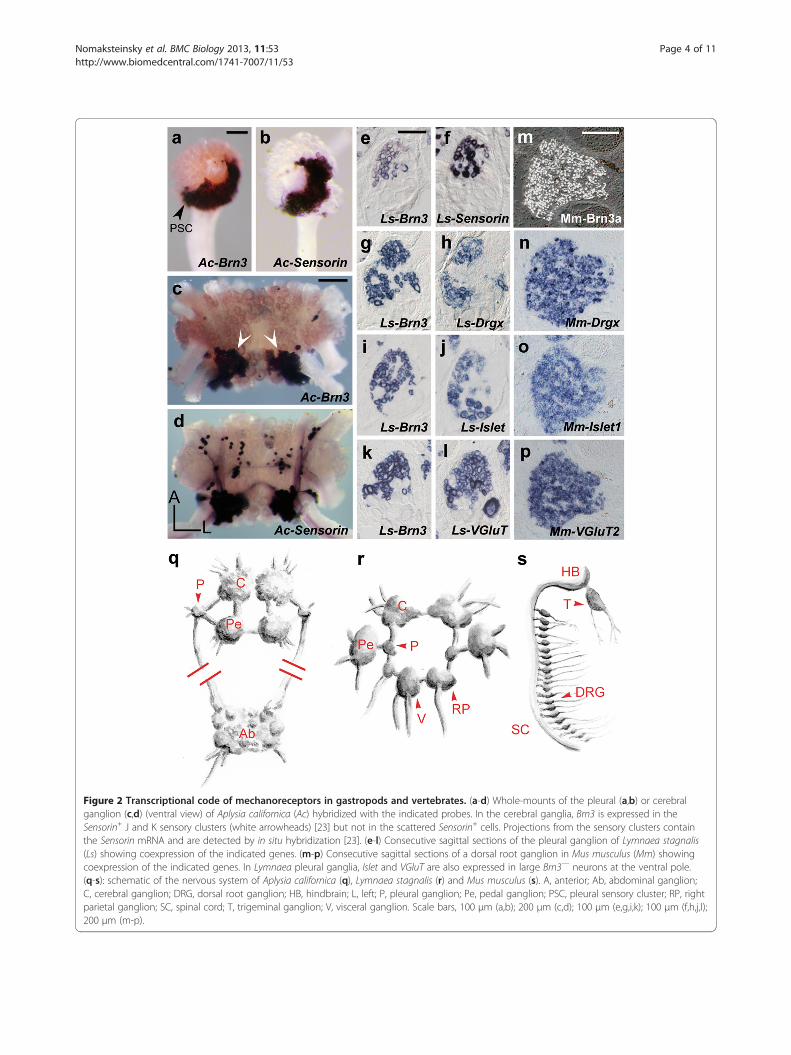
Figure 2 Transcriptional code of mechanoreceptors in gastropods and vertebrates. (a-d) Whole-mounts of the pleural (a,b) or cerebralganglion (c,d) (ventral view) of Aplysia californica (Ac) hybridized with the indicated probes. In the cerebral ganglia, Brn3 is expressed in theSensorin+ J and K sensory clusters (white arrowheads) [23] but not in the scattered Sensorin+ cells. Projections from the sensory clusters containthe Sensorin mRNA and are detected by in situ hybridization [23]. (e-l) Consecutive sagittal sections of the pleural ganglion of Lymnaea stagnalis(Ls) showing coexpression of the indicated genes. (m-p) Consecutive sagittal sections of a dorsal root ganglion in Mus musculus (Mm) showingcoexpression of the indicated genes. In Lymnaea pleural ganglia, Islet and VGluT are also expressed in large Brn3— neurons at the ventral pole.(q-s): schematic of the nervous system of Aplysia californica (q), Lymnaea stagnalis (r) and Mus musculus (s). A, anterior; Ab, abdominal ganglion;C, cerebral ganglion; DRG, dorsal root ganglion; HB, hindbrain; L, left; P, pleural ganglion; Pe, pedal ganglion; PSC, pleural sensory cluster; RP, rightparietal ganglion; SC, spinal cord; T, trigeminal ganglion; V, visceral ganglion. Scale bars, 100 μm (a,b); 200 μm (c,d); 100 μm (e,g,i,k); 100 μm (f,h,j,l);200 μm (m-p).
Nomaksteinsky et al. BMC Biology 2013, 11:53 Page 4 of 11http://www.biomedcentral.com/1741-7007/11/53
Figure 3 Transcriptional code of putative somatic motoneuronsin the pedal ganglia of Lymnaea stagnalis. Consecutive sagittalsections (respectively a,b and c,d) through the pedal ganglionhybridized with the indicated probes. In e, the ganglion washybridized with Ls-Mnx after retrograde filling with biocytin throughthe three pedal nerves to the foot. The three nerves to the neck andcolumella, also involved in locomotion, were not filled. The anteriorcluster of Mnx+ cells is filled, as well as about half of the posteriorcluster. Scale bars, 100 μm.
Nomaksteinsky et al. BMC Biology 2013, 11:53 Page 5 of 11http://www.biomedcentral.com/1741-7007/11/53
vesicular glutamate transporter VGluT (Figure 2k,l,p), in linewith the glutamatergic phenotype of molluscan mechanore-ceptors [26]. Thus, the unique Brn3+/Drgx+/Islet+/VGluT+
transcriptional signature of first-order somatic sensory neu-rons in vertebrates, is also a hallmark of their molluscancounterparts.
Shared molecular distinction between somatic andvisceral motoneurons in gastropods, cephalopods andvertebratesIn vertebrates, locomotion depends on somitic muscles,innervated by spinal cord motoneurons that depend onthe homeogenes Mnx1 [13] and Lhx3/4 [14] and useacetylcholine as neurotransmitter. In gastropods, themuscles of locomotion (in the foot and body wall orattached to the columella) are innervated by thepedal ganglia [27]. We cloned Ls-Mnx, the Lymnaeaorthologue of Mnx1 [see Additional file 1: Figure S1d]and found that it was largely restricted to two clusters ofneurons in the pedal ganglia (Figure 3a), which co-expressed the orthologues of Lhx3/4 (Figure 3c andAdditional file 1: Figure S1e) and the vesicular acetyl-choline transporter VAChT (Figure 3b,d). At least someof these cells projected in the pedal nerves, as assessedby retrograde filling (Figure 3e-g) and most likely arelocomotory neurons. These data extend the observationsthat some motoneurons in Drosophila also express Lim3(the Lhx3/4 orthologue) and Mnx [28,29], that some C.elegans motoneurons express a Mnx orthologue [30],and that the Nkx6+ domain of the annelid nerve cordgives rise to Mnx+/VAChT+ putative motoneurons [31].Thus, the Mnx+/Lhx3/4+ molecular signature has beenassociated with somatic motoneurons since the originof bilaterians.We next examined the visceral nervous system, whose
sensorimotor circuits in vertebrates largely coincide withthe expression pattern of the paralogous homeogenesPhox2a and Phox2b (hereafter collectively designatedas Phox2) and depend on the latter for their formation[12,15-17]. Among deuterostomes, we previously foundthat the Ciona intestinalis orthologue of Phox2 is specif-ically expressed in neurons of the cerebral ganglionof postmetamorphic animals that motorize the respira-tory and digestive ‘branchial basket’ [2]. Concerningprotostomes, the C. elegans orthologue of Phox2 marksspecifically five neurons [32] whose function is eitherunknown (the SIAs) or hard to classify as somatic or vis-ceral (ALA, involved in behavioral quiescence [33]). Theinconclusiveness of the latter finding is compounded bythe fact that most visceral organs in C. elegans eitherlack innervation (such as the intestine) or are missingaltogether (such as a cardiovascular or a respiratory ap-paratus). In A. californica, several neurons with cardio-vascular, respiratory or excretory functions have been
identified by size and location in the abdominal gan-glion. One of the best documented, the multimodalmotoneuron L7, directly innervates the muscles of thegill, siphon, epineural sheath, heart and abdominal aorta[34] (and references therein), and serves as premotorneuron for the branchial ganglion [35]. We clonedAc-Phox2, the Aplysia orthologue of Phox2 [seeAdditional file 1: Figure S1b] and found it expressed inL7, recognizable by its large size and position at the leftborder of the abdominal ganglion, rostral to the otherlarge (and also Phox2+) neuron in the region, L11, whichprojects in the genital nerve but whose function is
Nomaksteinsky et al. BMC Biology 2013, 11:53 Page 6 of 11http://www.biomedcentral.com/1741-7007/11/53
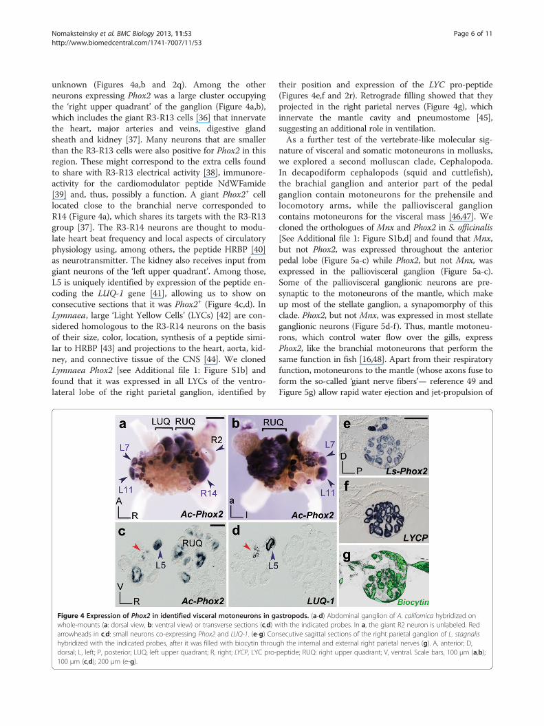
unknown (Figures 4a,b and 2q). Among the otherneurons expressing Phox2 was a large cluster occupyingthe ‘right upper quadrant’ of the ganglion (Figure 4a,b),which includes the giant R3-R13 cells [36] that innervatethe heart, major arteries and veins, digestive glandsheath and kidney [37]. Many neurons that are smallerthan the R3-R13 cells were also positive for Phox2 in thisregion. These might correspond to the extra cells foundto share with R3-R13 electrical activity [38], immunore-activity for the cardiomodulator peptide NdWFamide[39] and, thus, possibly a function. A giant Phox2+ celllocated close to the branchial nerve corresponded toR14 (Figure 4a), which shares its targets with the R3-R13group [37]. The R3-R14 neurons are thought to modu-late heart beat frequency and local aspects of circulatoryphysiology using, among others, the peptide HRBP [40]as neurotransmitter. The kidney also receives input fromgiant neurons of the ‘left upper quadrant’. Among those,L5 is uniquely identified by expression of the peptide en-coding the LUQ-1 gene [41], allowing us to show onconsecutive sections that it was Phox2+ (Figure 4c,d). InLymnaea, large ‘Light Yellow Cells’ (LYCs) [42] are con-sidered homologous to the R3-R14 neurons on the basisof their size, color, location, synthesis of a peptide simi-lar to HRBP [43] and projections to the heart, aorta, kid-ney, and connective tissue of the CNS [44]. We clonedLymnaea Phox2 [see Additional file 1: Figure S1b] andfound that it was expressed in all LYCs of the ventro-lateral lobe of the right parietal ganglion, identified by
Figure 4 Expression of Phox2 in identified visceral motoneurons in gawhole-mounts (a: dorsal view, b: ventral view) or transverse sections (c,d) warrowheads in c,d: small neurons co-expressing Phox2 and LUQ-1. (e-g) Conhybridized with the indicated probes, after it was filled with biocytin throudorsal; L, left; P, posterior; LUQ, left upper quadrant; R, right; LYCP, LYC pro-100 μm (c,d); 200 μm (e-g).
their position and expression of the LYC pro-peptide(Figures 4e,f and 2r). Retrograde filling showed that theyprojected in the right parietal nerves (Figure 4g), whichinnervate the mantle cavity and pneumostome [45],suggesting an additional role in ventilation.As a further test of the vertebrate-like molecular sig-
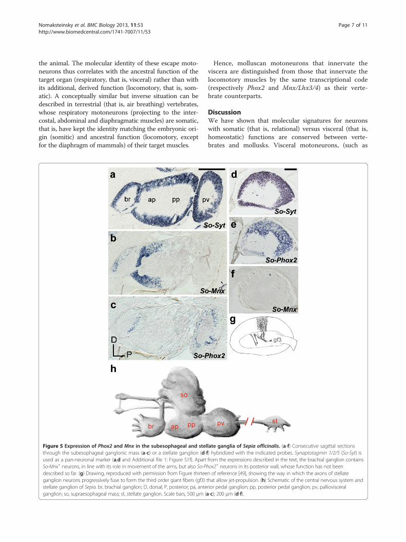
nature of visceral and somatic motoneurons in mollusks,we explored a second molluscan clade, Cephalopoda.In decapodiform cephalopods (squid and cuttlefish),the brachial ganglion and anterior part of the pedalganglion contain motoneurons for the prehensile andlocomotory arms, while the palliovisceral ganglioncontains motoneurons for the visceral mass [46,47]. Wecloned the orthologues of Mnx and Phox2 in S. officinalis[See Additional file 1: Figure S1b,d] and found that Mnx,but not Phox2, was expressed throughout the anteriorpedal lobe (Figure 5a-c) while Phox2, but not Mnx, wasexpressed in the palliovisceral ganglion (Figure 5a-c).Some of the palliovisceral ganglionic neurons are pre-synaptic to the motoneurons of the mantle, which makeup most of the stellate ganglion, a synapomorphy of thisclade. Phox2, but not Mnx, was expressed in most stellateganglionic neurons (Figure 5d-f). Thus, mantle motoneu-rons, which control water flow over the gills, expressPhox2, like the branchial motoneurons that perform thesame function in fish [16,48]. Apart from their respiratoryfunction, motoneurons to the mantle (whose axons fuse toform the so-called ‘giant nerve fibers’— reference 49 andFigure 5g) allow rapid water ejection and jet-propulsion of
stropods. (a-d) Abdominal ganglion of A. californica hybridized onith the indicated probes. In a, the giant R2 neuron is unlabeled. Redsecutive sagittal sections of the right parietal ganglion of L. stagnalisgh the internal and external right parietal nerves (g). A, anterior; D,peptide; RUQ: right upper quadrant; V, ventral. Scale bars, 100 μm (a,b);
Nomaksteinsky et al. BMC Biology 2013, 11:53 Page 7 of 11http://www.biomedcentral.com/1741-7007/11/53
the animal. The molecular identity of these escape moto-neurons thus correlates with the ancestral function of thetarget organ (respiratory, that is, visceral) rather than withits additional, derived function (locomotory, that is, som-atic). A conceptually similar but inverse situation can bedescribed in terrestrial (that is, air breathing) vertebrates,whose respiratory motoneurons (projecting to the inter-costal, abdominal and diaphragmatic muscles) are somatic,that is, have kept the identity matching the embryonic ori-gin (somitic) and ancestral function (locomotory, exceptfor the diaphragm of mammals) of their target muscles.
Figure 5 Expression of Phox2 and Mnx in the subesophageal and stelthrough the subesophageal ganglonic mass (a-c) or a stellate ganglion (d-used as a pan-neuronal marker (a,d and Additional file 1: Figure S1f). ApartSo-Mnx+ neurons, in line with its role in movement of the arms, but also So-Phdescribed so far. (g) Drawing, reproduced with permission from Figure thirteeganglion neurons progressively fuse to form the third order giant fibers (gf3)stellate ganglion of Sepia. br, brachial ganglion; D, dorsal; P, posterior; pa, anteganglion; so, supraesophageal mass; st, stellate ganglion. Scale bars, 500 μm (
Hence, molluscan motoneurons that innervate theviscera are distinguished from those that innervate thelocomotory muscles by the same transcriptional code(respectively Phox2 and Mnx/Lhx3/4) as their verte-brate counterparts.
DiscussionWe have shown that molecular signatures for neuronswith somatic (that is, relational) versus visceral (that is,homeostatic) functions are conserved between verte-brates and mollusks. Visceral motoneurons, (such as
late ganglia of Sepia officinalis. (a-f) Consecutive sagittal sectionsf) hybridized with the indicated probes. Synaptotagmin 1/2/5 (So-Syt) isfrom the expressions described in the text, the brachial ganglion containsox2+ neurons in its posterior wall, whose function has not beenn of reference [49], showing the way in which the axons of stellatethat allow jet-propulsion. (h) Schematic of the central nervous system andrior pedal ganglion; pp, posterior pedal ganglion; pv, pallioviscerala-c); 200 μm (d-f).
Nomaksteinsky et al. BMC Biology 2013, 11:53 Page 8 of 11http://www.biomedcentral.com/1741-7007/11/53
cardiorespiratory neurons) express the orthologue of thevertebate pan-visceral determinant Phox2b in opistho-branch and pulmonate gastropods and a decapodiformcephalopod. No other transcription factor or neurotrans-mitter phenotype marks these neurons, specifically orexhaustively, in vertebrates, precluding a more complexsignature. However, Phox2b expression is highly selectivefor visceral neurons (both motor and sensory) in verte-brates [16] and, thus, in combination with hodologicalcriteria, constitutes a strong argument for homology.Neurons with modalities clearly equivalent to those ofvisceral sensory neurons in vertebrates (which monitortaste or blood pressure for example) are not described toour knowledge in mollusks, precluding exploration ofthis broad neuronal identity. Of note, in vertebrates, theviscerosensory phenotype is imposed by Phox2b on asomatosensory default identity [12], suggesting that theformer is evolutionarily more recent than the latter. Loco-motory (somatic) motor neurons express the orthologuesof the homeobox genes Mnx1 and Lhx3/4 and VAChT inboth gastropods and cephalopods, like their counterpartsin the spinal cord of vertebrates. Finally, somatic sensoryneurons (such as mechanoreceptors), characterized in gas-tropods by previous electrophysiological studies or expres-sion of the peptide sensorin A, selectively express theorthologues of the homeodomain genes Brn3, Drgx, andIslet1 and VGluT, like their physiological counterparts inthe dorsal root and trigeminal ganglia of vertebrates.What evolutionary relationship can explain the conser-
vation of molecular signatures in neurons with visceralversus somatic functions between deuterostomes andprotostomes? The simplest hypothesis is that the cellsare phylogenetically homologous, that is, one could tracetheir ancestry to an original neuron or neuronal clusterin the common ancestor, as was proposed for ciliaryphotoreceptors in annelids and vertebrates [1] or forbranchial motoneurons in vertebrates and urochordates[2]. This might also be the case for somatic motoneu-rons in vertebrates and lophotrochozoans: their distribu-tion is spatially discrete in each phylum and reconcilablebetween phyla. In vertebrates, locomotory Mnx+ neu-rons are born in a ventral Nkx6.2+ domain of the spinalcord, topologically and molecularly similar to the ventraldomain of the nerve cord of annelids, which producesMnx+/VAChT+ neurons, presumably motor [31]. In mol-lusks, they are restricted to the pedal ganglia, conceiv-ably homologous to the ventral nerve cord of annelids.On the other hand, phylogenetic homology between
vertebrates and mollusks is unlikely, at least in mostcases, for somatic sensory neurons and visceral motorneurons, due to their anatomical distribution, wide-spread in each species (Figures 1, 3, 4) and hard toreconcile between them. Moreover, Phox2 is expressedin synapomorphic structures of vertebrates (the neural
crest-derived autonomic ganglia) and cephalopods (thestellate ganglion). In this case, the most likely evolution-ary scenario is that, in the last common ancestor, a ‘sem-inal regulatory interaction’ [50] arose between Brn3 andthe somatic sensory phenotype and between Phox2 andthe visceral phenotype. Although no target gene hasbeen uncovered yet that would explain the physiologicaldichotomy these transcription factors specify, one canhypothesize, for example, that they direct axonal projec-tions towards somatic versus visceral targets. Subse-quently, this regulatory interaction would have beenconserved, while Brn3 and Phox2 acquired additionalexpression sites, giving rise to novel groups of cells ofthe same broad type along each evolutionary lineage.According to this view, the relationship between the dif-ferent kinds of somatosensory neurons or visceromotorneurons would be neither of phylogenetic homology(between species) nor of ‘sister cell types’ (within aspecies [6]), but instead, both between and within species,of ‘deep’ [51,52] or ‘generative’ [53] homology, akin to thatproposed for bilaterian appendages.
ConclusionsRegardless of the exact nature of what has been con-served across the protostome-deuterostome boundary,either neuronal groups or regulatory links betweentranscription factors and neuronal traits, our datashow that the viscerosomatic duality of the nervoussystem, as described in vertebrates, was already part ofthe urbilaterian body plan. Some of its componentsmight be even more ancient, as suggested by expression ofa Brn3 orthologue in the sensory ‘rhopalia’ of scyphozoanand cubozoan jellyfish [54]. To our knowledge, Mnx hasnot been analyzed in the nervous system of cnidarians.Finally, no straightforward orthologue of Phox2 emergesfrom sequence analysis of paired-like homeobox genes inCnidaria [see Additional file 1: Figure S1b], which compli-cates the search for a pre-bilaterian origin of the visceralnervous system.
MethodsTissue processingGanglia from adult A. californica (100 to 150 g) weredissected in artificial seawater (460 mM NaCl, 10 mMKCl, 55 mM MgCl2, 11 mM, CaCl2, 10 mM (4-(2-hydroxyethyl)-1-piperazineethanesulfonic acid (HEPES),pH 7.6), protease digested for two hours at 37°C, fixedwith 4% paraformaldehyde (4% PFA) in phosphate buff-ered saline (PBS) overnight at 4°C, desheathed anddehydrated in ethanol. S. officinalis fertilized eggs wereremoved from their envelopes in artificial seawater andfixed overnight with 4% PFA when they reached stage 29[55], dechorionated, fixed again with 4% PFA for a wholeday and dehydrated in methanol. Samples destined to be
Nomaksteinsky et al. BMC Biology 2013, 11:53 Page 9 of 11http://www.biomedcentral.com/1741-7007/11/53
sectioned were rehydrated in PBST (PBS, 0.1% Tween-20), incubated overnight at 4°C in (PBS, 15% sucrose),incubated for 50 minutes at 65°C in gelatine (PBS, 7.5%gelatine, 15% sucrose, pH 7.2), embedded in gelatine,frozen for one minute at −50°C in isopentane and keptat −80°C until sectioning. Ganglia from adult L. stagnalis(20 to 40 mm) specimens were dissected in a physiologicalsolution (40 mM NaCl, 1.7 mM KCl, 1.5 mM MgCl2, 4.1mM CaCl2, 10 mM HEPES, pH7.9), fixed with 4% PFAand processed for gelatine embedding as described above.cDNA from Rattus norvegicus Drgx, R. norvegicus Islet1and R. norvegicus VGluT2 sequences and a mouse anti-Brn3a monoclonal antibody (MAB1585 Millipore, Biller-ica, MA, USA) were used for mice procedures.
In situ hybridization on sectionsFrozen sections (12 μm) were thawed and air dried,washed briefly in PBST, treated 2 × 10 minutes with RIPAbuffer (150 mM NaCl, 1% NP-40, 0.5% Na deoxycholate,0.1% SDS, 1 mM ethylenediaminetetraacetic acid (EDTA),50 mM Tris pH 8.0), postfixed with 4% PFA for 15 mi-nutes, washed 3 × 5 minutes in PBST, acetylated with(100 mM triethanolamine, pH 8.0, 0.25% acetic anhyd-ride) for 15 minutes on a rocker table and washed 3 × 5minutes in PBST. Endogenous alkaline phosphatases wereinactivated by a 60-minute incubation in PBS at 80°C.The slides were then prehybridized for 60 minutes inhybridization solution (50% formamide, 5X SSC, 5XDenhardt’s, 500 μg/mL herring sperm DNA, 250 μg/mLyeast RNA) at 65°C and hybridized with the digoxigenin-labeled RNA (Roche, Penzberg, Germany) probe (500 ng/mL) overnight at the same temperature. The slides werewashed twice in 50% formamide, 2X SSC, 0.1% Tween-20)for 60 minutes and in 0.2X SSC at 65°C for 60 minutes.The slides were then rinsed 3 × 10 minutes in buffer 1(100 mM maleic acid, pH 7.5, 150 mM NaCl, 0.1%Tween-20), blocked in buffer 2 (buffer 1, 10% sheepserum) for 60 minutes and incubated overnight at 4°Cwith alkaline phosphatase-coupled anti-DIG antibody(Roche) diluted 1/2000 in buffer 2. The slides werewashed 2 × 10 minutes in buffer 1, incubated for 30 mi-nutes in buffer 3 (100 mM Tris, pH 9.5, 100 mM NaCl,50 mM MgCl2, 0.1% Tween-20) and the signal wasrevealed in filtered buffer 4 (3.5 μL NBT (4-nitrobluetetrazolium chloride) (Roche) 100 mg/mL), 3.5 μL BCIP((5-bromo-4-chloro-3-indoylphosphate) (Roche) 50 mg/mL) in buffer 3). The slides were washed 3 × 5 minutesin PBST and then postfixed overnight with 4% PFA,washed briefly in PBST and then either washed in waterand mounted in Aquatex (Merck) or, when nerves hadbeen retrogradely filled, treated as follows: sectionswere permeabilized 2 × 10 minutes in PBS, 0.3% TritonX-100, blocked 20 minutes in blocking solution (PBS,10% fetal calf serum, 0.1% Triton X-100), incubated for
two hours in the dark with ExtrAvidin-fluoresceinisothiocyanate (FITC, Sigma, Saint-Quentin Fallavier,France) diluted 1/400 in blocking solution, washed 3 ×10 minutes in PBST in the dark and mounted in Mowiol(Calbiochem, Darmstadt, Germany).
In situ hybridization on whole-mountsThe dehydrated desheathed CNS were progressivelyrehydrated in PBST, incubated for 20 minutes in PBS,0.3% Triton X-100, rinsed in PBST, permeabilized withPBST, 10 mg/mL Proteinase K for 60 minutes, washed2 × 5 minutes in PBST, postfixed in 4% PFA for 30 mi-nutes, washed 2 × 5 minutes in PBST, incubated for 10minutes in 100 mM triethanolamine, pH 8.0, acetylated2 × 10 minutes with 100 mM triethanolamine, pH 8.0,0.25% acetic anhydride, washed 2 × 5 minutes in PBST,incubated at 80°C in PBS for 60 minutes, rinsed in PBST,prehybridized for 60 minutes in hybridization buffer(50% formamide, 1.3X SSC, 5 mM EDTA, 50 μg/mLyeast RNA, 2% Tween-20, 0.5% CHAPS) at 65°C and hy-bridized overnight with the probe (200 ng/mL) at thesame temperature. The samples were rinsed twice andwashed 2 × 30 minutes in prewarmed hybridization so-lution at 65°C. Then they were washed for 10 minutes atroom temperature in a 1:1 mixture of hybridization solu-tion and buffer 1, 60 minutes in buffer 1, blocked 60 mi-nutes in buffer 1/ 20% FCS, incubated overnight at 4°Cwith alkaline phosphatase-coupled anti-DIG antibody(Roche) diluted 1/2000 in buffer 1/ 2% FCS, rinsed threetimes and washed 3 × 60 minutes in buffer 1, then left fortwo days in buffer 1, incubated 2 × 30 minutes in buffer 3,revealed in the dark with filtered NBT/BCIP (Sigma),washed in the dark 3 x 10 minutes with PBST, postfixedovernight with 4% PFA, rinsed in PBST and kept at 4°C inTissue-TekWoptimal cutting temperature (O.C.T.) embed-ding medium (Sakura, Tokyo, Japan). Except for revela-tion, all steps were performed with agitation.
Immunofluorescence for detection of mouse Brn3aSections were dried, washed for 5 minutes in PBS,permeabilized 2 × 10 minutes in PBS/ 0.3% TritonX-100, blocked for 20 minutes in blocking solution(PBS, 10% FCS, 0.1% Triton X-100), incubated overnightat 4°C with the mouse anti-Brn3a monoclonal antibody(MAB1585) diluted 1/200 in blocking solution, washed3 × 10 minutes in PBST, incubated for 2 hours with thefluorescent goat anti-mouse Cy3-coupled secondary anti-body (115-165-003 Jackson ImmunoResearch, Newmarket,UK), washed 3 × 10 minutes in PBST in the dark andmounted in Mowiol (Calbiochem). The image wasconverted to gray scale, inverted and superimposed ona Nomarski photomicrograph in Photoshop CS3.
Nomaksteinsky et al. BMC Biology 2013, 11:53 Page 10 of 11http://www.biomedcentral.com/1741-7007/11/53
Cloning of orthologuesStarting with total RNA extracted using RNeasy LipidTissue Mini Kit (Qiagen), fragments of newly clonedcDNAs were amplified by PCR and rapid amplification ofcDNA ends-PCR (RACE-PCR) as described previously[56]. Orthology was assigned by phylogenetic analysis(see below and Additional file 1: Figure S1). Ls-VAChTand Ls-VGluT were PCR-amplified using oligonucleotidesderived from the GenBank sequences (accession numbers:AF484093 and AB469850, respectively).
Nerve backfillingThe ganglia of a 20 to 40 mm Lymnaea stagnalis speci-men were dissected in a 1:1 mixture of physiologicalmedium with Leibovitz's L15 medium (21083–027, Lifetechnologies, Saint Aubin, France) complemented with50 U/mL pennicilin, 50 μg/mL streptomycin and at thefinal concentration of 0.3 M glucose, cutting short allnerves except those of interest. The preparation waspinned at the bottom of a sylgard dish where two com-partments were delineated with Vaseline, separating theganglia from the distal part of the nerves to bebackfilled. The nerve compartment was emptied, filledwith distilled water (dH2O), nerves were recut and, aftera 10-minute incubation, dH2O was replaced by a satu-rated solution of biocytin (Sigma) in dH2O. After 24hours at room temperature and in the dark, the gangliawere fixed overnight at 4°C in 4% PFA and processed forin situ hybridization and biocytin revelation usingExtravidin-FITC (see above).
Phylogenetic analysesGene orthologies were assessed using protein sequencesaligned with ClustalX2 [57] and the software PhyML[58] with 1,000 bootstrap replicates and the model sug-gested by ProtTest 2.4 [59]. The closest groups of homeo-genes were used as outgroups [60]. Trees were drawn withDendroscope v3.0.14 [61].
Ethical approvalExperiments on mouse embryos have been approved byThe Research Ethic committee « Charles Darwin », Paris(approval number Ce5/2012/064).
Additional file
Additional file 1: Figure S1. Support for orthology assignment of thetwelve sequences cloned in this study. Figure S1 shows six phylogenetictrees for genes shared among Bilateria. Figure S2 shows amino acidsequence alignment between the Aplysia and Lymnaea Sensorin genes.
AbbreviationsCNS: Central nervous system; EDTA: Ethylenediaminetetraacetic acid;FCS: Fetal calf serum; FITC: Fluorescein isthiocyanate; HEPES: 4-(2-hydroxyethyl)-1-piperazineethanesulfonic acid; LYC: Light yellow cells;
PBS: Phosphate-buffered saline; PBST: PBS-Tween; PCR: Polymerase chainreaction; PFA: Paraformaldehyde; RACE-PCR: Rapid amplification of cDNAends-PCR.
Competing interestsThe authors declare that they have no competing interests.
Authors’ contributionsMN, ZC and HCS isolated genes and performed expression studies; GF andMN performed retrograde filling experiments; SK, LB and EK provided animaltissues from Aplysia and Sepia and participated in cell identifications; MN andJFB designed the study, analyzed the data and wrote the paper. All authorsread and approved the final manuscript.
AcknowledgementsWe thank E. Konstantinov for technical assistance, H. Roest Crollius and R. deRosa for advice on the phylogenetic analyses and C. Goridis for criticalreading of the manuscript. This work was supported by a grant of theAgence Nationale pour la Recherche (to JFB and GF), of the Fondation pourla Recherche Médicale (to MN) and by institutional support from the Centrede la Recherche Scientifique (CNRS), Institut National de la Santé et de laRecherche Médicale (INSERM) and École normale supérieure.
Author details1Institut de Biologie de l’École normale supérieure (IBENS), CNRS UMR8197,INSERM U1024, Paris, France. 2Paris Sciences et Lettres University, Paris,France. 3Department of Neuroscience, College of Physicians and Surgeons ofColumbia University, New York, USA. 4Museum National d’Histoire Naturelle,CNRS UMR7208, Université Pierre et Marie Curie, Paris, France. 5Institut deNeurobiologie Alfred Fessard, CNRS UPR3294, 91198, Gif-sur-Yvette, France.6Howard Hughes Medical Institute, College of Physicians and Surgeons ofColumbia University, New York, USA. 7Kavli Institute for Brain Science, Collegeof Physicians and Surgeons of Columbia University, New York, USA.
Received: 1 February 2013 Accepted: 22 March 2013Published: 30 April 2013
References1. Arendt D, Tessmar-Raible K, Snyman H, Dorresteijn AW, Wittbrodt J: Ciliary
photoreceptors with a vertebrate-type opsin in an invertebrate brain.Science 2004, 306:869–871.
2. Dufour HD, Chettouh Z, Deyts C, de Rosa R, Goridis C, Joly JS, Brunet JF:Precraniate origin of cranial motoneurons. Proc Natl Acad Sci USA 2006,103:8727–8732.
3. Tessmar-Raible K, Raible F, Christodoulou F, Guy K, Rembold M, Hausen H,Arendt D: Conserved sensory-neurosecretory cell types in annelid and fishforebrain: insights into hypothalamus evolution. Cell 2007, 129:1389–1400.
4. Vopalensky P, Pergner J, Liegertova M, Benito-Gutierrez E, Arendt D, KozmikZ: Molecular analysis of the amphioxus frontal eye unravels theevolutionary origin of the retina and pigment cells of the vertebrate eye.Proc Natl Acad Sci USA 2012, 109:15383–15388.
5. Abitua PB, Wagner E, Navarrete IA, Levine M: Identification of arudimentary neural crest in a non-vertebrate chordate. Nature 2012,492:104–107.
6. Arendt D: The evolution of cell types in animals: emerging principlesfrom molecular studies. Nat Rev Genet 2008, 9:868–882.
7. Bichat X: Recherches physiologiques sur la vie et la mort. Paris: Brosson etGabon; 1802.
8. Guthrie S: Patterning and axon guidance of cranial motor neurons. NatRev Neurosci 2007, 8:859–871.
9. D'Amico-Martel A, Noden DM: Contributions of placodal and neural crestcells to avian cranial peripheral ganglia. Am J Anat 1983, 166:445–468.
10. Romer AS: The vertebrate as a dual animal -somatic and visceral. Evol Biol1972, 6:121–156.
11. Badea TC, Williams J, Smallwood P, Shi M, Motajo O, Nathans J:Combinatorial expression of brn3 transcription factors in somatosensoryneurons: genetic and morphologic analysis. J Neurosci 2012, 32:995–1007.
12. D'Autréaux F, Coppola E, Hirsch MR, Birchmeier C, Brunet JF: HomeoproteinPhox2b commands a somatic-to-visceral switch in cranial sensorypathways. Proc Natl Acad Sci USA 2011, 108:20018–20023.
Nomaksteinsky et al. BMC Biology 2013, 11:53 Page 11 of 11http://www.biomedcentral.com/1741-7007/11/53
13. Arber S, Han B, Mendelsohn M, Smith M, Jessell TM, Sockanathan S:Requirement for the homeobox gene Hb9 in the consolidation of motorneuron identity. Neuron 1999, 23:659–674.
14. Sharma K, Sheng HZ, Lettieri K, Li H, Karavanov A, Potter S, Westphal H, PfaffSL: LIM homeodomain factors Lhx3 and Lhx4 assign subtype identitiesfor motor neurons. Cell 1998, 95:817–828.
15. Pattyn A, Morin X, Cremer H, Goridis C, Brunet JF: The homeobox genePhox2b is essential for the development of autonomic neural crestderivatives. Nature 1999, 399:366–370.
16. Brunet JF, Pattyn A: Phox2 genes - from patterning to connectivity.Curr Opin Genet Dev 2002, 12:435–440.
17. Goridis C, Dubreuil V, Thoby-Brisson M, Fortin G, Brunet JF: Phox2b,congenital central hypoventilation syndrome and the control ofrespiration. Semin Cell Dev Biol 2010, 21:814–822.
18. Raible F, Arendt D: Metazoan evolution: some animals are more equalthan others. Curr Biol 2004, 14:106–108.
19. Kandel ER: Behavioural Biology of Aplysia: Contribution to the ComparativeStudy of Opisthobranch Molluscs. San Francisco: WH Freeman; 1979.
20. Clyne PJ, Certel SJ, de Bruyne M, Zaslavsky L, Johnson WA, Carlson JR: Theodor specificities of a subset of olfactory receptor neurons are governedby Acj6, a POU-domain transcription factor. Neuron 1999, 22:339–347.
21. Finney M, Ruvkun G: The unc-86 gene product couples cell lineage andcell identity in C. elegans. Cell 1990, 63:895–905.
22. O'Brien EK, Degnan BM: Developmental expression of a class IV POUgene in the gastropod Haliotis asinina supports a conserved role insensory cell development in bilaterians. Dev Genes Evol 2002,212:394–398.
23. Brunet JF, Shapiro E, Foster SA, Kandel ER, Iino Y: Identification of apeptide specific for Aplysia sensory neurons by PCR-based differentialscreening. Science 1991, 252:856–859.
24. Rebelo S, Reguenga C, Osório L, Pereira C, Lopes C, Lima D: DRG11immunohistochemical expression during embryonic development in themouse. Dev Dyn 2007, 236:2653–2660.
25. Dykes IM, Tempest L, Lee SI, Turner EE: Brn3a and Islet1 act epistatically toregulate the gene expression program of sensory differentiation.J Neurosci 2011, 31:9789–9799.
26. Dale N, Kandel ER: L-glutamate may be the fast excitatory transmitter ofAplysia sensory neurons. Proc Natl Acad Sci USA 1993, 90:7163–7167.
27. Lacaze-Duthiers H: Du système nerveux des mollusques gastéropodespulmonés aquatiques et d’un nouvel organe d’innervation. Arch zoolexptl et gén 1872, 1:437–700.
28. Broihier HT, Skeath JB: Drosophila homeodomain protein dHb9 directsneuronal fate via crossrepressive and cell-nonautonomous mechanisms.Neuron 2002, 35:39–50.
29. Odden JP, Holbrook S, Doe CQ: Drosophila HB9 is expressed in a subsetof motoneurons and interneurons, where it regulates gene expressionand axon pathfinding. J Neurosci 2002, 22:9143–9149.
30. Von Stetina SE, Fox RM, Watkins KL, Starich TA, Shaw JE, Miller DM 3rd:UNC-4 represses CEH-12/HB9 to specify synaptic inputs to VA motorneurons in C. elegans. Genes Dev 2007, 21:332–346.
31. Denes AS, Jékely G, Steinmetz PR, Raible F, Snyman H, Prud'homme B,Ferrier DE, Balavoine G, Arendt D: Molecular architecture of annelid nervecord supports common origin of nervous system centralization inbilateria. Cell 2007, 129:277–288.
32. Pujol N, Torregrossa P, Ewbank JJ, Brunet JF: The homeodomain proteinCePHOX2/CEH-17 controls antero-posterior axonal growth in C. elegans.Development 2000, 127:3361–3371.
33. Van Buskirk C, Sternberg PW: Paired and LIM class homeodomain proteinscoordinate differentiation of the C. elegans ALA neuron. Development2010, 137:2065–2074.
34. Alevizos A, Bailey CH, Chen M, Koester J: Innervation of vascular andcardiac muscle of Aplysia by multimodal motoneuron L7. J Neurophysiol1989, 61:1053–1063.
35. Kurokawa M, Ohsuga K, Kuwasawa K: A reexamination of the synapticconnection between neuron L7 of the abdominal ganglion and neuronsof the branchial ganglion in Aplysia californica, A. kurodai and A. juliana.Neurosci Lett 1998, 241:49–52.
36. Coggeshall RE, Kandel ER, Kupfermann I, Waziri R: A morphologicaland functional study on a cluster of identifiable neurosecretory cellsin the abdominal ganglion of aplysia californica. J Cell Biol 1966,31:363–368.
37. Rittenhouse AR, Price CH: Electrophysiological and anatomicalidentification of the peripheral axons and target tissues of Aplysianeurons R3-14 and their status as multifunctional, multimessengerneurons. J Neurosci 1986, 6:2071–2084.
38. Frazier WT, Kandel ER, Kupfermann I, Waziri R, Coggeshall RE: Morphologicaland functional properties of identified neurons in the abdominalganglion of Aplysia Californica. J Neurophysiol 1967, 30:1288–1351.
39. Morishita F, Nakanishi Y, Sasaki K, Kanemaru K, Furukawa Y, Matsushima O:Distribution of the Aplysia cardioexcitatory peptide, NdWFamide, in thecentral and peripheral nervous systems of Aplysia. Cell Tissue Res 2003,312:95–111.
40. Campanelli JT, Scheller RH: Histidine-rich basic peptide: a cardioactiveneuropeptide from Aplysia neurons R3-14. J Neurophysiol 1987,57:1201–1209.
41. Landry C, Crine P, DesGroseillers L: Differential expression of neuropeptidegene mRNA within the LUQ cells of Aplysia californica. J Neurobiol 1992,23:89–101.
42. Wendelaar Bonga SE: Ultrastructure and histochemistry of neurosecretorycells and neurohaemal areas in the pond snail Lymnaea stagnalis (L.).Z Zellforsch Mikrosk Anat 1970, 108:190–224.
43. Smit AB, Hoek RM, Geraerts WP: The isolation of a cDNA encoding aneuropeptide prohormone from the light yellow cells of Lymnaeastagnalis. Cell Mol Neurobiol 1993, 13:263–270.
44. Boer HH, Montagne-Wajer C: Functional morphology of theneuropeptidergic light-yellow-cell system in pulmonate snails. Cell TissueRes 1994, 277:531–538.
45. Elo JE: Das nervensystem von limnaea stagnalis (L.) Lam. Ann Zool SocZool-Bot Fenn Vanamo 1938, 6:1–40.
46. Boycott BB: The functional organization of the brain of the cuttlefishsepia officinalis. Proc Roy Soc B 1961, 153:503–504.
47. Young JZ: The nervous system of Loligo. II. Suboesophageal centres.Proc Roy Soc B 1976, 274:101–167.
48. Coppola E, D'Autréaux F, Nomaksteinsky M, Brunet JF: Phox2b expressionin the taste centers of fish. J Comp Neurol 2012, 520:3633–3649.
49. Young JZ: Fused neurons and synaptic contacts in the giant nerve fibresof cephalopods. Phil Trans R Soc Lond B 1939, 229:465–503.
50. Scott MP: Intimations of a creature. Cell 1994, 79:1121–1124.51. Shubin N, Tabin C, Carroll S: Fossils, genes and the evolution of animal
limbs. Nature 1997, 388:639–648.52. Shubin N, Tabin C, Carroll S: Deep homology and the origins of
evolutionary novelty. Nature 2009, 457:818–823.53. Butler AB, Saidel WM: Defining sameness: historical, biological, and
generative homology. Bioessays 2000, 22:846–853.54. Nakanishi N, Yuan D, Hartenstein V, Jacobs DK: Evolutionary origin of
rhopalia: insights from cellular-level analyses of Otx and POU expressionpatterns in the developing rhopalial nervous system. Evol Dev 2010,12:404–415.
55. Lemaire J: Table de développement embryonnaire de Sepia Officinalis L.(Mollusque Céphalopode). Bull Soc Zool France 1970, 95:773–782.
56. Nomaksteinsky M, Röttinger E, Dufour HD, Chettouh Z, Lowe CJ, MartindaleMQ, Brunet JF: Centralization of the deuterostome nervous systempredates chordates. Curr Biol 2009, 19:1264–1269.
57. Larkin MA, Blackshields G, Brown NP, Chenna R, McGettigan PA, McWilliamH, Valentin F, Wallace IM, Wilm A, Lopez R, Thompson JD, Gibson TJ,Higgins DG: Clustal W and Clustal X version 2.0. Bioinformatics 2007,23:2947–2948.
58. Guindon S, Gascuel O: A simple, fast, and accurate algorithm to estimatelarge phylogenies by maximum likelihood. Syst Biol 2003, 52:696–704.
59. Abascal F, Zardoya R, Posada D: ProtTest: selection of best-fit models ofprotein evolution. Bioinformatics 2005, 21:2104–2105.
60. Larroux C, Luke GN, Koopman P, Rokhsar DS, Shimeld SM, Degnan BM:Genesis and expansion of metazoan transcription factor gene classes.Mol Biol Evol 2008, 25:980–996.
61. Huson DH, Richter DC, Rausch C, Dezulian T, Franz M, Rupp R:Dendroscope: an interactive viewer for large phylogenetic trees.BMC Biol 2007, 8:460.
doi:10.1186/1741-7007-11-53Cite this article as: Nomaksteinsky et al.: Ancient origin of somatic andvisceral neurons. BMC Biology 2013 11:53.