Vexenat, Julio Alejandro (1998) The epidemiology of visceral leish ...
425
Vexenat, Julio Alejandro (1998) The epidemiology of visceral leish- maniasis in Teresina, Piaui State, Brazil, with special emphasis on diagnosis and transmissibility of canine infection. Doctoral thesis, London School of Hygiene Tropical Medicine. Downloaded from: http://researchonline.lshtm.ac.uk/768480/ Usage Guidelines Please refer to usage guidelines at http://researchonline.lshtm.ac.uk/policies.html or alterna- tively contact [email protected]. Available under license: Copyright the author
-
Upload
khangminh22 -
Category
Documents
-
view
0 -
download
0
Transcript of Vexenat, Julio Alejandro (1998) The epidemiology of visceral leish ...

Vexenat, Julio Alejandro (1998) The epidemiology of visceral leish-maniasis in Teresina, Piaui State, Brazil, with special emphasis ondiagnosis and transmissibility of canine infection. Doctoral thesis,London School of Hygiene Tropical Medicine.
Downloaded from: http://researchonline.lshtm.ac.uk/768480/
Usage Guidelines
Please refer to usage guidelines at http://researchonline.lshtm.ac.uk/policies.html or alterna-tively contact [email protected].
Available under license: Copyright the author

THE EPIDEMIOLOGY OF VISCERAL
LEISHMANIASIS IN TERESINA, PIAUI STATE,
BRAZIL, WITH SPECIAL EMPHASIS ON THE
DIAGNOSIS AND TRANSMISSIBILITY OF
CANINE INFECTION
A thesis submitted in fulfilment of
the requirements for the degree of
Doctor of Philosophy
of the University of London
1998
Julio Alejandro Vexenat
Department of Infectious and Tropical Diseases
London School of Hygiene and Tropical Medicine
1

ABSTRACT
Visceral leishmaniasis (VL) is endemic in the semi-arid region of north-eastern
Brazil. The causative agent is Leishmania chagasi (infantum). The domestic,
peri-domestic and anthropophilic sandfly vector is Lutzomyia longipalpis. The
domestic dog may be the principal reservoir of infection in endemic urban
regions. The primary aim of this project was to study comparative diagnosis
and transmissibility of natural and experimental canine VL in the city of
T eresina, Piaui state, Brazil.
Most (67 %) of human cases of VL in Teresina occurred in children
under 5 years of age, adult VL predominated in males, and there was a seasonal
increase in incidence of cases. No phenotypic diversity was found between L.
chagasi isolates from dogs and humans, although limited comparisons were
performed.
Clinical diagnosis, parasitological diagnosis and serology were
compared in a cohort of 209 dogs, comprised of both symptomatic and
asymptomatic animals. Presence of clinical symptoms was not sufficiently
sensitive to diagnose canine VL: only 42 % of dogs positive by reference
standard criteria (RS positive) were symptomatic. Parasitology was less
sensitive than serology for the diagnosis of canine VL and no parasitological
test sho~ed more than 60 % sensitivity in comparison with RS criteria. The
indirect fluorescent antibody test (IF A T) and DOT -enzyme linked
imrnunosorbent assay (DOT -ELISA) were the most sensitive of the serological
assays tested. The direct agglutination test (OAT) was highly specific but
lacked sensitivity. Serum samples were more sensitive than filter paper blood
2

spot samples. The Lmet2 DNA probe was generally less sensitive than
traditional parasitological and serological methods for diagnosis of canine VL,
although the probe was useful for screening sandflies for L. chagasi infections
(below). The chance of demonstrating parasites in canine VL increased with
the serological titre. Nevertheless, parasitoIogicalIy positive dogs could be
found among those that were serologically negative.
Large numbers of Lu. longipalpis were found in pigsties and chicken
houses in the city of Teresina. The Lmet2 probe was shown to be effective for
determining prevalence rates of L. chagasi infection in wild caught sandflies.
Prevalence of natural infection in sandflies was particularly high when flies
were caught in kennels where there were dogs with disseminated cutaneous
infections. Experimental studies demonstrated that Lu. longipaJpis could be
very readily infected with L. chagasi by feeding on dogs with canine VL and
that transmissibility was associated with amastigote infection of the skin.
Altered skin of symptomatic dogs was more infective than normal skin of
symptomatic animals. Although symptomatic animals were more infective than
asymptomatic animals, asymptomatic dogs with normal skin were still infective
to large numbers of sandflies and asymptomatic dogs cannot, therefore, be
excluded as a significant reservoir of infection.
Transmission of experimental canine VL was demonstrated by a single
infective sandfly bite. In a cohort of 25 experimental animals many dogs
developed discrete, self-curing, cutaneous lesions, typical of cutaneous
leishmaniasis. Seroconversion was the most sensitive test for canine VL, but
seronegativity was not a reliable indicator of the absence of infection. Bone
3

marrow positivity was only seen in dogs that were serologically positive.
Apparent recovery from L. chagasi infection was seen, with serological
reversIOn.
Aminosidine, dependent on dose, duration of treatment and clinical
status of the infected animal, was shown to be capable of producing clinical
recovery and clinical cure in a small proportion of infected dogs, but could not
be recommended as a systematic method of control.
Single applications of ultra-low volume pyrethroid insecticide to
individual animal pens was not effective for controlling Lu. iongipaipis.
Nevertheless, pyrethroid insecticides had a high residual activity against Lu.
iongipalpis when sprayed on to the walls of animal enclosures. Lambda
cyhalothrine (ICON) was the most effective of three pyrethroid insecticides
tested in the laboratory against Lu. iongipalpis.
Overall, this project has produced unique observations on canine VL,
supports the fundamental role of the dog as a reservoir host, and explains why
culling of seropositive dogs is likely to have limited impact as a disease control
strategy.
4

DEDICATION
to you Cassia,
for your perseverance, courage, presence and mainly for
assuming the role of the father in my absence
to
Stephane,
Alessandra,
Isabella,
Andrea Luana,
and
Carina,
sony to have been an absent father ... !
Finally to my parents,
Juana and Aureliano Vexenat
5

ACKNOWLEDGEMENTS
I would like to thank Prof. Michael Alexander Miles for his incredible
patience, and for assuming the difficult task of supervisor.
The results included in this thesis could not have been obtained without the
joint cooperation and effort of several institutions and persons who represent
them.
Brasilia University, Brasilia DF, Brazil
Dr Albino Magalhaes, Dr Augusto Cesar Cuba Cuba, Dr Nicolau Raick,
Prof. Philip D. Marsden (in memoriam).
Catholic University, Brasilia DF, Brazil
Pe. Decio Batista (Dean and Parish Priest), and Marisa Sine, Clerio P. de
Andrade, Jonio, all students of this University
Centre for Zoonoses in Teresina, Piaui, Brazil
Dr Augusto Cesar, Dr Vania Carvalho and Joao de Deus
Centre for Unified Teaching, Brasilia (CEUB)
Getulio A. M. Lopes
CNPq (UK)
Prof. Eduardo Oswaldo Cruz (Official Representative)
Federal University of Piaui, Teresina, Piaui, Brazil
Dr Alice R. Christofoletti, Dr Helida, Dr Jose Adail Fonseca de Castro, Dr
Reginaldo Roriz, Dr. Ivete and the technical staff: Edecio, Edimilsol1,
6

Jesus, Maria das Gracas (baixinha), Ze, Zezinho: in memory of Carlos
Augusto dos santos (carlinho) who died in November 1993. Thanks also
to Andrea, Carini, Gilson, Marcelo Mauro, Patricia Arrais Mascarenhas,
and Regina, all students of this University.
Health Institute ofthe Federal District, Brasilia DF, Brazil
Dr Belchior Carlos de Godoy
Health Ministry of Brasilia, Brasilia DF, Brazil
Dr Mariza Mendes Lacerda
Health Secretary of Teresina, Piaui, Brazil
Dr Lucia (Lucia da Raiva)
Hospital for Infectious Diseases, Teresina, Piaui, Brazil
Dr Fernando C. Lima, Dr Ampararo Salmito, Dr Carlos H. N. Costa
London School of Hygiene andTropical Medicine
Dr Amanda Goldin, Dr Andrew Falconar, Dr Armando Gonzalez Ruiz, Dr
David Evans, Dr R. Dillon, Dr K.Howard, Dr lain Frame, Dr Stuart
Wilson, Dr D. Carucci, Adam Witney, Ana Thomas, Aura Aguirre, David
Thomas, Hernan Carrasco, Laila, Nelson Salazar, Nick, Rosetta, Ruby
Siddiqui, Ruth McNerney, Sullayman Adagu and John Eyers.
National Health Foundation, Piaui, Brazil
Dr Francisco das Chagas Pereira Alves, Dr Jean Tavares, Edgar Lacerda,
Francisco Rocha, Henrique Furtado Campos and Jesus
Panamerican Health Organization, Brasilia DF, Brazil
Dr Jorge Arias
7

State University of Piaui, Teresina, Brazil
Dr Almir Bittencourt (Dean), and Regis Brandim. Celia. Clara Giovanni
do N. Almeida, Francisco Araujo, Gizelia Maria Soares da Silva, Marcia
Percilia M. Parente, Maria Gardenia Sousa Batista, Mauro Klinger, Paulo
Lopes Sobrinho, Roberto Coelho Farias and Cassandra, all students of this
University.
University of Oporto, Portugal
Dr Miguel Cabral
Finally, and special thanks to Marisa Sine, Patricia Arrais Mascarenhas,
Henrique Furtado Campos and Edgar Lacerda, because each one of them
has been an active, particular and special participant in the work.
I thank Caroline Newman for generous assistance with the formatting and
presentation of the text and figures.
I am particularly grateful to the Sir Halley Stewart Trust for personal
support, to Zeneca Public Health for field research expenses, and to the
European Commission (STD-3), WHO, and the Department for
International Development (DFID, UK) for support of the Teresina project.
The Moorgate Trust fund generously financed facilities for trials of
treatment against canine VL.
8

SUMMARY OF CONTENTS
TITLE.
ABSTRACT 2
DEDICATION 5
ACKNOWLEDGEMENTS 6
SUMMARY OF CONTENTS 9
TABLE OF CONTENTS 10
LIST OF FIGURES 13
LIST OF TABLES 18
LIST OF ABBREVIA nONS. 20
CHAPTER 1: Introduction 22
CHAPTER 2: Materials and Methods 56
CHAPTER 3: Epidemiology of visceral leishmaniasis in Teresina 95
CHAPTER 4: Comparative diagnosis of canine visceral leishmaniasis 122
CHAPTER 5: Transmissibility of canine visceral leishmaniasis 150
CHAPTER 6: Experimental transmission 165
CHAPTER 7: Drug treatment of canine visceral leishmaniasis 194
CHAPTER 8: Preliminary results on the applicability ofpyrethroid
insecticides to vector control (1993-1994) 215
CHAPTER 9: Discussion 226
CHAPTER 10: Conclusions 246
REFERENCES 250
AW~rnXI 2~
APPENDIX II 299
APPENDIX III 300
Annex: Published work
9

TABLE OF CONTENTS
CHAPTER 1: INTRODUCTION 1.1 History 22 1.2 Taxonomy and life cycle 23 1.3 The epidemiology of the leishmaniases 28 1.4 Visceral leishmaniasis 28
1.4.1 Visceral leishmaniasis in the Old World 35 1.4.2 Visceral leishmaniasis in the New World 36
1.5 Visceral leishmaniasis in Brazil . 37 1.5.1 Visceral leishmaniasis in Teresina, Piaui State. 38
1.6 Host immune response 38 1.7 Diagnosis . 40
1.7.1 Clinical diagnosis 40 1.7.2 Parasitological diagnosis 41 1.7.3 Molecular biological diagnosis. 42 1.7.4 Serology. 43
1.8 Treatment of the leishmaniases 46 1.9 Drug combinations 49 1.10 Treatment of canine VL . 49 1.11 Intervention and control . 50
1.11.1 Control of the vector 50 1.11.2 Control of the reservoir 52 1.11.3 Control of human cases 53
1.12 Objectives of the research project 54
CHAPTER 2: MATERIALS AND METHODS 2.1 Study area: Teresina, and epidemiological observations . 2.2 Sandflies .
2.2.1 Capture and identification 2.2.2 Laboratory colony 2.2.3 Detection of natural infections
56 61 61 63 64
2.3 Diagnosis of canine visceral leishmaniasis 66 2.3.1 Clinical diagnosis 67 2.3.2 Parasitological diagnosis 67 2.3.3 Diagnosis by detection of DNA. 69 2.3.4 Serological diagnosis 71
2.3.4.1 Immunofluorescent Antibody Test (IF AT) 74 2.3.4.2 Enzyme Linked Immunosorbent Assay 74 2.3.4.3 Dot Enzyme-Linked Immunosorbent Assay 76 2.3.4.4 Direct Agglutination Test 77
2.4 Isolation and characterization of Leishmania 81 2.4.1 Isoenzyme electrophoresis 2.4.2 Development of infection in sandflies 2.4.3 Behaviour of isolates in hamsters 2.4.4 Characterization by DNA probing
2.5 Experimental transmission 2.5.1 Pilot study
10
82 83 85 85 85 85

2.5.2 Principal study . 86 2.5.2.1 Prophylaxis and control of other infectious diseases 88 2.5.2.2 The kennel 88 2.5.2.3 Nutrition and maintenance 90 2.5.2.4 Transmission . 9() 2.5.2.5 Follow-up 90
2.6 Treatment trial with aminosidine . 91 2.6.1 Selection of animal groups 91 2.6.2 Treatment 92 2.6.3 Clinical, parasitological and serological examination 93
2.7 Treatment trial with buparvaquone (Butalex) 94
CHAPTER 3: EPIDEMIOLOGY OF VISCERAL LEISHMANIASIS IN TERES INA
3.1 Prevalence and incidence of human and canine VL 95 3.2 Characterization of Leishmania isolates . 104 3.3 Sandfly fauna 109
CHAPTER 4: COMPARATIVE DIAGNOSIS OF CANINE VISCERAL LEISHMANIASIS
4.1 Clinical diagnosis. 122 4.2 Parasitological diagnosis 122 4.3 Diagnosis by detection of DNA 125 4.4 Serological diagnosis 129
4.4.1 Specificity and sensitivity 129 4.4.2 Pilot study I 134 4.4.3 Serology with samples from blood spots
collected on filter paper in Teresina, Piaui 135 4.4.4 Serum samples from an endemic area in Corsica 139 4.4.5 Study II . 139 4.4.6 Comparative diagnostic analysis for all dogs. 141
CHAPTER 5: TRANSMISSIBILITY OF CANINE VISCERAL LEISHMANIASIS
5.1 Detection of natural infections in sandflies 5.2 Xenodiagnosis studies of naturally infected dogs.
CHAPTER 6: EXPERIMENTAL TRANSMISSION 6.1 The infective bite. 6.2 The incubation period 6.3 The latent period . 6.4 The infectious period 6.5 Metastasis/skin dissemination 6.6 Seroconversion
CHAPTER 7: DRUG TREATMENT OF CANINE VISCERAL LEISHMANIASIS
150 152
165 169 169 170 170 173
7.1 Treatment of canine VL with aminosidine (paromomycin) 194 7.1.1 Treatment with 20 mg/kg/day for IS days 195 7.1.2 Treatment with 80 mg/kg/day for 20 days 196 7.1.3 Treatment with 40 mg/kg/day for 30 days 201
II

7.2 Treatment of canine visceral leishmaniasis with buparvaquone (Butalex) . 21 I
CHAPTER 8: PRELIMINARY RESULTS ON THE APPLICABILITY OF PYRETHROID INSECTICIDES TO VECTOR CONTROL (1993-1994)
8.1 Study design 215 8.1.1 Focal ultra low volume (UL V) spraying
of pigsties with lambda-cyhalothrin (ICON) 215 8.1.2 Residual activity of ICON against Lutzomyia
longipalpis in sprayed pigsties and chicken houses (1995) 218
8.1.3 Laboratory comparisons of efficacy of pyrethroid insecticides and DDT 221
CHAPTER 9: DISCUSSION 9.1 Epidemiology of visceral leishmaniasis in Teresina:
human visceral leishmaniasis 226 9.2 Canine visceral leishmaniasis 227 9.3 Other domestic reservoirs. 229 9.4 Characterization of Leishmania isolates 229 9.5 Sandflies: fauna and distribution . 230 9.6 Natural infection in sandflies 231 9.7 Comparative diagnosis 231
9.7.1 Clinical diagnosis 23 1 9.7.2 Parasitological diagnosis 232 9.7.3 Diagnosis by detection of DNA. 232 9.7.4 Serology. 233
9.7.4.1 Specificity 233 9.7.4.2 Sensitivity 234
9.7.4.2.1 Serum samples 234 9.7.4.2.2 Eluates from blood spots 235
9.8 Transmissibility 237 9.9 Experimental infection 238 9.10 Treatment 240 9.11 Vector control 243 9.12 Concluding discussion 244
CHAPTER 10: CONCLUSIONS 246
REFERENCES 250
APPENDIX I . 294
APPENDIX II 299
APPENDIX III 300
Annex: Published work
12

LIST OF FIGURES
Figure 1.1 Life cycle of Leishmania 29
Figure 1.2 Distribution of visceralising Leishmania 33
Figure 2.1 Map ofPiaui State, showing the location of the city of Teresina, Piaui, Brazil. 57
Figure 2.2A Temperature, rainfall and humidity - Monthly 1985-1990 58
Figure 2.2B Accumulated rainfall, wet and dry season 1985-1989 58
Figure 2.3A Small slum located between two good quality buildings 59
Figure 2.3B The lack of basic sanitation and drinking water is characteristic of the slums: residents queuing to obtain water 59
Figure 2.4 A typical house, with a chicken shed nearby made of wooden stakes driven into ground 60
Figure 2.5 A family representing three generations, the grandmother, the mother and six children 60
Figure 2.6 CDC light-trap placed inside a chicken shed, operated overnight by a set offour 1,5v batteries 62
Figure 2.7 Chicken shed 62
Figure 2.8 Holding a dog to locate the site for sternal puncture . 68
Figure 2.9 Collecting blood spots from a dog onto filter paper 73
Figure 2.10 The dot-ELISA test 78
Figure 2.11 DAT agglutination plate 80
Figure 2.12 Classification of leishmanial parasites, according to Lainson and Shaw (1979) 84
Figure 2.13 Construction of the animal house 87
Figure 2.14 The kennel house complete and showing the internal divisions and raised floor for hygienic maintenance of dogs 89
Figure 3.1 Reported cases of human visceral leishmaniasis in Teresina - Number of cases/month (1981-1993) 96
13

Figure 3.2 Cases of human visceral leishmaniasis in the state ofPiaui and in the city of Teresina, 1985-1993 97
Figure 3.3 Incidence of human visceral leishmaniasis and age and sex distribution, T eresina 1987-1993 . 98
Figure 3.4 Seropositivity rates in dogs and number of human cases, Teresina, 1983-1993 101
Figure 3.5 Distribution of human cases and seropositive dogs, Teresina, 1993. 102
Figure 3.6 Seropositive dogs in districts without records of human cases, Teresina, 1993 . 103
Figure 3.7 Electrophoretic patterns ofMPI and SOD 106
Figure 3.8 Electrophoretic patterns ofNHI and ES 107
Figure 3.9 Electrophoretic patterns of 6PGD 108
Figure 3.10 Positive Lutzomyia longipalpis (A 1 - 6, B 1 - 6, C 1 - 6, and D 1 - 6) and (E - 6) negative control, probed with Lmet2 chemiluminescent detection 111
Figure 3.11A Sandfly bites on a child and an adult 115 andB
Figure 3.12 Sandflies captured during a single night, with CDC light-trap in a pig-sty . 116
Figure 3.13 Sandflies on the body of a pig. 116
Figure 3.14 Spraying a house 119
Figure 3.15A Distribution of human visceralleishrnaniasis cases and insecticide (residual and UB V spraying) by district in the city of Teresina, 1987-1990 120
Figure 3.15B Distribution of human visceralleishrnaniasis cases and insecticide (residual and UBV spraying) by district in the city of Teresina, 1991-1993 121
Figure 4.1 A. Asymptomatic Doberman pinscher and B. Cocker spaniel with tiny flaked area on snout . 123
14

Figure 4.2
Figure 4.3
Figure 4.4
A. Symptomatic dog and B. Long claws of a symptomatic dog
Post mortem examination of tissue samples from Leishmania chagasi infected dog by chemiluminescent DNA probe
Examples of Leishmania donovani-complex DNA detected in skin samples, by chemiluminescent (A, B and C) and (Cl)
124
126
radioactive Lmet2 probes 128
Figure 4.5 Comparative specificity ofIFAT, dot-ELISA, ELISA and DAT 132
Figure 4.6 Non-specific antibodies detected by ELISA in dog sera from London . 133
Figure 4.7 A Comparative sensitivity between 4 serological tests and parasitological diagnosis using serum samples and eluates from blood spots. Parasitological +ve dogs as reference standards (RS). 136
Figure 4. 7B Comparative sensitivity between 4 serological tests and parasitological diagnosis using serum samples and eluates from blood spots. Parasitologically +ve and/or serologically +ve dogs as reference standard (RS) . 137
Figure 4.8 Comparative sensitivity between 4 serological tests, parasitological diagnosis and DNA probing, with serum samples from an endemic area of Corsica. Reference standard (RS) as defined in the text . 140
Figure 4.9 Comparative titers ofIFAT and dot-ELISA with serum samples of dogs from Teresina 142
Figure 4.10 Antibody distribution and parasitologically positive dogs, study II 143
Figure 4.11 Comparative sensitivity of diagnostic methods 146
Figure 4. 12 Comparative sensitivity of serological methods 147
Figure 4. 13 Comparative sensitivity of parasitological methods 148
Figure 4. 14 Comparison of antibody level by IF AT and results of parasitological examination 149
15

Figure 5.1 Percentages of sandflies that were infected following xenodiagnoses of skin lesions (altered skin) or normal skin for symptomatic or asymptomatic dogs that were serologically positive (by IF AT)
Figure 6.1 A Xenodiagnosis with sandfly access restricted to a skin lesion (Dog D 1 )
Figure 6.1B Xenodiagnosis on the abdomen (Dog D 1 )
Figure 6.2 Lesions at the site of an infective sandfly bite at 22 days, A, B, C 38 days and 65 days
Figure 6.3 Dog 2. A, 43 days old, B, 5 months after infection and C, 8 months after infection
Figure 6.4 Dog 05: Skin lesions at days 66,89, 130 (4 pups born) 150 and 240
Figure 6.5 Summaries of results of monitoring clinical, serological, parasitological and xenodiagnosis status in the experimental cohort of dogs fed on by infected
164
167
167
168
171
172
Lutzomyia longipalpis 174-186
Figure 6.6A Dog 06: days 89 (upper) and 240 (lower) after infective feed 187
Figure 6.6B Dog 07: days 66, 66 (possible, but unconfirmed secondary lesion), 120, 150, 170 and 240 after infective feed 188
Figure 6.6C Dog 15: days 30,66,89, 120 and 240 after infective feed 189
Figure 6.6D Dog 11: days 66, 89, 194 and 240 after infective feed 190
Figure 6.6E Dog 13: days 66, 75, 89, 120, 162 and 240 after infective feed 191
Figure 6.6F Dog 02: days 66, 120, 193 and 240 after infective feed 192
Figure6.6G Dog 09: days 66,89, 155, 190 and 240 after infective feed 193
Figure 7.1 Aminosidine 20 mglkg/day for 15 days: relapse after a period of improvement (dogs 2, 3) 197
Figure 7.2 Mean weight fluctuations in three Dobermanns following treatment with aminosidine (20 mglkg/day/IS days) and in conjunction with subsequent relapse (see Figure 7.1) 198
16

Figure 7.3
Figure 7.4
Figure 7.5
Figure 7.6
Figure 7.7
Figure 8.1
Figure 8.2
Figure 8.3
Figure 8.4
Figure 8.5
Figure 8.6
Figure 8.7
Decrease in the body weight of a dog during treatment with aminosidine at 80 mg/kg/day for 20 days 200
Summary of treatment with aminosidine at 80 mg/kg/day for 20 days: dogs 1-6 . . 202-204
Summary of treatment with aminosidine at 40 mglkg/day for 30 days: dogs 1-12 . 205-210
Clinical improvement following aminosidine treatment
Clinical improvement following aminosidine treatment
Impact of focal UL V sparying with lambda-cyalothrin (ICON) on numbers of Lutzomyia longipalpis in three pigsties .
212
213
217
Monitoring residual activity oflCON (1O% csp, 30 mg ai/m2)
on the internal and external walls of two pigsties. Mean mortality for 3 cones of 60 sandflies, 24 hrs after exposure 219
Monitoring residual activity oflCON (1O% wp, 30 mg ai/m2)
on the internal and external walls of two chicken houses. Mean mortality for 3 cones of 60 sandflies 24 hrs after each exposure . 220
Comparative mortality ('knock-down') following exposure of Lu. longipalpis to ICON (0.1%) and DDT (4%) in standard WHO tests; 200 sandflies per insecticide and approximately 30 per each of the seven exposure groups: two test results, A and B, shown 222
Comparative mortality ('knock-down') following exposure of Lu. longipalpis to ICON {0.1 %),DM (0.025 %) and PE (0.25 %) . 223
Comparative mortality ('knock-down') at 1 hr (A) and 24 hr (B) after exposure of Lu. longipalpis to ICON, DM, PE and DDT for 5 min, 15 min or 30 min 224
Comparative susceptibility of engorged and non-engorged Lu. longipalpis to ICON 225
17

LIST OF TABLES
Table 1.1 Taxonomy of the subgenus Leishmania Safjanova 1982, as recommended by Lainson & Shaw (1987) . 24-25
Table 1.2 Taxonomy of the subgenus Viannia, n. subgen. as recommended by Lainson & Shaw (1987) 26
Table 1.3 Clinical aspects, reservoirs, vectors and known geographical distribution of the subgenus Leishmania in the Old World 30
Table 1.4 Clinical aspects, reservoirs, vectors and known geographical distribution of the subgenus Leishmania in the New World 31
Table 1.5 Clinical aspects, reservoirs, vectors and known geographical distribution of the subgenus Viannia 32
Table 3.1 Seropositivity rate (IF AT) in dogs 1985-1993 100
Table 3.2 Breed of dogs represented in the diagnostic groups Teresina (1992-1993) . 105
Table 3.3 Classification of flagellates according to distribution in the intestine of experimentally infected Lu. longipa/pis, Teresina, Piaui. 110
Table 3.4 Percentage of houses with Lu. longipa/pis, January 1983-June 1986 112
Table 3.5 Distribution of sandflies by district, Teresina, 1987-1988 and number of flies recorded . 114
Table 3.6 Distribution of sandflies by districts Teresina, September 1992 - May 1993 . 118
Table 4.1 Parasitological diagnosis compared with DNA probing for naturally infected dogs 127
Table 4.2 The diagnosis of canine VL: results obtained with 18 dogs with, and 37 dogs without clinical signs of the disease from Teresina, Brazil . 130
Table 4.3 Comparison oflocal and commercial antigens (in IF AT) 138
Table 4.4 Comparison of antibody titre and parasitological status in asymptomatic and symptomatic dogs 144
18

Table 5.1 Natural infections in sandflies detected by dissection 5 - 7 days after capture 151
Table 5.2 Xenodiagnosis on 13 naturally infected dogs . 153
Table 5.3 Results of parasitological examination, direct and indirect xenodiagnoses on skin lesions or apparentely normal skin of symptomatic or asymptomatic dogs 155
Table 5.4 Comparison of clinical and parasitological status in 40 seropositive dogs 156
Table 5.5 Comparison of associations between parasitological status of skin and bone marrow (BM) in naturally infected dogs used for xenodiagnoses. 158
Table 5.6 Comparison of association between result of xenodiagnoses and parasitological status of skin 159
Table 5.7 Comparison of association between result of xenodiagnoses and parasitological status of bone marrow 160
Table 5.8 Comparison of infectivity, clinical status and skin condition at site of xenodiagnosis 162
Table 6.1 Summary of parasitological and serological examinations on a cohort of 25 dogs experimentally infected by sandfly bite 166
Table 7.1 Summary of results of aminosidine treatment of canine visceral leishmaniasis . 199
Table 7.2 Treatment of seven dogs with naturally acquired sympatomatic canine visceral leishmaniasis by buparvaquone (S mglkg, intramuscularly, four doses over 12 days) . 214
Table 8.1 Results of collection performed in 3 pig-sties Teresina, Piaui, June - October 1993 . 216
19

LIST OF ABBREVIATIONS
4CII N 4- chloro-l naphthol ACA e-aminocaproic acid AIDS acquired immunodeficiency syndrome BM bone marrow BR Brazil CaCl2 calcium chloride Cat. catalogue CB coating buffer CDC Centre for Disease Control CFT complement fixation test CL cutaneous leishmaniasis cm2 centimetre square DAB 3,3'-diaminobenzidine OAT direct agglutination test DCL diffuse cutaneous leishmaniasis DM deltamethrin DNA deoxyribonucleic acid dot-ELISA dot enzyme-linked immunosorbent assay OTT dithiothreit01 EDT A ethylenediaminetetra-acetic acid ELISA enzyme linked immunosorbent assay FAST-ELISA fast enzyme linked immunosorbent assay FCS foetal calf serum FITC fluorescein isothiocyanate FNSINHF National Health Foundation Gm giemsa staining H20 distilled water H2S04 sulphuric acid HOIC Hospital de Doencas Infecto-contagiosas hr hour ICON lambda-cyhalothrin IF AT indirect fluorescent antibody test IFNy gamma interferon rnA indirect haemagglutination test IgGIM immunoglobulin GIM INFP infectious period IP incubation period K KCI kDNA KH2P04 LP LSHTM M MCL
potassium potassium chloride kinetoplast DNA potassium dihydrogen phosphate latent period London School of Hygiene and Tropical Medicine molar mucocutaneous leishmaniasis
20

mm ml mm mM MCL MPI N Na2HP04 Na2N3 NaCl NaOH NH\ run No. OPD PBS PBSIM PBS/T PBS/TIM PCR PCR-SHELA PE 6PGD pH PKDL RAPD RNA rpm RS S Sb SC SOD SSC TBS TBSIM TFNa UFP ul ULV UV VL v/v vs W w/v WHO
minute millilitre millimetre millimolar mucocutaneous leishmaniasis mannose phosphate isomerase number disodium hydrogen phosphate sodium azide sodium chloride sodium hydroxide nucleoside hydrolase 1 Nanometre number o-phenylenediamine hydrochloride phosphate buffer saline phosphate buffer saline/milk powder phosphate buffer saline/O.OS % Tween phosphate buffer saline/O. 05 % Tween 12 % milk powder polymerase chain reaction polymerase chain reaction-solution hybridization enzyme linked assay permethrin 6-phosphogluconate dehydrogenase (decarboxylating) hydrogen potential post kala-azar dermal leishmaniasis random amplified polymorphic DNA ribonucleic acid revolutions per minutes reference standard south antimony (pentavalent) seroconverSlon superoxide dismutase sodium saline citrate Tris-buffer solution Tris-buffer Solutionl2 % milk tumour necrosis factor alpha Federal University ofPiaui microlitre ultra low volume ultraviolet visceral leishmaniasis volume per volume versus ( against) west weight per volume World Health Organization
21

CHAPTER 1: INTRODUCTION
1.1 History
More than four centuries have elapsed since the first description of clinical
leishmaniasis in the nasal cavities of an indigenous inhabitant of the Andean
region (Oviedo, 1535; Pizarro,1571). Cunningham(1885) was the first to observe
parasites inside the macrophage and Borovky (1898) confirmed this discovery.
Leishman (1903, 1904) found a similar intracellular parasite in the visceral organs
of some fatal cases of kala azar from India, and recognized that the morphology
was related to that of trypanosomes. In the same year, Donovan also saw similar
organisms in patients from India.
Laveran and Mesnil named these parasites Piroplasma donovani, but Ross
correctly and justly amended the name to Leishmania donovani in 1903(in
Lainson & Shaw, 1987; Killick Kendrick & Rioux, 1991; Elnaiem et al., 1994).
In 1904 Rogers demonstrated the flagellate stage of L.donovani in vitro
and Nicolle in 1908 described leptomonad forms cultured in blood agar medium
(NNN).
The first records of leishmaniasis in America were by Lindenberg (1909)
and Carini & Paranhos (1909), independently, in the south of Brazil. They found
clinical cases of cutaneous leishmaniasis and considered that the infection might
be due to Leishmania tropica, but Vianna (1911), found the organisms to be
different from L.tropica and named them L. braziliensis.
The first record of autochthonous visceral leishmaniasis of man in the
New World was by Migone, in Paraguay in 1913.
22

1.2 Taxonomy and life cycle
The leishmaniases are caused by parasitic protozoa of the genus
Leishmania and sandflies are the bridge between the reservoir and the new hosts:
the infection is aquired by the bite of an infected sandfly.
Although the parasites of this genus are similar morphologically, there are
several quite distinct species and different pathologies; basically, four clinical forms
exist in man, namely cutaneous (CL) diffuse cutaneous (DCL) mucocutaneous
(MCL) and visceral leishmaniasis (VL).
The classification can be summarized as follow:
Classification of the genus Leishmania
(Lainson and Shaw, 1987)
Kingdom Sub-Kingdom Phylum
Sub-Phylum Class Order
Sub-Order Family
Genus
PROTISTA Haeckel, 1866 PROTOZOA Goldfuss, 1817 SARCOMASTIGOPHORA Honigberg and Balamuth, 1963 MASTIGOPHORA Deising, 1866 ZOOMASTIGOPHOREA Calkins, 1909 KINETOPLASTIDA Honigberg, 1963, emend. Vickerman, 1976 TRYPANOSOMA TINA Kent, 1880 TRYPANOSOMATIDAE Doflein, 1901, emend. Grobben, 1905 Leishmania Ross, 1903
The taxonomy of Leishmania species is summarized in Tables 1.1 and 1.2.
23

Table 1.1
Taxonomy of the subgenus Leishmania Sarjanova 1982, as recommended by
Lainson & Shaw (1987)
a. The Leishmania (L.) donovani-complex
L. (L.) donovani (Laveran and Mesnil, 1903) Ross, 1903 (Old World)
L. (L.) infantum Nicolle, 1908 (Old World)
L. (L.) chagasi Cunha and Chagas, 1937 (New World)
Other possible species:
L. (Lo) archibaldi
Leishmania (L.) sp.
Leishmania (L.) spo
Leishmania (L.) sp.
Leishmania (L.) sp.
Leishmania (L.) sp.
Castellani and Chalmers, 1919
Kenya
Eastern Pyrennes
Italy
Iraq
China
Other species, outside the donovani-complex:
L. (L.) tropica (Wright, 1903) Luhe, 1906
L. (L.) aethiopica
L. (Lo) gerbilli
L. (Lo) major
Leishmania (L.) sp.
Leishmania (L.) sp.
Leishmania (L.) sp.
Bray, Ashford and Bray, 1973
Wang, Qu and Guan, 1974
Yakimoffand Schoklor, 1914 emend Bray, Ashford
and Bray, 1973
Namibia
Namibia
Ethiopia
24

b. Leishmania (L.) mexicana-complex:
L. (L.) mexicana Biagi 1953 emend. Gamham, 1962
L. (L.) enriettii Muniz and Medina, 1948
L. (L.) amazonensis
L. (L.) aristides;
1986
L. (L.) venezuelensis
Leishmania (L.) sp.
Leishmania (L.) sp.
Other possible members:
L. (L.) pifanoi
L. (L.) garnhami
Leishmania (L.) sp.
Leishmania (L.) sp.
Lainson and Shaw, 1972
Lainson and Shaw, 1979 emend Lainson and Shaw,
Bonfante-Garrido, 1980
Dominican Republic
Belize, Central America
Medina and Romero, 1959 emend Medina and
Romero, 1962
Scorza et al., 1979
Trinidad
Caratinga, Minas Gerais State, Brazil
c. Leishmania (L.) hertigi-complex
L. (L.) hertigi ReITer, 1971
L. (L.) deanei Lainson and Shaw, 1977
25

Table 1.2
Taxonomy of the subgenus Viannia, n. subgen. as recommended by Lainson
& Shaw (1987)
The Leishmania (V.) braziliensis-complex
L. (L.) braziliensis
L. (L.) guyanensis
L. (L.) panamensis
L. (L.) peruviana
Vi anna, 1911 emend Matta, 1916
Floch, 1954
Lainson and Shaw, 1972
Velez, 1913
Unnamed species of the L. (L.) braziliensis-complex
Leishmania (V.) sp.
Leishmania (V.) sp.
Leishmania (V.) sp.
Belize, Central America: in man
Para State, Brazil (South of the Amazon
River) Cholepus didactylus
Itaituba, Para State, Brazil, in Didelphis
marsupialis
Unnamed parasites of the subgenus Viannia
Leishmania (V.) sp.
Leishmania (V.) sp.
Leishmania (V.) sp.
Leishmania (V.) sp.
Para State, Brazil, from Lu. tuberculata
Para State, Brazil, from Psychodopygus sp.
Para State, Brazil, from Lu. uhiquitalis
Para State, Brazil, from Dasypus
novemcinctus
26

The genus Leishmania has two principal morphological forms, the
promastigote and the amastigote.
The promastigotes are found naturally in the sandfly midgut (Killick
Kendrick et al.,1974) and have single free flagellum. These forms can also be
found in culture medium.
Killick-Kendrick (1979) recently reported other forms in the sandfly,
including the paramastigote, in which the kinetoplast is not anterior to the nucleus
and appears to be alongside it.
The amastigotes are apparently aflagellate, ovoid forms. Although the
amastigotes of Leishmania of various species found in mammal hosts are
frequently reported to be morphologically identical, different amastigotes
morphologies have been described by some authors (Parrot et al., 1932; Kellina,
1962). However, the most significant observations in this context were made by
Shaw and Lainson (1976) in which they demonstrated consistent differences
between Leishmania (L.) amazonensis and L. (V.)braziliensis, with the former
being larger than the second in giemsa-stained smears. When stained with giemsa,
the amastigotes are seen as rounded or oval intracellular bodies or, as extracellular
if the cell has ruptured.
The nucleus is apparently round or oval and the kinetoplast is "bacilliform".
When the female sandfly (Diptera, Phlebotomidae, Phlebotominae) takes an
infected bloodmeal from a host infected with Leishmania after 72 hr the
amastigotes elongate and begin to form the flagellar pocket from which the
flagellum will emerge. Within 96 hr of infection, the promastigotes are totally
formed and in active reproduction by binary fission, and invading different parts of
the insect gut. A further transformation, occurs when the parasites migrate to the
pharynx 5 to 6 days after infection, where the formation of the infective stage
takes place, which is known as the metacyclic form. Some of these forms may
27

migrate to the lumen of the prosboscis. In subsequent bloodmeals these forms are
injected into the next vertebrate host.
In the vertebrate, some promastigotes survive the immunological response
of the host. They lose their flagellum and transform to amastigotes inside
macrophages where they begin a new cycle of multiplication. The sequence of the
development of infection in the sandfly and in other mammalian hosts is
summarized in Figure 1.1.
1.3 The epidemiology of the leishmaniases
The leishmaniases are cosmopolitan diseases, which are present on all the
continents except Australasia and Antartica. They, occupy second place of
importance in the six tropical diseases selected by the WHO as major public
health problems. (Modabber, 1987). Besides the medical importance, the
leishmaniases can also cause serious economic problems, mainly in countries
from the third world, which already have several other disease problems.
Parasites of different complexes, with respective vector-reservoir
associations, are responsible for four main clinical types of leishmaniasis,
distributed geographically as summarized in Tables 1.3; 1.4, and 1.5).
1.4 Visceral leishmaniasis
Viscerotropism is characteristic almost exclusively of parasites of the
L.donovani-complex, although exceptionally Leishmania (L.) amazonensis and
Leishmania (V.) braziliensis, in the New world, may produce visceralization
(Barral el al., 1991; Hernandez et al., 1993; Almeida et aI., 1996). The
geographical distribution of visceral disease is shown in Figure 1.2.
28

Figure 1.1
(1) Delivery of promastigotes into host skin by the bite of the sandfly vecto .... ')
(2) attachment and engulfment by phagocytosis of promastigotes, by ~
macrophage; (3) fusion of phagosome containing a pro mastigote with
lysosome in macrophage; (4) differentiation of promastigote into amastigote
in the phagolysosome of the infected macrophage; (5,6) multiplication of the
amastigote inside the macrophage; (7) rupture of the parasitized macrophage
and uptake of amastigotes by a new macrophage; (9) ingestion of parasitized
macrophage by a sandfly after blood meal taken from infected human host 0 ..
reservoir animal; (10-16) development of promastigotes in the sandfly vecto .. ;
(13) para mastigote forms.

(f)"':', '(;1!)','" ',", ' • "1.-, .' ~ I ..
:,,~ " ,
J VL CL
. " , ~ . ~<W o .
:'w ~@,
~V, ~, ~ ~@~. U ___ , ~ o.' o - ~ C) . '7 9
' @ I -=-II __ -== __
~IO ~J
" @ r."1 @ ~. ~ . ~
I" · - ~ d / II
\~J~ "~~ I~f .~ .~ ~~'~ -",,- .' -or ........
• ~ , 13
Figure 1.1
f Leishmania Life cycle 0

Table 1_3
Clinical aspects, reservoirs, vectors and known geographical distribution of the subgenus Leishmania in the Old World
1. the subgerus LeishRania Saf' jaoova 1982 Old World !\ulan Proven or suspected
LeislNnia sp. patlKllogy reserwir
L.(L.) 'oAonni UL PKDL HollO IlPifns
L.U.) inhntWl Clnis r::us Clnis \Ip,\S VUlt:s wlPts !lYc erfutts procyonoi4es
L.(L.) uchihlldi ULC ftC Acolilgs IllIingens A"",iclnthis niloticus lit tas rlttls (;enetta ffne ttl felis sfI'lIiI
!.tislNnii (L.) sp.Xfn~a UL t PXDL 11_ slPifns
!.tislNnil (L.) sp.([.Pmnm) C HollO n.piens !.tislNnil (L.) sp.(\tll~) n/h.Ncard Vulres 1/\I\nS
ClnIS tlll\ uris ~islNnia n.> sp.()raq) UL HollO sHirns
Clnis III liuis ~ishlwlii (L.) sp.(China) UL Rhollholll':ls ,1'hIUS
Clnis tllli lUis
L.n.} tropic, C r/UL Clnis fllliliuis Melts Melts
L.(L.) aetlliopin ( beL PNcl'4il 1IUtssillici lIe=~\IlX ~,i It \IlX arvOrfUS ~nYil ,jaMstOlli c:r cet...,s l1olIO sapiens
L. (L.) "rIIi1li nIlI. r.cord IhDMotIss .,illUS
L. <Ln IIlJor C Rlt~= '" fC 1\11$ willS
P\lS to~ .. i ArviclII\ IS lIil,UC\lS
Lels_ia <L.) sp. (HlIIi hit> C 10M sapiens
Leis_II (L.) sp. (HlIIibil) nIlI. HeGrl\. ProcwIh. captasls
Leis_II (L.) n. <Afthiopil) nIlI.Ncord 1"",lcantllls Ip.
C IICL IIC Il/h.record Pm!.
= cutaneo1IS leishsaniasis = ctiffuse c:utaneous leislllaDiasis = ~ lelslglDiasl$
r/VL VI. vIi
c vitbOUt record of ImIan cases c post bla-Uar cIerUl leisbMDjasis = rarely ri.soera11elslmani uis = viscen1 leisbIaDiasis = vitbout iJlforaatioa
30
Proven or suspected vector K1OI1l geographical distribution
•• I!'ffnti res '.chlRfnSI5
India-hkistan, N.pi!
•• UilSi (.ntul Asia r .PtnliciosllS $outMst Asit P.lII,jor Africa J.t:rriii twi • ohhi
P.chin~ft$i5 P.PtnlICIOSUS
r .ll'ientllil Southern and centul Sudan rut and South Ethiopia Somlia, Centnl Africa Rfpublic Con90
J.lIlI'tini .celiu
P .VlnlOMerfnae
J(.n~a, south Et)!iopia and SOMalia
wli Eastern Pyr.nttS, Funef lnd Spain
wli tustln~,ltll~
w/i Iraq
P.lII,jor wi Northern Gansu lnd Innn "onvolia
r.Nntui Bul farilt Crete, southern France, J,sm:ti GHece, tll~, Portugal. blin. lnd • c 41 Y~fOSIIYil •
A ricrAl9tri~, TuniSit, noroceo Ash- srul, ardin, ran, luq India
'.II.,iPts '.pHlfer
Etlliopil..-4.KtnVI
wli Sout)! lIon901i I
l·",,~i ,ortM,t Cllillntl. hkUtan .ClIICUI:f Uf\1ftll A, In s ftt nor lnd ...... , lut Iud! Ara i~ sou Mst Iran ._ .. t:nlis I" II it, 0 lnc ~i~l, nul ~ll "tt,Uil. r.Ull. btlliofil I I, Sefte91I ,Su lA, Kenva, "' I ,er '.rossi .Millil
wli MlI1ibh
wli Aethiopia

Table 1.4
Clinical aspects, reservoirs, vectors and known geographical distribution of the subgenus Leishmania in the Hew World
1. TOO subgeoos LeishMnia Sar' jaooua 1982 New Uorld liwn Proven or suspect Proven or suspected
Leishllania sp. PatMlogy reservoir vector 1I0/Il geographical distribution
L.(L.) CNfUi UL "'clloJlfX vetllius Lu..lonfjpdpis ~rf.ntin'. Bolivia. Btlli I. Cercloclon thaus Co Of'Ibi I. Eculdor. hn,ul~ Cmis lIIi Iiaris Uennu.la. Gulttmll. GUld.luPf.
HonduruA
nartiniqu •• nnico .nd E1 Salva or.
tiulphis IW'SUPialis ColOf'1bil W. f'IIII"lSi Col Of'Ibi I.
L.(L.) IIfxiclIll C DeL r/ne 0101,IOlli15 r~ti5 Lu..OIIlfCI OIIlfCI ~cltln hninsula. Belu •• !!'ictOl!!ls ~UlUCti lttmh~ southtrn hns. Ind $ll11Odon ispidus northtrn nico
L.(L.) fnl'iftti non i nhc/to CIvil PGl'Cfllus Lu..lIOnticoll Parlna Stlte,Bruil L.(L.) llIUonen5is e Del Profchill!ls 1 Lu..lh,visClltflh,ta Tht Amloo BlSin.Bahii Shte.
Or!/ZOIl!lS cap to, Bruil O.concolol', .Miccon.11 i Hu,colI!IS ,pi nosus, II!c tOIl!lS slIUlIIi pes , DUYPNctl rrvN1olophl 1IiI'iIO~ 1llU'1ftii K.ctnfl'fl II!hc Fs IMI Cilldtti. Didflph , IlU'SlIPillis, PhillOul' 'C5Su.n IIld Ctrclocyon t us
L.(L.) aristidui nih. record OrIIO",S capito, Prilfchl:lS sfttispi nosus, DUIPNC I fWlc t~ tl IIld
C IlCL
L.(L.) vfnnu.ltnsis C
Leis_ia (L.) sP. Del
Leis_ia (L.) sp. wli
L.(L.) ,iflOOi ( pel
L.(L.) tarnhllli (
Leis_ia (L.) sp. C
Lels_la (L.) sp. (
Leis_ia (L.) sp. nIh.reurd
Leis ..... 'a (L.) sp. wli
L.U.) he"titi nih. record
L.(L.) "wi nIh.rfcord
.. c:utaDeous leisblaDiasis • diffuse CIltaDeou& lei!!lwplasis .. I1lCOCUtaDeo1Is leislllaJliasis .. vitboat reoord of bauD cases
IIiI'iIOsa rofllllSOIll
HollO sapifns
HollO saplfns
110110 sapiens
MfttNII!IS 1ft0lli11lS
ti.lrhis IlU'SlIPialis
~~s capito, tr,CIIII!IS sP., 'teNII!IS ;tr.IIIS ,
(a. aNII!/$ 111411', Jllnlafa fucata Iftd ".lIit s . ._ saplefts
Protchi,,!/s .illidiatllS
10110 SlPita,
(m4o. N tllschildi
Cotldo. preheRsllis
lie D/II.record 1m. rty(: vIi
.. post kala-ull denal leisblaDiasis
.. rarely noocutaneous leisbIaDiasls = vitbout iDfOnatiOD
31
.
w/i TM SUirdi .PIOil'll.
wli hrqUiSIMtO. Stlt. of Lan. U.nUlif I
wli Tht DOf'Iinican ~fPUblic
wli Bfliu. Cfntrll AMriCI
Lu.lIlYisClltflllta tht S~tt 01 YlrfCUV, Lm and "inn a, V~f~Uf I
Lu.. tOllntfndi tht VfnnUf hn Andu
Lu..t1aviscat.llata Trinidad, Uut Indiu
wli hit do Rlbtlrt, Sao Pulo Shte, sou htra rul •
wli Clrltlnfa, "Inu Ger&is, Brui I
wli Cltatlnfl, "Inu Gerais, Brui I
wli . .. hnllll and Costa Rica
Lu..lvcatl Pillii and hra Stitt. Bmi I

Table 1.5
Clinical aspects, reservoirs, vectors and know qeoqraphical distribution of the subgenus Viannia
New Uorld z. Ie sUgezs Viuail •• II~
IUu lrogea II' saspectei Provea II' saspecte4 J.eisJllUil Ill. patIMIlogy IesInIlr Vector ..... JeIIINPllCil illtrikti.
L.CW.) HIli.uls een em'llI!IS fP" ~l' In Brut ftattl Ir "1:1 ruais, Rio dt m ro SIl'l an t~ltls7.!t£mrs· 'ria, a, lao au t ara and S I, MIllS .--=t. •• II IItMtUflu tt
iiilfit .ptS a Lira
.'II'~ J:' is ., I.
L.CW.) IIIlMlSis C lI.hsions P9'fi' t:EI!S, rrtll of fiutr Alluonu, :cd'';.' ur=a Hilt r:van,~ rull n lM UI' onlS ,f.i'r IIII'RPlal Is tatt an poss ~ V orallll
LoCI.) .... Is C s.hsion I =-', Ew.mr:~ 'anll\l, Costa Ilca and Col 0IIII1 a
. c,!. :Ulr.' :Io:n Ie 1'.
~i:i!l-' . ..-s I
LoCW.) " .... Iana C en Cals IMllill'is 1:'''lWIsiS IIId Pt"'vn Andes and 10rU ot .Hft'ICU'III AI'I'. lallll
LeiS_II ".) sp. 10M SQieu wli iO"llltrn 'elllt, Ctntl'lI Alltriul on uru
Leis_I .. C •• ) Sl. n/II.ncord Qat.,. ti_tlllS wli ~fv' S;te ft' IOIItII et Alluonu vel', ru I
Iotls_la ct.) sp. n!II.ncord Jidebllis lIII'RPialis wli IP.~bl-11 Utllt:\. lr,;smzon Hlf 9, Ira ,rul
IotlslNlla (.,) s" 1IIh. HCOrd wli Lt. tutl'Cllata ~!:UII~:mt, 't1t11. 'lrl State,
Lals1Nlla (t» s,. aIII.neord wli I:,Sf.,La,:t:lst se~ :t,.CarHu, 'UI state, .pulMIS s no 0 ru
Iotl"a CW.) sp. alii. HCOrd wli La.altd tal Is l!"E '5an~tIIl Hm-. SI"' S Ir U. Irl... .t rul1 Iotls_11 Ct,) s,. alii. HCOrd IuJpus MftIICllICtIS 1::=,. 1ft ':r Jtate~~ 'Ourlde ~lo Ir >';0 0 I lon,llffir.
III :y ant AMiOllS ta • Iru LaI .... 11 CW,) sp, C ... SQitlS wli ::~II;~I'tlr Itlfllo 'lra Statt
Iotls_la CW,) sP. C s.!tsion .... apieu wli Jlra'rtinlS, Sma fOl c:rru , Ira tate, DO • ru
C c: cutaDeous leisblaniasis C ••• lesiOD .. c:ataDeous leisbElliasis, IIIltiple lesiOllS C.s.lesiOD .. c:ataDeous leisbaDiasis, siDgle lesion Leis'-ni. (V. J bruflensis-a>aple><
lie .. IIICOCIIt:aDeou leislllaDiasis Ijb.reccmJ .. wltboat record of balD cases v/i .. ritboat iDfollllltioa
32

Figure 1.2
Distribution of viscenlising Leishmania (rcdnlwn from Lainson & Sh;lW •
t 987). Leishmallia (L.) CltllgllSi: 1, Mexico; 2, ('cntnll Amc.-ica; 3., '!
Guadeloupe, Martinique; 4, Colombia; 5, Venezuela; 6, AmaZ()llia~
7, Brazilian hyperendemic focus. Leisitnumia (L.) illfillltllm: 8, canine focus
Okhlhoma (? introduced); 10, Tunisia/Algeria; ll, ci.-cum-Mcditcrrane;ln;
12, Chinese highlands_ Leishmania (L.) ssp_: 9, Ccvennes; 13, Sudan; l4, E.'st
African mac.-ofocus; 15, «enyan epidemic focus; J(), '! I(zyl () '~d ;1.
Leishmania (L.) donovalli: 17, Kashmir sporadic focus; 18, Indian epidclllic
focus; 19, Sinkiang visceral llfca; 20, Chinese epidemic area; 21, southcl.n
Chinese area (said to be extinct). • , Sponldic human cases;
... , c~lDine cases only; C.:: , presumed limits of cndemic zonc;
, low to moderate endemicity, , High cndemicity-endchlic.

I
~ ) ~ .
Figure 1.2
._ 6.

The clinical aspects of human VL, which can occur together or separately.
are as follows:
a. peripheral blood
i. low number of white blood cells
ii. eosinophils frequently absent
iii. reduced number of lymphocytes and monocytes
iv. progressive anaemia
v. neutropenia
b. symptoms
i. swelling of the abdomen
ii. abdominal pain
iii. diarrhoea and dysentery
iv. cough
v.headache
vi. vomiting
c.generalappearence
i. splenomegaly
ii. hepatomegaly
iii. jaundice
iv. ascites
v. lymph node enlargement
vi. respiratory system can be involved, with
secondary bacterial or viral infection.
34

vii. cardiovascular system, due to the development
of anaemia. is indirectly affected.
viii. nervous system, joints and renal lesions, also
can be involved.
Another aspect ofVL, in some countries, is classical post-kala azar dermal
leishmaniasis, (PKDL). Sometimes this occurs without previous manifestation of
clinical VL. This pathology is characterized by the presence of hypo-pigmented
macules, erythema and nodules distributed in different places on the body. In
some patients the disseminated nodules resemble leprosy.
1.4.1 Visceral leishmaniasis in the Old World
a. Indian region and southwest Asia
Two large outbreaks are summarised in the scientific literature, cases
between 1977 and 1982 and another 100,000 cases, with a mortality rate of
28.7%, in 1977 (Thakur et at. 1992). On both occasions, more than 60% of the
cases were found in people between 10 and 29 years old. The mean age of cases
may increase between outbreaks as herd immunity declines (Basu and Mallik,
1995).
VL in these regions has its own characteristics:
i. the aetiological agent is Leishmania (L.) donovani.
ii. beyond the classical clinical visceral aspect, post kala azar dermal
leishmaniasis (PKDL) is found concomitantly.
iii. the vectors, P.argentipes and P.chinensis, maintain an active transmission
man-sandfly-man when they feed on infected blood. Here, the parasite can be
demonstrated in the peripheral blood of man in more than 90% of human cases
(Lainson and Shaw, 1987).
iv. a non-human reservoir is not known.
35

b. Central Asia, countries of the Mediterranean region and Africa
In these regions the parasites have been isolated and classified as
Leishmania (L.) infantum and L. (L.) donovani (in Africa). Although dogs arc the
major reservoir for human infection in urban areas, other Canidae were also
found commonly infected and the disease in man is manifested mainly in
children (infantile VL).
In Yugoslavia and Italy, where man is an accidental host, the domestic
rodents, Rattus rattus and R.norvegicus, have epidemiological importance.
In some areas the parasites were isolated only from Canidae, without
registered human cases, as in Congo, Somalia and Senegal.
Although P.ariasi and P.perniciosus are the principal vectors, others
species have also been incriminated as vectors (Table 1.1)
1.4.2 Visceral leishmaniasis in the New world
The aetiological agent of American VL (AVL), is Leishmania (L.) chaRasi
and dogs, in urban areas, are considered to be the principal source of infection for
the sandily vector, Lu.longipalpis. This sandily species has a coincidental
distribution with the disease, and has also been found infected with promastigotes
in highly endemic areas (Ferro et aI., 1995). Lu. evansi is a secondary vector in
Colombia (Travi et aI., 1990).
This disease mainly affects juveniles from slums or poor people and a
relationship with malnutrition has been suggested (Cerf et al., 1987; Badaro el al.,
1985).
Geographically A VL is distributed in Argentina, Brazil, Bolivia,
Colombia, Ecuador, Paraguay, Surinam, Venezuela, Guatemala, Honduras,
Nicaragua, Mexico, El Salvador and Guadalupe (Lainson and Shaw, 1987).
36

1.5 Visceral leishmaniasis in Brazil
In Brazil, the first report of VL was by Penna (1934). He not only found
amastigotes in 41 of 47,000 liver fragments, studied while investigating yellow
fever, but he also described the first geographical distribution of the disease.
Three years later, in 1937, Deane emphasized the presence of VL, describing the
clinical aspects of new cases from Para State (Deane, 1938).
In 1938 Chagas et al. presented separate reviews on the clinical aspects
and treatment of VL. In the same year Ferreira et al., (1938) published the first
studies on development ofpromastigotes in Lu.longipalpis.
A review of the scientific literature (Ward, 1977) showed that, of 3,762
human cases registered in South America up to 1976,3,701, (98,3%), originated
from Brazil.
Lu. /ongipa/pis was definitely proven to be a vector only in 1977, when
the first experimental transmission in the laboratory was obtained (Lainson et at.,
1977).
Today VL is reported mainly from the poorest states of the semi-arid
northeast of Brazil (Ceara, Bahia, Piaui, Pernambuco, Rio Grande do Norte) and
sporadically from Parnaiba, Alagoas, Sergipe, Goias, Mato Grosso, Espirito Santo
and Para. In all the states where VL was or is epidemic, common features were or
are, the association with the presence of infected dogs and abundant
Lu.longipalpis, (Deane & Deane, 1954; Deane, 1956; Alencar, 1956; Alencar et
al., 1956; Sherlock & Almeida, 1969; Senra et al., 1985; Alvim et al., 1986 and
Costa et aI., 1990). More than 90% of the cases of VL in Brazil have been
reported from the north eastern region.
Although foxes (Lyealopex vetu/us, Cerdoeyon thou.\') and opossums
(Didelphis albiventris) are occasionally found infected, infection rates of more
than 20% have been recorded for dogs in endemic areas (Silveira el aI., 1982;
37

Lainson el af., 1984). Lainson and Shaw (1987) and Lainson e{ at. (1 9l)O)
demonstrated that C. thou.\' could be infective to sandf1ies in the absence or any
clinical signs of VL.
Leishmania (L.) chagasi, is the causative organism of VL in man. but
recently Barral el al.( 1986) isolated atypical (Almeida et af.. 1996) Leishmania
(L.) amazonensis from the bone marrow of patients with symptoms typical of VL
in Jacobina, Bahia State, and Hernandez et al., (1993) isolated Leishmania
(V) braziliensis from one other VL patient.
1.5.1 Visceral leishmaniasis in Teresina, Piaui State
VL has been known in Piaui State since 1934 but in 1980 an epidemic
occurred throughout the State, in which the principal focus was the urban area of
Teresina, the State capital (Costa et aI., 1990). This author described the outbreak
in 1984, in which the majority of cases affected children less than 10 years old.
1.6 Host immune response
Mitchell (1984) showed clearly that T cells were involved in resistance to
infection, working with L.major. He used mouse strains resistant to L.major; after
thymectomy the mice became susceptible. After transfer of small numbers of T
cells, from non-infected syngeneic mice, the infection was rejected.
Studies by Wilson et al. (1987) with golden hamsters, demonstrated that
after the intradermal inoculation of promastigotes, neutrophils and mononuclear
phagocytes can ingest the parasites but only neutrophils appeared to eliminate the
infection, and this was thought to occur due to production of hydrogen peroxide.
Another factor present in resistant mice, is increased amounts of IFNy in
the lymph nodes and spleen after administration of infective parasites (Locks ley et
al., 1987; Heinzel et al., 1989). The administration of neutralizing anti-IFNy
during the course of infection, induced appearance of spontaneous lesions in such
resistant mice (Muller and Louis, 1989). IFNy and TNFa act synergistically to
38

promote the action of nitric oxide synthase (NOS) which induces the production
of toxic intracellular nitric oxide. The cytokines IL4 and IL 1 0 have an opposite
effect and the intracellular survival of Leishmania thus depends on cytokine
balance, recovery from infection in mice being linked to yIFN, IL2, IL 12 and a
Th 1 type immune response.
A newly described factor that may be influence immune response in the
leishmaniases is the composition of the saliva of Lu.longipalpis, which is said to
enhance leishmanial infection in animals (Titus & Ribeiro, 1988). Furthermore
substances in saliva can inhibit the ability of the macrophages to produce
hydrogen peroxide in response to an activating stimulus from IFNy (Theodos et
al., in press).
A description of infective or "metacyclic" promastigotes of Leishmania
(L.J donovani, produced in vitro, was given by Howard et al. (1987). These
metacyclic forms, already investigated by Killick-Kendrick (1979) in sandflies
have a set of their own characteristics: they are resistant to lysis by complement
from normal human and guinea-pig serum, can survive in normal human serum at
370 C and they are more infective for BALB/c mice than promastigotes of the
same strain at the logarithmic-phase.
In patients with active VL and DCL, there is a lack of cell-mediated
immune response and for this reason the parasites mUltiply quickly and there is
less production of lymphokines which mediate a range of inflammatory responses,
consequently there is no delayed type hypersensitivity (DTH) (Wyler 1982;
Haldar et al., 1983; Ho et aI., 1983; Sacks et al., 1987). As stated above, in mice,
but not necessarily so clearly in patients, protection and recovery are associated
with a Thl type immune response rather than Th2 (Cillari el aI., 1995).
There is high production of non-specific immunoglobulin, especially IgG
and specific anti Leishmania antibody during VL but these antibodies do not
39

appear to be protective. It is not clear, particularly in dogs infected by sandl1ies.
how soon after infection antibodies are produced and can be detected (see
Discussion). Presence of IgG 1 in canine VL may be associated with progressive
disease (Deplazes et ai., 1995).
1. 7 Diagnosis
Diagnosis is the process of determining definitely whether or not the
suspected disease is present. For VL, the first stage is to perform a differential
clinical diagnosis, through which there may be suspicion of the presence of a case
of VL but the conclusive and final decision usually depends on the demonstration
of the parasite.
Parasitological diagnosis confirms that the parasite IS present. In
symptomatic patients parasitological methods can be very effective but these
methods cannot be applied in the field and some, such as splenic puncture, can be
hazardous when performed by persons without experience. Detection of parasite
nucleic acids (DNA, RNA) is a new alternative to parasitological diagnosis but
residual DNA from dead amastigotes could be mistaken for active infection.
Serological examination detects exposure to the infective agent but not
necessarily active disease; it is therefore an indispensable tool, although not
always sufficiently specific and sensitive.
1.7.1 Clinical diagnosis
Similar fever, splenomegaly, anaemia and skin alterations, present m
symptomatic human VL and PKDL, can be found in several other diseases.
In the presence of a continuous fever, the neutropenia can be one way to
exclude the most common diseases of tropical regions such as malaria,
trypanosomiasis, brucellosis and tuberculosis. Splenomegaly and anaemia are also
characteristic of other tropical diseases such as malaria, schistosomiasis mansoni.
40

systemic histoplasmosis. Stain and hypopigmentation. can suggest lepros}".
syphilis or superficial mycosis (Manson-Bahr, 1987).
In regions where the dog is the principal reservoir symptomatic dogs
infected with Leishmania (L.) infan/um or L. (L.) chagasi, present clinically with
anaemia, splenomegaly, depilation, cutaneous lesions, and long claws. (Millin el
al., 1975; Abranches et al., 1991; Marzochi et al., 1994).
In both situations, symptomatic humans or dogs, the conclusive diagnosis
IS the detection of the parasite. When suspect dogs are asymptomatic,
parasitological or serological diagnosis may be indispensable indicators of which
animals must be destroyed (or treated).
1. 7.2 Parasitological diagnosis
Parasitological diagnosis of human VL requires traumatic and invasive
procedures, in the form of bone marrow punture and/or spleen or lymph node
aspirations, for which hospitalization ofthe patient is usually recommended.
The samples collected are examined by microscopy of giemsa-stained
smears or indirectly by inoculation of infected tissues into semi-solid blood agar
or into susceptible animals such as the golden hamster (Evans, 1989a,b).
Frequently, by giemsa-staining of spleen aspirates it is easier to find amastigotes
than by examining samples from liver, lymph nodes or bone marrow (Ho et al.,
1948; Siddig et ai., 1988; Zijlstra et al., 1992). By spleen puncture, however, the
patients are subjected to a procedure that may have fatal consequences (Abdel
Hameed, 1993). Examination of white blood cell concentrates can be useful for
diagnosis of VL in AIDS (lzri et al., 1996).
Diagnosis of canine VL uses the same parasitological procedures as for
human VL, and can be easier (Lanotte, 1975). In addition samples from dogs
obtained by biopsy or scarification of the margins of ulcers or of skin that is
41

apparently normaL can reveal amastigotes by giemsa-staining (Lestoquard &
Donatien, 1936; Abranches el al., 1991; Vexenat et a/., 1994).
1.7.3 Molecular biological diagnosis
Molecular biological techniques are another tool which are being used to
corroborate the diagnosis of various diseases.
In VL, the sensitive DNA probe(Lmet2) which is specific for the
L. donovan i-complex (Howard et aI., 1991) has been shown to detect parasite
DNA in various tissues of naturally infected dogs from Brazil (Vexenat et al.,
1994) and in popliteal lymph node aspirates in dogs in Pakistan (Rab & Evans,
1995). Furthermore, this probe has been shown to detect less than 100 organisms
in experimentally infected or wild-caught sandflies, from Brazil and Ethiopia
(Howard et af., 1991; Howard et af., 1990). Recently this technique was used to
detect naturally infected sandflies from the endemic area of Teresina, Piaui, Brazil
(Results and Vexenat et ai., 1994).
The technique, has also been adapted to a non-radioactive
chemiluminescent detection system, (Wilson et al., 1992)
The polymerase chain reaction (peR) is another way to diagnose
leishmaniasis, with high sensitivity. By amplification of L.donovani kDNA from
a tiny biopsy obtained by spleen aspiration, it was possible to diagnose some
patients (Smyth et af., 1992) and kDNA of Leishmania (V) braziliensis was
recognised by a peR test on blood from a patient with a clinical history of
cutaneous leishmaniasis 30 years before (Guevara et af., 1993). The LMet2 probe
has been adapted to a colorimetric peR assay (Qiao e/ aI., 1995). In this system
the peR product is detected by solution hybridization enzyme linked
immunosorbent assay (PCR-SHELA) with high sensitivity and specificity for the
Leishmania donovani complex. Although these results were obtained with tissue
samples from experimental mice, similar methodology has given excellent results
42

for detection of Onchocerca volvulus DNA in human samples and without cross
reactions.
1.7.4 Serology
A principal characteristic of a patient with progressive VL is the
production of large quantities of immunoglobulin, mainly 19O, during the course
of the disease. The non-spepecific immunoglobulin, can be detected by non
specific precipitation, when the aldehyde test is applied (Napier, 1922).
Various technologies have become available with improved sensitivities
and specificities for the detection of specific antibodies:
a. complement fixation-test (eFT) which basically consists of the indirect
detection of antibodies.
i. the serum is titred previously and a fixed amount of Leishmania antigen is
added. If the antibody is present immune complexes will be formed.
ii. complement is added to the mixture. If the complexes are present. the
complement will be consumed.
iii. finally, sensitized red cells are added and if these cells are not lysed, this
indicates that the complement was consumed and consequently anti-Leishmania
antibodies were present. Chung and Chang (1951) demonstrated a high correlation
between CFT positive and parasitologically positive patients.
Nussenzweig et al. (1957) used this methodology widely in a serological
survey of canine VL from Fortaleza, Ceara, Brazil.
b. indirect haemagglutination (IHA) is the demonstration of the presence of
antibodies, captured by red cells previously sensitised with a specific antigen.
Manson-Bahr (1967) using this methodology found an association between I HA
43

positive and active cases of human VL but the level of antibodies decreased altcr
recovery of the patients.
c. immunofluorescent antibody test (IF AT) here the antigen is fixed onto a
microscope slide. Consecutive addition of antibody and the IgG anti-animal
species antibody, conjugated with fluorescein, will form a complex that is easily
visible through an immunofluorescence microscope.
Initially this technique was used by Shaw & Voller (1964). The authors,
usmg sera from human VL, found cross reactions antigenically with other
trypanosomatids. Although several authors have demonstrated some cross
reactions with malaria, Chagas disease and Hansen's disease (Latif et al., 1979)
and the standardization of this methodology has been difficult, it is actually
widely used in different countries.
d. enzyme linked immunosorbent assay (ELISA); the use of plates, previously
sensitised with whole or soluble antigen, permits a sequence of successive
adhesions: antigen; antibody; IgO anti-animal species antibody, conjugated with
an enzyme, and finally, after addition of the substrate, there is an easily visible
chromogenic reaction. The amount of antibody is measured comparatively by
optical density using a spectophotometer.
The advantage of this technique (Hommel et al., 1978) is that significant
cross reactions with malarial or trypanosomal sera were not found . Furthermore,
ELISA can be performed with tiny blood spot samples collected from the tip of
the finger onto filter paper and it can be applied in seroepidemiological surveys.
Due to the high correlation with patients who are parasitologically positive. this
technique like the DA T (below) has been considered a way of avoiding the use of
spleen aspiration.
44
e. dot enzyme linked immunosorbent assay (dot-ELISA): the dot-ELISA.
described by Pappas el af. (1983) is a visually-read ELISA that uses small
volumes of parasite antigen "dotted" onto nitrocellulose membrane. The substrate
produces a dark insoluble precipitate, which contrasts with the white colour of the
nicrocellulose membrane. The author only observed cross reaction with sera from
Chagas disease patients. Protein A conjugate, has a great ability to bind to IgG
and IgM (Lagone, 1978). Recently, Jaffe & Zalis (1988) exploited this
characteristic in the dot-ELISA test with a protein A/peroxidase conjugate,
obtaining good results. In the same year Dr. Patrick B. McGreevy, from the
USAMRU, used the dot-ELISA test with protein A to evaluate the test for canine
VL in Teresina, Piaui. (unpublished data).
In a recent study no cross-reactivity with sera from patients of malaria,
tuberculosis, leprosy, amoebiasis and filariasis was observed, and besides the dot
ELISA was found to be more sensitive than ELISA, and inexpensive when
applied in the field (Suman et al., 1993). Dietze et al. (1995) also found dot
ELISA to be more sensitive and specific for canine VL than the conventional
plate ELISA. Senaldi et al. (1996) suggested dot-ELISA may be suitable for
production of a low-cost field kit, and they also adapted the system for antigen
detection, although this was less effective.
f. direct agglutination test (DAT) is a simple and relatively inexpensive
serological method (Harith et ai., 1986). As antigen promastigotes are used which
are fixed and stained with commassie brilliant blue. In comparative studies
performed by Gennene et al. (1992) the sensitivity was 80% but in 4
parasitologically positive patients the DAT was negative (Zijlstra el al., 1992). A
recent study (EI Harith et al., 1996) showed that DA T was valuable for speci tic
diagnosis of PKDL.
45

The report a WHO Expert Committee (1990) has recommended the lise of
dot-ELISA and DA T under field conditions.
Serological recognition of a recombinant antigen. rK39. may correlate
with the presence of active VL (Badaro et aI., 1996).
1.8 Treatment of the leishmaniases
The first Brazilian report on treatment of leishmaniases was by the great
scientist Gaspar Vianna (1912) although British and French workers had
demonstrated the effect of antimony in the treatment of African trypanosomiasis,
(1906-1907). Tartar emetic (antimony potassium tartrate) was widely used in
Asia, where mortality was reduced to 95%. Despite its toxicity this drug has also
been used for the treatment of infantile VL (Mediterranean region). Due the high
toxicity less hazardous derivatives soon appeared. Thus, the advent of the
trivalent antimonial, Stibophen, was represented by "Fuadin, Reprodral", but in
1920 the improved pentavalent antimonials become available on the
pharmacological market, as "Pentostam, Bayer 561 and Solustibosan".
Pentostam, besides being, a drug with less toxicity, was demonstrated to be
effective by intravenous administration. In Brazil, a similar drug was employed,
Glucantime, (Meglumine Antimoniate).
In this same era, several other alternative treatments appeared: "
Neostibosan (Ethylstibamine); Urea stibamine (Carbostibamine, Carbatine,
Stiburea); Stilbamidine isethionate; Pentamidine isethionate (Lomidine, M &
800); Berberine chloride; Cycloguanil pamoate (Cyc1oguanil Embonate,
Camolar); Quinacrine (Mepacrine); Amphotericin B (Fungizone); Monomycin;
Rifampicin (Rifaldin) and aminosidine (Paromomycin), which is synonymous
with the monomycin of topical application on lesions of L.major in BALB/c mice
(EI-On et al.,1984,1985) and has recently been used in human CL (El-on e/ (fl,.
1992).
46

At present. for treatment we have:
a. the first-line of the alternative drugs, the pentavalent antimonials (Pentostam)
and meglumine antimoniate (Glucantime) (Bryceson, 1987: Navin el (//., I l)l):2;
Goodwin, 1995) but with awareness that:
i. it is a highly toxic drug (Veiga et ai., 1983; Rees et ai., 1980; Balzan & Fenech,
1992).
ii. in countries of the New World, where the drug mainly used is meglumine
antimoniate (Glucantime) the reliable percentage of active drug contained in each
batch is still unknown and what is more the quality varies depending on the
manufacturer (Davidson & Croft, 1993).
iii. the product is easily altered by physical factors, such as temperature and light
exposure, thus losing part of the activity (personal information from Dr.J.G.
Dorea in hair from patients after antimony therapy; Dorea et al., 1990; Dorea et
al., 1989).
iv. 2 hr after intramuscular administration of 10 mg Sb Ikg , only 10 ug are found
in each ml of blood and 24 hr later 80% of the antimony has been excreted
through the urine (Croft, 1988; Rees et ai, 1980). Rapid drug excretion also
occurs in dogs (Valladares et al., 1996).
v. isolates of L.donovani have different sensitivies to Sb and resistance may be
induced by inadequate use of the drug, (Grogl et al., 1989, 1992; WHO, 1990;
Gramiccia et al., 1992). Thakur et al. (1988) found 10% of cases in India did not
respond to Sb.
b. Allopurinol (Ribonucleoside) this drug must be used associated with an
antimonial, because alone it is not effective (Kager et al., 1981; Chunge e/ ul ..
1985). Recently, Walton et al. (1983) used allopurinol orally in primates and
47

Saenz et af. (1989) showed clinical and parasitological cure. also administering
the drug orally in patients with American cutaneous leishmaniasis.
c. Ketoconazole (anti-fungal, oral drug) Wah et af. (1990) reported that 4 of 5
human VL patients resistant to Sb were cured. This drug is producing promising
results on the treatment of different clinical aspects of the leishmaniases (el
Hassan et ai., 1992; Navin et al., 1992; Norton et ai., 1992). Recently, dogs,
naturally infected with L.donovani-complex, were treated in Texas, USA (Sellan
et al., 1993). Two promising oral drugs derivatives from the phospholipid
(miltofosine and ilmofosine) were shown to be highly active as anti L. donovani
agents in experimental studies performed on mice, and these may deserve further
testing (Croft et ai., 1993).
d. Pentamidine (aromatic diamidine) this is an effective drug in VL but also
highly toxic, causing development of diabetes (Bryceson, 1987). Thakur el al.,
(1991), cured 312 patients that were resistant to Sb.
e. Amphotericin B (Fungizone) although this drug is highly toxic to the
Leishmania parasite unfortunately in addition it is expensive and highly toxic in
humans, producing hypomagnesaemia, hypotesion, neurotoxicity and
cardiotoxicity. Patients, must be hospitalized, the administration must be slow, by
the intravenous route, with the possibility of forming dangerous thromboses. An
alternative which reduces the risk of collateral efects, is the use of liposomal
Amphotericin B (AmBisome) (Croft et ai., 1991; di Martino et al., 1993). Lipid
associated amphotericin B has been shown to be effective in the treatment of VL
(Dietze et al., 1993, 1995; Davidson et aI., 1994; Seaman el al., 1995).
48

f. Aminosidine (Paromomycin) is an aminoglycoside antibiotic, which is anti
leishmanial in vitro (Neal, 1987). The collateral effects are mainly ototoxicity and
nephrotoxicity. Various authors are using this drug, with satisfactory results
(Chunge et al., 1990; Scott et al., 1992). The use of aminosidine to treat Del
caused by L.aethiopica was recently reported (Teklcmariam et al., 1994). Studies
in Kenya, India and UK have shown that it is a safe and effective first line
alternative to pentavalent antimony for treatment of newly diagnosed and
unresponsive VL. CL in Honduras apparently fails to respond (Neva et al., 1997).
Further aspects of (aminosidine) therapy are reviewed in sections on experimental
chemotherapy of canine VL (Results, Discussion).
1.9 Drug combinations
Although there are few studies on selection of drug combinations, several authors
have demonstrated that the use of this option may reduce the long period of toxic
drug administration. The combination of aminosidinel stibogluconate for 20 days
appeared to be an effective, economical and safe replacement for stibogluconate
for 40 days (Chunge et al., 1990; Seaman et al., 1993; Thakur et ai, 1995).
Several authors have tested cytokines as a supplement to chemotherapy of
VL, although the complexity of cytokine interactions is still not fully understood.
Seven days treatment with IL-12 reduced liver parasite burden in mice.
1.10 Treatment of canine VL
There is no satisfactory treatment for canine VL. Repeated antimonial
therapy is sometimes used in Europe but relapse is usual and carries the risk of
encouraging disease transmission and the development of drug resistance
(Gramiccia et aI., 1992). Antimonials have failed to be effective for canine VL in
the New World (Marzochi et al., 1985). Treatment of canine VL is discussed in
detail in chapter 7.
49

1.11 Intervention and control
Intervention is action and in the leishmaniases the expected result of an
intervention is the control of the disease through interruption of the transmission
cycle. The principal targets of intervention are the vectors, the parasite, the
reservoir and the human host.
Sandflies are the only proven vectors of the leishmaniases. Considering
the lack of a really efficient drug without collateral effects and the difficulties of
controlling the reservoir control of the vectors is considered to be an extremely
important method of intervention.
Vioukov (1987) grouped the methods of control of sandflies as: chemical
methods (principally insecticides), biological methods (parasites or predators of
sandflies), genetic methods (eg. the possibilty eradicating the vector by
sterilization of male sandflies) and ecological methods (attempting to alter or
produce an adverse environment for sandflies).
1.11.1 Control of the vector
The most interesting point discussed by Vioukov is the decrease in the
incidence of the leishmaniases in almost all regions after the malaria control
campaign. Although in a few countries the antimalarial operation did not produce
a significant decrease in sandfly vectors (Greece and Iraq) in those cases the
ecology of the sandflies was different to the ecology of the vectors of malaria (i.e.,
the sandflies were not exposed to the insecticides). In Iran, an increase in human
CL occurred immediately after the cessation of antimalarial DDT spraying
(Seyedi-Rashti & Nadim, 1975).
50

In Brazil, Nery-Guimaraes & Bustamante (1954) used peridomiciliary
DDT spraying in a focus of CL in Rio de Janeiro and the incidence of human CL
cases fell from 12.4% to 0.3%.
Deane et ai., 1955, evaluated the effect of DDT and they observed the
immediately disappearance of sandflies indoors after spraying but 7 months later
the population of sandflies returned to normal. This author in 1956 suggested that
a high density of Lu.iongipaipis is necessary to maintain the incidence of human
cases.
Spraying of houses with DDT as part of a three-pronged attack, with the
elimination of infected dogs and the treatment of diagnosed human VL, produced
the almost total disappearence of the disease in the endemic areas of Ceara and
Bahia, Brazil (Deane et ai., 1955, Alencar, 1961; Sherlock & Almeida, 1970).
Spraying with DDT in a tropical forest (Brazil) eliminated Lu. umbratilis for 11
months (Ready et al., 1985). Results on the use of deltamethrin, were obtained by
Le Pont et al., (1989) in Bolivia proving reduction in the vectors of VL, and
recently Falcao et ai., 1991, showed that deltamethrin has a residual action during
12 months but only inside houses.
Lane (1991) emphasized that spraying methods have seldom been modified
specifically for sandfly control and that antimalarial products were usually
employed, and with the same strategies. According to the author, DDT was the
insecticide with the best results in sandfly control although records of resistance
already exist (EI-Sayed et aI., 1989).
51

1.11.2 Control of the reservoir
A WHO report (1990), on the control of the leishmaniases described the
use of strychnine in meat bait in Iraq to eliminate canids, producing a fall in the
number of human cases of leishmaniasis but such a drastic measure is not always
possible.
In northern Europe the treatment of symptomatic and asymptomatic
seropositive dogs is routine but recently Gramiccia et af. (1992) reported the
isolation of a strain that is resistant to antimonials, alerting us to the possibility of
producing resistant parasites by improper and consecutive use of the drug.
Generally the dogs in the Mediterranean have great financial or sentimental
value and the owners can offer treatment. The treatment is however, a palliative
measure, as many dogs relapse, although the use of antimonials, once or twice a
year, may reduce the infectivity to sandflies (WHO, 1990).
In some countries a measure which is proving efficient for serological
screening of dogs is to associate the control of canine VL with campaigns against
rabies.
In Brazil VL, is concentrated mainly in urban areas. Serological diagnosis
for mass screening of dogs cover only 50% of the animals. Serology in use does
not detect all the positive dogs and antimonials are not effective for treatment of
infected animals (Marzochi et ai., 1985; Marzochi et ai., 1994).
Although large numbers of dogs are eliminated each year in endemic areas
VL is increasing.
52

1.11.3 Control of human cases
The importance of treatment of human cases is unquestionable. The reason
is that this disease is lethal in untreated children, although infections may be
asymptomatic (Bryceson, 1987; Badaro et al., 1986a,b,c). The possibility that
human VL cases with amastigotes in normal skin could maintain a cycle man
sandfly-man has not been discarded (Sherlock & Miranda, 1992; Rab et at.,
1992).
There is no efficient drug without collateral effects but in Brazil the
antimonials are still efficient for treatment of the human VL and few cases were
registered as resistant.
The majority of fatal cases occur because the patients (mainly children
from slums) arrive at medical centres almost dying.
Recently Grimaldi & Tesh (1993) emphasized that in theory, the control of
leishmaniasis is possible by interruption of the transmission cycle and that
different approches have been used but with limited success. There is an urgent
need for parallel interventions controlling the vector, eliminating the reservoir,
and curing all the human cases or for a clear assessment of intervention priorities
and how interventions should be delivered.
53

1.12 Objectives of the research project
1. To analyse epidemiological data on VL in the city ofTeresina, Piaui.
2. To compare the following diagnostic procedures for canine VL:
a. serology (IFAT; ELISA; dot-ELISA; DAT) with serum samples
and blood eluted from samples collected on filter paper.
b. parasitology (giemsa-staining of smears from skin biopsies,
bone marrow and spleen aspirates).
c. new DNA based detection methods (DNA probe testing
applicability in field work).
3. To determine, in a sample cohort whether asymptomatic infections are
common in dogs, whether such carriers can act as a reservoir of
infection and to study the ease of transmissibility to colony-bred
Lu.longipalis.
4. To detect the presence of naturally infected sandflies, through individual
dissection and application of DNA probing.
54

5. To establish a cohort of experimentally infected dogs with the following
objectives:
a. to determine the number of infected sandflies required to
transmit the infection from dog to dog by sandfly bite.
b. to determine the time required for experimentally infected dogs
to become infective to sandflies.
c to determine the time required for seroconversion.
6. To test aminosidine (paromomycin) as a method of treating naturally
infected dogs.
7. To recommend improved strategies for disease control based on research
findings from the project.
55

CHAPTER 2: MATERIALS AND METHODS
2.1 Study area: Teresina, and epidemiological observations
Teresina is the capital of Piaui State in northeastern Brazil (5 09'S, 42
4S'W).It is located strategically between the river Parnaiba and the river Poty
(Figure 2.1). The climate is typically tropical and hot with temperatures
oscillating between 22°C and 32°C (Figure 2.2A). The rainy season occurs
between December and April with an accumulative rainfall of 157.37 mm and the
dry season from May to November with 19.87 mm (Figure 2.2B). This area has
scattered empty plots of land and gardens with many trees. The principal
characteristic of this city is the contrasting mixture of classical houses of the rich,
in many cases with pedigree dogs in kennels, and small or large slums mostly
without even basic sanitation (Figure 2.3A, 2.3B).
The slum houses are usually constructed of mud on a wooden frame with a
palm-thatched roof It is very common to find chicken sheds and pig-sties nearby
made of wooden stakes driven into the ground (Figure 2.4). Often one dog or
more and/or cat may also be present.
The human population of the slums basically consists of immigrant
unemployed who are very poor. Each family may have many children (Figure
2.5).
Epidemiological data were assembled initially from previous Ministry of
Health (FNS) records and were then subsequently collected in collaboration with
the FNS field workers. Details are given in the relevant section of the Results
(chapter 3).
56

Figure 2.1
Map of Piaui State, showing the location of the city of Teresina,
Piaui, Brazil

Map of Brazil, showing Piaui state located in the northeast region a:
z
I I I I I o 50 100 150 200 kilometres
1:5000000
Figure 2.1

Figure 2.2 A
Temperature, rainfall and humidity - Monthly 1985-1990
Figure 2.2 B
Accumulated rainfall, wet and dry season 1985-1989

100 Relative humidity (%)
..... 80
60 Rainfall (mm8)
-t-40
Temperature ( DC) - 20
0
250
200
r 1SO E .!
~ 100 "; a:
50
0
. ..•..... 0,
0.
~ " ..
1985 1986
~ . 0' °
0. ~""'" .........•.•. . . '. •• "0: :
. ... ~
1987 1988 1989 1990
Figure 2.2 A
_ 1985
m 1986
o 1987
121 1988
I!:!I 1989
DECI APR MAY /NOV
Fiqure 2.2 B

Figure 2.3A
Small slum located between two good quality buildings
Figure 2.38
The lack of basic sanitation and drinking water is characteristic of
the slums: residents queuing to obtain water

Figure 2. 3A
Figure 2.3B

Figure 2.4
A typical house, with a chicken shed nearby made of wooden
stakes driven into ground
Figure 2.5
A family representing three generations, the grandmother, the
mother and six children

Figure 2.4
Figure 2.5

2.2 Sandflies
2.2.1 Capture and identification
Between September 1992 and November 1993, sandtlies were collected
by two sampling methods, pooters and CDC light-traps. The pooters for collecting
sandflies were as described by Perfil'ev (1968) and Rioux et al.(1969) and were
principally used for catching engorged females to investigate the presence of
flagellate infections. The CDC light-traps (Figure 2.6) were used when flies were
numerous as, for example, in pig-sties and chicken sheds (Figure 2.7).
The captures were always performed between 6 pm and 10 pm with
pooters for catches on pigs, dogs, chickens, and man. Two or three CDC light
traps were placed inside chicken sheds, pig-sties and kennels, overnight, twice a
week.
A proportion of the sandflies collected was placed in 70% ethanol and
labelled. Specimens were identified using temporary mounts, (CMCP-lO low
viscosity N. Y) and microscopical examination of mainly the genitalia and the
armatures of the cibarium and pharynx.
In 1995 and 1996 further intensive captures were performed in rural and
peri urban areas around the city to extend information on the local sandfly fauna as
part of a Wellcome Trust project on genetic diversity and behaviour of Lutzomyia
whitmani (in collaboration with Diarmid Campbell-Lendrum).
61

Figure 2.6
CDC light-trap placed inside a chicken shed, operated overnight
by a set of four 1,5v batteries
Figure 2.7
Typical chicken shed infested with Lu. longipalpis in Teresina


2.2.2 Laboratory colony
A laboratory colony of Lu.longipalpis was established from eggs produced
by two batches of sandflies:
a) blood fed females collected in dog kennels.
b) flies collected by CDC light traps placed inside pig-sties and chicken sheds.
Females were offered meals on golden hamsters anaesthetised with Ketalar
(ketamine hydrochloride). After one day the engorged females were transferred by
aspirator to plastic pots for oviposition. These plastic pots have humid gypsum in
the bottom and lids with a hole covered with nylon. A small plug of absorbent
cotton wool with a 50% solution of maize honey (commercially available food for
children in Brazil) dissolved in distilled water was placed on the top ofthe nylon.
The first and the second larval stages were fed with a special food
prepared with a mixture following:
a) dried pig faeces (9.5 parts in 10)
b) dried sandflies collected by CDC light traps (0.5 parts in 10)
c) distilled water (lO ml to 100 gms of a) + b»
After drying this mixture the food was ground and the powder used
without sterilization. The other larval stages, third and fourth, were fed with dried
Daphnia (marketed for aquarium fish).
When the adults emerged around 30 days after blood-feeding they were
transferred to acrylic boxes where they were offered a blood meal, following the
routine described above.
63

The average temperature in the insectary was 23°C during the day and
27°C at night.
The colony was used to perform xenodiagnoses and to determine the
percentage of sandflies infected when fed on asymptomatic or symptomatic
naturally infected dogs.
Initially, during the two first months the females of Lu.longipalpis were
fed on human blood because there were no hamsters in the University. Hamsters
obtained from a permanent colony located in the laboratory of parasitology of the
University of Brasilia and maintained in conditions that prevented exposure to
naturally infected sandflies, were used thereafter for the maintenance of the colony.
In preliminary observations of a sandfly colony maintained in the University
of Brasilia, some relationship between the blood meal used to feed the female
sandflies and the number of eggs oviposited was noted (unpublished data).
During maintenance of the new colony at Teresina 81 Lu./ongipaipis
females, 38 fed on human blood and 43 fed on hamsters, were separated
individually and the eggs were counted after oviposition. A minimum of 29 and a
maximum of 82 eggs were oviposited for each female when the blood meal was
human and a minimum of 14 and a maximum of 58 eggs were oviposited by each
female when the meal offered was golden hamster.
The colony produced an average of 1,500 adult flies per month, and 50%
of them were females. This colony in all of its stages, was always free of fungi.
2.2.3 Detection of natural infections
Two methods were used for detection of natural infections in sandflies:
a. dissection of engorged females
i. after collection the sandflies were kept alive on a diet of raisins for between 4
and 7 days at a temperature of 24 ° C, until oviposition.
64

ii. before dissection, all the flies were briefly wetted in 2% neutral detergent in
saline solution and washed (x3) in saline solution (Appendix I).
iii. the females were dissected individually in one drop of sterile saline solution
under a stereoscopic dissecting microscope and then examined microscopically at
x400 and xl 000 magnifications.
iv. spermathecae and cibarium armatures of the dissected flies were also observed
for species identification and the position of the promastigotes in the gut of flies
was noted. The remaining portion of the gut after examination was transferred to
Hybond N (Amersham) for parallel detection of infection, using the DNA probe.
v. some slides with pro mastigote infections were fixed with methanol and stained
with giemsa solution (Appendix I) to examine the morphology ofthe parasites.
b. chemilumioescent DNA probe technology (Howard et al.,1991,1992 and
McNerney et aI., 1993b).
Flies captured with pooters from a single house where there were two human cases
of VL and from several dog kennels were placed at -20°C for 2 min. Immediately
after, when they were inactive but not dead, they were squashed ooto Hybond N
(Amersham) using a separate wooden toothpick pre-soaked in phosphate buffer
saline (PBS, pH 7.2; Appendix I) for each specimen. The membranes were
processed as described in section 2.3.3.
Further sandfly collections for DNA probing were subsequently made at
several locations in the city, as described in the Results.
65

2.3 Diagnosis of canine visceral leishmaniasis
Eight groups of samples, or parts of them, were included in this study:
a. 49 dogs from Teresina, Brazil, (pilot study I) were examined clinically,
parasitologically, serologically and some dogs were also tested by detection of
Leishmania donovani-complex DNA.
b. 388 samples from blood spots collected on filter
Brazil.
c. 129 sera from a Mediterranean endemic area, Corsica.
paper, from Teresina,
d. 160 dogs from Teresina, Brazil, (pilot study D), were examined, clinically,
parasitologically and serologically.
e. 99 sera from The Institute of Health of the Federal District, Brasilia, Brazil.
f. 27,436 blood samples collected as part of the dog rabies control programme in
1993 in collaboration with FNS, UFP and the Zoonosis Centre.
g. a series of25 dogs infected experimentally.
h. 34,155 dogs examined in 1995 as part of a study to gather information on the
distribution of dog breeds in the city and the prevalence of L. chagasi infection in
different breeds.
66

In phase 1 a total of 110,000 dwellings were visited by 40 lield
assistants, over 60 working days, and the breed, age and sex of each household
dog were noted.
In phase 2, after estimating the breed distribution by district a small
sample of each breed was selected and serological diagnosis was performed.
2.3.1 Clinical diagnosis
The clinical examination ofthe dogs considered the following aspects:
a.age
h. sex
c. breed
d. external aspect, (normal, thin or fat)
e. history of weight loss
f. conjunctivitislkeratitis
g. size of the claws
h. depilation and exfoliative dermatitis
i. external lesion (how many and where located)
j. nasal haemorrhage
2.3.2 Parasitological diagnosis
The parasitogical examination was made by microscopy of giemsa~
stained skin impression smears and sternal bone marrow aspirates (Figure 2.8).
The material for microscopical examination was obtained by scarification or
biopsy of the margins of ulcers and occasionally by spleen~aspiration.
These samples were collected from dogs previously anaesthetised with 0.5 ml
ketamine chlorhydrate. Giemsa~staining of samples of different organs from
the post~mortem examination of dogs was done to examme the
parasitological distribution. After impression of the samples onto the slides.
these were left for 2 min. at room temperature to dry and
67

Figure 2.8
Holding a dog to locate the site for sternal puncture

Figure 2.8

immediately fixed with methanol for 3 mm. Then the slides were immersed in
giemsa solution for 45 min and washed quickly in water. Observation was made
with xl 000 magnification using an oil immersion/objective.
2.3.3 Diagnosis by detection of DNA
a. selection of samples
Small drops of bone marrow, samples of skin, spleen and liver, without
excessive blood contamination, were manipulated using a separate wooden tooth
pick and placed onto Hybond N (Amersham) membranes.
The membranes were labelled and dried at room temperature for 1 hr,
sealed in plastic bags and stored at room temperature until processing.
b. denaturation
The following solution/times were used:
i. denaturing solution/IO min
ii. neutralising solutioniS min
iii. rinse in 2xSSC (Appendix I)
° After drying at room temperature, the membranes were placed at 80 C for
2 hr and incubated at 37°C for 1 hr in proteinase K solution (Appendix I). All the
membranes were stored dry at 4°C until hybridisation.
c. prehybridisation and hybridisation
Prehybridisation and hybridisation were processed inside commercial
plastic bags and the volume of solution used per 50 cm2 of membrane area was as
given below.
69

i. the membranes were incubated in 5 ml of prehybridisation solution (Appendix I)
for 30 min at 37°C.
ii. the excess of the previous solution was poured off and 10 ul of Lmet2 probe/ml
(previously boiled for 5 min and transferred quickly to ice) were added
immediately to fresh prehybridisation solution. The solution was mixed and
incubated with the membranes overnight at 37°C.
d. detection
i. each membrane was rinsed (x2) with 50 ml of washing solution 1 (Appendix I)
at room temperature.
ii. washed (x3) in 500 ml of wash solution 2 (Appendix I) for 15 min at 3 7°C.
iii. the membranes were rinsed in 50 ml of tris-buffer saline (TBS; Appendix I)
covered with 20mJ of blocking solution (TBSM; Appendix I) and incubated for 30
min at room temperature.
iv. the blocking solution was replaced and 10 ml of antibody solution (Appendix
I) added, incubating at room temperature for 30 min.
v. the membranes were washed (x6) in 500 ml of wash solution 3 (Appendix I)
for 30 min and 5 min in 50 ml of2xSSC.
vi. after pouring off the excess solution, the membranes were placed, sample side
up, on transparent plastic and sprayed with chemiluminescent substrate (Appendix
70

I). The membranes were covered with another transparent plastic sheet and
removed before closing the bags.
vii. the plastic bags containing the membranes were placed in an autoradiography
cassette together with an X-ray film for between 2.5 and 16 hr.
viii. After development of the film the presence of a dark signal, in the absence of
background, indicated the presence of parasites of the L.donovani-complex.
e. peR
Skin snip samples from the pilot study group of 49 dogs were tested by PCR using
kinetoplast DNA mini circle primers in collaboration with Douglas Barker
(Cambridge) but PCR was not adopted as a routine assay for other comparisons
(see Discussion).
2.3.4 Serological diagnosis
Two different types of blood-samples were used for the serological
investigations:
a. sera, obtained by centrifugation of venous blood at 4,000 rpm for 5 min. Before
bleeding all dogs were secured by a knot placed under the mouth and behind the
neck. Dogs that were difficult to handle were anaesthetised with ketamine
chlorhydrate. Each sample of serum was mixed with 100% (v/v) of
glycerol/sodium azide 0.04% (Appendix I) and stored at - 20°C.
71

b. antibody eluates from blood spots collected on filter paper (Figure 2.9). The
blood was obtained by puncture at the point of the ear. The blood spots collected
on filter paper were labelled and dried at room temperature for 6 hr, sealed in
plastic bags and stored at - 200 C.
The serological methods used were the immunofluorescent antibody test
(IFAT; (Walton et al.,1972), the enzyme linked immunosorbent Assay (ELISA),
the dot enzyme-linked immunosorbent Assay (dot-ELISA; Pappas et aI., 1985) and
the direct agglutination test (OAT; Harith et at.,1986). Two newer serological
tests, the FAST-ELISA and the latex agglutination (Nantulya, 1994) were also
assessed (see Discussion).
Serology of a pilot group of 49 dogs was made to evaluate the following
variables:
i. lyophilised reference antigen used in Brazil vs. antigen prepared with a strain
isolated from a naturally infected dog in Teresina, Brazil.
ii. slides with antigen fixed with acetone vs. slides with antigen only dried at room
temperature before use.
iii. the optimum dilution of the conjugate.
iv. to choose the best membrane for the dot-ELISA method.
v. to obtain preliminary information 10 the sensitivity and specificity of the
serological methods.
72

Figure 2.9
Collecting blood spots from a dog onto filter paper

Figure 2.9

2.3.4.1 Immunofluorescent Antibody Test (IF A T)
Cultured promastigotes were washed (x3) with sterile PBS by resuspension
and centrifugation at 3.400 rpm for 5 min at 4°C. The final pellet was resuspended
in saline solution at 5 x 10⁸ cells/ml and 5ul volumes of the cell suspension were
immediately dispensed into each well of the microscope slides (Henley Essex) and
the slides were allowed to dry in air. All the slides were wrapped in tissue paper,
placed in plastic bags and stored at -20°C. Consecutive dilutions of serum samples,
beginning with 1 :50 were added into each well and the slides were incubated for
30 min at room temperature in a humid chamber. The solution used for diluting the
serum samples was PBSrrlM (Appendix I). After incubation, the slides were
washed (x3) for 10 min with PBST (Appendix I). Five ul of a 1:50 dilution of
anti-dog IglFITC conjugate in PBSTlMll:10,000 Evan's Blue was added to each
well and the slides were incubated for 30 min at 37°C in a humid chamber. After
this incubation the slides were washed (x5) in PBS and allowed to air dry in the
dark. Each slide was mounted in PBS/glycerol (Appendix I) and observed at
xIOOO magnification under a fluorescent microscope in a dark-room.
2.3.4.2 Enzyme Linked Immunosorbent Assay
Promastigotes cultured in liquid medium RPMI 1640 (Appendix I) with
10% of foetal calf serum (FCS) were harvested during the log-phase, washed (x3)
in PBS and resuspended in sterile distilled water 1 :40 (v/v). The parasites were
frozen in liquid nitrogen and thawed in water at 37°C (x3) followed by
ultrasonication (x5) of 15 seconds each in a Soniprep 150 MSE Scientifc
Instruments ultrasonic disintegrator (Fisons Ltd). This suspension was centrifuged
at 30.000 rpm for 30 min at 4°C. The supernatant was used with a final protein
concentration of 5 uglml in coating buffer solution (CB, pH 9,6; Appendix J). The
concentration of protein was determined by the BCA Protein Assay Reagent
(Pierce Chemical Company Cat.23225). The final concentration of the antigen
74

was determined through a checkerboard titration. Duplicated plates, with
progressive dilutions of antigen, were exposed to progressive dilutions of standard
negative and positive serum at (I: 100 to 1 :6400 dilution).
The plates were coated with 100 ul of the antigen in each well overnight
and washed (x5) with PBS. Each well was blocked with 200 ul of CB/2% Marvel
for 1 hr at 37°C, washed again (x5) with PBS and stored at -20°C until further
use.
Two hundred ul of sera diluted 1 :200 in PBSIM was added to each well of
the ELISA plate and the plate was incubated for 1 hr at 37°C in a humid chamber
and washed (x5) with PBS. Immediately each well was coated with 100 ul of
peroxidase-conjugated, affinity purified, rabbit anti-dog IgG (H+L) conjugate,
diluted at 1: 1,000 in PBSTIM, and incubated for 1 hr at 37°C.
The optimum dilution .of the conjugate was found through a checkerboard
titration. Duplicate plates, sensitised with a standardised antigen concentration and
exposed to progressive dilutions of sera (negative controls and positive controls)
were developed with 4 progressive dilutions of the conjugate, (1:500; 1: 1000;
1 :2000 and 1 :4000) to select the most suitable.
The plates were washed again (x5) with PBS and 100 ul of substrate
solution 1 (Appendix I) were added. The plates were left at room temperature for
5 min and immediately the chromogenic reaction was stopped with 50 ul of 2.5M
H2S04 (Appendix I). Each well was read spectophotrometrically at 490nrn wave
length.
75

2.3.4.3 Dot Enzyme-Linked Immunosorbent Assay
a. Preparation of antigen solution
The antigen used in this method was made with promastigotes washed (x3)
in TBS and resuspended in TBS lOx at a dilution equivalent to 108 parasites/mt.
b. Preparation of membrane
Areas of 0.25 cm2 of Immunodyne membrane, (Pall Immunodyne PIN
BIA030HC5 rating:3.0 micron lot. 100941) were glued onto the teeth of 8 teeth
combs. One ul of the antigen was dotted onto the centre of the membrane. The
combs containing the membranes with the antigen were left at room temperature
for 5 min.
c. Formation of immune complex
The combs were immersed in TBS/M blocking solution for 30 mm. at room
temperature and washed (x3) in distilled water. The combs were immersed in a
microtitration plate containing the sera diluted 1 :200 and incubated for 30 min at
room temperature. The combs were washed (x5) in distilled water and placed into
another plate with 100 ullwell protein A/peroxidase conjugate 1: 1,000 in TB SIM
and incubated for 15 min. Although some authors recommend a 1:2,000 dilution,
we opted for the 1: 1000 dilution because, when the conjugate was used at the
lower dilution, some positive control sera gave low antibody titres (Gandhi, 1986;
Jaffe & Zalis, 1988).
The combs were washed (x5) with distilled water.
76

d. Detection methods
The combs were transferred to another plate containing 100 ul of the substrate
solution 2 (Appendix I), for 5 min at room temperature. Immediately the combs
were transfered to 0.02M H2S04 solution and then given a final wash in distilled
water (x3), and dried between paper towels and the colour intensity of each spot
evaluated visually (Figure 2.10).
2.3.4.4 Direct Agglutination Test
a. Preparation of antigen for DAT
Parasites of L.donovani, strain MCANIBRl891M12730 were used to
prepare the DAT antigen. This antigen was prepared as described by Harith et al.
(1988). Promastigotes were mass cultured in liquid medium RPMI:FCS and
harvested by centrifugation at 5,000 rpm for 15 min at 4°C. They were washed
(x5) by resuspension and centrifugation at 3,400 rpm for 10 min at 4°C in cold
Locke's solution (Appendix I). The pellet was resuspended in trypsin solution
(Appendix I) and incubated for 45 min at 37°C. The suspension was washed (x5)
in Locke's solution and after the final wash the pellet was resuspended in cold
Locke's solution to a concentration of 2x 1 08 cells/mI. An equal volume of 2%
(v/v) formaldehyde in cold Locke's solution was added to the pellet which was
then left for 20 hr at room temperature. After centrifugation of the suspension at
5,000 rpm for 10 min, the pellet was washed (x2) in cold citrate saline solution
(Appendix I) and resuspended to the same volume.
Commassie brilliant blue R-2S0 was added to obtain a final concentration
of 0,1 % (w/v), for 90 min and then the suspension was left stirring at moderate
speed on a magnetic stirrer. Finally the parasites were harvested by centrifugation
and the pellet washed (x5) in citrate saline solution. The pellet was then
77

Figure 2.10
The dot-ELISA test

I AIl.«N1 dt14h'd (nuo
JUlu"JHHdyn,~ tUt':Iullr.1'w
I Pro"'"" A Ik"roXl(J.lst" cof.yugaU·
'------ --of) RI."duu'd chruwttgt'flu:
.. .u1l~1I'
I f I
I
Figure 2.10
2 'lll..'VM bhKittO$t 'J. nhlll(HI
Pmlt'lO 1\ Ik'(1lXHta~'
I\ntdh)uy
1\'lIJS\m

resuspended at a final concentration of 7. 5x 1 08 Iml in citrate saline solution
containing 0.43% (v/v) formaldehyde. The DAT antigen was stored in a dark
bottle at 4°C.
h. DA T with dog sera
Microtitre plates with "V"-shaped wells were used to dilute the sera, beginning
with dilution of 1: 1,600 to make the initial screen. The diluent used for sera was
saline solution 1 % FCS and 0,1 M 2-mercaptoethanol (Appendix I). Fifty ul of
diluted serum were incubated at 37°C for 30 min in a humid chamber. After the
incubation, the serum samples were mixed with 50 ul of OAT antigen and left
protected from light in humid chamber overnight at room temperature. All tests
that did not show a central agglutination point were considered positive (Figure
2.11).
c. DA T with eluates from blood spots.
The technique used was as described Coutinho et al.(1985).
One circle with a diameter of 6 mm was obtained for each sample and distributed
in each well of the microtitre plate. Immediately 140 ul of PBS solution was added
into each well and incubated overnight. This first dilution was equivalent to 1 :40.
Subsequent dilutions were made using saline solutionll% FCS. The OAT method
with eluates from blood spots was the same as that with dog sera, but beginning
the first dilution at the equivalent of 1 :400.
79

Figure 2.11
DA T agglutination plate

Figure 2.11

2.4 Isolation and characterization of Leishmania
Promastigotes were isolated from naturally infected dogs using biphasic
culture medium (Appendix I). The liquid phase was 1 ml of RPM! 1640 10%
inactivated FCS. Tubes were stored at 4°C.
Syringes of 20 ml containing 1 ml of sterile PBS with gentamicin, 50 uglml
and 1 1/4"x 21 G gauge needles, were used to aspirate bone marrow, liver and
spleen from dogs. Samples of material collected were cultured on biphasic culture
medium at 24°C. Culture were examined weekly for the presence ofpromastigotes
for up to 4 weeks.
Biopsies of skin, spleen and liver from naturally infected dogs were
homogenised in PBS and the suspension was inoculated intraperitoneally into
hamsters. Around 30 days after the inoculation parasites were isolated from spleen
of the hamsters following the routine described above.
Isolates were cryopreserved using sterile cryoprotectant, Lockets/glycerol
16% (Appendix I) 1:5 (v/v).
Mass cultivation was performed usmg liquid medium RPM! 1640110%
FCS. Cultures were used for characterization of isolates by the following methods.
a. isoenzyme electrophoresis
b. development of the infection in sandflies.
c. behaviour of the organism in hamsters
d. DNA probing
81

The strains used to perform the caracterizations by isoenzyme
electrophoresis were isolated by Dr. Patrick McGreevy, during his experimental
studies in Teresina, Piaui, in 1980. Those strains are cryopreserved in
LSHTMIUK with the following codes:
MCAN/BRl891 Ml2732 MCANIBRl891 M12737
MCAN/BRl891 M12742 MCANIBRl89/M12727
MHOMIBRl891 M12728 MCAN/BRl891 M12729
MCAN/BRl891 M12730 MCANIBRl891 M12731
MCANIBRJ891 M12733 MCAN/BRJ891 M12734
MCAN/BRl891 M12735 MCAN/BRl891 M12736
MCAN/BRl891 M12738 MCAN/BRJ891 M12739
MCANIBRJ891 M12740 MCANIBRJ891 M12741
2.4.1 Isoenzyme electrophoresis
After cultivation the promastigotes were harvested by centrifugation from
culture medium RPM! 10% FCS. The parasites were washed in saline solution
(x5) for 10 min at 4°C. After the last centrifugation, the supematent was
discarded. The pellet was mixed (1: 1) with enzyme stabilizer (Appendix I) and
frozen/thawed in liquid nitrogen (x3). This suspension was centrifuged at 30.000
rpm for 20 min.
82

Fifteen ul aliquots of the supernatant were dropped into liquid nitrogen and
stored as frozen beads. The method used was a described by Evans (1989b). Five
enzymes were used as follows:
Esterase (ES)
Nucleoside hydro lyase 1 (NH I)
Phosphogluconate dehydrogenase (6PGD)
Superoxide dismutase (SOD)
Mannosephosphate isomerase (MPI)
E.C.3.l.l.1
E.C.3.2.2.2
E.C.l. l. 1.44
E.C.l.IS.l.I
E.C.S.3.1.8
The solutions and staining conditions used for electrophoresis are in Appendix II.
2.4.2 Development of infection in sandflies
The distribution of some Leishmania isolates in xenodiagnosis sandflies,
was observed at the time of dissection. Flies were washed for 1 min. in neutral
detergent (one drop in 50rnl saline solution) to remove hairs from the wings and in
saline solution for 3 min. (x3). The gut was extracted and examined under the
microscope at x450 magnification. Thus, the type of infection could be determined,
whether hypopylarian, peripylarian or suprapylarian (Figure 2.12) as described by
Lainson & Shaw (1979).
83

Figure 2.12
Classification of leishmanial parasites, according to Lainson and Shaw (1979)

Grouping of the leishmanias according to their development in the Andlly vectors
_____ .,~t~
Section
HYPOPYLARIA Section
PERIPYlARIA
L adI.n L ,.,.,.toMe (lizards) subspecies of L MuM.",is (mamrNlls)
Figure 2.12
Section SUPRAPYLARIA

2.4.3 Behaviour of isolates in hamsters
Occasionally events beyond control, such as lack of electricity or lack of
culture medium, meant that isolation of parasites could not be performed.
Inoculation of tissue samples intraperitoneally into hamsters, was used as an
alternative procedure when cultures were not available.
Samples of bone marrow, spleen, liver or biopsies from skin, were
inoculated intraperitoneaUy into golden hamsters. After approximately 30 days,
the animals were necropsied and examined carefully by giemsa-staining of
different organs andlor isolation of the parasites into culture medium (section
2.4). Visceralization and the dissemination of infection were noted.
2.4.4 Characterization by DNA probing
When samples of bone marrow, biopsies of spleen, liver or skin from
naturally infected dogs, were applied to Hybond N membranes, probing not only
detected the parasite but also identified the species due to the specificity of the
Lmet2 probe, which only recognises parasites of the L.donovani-complex when
less than 104 target organisms are present, (Wilson et al., 1992).
2.5 Experimental transmission
2.5.1 Pilot study
Two dogs were used to begin the studies of infections transmitted by
experimentally infected sandflies.
a. Dog 1
A total of 20 Lu.longipalpis females were fed on a naturally infected dog, which
had amastigotes disseminated throughout the skin. Seven days later, 7 flies had
survived, only 4 of which where observed to feed when offered the internal part of
the left ear of a dog of 70 days old, from a non-endemic area, Brasilia OF, Brazil.
Immediately after the flies were fed, they were dissected to prove whether or not
8S

they were infected. Serology was performed with sera collected before and during
the experimental study, using the techniques described in section 2.3.4. After the
twenty second day of infection, when a tiny erythema was observed at the site
where the sandflies were fed, skin biopsies and xenodiagnoses were performed.
h. Dog2
Flies collected in a kennel where a single naturally infected dog was present. were
kept alive on a diet of 50% glucose solution (maize honey) at 24°C for 6 days. On
the seventh day, thirteen female flies, were offered the belly of a young dog, 43
days old and 3 were observed to feed. This dog was the litter-mate of dog 1. All
the specimens were dissected to prove whether or not they were infected.
The follow-up of the both dogs, was performed during the course of these
experimental infections, by serology, skin biopsies, bone marrow puncture and
xenodiagnosis.
This study was performed in an animal house, built specially for the
purpose (Figure 2.13). All the windows were covered with mesh, to avoid
entrance of sandflies.
2.5.2 Principal study
Twenty-three further young dogs that were clinically healthy and
parasitologically negative by IFAT, were selected from a non-endemic area
(Brasilia DF) for this experimental study.
86

Figure 2.13
Construction of the animal house

Figure 2.13

2.5.2.1 Prophylaxis and control of other infectious diseases
As prophylaxis and control of other infectious diseases, all the dogs were
immunized with anti-rabies vaccine (Rai-Vaci) and (Vanguard 5/CV -L) against
cinomosis, adenovirus type 2, coronavirus, influenza, parvovirus and canine
leptopirosis. Flotril 2.5% (Enrofloxine antibiotic) was used occasionally for
prophylaxis and control of respiratory infections. Lopatol (CffiA-GEIGY) was
given as an anti-helminthic and TRIATOX to protect against ectoparasites,
according to veterinary prescriptions.
All the dogs selected were transported by air to the city of Teresina (piaui
State) and housed in a specially designed, newly constructed kennel.
2.5.2.2 The kennel
This building was strategically designed considering principally that the city
of Teresina is located in an endemic area of VL. Consequently to avoid natural
infection of the study dogs, all the windows were covered with fine, sandfly-proof
mesh and the internal and external walls were sprayed with residual deltamethrin
every three months. UL V spraying with sumithion or malathion was also used
inside and around the building twice a month. The kennel, located on the campus
of the University of Piaui occupied an area of two hundred square meters (Figure
2.14).
88

Figure 2.14
The kennel house complete and showing the internal divisions and.
raised floor for hygienic maintenance of dogs


Five of a total of seven rooms had metal divisions with the objective or
physically isolating 5 individual dogs or groups of three, dependent on the design
of the experiment. Another improvement to the original structure was the
construction of a second level at 80 cm from the floor, using a stainless sleel
grating (Figure 2.14). This assisted with cleanliness of the kelmel, which was
washed daily using neutral detergent and mild bleach solution.
2.5.2.3 Nutrition and maintenance
Dog ration (Purina plus) with milk and cooked chicken heads and feet
were offered daily to each animal. Drinking water was always available.
2.5.2.4 Transmission
For the principal study colony-bred Lu. longipalpis were fed by
xenodiagnosis on a naturally infected dog with amastigotes disseminated
throughout the skin. Seven days later, after oviposition, groups of 7 to 15
surviving flies were fed on the internal part of the right ear of each experimental
animal (see Results).
2.5.2.5 Follow-up
The follow-up post-infection was performed weekly during two months, at
every fifteen days during 4 months and subsequently monthly.
F or all the dogs a careful clinical examination was performed
concentrating on the site where the sandflies had fed. Altered skin was suspected
to indicate early presence of amastigotes. Presence of amastigotes in the skin was
detected by giemsa staining smears obtained by skin scarification and hy
90

xenodiagnosis using a colony-bred Lu. longipalpis, fed on the altered skin or
where the infected flies had been fed. Both parasitological examination and
xenodiagnosis on other part of the body of the experimentally infected dogs were
used routinely during the follow-up to demonstrate dissemination of the disease.
Giemsa staining of smears of bone marrow aspirates was also used as another
follow-up parameter, to demonstrate systemic dissemination. Examination of
spleen or liver aspriates was used exceptionally because of their hazard to the
animal.
To demonstrate seroconversion, serological follow-up was performed
using IF AT only, as described in section 2.3.4.1, based on the earlier comparison
of serological methods that showed IF AT to be one of the most effective tests
available.
2.6 Treatment trial with aminosidine
2.6.1 Selection of animal groups
A total of twenty-one dogs were treated. All animals had been brought to
the Department of Parasitology for clinical, parasitological and serological
examination because the owners suspected the presence of canine VL. Dogs were
parasitologically proven to be infected with L. chagasi (Results) before entering
the treatment trial and were allocated sequentially into the experimental groups.
The owners of the dogs donated the animals to the trial in the knowledge that, in
accordance with local public health recommendations, the only alternative was to
have them destroyed.
91

The first group of dogs consisted of three adult Dobermann pinschers. The
second group, of six adults, was comprised of two Dobermann pinschers, one
German shepherd, one tawney boxer, one English cocker spaniel and one toy
terrier. The third group of twelve adults comprised three Dobermann pinschers,
one German shepherd, one 'fila Brasileiro', two toy terriers, one Irish setter, one
short-haired dachshund, one bichon fuse and two of unidentified breed.
2.6.2 Treatment
Treatment was with aminosidine sulphate (Gabbromicina, Farmitalia Carlo
Erba) dissolved in sterile distilled water and administered by intra-muscular
injection, in accordance with the manufacturer's instructions. Group one was
given 20 mglkg of body weight, daily, for fifteen days; group two, 80 mglkg for
twenty days, and group three, 40 mglkg for thirty days.
The three groups were not treated simultaneously, results from group 1
were available before groups 2 and 3 were started. No specific control group of
untreated animals was set up for this experiment, but during the course of this
project several animals of similar clinical and parasitological status were
maintained for experiments on the transmissibility of canine VL to sandflies
(Vexenat et aI., 1993; Vexenat et al., 1994). All such animals showed progressive
symptomatic canine VL, none recovered, and towards the terminal stages of the
disease they were humanely killed.
92

2.6.3 Clinical, parasitological and serological examination
All animals were examined clinically for signs of depilation and exfoliative
dermatitis, external lesions, abnormal claws, and conjunctivitislkeratitis, and
compared with the normal weight range for the breed (Table 1) (Pugnetti, 1988).
Parasitological examination was by microscopy of Giemsa-stained impression
smears of skin biopsies and sternal bone marrow aspirates. All animals included in
the study had amastigotes in the skin and/or bone marrow. Bone marrow samples
were obtained by elevation of the head of the dog in a sitting position and sternal
puncture, which was rapid and well-tolerated. Serological examination was by the
indirect immunofluorescent antibody test (IF AT) as described previously, based on
comparisons of the IFAT, enzyme linked immunosorbent assay (ELISA) and the
direct agglutination test (DAT) (Vexenat et al., 1993). Clinical, parasitological
and serological examination was performed prior to treatment, repeated five days
after start of treatment and subsequently approximately every fifteen days, or less
frequently for dogs with clinical recovery and long-term survival (Results).
Impression smears of spleen and liver were examined post-mortem.
Possible adverse effects of treatment were followed by monitoring loss of
appetite, weight loss, ocular changes, and lack of response to auditory or olfactory
signals. True adverse effects were considered to be those occurring within one
month of the end of treatment. Nevertheless we also noted and report here
(Results) any event that occurred during follow up at any time since we could not
predict long term effects of the drug.
93

2.7 Treatment trial with buparvaquone (Butalex)
Seven naturally infected dogs that had acquired infection in the urban
endemic area of Teresina, Piaui state, Brazil were treated with Butalex® (Pitman
Moore). All animals were symptomatic and parasitologically positive. Treatment
was by intramuscular injection into the upper rear leg, with 5 mglkg of body
weight, on days 0, 4, 8 and 12 (four doses, total period 12 days). The clinical and
parasitological status of each animal was evaluated before, during and after
treatment, as described above for the aminosidine trial (2.6.3).
94

RESULTS
CHAPTER 3: EPIDEMIOLOGY OF VISCERAL
LEISHMANIASIS IN TERESINA
3.1 Prevalence and incidence of human and canine VL
A retrospective study (1981-1992), of epidemiological data on VL in
Teresina, from the National Health Foundation, (NHF) of the Ministry of Health,
gave a picture that was not encouraging with respect to the immediate control of
this disease. The outbreak described by Costa et al. (1990) and the recent data
described here, confirm the continuing importance of suburban endemic VL.
The human cases of VL that occurred in Teresina in each calendar month
for the period 1981 to December 1994 are summarised in Figure 3.1. According
to data available since 1985 in the records of the NHF, which recorded cases of
human VL in the entire State of Piaui, the majority of registered human cases were
concentrated in the city of Teresina. (Figure 3.2).
Incidence was calculated from demographic (census) data. The age and
sex distributions of hUlnan cases of VL were analysed for comparison with the
characteristics of other outbreaks. The analysis of incidence showed that the vast
majority of human cases, 800 of 1045 (77%) were found in children under the age
of 10 years. A comparison of the incidence by sex and age demonstrated that
human VL predominated in males for those cases who were more than 10 years
old, according to analysis of data on 1045 patients during the period 1987-1993.
These data were statistically significant for ages 21 to 45 (R. Siddiqui, personal
communication. (Figure 3.3)
95

Figure 3.1
Reported cases of human visceral leishmaniasis in Teresina
Number of cases/month (1981-1993)

(/)
Q) (/)
ro u
"'"" 0 ~ Q) .0 E ::J 2:
100
80
60
40
20
o ~i I i II i IIIII i I [11111 iii II i III Ii II iii IIII II i II iii IIII i 111111111I111.1~'¥I¥IIIi!tTfII.r111 i i II i I j 11111111111111 "III i j Ii IIIII 11111111 IIIII11 i i IIII1 iii i i
1981 1982 1983 1984 1985 1986 1987 1988 1989 1990 1991 1992 1993
Month Figure 3.1

Figure 3.2
Cases of human visceral leishmaniasis in the state of Piaui and ill
the city of Teresina, 1985-1993

500
N 400 U
m b e r 300
0
f 200
C
a s e 100 s
o 81
City of Teresino _ I 44
State of Piaui ~
82 83 84 85 86 87
196 I 174 I 272 I 1-49 41 18 44 317 125
Figure 3.2
88
37
61
89 90 91 92 93
80 97 106 139 447 158 201 166 217 483

Figure 3.3
Incidence of human visceral leishmaniasis and age and sex
distribution, Teresina 1987-1993

10 r-
N
c r .-- r-
..-1 r
,--- r >
r
o i.
E >
N 0.1 ......
c t···, I .,.
E : . ': ...
0.01 I
0-6 8-10 11-16 18-20 21-26 28-30 31-36 38-40 41-46 48-60 61-66 158-80 81-86 88-70 71-715 78-80
AGE GROUP
_Tot., VL 0.... OM.'. _Fem.'e
Figure 3.3

To compare the recent percentage of seropositive dogs with the percentage
in previous years the data registered in the 1985-1991 records from the NHF were
assembled together with the data for 1992 onwards from the NHF computer.
According to all these records a total of 559,977 dogs were examined between
1985 and 1993. Of them 10,944 were diagnosed as seropositive. In addition a
total of 27,936 of36,527, (76%) of dogs from 1993, were examined using both
IFAT and dot-ELISA as part of this project, during the rabies vaccination
campaign. (Table 3.1)
To determine if there was a relationship between the epidemic of human
VL and the percentage of seropositive dogs a comparative study was performed,
using the data for human and canine VL of previous years found in NHF records
and our more recent data. The parallel analysis of the number of human cases that
occurred between 1982 and 1993, and the percentage of seropositive dogs showed
an increasing trend in human VL, whilst the percentage of seropositive dogs has
apparently been more or less stable for the last 6 years, although seropositivity rate
apparently fluctuated somewhat between 1985 and 1987, perhaps in association
with the intensive control campaign at that time (Figure 3.4). In assembling these
data we have assumed that the reliability of the serological tests did not change and
that there was no major change in the way dogs were selected for examination.
To determine if there was some relationship between the presence of
human VL and the records of seropositive dogs per district, the number of
patients found in each district, was plotted against the number of seropositive
dogs for January to September of 1993. (Figure 3.5). No relationship was
apparent between the presence of human cases and records of seropositive dogs.
There were districts, in that period, where human cases were never notified but
seropositive dogs were registered. (Figure 3.6).
99

Table 3.1
Seropositivity rate (IFAT) in dogs 1985 - 1993
Year dogs dogs percentage examined seropositive seropositive
1985 27,964 1. 616 5.7
1986 48,084 644 1.3
1987 55,190 1.361 2.4
1988 61,912 1.134 1.8
1989 43,171 827 1.9
1990 120,459 1. 903 1.5
1991 108,261 1. 797 1.6
1992 58,409 885 1.5
1993* 40,861 1. 796 4.4
Total 559,977 10.944
(*) = includes 27,436 dogs examined by both IFAT and
dot-ELISA
100

Figure 3.4
Seropositivity rates in dogs and number of human cases, Teresina,
1983-1993

7. seropositive dogs
Number I human cases
1000
100
10
---B-
?
1982 11983119841198511986119871198811989 1199011991 1 19921 1993
3.57
196
17.2
174
7.3 272
5.8
149
1.3
41
2.17
18
Figure 3.4
1.94
37
1.73
80
1.59
97
1. 78
106
1.99
139
2.12
575

Figure 3.5
Distribution of human cases and seropositive dogs, Teresina, 1993

Districts V.BAMTA CRUI V.PROGREaSO
V.DA GLORIA Y.8ELTERRA
Y .BAMDE IRANTE YERMELHA
"'RIEA GRANDE "'LENCA
UNINERSIOADE TREa ANDAREa
TODOB oa 8ANToa STAND DE TlRg
80lNH 80CO"0
aATELITE 8AO "EDRO
SAO CaRAI~TJO~8 aACO BIT
aACY RENAaCENCA
Rni~~~a "ROMORAR
POTY YELHO PORENQUANTO
PLANALTO PlCARREIRA
PICARRA PARQUE PIAUI PARQUE IDEAL
NOVO HORIZONTE NOIYOa
MORR08 MORRO URUGUAY MONTE CASTELO
MATA 0~Alt'l.'l,~~8 MACAU8A
LORI"'L PARENTE KM?
ITARARE HORTO I'LORESTAL
FLORESTA FATIMA
E8lACA IERO DI8TRITO INDUSTRIAL
CIDADE NO'" CIDADE JARDIM
CENTRg CAMINHO NOY SU::tOLaA AJr8\f
ANGELIM DE CIMA
AEROA,,"oE~t8
o 5 10 15 20 25 _Hum.n c •••• cf YL ~C."I". VL
Figure 3.5

Figure 3.6
Seropositive dogs in districts without records of human cases,
Teresina, 1993

V.SAO FRANCISCO V.SANTA HELENA
SANTA SOFIA PI RAJA
MOCAMBINHO MATINHA
MARQUES MAFUA
ILHOTAS CONJUNTO ITAPERU
CABRAL AGUAMINERAL
ACARAPI V.MORAOA 00 SOL
V.MARIA V.CARLOS FALCAO
RECANTO oj PALMEIRAS MORAOA DO SOL
FAVELA MOLAMBINHO DIRCEU
CRISTO REI COMPRIOA
V.SAO JOSE V.PARQUE ANTARTICA
V.NOVA V.CONFIANCA
V.CAROLINA VELHO MONGE
TABULETA PORTO ALEGRE
N.SRA.OAS GRACAS
10 15 20 25 30 35 40
_ Number of cases
Fiqure 3.6

To perform an initial comparison between the structure of the camne
population and that found for other endemic regions (Abranches el al., 1991) a
selected group of209 dogs from Teresina, Piaui, which were studied as part of this
project between 1992 and 1993, were grouped by breed. A total 51 % of dogs
were undetermined breeds (mongrels) and 49% were represented by 19 different
breeds. The Dobermann pinscher, the German shepherd, crossbreeds of both and
the Pekingese were the most abundant breeds among infected animals in this
preliminary group (Table 3.2).
3.2 Characterization of Leishmania isolates
To characterize Leishmania parasites from Teresina by isoenzyme
electrophoresis the isolates made by McGreevy were used, which were already
available to us in the LSHTM cryobank. These parasites were isolated from one
human case and from dogs with VL.
Electrophoretic enzyme profiles of 9 isolates (one human and eight from
dogs) were distinct from the L.(V.)braziliensis strain, MHOM/BRl851LTB300 and
indistinguishable from the L.chagasi WHO reference strain, MHOM/BRl741 PP75.
No differences were seen between the L. chagasi isolates from Teresina in their
enzyme profiles but this clearly deserves further study with a larger number of
strains (Figures 3.7, 3.8 and 3.9).
The Leishmania (V.) braziliensis strain was used because it is common in
some areas of CL and MCL in Brazil and dogs are found naturally infected with
this species, although there are no records of visceralization produced by L. (V.)
braziliensis, in dogs. L. (L.) amazonensis was not used as a control because this
species has never been isolated from dogs.
104

Table 3.2
Breed of dogs represented in the diagnostic groups Teresina. (1992-1993)
Breed
Doberman pinscher German shepherd dog Crossbreed d. and g.s.d Great dane Fila Brasileiro Short-haired st.bernard Rottweiler Beagle Irish setter Afghan hound Akita inu Boxer Pointer Pekingese Toy terrier(Pinscher) Cocker spaniel Puli Basset Fox terrier
Sub-total
Undetermined
TOTAL
Observed number
29 11 10
7 6 2 2 2 1 1 1 1 1
14 5 4 3 1 1
102 (48.8%)
+ve *
26 9 8 6 2 2 2 2 1 1 1 1 0
12 3 3 3 1 0
83
107 (51.1%) 84
209 167
25.4 8.8 7.8 5.8 1.9 1.9 1.9 1.9 0.9 0.9 0.9 0.9 0,0
11.7 2.9 2.9 2.9 0.9 0.0
81.3
78.5
79.9
* = dogs considered positive according to the reference standard
105

Figure 3.7
Electrophoretic patterns of MPI and SOD

Iozj ~.
LO
~ CD
w
-...J
o
HCAN/BR/89/ M12742
MCAN/BR/89/ M12731
MCAN/BR/89/ M12733
MCAN/BR/89/ M12734
' MCAN/BR/89/ M12730
MCAN/BR/89/ M12738
MHOM/BR/85/ LTB300
MCAN/BR/89/ M12740
MCAN/BR/89/ M12736
MHOM/BR/89/ M~2728
MHOM/BR/74/ PP75
MCAN/BR/89/ H~27J7
o .. +
IMCAN/BR/89/ H12742
MCAN/BR/89/ M12731
MCAN/BR/89/ M12733
MCAN/BR/89/ M12734
MCAN/BR/89/ M12730
MCAN/BR/89/ M12738
MHOM/BR/85/ LTB300
MCAN/BR/89/ M12740
MCAN/BR/89/ M12736
KHOM/BR/89/ M12728
MROM/BR/74/ PP75
MCAN/BR/89/ M12737

Figure 3.8
Electrophoretic patterns of NHI and ES

"Zj ..... tQ C Ii CD
w CO
MCAN/BR/89/ Ml2740
MCAN/BR/89/ Ml2738
MCAN/BR/89/ M12736
MCAN/BR/89/ Ml2734
MCAN/BR/89/ Ml2733
MCAN/BR/89/ Ml2730
MHOM/BR/85/ LTB300
MCAN/BR/89/ Ml273l
MHOM/BR/89/ Ml2728
MCAN/BR/89/ M12742
MHOM/BR/74/ PP75
MCAN/BR/89/ Ml2737
o o ~----------------------4.-+
MCAN/BR/89/ Ml2737
MHOM/BR/74/ PP75
MCAN/BR/89/
MHOM/BR/89/ Ml
MCAN/BR/89/ Ml273
MHOM/BR/85/ LTB300
MCAN/BR/89/ Ml2730
MeAN/BR/89/ Ml2733
MCAN/BR/89/ Ml2734
MCAN/BR/89/ Ml2736
MCAN/BR/89/ M12738
MCAN/BR/89/ Ml2740

Figure 3.9
Electrophoretic patterns of 6PGD

"'Zj ~.
I.Q
R CD
W
\0
o
HCAN/BR/89/ M12742
HCAN/BR/89/ M12731
HCAN/BR/89/ H12733
HCAN/BR/89/ H12734
HCAN/BR/89/ H12730
HCAN/BR/89/ H12738
HHOH/BR/85/ LTB300
HCAN/BR/89/ H12740
HCANjBRj89/ Hl2736
HHOH/BRj89/ H12728
HHOH/BR/74/ PP75
HCAN/BR/89/ M12737

Another method used to characterize the parasites from naturally infected
dogs was observation of the development of the promastigotes in the intestine of
sandflies. Xenodiagnoses of 13 naturally infected dogs were performed and the
flagellates found by dissection after oviposition were distributed in the stomach
and the pharynx, that is the infections were suprapylarian, according to Lainson
and Shaw (1979). For comparison the xenodiagnosis performed on an
experimentally infected hamster with a strain of Leishmania (V.) braziliensis
(MHOMfBRl851L TB300) showed a peripylarian infection. (Table 3.3).
One molecular biological method, the Lmet2 DNA probe, was able not
only to detect the parasites, but also to characterise them. When samples of
infected sandflies from xenodiagnoses were applied to Hybond N membrane, the
probe detected DNA of the L.donovani-complex (Figure 3.10). Experiments
were performed jointly with Dr. Keith Howard (LSHTM).
Finally of 13 tissue samples from infected dogs inoculated into golden
hamsters, all produced evident splenomegaly at 30 days, characteristic of the L.
donovani-complex.
We conclude that all 9 isolates were L. chagasi by isoenzyme phenotype
and behaviour in sandflies and hamsters.
3.3 Sandfly fauna
Studies of sandfly fauna and population dynamics had not been performed
in this city. The only data were those published by Costa et al. (1990) and the
records of the NHF (Table 3.4).
109

Table 3.3
Classification of flagellates according to distribution in the intestine of experimentally infected Lu.longipalpis, Teresina, Piaui.
Dog Section
06a suprapylaria
07a suprapylaria
08a suprapylaria
67 suprapylaria
68 suprapylaria
69 suprapylaria
74 supra pyla ria
105 suprapylaria
112 suprapylaria
146 suprapylaria
147 suprapylaria
148 suprapylaria
170 suprapylaria
MHOMIBRl85! LTB300 peripylaria
110

Figure 3.10
Positive Lutzomyia longipalpis (AI - 6, BI - 6, Cl- 6, and Dl - 6)
and (E - 6) negative control, probed with Lmet2 chemiluminescent
detection

, 1..3 It J& A • • , , , •
'& '- " • • , • C • " .. .,. • • o. • • 'I
• E.
Figure 3.10

Table 3.4
Percentage of houses with Lu. iongipaipis, January 1983-June 1986
Year Study period (Jan- Number of cases Houses positive/ houses examined June; July-Dec)
%
1983 1 67 166/2.099 7,9 2 97 371416 8,9
1984 136 308/1.452 21,2 2 136 34/920 3,7
1985 1 87 42/1.030 4,1
2 62 3011.029 2,7
1986 22 18/551 3,3
from Costa, C.H.N. et a/. Rev. Saude publ., S. Paulo, 24(5), 1990
112

A summary of the only entomological data found in the records of the NHF
for 1987-1988 shows some captures of sandflies by district. Records of the
presence of other sandfly species were also found but details on the methodology
used to obtain those results are not known. (Table 3.5). Almost all the specimens
collected were identified as Lu.long;palpis but Lu.whitmani, Lu.migonei,
Lu.intermedia and Lu.lenti, were occasionally mentioned.
During this project large concentrations of sandflies were found in some
parts of the city attacking children and adults at night and occasionally during the
day. The bites were multiple and disseminated in some cases (Figure 3.11 A and
B) In some houses sandflies were restricted to pig-sties and/or chicken sheds
(Figure 3.12 and 3.13 and see below).
Captures of sandflies were performed between September-1992 and May-
1993 to determine the distribution of Lu.longipalpis and to investigate the
presence of naturally infected sandfllies in the city of Teresina. During the project
36,395 sandflies were collected in 30 districts of the city. A total of 34,839
sandflies were identified as Lu.longipalpis, 1,516 Lu.whitmani, and 40 other
sandflies species represented by 14 Lu.shannoni, 9 Lu./enti, 8 Lu.fischeri, 3
Lu.gomezi, 2 Lu.intermedia, 1 Lu.migonei, 1 Lu.pessoai, 1 Lu.hahiensis and 1
Lu.tupinamhai. 1,493 of 1516, (98%) of the Lu.whitmani specimens, were
collected concomitantly with Lu.longipa/pis, but only in two sites of capture,
located in the districts of Cidade Satelite and Uninga (UFPi). In those sites, the
common factor was the presence of a large number of goats. An interesting detail
was that 305 of 382 (80%) of the females of Lu. whitmani were collected on the
walls inside the houses and despite the presence of the goats, Lu. whitman; was
never collected on those animals during these captures.
113

Table 3.5
Distribution of sandflies by district, Teresina, 1987-1988 and number of flies recorded
District 1987 1988
Aeroporto 4 5 Angelim de cima 35 Catarina 2 Cidade satelite 54 35 Horto florestal 2 lninga 5 Jockey club 2 Km7 2 Lorival parente 6 5 Memorare 71 76 Picarreira I 11 Picarreira II 13 Pio XII 5 Planalto 7 2 Redencao 210 2 Sao cristovao 36 41 Saojoao 5 Sao pedro 1 Stand de tiro 3
Vicosa 18 Vila bandeirantes 2 Vila operaria 2 3
Zona rural 114
114

Figure 3.11A and B
Sandfly bites on a child and an adult


Figure 3.12
Sand flies captured during a single night, with CDC light-trap in a
pig-sty
Figure 3.13
Sand flies on the body of a pig

N M .... 1 .-I
M M , <1) c:l 1--1 H ::s ::s O'l O'l
.r-! 'r-! ~ ~

Another place of capture, was the Zoological Garden, which is in a riverine
area, with residual natural forest and mature trees, on the outskirts of the city.
Captures were performed in this place with the objective of knowing if
Lu.longipalpis, was present in a different ecotope, with the other animal species.
All the other sandtly species, mentioned above were found in this place. (Table
3.6).
Further collections were made from April to August 1995 in collaboration
with Diarmid Campbell-Lendrum. In addition to Lu. longipalpis, Lu. whitmani,
Lu. shannoni, Lu. lenti, Lu. intermedia, Lu. gomezi and Lu. migonei, examples
were captured of Lu. quinquefer, Lu. trinidadensis, and Lu. termitophila in CDC
traps close to houses within the city boundaries. CDC captures made in the forest
close to Teresina further yielded specimens of Lu. goiana, Lu. cortellezzi or
Lu. corumbaensis or Lu. sal/ensi and Ps. wellcomei or Ps. complexa (definite
classification of these last two species was not possible as no males were
captured.)
Despite the efforts of personnel of the NHF, spraying with insecticide was
not constant and it did not cover the entire city, (Figure 3.14). This task
depended on the availability of insecticide, which was not always sufficient and
generally used only in areas with abundant human cases and/or reported presence
of the vector. The insecticides used for a residual effect were deltamethrin and
DDT, and sumithion and malathion for ultra- low volume (UBV) spraying.
(Figure 3.15A and B).
117

Table 3.6
Distribution of sandflies by districts Teresina, September 1992 - May 1993
Place Total
Lu.longipalpis Aeroporto Buenos aires Fatima Horto florestal Itarare I Jockey club KIll 7 Lourival parente Moe amb inho Monte castelo Morada do sol Morada nova Morro da Esperanca Picarreira Primavera Promorar Renacenca Sao cristovao Socopo Sta Fe Taboleta Universidade (Kennel) Uruguay Vila Bandeirante Vila da paz Vila operaria Vila sta. Belena Cidade satelite Ininga (UFPi) Zoological Gardens Lu. whitllJani Cidade satelite Ininga (UFPi) Zoological Gardens
other species Zoological Gardens
Total
Method
pooter pooter pooter pooter pooter pooter/CDC pooter/CDC pooter pooter pooter pooter pooter pooter/CDC pooter pooter pooter pooter pooter pooter/CDC pooter/CDC pooter pooter pooter pooter/CDC pooter pooter/CDC pooter pooter pooter/CDC pooter
pooter/CDC pooter/CDC pooter
pooter
Male
19 55
6 7 3
182 188
10 3
15 121 121
15,892 7
15 1
12 67
331 7,300
9 68 43
123 23
1,312 320
32 4,201
15
55 1,060
19
23
31,658
Female
9 (6*) 28 7 (6*) 62 5* 11 2* 9 1* 4
99 (16*) 281 98 (8*) 286 o 10 1* 4 7* 22
551 (262*) 672 78 (51*) 199
960 (457*) 16,852 5* 12
10 (9*) 25 8* 9 o 12
33 (21*) 100 122 (44*) 453
1,541 (67*) 8,841 6* 15
38 (12*) 106 28* 71 15* 138 11*
332 (54*) 38* 13 (5*)
320 (89*) o
77 301
4
17
4,737
34 1,644
358 45
4,521 15
132 1,361
23
40
36,395
* = All flies not identified were squashed onto Bybond N
(ADlersham) for detection of infection, using the DRA probe
118

Figure 3.14
Spraying a house

M

Figure 3.15 A
Distribution of human visceral leishmaniasis cases and insecticide
(residual and UBV spraying) by district in the city of Teresina,
1987-1990.

1987 1988 1989 1990
INSECTICIDE
Residual
UBV
Residual and UBV
NUMBER OF HUMAN
2 to 5
6 to 10
_ 11 to 20
21 to 30
_ 31 to 40
_ More than 41
Figure 3.15 A

Figure 3.15 B
Distribution of human visceral leishmaniasis cases and insecticide
(residual and UBV spraying) by district in the city of Teresina,
1991-1993.

1991 1992 1993
..
Figure 3.15 B
INSECTICIDE
Residual
UBV
~ Residual and UBV
NUM BER OF HUMAN
2105
61010
_ 1110 20
_ 211030
_ 311040
_ More than 4 1

CHAPTER 4: COMPARATIVE DIAGNOSIS OF CANINE
VISCERAL LEISHMANIASIS
4.1 Clinical diagnosis
The dogs used in these diagnostic comparisons were possible cases of VL
or stray animals obtained from the Zoonosis Centre or were brought by their
owners to the parasitology laboratory to be screened for VL. A total of 209 dogs
were included. In addition some comparisons of diagnostic methods were
performed as part of the experimental transmission study (chapter 6). Among the
209 animals there were two clinical groups: 112 (54%) were symptomatic dogs
and 97 (46%) were asymptomatic (Figures 4.1,4.2). There were two subgroups
of symptomatic dogs: a. dogs that were clinically suspect (oligo symptomatic) and
b. dogs with obvious terminal stage symptomatology. The characteristics of the
suspect, oligo symptomatic dogs were principally: lack of appetite, slow weight
loss, some hypochromic areas on the skin, some small lesions but with a general
aspect of good health. (Figure 4.1). In the terminal stages of VL the principal
characteristics were: obvious weight loss, generalized depilation, acute
conjuntivitis, long claws and disseminated lesions, (Figure 4.2).
No symptomatic dogs were found in the control group from Brasilia, OF,
nor were parasites isolated from them by parasitological examination.
4.2 Parasitological diagnosis
a. giemsa-staining
Giemsa-staining (Gm) of biopsies of skin lesions or of aspirates from bone
marrow (BM) or spleen was the method used to determine the percentage of
parasitologically positive dogs. It was not always possible to perform all these
tests because sometimes the dog owners refused. In 81 of 112 (72%)
symptomatic dogs and in 34 of97 (35%) asymptomatic dogs, amastigotes were
122

Figure 4.1
A. asymptomatic Doberman pinscher, B. Cocker spaniel with tiny
flaked area on snout

A
Figure 4.1

Figure 4.2
A. symptomatic dog, B. long claws of a symptomatic dog

N

demonstrated. Thus amastigotes were overall found more easily m the
symptomatic group.
b. diagnosis by growth in vitro
Cultivation of samples from BM and/or liver aspirates in " Difco" blood agar
medium was not the most efficient means of demonstrating the parasite but in
some cases the dog was orily parasitologically positive by isolation of parasites in
culture.
c. xenodiagnosis
Xenodiagnosis was used to determine the percentage of sandflies which could
become infected on a naturally infected dog and also to know if apparently normal
skin from a naturally infected dog is infective to sandflies. The results are
described as part of the experiments on transmissibility (chapter 5).
4.3 Diagnosis by detection of DNA
Preliminary evaluation (with M. K. Howard) of the chemiluminescent Lmet2 probe
with post-mortem samples showed that it could detect Leishmania donovani
complex amastigotes widely disseminated in a parasitologically positive . dog
(Figure 4.3). Nevertheless a pilot study indicated that the Lmet2 DNA probe
(radioactive or chemiluminescent technique) was not the most sensitive method of
detecting parasites in tissue samples (BM, skin biopsies, and/or white blood cells).
In comparing this technology with the known methods of parasitological diagnosis,
only 5 dogs were diagnosed by the radioactive probe and 4 by the
chemiluminescent method out of 20 parasitologically positive dogs. (Table 4.1,
Figure 4.4).
125

Figure 4.3
post mortem examination of tissue samples from Leishmania
chagasi infected dog by chemiluminescent DNA probe. A: skin: 1,
scrotum; 2, right thigh; 3, left thigh; 4, right thorax; 5, left thorax;
6, left eye; 7, right eye; 8, left outer ear; 9, left inner ear; 10, right
outer ear; 11, right inner ear; 12, nose; 13, mouth; 14, mouth; B:
positive control; C: 1 spleen; 2, liver; 3, lung; 4, kidney; 5, lymph
node; 6, heart; 7, testicle; 8, pancreas; 9, intestine.

to
Figure 4.3

Table 4.1
Parasitological diagnosis compared with DNA probing for naturally
infected dogs.
Giasa mi1le4
Dog be IIU'lW Skit \iopsy -81 • 12 • 13 • 15 • 1£ • !B • 21 -Z3 • 33 -~ • :fi • 1/ -39 -48 -41 • 42 -44· • 4S • 46 • 49 --• : +te - :-we
• • • • -• • • • --
• • • • • • • • •
CIllture 1M prohe
Bolle IIl!'l'OW WioIr:the a..i lIIileSCelt
• - -• - -- - -• - -- - -• • • - • -• • • • • • • - -- - -• • • • - -• - -• - -- - -• - -- - -• - -• - -

Figure 4.4
Examples of Leishmania donovani-complex DNA
detected in skin samples, by chemiluminescent (A, 8 and
C) and (el) radioactive Lmet2 probes.

CD
.... u
Figure 4.4

In a more detailed assessment of the Lmet2 probe (with samples obtained
and tested through an EC sponsored visit by Dr M. Cabral) the probe was able to
detect some positive bone marrow samples missed by parasitological examination
(Table 4.2).
PCR assays using kDNA primers and agarose gel electrophoresis were
discontinued due to difficulties in interpretation (Discussion).
4.4 Serological diagnosis
A total of 837 dog samples were used for the comparative serological
study, excluding samples from the experimental infections (chapter 6).
a. A Total of 111 samples, (99 sera from a non-endemic area, Brasilia OF, Brazil
and 12 sera from London, UK), which were used as negative controls.
b. 129 serum samples from a Mediterranean area, Corsica.
c. 388 blood spot samples collected on filter paper, from Teresina, Brazil.
d. 209 serum samples of a selected group of dogs from Teresina, Piaui. Blood spot
samples were also collected from 49 dogs of this group to compare serology with
serum or eluate samples.
4.4.1 Specificity and sensitivity
a. specificity
To determine the specificity of the serological methods 62 serum samples
(50 sera from Brasilia OF, Brazil and 12 sera from London UK) were initially
used.
Non-specific antibodies were detected in 2 of 50 sera from Brasilia DF,
Brazil, by IFAT and dot-ELISA and ELISA; and in 4 of the 12 sera from London
UK by ELISA only, whereas all of the control sera were negative by DAT.
129

Table 4.2
The diagnosis of canine VL: results obtained with 18 dogs with, and 37 dogs without clinical signs of the disease from Teresina, Brazil
Number of dogs Lmet2DNA Giemsa and/or IFAT probe culture
Dogs with 12 + + + clinical signs 5 + +
1 + +
Positive 17/18 13118 18/18
Dogs without 7 + + + clinical signs 13 + +
4 + 1 + + 7 + 5
Positive 24/37 8137 28/37
Control dogs (Glasgow)
Negative 16116 16/16 25125
130

The specificity was determined by the following formula:
Number of negative dogs that are also test -ve (N-)
____________________________________________ xlOO
Total number of negative dogs
Thus, specificities of the serological methods based on these controls were:
ELISA 90.3%; IFAT and dot-ELISA, 96.7% and DAT 100%, (Figure
4.5). In an additional group of negative control dogs from Brasilia DF,
Brazil, which was analysed only by IFAT and dot-ELISA, non-specific
antibodies were detected in 2 of 49 serum samples by both tests and the
specificity was therefore 95.9%. On the basis of the specificities of >95%
and the low sensitivity of DAT (below) IFAT and dot-ELISA were
selected as the most appropiate serological assays.
Surprisingly a high level of non-specific antibodies was detected by ELISA
on serum samples from female dogs in London UK (Figure 4.6).
b. sensitivity
To determine the sensitivity of the different tests used, a reference standard
for dogs considered to be positive was selected as those that were
parasitologically positive or had a positive DAT or had both IFAT and dot
ELISA positive.
131

Figure 4.5
Comparative specificity of IF AT, dot-ELISA, ELISA and DA T

o N
o o o ex)
o CD
o ~
A1IOI::JI03dS
o N
o
<C en -....I W
<C en -....I W
I .... 0
"'C
-
Ll')
• oqo
Q)
~ 01
'ro! t&..

Figure 4.6
Non-specific antibodies detected by ELISA in dog sera from
London

1.6
1.4
1.2 Titre E c:
1 0 0') D 1 :100 o:t
~ 0.8 ftl
... ::::::::.,
1 :400 ,g ... ~ 0.6 c:t ::::'{@:iii:l 1 : 1 600
0.4 1 :6400
0.2
0 C/·ve C/+ ve 2 3 4 5 6 7 8 9 10 11 12
1:100 0 .103 1.362 0 .112 0 .108 0.101 0 .098 0.099 0.101 0 .089 0.11 0 .337 0.749 0 .817 0.821
1'400 0 .082 0.935 0 .073 0.097 0.072 0 .065 0 .077 0.079 0 .039 0.088 0 .287 0 .545 0 .521 0.511: I
1:1600 0 .054 0.487 0.051 0 .075 0.041 0 .042 0 .047 0.046 0.019 0 .049 0.192 0 .301 0.275 0.287
1'6400 0.031 0 .194 0 .032 0 .041 0.04 0 .029 0 .031 0.036 0 .03 0 .033 0 .109 0. 167 0 .172 0.155
1 to 8 male dogs / 9 to 12 female dogs
Figure 4.6

The sensitivity was determined by the following formula:
N umber of reference standard (RS) positive dogs that are also test +ve (N+)
Total number of reference standard positive dogs (RS +ve)
4.4.2 Pilot study I
x 100
This group, which was studied when the project began, consisted of 49 of
the 209 dogs selected in Teresina, Piaui. The results served as a basis for the
adoption of the diagnostic methods used subsequently. A comparative serological
study of the 49 dogs was made, with four different methods, and with both serum
samples and eluates from blood spots collected on filter paper. The antigen was
freshly prepared (and made with a strain isolated from a dog in Teresina, Piaui).
The results were as follows:
a. serum samples
i. in 20 of 20 (100%) of the parasitologically positive dogs IFAT, dot-ELISA and
ELISA tests were positive.
ii. DAT failed to detect 4 of20 (20%) of the parasitologically positive dogs.
The sensivities, (N+ve/RS+ve) of the serological tests for this group were
estimated according to the reference standard adopted: in 35 of 49 dogs both
IFAT and dot-ELISA were positive, 33 of those 35 were positive by ELISA and
20 by DAT. The sensitivities were (IFAT and dot-ELISA 100%) therefore ELISA
94.2% and DAT 60%.
134

b. eluates from blood spots
i. in 20 of20 (100%) of the parasitologically positive dogs IFAT and dot-ELISA
tests were positive.
ii. in 19 of 20 (95%) of parasitologically positive dogs the ELISA was positive.
iii. DAT failed to detect 4 of 20 (20%) of parasitologically positive dogs.
For the eluates from the blood spots the sensitivities of the serological tests in
comparison with the serum samples and according to the formula (N+veIRS+ve)
were therefore: IFAT and dot-ELISA 85.7%, ELISA 82.8% and DAT 60%.
c. parasitology
The same formula gave a sensitivity of 57.1% for the parasitological
methods.
The results of these comparisons are summarized in Figure 4.7 A and B.
d. source of antigen
When these 49 eluate samples were analysed by IF AT using the freshly
prepared local strain antigen and another antigen manufactured in Brazil, which
is used routinely, the results were as in Table 4.3.
4.4.3 Serology with samples from blood spots collected on filter paper in
Teresina, Piaui.
Samples collected from dogs, without parasitological examination, were
analysed comparatively using IFAT, dot-ELISA, ELISA and DAT. A total of 44
of 388 (11.3%) samples were positive by IFAT and dot-ELISA, 32 of these 44
seropositive dogs were positive by ELISA and 13 of the 44 samples were positive
by DAT.
135

Figure 4.7 A
Comparative sensitivity between 4 serological tests and
parasitological diagnosis using serum samples and eluates from
blood spots. Parasitologically +ve dogs as reference standards
(RS)

\l I- 0 <t 0 -c .c
\l (l) ~
<t ::l -en - w ..J ICC
W I"" •
0 ~
<t (l) ~
en - ~ - Cl. ..J E '''; W f&.4
I ca .... 0 0
"C E ::l
~ ... (l)
u. en -0 0 0 0 0 0 0 N 0 CO <C V N ~ t-
0/0 Al.!A!l.!SUaS

Figure 4.7B
Comparative sensitivity between 4 serological tests and
parasitological diagnosis using serum samples and eluates from
blood spots. Parasitologically +ve and/or serologically +ve dogs as
reference standard (RS)

!=t "'C 0 0
c -.c "C 0)
<C ...., :::::J
UJ -- W -I D c:I W
I"-
<C • tn ~
UJ '0) Q) - - ~ -I C- Ol w E '1"1 ~ I
CO ...., 0 tn
"C E
~ :::::J .. 0)
LL UJ -0 0 0 0 0 0 0 N 0 CX) (g .q- N t- ~
% A~!A!~!SUas

Table 4.3
Comparison of local and commercial antigens (in IFAT)
IFAT +ve
IFAT -ve
0/0 +ve
Fresh antigen
from local
strain
30
19
61.2
138
Commercial antigen
used routinely
in Brazil
25
24
51.0

The respective sensitivities were (assuming IFAT and dot-ELISA to be
100%) were ELISA 72.7% and DAT 29.5%.
4.4.4 Serum samples from an endemic area in Corsica.
In a comparative analysis performed with 129 serum samples from an
endemic area in Corsica, the following results were found:
Of 20 samples from dogs parasitologically positive by detection of
L.donovani-complex DNA (radioactive methodology, in collaboration with Dr I.
A. Frame) and/or by giemsa staining and microscopy, 19/20, (95%) were positive
by dot-ELISA, 18 (90%) by IFAT and ELISA and the DAT failed to detect
antibodies in 5 of 20 (25%) of positive dogs.
A total of 29 samples were considered (RS+ve) positive (27 samples with
IFAT and dot-ELISA positive, 1 sample positive only by detection of L.donovani
complex DNA and another only OAT positive). The sensitivities of the respective
tests in this group of dogs was as follows:
IFAT and dot-ELISA 93.1%, ELISA 86.2% and OAT 62%. The
sensitivity of the parasitological method used was 58.6% for microscopical
examination of bone marrow aspirate (BM) and 48.2% for the Lmet2 probe.
(Figure 4.8).
4.4.5 Study II
This study was the continuation of Pilot study I. A total of 160 dogs from
Teresina were analysed by only two serological tests (IFAT and dot-ELISA) and
results were compared with clinical and parasitological examinations of all the
139

•
Figure 4.8
Comparative sensitivity between 4 serological tests, pansitolO&ieal
diagnosis and DNA probing, with serum samples from ID endtlaje
area of Corsica. Reference standard (RS) as defined in the text.
,

100~1 ------------------------------------------------~
S 80 E N S 60 I T I 40 V I T Y 20
o I FAT dot-ELISA ELISA OAT
.. IFAT
CJ OAT
~ dot-ELISA
lilmllm!l 8M (Gm) +ve
Figure 4.8
8M (Gm) DNA probe
.. ELISA
c::::J DNA probe

dogs. The ELISA test was not used at this second stage, because the specificity
was less than IFAT and dot-ELISA and nor was the DAT, because of its lower
sensitivity and as it was not possible to manufacture sufficient DAT antigen in the
Parasitology laboratory during the other aspects of this study. The tit res of
seropositive dogs varied between 1: 100 and 1 :6400 by IFAT, and between 1:200
and 1:12400 by dot-ELISA; in 130 of 160 serum samples a comparative analysis
of titres between IF AT and dot-ELISA was performed (Figure 4.9).
In this group 129 of 160 (80.6%) of the dogs were serologically positive
but, parasites were demonstrated in 3 of31 (9.6) of the seronegative dogs.(Figure
4.10, IFAT). The summary of the analysis of antibody titres, parasitology and
clinical assessment (asymptomatic, symptomatic) is shown in Table 4.4.
The comparison between the sensitivity of the parasitological and
serological methods was as follows: serology, 129 of 132 RS+ve dogs (97.7%)
were serologically positive; combined parasitological methods, 95 of 132 RS+ve
(71.9%) for this group of dogs.
4.4.6 Comparative diagnostic analysis for all dogs.
To perform these comparative analyses of sensitivity, we split the reference
standard (RS) as before into RS parasitologically positive dogs and RS
parasitologically positive dogs and/or dogs that had positive DAT or had
both IFAT and dot-ELISA positive. In this group no dog was positive only by
the DAT.
141

Figure 4.9
Comparative titers of IFAT and dot-ELISA with serum samples of
dogs from Teresina

40'~----------------------------------~
30 (J) (!) o C LL
o 20 a: w al ~ ;:)
z 10
o -va 100 200 400 800 1600 3200 6400 6400+
SEROLOGICAL TITRE
-IFAT + dot-ELISA Figure 4.9

Figure 4.10
Antibody distribution and parasitologically positive dogs, study II

60~i------------------------~
en ~ o
40
c 30 u. o a: w m 20 ~ ::J Z
10
o
Number of dogs
Parasitl + ve dogs
-ve 1:100 1:200 1:400 1:800 1:1600 1:3200 1:6400
31 11 11 24 44 33 5 1
3 3 5 15 34 30 4 1
1
Figure 4.10
Serological titre by IFAT
Number of dogs
Parasitl + ve dogs
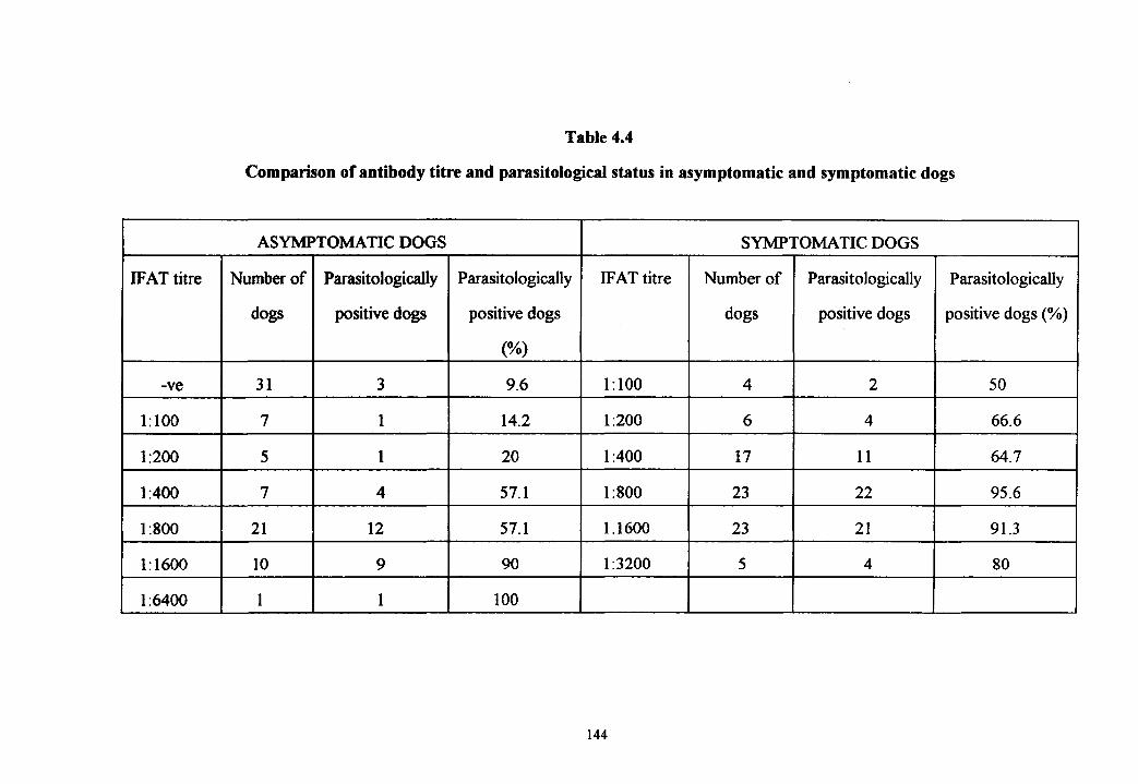
Table 4.4
Comparison of antibody titre and parasitological status in asymptomatic and symptomatic dogs
ASYMPTOMATIC DOGS SYMPTOMATIC DOGS
IFAT titre Number of Parasitologically Parasitologically IFAT titre Number of Parasitologically Parasitologically
dogs positive dogs positive dogs dogs positive dogs positive dogs (%)
(%)
-ve 31 3 9.6 1:100 4 2 50
1:100 7 1 14.2 1:200 6 4 66.6
1:200 5 1 20 1:400 17 11 64.7
1:400 7 4 57.1 1:800 23 22 95.6
1:800 21 12 57.1 1.1600 23 21 91.3
1:1600 10 9 90 1:3200 5 4 80
1:6400 1 1 100
144

This comparative analysis of diagnostic methods showed that serology is
not 100% sensitive but it is the best, followed by the parasitology, and then
symptomatology. The results with the LMet2 DNA probe, but with a smaller
group of animals, are described above (section 4.3) (Figure 4.11).
Comparison of the serological methods showed that the IFAT and the dot
ELISA were the most sensitive tests for detection of parasitologically positive
dogs, followed by the ELISA and the DAT (Figure 4.12).
Comparison of the parasitological methods showed that for each of the
techniques used the sensitivity was never more than 60%, in comparison with the
reference standard (RS) as defined above. Although demonstration of the parasite
by culture of samples from spleen aspiration and BM puncture was more sensitive
than the examination of the same samples by giemsa staining and microscopy,
culture is not practical for routine used and from wild animals, gives increased
contamination (Vexenat et al., 1986) (Figure 4.13).
Two particularly important results from these comparisons between the
demonstration of the parasite and the detection of the presence of antibodies, were
that:
a. 6.6% of the serologically negative dogs were parasitologically positive.
b. the chance of demonstrating the parasites increased with increasing antibody
levels. (Figure 4.14, IFAT).
The significance of these observations will be discussed below.
145

Figure 4.11
Comparative sensitivity of diagnostic methods

120~------------------------------~
100
> ... -> -... -U'J 2 w en
Parasl +ve
80
60
40
20
o
Paras/serol/ + va
Serol.
98.2
98.2
Reference standard:
Parasit. Svmptomat.
66.9
68.2 41.9
Parasl + ve Paras/seroll + ve Figure 4.11

Figure 4.12
Comparative sensitivity of serological methods

120~1 -----------------------------------------
100-··· .......................................................... ---
~ > .... -> -.... -en 2 w en
Paras! +ve
80
60
40
20
o
Paras/seral! + va
IFAT
98.2
98.2
Reference standard:
dot-ELISA ELISA OAT
98.2 97.9 80
98.2 97.1 62.1 -
Parasl + ve Paras/seroll + ve
Figure 4.12
I
I

Figure 4.13
Comparative sensitivity of parasitological methods

100,.-----------------------------------------
80 t-- ••••••••••••••••••.••.•••.•••••••..••••••••••••••.•••••••....•..
w Cl
~ 2 w CJ a: w a.
Paras/ + ve
60
40
20
o
Paras/seroll + ve
Skin BM
78.4 55.9
58.4 42.5
Reference standard:
Spl. Spl/(C) BM(C)
47.3 77.2 67
24.6 54.8 52
Paras! + ve Paras/serall + ve
Figure 4.13

Figure 4.14
Comparison of antibody level by IFA T and results of
parasitological examination.

100~i --------------------~
No. of dogs
'#. "C r:: ca o 2
80
60
~ 40 o c
20
o
% paras it/ + ve dogs
No. paras it/ + ve dogs ---
. - - - - - - - - - - - - - - - - - - - - - - . - . - - - - - - - - .... .
- . - - - - - - - - - - - - - - - - - - - - -
a
-ve 100 200 400 800 1600 3200 6400
45 12 12 24 45 38 7 27
6.6 41.6 33.3 58.3 77.7 78.9 42.8 74
3 5 4 14 35 30 3 20 I
No. of dogs
~ % parasitl + ve dogs
II No. parasitl + ve dogs
Figure 4.14

CHAPTER 5: TRANSMISSIBILITY OF CANINE VISCERAL
LEISHMANIASIS
5.1 Detection of natural infections in sandflies
In a preliminary attempt to detect naturally infected sandflies and to
investigate whether they were found in association with naturally infected
dogs, collections of females were made in three places:
(a) a communal kennel with approximately 20 dogs of unknown origin, of
unidentified breeds, and with an unknown percentage of infected dogs. Here
engorged female sandflies were collected by pooter.
(b) a slum house with one pig-sty, one chicken shed and one parasitologically
positive dog. Engorged female sandflies were separated from collections made
by CDC light-trap. A large proportion of the sandflies examined may have fed
on pigs or chickens.
(c) a lUXUry house with a kennel and one infected dog that had disseminated
amastigotes in the skin. Here engorged female sandflies were collected on the
wall inside the kennel.
When dissected between 5 and 7 days after collection positivity rates
by microscopical examination in the sandflies were between 0.6 and 67.3%
(Table 5.1).
The identification of naturally infected sandflies by dissection
is an arduous and time-consuming task. To test the
chemiluminescent DNA probe technology in the field 200 female sandflies
were collected inside and outside a house where
150

Table 5.1
Natural infections in sandflies detected by dissection 5 - 7 days after capture
Place and source Flies +ve
A communal kennel with approximately 02
20 dogs of unknown origin,unidentified
breeds and with an unknown percentage
of infected dogs (flies collected by
pooter).
A slum house with one pig-sty, one
chicken shed and one parasitologically
positive dog (flies collected by CDC
light-trap).
A lUxury house with one infected dog
in a kennel (engorged flies collected
on the internal wall).
Totals
151
05
33
40
Flies -ve 0/0 +ve
16 11.1
790 0.6
16 67.3
822

there was an untreated human case of VL waiting for hospitalisation and also ~I
serologically positive dog. The sandflies were processed as described in the
Materials and Methods. In two of 200 flies examined DNA of the Leishmunia
donovani-complex was detected by the Lmet 2 probe.
To determine the percentage of kennels with naturally infected sandl1ies
using the DNA probe method (through which it is possible to examine a large
number of flies) another set of 1,234 female sandflies were collected and squashed
onto Hybond N (Amersham). The flies were collected in kennels located in
different houses and different districts of the city of Teresina. When a large
quantity of sandflies was collected at a site only the females which had fed or
apparently had eggs in the abdomen were used. In this case the radioactive Lmet2
DNA probe was used as radioactive facilities were available; occasionally the
chemiluminescent method had given background on the filters and we wished to
guarantee an absolutely clear result at the first attempt.
5.2 Xenodiagnosis studies of naturally infected dogs
Early in the Teresina project colony-bred Lu longipalpis were fed on
altered skin of 13 naturally infected (parasitologically +ve) dogs. Dissection after
5 - 7 days showed that between 11 % (lout of 9) and 78% of flies became infected
and with 10 of the 13 host animals more than 50% of flies contained
promastigotes when they were dissected (Table 5.2). This compares with a rate
of 100% obtained by Molina on xenodiagnosis of patients with VL, but all these
subjects were suffering from AIDS (Molina et al., 1994b).
On one dog (8a, Table 5.2) a second batch of flies was fed on apparently
normal skin. Although the infection rate was reduced 6% (4 out of 65) t1ies
became infected, as compared to 35% (57 out of 163) fed on altered skin ("skin
lesions") of the same animal. This result indicated that sandflies very easily
acquired infection from parasitologically positive dogs if they fed on skin lesions
152

Table 5.2
Xenodiagnoses on 13 naturally infected dogs
Dog no. Flies +ve %+ve
Flies fed
6a 32/45 71%
7a 76/121 63%
8a 57/163 35%
8a'" 4/65 6%
67 41/53 77%
68 34/63 65%
69 7/9 78%
74 69/124 56%
105 9/16 56%
112 1/9 11%
146 92/132 70%
147 61/83 73%
148 42/69 61%
170 12/46 26%
'" This batch only fed on apparently normal skin, all other batches fed on
altered skin.
153

and suggested that host clinical and parasitological status should be compared in
more detail with host infectivity to sandflies. The comparison of serological
positivity and parasitological positivity had already indicated an association
between serological titre and parasitological positivity (chapter 4, Figure 4.14)
and, by implication, between serological titre and infectivity.
A group of 40 IF AT positive dogs was examined clinically (classified as
symptomatic or asymptomatic) and parasitologically (sternal puncture aspirates
and skin) and then used for comparative xenodiagnoses, with one batch of flies
fed on skin lesions (altered skin) and a second batch fed on apparently normal
skin. Artificial xenodiagnosis was perfonned in parallel by membrane feeding
and heparinised blood samples were also cultured (Materials and Methods). It
was intended that the membrane feeding and the culture of heparinised blood
samples would indicate, by comparison with direct xenodiagnoses, whether
sandflies had acquired infection from the skin of the dogs or, possibly, from
amastigotes circulating in blood/infected blood cells (Table 5.3).
Comparisons derived from the data shown in Table 5.3 are summarised in
Table 5.4.
Certain simple observation may be made from the comparisons summarised in
Table 5.4, as follows:
1. Both microscopy of giemsa stained impression smears and culture of
skin biopsies were more sensitive with altered skin than normal skin
for detecting L. chagasi infection in symptomatic dogs.
2. In this particular study with symptomatic dogs culture of skin biopsies
did not detect any infections that were missed by microscopy of
giemsa stained impression smears.
154

Table 5.3
Results of parasitological examination, direct and indirect
xenodiagnoses on skin lesions or apparently normal skin of
symptomatic or asymptomatic dogs

Dog
01 02 03 04 05 06 07 08 09 \0 II 12 13 14 15 16 17 18 19 20 21 22 23 24 25 26 27 28 29 30 31 32 33 34 35 36 37 38 39 40
Table 5.3
Giemsa Culture Direct xeno Membrane (+/exam) fed
NL NS BM NL NS BM BC NL 0/0 NS 0/0 BLOOD + + + + + + - 45/55 81 12/68 17 0/35
SL - - NO - - - NO 0/32 0 0/30 SL - + NO + + - NO 0/47 0 0/23 + + + + + + - 1221145 84 76/99 76 0/45
SL - - - - - - NO 0/43 0 0/32 + + - + + + + 387/422 91 202/63231 0/122
SL - + NO - - - NO 0/21 0 0/32 SL - + NO - - - NO 0/22 0 0/30 SL + - NO - + - NO 4/29 13 0/34 SL + + NO + + - NO 6/41 14 0/20 SL - - NO - + - NO 0/33 0 0/24 SL - - NO - + - NO 0/22 0 0/20 SL - - NO - + - NO 0/23 0 0/32 SL + + NO + + - NO 22/24 91 0123 SL - - NO - + - NO 2/34 5 0/21 + - - + - - - 15118 83 0/21 0 0/34 + - - + - - - 12119 63 0/35 0 0/23 - - - - - - - 0/30 0 0/24 0 0/32 + - - + - - - 0/21 0 0/34 0 0/37
SL + - NO + + + NO 32/36 88 0142 + - - - - - - 4/25 16 0/35 0 0/32 + - - - - + - 5/34 14 0/26 0 0/28
SL - - NO - + + NO 0/23 0 0/26 SL - - NO - + - NO 0/25 0 0/20 SL + - + - + - ND 21128 75 0/32 + - - - + - 6/18 33 0/22 0 0/25 + - - - - + - 2/26 7 0/27 0 0/22
- - + - - - - 0/33 0 0/23 0 0/30 + - + + - - - 3/30 10 0/21 0 0/19 + - + + - - - 8/34 23 0/31 0 0/34 + - + + - + - 12/18 66 0124 0 0/26
SL - + NO - + - ND 0/32 0 0/21 SL - - NO - + - NO 0123 0 0/25 + + + + + + - 10/29 34 2/31 6 0/24 + + - + + + + 15/19 78 4128 14 0/20 + + - + + + - 18/25 72 5142 11 0/21 + + - + + + - 27/38 71 3/29 10 0/32 + + - + + + + 38/43 88 13/67 19 0141 + - - + - - - 5/32 15 0/48 0 0/38 + - - + - - - 3/22 13 0/32 0 0/26
SL = skin lesion (altered skin); NS = normal skin; NL = no skin lesion (altered skin) present; BM = examination of bone marrow aspirate (by giemsa staining and microscopy); Be = blood culture; NO = not done. Dogs recorded as NL m column 2 (SL) correspond with animals designated asymptomatic.
155

Table 5.4
Comparison of clinical and parasitological status in 40 seropositive dogs
Examination
Microscopy: altered skin
Microscopy: normal skin
Culture: altered skin
Culture: normal skin
Culture: blood
Microscopy: bone marrow
Culture: bone marrow
Xenodiagnosis: altered skin
Xenodiagnosis: normal skin
Symptomatic (22)
( +ve/examined)
20122
8122
16/221
8/221
3/22
7/22
12/223
19/22
8122
Clinical status
Asymptomatic (18)
( +ve/examined)
5118
4/182
2/18
6/18
14/184
6/18
1 No additional infections detected as compared to microscopy
2 One additional infection detected but two missed in comparison witb microscopy
3 Eight additional infections detected but tbree missed in comparison witb
microscopy
4 Ten additional infections detected but two missed in comparison with microscopy
156

3. In contrast culture of bone marrow detected 8 additional infections, as
compared to microscopy of bone marrow, with the 22 symptomatic
dogs.
4. Only 5 of the 40 dogs, 3 symptomatic and 2 asymptomatic, were
positive by blood culture.
5. Overall similar proportions of asymptomatic dogs were found to be
infected by the parasitological methods and xenodiagnosis, except that
altered skin could not be examined in these animals.
A simple attempt was made to see if parasitologically positive skin and
bone marrow were associated in this group of 40 seropositive dogs, as shown in
Table 5.5. Consistent with observations on the cohort of experimentally infected
dogs (Chapter 6) 13 of 31 dogs that were bone marrow positive were skin
negative and 6 of 24 dogs that were skin positive were bone marrow negative.
Similarly, the parasitological status of skin was compared with the result of
xenodiagnosis (Table 5.6). Here there was a close association between skin
positivity and xenodiagnosis positivity in that 23 of 25 skin positive dogs were
xenodiagnosis positive; only 2 of these were xenodiagnosis negative and only 1
skin negative dog was xenodiagnosis positive.
By way of further comparison 12 bone marrow positive dogs were
xenodiagnosis negative and 12 of 3 1 bone marrow positive dogs were
xenodiagnosis negative and 5 of 24 xenodiagnosis positive dogs were bone
marrow negative (Table 5.7). These comparisons suggest that parasitologically
skin is a clear indicator of ability to transmit infection to sandflies.
157

Table 5.5
Comparison of associations between parasitological status of skin and bone
marrow (BM) in naturally infected dogs used for xenodiagnoses
BM+ve BM-ve
Skin +ve 18 6 24
Skin -ve 13 3 16
31 9 40
X" = 0.2
Fisher's exact test = 0.4
158
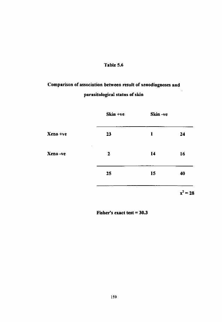
Table 5.6
Comparison of association between result of xenodiagnoses and
parasitological status of skin
Skin +ve Skin -ve
Xeno +ve 23 1
Xeno -ve 2 14
25 15
Fisher's exact test = 30.3
159
24
16
40

Table 5.7
Comparison of association between result of xenodiagnoses and
parasitological status of bone marrow
BM+ve BM-ve
Xeno +ve 19 5
Xeno -ve 12 4
31 9
Fisher's exact test = 0.3
160
24
16
40
Xl= 0.1

Xenodiagnosis was performed on 3 of the 5 dogs that were
parasitologically positive by blood culture. In these three cases 91% (387 of 422),
78% (15 of 19) and 88% (38 of 43) sandflies became infected. All three animals
had, however, parasitologically positive skin (Table 5.3). It cannot be concluded,
therefore, that the blood infections contributed to the high numbers of sandflies
that acquired infection when fed on these animals, although it may have done.
Surprisingly, none of the total of 251 sandflies acquired infection when fed
artificially by membrane feeding on blood derived from these five animals that were
parasitologically positive by blood culture. This may suggest that sandflies could
not readily acquire infection from the small quantity of blood that they took up, in
comparison with the relatively large volume placed into culture tubes, and supports
the conclusion that sandflies normally acquire infection from amastigotes in the
skin of infected dogs. Altem~tively, the process of membrane feeding may have
destroyed the infectivity of blood-borne amastigotes. This is unlikely, however, as
amastigotes derived from hamsters by trituration and filtration through glass wool
and mixed with defibrinated, heat-inactivated (56°C, 30 m) dog blood were able to
convey infection to all sandflies that fed on this control suspension by membrane
feeding (amastigotes numerous, not counted; 20 flies dissected).
Table 5.8 summarises the data on infectivity to sandflies (transmissibility),
clinical status of the dog (symptomatic or asymptomatic) and the condition of the
skin at the site at which sandflies fed. It can be seen that for symptomatic dogs
altered skin or skin with lesions was more infective to sandflies than normal skin,
in that 86% of xenodiagnoses on altered skin were positive and 36% on normal
skin were positive. Although a high percentage of sandflies fed on altered skin
acquired infection (64.9%) a considerable proportion (22.7%) also acquired
infection from normal skin. For asymptomatic animals, in which altered skin was
not available as a site for xenodiagnosis, 16.2% of sandflies still acquired infection
161

Table 5.8
Comparison of infectivity, clinical status and skin condition at site of
xenodiagnosis
Clinical status of
dog and site of
xenodiagnosis
Symptomatic
skin lesions
normal skin
Asymptomatic
normal skin
Xenodiagnosis result
No of dogs +
No of dogs examined
19/22 (86%)
8/22 (360/0)
6/18 (33%)
162
No of flies +
No or flies examined
737/1136 (64.9%)
31711399 (22.7%)
871538 (16.2%)

from 33% (6 of 18 of the asymptomatic dogs). Overall and consistent with
comparative data on naturally infected animals, these data clearly indicate that
asymptomatic dogs cannot be excluded as a significant source of infection to
sandflies considering that a single infected sandfly may transmit infection to a naive
host (Chapter 6). The facility with which flies acquire infection from dogs implies
that they are an overwhelmingly important source of infection and potential
reservoir host, if the same sandfly populations are vectors of canine and human
VL.
Figure 5.1 shows the percentages of sandflies infected, for individual dogs,
with the three groups of animals shown in Table 5.7.
163

Figure 5.1
Percentages of sandflies that were infected following
xenodiagnoses of skin lesions (altered skin) or normal skin for
symptomatic or asymptomatic dogs that were serologically positive
(by IFAT)

8
c c .-
.:::£ .:::£ C/) (/)
co c co E 0 E ~
~ 0 C/) 0 c Q)
C - C/)
(/) C/) C) C) C) 0 0 0 "'0
"'0 "'0 0. 0. 0. E E E > > > C/)
CJ) CJ) « ~~j II II
••••••••••••••• , ........................ ;0 ....................................................... !............... . ...... ... .... . .. .. ..
o 00
o <0
• .. ....... t ......... s ... .
...' ......
o C'J
sa!l!pues aA + !O a5eluaOJad
t-I . In
Q) H ::3 t:7l .~
~
...
...........
. ....... -
o
en en o o

CHAPTER 6: EXPERIMENTAL TRANSMISSION
The results of the experimental infection of a cohort of dogs are summarised in
Table 6.1.
6.1 The infective bite
In one (dog D 1) of two pilot animals used in this study cohort of 2S dogs a single
engorged fly of seven (which were collected in a lUxury house with one infected
dog in a kennel) was found to be parasitologically positive by microscopical
examination and it was concluded that this fly was responsible for transmission.
Within 22 days after the infective feed, a tiny erythema was noted at the site of the
bite (left inner ear of a young dog of 70 days old) and immediately skin biopsies
and xenodiagnosis were performed on this site. To perform the xenodiagnosis
exactly on the suspect site, the ear was covered and only the lesion exposed to
sandflies (Figure 6.1A). Large numbers of amastigotes were found by
microscopy of giemsa stained impression smears of biopsies; 7 of 14 sandt1ies
dissected 5 days after feeding were positive.
Follow-up xenodiagnoses were done on three separate occasions, weekly.
biweekly or monthly (see results below) according to the time post-infection using
local colony-bred sandflies. To detect any early skin dissemination comparative
xenodiagnoses were performed at other sites: 43 of 73 flies that fed on the lesion
were positive by microscopical examination and all 187 flies fed on the right ear
and abdomen were negative. Figure 6.1B ilustrates xenodiagnosis on the
abdomen. Figure 6.2 shows the lesion in dog D, at 22, 38 and 65 days
post infection. Regrettably this animal died, at 145 days post infection, during
parasitological examination by spleen puncture, without evidence of
dissemination of the infection from the infected lesion, and with IF A T and dot
165

Table 6.1
Summary of parasitological and serological examinations on a cohort of 25
dogs experimentally infected by sandfly bite. Dogs are grouped according to
course of infection, not chronologically (see text)
Weeks post-infection
Srx Dogs 3-4 5-10 11-16 17-20 21-24 29-32 33-36 37-40 50
g 06 .DO •• 0 .D ADO ADO
g 07 .0.0 •• ·.0 •• 0 ADO AD 0
c! ·.0 •• .D .D .DO A DO
c! M2 ADO •• •• ..0
17 ADO .D
11 .0.0 •• 13 ·.0 •• 05 •• 0 •• V2 •• 0 .D
ADO AD
•• 0 •• • DO .D
02 AD 0
c! 112 .DO •• c! 09 ADO AD
PI ADO ADO
P2 ADO ·.0
P3 ADO AD
ADO AD
.DO .D
•• 0
ADO • = Xenodiagnosis +\,r
M6 • = Bone marrow +\,r
d ADO fl = Skin -ve
cJ M7 ADO o = Xenodiagnosis -ve o = Bone marrow -ve
cJ M8 ADO = Seroconversion ... EJ = Serorcversion
) = neath

Figure 6.1 A
Xenodiagnosis with sandfly access restricted to a skin lesion (Dog
Dl)
Figure 6.1 B
Xenodiagnosis on the abdomen (Dog DI)

Figure 6.2 A, B, C
Lesions at the site of an infective sandfly bite at 22 days, 38 days
and 65 days

A
Figure 6.2

ELISA titres of I: 100 (which became positive at day 129). Observations 011 the
second pilot animal dog 02 are incorporated into the description of Results for the
overall study cohort.
6.2 The incubation period
The incubation period (lP) is defined as the time from initial infection to
the point at which signs of disease appear (or are diagnosed). This was
determined by clinical examination for abnormalities at the site of the infective
bite (or elsewhere) with confirmation that amastigotes were present in the skin or
in the bone marrow.
In 12 of 25 (48%) of the infected dogs the IP, for the experimental
procedures used here, was shown to be between three and four weeks after the
infective bite. In 3, 5, 3 and 2 of 25 of the other infective dogs the IP was 5-10,
11-16, 17-20 and 25-28 weeks respectively (Table 6.1). The overall rate of
infection in this study was thus 100%, although the presistence and course of
infections varied. In this case the incubation period and prepatent periods were
approximately the same as appearance of skin alterations usually coincided with
the detection of amastigotes.
6.3 The latent period
The Latent Period (LP) is defined as the time from initial infection to the
point at which the individual becomes infectious to others (here taken to be
infective to sandflies); in this study it was recorded for 22 of 25 (88%) of the
infected dogs. Early infection of Lu. longipalpis was obtained through
xenodiagnosis performed on the transmission site. All the flies were examined
after 6 days to determine the LP and the percentage of positive flies. The LP was
demonstrated to be 3-4, 5-10,11-16,17-20, and 25-28 weeks in 8 (32%),4,4.5
and I dog respectively (Table 6.1). It should be noted, however. that not all dogs
that previously had negative xenodiagnoses were rexenodiagnosed at each time
169

point. LP was not detenmned by xenodiagnosis for 3 (12%) of the cohort: PI, P3,
M5 (died), at the end point shown in Table 6.1.
6.4 The infectious period
The infectious period (INFP) defined as the period of time during which an
individual is infectious to others (here considered to be infectivity to sandflies),
was also demonstrated using xenodiagnosis. The INFP for experimental canine
VL, was observed in 22 or the 25 infected dogs (although some dogs are still alive
and infectious). The majority of dogs were infectious from weeks 5-10 to weeks
17-20. Comparisons between the rates of infection for sandflies showed, although
it was possible to obtain early infection, that the highest rate was 5-10 weeks post
infection. Note, however, that few of the dogs had developed disseminated
cutaneous infections (6.5, below).
6.5 Metastasis/skin dissemination
Metastasis andlor skin dissemination were demonstrated by periodic
parasitological examination of bone marrow and altered or normal skin in various
parts of the body of each infected dog (Materials and Methods).
This clinical development of canine VL was observed in only two of the 25
infected dogs, although clinical observation is continuing. The dog D2 was
parasitologically positive in normal skin at day 65 post-infection and never
recovered (Figure 6.3), and in dog OS, but in this case disseminated amastigotes
and skin lesions on the ear were a temporary feature during pregnancy prior to the
birth offour puppies (Figure 6.4).
170

Figure 6.3
Dog 2. A, 43 days old, B, 5 months after infection and C, 8 months
after infection

A
c
Figure 6.3

Figure 6.4
Dog 05: Skin lesions at days 66, 89, 130 (4 pups born) 150 and 240

Figure 6.4

6.6 Seroconversion
Seroconversion (SC) was detected in 20 (80%}ofthe infected dogs.
The indirect immunofluorescent test (IF AT) was used to determine the
time of SC and to estimate the antibody titre, as this had been shown previously to
be one of the most effective tests. The minimal period to SC was eleven weeks.
Serologically -ve dogs were not only parasitologically +ve, but also infective to
sandflies. The comparison of the presence of antibodies and the infective rate for
sandflies, demonstrated that 60% of the infected dogs were infective to sandflies
(fed on skin lesions) during the seronegative phase with 32% of sandflies acquiring
infection.
In three animals (11, 13, 05) low seropositivity reverted. None of these
animals had become bone marrow positive at the end point in Table 6.1 and all
three were parasitologically negative at 37-40 weeks (although infection had been
detected previously (Figure 6.5). Bone marrow positivity was only seen in dogs
that were serologically positive (Table 6.1).
Examples of skin lesions for other dogs in the cohort are shown in Figures
6.6A-G.
173

Figure 6.5
Summaries of results of monitoring clinical, serological,
parasitological and xenodiagnosis status in the experimental
cohort of dogs fed on by infected Lutzomyia longipa/pis
£. = Skin +vc • = Xenodiagnosis +\'c • = Bone marrow +vc
1:. = SI<in -ve o = Xer.todiagnosis -ve o = Bone marrow -ve
II = Nodule
• = Lesion
II] = Scar

Titre Titre ... ... N N W !-> ..... ..... N N W ~
0'1 b bt § en b 0'1 U1 0 (n 0 0'1 0 0'1
0 8 8 8 8 8 0 8 8 8 8 8 $ 8
3-4 o.t> : 3-4+ III IOO~ :
~ 5-10 .~ : ~ 5-10 o.~ :
CD 0 CD 0 ~ 11 -16 ~ 11-16
'" 0 '" a '0 17-20 O.~ : co '0 17-20 . .;- O~ : co 0 0 .... -.~
'" 21-24 0 '" 21 -24 r-+ r-+ 0 I I
~ 25-28 ~
o.~ : '-J ~ 25-28 oot> : (j) -h -h
CD CD o 29-32 oot> : o 29-32 r-+ r-+
0 0 ~ 33-36 ~ 33-361
37-40 oat> : 37-40 loot> :
41-44 41-44

Figure 6.S
Summaries of results of monitoring clinical, serological,
parasitological and xenodiagnosis status in the experimental
cohort of dogs fed on by infected Lutzomyia longipalpis
A = Skin +vc • = Xenodiagnosis +"c • = Bone marrow +vc
h. = Skin -ve 0 = Xenodiagnosis -ve o = Bone marrow -ve
1'1 = Nodule
• = Lesion
III = Scar

Dog 15 3,500 ,--------------------,
3,000 ................ . ........................ .
2,500
2,000
Q)
~ 1,500
1,000
500 .. ..... ................... - ................
• • • • • /),.
• • C C C C 0 0 0
0 .. .~ ... ~.:
I I I L I I I I I I I I I I I I
"i 0 <0 0 ..,. eo N <0 0 ~ :;; or;- ~ ~ N <'? M "i M .;, ~ r-.. N Ol r-.. ~ N N M M
Weeks post-infection
Dog M2 3,500 ,----------------------,
3,000-
2,500
2,000
Q)
~ 1,500
1,000
500 /),. & • & C • •• ° 0 o*--~---------------------~
I I I I I I I I I I I I I
"i 0 <0 0 .:t- eo N <0 M or;- N ~ N C") M .;, ,.:..
N .;, a, M N N C")
Weeks post-infection

Figure 6.5
Summaries of results of monitoring clinical, serological,
parasitological and xenodiagnosis status in the experimental
cohort of dogs fed on by infected Lutzomyia /ongipa/pis
~ = Skin +vc • = Xenodiagnosis +\'c • = Bone marrow +vc
6. = Skin -ve 0 = Xe,:,odiagnosis -ve o = Bone marrow -ve
II = Nodule
• = Lesion
III = Scar

Dog 17 3 ,500 r-------------------,
3,000
2 ,500
2,000
OJ
~ 1,500
1,000
500 t. 4 4 [] [] . ° ° Ol~------------------------------~
) I J I
Weeks post-infection
Dog 11 3,500r---------------------~
3,000- .. ..... . ......... . . ..... ........ .. ....... .
2,500
2,000
cu ~ 1,500
1,000 . . ....................................... .
500 . . ~ ...... . ... . • .... .. . .. ... . . . .. t. 4 t.
• • • [] []
° ° =:::::::::::::: 0 0
l~1.W~i< -.I. -.I. .J I I I I I
I I I
~ a <0 a "<t- oo N <0 a ~ M :;; "7 N <';'I N M M ~ ,..:.
N .;, 0, M r-
~ N N M M
Weeks post- infection

Figure 6.S
Summaries of results of monitoring clinical, serological,
parasitological and xenodiagnosis status in the experimental
cohort of dogs fed on by infected Lutzomyia longipalpis
A = Skin +vc • = Xenodiagnosis +vc • = Bone marrow +\'(~
l:J. = Skin -ve 0 = Xenodiagnosis -ve o = Bone marrow -ve
• = Nodule
• = Lesion
[I = Scar

Titre Titre ..... ..... tv I\) ~ w ..... ..... I\) I\) ,w ,w U1 0 U1 § <n <n U1 0 <n 0 U1 0 U1
0 8 8 8 8 8 0 8 8 8 8 8 8
3-4 0 •• : 3-4 o.~ :
~ 5-10 .~ :
0 ~ 5-10 .. : CD CD 0 ~ 11-16
0 ~ 11-16 0 en en
"0 17-20 (C '0 17-20 (C 0 0 .'
en 21 -24 ~;1 •• 0 en 21-24 O~
~ r+ r+ I I
::J 25-28 ~ oo~ 01 ::J 25-28 W .... -to. CD CD o 29-32 o 29-32 r+ r+ 0 0 ::J 33-36 ::J 33-36-
37-40 ob~ : 37-40 + loo~ :
41-44 41 -44

Figure 6.5
Summaries of results of monitoring clinical, serological,
parasitological and xenodiagnosis status in the experimental
cohort of dogs fed on by infected Lutzomyia /ongipa/pis
~ = Skin +vc • = Xenodiagnosis +"c • = Bone marrow +"l'
~ = Skin -ve o = Xenodiagnosis -ve o = Bone marrow -ve
I'IJ = Nodule
• = Lesion
1m = Scar

DogV2 3,500 ,---------------________________ -.
3,000 ....... . ... . ........ ..... .... .
2,500 ... , .... .. .. ..... . . .. ........ .
2,000 ........ . ... . .. .... . ..... . .... .. ........ . .
<D J:;i 1 ,500 ... .. .. . .. . ... . ......... .. . . .. . . ... .... . . . ~
1,000 ...... .. . . ........... . .. . ............. . .. .
500 ......... . .. . ..........• ........ :A: ....... .
• • •• • 0 0
01~~0--------~~======~--~
.1 I I I .l. I I I I
~ 0 co 0 '<t co N <0 Sf M ;;; "7 ~ ~ ~ "? C")
r-- N It') en ~ r--N N C") C")
Weeks post-infection
Dog M1 3,500.--------------------------------,
3,000
2,500
2,000
Q)
~ 1,500
1,000
. .... .. ... . ... .... .. ..... ........... ....
~ ~ • ~ ° o~~o~------==~========~~
500
.l. I I I J
"t 0 co 0 '<t co N <0 "7 N ~ N C") "? C") :;; ,..:.. .;, a-. - N
C")
N N M
Weeks post- infection

Figure 6.5
Summaries of results of monitoring clinical, serological,
parasitological and xenodiagnosis status in the experimental
cohort of dogs fed on by infected Lutzomyia /ongipa/pis
~ = Skin +vc • = Xenodiagnosis +vc • = Bone marrow +vc
A = Skin -ve o = Xenodiagnosis -ve o == Bone marrow -ve
II = Nodule
• = Lesion
WI = Scar

Dog 01 3,500 .--------------------------------------
3,O()(} .................................... . .... .
2,500 . ............ .
2,000 . ...... . ................. .. ......... .. ... .
(l)
.~ 1,500 ................................. ..... ... . I-
1,000 ........ - ....... . .
500 .. . ... .. ... • .. 4 • • • • °
O~---~O~----------~--~~====~,---~
I I ( I I I
<D 0 '<t" "7 '" C':" ,..!.
N
o
Weeks post-infection
Dog M3 3,500 r---------------------------------~
3,000
2,500
2,000
Q)
~ 1,500
1 ,000
500 ............. ..... . ................. . , • • • 0 0 • ~ ° ° ° '"
I I {" I I I
~ 0 <D 0 '<t" (W) ~ "7 '" <-;l .;, ,..!.
N
Weeks post-infection

Figure 6.5
Summaries of results of monitoring clinical, serological,
parasitological and xenodiagnosis status in the experimental
cohort of dogs fed on by infected Lutzomyia /ongipa/pis
A = Skin +vc • = Xenodiagnosis +\'c • = Bone marrow +\'c
1:. = Skin -ve o = Xenodiagnosis -ve o = Bone marrow -ve
III = Nodule
• = Lesion
[I = Scar

Titre Titre ..... ..... t-l tv W !.ol ..... ..... tv t-l W W (]'I 0 (]'I 0 b1 0 (]'I
(J'I 0 (]'I 0 (]'I 0 (]'I 8 8 8 8 8 8 8 · 8 8 8 8 8 0 0 0 0
3-4 oo~ : 3-4nOOI> :
~ 5-10 . ~ : ~ 5-10 .
0 CD 0 ~ 1H6i ~. ~ 11-16 , O.~ a a C/l "0 17-20 c.c "0 17-20.. : co 0
0 C/l 21-24 . ~~. 0 ~ 21·24 t I 0
.-+
d; I
:J 25-28 N :J 25-28
N ~
~ CD CD (') 29-32 (') 29-32 .-+ !:! . 0 o 33-36 I • I I •• :J 33-36 :J
37-40 + 'eol> : 37-40 +1 1 I·
41 -44 I 41-44 ~

Figure 6.5
Summaries of results of monitoring clinical, serological,
parasitological and xenodiagnosis status in the experimental
cohort of dogs fed on by infected Lutzomyia longipaJpis
~ = Skin +"c • = Xenodiagnosis +vc • = Bone marrow +\'c
A = Skin -ve o = Xer:todiagnosis -ve o = Bone marrow -ve
• = Nodule
• = Lesion
• = Scar

Titre Titre .... .... N N W W .... .... N !" w S'" C11 0 C11 0 (n 0 (n C11 § (n § C11 0 C11
0 8 8 8 8 8 8 0 8 8 8 8 8
3-4 001> : 3-4 oot> :
~ 5-10 001> : 0 ~ 5-1 0 ot> : 0 CD CD CD a ~ 11-16 a 7\ 11-1 6 en en
CO "0 CO "0 17-20 '.:.- o.~ : o 17-20 0
0 en
\J en 21-24 .~ .-+ .-+ I 21 -24 I
(0 ::::l ~ ::::l 25-28 -+.
-+. CD 25-28 (I) 0 o 29-32 .-+ .-+ o 29-32 .o~ g 33-36 + ''if! ~ .Ol> ::::l
33-36 37-40
37-40 41-44

Figure 6.S
Summaries of results of monitoring clinical, serological,
parasitological and xenodiagnosis status in the experimental
cohort of dogs fed on by infected Lutzomyia longipalpis
• = Skin +vc • = Xenodiagnosis +,'c • = Bone marrow +vc
t. = Sldn -ve o = Xenodiagnosis -ve o = Bone marrow -ve
II = Nodule
• = Lesion
III = Scar

Titre Titre ...... ...... t-.> !" ~ ~ t-.> t-.> ~ W (11 0 (11 0 (11 0 (11
(11 0 (11 0 (11 0 (J1
0 8 8 8 8 8 8 0 8 8 8 8 8 8 0 0
34+ lOD~ . I
I 3-4 oot> ·
~ 5·10 at> ' 0
~ 5·10
I r· (l) (l)
0 (l) (l)
%& •• ~ 7\ 11.16 0 7\ 11.16 0 en en
'0 (.Q "0 ~ . c.a o 17·20 o 17·20 ~It : en en r-+ -U r-+
'\T \J I 21 ·24 oot> · I 21 ·24
:J CAl :J ~ ~ N (l) 25.28 en 25.28 C'l C'l r-+ M-
o 29·32 o 29·32 + 'I o~ :J :J
33·36 33·36 +
37·40 37-40

Figure 6.5
Summaries of results of monitoring clinical, serological,
parasitological and xenodiagnosis status in the experimental
cohort of dogs fed on by infected Lutzomyia longipa/pis
.A. = Skin +vc • = Xenodiagnosis +\'c • = Bone marrow +vc
A = Sldn -ve o = Xenodiagnosis -ve o = Bone marrow -ve
rI = Nodule
• = Lesion
• = Scar

Dog P4 3,50° r-----------------------------------~
3,000 . . ........................... .. ......... .
2,500 .. ... , ... - ._ . . ......... . . - ........ _.
2,000 ...... . . ... .. ... . . .. -,. ,_ ........... 0_ •••
Cl>
.~ 1,500 I-
1,000
500 ............................• ............. t:.. t:.. A • o 0 0 •
000
~----------------~~~----------~
Weeks post-infection
Dog V1 3,500~-------------------------------,
3,000 ., _ ............. _ ... . .................... .
2,500 . .... . ........ ... .. ........................
2,000 ........... .. .. . .................. . .... ...
Cl>
~ 1,500 .................. . _ ... . ......... ... ..... .
1,000 .............. _ ......................... .
500 ....... .. . t:.. .... ... . ~ ..... . .... .. 0 0 00. •
Oll-~O~----------~======~------"lJlli~
I I 1 I I I
"1 I I I - I I
"'i 0 <0 0 .;t- eo ('oj CD 0
'7 ~ ~ N (Y) C") .;t-C") .;, .;, a, M ,..:..
"- N N ('oj M M
Weeks post-infection

Figure 6.5
Summaries of results of monitoring clinical, serological,
parasitological and xenodiagnosis status in the experimental
cohort of dogs fed on by infected Lutzomyia longipa/pis
• = Skin +vc • = Xenodiagnosis +vc • = Bone marrow +vc
II = Skin -ve 0 = Xenodiagnosis -ve o = Bone marrow -ve
JI = Nodule
• = Lesion
• = Scar

Dog M4 3,500 .------------------------------------.
3,000
2,500
2,000 .................................. .. ..... .
Q.)
~ 1,500 ...... . ...... ... - .. ..... .. . ........... . . . . F
Q.)
1,000 ................................... . ..... .
500 ............................... • .......... ... . • • o -Ol~----~--------==~==------~
-I I
o <D '7 It)
Weeks post-infection
Dog M5
o N ,..:..
3,500 ,-------------------,
3,000-
2,500
2,000
.:::; 1,500 F
1,000
500 I:. . · 0 ····
I:. •
o~----~g~------=------=-----------~ .--~
I
Weeks post-infection
o N ,.:.

Figure 6.5
Summaries of results of monitoring clinical, serological,
parasitological and xenodiagnosis status in the experimental
cohort of dogs fed on by infected Lutzomyia /ongipa/pis
A = Skin +vc • = Xenodiagnosis +"C • = Bone marrow +vc
II = Skin -ve o = Xenodiagnosis -ve o = Bone marrow -ve
rI = Nodule
• = Lesion
111 = Scar

Dog M6 3,5oo ,-------------------------------~
3,000- ......................................... .
2,500
2,000 ~ ........... . _ . . _ ........ _ ............ _ ... .
Q.) .~ 1 ,500 ......................................... . I-
1,000 ....... .. ............. ... ...... ....... .... .
500
• • ....... ~ : ............... ~ .... / ...... .
o ° / )
I I I
"i o <0 M
I o N ,..:..
Weeks post-infection
Dog M7 3,5OO~------------·------------------~
3,000- ....... . .. . .... ........... . ............ . . .
2,500 .. _ .......................... ... ........... -"
2,000 ..................................... _ ..... .. . .
Q) .l=; 1,500 .................. .... . .. ................ .
F
1,000
500 ··········· g ··················· ~2 ····· Ol~--~--------~~--------~----~
I I I I " T
"i 0 <0 0 .q-
M ~ '7 <;-' ~ .;, .... N
Weeks post-infection
00 N .;, N

Figure 6.5
Summaries of results of monitoring clinical, serological,
parasitological and xenodiagnosis status in the experimental
cohort of dogs fed on by infected Lutzomyia /ongipa/pis
~ = Skin +vc • = Xenodiagnosis +\'c • = Bone marrow +\'C
A = Sldn -ve o = Xenodiagnosis -ve o = Bone marrow -ve
fII = Nodule
• = Lesion
III = Scar

Dog M8 3 ,500 r-------------------,
3 ,000- .......................... . ........ .
2,500 ........... . . . .......................... .
2,000 '" .. - ................ ·e •••• •••• ••••••••••
CD .~ 1,500 ............. . ........ .... ............... . I-
',000 ............... ........... .. .......... . , . .
I I
o .. LO
I I
co
Weeks post-infection
00 C','I LO N

Figure 6.6A
Dog 06: days 89 (upper) and 240 (lower) after infective feed

Figure 6.6 A

Figure 6.6B
Dog 07: days 66, 66 (possible, but unconfirmed secondary lesion),
120, 150, 170 and 240 after infective feed


Figure 6.6C
Dog 15: days 30, 66, 89, 120 and 240 after infective feed

Figure 6.6 C

Figure 6.6D
Dog 11: days 66, 89, 194 and 240 after infective feed

Figure 6.6 D

Figure 6.6 E
Dog 13: days 66, 75, 89, 120, 162 and 240 after infective feed

Figure 6.6 E

Figure 6.6 F
Dog 02: days 66, 120, 193 and 240 after infective feed

Figure 6.6 F

Figure 6.6 G
Dog 09: days 66, 89, 155, 190 and 240 after infective feed

Figure 6.6 G

CHAPTER 7: DRUG TREATMENT OF CANINE VISCERAL
LEISHMANIASIS
7.1 Treatment of canine VL with aminosidine (paromomycin)
The dog is considered to be an important reservoir of infection for human
VL, except in endemic areas of the Old World where the disease agent is L.
donovani, and not L. infantumlchagasi. Although residual or ultra low volume
insecticide spraying may be useful for attacking the sandfly vectors there are two
principal difficulties obstructing the direct control of canine VL.
Firstly, diagnosis of canine VL is unreliable, due to the lack of a proven,
highly sensitive, specific, simple assay for antibodies that can be performed on site
during household visits. The alternative approach of parasitological diagnosis
requires intensive effort, and diagnostic DNA probes for field use have yet to be
perfected.
Secondly, there is no effective drug for the treatment of canine VL.
Pentavalent antimonials, which are of great value for treatment of the human
disease (Marsden, 1985; Olliaro and Bryceson, 1993) are used in some countries
of the Mediterranean region to treat symptomatic canine VL or asymptomatic dogs
that are serologically positive. Clinical signs of canine VL may disappear, although
infections usually relapse soon after antimonial treatment is withdrawn. Treatment
is often repeated every few months but such repeated use of non-curative doses of
antimonials is hazardous: drug-resistant organisms are likely to arise, which may be
transferred to humans, and apparently healthy infected dogs may act as a
prolonged source of infection to sandflies (Gradoni et al., 1987; Gramiccia et al.,
1992). Attempts to use antimonials for the treatment of canine VL in the New
World have been unsuccessful (Marzochi et aI., 1985).
194

During the last decade there has been renewed interest in the anti
leishmanial activity of the aminoglycoside antibiotic aminosidine (paromomycin)
(Schillings and Schaffner, 1961). The drug is active in vitro and in vivo against
various Leishmania species (Neal, 1968; Neal et al., 1995). Clinical studies have
confirmed that it is a very useful alternative to antimonials for the treatment of
human VL, although like antimonials it requires parenteral administration and is
not absorbed through the alimentary tract (Chunge et aI., 1990; Scott et aI., 1992;
Hassan et aI., 1995). Single-agent aminosidine treatment was found to have
limited efficacy in cutaneous leishmaniasis in South America (Soto et aI., 1994).
Aminosidine has been used with great success in three clinical trials of combination
therapy with antimonials for human VL (Chunge et aI., 1990; Thakur et al., 1992,
1995; Seaman et aI., 1993).
The aims of these experiments (Materials and Methods) was to follow
clinical recovery, relapse, clinical and parasitological cure in symptomatic canine
VL treated with aminosidine.
7.1.1 Treatment with 20 mglkg/day for 15 days
The most striking observation from this preliminary group of three dogs
was the early and dramatic clinical improvement, with the disappearance of
conjunctivitis, increase in appetite, weight gain and general improvement in skin
and coat condition. All three animals were considered to be underweight prior to
commencement of treatment, although there was no evidence that they had been
denied food and apparent loss of appetite/weight loss was one reason why the
owners had asked for the animals to be examined. The only detectable side-effect
was a temporary loss of hair over five days during the second week of treatment
prior to replacement by a healthy coat. At the end of treatment the three animals
were parasitologically negative by examination of both bone marrow and skin.
Between 50 days (dogs 1 and 2) and 100 days (dog 3) after initiation of treatment,
195

however, amastigotes were again found, first in the skin and then in the bone
marrow (Figure 7.1). About the same time, or slightly later, symptoms of VL
returned becoming severe with weight loss, disseminated dermatitis, hair loss, and
conjunctivitis. Deterioration was progressive and all three animals were killed at
day 157, as there was no prospect of recovery, to prevent suffering. The dramatic
weight gain, to levels typical for healthy examples of this breed, that accompanied
clinical improvement, and subsequent decline in bodyweight associated with
relapse are shown in Figure 7.2.
7.1.2 Treatment with 80 mglkg/day for 20 days
In view of the relapse of all three dogs in group 1 it was decided to use a
higher dose (80 mg/kg) and a slightly prolonged schedule for treatment (20 days)
in a second group of animals. A third group (below) was also set up with an
intermediate dose level (40 mg/kg) and a yet more prolonged schedule (30 days).
Dogs in the 80 mg/kg group either had severe VL (symptomatic) or less severe but
obvious signs of VL (oligo symptomatic; Table 7.1) and all had amastigotes in both
skin and bone marrow. Response to treatment was not uniform. Two dogs died
five days or one day before treatment ended, and a third dog died four days after
treatment had ended. All three of these animals showed adverse effects including
appetite loss, weight loss, acute dehydration (sunken eyes), loss of scent
perception and deafuess; two also had keratitis. Weight loss as a possible adverse
effect of treatment is shown in Figure 7.3. There were no detectable amastigotes
in liver or spleen post-mortem.
196

Figure 7.1
Aminosidine 20 mglkglday for 15 days: relapse after a period of
improvement (dogs 2, 3), TIK = terminal pbase/killed

DOG 2 T/K
I I I I
TREATMENT
INFECTION
MARROW + VE
SKIN +VE
SYMPTOMS
CONJUNCTIVITIS
HAIR LOSS
DISSEM.LESIONS
WEIGHT LOSS
I I I I I I I
0 5 10 15 20 50 100 160
DAYS
DOG 3
T/K I
TREATMENT
INFECTION
MARROW +VE
SKIN +VE :
SYMPTOMS
CONJUNCTIVITIS
HAIR LOSS L:::::::J .
DISSEM .LESIONS :
WEIGHT LOSS
I I I I
0 5 10 16 20 100 150
DAYS
Figure 7.1

Figure 7.2
Mean weight fluctuations in three Dobermanns following treatment with aminosidine (20 mglkglday/lS days) and in conjunction with subsequent relapse (see Figure 7.1)

40~1----------------------------------------~ 35
30 ... ···································· ,-....
~ 25 --+-I fn 20 • ...-4 o ~ 15
10
5
o 1 15 30 45 60 75 90 105 120 135 150 157
Day
Figure 7.2

Table 7.1
Summary of results of aminosidine treatment of canine visceral leishmaniasis
Dose No. of dogs CCBT AE D RID CPC
20 mg/kglday 15 days
80 mg/kglday 20 days
40 mg/kglday 30 days
3
6
12
(%)
Symp 0/3 0/3 3/3 0/3
Oligo 4 114 114 2/4 114
Symp 2 2/2 2/2
Oligo 7 3/7 1/7 3/7 3/7
Symp5 1/5 1/5 4/5 0/5
CCBT = clinical condition before treatment
AE = adverse effects occurring within one month of the end of treatment (later
clinical events were also recorded)
D = died within one month of treatment
RID = relapsed and died
CPC = clinical and parasitological cure
199

Figure 7.3
Decrease in the body weight of a dog during treatment with
aminosidine at 80 mg/kglday for 20 days

Decrease of the corporal weight of a dog, during treatment with Aminosidine
80 mg/kg/day/20 days
25~-------------------------------------------~
20
K
15 o 9 r a 10 m s
5
o 1 5 10 15 20
Day
Figure 7.3

A fourth dog survived treatment with dramatic clinical improvement
including restored appetite, weight gain and disappearance of oedema, was
apparently entirely healthy apart from incipient blindness that began around day 60,
but died suddenly of unknown causes. A single atypical amastigote was found
post-mortem in the liver. A fifth dog also recovered dramatically, relapsed and
became parasitologically positive seven months after treatment began; deafuess and
blindness were noted during the terminal phase of the disease.
One dog in this group survived and after four years is in excellent general
health, parasitologic ally negative, but with defective vision that arose two years
after treatment (Figure 7.4).
7.1.3 Treatment with 40 mg/kglday for 30 days
One dog lost weight and died before treatment was complete and a second
died shortly after treatment; amastigotes were found in both animals post-mortem.
Two dogs did not clear their skin infection during treatment, were killed thirty
days after treatment and had amastigotes in both liver and bone marrow post
mortem. Five dogs appeared to have cleared their infections, with weight gain and
dramatic clinical improvement, but relapsed between one and four months later
with progressive canine VL and parasites in skin and bone marrow.
Three of the twelve dogs have survived for more than four years with no
detectable L. chagasi infection (Figure 7.5).
Possible side-effects in this group were weight loss in one dog that died
during treatment, signs of deafuess in four dogs (beginning at between days 25 and
60), and keratitis leading to partial or total blindness in six dogs (beginning at
between days 28 and 500). Two of the three dogs that have survived for more
than four years and are apparently totally cured of infection have no detectable
side-effects.
201

Figure 7.4
Summary of treatment with aminosidine at 80 mglkg/day for 20
days: dogs 1-6, D = died, TID = terminal phase/died, S.A. = still
alive. Late occurring blindness and/or deafness were not
necessarily attributable to preceeding aminosidine therapy
(see text)

TREATMENT
INFECTION MARROW + VE
SKIN + VE
SYMPTOMS
CONJUNCTIVITIS
APPETITE lOSS
WEIGHT lOSS
DEHYDRATION
SUNKEN EYES
SENSE OF SMELL LOSS
BLINDNESS
DEAFNESS
TREATMENT
INFECTION
DOG 1
0
0 5 10 15
DAYS
DOG 2
MARROW + VE i=:::::::::::::::::::J
SKIN + VE {=::::;::~:::::J
SYMPTOMS
o
CONJUNCTIVITIS i----~
APPETITE LOSS
WEIGHT LOSS
DEHYDRATION
SUNKEN EYES
SENSE OF SMELL lOSS
BLI NDNESS
DEA FNESS
o 5 10 15
DAYS
Figure 7.4

TREATMENT
INFECTION
DOG 3
o
MAR ROW + V E i=:::::=:::::::J
SKIN +VE {==:::::::'=:::J
SYMPTOMS CONJUNCTIVITIS
APPETITE lOSS
WEIGHT lOSS
DEHYDRATION
SUNKEN EYES
SENSE OF SMell lOSS
BLINDNESS
DEAFNESS
TREATMENT
INFECTION MARROW +VE
SKIN +VE
LIVER +VE
SYMPTOMS CONJUNCTIVITIS
APPETITE lOSS
WEIGHT lOSS
DEHYDRATION
SUNKEN EYES
SENSE OF SMelL lOSS
BLINDNESS
DEAFNESS
o 5 10 15 20
DAYS
DOG 4
I I
I I I
0 5 10 15 20
DAYS
Figure 7.4 (continued)
o
60 80

DOG 5
TID I
TREATMENT
INFECTION
MARROW + VE
SKIN + VE
SYMPTOMS CONJUNCTIVITIS
APPETITE LOSS
WEIGHT LOSS
DEHYDRATION
SUNKEN EYES
SENSE OF SMELL LOSS
BLINDNESS : :
DEAFNESS
I I I
0 5 10 15 20 150 200
DAYS

Figure 7.5
Summary of treatment with aminosidine at 40 mglkglday for 30
days: dogs 1-12, D = died, S.A. = still alive (Note dogs are not
shown in chronological order of recruitment)

TREATMENT
INFECTION
I I I
MARROW + VE -l=====:::J SKIN +VE
LIVER +VE
SYMPTOMS CONJUNCTIVITIS
APPETITE LOSS
WEIGHT LOSS
DEHYDRATION
SUNKEN EYES
SENSE OF SMELL LOSS
D
BLINDNESS : r=?=
DEAFNESS
I I I I
o 5 10 1 5 20 25 30
DAYS
Figure 7.5

DOG 3
I J I I I I
TREATMENT
INFECTION MARROW +VE
SKIN + VE i=======:=~=
LIVER +VE
SYMPTOMS CONJUNCTIVITIS
APPETITE
WEIGHT LOSS
DEHYDRATION
SUNKEN EYES
SENSE OF SMELL LOSS
BLINDNESS
DEAFNESS I I I , , ,
o 5 10 15 20 25 30
DAYS
DOG 4
TREATMENT
INFECTION MARROW +VE
SKIN +VE
LIVER +VE
SYMPTOMS CONJUNCTIVITIS
APPETITE LOSS
WEIGHT LOSS
DEHYDRATION
SUNKEN EYES
SENSE OF SMELL LOSS
BLINDNESS
DEAFNESS ,
0 5 10 15 20 25 30
DAYS
D
P.M
50 60
I
, I
40 50
Figure 7.5 (continued)
0
P.M
c::J
c::J
60


DOG 7
-L
TREATMENT
INFECTION
MARROW + VE -\=====::::1
SKIN + VE
SYMPTOMS CONJUNCTIVITIS
APPETITE LOSS
W EIGHT LOSS
DEHYDRATION
SUNKEN EYES
SENSE OF SMELL LOSS
BLINDNESS
DEAFNESS I I I I I I
o 5 10 15 20 25 30
DAYS
DOG 8
TREATMENT
INFECTION
MARROW + VE -!====::::l
SKIN + VE -1===::::1 SyMPTOMS
CONJUNCTIVITIS
APPETITE LOSS
WEIGHT LOSS
DEHYDRATION
SUNKEN EYES
SENSE OF SMELL LOSS
BLINDNESS
DEAFNESS
I I I I I I
D
80 100 120
D
o 5 10 15 20 25 30 150
DAYS
Figure 7.5 (continued)

DOG 9 o
TREATMENT
INFECTION
MARROW + VE -l====::::J
SKIN + VE -!====::::J SYMPTOMS
CONJUNCTIVITIS
APPETITE
WEIGHT LOSS
DEHYDRATION
SUNKEN EYES
SENSE OF SMELL LOSS
BLINDNESS
DEAFNESS
I I I I T
o 5 10 15 20 25 30 160
DAYS
DOG10 S.A.
TREATMENT
INFECTION
MARROW + VE i====== SKIN +VE
SYMPTOMS CONJUNCTIVITIS
APPETITE LOSS
WEIGHT LOSS
DEHYDRATION
SUNKEN EYES
SENSE OF SMELL LOSS
BLINDNESS
DEAFNESS
~--,--, I I I o 5 10 15 20 25 30 800
DAYS
Figure 7.5 (continued)

DOG 11 S.A.
-1 I I
TREATMENT
INFECTION
MARROW + VE
SKIN + VE
SYMPTOMS CONJUNCTIVITIS
APPETITE lOSS
WEIGHT lOSS
DEHYDRATION
SUNKEN EYES
SENSE OF SMEll lOSS
BLINDNESS
DEAFNESS
I
0 5 10 15 20 25 30 800
DAYS
DOG12 S.A.
-1 I
TREATMENT
INFECTION
MARROW +VE
SKIN +VE
SYMPTOMS CONJUNCTIVITIS
APPETITE lOSS
WEIGHT lOSS
DEHYDRATION
SUNKEN EYES
SENSE OF SMEll lOSS
BLINDNESS
DEAFNESS
I I I I I I
0 5 10 15 20 25 30 800
DAYS
Figure 7.5 (continued)

The overall Results of the trial in terms of clinical status before treatment
and outcome of treatment are summarised in Table 7.1. Examples of clinical
improvement are shown in Figures 7.6 and 7.7.
7.2 Treatment of canine visceral leishmaniasis with buparvaquone
(Butalex)
The hydroxynapthoquinone buparvaquone is an effective treatment for
Theileria (East-coast fever) of cattle by single intramuscular injection of 2.5 mg/kg
(McHardy, 1985). Buparvaquone has also been found to be a promising lead
compound for the treatment of Leishmania donovani, with EDso values of between
0.12 and 0.005 ~ against amastigotes in an in vitro macrophage model (Croft et
al., 1992). Treatment of Leishmania donovani infection in the BALB/c mouse
model was disappointing but up to 62 % suppression of liver amastigote numbers
was achieved by subcutaneous administration with 100 mg/kg/day for 5 days.
These results suggested that buparvaquone should be tested in a different animal
model.
Accordingly we treated seven naturally infected dogs that had acquired
infection in the urban endemic area of Teresina, Piaui state, Brazil. All animals
were symptomatic and parastiologically positive. Treatment was by intramuscular
injection with 5 mg/kg of body weight every 3 days for a total period of 12 days
(four doses). The clinical and parasitological status of each animal was evaluated
before, during and after treatment (Table 7.2).
Where there was disseminated dermatitis minor clinical improvement was
seen with partial loss of hair and replacement of healthy hair. This improvement
occurred in two animals and was much less dramatic than that obtained with
aminosidine (above). Single lesions deteriorated following this schedule of
buparvaquone and lesions became more widespread. Dogs died with progressive
canine VL or were killed to prevent suffering.
211

Figure 7.6
Clinical improvement following aminosidine treatment: top, before treatment; centre, 6 months after treatment; lower, possible side effect 6 months after treatment (80 mg/kg, dog 6)

Figure 7.6

Figure 7.7
Clinical improvement following aminosidine treatment: top, before treatment; lower, one month after treatment (80 mg/kg, dog 4)


Table 7.2
Treatment of seven dogs with naturally acquired symptomatic canine visceral leishmaniasis by buparvaquone (5 mg/kg, intramuscularly, four doses over 12 days)
BREED (age, sex, weight)
1. Mongrel (2 yr, female, 18 kg)
2. Basset (3 yr, female, 13 kg)
3. Mongrel (1 yr, female, 7 kg)
4. Mongrel (1 yr, male, 10 kg)
5. Pekinese (2 yr, female, 4 kg)
6. Mongrel lesion
(3 yr, female, 5 kg)
7. Mongrel (3 yr, male, 6 kg)
CLINICAL CONDITION) (parasitological status)2
Before treatment
Ear lesion (skin, b.m., liver +ve)
Nose lesion (skin +ve)
Ear lesion (skin, b.m. +ve)
Lip lesion (skin, b.m. +ve)
Lip lesion (skin +ve)
Skin lesion
(skin, b.m. +ve)
Ear lesions (skin, b.m. +ve)
) All animals died or killed between days 41 and 85.
After treatment
Lesion extended (skin, b.m., liver +ve)
Lesion extended (skin +ve)
Disseminated lesions (skin, b.m. +ve)
Hair replacement, lesion extended, second lesion (skin, b.m. +ve)
Lesion extended (skin +ve)
Hair replacement,
extended and multiple (skin +vel
Lesions extended (skin +ve)
2 b.m. = bone marrow, by microscopy of Giemsa-stained impression smears.
214

CHAPTER 8: PRELIMINARY RESULTS ON THE
APPLICABILITY OF PYRETHROID INSECTICIDES TO
VECTOR CONTROL (1993-1994)
8.1 Study design
After consultation with the technical personnel of Zeneca Public Health, a
protocol was elaborated, with the objective of preliminary testing of the efficiency
of a pyrethroid insecticide. A comprehensive study was not possible with the
scope and duration of the present project.
8.1.1 Focal ultra low volume (ULV) spraying of pigsties with lambda
cyhalothrin (ICON) (by "Thermal Fog" with diesel diluent)
The necessary requirement for the accomplishment of the protocol was that there
had to be test sites with large numbers of sandflies, such as infested domestic
animal shelters. Sandflies were collected at the chosen sites during 5 months,
June-October 1993. The sites were 3 pig-sites with similar characteristics,
according to the protocol. A total of 90,493 sandfly specimens were collected,
71,899 (79.4%) males and 18,494 (20.4%) females (Table 8.1). An interesting
aspect, which deserves to be emphasized, was that in one of the sites of capture
the number of collected sandflies reduced considerably with the removal of the
pig from the pig-sty. (The pig was sold to pay the electricity bill.)
The impact of focal spraying of ultra low volume (UL V) lambda
cyhalothrin (ICON) on the three pigsties previously monitored, as summarised in
Table 8.1, is shown in Figure 8.1. Site A, B and C correspond, respectively, with
Morro da Esperanca, Universidade and Vila Operaria (Table 8.1). Spraying was
done in January 1994 after a new pig had been given to residents at site A. An
immediate, but not total, reduction in fly numbers (flies) CDC trap (night) is
indicated in Figure 8.1, 1 day after focal UL V spraying. After 7 or 15 days.
215

Table 8.1
Results of collection performed in 3 piq-sties Teresina , Piaui, June - october / 1993.
Total of captures
Month performed Place Method Kale Female Total
3 • CDC(3) 8,943 1,590 10,533 june 3 •• CDC(2) 3,528 480 4,008
3 ••• CDC(1) 5,380 2,390 7,770
3 • CDC(3) 10,114 3,101 13,215 july 3 •• CDC(2) 3,230 679 3,909
3 ••• CDC(1) 3,201 1,408 4,609
3 • CDC(3) 7,413 1,603 9,016 auqust 3 •• CDC(2) 2,521 483 3, ,004
3 ••• CDC(1) 3,001 989 3,990
3 • pooter • 167 45 212 september 3 •• CDC(5) 8,142 1,810 9,952
3 ••• CDC(1) 3 4 818 630 4,448
3 • pooter • 98 32 130 october 3 •• CDC(5) 9~328 2,313 11,641
3 ••• CDC(1) 3,115 941 4,056
Total 71.899 18,494 90,493
- Without piq in the pi9sty • - Piq-sties /Morro da Esperanca (centre ot the city)
•• - Piq-sties /Universidade (University) •• * - Piq-sties /Vila operaria (peripheral area ot the city)

Figure 8.1
Impact of focal UL V spraying with lambda-cyalothrin (ICON) on
numbers of Lutzomyia /ongipa/pis in three pigsties

3,500 3,000 2,500 2,000 1,500 1,000
500
SITE C
SITE 8
SITE A - ---
t. ................. :, ... ,..,.,., .. w.:;;::::?!- - - - . . - . tit,
~ . ~ . . . .
......
..... - ....
1 DAY AFTER 7 DAYS AFTER 15 DAYS AFTER
2,220 421 1,328 2,301
1,340 140 2,330 2,120
3,224 335 2,490 2,932 ---~- --~ ~~ .J
SANDFLIES COLLECTED WITH CDC OVERNIGHT
DJ D m SITE A SITE 8 SITE C
Figure 8.1

however, fly numbers were restored presumably by immigration or emergence.
the implication being that only total (and repeated) coverage by UL V spraying
would be a realistic vector control strategy in the study area.
8.1.2 Residual activity of ICON against Lutzomyia longipalpis in sprayed
pigsties and chicken houses (1995)
In accordance with the manufacturer's instructions the internal and
external walls of two pigsties and two chicken houses were sprayed with ICON
using 10% wettable powder at 30 mg of active ingredientim2. Both the pigsties
and chicken houses were constructed of wood and large numbers of Lu.
longipalpis had been captured there, independently of the season of the year. All
the internal and external surfaces were sprayed. One additional pigsty and one
chicken house with similar characteristics were selected as unsprayed controls.
Retention of residual activity was tested by 'cone-tests' (WHO, 1978)
using the stable colony of Lu. longipalpis that had been established at the
Parasitology Laboratory of the University of Teresina. Sixty sandflies were used
in each cone and they were exposed to the wall surface for 5 minutes, after which
surviving sandflies were taken back to the laboratory and the numbers of
survivors at 24 hrs was recorded. The results are summarised in Figures 8.2 and
8.3, as means for three cones of 60 sandflies at each exposure.
After 4 months a high level of residual activity was retained, especially on
the internal walls of the chicken houses, which were protected from the weather
and light. Activity was lost more rapidly from the external walls of the chicken
houses and from both the external and internal walls of the pigsties, possibly
because internal walls were periodically cleaned by the owners and the lower
walls were abraded by contact with the pigs.
218

Figure 8.2
Monitoring residual activity of ICON (10 % csp, 30 mg ailm2) on
the internal and external walls of two pigsties. Mean mortality for
3 cones of 60 sand flies, 24 hrs after exposure

100
80
60 % mortality
40
20
o V / / / / / / /
o 15 min 7 days 15 days 30 days 60 days 90 days 120 days
Time after spraying
Figure 8.2
PS 3 ext (control)
PS 3 int (control)
PS 2 ext
PS 2 int
PS 1 ext
PS 1 int

Figure 8.3
Monitoring residual activity of ICON (100/0 wp, 30 mg ailm2) on
the internal and external walls of two chicken houses. Mean
mortality for 3 cones of 60 sand flies 24 hrs after each exposure

100
80
60 % mortality
40
20
OV L L L L L L L ,I
o 15 min 7 days 15 days 30 days 60 days 90 days 120 days
Time after spraying
Figure 8.3
CH 3 ext (ccntrol)
CH 2 int
CH 1 ext
(ccntrol)

8.1.3 Laboratory comparisons of efficacy of pyrethroid insecticides and DDT
Comparisons of the efficacy of three pyrethroids ICON (0.1 %),
deltamethrin (DM, 0.25 %) and permethrin (PE, 0.25 %) and DDT (4 %) were
performed at the London School of Hygiene and Tropical Medicine using test
papers in standard WHO exposure tests.
Two initial tests compared mortality C knock-down') produced by ICON
and DDT after 5 mins, 15 mins, 30 mins or 60 mins exposure and observation at 1
hr, 2 hrs, 12 hrs, 24 hrs, 48 hrs, 72hrs and 96 hrs. ICON appeared to be more
effective than DDT (Figure 8.4).
Mortality (' knock down') due to exposure to each of the three pyrethroids
ICON (0.1 %), DM (0.025 %) and PE (0.025 %) for 5 min, 15 min and 30 min
was compared, 24 hrs after exposure, with approximately 50 sandflies for each
insecticide, per exposure group (Figure 8.5). On the basis of this test there
seemed little difference between the three pyrethroid formulations.
When more sandflies became available more extensive comparisons were
performed including ICON, DM, PE and DDT in the same tests and recording
mortality C knock down') after 1 hr and 24 hrs, with exposure times of 5 min, 15
min and 30 min (Figure 8.6). Results of observations at 24 hrs, which are a better
indicator of mortality rather than temporary 'knock-down' indicated that ICON
was the most effective insecticide at short exposure times (Figure 8.68).
Finally the efficacy of ICON with engorged and non-engorged Lu.
longipalpis was compared and no difference in susceptibility detected (Figure
8.7).
221

Figure 8.4
Comparative mortality ('knock-down') following exposure of Lu.
longipalpis to ICON (0.1 010) and DDT (4 0/0) in standard WHO
tests; 200 sandflies per insecticide and approximately
30 per each of the seven exposure groups: two test results, A and
B, shown

120
100 ~ Control
-+ 5 min/ICON
*- ·90 ... 5 min/DDT >-I-- ..... 15 min/ICON -' 60 0:(
l- -*- 15 min/DDT a: 0
-+ 30 min/ICON :!! 40
...:.- 30 min/DDT
20 ~ 60 min/lCON
0 A Immlobs. 1hs 2hs 12hs 24hs 48hs 72hs 96hs
120
100 ~ Control
-+ 5 min/ICON
*->-
80 ... 5 min/DDT I-
..... 1 5 min/ICON -' 60 0:(
l- -*- 16 min/DDT a: 0 -+ 30 minllCON :IE 40
...:.- 30 min/DDT
20 ~ 60 min/ICON
0 B
fmmlobs. 1hs 2hs 12hs 24hs 48hs 72hs 96hs
Figure 8.4

Figure 8.5
Comparative mortality ('knock-down') following exposure of Lu.
longipalpis to ICON (0.1 0/0), OM (0.025 %) and PE (0.25 %)
(see text)

120 ,---------------------------------------~
100
80 . ....................... ... .
60 ........................ . .. .
40 .......................... .
20
o 5' 15'
Exposure
. ICON • OM " PE
30'
120 r-------------------------------------~
100 .... ... .......................... .
80 ......................... .. .
60 ........................... .
40
20
o 5' 15'
Exposure
.ICON !II OM PE
Figure 8.5
30'
A
B

Figure 8.6
Comparative mortality ('knock-down') at 1 hr (A) and 24 hr (D)
after exposure of Lu. longipalpis to ICON, DM, PE and DDT for S
min, 15 min or 30 min (see text)

120 .-------------------------------------~
100 - ... . . .. ..... ... . .... . .............. . ............. . . . ~
~~ - 80 -> I-::J 60 - .. ~ a:: ~ 40 - "
20 -
CONTROL ICON (0 .1%) DDT (4%)
Exposure
.5' 0 15' . 30' Sandflies with each insacticide 200
OM (0.026%) PE (0 .26%)
120.-------------------------------------~
100 - . . ..... ..... .... , . . . .. . ................... . . . .... ..... .
tao- .......... .. . ................ "' r- .......... .
> I-::J 60 - .......... ~ a:: 040 - ... :!!E
20 -
CONTROL ICON (0 .1%) DDT (4%) OM (0.026%)
Exposure
• 5 minutes D 15 minutes • 30 minutes Sandflies with each insecticide = 200
Figure 8.6
PE (0 .25)
A
B

Figure 8.7
Comparative susceptibility of engorged and non-engorged
Lu. longipalpis to ICON

120~-------------------------------.
100
-~ 80 > I-:::i 60 ~ a:.
~ 40
20
CONTROL
5'e
5'n/e
, 5'e
, 5'n/e
1 hs 2 hs 24 hs 48 hs 72 hs 96 hs 120 hs 148 hs 168 hs
4 4
40 97.5
52.5 95
90.4 95.2
88.6 88.6
8 8 24
97.5 100 100
95 100 100
92.B 92.B 100
84 86.3 95.4
-- CONTROL
-t- 5'e
'*' 5'n/e
.;:r- 15'e
'* 15'n/e
Figure 8.7
28 28 32 52
100 100 100 100
100 100 100 100
100 100 100 100
95.4 97.7 97.7 100

9: DISCUSSION
9.1 Epidemiology of visceral leishmaniasis in Teresina: human visceral
leishmaniasis
The first outbreak of human VL registered in Piaui State was in 1984, and
the principal focus was in Teresina (Costa et al., 1990).
Curiously, Sherlock's review (1987) on the periodicity of VL in Brazil,
predicted another outbreak in 1993 or 1994, emphasising that this cycle occurs
every ten years, although the causes of the periodic epidemic cycle are not known.
The prediction proved accurate; a second outbreak was registered in the city of
Teresina in 1993.
The reason for the first outbreak in Teresina, according to Costa et al.
(1990) was mainly due to:
a. the increase in human popUlation density;
b.the lack of serious problems with malaria and Chagas disease, which meant that
the city was not widely sprayed and consequently there was no indirect control of
VL;
c. the possible presence of populations of Lu.longipalpis that were partcularly
anthropophilic as suggested by Ward et aI., (1983).
In addition the immune status of the population, that is the number of
individuals not previously exposed to infection (herd immunity), may be an
important factor.
The strategic geographical location of Teresina, as an obligatory route for
land communication between neighbouring states, the long season of drought in
226

the states of Piaui and Ceara, and the existence of a specialised hospital has led to
increasing immigration of the sick and the unemployed.
A comparison between the time at which the increase in human VL occurs
in each year (1985-1993) with the rainfall and humidity shows an association. This
association has been shown to be statistically significant (Qiao, personal
communication). This might be explained by the increase in the density of sandflies
during the rainy season.
The spraying in 1993 and in preVIous years was palliative and not
preventative, suggesting that the fall in the number of cases towards the end of
each year was dependent on the seasonal transmission and not on spraying against
the sandfly vector (Deane & Deane, 1955).
The analysis of the incidence of human VL, showed a predominance in
children who were less than 5 years old. This might be because at this age there is
more exposure to the vector (Costa et al., 1990) but could also be explained by
lack of immunocompetence, perhaps induced by malnutrition (Actor, 1960~ Badaro
et al., 1986a and b; Cerf et aI., 1987~ Evans et al., 1992).
Another interesting observation was the difference in incidence between the
ages of 11 and 55 years, for males and females. Two reasons might be considered
for the higher incidence of VL in males, greater exposure to sandflies or
hormonally associated reduction in susceptibility of females during their
reproductive life (Alexander & Stimon, 1988a,b).
9.2 Canine visceral leishmaniasis
Among the 209 dogs (107 mongrels and 102 of known breeds - not
selected to represent the distribution of breeds in the city) that were studied with
comparative diagnostic methods in Teresina, 84 of 107 (78%) of the mongrel dogs
were RS+ve and the Doberman Pinscher, German Shepherd, their crossbreed,
which is favoured as watchdogs, and the Pekingese "toy dog" were the breeds
227

most affected by VL. Similar results were reported by Abranches et al. (1991) for
known breeds, but only 7% of mongrel dogs were reported as positive.
Although the number of human VL cases increased in 1993, the percentage
of seropositive dogs did not. In the districts of the city no association was
apparent between the number of seropositive dogs and the presence of human VL,
since some districts with human VL had no records of canine infection and other
with seropositive dogs had no record of human VL.
It is possible that:
a. some people acquired infection in other or neigbouring districts.
b. some infected dogs were not detected by the serology (see below), either
because the methods were not sensitive enough or because samples were not
obtained from all dogs in each district or because samples were taken from dogs at
a time of year that was not relevant to the human cases recorded. The dog sera
were taken in March of 1993; the human cases used in the comparison were also
from 1993, ideally this comparison should be repeated with human cases that
subsequently arose, in 1994.
c. in districts with human VL and with infected dogs, there was a high density of
sandflies and a high population of susceptible children less than 5 years old.
d. in districts with infected dogs but no human VL there were fewer sandflies, less
exposure to sandfly bites or fewer susceptible people.
It has been reported that naturally infected dogs are not associated with the (risk
of) acquisition of human VL (Costa et ai., 1989; Evans et aI., 1992) but for the
reasons indicated above (a, b) it is difficult to test this association.
228

9.3 Other domestic reservoirs
Although L. chagasi has never been reported in goats. these and pigs and
equine hosts must be investigated. They are certainly used as sandlly
congregation (lekking) sites. Brazil (1982) found a typical amastigote in the skin
from one pig, but the species was not identified; equines are known to be
susceptible to L. braziliensis (Vexenat et al., 1986; Aguilar et af., 1989).
Cats have not been shown to be an important reservoir. Sherlock and
Miranda (1992) found a typical amastigote by giemsa staining but L. chagasi has
never been identified. Deane examined 142 cats and all were negative (Ward.
1977).
Negative results from the examination of Rattus rattus. in an endemic of
VL from Bahia (Sherlock, 1988) is not a reason to ignore this rodent or other
peridomestic animals as a reservoir, since the rat has been associated in South
West Saudi Arabia with confirmed cases of VL (Ibrahim et al., 1992; Bettini et
al.,1980; Pozio et al., 1981). It is assumed that foxes in Piaui State carry VL but
we have not studied this and their epidemiological importance to endemic VL in
Teresina is not known
9.4 Characterization of Leishmania isolates
The parasites isolated from dogs and from one human VL case in Teresina
were identified as L. chagasi, this was confirmed by enzyme electrophoresis and
by the chemiluminescent DNA probe.
More human strains must be characterised; dog strains, isolated from bone
marrow and/or liver and/or spleen must be compared with those isolated from
skin to determine if dermotropism is characteristic of the parasite strain, or the
condition (breed, age, nutritional status, etc,) of the dog. Ponce et af. (1991) have
reported L. chagasi as an agent of uncomplicated human cutaneous leishmaniasis
in Honduras.
229

9.5 Sandtlies: fauna and distribution
Preliminary observations demonstrated that Lu.longipalpis IS widely
distributed allover the city.
Lu. whitmani was the second most abundant species but only found in a few
isolated districts. Another 9 species (section 3.2) were found only in the
Zoological garden on the outskirts of the city. Further collections (data not
shown) were made in conjunction with Dr Diarmid Campbell-Lendrum.
Large numbers of sandflies were associated with the presence of pigs
and/or chickens. Removal of the domestic pig from one sty caused an immediate
and dramatic decline in sandfly numbers (Results). Morrison et aI., (1994, 1995)
demonstrated that cows and pigs were the preferred host of Lu.longipalpis and
suggested that this species is an opportunistic anthropophilic sandfly.
In one interesting site (Santa Fe district) we collected 8,841 sandflies in 5
days from an isolated house located on the comer of a block, without pigs or
chickens. Surprisingly, sandflies were never found in neighbouring houses nor in
various types of natural vegetation within a 100 metre range of the house. Two
healthy adults and 2 healthy children, who were badly bitten by sandflies, were
living in this house. There was also one serologically positive dog, which was
repeatedly negative parasitologically on skin, bone marrow and spleen samples.
Flagellates were not found in 67 engorged sandflies collected from where the
serologically positive dog slept.
This house was an example of a high population of sandflies, with a
seropositive and possibly infected dog but no easily transmissible source of
infection. Serological follow-up of the family group was negative during 8 months
of observation.
230

9.6 Natural infection in sandflies
Naturally infected sandflies were most often associated with the presence
of dogs with amastigotes disseminated in the skin.
The (chemiluminiscent) DNA probe was found to be a useful new tool for
the detection of infected sandflies. Using the probe it is possible to examine large
numbers of flies and characterise automatically the species of the parasite.
Specific probes for other species of Leishmania are needed, especially in
areas where the sandflies are found infected with morphologically similar
flagellates (Hoch et aI., 1986).
67% of engorged sand flies collected in a kennel where there was a
parasitologically positive dog with amastigotes disseminated in the skin were
infected. This could be epidemiologically important as such a dog could act as an
epicentre of transmission. If high infectivity to sandflies coincides with the
beginning of the annual transmission season, neighbouring dogs could be infected
and each become another epicentre, possibly for the following transmission
season. This chain of infection sooner or later will affect children of surrounding
slums, which are common throughout the city.
Of a collection of 1234 Lu. longipalpis, from 24 houses with dog kennels,
dispersed throughout the city of Teresina, 3 flies were clearly probe positive - a
rate of 0.24%.
9.7 Comparative diagnosis
9.7.1 Clinical diagnosis
Symptomatology in the group of studied dogs was not diagnostic for the
presence or absence of the infection, as only 70 of 167 (42%) of the RS+ve dogs
were symptomatic. Abranches et aI., (1991) found 46% of suspected dogs
clinically symptomatic but 54% of seropositive dogs asymptomatic, suggesting that
canine VL has a prolonged asymptomatic period. Long prepatent states can result
231

in the maintainance of the parasites for several years, sometimes with spontaneous
recovery of the infected dog (Lanotte et al., 1970).
9.7.2 Parasitological diagnosis
A total of liS of 167 (68%) of the RS+ve dogs were parasitologically
positive, when results of all parasitological methods were combined.
L. chagasi has the capacity to invade almost all internal organs of an infected dog
(Vexenat et a/., 1994) but this is not always seen in the infected dogs from
Teresina. In our observations 64% of the parasitologically positive dogs had
amastigotes demonstrated in the skin, which. as explained below, is the reason for
high infectivity to sandflies.
Evans et al. (1990) reported the detection of parasites m 40% of
seropositive dogs but amastigotes were not searched for in the skin.
Abranches et al . (1991) reported that the search for amastigotes in
popliteal lymph nodes was most efficient, in 49 of 65 (75%) of dogs examined, and
amastigotes were present in 20% of the seropositive dogs by examination of
healthy skin.
Our preliminary observations show that the presence of amastigotes is most
common in the skin and positive skin was most frequent in dogs of known breed
(but not in a representative sample).
It is possible that in luxurious houses where dogs are used as guard dogs,
generally better fed and susceptible breeds, that life-expectancies of animals with a
chronic abundant skin infection are much greater than in the slums.
9.7.3 Diagnosis by detection of DNA
Although the radioactive and recently the chemiluminescent DNA probe
have been described as tools for identifYing parasites of the L. donovani-complex,
and they are extremely useful for screening large numbers of sandflies (as
discussed above), the sensitivity for diagnosis of canine VL with skin biopsy
232

samples was overall not greater than the traditional serological and parasitological
methods. With bone marrow aspirates some infection missed by microscopy could
be detected.
Blood in tissue samples inhibits sensitivity of the probe (McNerney et aI.,
1993 a, b) and treatment of membranes with protease before hybridization restores a
positive signal. Further studies are required with this modified method.
Several authors have described the PCR technique as an alternative for
diagnosis of VL, the results obtained are still questioned, mainly due to the high
rate of contamination (Kwok & Higuchi 1989, and Wilson 1993). Our pilot study
with a kinetoplast DNA probe was not encouraging, partly because of the agarose
detection method.
Recently Qiao et al. (1994) described a rapid, convenient and simple
method for detection of peR product the PCR-SHELA (pCR-solution
hybridization-enzyme linked immunoassay). This technique gives a high
sensitivity and specificity for the L. donovani-complex. These results were
obtained experimentally on mouse samples. The endemic area of Teresina, could
be an excellent situation to perform a better evaluation. Similar methodology
showed excellent results for detection of Ochocerca vulvulus DNA in human
samples without cross-reactions (Nutman et al., 1994).
9.7.4 Serology
9.7.4.1 Specificity
OAT showed 100% specificity when serum samples from two non-endemic
areas ofVL were used, Brasilia, OF, Brazil and London, UK. Serum samples from
dogs with other diseases were not compared, except that two serum samples from
symptomatic dogs with Dirofilaria sp. demonstrated by giemsa from ear skin
biopsies, were serologically negative. More studies on cross reactions with
veterinary disease must be performed.
233

(lFA T, dot-ELISA) and ELISA detected 9.7% and 4.3% of nonspecific antibodies
respectively, at the threshold of the cut-off, in the control serum samples ii'om
Brazil. Curiously ELISA detected high titres of nonspecific antibodies in 4 of 12
(33%) of the serum samples from London UK, that is an all the samples from
female dogs. Absolute specificity using control negative sera is seldom reported,
but some authors registered high indices of cross-reactions, and low sensitivities
when ELISA was compared with dot-ELISA, (Pappas et aI., 1984a,b). On the
other hand Evans et al. (1990) justify the 50% more positive dogs by ELISA than
IF A T as truly infected dogs.
9.7.4.2 Sensitivity
9.7.4.2.1 Serum samples
IF AT, dot-ELISA, ELISA and DAT showed percentage sensitivities of
98.2%, 98.2%, 97.9% and 80% respectively when serum samples were used and
the RS considered as parasitologically positive dogs, but when the RS was based
on parasitology/reference serology (as defined in the Material and Methods), the
sensitivity of OAT was reduced to 60%.
According to EI-Harith (1986) DAT possesses high specificity and
sensitivity, but in our results not only was it less sensitive than IF AT, dot-ELISA
and ELISA but also 20% of the parasitologically positive dogs were negative.
The DA T antigen used was manufactured exactly as the author reccommended,
and a local strain was used. Batch variability is, however, a persistent problem
with DAT antigen and it is possible that this is the cause of our low sensitivities.
Indeed, the same group of serum samples (pilot study I) were analysed in parallel
by EI-Harith, the results obtained were more compatible with IF AT, dot-ELISA
and ELISA, reinforcing his published characteristics of the OAT serological test.
These results confirm that this technique is difficult to standardise because
antigen batches vary and reccability cannot be guaranteed, even with the personal
234

skill of the author in antigen production some batches fail (EI-Harith, personal
comunication). Considering that DAT antigen cost $1,600 per litre and up to
10.000 duplicate tests are possible, the cost of each is about $ 0.16.
Low sensitivity and high negative predictive values for the DAT have been
reported by Zijlstra et at. (1991, 1992). Similar results was observed by EI-Toum
et al. (1992). We cannot recommend DAT as the test of choice for diagnosis of
canine VL.
IF AT and dot-ELISA, the serological tests that were most sensitive, were
negative in 3 of lIS (2.6%) of the parasitologically positive dogs or in other
words, 3 of 45 (6.6%) serologically negative dogs were parasitologically positive.
Evans et at. (1990) reported that 16.6% of the IFAT negative dogs were
parasitologically positive. It is not known from these observations on natural
infections if these seronegative dogs that are parasitologically positive are recent
infections or asymptomatic dogs that would recover from infection. (Lanotte et
at., 1979). The experimental infections (below) however clearly demonstrated that
serology commonly failed to detect many recent infections, long after infection had
been demonstrated parasitologically. In a few dogs seroreversion was associated
with recovery.
The presence of high levels of antibodies in dogs was shown to be
associated with to chance of demonstrating the parasite. Similar results were also
reported by Abranches et at. (1991). The transmissibility study and experimental
study confirmed that high seropositivity was an indicator of high skin parasitaemia
and high transmissibility to sandflies.
9.7.4.2.2 Eluates from blood spots
The routine of collecting blood on filter paper began with Nussenzweig et
at. (1957) and Alencar et al. (1974) using the eFT as the serological method.
235

Thus it was possible not only to encompass large numbers of endemic areas in the
field but also to transport the samples easily by maiL
In our results loss of sensitivity of between 11.4% and 20% was registered
when eluted blood samples from filter paper were used in the comparative
serological diagnosis. Although Coutinho et al. (1985) found 95% of sensitivity
their result was obtained from a comparative study with parasitologically positive
dogs as RS and using as a cut-off 1 :40 dilution. Loss of sensitivity for eluted
blood in IF AT and ELISA has also been reported by Evans et al. (1990). The
blood spots on filter paper used in our pilot study were preserved in good
condition and mantained at 40 C until performing the test. Large losses in
sensitivity could occur however when samples collected in the field go through
different temperature variations and transfer to ideal conditions is delayed.
Another factor which was shown to reduce the sensitivity of the IF AT was
the use of an antigen which was supplied routinely to the parasitology laboratory
from the UFPi. The eluted blood samples showed 14.2% greater sensitivity when
the antigen used was manufactured from an autochthonous strain from the endemic
area of Teres ina.
In conclusion, the total loss of sensitivity was 28.4% with eluted blood
samples and without antigen from a local strain.
The protective immunity against leishmaniasis is almost wholly T cell
mediated (Liew, 1990). Cabral et al. (1992) found a high rate of infection in
asymptomatic dogs using lymphocyte proliferation assays. As these dogs had a cell
mediated response they presumably had been infected but were resistant to disease.
It is not known if they were parasitologically positive. We did not attempt to
monitor progress to a cell mediated immune response in our experimental cohort
(below) but lymphocyte preparations were made and stored at one time point.
236

Similarly, we have not examined antibody responses to particular antigens
(Carrera et aI., 1996).
9.8 Transmissibility
Lu.lonRipalpis were easily infected experimentally from naturally infected
dogs when amastigotes were disseminated in the skin. The percentage of infected
sandflies after xenodiagnosis on a naturally infected dog in some cases reached
78%. This percentage is greater than the experimental rate of infection found in
Bahia (29%).
Results clearly demonstrated that Lu.longipalpis can be infected on skin
with no apparent lesion and on asymptomatic dogs. Similar observations were
reported by Adler & Theodor (1932, 1935). Furthermore, transmissibility to
sandflies was clearly associated with skin positivity, but not bone marrow
positivity, and skin and bone marrow positivity were disassociated. No evidence
was found, from parasitological examinations or membrane feeding experiments,
that blood itself, rather than skin, was the primary source of infection.
Although Deane (1956) obtained 15% of positive sandflies by
xenodiagnosis on untreated symptomatic human VL, we need to know how many
sandflies are likely to become infected on a symptomatic patient before
treatment, in the domestic environment and whether this acts as a source of new
human VL.
In India L. donovani, is easily found in peripheral blood and the
transmission is human-sandfly -human, but in contrast L. chagasilinfantum are
said to be rarely found in human blood.
Treated human cases do not provide a source of infection to sandflies
(Deane, 1956) and preliminary observations in Teresina (C. H. Costa and R.
Siddiqui, personal communication) suggest that asymptomatic human VL is not a
significant source of sandt1y infection.
237

Preliminary observations (not included in this report), demonstrated that
Lu. whitmani is highly sensitive to infection when xenodiagnoses were performed
on a naturally infected dog, but it is not yet known if this species can transmit
L.chagasi.
9.9 Experimental infection
Transmission to dogs by sandfly bites were reported by Rioux et af. (1979)
who showed that L. infantum can be transmitted to a dog by exposure to a single
sandfly bite, and Lainson et al. (1990) who infected one fox C.thous by bites
from 2 laboratory-bred Lu./ong;pa/pis. The seroconversion in this animal was
noted at 5 weeks (IF AT 1: 1,280). An interesting detail from the experiment of
Lainson et al. (1990) is that xenodiagnoses on the body of this animal were
positive only 15 weeks after the infected sandflies had fed on the head of the fox.
Amastigotes were demonstrated by blood-agar cultures from skin, and hamsters
inoculated with suspensions of bone marrow, liver and spleen also were also
positive.
Experimental infections in dogs were also produced by Oliveira et al.
(1993) but by intravenous inoculation. Six of 8 (75%) dogs inoculated
intravenously with a suspension of amastigotes become positive parasitologically
and clinically. Another group of animals inoculated consecutively intraperitoneally
with high numbers of promastigotes were always asymptomatic and
parasitologically negative during 28 months of observations. The author compared
this result with data from Alencar (1959) who suggested that the greater part of
naturally infected dogs are asymptomatic. The conclusions of Oliveira et al. (1993)
are doubtful, however, because firstly, there may have been a loss of infectivity
from serial subculture in the laboratory and presence of metacyclic forms was not
monitored or established by control and secondly, because intraperitoneal
inoculation is an artificial route ofinfection (Evans 1989a). Paranhos et al. (1993)
238

demonstrated that 108 promastigotes from cultures of L. chagasi can infect dogs.
These promastigotes were previously showed to be highly infective to hamsters but
2 x 10⁵ promastigotes mixed with sandfly salivary gland extract failed to infect
dogs. Titus & Ribeiro (1988) reported the enhanced infectivity of promastigotes
when they were mixed with sandfly salivary gland extract.
As far as we are aware, our study of experimental transmission is the first
in which infection was transferred to dogs by the bite of experimentally infected
sandflies. Some features of this experiment were surprising. Although the two
pilot animals showed fairly rapid progression to VL, many other animals in the
cohort developed apparently self-curing lesions typical of those described for
cutaneous leishmaniasis. The long-term fate of these animals is not yet certain,
some remain serologically and parasitologically positive, whilst others that have
reverted to seronegativity may presumably make a full recovery. As long as
resources allow these animals are being followed since they are unique and the
nature of their immune response may also be of interest. It is likely that a higher
proportion of dogs in households may progress to VL due to restricted diet or
malnutrition, which are known to influence the outcome of VL infections, and yet
were not a factor in the experimental cohort as food and water was given freely.
Of fundamental importance from this experiment is the obselVation that
serological positivity was not a reliable indicator of the transmissibility of the
infection. Possibly the cutaneous nature of the lesion in some way diminishes the
ability of the parasite to trigger a host antibody response, just as lower antibody
responses are seen in cutaneous leishmaniasis due to L. braziliensis. Antibody
responses were, however, consistently associated with a systemic infection, as
indicated by parasitological positivity of bone marrow. As the infection was easily
transmitted from skin lesions, or the normal skin of asymptomatic animals, it
seems a forlorn hope that serological SUlVeyS and culling of dogs can prevent
239

endemic/epidemic VL. Other authors (Molina et af., 1994a) have also shown that
asymptomatic dogs can be highly infective to sandflies. Indeed, the epidemic in
Teresina was not prevented by these measures over many years, although the
efficiency of their implementation is in doubt due to fluctuating resources and the
inevitable time lag between obtaining results of serology and the tracing and killing
of infected animals.
9.10 Treatment
Pentavalent antimonials are well-tolerated in dogs but are not a suitable
treatment for canine VL. Animals with clinical signs usually relapse rapidly after a
course of antimonial therapy. Repeated treatment carries the hazard of maintaining a
reservoir for the propagation of new infections and, more importantly, of selecting
Leishmania populations that are less susceptible to treatment, or have become
resistant to drugs that are first line therapeutic agents for human VL (Gramiccia et
al., 1992; Grogl et af., 1992). Failure of antimonial therapy for canine VL may in
part be due to the more rapid elimination of antimonials in dogs as compared to
humans, even when administered by the subcutaneous route (Tassi et al., 1994).
Aminosidine has become an important drug for treatment of human VL, especially in
combination with pentavalent antimonials, with excellent cure rates in patients who
are not immunocompromised by AIDS. The only side-effects recorded in humans is
minor loss of hearing range, and this occurs rarely (Olliaro and Bryceson, 1993).
The mode of action of aminosidine is inhibition of protein synthesis by attachment to
the small ribosomal subunit. Doses recommended for treatment of human infections,
whether bacterial or protozoal are between 10 mg/kg and 15 mg/kg, once or twice
daily for up to ten days, with intervals of twenty days between repeated treatment
cycles. Treatment is not advisable if there is impaired renal or cochleovestibular
function. Possible side-effects are nephrotoxicity, neuro-muscular damage and
weight loss. The therapeutic window between efficacy and toxicity is said to be
240

more narrow for dogs. We have tested the ability of aminosidine to produce clinical
improvement and clinical cure in canine VL
Three groups of animals were treated. In the first 20 mg/kg for fifteen days
produced dramatic clinical improvement but all three animals eventually relapsed
with progressive canine VL. There were no side~effects, all signs being consistent
with the disease progression, except that the unhealthy coat was shed prior to
replacement with fresh growth of healthy hair. Clinical improvement was associated
with apparent clearance of infection, such that it was not detectable by skin biopsy or
bone marrow aspiration, but it returned to both sites during the relapses.
A four~fold increase in dose and extended treatment produced severe side
effects that included loss of appetite, weight loss, evidence of nephrotoxicity, and
neuro-toxicity affecting hearing, vision and sense of smell. Only one dog in this
group was cured, but with impaired vision.
An intermediate dose of 40 mg/kg for thirty days was more successful in that
three of twelve (25 %) dogs survived for more than four years with no evidence of L.
chagasi infection. Nevertheless two animals died during or just after treatment and
others either did not clear their infection or relapsed at variable intervals. Possible
adverse effects were still seen, principally loss of hearing and vision, but, as they did
not arise until long periods after treatment had ended, it is doubtful that they are
attributable to the drug treatment.
The clinical condition of dogs prior to treatment is shown in Table 7.1. It
might be expected that dogs with the most severe VL, termed here symptomatic,
would be most likely to suffer side-effects and die during or just after treatment,
possibly in part due to the (kidney) damage caused by antigen release and deposition
of complexes in the kidneys. Similarly, animals not in the terminal phase of the
disease (oligosymptomatic) should be best able to withstand treatment and have the
best chances of clinical cure. Table 7.1 is not particularly informative on the tirst of
241

these predictions but and the second is clearly apparent: all clinically cured dogs
were initially considered to be oligosymptomatic.
As far as we are aware clinical cure canine VL with proven skin and bone
marrow infections and overt clinical signs of disease has rarely been achieved.
Nevertheless, aminosidine treatment clearly has no value at present as a routine
control measure against canine VL in endemic areas such as Teresina: it is not a
satisfactory alternative to the humane destruction of dogs. For highly prized,
irreplaceable pedigree stock, it could be used to save selected animals, assuming that
rigorous measures were taken to prevent transmission to sandflies from treatment
failures. Ineffective treatment carries the risk of generating organisms resistant to
aminosidine, for which three resistance mechanisms have now been proposed. In
Leishmania resistance apparently depends on increased production of ribosomal
RNA but not on enzymic inactivation of the drug or base substitutions in the samll
ribosomal subunit. Indeed, since our work was done, Poli et al. (1997) have
published a study on aminosidine treatment of canine VL, in which, surprisingly,
treated dogs were only followed up for 60 days. Poli et af. therefore allowed no time
for detection of relapses, and the suggestion that aminosidine might be used for
routine treatment on the basis of their data may be dangerous and must be regarded
with extreme caution. Furthermore all the dogs treated by Poli et al. remained
parasitologically positive.
Achievement of clinical recovery and limited cure suggest that further trials
with aminosidine may be worthwhile, for example with slow release formulations or
drug combinations. Combined aminosidine and antimony therapy is said to enhance
the presistence of high serum concentrations of antimony, which may allow
reduction of therapeutic doses and increased intervals between administrations but
concomitant elevation of levels in skin, presumably crucial to cure of canine V L. has
not been investigated (Belloli el al., 1995). Kidney lesions in canine VI. due to
glomerular and tuhular damage from deposition of immunoglobulins is well known 242

(Mancianti el al.. 1989) and is likely to be precipitated by antigen release during
aminosidine treatment. Supportive therapy might be used to reduce the side-effects
of aminosidine in dogs.
Whilst we cannot exclude efficacy of buparvaquone with higher doses than
those used in this project, which have had limited success in mice, or with more
prolonged treatment or improved delivery, the buparvaquone trial was not
encouraging. We conclude that buparvaquone at the doses used here has no role in
the treatment of symptomatic canine VL and appears to exacerbate progression of
disease.
9.11 Vector control
Our studies of vector control were not complete but allowed some
preliminary conclusions. Thus it was clear that occasional ultra-low volume
spraying of domestic animal pens that were highly infested with sandflies only had a
temporary or transitory effect on sandfly numbers. It seemed more effective in one
case to remove the pig from the animal pen as sandfly numbers declined rapidly
when the pig was sold. In contrast residual application of pyrethroid insecticides
appeared to be a highly effective means of killing all sandflies exposed for relatively
short periods to sprayed surfaces, for at least four months, and probably for much
longer, after the insecticide had been used. This results was supported by the
laboratory tests on pyrethroids. Although more extensive studies are required, it
seems clear that sandfly control with safe agents must be a primary means of
preventing epidemic outbreaks by dramatically reducing vector numbers.
A new mechanism of control with pyrethroids involves either topical
application to dogs, or using pyrethroid treated dog collars to reduce vector attack.
Killick Kendrick el al. (1997) have very promising results with pyrethroid
impregnated collars. which reduced sand fly biting rates by up to 95% for at \east 34
weeks. This method is due to be tested in field trials in Brazil (Killick Kendrick,
personal communication). 243

9.12 Concluding discussion
I t is apparent that the successful control of VL by culling dogs must depend
on the diagnosis of canine VL at an early stage, even though animals with the
highest antibody titers have higher parasite burdens and are more infective, because
the dog is such a highly infective source to the sandfly vector. Serology, which was
the most efficient method of diagnosis, failed to detect all the infected dogs. The
failure rate, of 6.6% (for serum samples) in comparison with parasitology. and the
long time to seroconversion in experimental infections suggest that a substantial
proportion of infected dogs must be missed, and perhaps at least twice the 6.6% level
because parasitology itself has a low (approximately 50%) sensitivity for RS+ve
dogs. Serology with filter paper eluates are generally used for which the failure rate
in comparison with serology on serum was much higher (14.3%). These data from
naturally infected dogs therefore indicate that, at best, about 85% and 70% of the
infected dogs that are examined will be removed by serology with serum and filter
paper samples respectively. The high proportion of seronegative dogs in the
population suggests that many early infections may be missed. It is not surprising
therefore that serological surveillance of dogs did not prevent a further epidemic
outbreak in Teresina. The data on naturally infected dogs were amply confirmed and
extended by the experimental study. Seronegative dogs that are parasitologically
positive and infective to sandflies were commonplace.
Complex mathematical models have been proposed for canine VL, in
particular to estimate basic case reproduction number (Ro). Our data shows that
assumptions (Hasibeder et al 1992) that there is a latent period after an infective bite
when the dog is not infective but serologically positive and that the asymptomatic
and non-infective periods are identical are untenable. Our study indicates that a low
Ro will maintain the endemic even during serological surveillance and killing of
seropositive dogs.
244

Nevertheless. seropositive animals were most likely to be parasitologically
positive and infective to sandflies. Furthermore, antibody titer rose with increasing
parasitological positivity and increasing infectivity to sandflies. Culling of
seropositive dogs therefore removed the most infective animals but would have no
hope alone of stopping an epidemic or continuing endemic transmission.The
transmissibility data indicate that dog skin infections are the overwhelming source of
infectivity to sandflies. Formal, epidemiological, proof of the link between canine
and human infections, although not shown here and difficult to achieve, is still a
feasible and worthwhile endeavour. Alternative, serological or molecular biological
methods might have a role in surveillance and control for canine VL but only if they
are much more sensitive than present procedures, low-cost, and suitable for field use
with a virtually instant result.
Aminosidine has been shown not to be the answer for treatment of canine
VL. Although limited cure was obtained, side-effects were severe. New treatments
are worthy of investigation as a 'single-shot' eradicative treatment would be
valuable.
In conclusion, improved control of the vector, with pyrethroid insecticides,
must be the priority and developed (in part based on better knowlege of the ecology
and seasonal distribution of sandflies). New methods to protect dogs from sandfly
attack could contribute to control. The most important relatively unexplored
approach to control of canine VL is the development of a reliable, heat stable,
vaccine that could be administered with the annual rabies vaccination campaigns that
occur in many endemic areas of VL. Future research on canine VL should therefore
be directed, in order of priority, to (a) improved implementation of available
insecticide control methods, (b) vaccine development, (c) methods for long-term
protection of dogs against sandfly attack, (d) rapid, simple diagnosis of all infected
animals.
245

CHAPTER 10: CONCLUSIONS
1. The majority (77%) of human cases ofVL in Teresina was found in children
under ten years of age.
2. Comparisons of incidence by sex and age demonstrated that adult VL
predominated in males.
3. Increase in incidence of human VL in Teresina was shown to be seasonal.
4. No relationship could be shown between records of seropositive dogs per
district and the number of cases of human VL per district. This relationship
may be confounded by the low sensitivity of the serology, or by the time lag
between abundance of parasitologically positive dogs and any subsequent
increase in human cases.
5. The agent ofVL in Teresina isL. chagasi (L. injantum). No phenotypic
diversity was found but further comparisons of phenotypic and genotypic
diversity are required with isolates from both dogs and humans.
6. Large numbers of sandflies were found in pigsties and chicken houses.
7. Prevalence of natural infection in sandt1.ies was particularly high when flies
were captured from kennels where there were dogs with disseminated
cutaneous infections.
8. The L. donovani-specific probe was applicable to determining prevalence rates
of L. chagasi infection in wild caught sandflies.
246

9. Culling of seropositive dogs and sporadic spraying failed to prevent a new
urban epidemic ofYL in Teresina, approximately ten years after the previous
epidemic.
10. Symptomology was not sufficiently sensitive to diagnose the presence, or
absence, of canine YL.
11. Parasitology was less sensitive than serology for the diagnosis of canine VL.
No parasitological test showed more than 60% sensitivity in comparison with
reference standard (RS) criteria.
12. The IFAT and dot-ELISA tests were the most sensitive of the serological
assays. The DAT test was highly specific but lacked sensitivity.
13. The chance of demonstrating parasites in canine VL increased with the
serological titre.
14. Parasitologically positive dogs could be found among those that were
serologically negative.
15. Lu. longipalpis could be very readily infected with L. chagasi by feeding on
dogs with canine VL.
16. Transmissibility of L. chagasi from dog to sandflies was associated with
amastigote infection in the skin.
17. No evidence was found that blood, rather than skin, was the primary source of
infection for Lu. longipalpis.
18. Altered skin of symptomatic dogs was more infective than normal skin of
symptomatic animals.
247

19. Symptomatic dogs were more infective than asymptomatic animals, yet
asymptomatic animals, with normal skin, were still infective to large numbers
of sandflies. Asymptomatic animals cannot therefore be excluded as a
significant reservoir of infection.
20. Transmission of experimental canine L. chagasi infection was demonstrated by
single infective sandfly bite.
21. Many infected dogs developed discrete, self-curing, cutaneous lesions, typical
of cutaneous leishmaniasis.
22. Seroconversion with the most sensitive of the available assays was not a
reliable indicator of presence of infection or of infectivity to sandflies.Bone
marrow positivity was only seen in dogs that were serologically positive.
23. Apparent recovery from L. chagasi infection was seen, and serological
reversion, although the long-term fate of such dogs remains to be determined.
24. Arninosidine, dependent on dose, duration of treatment and clinical status of
the infected animal, was shown to be capable of producing clinical recovery
and clinical cure in a small proportion of infected dogs. Aminosidine treatment
cannot, however, be recommended as a systematic control strategy against
canine VL in endemic areas.
2S. Buparvaquone, with the doses and duration of treatment tested, was not an
effective treatment for canine VL.
248

26. Focal single application of ultra-low volume pyrethroid insecticide to domestic
animal pens highly infested with Lu. Jongipalpis was not an effective measure
for vector control.
27. Pyrethroid insecticides retained high residual activity against Lu. longipalpis
for several months when sprayed on the walls of domestic animal pens.
28. Laboratory comparisons confirmed the efficacy ofpyrethroid insecticides for
killing Lu. JongipaJpis and suggested that lambda cyhalothrine (ICON) was the
most effective of three pyrethroid insecticides at short exposure times.
29. Overall, this project has confirmed the fundamental role of the dog as a
reservoir host of epidemic VL in Teresina, given a new perspective on present
and future strategies for control, emphasised the importance, in conjunction
with treatment of human VL, of vector control, and explained why culling of
seropositive dogs is likely to have limited impact.
249
REFERENCES
Abdel-Hamecd, A.A. (1993) Spleen puncture for the diagnosis of visceral
leishmaniasis. Transactions of the Royal Society of Tropical Medicine ond
Hygiene, 87: 607.
Abranches, P., Silva-Pereira, M.e.D., Conceicao-Silva, F.M., Santos-Gomes,
G.M. and Janz, J.G. (1991) Canine leishmaniasis: Pathological and
ecological factors influencing transmission of infection. Journal of
Parasitology. 77(4): 577-561.
Actor, P. (1960) Protein and Vitamin intake and visceral leishmaniasis in the
mouse. Experimental Parasitology, 10: 1-20.
Adler, S. and Theodor (1932) Investigations on Mediterranean Kala-Azar.VI
Canine visceral leishmaniasis. Proceedings of the Royal Society. Series B.
Biological Sciences (London), 110: 402-412.
Adler, S. and Theodor (1935) Investigations on Mediterranean Kala-Azar.VII
Further observations on canine visceral leishmaniasis.Proceedings of the
Royal SOciety, Series B, Biological Sciences (London), 116: 494-504.
250

Aguilar, C.M., Rangel, E.F., Garcia, L., Fernandez, E., Momen. H .•
Grimaldi, G. and de Vargas, Z. (1989) Zoonotic cutaneous leishmaniasis
due to Leishmania (Viannia) braziliensis associated with domestic animals
in Venezuela and Brazil. Memorias do Instituto Os waldo Cruz, 84: 19-28.
Alencar, J.E. (1956) Leishmaniose visceral no Novo Mundo. Medicina Puhiica.
196: 71-85.
Alencar, J.E. (1959) Calazar canino; Contribuicao para 0 estudo da
epidemiologia do calazar no Brasil. Thesis.Livre docencia en Parasitologia,
Faculdade de Medicina. University of Ceara, Fortaleza, 342 p.
Alencar, J.E. (1961) Profilaxia do calazar no Ceara, Brasil. Revista do InstilUto
de Medicina Tropical de Sao Paulo. 3: 175-180.
Alencar, J.E., Almeida, Y.M., Silva, Z.F., Paiva, A.S. and Fonseca, M.F.
(1974) Aspectos actuais do calazar no Ceara. Revista Brasileira de
Malari%gia e Doencas tropicais, 26: 27-53.
Alencar, J.E., Diniz, J.O., Pessoa Filho, F.P., Aragao, T.e. and Correia, T.A.
(1956) Alguns focos de Calazar no Piaui. XIII Congresso Brasileiro de
Higiene, Fortaleza.
Alexander, J. and Stimon, W.O. (1988a) Sexual hormones and the course of
parasitic infection. Parasitology Today, 4: 189-193.
251

Alexander, J. and Stimon, W.H. (1988b) Sexual differences and cross
immunity in DBA!2 mice infected with Lmexicana and L.m£ljor.
Parasi{%KY. 96: 297:302.
Almeida, M.e., Cuba-Cuba, e.A., Moraes, M.A.P. and Miles, M.A. (l99()
Dissemination of Leishmania (Viannia) braziliensis.
Comparative Pathology, 115: 311-316.
Journal or
Alvim, M.G., Alvim, A.C., Alvim, M,O,B, and Vale, J.J,F. (1986) Situacao
atual do calazar na Ilha de Sao Luiz. Estado do Maranhao. Revista da
Sociedade Brasileira de Medicina tropical., 19(supl.): 73.
Badaro, R., Benson, D., Eulalio, M.C., Freire, M., Cunha, S., Netto, E.M.,
Pedral Sampaio, D., Madureira, C., Burns, J.M., Houghton, R.L.,
David, J.R. and Reed, S.G. (1996) rK39: a cloned antigen of Leishmania
chagasi that predicts active visceral leishmaniasis. Journal Qlll1/eclious
Diseases, 173: 758-61.
Badaro, R., Carvalho, E.M., Orge, M.G.O., Texeira, R. and Rocha, H. (1985)
lmunidade humoral e celular em individuos curados de leishmaniose
visceral. Revisla Brasileira da Sociedade de Medicina Tropical, 18: 77-83.
252

Badaro, R., Carvalho, E.M., Rocha, H •• Queiroz. A.C. and .Jones, T,<',
(1986c) Leishmania donovani: An opportunistic microhe associated ",ith
progressive disease in three immunocompromised patients. I-ane('/, I: (,47-
648.
Badaro, R., Jones, T.C., Carvalho, E.M., Sampaio, D., Reed, S.G .• Barral, A.,
Teixeira, R. and Jr., Johnson, W.D. (l986b) New perspective on a
subclinical form 0 visceral leishmaniasis. Journal or Infectious Diseases.
154(6): 1003-1011.
Badaro, R., Jones, T.C., Lorenco, R., Cerf, B. J., Sampaio, D., Carvalho,
E.M., Rocha, H., Teixeira, R. and Jr., Johnson, W.D.J (1986a) A
prospective study of visceral leishmaniasis in an endemic area of Brazil.
Journal of Infectious Diseases., 154(4): 639-649.
Balzan, M. and Fenech, F. (1992) Acute Renal failrure in visceral leishmaniasis
treated with sodium stibogluconate, Transactions of the Royal Society of
Tropical Medicine and Hygiene, 86: 515-16.
Barral, A., Badaro, R., Barral Netto, M., Grimaldi,Jr.G, Momen, H. and
Carvalho, E.M. (1986) Isolation of Leishmania amazonensis from the bone
marrow in case of American visceral leishmaniasis, American Journal of
Tropical Medicine and Hygiene, 35(4): 732-734.
253

Barral, A., Pedral Sampaio, D., Grimaldi, Jr. G., Momen, H., McMnhnn
Pratt, D., de Jesus, A.R., Almeida, R., Badaro, I{., Harral-Neto, 1'1.,
Carvalho, E. and Johnson, D.W. (1991).Lcishmilniilsis in Bahia. Bra/II:
evidence that Leishmania amazonensis produces a wide spectrum of clinical
disease. American Journal o/Tropical Medicine and Hygiene .. 44: 53(,-54C,.
Basu, D. and Mallik, K.K. (1995) Changes in age incidence of kala-azar in India.
Indian Journal of Public Health, 39: 26.
Belloli, C., Ceci, L., Carli, S., Tassi, P., Montessica, c., De-Natale, G. und
Marcotrigiano, G. (1995) Disposition of antimony and aminosidine in dogs
after administration separately and together; implications for therapy of
leishmaniasis. Research in Veterinary Science. 58: 123-127.
Bettini, S., Pozio, E. and Gradoni, L. (1980) Leishmaniasis in Tuscany (Italy):
(II) Leishmania from wild Rodentia and Carnivora in humaman and canine
leishmaniasis focus. Transactions of the Royal Society of Tropical Medicine
and Hygiene., 74: 77.
Borovky, P.F. (1898) Voenno-Medicinskij Zumal(Military Medical Journal).
Part 195, No. 11(76th year), 925 (In Russian). In: Steele, J.B. (1982)
Handbook series in Zoonoses.
254

Bryceson, A. (1987). Therapy in man,p.847-907.In Peters, W. & Killick
Kendrick, R.(eds), The Leishmaniasis in biology and medicine, Vol. :2
Academic Press, Ltd., London.
Cabral, M., O'Grady, J. and Alexander, J. (1992) Demonstration of
Leishmania specific cell mediated and humoral immunity in asymptomatic
dogs. Parasite Immunology, 14: 531-539.
Carini, A. and Paranhos, U. (1909).Identification de I'Ulcera de Bauru avec Ie
bouton d'Orient. Bulletin de la Societe de Pathologie Exotique, 2: 225.
Carrera, L., Fermin, M.L., Tesouro, M., Garcia, P., Rollan, E., Gonzalez,
J.L., Mendez, S., Cuquerella, M. and Alunda, J.M. (1996) Antibody
response in dogs experimentally infected with Leishmania inIanlum:
infection course antigen markers. Experimental Parasitology, 82: 139-46.
Cerf, B.J., Jones, T.C., Badaro, R., Sampaio, D., Teixeira, R. and
Jr.,Johnson, W.D. (1987). Malnutrition as risk factor for severe visceral
leishmaniasis. Journal of Infectious Diseases, 156(6): 1030-1033.
Chagas, E., Cunha, A.M., Ferreira, L.C., Deane, L., Deane, G., Paumgartten,
M.P. and Sa, B. (1938) Leishmaniose Visceral Americana (Relatorio dos
trabalhos realizados pela Comissao encarregada do estodo da Leishmaniose
Visceral Americana em 1937) Memorias do Instituto Oswaldo Cruz, 33: 89-
229.
255

Chung, H.L. and Chang, N.C. (1951) A kala-azar complement tixation It'st: lIs
diagnostic and prognostic value. Chinese Medical Journal. 69: I-I H.
Chunge, C.N., Gachihi, G., Muigai, R. Wasuna, K., Rashid •. J.R., (,huhl)"
J.D., Anabwani, G., Oster, C.N. and Bryceson, A.D.M. (I 985).Vis<.:cral
leishmaniasis unresponsive to antimonial drugs III. Successful treatment
using a combination of sodium stibogluconate plus allopurinol.
Transactions of the Royal Society of Tropical Medicine and Hygiene .. 79:
715-718.
Chunge, C.N., Ow ate, J., Pamba, H.O. and Donno, L. (1990). Treatment of
visceral leishmaniasis in Kenya by arninosidine alone or combined with
sodium stibogluconate.· Transactions of the Royal Society ol Tropical
Medicine and Hygiene, 84: 221-225.
Cillari, E., Vitale, G., Arcoleo, F., D' Agostino, P., Mocciaro, c., Gambino. G .•
Malta, R, Stassi, G., Giordano, C., Milano S. et al. (1995) In vivo and in
vitro cytokine profiles and mononuclear cell subsets in Sicilian patients with
active visceral leishmaniasis. Cytokine, 7: 740-5.
Costa, C.U., Pereira, ".F., Pereira, F.e.A., Tavares, J.P., Araujo, M.V. nnd
Goncalves, M.J.O (1989). Avaliacao do risco de coabitacao com cues para
a ocorrencia de calazar em area endemica. XXVI Congre,\',\'o da Sociee/llt/e
Brasileira de Medicina Tropical. Anais. Natal.
256

Costa, C.H.N., Pereira, H.F. and Araujo, M. V. (1990). Epidemia de
Leishmaniose Visceral no estado do Piaui, Brazil. 1980-1986. Revis/(/ de
Saude Publica de Sao Paulo. 24(5): 361-72.
Coutinho, S.S., Nunes, M.P., Marzochi, M.e.A. and Tramontano, N. (1985) A
survey for Americam cutaneous and visceral leishmaniasis among 1.342
dogs from areas in Rio de Janeiro (Brazil) where the human diseases occur.
Memorias do Instituto Oswaldo Cruz, 80:1, 17-22.
Croft, S.L. (1988) Recent developments in the chemotherapy of Leishmaniasis.
Elsevier publications, Cambridge. 9: 376-381.
Croft, S.L., Davison, R.N. and Thornton, E.A. (1991). Liposomal amphotericin
B in the treatment of visceral leishmaniasis. Journal of Antimicrobial
Chemotherapy, 28, Supl.B: 111-118
Croft, S.L., Hogg, J., Gutteridge, W.E., Hudson, A.T. and Randall, A.W.
(1992) The activity of hydroxynaphthoquinones against Leishmania
donovani. Journal of Antimicrobial Chemotherapy, 30: 827-832.
Croft, S.L., Neal, R.A., Thornton, E.A. and Hermann, D.B.J (1993)
Antileishmanial activity of the ether phospholipid ilmofosine, .Transactions
of the Royal Society of Tropical Medicine and Hygiene, 87 :217-219.
257

Cunningham, D.O. (1885). On the presence of peculiar parasitic organisms in the
tissue culture of a specimen of Delhi boil. SCientijic Memoirs by Medical
Officers o(the Army oj1ndia, 1: 21.
Davidson, R.N. and Croft, S.L. (1993). Recent advances in the treatment of
visceral leishmaniasis. Transactions of the Royal Society of Tropical
Medicine and Hygiene, 87: 130-131 and 141.
Davidson, R. N., Di Martino, L., Gradoni, L., Giacchino, R., Russo, R.,
Gaeta, G.B., Pempinello, R., Scott, S., Raimondi, F., Cascio. A .•
Prestileo, T., Caldeira, L., Wilkinson, R.J. and Bryceson, A.D.M. (1994)
Liposomal amphotericin B (AmBisome) in Mediterranean visceral
leishmaniasis: a multi-centre trial. Quarterly Journal of Medicine, 87: 75-
81.
Deane, L. (1938) Leishmaniose visceral neotropica. 0 Hospital, Rio de Janeiro,
13: 315-321.
Deane, L.M. (1956) Leishmaniose Visceral no Brasil, Estudos sobre reservatorios
e transmisores realizados no Ceara. Thesis de Livre Docencia, Faculdade de
Medicina, Universidade de Sao Paulo.
Deane, L.M. and Deane, M.P. (1954) Encontro de caes natural mente infectados
pela Leishmania donovani, no Ceara. Hospital, RJ, 45: 703-707.
258

Deane, L.M. and Deane, M.P. (1955) Sobre a biologia do Phlebotomus
longipalpis transmissor da leishmaniose visceral, em area endemica do
Estado do Ceara. I. Distribuicao, predominancia e variacao estacional.
Revista Brasileira de Biologia, 15: 83-95.
Deane, L.M., Deane, M.P. and Alencar, J.E. (1955) Tipo de regiao e
prevalencia de leishmaniose visceral em uma regiao endemica do Ceara.
Revista Paulista de Medicina., 46:130-131.
Deplazes, P., Smith, N.C., Arnold, P., Lutz, H. and Eckert, J. (1995) Specific
IgGl and IgG2 antibody responses of dogs to Leishmania in/antum and
other parasites. Parasite Immunology, 17: 451-8.
Dietze, R., Fagundes, S.M.S., Brito, E.F., Milan, E.P., Fietosa, T.F., Suassuna,
F.A.B., Fonschiffrey, G., Ksionski, G. and Dember, J. (1995) Treatment
of kala-azar in Brazil with Amphocil® (amphotericin B cholesterol
dispersion) for 5 days. Transactions of the Royal Society of Tropical
Medicine and Hygiene, 89: 309-311.
Dietze, R., Milan, E.P., Berman, J.D., Grogl, M., Falqueto, A., Fietosa, T.F.,
Luz, K.G. Suassuna, F.A.B., Marinho, L.A.C. and Ksionski, G. (1993)
Treatment of Brazilian kala-azar with a short course of Amphocil®
(amphotericin B cholesterol dispersion). Clinical Infectious Diseases. 17:
981-986.
259

Di Martino, L., Raimondi, F., Scotti, S., Davison, R.N., Gradoni, L. ~md
Giancchino, R. (1993) Efficacy and tolerability of liposomal amphotericin
B in Italian infants with visceral leishmaniasis. Transactions of'the Rova! . .
Society o(Tropical Medicine and Hygiene, 87: 477.
Donovan, C. (1904) Human Piroplasmosis. British Medical Journal, 2: 651-653.
Dorea, J.G., Merchan-Hamann, E., Ryan, D.E. and Holzbecher, J. (1989)
Retection of antimony in hair during leishmaniasis treatment.Clinica
Chimica Acta. 22(3): 341-345.
Dorea, J.G., Merchan-Hamann, E., Ryan, D.E. and Holzbecher, J. (1990)
Retention of antimony in skin biopsies of leishmaniasis patients after
treatment with N-methylglutamine antimoniate. Clinical Chemistry, 36(4):
680-682.
Elnaiern, D.A., Ward, R.D. and Young, P.E. (1994) Development of
Leishmania chagasi (Kinetoplastida: Trypanosomatidae) in the second
blood-meal of its vector Lutzomyia longipalpis (Diptera: Psychodidae).
Parasitology Research. 80: 414-419.
260

EI Harith, A., Chowdhury, S., AI-Masum, A., Semiao Santos, S., Das, P.K.,
Akhter, S., Vetter, J.C. and Haq, I. (1996) Reactivity of various
leishmanial antigens in a direct agglutination test and their value in
differentiating post-kala azar dermal leishmaniasis from leprosy and other
skin conditions. Journal of Medical Microbiology. 44: 141-6.
EI-Hassan, M.A., Ghalib, H.W., Zijlstra, E., Satti,. M., Ali, M.S. and Ali,
H.M. (1992) Transactions of the Royal Society of Tropical Medicine and
Hygiene, 86: 245-248.
EI-On, J., Halevy, S., Grunwald, M.H. and Weinrauch, L. (1992) Topical
treatment of Old World cutaneous leishmaniasis caused by Leishmania
major: a double-blind control study. Journal of the American Academy of
Dermatology 27: 227-231.
EI-On, J., Jacobs, G.J., Witztum, E. and Greenblatt, C.L. (1984).Development
of topical treatment for coutaneous leishmaniasis caused by Leishmania
major in experimental animals. Antimicrobial Agents Chemotherapy, 26:
745-751.
EI-On, J., Weinrauch, L., Livshin, R., Evan-Paz and Jacobs, G.P.
(1985).Topical treatment of recurrent cutaneous leishmaniasis with ointment
containing paromomycin and methylbenzathonium chloride. British
Medical Journal, 291: 704-705.
261

EI-Sayed, S., Hemingway, J. and Lane, R.P. (1989) Susceptibility baselines for
DDT metabolism related enzyme systems in the sandflies Phlebotomus
papatasi (Scopoli)(Diptera:Psychodidade). .Bulletin of Entomologic({1
Research. 79: 679-684.
EI-Toum, LA., Zijltra, E.E., Ali, M.S., Ghalib, H.W., Satti, M.M.H., El
Toum, B. and EI-Hassan, A.M. (1992). Congenital Kala-Azar and
leishmaniasis in placenta. American Journal of Tropical Medicine and
Hygiene., 46: 57-62.
Evans, D.A. (1989a) Leishmania. In: Methods of cultivating parasites in-vitro,
edited by Taylor, A.E.R. & Barker, l.R. London: Academic Press, p. 52-75.
Evans, D.A. (1989b) In: Handbook on isolation, characterization and
cryopreservation of Leishmania, edited by Evans, D.A. Geneva.
Switzerland: UNDP/World BankJWHO(TDR).
Evans, T.G., Teixeira, M.J., Mc Auliffe, I.T., Vasconcelos, A.W., Sousa, A.Q.,
Lima, J.W.O. and Pearson, R.D. (1992) Epidemiology of visceral
leishmaniasis in Northeast Brazil. Journal of l1?fectious Diseases, 166:
1124-1132
262

Evans, T.G., Vasconcelos, LA.B., Lima, J.W.,Teixeira, J.M., McAullife, LT.,
Lopes, V.G., Pearson, R.D. and Vasconcelos, A.W. (1990) Canine
visceral leishmaniasis in northeast Brazil: Assessment of serodiagnostic
methods. American Journal of Tropical Medicine and Hygiene, 42 (2): 118-
123.
Falcao, A.L., Falcao, A.R., Pinto, C.T. , Gontijo, C.M.F. & Falqueto A. (1991)
Effect of Deltamethrin spraying on the sandfly population in focus of
American Cutaneous Leishmaniasi. Memorias do instituto Oswaldo Cruz,
86(4): 399-404.
Ferreira, L.C., Deane, L. and Mangabeira, O. (1938) Infeccao de Plhebotomus
longipalpis pela Leishmania chagasi. Hospital(RJ), 14: 2.
Ferro, C., Morrison, A.C., Torres, M., Pardo, R., Wilson, M.L. and Tcsh,
R.B. (1995) Age structure, blood-feeding behaviour, and Leishmania
chagasi infection in Lutzomyia iongipaipis (Diptera: Psychodidae) at an
endemic focus of visceral leishmaniasis in Colombia. Journal of Medical
Entomology, 32: 618-29.
Gandhi, B.M. (1986) A dot immunobinding assay (DIA) on nitrocellular
membrane for the serological detection of antibodies to EntamoeiJa
histolytica. Transactions of the Royal Society (?l Tropical Medicine and
Hygiene, 80, 883-885.
263

Gennene, M., Akuffo, H., Fehniger, T.E., Negese, Y. and Nilsen, R. (1992)
Comparison of parasitological and immunological methods in the diagnosis
of leishmaniasis in Ethiopia. Transactions of'the Royal Society of'Tropicol
Medicine and Hygiene, 86: 154-157.
Goodwin, L.G. (1995) Pentostam® (sodium stibogluconate); a 50-year personal
reminiscence. Transactions of the Royal Society ~f Tropical Medicine and
Hygiene, 89: 339-341.
Gradoni, L., Maroli, M., Gramiccia, M. and Mancianti, F. (1987)
Leishmania infantum infection rates in Phlebotomus preniciosus fed on
naturally infected dogs under antimonial treatment. Medical and Veterinary
Entomology, 1: 339-342.
Gramiccia, M., Smith, D.F., Angelici, M.e., Ready, P.D. and Gradoni, L.
(1992) A kinetoplast DNA probe diagnostic for Leishmania infantum.
Parasitology, 105: 29-34.
Grimaldi, G.J. and Tesh, R.B. (1993) Leishmaniases of the New World:
Current Concepts and Implications for Future Research. Clinical
Microbiology Reviews, 6 (3): 230-250.
Grogl, M., Oduola, L.D., Cordero, L.D.C. and Kyle, D.E. (1989) Leishmania
S,\p: development of Pentostam-resistant clones in vitro discontinuous drug
exposure. Experimental Parasitology, 69: 78-90.
264

Grogl, M., Thomason, T.N. and Franke, E.n. (1992) Drug resistance 111
leishmaniasis: its implication in systemic chemotherapy of cutaneous and
mucocutaneous disease. American Journal 0/ Tropical Medicine (fnd
Hygiene, 47: ll7-26.
Guevara, P., Ramirez, J.L., Rojas, E., Scorza, J.V., Gonzalez, N. and Ancz,
N. (1993) Leishmania braziliensis in blood 30 years after cure. Lancet, 341:
1341.
Haldar , J.P., Ghose, S., Sarna, K.C. and Ghose, A.C. (1983) Cell-mediated
immune response in India Kala-Azar and PKDL. Infection and ImmunUy,
42(2): 702-707.
Harith, A.E., Kolk, A.H.J., Kager, P.A., Leeuwemburg, J. Muigai, R., Kiugu,
S. and Laarman, J.J. (1986) A simple and economical direct agglutination
test for serodiagnosis and sera-epidemiological studies of visceral
leishmaniasis. Transactions of the Royal Society of Tropical Medicine and
Hygiene, 80: 583-587.
Harith, A.E., Kolk, A.H.J., Leeuwemburg, J, Muigal, R., Huigen, E., Jelsma,
T. and Kager, P.A. (1988) Improvement of a Direct Agglutination Test for
field studies of visceral leishmaniasis. Journal of Clinical Microhiu/oKY,
26(7): 1321-1325.
265

Hasibeder G., Dye, C. and Carpenter, J. (1992) Mathematical modelling and
theory for estimating the basic reproduction number of canine leishmaniasis.
Parasitology. 105: 43-53.
Hassan, M., Baat, D.B. and Hassan, K. (1995) A new breakthrough in treatment
of visceral leishmaniasis in children. Journal ol the Pakistan Medical
Association, 45: 155-157.
Heinzel, F.P., Sadick, M.D., Holaday, B.J., Coffman, R.L. and Locksley,
R.M. (1989) Reciprocal expression of interferon- y or interleukin 4 during
the resolution or progression of murine leishmaniasis. Journal oj'
Experimental Medicine, 169: 59-72.
Hernandez, D., Rodriguez, N., Martinez, C., Garcia, L. and Convit, J. (1993)
Leishmania braziliensis causing visceral leishmaniasis in a patient with
human immunodeficiency virus infection, identified with the aid of the
polymerase chain reaction. Transactions of the Royal Society of Tropical
Medicine and Hygiene, 87,627-628.
Ho, M., Koech, D.K. and Bryceson, A.D.M. (1983) Immunosuppression in
Kenyan visceral leishmaniasis. Clinical and Experimental Immunology, 51:
207-214.
266

Ho, E.A., Soong, T.H. and Li, Y. (1948) Comparative merits of sternum. spleen
and liver punctures in the study of human visceral leishmaniasis.
Transactions of the Royal Society of Tropical Medicine and Hygiene. 41(5):
629·636.
Hoch, A.L., Ryan, L., Vexenat, J.A., Rosa, A de C. and Barretto, A.M.e.
(1986) Isolation of Leishmania braziliensis braziliensis and other
trypanosomatids from phlebotomines in mucocutaneous leishmaniasis
endemic area· Bahia, Brazil. Memorias do lnstituto Oswaldo Cruz,
81(Supl):BI44.
Hommel, M., Peter, W., Ranque, J., Quilici, M. and Lanotte, G. (1978) The
micro· ELISA technique in the serodiagnosis of visceral leishmaniasis.
Annal of Tropical Medicine and Parasitology, 72(3): 213·218
Howard, M.K., Kelly, J.M., Lane, R.P. and Miles, M.A. (1991) A sensitive
repetitive DNA probe that is specific to the Leishmania donovani-compiex
and its use as an epidemiological and diagnostic reagent. Molecular and
Biochemical Parasitology., 44: 63-72.
Howard, M.K., Ogunkolade, W., Bryceson, A.D.M., Davidson, R.N., Moody,
A.H. and Miles, M.A. (1992) A DNA probe for visceral leishmaniasis.
Transactions of the Royal Society of Tropical Medicine and Hygiene. 86:
35-36.
267

Howard, M.K., Sayers, G. and Miles, A.A. (1987) Leishmania dOl1o\'(fl1i
metacyclic promastigotes: Transformation in vitro. lectin agglutination.
complement resistance, and infectivity. Experimental Parasitology. 64: 147-
156.
Howard, M.K., Vexenat, A., Cuba-Cuba, C.A., de Castro, J.A.F., Bryceson,
A., Moody, A., Davidson, R. and Miles, M.A. (1990). Epidemiological
and Diagnostic Evaluation of Leishmania donovani-complex nuclear DNA
probe. Commission o/the European Communities, 39:
Ibrahim, E.A., AI-Zahrani, M.A., AI-Tawaigr, A.S., AI-Shamary, F.J. Hud
Evans, D.A. (1992) Leishmania infecting man and wild animal in Saudi
Arabia. The black rat (Rattus rattus) a probable resrvoir of visceral
leishmaniasis in Gizan province, south-west Saudi Arabia. Transactions oj'
the Royal Society o.fTropical Medicine and Hygiene, 86: 513-514.
Izri, M.A., Deniau, M., Briere, C., Rivollet, D., Petithory, J.e., Houin, R. Hnd
Rousset, J.J. (1996) Leishmaniasis in AIDS patients: results of
leukocytoconcentration, a fast biological method of diagnosis. Bulletin of
the World Health Organization, 74: 91-3.
Jaffe, c.L. and Zalis, M. (1988) Use of purified parasite proteins from
Leishmania donovani for the rapid serodiagnosis of visceral leishmaniasis.
Journal of1nfeclious Diseases. 157(6): 12212-1220.
268

Kager, P.A., Rees, P.H., WeHde, B.T., Hockmeyer, W.T. and Lyerly, W.H.
(1981) Allopurinol in treatment of visceral leishmaniasis. Transactions oj
the Royal Society (~rTropical Medicine and Hygiene.,75: 556-559.
Kellina, O. (1962) 0 razmerakh leishmanial'nyhk form Leishmania tropica maior
i Leishmania tropica minor,(Medskaya Parazitologie,31 :716, In: Molyneux,
D.H. & Killick-Kendrick, R.(The Leishmaniasis in: Biology and Medicine),
(1987) Vol.I(eds) Peters,.W & Killick-Kendrick.
Killick-Kendrick, R. (1979) The biology of Leishmania in phlebotomine
sandflies,p.345-460. In W.H.R. Lumsden & D.A. Evans (eds), BioloA'yof
Kinetoplastida, Academic Press, LondonINew York.
Killick-Kendrick, R., Molyneux, D.H. and Ashford, R.W. (1974). Leishmania
in phlebotomid sandflies. I.Modifications of the flagellum associated with
attachment to the mid-gut and oesophagel valve of the sandfly. Proceeding
of the Royal Society, London B: 187,409-419.
Killick-Kendrick, R. and Rioux, J.A. (1991) Intravectorial cycle of Leishmania
in sandflies. Annales de Parasit%gie Humaine et Comparee, 66: SuppI. I:
71-74.
269

Killick-Kendrick, R., Killick-Kendrick, M., Focheux, e., Dereure, .1., Pucch,
M-P. and Cadiergues, M.e. (1997) Protection of dogs from bites of
phlebotomine sandflies by deltamethrin collars for control of canllle
leishmaniasis. Medical and Veterinary Entomology, 11: 105-111.
Kwok, S. and Higuchi, R (1989) Avoinding false positives with peR. Nature.
339: 237-238.
Lagone, J.J. (1978) Protein A: A tracer for general use in immunoassay. Journal
of Immunological Methods, 24: 269-285.
Lainson, R., Dye, e., Shaw, J.J., McDonald, D.W., Curtenay, 0., Sousa, A.A.
and Silveira, F.T. (1990) Amazonian visceral leishmaniasis- Distribution
of the vector Lutzomyia longipalpis (Lutz & Neiva) in relation to the
Cerdocyon thous (Linn) and the efficiency or this reservoir host as a source
of infection. Memorias do Instiluto Oswaldo Cruz, 85: 135-137.
Lainson, R. and Shaw, J.J. (1979) The role of the animals in the epidemiology
of South American Leishmaniasis, in Biology of the Kinetoplastida, Vol.lI;
Lumsden, W.H.R. & Evans, D.A..,(eds) Academic Press, New York.
Lainson, R. and Shaw, J.J. (1987) Evolution, classification and geographical
distribution, p 1-20. In W.Peters & R.Killick-Kendrick (eds) The
Leishmaniases in Biolo?J' and Medicine, Vol.1.
270

Lainson, R., Shaw, J.J., Ryan, L., Ribeiro, R.S.M. and Silveira, F.T. (1984)
Presente situacao da Leishmaniose visceral na Amazonia. com especial
referencia a um novo surto da doenca ocorrido em Santarem, Estado do
Para. Boletin Epidemioiogico, Ministerio da Saude, Fundacao Servico.\' de
Saude Publica do Brasil, Numero Especial, July.
Lainson, R., Ward, R.D. and Shaw, J.J., (1977). Experimental transmission of
Leishmania chagasi causative agent of neotropical visceral leishmaniasis by
the sandfly Lutzomyia iongipalpis. Nature, London, 266: 628.
Lane, R.P. (1991) The contribution of sandfly control to leishmaniasis control.
Annual Society of Belgium Medicine Tropical. 77(1): 65-74 .
Lanotte, G. (1975) Le foyer de leishmaniose viscerale des Cevennes. Limites et
structures.Essai methodologique. Doctoral Thesis. Faculte de Medicine de
Montpellier,Montpellier, 268.
Lanotte, G., Rioux, J.A., Perieres, J. and Vollhardt, Y. (1979) Ecologie des
Leishmanioses dans Ie sud de la France.lO. Les formes evolutives de la
leishmaniose viscerale canine. Elaboration d'une typologie bio-clinique a
finalite epidemiologique. Annales de Parasitologie (Paris) 54(3): 277-295.
Latif, B.A.M., AI-Shenawi, F.A. and AI-Alowshi, T.I. (1979) The indirect
fluorescent antibody test for diagnosis of Kala-Azar infection in Iraq.
Annals a/Tropical Medicine and Parasit%!-,'Y, 73: 31-35.
271

Le Pont, F., Mariscal, J., Desjeux, P., Richard, A. and Mouchet, J. (1989)
Impact de pulverisations de Deitanethrin dans un foyer de leishmaniose de
Bolivie. Annales de la Societe Beige de Medecine Tropicale, 69: 223-232.
Leishman, W.B. (1903) On the possibility of the occurrence of trypanosomiasis
in India. British Medical Journal.,I: 1252.
Leishman, W.B. (1904) Discussion on the Leishman-Donovan body. British
Medical Journal, 2: 642-645.
Lestoquard, F. and Donatien, A. (1936) Etude des Leishmania du derme cutane.
Bulletin de la Societe de Pathologie Exotique, 29: 422-430.
Liew, F.Y., (1990) Regulation of cell mediated immunity in leishmaniasis.
Current topics in Microbiology and Immunology, 55: 53-64.
Lindenberg, A. (1909) L'ulcere de Bauru ou Ie bouton d'Orient au Bresil
Communication preliminaire. Bulletin de fa Societe de Pathologie
Exotique, 2: 252.
Locksley, R.M., Heinzel, F.P., Sadick, M.D., Holaday, B.J. and Gardner,
K.D. (1987) Murine cutaneous leishmaniasis: Susceptibility correlates with
differential expansion of helper T cell subsets. Annales de l'Institute Pasteur
Immunologie, 138: 744-749.
272

McHardy, N. (1985) Butalex (buparvaquone): A new therapeutic for
theileriosis. Current Methods of Diagnosis and Con/rol, 59-66.
McNerney, R., Frame, I.A., Vexenat, J.A., Fonseca de Castro, J.A., Costa,
C.H.N, Howard, M.K., Dillon, R., Wilson, S. and Miles, M.A. (1993a)
Visceral leishmaniasis in Teresina, N.E Brazil: towards a DNA probe kit
and its adaptation to processing blood-contaminated samples. Archives de
l'Institute Pasteur de Tunis, 70: 405-18.
McNerney, R, Wilson, S., Fonseca de Castro, J.A., Miles, M.A. and Vexenat,
J.A. (1993b). Using a chemiluminiscent DNA probe to detect parasites in a
"low tech"laboratory.Transactions of the Royal Society of Tropical
Medicine and Hygiene., 87(4): 380.
Mancianti, F., Poli, A. and Bionda, A. (1989) Analysis of renal immune
deposits in canine leishmaniasis. Preliminary results. Parassitologia, 31:
213-30.
Manson-Babr, P.E.C. (1967) Cryptic infections of humans in endemic Kala
Azar area. East African Medical Journal, 44(4): 177-182.
Manson-Bahr, P.E.C. (1987) Diagnosis,In: The Leishmaniasis in Biology and
Medicine, edited by Peters, W. & Killick-Kendrick, R. London: Academic
Press, p.703-729.
273

Marsden, P.O. (1985) Pentavalent antimonials: old drugs for new diseases.
Revista da Sociedade Brasileira de Medicina Tropical. 18: 187-198.
Marzochi, M.C.A., Coutinho, S.G., Sabrosa, P.c., Sousa, P.P., Toledo, L.M.
and Rangel Filho, F.B. (1985) Leishmaniose visceral canina no Rio de
Janeiro, Brasil. Cadernos de Saude Publica, RJ, 1: 432-446.
Marzochi, M.C.A., Marzochi, K.B.F. and Carvalho, R.W. (1994) Visceral
leishmaniasis in Rio de Janeiro. Parasitology Today, 10(1): 37-40.
Migone, L.E. (1913) Urn caso de Kala-Azar a Assuncion (Paraguay). Bulletin de
fa Societe de Pathologie Exotique, 6: 118-120.
Millin, J., Fine, J.M., Grorilade, J. and Gronlade, P. (1975). Leishmaniose et
dy sglobulinemie chez un chien. Revista de Medicina Veterinaria.,126:
1655-1664.
Mitchell, G.F. (1984).Host-protective immunity and suppression in a parasitic
disease: Murine coutaneous leishmaniasis. Immunology Today, 5: 224·226.
Modabber, F. (1987) The leishmaniasis. In: Maurice J,Pearce A.M. (eds)
Tropical disease reserch. A global partnership, 8th Programme Report of
the UNDP/World Bank/WHO special programme for research and training
in tropical diseases. WHO, Geneva, pp 99-112.
274

Molina, R. and Alvar, J. (1996) A simple protocol for the indirect xenodiagnosis
of Leishmania infantum in the blood of HIV -infected patients. Annals oj
Tropical Medicine and Parasitology, 90: 639-40.
Molina, R., Amela, c., Nieto, J., San-Andres, M., Gonzalez, F., Castillo, .J.A.,
Lucientes, J. and Alvar, J. (1994a) Infectivity of dogs naturally infected
with Leishmania infantum to colonized Phlebotomus perniciosus.
Transactions of the Royal Society of Tropical Medicine and Hygiene, 88:
491-493.
Molina, R., Canavate, c., Cercenado, E., Laguna, F., Lopez-Felez, R. and
Alvar, J. (1994b) Indirect xenodiagnosis of visceral leishmaniasis in 10
HIV-infected patients using colonized Phlebotomus perniciosus. [Letter].
AIDS, 8: 277-9.
Morrison, A.C., Ferro, c., Pardo, R., Torres, M., Devlin, B., Wilson, M.L.
and Tesh, R.B. (1995) Seasonal abundance of Lutzomyia longipalpis
(Diptera: Psychodidae) at an endemic focus of visceral leishmaniasis in
Colombia. Journal 0/ Medical Entomology, 32: 538-548.
Morrison, A.C., Ferro, C. and Tesh, R.B. (1994) Host preferences of the sandfy
Lutzomyia longipalpis at endemic focus of American visceral leishmaniasis
in Colombia. American Journal o/Tropical Medicine and Hygiene, 49: 68-
75.
275

Muller, I. and Louis, J.A. (1989) Immunity to experimental infection with
Leishmania major: Generation of protective L3T4+ T cell clones
recognizing antigen(s) associated with live parasites. European Journal of
Immunology, 55: 2037-2041.
Nantulya, V.M. (1994) Suratex®: A simple latex agglutination antigen test for
diagnosis of Trypanosoma evans; infections (Surra). Tropical Medicine and
Parasitology, 45: 9-12.
Napier, L.E. (1922) A new serum test for Kala-Azar. Indian Journal of Medical
Research, 9(4): 830.
Navin, R., Arana, B.A., Arana, T., Berman, J.D. and Chajon, J.F. (1992)
Placebo controlled clinical trial of sodium stibogluconate (Pentostam)
versus ketoconazole for treating cutaneous leishmaniasis in Guatemala.
Journal of Infectious Diseases, 165(37): 528-534.
Neal, R.A. (1968) The effect of antibiotics of the neomycin group on
experimental cutaneous leishmaniasis. Annals of Tropical Medicine and
Parasitology, 62: 54-62.
Neal, R.A. (1987) Experimental chemotherapy.,In: The Leishmaniasis in Biology
and Medicine, edited by Peters, W. & Killick-Kendrick, R. London:
Academic Press, p. 793-845.
276

Neal, R.A., Allen, S., McCoy, N., Olliaro, P. and Croft, S.L. (1995) The
sensitivity of Leishmania species to aminosidine. Journal ojAntim;croh;o/
Chemotherapy, 35: 577-584.
Nery-Guimaraes, F. and Bustamante, F.M. (1954) ApIicacao domiciIiaria de
DDT como base da profilaxia de Leishmanioses. Estudo de um foco de
Leishmaniose mucocutanea cinco anos depois da aspersao periodica com
aquele inseticida.Revista Brasileira de Malariologia e Doencas Tropicais,
6: 127-130.
Neva, F.A., Ponce, c., Ponce, E., Kreutzer, R., Modabber, F. and Olliaro, P.
(1997) Non-ulcerative cutaneous leishmaniasis in Honduras fails to respond
to topical paromomycin. Transactions of the Royal Society of Tropical
Medicine and Hygiene, 91: 473-475.
Nicolle, C. (1908) Culture du parasite du bouton d'Orient,C.R.Acad.Sci. ,146: 842.
Norton, S.A., Frankenburg, G. and Klaus, S.N. (1992) Cutaneous
leishmaniasis acquired during military service in the middle last. Archives or Dermatology, 128(1): 83-87.
277

Nussenzweig, V., Nussenzweig, R.S. and De Alencar, J.E. (1957). Observations
on the diagnosis and epidemiology of canine visceral leishmaniasis in the
Fortaleza area,Ceara, Brazil. Serological survey by means of the
complement fixation reaction with an antigen extracted from tubercle
bacilli. Hospital, (RJ)., 52: 111-129.
Nutman, T.B., Zimmerman, P.A., Kubofcik, J. and Kostyu, n.D. (1994) A
universally applicable diagnostic approach to Filarial and other infections.
Parasitology Today, 10(6): 239-243.
Oliveira, G.G.S., Santoro, F. and Sadigursky, M. (1993) Subclinical form of
experimental visceral leishmaniasis in dogs. Memorias do instituto Oswaldo
Cruz, 88(2): 243-248.
Olliaro, P. and Bryceson, A.n.M. (1993) Practical progress and new drugs for
changing patterns of leishmaniasis. Parasitology Today, 9: 323-328.
Pappas, M.G., Hajkowski, R., Cannon, L.T. and Hockmeyer, W.T. (1984b)
Dot enzyme-linked immunorbent assay (dot-ELISA): comparison with
standard ELISA and complement fixation assays for the diagnosis of human
visceral leishmaniasis. Veterinary Parasitology, 14: 239-249.
278

Pappas, M.G., Hajkowski, R., Diggs, Carter, L. and Hockmeyer, W.T. (1985)
Disposable nitrocellulose filtration plates simplify the dot-ELISA for
serdiagnosis of visceral leishmaniasis. Transactions of'lhe Royal Sociely of'
Tropical Medicine and Hygiene, 79: 136.
Pappas, M.G., Hajkowski, R. and Hockmeyer, W.T. (1983) Dot enzyme-linked
immunosorbent assay (Dot-ELISA): a micro technique for the rapid
diagnosis of visceral leishmaniasis. Journal of Immunological Methods, 64:
205-214.
Pappas, M.G., Hajkowski, R. and Hockmeyer, W.T. (1984a) Standarization of
the date enzyme-linked irnmunosorbent assay (dot-ELISA) for human
visceral leishmaniasis: American Journal of Tropical Medicine and
Hygiene, 33(6): 1105-1111.
Paranhos, M., dos Santos, W., Sherlock, I., Oliveira, G.S. and de Carvalho,
L.C.P. (1993) Development of eosinophilia in dogs intradermically
inoculed with sandfly saliva and Leishmania (Leishmania) chagasi
stationary-phase promastigotes.Memorias do instituto O.~·waldo Cruz, 88(2):
249-251.
Parrot, L., Donatien, A. and Lestoquard, F. (1932). Algerie Medicale, 10,
extrait, cited by Adler, 1964, In Advances in Parasit%!O', Vol.2 Academic
Press: London.
279

Penna, H.A. (1934) Leishmaniose visceral no Brasil. Brasil Medico, 48: 949~950.
Perfil'ev, P.P. (1968) Fauna of the USSR, Diptera, VoUIl, NO.2-Phlebotomidac
(Sandflies) Academic Science USSR, new series, No.92.
Poli, A., Sozzi, S., Guidi, G., Bandinelli, P. and Mancianti, F. (J 997)
Comparison of aminosidine (paromomycin) and sodium stibogluconate for
treatment of canine leishmaniasis. Veterinary Parasitology, 71: 263-271.
Ponce, c., Ponce, E., Morrison, A., Cruz, A., Kreutzer, R., McMahon-Pratt,
D., and Neva, F. (1991) Leishmania donovani chagasi: new clinical variant
of cutaneous leishmaniasis in Honduras. Lancet, 337 (8733): 67-70.
Pozio, E., Gradoni, L., Bettini, S. and Gramiccia, M. (1981). Leishmaniasis in
Tuscany (Italy): V. Further isolation of Leishmania from Rattus ratus in the
province of Grosseto. Annals of Tropical Medicine and Parasitology. 75:
393.
Pugnetti, G. (1988) The Macdonald Encyclopaedia of Dogs. London: Little
Brown Book Company.
Qiao, z. (1993). A rapid, convenient and simple method for detection of peR
product ~ PCR - SHELA (PCR) - solution hybridization - enzyme - linked
immuno assay). Project report (in press).
280
Qiao, Z., Miles, M.A. and Wilson, S.M. (1995) Detection of parasites of the
Leishmania donovani-complex by a polymerase chain reaction-solution
hybridization enzyme-linked immunoassay (PCR-SHELA). Parasil%g}!,
110: 269-275.
Rab, M.A. and Evans, D.A. (1995) Leishmania inIantum in the Himalayas.
Transactions 0/ the Royal Society 0/ Tropical Medicine and Hygiene, 89:
27-32.
Rab, M.A., Hassan, M., Bux, D., Mahmood, M.T. and Evans, D.A. (1992) The
isolation and cultivation of Leishmania in/anlum from apparently normal
skin of visceral leishmaniasis patients in norther Pakistan. Transactions of
the Royal Society o/Tropical Medicine and Hygiene, 86: 620-1.
Ready, P.D., Arias, J.R. and Freitas, R.A. (1985).A pilot study to control
Lutzomyia umbratilis (Diptera, Psychodidae), the m~jor vector of
Leishmania Braziliensis guyanensis, in a peri-urban rainforest of Manaus,
Amazonas State, Brazil. Memorias do Instituto Oswaldo Cruz, 80: 27-36.
Rees, P.H., Keating, M.T., Kager, P.A. and Hockmeyer, W.T. (1980). renal
clearence of pentavalent antimonial (sodium stibogluconate), Lancel, ii,
226-229.
281

Rioux, J.A., Golvan, Y.J., Croset, H., Tour, S., Houin, R., Abonnec, E.,
Petitdidier, M., Vollhardt, Y., Dedet, J.P., Albaret, .J.L., LanoUe, (;.
and Quilici, M. (1969). Epidemiologie des leishmaniases dans Ie sud de la
France.,pp.233.Monographie INSRMNo.37, Paris.
Rioux, J.A., Killick-Kendrick, R., Leaney, A.J. Young, c.J., Turner, D.P.,
Lanotte, G. and Bailly, M. (1979) Ecologie des leishmanioses dans Ie sud
de la France. II-La leishmaniose viscerale canine: succes de la transmission
esperimentale "Chien-Phlebotome-Chien" par la piqure de Phlebotomus
ariasi. Annales de Paras ito logie, 54(4): 401-407.
Rogers, L. (l904).Preliminary note on the development of trypanosoma in
cultures of the Cunningham-Leishman-donovan bodies of cachexial fever
and Kala-Azar. Lancet, 2: 215.
Ross, R. (1903) Note on the bodies recently described by Leishman and Donovan
and further notes on Leishman'sbodies. British Medical JournaL, 2: 1261
and 1401.
Sacks, D.L., Lal, S.L., Shrivastava, S.N., Blackwell, J. and Neva, F.A. (1987)
An analysis of T cell responsivesness in Indian Kala-Azar. Journal o{
Immunology, 138(3): 908-913.
Sacks, D.L. and Perkins, P.V. (1984) Identification of an infective stage of
Leishmania promastigotes. Science, 223: 1417-1419.
282

Saenz, R.E., Paz, H.M., Johnson, C.M., Marr, J.J., Nelson, D.J., Pattishnl,
K.H. and Rogers, M.D. (1989) Journal of infectious Diseases, 160(1):
153-158.
Safjanova, V.M. (1982) Classification of the genus Leishmania Ross.Chapter
11.pp.9S-l0l,(in Russian}. In The Leishmaniasis. Protozoology, PlIrl
7.p.220.Academy of Sciences USSR All Union Society of Protozoologist:
Leningrad.
Schillings, R.T. and Schaffner, C. (1961) Differentiation of catenulin
neomycin antibiotics: identity of catenulin, paramomycin, hydroxymycin
and aminosidin. Antimicrobial Agents and Chemotherapy, 4: 274-285.
Scott, J.A.G., Davidson, R.N., Moody, A.H., Grant, H.R., Felmigham, D.,
Scott, G.H.S. and Olliaro, P. (1992) Aminosidine (paramomycin) in the
treatment of leishmaniasis imported into the United Kingdom. Transactions
of the Royal Society o.fTropical Medicine and Hygiene, 86: 617-619.
Seaman, J., Boer, c., Wilkinson, R., de Jong, J., de Wilde, E., Sondorpe, E.
and Davidson, R. (1995) Liposomal amphotericin B (AmBisome) in the
treatment of complicated kala-azar under field conditions. Clinical
Infectious Diseases, 21: 188-93.
283

Seaman, J., Pryce, D., Sondorp, H.E., Moody, A., Bryceson, A.D. and
Davidson, R.N. (1993) Epidemic visceral leishmaniasis in Sudan: a
randomized trial of aminosidine plus sodium stibogluconate versus sodium
stibogluconate alone. Journal of Infectious Diseases, 168: 715-20.
Sellan, R.K., Menard, M.M., Meuten, D.J., Lengerich, T.V., Steurer, P.J. and
Breitschwerat, L.B. (1993) Journal of Veterinary Internal Medicine, 11:
16-19.
Senaldi, G., Xiao su, H., Hoessli, D.C. and Bordier, C. (1996) Serological
diagnosis of visceral leishmaniasis by a dot-enzyme immunoassay for the
detection of a Leishmania donovani-related circulating antigen. Journal ol
Immunological Methods, 193: 9-15.
SeRra, M. dos Santos, Pirreritel, P.S.R. and de Sousa, P.E.F. (1985)
Leishmaniose visceral em SantaremIP A: aspectos gerais do controle e
inquerito sorologico em caes e tratamento dos casos humanos. Revista
Brasileira de Malariologia e Doencas Tropicais, 37: 47-59.
Seyedi-Rashti, M.A. and Nadim, A. (1975) Reestablishment of cutaneous
leishmaniasis after cessation of anti-malaria spraying. Tropical and
Geographic Medicine. 27: 79-82.
284

Shaw, J.J. and Lainson, R. (1976) Leishmaniasis in Brazil.XI. Observations on
the morphology of Leishmania of the braziliensis and mexicana-complexes.
American Journal o/Tropical Medicine and Hygiene., 79: 9.
Shaw, J.J. and Voller, A. (1964) The detection of circulating antibody to Kala
Azar by means of immunofluorescent techniques. Transactions 0/ the Royal
Society of Tropical Medicine and Hygiene., 58:349-352.
Sherlock, I.A. (1987) Periodicidade epidemica da leishmaniose visceral no
Brasil.Revista da Sociedade Brasileira de Medicina Tropical,.20.Supl.
Sherlock, I.A. and Almeida, S.P. (1969) Observacoes sobre calazar en Jacobina,
Bahia.II Leishmaniose canina.Revista Brasileira de Malari%gia e Doencas
Tropicais, 11(3): 535-540.
Sherlock, LA. and Almeida, S.P. (1970) Notas sobre calazar canino no Estado
da Bahia. Revista Brasileira de Malariologia e Doencas Tropicais., 22:
231-242.
Sherlock, lA. and Miranda, J.e. (1992) Observations on the Ecology of
Visceral Leishmaniasis in Jacobina, State of Bahia, Brazil,( 1982-1986)
In:Leishmaniasis control strategies: Acritical evaluation of IDRC-supported
research. Proceedings of a workshop held in Merida, Mexico.
285

Sherlock, LA., Miranda, J.C., Sadigursky, M. and Grimaldi Jr.G. (1988)
Observacoes VI. Investigacoes sobre reservatorios silvestres e comensais.
Revista da Sociedade de Medicina Tropical.21(l): 23-27.
Siddig, M., Ghalib, H., Shillington, D.C. and Petersen, E.A. (1988) Visceral
leishmaniasis in the Sudan: comparative parasitological methods of
diagnosis. Transactions of the Royal Society of Tropical Medicine and
Hygiene, 82: 66-68.
Silveira, F.T., Lainson, R., Shaw, J.J. and Povoa, M.M. (1982) Leishmaniasis
in Brazil XVIII. Further evidence incriminating the fox Cerdocyon thous
(L.) as a reservoir of Amazonian Visceral Leishmaniasis. Transactions (?l
the Royal Society of Tropical Medicine and Hygiene 77: 830-832.
Smyth, A.J., Ghosh, A. Hassan, M.Q., Basu, D., De Bruijin, M.R.L., Adhya,
S., Mallik, K.K. and Barker, D.C. (1992) Rapid and sensitive detection of
Leishmania kinetoplast DNA from spleen and blood samples of Kala-Azar
patients. Parasitology, 105: 183-192.
Soto, J., Grogl, M., Berman, J. and Olliaro, P. (1994) Limited efficacy of
injectable aminosidine as single-agent therapy for Colombian cutaneous
leishmaniasis. Transactions of the Royal Society of Tropical Medicine and
Hygiene. 88: 695-8.
286

Suman, G., Srivastava, J.K., Ray, S., Chandra, R., Srivastava, V.K. and
Katiyar, J.e. (1993) Evaluation of enzyme-linked immunosorbent assay in
the diagnosis of Kala-Azar in MaIda district (West Bengal). Indian Journal
of Medical Research, 97: 242-246.
Tassi, P., Ormas, P. Madonna, M., Carli, S., Belloli, C., Natale, G. de, Ceci,
L. and Marcotrigiano, G.O. (1994) Pharmacokinetics of N-
methylglucamine antimoniate after intravenous, intramuscular and
subcutaneous administration in the dog. Research in Veterinary Science,
56: 144-150.
Teklemariam, S., Hiwot, A.G., Frommel, D., Miko, T.L., Ganlov, G. &
Bryceson,A. (1994) Aminosidine and its combination with sodium
stibogluconate in the treatment of diffuse cutaneous leishmaniasis caused by
Leishmania aethiopica. Transactions of the Royal Society of Tropical
Medicine and Hygiene, 88: 334-9.
Thakur, C.P., Bhowmick, S., Dolff, L. and Olliaro, P. (1995) Aminosidine plus
sodium stibogluconate for the treatment of Indian kala-azar; a randomized
dose-finding clinical trial. Transactions of the Royal Society of Tropical
Medicine and Hygiene. 89: 219-223.
287

Thakur, c.P., Kumar, M. Kumar, P., Mishra, B.N. and Pandey, A.K. (1988)
Rationalization of regimens of treatment of Kala-Azar with sodium
stibogluconate in India: a randomized study. British Medical Journal. 296:
1557-1561.
Thakur, c.P., Kumar, M. and Pandley, A.K., (1991). Comparison of regimens
of treatment of antimony-resistant Kala-Azar patient: a randomized
study .,American Journal of Tropical Medicine and Hygiene., 45: 435-441.
Thakur, C.P., Olliaro, P., Gothoskar, S., Bhowmick, S., Choudhury, B.K.,
Prasad, S., Kumar, M. and Verma, B.B. (1992). Treatment of visceral
leishmaniasis (Kala-azar) with aminosidine (paramomycin), antimonial
combinations, a pilot study in Bihar, India. Transactions of the Royal
Society o/Tropical Medicine and Hygiene, 86: 615-16.
Theodos, C.M., Nong, Y.H., Remold, H.G. and Titus, R.G. (1993) Salivary
gland material from the sandfly Lutzomyia longipalpis has an inhibitory
effect on macrophage function in vitro. Parasite Immunology, 15: 481-7.
Titus, R.G. and Ribeiro, J.M.C. (1988) Salivary gland lysates from the fly
Lutzomyia longipalpis enhance Leishmania infectivity. Science, 239: 1306-
1308.
288

Travi, B.L., Velez, I.D., Brutus, L., Segura, I., Jaramillo, C. and Montoya, .J.
(1990) Lutzomyia evansi, an alternate vector of Leishmania chaRasi in a
Colombian focus of visceral leishmaniasis. Transactions of the Royal
Society of Tropical Medicine and Hygiene, 84: 676-7.
Valladares, J.E., Alberola, J., Esteban M. and Arboix, M. (1996) Disposition
of antimony after the administration of N-methylglucamine antimoniate to
dogs. Veterinary Record, 138: 181-3.
Veiga, J.P.P., Wolff, E.F., Sampaio, R.N. and Marsden, P.D. (1983) Renal
tubular dyfunction in patients with muco-cutaneous leishmaniasis treated
with pentavalent antimonials. Lancet, ii, 569.
Vexenat, J.A., Barreto, A.C., Rosa, A.C.O., Sales, c.c. and Magalhaes, A.V.
(1986) Infeccao Natural de Equus asinus por Leishmania Braziliensis
Braziliensis-Bahia, Brasil. Memorias do Instituto Os waldo Cruz. 81 (2):
237-238.
Vexenat, J.A., Fonseca de Castro, J.A., Cavalcante, R., da Silva, M.R.P.,
Batista, W.H., Furtado Campos, J.H., Alves Pereira, F.C., Tavares, J.P.
and Miles, M.A. (1993) Preliminary observations on the diagnosis and
transmissibility of canine visceral leishmaniasis in Teresina, N.E. Brazil.
Archives de l'Institute Pasteur Tunis, 70 (3-4): 467-472.
289

Vexenat, J.A., Fonseca de Castro, A.J., Cavalcante, R., Tavares, J.P., da
Silva, M.R.B., Batista, W.H., Furtado Campos, J.H., Howard, M.K.,
Frame, I., McNerney, R., Wilson, S. and Miles, M.A. (1994) Visceral
leishmaniasis in Teresina, Piaui State, Brazil: Preliminary observations on
the detection and transmissibility of canine and sandt1y infections.
Memorias do lnstituto Oswaldo Cruz, 89: 131-135.
Vexenat, J.A., Olliaro, P.L., Fonseca de Castro, J.A., Cavaicante, R., Furtado
Campos, J.H., Tavares, J.P. and Miles, M.A. (1998) Clinical recovery
and limited cure in canine visceral leishmaniasis treated with aminosidine
(paromomycin). American Journal of Tropical Medicine and Hy~iene.
58(4} (in press).
Vexenat, J.A., Petterson, N.E., Rosa, A. de C.,Barretto, A.C. & Pereira, T.R.
(1986) Medio de cultivo en el aislamiento de Leishmania sp. y otms
tripanosomatideos, en animales salvajes. II Congresso Latino Americano y
V Colombiano de Medicina Tropical. Bogota, Coiombia.(Spl.)
Vianna, G. de O. (1911) Sobre uma nova espeCle de Leishmania (Nota
preliminar). Brasil Medico, 25: 411.
Vianna, G. de O. (1912) Tratamento da leishmaniose pelo tartaro
emetico.Comunicacao ao 7mo.Congresso Brasileiro de Medicina e Cirugia.
Belo Horizonte. Arquivo Brasileiro de Medicina. RJ. 2: 426.
290

Vioukov, V.N. (1987) Control of transmission. In: The Leishmaniasis in Biology
and Medicine, edited by Peters, W. & Killick-Kendrick, R. London:
Academic Press,Vol.2 pp. 909-928.
Wali, J.P., Aggarwal, P., Gupta, U., Salujja, S. and Singh, S.(1990).
Ketoconazole in treatment of visceral leishmaniasis. Lancet, 336:810-811.
Walton, B.C., Brooks, W.H. and Arjona, I., (1972). Serodiagnosis of American
leishmaniasis by direct fluorescent antibody test. American Journal (~r
Tropical Medicine and Hygiene, 21: 296-299.
Walton, H.C., Harper, J.III. and Neal, R.A. (1983) Effectiveness of
Allopurinol against Leishmania braziliensis panamensis in Aou(us
trivirgatus. American Journal o/Tropical Medicine and Hygiene.,32: 46-50.
Ward, R.D. (1977) New World Leishmaniasis: A review of the Epidemiological
changes in the last three decades. Proceedings of the XV International
Congress of Entomology, Washington.
Ward, R.D., Ribeiro, A.L., Ready, P.D. and Murtagh, A. (1983) Reproductive
Isolation between different forms of Lutzomyia longipalpis (Lutz e Neiva),
(Diptera: Psychodidade), the vector of Leishmania donovan; chagasi Cunha
e Chagas and its significance to Kala-azar distribution in South America.
Memoria.\· do lnstituto Oswaldo Cruz. 78(3): 269-80.
291

Wilson, S.M. (1993) Application of nucleic acid-based technologies to the
diagnosis and detection of disease. Transactions olthe Royal Sociell' oj
Tropical Medicine and Hygiene, 87: 609-611.
Wilson, M.N., Innes, D.J., de Q. Sousa, A. and Pearson, R.D. (1987). Early
histopathology of experimental infection with Leishmania donovani in
hamsters. Journal of Parasitology, 73(1): 55-63.
Wilson, S.M., McNerney, R., Moreno, M.B., Frame, I. and Miles (1992)
Adaptation of radioactive Leishmania donovani-complex DNA probe to a
chemiluminiscent detection system gives enhanced sensitivity for diagnostic
and epidemiological applications. Parasitology, 104: 421-426.
World Health Organization. Methods for laboratory and simulated field
evaluation of insecticides (1978) Geneva.
World Health Organization. Control of the leishmaniasis (1990) Geneva, 158
pp (Technical Report Series, 793).
Wyler, D.J. (1982) In vitro parasite-monoyte interaction in human leishmaniasis.
Evidence for an active role of the parasite in attachment. Journal Clinical
Investigations, 70: 82-88.
292

Zijlstra, E.E., Siddig Ali, M., EI-Hassan, A.M., EI-Toum, LA., Satti, M.,
Ghalib, H.W. and Kager, P.A. (1991) Performance of the direct
agglutination test in diagnosis and sero-epidemiological survey of Kala
Azar in the Sudan. Transactions of the Royal Society of Tropical Medicine
and Hygiene, 85: 474-476.
Zijlstra, E.E., Siddig Ali, M., EI-Hassan, A.M., El-Toum, I.A., Satti, M.,
Ghalib, H.W. and Kager, P.A. (1992) Kala-azar: a comparative study of
parasitological methods and the direct agglutination test in diagnosis.
Transaction of the Royal Society of Tropical Medicine and Hygiene, 86:
505-507.
293

Appendix I
2-Mercaptoethanol O.1M
2-Mercaptoethanol ..................... 3.00 ml Foetal calf serum (FCS) ............... O.42 ml Saline solution ...................... 39.00 ml
Antibody solution
Anti-digoxigenin alkaline phosphatase (Boehringer Mannheim,Cat.mp/1093274) 1:10.000 TBS/3% milk powder 0,5% TWeen 20 (v/v)
Chemiluminescent substrate solution
Lumiphos (Spray) Cambridge Biochemicals Cat.SU-07-200
Research
Citrate saline solution (pH 7.4)
Na3 citrate........... 14 70 g . . . . . . . . . . . . . . . . NaCl................... 8 70 g Distilled water to····.::::::::::::iooo:oo ml
Coating buffer solution xlO - (CB, pH 9.6)
Na2C03···················...... 7 95 . . . . . .. . NaHC03 .............................•• 16 • 75 NaN 3 •...............•.•.........•...•• 1 • 00 Distilled water to .................. 500.00
Denaturing solution
1.5M NaCl / 0.5M NaOH
Enzyme stabilizers
a.
g g g ml
Reagent A. dithiothreitol (DTT) .•.... 0.0308 g Reagent B. e-aminocaproic acid(ACA) .. 0.0262 g
b. EDTA 200 rnM solution EDTA dihydrate ....................... 7.445 g Distilled water ...................... 50.00 ml
c. Add 1.00 ml of the EDTA solution to reagent A, shaking to dissolved. Add this solution to reagent

B. This is the stock stabilizer solution and can be kept at 40 C for one week. Dilute 0,1 ml of the stock solution in 10 of distilled water just before use.
Giemsa stock solution
Methanol ........................... 60.00 ml Glycerol ........................... 7 S . 00 ml Giemsa powder. . . . . . . . . . . . . . . . . . . . . . . . . . . . . . 1 . 00 9 Incubate for 24 hr at S 70 C and filter with paper (Whatman No.1), storing in dark bottle at room temperature. Dilute so ul/ml PBS before use.
Glycerol/sodium azide
Glycerol ........................... 500.00 ml NaN 3 ...••.•....••..•.•..•.•....••..•. 0 • 20 9
Haemin stock solution
Haemin powder.............. 0002Sg . . . . . . . .. . NaRO •••••.• . - • . • • • • • • • • • • • 0 20 9 Distilled water ........... ········ ·s·o·o·oo ml . . . . . . . . .
Locke's solution
NaCl ................................. 9.20 9 CaC12_························ ....... 0.24 g NaHC03_·························· .... 0.15 g Kel .................................. 0.42 9 D-Glucose ............................ 1.00 9 Distilled water up to .•....•..•... lOOO.OO ml
Locke's/16% glycerol
Locke's solution ....•..............•.. 84.00 ml Glycerol .............................. 16.00 ml D-glucose .............................. 0.50 9
Medium ('Difco' Blood agar medium)
'Bacto'Blood agar base (Difco) ..•.... 4.00 g Distilled water ...•................ 100.00 ml Defibrinated rabbit blood ........... 15.00 ml
Neutralising solution
1.SM NaCl/O.SM tris pH 7.2/1mM EDTA

Phosphate buffer saline lOx - (PBS, pH 7.2)
NaCl .••••.•••••••••••.•••..••••••.•• 400.00 9 KH 2 PO 4 ••••••••••••••••••••••••••••••• 10 • 00 9 Na2HP04 - 12 H20 .................... 144.00 9 Kel ....•.....................•....... 10.00 g Distilled water .................... 5000.00 rnl
Phosphate buffer saline /10% glycerol (v/v)
PBS solution 1x ..................... 90.00 rnl Glycerol •..•.••.•.•.•..........•....• 10.00 ml
Phosphate buffer saline / 0.05% Tween (PBST)
PBS solution 1x ..................... 99.95 rnl Tween 20 ••••••••••••••••••••••••••••• 0 • 05 ml
Phosphate buffer saline / 0.05% Tween /2\ milk(PBST/M)
PBST solution ............................ 100.00 ml Marvel Dried Skimmed milk (low fat) ... 2.00 9
Phosphate citrate buffer (pH 5.5)
a. Solution A. citric acid ......•.... 21.01 9 distilled water ..... 1000.00 ml
Solution B. Na2HP04- 12 H20 ...... 35.00 9 distilled water ..... 1000.00 ml
b. Solution A ...............•....... 485.00 ml Solution B ....................... 515.00 ml
Stored at -20 C until use.
Polyoxyethylenesorbitan monolaurate
(Tween 20) pH (1% solution):approx. 6.0 (Sigma Chemical Co. cat. #P5927)
Prehybridisation solution
Formamid 50% (v/V) SOS (sodium dodecyle sulphate) 0.5% (w/v) N-lauroylsarcosine 0.1% (w/v) Dried skimmed milk powder 3% (w/V) ~oiled sonicated herring sperm DNA 100 ug/ml l.n 5 x SSC

Proteinase K solution
Proteinase K type XXVIII (Sigma P4914) 100 ug/ml Tris base / 10mM Tris pH 7,8 EDTA 5mM/ SDS 0.5% (w/v)
RPMI medium 1640/10% foetal calf serum (FCS)
RPMI medium 1640 ................... 450.00 ml Haemin stock solution ................ 1.00 ml (heat-inactivated)Foetal calf serum (FCS) ......................... 50.00 ml Gentamicin 80 mg/ml ................. 1.00 ml
Saline solution
NaCl ................................. 0.87 9 Distilled water .................... 100.00 ml
Saline solution 2% neutral detergent
Saline solution .................... 98.00 ml Neutral detergent .................... 2.00 ml
SSC 20x - (pH 7.4)
Na3 citrate ......................... 88.20 9 NaCl ............................... 173.30 9 Distiled water .................... 1000.00 ml
Substrate solution 1
O-phenylenediamine hydrochloride (OPD) ... 0.040 9 Phosphate citrate buffer pH 5.5 .......• 100.00 ml Activated with 30 ul of hydrogen peroxide (30x)
Substrate solution 2
a. Reagent A. 3,3'-diaminobenzidine ( DAB) • • . . . . . . . . • . . • • . . • • . • • . . . . . • • 0 . 0 109
methanol ................ 5.00 ml Reagent B. 4-chloro-1-naphthol (4CIIN) ........................... 0.030 g
methanol ................ 5.00 ml
b.PBS solution ..................•.... 40.00 ml Reagent A. Reagent B. Activated with 30 ul of hydrogen peroxide (30x)

Sulphuric acid 2.5M
H2S04' ••••••••.•••••••••••••••••••••• 13.30 ml Distilled water ...................... 86.70 ml
Tris-Buffer solution (TBS)
NaCl .................................. 8.00 9 KCl ................................... 0.20 9 Tr isba s e ............................. 3 . 00 9 Distilled water up to .............. 1000.00 ml
Tris-Buffer solution /2% Milk (TBSM)
TBS solution ....................... 100.00 ml Marvel dried skimmed milk ............ 2.00 9
Trypsin solution
Difco 1:250 Trypsin .................. 0.40 9 Locke's solution ................... 100.00 ml
Wash solution 1
SDS 0.1% N-lauroylsarcosine 0.02% in 2 x sse
Wash solution 2
SDS 0.1% N-lauroylsarcosine 0.02% in 0.01 x sse
Wash solution 3
TBS solution ...•.....•...•.......... 99.50 ml Tween 20········ ........................... 0.50 ml

Enzymes
E.C.3.1.1.1. ES
E.C.3.2.2.2. NH
E.C.l.l.l.44 6PGD
E. C.!. 15 . 1. 1 SOD
E.C.5.3.1.8 MPI
Electrode buffers:
Tank Gel buffer dil
B 1:9
C 1:4
B 1:9
E 1:9
F 1:4
Time Volt/ (min) plate
150 300
120 200
150 300
150 300
150 300
Substrate
4-methylurnbelliferyl butyrate 1 mg dissolved in 200 ul acetone
inosine 20 mg
6-Phosphate gluconate (10 mg/ml) 1.0 ml
Riboflavin 0.6 mg
Mannose-6-phosphate 10 mg
B - O.lM Tris, O.lM maleic acid, O.OOlM Magnesium acetate, O.OOlM EDTA, pH 7.4 C - O.lM Tris, O.lM maleic acid, O.OOlM Magnesium acetate, O.OOlM EDTA, ph 8.0 E - O.08M Disodium hydrogen phosphate,0.Ol89M Sodium dihydrogen phosphate, pH 7.4 F - O.lM Tris, D.D1M Sodium dihydrogen phosphate, pH 7.6
MTT methylthiazole tetrazolium, Smg/m1 MPS phenozine methosulphate, 2mg/ml
Visualization method
UV, filter paper
MTT 1. 0 ml PMS 1.0 ml Agar 16.83 ml
MTT 1. 0 ml PMS 1. 0 ml Agar 17.6 ml
NBT (5 mg/ml) 1.0 ml Agar 20.0 ml
MTT 1.0 ml PMS 1.0 ml Agar 16.83 ml
Appendix II

Appendix III
Dog number IFAT dot-ELISA ELISA DAT
14 1600 3200 1600 25600 27 3200 51200 1600 12800 32 800 3200 400 38 1600 800 1600 25600 39 200 12800 41 3200 3200 400 43 6400 800 1600 6400 44 200 45 200 46 200 47 6400 12800 6400 102400 50 200 52 800 51200 61 3200 400 67 102400 75 200 3200 81 3200 90 1600 200 1600 102400 92 400 800 95 1600 800 1600 107 3200 800 1600 108 6400 3200 1600 25600 120 3200 3200 1600
133 6400 12800 102400 25600 134 800 800 400 138 800 3200 1600 12800 143 6400 3200 1600 102400 157 200 200 400 164 800 3200 400 167 800 3200 400 25600

Appendix III
Dog number IFAT dot-ELISA ELISA OAT
170 200 175 200 200 179 6400 12800 6400 25600 181 200 800 6400 201 400 200 213 200 400 214 200 500 200 506 200 200 507 200 800 1600 508 200 509 800 510 200 400 521 200 1600 524 200 525 200 528 200 532 800 200 1600 533 200 1600 536 200 200 537 400 800 1600 538 400 200 1600 548 800 553 800 560 200 565 200 200 1600 687 800 200 593 200 595 3200 3200 800 12800 609 200

PUBLICATIONS
MCNERNEY, R., FRAME, l.A., VEXENAT, lA., FONSECA DE CASTRO, lA., HOWARD, M.K., DILLON, R., WILSON, S. AND MILES, M.A. (1993) Visceral leishmaniasis in Teresina, N.E. Brazil: towards a DNA probe kit and its adaptation to processing blood-contaminated samples. Archs. inst. Pasteur
Tunis, 70: 405-418.
VEXENAT, J.A., FONSECA DE CASTRO, lA., CAVALCANTE, R., SILVA, M.R.P. DA, BATISTA, W.H., FURTADO CAMPOS, J.H., ALVES PEREIRA, F.e., TAVARES, J.P. AND MILES, M.A. (1993) Preliminary observations on the diagnosis and transmissibility of canine visceral leishmaniasis in Teresina, N.E. Brazil. Archs. inst. Pasteur Tunis, 70: 467-472.
VEXENAT, lA., CASTRO, lA.F. DE, CAVALCANTE, R., TAVARES, J.P., DA SILVA, M.R.B., BATISTA, W.H., CAMPOS, J.H.F., HOWARD, M.K., FRAME, l., MCNERNEY, R., WILSON, S. AND MILES, M.A. (1994) Visceral leishmaniasis in Teresina, State of Piaui, Brazil: preliminary observations on the detection and transmissibility of canine and sandt1y infections. Memorias do Instituto Oswaldo Cruz, 89: 131-135.
VEXENAT, lA., CROFT, S.L., FURTADO CAMPOS, IH. AND MILES, M.A. (1998) Failure of buparvaquone (Butalex) in the treatment of canine visceralleishmanisis. Veterinary Parasitology, 73(3): in press.
VEXENAT, lA., OLLIARO, P.L., FONSECA DE CASTRO, lA., CAVALCANTE, R., FURTADO CAMPOS, J.H., TAVARES, J.P. AND MILES, M.A. (1998) Clinical recovery and limited cure in canine visceral leishmaniasis treated with aminosidine (paromomycin). American Journal oj" Tropical Medicine and Hygiene, 58(4): in press.

Published abstracts
MCNERNEY, R., WILSON, S.M., MILES, M.A., FONSECA DE CASTRO, 1.A. AND VEXENAT, A. (1993) Using a chemiluminescent DNA probe to detect parasites in a 'low tech' laboratory. Trans. R. Soc. Trop. Med. Hyg., 87: 380.
TAVARES, lP., PEREIRA, A.F.C., CASTRO, lA.F. DE, GOLDIN, A., SIDDIQUI, R., VEXENAT, A., CAMPOS, lD.F., PEREIRA, H.F., LIMA, F.C., COSTA, C.H.N. AND MILES, M.A. (1993) The distribution of visceral leishmaniasis in the suburban focus of Teresina, Piaui State, Brazil. Memoria,\' do Instituto Oswaldo Cruz, 88: 235.
VEXENAT, lA., FONSECA DE CASTRO, 1.A, CAVALCANTE, R., TAVARES, lP., DA SILVA, M.R.B., BATISTA, W.H., FURTADO CAMPOS, lH., HOWARD, M.K., FRAME, 1., MCNERNEY, R., WILSON, S. AND MILES, M.A. (1993) Visceral leishmaniasis in Teresina, Piaui State, Brazil: preliminary observations on the detection and transmissibility of canine and sandfly infections. Memorias do Instituto Oswaldo Cruz, 88 (Supl.): 234.
SIDDIQUI, M.R., COSTA, C.RN., TAVARES, J.P., PEREIRA, F.C.A .. FONSECA DE CASTRO, lA, VEXENAT, lA AND MILES, M.A (1994) Detection of asymptomatic visceral leishmaniasis. Memorias do instituto Oswaldo Cruz, 89: 182.
VEXENAT, 1.A., CABRAL, M., FURTADO CAMPOS, H., TAVARES, J., ALVES, F. DAS CHAGAS, RORIZ, R., CASTRO A, PARENTE, M.P., SILVA, G.S. DA AND MILES, M.A. (1994) Canine visceral leishmaniasis in Teresina, Piaui, Brazil: diagnosis and transmissibility. Memorias do Instituto Oswaldo Cruz, 89: 181.
VEXENAT, lA., FURTADO CAMPOS, J.H., SIDDIQUI, R., FONSECA DE CASTRO, lA, COSTA, C.H., MONJOUR, L., CABRAL, M., TAVARES, lP. AND MILES, M.A. (1997) Visceral leishmaniasis: epidemiology and disease control. EUROLEISH VI Workshop (European Commission): Epidemiology of Leishmaniases, Lima, Peru, 6 - 11 July 1997, p. 32-33.

Mem.lnst Oswaldo Cruz, Rio de Janeiro, Vol. 89(2): 131·135, apr./iun. 1994
Visceral Leishmaniasis in Teresina, State of Piaui , Brazil: Preliminary Observations on the Detection and
Transmissibility of Canine and Sandfly Infections JA Vexenat, JA Fonseca de Castro*, R Cavalcante*, JP Tavares**, MRB da
Silva*, WH Batista***, JH Furtado Campos*, MK Howard****, I Frame****, R McNerney****, S Wilson**** , MA Miles****!
Universidade de Brasilia, 70910-9QO Brasilia, OF, Brasil * Universidade Federal do Piau;, Campus do lninga, Teresina, PI, Brasil ** FWlda~o Nacional de Saude, Teresina, PI, Brasil *** Universidade Estadual do Piaui, Rua Joao Cabral S/~. 64002·150 Teresina, PI, Brasil ****Departments of Medical Parasitology and Clinical
Sciences, London School of Hygiene and Tropical Medicine, Keppel Street, London we I E 7HT, UK
A Leislunania donovani-complex specific DNA probe was used to confirm the widespread dissemination of amastigotes in apparently normal skin of dogs with canine visceral leishmaniasis. When Lutzomyia longipalpis were fed on abnormal skin of five naturally infected dogs 57 of 163 (35~) flies became infected: four of 65 flies (6~) became infecred when jed all apparelltly lIormal skin. The bite of a single sandfly that had fed seven days previously on a naturally infected dog transmitted the infection to a young dog jrom a non-endemic area. Within 22 days a lesion had developed at the site of the infective bite (inner ear): 98 days after infection organisms had lIot disseminated throughout the skin, bone marrow, spleen or liver alld the allilllal was still serologically negative by indirect immunofluorescence and dot-enzyme-linked imll/unosorbellf assay. When fed Lu. longipalpis were captured from a kennel with a sick dog known to be illfected, 33 out of 49 (67~) of flies contained promastigotes. 111 contrast ollly two injections were detected amollg more thall 200 sandflies captured in houses. These observations cOllfirm the ease oftrallsmissibility ofL. chagasifrolll dog tosandfly to dog in Teresilla. It is likely that calline VL is the major source of hUlllall VL by the trallsmission route dog-salldfly-human. The Lmet2 DNA probe was a useful epidemiological tool for detecting L. chagasi in salldflies.
Key words: canine visceral leishmaniasis - Lutzomyia IO/lgipa/pis - xenodiagnosis - experimental transmission -DNA probe - chemilwninescence
Human visceral leishmaniasis (VL) due to Leishmallia chagasi is endemic to the semi-arid northeastern region of Brazil: canine VL is also widespread and infection rates of more than 20% have been recorded in dogs. The anthropophilic sandfly vector is Lutzomyia longipalpis. VL was first reported from State of Piaul in 1934 but in the 1980s there was a large outbreak in the State capital, Teresina, and the incidence of human VL has begun to rise again in 1992/93 (Costa et a1. 1990, Tavares et al. 1993)
Traditionally the detection of L chagasi in infected dogs and sandflies has depended on
Financed by the Brazilian Ministry of Health, the Sir Halley Stewart Trust, the WHO, the EEC, the ODA (UK) and Zeneca Saude PUblica. + Corresponding author Received 2 September 1993 Accepted 21 February 1994
parasitological methods with the aid of serology to identify those dogs most likely to be infected. In the long tenn DNA probe technology is likely to provide additional tools for the rapid identification of infectious agents in large numbers of samples. The introduction of sensitive chemiluminescent detection procedures as an altemative to radiolabelling means that DNA probes will become more widely available (Wilson et a!. 1992). An L dOl/oval/i complex-specific probe (Lmet2; Howard et a!. 1991) has been developed as a tool to help identify the L. dOllovalli-complex in sandflies and in samples derived from mammalian hosts (Howard et a1. 1992,Oehre-Michael et a1. 1993). TIlis probe has been used successfully in the laboratory with chemiluminescence (Wilson et al. 1992) and tlus has suggested the production of DNA probe kits for Leishmallia (McNerney et a1. 1994).
The skin is a major site of L chagasi infection in dogs that have overt symptomatic VL and yet infection rates in wild-caught sandflies are

Mem. Inst Oswaldo Cruz. Rio de jJnelro, Vol. 89(2), Jpr./j un. 1994 133
though less frequently, confinning the disseminated nature of the cutaneous infection in canine VL (Table). At day 5 after feeding, when gut contents and residual blood meals from dissected flies were transferred to membranes and probed, around 80% of microscopically positive flies were also probe-positive (Fig. 2, chemilwninescent probe). We have subsequently shown that protease treatment of blood-contaminated samples can restore sensitivity (McNerney et at. 1994) but chemiluminescent signals are still not necessarily obtained with such samples for flies in which very low numbers of organisms are seen by microscopy. At day 7, when there was less residual blood in the flies, 24 flies that were posi tive by dissection and microscopy were also positive with the DNA probe (Fig. 2, radiolabelled probe). The sensitivity of the Lmet2 probe with and without protocols to improve sensitivity for contaminated blood samples is discussed elsewhere (McNerney et at. 1994).
Transmissibility - Four sand flies, fed seven days previously on a naturally infected dog, were
A
8
C
D
E
• , • • 1
•
• ,
•
2
, • • • 3
• • • •
4
•
, . , . • •
5 6
•
Fig. 2: Top - Lutzomy;a longipalpis dissected, examined by microscopy, transferred to nylon membrnne and subsequently probed with Lmet2, chemilwninescent detection. Bottom-Lu. IOllgipalpis processed as above and probed with rndiolabelled Lmet2: 24 microscopically positive flies were also probe-positive and negative controls (E 1-6) were negative.
TABLE
Promastigote infection rntes in LutZO IllYW longipalpis 5-7 days after capture in kennels or after feeding on
five naturally infected dogs
Source Flies .ve Flies -ve "'+ve
Xenodiagnosis (lesion) 57 106 35
Xenodiagnosis (lesion free) 4 61 6
Kennel (infected dog) 33 16 67
Kennel (severn I dogs) 2 16 II
Total 96 199
re-fed on a young dog from a non-endemic area: one of the four flies was shown subsequently by dissection and microscopy to have been infected at the time of the second feed . Within 22 days a lesion was present at the site of the bite (left inner ear) . At 98 days after the infective feed the lesion had grown considerably, but apparently remained restricted to the inner ear. Interestingly, serology (indirect immunofluorescence and dot-enzymelinked immunosorbent asssay) remained negative 98 days after infection and no amastigotes were found in bone marrow aspirates or spleen and liver biopsies. Other work has suggested that there may be a long prepatent period for seropositivity in canine VL (Ashford & Bettini 1987, Dye et at. 1993). To check whether the cutaneous infection had disseminated away from the original site of infection, xenodiagnoses with colony-bred sandflies were performed on three separate occasions and on each occasion flies were fed at three sites - the left inner ear (on the lesion), the right inner ear and the abdomen. For flies fed on the left ear 48 out of 73 flies (66%) became infected (12/17,23/39 and 13/17 on each occasion respectively) . None of 108 and 79 flies fed respectively on the right ear and abdomen became infected, indicating that the infection had not yet disseminated from the original lesion . Clearly no general conclusions can be drawn from this one animal as to whether such ease of transmissibility dog-sand fly -dog is commonplace in Teresina but this would accord with the high prevalence of canine VL in the city: further experimental studies would clarify the course. of infection in dogs and allow selection of the most appropriate means of parasitological and serological diagnosis.
L. chagasi in wila-caught Lu. longipalpis -Dissection and microscopy of fed flies captured in a kennel with a sick dog subsequently shown to be infected demonstrated that a high proportion of such flies had promastigote infections (67%, Table) . Infected flies were also found in a communal kennel with approximately 20 dogs which mayor may not have been infected (Table) . The Lmet2 DNA probe faci litates the

134 Visceral Leishmaniasis in Teres lna. BraZil • JA VexenJI el al.
screening of large numbers of sandOies for L donovani complex infections and other studies have shown that sensitivity compares favourably with microscopy (Gebre-Michael et aJ. 1993). We have as yet examined a relatively small nwnber (- 200) wild-caught peri domestic and domestic Lu. longipalpis from Teresina by this method. Two infections were detected, both from sandnies captured in houses, confimling the potential usefulness of this procedure (Fig . 3).
1 2 3 4 5
A
B
c
D
E Fig. 3: two (A4, D2) of a series of approximately 200 wild-caught domestic LU/'lomyia IOflgipalpis were probe-positive, Lmet2 radiolabelled (composite Figure) .
Overall these preliminary observations confinn the ease with which Lu. longipalpis can acquire infection from dogs with canine VL and suggest that the infection is highly transmissible dog-sand ny-dog. Similarly Rioux et aJ. (1979) have shown that L in/alllUlII can be transmitted to a dog by a single sandny. It is less certain that dog-sand ny-human is the main source of human VL (Evans et. al. 1992, Tavares et al. 1993) but experimental and circumstantial evidence suggest that this transmission route is much more important than human-sandny-human transmission. In the Mediterranean fonn of human VL due to L ;n/allfulII and in hllman VL in Brazil blood parasitaemias are reported to be low (Ashford & Bettini 1987, Lainson & Shaw 1987) and Deane (1956) could only demonstrate blood parasitaemias in four of 43 symptomatic patients. Although sandflies can be infected by feeding on VL patients in Brazil and the Mediterranean region, Deane (1956) found that only four of 14 cases of human VL were infective to sandflies, whereas 12 out of 16 dogs were infective. LIke
humans, dogs have low numbers of circulating L iniantulll or L cliagnsi, but their efficiency as a source of infection is thought to be due to the abundant amastigotes in the skin. Amastigotes can be found in biopsies of apparently nonnal skin from patients with VL, but this is much less commonplace than in dogs. TIlUS Deane (1956) reported that five of 27 patients were positive by skin biopsy, and only one had abundant amastigotes, whereas 38 of 49 dogs were biopsy positive. Nevertheless in a recent study in State of Ceara, Vasconcelos et aJ. (1993) were able to recover Lchagasi from seven of 18 patients with proven kala-azar by culture of skin biopsies on Difco blood-agar. In contrast to L chagasi/inialltUIIl, in Indian kala-azar due to L donovan;, large numbers of amastigotes can be found in the peripheral blood, many clinically ill patients can be shown to be parasitaemic and the infection is highly transmissable from humans to sandnies.
The Lmet2 DNA probe has proved to be a useful tool, especially for detecting infections in sand flies. The value of the probe in its present fonn for the routine diagnosis of canine VL is uncertain:it seems unlikely to replace Giemsa staining of skin biopsy impression or of bone marrow, spleen or popliteal lymph node aspirates except, perhaps, with further simplified procedures, if large numbers of animals are to be examined as part of widespread control programmes. Several rapid serological procedures are available, which are likely to be more practical for field use, although further comparative experimental studies are needed to detennine how frequently they become positive early in the infection.
ACKNOWLEDGEMENTS
To Centre of Zoonoses in Teresina, particularly Vania Carvalho and Augusto Cesar, for their collaboration and support. To Dr Fernando Lima and Dr Amparo Salmito for general support from the Hospital for Infectious Diseases, Teresina. To Carlos HN Costa (Teresina), and Cesar Cuba Cuba (Brasilia) for helping to initiate collaboration between the Ministry of Health in Teresina and the London School of Hygiene and Tropical Medicine.
REFERENCES
Ashford RW, Bellini S 1987. Ecology and epidemiology: Old World, p. 365-424. In W Peters, R KillickKendrick (eds) The Leishmaniases in Biology and Medicine, VoL I.
Costa CHN, Pereira HF, Araujo MV 1990. Epidemia de leishmaniose visceral no Estado do Piaui, Brasil, 1980-1986. Rev SmMe Publ 24: 361-372.
Deane LDM, 1956. Leishlllalliose visceral no Brasil. ESludos sbre reservaldrios e Irallsm;ssores realizados 110 Eslado do Ceard. Servi~o Nacional de Educa~iio Sanitaria, Rio de Janeiro, Brasil. I 62pp.
Dye C, Vidor E, Dereure J 1993. Serological diagnosis of leishmaniasis: on detecting infection as well as disease. Epidellliol II/feet 103: 647-656.

Mem. Inst Oswaldo Cruz. Rio de Janeiro, Vol. 89(2), apr./jun. 1994 135
Evans TO. Teixeira MJ, McAuliffe IT, Vasconcelos I, Vasconcelos AW, Sousa A de A Lima IW, Pearson RD, 1992. Epidemiology of visceral leis1unaniasis in northeast Brazil. J Infecl Dis 166: 1124-32.
Oehre-Michael T, Lane RP, Frame lA, Miles MA 1993. Leishmania donovani infections in phlebotomine sandflies from the kala-azar focus at Aba Roba in Ethiopia: DNA probe compared with conventional detection methods. Med Vet Entomol 7: 294-296
Howard MK, Kelly 1M, Lane RP, Miles MA 199\. A sensitive repetitive DNA probe that is specific to the Leishmania donovani complex and its use as an epidemiological and diagnostic reagent. Mol Biochem Parasilot44: 63-72.
Howard MK, Ogunkolade W, Bryceson ADM, Davidson R. Moody AH. Miles MA 1992. A DNA probe for human visceral leishmaniasis. Trans R Soc Trop Med Hyg 86: 35-36.
Lainson R, Shaw 11 1987. Evolution, classification and geo~raphical distribution, p. 1-120. In W Peters. R Kilhck-Kendrick (eds) The Leishmaniases in Biology and Medicine Vol. I.
McNerney R, Frame lA, Vexenat J, Fonseca de Castro lA, Costa CHN, Siddiqui R, Dillon R. Wilson S. Miles MA 1994. Visceralleislunaniasis in Teresil18. NE Brazil: towards a DNA probe kit and its ad.'1ptation to processing blood-contaminated samples. Arch Parasitol (in press).
Miles MA. Lainson R, Shaw 11. Povoa M. De Souza AA 1981. Leishmaniasis in Brazil: XV. Biochemi-
cal distinction of Leishmania mexicana amaz;OMnsis, 1.. braziliensis braziliensis and 1.. braziliensis guyanensis - aetiological agents of cutaneous leishmaniasis in the Amazon Basin of Brazil. Trans R Soc Trop Med Hyg 75: 524-529.
Rioux I-A, Killick-Kendrick R, Leaney Al, Young CJ, Turner DP, Lanoue 0, Bailly M 1979. Ecologie des leis1unanioses dans Ie sud de la France 11. La leishmaniose viscrale canine - success de la transmission exprimentale ·Chien-PhI6botome-Chien" par la pinire de Phlebotomus ariasi. Ann Parasitol Huma Comp 54: 401-407.
Tavares IP, Alves Pereira FC, Fonseca de Castro lA, Ooldin A. Siddiqui R. Vexenat A, Furtado Campos 10, Pereira HF, Lima FC, Costa CHN, Miles MA 1993. The distribution of visceral leislunaniasis in the suburban focus of Teresina. State of Piaui, Brazil. Mem Inst Oswaldo Cruz 88 Supt. 235.
Vasconcelos AB de, Sousa A de Q. Vasconcelos AW. Diogenes MIN, Mornen H. Orimaldi G, Menezes DB. Sleigh AC 1993. Parasitisme cutan par Leishmania (Leishmania) chagasi au COUl'S de la leishmaniose viscrale Sud-Amricaine. Bull Soc Path Ex 86: 101-105.
Wilson SM. McNerney R. Moreno MB. Frame I, Miles MA 1992. Adaptation of a radioactive 1.. donovani complex DNA probe to a chemiluminescent detection system gives enhanced sensitivity for diagnostic and epidemiological applications. Parasitology 104: 421-426.

132 Visceral Leishmaniasis in Tell"""'. Brazil - lA V('xenat et al.
general1y reported to be rather low. The dog is considered to be the major reservoir of hwnan disease but the presence of an infected dog is apparently not necessarily associated with the occurrence of hwnan VL in a family: this has led to the suggestion that hwnan-sandfly-hwnan transmission may be an important source of human infections, although there are other explanations of the observed distribution of hwnan VL (Evans et al. 1992, Tavares et a\. 1993).
Here, we describe preliminary work with the Lmet2 DNA probe on the distrib'ltion of L chagasi infection in naturally infected dogs, on the transmissibility of such infections and on the detection of L chagasi in infected sandflies.
MATERIALS AND METHODS
Dogs suspected of infection with L chagasi were either referred to us following a positive indirect immunofluorescent antibody test (lFA nor were strays that would have been destroyed. Parasitological examination was by microscopy of Giernsa-stained skin impression smears and sternal bone marrow aspirates using standard methods. L chagasi isolates were cultured on Difco blood-agar mediwn (Miles et al. 1981). Colony-bred Lu.longipalpis were used for xenodiagnoses. Peri domestic or domestic Lu. longipalpis were caught in the suburbs of Teresina either by hand using suction or battery operated 'pooters' or in CDC light traps. Xenodiagnosis flies or wild-caught flies were dissected and examined microscopically.
The Lmet2 DNA probe, either radiolabelled or with chemilwninescent detection, was used to screen tissue samples from dogs and to examine sandflies for the presence of L chagasi. The basis of this method is described by Wilson et a1. (1992) and full details of protocols are given elsewhere (Howard et a\. 1991, McNerney et a1. 1994). Briefly, skin biopsies from dogs were preblotted onto filter paper to remove excessive blood and then touch-blotted onto a nylon membrane (Hybond N, Amersham). Dissected sandflies were transferred to nylon membranes or undissected flies were squash-blotted directly on the nylon membrane by rolling a plastic or presoaked wooden tooth pick onto the abdomen of the fly (Oebre-Michael et al. 1993). Radiolabelled probe was produced as described by Howard et al. (1991, 1992) and digoxygenin (Boehringer Mannheim) labelled probe was produced by polymerase chain reaction (PCR) (Wilson et al. 1992).
RESULTS AND DISCUSSION
L. chagasi in symptomatic and asympTOmatic dogs - Giemsa-staining of skin biopsy impression smears revealed abundant amastigotes in abnormal skin of 60% of dogs (55 of 92) with
symplOmatic visceral leishmaniasis. Intensive microscopy of impression smears from apparently unchanged areas of skin could detect amastigotes in some samples. Amastigotes could also be found in skin of more than 30% of dogs (18 of 57) that were IFAT positive but apparently healthy, for example, by microscopy of skin biopsies taken from the slightly raised edge of the external ear. AroWld 40% of symptomatic dogs (38 of 92) had parasitologically positive sternal bone marrow aspirates by microscopy of single samples (Vexenat et a\. unpublished).
The radiolabelled Lmet2 DNA probe (Fig. I) and Lmet2 with chemilwninescent detection (not shown, McNerney et al. 1994) were able to detect amastigotes in the skin of infected dogs, although we have not yet systematically compared probe sensitivity with parasitological and serological means of diagnosis. The probe also confirmed the widespread dissemination of amastigotes in the skin, even in skin that was apparently normal: post mortem tissue samples of one animal were probe positive with skin from the right and left outer ears (but not the inner ears), the nose, adjacent to the mouth and eyes, the right and left thorax, the right and left thigh and the scrotum; positive internal organs were the spleen, liver, lymph node, intestine, heart and testicle but not the lung, kidney and pancreas. Samples from the post mortem of a dog, which had to be destroyed for other reasons in a non-endemic area, were negative (Fig. 1). As described elsewhere, excess blood from tissues was removed by pre-blotting as blood contamination is known to interfere with probe sensitivity (McNerney et al. 1994).
1234587 :1" · · .. C. . .. 8 9 101112
• •• •
13
• 14
•
Fig I: post mortem examination of tissue samples front a Leishmania cluJgasi infected dog with radiolabelled Lmet2 DNA probe. A: skin: I, scrotwn; 2, right thigh; 3, left thigh; 4, right thorax; 5, left thorax; 6, left eye; 7, right eye; 8, left outer ear; 9, left inner ear; 10, right outer ear, II, right inner ear; 12, nose; 13, mouth; 14, mouth; B: positive control; C: I, spleen; 2, liver; 3, lung; 4, kidney; 5, lymph node; 6, heart; 7, testicle; 8, pancreas; 9, intestine; A similar series of samples from the post mortem of a dog in a non-endemic area were probe-negative.
Lu. longipalpis fed on infecred dogs -Xenodiagnoses were performed on dogs that had acquired natural infections of L chagasi in Teresina and flies were examined 5-7 days after taking a blood meal. If flies were fed on skin with changes characteristic of canine VL they easily became infected (Table). Flies fed on apparently normal skin also became infected, al-
\

PRELIMINARY OBSERVATIONS ON THE DIAGNOSIS AND TRANSMISSInILITY OF CANINE VISCERAL LEISHMANIASIS IN TERESINA, N.E. BRAZIL
J. A. Vexenat1, J. A. Fonseca de Castr02, R. Cavalcante2, M. R. P. da Silva2, W. H. Batista3, J. H. Furtado Campos2, F. C. Alves Pereira4, J. P. Tavares4. M. A. Miles5
1 Federal University of Brasilia. Brazil; 2 Federal University of Piaui. Brazil; 3 State University of Piaui. Brazil; 4 National Health Foundation. Piaui State. Brazil; 5 London School of Hygiene and Tropical Medicine. UK.
Abstract
A pilot group of 49 dogs and control groups from non-endemic areas were examined serologically for the presence of visceral leishmaniasis (VL) by direct agglutination test (DAn, indirect immunofluorescence (IFAT) enzyme-linked immunosorbent assay (ELISA) and DOT-ELISA. Results indicated that DAT is less sensitive than the other assays and that serology with filter paper blood samples is less sensitive than with serum. Promastigote infections were common in fed LUlzomyia longipalpis taken from a dog kennel inhabited by a dog carrying Leishmania chagasi. COlony-bred Lu. longipalpis readily acquired L. chagasi infection when fed on skin lesions of dogs naturally infected with L. chagasi: a small proportion of flies also became infected when fed on apparently normal skin. Widespread distribution of amastigotes in normal skin of asymptomatic animals was shown both by intensive microscopy and by probing skin biopsy samples with the Lme12 L. donovani -complex specific DNA probe. It was demonstrated that an immunologically naive dog could be infected by a single experimentally infected sand fly. Abundant amastigotes present within the resultant lesion 22 days later were transmissible to sand flies but serology remained negative at least 45 days after the infective bite. Experimental transmission of canine VL by sand fly bite is a valuable approach for determining which diagnostic procedures are most sensitive, specific and suitable for field application in suburban households.
Introduction
The major endemic areas of visceral leishmaniasis (VL) in Brazil are in the semi-arid northeastern re~ion. Canine VL is at least as widely distributed as the human disease and infection rates of more than 20% have been recorded in dogs. Lutzomyia longipalpis is the anthropophilic sand fly vector. VL has been known from Piaui State since 1934 but in the 1980s a large outbreak took place in which the principal focus was. the urban area of the State capital Teresina. The incidence of symptomatic human infection peaked in 19841• Despite some continued intervention by serological monitoring and culling of dogs, periodic application of insecticides and the systematic treatment of human disease.

in villages in stony hills at less than 1()() Km from L. killicki MON.8 (L. tropic. complex) foci in ncighbouring Tunisia suggcst that the latter parasite mig" occur in Libya as well. VL is of the infantile mediterranean type in Libya with very few adult cases. I occurs sporadically in two main northern foci in the Tripoli region and tth Eastern part of the country in the whole area of the narrow strip of coastal plail from Benghazi to Tobruk, bounded by the Grecn Mountain to the South. Thesl foci correspond to the most humid zones in Libya, where the annual rainfall varies from 250 ~o 600 mm. It is important to cite the existence of newly described foci in the southern arid and dry saharian areas such as Oubari (Outrun, Wadi El Hala Baladiyac)(SOuth West of Sebha) where 16 cases occured from 1985 to 1990 according to Muhabresh (1990). In addition, other cases originating from unusual foci such as AI-Kufrah Oasis (South East) and Uzu (ncar the Chad border in the South East) were recently diagnosed by the pediatricians of the Children Hospital of Tripoli. The causative agent of VL in Libya is unknown. Canine leishmaniasis was reported by Dar et al. in 1987, and more recently confirmed by our team. The parasite species need to be identified properly and this is currently being carried out on 2 isolates from dogs from the Green Mountain area. Three candidates VL vectors are recorded in Libya: P. perniciosus , P. longicuspis and P. langeroni . Further investigation is needed for their incrimination. In regard to vectors, lillIe is known of the sandl1ies in Libya. The results of our entomological surveys during the 1991 and 1992 transmission season, using CDC lighl traps and stricky papers will be presented and discussed. 10,409 sandflies were collected particularly in VL and CL transmission foci. The following 21 species were identified, of which 12 species arc recorded for the first lime in Libya:
• Phlebotomus (P.) papatasi (4724), P. bergeroti (55), P. Sergenti (679), P. alexandri (290), P. chabaudi (50), P. perniciosus (12), P. /ongicuspis (546), P. langeroni (33), P. orien/alis (or ncar P. orienta/is, to be conflfmed) (518), P. /obbi (1).
• Sergentomyia (S.) minUla (3131), S. antennata (137), S. fa//ax (193), S. schwe/zi (3), S. bedfordi (1), S. cineta (1), S. clydei (25), S. adleri (7), S. christophersi (1), S. dreyfussi (J), S. palestinensis (1).

DEMONSTRATION OF NATURAL LEISHMANIA INFECTION IN ASYMPTOMATIC DOGS IN THE ABSENCE OF SPECIFIC HUMORAL IMMUNITY
M. Cabra(l *, R. McNcrncy3, S. Gomcs l , J. O'Grady2, I. Framc3, J.C. Sousa2, M.A. Milcs2, 1. Alexander2
1 LaborJlOriu de Microbiologia, Faculdade de Farmacia. 4000 Porlo; 2 Deparlcment of Immunology, University of Strathclyde, The Todd Centre,
Glasgow G40NR 3 Dcpanmcnt of Medical Parasitology. London School of lIygiene and
Tropical Medicine. Keppel Street. London WC} E 7 liT
Abstract
Asymptomatic dogs from a Kala-Azar endemic region were screened for infection status by parasitological, immunological and molecular techniques. Bone marrow was examined for the presence of parasites by NNN culture and by using the Lmet 2 DNA probe. All the samples were negative in culture but 24 of 41 were positive as determined by the probe. Cellular and humoral immunity were detected by T cell proliferation assays and IFAT respectively. Specific cellular and humoral immunity were found in 20 and 26 dogs respectively out of a total of 41 dogs examined. The vast majority of dogs with Leishmania-specific antibodies were found to be parasilologically positive using the DNA probe while almost half those that had demonstrable cellular immunity were apparently parasite free. The observation that dogs can develop cellular immunity following natural infection clearly indicates that there is a spectrum of canine leishmaniasis similar to that observed in the human disease. The prevalence of dog leishmanial infection must also be highcr than was prcsumed.
lntro<! uction
Visceral leishmaniasis or Kala-Azar is a disease of the mononuclear fagocitic system causing high mortality in untreated patients l . It is endemic in all southern european countries including Portugal2•3, where the High Douro river valley region, in the north of lhe country. is the main endemic focus of disease4, Wild canidae and rats are the natural hosts for Mediterranean Kala-Azar but domestic dogs are the main reservoir of the diseaseS. Human leishmaniasis is associated with high titers of anti-Leishmania antibodies and no sp~cific cellular immunity. However, the vast majority of infections arc asymptomatic and these demonstrate skin test reactivity to Leishmania antigens6•3• As in man, the disease in dogs is normally associated with high tilers of anti-Leishmania antibodies7 but unlill recently there have been few studies on Leishmania -specific cellular immunity in dogs. However, in a preliminary survey we identified asymptomatic dogs with specific cel\ular
• Corresponding aUlh\lT : M. Cabral. Department of Microbiology Faculdadc dcFann~cja Univcrsdadc do Pono - Portugal - Telephone N° 022002564 • Tl!ldax W 02 2003077.

472 .. '.
J. A. VEXENAT ET AL.
between the acquisition of infection and conversion to seropositivity in dogs. although Corrcdor ct al.7 reported a mean seroconversion time of 4.4 months in sentinel animals. A comparison of serology, parasitology and sensitive DNA-based detection in a cohort of animals experimentally infected by sand fly bite should allow a definitive conclusion to be drawn as to which procedure is most sensitive, specific and suitable for field application in suburban households.
Acknowledgements
We thank the Centre for Zoonoses in Teresina. particulurly Dr. Vunia Carvulho. for their collaboration and support. This research was supported by the Brazilian Ministry of Health. the Overseas Development Administnltion (UK) Programme Grant on Appropriate Technology, an EEC Science and Technology for Development contract and the WHO special Programme for Research and Training in Tropical Diseases. We thank Dr. Richard Ward for providing some of the Lu. longipalpis used in xenodiagnosis. We thank Cesar Cuba Cuba (University of Brasilia) and Carlos H.M. Costa (Hospital for Infectious DisC<lses, Teresina) for helping to initiate collaboration between the Ministry of Health in Teresina ,\Od the London School of Hygiene and Tropical Medicine.
References
1. Costa, C.H.N., Pereira, H.F. And Araujo, M.V. Epidemia de leishmaniose visceral no estado do Piaui. Brasil. Rev. Saude Publ .• (1990). 24:361-72.
2. EI Harith, AE., Kolk AH.J., Leeuwenburg, J., Muigai, R., Huigen, E., Jelsma, T., Kager, P. Improvement of a direct agglutination test for field studies of visceral leishmaniasis. l.C/in. Microbiol. (1988), 26: 1321-1325.
3. McNerney. R, Frame. LA .• Vexenat. J .• Fonseca De Castroj, A., Costa. C.H.N .• Siddiqui, R., Dillon, R., Wilson, S. And Miles, M.A.M. Visceral Leishmaniasis in Teresinu. N E Bra:lil: Towurds a DNA probe kit and its adaptation to processing blood-contaminated samples. (1993), in press
4. Howard, M.K., Ogunkolade, B.W., Bryccson, AD.M., Moody, A.H. and Miles, M.A. A DNA probe for visccml Icishmaniasis. Trans. R. Soc. Trop. Mcd.llyg., (1992), 86:35-36.
5. Smyth, AJ., Ghosh, A., Hassun, Md.Q .• Basu. D., De Bruijn. M.H.L., Adhya, S., Mullik, K.K. And Barker, D.C. Rapid and sensitive detection of Leishmania kinetoplust DNA from spleen and blood samples of kala-azar palients. Parasitology, (1992).105.183-192.
6. Evans, T.G., Vasconcelos, I.A.B., Lima, J.W., Teixeira. J.M., McAullife, LT., Lopes, V.G., Pearson, R.D. And Vusconcelos AW. Canine visceral leishmaniasis in northeast Brazil: <lssessll1ent of serodiagnostic methods. Am.J.Trop.Mcd.Hyg. (1990),42(2) 118-123 .
7. Corredor, A., Gallego, J.F., Tesh, R.B., Morales, A., Ferro De Carrasquilla, C., Young, D.G., Kreutzer, R.D., B~shcll, J., P~lau, ~.T:, C~ceres, E. A~d Pelae;';, D. Epidemiology of Visceral Lelshmul11asls In Colombia. Am.J.Trop.MedJ/yg. (1989),40(5) 480-486.

400
incidence is again rising in 1992/93. The city is growing rapidly due to immigration from rural areas and VL is parlicularly common in the expanding suburban slums. One aspect of our research interests in Teresina concerns the comparison of serological. parasitological and DNA-based methods for the diagnosis of canine VL. Here we summarize preliminary results from work initiated under the auspices of the World HealLh Organization (1992-1993) and an EEC STD3 contract (1993-1995). A Culler account of these observations and of experiments in progress will be published elsewhere.
Materials and methods
A pilot group of 49 dogs was examined by direct agglutination test (DAT). indirect immunofluorescence (IFAT). enzyme-linked immunosorbent assay (ELISA) and DOT ELISA. These dogs were eirher strays that would have been destroyed or were dogs referred to us following a positive serological screening by (IFAT). Rates of L. chagasi infection cannot therefore be taken as indicative of the overall prevalence in the dog population oC Teresina. Both serum and filt.er paper blood spots were used for serology. The following additional samples were also collected for future comparisons: blood spots on glass fibre discs - for examination by polymerase chain reaction (PCR) amplification of Leishmania DNA; white blood cells on nylon membranes after purification by centrifugation (Lymphoprep) - for probing with the Lmet2 L. donovani-complex specific DNA probe (McNerney et a!.. this volume); bone marrow samples from sternal puncture aspirates - for Giemsa staining. culture. PCR amplification or DNA probing. and skin biopsies from the external ear - for Giemsa staining and DNA-based detection of Leishmania. The DAT test was performed using the modified procedure described by EI Harith et al.2 with agglutination at 1:6400 serum dilution regarded as positive. IFAT was perfonned with acetone-fixed. cultured promastigotes using fluoroscein (DT AF)-conjugated rabbit anti-dog IgG (H+L. Jackson) with fluorescence at 1:100 regarded as positive. ELISA was performed with sonicated promastigotes at an equivalent of 106 parasites per well on Immulon 2 plates (Dynatech) using peroxidase-conjugated rabbit anti-dog IgG (H+L. Jackson) with a visual coloured product at 1:200 regarded as positive. The antigen for DOT ELISA was supernatant from sonicated promastigotes (jot-blotted onto nitrocellulose membranes at a concentration of 6 mg protein/ml and subsequently developed by a protein A-peroxidase (Sigma P8651) ELISA with visual product at 1:200 regarded as positive. Ten control sera from English beagles and 50 control sera from a non-endemic area (Brasilia) are being used to validate the serological assays. No positive DATs were found with control sera. Occasional borderline positives were seen with IFAT (l: 100). ELISA (1 :200) and dot-ELISA (1 :200). Sand flies were captured in the suburbs of Teresina in CDC light traps or by hand using battery operated or suction 'pootcrs·. Colony-bred Lu. /onqipa/pis were used for xenodiagnoses. Wild-caught flies or xenodiagnosis flIes were dissected and examined microscopically and/or blotted onto membranes for probing with the Lmet2 L. donovani-complex specific DNA probe3.

Results and discussion
Comparative serology and parasitology
Serological and parasitological data arc so far only available from the pilot series of 49 dogs, which we have used to establish protocols for methods to be applied to larger cohorts of animals. The results of DNA-based methods for the detection of infection4•S arc not yet available. Nevertheless some general points of interest emerge from this preliminary study. Minimum positive titres observed with serum from dogs in Teresina were 1:200 for IFAT (3 animals). 1:400 for ELISA (3 animals) and 1:200 for DOT·ELISA (2 animals). The vast majority of cnd-points with positive sera were at much higher titres (6,400 . 102,400) and low titres on a single assay were frequently supported by high titres in the parallel tests or by parasitologically proven infection (not shown). Nevertheless we cannot exclude rare non-specific borderline titres among IFAT, ELISA and DOT ELISA results from Teresina (see Materials and methods). OAT with serum or filter paper blood missed four of 20 parasitologically proven positive animals: all 20 were detected by IFAT. ELISA and DOT·ELISA with serum· one animal was missed by both IFAT and ELISA with filter paper blood (Table 1). Among the 29 parasitologically negative animals, six were positive by OAT on serum but none by DAT on filtcr paper blood. Between 13 and 17 of these animals were positive with serum by the other tests but only between 10 and 11 with filter paper blood samples. There was a remarkable consistency between the animals that were positive with IFAT. ELISA and DOT-ELISA. Twelve dogs were negative in all assays (Table 1). There is at present no acceptable gold standard for the detection of L. chagasi infection in dogs. If microscopical detection of parasites in skin biopsy, bone marrow aspirate or cultured bone marrow aspirate is taken as Ihe standard for comparison then the sensitivity of DAT was 75% and IFAT, ELISA and DOT·ELISAs had sensitivities of 100%. On this basis specificity of DAT was 93% and that for the other tests was very low (41 %,55% and 48% respectively). If a combination of any three positive serological tests is taken as the standard
then the sensitivities were estimated as: OAT, 61 %; IFAT, 100%; ELISA, 97% and DOT·ELISA 100%. In this case specificities were: OAT, 100%; IFAT, 75%; ELISA, 94%, and DOT·ELISA 88%. These data suggest some preliminary conclusions. Firstly. OAT is less sensitive than the other assays and this lack of sensitivity was not remedied by using lower cut off points in the test (not shown). Secondly, serology on filter paper blood is less sensitive than on serum, as nOled by other authors6. Thirdly.IFAT, ELISA and DOT·ELISA detected all animals that could be shown to be parasitologically positive. Fourthly. compatibility between IFA T, ELISA and DOT·ELISA and the abundance of hj~h lilIes, were in accord with the known poor reliability of ~ara~itological e.xaml~ation.. .. . . If this first conclUSion IS uphcld With wldc! studies It ImpIJes that the OAT I~ ~ot suitable even with serum, for the serological survey of dogs. A more sens!Uve test is ~quired which can be used directly in suburban households on serum or whole blood.

400 J. A. VEXENAT ET AL.
incidence is again rising in 1992/93. The city is growing rapidly due to immigration from rural areas and VL is particularly common in the expandin~ suburban slums. One aspect of our research interests in Teresina concerns the comparison of serological. parasitological and DNA-based methods for the diagnosis of canine VL. Here we summarize preliminary results from work initiated under the auspices of the World Health Organization (1992-1993) and an EEC STD3 contract (1993-1995). A fuller account of these observations and of experiments in progress will be published elsewhere.
Materials and methods
A pilot group of 49 dogs was examined by direct agglutination test (DAT). indirect immunofluorescence (I FAT). enzyme-linked immunosorbent assay (ELISA) and DOT ELISA. These dogs were eilher strays that would have been destroyed or were dogs referred to us following a positive serological screening by (IFAT). Rates of L. chagasi infection cannot therefore be taken as indicative of the overall prevalence in the dog population of Teresina. Both serum and filter paper blood spots were used for serology. The following additional samples were also collected for future comparisons: blood spots on glass fibre discs - for examination by polymerase chain reaction (PCR) amplification of Leishmania DNA; white blood cells on nylon membranes after purification by centrifugation (Lymphoprep) - for probing with the Lmet2 L. donovani-complex specific DNA probe (McNerney et al.. this volume); bone marrow samples from sternal puncture aspirates - for Giemsa staining. culwre. PCR amplification or DNA probing. and skin biopsies from the external ear - for Giemsa staining and DNA-based detection of Leishmania. The DAT test was performed using the modified procedure described by El Harith et a1.2 with agglutination at 1 :6400 serum dilution regarded as positive. IFAT was ~rfonned with acetone-fixed. cultured promastigotes using fluoroscein (DT AF)-conjugated rabbit anti-dog IgO (H+L. Jackson) with fluorescence at 1: 100 rcgarded as positive. ELISA was performed with sonicated promastigoles at an equivalent of 106 parasites per well on Immulon 2 plates (Dynatcch) using peroxidase-conjugated rabbit anti-dog IgG (H+L. Jackson) with a visual coloured product at 1 :200 regarded as positive. Thc antigen for DOT ELISA was supernatant from sonicated promastigotes riot-blotted onto nitrocellulose membranes at a concentration of 6 mg protein/ml and subsequently developed by a protein A-peroxidase (Sigma P8651) ELISA with visual product at 1:200 regarded as positive. Ten control sera from English beagles and 50 control sera from a non-endemic area (Brasilia) are being used to validate the serological assays. No positive DATs were found with conlrol sera. Occasional borderline posilives were seen wilh IFAT (1:100). ELISA (1:200) and dOL-ELISA (1:200), Sand flies were captured in the suburbs of Teresina in CDC light traps or by hand using battery operaled or suction 'pooters', Colony-bred Lu. /ollqipalpis were used for xenodiagnoses. Wild-caught nies or xenodiagnosis flies were dissecled and examined microscopically and/or blotted onlO membranes for probing with the Lmct2 L. donovani-complex specific DNA probe3•

Results and discussion
Comparative serology and parasitology
Serological and parasitological data arc so far only available from the pilot series of 49 dogs, which we have used to eSlablish protocols for methods to be applied to larger cohorts of animals. The results of DNA·based methods for the detection of infection4•S arc not yet available. Nevertheless some general points of interest emerge from this preliminary study. Minimum positive titres observed with serum from dogs in Teresina were 1:200 for IFAT (3 animals), 1:400 for ELISA (3 animals) and 1:200 for DOT·ELISA (2 animals). The vast majority of cnd-points with positive sera were at much higher lilrcS (6,400 • 102,400) and low titres on a single assay were frequently supported by high tilres in the parallel tests or by parasitologically proven infection (not shown). Nevertheless we cannot exclude rare non·specific borderline titres among IFAT, ELISA and DOT ELISA results from Teresina (see Materials and methods). DAT with serum or filter paper blood missed four of 20 parasitologically proven positive animals: all 20 were detected by IFAT. ELISA and DOT·ELISA with serum· one animal was missed by both IFAT and ELISA with filter paper blood (Table 1). Among the 29 parasitologically negative animals, six were positive by DAT on serum but none by OAT on filter paper blood. Between 13 and 17 of these animals were positive with serum by the other tests but only between 10 and II with filter paper blood samples. There was a remarkable consistency between the animals that were positive with IFAT, ELISA and DOT·ELISA. Twelve dogs were negative in all assays (Table 1). There is at present no acceptable gold standard for the detection of L. chagasi infection in dogs. If microscopical detection of parasites in skin biopsy, bone marrow aspirate or cultured bone marrow aspirate is taken as the standard for comparison then the sensitivity of OAT was 75% and IFAT, ELISA and DOT·ELISAs had sensitivities of 100%. On this basis specificity of DAT was 93% and that for the other tests was very low (41 %, 55% and 48% respectively). If a combination of any three positive scrological tests is taken as the standard
then the sensitivities were estimated as: DAT. 61 %; IFAT, 100%; ELISA, 97% and DOT·ELISA 100%. In this case specificities were: DAT, 100%; IFAT, 75%: ELISA, 94%, and DOT·ELISA 88%. These data suggest some preliminary conclusions. Firstly. OAT is less sensitive than the other assays and this lack of sensitivity was not remedied by using lower cut off points in the test (not shown). Secondly. serology on filter paper blood is less sensitive than on serum. as noted by other authors6. Thirdly,IFAT, ELISA and DOT·ELISA detected all animals that could be shown to be parasitologically positive. Fourthly, compatibility between IFAT, ELISA and DOT·ELISA and the abundance of hi~h t.ilres, were in accord with the known poor reliability of ~ara~itological e.xaml~auon.. .. . . If this first conclUSion IS upheld With Wider studlcs It Implies that the DA T IS not suitable even with serum. for the serological surv~y of dogs. A more sensitive test is r~quired which can be used directly in suburban households on serum or whole blood.

Table 1 Comparative serology and parasitology with sera and filter paper blood samples'of 49 dogs from Teresina, Piaui State, Brazil.
Parasitologically Parasitologically + VE [20]1 - VE [29]
Serum Filter paper Serum Fi Iter paper blood blood
OAT +VE 16 16 6 0
OAT -VE 4 4 23 29
IFAT +VE 20 19 17 12
IFAT -VE 0 122 17
ELISA +VE 20 19 13 10
ELISA -VE 0 16 19
DOT-ELISA +VE 20 20 15 10
DOT-ELISA -VE 0 0 14 19
Parasitologically posilive by either microscopy of skin biopsy or bone marrow aspirate or culture of bone marrow.
2 Negative on all tests.
Infections in wild-caught sandflies
Few L. chagasi infections were detected when flies captured in CDC light traps or caught by hand from in and around houses were probed with the Lmet2 L. donovani-complex specific DNA probe (nol shown). In conl1ast, promastigote infections were common in fed Lu. longipa/pis taken from a dog kennel in which there was a dog subsequently shown to be carrying L. chagasi (Table 2). The (suprapylarian) infections in the sand flies, which have not been tested with the Lmet2 probe, were assumed to be L. chagasi.
Xenodiagnoses oj injected dogs
Colony-bred Lu. longipa/pis were fed on dogs that had acquired natural infections of L. chagasi in Teresina. Flies were dissected 5-7 days after feeding. Those flies that fed on skin with lesions very readily became infected, as did a
small proportion of flies fed on apparently normal skin (Table 3). The widespread distribution of amasLigoLes throughout normal skin of asymptomatic animals could be confirmed by either intensive microscopy or by probing skin biopsy samples with the Lmet2 L. donovani-complex specific DNA probe (not shown). Lu. /ongipa/pis thus appears to. very readily acquire L. chagasi infection when feeding on infectcd dogs in Tercsina eithcr in the laboratory or in the field.

Table 2. Promastigote infection rates in fed iUlzomyia /ongipaipis dissected 5-7 days after capture from dog kennels in the suburbs of Teresina. Piaui State. Brazil
KENNEL
Dog subslXjucntly shown infected
Dogs of unknown status
TOTALS
FLIES + VE
33
2
35
FLIES· VE
16
16
32
%+VE
67
11
Table 3. Promastigote infection rates in IUlzomyia longipalpis dissected 5-7 days after feeding on dogs naturally infected with Leishmania chagasi in Teresina. Piaui State. Brazil
KENNEL
Skin with lesions
Lesion free skin
TOTALS
FLIES + VE
57
4
61
FLIES· VE
106
61
167
%+VE
35
6
Definitive comparisons of diagnostic procedures and transmissibility could be performed with dogs from non-endemic areas exposed to infection by sand fIy bite. Accordingly we have constructed a sand fIy proof kennel facility at the University of Teresina. To assess whether transmission by sand fly bite was possible one of three young dogs from a non-endemic area (Brasilia) was exposed to the bite of four sand flies that had fed seven days previously on a naturally infected dog. Only one of these flies was subsequemly shown by disllection and microscopy to be infected. Within 22 days a small lesion containing many amastigotes had appeared at the site of the bite (inner ear) and seven out of 14 colony-bred Lu. iongipaipis readily acquired infection when fed at the site. as again shown by dissection and microscopy seven days after feeding. Interestingly. serology remains negative 45 days after the infective bite and bone marrow asp'irate and liver biopsy are also parasitologically negative. Clearly. it is impossIble to draw any general conclusions from this single animal. Transmission of L. chagasi infection to immunologically naive dogs by the bite of experimentally infected Lu. iongipalpis seems. however. to be a valuable expenmental approach. It has been suggested that there may be a long lag-time

•
Archs. Inst. Pasteur Tunis. 1993. 70 (3-4).405 - 418
VISCERAL LEISHMANIASIS IN TERESINA, N. E. BRAZIL: TOWARDS A DNA PROBE KIT AND ITS ADAPTATION TO PROCESSING nLOOD·CONTAMINA TED SAMPLES
R. McNerneyl,!. A. Framel , J. A. Vexenat2, J. A. Fonseca de Castro3, M. K. Howard 1 , R. Dillon 1, S. Wilsonl. M. A. Milesl .
1 London School of Hygiene and Tropical Medicine. UK. 2 Federal University of Brasilia. Brazil. 3 Federal University of Piaui. Brazil.
Abstract
The LmeL2 chemiluminescent DNA probe is a valuable tool for identifying parasites of the Leishmania donovani ·complex in sand flies, dogs and human samples. Recent blood meals in sand flies or blood contamination of t!ssue samples inhibited probe sensitivity, whether radiolabelled or chemiluminescent detection systems were used. Treatment of membranes with protease before hybridisation restored positive signal. Alternatively samples could be lysed with protease and applied to membranes with a vacuum blotting apparatus. The Lmet2 protocol provides the basis for a DNA probe kit that is adaptable for use with a wide range of other probes.
Introduction
The detection and identification of protozoan parasites belonging to the Leishmania donovani-complex (L. donovani. L. infantum. L. chagasi) has previously relied on microscopical and immunological techniques or on the isolation and culturing of the parasite. The advent of DNA probe technology has provided exciting new research tools enabling the rapid identification of organisms in large numbers of samples. Until recently the need for radiolabelling restricted the use of DNA probes but the development of stable, sensitive alternatives allows wideSi)rcad application of this technology. The relative ease with which stable labeIled probes can be produced and the availability and low cost of other reagents required has encouraged us to develop a prototype 'DNA probe kit' that is suitable for use in 'low·tech' laboratories and in those countries where visceral leishmaniasis is endemic. The LmeL2 probe) has been shown to be an extremely useful tool in identifying parasites of the L. donovani -complex in sand flies, dogs and human samples2•3• Adaptation to a chemiluminescent detection system4 has allowed its use in a 'low-tech' laboratory in Teresina, Piaui State, Brazil, where incidence of symptomatic human visceral leishmaniasis is again rising despite previous successful interventions5. Here we describe the problems encountered and progress made in the development and validation of this assay, in particular the effects of whole blood contamination of samples on the sensilivity of the test.

406 R. MCNERNEY ET AL.
Materials and methods
The presence of L. donovani genomic DNA is detected by using a DNA probe which binds preferentially to a 60 base pair repeat sequence found in the nuclear genome of organisms of the L. donovani -complex. Briefly, the parasite DNA is immobilized on a nylon membrane (Hybond N, Amersham). Samples may be squash-blotted or touch-blotted directly onto the membrane or pretreated and applied through a vacuum slot-blot apparatus. The Lmet2 DNA probe is produced and labelled with the hapten digoxigenin (Boehringer Mannheim) by polymerase chain reaction. Following the hybridisation step overnight at 37°C, when the labeIled DNA probe binds to its target DNA, the membrane is washed to remove excess probe and 'block¢d' in a solution of milk powder. The probe is detected via an alkaline phosphatase-labelled antibody (Boehringer Mannheim) against the digoxigenin label. After extensive washing to remove excess antibody the membrane is sprayed with a chemiluminescent substrate (Lumi-PhosTM, Cambridge Research Biochemicals) and then exposed to X-ray film in a lightproof cassette: if a darkroom facility is not available a 'black bag' and 'developing tank' can be used. The time of exposure depends on the sensitivity required, an initial exposure of 3 hours is recommended. The chemiluminescent signal will be maintained for up to 48 hours, if it is not convenient to expose the membrane immediately it can be stored frozen at -20oe for up to three wecks before returning it to room temperature and proceeding with the detection of the signal. A full protocol for the use of the probe is shown in the Appendix. The samples used in the series of experiments described here are referred to in the Results and Figure legends.
Results and discussion
Probing sand flies
The Lmet2 chemiluminescent DNA probe has previously been shown to be capable of detecting less than 100 parasites when they are applied directly from culture to nylon membranes. The sensitivity for tissue samples and sand flies blotted directly and untreated onto membranes was subsequently assessed. A simple comparison of sensitivity for the radiolabelled and digoxigenin labelled Lmet2 probe with sand fly squash-blots is shown in Figure 1. Wild-caught Lutzomyia /ongipalpis from Para State, Brazil, kindly provided by Dr. R. Quinnell, were probed first with radioactively labelled Lmet2 probc, then the membrane was 'stripped' (50% formam ide, 50% lOmM sodium phosphate pH 6.5, 65°C, 30m; wash x 2 in SSC/O.l % SDS; Appendix) and the membrane re-probed with thc digoxigenin labelled probe. It is clear that one of the 36 flies on this membrane were naturally infected with parasites of the L. donovani-complcx. Identical resulLs were obtained by both labelling systems. The chCfliluminescent result was obtained within 24 hours whereas the result with rad olabc1lcd probe required 4 days due to the long exposure time for autorddiography. Lu. lon!{ipalpis were fed in Teresina on a dog naturally infected with L. chagasi to provide material for a comparison of microscopy and DNA probe detection of

VISCERAL LEISHMANIASIS N.E. BRAZIL 407
A B
Fig. 1 : Comparison of radiolabelled and digoxygenin labelled probe for sandOy squash blots. A. Membrane probed with 32P labelled probe. B. The membrane stripped by treatment (twice) for 30 minutes at 65°C in 50% forrnamide in 10mM sodium phosphate pH 6.5. and reprobed with the digoxigenin probe.
The positive control of 104 Leishmania donovani parasites MHOM/ET/67/ HU3) can be seen at the bOllom of the membrane. The posi tive Oy is indicated by the arrow.

408 R. MCNERNEY ET AL.
c
Fig. 2: Two membranes were prepared both with triplicate samples of 104 L.donovani parasites spotted in a row at the top of the membrane. Samples of 1()4 parasites were mixed with individual male or female Phlebotomus langeroni flies and applied to the membrane as indicated in the diagram. The female flies had blood fed four hours previously. Both membranes were probed with the digoxigenin labelled Lmet2 probe. (Labelled C). The membranes were stripped and reprobed with radiolabelled Lmet2 probe. (LabeIled 32P). The positive signal is absent from those samples which contained the female blood fed flies.

VISCERAL LEISHMANIASIS N.E. BRAZIL 409
infected sand flies. Five days after feeding a proportion of the flies were dissected and the guts examined microscopically for the level of parasite infection. The dissected gut was then transferred to Hybond N membrane. A series of whole. undissected flies was also squashed directly onto the membrane.
As also reported by Gebre-Michael et al.3 for wild-caught sand flies from Ethiopia the results (not shown) indicated a good correlation between the microscopical examination and DNA probe detection of organisms in dissected flies. With the flies that were squashed whole, however, the proportion of DNA probe positive flies was less than that expected. This suggested a drop in sensitivity when whole recently blood-fed flies were squashed directly onto the membrane. To investigate this further Phlebotomus langeroni male and blood-fed female (4 hours after feeding) flies were 'spiked' with 10,000 L. donovani parasites and then probed with Lmet2 and chemiluminescence. The results (Fig. 2) demonstrated that blood-fed female flies inhibited detection by the probe whereas positive signals were obtained from all the spiked male samples. When the membranes were re-probed with radiolabelled Lmea no signal was obtained from the female fly samples. Similar results (not shown) were obtained with flies fed 30 hours previously. It should be noted that the flies were squashed in a manner to ensure the maximum spread of the blood meal across the membrane in order to mimic the most serious blocking effect that could be obtained. Interestingly, the positive signal was inhibited whether the parasites were applied to the membrane before, at the same time or after the blood-fed flies were squashed onto the membrane.
Probing tissue samples
Results with canine ear skin biopsies taken in Teresina from dogs suspected of carrying L. chagasi are shown in Fig. 3. These results were obtained within 24 hours of biopsy and without the use of a darkroom using the protocol summarised in the Appendix. Interestingly, Dog C, which had a single discrete skin ulcer, was found to be negative by both DNA probe and direct agglutination test (DA T) serology although amastigotes were seen by microscopy. This suggested the presence in Teresina of infections in dogs by Leishmania other than those belonging to the L. donovani -complex. Unfortunately this dog was not available for follow-up examination. The potential sensitivity of the DNA probe for clinical samples such as bone marrow or splenic aspirate which may be heavily contaminated with haemoglobin, a suspected inhibitor of the chemiluminescent signal, was investigated with samples of whole blood spiked with L. donovani and applied directly onto Hybond N. The level of inhibition increased as the volume of blood applied to the membralle was raised; fresh untreated blood inhibited more strongly than stored buffy coat blood, and serum did not inhibit the signal (data not shown).
Improving sensilivity with blood contaminated samples
Treatment of membranes with protease before hybridisation successfully restored the positive signal, allhough some loss of sensitivity as compared to

410 R. MCNERNEY ET AL.
DOG T A B c
+ + + -ve
DOG MICROSCOPY DAT DNA
T +ve +ve +ve
A +ve +ve +ve
B +ve +ve +ve
C +ve -ve -ve
Fig. 3: Skin biopics wken from the ears of dogs su 'pcctcd of being infected wi th Leishmania chagasi were squa. hed direcLly onto the nylon membrane for probing with digoxygenin labelled LmeL2. Samples were also Laken for staining and microscopical examination and for a Direct AgglutiO'llion Test (DAT). All the animals were found positive by micro copy. Dogs T, A, and B were found positive by DNA probe and DAT. Dog C was found negative by both DNA probe and DA T (sec text).

D
D+H D+h
D
D+H
D+h
VISCERAL LEISHMANlASIS N.E. BRAZIL
(
1000 500 250 1000 500 . 250
Fig.4: Treaunent of membranes with protease.
411
+.p
Duplicate membranes were prepared with 1,000, 500 and 250 Leishmania donovani parasites spolled onto the membrane. The parasites were spotted in either IJ..l.l tris buffered saline (D), IJ..l.l fresh untreated whole blood or IJ..l.\ buffy coat blood. One membrane was treated with protease (+P) before they were probed with the digoxigenin labelled Lrnet2 probe. The protease treated membrane gave positive signals whereas the untreated membrane did nol. The signal was more readily restored in the srlmpIc mixed with stored buffy coat blood than with freshly obtained untreated blood .

412 R. MCNERNEY ET AL.
blood-free samples was still observed. Results for duplicate membranes, one of which was treated for I hour at 37°C in 1001lg/ml proteinase K (Sigma) in lOmM Tris pH 7.8, 5mM EDTA, 0.5% SDS are shown in Figure 4. Dark staining due to haemoglobin is still visible on the membrane that was not treated with protease. Signal can also be restored by treating membranes with hydrogen peroxide solution, although this is less reproducible with a wide range of samples and the concentration and time of exposure to hydrogen peroxide need to be carefully controlled to produce optimum results (data not shown). The use of protease instead of peroxide is also preferred as proteolysis may serve to remove causes of non-specific binding reported by other authors6• An alternative approach to post-treatment of membranes is to lyse samples with protease and subsequently apply them to the Hybond N with a vacuum slot-blot apparatus (Millipore). This technique has been successful with a chemiluminescent DNA probe procedure for malaria parasites in human blood samples (manuscript in preparation). The use of slot-blot apparatus permits concentration of samples and therefore increases the sensitivity of the assay. We conclude that heavy blood contamination can interfere wi!.h the sensitivity of both radiolabelled and chemiluminescent detection of !.he L. donovani -complex by the Lmet2 DNA probe but sensitivity can be largely restored by modifications to the protocol. Blood proteins appear to have a non-specific blocking effect on membranes and haemoglobin inhibits the production of the chemiluminescent signal. Recently blood-fed sand flies can be stored until !.he blood meal declines by digestion (7 days). Membranes carrying blood-contaminated samples can be treated with protease to remove blood proteins before the membranes are probed and positive signals are usually seen clearly even though sensitivity may not be restored to the levels obtained with blood-free samples. The most sensitive procedure with heavily blood-contaminated material appears to be pre-lysis of the samples with proteinase K and then use of a slot-blot or similar apparatus for application to membranes, although this is less convenient for field use in endemic areas. The Lmet2 DNA probe has proved applicable to the detection o~ L. donovani -complex infections in wild-caught sand flies in Brazil and in Ethiopia3 and is useful for detecting these organisms in skin biopsy samples, or for the identification of recent isolates 2. The improved protocol that we have described means that the technique is also of value with material that is unavoidably blood-contaminated (bone marrow, spleen aspirates, peripheral blood). The Lmet2 protocol (Appendix) provides the basis for a DNA probe kit that is adaptable for use with other probes such as a Leishmania braziliensis-complex probe (Frame et aI., in preparation) and a Plasmodium Jalciparum -specific probe.
Acknowledgments
We thank the Centre for Zoonoses in Teresina, particularly Dr. Vania Carvalho, for their collaboration and support. This research was supporcd by the Brazilian Ministry of Health, the Overseas Development Administration (UK) Programme Grant on Appropriate Tcchnology, an EEC Science and Tcchnology for Development contract and the WHO Special Programme for Research and Training in Tropical Diseases. We thank Dr. R. Quinnell for the sand fly

VISCERAL LEISHMANIASIS N.E. BRAZIL 413
squash-blots shown in Fig. 1. We thank Cesar Cuba Cuba (University of Brasilia) and Dr. C.R.N. Costa (Hospital for Infectious Diseases, Teresina) for helping to initiate collaboration between the Ministry of Health in Teresina and the London School of Hygiene and Tropical Medicine.
References
1. Howard, M.K., Kelly, 1.M., Lane, RP. And Miles, MA A sensitive repetitive DNA probe that is specific to the Leishmania donovani complex and its use as an epidemiological and diagnostic reagent. Mol. Biochem. Parasitol., (1991) 44:63-72.
2. Howard, M.K., Ogunkolade, B.W., Bryceson, A.D.M., Moody, A.H. And Miles, M.A. A DNA probe for visceral leishmaniasis. Trans. R. Soc. Trop. Med. Hyg., (1992), 86:35-36.
3. Gebre-michael, T., Lane, RP., Frame, 1.A., Howard, M.K., Minter, D.M. And Miles, M.A. The detection of natural infections (Leishmania donovani) in wild-caught sand flies from southern Ethiopia: dissection, culture and DNA-probe compared. Med. Vet. Entornol., (1993) in press.
4. Wilson, S.M., Mcnerney, R., Morero, M.B., Frame, LA. and Miles, MA Adaptation of a radioactive L. donovani complex DNA probe to a chemiluminescent detection system gives enhanced sensitivity for diagnostic and epidemiological applications. Parasitology, (1992) 104:421-426.
5. Costa, C.H.N., Pereira, H.P. and Araujo, M.V. Epidcmia de leishmaniose visceral no estado do Piaui, Brasil. Rev. Saude Publ., (1990) 24:361-72.
6. Ashall, F., Yip-chuck, D.A.M.. Luquetti. A.O. and Miles, M.A. Radiolabelled total parasite DNA probe specifically detects Trypanosoma cruz; in mammalian blood. J. Clin. Microbiol., (1988), 26:576-578.

414 R. MCNERNEY ET AL.
APPENDIX
The L. donovani -complex specific DNA probe [LEISHAID: donaprobe}.
The LEISHAID donaprobe is a 58-60 base pair imperfect DNA repeat sequence that is specific to Leishmania donovani -complex organisms. The repeat scquence is present on at least six chromosomes of the L. donovan; genome, of sizes ranging from 490 kb to greater than 2200 kb. Undcr appropriate hybridization conditions the LEISHAID: donaprobc hybridizes with equal sensitivity to L. donovani, L. injantum, L. chagasi and L. donovani sensu lalO but not to other Leishmania species. The detection sensitivity is around 50 organisms, amastigotes or promastigotes, but may bc less for organisms in mammalian tissues or sand flies.
Applications
The LEISHAID: donaprobe is applicable to the dctection and identification of L. donovani-complex organisms in the following sources:
a) Wild-caught or experimentally infected sand fly vectors.
b) Touch-blots of skin biopsies from infected dogs.
c) Primary or secondary cultures of Leishmania promastigotes isolated from aspirates or biopsies of bone marrow, spleen, liver and skin. Cultures can be tested with the LEISHAID: donaprobe, as long as viable leishmanias are present.
d) Touch-blots or dot-blots of aspirates or biopsies from bone marrow, spleen, liver and skin provided that they are free of heavy blood contamination (see Limitations, below).
Limitations
Un the absence of proteinase treatment (McNerney et aI., above) heavy blood contamination will interfere with the performance of this test and reduce or obliterate signal. Irrespective of whether proteinase is used, recently fed sand flies should be maintained for seven days after feeding or until the obvious blood meal disappears. Sand flies can be kept in humid (but not wet) containers at 22-24°C, on a diet of sterile saturated sucrose soaked onto cotton wool. Excess blood from biopsy samples must be removed by pressing onto filler paper before the preparation of touch-blots. 2.At lowered stringencies the LEISHAID: donaprobe will give faint signals with 5 x 10⁴ or more L. major and L. mexicana4 . In practise this is not a limitation to probing infected sand flies or other sources of organisms providing that high numbers of promastigotes or amasligOles are predilulcd and that appropriate controls arc incorporated into the test series. 3.The LEISHAID: donaprobe is not intcnded for the sole and unequivocal diagnosis of human visccral leishmaniasis. If applied to human samples it should bc used as an adjunct to diagnosis or for research purposes.

VISCERAL LEISHMANIASIS N.E. BRAZIL 415
Tcst summary
The digoxigenin-Iabelled LEISHAID: dona probe binds to target parasites immobilised on a nylon membrane. Bound probe is detected with anti-digoxigenin conjugated to alkaline phosphatase using a chemiluminescent substrate and X-ray film.
Sample preparation
1. Santi flies: Firmly squash the abdomen of sand flies OnLO nylon membrane using a separate disposable plastic tooth pick (cocktail stick) or other suitable implemcill for each sand fly. Roll the stick across the abdomen of the fly with enough force to push out the intestinal contents, but without damaging the fabric of the nylon membrane, which can lead to false positives. If wooden cocktail sticks are used they must be prewetted in 2 x SSC before use. Sand flies may be prewetted in isotonic saline (0.9% NaCl) and a trace of detergent for easy handling or can be cooled at 4°C to immobilise and then squashed dry. Fly remnants can be removed from the membrane for subsequent identification of vector species.
2. Biopsies liquid tissue samples and cultures: remove excess blood from biopsies by pressing OnLO filter paper with a plastic tooth pick (cocktail stick) disposable scalpel blade or sterile (flamed) forceps. Touch-blot OnLO the membrane. Use enough force to wet the membrane but avoid damaging the fabric of the membrane. Dot-blot liquid tissue samples or cultures onto the membrane. Note that samples that conulin large numbers 5 x 10⁴/lll of promastigotes or amastigotes should be diluted prior to use (see Limitations).
3. Apply positive and negative control DNA to the membrane -Illl of each can be applied with a micropipette.
4. The membranes can be stored dry and a number of sample applications made before processing.
Processing of membranes
Denaturation
Denaturation and neutralisation is performed in trays overlaying the membrane on pieces of filter paper soaked in the relevant solutions.
Step 5 can be omitted for parasites and purified DNA.
1. Soak the membrane, sample side uppermost, in denaturing solution for 10 min.
2. Transfer the membrane [0 neutralising solution for 5 min.
3. Rinse the membrane in :2 x SSe.

416 R. MCNERNEY ET AL.
4. Air dry the membrane and fix ~he DNA to membranes by baking at 80°C (see manufacturer's instructions) or UV cross-linking on a transilluminator (the exact dose of UV may be determined empirically with control samples) or in a machine designed for that purpose (see manufacturer's instructions).
5. Cover the membrane with proteinase K solution at 37°C and incubate for 1 hr. At this point the membranes can be rinsed in 2 x SSC and stored dried at room temperature or +4°C until hybridisation. Samples may alternatively be treated with proteinase K prior to application to membranes using a slotblotter or similar device (Hybrislot, BRL, see McNerney et al., above).
Prehybridisation and hybridisation
Hybridisation can be performed in heat-sealed plastic bags, sealable plastic bags or in commercial roller bottles designed for use with a hybridisation oven.
The volumes in the following protocol are included as a guide and are calculated for a 50 cm2 membrane processed in plastic bags.
1. Incubate the membrane in 5ml prehybridisation solution for 30 min at 37°C.
2. Pour off most of the prehybridisation solution and add 10111 of probe/ml of prehybridisation solution. The probe must be boiled for 5 min and either stored on ice prior to addition or added to the prehybridisation solution immediately. Mix and then return this solution to the membrane. Incubate at 37°C for 16 hours (overnight).
Detect jon
1. Remove the membrane and rinse the membrane in two 50ml changes of wash solution 1 at room temperature.
2. Wash for 15 min in three changes of wash solution 2 (500ml in total) at 37°C.
3. Rinse the membrane in 50ml TBS and then cover the membrane with 20ml of blocking solution. Leave this for 30 min at room temperature.
4. Replace the blocking solution with 10ml of antibody solution and incubate at room temperature for 30 min.
5. Wash the membrane for 30 min at room temperature in six changes of wash solution 3 (total volume 500ml).
6. Wash the membrane for 5 min in 50ml 2 x SSC.
7. Drain the membrane of excess solution and place, sample side up, on a layer of transparent plastic e.g. sealable plastic for bags, acetate sheets etc.

VISCERAL LEISHMANIASIS N.E. BRAZIL 417
8. Spray the membrane sparingly with chemiluminescent substrate, cover the membrane with another layer of plastic and smooth to remove air bubbles. This should be scaled to prevent drying of the membrane.
9. Place the membrane in an autoradiography cassette or other such light tight device lined with filter paper (no intensifying screens required) and expose to X-ray film for the required exposure (2.5 - 16 hours). Multiple consecutive exposure can be made over a 24hr period.
Trouble shooting
High Background
- increase blocking step, the concentration of milk powder can be increased to 15%.
- increase the volume or number of post antibody washes (wash solution 2). - decrease the amount of probe. - membrane allowed to dry, keep wet throughout prehybridization and detec-
tion stages and ensure that membranes are not stuck together during processing.
- increase volumes and duration of post hybddisation washes. - membrane contaminated, handle carefully with forceps or wear clean gloves,
do not use excessive force when applying samples. equipment contaminated, check all containers and surfaces are clean.
- check substrate is not contaminated.
Spotty or streaked background
- antibody conjugate clumping, centrifuge briefly before taking conjugate to dilute.
- hybridisation mix not fully dissolved, make fresh stock solutions. filter hybridisation solutions.
- probe degraded or contaminated, try fresh aliquot. - static discharge upon removing film - wipe plastic with anti-static cloth be-
fore loading cassette.
Low signal
- increase autoradiograph exposure time. - check substrate has not degraded. - check film developing. - DNA not fixed correctly. - hybridisation wash too stringent. - membranes not stored dry. DNA degraded.
Solutions required
SSC 20 x stock solution Denaturing solution
3M NaCI/O.3M Na3citrate 1.5M NaCI/O.5M NaOH

418
Neutralising solution
Proteinase K solution
Prehybridisation solution
TBS 10 x stock solution
Blocking solution
Antibody solution
Wash solution (1)
Wash solution (2)
Wash solution (3)
Substrate
Strip solution (1)
Strip solution (2)
Materials required
R. MCNERNEY ET AL.
I.5M NaCI/O.SM tris/pH7.2/lmM EOTA
proteinase K type XXVIII (Sigma P4914) lOO~g/ml (I - 2 units/ml)/IO mM Tris pH7.8/SmM EOT AIO.5% (w/v) SOS
50% (v/v) formam ide 5xSSC 0.5% (w/v) sodium dodecyl sulphate (SOS) 0.1 % (w/v) N-lauroylsarcosine 3% (w/v) dried skimmed milk powder l00~g/ml boiled sonicated herring sperm DNA
1M Tris pH7.S/1.5M NaCI
3% milk powder in 1 x TBS (heat to 65°C and stir while cooling)
anti-digoxigenin alkaline phosphatase (Boehringer Mannheim (Cat. mp/l093274) 1:10,000 in TBS 3% milk powder O.S% (v/v) Tween 20 2xSSC 0.1 % sodium dodecyl sulphate (SOS) 0.02% N-Iauroylsarcosine
om x SSC 0.1 % sodium dodecyl sulphate (SOS) 0.02% N-Iauroylsarcosine
1 x TBS/O.S% Tween 20
Lumiphos (Spray) Cambridge Research Biochemicals Cat. No. SU-07-200
IOmM sodium phosphate pH 6.5/50% formamid
2 x SSC 0.5% SOS
Nylon membrane (Amersham Hybond N recommended)
X-ray film, casseLte and development facilities
PipeLtes, forceps, plastic bags, washing trays, acetate or plastic sheet, water t or oven.

TROPICAL MEDICINE Tuesday Jan 06 03:45 PM Allen Press • A V I ION System GALLEY 59
tmed 58 412 Mp 59 File # 12em
J~i\ 12 ISSS j (, >.t' che<.i<ad thi~ p;.-... ~.f . . . -" -'Hked "Ii ch,'n~~::' t h~JVd 1"'"
~"" J. Trnp. MrJ HY6. 51'("),199111. rr. OOO...()OO 'hWnl r:.' ccrre' II ",. I oN; " Si/;',·"G_ - fA
CLINICAL RECOVERY AND LIMITED CURE IN CANINE VISCERAL LEIS~I~§, ",:.- .-.----1Tf,
CO(Iyrlchc C 19911 by The American Snciely 0( Tmrka' MedicilM! Ind IIYClerte
TREATED WITH AMINOSIDINE (PAROMOMYCIN) 0011 1(1, •
J. A. VEXENAT, P. L. OLLIARO, J. A. FONSECA DE CASTRO, R. CAVALCANTE, 1. H. FURTADO CAMPOS, J. P. TAVARES, ANO M. A. MILES
Medical Partl.filnioRY La'mrfllory. Un;~r.'i1y of Brasilia, Brasilia. Brazil; UNDPlWorlJ BlI1,k/WlI,Id Health Orxaniullinn Sirecini PrnRram for Rt!.f(!orch and Training in Trnl,;cui Di.ft!tUes. Geneva, SWilter/end; Medical P"ra.f;l(Jln~y lAhnratnry. Federal
Uni'llf!f.fity 0/ Piau;, Tue.d"l,. Piaui. Brozil; NarhlMI Health Foundation, Tertlino. PillU;, Brazil; PathoRen Molecular DinioRY and Biocllt!mistry Unit. Departmen' of ltt/eClilJUS and Tropical Di.fea.rt." London Sehna' of Hygiene and Tropical Medicine.
London. United Kingdom
Abstract. Three groups of three, six, and 12 dogs with parasitologically proven clinical visceral leishmaniasis (uishmanw chagasi infection) were treated with intramuscular aminosidine sulfate at doses of 20 mg/kglday for 15 days; 80 mg/kglday for 20 days, and 40 mg/kglday for 30 days, respectively. Follow-up was by parasitologic examination of bone marrow and skin, serology using the indirect immunofluorescent antibody tes~ and clinical examination for signs of visceral leishmaniasis or adverse effects of trealmenl. In animals treated with 20 mg/kglday, for I S days, there was dramatic clinical improvement with disappearance of conjunctivitis, increase in appetite, weight gain, and recovery of normal skin condition and a healthy coat, but parasitologic relapse occurred between SO and 100 days after initiation of treatment. Adverse effects were seen with treatment with 80 mglkglday for 20 days; three dogs died during or just .ner treatment, two showed temporary recovery, and onc showed total clinical and parasitologic cure that was maintained for four years. Although adverse effects and relapses were seen in some dogs treated with 40 mg/kglday for 30 days, three of 12 dogs showed complete parasitologic and clinical cure that was sustained for at least four years. Aminosldine trealment cannot be recommended as an alternative to the humane destnlction of dogs for the control of canine visceral leishmaniasis because ineffective treatment may prolon, earner status or encourage development of drug resistance. This drug may be a therapeutic option if there is no danler of a dOl acting as 8 reservoir of infection. Achievement of clinical recovery and limited cure with aminosidine sUlgests that further trials would be of value, possibly in combination with other antl-Ieishmanial drulS and with supportive measures to reduce adverse effects.
Canine visceral leishmaniasis (VL) is caused by the protozoan parasite uishmanw in/anturn in the Old World and by uishrnania chagasi in the New World.' These two uishmania species are very closely related and some authorities consider them to be virtually indistinguishable.' Clinically, Old World and New World canine VL give similar presentations, causing signs such as swelling of the popliteal lymph nodes. dermatitis around the eyes and nose. disseminated alopecia, weight loss, edema, and hepatosplenomegaly.I-'
The dog is considered to be an important reservoir of infection for human VL, except in endemic areas of the Old World where the disease agent is 1... donovan;, and not L. ;"/'llflumldlaga.fl. Although residual or ultra-low volume insecticide spraying may be useful for attacking the sandny vectors, there are two principal difficulties obstructing the direct control of canine VL.
First, diagnosis of canine VL is unreliable, due to the lack of a proven, highly sensitive, specific, simple assay for antibodies that can be perfonned on site during household visits. Serologic screening followed by !he elimination of seropositive dogs has been adopted as the primary direct method for control of canine VL in Latin America, and yet its impact on disease transmission is not clear. The alternative approach of parasitologic diagnosis requires intensive effort, and dialnostic DNA probes for field use have yet to be perfected.
Second, !here is no effective drug for the treatment of canine VL. Pentavalent antimonials, which are of great value for treatment of the human discase,'" 7 are used in some countries of the Mediterranean region to treat symptomatic clnlne VL or asymptomatic dogs that are serologically positive. Clinical signs of canine VL may disappear, although infec-
tions usually relapse soon after antimonial treatment is withdrawn. Trealment is often repeated every few months but such repeated use of noncurative doses of antimonials is hazardous: drul-resistant arianisma' are likely to arise, which may be transferred to humans, and apparently healthy infected dogs may act as a prolonged source of infection to sand flies."'· Attempts to use antimonials for the treatment of canine VL in the New World have been unsuccessfu!."
During the last decade there has been renewed interest in the anti-Ieishmanial activity of the aminollycoside antibiotic aminosidine (paromomycin)." The drul is active in vitro and in vivo against various Leishmania species, a .•• Clinical studies have confirmed tl,at it is a very useful Rltemntive to antimonials for the trcaunent of human VL, allhaugh like antimonials it requires parenteral administration.'J-n Singleagent aminosidinc treatment was found to have limited efficacy in cutaneous leishmaniasis in South America." Aminosidine has been used with great success in clinical trials of combination therapy with antimonials for human VL.'~ , ... ,
The aims of these experiments were to follow clinical recovery, relapse, and clinical and parasitololic cure in symptomatic canine VL treated with aminosidine.
MI!llIODS
Study area. This study took place in Teresina, the capital of Piaui state in northeastern Brazil, where human and canine VL are endemic. There have been two epidemics of human disea .. , one in 1993, and a previous outbreak in the early I 980s.'L " The city is located between and adjacent to two rivers, the Parnaiba and the Poty, which innuence the vegetation and climate. The climate In Teresina Is tropical
MS 96-378

TROPICAL MEDICINE Tuesday Jan 06 03:45 PM Allen Press • A V liON Syslem GALLEY 60
tmed 58 412 Mp 60 File # 12em
VEXENA T AND OTHERS
with temperatures Huctuating between 22"C and 32'C. There is a rainy season between December and April (approximate cumulative rainfall = 160 mm) and a dry season from May to November (rainfall = 20 mm). Transmission of VL is also thought to be seasonal with changing vector abundance.
A new kennel facility was built at the Department of Para.itology. Federal University of Piaui such that treated dogs could bc kcpt hygicnically and scparatcly without expo.ure to scuuJny biles. Doors and windows were screened against sand nics with finc mcsh. and the entire building was sprayed monthly with residual pyrethroid insecticide.
Selection of animal groups. A total of 21 dogs were treated. All animals had been brought to the Department of Parasitology for clinical. parasitologic. and serologic examination because the owners suspected the presence of canine VL. Dogs were parasitologically proven to be infected with L charasi before entering the treatment trial and were allocated sequentially into the experimental groups. The owners of the dogs donated the animals to the trial in the knowledge that In accordance with local public health recommendations. the only alternative was to have them destroyed.
The first group of dogs consisted of three adult Dobermann pinschers. The second group of six adults was composed of two Dobermann pinschers. one German shepherd. one tawny boxer. one English cocker spaniel. and one toy terrier. The third group of 12 adults was composed of three Dobermann pinschers. one German shepherd. one fila Brasileiro, two toy terriers. onc Irish setter. one short-haired dachshund. one bichon frise. and two of unidentified breed.
Treatment. Treatment was with aminosidine sulfate (Gabbromicina8 ; Farmitalia Carlo Erba. Milan. Italy) dissolved in sterile distilled water and administered by intramuscular injection. in accordance with the manufacturer's instructions. Group one was giYen 20 mglkg of body weight daily for 15 days. group two 80 mg/kg for 20 days. and group three 40 mglkg for 30 days.
The three groups were not treated simultaneously; results from group I were available before groups 2 and 3 were started. No specific control group of untreated animals was set up for this experiment. but during the course of this project several animals of similar clinical and parasitologic status w~re maintninecl (or expcdments on the fransmisnihility of canine VL to sandflies.n." All such animals showed progressive symptomatic canine VL. none recoyered. and towards the terminal stages of the disease they were humanely killed.
Clinical, parasitologic, and .erologlc examination. All animals were examined clinically for signs of depilation and exfoliative dermatitis. external lesions. abnormal claws, and conjunctivitis/keratitis. and compared with the normal weight range for the breed." Parasitologic examination was by microscopy of Giemsa-stained impression smears of skin biopsies and sternal bone marrow aspirates. All animals included in the study had amastlgotes in the skin andlor bone marrow. Bone marrow samples were obtained by eleyation of the head of the dog in a sitting position and sternal puncture, which was rapid and well-tolerated. Serologic examination was by the indirect immunofluorescent antibody test (IFAT) as described previously, based on comparisons of the IFAT. enzyme linked Immunosorbent assay (ELISA) and the direct agglutination test." Clinical, parasitologiC, and sero-
logic examinations were performed prior to treatment. repeated five days aner start of treatment and subsequently approximately every i5 days. or less frequently for dogs with clinical recovery and long·term survival. Impression smears of spleen and liver were examined post-mortem.
Possible adyerse effects of treatment were followed by monitoring loss of appetite. weight loss. ocular changes. and lack of responsc to auditory or olfactory signals. True adverse efrects were considered to be (hose occurring within one month of thc cnd of treatment. Nevertheless. we also noted and report here any event that occurred during followup at any time since we could not predict long·term erfects of the drug.
RESULTS
Treatment with 10 m&fkg/day (or 15 day •• The most striking observation from this preliminary ,roup of three dogs was the early and dramatic clinical improvemen~ with the disappearance of conjunctivitis, increase in appetite, weight gain. and general improvement in skin and coat condition. All three animals were considered to be underweight prior to commencement of treatment, although there was no evidence that they had been denied food and apparent loss of appetite/weight loss wu one reason why the owners had asked for the animals to be examined. The only detectable side effect was a temporary widespread loss of hair over five days during the second week of treatment and prior to replacement by a healthy COlt. At the end of treatment, three animals were parasitologically negative by examination of both bone marrow and skin. Between 50 days (dogs I and 2) and 100 days (dog 3) after initiation of treatment. however, amastigotes were aBain (ound, first in the skin and then In the bone marrow (FIgure I). About the same time, or slightly later. symptoms of VL returned, becoming severe with weight loss. disseminated dermatitis. hair loss. and conjunctivitis. Deterioration was progressive and all three animals were killed at day 157 because there was no prospect of recovery. to prevent suffering. The dramatic weight gain, to levels typical for healthy examples of the breed. that accompanied clinical improvement. and the subsequent dec","", in bndy weight auociated wilh relapse are shown in Figure 2.
Treatment with 80 m&fkg/day for 10 days. In view of the relapse o( all three daBS in ,roup I. it wu decided to use a higher dose (80 mglkl) and a slightly prolonged schedule for treatment (20 days) in a second group of animals. A third group (below) was also set up with an intermediate dose level (40 m&fkg) and a more prolon,ed schedule (30 days).
Dogs in the 80 mg/ks group either had severe VL (symptomatic) or less severe but obvious signs of VL (oll,osymptomatic; Table I) and all had amastigotes In both skin and bone marrow. Response to treatment was not uniform. Two dOIS died within five day. or one day before treatment ended, and a third dOl died four daYI aIIer treatment had ended. All three of these animals Ihowed adverse effects Includins appetite lOIS, weisht lOIS, acute dehydration (sunken eyes). lOIS of scent perception. and deafness; two also had keratitis. There were no detectable amastlgotes In the liver or Ipleen post-mortem.

TROPICAL MEDICINE Tuesday Jan 06 03:45 PM Allan Press • A V liON System GALLEY 61
tmed 58 412 Mp 61 File # 12em
AMINOSIDINE AND CANINE VISCERAL LEISHMANIASIS
DOG 2 DOG 3 T/K
TftE~lMENT ~. T/K
I ..-I....,---._.!-; .•
TREATMENT .-INFECTION
!~ I
MAllnow 4VI
.... ""OW tv. SKiN .vt
SIIr,IM tYl
SYMPTOMS
~ C:ONJtJNC"V.lI'
HA'" lO" ~ CONJUPrlt;lIYITIl
"AIIil lOSI = DlllrN.LEltOHS
Pf$"M.lUtOHI WlION'LOil
........--.-• • ,.
" .. 50 100 'SO W"G~llOS!l., . . I'· .. ~ .. j- -,----.-
alit'S . , . .. " If> om
FIOURE.1. Aminosidine. 20 mgl'kl/day (or I da)'s: relapse .ner • period or improvemenl (dOIS 2 and 3). TIK - terminal phaselkillcd; + VE - positive; DISSEM. - disseminated.
A fourth dog survived treatment with dramatic clinical improvement, including restored appetite, weight gain, and disappearance of edema, was apparently entirely healthy apart from incipient blindness that began around day 60, but died suddenly of unknown causes. A single alypical amastigote was found post-mortem in the liver. A fifth dog also recovered dramatically, relapsed and became paruitologically positive seven months after trealment began; deafnes. and blindness were noted during the terminal phase of the disease. One dog in this group survived and after four years is in excellent general health, parasitologically negative, but with defective vision that arose two years after treatment.
Treatment with 40 mglkgfday for 30 days. One dog lost weight and died before treatment was complete and I second died shortly after treatment; amastigotes were found in both animals post-mortem. Two dogs that did not clear their skin infeclion during treatment were killed 30 days after treatment and had amastigotes in both the liver and bone marrow post-mortem. Five dogs appeared to have cleared their infeclions, with weight gain and dramatic clinical improve ..
00
J ~ 1111fllJtlili • IS 30 <IS' GO I 75 10' 101 120 1,. iSOT "7
,~
FlaUM 2. Mean weicht Ructuationl In three Dobermann pinschen followinl 'reatment with Iminosidine (20 ma/k"dIY ror IS day.) and in conjunction with subsequent rel.pae (Ficure I).
ment, but relapsed between one and four months Iller with progressive canine VL and parasites In the skin and bone marrow. Three of the 12 dogs have survived for more than four years with no detectable L. chagasi infection (Figure 3).
Possible side errects in this group were weight loss in one dog that died during treatment, signs of deafnCS5 in four dogs (beginning at between days 25 and 60), and keratitis leading to partial or total blindness in six dogs (beginning at between days 28 and 500). Two of the three dogs that have survived for more than four years and are apparently totally cured of infection have no detectable side effects. The overall results of the trial in terms of clinical status before treatment and outcome of treatment are summarized in Table \.
DISCUSSION
Pentavalent antimonials are tolerated in dogs, although there is a narrow therapeutic window, but are not a suitable treatment for canine VL. Animals with clinical signs usually relapse rapidly after a course of antimonial therapy. Repeated treatment carries the hazard of maintaining a reservoir for the propagation of new infections and, more impottanlly, of selecting Leuhmania populations that are less susceptible to treatment, or have become resistant to drugs that are first line therapeutic agents for human VL.~·· Failure of antimonial therapy for canine VL may in part be due to the more rapid elimination of antimonial. in dOIS as compared with humans, even when admini.tered by the subcutaneous route."
Aminosidine has become an important drug for trealmenl of human VL, especially in combination with pentavalent antimonial., with excellent cure rates and a good record of tolerability. Possible types of toxicity are primarily ototoxicity and nephrotoxicity, bUI the only significant side effect

TROPICAL MEDICINE Tuesday Jan 06 03:45 PM Allen Press • A V I ION System GALLEY 62
tmed 58 412 Mp 62 File # 12em
VEXENAT AND OTHERS
TABU! I Summary of results of nminosidine treatment of canine visceral leishmaniasis
20 mglkg/dny ror 15 days 80 mgikg/dQy ror 20 days
40 mg/kg/day ror 30 days
Nn. nl~s
3 6
12
Cliniul cnndi~nn·
Symp Oligo 4 Symp 2 Oligo 7 Symp 5
• Clinic .. 1 cnndlljntl hernre trenLmenl. Sy",!, .. lym('tfInUIl!c: Olicn .. mi,n,ympIM\IIUc.
Advusccrrec:lIt
013 114
212 3n 115
Died, Rcllrsed and died C_d(")
013 313 013 114 214 114 212 0 0 In 3n 3n 1/5 4JS 0/5
t Adverse ~rrec::lJ. nrl:urrin, wilhin nne mnIIth nl 1M end nlln!Itmr:.n (IIIC" clininl evenll were • .,,, ftiCunJed. lee test ud Fi,ure :I). t Died within nne mnnlh nr lreaunenl. I CliniCiI and pvuiloin,ic cure:.
recorded in humans treated for VL is minor loss of hearing range, and this occurs rarely." The mode of action of aminosidine is inhibition of protein synthesis by attachment to the small ribosomal subunit.21 Doses used (or treatment of human VL are between 6 mg/kg and 20 mglkg, once a day for up to 21 days alone or combined with antimony. We have tested the ability of arninosidine to produce clinical improvement and clinical and parasitologic cure in canine VL.
Three groups of animals were treated. In the firs~ 20 mglkg for t.S days produced dramatic clinical improvement but all three animals eventually relapsed with progressive canine VL. There were no side effects, with all signs being consistent with the disease progression, except that the unhealthy coat was shed prior to replacement with fresh growth of healthy hair. Clinical improvement was associated with apparent clearance of infection, such that it was not detectable by skin biopsy or bone marrow aspiration, but it returned to both sites during the relapses.
A four-fold increase in dose and extended treatment produced severe side effects that included loss of appetite, weight loss, evidence of nephrotoxicity, and neurotoxicity affecting hearing, vision, and sense of smell. Only one dog in this group was cured, but it developed impaired vision almost two years after treatment.
An intermediate dose of 40 mglkg for 30 days was more successful in that three (25%) of 12 dogs survived for more than four years with no evidence of L. chagasi infection. Nevertheless, two animRI~ died during or just after treatment and others either did not clear their infection or relapsed at variable intervals. Possible late side effects were still seen, principally loss of hearing and vision, but since these did not arise until long periods after treatment had ended, the pathogenesis of such delayed toxic effects is not clear, and it is doubtful that they are attributable to the preceding aminosidine therapy. True .ide effects were considered to be those occurring within one month of the end of treatment (Figure 3).
The clinical condition of dogs prior to treatment is shown in Table 1. It might be expected that dogs with the most severe VL, termed here symptomatic, would be most likely to suffer side effects and die during or just after treatment, possibly in part due to the damage caused by antigen release and deposition of complexes in the kidneys. Similarly, animals not in the terminal phase of the disease (ollgosymptomatic) should be best able to withstand treatment and have the best chances of clinical cure. The data in Table 1 are not particularly informative on the first of these predictions but
the second trend is clearly apparent: all clinically cured dogs were initially considered to be oligosymptomatic.
As far as we are aware, clinical and parasitologic cure of canine VL with proven skin and bone marrow infections and overt clinical signs of disease has rarely been achieved. Although aminosidine did produce a proportion of such cures, its use clearly has no value at present as I routine control measure against canine VL in endemic areas such as Tere-slna: it is not a satisfactory Ilternltive to the humane destruction of dogs. For highly prized, Irreplaceable pedigree stock, It could be used to save selected animals, assuming that rigorous measures were taken to prevent transmission to sand nies from treatment failures. Ineffective treatment carries the risk of generating organisms resistant to aminosIdine, for which three resistance mechanisms have now been proposed.n/Leishmania, resistance to aminosjdine apparently It. depends on increased production of ribosomal RNA but not on enzymatic inactivation of the drug or base substitutions in the small ribosomal subuniL"
In future studies, it would be of value to investigate the response of relapse infections to a second course of aminosIdine treatment, at an early stage In the relapse, when relatively few organisms were present. Isolation of Lei.rhma"", pretreatment and during relapses would enable In vitro assays to determine if recrudescent organisms were less susceptible (resistant) to drug action.
Achievement of clinical recovery and cure suggest that further trials with aminosidine may be worthwhile. for example, with slow-release formulations or drug combinations. Combined aminosidine and antimony therapy Is said to enhance the persistence of high serum concentrations of antimony, which may allow reduction of therapeutic doses and increased Intervals between administrations. but concomitant elevation of levels In skin, presumably crucial to cure of canine VL, has not been investigated." Kidney lesions In canine VL due to glomerular and tubular damage from deposition of immunoglobulins are welllcnown" and are likely to be precipitated by antigen release during aminosidine treatment. Supportive therapy might be used to reduce the side effects of aminosidine in dOli.
Acknowledgments: We are grateful to Or. earfOi Hcnriquc Cost. for his invitation to work in Teresina.
Pinanc:laJ IUpport: The research proaratn In Tereslno wu .upported by the Druili.n Ministry of Health, the European Commiuion, the Sir Halley Stewon Trost, Zeneca Public Health, PIc .. and the Overseu Development Administration (United Kinadom). The MOOfIllle Trost Pund ,eneroo,ly provided a lrant to initiate this study and to

TROPICAL MEDICINE Tuesday Jan 06 03:45 PM Allen Press • A V liON System GALLEY 63
tmed 58 412 Mp 63 File # 12em
AMINOSIOINE AND CANINE VISCERAL LEISHMANIASIS
MA~1tOW .vt
llYn .vt
~ CO'UuttCl'M'"
.',nlT! \0"
WlfGHflOU
OIHYDA""ON
IUNUM IY"
""Il or IMUl lOll
""'''lTnl Lon
WflCMT lO" j OIMYDU:TtOIf
.UNkIN un
DOG 2 o
•. ,.
=
: . '" T~
•• 10.1110 1110
DAll
DOG10 S.A.
J II"" 0' ,.,,11 ut •• j I..,WON'"
OIA,NUI 1-1 +..,-;-;,....;.-;-. -f o I;' ,'.~ 1~~
r-J ... lAYS
DOG6
~'
INFECTION ~.:. MAJlI'C)W .. Vt: :
1((1""V(' ~
Uy,~ .. VI j : ;
~ COtlJUNCnvlTl1
A"uru
. ..,.
DOG 11 S.A.
~ I"; !!ill!!!!! I :
n .. ,,:;:::r'· CONNNctlVlTtI :
__ '"Till LOIS ; :.,. I. ,.:,
..... ~l.!: ,,' ntMONUI ~ i;!! OIAnnu : ::.:
• I 1011,..:10 101
IAU
FlauRE 3. Aminosidine. 40 msJlcglday for 30 days: dogs 2 Ind 6 both died in ,ood clinicil condition but ~re paruitolo,icllfy po.iti.e. o - died, do,s 10 and 11 bolh parasitologicllfy neg.ti.e It 800 dlY. of follow-up. S.A .•• tilllli.e. Late accunin, blindness Indior cleafne" in dogs 2.6. and 10 were not necessarily Illributableto precedin, Imino.idine therapy (see text). +VR - positi.e, P.M. - post-mortem .
• ~Jist with provision or appropriate racllities; supplementary fundin, and aminosidine were donaled by FannillUa·Carlo Erba (now Phar· mocia-Upjohn).
Aulhon' addres ... : I. A. Vexe.at. Medlcll P .... llolol1 Labonlory. Uni.ersity of Bnsilil. Brasilia OF, Brazil. P. L. Olllaro, UNOPI World Bank/World Hellth Orlanlution Special Pro,ram for Re,search and Trainin. in Tropical Diseases. Geneva. Switzerland. I. A. Fonseca de Castro, R. Cavalcante, and 1. H. Furtado Campo., Mcd· ieal P.r •• itoloIY Laboratory. Federal University of Piaui. Teresina. Piaui. Brazil. J. P. Tavares, National Health Foundation, Piaui State, Brazil. M. A. Mile •• Pltholen Molecullr BiololY and Biochemistry Unit. Departmenl of Infectiou. and Tropical Dlse.ses. London
School or Hy,iene and Tropical Medicine. Keppel Street, London WCIR 7HT. United Kinldom.
Reprinl requests: M. A. Miles. Pa.holen Molecular Biol"lY and Biochemistry Unil. Departmenl of Infeclious and Trnpical 0; ....... London School of Hy,ie .. and Tropical Medicine. Keppel S.ree •• London WCIR 7HT, United Kinldom.
REFERENCES
J. World Health Or,anizllion. 1990. Control of the leishmaniose •. World Health Or,an Tech Rep S,,: 793.

TROPICAL MEDICINE Tuesday Jan 06 03:45 PM Allen Press • A V liON System GALLEY 64
tmed 58 412 Mp 64 File # 12em
VEXENAT AND OTHERS
2. Shaw J, 1994. Taxonomy of the genus Leishmania: present and future trends and their implications. Mem frut O.fwaldo Cruz 89: 471-478.
3. Deane LM. Deane MP, 1962. Visceral leishmaniasis in Braz.iI: geographical distribution and transmission. Rev/n.f' Med Trap Sao Paulo 4: 198-212.
4. LanoUe G. Rioux lA, Periers J. Vollhardt Y. 1979. Ecologic des leishmanioses dans Ie :sud de 18 France. 10. Lc5 (onnes evolulivcs de 10 Ici5hmanio!lic vi5cernlc c:lnine. Elabomtion d' unc typologie bio-clinique II finalite cpiucmiologiquc. Ann Parmilo/ Hum Comp 54: 277-295.
S. Alvllr J, Molina R. San Andres M, Tesouro M, Nieto J, Vitutia M. Gonzalez F, San And",s MD. Boggio J. Rodrigues F, Sainz A. Escacena C. 1994. Canine leishmaniasis: clinical, parasitological and entomological follow~up after chemotherapy. Ann Trop M,d Parasiwl88: 371-378.
6. Manden PD. 19805. Pentavalent InlimoniaiJ: old drugs for new diseases. R,v Soc Bra.. M,d Trop IB: 187-198.
7. Olliaro P. Bryceson ADM. 1993. Practical progress end new droll for chaneinl patlems of leishmaniasis. l'arasitol Today 9: 323-328.
8. Grogl M. Thomason TN. Franke ED. 1992. DNg .... i'lanc. in leishmaniasis: its implication in systemic chemotherapy of culaneous and mucocutaneQUS disease. Am J Trap Med HYB 47: 117-126.
9. Or.doni L. Maroli M. Oramiccia M. Mancllnti. Po 1987. u;.Jh~ mania infanl"," infcclion rates in Phl,botomus pi!rniciosus fed on', natunlly infected dOIS under antimonial treatment. M.d Vel Enromol I: 339-342.
10. Gramiccia M. Grodoni L. Orsini S. 1992. Decreased s.nsilivily to antimoniate (Glucantime) of Leis/lmania in/an,um isolated from dolS after sevenl counes of dNa treatment. ANI Trop M.d Para .• iloI1l6: 613-620.
II. Marzochi MCA. Marzochi KFB. Carvalho RW. 1994. Visc.ral leishmaniasis in Rio de Janeiro. PorfUlrol Today 10: 37-"0.
12. Schill in" RT. Schaffner C. 1961. Diff.rentiation of catenulin· neomycin antibiotiea: identity or catenulin, paramomycln. hy. droxymycin and aminosidin. Ar.rimicrob A,~nll ChemolMT 4: 274-285.
13. Neal RA. 1968. Th •• ff.cl of anliblolic. of !he neomycin ,rcup on ex.perimental cutaneous leishmaniasis. Ann Trtlp Mtd Par· wilo/ 62: 54-62.
14. Neal RA. Allen S. McCoy N. Olliaro P, Croft SL. 1995. Th. sensitivity of uislunania species to aminosidine. J Antimi· erob C/~molher 35: 577-584 ..
15. Chunge CN. OWOI. J. Pomba HO. Donno L. 1990. Trealmenl of visceral leishmaniasis in Kenya by arninosidine alone or combined with sodium ,libo,luconalo. TrQlIS R Soc Trop M.d Hyg 84: 221-225.
16. Scoll JAD. Davidson RN. Moody AH. Granl HR. F.lmin,ham D. Scotl GMS. Olliaro P. Bryce,on. ADM. 1992. Aminosidin. (paramomycin) in the 1IO.lmenl of leishmaniasis impolled inlo Ih. Unit.d Kingdom. TraIlS R Soc Trap M,d HY8 86: 617-619.
17. Has .. n M. Baal DB. Hassan K. 1995. A new breaklhroulh in
treatment of visceral leishmaniasis in children. J Pule Med Assoe 45: 155-157.
18. SOlO J. Orogl M. Bennan J, Olliaro p. 1994. Umitcd efficacy of injectable aminosidine as single-agent therapy for Calom· bian cutaneous leishmanisis. Tram R Soc Tmp Med HYI 811: 695-698.
19. Thakur CPo Ollinro P. Gothoskar S. Bhowmick S, Choudhury BK. Prasad S. Kumar M. Venna BB. 1992. Trealmenl of viscernl leishmaniasis (kala-alar) with ,uninosidine (cr:tmmo· mycin)-ancimanial combinations. a pilot study in Bihar, India. Trans R Soc Trop M.d Hyg 86: 615-616.
20. Thakur CPo Bhowmick S, Dolfi L, Olliaro P, 1995. Aminosidine plus sodium stibogluconale far the treatment of Indian kala· nar: a randomized dose· finding clinical trial. Treml R Soc Trop M,d Hyg 89: 219-223.
21. Seaman J. Pryc. 0, Sondorp HB. Moody A. Bryceson ADM. Davidson RN, 1993. Epidemic visceral leishmlniasis in Su· dan: I randomized lrial of aminosidine plus sodium Itioo.aluconate versus sodium stibo,luconate alone. J ltt/e(:1 Dis 16B: 715-720.
22. Cosla CHN. Pereira HF. Araujo MV. 1m. Epidemia do leish· maniose visceral no estado do Piaui. Bruil. R~lI Sa"de Pub· lica 24: 361-372.
23. Vex.nal JA. Fonseca d. Caslro JA. Cavalcanl. R. da Silva MRP. Bali.1I WHo Furtado Campo. JH. Alves P.relra PC. Tavares IPo Miles MA. 1993. Preliminary observations on the dia.· nosis and lransmissibHity of canine viscenl leishmaniasis in Teresi ••• N.B. Brazil. Arch I .. , Post Tw.u 70: 467-472.
2>4. Vexenal JA. Fonseca de Caslro JA. Cavalcanle R, Ton .... JP. Silva MRB do. Balisla WHo Furtado Campos JH. Howard MK. Frame I. McNerney R, Wilaon S. Mllea MA. 1994. Vis· ceral I.ishmaniasi. in T ..... ina. Sial. of Pi.ui. Bruil: preliminary obaervations on the detection and tnnsmislibilily of canine and sandOy infections. Mtm Ins. Oswtlldo Cr"l 89: 131-135.
25. Puln.lti G. 1988. Th. Macdonold EncycuJJlMdia 01 Do,s. Lon· don: Little Brown Book Company.
26. Ta .. i P. Onna. P. Madonna M. Carli S. B.l1ol1 C. NaIll. G de. Ceci L, Marcolriliano GO. 199<1. PharrnacokinelK:s or N· methylaluclmine antimoniate after intravenous. intramuscular and subeulan.cu. odministrallon in the do,. Res V., Sd S6: 144-150.
27. Fon, D. Chan MM. Rodrisu.z R. Gately W. B.rman JD. Gro,l. M. 1994. Paromomycin resistance i. uuhmtulia lropica: lack of c"",,lalion with mulation In the small subunil ribosomal RNA ,ene. Am J Trap M.d HYI 51: 7S8-766.
28. Bolloli C. Ceci L. Carli S. Tas,I P. Montenica C. De·Nltale G. Man:otriliano G. Orrnu P, 1995. Disposilion of anlimony and aminOlidine in do,. aner administration separately and t().. lether, implications for therapy of leilhmaniuis. Res Vet Sci 58: 123-127.
29. PoU A. Abnmo P, Mancilnli P. Ni,ro M, Pieri S. Blonda A. 199t. Renal involv.menlln canine leishm.nlasis. A Ii,hl·microscapic. immunohistochemical and elmrvn·micrwc;opic lIudy. Nephron $7: 444-452.

ELSEVIER Veterinary Parasitology 1150 (J 997) xxx C
Short Communication
veterinary parasitology
Failure of buparvaquone (Butalex) in the treatment of canine visceral leishmaniosis
lA. Vexenat a,., S.L. Croft b, J.H. Furtado Campos c, M.A. Miles b
• Medical Parasitology Laboratory. University of Brasilia. Brasilia DF. Brazil b Department of Medical Parasitology, London School of Hygiene and Tropical Medicine. London Wei E
7HT. UK C National Health Foundation. Piaui State. Brazil
Accepted 29 November 1996
Abstract
Buparvaquone (Butalex), a therapeutic for theileriosis, has been shown to have anti-Ieishmanial activity in vitro. Seven dogs with symptomatic, parasitologically positive, canine visceral leishmaniosis were treated with Butalex at 5 mg kg - I body weight using four doses over 12 days. Two animals showed minor clinical improvement (growth of healthy hair) but all remained parasitologically positive and disease progression was not halted.
Keywords: Dog; Leishmania donovani; Ltishmania infantum- Ltishmania chagasi; Buparvaquone; Control methods-Protozoa
Human visceral leishmaniosis (Kassai et aI., 1988) (VL) caused by the Leishmania donovani complex is endemic in at least 62 countries, with an estimated annual incidence of more than 500000 clinical cases per annum (de Gorgolas and Miles, 1994). In both the New and Old Worlds, where human VL is caused by Leishmania chagasiLeishmania infantum, the dog is considered to be a significant reservoir host. In addition, canine VL is a widespread veterinary problem in the Mediterranean region, even where there is little human disease. Canine VL can be highly transmissible and dogs may be infective to sandflies before they become serologically positive (Vexenat et al., 1994) .
• Corresponding author.
0304-4017/97/$17.00 Copyright ~ 1997 Elsevier Science B.V. All rights reserved. PlI S0304-4017(96)01150-8

2 J.A. Vexenat et al. / Veterinary Parasitology 00 (19971000-000
Frequently, dogs that are parasitologicalJy and/or serologically positive are killed, as there is no reliable treatment. Repeated antimonial therapy is sometimes given in Mediterranean countries, but symptomatic dogs usually relapse and such treatment may select for organisms that are less susceptible or resistant, which if propagated, may complicate treatment of human VL (Gramiccia et aI., 1992). Aminosidine has provided clinical improvement and limited cure in canine VL, but treatment is frequently followed by severe side-effects or relapse (Vexenat et aI., 199/). An amphotericin B liposome formulation (AmBisome), highly effective in the treatment of human VL, failed to produce parasitological cure in canine VL caused by L. infantum (Oliva et aI., 1995).
Buparvaquone (BW nOC), 2-[ lrans( 4-t-buty1cyclohexyI)-methyl]-3-hydroxy-I,4-naphthoquinone, was synthesized as part of a programme to prepare hydroxynaphthoqui nones with antimalarial activity. In initial studies, it showed activity against Plasmodium falciparum and Eimera tenella, but had exceptional activity against Theileria species (Hudson el aI., 1985). It was subsequently developed as Butalex ®, as a therapeutic for theileriosis (Pitman-Moore, Harefield, UK) (McHardy, 1992). Buparvaquone has also been found to be a promising lead compound for the treatment of Leishmania donovani infections, with EDso values of between 0.12 and 0.005 j.LM against amastigotes in an in vitro macrophage model (Croft et aI., 1992). Treatment of Leishmania donovani infection in the BALB/c mouse model was limited, with 62% suppression of liver amastigote numbers achieved by s.c. administration of lOOmgkg- 1 day-I for 5 days. However, the intrinsic anti-Ieishmanial activity ofbuparvaquone (Croft et aI., 1992), together with the development of a veterinary fonnulation (McHardy, 1992), suggested that the potential of buparvaquone for the treatment of canine leishmaniosis should be examined.
Seven naturally infected dogs that had acquired infection in the urban endemic area of Teresina, Piaui state, Brazil, were treated with Butalex ® (Pitman-Moore). All animals were symptomatic and parasitologically positive. Treatment was by i.m. injection into the upper rear leg, with 5 mg kg - J of body weight, on Days 0, 4, 8 and 12 (four doses, total period 12 days). The clinical and parasitological status of each animal was evaluated before, during and after treatment (Table 1).
Table I Treatment of seven dogs with naturally acquired symptomatic canine visceral leishmaniosis by buparvaquone (5 mg kg - 1 , intramuscularly, four doses over 12 days)
Breed (age, sex, weight) Clinical condition' (parasitological status)b
(I) Mongrel (2 years, female, 18kg) (2) Basset (3 years, female. 13 kg) (3) Mongrel (1 year, female, 7 kg) (4) Mongrel (1 year, male, lOkg) (5) Pekinese (2 years, female, 4 kg) (6) Mongrel (3 years, female, 5kg) (7) Mongrel (3 years, male, 6kg)
Before treatment
Ear lesion (skin, b.m., liver + ve) Nose lesion (skin + ve) Ear lesion (skin, b.m. + ve) Lip lesion (skin, b.m. + ve) Lip lesion (skin + ve) Skin lesion (skin, b.m. + ve) Ear lesions (skin, b.m. + ve)
After treatment
Lesion extended (skin, b.m., liver + ve) Lesion extended (skin + ve) Disseminated lesions (skin, b.m. + ve) Hair replacement, lesion extended, second lesion (skin, b.m. + ve) Lesion extended (skin + ve) Hair replacement, lesion extended and multiple (skin + ve) Lesions extended (skin + ve)
IAII animals died or killed between Days 41 and 85. 2 b.m., Bone marrow; by microscopy of Giemsa-stained impression smears.

l.A. Vexenat et al. / Veterinary Parasitology 00 (1997) 000-000 3
In two animals where there was disseminated dennatitis, minor clinical improvement was seen, with partial loss of hair and replacement by healthy hair. This improvement did not occur in all animals and was much less dramatic than that obtained with aminosidine (Vexenat et aI., 199/). Singl~ lesions deterior~ted ~fter this sc.hedule. of buparvaquone, and lesions became more Widespread. Dogs died With progressive can me VL or were kilIed to prevent suffering.
Whereas we cannot exclude efficacy of Butalex ® at higher doses or with more prolonged treatment, the results of this limited trial are not encouraging. We conclude that Butalex ® has no role in the treatment of symptomatic canine VL.
Acknowledgements
We thank Pitman-Moore Ltd, Harefield. UK, for the gift of Butalex ®. We are grateful to the European Commission, Sir Halley Stewart Trust. and Zeneca Public Health for financial support. The Moorgate Trust Fund generously provided a grant to assist with provision of appropriate facilities.
References
Croft, S.L., Hogg, J., Guneridge, W.E., Hudson, A.T. and Randall, A.W., 1992. The activity of hydroxynaphthoquinones against Leishmania donovani. J. Antimicrob. Chemother., 30; 827-832.
de Gorgolas, M. and Miles, M.A., 1994. Visceral leishmaniasis and AIDS. Nature, 372: 734. Gramiccia, M., Gradoni, L. and Orsini, S., 1992. Decreased sensitivity to meglumine antimoniate (Glucantime)
of Leishmania infantum isolated from dogs after several courses of drug treatment. Ann. Trop. Med. Parasitol., 86: 613-620.
Hudson, A.T., Randall, A.W., Fry, M., Ginger, C.O., Hill, B., Latter, V.S., McHardy, N. and Williams, R.B., 1985. Novel anti-malarial hydroxynaphthoquinones with potent broad spectrum anti-protozoal activity. Parasitology, 90: 45-55.
Kassai, T., Cordero del Campillo, M., Euzeby, J., Gaafar, S., Hiepe, Th. and Himonas, C.A., 1988. Standardized nomenclature of animal parasitic diseases (SNOAPAD). Vet. Parasitol., 29: 299-326.
McHardy, N., 1992. Butalex (buparvaquone): a new therapeutic for theileriosis. In: T.T. Dolan (Ed.), Recent Developments in the Research and Control of Theileria annUlala: Proceedings of a Workshop held at ILRAD, Nairobi, Kenya, 17-19 September 1990. International Laboratory for Research on Animal Diseases, Nairobi.
Oliva, G., Gradoni, L., Oatamella, P., De Luna, R., Conese, L., Orsini, S., Davidson, R.N. and Persechino, A., 1995. Activity of Iiposomal amphotericin B (AmBisome) in dogs naturally infected with Leishmania in/onrum. J. Antimicrob. Chemother., 36: 1013-1019.
Vexenat, J.A., de Castro, J.A.F., Cavalcante, R., Tavares, J.P., da Silva, M.R.B .• Batista, W.H., Campos, J.H.F., Howard, M.K., Frame, I., McNerney, R., Wilson, S. and Miles. M.A., 1994. Visceral leishmaniasis in Teresina, State of Piaui, Brazil: preliminary observations on the detection and transmissibility of canine and sandtly infections. Mem. Inst. Oswaldo Cruz, 89: 131-135.
Vexenat, J.A., Olliaro, P.L., Fonseca de Castro, J.A., Cavalcante, R., Funaclo Campos, J.H., Tavares, J.P. and Miles, M.A., 199f. Clinical recovery and limited cure in canine visceral leishmaniasis treated with aminosidine (paromomycin). f3ubmittttl.) IL. -r....,... u. J IJw.s:, (' fl' \
f11VI. v' I~' jVUf. "OJ' 11'\ f~~