FoxP3⁺ regulatory T cells attenuate experimental necrotizing enterocolitis
10
FoxP3 + Regulatory T Cells Attenuate Experimental Necrotizing Enterocolitis Bridgette M. Dingle, Yuying Liu, Nicole Y. Fatheree, Juleen Min, J. Marc Rhoads, Dat Q. Tran* Department of Pediatrics, The University of Texas Medical School, Houston, Texas, United States of America Abstract Necrotizing enterocolitis (NEC) results from severe intestinal inflammation in premature infants. FoxP3 + regulatory T cells (Tregs) are central to gut homeostasis. While Treg proportions are significantly reduced in the ileums of premature infants with NEC, it is unknown whether they play a critical function in preventing NEC. This study investigated Treg development in newborn rat pups and their role in experimental NEC induction. Utilizing an established rat model of experimental NEC, the ontogeny of T cells and Tregs in newborn pups was characterized by flow cytometry. To investigate the functions of Tregs, newborn pups were given Tregs harvested from adult rats prior to NEC induction to assess clinical improvement and mechanisms of immune regulation. The results revealed that there were few Treg numbers in the terminal ileums of newborn rats and 8-fold reduction after NEC. Adoptive transfer of Tregs significantly improved weight loss, survival from 53% to 88%, and NEC incidence from 87% to 35%. The Tregs modulated the immune response as manifested in reduced CD80 expression on antigen presenting cells and decreased T cell activation within the mesenteric lymph nodes. These findings suggest that while Tregs are present in the intestines, their numbers might be insufficient to dampen the excessive inflammatory state in NEC. Adoptive transfer of Tregs attenuates the severity of NEC by limiting the immune response. Strategies to enhance Tregs have a therapeutic potential in controlling the development of NEC. Citation: Dingle BM, Liu Y, Fatheree NY, Min J, Rhoads JM, et al. (2013) FoxP3 + Regulatory T Cells Attenuate Experimental Necrotizing Enterocolitis. PLoS ONE 8(12): e82963. doi:10.1371/journal.pone.0082963 Editor: Derya Unutmaz, New York University, United States of America Received September 25, 2013; Accepted October 29, 2013; Published December 18, 2013 Copyright: ß 2013 Dingle et al. This is an open-access article distributed under the terms of the Creative Commons Attribution License, which permits unrestricted use, distribution, and reproduction in any medium, provided the original author and source are credited. Funding: This work was supported by the Pediatric Department start-up fund and by a grant from the National Institutes of Health (1R01HL113304-01) to DQT and supplemented with funding from Richard W. Mithoff Endowed Chair (Kathleen A. Kennedy MD, MPH). The funders had no role in study design, data collection and analysis, decision to publish, or preparation of the manuscript. Competing Interests: The authors have declared that no competing interests exist. * E-mail: [email protected] Introduction Necrotizing enterocolitis (NEC) is a gastrointestinal emergency that results from severe inflammation and necrosis of the bowel wall in premature infants [1–3]. It is considered to be a leading cause of morbidity and mortality in the neonatal intensive care unit (NICU). Prematurity may be the most important risk factor for the development of NEC. Among premature infants, the very low birth weight (VLBW,1500 g) group has the highest risk. Despite advances in clinical care and medical technology that have improved the ability to support premature infants, the prevalence of NEC has not decreased [4,5]. The mortality rate for patients with surgical NEC varies between 20–50% and approaches 100% in infants with panintestinal involvement [6,7]. For those that survive the surgical intervention, up to 25% develop significant morbidity which includes feeding abnormalities, failure to thrive, intestinal obstruction, short bowel syndrome, and parental nutrition associated intestinal disease. Although this phenomenon has been described over a century ago, currently the etiology of NEC remains elusive. Since prematurity is a major risk factor, the susceptibility to NEC might be secondary to a developmental process involving immature intestinal integrity and immune regulation. One possible factor that could account for the development of NEC is insufficient FoxP3 + regulatory T cells (Tregs) relative to effector cells. Tregs are critical for establishment of immune homeostasis and maintenance of tolerance [8,9]. In mice, Tregs have a delayed migration out of the thymus relative to conventional T cells [10,11]. There is also a delayed ontogeny of Tregs in the intestinal tract of mice [12,13]. Neonatal thymectomy at day 3 of life results in autoimmunity that is preventable by adoptive transfer of Tregs. In humans, patients with immune dysregulation, polyendocrino- pathy, enteropathy and X-linked syndrome (IPEX), a rare condition resulting from deficiency in Tregs due to mutations in FOXP3 gene, develop severe gastrointestinal inflammation char- acterized by severe villous atrophy and extensive lymphocytic infiltrates of the intestinal mucosa [14]. Interestingly, the gastrointestinal disease is exacerbated when switched from breast milk to cow-based formula, suggesting a dysregulated immune response and lack of tolerance to foreign antigens in the absence of Tregs. The critical role of Tregs in maintaining intestinal homeostasis and preventing colitis and inflammatory bowel disease (IBD) also has been supported by established murine studies [15– 17]. There is accumulating evidence that Tregs play a vital function in controlling the pathogenesis of IBD in humans [18– 21]. Therefore, it is plausible that the development, diversity or quantity of Tregs is limited in the intestines of premature infants, resulting in a susceptibility to NEC due to an excessive inflammatory state in this disease. Insufficient quantity or maturation of Tregs could result in dysregulated immune responses to myriad of antigens. A previous study attempted to investigate whether there were inadequate Tregs in the intestines of infants with NEC, but was unable to detect a difference in the quantities of Tregs between preterm and term paraffin-embedded PLOS ONE | www.plosone.org 1 December 2013 | Volume 8 | Issue 12 | e82963
-
Upload
independent -
Category
Documents
-
view
5 -
download
0
Transcript of FoxP3⁺ regulatory T cells attenuate experimental necrotizing enterocolitis
FoxP3+ Regulatory T Cells Attenuate ExperimentalNecrotizing EnterocolitisBridgette M. Dingle, Yuying Liu, Nicole Y. Fatheree, Juleen Min, J. Marc Rhoads, Dat Q. Tran*
Department of Pediatrics, The University of Texas Medical School, Houston, Texas, United States of America
Abstract
Necrotizing enterocolitis (NEC) results from severe intestinal inflammation in premature infants. FoxP3+ regulatory T cells(Tregs) are central to gut homeostasis. While Treg proportions are significantly reduced in the ileums of premature infantswith NEC, it is unknown whether they play a critical function in preventing NEC. This study investigated Treg developmentin newborn rat pups and their role in experimental NEC induction. Utilizing an established rat model of experimental NEC,the ontogeny of T cells and Tregs in newborn pups was characterized by flow cytometry. To investigate the functions ofTregs, newborn pups were given Tregs harvested from adult rats prior to NEC induction to assess clinical improvement andmechanisms of immune regulation. The results revealed that there were few Treg numbers in the terminal ileums ofnewborn rats and 8-fold reduction after NEC. Adoptive transfer of Tregs significantly improved weight loss, survival from53% to 88%, and NEC incidence from 87% to 35%. The Tregs modulated the immune response as manifested in reducedCD80 expression on antigen presenting cells and decreased T cell activation within the mesenteric lymph nodes. Thesefindings suggest that while Tregs are present in the intestines, their numbers might be insufficient to dampen the excessiveinflammatory state in NEC. Adoptive transfer of Tregs attenuates the severity of NEC by limiting the immune response.Strategies to enhance Tregs have a therapeutic potential in controlling the development of NEC.
Citation: Dingle BM, Liu Y, Fatheree NY, Min J, Rhoads JM, et al. (2013) FoxP3+ Regulatory T Cells Attenuate Experimental Necrotizing Enterocolitis. PLoSONE 8(12): e82963. doi:10.1371/journal.pone.0082963
Editor: Derya Unutmaz, New York University, United States of America
Received September 25, 2013; Accepted October 29, 2013; Published December 18, 2013
Copyright: � 2013 Dingle et al. This is an open-access article distributed under the terms of the Creative Commons Attribution License, which permitsunrestricted use, distribution, and reproduction in any medium, provided the original author and source are credited.
Funding: This work was supported by the Pediatric Department start-up fund and by a grant from the National Institutes of Health (1R01HL113304-01) to DQTand supplemented with funding from Richard W. Mithoff Endowed Chair (Kathleen A. Kennedy MD, MPH). The funders had no role in study design, data collectionand analysis, decision to publish, or preparation of the manuscript.
Competing Interests: The authors have declared that no competing interests exist.
* E-mail: [email protected]
Introduction
Necrotizing enterocolitis (NEC) is a gastrointestinal emergency
that results from severe inflammation and necrosis of the bowel
wall in premature infants [1–3]. It is considered to be a leading
cause of morbidity and mortality in the neonatal intensive care
unit (NICU). Prematurity may be the most important risk factor
for the development of NEC. Among premature infants, the very
low birth weight (VLBW,1500 g) group has the highest risk.
Despite advances in clinical care and medical technology that have
improved the ability to support premature infants, the prevalence
of NEC has not decreased [4,5]. The mortality rate for patients
with surgical NEC varies between 20–50% and approaches 100%
in infants with panintestinal involvement [6,7]. For those that
survive the surgical intervention, up to 25% develop significant
morbidity which includes feeding abnormalities, failure to thrive,
intestinal obstruction, short bowel syndrome, and parental
nutrition associated intestinal disease.
Although this phenomenon has been described over a century
ago, currently the etiology of NEC remains elusive. Since
prematurity is a major risk factor, the susceptibility to NEC might
be secondary to a developmental process involving immature
intestinal integrity and immune regulation. One possible factor
that could account for the development of NEC is insufficient
FoxP3+ regulatory T cells (Tregs) relative to effector cells. Tregs
are critical for establishment of immune homeostasis and
maintenance of tolerance [8,9]. In mice, Tregs have a delayed
migration out of the thymus relative to conventional T cells
[10,11]. There is also a delayed ontogeny of Tregs in the intestinal
tract of mice [12,13]. Neonatal thymectomy at day 3 of life results
in autoimmunity that is preventable by adoptive transfer of Tregs.
In humans, patients with immune dysregulation, polyendocrino-
pathy, enteropathy and X-linked syndrome (IPEX), a rare
condition resulting from deficiency in Tregs due to mutations in
FOXP3 gene, develop severe gastrointestinal inflammation char-
acterized by severe villous atrophy and extensive lymphocytic
infiltrates of the intestinal mucosa [14]. Interestingly, the
gastrointestinal disease is exacerbated when switched from breast
milk to cow-based formula, suggesting a dysregulated immune
response and lack of tolerance to foreign antigens in the absence of
Tregs. The critical role of Tregs in maintaining intestinal
homeostasis and preventing colitis and inflammatory bowel disease
(IBD) also has been supported by established murine studies [15–
17]. There is accumulating evidence that Tregs play a vital
function in controlling the pathogenesis of IBD in humans [18–
21]. Therefore, it is plausible that the development, diversity or
quantity of Tregs is limited in the intestines of premature infants,
resulting in a susceptibility to NEC due to an excessive
inflammatory state in this disease. Insufficient quantity or
maturation of Tregs could result in dysregulated immune
responses to myriad of antigens. A previous study attempted to
investigate whether there were inadequate Tregs in the intestines
of infants with NEC, but was unable to detect a difference in the
quantities of Tregs between preterm and term paraffin-embedded
PLOS ONE | www.plosone.org 1 December 2013 | Volume 8 | Issue 12 | e82963
intestinal samples by immunohistochemistry due to technical
limitations [22]. Recently, a more detailed study from these
investigators was performed using flow cytometry to quantify
Tregs in the lamina propria of resected ileums from gestational
age-matched infants with and without NEC [23]. They demon-
strated that the proportion of Tregs was significantly decreased in
premature infants with NEC, suggesting a possible role in
controlling excessive inflammation.
Currently, there has been no study to demonstrate a direct
function of Tregs in controlling the development and progression
of NEC. Insights into the role of Tregs in mediating the
pathogenesis of NEC would have an enormous impact in
understanding this devastating disease and developing therapeutic
strategies for prevention or treatment. Recently, we have
demonstrated that feeding probiotic Lactobacillus reuteri to newborn
rat pups reduced the incidence and severity of experimental NEC
in a well-established rat model [24] and that this therapeutic effect
correlated with increased frequencies of Tregs in the ileums [25].
To investigate whether Tregs could control the development of
NEC, we utilized the same established rat model of experimental
NEC [26].
In this study, we initially characterized the ontogeny of T cells
and Tregs in newborn pups by flow cytometry. We further
determine the functions of Tregs in experimental NEC when
newborn pups were given Tregs harvested from adult rats prior to
NEC induction to assess clinical improvement and mechanisms of
immune regulation.
Materials and Methods
Animal model and experimental designEthics Statement: Studies were approved by the Animal Welfare
Committee of the University of Texas Health Science Center at
Houston (# HSC-AWC-10-147).
Experimental groups: All in vivo experiments for NEC model
were performed using newborn Sprague-Dawley rat pups (Harlan
Laboratories, Indianapolis, IN) weighing 5–6 g. To characterize
the changes of Tregs in NEC, NEC group (n = 26) of newborn rats
that were separated from their dams, housed in an incubator, fed
with formula and induction of NEC, was compared with DAM-fed
control group (n = 26) of newborn rats that stayed with their
moms. To examine the effects of functional Tregs on newborn rats
with NEC, two groups of newborn rats that were separated from
their dams, housed in an incubator and fed with formula were
intraperitoneally injected (ip) Tregs (NEC+Tregs group, n = 26) or
saline (NEC group, n = 38), followed by induction of NEC.
Further, to examine whether Tregs attenuated NEC induction by
limiting T cell activation, two groups of newborn rats were
injected (ip) Tregs (CD4+CD25+) (n = 8) or Teffs (CD4+CD252)
(n = 10), respectively, followed by induction of NEC. For ontogeny
experiments, newborn rats were euthanized at day of life (DOL) 0–
7, 10, 14, and 20 to harvest the spleen, mesenteric lymph node
(MLN), thymus, and terminal ileum, n = 6 at each time point.
Experimental NEC model: One day old (DOL1) newborn rats
were separated from the dam, housed in an incubator and starved
for 12 h before the initiation of formula feeding on day 2 with
100–200 ml of formula using sterile Instech Solomon 22G 35 mm
feeding needles, four times daily every 6 hours for 3 days. To
induce NEC, rat pups were subjected to 10 min of hypoxia (5%
oxygen, 95% nitrogen) three times daily in a Modular Incubator
Chamber (Billups-Rothenberg, Del Mar, CA) for 3 days. The
formula consisted of 15 g Similac 60/40 (Ross Pediatrics,
Columbus, OH) in 75 ml of Esbilac canine milk replacement
(Pet-Ag, Hampshire, IL) [26,27]. Animals were monitored every
three hours during 3-day period of study. No analgesia was offered
to rats or mice in this experimental NEC model in previously
published studies [26,28]. However, if animals were in pain,
demonstrating labored respirations, severe abdominal distension
or gastrointestinal bleeding, we euthanized them at this point by
peritoneal injection of pentobarbital (FatalPlus) at 1000 mg/kg.
Otherwise, pups were euthanized on day 4 after live animal
numbers were counted to collect tissues. The % of survival was
calculated.
Adult Sprague Dawley rats (2–month old) were euthanized
using a CO2 chamber followed by cervical dislocation. The spleen,
peripheral lymph nodes and MLN were collected to isolate Tregs
for adoptive transfer.
Tissue harvest and NEC evaluationFollowing incision of the abdomen, the gastrointestinal tract was
carefully removed. The small intestine was evaluated visually for
typical gross signs of NEC, such as intestinal distension, wall
hemorrhage or necrosis. The terminal ileum was defined as the
distal 20% of the length of the small bowel [29]. The terminal
5 cm of small intestine (ileum) was excised. The terminal 1 cm of
each sample was formalin fixed and processed by the Cellular and
Molecular Morphology Core Lab (the Texas Medical Center
Digestive Diseases Center, Houston, TX) and stained with
hematoxylin and eosin (H&E) for histological evaluation. The
remaining 4 cm of small intestines was used for isolation of
lymphocytes. Histological NEC scores were defined as described
previously [24,27]. Briefly, histological scores in the ileum were
scored by 2 blinded evaluators on a scale of 0 (normal), 1 (mild,
separation of the villous core, without other abnormalities), 2
(moderate, villous core separation, submucosal edema, and
epithelial sloughing), and 3 (severe, denudation of epithelium with
loss of villi, full-thickness necrosis). The analysis was performed on
multiple 5 mm thick sections similar to other published studies [30–
32]. The final score was based on the worst-appearing microscopic
field. Animals with histological scores $ 2 were defined as having
NEC. Histological scores were only obtained from the tissues of
live animals, therefore, N = 20 for NEC group and N = 23 for
NEC+Tregs group.
Tissue Preparation for Flow CytometrySingle cell suspensions from the spleen, thymus, and total MLN
were obtained by gently fragmenting and filtering the tissues
through 40 mm cell strainers (BD Biosciences) into MACS buffer
consisting of phosphate buffered saline (PBS), 0.5% bovine serum
albumin (BSA) (Hyclone Laboratories) and 2 mM EDTA (Lonza).
The red blood cells (RBCs) from the spleen were lysed using ACK
Lysing Buffer (Quality Biological, Inc.). For the terminal ileum,
tissue was incubated for 30 minutes at 37uC in RPMI-1640
(Sigma) complete medium containing collagenase V from
clostridium histolyticum (Sigma) at the concentrations of
0.1 mg/mL followed by vigorously vortexing for 1 min. After-
wards, it was dissociated through a 40 mm cell strainer to achieve
single cell suspension. Cell count for the terminal ileum was based
on this fixed length. For dendritic cell (DC) analysis, the MLN
were digested similar to the intestines. Cell count was performed
with a hemocytometer and trypan blue exclusion.
Flow Cytometric AnalysisCells were surface stained using the following mouse anti-rat
antibodies: CD3 (1F4), CD4 (OX-38), CD8a (OX-8), CD25 (OX-
39), TCR cd (V65), CD80 (3H5), CD86 (24F), CD62L (OX-85),
CD103 (OX-62), RT1D (OX-17), CD11b/c (OX-42) all from
BioLegend and granulocyte marker (HIS48) from eBioscience.
Effect of Tregs on Necrotizing Enterocolitis
PLOS ONE | www.plosone.org 2 December 2013 | Volume 8 | Issue 12 | e82963
Intracellular staining for FoxP3 and Helios was performed with
fixation/permeabilization kit per manufacturer protocol
(eBioscience) and detected with anti-FoxP3 (FJK-16s, eBioscience)
and anti-Helios (22F6, Biolegend). For intracellular staining of
IFNc (DB-1, Biolegend) and IL17A (eBio17B7, eBioscience), the
cells were stimulated with 50 ng/mL phorbol 12-myristate 13-
acetate (PMA), 1 mg/mL ionomycin and 5 mg/mL brefeldin A (all
Sigma-Aldrich) for 5 hours and processed with the same fixation/
permeabilization kit. All samples were analyzed with BD
FACSCalibur. Data were processed with FlowJo (Tree Star, Inc.).
Adoptive Treg ImmunotherapyThe spleen, peripheral lymph nodes and MLN collected from 7
adult rats were processed sterilely into cell suspension as described
above and re-suspended in MACS buffer. The typical cell yield
from 7 adult rats was ,1.5 billion cells. The single cell suspension
was labeled with anti-rat CD25 PE Abs (0.2 mg/ml) at 0.03 ml per
106 cells for 30 minutes at 4uC, washed and then incubated with
anti-PE beads (Miltenyi Biotecs) at 0.1 ml per 106 cells. The
CD25+ cells were isolated by a two-step process using the
AutoMACS Cell Separator (Miltenyi Biotecs), first with possels
followed by possel mode. Starting with the CD252 population, the
CD4+CD252 T effector cells (Teffs) were isolated by the same
method using anti-CD4 PE and anti-PE beads. For tracking the
Tregs in vivo, the cells were labeled with 10 mM CFSE using a
CellTrace CFSE Kit (Life Technologies) according to manufac-
turer instructions. The CFSE-labeled Tregs were resuspended in
sterile 0.9% sodium chloride solution at a concentration of 106 per
50 ml. Newborn rats were injected intraperitoneally with 50 ml
containing 106 Tregs as NEC+Tregs group, or 50 ml containing
106 Teffs as NEC+Teffs, or 50 ml of sterile 0.9% saline as NEC
control group before induction of NEC.
StatisticsExperimental results were expressed as mean 6 standard error
(SE). The Wilcoxon-Mann Whitney test was used for statistical
analysis of all the experiments to determine the differences
between groups (GraphPad Prism v6.02 and Stata 11 SE). Chi-
square test was used for comparison of Kaplan-Meier survival (%)
curves between NEC+Tregs vs. NEC groups. A p-value of ,0.05
was considered statistically significant.
Results
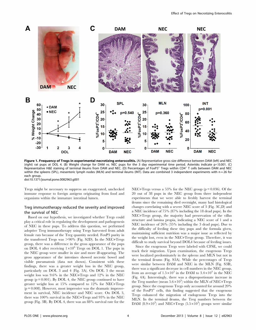
Frequency of Tregs in experimental necrotizingenterocolitis
Multiple factors have been implicated as triggers initiating
NEC. These triggers include hypoxia, hemodynamic instability,
formula osmolality, infections and hypothermia. In this rat model
of experimental NEC, the optimal induction of NEC requires a
combination of formula feeding and hypoxia. During the 3 day of
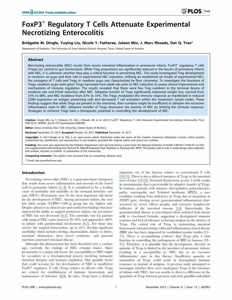
induction, there was a dramatic weight loss in the NEC group
compared to the dam-fed group, based on size and weight
(Fig. 1A,B). While the average starting weight was around 6 g for
both groups, the dam-fed pups gained an average of 45% on day
4. In the NEC group, there was an average weight loss of 4% on
day of life (DOL) 2, 8% on DOL 3 and 12% on DOL 4. The dam-
fed pups had 100% survival over the 3 days, while the NEC group
had a survival rate that dropped to 95% on DOL 3 and 58% on
DOL 4 (p,0.05). All pups in the dam-fed group received NEC
scores of 0, indicating normal ileal histology (Fig. 1C). In the NEC
group, there was around 75% incidence of NEC development,
with many receiving a maximum score of 3, indicating severe villus
denudation and full thickness necrosis (Fig. 1C). We next
examined whether the frequencies of Tregs were perturbed in
the NEC group. While the percentages of FoxP3+ Tregs within
CD4+ T cells were similar in the spleen and MLN of DAM versus
NEC, there was a significant reduction from an average of 52%
Tregs within the terminal ileums of DAM to 35% Tregs of NEC
group (Fig. 1D, S1). This finding is consistent with our previous
report [25]. This observation prompted our hypothesis that there
might be insufficient Treg numbers in the newborn rats, resulting
in the susceptibility to NEC induction.
The ontogeny of T cells and Tregs in dam fed control ratpups
The normal ontogeny of T cells and Tregs was investigated in
newborn rat in order to understand the role of Tregs in NEC. T
cell distributions were assessed in the thymus, spleen, MLN and
terminal ileum throughout the first 20 days of life. Figure 2A is a
flow cytometric representation of an intestinal sample from DOL
4, showing the percentage of CD4+ and CD8+ within CD3+ T cells
and the percentage of FoxP3+ within CD4+CD82 T cells. The
average percentage of CD4+CD82 T cells remained steady below
15% in the thymus and terminal ileum (Fig. 2B). There was a
progressive increase in the percentage of CD4+ within the spleen
and MLN, beginning with around 15% and 25% at DOL 0,
respectively. In the thymus, the CD8+CD42 T cells declined from
15% on DOL 0 to below 10% (Fig. 2C). The percentage of CD8+
in the spleen and MLN was higher than that of CD4+ during the
first few days of life and then became lower than CD4+ cells.
However, the most remarkable finding was the disproportionately
greater percentage and number of CD8+ compared to CD4+
within the terminal ileum (Fig. 2C,F). Over 75% of CD3+ T cells
were CD8+ in the terminal ileum and remained at this high level
throughout the 20 days of life. Since we wanted to measure total
cellular content within the intestines, the majority of CD8+ T cells
(.75% CD103+) most likely represented intraepithelial lympho-
cytes (IEL) and not just lamina propria cells. In contrast, the study
by Weitkamp et al. showed that in the lamina propria of human
infant intestines there were 3–4 times as many CD4+ as CD8+ cells
[23].
Tregs were quantified based on the expression of FoxP3 within
CD4+CD82 T cells (Fig. 2D). On DOL 0, the percentages of
Tregs within all tissues were below 5%. The percentages of Tregs
increased to 5–15% within the spleen and MLN, which were
similar to reported percentages in mice [33]. Treg percentages in
the thymus remained stable below 5%. Interestingly, the frequency
of Tregs climbed rapidly in the terminal ileum, peaking at over
60% on DOL 6 and then progressively decreasing to adult levels.
Within the Treg population for the first 14 days of life, there was a
progressive reduction in the Treg subset that expressed the
transcription factor Helios (Fig. S2A, B, C) [34,35]. The greatest
reduction occurred in the intestinal compartment, supporting the
evidence of gut environment preferential extrathymic Treg
development from naı̈ve T cells [36].
While over 40% of CD4+ T cells were Tregs starting on DOL 2,
it should be noted that the quantitative numbers of CD4+ and
Tregs were remarkably low relative to CD8+ cells in the terminal
ileum (Fig. 2F). The average number of CD8+ in the terminal
ileum on DOL 4 was 96105, which was over 30 times greater than
CD4+ (2.86104) and over 100 times greater than Tregs (8,800). In
the MLN, the average number of CD8+ (1.766106) and CD4+
(1.756106) was similar, while the number of Tregs (1.496105) was
12 times less than CD8+ or CD4+ cells (Fig. 2E). These findings
support the hypothesis that while Tregs are present early in
postnatal life, their numbers might be insufficient to control
perturbed immune responses. In premature infants, the quantity of
Effect of Tregs on Necrotizing Enterocolitis
PLOS ONE | www.plosone.org 3 December 2013 | Volume 8 | Issue 12 | e82963
Tregs might be necessary to suppress an exaggerated, unchecked
immune response to foreign antigens originating from food and
organisms within the immature intestinal lumen.
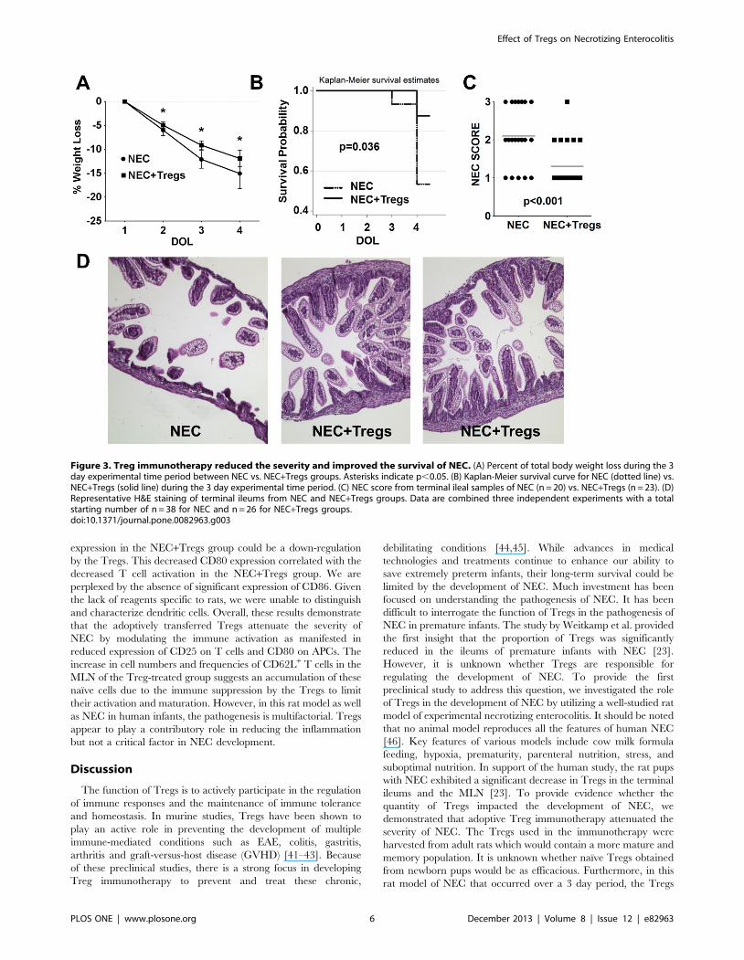
Treg immunotherapy reduced the severity and improvedthe survival of NEC
Based on our hypothesis, we investigated whether Tregs could
play a critical role in regulating the development and pathogenesis
of NEC in these pups. To address this question, we performed
adoptive Treg immunotherapy using Tregs harvested from adult
female rats because of the Treg quantity needed. FoxP3 purity in
the transferred Tregs was .90% (Fig. S2D). In the NEC+Tregs
group, there was a difference in the gross appearance of the pups
on DOL 4 after receiving 16106 Tregs on DOL 1. The pups in
the NEC group were smaller in size and more ill-appearing. The
gross appearance of the intestines showed necrotic bowel and
visible pneumatosis (data not shown). Consistent with these
findings, there was a greater weight loss in the NEC group,
particularly on DOL 3 and 4 (Fig. 3A). On DOL 3 the mean
weight loss was 9.6% in the NEC+Tregs and 12% in the NEC
group (p = 0.001). By DOL 4, the NEC group continued to have
greater weight loss at 15% compared to 12% for NEC+Tregs
(p = 0.008). However, most impressive was the dramatic improve-
ment in survival, NEC incidence and NEC score. On DOL 3,
there was 100% survival in the NEC+Tregs and 93% in the NEC
group (Fig. 3B). By DOL 4, there was an 88% survival rate for the
NEC+Tregs versus a 53% for the NEC group (p = 0.036). Of the
20 out of 38 pups in the NEC group from three independent
experiments that we were able to freshly harvest the terminal
ileums since the remaining died overnight, many had histological
changes correlating with a severe NEC score of 3 (Fig. 3C,D) and
a NEC incidence of 75% (87% including the 18 dead pups). In the
NEC+Tregs group, the majority had preservation of the villus
structure and lamina propia, indicating a NEC score of 1 and a
NEC incidence of 26% (35% including the 3 dead pups). Due to
the difficulty of feeding these tiny pups and the formula given,
maintaining sufficient nutrition was a major issue as reflected by
the weight lost, even in the NEC+Tregs group. Therefore, it was
difficult to study survival beyond DOL4 because of feeding issues.
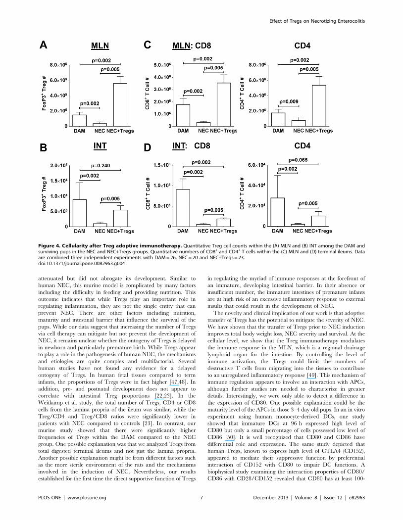
Since the exogenous Tregs were labeled with CFSE, we could
track their migration. Upon examination, the exogenous Tregs
were localized predominately in the spleens and MLN but not in
the terminal ileums (Fig. S3A). While the percentages of Tregs
were similar between DAM and NEC in the MLN (Fig. S3B),
there was a significant decrease in cell numbers in the NEC group,
from an average of 1.56105 in the DAM to 3.46104 in the NEC
(Fig. 4A). Interestingly, there was a disproportionate increase in
the Treg number (mean 5.66105) within the MLN of NEC+Tregs
group. Since the exogenous Tregs only accounted for around 20%
of the FoxP3+ cells, this finding suggested that the exogenous
Tregs enhanced the migration of endogenous Tregs into the
MLN. In the terminal ileums, the Treg numbers between the
DAM (8.96103) and NEC+Tregs (5.56103) groups were similar
Figure 1. Frequency of Tregs in experimental necrotizing enterocolitis. (A) Representative gross size difference between DAM (left) and NEC(right) rat pups at DOL 4. (B) Weight change for DAM vs. NEC pups for the 3 day experimental time period. Asterisks indicate p,0.001. (C)Representative H&E staining of terminal ileums from DAM and NEC. (D) Percentages of FoxP3+ Tregs within CD4+ T cells between DAM and NECwithin the spleens (SPL), mesenteric lymph nodes (MLN) and terminal ileums (INT). Data are combined 3 independent experiments with n = 26 foreach group.doi:10.1371/journal.pone.0082963.g001
Effect of Tregs on Necrotizing Enterocolitis
PLOS ONE | www.plosone.org 4 December 2013 | Volume 8 | Issue 12 | e82963
not because of a reconstitution by the exogenous Tregs, but
because of a greater preservation in normal cellularity and gut
integrity (Fig. 3D, 4B). Consistent with the decrease percentages of
Tregs in the terminal ileums of the NEC group, there was also a
significant reduction in cell numbers (16103) as compared to
DAM group (Fig. 4B).
We next examined the effect of Treg treatment on the cellularity
within the MLN and terminal ileums. Based on our flow
cytometric analysis and frequency in each subset, we calculated
the cell number for CD8+ and CD4+ T cells (Fig. S3C). When we
quantified the cellular composition among the three groups, we
noted a significant increase in CD8+ and CD4+ T cells within the
MLN of NEC+Tregs (Fig. 4C). In contrast, the CD8+ and CD4+ T
cells were significantly less in the NEC as compared to DAM
group. In the terminal ileums of the NEC group, there was a
dramatic reduction in these cells due to end-stage inflammation,
necrosis and tissue destruction (Figure 1C, 3D, 4D). However,
there was a notable improvement in the total cellularity and T cells
in the terminal ileums of NEC+Tregs as compared to the NEC
group. The improved outcome with Treg immunotherapy
indicates that Tregs can blunt NEC progression and supports
the hypothesis that a contributing factor to the pathogenesis of
NEC might be due to insufficient Treg numbers or maturation to
regulate immune responses and inflammation.
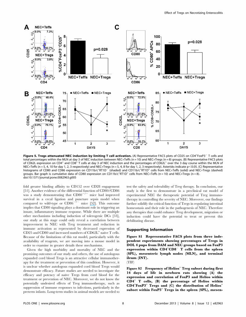
Tregs attenuated NEC induction by limiting T cellactivation
To investigate the mechanism of Treg mediated suppression of
NEC severity, we examined the level of immune activation.
Multiple studies have shown that Tregs suppress immune
responses by down-regulating CD80 and CD86 expression on
DCs, which are critical costimulatory molecules involved in T cell
activation [37–40]. Therefore, we repeated the experiments to
examine the level of immune activation between the NEC+Teffs
(CD4+CD252) and NEC+Tregs (CD4+CD25+) groups. On the
last day of NEC induction, we measured the level of CD25 and
CD62L expression on CD4+FoxP32 within the MLN. We
observed a significant reduction in CD25 expression, an activation
marker, within the NEC+Tregs group (Fig. 5A). In the NEC+Teffs
group, there was a significant reduction in CD4+CD62L+ and
CD8+CD62L+ T cells by day 3 of NEC (Fig. 5B). CD62L, also
known as L-selectin, is expressed on naı̈ve T cells. It allows for
homing to high endothelial venules and migration into peripheral
lymph nodes. It is rapidly shed from the lymphocytes upon
activation. The lower frequency of CD62L+ T cells in the NEC
group is consistent with our observation of reduced cell numbers in
the MLN (Fig. 4C). These results suggested that Tregs might be
targeting APCs to limit T cell activation. Given the limited
available reagents to phenotype the APCs, we were only able to
assess the APCs based on their expression of CD11b/c and MHC
class II (RT1D). Within the CD11b/c+ cells localized in the MLN,
around 20–30% are granulocytes, ,5% expressed CD103, and 8–
18% expressed RT1D (Fig. S4). Examination of CD11b/
c+RT1D+ APCs (,25–60% granulocytes) in the MLN of
NEC+Teffs group revealed that an average of 80% expressed
CD80 while only 58% in the NEC+Tregs group (Fig. 5C). There
was no appreciable expression of CD86 and no differences in the
level of CD54 (data not shown) between the two groups. Since
CD80 could be constitutively expressed on APCs, the decreased
Figure 2. The ontogeny of T cells and Tregs in dam fed control rat pups. (A) Representative flow cytometric plots from terminal ileum onDOL 4 with initial gating on CD3+ T cells to determine the percentages of CD4+ and CD8+ T cells (top plot) and FoxP3+ Tregs within CD82CD4+ T cells(bottom plot). Frequency of (B) CD4+, (C) CD8+ and (D) Tregs within the spleen (SPL), thymus (THY), mesenteric lymph nodes (MLN) and terminalileum (INT). Absolute cell counts of CD8+, CD4+, and Tregs in the (E) MLN and (F) terminal ileums on DOL 4. Data are derived from n = 6–16 pups ateach time point for B–D and n = 6 for E–F.doi:10.1371/journal.pone.0082963.g002
Effect of Tregs on Necrotizing Enterocolitis
PLOS ONE | www.plosone.org 5 December 2013 | Volume 8 | Issue 12 | e82963
expression in the NEC+Tregs group could be a down-regulation
by the Tregs. This decreased CD80 expression correlated with the
decreased T cell activation in the NEC+Tregs group. We are
perplexed by the absence of significant expression of CD86. Given
the lack of reagents specific to rats, we were unable to distinguish
and characterize dendritic cells. Overall, these results demonstrate
that the adoptively transferred Tregs attenuate the severity of
NEC by modulating the immune activation as manifested in
reduced expression of CD25 on T cells and CD80 on APCs. The
increase in cell numbers and frequencies of CD62L+ T cells in the
MLN of the Treg-treated group suggests an accumulation of these
naı̈ve cells due to the immune suppression by the Tregs to limit
their activation and maturation. However, in this rat model as well
as NEC in human infants, the pathogenesis is multifactorial. Tregs
appear to play a contributory role in reducing the inflammation
but not a critical factor in NEC development.
Discussion
The function of Tregs is to actively participate in the regulation
of immune responses and the maintenance of immune tolerance
and homeostasis. In murine studies, Tregs have been shown to
play an active role in preventing the development of multiple
immune-mediated conditions such as EAE, colitis, gastritis,
arthritis and graft-versus-host disease (GVHD) [41–43]. Because
of these preclinical studies, there is a strong focus in developing
Treg immunotherapy to prevent and treat these chronic,
debilitating conditions [44,45]. While advances in medical
technologies and treatments continue to enhance our ability to
save extremely preterm infants, their long-term survival could be
limited by the development of NEC. Much investment has been
focused on understanding the pathogenesis of NEC. It has been
difficult to interrogate the function of Tregs in the pathogenesis of
NEC in premature infants. The study by Weitkamp et al. provided
the first insight that the proportion of Tregs was significantly
reduced in the ileums of premature infants with NEC [23].
However, it is unknown whether Tregs are responsible for
regulating the development of NEC. To provide the first
preclinical study to address this question, we investigated the role
of Tregs in the development of NEC by utilizing a well-studied rat
model of experimental necrotizing enterocolitis. It should be noted
that no animal model reproduces all the features of human NEC
[46]. Key features of various models include cow milk formula
feeding, hypoxia, prematurity, parenteral nutrition, stress, and
suboptimal nutrition. In support of the human study, the rat pups
with NEC exhibited a significant decrease in Tregs in the terminal
ileums and the MLN [23]. To provide evidence whether the
quantity of Tregs impacted the development of NEC, we
demonstrated that adoptive Treg immunotherapy attenuated the
severity of NEC. The Tregs used in the immunotherapy were
harvested from adult rats which would contain a more mature and
memory population. It is unknown whether naı̈ve Tregs obtained
from newborn pups would be as efficacious. Furthermore, in this
rat model of NEC that occurred over a 3 day period, the Tregs
Figure 3. Treg immunotherapy reduced the severity and improved the survival of NEC. (A) Percent of total body weight loss during the 3day experimental time period between NEC vs. NEC+Tregs groups. Asterisks indicate p,0.05. (B) Kaplan-Meier survival curve for NEC (dotted line) vs.NEC+Tregs (solid line) during the 3 day experimental time period. (C) NEC score from terminal ileal samples of NEC (n = 20) vs. NEC+Tregs (n = 23). (D)Representative H&E staining of terminal ileums from NEC and NEC+Tregs groups. Data are combined three independent experiments with a totalstarting number of n = 38 for NEC and n = 26 for NEC+Tregs groups.doi:10.1371/journal.pone.0082963.g003
Effect of Tregs on Necrotizing Enterocolitis
PLOS ONE | www.plosone.org 6 December 2013 | Volume 8 | Issue 12 | e82963
attenuated but did not abrogate its development. Similar to
human NEC, this murine model is complicated by many factors
including the difficulty in feeding and providing nutrition. This
outcome indicates that while Tregs play an important role in
regulating inflammation, they are not the single entity that can
prevent NEC. There are other factors including nutrition,
maturity and intestinal barrier that influence the survival of the
pups. While our data suggest that increasing the number of Tregs
via cell therapy can mitigate but not prevent the development of
NEC, it remains unclear whether the ontogeny of Tregs is delayed
in newborn and particularly premature birth. While Tregs appear
to play a role in the pathogenesis of human NEC, the mechanisms
and etiologies are quite complex and multifactorial. Several
human studies have not found any evidence for a delayed
ontogeny of Tregs. In human fetal tissues compared to term
infants, the proportions of Tregs were in fact higher [47,48]. In
addition, pre- and postnatal development does not appear to
correlate with intestinal Treg proportions [22,23]. In the
Weitkamp et al. study, the total number of Tregs, CD4 or CD8
cells from the lamina propria of the ileum was similar, while the
Treg/CD4 and Treg/CD8 ratios were significantly lower in
patients with NEC compared to controls [23]. In contrast, our
murine study showed that there were significantly higher
frequencies of Tregs within the DAM compared to the NEC
group. One possible explanation was that we analyzed Tregs from
total digested terminal ileums and not just the lamina propria.
Another possible explanation might be from different factors such
as the more sterile environment of the rats and the mechanisms
involved in the induction of NEC. Nevertheless, our results
established for the first time the direct supportive function of Tregs
in regulating the myriad of immune responses at the forefront of
an immature, developing intestinal barrier. In their absence or
insufficient number, the immature intestines of premature infants
are at high risk of an excessive inflammatory response to external
insults that could result in the development of NEC.
The novelty and clinical implication of our work is that adoptive
transfer of Tregs has the potential to mitigate the severity of NEC.
We have shown that the transfer of Tregs prior to NEC induction
improves total body weight loss, NEC severity and survival. At the
cellular level, we show that the Treg immunotherapy modulates
the immune response in the MLN, which is a regional drainage
lymphoid organ for the intestine. By controlling the level of
immune activation, the Tregs could limit the numbers of
destructive T cells from migrating into the tissues to contribute
to an unregulated inflammatory response [49]. This mechanism of
immune regulation appears to involve an interaction with APCs,
although further studies are needed to characterize in greater
details. Interestingly, we were only able to detect a difference in
the expression of CD80. One possible explanation could be the
maturity level of the APCs in those 3–4 day old pups. In an in vitro
experiment using human monocyte-derived DCs, one study
showed that immature DCs at 96 h expressed high level of
CD80 but only a small percentage of cells possessed low level of
CD86 [50]. It is well recognized that CD80 and CD86 have
differential role and expression. The same study depicted that
human Tregs, known to express high level of CTLA4 (CD152),
appeared to mediate their suppressive function by preferential
interaction of CD152 with CD80 to impair DC functions. A
biophysical study examining the interaction properties of CD80/
CD86 with CD28/CD152 revealed that CD80 has at least 100-
Figure 4. Cellularity after Treg adoptive immunotherapy. Quantitative Treg cell counts within the (A) MLN and (B) INT among the DAM andsurviving pups in the NEC and NEC+Tregs groups. Quantitative numbers of CD8+ and CD4+ T cells within the (C) MLN and (D) terminal ileums. Dataare combined three independent experiments with DAM = 26, NEC = 20 and NEC+Tregs = 23.doi:10.1371/journal.pone.0082963.g004
Effect of Tregs on Necrotizing Enterocolitis
PLOS ONE | www.plosone.org 7 December 2013 | Volume 8 | Issue 12 | e82963
fold greater binding affinity to CD152 over CD28 engagement
[51]. Another evidence of the differential function of CD80/CD86
was a study demonstrating that CD802/2 mice had improved
survival in a cecal ligation and puncture sepsis model when
compared to wild-type or CD862/2 mice [52]. This outcome
implies that CD80 signaling plays a dominant role in triggering an
innate, inflammatory immune response. While there are multiple
other mechanisms including induction of tolerogenic DCs [53],
our study at this stage could only reveal a correlation between
improvement in NEC with Treg treatment and reduction in
immune activation as represented by decreased expression of
CD25 and CD80 and increased numbers of CD62L+ naı̈ve T cells.
Because of the limitations of this rat model, particularly with the
availability of reagents, we are moving into a mouse model in
order to examine in greater details these mechanisms.
Given the high morbidity and mortality of NEC and the
promising outcomes of our study and others, the use of autologous
expanded cord blood Tregs is an attractive cellular immunother-
apy for the treatment or prevention of this condition. However, it
is unclear whether autologous expanded cord blood Tregs would
demonstrate efficacy. Future studies are needed to investigate the
efficacy and potency of naı̈ve Tregs from cord blood for the
treatment or prevention of NEC. Moreover, we do not know the
potentially undesired effects of Treg immunotherapy, such as
suppression of immune responses to infections, particularly in the
preterm infants. Long-term studies in animal models are needed to
test the safety and tolerability of Treg therapy. In conclusion, our
study is the first to demonstrate in a preclinical rat model of
experimental NEC the therapeutic potential of Treg immuno-
therapy in controlling the severity of NEC. Moreover, our findings
further solidify the critical function of Tregs in regulating intestinal
homeostasis and their role in the pathogenesis of NEC. Therefore
any therapies that could enhance Treg development, migration or
induction could have the potential to treat or prevent this
debilitating disease.
Supporting Information
Figure S1 Representative FACS plots from three inde-pendent experiments showing percentages of Tregs inDOL 4 pups from DAM and NEC groups based on FoxP3expression within CD4+CD82 T cells from the spleen(SPL), mesenteric lymph nodes (MLN), and terminalileum (INT).
(TIF)
Figure S2 Frequency of Helios+ Treg subset during first14 days of life in newborn rats showing (A) theexpression and correlation of FoxP3 and Helios withinCD4+ T cells, (B) the percentage of Helios withinCD4+FoxP3+ Tregs and (C) the distribution of Helios+
subset within FoxP3+ Tregs in the spleen (SPL), mesen-
Figure 5. Tregs attenuated NEC induction by limiting T cell activation. (A) Representative FACS plots of CD25 on CD4+FoxP32 T cells andtotal percentages within the MLN at day 3 of NEC induction between NEC+Teffs (n = 10) and NEC+Tregs (n = 8) groups. (B) Representative FACS plotsof CD62L expression on CD4+ and CD8+ T cells at day 3 of NEC induction and the percentages of CD62L+ over the 3 day course within the MLN ofNEC+Teffs (n = 5, 4, 10 for day 1, 2, 3 respectively) and NEC+Tregs (n = 5, 4, 8 for day 1, 2, 3 respectively). Asterisks indicate p,0.05. (C) Representativehistograms of CD80 and CD86 expression on CD11b/c+RT1D2 (shaded) and CD11b/c+RT1D+ cells from NEC+Teffs (solid) and NEC+Tregs (dashed)groups. Bar graph is cumulative data of CD80 expression on CD11b/c+RT1D+ cells from NEC+Teffs (n = 10) and NEC+Tregs (n = 8).doi:10.1371/journal.pone.0082963.g005
Effect of Tregs on Necrotizing Enterocolitis
PLOS ONE | www.plosone.org 8 December 2013 | Volume 8 | Issue 12 | e82963
teric lymph nodes (MLN), thymus (THY) and terminalileum (INT). (D) Post-sort FoxP3 purity in theCD4+CD25+ Tregs and the CD4+CD252 non-Treg controlfrom three independent experiments.(TIF)
Figure S3 Representative FACS plots showing (A) thelocalization of exogenous CFSE-labeled Tregs within thespleen (SPL), MLN and INT, gating on CD4+FoxP3+ cells,(B) the percentage of Tregs within CD4+CD82 T cellsfrom MLN and INT of DOL 4 pups in DAM and thesurviving NEC and NEC+Tregs groups, and (C) thepercentages of CD4 and CD8 within CD3+ T cells fromMLN and INT of DOL 4 pups in DAM and the survivingNEC and NEC+Tregs groups used to calculate cellnumbers.(TIF)
Figure S4 (A) Representative FACS plots depicting thepercentages of CD11b/c, RT1D, granulocytes andCD103 cells from MLN. (B) Representative histograms of
CD80 and CD86 expression on CD11b/c+RT1D2 (shaded) and
CD11b/c+RT1D+ cells from NEC+Teffs (solid) and NEC+Tregs
(dashed) groups. Bar graph is cumulative data of CD80 expression
on CD11b/c+RT1D+ cells from NEC+Teffs (n = 10) and NEC+-Tregs (n = 8) groups. All data are from MLN of day 3 NEC
induction within NEC+Teffs and NEC+Tregs groups.
(TIF)
Acknowledgments
We are grateful for the statistical analysis from our biostaticians, Dr. Syed
S. Hashmi, M.D., MPH, Ph.D. and Cynthia S. Bell, M.S. We truly
appreciate Gene Lay, DVM (CEO Biolegend) for the reagent support. We
thank Dr. Elizabeth Donnachie and Dr. Miguel Escobar for generously
providing access to their BD FACSCalibur.
Author Contributions
Conceived and designed the experiments: DQT. Performed the experi-
ments: BMD YL NF JM DQT. Analyzed the data: BMD YL DQT. Wrote
the paper: BMD DQT YL. Edited the Manuscript: JMR.
References
1. Henry MC, Moss RL (2009) Necrotizing enterocolitis. Annu Rev Med 60: 111–
124.
2. Lin PW, Nasr TR, Stoll BJ (2008) Necrotizing enterocolitis: recent scientificadvances in pathophysiology and prevention. Semin Perinatol 32: 70–82.
3. Neu J, Walker WA (2011) Necrotizing enterocolitis. N Engl J Med 364: 255–
264.
4. Luig M, Lui K (2005) Epidemiology of necrotizing enterocolitis-Part II: Risks
and susceptibility of premature infants during the surfactant era: a regionalstudy. J Paediatr Child Health 41: 174–179.
5. Neu J, Mshvildadze M, Mai V (2008) A roadmap for understanding and
preventing necrotizing enterocolitis. Curr Gastroenterol Rep 10: 450–457.
6. Henry MC, Lawrence MR (2005) Surgical therapy for necrotizing enterocolitis:bringing evidence to the bedside. Semin Pediatr Surg 14: 181–190.
7. Petrosyan M, Guner YS, Williams M, Grishin A, Ford HR (2009) Current
concepts regarding the pathogenesis of necrotizing enterocolitis. Pediatr Surg Int25: 309–318.
8. Sakaguchi S, Miyara M, Costantino CM, Hafler DA (2010) FOXP3+ regulatory
T cells in the human immune system. Nat Rev Immunol 10: 490–500.
9. Tang Q, Bluestone JA (2008) The Foxp3+ regulatory T cell: a jack of all trades,
master of regulation. Nat Immunol 9: 239–244.
10. Lahl K, Loddenkemper C, Drouin C, Freyer J, Arnason J, et al. (2007) Selectivedepletion of Foxp3+ regulatory T cells induces a scurfy-like disease. J Exp Med
204: 57–63.
11. Nishizuka Y, Sakakura T (1969) Thymus and reproduction: sex-linkeddysgenesia of the gonad after neonatal thymectomy in mice. Science 166:
753–755.
12. Atarashi K, Tanoue T, Shima T, Imaoka A, Kuwahara T, et al. (2011)Induction of colonic regulatory T cells by indigenous Clostridium species.
Science 331: 337–341.
13. Fontenot JD, Dooley JL, Farr AG, Rudensky AY (2005) Developmentalregulation of Foxp3 expression during ontogeny. J Exp Med 202: 901–906.
14. Moraes-Vasconcelos D, Costa-Carvalho BT, Torgerson TR, Ochs HD (2008)
Primary immune deficiency disorders presenting as autoimmune diseases: IPEXand APECED. J Clin Immunol 28 Suppl 1: S11–S19.
15. Barnes MJ, Powrie F (2009) Regulatory T cells reinforce intestinal homeostasis.
Immunity 31: 401–411.
16. Collins CB, Aherne CM, McNamee EN, Lebsack MD, Eltzschig H, et al. (2012)
Flt3 ligand expands CD103(+) dendritic cells and FoxP3(+) T regulatory cells,
and attenuates Crohn’s-like murine ileitis. Gut 61: 1154–1162.
17. Maloy KJ, Powrie F (2011) Intestinal homeostasis and its breakdown in
inflammatory bowel disease. Nature 474: 298–306.
18. Boden EK, Snapper SB (2008) Regulatory T cells in inflammatory boweldisease. Curr Opin Gastroenterol 24: 733–741.
19. Hardenberg G, Steiner TS, Levings MK (2011) Environmental influences on T
regulatory cells in inflammatory bowel disease. Semin Immunol 23: 130–138.
20. Maul J, Loddenkemper C, Mundt P, Berg E, Giese T, et al. (2005) Peripheral
and intestinal regulatory CD4+ CD25(high) T cells in inflammatory bowel
disease. Gastroenterology 128: 1868–1878.
21. Veltkamp C, Anstaett M, Wahl K, Moller S, Gangl S, et al. (2011) Apoptosis of
regulatory T lymphocytes is increased in chronic inflammatory bowel disease
and reversed by anti-TNFalpha treatment. Gut 60: 1345–1353.
22. Weitkamp JH, Rudzinski E, Koyama T, Correa H, Matta P, et al. (2009)
Ontogeny of FOXP3(+) regulatory T cells in the postnatal human small
intestinal and large intestinal lamina propria. Pediatr Dev Pathol 12: 443–449.
23. Weitkamp JH, Koyama T, Rock MT, Correa H, Goettel JA, et al. (2013)
Necrotising enterocolitis is characterised by disrupted immune regulation and
diminished mucosal regulatory (FOXP3)/effector (CD4, CD8) T cell ratios. Gut
62: 73–82.
24. Liu Y, Fatheree NY, Mangalat N, Rhoads JM (2012) Lactobacillus reuteri
strains reduce incidence and severity of experimental necrotizing enterocolitis
via modulation of TLR4 and NF-kappaB signaling in the intestine. Am J Physiol
Gastrointest Liver Physiol 302: G608–G617.
25. Liu Y, Fatheree NY, Dingle BM, Tran DQ, Rhoads M (2013) Lactobacillus
reuteri DSM 17938 changes the frequency of Foxp3+ regulatory T cells in the
intestine and mesenteric lymph node in experimental necrotizing enterocolitis.
PLoS ONE 8 (2): e56547.
26. Nadler EP, Dickinson E, Knisely A, Zhang XR, Boyle P, et al. (2000) Expression
of inducible nitric oxide synthase and interleukin-12 in experimental necrotizing
enterocolitis. J Surg Res 92: 71–77.
27. Liu Y, Zhu L, Fatheree NY, Liu X, Pacheco SE, et al. (2009) Changes in
intestinal Toll-like receptors and cytokines precede histological injury in a rat
model of necrotizing enterocolitis. Am J Physiol Gastrointest Liver Physiol 297:
G442–G450.
28. Dvorak B, Khailova L, Clark JA, Hosseini DM, Arganbright KM, et al. (2008)
Comparison of epidermal growth factor and heparin-binding epidermal growth
factor-like growth factor for prevention of experimental necrotizing enterocolitis.
J Pediatr Gastroenterol Nutr 47: 11–18.
29. Shneider BL, Dawson PA, Christie DM, Hardikar W, Wong MH, et al. (1995)
Cloning and molecular characterization of the ontogeny of a rat ileal sodium-
dependent bile acid transporter. J Clin Invest 95: 745–754.
30. Liedel JL, Guo Y, Yu Y, Shiou SR, Chen S, et al. (2011) Mother’s milk-induced
Hsp70 expression preserves intestinal epithelial barrier function in an immature
rat pup model. Pediatr Res 69: 395–400.
31. Ran-Ressler RR, Khailova L, Arganbright KM, Adkins-Rieck CK, Jouni ZE, et
al. (2011) Branched chain fatty acids reduce the incidence of necrotizing
enterocolitis and alter gastrointestinal microbial ecology in a neonatal rat model.
PLoS ONE 6: e29032.
32. Tayman C, Uckan D, Kilic E, Ulus AT, Tonbul A, et al. (2011) Mesenchymal
stem cell therapy in necrotizing enterocolitis: a rat study. Pediatr Res 70: 489–
494.
33. Sakaguchi S, Sakaguchi N, Shimizu J, Yamazaki S, Sakihama T, et al. (2001)
Immunologic tolerance maintained by CD25+ CD4+ regulatory T cells: their
common role in controlling autoimmunity, tumor immunity, and transplantation
tolerance. Immunol Rev 182: 18–32.
34. McClymont SA, Putnam AL, Lee MR, Esensten JH, Liu W, et al. (2011)
Plasticity of human regulatory T cells in healthy subjects and patients with type 1
diabetes. J Immunol 186: 3918–3926.
35. Thornton AM, Korty PE, Tran DQ, Wohlfert EA, Murray PE, et al. (2010)
Expression of Helios, an Ikaros transcription factor family member, differentiates
thymic-derived from peripherally induced Foxp3+ T regulatory cells. J Immunol
184: 3433–3441.
36. Sun CM, Hall JA, Blank RB, Bouladoux N, Oukka M, et al. (2007) Small
intestine lamina propria dendritic cells promote de novo generation of Foxp3 T
reg cells via retinoic acid. J Exp Med 204: 1775–1785.
37. DiPaolo RJ, Brinster C, Davidson TS, Andersson J, Glass D, et al. (2007)
Autoantigen-specific TGFbeta-induced Foxp3+ regulatory T cells prevent
autoimmunity by inhibiting dendritic cells from activating autoreactive T cells.
J Immunol 179: 4685–4693.
Effect of Tregs on Necrotizing Enterocolitis
PLOS ONE | www.plosone.org 9 December 2013 | Volume 8 | Issue 12 | e82963
38. Tran DQ, Glass DD, Uzel G, Darnell DA, Spalding C, et al. (2009) Analysis of
adhesion molecules, target cells, and role of IL-2 in human FOXP3+ regulatoryT cell suppressor function. J Immunol 182: 2929–2938.
39. Tran DQ, Shevach EM (2009) Therapeutic potential of FOXP3(+) regulatory T
cells and their interactions with dendritic cells. Hum Immunol 70: 294–299.40. Wing K, Onishi Y, Prieto-Martin P, Yamaguchi T, Miyara M, et al. (2008)
CTLA-4 control over Foxp3+ regulatory T cell function. Science 322: 271–275.41. Miyara M, Gorochov G, Ehrenstein M, Musset L, Sakaguchi S, et al. (2011)
Human FoxP3+ regulatory T cells in systemic autoimmune diseases.
Autoimmun Rev 10: 744–755.42. Muller YD, Golshayan D, Ehirchiou D, Wekerle T, Seebach JD, et al. (2009) T
regulatory cells in xenotransplantation. Xenotransplantation 16: 121–128.43. Sakaguchi S (2011) Regulatory T cells: history and perspective. Methods Mol
Biol 707: 3–17.44. Riley JL, June CH, Blazar BR (2009) Human T regulatory cell therapy: take a
billion or so and call me in the morning. Immunity 30: 656–665.
45. Tang Q, Bluestone JA, Kang SM (2012) CD4(+)Foxp3(+) regulatory T celltherapy in transplantation. J Mol Cell Biol 4: 11–21.
46. Sodhi C, Richardson W, Gribar S, Hackam DJ (2008) The development ofanimal models for the study of necrotizing enterocolitis. Dis Model Mech 1: 94–
98.
47. Michaelsson J, Mold JE, McCune JM, Nixon DF (2006) Regulation of T cell
responses in the developing human fetus. J Immunol 176: 5741–5748.
48. Mold JE, Michaelsson J, Burt TD, Muench MO, Beckerman KP, et al. (2008)
Maternal alloantigens promote the development of tolerogenic fetal regulatory T
cells in utero. Science 322: 1562–1565.
49. Davidson TS, Shevach EM (2011) Polyclonal Treg cells modulate T effector cell
trafficking. Eur J Immunol 41: 2862–2870.
50. Zheng Y, Manzotti CN, Liu M, Burke F, Mead KI, et al. (2004) CD86 and
CD80 differentially modulate the suppressive function of human regulatory T
cells. J Immunol 172: 2778–2784.
51. Collins AV, Brodie DW, Gilbert RJ, Iaboni A, Manso-Sancho R, et al. (2002)
The interaction properties of costimulatory molecules revisited. Immunity 17:
201–210.
52. Nolan A, Kobayashi H, Naveed B, Kelly A, Hoshino Y, et al. (2009) Differential
role for CD80 and CD86 in the regulation of the innate immune response in
murine polymicrobial sepsis. PLoS ONE 4: e6600.
53. Lan Q, Zhou X, Fan H, Chen M, Wang J, et al. (2012) Polyclonal CD4+Foxp3+Treg cells induce TGFbeta-dependent tolerogenic dendritic cells that suppress
the murine lupus-like syndrome. J Mol Cell Biol 4: 409–419.
Effect of Tregs on Necrotizing Enterocolitis
PLOS ONE | www.plosone.org 10 December 2013 | Volume 8 | Issue 12 | e82963