Nanomolar CFTR Inhibition by Pore-Occluding Divalent Polyethylene Glycol-Malonic Acid Hydrazides
Upload
independentCategory
view
3download
0
Chemistry & Biology
Article
Solubilizing Mutations Used to Crystallize One CFTRDomain Attenuate the Trafficking and Channel DefectsCaused by the Major Cystic Fibrosis MutationLuısa S. Pissarra,1,5 Carlos M. Farinha,1,2,5 Zhe Xu,3 Andre Schmidt,2 Patrick H. Thibodeau,4,6 Zhiwei Cai,3
Philip J. Thomas,4 David N. Sheppard,3 and Margarida D. Amaral1,2,*1Department of Chemistry and Biochemistry, Faculty of Sciences, University of Lisboa, 1749-016 Lisboa, Portugal2Centre of Human Genetics, National Institute of Health, 1649-016 Lisboa, Portugal3Department of Physiology and Pharmacology, School of Medical Sciences, University of Bristol, BS8 1TD Bristol, United Kingdom4Department of Physiology, University of Texas Southwestern Medical Center Dallas, TX 75390, USA5These authors contributed equally to this work.6Present address: Department of Cell Biology and Physiology, University of Pittsburgh School of Medicine, Pittsburgh, PA 15261, USA.*Correspondence: [email protected]
DOI 10.1016/j.chembiol.2007.11.012
SUMMARY
Cystic fibrosis (CF) is caused by mutations in theCF transmembrane conductance regulator (CFTR)Cl� channel. F508del, the most frequent CF-causingmutation, disrupts both the processing and functionof CFTR. Recently, the crystal structure of the firstnucleotide-binding domain of CFTR bearing F508del(F508del-NBD1) was elucidated. Although F508del-NBD1 shows only minor conformational changesrelative to that of wild-type NBD1, additional muta-tions (F494N/Q637R or F429S/F494N/Q637R) wererequired for domain solubility and crystallization.Here we show that these solubilizing mutations incis with F508del partially rescue the trafficking defectof full-length F508del-CFTR and attenuate its gatingdefect. We interpret these data to suggest that thesolubilizing mutations utilized to facilitate F508del-NBD1 production also assist folding of full-lengthF508del-CFTR protein. Thus, the available crystalstructure of F508del-NBD1 might correspond toa partially corrected conformation of this domain.
INTRODUCTION
The common life-shortening genetic disease cystic fibrosis (CF)
is caused by mutations in the ATP-binding cassette (ABC) trans-
porter, cystic fibrosis transmembrane conductance regulator
(CFTR) (Rowe et al., 2005). Over 1500 CFTR gene variants
have been described so far, most presumed to cause CF. How-
ever, one single mutation, the deletion of phenylalanine 508
(F508del), accounts for approximately 70% of all CF chromo-
somes worldwide (http://www.genet.sickkids.on.ca/cftr/).
CFTR is a Cl� channel that is expressed at the apical mem-
brane of epithelial cells, where it regulates fluid and electrolyte
transport and is regulated by cAMP-dependent phosphorylation
and cycles of ATP binding and hydrolysis (Cai et al., 2007). CFTR
has a modular design composed of two membrane-spanning
62 Chemistry & Biology 15, 62–69, January 2008 ª2008 Elsevier Ltd
domains (MSDs) which form the channel pore, two cytoplasmic
nucleotide-binding domains (NBDs) which regulate channel gat-
ing, and a unique cytoplasmic regulatory domain (RD), the phos-
phorylation of which regulates channel activity.
The most critical problem associated with the F508del muta-
tion, which is located in NBD1, is its impaired folding, which pre-
vents the mutant protein from acquiring a native conformation
and trafficking to the cell membrane (Riordan, 2005). Hence,
F508del-CFTR is recognized as misfolded and retained by endo-
plasmic reticulum (ER) quality control (ERQC), which targets
F508del-CFTR for degradation by the ubiquitin-proteasomal
pathway (UPP) (Vashist and Ng, 2004). Whereas almost all
F508del-CFTR is targeted for degradation by the UPP, numer-
ous studies have demonstrated that the mutant protein can
mature under specific conditions, including incubation either at
low temperature (Denning et al., 1992), with cellular osmolytes
(Brown et al., 1996), or in the presence of second-site suppres-
sors (Teem et al., 1993). Some function is restored by these ma-
nipulations, suggesting that a therapeutic strategy for CF based
on enhancing the maturation of F508del-CFTR is possible.
A variety of studies have thus focused on understanding CFTR
folding and structure so as to elucidate the molecular basis of
CF. Recently, high-resolution structures of murine and human
NBD1 have provided invaluable information about the structure
of NBD1 (Lewis et al., 2004, 2005). Together with the atomic-res-
olution structures of other ABC transporters (Hollenstein et al.,
2007), these NBD structures also provide insight into the putative
interactions of NBD1 with other domains of CFTR.
To better understand, at a molecular level, how the F508del-
CFTR mutation perturbs the structure of NBD1, Lewis et al.
(2005) elucidated the crystal structure of F508del-NBD1. How-
ever, to promote domain solubilization and, hence, crystalliza-
tion, Lewis et al. (2005) incorporated a series of mutations into
the human NBD1 sequence. Some of these mutations repre-
sented sequence changes between human CFTR and CFTRs
from other species, whereas others (G550E, R553Q, and
R555K) had been previously identified as F508del-CFTR rever-
tant mutations (Teem et al., 1996; deCarvalho et al., 2002; Chang
et al., 1999). F508del-CFTR revertants are second-site mutations
in cis with F508del which rescue the cell-surface expression and
All rights reserved
Chemistry & Biology
Rescue of F508del-CFTR by Solubilizing Mutations
Cl� channel function of F508del-CFTR. Of note, one of these
revertants, R553Q, was found in an F508del-homozygous patient
with mild CF disease (Dork et al., 1991). In addition, another rever-
tant mutation, G550E, likely acts by altering the structure of NBD1
(Roxo-Rosa et al., 2006). Examination of the structures of wt- and
F508del-NBD1 revealed little or no structural changes besides
deformation of the local surface topography around the F508 res-
idue (Lewis et al., 2005). Based on these data and a substantial
amount of biochemical, biophysical, and cell biological data,
new hypotheses were then proposed (Chen et al., 2004; Thibo-
deau et al., 2005; Du et al., 2005; Cui et al., 2006). Because the
F508 residue lies at a domain-domain interface, these hypothe-
ses argue that altered domain-domain interactions, resulting
from the F508del mutation, impact upon the global structure of
CFTR. Furthermore, they suggest that impairment of these inter-
domain interactions is the fundamental defect associated with
the F508del mutation. However, it remains plausible to envisage
that a structure of F508del-NBD1 with more conformational dif-
ferences than just those at the surface described by Lewis et al.
(2005) would likely further disrupt such interdomain interactions,
in particular those involving NBD1.
Because of the presence of F508del revertant mutations in the
crystal structure of F508del-NBD1, further F508del-NBD1 struc-
tures were produced (Lewis, 2005) lacking known F508del rever-
tants (http://www.rcsb.org/pdb/). However, these new F508del-
NBD1 crystal structures still required either two (F494N/Q637R;
Protein Data Bank [PDB] ID code: 2BBT) or three (F429S/F494N/
Q637R; PDB ID code: 2BBS) additional mutations for domain
solubility and, hence, crystal formation (Lewis, 2005).
Given the ability of solubilizing mutations to increase the yield
of NBD1 and facilitate its crystallization (Lewis et al., 2005), we
Chemistry & Biology
hypothesized that these mutations might also promote the fold-
ing of both isolated NBD1 and full-length CFTR protein. To test
these ideas, we investigated the effects of the mutations
F494N/Q637R and F429S/F494N/Q637R on wt- and F508del-
CFTR by studying: (1) the in vivo folding yield of NBD1, (2) the
processing and trafficking of the full-length CFTR protein, and
(3) the Cl� channel function of CFTR. Our data reveal that solubi-
lizing mutations attenuate the trafficking and gating defects of
F508del-CFTR.
RESULTS
While studying the effects of F508del on the structure of NBD1
from CFTR, Lewis (2005) introduced the mutations F494N/
Q637R (double; D) and F429S/F494N/Q637R (triple; T) into
NBD1 to improve domain solubility and crystallization. To investi-
gate whether these variants counteract the effects of F508del on
protein processing and channel function, we performed three
types of experiments. First, the yield of these NBD1 variants was
tested when expressed in bacteria. Second, full-length CFTR fold-
ing and maturationwereassessed innovel BHK cell lines stablyex-
pressing wt- and F508del-CFTR in cis with these mutations. Third,
their effects on cell-surface expression and function were deter-
mined using biochemical and electrophysiological techniques.
Solubilizing Mutations Improvewt- and F508del-NBD1 YieldTo explore whether F429S, F494N, and Q637R improve the yield
of soluble NBD1, wt-NBD1 and F508del-NBD1 were expressed
in bacterial cells in the absence and presence of the solubilizing
mutations. Confirming the data of Lewis et al. (2005), Figure 1A
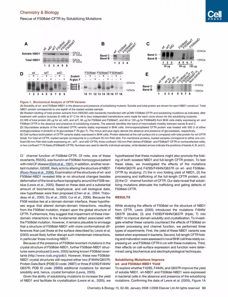
Figure 1. Biochemical Analysis of CFTR Variants
(A) Solubility of wt- and F508del-NBD1 in the absence and presence of solubilizing mutants. Soluble and total protein are shown for each NBD1 construct. Total
NBD1 protein corresponds to one eighth of the loaded soluble sample.
(B) Western blotting of total protein extracts from HEK293 cells transiently transfected with pCMV-F508del-CFTR and solubilizing mutations as indicated, after
treatment with sodium butyrate (5 mM) at 37�C for 48 hr (two independent transfections were made for each clone shown for the solubilizing mutants).
(C) WB of total protein (30 mg for wt, wtD, and wtT, 60 mg for F508del and F508delT, and 60 or 120 mg for F508delD) from BHK cells stably expressing wt- and
F508del-CFTR in the absence and presence of solubilizing mutants. The asterisk identifies the band of intermediate mobility between bands B and C.
(D) Glycosidase analysis of the indicated CFTR variants stably expressed in BHK cells. Immunoprecipitated CFTR protein was treated with 500 U of either
endoglycosidase H (endoH) or N-glycosidase F (N-glyc F). The minus and plus signs denote the absence and presence of glycosidases, respectively.
(E) Cell-surface biotinylation of CFTR variants stably expressed in BHK cells. Protein detected at the cell surface (m) is compared with total protein for wt-CFTR
(total). For total wt-CFTR, loaded sample corresponds to a confluent 35 mm Petri dish. For membrane proteins, loaded samples correspond to either one con-
fluent 60 mm Petri dish (cells expressing wt-, wtT-, and wtD-CFTR), three confluent 100 mm Petri dishes (F508del- and F508delT-CFTR or nontransfected cells),
or two confluent T175 flasks (F508delD-CFTR). Numbers are used to identify individual samples, while labeled arrows indicate the positions of bands A, B, and C.
15, 62–69, January 2008 ª2008 Elsevier Ltd All rights reserved 63

Chemistry & Biology
Rescue of F508del-CFTR by Solubilizing Mutations
(lanes 4–6) demonstrates that F508delD- and F508delT-NBD1
generated greater amounts of soluble NBD1 than did F508del-
NBD1. Interestingly, the data also indicate that F508delD- and
F508delT-NBD1 had marginally higher folding yields than did
wt-NBD1 (compare lane 1 to lanes 5 and 6) by 1.2- and 1.5-fold,
respectively (the solubility of F508del-NBD1 was 0.6 that of
wt-NBD1). Moreover, the soluble yields of wtD- and wtT-NBD1
were enhanced relative to that of wt (lanes 1–3) by 1.8- and 2.0-
fold, respectively. These data thusconfirmthat the solubilizingmu-
tations markedly increase the low yield of soluble F508del-NBD1.
Solubilizing Mutations Rescue F508del-CFTRfrom ER MislocalizationWhen western blotting (WB) is used to investigate the processing
of CFTR, two different forms of wt-CFTR protein are visualized: an
immature core-glycosylated form that is found in the ER (150 kDa;
band B) and a mature fully glycosylated form that has been pro-
cessed through the Golgi apparatus and delivered to the cell
membrane (170–180 kDa; band C) (Cheng et al., 1990). To inves-
tigate the processing of F508del-CFTR in cis with the double or
triple solubilizing mutations, total protein extracts from HEK293
cells transiently transfected with F508del-, F508delD-, and
F508delT-CFTR cDNAs were assessed by WB analysis using
the anti-CFTR M3A7 antibody. To augment the expression of
these CFTR constructs, cells were treated with sodium butyrate
(5 mM) at 37�C for 24 hr (Cheng et al., 1995). Figure 1B demon-
strates that some fraction of F508delD- and F508delT-CFTR
(lanes 3 and 4) are processed to the mature form, whereas no
measurable mature F508del-CFTR is observed (lane 2).
Butyrate has been reported to partially rescue the cell-surface
expression of F508del-CFTR (Cheng et al., 1995). Therefore, the
effects of solubilizing mutations on F508del-CFTR processing in
the absence of butyrate were assessed by WB using novel BHK
cell lines stably expressing wt- or F508del-CFTR in cis with the
solubilizing mutations. Figure 1C (lanes 1–3) demonstrates that
the solubilizing mutations only slightly affected the proportion
of processed wt-CFTR at steady state (wt: 89 ± 7%, n = 5;
wtD: 93 ± 3%, n = 3; wtT: 90 ± 2%, n = 3; where 100% corre-
sponds to total CFTR in each sample, i.e., band B + band C).
However, these mutations promoted the maturation of
F508del-CFTR. F508delD- (Figure 1C, lanes 6 and 7) and espe-
cially F508delT-CFTR (Figure 1C, lane 5) generated higher mo-
lecular mass forms of CFTR, consistent with full glycosylation
(band C), that were absent in F508del-CFTR (Figure 1C, lane 4).
Of note, in addition to band C, a band of intermediate mobility
(between bands B and C, marked with an asterisk in Figure 1C)
was consistently detected in both F508delD- and F508delT-
CFTR, which is absent in samples of wt-, wtD-, or wtT-CFTR.
To confirm the identity of the bands observed with F508delD-
and F508delT-CFTR, we used endoglycosidase H (endoH) and
N-glycosidase F (N-glyc F) to assess the glycosylation status of
these CFTR variants. Like band C of wt-, wtT-, and wtD-CFTR
(Figure 1D, lanes 3, 6, and 9), the higher molecular mass forms
of F508delT- and F508delD-CFTR were sensitive to N-glyc F
(Figure 1D, lanes 15 and 18) which, by removing glycans from
bands B and C, convert CFTR to its bare polypeptidic chain
(band A). However, the intermediate band of F508delT- and
F508delD-CFTR was also sensitive to EndoH (Figure 1D, lanes
14 and 17), indicating that its glycosylation status is distinct from
64 Chemistry & Biology 15, 62–69, January 2008 ª2008 Elsevier Ltd
that of band C of wt-CFTR. However, this intermediate band might
correspond to a post-ER glycosylated form of CFTR, as it is also
absent in F508del-CFTR samples (Figure 1C, lane 4).
To determine whether F508delD- and F508delT-CFTR are
delivered to the plasma membrane, cell-surface biotinylation
assays were performed using a modified nonpermeant biotin
reagent that only binds (available) primary amines at the cell sur-
face from the external milieu. Figure 1E (lanes 2–4) reveals that
band C of wt-, wtD-, and wtT-CFTR were present at the cell
surface. By contrast, F508del-CFTR could not be detected in
the cell-surface fraction (lane 5), confirming the specificity of
this method for identifying protein at the cell surface. For both
F508delT-CFTR and F508delD-CFTR, processed forms previ-
ously detected by WB (Figure 1C, lanes 5–7) were also detected
in the cell-membrane fraction, although in reduced amounts
(lanes 6 and 7).
Solubilizing Mutations Restore Functionto F508del-CFTRTo further confirm the presence of processed forms of F508delD-
and F508delT-CFTR at the cell membrane, we analyzed CFTR
function using the iodide efflux technique. This method assesses
the activity of a large population of CFTR constructs at the
surface of intact cells.
When BHK cells expressing F508del-CFTR were incubated at
37�C, they failed to generate an efflux of iodide (Figure 2A, open
squares), whereas after 24 hr incubation at 26�C they exhibited
a sustained efflux of iodide (Figure 2A, black squares), albeit
1.4-fold smaller and with slower onset and decay than the large
transient efflux of iodide elicited by wt-CFTR at 37�C (Figure 2A,
gray circles; Figure 2E). Figures 2B and 2E demonstrate that, at
37�C, solubilizing mutations were without effect on the efflux of
iodide elicited by wt-CFTR. Indeed, neither the time course nor
the magnitude of the response differed between wt-, wtD-, and
wtT-CFTR (Figure 2B). Of note, at 37�C, both F508delD-
(Figure 2C) and F508delT-CFTR (Figure 2D) generated effluxes
of iodide with time courses similar to that of wt-CFTR but
magnitudes comparable to that of low-temperature rescued
F508del-CFTR (Figure 2E). Moreover, after incubation at 26�C,
the magnitude and time courses of F508delD- (Figure 2C) and
F508delT-CFTR (Figure 2D) iodide efflux were comparable to
that of wt-CFTR at 37�C (Figure 2E).
Solubilizing Mutations Alter the Functionof F508del-CFTR, but Not That of wt-CFTRTo learn whether solubilizing mutations alter the function of wt-
and F508del-CFTR Cl� channels, we used the excised inside-
out configuration of the patch-clamp technique. Visual inspec-
tion of single-channel records (Figure 3A) demonstrates that
the pattern of channel gating of low-temperature rescued
F508del-CFTR, recorded at 37�C, differs strikingly from that of
wt-CFTR. For F508del-CFTR, infrequent bursts of channel open-
ings are separated by closed periods dramatically longer than
those of wt-CFTR.
Figure 3 reveals that the solubilizing mutations were without
effect on the function of wt-CFTR. wtD- and wtT-CFTR had the
same current amplitude (i), open probability (Po), mean burst
duration (MBD), and interburst interval (IBI) as wt-CFTR (p > 0.1).
By contrast, these mutations enhanced the Po of F508del-CFTR
All rights reserved

Chemistry & Biology
Rescue of F508del-CFTR by Solubilizing Mutations
by altering channel gating (Figures 3A and 3C). Both mutations
restored MBD to values equivalent to that of wt-CFTR (Fig-
ure 3D). Of note, they also attenuated markedly the prolonged
IBI of F508del-CFTR (Figure 3E). F508delT-CFTR had the great-
est effect, decreasing IBI from 10-fold longer than wt-CFTR to
only 3-fold longer. As for wt-CFTR, the solubilizing mutations
were without effect on the i of F508del-CFTR (p > 0.5), which
was equivalent to that of wt-CFTR (Figure 3B). These data thus
demonstrate that solubilizing mutations are without effect on
the gating behavior of wt-CFTR but strongly attenuate the gating
of F508del-CFTR.
DISCUSSION
The most common CF mutation, F508del, causes a temperature-
sensitive folding defect that is recognized by the ERQC leading
to the targeting of newly synthesized F508del-CFTR for protea-
Figure 2. Iodide Efflux from BHK Cells
Expressing Solubilizing Mutants
(A–D) Time courses of iodide efflux from BHK cells
stably expressing the indicated CFTR variants cul-
tured at either 37�C or 26�C. During the periods
indicated by the bars, forskolin (10 mM) and genis-
tein (50 mM) were present in the efflux buffer. Data
are means (n = 4–9) at each point. Where not
shown, error bars are smaller than symbol size.
(E) Magnitude of peak iodide efflux generated by
different CFTR constructs expressed as a percent-
age of that of wt-CFTR. Asterisks indicate signifi-
cant differences from wt-CFTR (p < 0.05), whereas
crosses indicate significant differences between
the same CFTR variants cultured at 37�C and
26�C (p < 0.05). Data are expressed as means ±
SEM of n R 4 observations.
somal degradation (Ward et al., 1995;
Jensen et al., 1995). Because F508del
dramatically attenuates the folding yield
of isolated NBD1 at different tempera-
tures but has little effect on the measured
stability of the folded domain (Qu and
Thomas, 1996), the mutation likely affects
an early step in CFTR folding by imposing
a significant energy barrier between
a folding intermediate and the native con-
formation or by decreasing the energy
barrier for entering the off-pathway.
Thus, the observation that solubilizing
mutations enhance the yield of solubilized
NBD1 (Lewis et al., 2005) suggests that
these mutations might counteract the
relative change in energy barrier imposed
by F508del and, hence, improve NBD1
folding.
Understanding the alteration in bio-
chemical and biophysical properties of
full-length F508del-CFTR associated
with the inclusion of NBD1 solubilizing
mutations is important in several re-
Chemistry & Biology
spects. First, the analysis and interpretation of the structures of
F508del-NBD1 are potentially affected by the inclusion of these
mutations. Second, investigating the coordinated role that multi-
ple, nonadjacent sites to F508del play in a common, but com-
plex, protein fold might help understanding the principal ele-
ments that define the F508del-NBD1 fold. Finally, recognizing
the specific spatial constraints of F508del-NBD1 and pinpointing
differences between it and wt-NBD1 might aid the design of mol-
ecules with therapeutic potential aimed at correcting such differ-
ences (Moran and Zegarra-Moran, 2005).
Solubilizing Mutations Attenuate F508del-CFTRTrafficking and Functional DefectsTo determine how the double (D) and triple (T) solubilizing muta-
tions of NBD1 impact on the folding of NBD1 and CFTR, we first
evaluated their effects on the biochemical properties of NBD1
in vitro and then on the biochemical and functional properties
15, 62–69, January 2008 ª2008 Elsevier Ltd All rights reserved 65

Chemistry & Biology
Rescue of F508del-CFTR by Solubilizing Mutations
of full-length CFTR in vivo. Consistent with the data of Lewis et al.
(Lewis et al., 2005; Lewis, 2005), the solubilizing mutations dra-
matically enhanced the yield of soluble NBD1 produced in
Escherichia coli, independent of total protein expression for both
wt- and F508del-NBD1. Of note, the yield of F508del-NBD1
with solubilizing mutations was even higher than that of wt-
NBD1 without the solubilizing mutations.
Moreover, when introduced into full-length CFTR, the solubiliz-
ing mutations partially restored the maturation of F508del-CFTR,
as seen by the appearance of band C. These data indicate that
solubilizing mutations allow at least some F508del-CFTR to exit
the ER and traffic through the Golgi apparatus. However, the
amounts of processed F508delT-CFTR and especially F508delD-
CFTR were significantly less than that of wt-CFTR.
Interestingly, in addition to bands B and C, a band of interme-
diate mobility was observed for both F508delD- and F508delT-
CFTR. Susceptibility to endoH confirmed that this intermediate
band does not acquire the same state as wt-CFTR. This higher
molecular weight form of F508del-CFTR might correspond to in-
completely processed CFTR structures, probably lacking the
final glycan residues (e.g., fucose and/or sialic acid) (Hammerle
et al., 2000) which appear to be stabilized by the solubilizing mu-
tations. Because single-chain glycosylated forms of CFTR are
endoH resistant like those of wt-CFTR (Hammerle et al., 2000),
we consider it unlikely that CFTR variants containing solubilizing
mutations possess just one, instead of two, glycan chains. Nev-
ertheless, partially glycosylated forms of CFTR would be ex-
pected to be functional at the cell membrane, because glycosyl-
ation null CFTR mutants form regulated Cl� channels (Chang
et al., 1994; Farinha and Amaral, 2005). It is possible to envisage
that this intermediate band also occurs transiently during pro-
cessing of wt-CFTR, but is rapidly converted into the fully pro-
66 Chemistry & Biology 15, 62–69, January 2008 ª2008 Elsevier Ltd
cessed form and thus is not detected. Interestingly, we have
recently observed it (or a similar form) under general inhibition
of protein kinases (Schmidt et al., 2008).
Data from cell-surface biotinylation demonstrate that the band
C observed for the F508delD- and F508delT-CFTR variants is
present at the cell surface, albeit in reduced amounts compared
to that for wt-CFTR. Confirmation that both F508delD- and
F508delT-CFTR are present at the membrane was provided by
functional studies using iodide efflux and patch-clamp tech-
niques. By contrast, and as widely described, cAMP agonists
failed to elicit an efflux of iodide from cells expressing F508del-
CFTR at 37�C.
Studies of the single-channel activity of F508delD and
F508delT revealed that they had MBD values similar to wt-
CFTR. Using the ATP-driven NBD dimerization model of CFTR
channel gating (Vergani et al., 2005), these data suggest that
the solubilizing mutations might stabilize the NBD dimer of
CFTR. However, because the IBI values of F508delD- and
F508delT-CFTR are still greater than that of wt-CFTR, it is likely
that the energy barrier for channel opening remains greater than
that for wt-CFTR. Nevertheless, our data suggest that the solu-
bilizing mutations accelerate markedly NBD dimerization com-
pared with that of F508del-CFTR.
Taken together, these data indicate that the solubilizing muta-
tions partially rescue the trafficking defect and restore some
function to F508del-CFTR. Interestingly, iodide efflux from cells
expressing the F508delD-CFTR and F508delT-CFTR variants
was augmented by low-temperature incubation, indicating that
the effects of solubilizing mutations and low temperature on
F508del-CFTR trafficking and function are additive. This sug-
gests that solubilizing mutations and low temperature might
rescue F508del-CFTR by distinct mechanisms.
Figure 3. Single-Channel Currents of
wt- and F508del-CFTR in the Absence and
Presence of Solubilizing Mutants
(A) Representative single-channel recordings of
the indicated CFTR variants in excised inside-out
membrane patches from BHK cells. ATP (1 mM)
and PKA (75 nM) were continuously present in
the intracellular solution; voltage was �50 mV,
and there was a Cl� concentration gradient across
the membrane ([Cl�]int, 147 mM; [Cl�]ext, 10 mM).
Dashed lines indicate the closed channel state
and downward deflections correspond to channel
openings.
(B–E) Single-channel current amplitude (i), open
probability (Po), mean burst duration (MBD), and
interburst interval (IBI) of the indicated CFTR vari-
ants. Columns and error bars are means ± SEM
(wt: i, n = 4, Po, MBD, IBI, n = 7; wtD: i, Po, n = 7,
MBD and IBI, n = 3; wtT: i, Po, n = 4, MBD and
IBI, n = 3; F508del: n = 7 for all data; F508delD:
n = 6 for all data; F508delT: n = 4 for all data). The
asterisks and crosses indicate values that are
significantly different from those of wt- (p < 0.05)
and F508del-CFTR (p < 0.05), respectively. For
wt-CFTR, C127 cells were used to determine Po,
MBD, and IBI because these cells express low
levels of CFTR protein, whereas BHK cells were used to determine i. However, in one single-channel patch from a BHK cell expressing wt-CFTR, values of i,
Po, MBD, and IBI did not differ from those obtained using C127 cells expressing wt-CFTR. For F508del, F508delD, and F508delT, values of MBD and IBI
calculated using multichannel patches did differ from those acquired using single-channel patches.
All rights reserved

Chemistry & Biology
Rescue of F508del-CFTR by Solubilizing Mutations
Structural ImplicationsAn interesting aspect of the action of the solubilizing mutations
(double, F494N/Q637R; triple, F429S/F494N/Q637R) is their re-
mote location from that of F508. Based on this consideration,
Lewis et al. (2005) speculated that these mutations would not in-
fluence the structure of hNBD1. However, the effect produced by
F494N is not completely unexpected, as structural crosstalk be-
tween the side chain of the F508 residue (e.g., F508R) and the
Q loop or g-phosphate switch (e.g., residues W496 and M498)
has been previously shown to occur (Massiah et al., 1999).
Interestingly, the C terminus of the crystal structure of NBD1
contains a long a helix (H9b) that is not present in the crystal
structures of NBDs from other ABC transporters (Lewis et al.,
2004). Curiously, Q637R lies in this helix, known as the RD1 re-
gion (residues 590–672) of NBD1. This region was initially con-
sidered to be within the R domain (Riordan et al., 1989), but is
now regarded to be part of NBD1 based on amino acid sequence
analysis and functional studies (Dulhanty and Riordan, 1994;
Bianchet et al., 1997; Chan et al., 2000). Structural and experi-
mental evidence argue that helix H9b is either an integral compo-
nent of the NBD1 fold or a more ‘‘loose’’ structural element that is
highly favorable for the folding of this domain (Lewis et al., 2004).
In support of this notion, our data indicate that Q637R, at least
with one additional solubilizing mutation (F494N), might contrib-
ute to the increased solubility of F508del-NBD1 and to the partial
rescue of F508del-CFTR trafficking and function.
The third solubilizing mutation, F429S, further promotes the re-
vertant effect produced by the double mutant (F494N/Q637R) on
F508del-CFTR, as the triple mutant (F429S/F494N/Q637R) visi-
bly increased maturation of F508del-CFTR as measured by the
higher maturation yield at steady state of F508delT-CFTR com-
pared with that of F508delD-CFTR (Figure 1C). The F429 residue
occurs in an insertion (406–434), between the first (393–400) and
second (441–448) b strands of NBD1, which was found to be dis-
ordered in NBD1 crystals (Ostedgaard et al., 2000). This suggests
that F429S might lie on the surface of NBD1 in a position where it
likely interacts with the environment (Lewis et al., 2004, 2005).
Comparison with Other RevertantsUsing the same cellular system employed to investigate the sol-
ubilizing mutations, we recently examined the mechanism of
action of two other F508del-CFTR revertants, G550E and 4RK,
the simultaneous mutation of four arginine-framed tripeptides
(AFTs), R29K, R516K, R555K, and R766K (Roxo-Rosa et al.,
2006). We suggested that these mutations partially rescue
F508del-CFTR by distinct mechanisms. G550E likely alters the
conformation of NBD1, whereas 4RK allows F508del-CFTR to
escape endoplasmic reticulum retention/retrieval mediated by
AFTs. We also showed that both G550E and 4RK affected
wt-CFTR (Roxo-Rosa et al., 2006). By contrast, the present results
demonstrate that solubilizing mutations attenuate markedly
both the trafficking and functional defects of F508del-CFTR
without, however, affecting wt-CFTR.
Moreover, whereas the revertants 4RK and G550E restored
the iodide efflux of F508del-CFTR to wt levels (Roxo-Rosa
et al., 2006), the magnitude of iodide efflux elicited by
F508delD- and F508delT-CFTR was less than that of wt-CFTR.
This indicates that the solubilizing mutations are not as effective
as 4RK and G550E at augmenting either the cell-surface expres-
Chemistry & Biology
sion or function of F508del-CFTR. However, at steady state,
F508delD and F508delT produced amounts of band C similar
to those of G550E- or 4RK-F508del (present study and A.S.
and M.D.A., unpublished data). Thus, the surface levels of
F508del-CFTR rescued by different genetic variants do not
strictly correlate with the overall activity of F508del-CFTR as
a Cl� channel. Altogether, these data argue that different genetic
variants have distinct effects on the trafficking and function of
F508del-CFTR.
Moreover, analysis of CFTR channel gating revealed that the
NBD1 solubilizing mutations analyzed here restored the attenu-
ated MBD of F508del-CFTR to wt-CFTR levels and attenuated
dramatically the greatly prolonged IBI of F508del-CFTR. By con-
trast, both revertant mutations markedly prolonged the MBD of
F508del-CFTR, with G550E exceeding that of wt-CFTR by
4-fold (Roxo-Rosa et al., 2006). Moreover, only G550E suppressed
the prolonged IBI of F508del-CFTR (Roxo-Rosa et al., 2006), re-
ducing it to a level similar to that achieved here with F508delT-
CFTR. We interpret these data to suggest that the conformation
state(s) that solubilizing mutations achieve is closer to that of
wt-CFTR than the conformation produced by the revertant
G550E (Roxo-Rosa et al., 2006).
Consistent with this interpretation, the revertants, especially
G550E, extended the MBD and elevated the Po of wt-CFTR
(Roxo-Rosa et al., 2006), whereas wt-CFTR containing the solu-
bilizing mutations had the same MBD, IBI, and Po as wt-CFTR.
We thus suggest that the solubilizing mutations act specifically
on F508del-CFTR (in contrast to G550E) to abrogate its gating
defect, possibly through correction of NBD1 and/or CFTR fold-
ing. Hence, the conformation of F508del-CFTR containing solu-
bilizing mutations is markedly different from that achieved by
low-temperature correction, which fails to rescue the gating de-
fect of F508del-CFTR (Denning et al., 1992).
In conclusion, our data suggest that a crystal structure of
F508del-NBD1 without solubilizing mutations is required to de-
termine whether and how the F508del mutation perturbs the
structure of this domain. Meanwhile, it is plausible to envisage
that the F508del mutation per se alters the properties of NBD1
and, hence, also the interaction of NBD1 with other domains of
CFTR. Finally, solubilizing mutations are attractive sites to target
for the design of small-molecule CFTR correctors to treat CF.
SIGNIFICANCE
F508del, the most frequent mutation in CF, disrupts both the
trafficking and channel gating of CFTR. The available crystal
structure of F508del-NBD1 was determined after the intro-
duction of additional mutations (F494N/Q637R or F429S/
F494N/Q637R) to help domain solubilization and crystal
formation. Whereas comparison of this structure with that
of wild-type NBD1 revealed only minor conformational
changes, the present study demonstrates that these solubi-
lizing mutations partially rescue in vivo both the trafficking
and functional defects of full-length F508del-CFTR. These
data suggest that the existing crystal structure of F508del-
NBD1 is that of a partially corrected conformation and that
the structure of F508del-NBD1 without additional mutations
is still needed to gain further insight into how the F508del-
NBD1 differs from wt-NBD1.
15, 62–69, January 2008 ª2008 Elsevier Ltd All rights reserved 67

Chemistry & Biology
Rescue of F508del-CFTR by Solubilizing Mutations
EXPERIMENTAL PROCEDURES
Solubility of CFTR-NBD1
His-tagged hNBD1s (amino acids 388–677) containing different CFTR-NBD1
variants were cloned into the pET-SUMO vector and expressed in bacterial
BL21 cells at 15�C after IPTG induction as described previously (Thibodeau
et al., 2005). Total NBD1 was obtained from sonicated whole-cell lysates
and soluble NBD1 from the supernatant of a 30,000 3 g centrifugation.
Samples were run on 10% (w/v) Tris-tricine SDS-PAGE gels, transferred to
nitrocellulose membranes, and probed with an anti-His monoclonal antibody
and an anti-mouse HRP-labeled secondary antibody. Total NBD1 samples
correspond to one eighth of the loaded soluble samples.
Site-Directed Mutagenesis, Cells, and CFTR Expression
To introduce the solubilizing mutations F494N/Q637R and F429S/F494N/
Q637R into wt- and F508del-CFTR cDNAs in the pNUT expression vector,
we used the primers F429S, 50-GGTGATGACAGCCTCTCCTTCAGTAATTTC
TCA-30; F494N, 50-CATTCTGTTCTCAGAATTCCTGGATTATGCCTGG-30;
Q637R, 50-GAACTCCAAAATCTAAGGCCAGACTTTAGCTC-30 and the Quik-
Change site-directed mutagenesis kit (Stratagene). The identity of each solu-
bilizing mutation was verified by sequencing.
For transient expression, HEK293 cells were transfected with 1.5 mg DNA of
the pCMV-CFTR-Not6.2 construct containing different CFTR variants using
Fugene6 (Roche). Twenty-four hours posttransfection, sodium butyrate
(5 mM) was added for a further 24 hr to enhance CFTR expression.
To generate cell lines stably expressing high levels of CFTR variants, baby
hamster kidney (BHK) cells were transfected with CFTR cDNAs (1.5 mg) using
Lipofectin (Invitrogen) and selected for stable transfectants using methotrex-
ate (500 mM). Cell lines expressing different solubilizing mutations are referred
to as follows: wtD-CFTR, F494N-Q637R-CFTR; F508delD-CFTR, F494N-
F508del-Q637R-CFTR; wtT-CFTR, F429S-F494N-Q637R-CFTR; and F508delT-
CFTR, F429S-F494N-F508del-Q637R-CFTR. For each CFTR variant, seven
BHK clones were analyzed by western blotting (WB). Of these, the clone ex-
pressing the highest level of CFTR protein was selected for further analyses, en-
suring that CFTR expression levels among the different CFTR variants were
equivalent. Cells were cultured, seeded, and used as described previously
(Roxo-Rosa et al., 2006). In some experiments (see figure legends), cell-surface
expression of F508del-CFTR variants was enhanced by incubating cells at
26�C for 24 hr.
Biochemical Analyses of CFTR Protein
To assay for CFTR protein expression by WB, cells expressing CFTR variants
were lysed and extracts were analyzed as described (Farinha et al., 2004a)
using the anti-CFTR monoclonal antibody M3A7 (Chemicon). Densitometry
was performed as described previously (Farinha et al., 2004a).
Glycosidase Assays
Cells were metabolically labeled with [35S]methionine and lysed prior to CFTR
immunoprecipitation with the anti-CFTR monoclonal antibody M3A7 (Chemi-
con) as previously described (Farinha et al., 2004a). Immunoprecipitated
CFTR was then incubated with 500 U endoglycosidase H (endoH) and
N-glycosidase F (N-glyc F), both from New England Biolabs for 18 hr at
37�C as described (Farinha et al., 2004b). After digestion, samples were ana-
lyzed by electrophoresis followed by fluorography as described previously
(Farinha et al., 2004a).
Cell-Surface Biotinylation
To determine the membrane-localized fraction of CFTR variants, cell-surface
biotinylation was performed (Farinha et al., 2004b). Briefly, cells were cooled
to 4�C, washed with phosphate-buffered saline (PBS), and labeled with
1 mg/ml sulfo-NHS-LC-biotin (Pierce). Labeled cells were lysed in 1 ml of
RIPA buffer containing a cocktail of protease inhibitors. After biotinylation
and lysis, samples were subjected to CFTR immunoprecipitation with the
M3A7 antibody (Chemicon) and protein G agarose and then recaptured with
avidin-Sepharose beads (Pierce). For total protein, CFTR was eluted, whereas
for membranar proteins, samples were eluted using Laemmli sample buffer
without bromophenol blue and diluted in RIPA buffer 10-fold, and the biotiny-
lated fraction was captured using avidin-Sepharose beads (Pierce). Both total
68 Chemistry & Biology 15, 62–69, January 2008 ª2008 Elsevier Ltd
CFTR and biotinylated CFTR were then in vitro phosphorylated by PKA (Prom-
ega) using [g-32P]ATP (New England Nuclear) and analyzed by SDS-PAGE.
Iodide Efflux Technique
CFTR-mediated iodide efflux was measured at room temperature as de-
scribed (Lansdell et al., 1998) using the cAMP agonist forskolin (10 mM) and
the CFTR potentiator genistein (50 mM; Sigma-Aldrich). Prior to commencing
experiments, BHK cells were incubated for 1 hr in loading buffer containing
136 mM NaI, 3 mM KNO3, 2 mM Ca(NO3)2, 20 mM HEPES, and 11 mM glucose
(pH 7.4), with 1 M NaOH (osmolarity, 299 ± 0.48 mOsm; n = 36), and then
washed thoroughly with efflux buffer (136 mM NaNO3 replacing 136 mM NaI
in the loading buffer; osmolarity, 291 ± 0.24 mOsm; n = 60). The amount of
iodide in each sample of efflux buffer was determined using an iodide-selective
electrode (MP225; Thermo Electron). Data are expressed as means ± SEM
(standard error of the mean) of n observations; statistical analyses were per-
formed using Student’s t test, with a value of p < 0.05 considered statistically
significant.
Electrophysiology
CFTR Cl� channels were recorded in excised inside-out membrane patches
from BHK cells expressing CFTR constructs stably (wt-, wtD-, wtT-, and
F508del-CFTR) or transiently (F508delD- and F508delT-CFTR). For wt-, wtD-,
wtT-, F508delD-, and F508delT-CFTR, cells were incubated at 37�C prior to
study, whereas F508del-CFTR expressing cells were incubated at 28�C.
Data were acquired and analyzed using an Axopatch 200B patch-clamp am-
plifier and pCLAMP (version 9.2) software (both from Molecular Devices) as de-
scribed (Cai et al., 2006). The pipette (extracellular) solution contained 140 mM
N-methyl-D-glucamine (NMDG), 140 mM aspartic acid, 5 mM CaCl2, 2 mM
MgSO4, and 10 mM 2-([2-hydroxy-l,l-bis(hydroxymethyl)ethyl]amino)-ethane-
sulfonic acid (TES) (pH 7.3), with Tris ([Cl�], 10 mM). The bath (intracellular) so-
lution contained 140 mM NMDG, 3 mM MgCl2, 1 mM CsEGTA, and 10 mM TES
(pH 7.3), with HCl ([Cl�], 147 mM; [Ca2+]free, < 10�8 M) and was maintained at
37�C. To activate and sustain channel activity, PKA (75 nM) and ATP (1 mM)
were added to all intracellular solutions; voltage was �50 mV. The number
of active channels was determined based on the maximum number of simul-
taneous channel openings using the strategies described in Cai et al. (2006)
to minimize errors when counting the number of active channels. Data were
analyzed as described previously (Cai et al., 2006) and are expressed as
means ± SEM of n observations, where n is the number of excised membrane
patches; statistical analyses were performed using Student’s unpaired t test,
with a value of p < 0.05 considered statistically significant.
ACKNOWLEDGMENTS
This work was supported by grants POCTI/MGI/47382/2002 and POCTI/SAU/
MMO/58425/2004 and pluriannual funding of CIGMH (Portugal/European
Union) to M.D.A., the BBSRC and CF Trust UK to D.N.S., and NIH-NIDDK
49836 to P.J.T. L.S.P. and A.S. are recipients of SFRH/BD/9095/2002 and
SFRH/BD/19415/2004 doctoral fellowships, respectively (from FCT, Portugal),
and Z.X. is supported by the University of Bristol and an ORSAS award.
Received: June 12, 2007
Revised: November 5, 2007
Accepted: November 21, 2007
Published: January 25, 2008
REFERENCES
Bianchet, M.A., Ko, Y.H., Amzel, L.M., and Pedersen, P.L. (1997). Modeling of
nucleotide binding domains of ABC transporter proteins based on a F1-AT-
Pase/recA topology: structural model of the nucleotide binding domains of
the cystic fibrosis transmembrane conductance regulator (CFTR). J. Bioenerg.
Biomembr. 29, 503–524.
Brown, C.R., Hong-Brown, L.Q., Biwersi, J., Verkman, A.S., and Welch, W.J.
(1996). Chemical chaperones correct the mutant phenotype of the delta
F508 cystic fibrosis transmembrane conductance regulator protein. Cell
Stress Chaperones 1, 117–125.
All rights reserved

Chemistry & Biology
Rescue of F508del-CFTR by Solubilizing Mutations
Cai, Z., Taddei, A., and Sheppard, D.N. (2006). Differential sensitivity of the
cystic fibrosis (CF)-associated mutants G551D and G1349D to potentiators
of the cystic fibrosis transmembrane conductance regulator (CFTR) Cl� chan-
nel. J. Biol. Chem. 281, 1970–1977.
Cai, Z., Chen, J.-H., Hughes, L.K., and Li, H. (2007). The physiology and phar-
macology of the CFTR Cl� channel. In Chloride Movements across Cellular
Membranes, M. Pusch, ed. (San Diego: Elsevier Limited), pp. 109–143.
Chan, K.W., Csanady, L., Seto-Young, D., Nairn, A.C., and Gadsby, D.C.
(2000). Severed molecules functionally define the boundaries of the cystic
fibrosis transmembrane conductance regulator’s NH2-terminal nucleotide
binding domain. J. Gen. Physiol. 116, 163–180.
Chang, X.B., Hou, Y.X., Jensen, T.J., and Riordan, J.R. (1994). Mapping of
cystic fibrosis transmembrane conductance regulator membrane topology
by glycosylation site insertion. J. Biol. Chem. 269, 18572–18575.
Chang, X.B., Cui, L., Hou, Y.X., Jensen, T.J., Aleksandrov, A.A., Mengos, A.,
and Riordan, J.R. (1999). Removal of multiple arginine-framed trafficking sig-
nals overcomes misprocessing of delta F508 CFTR present in most patients
with cystic fibrosis. Mol. Cell 4, 137–142.
Chen, E.Y., Bartlett, M.C., Loo, T.W., and Clarke, D.M. (2004). The deltaF508
mutation disrupts packing of the transmembrane segments of the cystic fibro-
sis transmembrane conductance regulator. J. Biol. Chem. 279, 39620–39627.
Cheng, S.H., Gregory, R.J., Marshall, J., Paul, S., Souza, D.W., White, G.A.,
O’Riordan, C.R., and Smith, A.E. (1990). Defective intracellular transport and
processing of CFTR is the molecular basis of most cystic fibrosis. Cell 63,
827–834.
Cheng, S.H., Fang, S.L., Zabner, J., Marshall, J., Piraino, S., Schiavi, S.C., Jef-
ferson, D.M., Welsh, M.J., and Smith, A.E. (1995). Functional activation of the
cystic fibrosis trafficking mutant delta F508-CFTR by overexpression. Am. J.
Physiol. 268, L615–L624.
Cui, L., Aleksandrov, L., Hou, Y.X., Gentzsch, M., Chen, J.H., Riordan, J.R.,
and Aleksandrov, A.A. (2006). The role of cystic fibrosis transmembrane con-
ductance regulator phenylalanine 508 side chain in ion channel gating. J. Phys-
iol. 572, 347–358.
deCarvalho, A.C.V., Gansheroff, L.J., and Teem, J.L. (2002). Mutations in the
nucleotide binding domain 1 signature motif region rescue processing and
functional defects of cystic fibrosis transmembrane conductance regulator
delta F508. J. Biol. Chem. 277, 35896–35905.
Denning, G.M., Anderson, M.P., Amara, J.F., Marshall, J., Smith, A.E., and
Welsh, M.J. (1992). Processing of mutant cystic fibrosis transmembrane
conductance regulator is temperature-sensitive. Nature 358, 761–764.
Dork, T., Wulbrand, U., Richter, T., Neumann, T., Wolfes, H., Wulf, B., Maass,
G., and Tummler, B. (1991). Cystic fibrosis with three mutations in the cystic
fibrosis transmembrane conductance regulator gene. Hum. Genet. 87,
441–446.
Du, K., Sharma, M., and Lukacs, G.L. (2005). The deltaF508 cystic fibrosis
mutation impairs domain-domain interactions and arrests post-translational
folding of CFTR. Nat. Struct. Mol. Biol. 12, 17–25.
Dulhanty, A.M., and Riordan, J.R. (1994). A two-domain model for the R do-
main of the cystic fibrosis transmembrane conductance regulator based on
sequence similarities. FEBS Lett. 343, 109–114.
Farinha, C.M., and Amaral, M.D. (2005). Most F508del-CFTR is targeted to
degradation at an early folding checkpoint and independently of calnexin.
Mol. Cell. Biol. 25, 5242–5252.
Farinha, C.M., Mendes, F., Roxo-Rosa, M., Penque, D., and Amaral, M.D.
(2004a). A comparison of 14 antibodies for the biochemical detection of the
cystic fibrosis transmembrane conductance regulator protein. Mol. Cell.
Probes 18, 235–242.
Farinha, C.M., Penque, D., Roxo-Rosa, M., Lukacs, G., Dormer, R., McPher-
son, M., Pereira, M., Bot, A.G., Jorna, H., Willemsen, R., et al. (2004b). Bio-
chemical methods to assess CFTR expression and membrane localization.
J. Cyst. Fibros. 3 (Suppl 2), 73–77.
Hammerle, M.M., Aleksandrov, A.A., Chang, X.B., and Riordan, J.R. (2000). A
novel CFTR disease-associated mutation causes addition of an extra N-linked
oligosaccharide. Glycoconj. J. 17, 807–813.
Chemistry & Biology
Hollenstein, K., Dawson, R.J., and Locher, K.P. (2007). Structure and mecha-
nism of ABC transporter proteins. Curr. Opin. Struct. Biol. 17, 412–418.
Jensen, T.J., Loo, M.A., Pind, S., Williams, D.B., Goldberg, A.L., and Riordan,
J.R. (1995). Multiple proteolytic systems, including the proteasome, contribute
to CFTR processing. Cell 83, 129–135.
Lansdell, K.A., Delaney, S.J., Lunn, D.P., Thomson, S.A., Sheppard, D.N., and
Wainwright, B.J. (1998). Comparison of the gating behaviour of human and
murine cystic fibrosis transmembrane conductance regulator Cl� channels
expressed in mammalian cells. J. Physiol. 508, 379–392.
Lewis, H. (2005). Structures of human CFTR NBD1 confirm conservation of the
domain structuredespite thedelF508mutation.Pediatr.Pulmonol.40, 190–191.
Lewis, H.A., Buchanan, S.G., Burley, S.K., Conners, K., Dickey, M., Dorwart,
M., Fowler, R., Gao, X., Guggino, W.B., Hendrickson, W.A., et al. (2004). Struc-
ture of nucleotide-binding domain 1 of the cystic fibrosis transmembrane con-
ductance regulator. EMBO J. 23, 282–293.
Lewis, H.A., Zhao, X., Wang, C., Sauder, J.M., Rooney, I., Noland, B.W.,
Lorimer, D., Kearins, M.C., Conners, K., Condon, B., et al. (2005). Impact of
the deltaF508 mutation in first nucleotide-binding domain of human cystic
fibrosis transmembrane conductance regulator on domain folding and struc-
ture. J. Biol. Chem. 280, 1346–1353.
Massiah, M.A., Ko, Y.H., Pedersen, P.L., and Mildvan, A.S. (1999). Cystic fibro-
sis transmembrane conductance regulator: solution structures of peptides
based on the Phe508 region, the most common site of disease-causing
deltaF508 mutation. Biochemistry 38, 7453–7461.
Moran, O., and Zegarra-Moran, O. (2005). A quantitative description of the activa-
tionand inhibitionofCFTRby potentiators:genistein.FEBSLett. 579, 3979–3983.
Ostedgaard, L.S., Baldursson, O., Vermeer, D.W., Welsh, M.J., and Robert-
son, A.D. (2000). A functional R domain from cystic fibrosis transmembrane
conductance regulator is predominantly unstructured in solution. Proc. Natl.
Acad. Sci. USA 97, 5657–5662.
Qu, B.H., and Thomas, P.J. (1996). Alteration of the cystic fibrosis transmem-
brane conductance regulator folding pathway. J. Biol. Chem. 271, 7261–7264.
Riordan, J.R. (2005). Assembly of functional CFTR chloride channels. Annu.
Rev. Physiol. 67, 701–718.
Riordan, J.R., Rommens, J.M., Kerem, B., Alon, N., Rozmahel, R., Grzelczak,
Z., Zielenski, J., Lok, S., Plavsic, N., and Chou, J.L. (1989). Identification of the
cystic fibrosis gene: cloning and characterization of complementary DNA.
Science 245, 1066–1073.
Rowe, S.M., Miller, S., and Sorscher, E.J. (2005). Cystic fibrosis. N. Engl. J.
Med. 352, 1992–2001.
Roxo-Rosa, M., Xu, Z., Schmidt, A., Neto, M., Cai, Z., Soares, C.M., Sheppard,
D.N., and Amaral, M.D. (2006). Revertant mutants G550E and 4RK rescue cys-
tic fibrosis mutants in the first nucleotide-binding domain of CFTR by different
mechanisms. Proc. Natl. Acad. Sci. USA 103, 17891–17896.
Schmidt, A., Hughes, L.K., Cai, Z., Mendes, F., Li, H., Sheppard, D.N., and
Amaral, M.D. (2008). Prolonged treatment of cells with genistein modulates
the expression and function of the cystic fibrosis transmembrane conductance
regulator. Br. J. Pharmacol., in press.
Teem, J.L., Berger, H.A., Ostedgaard, L.S., Rich, D.P., Tsui, L.C., and Welsh,
M.J. (1993). Identification of revertants for the cystic fibrosis delta F508 muta-
tion using STE6-CFTR chimeras in yeast. Cell 73, 335–346.
Teem, J.L., Carson, M.R., and Welsh, M.J. (1996). Mutation of R555 in CFTR-
delta F508 enhances function and partially corrects defective processing.
Receptors Channels 4, 63–72.
Thibodeau, P.H., Brautigam, C.A., Machius, M., and Thomas, P.J. (2005). Side
chain and backbone contributions of Phe508 to CFTR folding. Nat. Struct. Mol.
Biol. 12, 10–16.
Vashist, S., and Ng, D.T. (2004). Misfolded proteins are sorted by a sequential
checkpoint mechanism of ER quality control. J. Cell Biol. 165, 41–52.
Vergani, P., Lockless, S.W., Nairn, A.C., and Gadsby, D.C. (2005). CFTR chan-
nel opening by ATP-driven tight dimerization of its nucleotide-binding
domains. Nature 433, 876–880.
Ward, C.L., Omura, S., and Kopito, R.R. (1995). Degradation of CFTR by the
ubiquitin-proteasome pathway. Cell 83, 121–127.
15, 62–69, January 2008 ª2008 Elsevier Ltd All rights reserved 69
Copyright © 2022 FDOKUMEN