Pharmacological correction of a defect in PPAR-γ signaling ameliorates disease severity in...
13
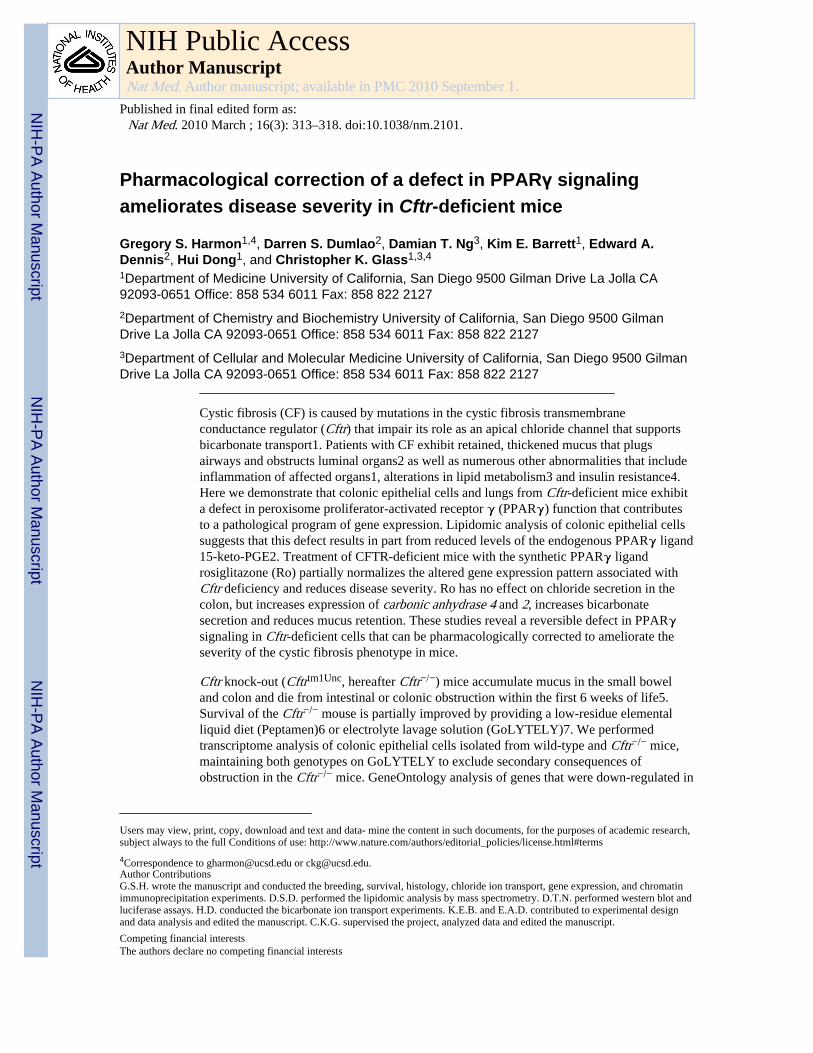
Pharmacological correction of a defect in PPARγ signaling ameliorates disease severity in Cftr-deficient mice Gregory S. Harmon 1,4 , Darren S. Dumlao 2 , Damian T. Ng 3 , Kim E. Barrett 1 , Edward A. Dennis 2 , Hui Dong 1 , and Christopher K. Glass 1,3,4 1 Department of Medicine University of California, San Diego 9500 Gilman Drive La Jolla CA 92093-0651 Office: 858 534 6011 Fax: 858 822 2127 2 Department of Chemistry and Biochemistry University of California, San Diego 9500 Gilman Drive La Jolla CA 92093-0651 Office: 858 534 6011 Fax: 858 822 2127 3 Department of Cellular and Molecular Medicine University of California, San Diego 9500 Gilman Drive La Jolla CA 92093-0651 Office: 858 534 6011 Fax: 858 822 2127 Cystic fibrosis (CF) is caused by mutations in the cystic fibrosis transmembrane conductance regulator (Cftr) that impair its role as an apical chloride channel that supports bicarbonate transport1. Patients with CF exhibit retained, thickened mucus that plugs airways and obstructs luminal organs2 as well as numerous other abnormalities that include inflammation of affected organs1, alterations in lipid metabolism3 and insulin resistance4. Here we demonstrate that colonic epithelial cells and lungs from Cftr-deficient mice exhibit a defect in peroxisome proliferator-activated receptor γ (PPARγ) function that contributes to a pathological program of gene expression. Lipidomic analysis of colonic epithelial cells suggests that this defect results in part from reduced levels of the endogenous PPARγ ligand 15-keto-PGE2. Treatment of CFTR-deficient mice with the synthetic PPARγ ligand rosiglitazone (Ro) partially normalizes the altered gene expression pattern associated with Cftr deficiency and reduces disease severity. Ro has no effect on chloride secretion in the colon, but increases expression of carbonic anhydrase 4 and 2, increases bicarbonate secretion and reduces mucus retention. These studies reveal a reversible defect in PPARγ signaling in Cftr-deficient cells that can be pharmacologically corrected to ameliorate the severity of the cystic fibrosis phenotype in mice. Cftr knock-out (Cftr tm1Unc , hereafter Cftr −/− ) mice accumulate mucus in the small bowel and colon and die from intestinal or colonic obstruction within the first 6 weeks of life5. Survival of the Cftr −/− mouse is partially improved by providing a low-residue elemental liquid diet (Peptamen)6 or electrolyte lavage solution (GoLYTELY)7. We performed transcriptome analysis of colonic epithelial cells isolated from wild-type and Cftr −/− mice, maintaining both genotypes on GoLYTELY to exclude secondary consequences of obstruction in the Cftr −/− mice. GeneOntology analysis of genes that were down-regulated in Users may view, print, copy, download and text and data- mine the content in such documents, for the purposes of academic research, subject always to the full Conditions of use: http://www.nature.com/authors/editorial_policies/license.html#terms 4 Correspondence to [email protected] or [email protected]. Author Contributions G.S.H. wrote the manuscript and conducted the breeding, survival, histology, chloride ion transport, gene expression, and chromatin immunoprecipitation experiments. D.S.D. performed the lipidomic analysis by mass spectrometry. D.T.N. performed western blot and luciferase assays. H.D. conducted the bicarbonate ion transport experiments. K.E.B. and E.A.D. contributed to experimental design and data analysis and edited the manuscript. C.K.G. supervised the project, analyzed data and edited the manuscript. Competing financial interests The authors declare no competing financial interests NIH Public Access Author Manuscript Nat Med. Author manuscript; available in PMC 2010 September 1. Published in final edited form as: Nat Med. 2010 March ; 16(3): 313–318. doi:10.1038/nm.2101. NIH-PA Author Manuscript NIH-PA Author Manuscript NIH-PA Author Manuscript
-
Upload
independent -
Category
Documents
-
view
0 -
download
0
Transcript of Pharmacological correction of a defect in PPAR-γ signaling ameliorates disease severity in...
Pharmacological correction of a defect in PPARγ signalingameliorates disease severity in Cftr-deficient mice
Gregory S. Harmon1,4, Darren S. Dumlao2, Damian T. Ng3, Kim E. Barrett1, Edward A.Dennis2, Hui Dong1, and Christopher K. Glass1,3,4
1Department of Medicine University of California, San Diego 9500 Gilman Drive La Jolla CA92093-0651 Office: 858 534 6011 Fax: 858 822 21272Department of Chemistry and Biochemistry University of California, San Diego 9500 GilmanDrive La Jolla CA 92093-0651 Office: 858 534 6011 Fax: 858 822 21273Department of Cellular and Molecular Medicine University of California, San Diego 9500 GilmanDrive La Jolla CA 92093-0651 Office: 858 534 6011 Fax: 858 822 2127
Cystic fibrosis (CF) is caused by mutations in the cystic fibrosis transmembraneconductance regulator (Cftr) that impair its role as an apical chloride channel that supportsbicarbonate transport1. Patients with CF exhibit retained, thickened mucus that plugsairways and obstructs luminal organs2 as well as numerous other abnormalities that includeinflammation of affected organs1, alterations in lipid metabolism3 and insulin resistance4.Here we demonstrate that colonic epithelial cells and lungs from Cftr-deficient mice exhibita defect in peroxisome proliferator-activated receptor γ (PPARγ) function that contributesto a pathological program of gene expression. Lipidomic analysis of colonic epithelial cellssuggests that this defect results in part from reduced levels of the endogenous PPARγ ligand15-keto-PGE2. Treatment of CFTR-deficient mice with the synthetic PPARγ ligandrosiglitazone (Ro) partially normalizes the altered gene expression pattern associated withCftr deficiency and reduces disease severity. Ro has no effect on chloride secretion in thecolon, but increases expression of carbonic anhydrase 4 and 2, increases bicarbonatesecretion and reduces mucus retention. These studies reveal a reversible defect in PPARγsignaling in Cftr-deficient cells that can be pharmacologically corrected to ameliorate theseverity of the cystic fibrosis phenotype in mice.
Cftr knock-out (Cftrtm1Unc, hereafter Cftr−/−) mice accumulate mucus in the small boweland colon and die from intestinal or colonic obstruction within the first 6 weeks of life5.Survival of the Cftr−/− mouse is partially improved by providing a low-residue elementalliquid diet (Peptamen)6 or electrolyte lavage solution (GoLYTELY)7. We performedtranscriptome analysis of colonic epithelial cells isolated from wild-type and Cftr−/− mice,maintaining both genotypes on GoLYTELY to exclude secondary consequences ofobstruction in the Cftr−/− mice. GeneOntology analysis of genes that were down-regulated in
Users may view, print, copy, download and text and data- mine the content in such documents, for the purposes of academic research,subject always to the full Conditions of use: http://www.nature.com/authors/editorial_policies/license.html#terms
4Correspondence to [email protected] or [email protected] ContributionsG.S.H. wrote the manuscript and conducted the breeding, survival, histology, chloride ion transport, gene expression, and chromatinimmunoprecipitation experiments. D.S.D. performed the lipidomic analysis by mass spectrometry. D.T.N. performed western blot andluciferase assays. H.D. conducted the bicarbonate ion transport experiments. K.E.B. and E.A.D. contributed to experimental designand data analysis and edited the manuscript. C.K.G. supervised the project, analyzed data and edited the manuscript.
Competing financial interestsThe authors declare no competing financial interests
NIH Public AccessAuthor ManuscriptNat Med. Author manuscript; available in PMC 2010 September 1.
Published in final edited form as:Nat Med. 2010 March ; 16(3): 313–318. doi:10.1038/nm.2101.
NIH
-PA Author Manuscript
NIH
-PA Author Manuscript
NIH
-PA Author Manuscript
Cftr−/− cells revealed significant enrichment for genes involved in lipid metabolism(Supplementary Fig. 1a)8, and KEGG (Kyoto Encyclopedia of Genes and Genomes)pathway analysis suggesting a defect in PPAR–dependent gene expression (p<0.05). Acorresponding set of genes was up-regulated in Cftr−/− cells, and these genes were enrichedfor functional annotations linked to inflammatory responses, despite an absence ofinflammatory cells in Cftr−/− colons by standard H&E staining. Analysis of PPAR isoformsin the intestinal tract revealed high levels of PPARγ in the colon of wild-type mice (Fig. 1a).While a previous study reported a decrease in PPARγ expression in the intact colons ofCftr−/− mice9, we found that PPARγ mRNA (Fig. 1a) and protein levels (Fig. 3d) weresimilar in wild-type and Cftr−/− colonic epithelial cells derived from mice maintained onGoLYTELY. We therefore tested the possibility that administration of a synthetic PPARγagonist might partially restore the abnormal pattern of gene expression observed in Cftr−/−
cells. Consistent with this hypothesis, transcriptome analysis of wild-type and Cftr−/−
colonic epithelial cells treated with the synthetic PPARγ agonist rosiglitazone (Ro) revealedthat Ro treatment increased the expression of 107 of the 388 transcripts that were down-regulated in Cftr−/− compared to wild-type cells, while reducing the expression of 75 of the328 up-regulated genes (Supplementary Fig. 1b and Supplementary Tables 1 and 2).
We investigated whether the effect of Ro treatment could have a functional consequence byrandomizing 4-week old Cftr−/− mice to receive either GoLYTELY and standard chow,water and standard chow, or water and standard chow mixed with Ro (20 mg/kg/d). Cftr−/−
mice receiving Ro were significantly less likely to suffer from bowel obstruction than micereceiving control chow without Ro, resulting in an increased survival rate (Log-Rank test,p<0.001, Fig. 1b). Several possibilities may account for treatment failure in the 50% of micegoing on to develop obstruction, including subclinical obstruction in this subset of miceprior to study onset that would result in reduced feeding behavior and drug consumption.Consistent with this, mortality rates were similar for mice treated with GoLYTELY or Ro.
Quantitative PCR confirmed down-regulation of known PPAR target genes, includingAcaa1b, Angptl4, Mgll and Hmgcs2 10-12, in Cftr−/− cells and restoration of theirexpression by Ro treatment (Fig. 2a and Supplementary Fig. 2a). Conversely, genes up-regulated in Cftr−/− cells, including Cxcl1, Cxcl2, Pap and Reg3g, were suppressed by Ro(Supplementary Fig. 3a). We generated mice lacking PPARγ in the intestinal epithelium toestablish the receptor specificity of the effects of Ro in the gastrointestinal tract. We matedmice carrying the exon 2 floxed allele of Pparg with mice in which the villin 1 promoterdrives intestinal-epithelial cell (IEC)-specific expression of Cre recombinase13, resulting inmore than 95% recombination of the floxed Pparg locus in colonic epithelial cells with acorresponding loss of PPARγ protein (Supplementary Fig. 2b). We refer to Vil1-Cre− Ppargf/f mice as Ppargf/f and Vil1-Cre+ Ppargf/f mice as PpargIEC−/−. Similar mice werepreviously described and noted to exhibit accumulation of Alcian blue positive mucins in thecolon and increased sensitivity to chemical colitis14.
Assessment of gene expression in PpargIEC−/− mice demonstrated reduced expression of thePPARγ target genes identified in the Cftr−/− mice and no induction by Ro (Fig. 2b).Conversely, genes up-regulated in the Cftr−/− mice were up-regulated in PpargIEC−/− cellsand were not suppressed by Ro. These results confirmed that Ro acted through colonicepithelial cell PPARγ to affect target gene expression (Supplementary Fig. 3b) andsuggested that loss of PPARγ activity in colonic epithelial cells contributed to cell-autonomous activation of inflammatory response genes. We crossed Cftr−/− mice withPpargIEC−/− mice to investigate a functional interaction between PPARγ and the CFphenotype. Mice with the combined deletion (Cftr−/− and PpargIEC−/−, hereafter Cftr/PpargDKO) were smaller at age 30-days compared to littermate controls (Cftr−/−/Ppargf/f) inboth male and female groups (p<0.001), while there was no weight difference between
Harmon et al. Page 2
Nat Med. Author manuscript; available in PMC 2010 September 1.
NIH
-PA Author Manuscript
NIH
-PA Author Manuscript
NIH
-PA Author Manuscript
Ppargf/f control and PpargIEC−/− mice (Supplementary Fig. 4a). Cftr−/− and Cftr/PpargDKO
mice demonstrated similar poor survival when switched from GoLYTEY to water at 4-weeks age, as expected. However, when we switched Cftr−/−/Ppargf/f mice and Cftr/PpargDKO mice from GoLYTELY to water at 8-weeks age, Cftr/PpargDKO mice were moreprone to death than control Cftr−/−/Ppargf/f (Log-Rank test, p<0.01)(Fig. 2c) and exhibitedmassive mucus accumulation resulting in bowel obstruction (Fig. 2d and Supplementary Fig.4b). Ro treatment had no effect on survival of Cftr/PpargDKO mice but prolonged survival of8-week old Cftr−/−/Ppargf/f mice (Log-Rank test, p<0.05). Furthermore, the ability of Ro tosuppress mucus accumulation in Cftr−/−/Ppargf/f mice was absent in Cftr/PpargDKO mice(Fig. 2d), demonstrating that Ro acted through PPARγ expressed in epithelial cells toameliorate the obstructive phenotype.
Expression levels of PPARγ target genes were also reduced in lungs of Cftr−/− mice,exemplified by Angptl4 and Acaa1b, and were restored by Ro treatment (Fig. 2e). Toaddress whether there is a defect in PPARγ function in human cells bearing mutations thatare common causes of CF, we made use of the IB3-1, C38 and S9 cell lines. IB3-1 cells arebronchial epithelial cells derived from a compound heterozygote CF patient (ΔF508/W1282X) expressing only ΔF508 CFTR protein due to instability of the W1282Xmutation15. The C38 cell line was derived from IB3-1 cells by transduction with afunctional N-terminal truncated CFTR allele that restores chloride secretion, while the S9cell line was transduced with a full-length version of CFTR. Although these cells areaneuploid and exhibit a number of differences with respect to primary bronchial epithelialcells, they allow a direct determination of CFTR-dependent alterations by comparison to theparental line16. Notably, basal levels of PPARγ target genes, such as Angptl4 and Adfp,were reduced in the IB3-1 cell line compared to the rescued C38 cell line and were inducedby Ro (Fig. 2f). Similar results were obtained in S9 cells (Supplementary Fig. 5).
We performed ion transport studies to determine whether Ro treatment ameliorated thecolonic phenotype of CFTR mice by affecting chloride secretion. Colonic tissue fromCftr−/− mice or human colonic T84 cells treated with a CFTR inhibitor (CFTRinh-172)demonstrated the expected defect in forskolin- or calcium-dependent stimulated chloridesecretion, but this defect was not affected by Ro treatment (Supplementary Fig. 6 and datanot shown). We therefore systematically searched for Ro-inducible, PPARγ-dependentgenes that might be involved in other types of compensatory ion transport (Fig. 3a,Supplementary Fig. 7). These studies identified carbonic anhydrase 4 and 2 (Car4, Car2) asbeing of potential interest because they were Ro-inducible in wild-type but not PpargIEC−/−
cells and have established roles in bicarbonate production and secretion in theintestine17,18. We confirmed reduced protein levels of Car4 and Car2 in the PpargIEC−/−
cells under basal conditions that were increased by Ro treatment in wild-type cells (Fig. 3b).Furthermore, Car4 and Car2 transcript and protein levels were reduced in colonic epithelialcells derived from Cftr−/− mice in comparison to cells derived from wild-type mice, andwere induced by treatment with Ro (Fig. 3c, d). Car4 and Car2 mRNA levels were alsoreduced in lungs of Cftr−/− mice and were inducible by Ro (Fig. 3e). Finally, Ro wascapable of inducing the homologous genes (CAIV and CAII) in the human lung epithelialcell line Calu3 (Fig. 3f). We investigated whether increased carbonic anhydrase geneexpression correlated with physiologic consequences by measuring bicarbonate secretioninduced by the heat-stable enterotoxin of E. coli (STa)19 in colonic tissue from Cftr−/− mice.These studies demonstrated a significant increase in bicarbonate secretion in colonic tissuederived from Ro-treated Cftr−/− compared to untreated control mice (Fig. 3g).
The observation that Cftr−/− colonic epithelial cells exhibit a defect in PPARγ-dependentgene expression but normal levels of PPARγ protein suggested a defect in PPARγ function.We performed chromatin immunoprecipitation (ChIP) experiments to quantify PPARγ
Harmon et al. Page 3
Nat Med. Author manuscript; available in PMC 2010 September 1.
NIH
-PA Author Manuscript
NIH
-PA Author Manuscript
NIH
-PA Author Manuscript
occupancy of PPAR response elements in wild-type and Cftr−/− colonic epithelial cells. Weevaluated known PPARγ binding sites in the case of the Hmgcs220 and Angptl421 genes.We identified putative PPREs in the Acaa1b, Mgll and Car4 genes by sequence analysis andconfirmed that they confer PPARγ-dependent transcriptional responses in enhancer assays(Supplementary Fig. 8). These experiments demonstrated equivalent binding of PPARγ topromoter proximal and intronic elements in the Angptl4, Hmgcs2 and Car4 genes andreduced binding of PPARγ to distal elements in the Acaa1b and Mgll genes in Cftr−/− cells(Fig. 4a). This binding was clearly specific for PPARγ because it was not observed inPpargIEC−/− cells (Fig. 4b). Thus, the expression of PPARγ target genes was reduced inCftr−/− cells even in cases in which genomic PPARγ binding to response elements wasequivalent to that in wild-type cells. We performed ChIP for TRAP220, a nuclear receptorcoactivator that is a component of the mediator complex, to confirm a functional defect inDNA-bound PPARγ. Because TRAP220 interacts directly with PPARγ in a ligand-dependent manner through LXXLL nuclear-receptor-interacting domains22,23, its PPAR-dependent interaction with PPRE-elements in vivo provided an assessment of the functionalactivity state of PPARγ. The binding of TRAP220 was reduced at all PPREs examined inCftr−/− colonic epithelial cells compared to wild-type cells, including sites at which PPARγbinding itself was equivalent (Fig. 4c). In contrast, binding of TRAP220 to the β-actinpromoter was equivalent in both cell types. Evidence that recruitment of TRAP220 toPPREs in wild-type cells was dependent on PPARγ was indicated by the marked reductionof TRAP220 binding to these sites in PPARγIEC−/− cells, with no alteration at the β-actinpromoter (Fig. 4d).
We utilized both gas chromatography and liquid chromatography mass spectrometry (GC/MS and LC/MS/MS) to quantitatively evaluate levels of fatty acids and eicosanoids presentin wild-type and Cftr−/− colonic epithelial cells in vivo. Among the 94 eicosanoid analytesthat were quantified, 15-HETE and 15-keto-PGE2 were the two most abundant speciespresent in wild-type cells that are capable of activating PPARγ in the low micromolarrange24,25. Although 15-HETE was unchanged, 15-keto-PGE2 was reduced by about 65%in Cftr−/− cells (p<0.002, Fig. 4e). In concert with this finding, expression of 15-hydroxyprostaglandin dehydrogenase (Hpgd), which is required for synthesis of 15-keto-PGE2 from PGE2, was also reduced by 70% in Cftr−/− cells (Fig. 4f). Using induction ofAngptl4 expression as a functional assay, 15-keto-PGE2 was found to promote moresustained induced expression than 15-HETE (Fig. 4g).
In concert, these studies build upon prior work suggesting reduced expression and functionof PPARγ in colon12 and airway epithelial cells26 in the setting of CFTR-deficiency. Here,we provide evidence for a reversible defect in PPARγ function in Cftr−/− colon and lungthat contributes to a pathogenic program of gene expression. Lipidomic analysis suggeststhat this defect results, at least in part, from a reduction in endogenous PPARγ ligands thatinclude 15-keto-PGE2. The corresponding reduction in Hpgd expression raises theinteresting possibility that reduced conversion of PGE2 to 15-keto-PGE2 might contribute tothe increased levels of PGE2 observed in CF patients27. The functional defect in PPARγactivity appears to contribute to the intestinal phenotype of Cftr−/− mice based on the abilityof Ro to reduce mortality and the increased disease severity in Cftr/PpargDKO mice. Due tothe large number of down- and up-regulated genes that are ‘corrected’ in Cftr−/− colonicepithelial cells by Ro treatment it is likely that multiple genes contribute to phenotypeattenuation. Mucus accumulation and overexpression of inflammatory response genes aretwo relevant pathogenic features of CF that are inhibited by Ro in a PPARγ-dependentmanner. Previous studies have demonstrated that PPARγ agonists suppress pro-inflammatory mediators and neutrophil recruitment in bronchoalveolar lavage fluidfollowing Psuedomonas aeruginosa infection26. Although inhibition of inflammation is awell-established function of PPARγ in several cell types and tissues, including colon28,29,
Harmon et al. Page 4
Nat Med. Author manuscript; available in PMC 2010 September 1.
NIH
-PA Author Manuscript
NIH
-PA Author Manuscript
NIH
-PA Author Manuscript
roles in regulation of mucus have not been previously described. Several mechanisms havebeen proposed to account for mucus accumulation in CF, including isotonic contraction ofthe air-surface layer30 and reduced mucus clearance possibly due to defects in bicarbonatetransport31. The effects of Ro on bicarbonate secretion and mucus accumulation in thecolon are consistent with the hypothesis that luminal bicarbonate plays an important role inthe normal transition of mucins from the compacted to expanded state32.
PPARγ ligands have been considered for treatment of CF based on their anti-inflammatoryactivities33, but clinical efficacy remains to be established. The present studies suggest thatadditional parameters be considered in the design of clinical trials. The relatively high rateof treatment failure in Cftr−/− mice suggests that appropriate dosing may be critical, asdocumented in clinical studies of the effect of ibuprofen on neutrophil migration into thelungs of CF patients34. Differences in the effects of 15-keto-PGE2 and 15-HETE onAngptl4 expression raise the possibility that not all PPARγ ligands may be equivalent withrespect to restoration of functional defects in the setting of CFTR deficiency. Finally,measurement of levels of 15-keto-PGE2 may provide a biomarker for selecting patientsmost likely to benefit from PPARγ agonists.
METHODSAnimals
All procedures were approved by the University of California, San Diego IACUC. Miceheterozygous for the S489X (B6.129P2-CFTRtm1Unc, or Cftr+/−) mutation were inbred >10generations. An electrolyte solution containing polyethylene glycol 3350 (GoLYTELY,Braintree Laboratories) was administered ad libitum to the colony to reduce intestinalobstruction of the Cftr−/− mice5. Four-week old male and female Cftr−/− mice wererandomized to 3 groups to receive 1) control rodent chow (ground Harlan 8604) andGoLYTELY, 2) control chow and water or 3) rosiglitzone 20 mg/kg/d in chow and water.For the first 72-hours, all mice were maintained on GoLYTELY until day 0 of study. Micewith signs of distress were euthanized and scored as study-related deaths.
Mice carrying the loxP-targeted PPARγ were described previously35. Ppargf/f mice werecrossed with Vil1-Cre mice to generate the intestinal epithelial specific deletion of PPARγ(PpargIEC−/−)13. Heterozygous loxP targeted PPARγ and Cre transgenic mice werebackcrossed 8 generations to C57Bl/6. PpargIEC−/− and Cftr−/− mice were mated, and doubleheterozygotes were backcrossed >8 generations to the original Cftr−/− colony. Ten Cftr/PpargDKO and Cftr−/−/Ppargf/f controls were maintained on GoLYTELY until 8-weeks ofage, weaned to water, and assessed for survival with or without treatment with rosiglitazone(20mg/kg/d). For histological analysis, mice were withdrawn from GoLYTELY and thecolon isolated on day 4. The tissue was cut longitudinally, fixed in 10% neutral bufferedformalin and paraffin embedded. 4 mm sections were cut, deparaffinised with xylene andstained with haematoxylin-eosin, Alcian blue, or PAS.
RNA isolation and quantitative PCRColonic epithelial cells were harvested from sibling female wild-type or Cftr−/− and Ppargf/f
or PpargIEC−/− mice as described36. Mice were fed control chow or Ro (20mg/kg/d) for fivedays prior to isolation to ensure adequate drug levels. Total RNA was isolated fromintestinal and colonic epithelial cells by TRIzol (Invitrogen) and mRNA enriched byRNeasy column purification (QIAGEN). Following first-strand cDNA synthesis,quantitative PCR was performed with SYBR-GreenER (Invitrogen) using an AppliedBiosystems 7300 Real-Time PCR System. Amplified transcripts were normalized to
Harmon et al. Page 5
Nat Med. Author manuscript; available in PMC 2010 September 1.
NIH
-PA Author Manuscript
NIH
-PA Author Manuscript
NIH
-PA Author Manuscript
standard housekeeping genes (GAPDH) using the ΔΔCT method as described by themanufacturer.
Western blotIntestinal epithelial whole-cell extracts were generated in RIPA buffer, quantified by the DCprotein assay (BioRad), separated by gel electrophoresis, and transferred to Immobilon-P(Millipore). Antibodies used were anti-PPARγ (C26H12, Cell Signaling), anti-CA II (H-70,Santa Cruz), anti-CA IV (M-50, Santa Cruz), and anti-βactin (AC-15, Sigma-Aldrich).Secondary antibodies were from Jackson Immunoresearch and Dako.
Cell cultureHuman lung Calu-3 cells (ATCC) were maintained in Dulbecco's modified Eagle's mediumsupplemented with 10% fetal calf serum (Hyclone), seeded on 12-mm Millicell-HA inserts(Millipore) and cultured for 21 days. Human colon HT-29 (ATCC) were maintained inMcCoy's 5A (Mediatech) with 10% fetal calf serum, seeded in 24-well inserts, starved 12hours in 0.5% serum and treated with 1 μM Ro, 10 μM 15-HETE, or 10 μM 15-keto-PGE2(Cayman Chemical). Human bronchial IB3-1 and C-38 cells (ATCC) were maintained inLHC-8 (Invitrogen), seeded on bovine collagen type 1 (BD Biosciences) coated 12-mmMillicell-CM inserts (Millipore) and cultured for 14-21 days at an air-liquid interface (ALI)to achieve differentiation in a 1:1 mixture of bronchial epithelial basal media (BEBM) andDMEM-H (Mediatech) supplemented with BEGM SingleQuots (Lonza)30.
Colonic epithelial ion transportProximal colon tissue was removed and placed in cold iso-osmolar solution containingmannitol and indomethacin (10 μM). Tissue was stripped of seromuscular layers andmounted on Ussing chamber inserts with a window area of 0.1 cm2. Experiments wereperformed under continuous short-circuited conditions (Voltage-Current Clamp, VCC 600;Physiologic Instruments) as previously described19. Measurements were recorded at 5-minperiods and the values for 10-min intervals averaged. The rate of luminal bicarbonatesecretion is expressed as μmol·cm−2·h−1.
Chromatin ImmunoprecipitationChromatin immunoprecipitation assay was performed as previously described37. Briefly,primary colonic epithelial cells were isolated by scraping, cross-linked with 1%formaldehyde, lysed, and sonicated to generate DNA fragments of 300-900 nucleotides.Protein-linked DNA was immunoprecipitated with anti-PPARγ (H-100 and E-8, SantaCruz), anti-Trap220 (C-19, Santa Cruz), or control rabbit or goat IgG (Santa Cruz), reversecross-linked at 65°C overnight and column purified (QIAGEN). Extracted DNA wasamplified by quantitative PCR in quadruplicate replicates and the results normalized tocontrol serum.
Lipidomics analysisSample preparation, liquid chromatography mass spectrometry, and gas chromatographymass spectrometry were conducted as previously described with details provided in thesupplementary methods38-40.
Statitistical analysisStandard deviation, standard error, Log-Rank and unpaired two-tailed t-test were performedwith SigmaStat (Systat Software). Kaplan-Meier curves were analyzed by Log-Rank testwith multiple pair-wise comparisons performed by the Holm-Sidak method. Measurements
Harmon et al. Page 6
Nat Med. Author manuscript; available in PMC 2010 September 1.
NIH
-PA Author Manuscript
NIH
-PA Author Manuscript
NIH
-PA Author Manuscript
of multiple samples are presented as means ± s.e.m or ± s.d. as indicated in the figurelegends and differences were analyzed for significance by t-test.
Supplementary MaterialRefer to Web version on PubMed Central for supplementary material.
AcknowledgmentsWe thank P. Quinton for advice and critical reading of the manuscript. We thank the late J. Isenberg (University ofCalifornia, San Diego) for providing CFTRtm1Unc mice, R. Sasik for assistance with microarray data analysis, andD. McCole for assistance with chloride transport studies. Microarray analysis was performed at the Biogem CoreFacility of the University of California, San Diego (G. Hardiman, Director) and histopathology was performed bythe UCSD Histopathology Core Facility (N. Varki, Director). These studies were supported by NIH grantsP01DK074868, GM 069338-03, DK063491 to C.K.G. and E.D; NIH DK007202 and FDHN FFTA to G.S.H.
References1. O'Sullivan BP, Freedman SD. Cystic fibrosis. Lancet. 2009
2. Zuelzer WW, Newton WA Jr. The pathogenesis of fibrocystic disease of the pancreas; a study of 36cases with special reference to the pulmonary lesions. Pediatrics. 1949; 4:53–69. [PubMed:18146464]
3. Freedman SD, et al. Association of cystic fibrosis with abnormalities in fatty acid metabolism. NEngl J Med. 2004; 350:560–569. [PubMed: 14762183]
4. Hardin DS, LeBlanc A, Lukenbough S, Seilheimer DK. Insulin resistance is associated withdecreased clinical status in cystic fibrosis. J Pediatr. 1997; 130:948–956. [PubMed: 9202618]
5. Snouwaert JN, et al. An animal model for cystic fibrosis made by gene targeting. Science. 1992;257:1083–1088. [PubMed: 1380723]
6. Eckman EA, Cotton CU, Kube DM, Davis PB. Dietary changes improve survival of CFTR S489Xhomozygous mutant mouse. Am J Physiol. 1995; 269:L625–630. [PubMed: 7491981]
7. Clarke LL, Gawenis LR, Franklin CL, Harline MC. Increased survival of CFTR knockout mice withan oral osmotic laxative. Lab Anim Sci. 1996; 46:612–618. [PubMed: 9001172]
8. Creating the gene ontology resource: design and implementation. Genome Res. 2001; 11:1425–1433. [PubMed: 11483584]
9. Ollero M, et al. Decreased expression of peroxisome proliferator activated receptor gamma in cftr−/− mice. J Cell Physiol. 2004; 200:235–244. [PubMed: 15174093]
10. Yu S, et al. Human peroxisome proliferator-activated receptor alpha (PPARalpha) supports theinduction of peroxisome proliferation in PPARalpha-deficient mouse liver. J Biol Chem. 2001;276:42485–42491. [PubMed: 11551940]
11. Tachibana K, et al. Gene expression profiling of potential peroxisome proliferator-activatedreceptor (PPAR) target genes in human hepatoblastoma cell lines inducibly expressing differentPPAR isoforms. Nucl Recept. 2005; 3:3. [PubMed: 16197558]
12. Lytle C, et al. The peroxisome proliferator-activated receptor gamma ligand rosiglitazone delaysthe onset of inflammatory bowel disease in mice with interleukin 10 deficiency. Inflamm BowelDis. 2005; 11:231–243. [PubMed: 15735429]
13. Madison BB, et al. Cis elements of the villin gene control expression in restricted domains of thevertical (crypt) and horizontal (duodenum, cecum) axes of the intestine. J Biol Chem. 2002;277:33275–33283. [PubMed: 12065599]
14. Adachi M, et al. Peroxisome proliferator activated receptor gamma in colonic epithelial cellsprotects against experimental inflammatory bowel disease. Gut. 2006; 55:1104–1113. [PubMed:16547072]
15. Hamosh A, Rosenstein BJ, Cutting GR. CFTR nonsense mutations G542X and W1282Xassociated with severe reduction of CFTR mRNA in nasal epithelial cells. Hum Mol Genet. 1992;1:542–544. [PubMed: 1284888]
Harmon et al. Page 7
Nat Med. Author manuscript; available in PMC 2010 September 1.
NIH
-PA Author Manuscript
NIH
-PA Author Manuscript
NIH
-PA Author Manuscript
16. Zeitlin PL, et al. A cystic fibrosis bronchial epithelial cell line: immortalization by adeno-12-SV40infection. Am J Respir Cell Mol Biol. 1991; 4:313–319. [PubMed: 1849726]
17. Leppilampi M, et al. Carbonic anhydrase isozyme-II-deficient mice lack the duodenal bicarbonatesecretory response to prostaglandin E2. Proc Natl Acad Sci U S A. 2005; 102:15247–15252.[PubMed: 16217040]
18. McMurtrie HL, et al. The bicarbonate transport metabolon. J Enzyme Inhib Med Chem. 2004;19:231–236. [PubMed: 15499994]
19. Sellers ZM, et al. Heat-stable enterotoxin of Escherichia coli stimulates a non-CFTR-mediatedduodenal bicarbonate secretory pathway. Am J Physiol Gastrointest Liver Physiol. 2005;288:G654–663. [PubMed: 15513951]
20. Rodriguez JC, Gil-Gomez G, Hegardt FG, Haro D. Peroxisome proliferator-activated receptormediates induction of the mitochondrial 3-hydroxy-3-methylglutaryl-CoA synthase gene by fattyacids. J Biol Chem. 1994; 269:18767–18772. [PubMed: 7913466]
21. Mandard S, et al. The direct peroxisome proliferator-activated receptor target fasting-inducedadipose factor (FIAF/PGAR/ANGPTL4) is present in blood plasma as a truncated protein that isincreased by fenofibrate treatment. J Biol Chem. 2004; 279:34411–34420. [PubMed: 15190076]
22. Ge K, et al. Transcription coactivator TRAP220 is required for PPAR gamma 2-stimulatedadipogenesis. Nature. 2002; 417:563–567. [PubMed: 12037571]
23. Yuan CX, Ito M, Fondell JD, Fu ZY, Roeder RG. The TRAP220 component of a thyroid hormonereceptor- associated protein (TRAP) coactivator complex interacts directly with nuclear receptorsin a ligand-dependent fashion. Proc Natl Acad Sci U S A. 1998; 95:7939–7944. [PubMed:9653119]
24. Wigren J, et al. Differential recruitment of the coactivator proteins CREB-binding protein andsteroid receptor coactivator-1 to peroxisome proliferator-activated receptor gamma/9-cis-retinoicacid receptor heterodimers by ligands present in oxidized low-density lipoprotein. J Endocrinol.2003; 177:207–214. [PubMed: 12740008]
25. Chou WL, et al. Identification of a novel prostaglandin reductase reveals the involvement ofprostaglandin E2 catabolism in regulation of peroxisome proliferator-activated receptor gammaactivation. J Biol Chem. 2007; 282:18162–18172. [PubMed: 17449869]
26. Perez A, et al. Peroxisome proliferator-activated receptor-gamma in cystic fibrosis lung epithelium.Am J Physiol Lung Cell Mol Physiol. 2008; 295:L303–313. [PubMed: 18556801]
27. Lucidi V, Ciabattoni G, Bella S, Barnes PJ, Montuschi P. Exhaled 8-isoprostane and prostaglandinE(2) in patients with stable and unstable cystic fibrosis. Free Radic Biol Med. 2008; 45:913–919.[PubMed: 18634869]
28. Su CG, et al. A novel therapy for colitis utilizing PPAR-gamma ligands to inhibit the epithelialinflammatory response. J Clin Invest. 1999; 104:383–389. [PubMed: 10449430]
29. Lewis JD, et al. An open-label trial of the PPAR-gamma ligand rosiglitazone for active ulcerativecolitis. Am J Gastroenterol. 2001; 96:3323–3328. [PubMed: 11774944]
30. Matsui H, et al. Evidence for periciliary liquid layer depletion, not abnormal ion composition, inthe pathogenesis of cystic fibrosis airways disease. Cell. 1998; 95:1005–1015. [PubMed: 9875854]
31. Garcia MA, Yang N, Quinton PM. Normal mouse intestinal mucus release requires cystic fibrosistransmembrane regulator-dependent bicarbonate secretion. J Clin Invest. 2009; 119:2613–2622.[PubMed: 19726884]
32. Quinton PM. Cystic fibrosis: impaired bicarbonate secretion and mucoviscidosis. Lancet. 2008;372:415–417. [PubMed: 18675692]
33. Nichols DP, Konstan MW, Chmiel JF. Anti-inflammatory therapies for cystic fibrosis-related lungdisease. Clin Rev Allergy Immunol. 2008; 35:135–153. [PubMed: 18546078]
34. Konstan MW, et al. Effect of ibuprofen on neutrophil migration in vivo in cystic fibrosis andhealthy subjects. J Pharmacol Exp Ther. 2003; 306:1086–1091. [PubMed: 12807998]
35. Akiyama TE, et al. Conditional disruption of the peroxisome proliferator-activated receptor gammagene in mice results in lowered expression of ABCA1, ABCG1, and apoE in macrophages andreduced cholesterol efflux. Mol Cell Biol. 2002; 22:2607–2619. [PubMed: 11909955]
36. Rogler G, et al. Establishment of long-term primary cultures of human small and large intestinalepithelial cells. Lab Invest. 1998; 78:889–890. [PubMed: 9690567]
Harmon et al. Page 8
Nat Med. Author manuscript; available in PMC 2010 September 1.
NIH
-PA Author Manuscript
NIH
-PA Author Manuscript
NIH
-PA Author Manuscript
37. Ogawa S, et al. A nuclear receptor corepressor transcriptional checkpoint controlling activatorprotein 1-dependent gene networks required for macrophage activation. Proc Natl Acad Sci U S A.2004; 101:14461–14466. [PubMed: 15452344]
38. Blaho VA, Buczynski MW, Brown CR, Dennis EA. Lipidomic analysis of dynamic eicosanoidresponses during the induction and resolution of Lyme arthritis. J Biol Chem. 2009; 284:21599–21612. [PubMed: 19487688]
39. Zarini S, Gijon MA, Folco G, Murphy RC. Effect of arachidonic acid reacylation on leukotrienebiosynthesis in human neutrophils stimulated with granulocyte-macrophage colony-stimulatingfactor and formyl-methionyl-leucyl-phenylalanine. J Biol Chem. 2006; 281:10134–10142.[PubMed: 16495221]
40. Quehenberger O, Armando A, Dumlao D, Stephens DL, Dennis EA. Lipidomics analysis ofessential fatty acids in macrophages. Prostaglandins Leukot Essent Fatty Acids. 2008; 79:123–129.[PubMed: 18996688]
Harmon et al. Page 9
Nat Med. Author manuscript; available in PMC 2010 September 1.
NIH
-PA Author Manuscript
NIH
-PA Author Manuscript
NIH
-PA Author Manuscript
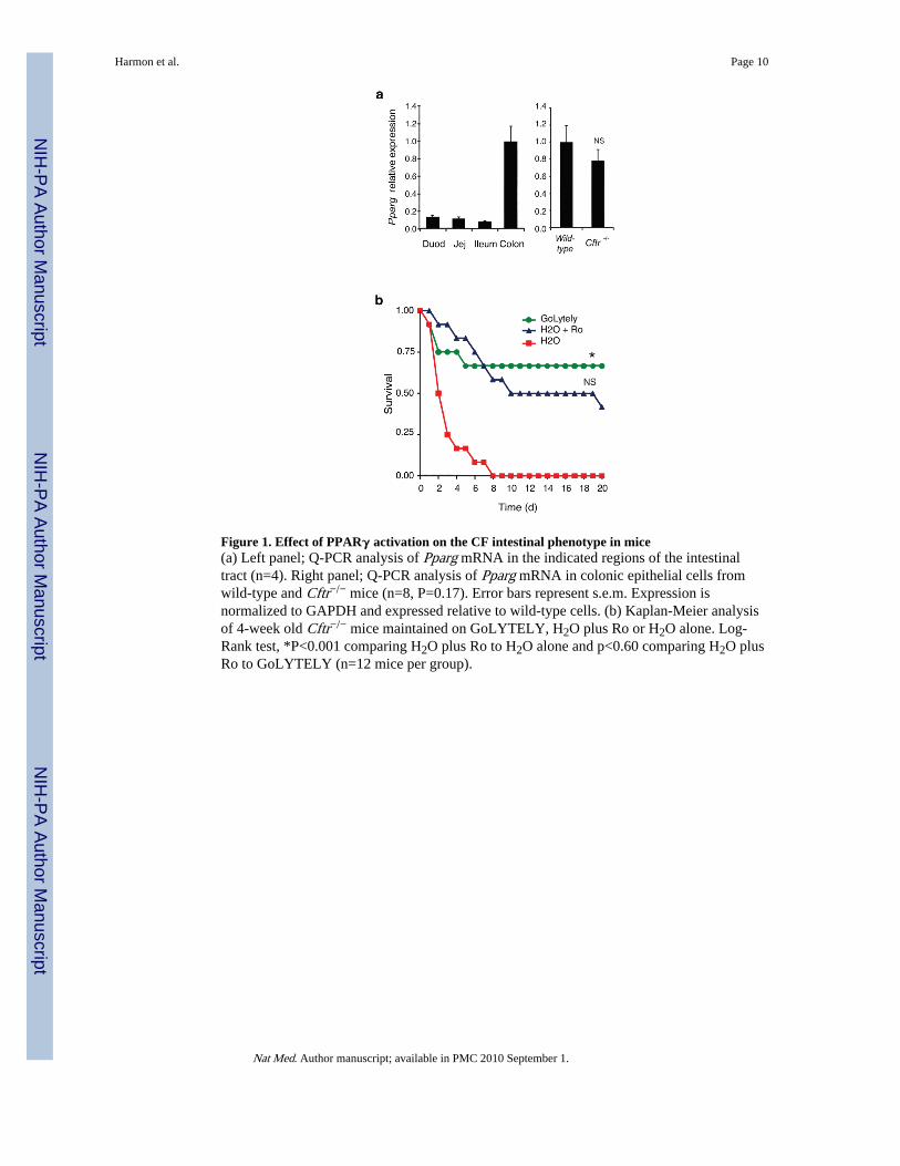
Figure 1. Effect of PPARγ activation on the CF intestinal phenotype in mice(a) Left panel; Q-PCR analysis of Pparg mRNA in the indicated regions of the intestinaltract (n=4). Right panel; Q-PCR analysis of Pparg mRNA in colonic epithelial cells fromwild-type and Cftr−/− mice (n=8, P=0.17). Error bars represent s.e.m. Expression isnormalized to GAPDH and expressed relative to wild-type cells. (b) Kaplan-Meier analysisof 4-week old Cftr−/− mice maintained on GoLYTELY, H2O plus Ro or H2O alone. Log-Rank test, *P<0.001 comparing H2O plus Ro to H2O alone and p<0.60 comparing H2O plusRo to GoLYTELY (n=12 mice per group).
Harmon et al. Page 10
Nat Med. Author manuscript; available in PMC 2010 September 1.
NIH
-PA Author Manuscript
NIH
-PA Author Manuscript
NIH
-PA Author Manuscript
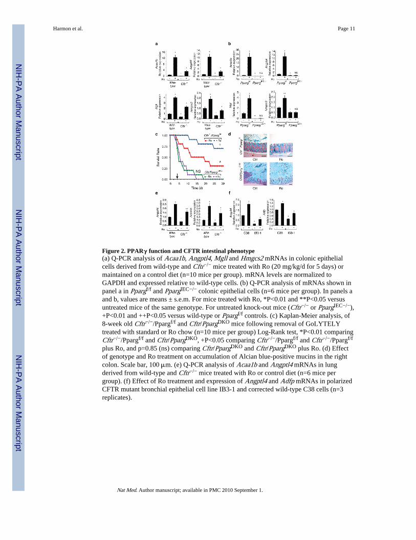
Figure 2. PPARγ function and CFTR intestinal phenotype(a) Q-PCR analysis of Acaa1b, Angptl4, Mgll and Hmgcs2 mRNAs in colonic epithelialcells derived from wild-type and Cftr−/− mice treated with Ro (20 mg/kg/d for 5 days) ormaintained on a control diet (n=10 mice per group). mRNA levels are normalized toGAPDH and expressed relative to wild-type cells. (b) Q-PCR analysis of mRNAs shown inpanel a in Ppargf/f and PpargIEC−/− colonic epithelial cells (n=6 mice per group). In panels aand b, values are means ± s.e.m. For mice treated with Ro, *P<0.01 and **P<0.05 versusuntreated mice of the same genotype. For untreated knock-out mice (Cftr−/− or PpargIEC−/−),+P<0.01 and ++P<0.05 versus wild-type or Ppargf/f controls. (c) Kaplan-Meier analysis, of8-week old Cftr−/−/Ppargf/f and Cftr/PpargDKO mice following removal of GoLYTELYtreated with standard or Ro chow (n=10 mice per group) Log-Rank test, *P<0.01 comparingCftr−/−/Ppargf/f and Cftr/PpargDKO, +P<0.05 comparing Cftr−/−/Ppargf/f and Cftr−/−/Ppargf/f
plus Ro, and p=0.85 (ns) comparing Cftr/PpargDKO and Cftr/PpargDKO plus Ro. (d) Effectof genotype and Ro treatment on accumulation of Alcian blue-positive mucins in the rightcolon. Scale bar, 100 μm. (e) Q-PCR analysis of Acaa1b and Angptl4 mRNAs in lungderived from wild-type and Cftr−/− mice treated with Ro or control diet (n=6 mice pergroup). (f) Effect of Ro treatment and expression of Angptl4 and Adfp mRNAs in polarizedCFTR mutant bronchial epithelial cell line IB3-1 and corrected wild-type C38 cells (n=3replicates).
Harmon et al. Page 11
Nat Med. Author manuscript; available in PMC 2010 September 1.
NIH
-PA Author Manuscript
NIH
-PA Author Manuscript
NIH
-PA Author Manuscript
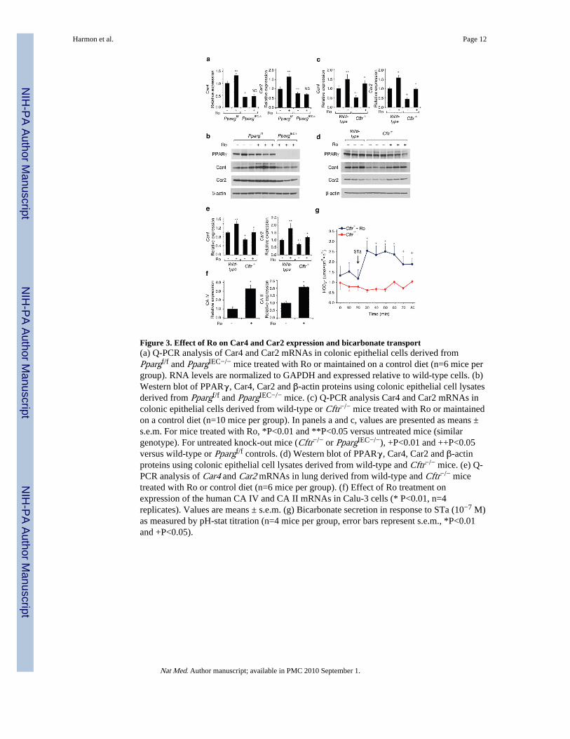
Figure 3. Effect of Ro on Car4 and Car2 expression and bicarbonate transport(a) Q-PCR analysis of Car4 and Car2 mRNAs in colonic epithelial cells derived fromPpargf/f and PpargIEC−/− mice treated with Ro or maintained on a control diet (n=6 mice pergroup). RNA levels are normalized to GAPDH and expressed relative to wild-type cells. (b)Western blot of PPARγ, Car4, Car2 and β-actin proteins using colonic epithelial cell lysatesderived from Ppargf/f and PpargIEC−/− mice. (c) Q-PCR analysis Car4 and Car2 mRNAs incolonic epithelial cells derived from wild-type or Cftr−/− mice treated with Ro or maintainedon a control diet (n=10 mice per group). In panels a and c, values are presented as means ±s.e.m. For mice treated with Ro, *P<0.01 and **P<0.05 versus untreated mice (similargenotype). For untreated knock-out mice (Cftr−/− or PpargIEC−/−), +P<0.01 and ++P<0.05versus wild-type or Ppargf/f controls. (d) Western blot of PPARγ, Car4, Car2 and β-actinproteins using colonic epithelial cell lysates derived from wild-type and Cftr−/− mice. (e) Q-PCR analysis of Car4 and Car2 mRNAs in lung derived from wild-type and Cftr−/− micetreated with Ro or control diet (n=6 mice per group). (f) Effect of Ro treatment onexpression of the human CA IV and CA II mRNAs in Calu-3 cells (* P<0.01, n=4replicates). Values are means ± s.e.m. (g) Bicarbonate secretion in response to STa (10−7 M)as measured by pH-stat titration (n=4 mice per group, error bars represent s.e.m., *P<0.01and +P<0.05).
Harmon et al. Page 12
Nat Med. Author manuscript; available in PMC 2010 September 1.
NIH
-PA Author Manuscript
NIH
-PA Author Manuscript
NIH
-PA Author Manuscript
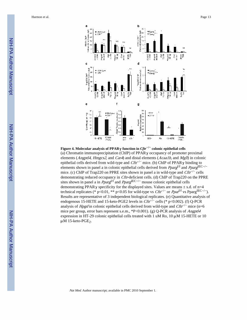
Figure 4. Molecular analysis of PPARγ function in Cftr−/− colonic epithelial cells(a) Chromatin immunoprecipitation (ChIP) of PPARγ occupancy of promoter proximalelements (Angptl4, Hmgcs2, and Car4) and distal elements (Acaa1b, and Mgll) in colonicepithelial cells derived from wild-type and Cftr−/− mice. (b) ChIP of PPARγ binding toelements shown in panel a in colonic epithelial cells derived from Ppargf/f and PpargIEC−/−
mice. (c) ChIP of Trap220 on PPRE sites shown in panel a in wild-type and Cftr−/− cellsdemonstrating reduced occupancy in Cftr-deficient cells. (d) ChIP of Trap220 on the PPREsites shown in panel a in Ppargf/f and PpargIEC−/− mouse colonic epithelial cellsdemonstrating PPARγ specificity for the displayed sites. Values are means ± s.d. of n=4technical replicates (* p<0.01, ** p<0.05 for wild-type vs Cftr−/− or Pparf/f vs PpargIEC−/−).Results are representative of 3 independent biological replicates. (e) Quantitative analysis ofendogenous 15-HETE and 15-keto-PGE2 levels in Cftr−/− cells (* p<0.002). (f) Q-PCRanalysis of Hpgd in colonic epithelial cells derived from wild-type and Cftr−/− mice (n=6mice per group, error bars represent s.e.m., *P<0.001). (g) Q-PCR analysis of Angptl4expression in HT-29 colonic epithelial cells treated with 1 uM Ro, 10 μM 15-HETE or 10μM 15-keto-PGE2.
Harmon et al. Page 13
Nat Med. Author manuscript; available in PMC 2010 September 1.
NIH
-PA Author Manuscript
NIH
-PA Author Manuscript
NIH
-PA Author Manuscript