Characterization of the two PPAR target genes FIAF ... - WUR eDepot
136
Characterization of the two PPAR target genes FIAF (Fasting-Induced Adipose Factor) and G0S2 (G0/G1 switch gene 2) Fokko J. Zandbergen
-
Upload
khangminh22 -
Category
Documents
-
view
0 -
download
0
Transcript of Characterization of the two PPAR target genes FIAF ... - WUR eDepot

Characterization of the two PPAR target genes FIAF (Fasting-Induced Adipose Factor) and
G0S2 (G0/G1 switch gene 2)
Fokko J. Zandbergen

Promotor Prof. dr. M.R. Müller Hoogleraar Voeding, Metabolisme & Genomics Afdeling Humane Voeding, Wageningen Universiteit Co-promotor Dr. ir. A.H. Kersten Universitair docent, Afdeling Humane Voeding, Wageningen Universiteit Promotiecommissie Dr. ir. K. Willems van Dijk Leids Universitair Medisch Centrum, Universiteit Leiden Prof. dr. M.H. Hofker Universiteit Maastricht Dr. ir. E.J.M. Feskens Wageningen Universiteit Prof dr. S.C. de Vries Wageningen Universiteit Dit onderzoek is uitgevoerd binnen de onderzoeksschool VLAG (Voeding, Levensmiddelentechnologie, Agrobiotechnologie en Gezondheid)
Characterization of the two PPAR target genes FIAF (Fasting-Induced Adipose Factor) and
G0S2 (G0/G1 switch gene 2)
Fokko J. Zandbergen
Proefschrift
ter verkrijging van de graad van doctor
op gezag van de rector magnificus
van Wageningen Universiteit,
Prof. dr. M.J. Kropff,
in het openbaar te verdedigen
op maandag 12 juni 2006
des namiddags te half twee in de Aula.

Fokko J. Zandbergen (2006) Characterization of the two PPAR target genes FIAF (Fasting-Induced Adipose Factor) and G0S2 (G0/G1 switch gene 2) Thesis Wageningen University, Wageningen, The Netherlands With abstract – with references – with summary in Dutch ISBN: 90-8504-392-1
Abstract The prevalence of obesity has increased dramatically over the last decades. Obesity, defined as excess body fat, develops if energy expenditure is lower than its intake and if the surplus energy is stored in adipose tissue as fat. Excess adipose tissue, especially around the waist, is associated with an increased risk for diseases such as type 2 diabetes and atherosclerosis. These disorders are major causes of death from cardiovascular disease in the Western world.
Common features of obesity, atherosclerosis and diabetes are insulin resistance and elevated plasma levels of triglycerides (TG) and low-density lipoprotein (LDL) cholesterol, whereas high-density lipoprotein (HDL) cholesterol is decreased.
A number of the pharmacological interventions to treat early stages of atherosclerosis and type 2 diabetes target the peroxisome proliferator-activated receptors (PPARs). Activation of these transcription factors results in the expression of a variety of target genes, many of which play important roles in lipid metabolism. There are three PPAR isoforms: PPARα, PPARβ or PPARδ, and PPARγ. Synthetic ligands for PPARα and for PPARγ decrease plasma TG levels and lower the concentration of LDL-cholesterol in blood whereas they elevate plasma HDL-cholesterol levels. Linked to their hypolipidaemic effect, they may also have hypoglycaemic effects, reducing chronically elevated insulin signalling and associated insulin resistance, which predisposes to the development of type 2 diabetes.
In an effort to gain more insight into the relationship between PPAR target gene expression and its beneficial effect on lipid metabolism with regard to atherosclerosis and type 2 diabetes, the expression of genes in liver of wild-type mice and mice that lack functional PPARα was compared during fasting. Among the genes that were found to be differentially regulated in the wild-type and the PPARα mice, were both the fasting-induced adipose factor (FIAF) and the G0/G1 switch gene 2 (G0S2) strongly up-regulated in the wild-type mice during fasting.
The research described in this thesis focuses on the characterization and elucidation of the function of these two genes and their protein products. FIAF belongs to the family of fibrinogen/angiopoietin-like proteins and was previously found to be highly expressed in adipose tissue and to be up-regulated in response to fasting, hence its name. For G0S2, which was also highly expressed in adipose tissue and which we found to be localized to the endoplasmic reticulum (ER), no homologous genes could be found. During adipogenesis, the differentiation of pre-adipocytes into fully differentiated adipocytes, the levels of mRNA and protein for FIAF and G0S2 were greatly up-regulated. Subsequent experiments indicated that G0S2 is a direct PPARγ and probable PPARα target gene with a functional PPRE (PPAR-responsive element) in its promoter. Using the same approach, a functional PPRE was found within intron 3 of the FIAF gene, establishing FIAF as being a direct PPAR target gene too.
The up-regulation of G0S2 mRNA during the differentiation of adipocytes seemed to be specific for adipogenesis, no up-regulation of G0S2 mRNA was observed during osteogenesis or myogenesis. Furthermore, G0S2 expression was associated with cell cycle arrest in 3T3-L1 pre-adipocytes, which is required for the differentiation of these cells into adipocytes. This indicates that G0S2 may be involved in adipocyte differentiation.
Further investigation showed that FIAF was present as the native protein and in truncated forms in both mouse and human blood plasma. Interestingly, truncated FIAF was produced by human liver and treatment with PPARα agonist markedly increased plasma levels of truncated FIAF, but not native FIAF, in humans. The levels of both truncated and native FIAF showed marked inter-individual variation but were not associated with body mass index and were not influenced by prolonged semistarvation.

To determine the physiological role of FIAF, we studied the effect of FIAF overexpression in a transgenic mouse model (FIAF-tg mice). The transgenic mice had markedly reduced adipose tissue stores compared to their wild-type littermates, despite similar food intake. The FIAF-tg mice also had elevated plasma levels of TG, glycerol, free fatty acids (FFA), and HDL as well as very low-density (VLDL) cholesterol. The increase of plasma TG levels was attributable to elevated VLDL levels. Oral lipid loading showed that the FIAF-tg mice had severely impaired plasma TG clearance. The effects on plasma TG levels are most likely the result of FIAF-mediated inhibition of the activity of lipoprotein lipase (LPL), a key regulator of plasma TG clearance. The elevated levels of FFA and glycerol are indicative of increased lipolysis, a notion supported by the increased expression level of adipose triglyceride lipase (ATGL) in the adipose tissue of FIAF-tg mice. Additional genes that were differentially expressed are involved in oxidative metabolism and uncoupling, which might explain the decreased weight of the FIAF-tg mice while their food intake was similar to that of their wild-type littermates. The elevated HDL levels might be the result of FIAF-mediated inhibition of other lipases in addition to LPL, e.g. endothelial and hepatic lipase (EL/HL).
Interestingly, after fractionation of mouse plasma by FPLC, the full length form of FIAF was present specifically in the HDL-containing fractions, whereas the truncated form of FIAF was specifically present in the LDL-containing fractions. In human plasma, both full length and truncated FIAF were only present in the HDL-containing fractions. In addition, the levels of truncated FIAF and HDL-cholesterol in human plasma correlated positively. Combined with our earlier finding that treatment with synthetic PPARα ligand increased the plasma levels of truncated FIAF in humans, this raises the possibility that FIAF might be involved in the mechanism by which PPARα ligand treatment increases HDL-cholesterol levels in humans, resulting in a protective effect on atherosclerosis.
The up-regulation of FIAF during fasting and the ability to inhibit plasma TG clearance indicate that FIAF might play an important role in repartitioning TG from adipose tissue to other tissues under circumstances of energy shortage. In addition, alterations in FIAF signalling might be involved in dyslipidemia, the presence of abnormal lipid levels in the blood. FIAF thus forms an interesting candidate for therapeutic targeting of dyslipidemia.
Contents Chapter 1 General introduction 9 Chapter 2 Review - FIAF/ANGPTL4: a potential target for dyslipidemia? 21 Future Lipidology 1(2), 227-236 (2006) Chapter 3 The direct peroxisome proliferator-activated receptor target fasting-induced adipose factor 35 (FIAF/PGAR/ANGPTL4) is present in blood plasma as a truncated protein that is increased by fenofibrate treatment Journal of Biological Chemistry 279, 34411-34420 (2004) Chapter 4 The G0/G1 switch gene 2 is a novel PPAR target gene 55 Biochemical Journal 392, 313-324 (2005) Chapter 5 The fasting-induced adipose factor/angiopoietin-like protein 4 is physically 77 associated with lipoproteins and governs plasma lipid levels and adiposity Journal of Biological Chemistry 281, 934-944 (2006) Chapter 6 Recombinant FIAF: Expression in and purification from insect cells 97 Chapter 7 General discussion 119 Samenvatting 127 Dankwoord 131 About the author 134 Education statement of the Graduate school VLAG 135

General introduction
9
CHAPTER 1 General introduction This thesis focuses on the function of two different proteins, and mainly addresses their role in the regulation of lipid metabolism. The main topic of the studies described here is the fasting-induced adipose factor (FIAF). In mice FIAF is mainly expressed in adipose tissue and is secreted into the circulation. Its expression is regulated by peroxisome proliferator-activated receptors (PPARs) [1,2]. PPARs are transcription factors that regulate the expression of numerous genes, many of which play important roles in energy metabolism [3]. From the work described in this thesis, FIAF was established as a direct PPAR target gene (Chapter 3). Moreover, FIAF appears to be an important determinant of adipose tissue size and of lipid levels in the blood (Chapter 5). Indeed, alterations in FIAF signalling might be involved in dyslipidemia, the presence of abnormal lipid levels in the blood. FIAF thus forms an interesting candidate for targeting dyslipidemia (Chapter 2). The second topic of this thesis is the G0/G1 switch gene 2 (G0S2), which was also established as a direct PPAR target gene. Furthermore, G0S2 expression was found to be associated with cell cycle arrest in preadipocytes, which is required for the differentiation of these cells into fat cells, or adipocytes (Chapter 4). The current chapter serves as an introduction to the physiology and underlying molecular biology of lipid metabolism and to underscore the importance of its proper regulation. It aims to place the work described in the following chapters into that perspective. Obesity, diabetes and cardiovascular disease - the metabolic syndrome Obesity
Mankind has witnessed a dramatic increase in the number of obese people over the last decades [4-7]. This increase is strongly associated with an increased abundance of highly palatable foods and with the rise in sedentary jobs and life-styles [8-10]. In parts of the world where these characteristics of technologically advanced societies are less prevalent, obesity is in general not a significant public health problem [11]. Obesity, defined as excess body fat (>25% in men, >35% in women) [12], develops if energy expenditure is lower than its intake and if the surplus energy is stored in adipose tissue as fat. Excess adipose tissue, especially around the waist, is considered to be the strongest risk factor for the metabolic syndrome [13,14]. The metabolic syndrome
The metabolic syndrome is defined as a constellation of interrelated risk factors of metabolic origin that in addition to obesity includes hypertension, atherogenic dyslipidemia, insulin resistance, and a proinflammatory and prothrombotic state [15]. Atherogenic dyslipidemia predisposes strongly to atherosclerosis, which is characterized by the progressive accumulation of lipid depositions in the artery wall, leading to an increased risk of developing atherosclerotic cardiovascular disease. Atherogenic dyslipidemia is characterized by elevated serum triglyceride (TG) and apolipoprotein B (apoB) levels, increased levels of small dense low-density lipoprotein (LDL) particles, and decreased high-density lipoprotein (HDL) cholesterol (HDL-C) levels. An increase of serum triglycerides is also called hypertriglyceridemia and is defined as plasma TG levels exceeding 150 mg/dl [14]. A recent survey estimated that approximately 30% of the US
CHAPTER 1
10
adult population exhibits hypertriglyceridemia [16] and that almost a quarter has the metabolic syndrome. People with the metabolic syndrome are at increased risk of developing atherosclerotic cardiovascular disease and type 2 diabetes [17,18], although another theory holds that insulin resistance is the primary cause of the metabolic syndrome [19]. Type 2 diabetes and atherosclerosis are major causes of death from cardiovascular disease in the Western world [20,21].
Several organisations have listed overlapping criteria for the clinical diagnosis of the metabolic syndrome (reviewed in [22]). In 2001, the Adult Treatment Panel III (ATPIII) of the National Cholesterol Education Program (NCEP) defined the diagnosis of the metabolic syndrome as having three or more of the following: Increased waist circumference (≥102 cm in men and ≥88 cm in women), indicating central obesity, elevated TG (≥150 mg/dl or 1.69 mmol/l), decreased HDL-C (<40 mg/dl or 1.04 mmol/l for men, <50 mg/dl or 1.29 mmol/l for women), elevated blood pressure (≥ 130/85 mm Hg) or on active treatment for hypertension, fasting glucose levels ≥100 mg/dl (5.6 mmol/l) [14,23].
It has been recognized for a long time that the risk factors of the metabolic syndrome often cluster together [24], but it is not completely clear how they are connected. Obesity has been implicated in the development of insulin resistance via the increased release of non-esterified fatty acids (NEFAs) into the circulation [25]. NEFAs are derived from lipolysis of adipose tissue TGs and are the primary source of energy under fasting conditions. The increased release of NEFAs in obese people can lead to excessive accumulation of fat in muscles and in the liver. Several mechanisms whereby increased fatty acids in these tissues could cause insulin resistance have been put forward [26-31], but have not been fully elucidated yet. Insulin resistance in muscle predisposes to hyperglycemia and a reduced sensitivity of the liver to insulin allows for increased gluconeogenesis, the production of glucose from various precursors, which enhances the hyperglycemia even further. Under fasting conditions, gluconeogenesis maintains plasma glucose concentrations and is responsible for providing adequate amounts of glucose to organs that rely on this substrate for their energy supply, predominantly the brain. Under fed conditions, the plasma insulin concentration increases in response to elevated plasma glucose levels in order to lower plasma glucose via enhanced uptake of glucose by peripheral tissues. The metabolic syndrome and cardiovascular disease - atherosclerosis
Besides possibly causing insulin resistance, an increase in liver fat, originating from adipose tissue-derived NEFAs, also promotes the formation of very low-density lipoprotein (VLDL) particles [32]. The higher NEFAs plasma levels in obesity could lead to an over-production of VLDL particles, which would lead to higher plasma TG levels and might generate a higher flux to atherogenic LDL particles in the plasma or into the artery wall, providing a basis for the association between obesity and atherosclerosis.
Hypertriglyceridemia and hyperglycemia are independent risk factors for cardiovascular disease (CVD) [33-36], and the importance of the role of inflammation in the development of atherosclerosis and CVD is increasingly recognized [37,38]. The mechanisms by which they could directly affect CVD are not entirely clear and are the subject of intense investigation. Probable mechanisms by which hyperglycemia is involved in the development of atherosclerosis are non-enzymatic glycosylation of proteins and lipids, resulting in so-called advanced glycosylation end products (AGE), increased oxidative stress, and activation of protein kinase C (PKC) with subsequent alteration in growth factor expression [39]. Both hypertriglyceridemia and hyperglycemia have recently been shown to be linked to the integrity of the endothelial glycocalyx, the layer of proteoglycans, glycoproteins and absorbed plasma proteins that covers and protects the vascular endothelium [40,41], but the mechanism by which the glycocalyx is
General introduction
11
diminished under hyperglycaemic and hyperinsulinaemic states remains to be elucidated. Hyperinsulinaemia might also indirectly inflict damage to the blood vessel walls by causing hypertension [42]. Lipid transport and metabolism Lipoprotein lipase and the triglyceride rich lipoproteins
As already mentioned before, an important mechanism contributing to the hypertriglyceridemia in obesity is probably the elevated VLDL production by the liver, driven by elevated levels of plasma NEFA’s [32]. Triglycerides from the TG-rich lipoproteins VLDL and chylomicrons are cleared from the circulation through the action of lipoprotein lipase (LPL), a key regulatory enzyme in lipid metabolism that is produced in muscle and adipose tissue and subsequently translocates to the site of vascular endothelial cells that faces the blood vessel lumen [43,44]. Chylomicrons originate from the intestine after a meal and transport the TGs absorbed from the diet towards the liver. During their transport, they are rapidly decreasing in size as LPL hydrolyzes the TGs, making the resulting free fatty acids (FFA) or NEFAs available for uptake by adipose tissue and muscle. The FFA are subsequently re-esterified into TG in adipose tissue, where they are stored in lipid droplets, whereas in muscle they are preferentially oxidized to generate energy. Similarly, the TG from VLDL particles are hydrolyzed by LPL, resulting in the formation of smaller IDL and LDL particles.
Not surprisingly, decreased LPL activity leads to higher plasma TG levels [45,46]. The TG-clearing effect of LPL is under tight control of several lipoprotein-associated proteins. Apolipoprotein CII and AV (apoCII and apoAV) both have a stimulating effect [47,48], whereas apolipoprotein CI and CIII (apoCI and apoCIII) both have an inhibitory effect on VLDL-TG clearance. The hypertriglyceridaemic effect of apoCI has been recently found to be caused by inhibition of LPL-mediated TG hydrolysis, whereas apoCIII in addition increases plasma triglycerides by blocking the recognition of TG-rich particles by receptors for VLDL, preventing whole particle uptake which is also known as the ligand or bridging function of LPL [49,50]. HDL
The increased plasma TG levels in hypertriglyceridaemic states often coincide with lowered plasma levels of HDL. This decrease is caused by the exchange of TG from VLDL with cholesterol esters from HDL, mediated by the action of cholesteryl ester transfer protein (CETP) [51]. HDL is strongly associated with protective effects on atherosclerosis [52,53]. In particular its proposed role in reverse cholesterol transport has been the subject of intense research [54]. Recently, it has been demonstrated that therapy with HDL or reconstituted HDL can limit the progression of atherosclerosis in both animals and human [55,56]. The role of adipose tissue, an endocrine organ, in the metabolic syndrome The majority of researchers studying the metabolic syndrome consider obesity to be the strongest risk factor for the metabolic syndrome, implying that adipose tissue plays an important role in its development. Until recently, adipose tissue was mainly regarded as serving to store excess energy in the form of fat. However, over the last decade it has become increasingly clear that adipose also plays an important role in systemic energy homeostasis. Indeed, adipose tissue communicates actively with other tissues in the body via secreted signalling factors that are called adipocytokines
CHAPTER 1
12
or adipokines [57,58]. Several of these adipocytokines have been reported to be involved in glucose metabolism and insulin sensitivity. Dysregulation of the delicate signalling network between adipose tissue and other tissues, for instance because of the secretion of abnormal amounts of adipocytokines, can result in metabolic disturbances like obesity and insulin resistance.
Leptin is probably the best known adipocytokine [59]. In addition to leptin, the secretion of inflammatory cytokines like TNFα and IL6 is also elevated in obese persons [60]. Elevated plasma levels of TNFα and IL6 may be associated with increased risk for cardiovascular disease [61,62]. Recently, visfatin, predominantly expressed in intra-abdominal adipose tissue, has been added to the repertoire of adipocytokines [63]. Visfatin is increased in obesity and, unexpectedly, has properties similar to that of insulin. Another factor that is secreted by adipocytes and that has received a lot of attention is adiponectin (Acrp30, AdipoQ) [64]. Adiponectin, plasma levels of which are decreased in obese individuals [65], increases insulin sensitivity in skeletal muscle, probably by activating AMP-activated protein kinase (AMPK), an important mediator in glucose metabolism [66,67]. Resistin, also an adipocytokine, on the other hand increases with obesity and decreases insulin sensitivity in mice [68,69].
Although the molecular biology underlying the association between increased adipose mass and insulin resistance is becoming increasingly clear, much less is known about the relation between adipocytokines and TG metabolism. Acylation-stimulating protein (ASP), which stimulates uptake and storage of plasma TG in adipocytes, is one of the few adipocytokines that are known to affect TG metabolism [70]. PPARs and the metabolic syndrome The expression level of several adipocytokines is under transcriptional control of peroxisome proliferator-activated receptors (PPARs). PPARs are transcription factors that belong to the family of nuclear hormone receptors, members of which are activated by a variety of compounds that are derived from food, e.g. retinoic acids, vitamin D and fatty acids [71]. There are three isoforms of PPARs: PPARα, PPARβ/PPARδ, and PPARγ. Each of them is expressed in various organs at different levels [72,73]. PPARα is mainly found in liver, skeletal muscle, kidney, heart and the vascular wall. This PPAR isotype has been well-studied with regard to its central function in hepatic fatty acid catabolism [74,75]. In addition, PPARα has more recently been implicated in glucose and amino acid metabolism [76,77]. PPARβ/δ is ubiquitously expressed and has been found to stimulate fatty acid oxidation in both adipose tissue and skeletal muscle [78,79], and to regulate hepatic VLDL production and catabolism [80]. PPARγ is predominantly found in adipose tissue and is known as the transcription factor that drives adipocyte differentiation, or adipogenesis [81,82]. The diverse role of PPARγ in adipogenesis includes the regulation of cell-cycle withdrawal [83], as well as induction of expression of fat-specific target genes that are involved in lipogenesis. Indeed, results from microarray studies indicate a general role for PPARγ in the regulation of lipid metabolism [84].
All three PPARs are stimulated by binding of small lipophylic compounds such as polyunsaturated fatty acids and various fatty-acid derived molecules [85,86]. Activated PPARs bind to recognition sequences on the DNA called PPAR-responsive elements (PPREs), resulting in the transcription of PPAR target genes. PPARs are interesting targets from the perspective of pharmacological treatment of the conditions that contribute to the metabolic syndrome, such as insulin resistance [87]. Fibrates are synthetic ligands for PPARα that are prescribed to hyperlipidemic patients to lower plasma triglyceride concentrations as well as to treat cardiovascular disease. Activation of PPARα by fibrates stimulates oxidation of fatty acids in the liver and increases LPL and decreases apoCIII expression, giving rise to decreased plasma TG
General introduction
13
levels [88]. Fibrate treatment also slightly lowers the concentration of LDL-cholesterol in blood whereas it elevates plasma HDL-C levels. The latter effect appears to be related to increased apoAI and apoAII expression, which are target genes of PPARα in the liver and are major constituents of HDL [89,90]. Possibly connected to their hypolipidemic effect, the fenofibrates may also have hypoglycemic effects, reducing chronically elevated insulin levels and associated insulin resistance. Another group of drugs called thiazolidinediones (TZDs) activate PPARγ and are mainly utilized in the therapeutic treatment of obesity-linked type 2 diabetes. The TZD-mediated activation of PPARγ stimulates the fat cells to differentiate and take up lipids from the circulation. The resulting decrease in plasma TG and NEFAs probably decreases the amount of NEFAs that would otherwise ‘spill over’ in skeletal muscle and the liver and have a lipotoxic effect in these tissues, which may be responsible for the development of insulin resistance. In addition, the TZD-mediated decrease in plasma NEFAs may also reduce gluconeogenesis in the liver under fed conditions. Both of these effects of TZD treatment probably account for their beneficial effect on type 2 diabetes, although other mechanisms are still under investigation. FIAF and G0S2 – PPAR target genes G0S2 – a PPAR target gene involved in adipogenesis
Part of the research presented in this thesis focuses on the function of the G0/G1 switch gene 2 (G0S2) (Chapter 4). G0S2 was first identified as a novel PPAR target gene by comparison of liver mRNAs of wild-type and PPARα-null mice using microarrays. The expression of G0S2 in the liver was up-regulated by fasting and by a synthetic PPARα ligand in wild-type but not in PPARα-null mice. The G0S2 mRNA level was highest in brown and white adipose tissue and was greatly up-regulated during adipogenesis. Transactivation, gel shift and chromatin immunoprecipitation assays indicated that G0S2 is a direct PPARγ and probable PPARα target gene with a functional PPRE (PPAR-responsive element) in its promoter. The up-regulation of G0S2 mRNA during the differentiation of adipocytes seemed to be specific for adipogenesis, since no up-regulation of G0S2 mRNA was observed during osteogenesis or myogenesis. In 3T3-L1 pre-adipocytes, expression of G0S2 was associated with growth arrest, which is required for 3T3-L1 adipogenesis. Together, the data described in Chapter 4 indicate that G0S2 is a novel target gene of PPARs that may be involved in adipocyte differentiation. FIAF – a PPAR target gene involved in lipid metabolism
Reviewed in chapter 2, the fasting-induced adipose factor (FIAF) was first described in the year 2000 and is also known as PPARγ angiopoietin-related protein (PGAR), Hepatic Fibrinogen / Angiopoietin-Related Protein (HFARP), and Angiopoietin-like protein 4 (ANGPTL4) [1,2,91,92]. The secreted glycoprotein of ~50 kDa belongs to the family of fibrinogen/angiopoietin-like proteins and, at least in mice, is most highly expressed in white and brown adipose tissue and to a lesser extent in other tissues such as heart, skeletal muscle, and liver [1,2] (Chapter 2). FIAF was first identified as a target gene of PPARα and PPARγ [1,2], and was found to be up-regulated by fasting and during adipogenesis. These findings led to the hypothesis that FIAF might be involved in the regulation of lipid metabolism. Subsequent studies have shown that FIAF potently elevates plasma triglyceride (TG) levels [92-94], probably by inhibiting LPL [92,95,96]. In addition to lipid metabolism, FIAF has also been associated with angiogenesis [97,98]. However, its role in angiogenesis remains ambiguous as both pro-angiogenic and anti-angiogenic effects have been observed for FIAF [98,99].
CHAPTER 1
14
To further characterize FIAF, the regulation of FIAF mRNA and protein was studied in liver and adipose cell lines as well as in human and mouse plasma (Chapter 3). The expression of FIAF mRNA was up-regulated in response to PPAR agonists. Furthermore, transactivation, chromatin immunoprecipitation, and gel shift experiments identified a functional PPRE within intron 3 of the FIAF gene, establishing FIAF as being a direct PPAR target gene. In human and mouse blood plasma, FIAF was found to be present both as the native protein and in a truncated form. Interestingly, the ratio in which these forms could be detected varied per tissue. Truncated FIAF was produced by human liver and treatment with the PPARα agonist fenofibrate markedly increased plasma levels of truncated FIAF, but not native FIAF, in humans. Levels of both truncated and native FIAF showed marked inter-individual variation but were not associated with body mass index and were not influenced by prolonged semistarvation. Together, these data suggest that FIAF, similar to other adipocytokines, may partially exert its function via a truncated form.
To determine the physiological role of FIAF, we studied the effect of FIAF over-expression in a transgenic mouse model (FIAF-tg mice, see chapter 5) characterized by elevated FIAF expression in peripheral tissues. The FIAF-tg mice had markedly reduced adipose tissue stores compared to their wild type littermates, despite similar food intake. The FIAF-tg mice also had elevated plasma levels of TG, glycerol, FFA, and HDL as well as VLDL-cholesterol. The increase of fasting plasma TG levels was attributable to elevation of VLDL levels and a good explanation of this observation is provided by the reported dose-dependent inhibition of the activity of LPL in an in vitro assay [92]. Indeed, oral lipid loading demonstrated that the FIAF-tg mice had severely impaired plasma TG clearance, likely the result of inhibited LPL activity. The elevated levels of FFA and glycerol are indicative of increased lipolysis. This notion was supported by increased expression levels of genes involved in oxidative metabolism and lipolysis in the adipose tissue of FIAF-tg mice. It could be speculated that the elevated HDL may be caused by the possible inhibition by FIAF of other lipases in addition to LPL, e.g. endothelial and hepatic lipase (EL/HL).
Interestingly, the full length form of FIAF was present specifically in the HDL-containing FPLC fractions of mouse plasma, whereas the truncated form of FIAF was specifically present in the LDL-containing fractions. In human plasma, both full length and truncated FIAF were only present in the HDL-containing fractions. In addition, the levels of truncated FIAF and HDL-C in human plasma correlated positively. Combined with our earlier finding that fibrate treatment increased the plasma levels of truncated FIAF, this raises the possibility that FIAF might be involved in the mechanism by which PPARα agonist treatment increases HDL-C levels in humans, resulting in a protective effect on atherosclerosis. From the background information provided in this introductory chapter, it is clear that the metabolic syndrome and its related diseases are very complex. Study at the molecular level of obesity and its accompanying disorders such as diabetes and cardiovascular disease may proof vital in understanding their relationships. In addition, a better understanding of the underlying molecular mechanisms may offer opportunities to halt the worldwide increase in the prevalence of the metabolic syndrome and its associated ailments. In this perspective, a lot of attention has focused on the role of signalling factors that are secreted from adipose tissue, and that might mechanistically link the development of obesity to type 2 diabetes and cardiovascular disease. Nuclear hormone receptors, with emphasis on PPARs, have received a lot of interest as targets for the treatment of cardiovascular disease and type 2 diabetes. The following chapters describe the properties of two PPAR target genes that are involved in lipid metabolism and that represent interesting targets for the therapeutic intervention in obesity, diabetes type 2, and associated
General introduction
15
atherogenic dyslipidemia, which is characterized by increased plasma TG and decreased levels of HDL. References 1. Kersten, S., Mandard, S., Tan, N. S. et al. (2000) Characterization of the fasting-induced adipose factor
FIAF, a novel peroxisome proliferator-activated receptor target gene. J. Biol. Chem. 275, 28488-28493 2. Yoon, J. C., Chickering, T. W., Rosen, E. D. et al. (2000) Peroxisome proliferator-activated receptor
gamma target gene encoding a novel angiopoietin-related protein associated with adipose differentiation. Mol. Cell. Biol. 20, 5343-5349
3. Kersten, S., Desvergne, B.and Wahli, W. (2000) Roles of PPARs in health and disease. Nature 405, 421-424
4. Flegal, K. M., Carroll, M. D., Kuczmarski, R. J.and Johnson, C. L. (1998) Overweight and obesity in the United States: prevalence and trends, 1960-1994. Int. J. Obes. Relat. Metab. Disord. 22, 39-47
5. Freedman, D. S., Khan, L. K., Serdula, M. K., Galuska, D. A.and Dietz, W. H. (2002) Trends and correlates of class 3 obesity in the United States from 1990 through 2000. Jama 288, 1758-1761
6. Flegal, K. M., Carroll, M. D., Ogden, C. L.and Johnson, C. L. (2002) Prevalence and trends in obesity among US adults, 1999-2000. Jama 288, 1723-1727
7. Okosun, I. S., Chandra, K. M., Boev, A. et al. (2004) Abdominal adiposity in U.S. adults: prevalence and trends, 1960-2000. Prev. Med. 39, 197-206
8. Bowman, S. A.and Vinyard, B. T. (2004) Fast food consumption of U.S. adults: impact on energy and nutrient intakes and overweight status. J. Am. Coll. Nutr. 23, 163-168
9. Hu, F. B., Li, T. Y., Colditz, G. A., Willett, W. C.and Manson, J. E. (2003) Television watching and other sedentary behaviors in relation to risk of obesity and type 2 diabetes mellitus in women. Jama 289, 1785-1791
10. Brownson, R. C., Boehmer, T. K.and Luke, D. A. (2005) Declining rates of physical activity in the United States: what are the contributors? Annu. Rev. Public Health 26, 421-443
11. Grundy, S. M. (2004) Obesity, metabolic syndrome, and cardiovascular disease. J. Clin. Endocrinol. Metab. 89, 2595-2600
12. Report of a WHO Expert Committee (1995) Physical status: the use and interpretation of anthropometry. World Health Organ. Tech. Rep. Ser. 854, 1-452
13. Bosello, O.and Zamboni, M. (2000) Visceral obesity and metabolic syndrome. Obes. Rev. 1, 47-56 14. Third Report of the National Cholesterol Education Program (NCEP) Expert Panel on Detection,
Evaluation, and Treatment of High Blood Cholesterol in Adults (Adult Treatment Panel III) final report (2002) Circulation 106, 3143-3421
15. Grundy, S. M., Brewer, H. B., Jr., Cleeman, J. I., Smith, S. C., Jr.and Lenfant, C. (2004) Definition of metabolic syndrome: Report of the National Heart, Lung, and Blood Institute/American Heart Association conference on scientific issues related to definition. Circulation 109, 433-438
16. Ford, E. S., Giles, W. H.and Dietz, W. H. (2002) Prevalence of the metabolic syndrome among US adults: findings from the third National Health and Nutrition Examination Survey. Jama 287, 356-359
17. Lakka, H. M., Laaksonen, D. E., Lakka, T. A. et al. (2002) The metabolic syndrome and total and cardiovascular disease mortality in middle-aged men. Jama 288, 2709-2716
18. (2001) Executive Summary of The Third Report of The National Cholesterol Education Program (NCEP) Expert Panel on Detection, Evaluation, And Treatment of High Blood Cholesterol In Adults (Adult Treatment Panel III). Jama 285, 2486-2497
19. Reaven, G. (2004) The metabolic syndrome or the insulin resistance syndrome? Different names, different concepts, and different goals. Endocrinol. Metab. Clin. North Am. 33, 283-303
20. McGovern, P. G., Pankow, J. S., Shahar, E. et al. (1996) Recent trends in acute coronary heart disease--mortality, morbidity, medical care, and risk factors. The Minnesota Heart Survey Investigators. N. Engl. J. Med. 334, 884-890
21. Wilhelmsen, L., Rosengren, A., Johansson, S.and Lappas, G. (1997) Coronary heart disease attack rate, incidence and mortality 1975-1994 in Goteborg, Sweden. Eur. Heart J. 18, 572-581
CHAPTER 1
16
22. Grundy, S. M., Cleeman, J. I., Daniels, S. R. et al. (2005) Diagnosis and management of the metabolic syndrome: an American Heart Association/National Heart, Lung, and Blood Institute Scientific Statement. Circulation 112, 2735-2752
23. Genuth, S., Alberti, K. G., Bennett, P. et al. (2003) Follow-up report on the diagnosis of diabetes mellitus. Diabetes Care 26, 3160-3167
24. Reaven, G. M. (1988) Banting lecture 1988. Role of insulin resistance in human disease. Diabetes 37, 1595-1607
25. Heptulla, R., Smitten, A., Teague, B., Tamborlane, W. V., Ma, Y. Z.and Caprio, S. (2001) Temporal patterns of circulating leptin levels in lean and obese adolescents: relationships to insulin, growth hormone, and free fatty acids rhythmicity. J. Clin. Endocrinol. Metab. 86, 90-96
26. Randle, P. J., Garland, P. B., Hales, C. N.and Newsholme, E. A. (1963) The glucose fatty-acid cycle. Its role in insulin sensitivity and the metabolic disturbances of diabetes mellitus. Lancet 1, 785-789
27. Shulman, G. I. (2000) Cellular mechanisms of insulin resistance. J. Clin. Invest. 106, 171-176 28. Ruderman, N. B., Saha, A. K., Vavvas, D.and Witters, L. A. (1999) Malonyl-CoA, fuel sensing, and
insulin resistance. Am. J. Physiol. 276, E1-E18 29. Petersen, K. F.and Shulman, G. I. (2002) Pathogenesis of skeletal muscle insulin resistance in type 2
diabetes mellitus. Am. J. Cardiol. 90, 11G-18G 30. Yu, C., Chen, Y., Cline, G. W. et al. (2002) Mechanism by which fatty acids inhibit insulin activation
of insulin receptor substrate-1 (IRS-1)-associated phosphatidylinositol 3-kinase activity in muscle. J. Biol. Chem. 277, 50230-50236
31. Bergman, R. N., Van Citters, G. W., Mittelman, S. D. et al. (2001) Central role of the adipocyte in the metabolic syndrome. J. Investig. Med. 49, 119-126
32. Barrows, B. R.and Parks, E. J. (2006) Contributions of different fatty acid sources to VLDL-triacylglycerol in the fasted and fed-states. J. Clin. Endocrinol. Metab.
33. Assmann, G., Schulte, H.and von Eckardstein, A. (1996) Hypertriglyceridemia and elevated lipoprotein(a) are risk factors for major coronary events in middle-aged men. Am. J. Cardiol. 77, 1179-1184
34. Hokanson, J. E.and Austin, M. A. (1996) Plasma triglyceride level is a risk factor for cardiovascular disease independent of high-density lipoprotein cholesterol level: a meta-analysis of population-based prospective studies. J. Cardiovasc. Risk 3, 213-219
35. Stamler, J., Vaccaro, O., Neaton, J. D.and Wentworth, D. (1993) Diabetes, other risk factors, and 12-yr cardiovascular mortality for men screened in the Multiple Risk Factor Intervention Trial. Diabetes Care 16, 434-444
36. Laakso, M. (1999) Hyperglycemia and cardiovascular disease in type 2 diabetes. Diabetes 48, 937-942 37. Ross, R. (1999) Atherosclerosis--an inflammatory disease. N. Engl. J. Med. 340, 115-126 38. Libby, P., Ridker, P. M.and Maseri, A. (2002) Inflammation and atherosclerosis. Circulation 105,
1135-1143 39. Aronson, D.and Rayfield, E. J. (2002) How hyperglycemia promotes atherosclerosis: molecular
mechanisms. Cardiovasc. Diabetol. 1, 1 40. Nieuwdorp, M., Meuwese, M. C., Vink, H., Hoekstra, J. B., Kastelein, J. J.and Stroes, E. S. (2005) The
endothelial glycocalyx: a potential barrier between health and vascular disease. Curr. Opin. Lipidol. 16, 507-511
41. van den Berg, B. M., Spaan, J. A., Rolf, T. M.and Vink, H. (2006) Atherogenic region and diet diminish glycocalyx dimension and increase intima-to-media ratios at murine carotid artery bifurcation. Am. J. Physiol. Heart Circ. Physiol. 290, H915-920
42. DeFronzo, R. A.and Ferrannini, E. (1991) Insulin resistance. A multifaceted syndrome responsible for NIDDM, obesity, hypertension, dyslipidemia, and atherosclerotic cardiovascular disease. Diabetes Care 14, 173-194
43. Korn, E. D. (1955) Clearing factor, a heparin-activated lipoprotein lipase. I. Isolation and characterization of the enzyme from normal rat heart. J. Biol. Chem. 215, 1-14
44. Havel, R. J. (1997) Postprandial lipid metabolism: an overview. Proc. Nutr. Soc. 56, 659-666 45. Havel, R. J.and Gordon, R. S., Jr. (1960) Idiopathic hyperlipemia: metabolic studies in an affected
family. J. Clin. Invest. 39, 1777-1790
General introduction
17
46. Weinstock, P. H., Bisgaier, C. L., Aalto-Setala, K. et al. (1995) Severe hypertriglyceridemia, reduced high density lipoprotein, and neonatal death in lipoprotein lipase knockout mice. Mild hypertriglyceridemia with impaired very low density lipoprotein clearance in heterozygotes. J. Clin. Invest. 96, 2555-2568
47. Haberbosch, W., Poli, A., Baggio, G., Fellin, R., Gnasso, A.and Augustin, J. (1984) Apolipoprotein C-II deficiency. The role of apolipoprotein C-II in the hydrolysis of triacylglycerol-rich lipoproteins. Biochim. Biophys. Acta 793, 49-60
48. Merkel, M., Loeffler, B., Kluger, M. et al. (2005) Apolipoprotein AV accelerates plasma hydrolysis of triglyceride-rich lipoproteins by interaction with proteoglycan-bound lipoprotein lipase. J. Biol. Chem. 280, 21553-21560
49. Berbee, J. F., van der Hoogt, C. C., Sundararaman, D., Havekes, L. M.and Rensen, P. C. (2005) Severe hypertriglyceridemia in human APOC1 transgenic mice is caused by apoC-I-induced inhibition of LPL. J. Lipid Res. 46, 297-306
50. Aalto-Setala, K., Fisher, E. A., Chen, X. et al. (1992) Mechanism of hypertriglyceridemia in human apolipoprotein (apo) CIII transgenic mice. Diminished very low density lipoprotein fractional catabolic rate associated with increased apo CIII and reduced apo E on the particles. J. Clin. Invest. 90, 1889-1900
51. Barter, P. J. (2002) Hugh sinclair lecture: the regulation and remodelling of HDL by plasma factors. Atheroscler. Suppl. 3, 39-47
52. Gordon, T., Castelli, W. P., Hjortland, M. C., Kannel, W. B.and Dawber, T. R. (1977) High density lipoprotein as a protective factor against coronary heart disease. The Framingham Study. Am. J. Med. 62, 707-714
53. Despres, J. P., Lemieux, I., Dagenais, G. R., Cantin, B.and Lamarche, B. (2000) HDL-cholesterol as a marker of coronary heart disease risk: the Quebec cardiovascular study. Atherosclerosis 153, 263-272
54. Lewis, G. F.and Rader, D. J. (2005) New insights into the regulation of HDL metabolism and reverse cholesterol transport. Circ. Res. 96, 1221-1232
55. Nissen, S. E., Tsunoda, T., Tuzcu, E. M. et al. (2003) Effect of recombinant ApoA-I Milano on coronary atherosclerosis in patients with acute coronary syndromes: a randomized controlled trial. Jama 290, 2292-2300
56. Nicholls, S. J., Cutri, B., Worthley, S. G. et al. (2005) Impact of short-term administration of high-density lipoproteins and atorvastatin on atherosclerosis in rabbits. Arterioscler. Thromb. Vasc. Biol. 25, 2416-2421
57. Ahima, R. S.and Flier, J. S. (2000) Adipose tissue as an endocrine organ. Trends Endocrinol. Metab. 11, 327-332
58. Trayhurn, P.and Beattie, J. H. (2001) Physiological role of adipose tissue: white adipose tissue as an endocrine and secretory organ. Proc. Nutr. Soc. 60, 329-339
59. Frederich, R. C., Lollmann, B., Hamann, A. et al. (1995) Expression of ob mRNA and its encoded protein in rodents. Impact of nutrition and obesity. J. Clin. Invest. 96, 1658-1663
60. Yudkin, J. S., Stehouwer, C. D., Emeis, J. J.and Coppack, S. W. (1999) C-reactive protein in healthy subjects: associations with obesity, insulin resistance, and endothelial dysfunction: a potential role for cytokines originating from adipose tissue? Arterioscler. Thromb. Vasc. Biol. 19, 972-978
61. Ridker, P. M., Rifai, N., Pfeffer, M., Sacks, F., Lepage, S.and Braunwald, E. (2000) Elevation of tumor necrosis factor-alpha and increased risk of recurrent coronary events after myocardial infarction. Circulation 101, 2149-2153
62. Ridker, P. M., Rifai, N., Stampfer, M. J.and Hennekens, C. H. (2000) Plasma concentration of interleukin-6 and the risk of future myocardial infarction among apparently healthy men. Circulation 101, 1767-1772
63. Fukuhara, A., Matsuda, M., Nishizawa, M. et al. (2005) Visfatin: a protein secreted by visceral fat that mimics the effects of insulin. Science 307, 426-430
64. Scherer, P. E., Williams, S., Fogliano, M., Baldini, G.and Lodish, H. F. (1995) A novel serum protein similar to C1q, produced exclusively in adipocytes. J. Biol. Chem. 270, 26746-26749
65. Arita, Y., Kihara, S., Ouchi, N. et al. (1999) Paradoxical decrease of an adipose-specific protein, adiponectin, in obesity. Biochem. Biophys. Res. Commun. 257, 79-83
CHAPTER 1
18
66. Tomas, E., Tsao, T. S., Saha, A. K. et al. (2002) Enhanced muscle fat oxidation and glucose transport by ACRP30 globular domain: acetyl-CoA carboxylase inhibition and AMP-activated protein kinase activation. Proc. Natl. Acad. Sci. U.S.A. 99, 16309-16313
67. Yamauchi, T., Kamon, J., Minokoshi, Y. et al. (2002) Adiponectin stimulates glucose utilization and fatty-acid oxidation by activating AMP-activated protein kinase. Nat. Med. 8, 1288-1295
68. Steppan, C. M., Bailey, S. T., Bhat, S. et al. (2001) The hormone resistin links obesity to diabetes. Nature 409, 307-312
69. Banerjee, R. R., Rangwala, S. M., Shapiro, J. S. et al. (2004) Regulation of fasted blood glucose by resistin. Science 303, 1195-1198
70. Murray, I., Sniderman, A. D.and Cianflone, K. (1999) Mice lacking acylation stimulating protein (ASP) have delayed postprandial triglyceride clearance. J. Lipid Res. 40, 1671-1676
71. Chawla, A., Repa, J. J., Evans, R. M.and Mangelsdorf, D. J. (2001) Nuclear receptors and lipid physiology: opening the X-files. Science 294, 1866-1870
72. Auboeuf, D., Rieusset, J., Fajas, L. et al. (1997) Tissue distribution and quantification of the expression of mRNAs of peroxisome proliferator-activated receptors and liver X receptor-alpha in humans: no alteration in adipose tissue of obese and NIDDM patients. Diabetes 46, 1319-1327
73. Vidal-Puig, A. J., Considine, R. V., Jimenez-Linan, M. et al. (1997) Peroxisome proliferator-activated receptor gene expression in human tissues. Effects of obesity, weight loss, and regulation by insulin and glucocorticoids. J. Clin. Invest. 99, 2416-2422
74. Mandard, S., Muller, M.and Kersten, S. (2004) Peroxisome proliferator-activated receptor alpha target genes. Cell. Mol. Life Sci. 61, 393-416
75. Kersten, S., Seydoux, J., Peters, J. M., Gonzalez, F. J., Desvergne, B.and Wahli, W. (1999) Peroxisome proliferator-activated receptor alpha mediates the adaptive response to fasting. J. Clin. Invest. 103, 1489-1498
76. Patsouris, D., Mandard, S., Voshol, P. J. et al. (2004) PPARalpha governs glycerol metabolism. J. Clin. Invest. 114, 94-103
77. Kersten, S., Mandard, S., Escher, P. et al. (2001) The peroxisome proliferator-activated receptor alpha regulates amino acid metabolism. Faseb J. 15, 1971-1978
78. Wang, Y. X., Lee, C. H., Tiep, S. et al. (2003) Peroxisome-proliferator-activated receptor delta activates fat metabolism to prevent obesity. Cell 113, 159-170
79. Wang, Y. X., Zhang, C. L., Yu, R. T. et al. (2004) Regulation of muscle fiber type and running endurance by PPARdelta. PLoS Biol. 2, e294
80. Akiyama, T. E., Lambert, G., Nicol, C. J. et al. (2004) Peroxisome proliferator-activated receptor beta/delta regulates very low density lipoprotein production and catabolism in mice on a Western diet. J. Biol. Chem. 279, 20874-20881
81. Tontonoz, P., Hu, E.and Spiegelman, B. M. (1994) Stimulation of adipogenesis in fibroblasts by PPAR gamma 2, a lipid-activated transcription factor. Cell 79, 1147-1156
82. Ren, D., Collingwood, T. N., Rebar, E. J., Wolffe, A. P.and Camp, H. S. (2002) PPARgamma knockdown by engineered transcription factors: exogenous PPARgamma2 but not PPARgamma1 reactivates adipogenesis. Genes Dev. 16, 27-32
83. Morrison, R. F.and Farmer, S. R. (1999) Role of PPARgamma in regulating a cascade expression of cyclin-dependent kinase inhibitors, p18(INK4c) and p21(Waf1/Cip1), during adipogenesis. J. Biol. Chem. 274, 17088-17097
84. Way, J. M., Harrington, W. W., Brown, K. K. et al. (2001) Comprehensive messenger ribonucleic acid profiling reveals that peroxisome proliferator-activated receptor gamma activation has coordinate effects on gene expression in multiple insulin-sensitive tissues. Endocrinology 142, 1269-1277
85. Krey, G., Braissant, O., L'Horset, F. et al. (1997) Fatty acids, eicosanoids, and hypolipidemic agents identified as ligands of peroxisome proliferator-activated receptors by coactivator-dependent receptor ligand assay. Mol. Endocrinol. 11, 779-791
86. Kliewer, S. A., Sundseth, S. S., Jones, S. A. et al. (1997) Fatty acids and eicosanoids regulate gene expression through direct interactions with peroxisome proliferator-activated receptors alpha and gamma. Proc. Natl. Acad. Sci. U.S.A. 94, 4318-4323
General introduction
19
87. Patsouris, D., Muller, M.and Kersten, S. (2004) Peroxisome proliferator activated receptor ligands for the treatment of insulin resistance. Curr. Opin. Investig. Drugs 5, 1045-1050
88. Staels, B., Vu-Dac, N., Kosykh, V. A. et al. (1995) Fibrates downregulate apolipoprotein C-III expression independent of induction of peroxisomal acyl coenzyme A oxidase. A potential mechanism for the hypolipidemic action of fibrates. J. Clin. Invest. 95, 705-712
89. Staels, B.and Auwerx, J. (1998) Regulation of apo A-I gene expression by fibrates. Atherosclerosis 137 Suppl, S19-23
90. Vu-Dac, N., Schoonjans, K., Kosykh, V. et al. (1995) Fibrates increase human apolipoprotein A-II expression through activation of the peroxisome proliferator-activated receptor. J. Clin. Invest. 96, 741-750
91. Kim, I., Kim, H. G., Kim, H. et al. (2000) Hepatic expression, synthesis and secretion of a novel fibrinogen/angiopoietin-related protein that prevents endothelial-cell apoptosis. Biochem. J. 346 Pt 3, 603-610
92. Yoshida, K., Shimizugawa, T., Ono, M.and Furukawa, H. (2002) Angiopoietin-like protein 4 is a potent hyperlipidemia-inducing factor in mice and inhibitor of lipoprotein lipase. J Lipid Res 43, 1770-1772
93. Ge, H., Yang, G., Yu, X., Pourbahrami, T.and Li, C. (2004) Oligomerization state-dependent hyperlipidemic effect of angiopoietin-like protein 4. J. Lipid Res. 45, 2071-2079
94. Xu, A., Lam, M. C., Chan, K. W. et al. (2005) Angiopoietin-like protein 4 decreases blood glucose and improves glucose tolerance but induces hyperlipidemia and hepatic steatosis in mice. Proc. Natl. Acad. Sci. U.S.A. 102, 6086-6091
95. Yu, X., Burgess, S. C., Ge, H. et al. (2005) Inhibition of cardiac lipoprotein utilization by transgenic overexpression of Angptl4 in the heart. Proc. Natl. Acad. Sci. U.S.A. 102, 1767-1772
96. Koster, A., Chao, Y. B., Mosior, M. et al. (2005) Transgenic angiopoietin-like (angptl)4 overexpression and targeted disruption of angptl4 and angptl3: regulation of triglyceride metabolism. Endocrinology 146, 4943-4950
97. Belanger, A. J., Lu, H., Date, T. et al. (2002) Hypoxia up-regulates expression of peroxisome proliferator-activated receptor gamma angiopoietin-related gene (PGAR) in cardiomyocytes: role of hypoxia inducible factor 1alpha. J. Mol. Cell. Cardiol. 34, 765-774
98. Le Jan, S., Amy, C., Cazes, A. et al. (2003) Angiopoietin-like 4 is a proangiogenic factor produced during ischemia and in conventional renal cell carcinoma. Am. J. Pathol. 162, 1521-1528
99. Ito, Y., Oike, Y., Yasunaga, K. et al. (2003) Inhibition of angiogenesis and vascular leakiness by angiopoietin-related protein 4. Cancer Res. 63, 6651-6657

20
Review - FIAF/ANGPTL4: a potential target for dyslipidemia?
21
CHAPTER 2 Review Fasting-induced adipose factor/angiopoietin-like protein 4: a potential target for dyslipidemia? Fokko Zandbergen, Susan van Dijk, Michael Müller, and Sander Kersten Summary Recently, several proteins with homology to angiopoietins have been discovered. Three members of this new group, designated angiopoietin-like proteins (ANGPTLs), have been linked to regulation of energy metabolism. This review will focus on the fasting-induced adipose factor (FIAF)/ANGPTL4 as an important modulator of plasma lipid metabolism. FIAF/ANGPTL4 is a direct target of the insulin-sensitizing thiazolidinediones and hypolipidemic fibrate drugs. The collective data suggests that FIAF/ANGPTL4 plays an important role in the systemic partitioning of fatty acids, especially under fasting conditions. FIAF/ANGPTL4 prevents the clearance of plasma triglycerides and appears to stimulate adipose tissue lipolysis, resulting in lipids being redirected from storage to the circulation. FIAF/ANGPTL4 thus represents an interesting candidate for therapeutic targeting of dyslipidemia. It can be hypothesized that alterations in FIAF/ANGPTL4 signaling might be involved in dyslipidemia. While the importance of FIAF/ANGPTL4 in lipoprotein metabolism is well established, the effects of FIAF/ANGPTL4 on glucose homeostasis currently remain ambiguous. Keywords: angiogenesis, angiopoietin-like proteins, dyslipidemia, fasting-induced adipose factor, lipid metabolism, lipoproteins, lipoprotein lipase, triglyceride clearance, tumorigenesis This chapter has been published in Future Lipidology 1(2), 227-236 (2006) Reproduced with permission of Future Medicine Ltd

CHAPTER 2
22
Introduction Over the past few decades the number of obese people worldwide has risen dramatically. Obesity develops if energy intake exceeds energy expenditure and surplus energy is stored as triglycerides (TGs) in adipose tissue. This poses serious health risks, as excess adipose tissue greatly increases the likelihood of developing disorders such as Type 2 diabetes and cardiovascular disease. The view of adipose tissue as an organ serving passively to store energy has changed recently, as it is now recognized that adipose tissue communicates actively with other tissues in the body. Fat cells produce and secrete factors called adipocytokines or adipokines that, together with signaling molecules from other tissues, establish an intricate signaling network dedicated to maintaining homeostasis. Dysregulation of this delicate interplay between adipose tissue and other tissues can result in metabolic disturbances such as obesity and insulin resistance. A recently discovered adipokine that exemplifies this inter-organ communication is fasting-induced adipose factor (FIAF), also known as peroxisome proliferator-activated receptor (PPAR)γ angiopoietin-related (PGAR), hepatic fibrinogen/angiopoietin-related protein (HFARP), angiopoietin-like protein (ANGPTL)4, and further referred to as FIAF/ANGPTL4 [1-4]. A growing number of papers suggest that FIAF/ANGPTL4 plays a major role in the regulation of lipid metabolism and influences angiogenesis. This review summarizes the properties of FIAF/ANGPTL4 and gives an overview of its functions as reported thus far, with special attention to lipid metabolism. The physiological implications of these functions are discussed and the questions that should be addressed to advance our understanding of FIAF/ANGPTL4 are highlighted. Finally, the authors speculate on the relevance of FIAF/ANGPTL4 for the treatment of disorders of lipid homeostasis. Angiopoietin-like proteins
FIAF/ANGPTL4 is a secreted glycoprotein with a predicted molecular weight of approximately 50 kDa. It is part of the family of ANGPTLs, members of which are characterized by the presence of a signal peptide, a coiled-coil domain and a C-terminal angiopoietin/fibrinogen-like domain. These properties are shared with the angiopoietins, but unlike these ANGPTLs do not bind to the Tie2 receptor. Whether angiopoietins and ANGPTLs act upon the Tie1 receptor is currently unclear. These receptor tyrosine kinases are specifically expressed in vascular endothelial cells and some hematopoietic cells and confer the diverse effects of angiopoietins on angiogenesis, blood vessel maturation and integrity of the vascular endothelium [5,6].
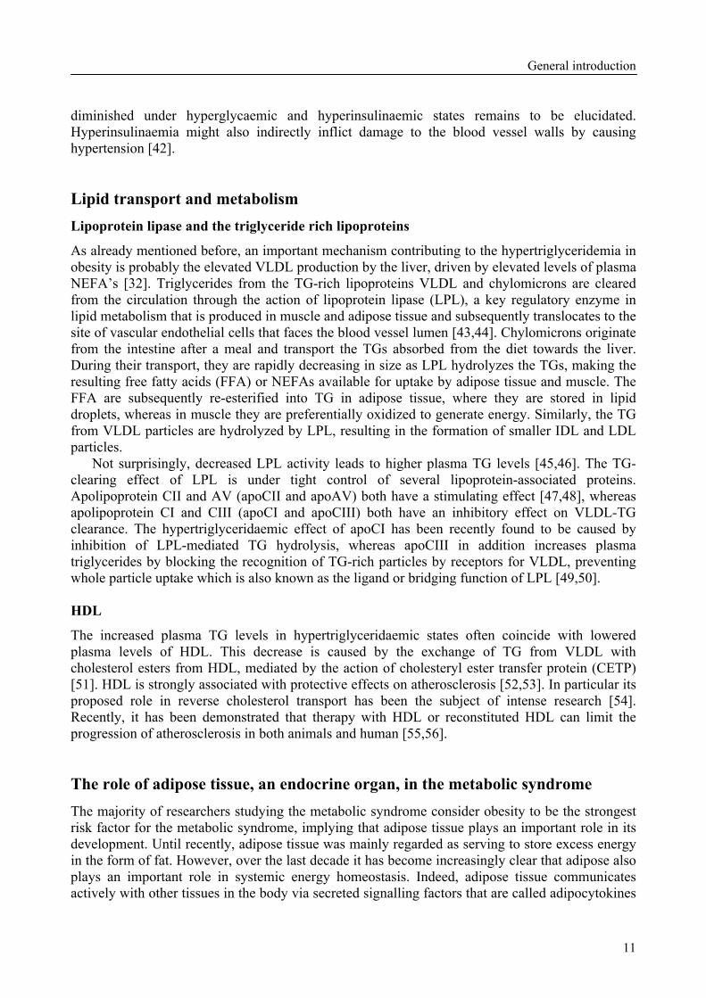
Besides FIAF/ANGPTL4, two other members of the ANGPTL family have been connected with regulation of nutrient metabolism; ANGPTL3 and ANGPTL6. ANGPTL3 displays the highest similarity to FIAF/ANGPTL4, sharing 31% identity at the amino acid level. In both mice and humans, ANGPTL3 is almost exclusively expressed in the liver, with much lower expression found in the kidneys (Fig. 1) [7,8]. Expression of ANGPTL3 in the liver is upregulated by the liver X receptor, a transcription factor and member of the nuclear hormone receptor superfamily, which is activated by oxysterols [9,10]. In mice ANGPTL3 potently elevates plasma TG levels [11], probably by inhibiting the activity of lipoprotein lipase (LPL) [12]. Furthermore, the protein is able to bind to human adipose cells and stimulates lipolysis in mature mouse 3T3-L1 adipocytes [13].
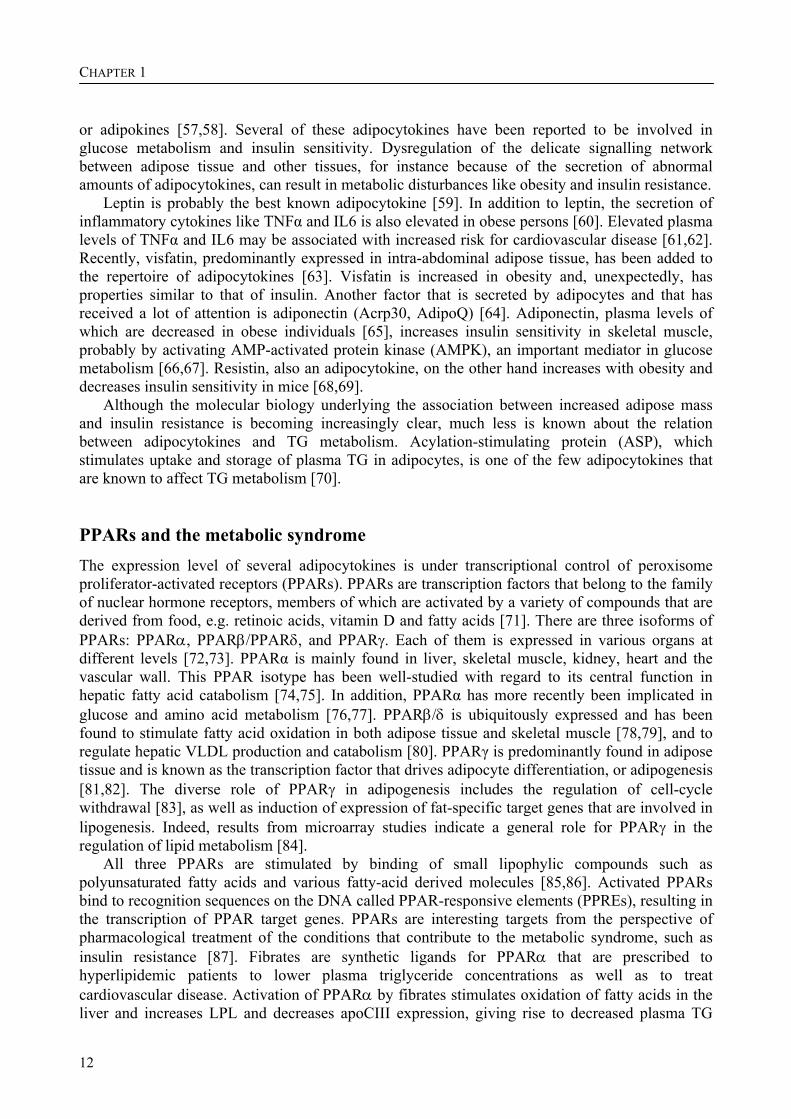
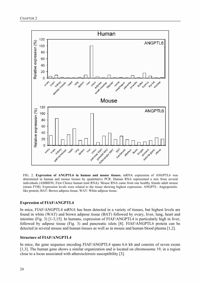
ANGPTL6, also referred to as angiopoietin-related growth factor, was recently reported to counter diet-induced obesity and insulin resistance [14]. In mice and humans, ANGPTL6 expression is highest in the liver, yet in mice expression is also reasonably high in brain, heart, skeletal muscle and adipose tissue (Fig. 2) [8]. ANGPTL6-deficient mice develop obesity, accumulate lipids in skeletal muscle and liver, and have reduced insulin sensitivity together with reduced energy expenditure. Conversely, mice overexpressing ANGPTL6 are lean and show increased insulin sensitivity paralleled by increased energy expenditure. These mice show protect-

Review - FIAF/ANGPTL4: a potential target for dyslipidemia?
23
FIG. 1. Expression of ANGPTL3 in human and mouse tissues. mRNA expression of ANGPTL3 was determined in human and mouse tissues by quantitative PCR. Human RNA represented a mix from several individuals (AMBION, First Choice human total RNA). Mouse RNA came from one healthy female adult mouse (strain FVB). Expression levels were related to the tissue showing highest expression. ANGPTL: Angiopoietin-like protein; BAT: Brown adipose tissue; WAT: White adipose tissue.
tion from diet-induced obesity and insulin resistance [14]. Thus, ANGPTL6 appears pivotal for the maintenance of energy homeostasis. Discovery of FIAF/ANGPTL4
FIAF/ANGPTL4 was discovered independently by at least three groups at approximately the same time. Kim and colleagues identified FIAF/ANGPTL4, which they named HFARP, using degenerate PCR on embryonic cDNAs in an effort to find additional members of the angiopoietin family [2]. FIAF/ANGPTL4 was named PGAR by Yoon and colleagues [3], who discovered PGAR using a subtractive cloning strategy to identify target genes of the nuclear hormone receptor PPARγ in adipose tissue. Finally, Kersten and colleagues identified FIAF/ANGPTL4 as a PPARα target gene by comparing mRNA from the livers of wild-type and PPARα-deficient mice [1].
CHAPTER 2
24
FIG. 2. Expression of ANGPTL6 in human and mouse tissues. mRNA expression of ANGPTL6 was determined in human and mouse tissues by quantitative PCR. Human RNA represented a mix from several individuals (AMBION, First Choice human total RNA). Mouse RNA came from one healthy female adult mouse (strain FVB). Expression levels were related to the tissue showing highest expression. ANGPTL: Angiopoietin-like protein; BAT: Brown adipose tissue; WAT: White adipose tissue.
Expression of FIAF/ANGPTL4
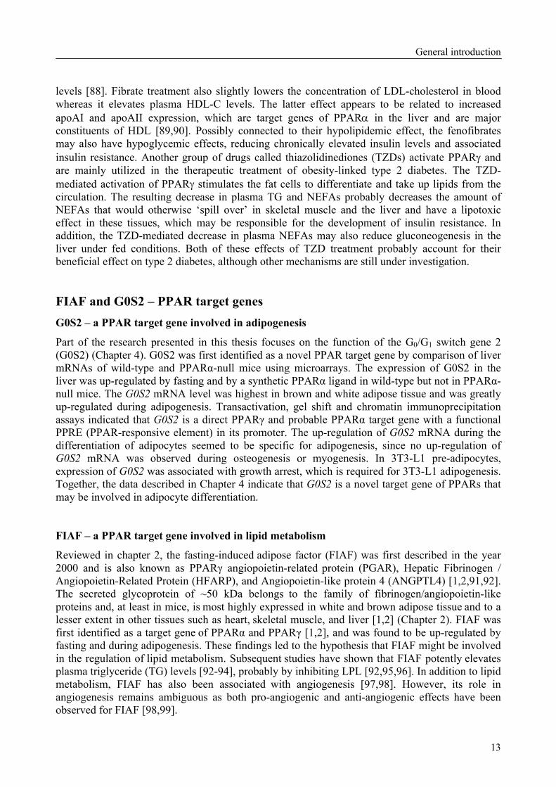
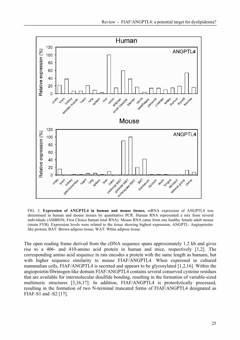
In mice, FIAF/ANGPTL4 mRNA has been detected in a variety of tissues, but highest levels are found in white (WAT) and brown adipose tissue (BAT) followed by ovary, liver, lung, heart and intestine (Fig. 3) [1-3,15]. In humans, expression of FIAF/ANGPTL4 is particularly high in liver, followed by adipose tissue (Fig. 3) and pancreatic islets [8]. FIAF/ANGPTL4 protein can be detected in several mouse and human tissues as well as in mouse and human blood plasma [1,2]. Structure of FIAF/ANGPTL4
In mice, the gene sequence encoding FIAF/ANGPTL4 spans 6.6 kb and consists of seven exons [1,3]. The human gene shows a similar organization and is located on chromosome 19, in a region close to a locus associated with atherosclerosis susceptibility [3].
Review - FIAF/ANGPTL4: a potential target for dyslipidemia?
25
FIG. 3. Expression of ANGPTL4 in human and mouse tissues. mRNA expression of ANGPTL4 was determined in human and mouse tissues by quantitative PCR. Human RNA represented a mix from several individuals (AMBION, First Choice human total RNA). Mouse RNA came from one healthy female adult mouse (strain FVB). Expression levels were related to the tissue showing highest expression. ANGPTL: Angiopoietin-like protein; BAT: Brown adipose tissue; WAT: White adipose tissue.
The open reading frame derived from the cDNA sequence spans approximately 1.2 kb and gives rise to a 406- and 410-amino acid protein in human and mice, respectively [1,2]. The corresponding amino acid sequence in rats encodes a protein with the same length as humans, but with higher sequence similarity to mouse FIAF/ANGPTL4. When expressed in cultured mammalian cells, FIAF/ANGPTL4 is secreted and appears to be glycosylated [1,2,16]. Within the angiopoietin/fibrinogen-like domain FIAF/ANGPTL4 contains several conserved cysteine residues that are available for intermolecular disulfide bonding, resulting in the formation of variable-sized multimeric structures [3,16,17]. In addition, FIAF/ANGPTL4 is proteolytically processed, resulting in the formation of two N-terminal truncated forms of FIAF/ANGPTL4 designated as FIAF-S1 and -S2 [17].
CHAPTER 2
26
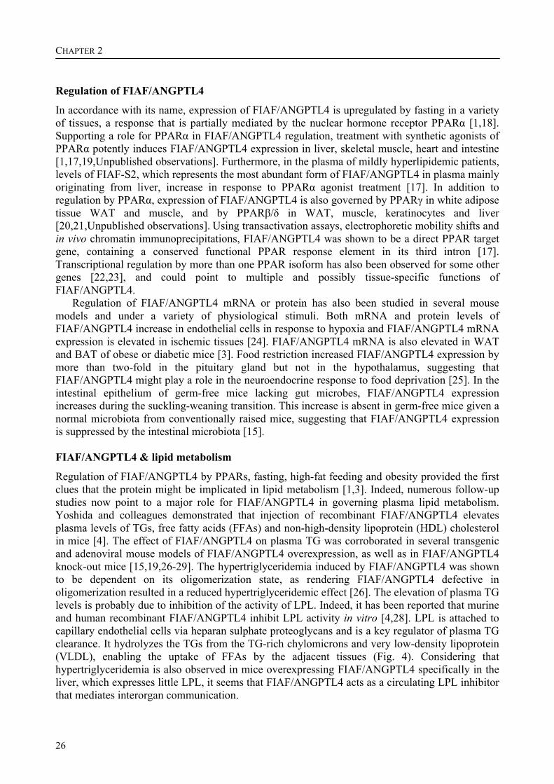
Regulation of FIAF/ANGPTL4
In accordance with its name, expression of FIAF/ANGPTL4 is upregulated by fasting in a variety of tissues, a response that is partially mediated by the nuclear hormone receptor PPARα [1,18]. Supporting a role for PPARα in FIAF/ANGPTL4 regulation, treatment with synthetic agonists of PPARα potently induces FIAF/ANGPTL4 expression in liver, skeletal muscle, heart and intestine [1,17,19,Unpublished observations]. Furthermore, in the plasma of mildly hyperlipidemic patients, levels of FIAF-S2, which represents the most abundant form of FIAF/ANGPTL4 in plasma mainly originating from liver, increase in response to PPARα agonist treatment [17]. In addition to regulation by PPARα, expression of FIAF/ANGPTL4 is also governed by PPARγ in white adipose tissue WAT and muscle, and by PPARβ/δ in WAT, muscle, keratinocytes and liver [20,21,Unpublished observations]. Using transactivation assays, electrophoretic mobility shifts and in vivo chromatin immunoprecipitations, FIAF/ANGPTL4 was shown to be a direct PPAR target gene, containing a conserved functional PPAR response element in its third intron [17]. Transcriptional regulation by more than one PPAR isoform has also been observed for some other genes [22,23], and could point to multiple and possibly tissue-specific functions of FIAF/ANGPTL4.
Regulation of FIAF/ANGPTL4 mRNA or protein has also been studied in several mouse models and under a variety of physiological stimuli. Both mRNA and protein levels of FIAF/ANGPTL4 increase in endothelial cells in response to hypoxia and FIAF/ANGPTL4 mRNA expression is elevated in ischemic tissues [24]. FIAF/ANGPTL4 mRNA is also elevated in WAT and BAT of obese or diabetic mice [3]. Food restriction increased FIAF/ANGPTL4 expression by more than two-fold in the pituitary gland but not in the hypothalamus, suggesting that FIAF/ANGPTL4 might play a role in the neuroendocrine response to food deprivation [25]. In the intestinal epithelium of germ-free mice lacking gut microbes, FIAF/ANGPTL4 expression increases during the suckling-weaning transition. This increase is absent in germ-free mice given a normal microbiota from conventionally raised mice, suggesting that FIAF/ANGPTL4 expression is suppressed by the intestinal microbiota [15]. FIAF/ANGPTL4 & lipid metabolism
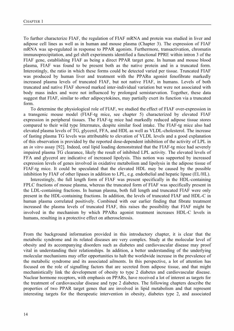
Regulation of FIAF/ANGPTL4 by PPARs, fasting, high-fat feeding and obesity provided the first clues that the protein might be implicated in lipid metabolism [1,3]. Indeed, numerous follow-up studies now point to a major role for FIAF/ANGPTL4 in governing plasma lipid metabolism. Yoshida and colleagues demonstrated that injection of recombinant FIAF/ANGPTL4 elevates plasma levels of TGs, free fatty acids (FFAs) and non-high-density lipoprotein (HDL) cholesterol in mice [4]. The effect of FIAF/ANGPTL4 on plasma TG was corroborated in several transgenic and adenoviral mouse models of FIAF/ANGPTL4 overexpression, as well as in FIAF/ANGPTL4 knock-out mice [15,19,26-29]. The hypertriglyceridemia induced by FIAF/ANGPTL4 was shown to be dependent on its oligomerization state, as rendering FIAF/ANGPTL4 defective in oligomerization resulted in a reduced hypertriglyceridemic effect [26]. The elevation of plasma TG levels is probably due to inhibition of the activity of LPL. Indeed, it has been reported that murine and human recombinant FIAF/ANGPTL4 inhibit LPL activity in vitro [4,28]. LPL is attached to capillary endothelial cells via heparan sulphate proteoglycans and is a key regulator of plasma TG clearance. It hydrolyzes the TGs from the TG-rich chylomicrons and very low-density lipoprotein (VLDL), enabling the uptake of FFAs by the adjacent tissues (Fig. 4). Considering that hypertriglyceridemia is also observed in mice overexpressing FIAF/ANGPTL4 specifically in the liver, which expresses little LPL, it seems that FIAF/ANGPTL4 acts as a circulating LPL inhibitor that mediates interorgan communication.
Review - FIAF/ANGPTL4: a potential target for dyslipidemia?
27
Based on its inhibitory effect on LPL activity, chronic elevation of FIAF/ANGPTL4 might be expected to decrease fat storage in adipose and muscle tissues. Indeed, transgenic mice over-expressing FIAF/ANGPTL4 in heart tissue displayed reduced cardiac LPL activity and decreased cardiac TG content. These mice also exhibited hypertriglyceridemia after 6 h of fasting [19]. In transgenic mice overexpressing FIAF/ANGPTL4 specifically in liver, no difference in lean or fat mass was observed compared with the wild type mice, either on chow diet or after a high fat/high carbohydrate feeding for 15 weeks [28]. In contrast, the gain in fat mass that occurs after conventionalization of germ-free mice was absent in FIAF/ANGPTL4 knock-out mice, indicating an effect of FIAF/ANGPTL4 on body fat. Before conventionalization, the FIAF/ANGPTL4-knockout mice already had a higher adipose LPL-activity compared with wild-type littermates [15]. The authors observed recently that in mice overexpressing FIAF/ANGPTL4 in peripheral tissues and within the physiological range, plasma TG clearance was severely impaired and adipose tissue weight was decreased by 50% compared to their wild-type littermates [29]. Together, the above studies reveal an important role for FIAF/ANGPTL4 in regulating the LPL-dependent clearance of plasma TGs. The striking effect on adipose tissue mass seems to be dependent on local changes in FIAF/ANGPTL4 expression, since no alterations in adipose tissue size were reported in mice overexpressing FIAF/ANGPTL4 in tissues other than fat. As inhibition of LPL appears to be mediated by circulating FIAF/ANGPTL4, additional mechanisms involving locally produced FIAF/ANGPTL4 need to be invoked to explain the reduction in fat stores. These include stimulation of adipose tissue lipolysis, as well as stimulation of fatty acid oxidation and uncoupling in adipose tissue (Fig. 4) [29].
Several inhibitors (apolipoprotein [apo]CI and apoCIII) and activators (apoAV and apoCII) of LPL are apoproteins physically associated with lipoprotein particles. Similarly, FIAF/ANGPTL4 may also be theorized to bind to lipoproteins. Indeed, the authors’ recent data indicate that, while native FIAF/ANGPTL4 is associated physically with HDL particles, truncated FIAF/ANGPTL4 is connected with low-density lipoprotein (LDL) particles, at least in mice (Fig. 4). In humans, both native and truncated FIAF/ANGPTL4 are bound to HDL. Levels of HDL-cholesterol, apoAI and apoAII in plasma are increased by FIAF/ANGPTL4 over-expression, supporting a link between FIAF/ANGPTL4 and HDL [29]. Furthermore, in healthy young adults, a positive correlation between plasma FIAF/ANGPTL4 levels and HDL-cholesterol, but not other lipid levels, was observed. Thus, FIAF/ANGPTL4 may be an important determinant of plasma HDL levels. It can be speculated that ANGPTL3 and/or ANGPTL6 may similarly bind to plasma lipoproteins. Alternatively, it is possible that the association with LDL and HDL is a unique feature of FIAF/ANGPTL4 amongst the ANGPTLs. Currently, it is not clear how HDL-associated FIAF/ANGPTL4 could account for inhibition of LPL activity and subsequent elevation of plasma TG-rich lipoproteins, but it might involve interference with the bridging function of LPL. Alternatively, LPL inhibition may be mediated by LDL-associated FIAF/ANGPTL4, whereas HDL-associated FIAF/ANGPTL4 might interact with similar lipases such as endothelial lipase and hepatic lipase, both of which are implicated in the clearance of HDL.
In response to FIAF/ANGPTL4 over-expression, plasma levels of FFA and glycerol were also elevated, suggesting that FIAF/ANGPTL4 stimulates adipose tissue lipolysis [29]. In agreement with this notion, Yoshida and colleagues found that injection of recombinant FIAF/ANGPTL4 elicited an abrupt rise in plasma FFA. In as much as inhibition of LPL would suppress plasma FFA, the stimulatory effect of FIAF/ANGPTL4 on plasma FFA and glycerol is probably mediated by an alternative, yet to be identified, mechanism, possibly involving a cellular receptor (Fig. 4).
FIAF/ANGPTL4 has also been implicated in regulation of glucose metabolism. In obese patients with Type 2 diabetes, serum levels of FIAF/ANGPTL4 were lower than those in healthy subjects with or without obesity [27].
CHAPTER 2
28
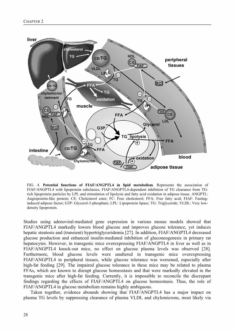
FIG. 4. Potential functions of FIAF/ANGPTL4 in lipid metabolism. Represents the association of FIAF/ANGPTL4 with lipoprotein subclasses, FIAF/ANGPTL4-dependent inhibition of TG clearance from TG-rich lipoprotein particles by LPL and stimulation of lipolysis and fatty acid oxidation in adipose tissue. ANGPTL: Angiopoietin-like protein; CE: Cholesterol ester; FC: Free cholesterol; FFA: Free fatty acid; FIAF: Fasting-induced adipose factor; G3P: Glycerol-3-phosphate; LPL: Lipoprotein lipase; TG: Triglyceride; VLDL: Very low-density lipoprotein.
Studies using adenoviral-mediated gene expression in various mouse models showed that FIAF/ANGPTL4 markedly lowers blood glucose and improves glucose tolerance, yet induces hepatic steatosis and (transient) hypertriglyceridemia [27]. In addition, FIAF/ANGPTL4 decreased glucose production and enhanced insulin-mediated inhibition of gluconeogenesis in primary rat hepatocytes. However, in transgenic mice overexpressing FIAF/ANGPTL4 in liver as well as in FIAF/ANGPTL4 knock-out mice, no effect on glucose plasma levels was observed [28]. Furthermore, blood glucose levels were unaltered in transgenic mice overexpressing FIAF/ANGPTL4 in peripheral tissues, while glucose tolerance was worsened, especially after high-fat feeding [29]. The impaired glucose tolerance in these mice may be related to plasma FFAs, which are known to disrupt glucose homeostasis and that were markedly elevated in the transgenic mice after high-fat feeding. Currently, it is impossible to reconcile the discrepant findings regarding the effects of FIAF/ANGPTL4 on glucose homeostasis. Thus, the role of FIAF/ANGPTL4 in glucose metabolism remains highly ambiguous.
Taken together, evidence abounds showing that FIAF/ANGPTL4 has a major impact on plasma TG levels by suppressing clearance of plasma VLDL and chylomicrons, most likely via
Review - FIAF/ANGPTL4: a potential target for dyslipidemia?
29
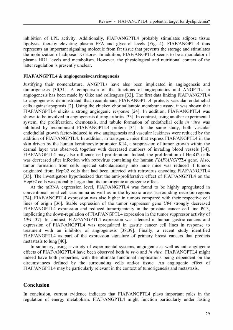
inhibition of LPL activity. Additionally, FIAF/ANGPTL4 probably stimulates adipose tissue lipolysis, thereby elevating plasma FFA and glycerol levels (Fig. 4). FIAF/ANGPTL4 thus represents an important signaling molecule from fat tissue that prevents the storage and stimulates the mobilization of adipose TG stores. In addition, FIAF/ANGPTL4 seems to be a modulator of plasma HDL levels and metabolism. However, the physiological and nutritional context of the latter regulation is presently unclear. FIAF/ANGPTL4 & angiogenesis/carcinogenesis
Justifying their nomenclature, ANGPTLs have also been implicated in angiogenesis and tumorigenesis [30,31]. A comparison of the functions of angiopoietins and ANGPTLs in angiogenesis has been made by Oike and colleagues [32]. The first data linking FIAF/ANGPTL4 to angiogenesis demonstrated that recombinant FIAF/ANGPTL4 protects vascular endothelial cells against apoptosis [2]. Using the chicken chorioallantoic membrane assay, it was shown that FIAF/ANGPTL4 elicits a strong angiogenic response [24]. In addition, FIAF/ANGPTL4 was shown to be involved in angiogenesis during arthritis [33]. In contrast, using another experimental system, the proliferation, chemotaxis, and tubule formation of endothelial cells in vitro was inhibited by recombinant FIAF/ANGPTL4 protein [34]. In the same study, both vascular endothelial growth factor-induced in vivo angiogenesis and vascular leakiness were reduced by the addition of FIAF/ANGPTL4. In addition, in transgenic mice that express FIAF/ANGPTL4 in the skin driven by the human keratinocyte promoter K14, a suppression of tumor growth within the dermal layer was observed, together with decreased numbers of invading blood vessels [34]. FIAF/ANGPTL4 may also influence cell proliferation. Indeed, the proliferation of HepG2 cells was decreased after infection with retrovirus containing the human FIAF/ANGPTL4 gene. Also, tumor formation from cells injected subcutaneously into nude mice was reduced if tumors originated from HepG2 cells that had been infected with retrovirus encoding FIAF/ANGPTL4 [35]. The investigators hypothesized that the anti-proliferative effect of FIAF/ANGPTL4 on the HepG2 cells was probably larger than its tumorigenic angiogenic effect.
At the mRNA expression level, FIAF/ANGPTL4 was found to be highly upregulated in conventional renal cell carcinoma as well as in the hypoxic areas surrounding necrotic regions [24]. FIAF/ANGPTL4 expression was also higher in tumors compared with their respective cell lines of origin [36]. Stable expression of the tumor suppressor gene U94 strongly decreased FIAF/ANGPTL4 expression and reduced tumorigenicity in the prostate cancer cell line PC3, implicating the down-regulation of FIAF/ANGPTL4 expression in the tumor suppressor activity of U94 [37]. In contrast, FIAF/ANGPTL4 expression was silenced in human gastric cancers and expression of FIAF/ANGPTL4 was upregulated in gastric cancer cell lines in response to treatment with an inhibitor of angiogenesis [38,39]. Finally, a recent study identified FIAF/ANGPTL4 as part of the expression signature of primary breast cancers that predicts metastasis to lung [40].
In summary, using a variety of experimental systems, angiogenic as well as anti-angiogenic effects of FIAF/ANGPTL4 have been observed both in vivo and in vitro. FIAF/ANGPTL4 might indeed have both properties, with the ultimate functional implications being dependent on the circumstances defined by the surrounding cells and/or tissue. An angiogenic effect of FIAF/ANGPTL4 may be particularly relevant in the context of tumorigenesis and metastasis. Conclusion In conclusion, current evidence indicates that FIAF/ANGPTL4 plays important roles in the regulation of energy metabolism. FIAF/ANGPTL4 might function particularly under fasting

CHAPTER 2
30
conditions to prevent uptake of plasma TGs by adipose tissue and stimulate adipose tissue lipolysis, directing fatty acid and TGs flux to the liver and possibly other organs. The involvement of FIAF/ANGPTL4 in angiogenesis and carcinogenesis/tumorigenesis, although highly interesting, needs further work in order to resolve apparent discrepancies in the published literature.
The rapid worldwide increase in the prevalence of obesity and the concurrent rise in its related disorders, such as diabetes and cardiovascular disease, have drawn attention to signaling factors secreted from adipose tissue that might mechanistically link the development of obesity to its associated ailments. Further studies might reveal opportunities for FIAF/ANGPTL4 in therapeutic intervention in obesity, diabetes Type 2 and associated atherogenic dyslipidemia, which is characterized by increased plasma TG and decreased HDL levels. Future perspective Based on the data collected in mice it can be hypothesized that inhibition of FIAF/ANGPTL4 will be effective to correct hypertriglyceridemia. However, since the effect of FIAF/ANGPTL4 on plasma lipoproteins in humans still needs to be clarified, it is premature to define the type of dyslipidemia for which targeting of FIAF/ANGPTL4 might be therapeutically useful. Nevertheless, several pharmaceutical companies are exploring FIAF/ANGPTL4 as a potential target for various metabolic disorders. Future investigations should focus on identification of the putative receptor for FIAF/ANGPTL4, the relevance of FIAF/ANGPTL4 binding to HDL, its functional implication in processes other than lipid metabolism, the differential roles of the various truncated and multimeric forms of FIAF/ANGPTL4, and the function of FIAF/ANGPTL4 in humans. In particular, genetic studies linking FIAF/ANGPTL4 to regulation of plasma lipoprotein levels in humans are eagerly awaited. Executive summary Angiopoietin-like proteins
• The angiopoietin-like proteins (ANGPTLs) currently encompasses a group of six proteins involved in angiogenesis and nutrient metabolism. They are approximately 50 kDa in size and are secreted from a variety of tissues. They share a common modular structure consisting of a signal peptide, a unique region of variable length, a coiled-coil domain and a carboxyl-terminal angiopoietin/fibrinogen-like domain.
Discovery of fasting-induced adipose factor/ANGPTL4
• Fasting-induced adipose factor (FIAF)/ANGPTL4 was discovered as a target of the nuclear receptors peroxisome proliferator-activated receptor (PPAR)α and γ, and by degenerate PCR in an effort to find additional members of the angiopoietin family.
Structure of FIAF/ANGPTL4
• In mouse and humans, the FIAF/ANGPTL4 gene spans 6.6 kb and consists of seven exons. FIAF/ANGPTL4 protein appears to be proteolytically processed in a tissue-specific manner to yield a protein of 25-35 kDa.
Regulation of FIAF/ANGPTL4
Review - FIAF/ANGPTL4: a potential target for dyslipidemia?
31
• Expression of FIAF/ANGPTL4 in several tissues is upregulated by fasting and by hypoxia. In human subjects, levels of FIAF/ANGPTL4 in plasma are increased after treatment with the synthetic PPARα agonist fenofibrate and the PPARγ agonist rosiglitazone. In mice, plasma levels of FIAF/ANGPTL4 are increased by fasting, and decreased by high-fat feeding. Expression of FIAF/ANGPTL4 is under direct control of PPARs.
FIAF/ANGPTL4 & lipid metabolism
• In mice, FIAF/ANGPTL4 reduces the clearance of triglycerides-rich plasma lipoproteins, most probably by inhibiting lipoprotein lipase. In addition, FIAF/ANGPTL4 appears to stimulate adipose tissue lipolysis, especially under fasting conditions. The effects of FIAF/ANGPTL4 on glucose metabolism are controversial. FIAF/ANGPTL4 is carried in blood by high-density lipoprotein (HDL) and/or low-density lipoprotein (LDL), depending on the species and processed form of FIAF. Finally, FIAF/ANGPTL4 may be a modulator of plasma HDL levels and metabolism.
FIAF/ANGPTL4 & angiogenesis/carcinogenesis
• Angiogenic as well as anti-angiogenic effects of FIAF/ANGPTL4 have been observed both in vivo and in vitro. Expression of FIAF/ANGPTL4 is both up and downregulated in a variety of cancers.
References 1 Kersten, S., Mandard, S., Tan, N. S., et al. (2000) Characterization of the fasting-induced adipose
factor FIAF, a novel peroxisome proliferator-activated receptor target gene. J. Biol. Chem. 275, 28488-28493
2 Kim, I., Kim, H. G., Kim, H., et al. (2000) Hepatic expression, synthesis and secretion of a novel fibrinogen/angiopoietin-related protein that prevents endothelial-cell apoptosis. Biochem. J. 346 Pt 3, 603-610
3 Yoon, J. C., Chickering, T. W., Rosen, E. D., et al. (2000) Peroxisome proliferator-activated receptor gamma target gene encoding a novel angiopoietin-related protein associated with adipose differentiation. Mol. Cell. Biol. 20, 5343-5349
4 Yoshida, K., Shimizugawa, T., Ono, M., Furukawa, H. (2002) Angiopoietin-like protein 4 is a potent hyperlipidemia-inducing factor in mice and inhibitor of lipoprotein lipase. J. Lipid Res. 43, 1770-1772
• Provides initial evidence that fasting-induced adipose factor (FIAF)/angiopoietin-like protein (ANGPTL)4 increases plasma triglycerides via inhibition of lipoprotein lipase (LPL).
5 Davis, S., Aldrich, T. H., Jones, P. F., et al. (1996) Isolation of angiopoietin-1, a ligand for the TIE2 receptor, by secretion-trap expression cloning. Cell 87, 1161-1169
6 Maisonpierre, P. C., Suri, C., Jones, P. F., et al. (1997) Angiopoietin-2, a natural antagonist for Tie2 that disrupts in vivo angiogenesis. Science 277, 55-60
7 Conklin, D., Gilbertson, D., Taft, D. W., et al. (1999) Identification of a mammalian angiopoietin-related protein expressed specifically in liver. Genomics 62, 477-482
8 Su, A. I., Cooke, M. P., Ching, K. A., et al. (2002) Large-scale analysis of the human and mouse transcriptomes. Proc. Natl. Acad. Sci. U.S.A. 99, 4465-4470
9 Kaplan, R., Zhang, T., Hernandez, M., et al. (2003) Regulation of the angiopoietin-like protein 3 gene by LXR. J. Lipid Res. 44, 136-143
10 Lehmann, J. M., Kliewer, S. A., Moore, L. B., et al. (1997) Activation of the nuclear receptor LXR by oxysterols defines a new hormone response pathway. J. Biol. Chem. 272, 3137-3140
CHAPTER 2
32
11 Koishi, R., Ando, Y., Ono, M., et al. (2002) Angptl3 regulates lipid metabolism in mice. Nat. Genet. 30, 151-157
•• Using positional cloning demonstrates that hypolipidemia in KK/San mice is caused by a mutation in the ANGPTL3 gene.
12 Shimizugawa, T., Ono, M., Shimamura, M., et al. (2002) ANGPTL3 decreases very low density lipoprotein triglyceride clearance by inhibition of lipoprotein lipase. J. Biol. Chem. 277, 33742-33748
13 Shimamura, M., Matsuda, M., Kobayashi, S., et al. (2003) Angiopoietin-like protein 3, a hepatic secretory factor, activates lipolysis in adipocytes. Biochem. Biophys. Res. Commun. 301, 604-609
14 Oike, Y., Akao, M., Yasunaga, K., et al. (2005) Angiopoietin-related growth factor antagonizes obesity and insulin resistance. Nat. Med. 11, 400-408
•• First paper linking ANGPTL6 with the regulation of energy homeostasis. Mutant ANGPTL6 mice are characterized in great detail to show an anti-obesity effect of ANGPTL6.
15 Backhed, F., Ding, H., Wang, T., et al. (2004) The gut microbiota as an environmental factor that regulates fat storage. Proc. Natl. Acad. Sci. U.S.A. 101, 15718-15723
• Suggests that the effect of intestinal bacteria on adiposity is mediated by FIAF/ANGPTL4. 16 Ge, H., Yang, G., Huang, L., Motola, D. L., Pourbahrami, T., Li, C. (2004) Oligomerization and
regulated proteolytic processing of angiopoietin-like protein 4. J. Biol. Chem. 279, 2038-2045 17 Mandard, S., Zandbergen, F., Tan, N. S., et al. (2004) The direct peroxisome proliferator-activated
receptor target fasting-induced adipose factor (FIAF/PGAR/ANGPTL4) is present in blood plasma as a truncated protein that is increased by fenofibrate treatment. J. Biol. Chem. 279, 34411-34420
• First report on the regulation of FIAF/ANGPTL4 protein and mRNA expression in humans. 18 Ge, H., Cha, J. Y., Gopal, H., et al. (2005) Differential regulation and properties of angiopoietin-like
proteins 3 and 4. J. Lipid Res. 46, 1484-1490 19 Yu, X., Burgess, S. C., Ge, H., et al. (2005) Inhibition of cardiac lipoprotein utilization by transgenic
overexpression of Angptl4 in the heart. Proc. Natl. Acad. Sci. U.S.A. 102, 1767-1772 20 Akiyama, T. E., Lambert, G., Nicol, C. J., et al. (2004) Peroxisome proliferator-activated receptor
beta/delta regulates very low density lipoprotein production and catabolism in mice on a Western diet. J. Biol. Chem. 279, 20874-20881
21 Schmuth, M., Haqq, C. M., Cairns, W. J., et al. (2004) Peroxisome proliferator-activated receptor (PPAR)-beta/delta stimulates differentiation and lipid accumulation in keratinocytes. J. Invest. Dermatol. 122, 971-983
22 Patsouris, D., Mandard, S., Voshol, P.J., et al. (2004) PPARalpha governs glycerol metabolism. J. Clin. Invest. 144, 94-103
23 Schoonjans, K., Peinado-Onsurbe, J., Lefebvre, A. M., et al. (1996) PPARalpha and PPARgamma activators direct a distinct tissue-specific transcriptional response via a PPRE in the lipoprotein lipase gene. Embo J. 15, 5336-5348
24 Le Jan, S., Amy, C., Cazes, A., et al. (2003) Angiopoietin-like 4 is a proangiogenic factor produced during ischemia and in conventional renal cell carcinoma. Am. J. Pathol. 162, 1521-1528
25 Wiesner, G., Morash, B. A., Ur, E., Wilkinson, M. (2004) Food restriction regulates adipose-specific cytokines in pituitary gland but not in hypothalamus. J. Endocrinol. 180, R1-6
26 Ge, H., Yang, G., Yu, X., Pourbahrami, T., Li, C. (2004) Oligomerization state-dependent hyperlipidemic effect of angiopoietin-like protein 4. J. Lipid Res. 45, 2071-2079
27 Xu, A., Lam, M. C., Chan, K. W., et al. (2005) Angiopoietin-like protein 4 decreases blood glucose and improves glucose tolerance but induces hyperlipidemia and hepatic steatosis in mice. Proc. Natl. Acad. Sci. U.S.A. 102, 6086-6091
• Provocative paper showing a huge suppressive effect of FIAF/ANGPTL4 on plasma glucose levels.
28 Koster, A., Chao, Y. B., Mosior, M., et al. (2005) Transgenic angiopoietin-like (angptl)4 overexpression and targeted disruption of angptl4 and angptl3: regulation of triglyceride metabolism. Endocrinology 146, 4943-4950
29 Mandard, S., Zandbergen, F., van Straten, E., et al. (2005) The fasting-induced adipose factor/angiopoietin-like protein 4 is physically associated with lipoproteins and governs plasma lipid levels and adiposity. J. Biol. Chem.
Review - FIAF/ANGPTL4: a potential target for dyslipidemia?
33
• ANGPTL4 is bound to plasma lipoproteins in mouse and human. 30 Oike, Y., Ito, Y., Maekawa, H., et al. (2004) Angiopoietin-related growth factor (AGF) promotes
angiogenesis. Blood 103, 3760-3765 31 Camenisch, G., Pisabarro, M. T., Sherman, D., et al. (2002) ANGPTL3 stimulates endothelial cell
adhesion and migration via integrin alpha vbeta 3 and induces blood vessel formation in vivo. J. Biol. Chem. 277, 17281-17290
32 Oike, Y., Yasunaga, K., Suda, T. (2004) Angiopoietin-related/angiopoietin-like proteins regulate angiogenesis. In t. J. Hematol. 80, 21-28
• Good overview comparing the functions of angiopoietin-like proteins in angiogenesis. 33 Hermann, L. M., Pinkerton, M., Jennings, K., et al. (2005) Angiopoietin-like-4 is a potential
angiogenic mediator in arthritis. Clin. Immunol. 115, 93-101 34 Ito, Y., Oike, Y., Yasunaga, K., et al. (2003) Inhibition of angiogenesis and vascular leakiness by
angiopoietin-related protein 4. Cancer Res. 63, 6651-6657 35 Li, K. Q., Li, W. L., Peng, S. Y., Shi, X. Y., Tang, H. L., Liu, Y. B. (2004) Anti-tumor effect of
recombinant retroviral vector-mediated human ANGPTL4 gene transfection. Chin. Med. J. (Engl) 117, 1364-1369
36 Stull, R. A., Tavassoli, R., Kennedy, S., et al. (2005) Expression analysis of secreted and cell surface genes of five transformed human cell lines and derivative xenograft tumors. BMC Genomics 6, 55
37 Ifon, E. T., Pang, A. L., Johnson, W., et al. (2005) U94 alters FN1 and ANGPTL4 gene expression and inhibits tumorigenesis of prostate cancer cell line PC3. Cancer Cell Int. 5, 19
38 Kaneda, A., Kaminishi, M., Yanagihara, K., Sugimura, T., Ushijima, T. (2002) Identification of silencing of nine genes in human gastric cancers. Cancer Res. 62, 6645-6650
39 Arao, T., Yanagihara, K., Takigahira, M., et al. (2006) ZD6474 inhibits tumor growth and intraperitoneal dissemination in a highly metastatic orthotopic gastric cancer model. Int. J. Cancer 118, 483-489
40 Minn, A. J., Gupta, G. P., Siegel, P. M., et al. (2005) Genes that mediate breast cancer metastasis to lung. Nature 436, 518-524

34
FIAF is present in blood plasma in a truncated form
35
CHAPTER 3 The direct peroxisome proliferator-activated receptor target fasting-induced adipose factor (FIAF/PGAR/ANGPTL4) is present in blood plasma as a truncated protein that is increased by fenofibrate treatment Stéphane Mandard, Fokko Zandbergen, Nguan Soon Tan, Pascal Escher, David Patsouris, Wolfgang Koenig, Robert Kleemann, Arjen Bakker, Frank Veenman, Walter Wahli, Michael Müller, and Sander Kersten Abstract The fasting-induced adipose factor (FIAF, ANGPTL4, PGAR, HFARP) was previously identified as a novel adipocytokine that was up-regulated by fasting, by peroxisome proliferator-activated receptor agonists, and by hypoxia. To further characterize FIAF, we studied regulation of FIAF mRNA and protein in liver and adipose cell lines as well as in human and mouse plasma. Expression of FIAF mRNA was up-regulated by peroxisome proliferator-activated receptor α (PPARα) and PPARβ/δ agonists in rat and human hepatoma cell lines and by PPARγ and PPARβ/δ agonists in mouse and human adipocytes. Transactivation, chromatin immunoprecipitation, and gel shift experiments identified a functional PPAR response element within intron 3 of the FIAF gene. At the protein level, in human and mouse blood plasma, FIAF was found to be present both as the native protein and in a truncated form. Differentiation of mouse 3T3-L1 adipocytes was associated with the production of truncated FIAF, whereas in human white adipose tissue and SGBS adipocytes, only native FIAF could be detected. Interestingly, truncated FIAF was produced by human liver. Treatment with fenofibrate, a potent PPAR agonist, markedly increased plasma levels of truncated FIAF, but not native FIAF, in humans. Levels of both truncated and native FIAF showed marked interindividual variation but were not associated with body mass index and were not influenced by prolonged semistarvation. Together, these data suggest that FIAF, similar to other adipocytokines such as adiponectin, may partially exert its function via a truncated form. This chapter has been published in the Journal of Biological Chemistry 279, 34411-34420 (2004)
CHAPTER 3
36
Introduction Obesity, defined as excess body fat, is associated with numerous secondary ailments, including hypertension, dyslipidemia, and insulin resistance, and is therefore an important health concern. As the prevalence of obesity is rising, there is an increasing interest in understanding the metabolic behavior of adipose tissue.
Since the discovery of leptin in 1994 [1], it has become clear that fat tissue not merely serves to store excess energy but also has an important endocrine function [2]. Over the past few years, several factors secreted by white adipose tissue (WAT)1, aptly named adipocytokines, have been identified and characterized, including resistin (also known as FIZZ 3 or ADSF (for adipocyte secreted factor)) [3-5], adiponectin (also known as adipoQ or ACRP30 (for adipocyte complement-related protein 30)) [6-8], acylation-stimulating protein (C3ades-Arg) [9], plasminogen activator inhibitor-1 [10], renin angiotensin system [11], metallothioneins [12], and the inflammatory cytokines interleukin-6, tumor necrosis factor-α, and tumor growth factor-β [13,14]. They have been implicated in a variety of different processes, ranging from blood pressure control to lipid metabolism and insulin sensitizing. Consequently, it has been tempting to attribute many of the clinical abnormalities associated with obesity, including insulin resistance, to altered secretion of particular adipocytokines.
Recently, we and others identified a new gene encoding the secreted fasting-induced adipose factor (FIAF), also known as PGAR (for PPARγ angiopoietin-related protein), ANGPTL4 (for angiopoietin-like protein 4), or HFARP (for hepatic fibrinogen/angiopoietin-related protein) [15-17]. Several nonexclusive functions for FIAF have so far been proposed. Expression of FIAF is dramatically up-regulated during hypoxia in both endothelial cells and cardiomyocytes [18,19], leading to the suggestion that FIAF may be involved in angiogenesis, in analogy with two other proteins that carry an angiopoietin/fibrinogen-like domain, angiopoietin-1 and angiopoietin-2. Subsequently, it was found that FIAF is able to induce a strong proangiogenic response in the chicken chorioallantoic membrane assay [19]. FIAF has also been proposed to act as an apoptosis survival factor in vascular endothelial cells [16].
The closest relative of FIAF is angiopoietin-like protein 3 (ANGPTL3). KK/Snk mice, a mutant strain of KK obese mice, carry a mutation in the gene for ANGPTL3, resulting in low plasma triglyceride and free fatty acid levels [20]. The low plasma triglyceride levels are possibly due to elevated lipoprotein lipase activity, which was reported to be inhibited by ANGPTL3 in vitro, whereas the low plasma free fatty acid levels may be connected to the impaired stimulatory effect of ANGPTL3 on adipose tissue lipolysis [21,22]. Similar to ANGPTL3, there is evidence that FIAF also may inhibit lipoprotein lipase activity and hereby influence plasma levels of triglycerides [18,23], thus connecting FIAF to lipid metabolism.
Expression of FIAF in liver and WAT was originally found to be up-regulated by the nuclear hormone receptors PPARα and PPARγ [15,17,23]. PPARs are ligand-activated transcription factors that mediate the effects of fibrates (PPARα) or thiazolidinediones (PPARγ) on DNA transcription [24,25]. PPARα is mainly expressed in brown adipose tissue and liver and plays an important role in the hepatic fatty acid oxidation, whereas PPARγ is the master regulator of adipogenesis. However, it is still unclear whether FIAF is a direct PPAR target gene, with a functional PPAR response element in its promoter.
In order to close in on the potential function of FIAF, we studied the regulation of FIAF mRNA and protein expression in vitro and in human blood plasma. Our main conclusions are that 1 The abbreviations used are: WAT, white adipose tissue; PPAR, peroxisome proliferator-activated receptor; SEAP, secreted alkaline phosphatase; FIAF, fasting-induced adipose factor; DMEM, Dulbecco's modified Eagle's medium; FCS, fetal calf serum; ChIP, chromatin immunoprecipitation; PPRE, PPAR response element; RT, reverse
transcriptase; IBMX, isobutylmethylxanthine.

FIAF is present in blood plasma in a truncated form
37
FIAF is a classical PPAR target gene in both humans and rodents and that FIAF protein is mainly present in blood plasma in a truncated form, whose levels show a large interindividual variability. Plasma levels of the truncated form of FIAF are increased by treatment with fenofibrate. Materials and methods Chemicals
Wy14643 was obtained from ChemSyn Laboratories. Rosiglitazone was from Alexis. Recombinant human insulin (Actrapid) was from Novo Nordisk. SYBR Green was from Eurogentec. Dulbecco's modified Eagle's medium, fetal calf serum, calf serum, and penicillin/streptomycin/fungizone were from BioWhittaker Europe (Cambrex Bioscience). Otherwise, chemicals were from Sigma. Primary human adipocyte differentiation
Isolation of stromal vascular cells was done as follows. Subcutaneous and visceral adipose tissues were obtained during gastric restriction surgery. Adipose tissue was collected in phosphate-buffered saline and cut into 3 x 3-mm pieces with scissors. The 3 x 3-mm pieces were further processed with a scalpel. Next, the pieces of adipose tissue were digested in DMEM-high glucose containing 4% bovine serum albumin and 2 mg/ml collagenase. 1–2.5 g of adipose tissue was digested in 5 ml of this solution at 37 °C on a shaking platform for 2 h. Next, the digest was transferred to a 5-ml syringe and gently pressed over a 500-µm sterile pore size disposable nylon mesh. Stromal vascular cells were separated from adipose cells by centrifugation (1 min, 170 g). Adipose cells were removed, and the stromal vascular cells were precipitated by centrifugation (5 min, 350 g). Red blood cells were lysed by resuspending the cell pellet in 10 ml of red cell lysis solution (154 mM NH4Cl, 10 mM KHCO3, 0.1 mM EDTA). After 5 min, stromal vascular cells were spun down (5 min, 350 g) and resuspended in DMEM/F-12 containing 10% fetal calf serum
(FCS), 2 mM glutamine, 100 IU/ml penicillin, and 100 µg/ml streptomycin and further referred to as complete medium. Approximately 107 cells were plated in a 75-cm2 flask. After 48 h, the medium was replaced by differentiation medium consisting of complete medium (instead of 10% FCS, 2% FCS was used) plus 15 mM NaHCO3,
15 mM HEPES, 33 µM biotin, 17 µM panthothenate, 200 pM T3, 1 µM dexamethasone, 500 nM insulin, 4 µg/ml transferrin, and 10 µM cPGI2. After an average period of 30 days of differentiation, extraction of total RNA was performed. SGBS cell line culture and induction of adipogenesis
The culture of the SGBS cells and their induction into mature human adipocytes were performed exactly as previously published [26]. 3T3-L1 adipogenesis assay
3T3-L1 fibroblasts were amplified in DMEM plus 10% calf serum and plated for final differentiation in DMEM plus 10% FCS. Two days after reaching confluence (which was day 0), the medium was changed, and the following compounds were added: isobutyl methylxanthine (0.5 mM), dexamethasone (1 µM), and insulin (5 µg/ml). On day 3, the medium was changed to DMEM plus 10% FCS and insulin (5 µg/ml). On day 6, the medium was changed to DMEM plus 10% FCS, which was changed every 3 days.
CHAPTER 3
38
Isolation of total RNA and RT-PCR
Total RNA was extracted from cells or tissue with Trizol reagent (Invitrogen) following the supplier's protocol. 3–5 µg of total RNA was treated with DNase I amplification grade (Invitrogen) and then reverse-transcribed with oligo(dT) using Superscript II RT RNase H (Invitrogen).
Real time quantitative PCR
Primers were designed to generate a PCR amplification product of 100–200 bp. Only primer pairs yielding unique amplification products without primer dimer formation were subsequently used for real time quantitative PCR assays. The following primer pairs were used: mFIAF (forward), 5'-GTTTGCAGACTCAGCTCAAGC-3'; mFIAF (reverse), 5'-CCAAGAGGTCTATCTGGCTCTG-3'; hFIAF (forward), 5'-CGTACCCTTCTCCACTTGGG-3'; hFIAF (reverse), 5'-GCTCTTGGCG-CAGTTCTTG-3'; mβ-Actin (forward), 5'-CTGACTGACTACCTCATGAAGATCCT-3'; mβ-Actin (reverse), 5'-CTTAATGTCACGCACGATTTCC-3'; hβ-Actin (forward), 5'-CTTCCTGGG-CATGGAGTC-3'; hβ-Actin (reverse), 5'-GCCAGGGTACATGGTGGT-3'; rPPARα (forward), 5'-CACCCTCTCTCCAGCTTCCA-3'; rPPARα (reverse), 5'-GCCTTGTCCCCACATATTCG-3'; rPPARβ (forward), 5'-AACGAGATCAGCGTGCATGTG-3'; rPPARβ (reverse), 5'-TGAGGAAG-AGGCTGCTGAAGTT-3'; rPPARγ (forward), 5'-ATGGAGCCTAAGTTTGAGTTTGCT-3'; rPPARγ (reverse), 5'-GGATGTCCTCGATGGGCTTCA-3'; hPPARα (forward), 5'-CTGGAAGC-TTTGGCTTTACG-3'; hPPARα (reverse), 5'-TGTCCCCGCAGATTCTACAT-3'; hPPARβ (forward), 5'-ACAGCATGCACTTCCTTCC-A-3'; hPPARβ (reverse), 5'-TCACATGCATGAAC-ACCGTA-3'; hPPARγ (forward), 5'-GAGCCCAAGTTTGAGTTTGC-3'; hPPARγ (reverse), 5'-C-AGGGCTTGTAGCAGGTTGT-3'.
PCR was carried out using Platinum Taq polymerase (Invitrogen) and SYBR Green on an iCycler PCR machine (Bio-Rad). Expression was related to actin, which did not change under any of the experimental conditions studied. Transactivation assay
A 350-nucleotide fragment surrounding the putative PPRE within intron 3 of the human or mouse FIAF gene was PCR-amplified from human or mouse genomic DNA (mouse strain C57/B6) and subcloned into the KpnI and BglII sites of pTAL-SEAP (Clontech). This reporter vector was transfected into human hepatoma HepG2 cells together with an expression vector for PPAR (mPPARα, mPPARβ/δ, or mPPARγ in pSG5) in the presence or absence of Wy14643 (50 µM), L165041 (5 µM), or rosiglitazone (5 µM), respectively. Transfections were carried out by calcium phosphate precipitation. A β-galactosidase reporter was co-transfected to normalize for differences in transfection efficiency. Secreted alkaline phosphatase activity was measured in the medium 48 h post-transfection via the chemiluminescent SEAP reporter assay (Roche Applied Science). β-Galactosidase activity was measured in the cell lysate by standard assay using 2-nitrophenyl-β-D-galactopyranoside as a substrate. Chromatin immunoprecipitation (ChIP)
Pure-bred wild-type or PPAR null mice on a sv129 background were used. Mice were fed by gavage with either Wy14643 (50 mg/kg/day) or vehicle (0.5% carboxymethylcellulose) for 5 days. Alternatively, mice were fasted or not for 24 h. After the indicated treatment, mice were sacrificed by cervical dislocation. The liver was rapidly perfused with prewarmed (37 °C) phosphate-buffered saline for 5 min followed by 0.2% collagenase for 10 min. The liver was diced and forced through a stainless steel sieve, and the hepatocytes were collected directly into DMEM containing
1% formaldehyde. After incubation at 37 °C for 15 min, the hepatocytes were pelleted, and ChIP
FIAF is present in blood plasma in a truncated form
39
was done using mouse PPAR -specific antibodies as previously described [27]. Sequences of primers used for PCR were 5'-TCTGGGTCTGCCCCCACTCCTGG-3' (forward) and 5'-GTGTG-TGTGTGGGATACGGCTAT-3' (reverse). Control primers used were 5'-AGTAACTTTGACAG-GAACCAGGGGTC-3' (forward) and 5'-TTTGGACTGGGAACTTAGCTTAGTTG-3' (reverse).
3T3-L1 cells were differentiated as described above. After cell lysis and sonication, the supernatant was diluted 20-fold in re-chIP dilution buffer (1 mM EDTA, 20 mM Tris-HCl, pH 8.1, 50 mM NaCl, 1% Triton X) prior to incubation with mouse PPARγ antibody. The remainder of the assay was carried out as described previously [27]. Gel shift
hRXRα and hPPARα proteins were generated from pSG5 expression vectors using the TNT coupled in vitro transcription/translation system (Promega). The following oligonucleotides were annealed: hFIAF-PPRE, 5'-TCGGGGAAAGTAGGGGAAAGGTCGTGG-3' and 5'-CTGGGCCA-GCGACCTTTCCCCTACTTTCCC-3'; mFIAF-PPRE, 5'-TCGGGGAAAGTAGGAGAAAGGTC-ACTGG-3' and 5'-CTGGCCAGTGACCTTTCTCCTACTTTCCC-3'; for specific competition malic enzyme PPRE, 5'-TCGCTTTCTGGGTCAAAGTTGATCCA-3' and 5'-CTGGTGGATCAA-CTTTGACCCAGAAAG-3'; for nonspecific competition ETS, 5'-TGGAATGTACCGGAAATA-ACACCA-3' and 5'-TGGTGTTATTTCCGGTACATTCCA-3'. Oligonucleotides were annealed and labeled by Klenow filling (Roche Applied Science) using Redivue [α-32P]dCTP (3000 Ci/mmol) (Amersham Biosciences). In vitro translated proteins (0.5–0.8 µl/reaction) were preincubated for 15 min on ice in 1x binding buffer (80 mM KCl, 1 mM dithiothreitol, 10 mM Tris-Cl, pH 7.4, 10% glycerol plus protease inhibitors) in presence of 2 µg of poly(dI·dC), 5 µg of sonicated salmon sperm DNA, and competitor oligonucleotides in a final volume of 20 µl. Then 1 ng (1 ng/µl) of radiolabeled oligonucleotide was added, and incubation proceeded for another 10 min at room temperature. Complexes were separated on a 4% polyacrylamide gel (acrylamide/bisacrylamide, 37.5:1) equilibrated in 0.5x TBE at 25 mA. Western blot
The mouse polyclonal antibody used was directed against the epitope CQGPKGKDAPFKDSE located in the N-terminal part of the mouse FIAF protein. The human polyclonal antibody used was directed against the epitope CQGTEGSTDLPLAPE also located in the N-terminal part of the human FIAF protein. The peptide affinity-purified antibodies were generated in rabbit and ordered
via Eurogentec's customized antibody production service. Western blotting was carried out using an ECL system (Amersham Biosciences) according to the manufacturer's instructions. The primary antibody was used at a dilution of 1:1000 (mouse) or between 1:2000 and 1:5000 (human), and the secondary antibody (anti-rabbit IgG, Sigma) was used at a dilution of 1:8000. All incubations were
performed in 1x Tris-buffered saline, pH 7.5, with 0.1% Tween 20 and 5% dry milk, except for the final washings, when milk was omitted. Human subjects
In experiment 1, blood was taken from 16 young adults after an overnight fast. In experiment 2, blood was taken after an overnight fast from 28 subjects before and after a 4-week treatment with 250 mg of micronized fenofibrate daily. In experiment 3, blood was taken after an overnight fast
from 20 men (body mass index ranging 22.7 to 39.8). Samples were from a published study [28]. In experiment 4, blood was taken after an overnight fast from 22 subjects before and after a 46-day semistarvation program (2.1 MJ/day). Samples were from a published study [29]. All human
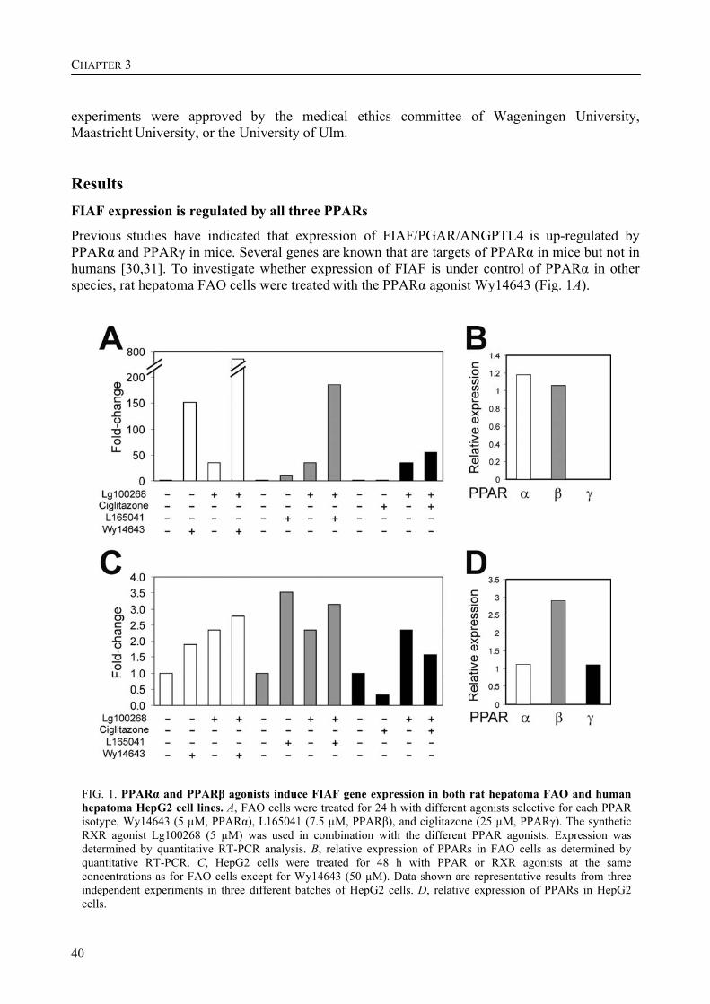
CHAPTER 3
40
experiments were approved by the medical ethics committee of Wageningen University, Maastricht University, or the University of Ulm. Results FIAF expression is regulated by all three PPARs
Previous studies have indicated that expression of FIAF/PGAR/ANGPTL4 is up-regulated by PPARα and PPARγ in mice. Several genes are known that are targets of PPARα in mice but not in humans [30,31]. To investigate whether expression of FIAF is under control of PPARα in other species, rat hepatoma FAO cells were treated with the PPARα agonist Wy14643 (Fig. 1A).
FIG. 1. PPARα and PPARβ agonists induce FIAF gene expression in both rat hepatoma FAO and human hepatoma HepG2 cell lines. A, FAO cells were treated for 24 h with different agonists selective for each PPAR isotype, Wy14643 (5 µM, PPARα), L165041 (7.5 µM, PPARβ), and ciglitazone (25 µM, PPARγ). The synthetic RXR agonist Lg100268 (5 µM) was used in combination with the different PPAR agonists. Expression was determined by quantitative RT-PCR analysis. B, relative expression of PPARs in FAO cells as determined by quantitative RT-PCR. C, HepG2 cells were treated for 48 h with PPAR or RXR agonists at the same concentrations as for FAO cells except for Wy14643 (50 µM). Data shown are representative results from three independent experiments in three different batches of HepG2 cells. D, relative expression of PPARs in HepG2 cells.
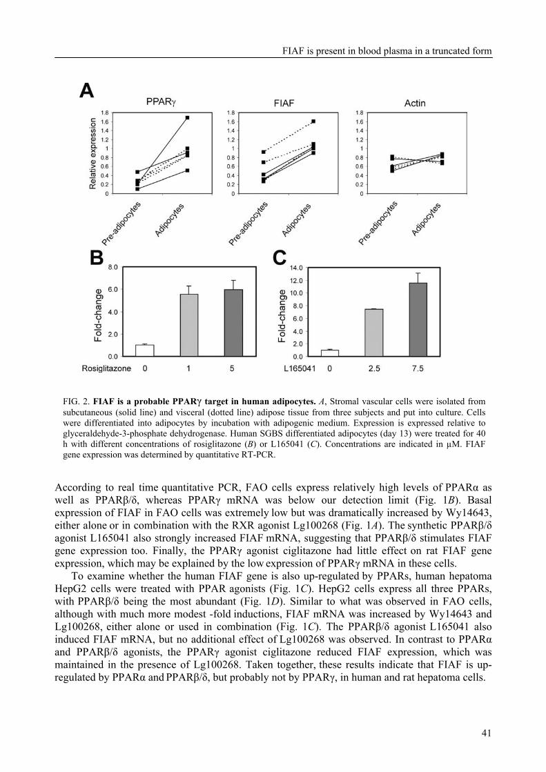
FIAF is present in blood plasma in a truncated form
41
FIG. 2. FIAF is a probable PPARγ target in human adipocytes. A, Stromal vascular cells were isolated from subcutaneous (solid line) and visceral (dotted line) adipose tissue from three subjects and put into culture. Cells were differentiated into adipocytes by incubation with adipogenic medium. Expression is expressed relative to glyceraldehyde-3-phosphate dehydrogenase. Human SGBS differentiated adipocytes (day 13) were treated for 40 h with different concentrations of rosiglitazone (B) or L165041 (C). Concentrations are indicated in µM. FIAF gene expression was determined by quantitative RT-PCR.
According to real time quantitative PCR, FAO cells express relatively high levels of PPARα as well as PPARβ/δ, whereas PPARγ mRNA was below our detection limit (Fig. 1B). Basal expression of FIAF in FAO cells was extremely low but was dramatically increased by Wy14643, either alone or in combination with the RXR agonist Lg100268 (Fig. 1A). The synthetic PPARβ/δ agonist L165041 also strongly increased FIAF mRNA, suggesting that PPARβ/δ stimulates FIAF gene expression too. Finally, the PPARγ agonist ciglitazone had little effect on rat FIAF gene expression, which may be explained by the low expression of PPARγ mRNA in these cells.
To examine whether the human FIAF gene is also up-regulated by PPARs, human hepatoma HepG2 cells were treated with PPAR agonists (Fig. 1C). HepG2 cells express all three PPARs, with PPARβ/δ being the most abundant (Fig. 1D). Similar to what was observed in FAO cells, although with much more modest -fold inductions, FIAF mRNA was increased by Wy14643 and Lg100268, either alone or used in combination (Fig. 1C). The PPARβ/δ agonist L165041 also induced FIAF mRNA, but no additional effect of Lg100268 was observed. In contrast to PPARα and PPARβ/δ agonists, the PPARγ agonist ciglitazone reduced FIAF expression, which was maintained in the presence of Lg100268. Taken together, these results indicate that FIAF is up-regulated by PPARα and PPARβ/δ, but probably not by PPARγ, in human and rat hepatoma cells.

CHAPTER 3
42
To better examine regulation of human FIAF expression by PPARγ, we turned to primary human preadipocytes. Upon stimulation with a mixture of hormones, these cells can be differentiated into
adipocytes. Stromal vascular cells from both subcutaneous and visceral adipose tissue were isolated and induced to differentiate into adipocytes. Expression of FIAF was higher in adipocytes
versus preadipocytes in all three subjects with cells from both subcutaneous and visceral origin (Fig. 2A). A similar induction of expression was observed for PPARγ, suggesting that FIAF is up-regulated by PPARγ during human adipocyte differentiation. In differentiated human SGBS adipocytes, both rosiglitazone and L165041 caused an induction of FIAF mRNA (Fig. 2, B and C, respectively), indicating that both PPARγ and PPARβ/δ regulate FIAF expression in human adipocytes. Together, these data suggest that FIAF is a PPARγ and possibly a PPARβ/δ target gene in human adipocytes. FIAF is a direct target gene of PPAR
To unequivocally determine FIAF as a direct target gene of PPARs, direct binding of PPAR to the FIAF promoter needs to be demonstrated. Comparative analysis of the hFIAF and mFIAF gene sequence upstream of the transcription start site did not reveal any conserved stretches of DNA that might harbor a PPRE. Transactivation studies with several kilobases of the immediate upstream sequence from both the mouse and human FIAF gene did not yield any significant activation of a reporter gene, suggesting that the responsive element may be located elsewhere. While scanning the FIAF gene sequence for PPREs, a putative PPRE was identified in a conserved region of intron 3 of the human and mouse FIAF gene (AGG(G/A)GAAAGGTC(G/A)) that differed little from the consensus PPRE (Fig. 3A). To determine whether this PPRE binds PPAR in vitro, gel shift experiments were carried out with in vitro translated/transcribed PPARα and RXRα. For both the human and mouse PPRE, a retarded complex was only observed in the presence of both PPARα and RXRα (Fig. 3B), indicating that this complex represents a PPARα/RXRα heterodimer. The complex disappeared in the presence of an excess of cold specific oligonucleotide but not nonspecific oligonucleotide. Similar results were observed for PPARγ (data not shown). These data indicate that PPAR is able to bind to the human and mouse PPRE within intron 3 in vitro.
To assess whether the PPRE within intron 3 is able to mediate PPAR-dependent transactivation, a 350-nucleotide fragment surrounding the human or mouse PPRE was cloned in front of the thymidine kinase promoter followed by a SEAP reporter. In HepG2 cells, co-transfection of the reporter vector with a PPARα, PPARβ/δ, or PPARγ expression vector increased SEAP activity, which was further enhanced by the addition of ligand (Fig. 3C). In this assay, PPARα seemed to be the most potent activator, followed by PPARβ/δ and PPARγ. These data suggest that the PPRE identified in intron 3 of the FIAF gene is able to mediate PPAR-dependent transactivation.
Finally, to find out whether PPARα and PPARγ are bound to this sequence in vivo, ChIP was performed using antibodies against PPARα or PPARγ. In human HepG2 cells, binding of PPARα to the sequence spanning the putative PPRE within intron 3 was enhanced by Wy14643 (Fig. 4A). No immunoprecipitation was observed with preimmune serum, and no amplification was observed for a control sequence. In mice, treatment with Wy14643 enhanced binding of PPARα to the PPRE sequence in liver, which was not observed in PPARα null mice (Fig. 4B). Similarly, fasting enhanced binding of PPARα to the PPRE sequence, which was not observed in the PPARα null
mice (Fig. 4C). With respect to PPARγ, previous data had shown that FIAF is up-regulated during mouse 3T3-L1 adipogenesis [17], indicating that it may be a direct PPARγ target gene. Using ChIP, we observed binding of PPARγ to the PPRE sequence in differentiated 3T3-L1 adipocytes but not in preadipocytes (Fig. 4D). These data clearly demonstrate that PPARα and PPARγ bind to
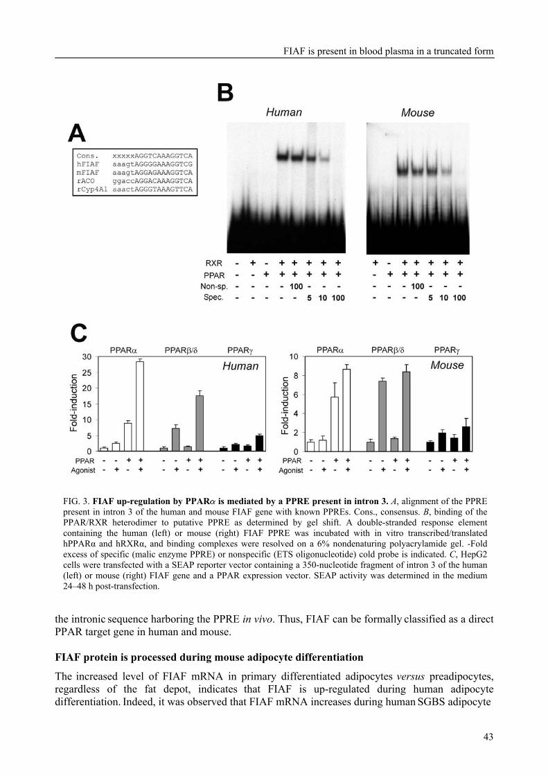
FIAF is present in blood plasma in a truncated form
43
FIG. 3. FIAF up-regulation by PPAR is mediated by a PPRE present in intron 3. A, alignment of the PPRE present in intron 3 of the human and mouse FIAF gene with known PPREs. Cons., consensus. B, binding of the PPAR/RXR heterodimer to putative PPRE as determined by gel shift. A double-stranded response element containing the human (left) or mouse (right) FIAF PPRE was incubated with in vitro transcribed/translated hPPARα and hRXRα, and binding complexes were resolved on a 6% nondenaturing polyacrylamide gel. -Fold excess of specific (malic enzyme PPRE) or nonspecific (ETS oligonucleotide) cold probe is indicated. C, HepG2 cells were transfected with a SEAP reporter vector containing a 350-nucleotide fragment of intron 3 of the human (left) or mouse (right) FIAF gene and a PPAR expression vector. SEAP activity was determined in the medium 24–48 h post-transfection.
the intronic sequence harboring the PPRE in vivo. Thus, FIAF can be formally classified as a direct PPAR target gene in human and mouse. FIAF protein is processed during mouse adipocyte differentiation
The increased level of FIAF mRNA in primary differentiated adipocytes versus preadipocytes, regardless of the fat depot, indicates that FIAF is up-regulated during human adipocyte differentiation. Indeed, it was observed that FIAF mRNA increases during human SGBS adipocyte
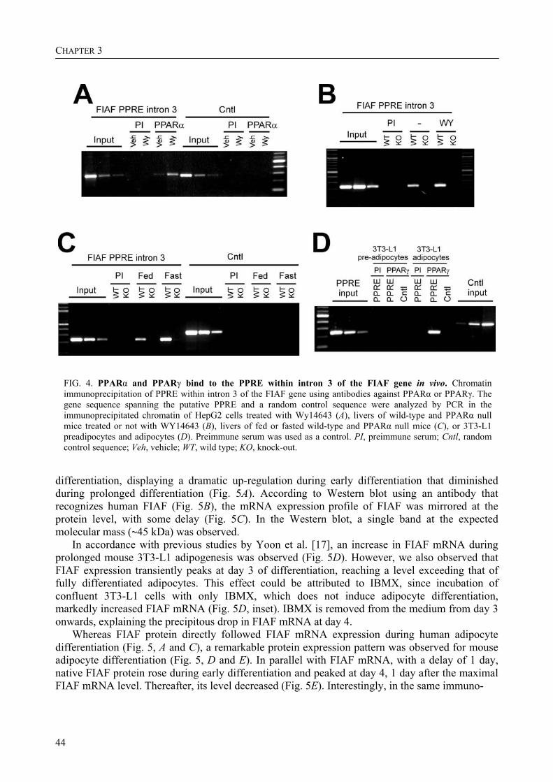
CHAPTER 3
44
FIG. 4. PPARα and PPARγ bind to the PPRE within intron 3 of the FIAF gene in vivo. Chromatin immunoprecipitation of PPRE within intron 3 of the FIAF gene using antibodies against PPARα or PPARγ. The gene sequence spanning the putative PPRE and a random control sequence were analyzed by PCR in the immunoprecipitated chromatin of HepG2 cells treated with Wy14643 (A), livers of wild-type and PPARα null mice treated or not with WY14643 (B), livers of fed or fasted wild-type and PPARα null mice (C), or 3T3-L1 preadipocytes and adipocytes (D). Preimmune serum was used as a control. PI, preimmune serum; Cntl, random control sequence; Veh, vehicle; WT, wild type; KO, knock-out.
differentiation, displaying a dramatic up-regulation during early differentiation that diminished during prolonged differentiation (Fig. 5A). According to Western blot using an antibody that recognizes human FIAF (Fig. 5B), the mRNA expression profile of FIAF was mirrored at the protein level, with some delay (Fig. 5C). In the Western blot, a single band at the expected
molecular mass ( 45 kDa) was observed. In accordance with previous studies by Yoon et al. [17], an increase in FIAF mRNA during
prolonged mouse 3T3-L1 adipogenesis was observed (Fig. 5D). However, we also observed that FIAF expression transiently peaks at day 3 of differentiation, reaching a level exceeding that of fully differentiated adipocytes. This effect could be attributed to IBMX, since incubation of confluent 3T3-L1 cells with only IBMX, which does not induce adipocyte differentiation, markedly increased FIAF mRNA (Fig. 5D, inset). IBMX is removed from the medium from day 3 onwards, explaining the precipitous drop in FIAF mRNA at day 4.
Whereas FIAF protein directly followed FIAF mRNA expression during human adipocyte differentiation (Fig. 5, A and C), a remarkable protein expression pattern was observed for mouse adipocyte differentiation (Fig. 5, D and E). In parallel with FIAF mRNA, with a delay of 1 day, native FIAF protein rose during early differentiation and peaked at day 4, 1 day after the maximal FIAF mRNA level. Thereafter, its level decreased (Fig. 5E). Interestingly, in the same immuno-
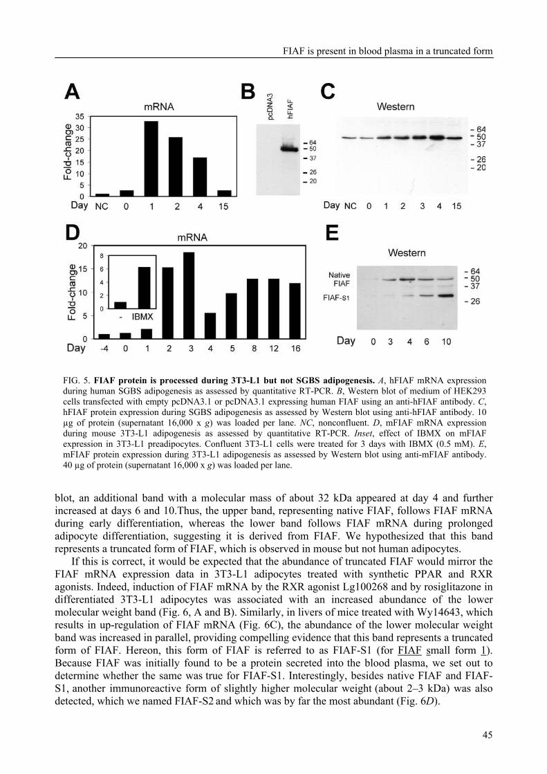
FIAF is present in blood plasma in a truncated form
45
FIG. 5. FIAF protein is processed during 3T3-L1 but not SGBS adipogenesis. A, hFIAF mRNA expression during human SGBS adipogenesis as assessed by quantitative RT-PCR. B, Western blot of medium of HEK293 cells transfected with empty pcDNA3.1 or pcDNA3.1 expressing human FIAF using an anti-hFIAF antibody. C, hFIAF protein expression during SGBS adipogenesis as assessed by Western blot using anti-hFIAF antibody. 10 µg of protein (supernatant 16,000 x g) was loaded per lane. NC, nonconfluent. D, mFIAF mRNA expression during mouse 3T3-L1 adipogenesis as assessed by quantitative RT-PCR. Inset, effect of IBMX on mFIAF expression in 3T3-L1 preadipocytes. Confluent 3T3-L1 cells were treated for 3 days with IBMX (0.5 mM). E, mFIAF protein expression during 3T3-L1 adipogenesis as assessed by Western blot using anti-mFIAF antibody. 40 µg of protein (supernatant 16,000 x g) was loaded per lane.
blot, an additional band with a molecular mass of about 32 kDa appeared at day 4 and further increased at days 6 and 10.Thus, the upper band, representing native FIAF, follows FIAF mRNA during early differentiation, whereas the lower band follows FIAF mRNA during prolonged adipocyte differentiation, suggesting it is derived from FIAF. We hypothesized that this band represents a truncated form of FIAF, which is observed in mouse but not human adipocytes.
If this is correct, it would be expected that the abundance of truncated FIAF would mirror the FIAF mRNA expression data in 3T3-L1 adipocytes treated with synthetic PPAR and RXR agonists. Indeed, induction of FIAF mRNA by the RXR agonist Lg100268 and by rosiglitazone in differentiated 3T3-L1 adipocytes was associated with an increased abundance of the lower molecular weight band (Fig. 6, A and B). Similarly, in livers of mice treated with Wy14643, which results in up-regulation of FIAF mRNA (Fig. 6C), the abundance of the lower molecular weight band was increased in parallel, providing compelling evidence that this band represents a truncated form of FIAF. Hereon, this form of FIAF is referred to as FIAF-S1 (for FIAF small form 1). Because FIAF was initially found to be a protein secreted into the blood plasma, we set out to determine whether the same was true for FIAF-S1. Interestingly, besides native FIAF and FIAF-S1, another immunoreactive form of slightly higher molecular weight (about 2–3 kDa) was also detected, which we named FIAF-S2 and which was by far the most abundant (Fig. 6D).

CHAPTER 3
46
FIG. 6. The truncated form of FIAF is up-regulated in concert with FIAF mRNA by synthetic RXR/PPAR agonists. mFIAF protein (30 µg of total protein loaded per lane) and mRNA expression in differentiated 3T3-L1 adipocytes (day 10) after treatment for 48 h with 5 µM Lg100268 (A) or rosiglitazone (B) as assessed by Western blot and quantitative RT-PCR are shown. C, mFIAF protein (12 µg of total protein loaded per lane) and mRNA in liver of mice fed WY14643 (0.1%) for 5 days. D, Western blot of mouse plasma using anti-mFIAF antibody in the absence or presence of the peptide epitope. E, presence of truncated mFIAF variants in mouse white adipose tissue, blood plasma, and mouse liver. The position of native mFIAF is shown by transfection of pcDNA3 expressing mFIAF into HEK293 cells. Medium was collected, precipitated using trichloroacetic acid, and prepared for SDS-PAGE.
Preincubation of the mouse FIAF antibody with its peptide epitope completely abolished all three forms. Notice that in Fig. 6E native FIAF is barely visible because the blot was exposed for less time. It is not inconceivable that FIAF-S2 might represent a phosphorylated or glycosylated form of FIAF-S1. Both FIAF-S1 and FIAF-S2 were well detected in mouse WAT, whereas only native FIAF and FIAF-S1 were detected in mouse liver (Fig. 6E). Together, these data suggest that FIAF is present in truncated forms in mouse blood plasma. FIAF-S1 and FIAF-S2 are present in human blood plasma
To establish that FIAF is also present in truncated forms in human plasma, Western blot was carried out on human blood plasma using an anti-hFIAF antibody. Almost copying the picture of mouse blood plasma, in human plasma both native human FIAF protein at 50 kDa but also two bands of lower molecular weight were observed, probably corresponding to FIAF-S1 and FIAF-S2 (Fig. 7A). Incubation with the peptide epitope caused the complete disappearance of all bands. The molecular weight of the putative FIAF-S1 and FIAF-S2 in human was lower than that of the same
species in mice. Omission of dithiothreitol in the SDS-sample buffer led to the appearance of a
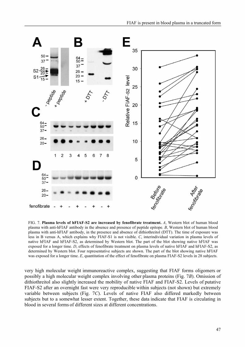
FIAF is present in blood plasma in a truncated form
47
FIG. 7. Plasma levels of hFIAF-S2 are increased by fenofibrate treatment. A, Western blot of human blood plasma with anti-hFIAF antibody in the absence and presence of peptide epitope. B, Western blot of human blood plasma with anti-hFIAF antibody, in the presence and absence of dithiothreitol (DTT). The time of exposure was less in B versus A, which explains why FIAF-S1 is not visible. C, interindividual variation in plasma levels of native hFIAF and hFIAF-S2, as determined by Western blot. The part of the blot showing native hFIAF was exposed for a longer time. D, effects of fenofibrate treatment on plasma levels of native hFIAF and hFIAF-S2, as determined by Western blot. Four representative subjects are shown. The part of the blot showing native hFIAF was exposed for a longer time. E, quantitation of the effect of fenofibrate on plasma FIAF-S2 levels in 28 subjects.
very high molecular weight immunoreactive complex, suggesting that FIAF forms oligomers or possibly a high molecular weight complex involving other plasma proteins (Fig. 7B). Omission of dithiothreitol also slightly increased the mobility of native FIAF and FIAF-S2. Levels of putative FIAF-S2 after an overnight fast were very reproducible within subjects (not shown) but extremely variable between subjects (Fig. 7C). Levels of native FIAF also differed markedly between
subjects but to a somewhat lesser extent. Together, these data indicate that FIAF is circulating in blood in several forms of different sizes at different concentrations.
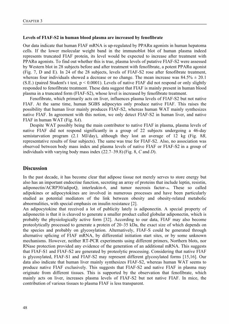
CHAPTER 3
48
Levels of FIAF-S2 in human blood plasma are increased by fenofibrate
Our data indicate that human FIAF mRNA is up-regulated by PPARα agonists in human hepatoma cells. If the lower molecular weight band in the immunoblot blot of human plasma indeed represents truncated FIAF protein, its level would be expected to increase after treatment with PPARα agonists. To find out whether this is true, plasma levels of putative FIAF-S2 were assessed by Western blot in 28 subjects before and after treatment with fenofibrate, a potent PPARα agonist (Fig. 7, D and E). In 24 of the 28 subjects, levels of FIAF-S2 rose after fenofibrate treatment, whereas four individuals showed a decrease or no change. The mean increase was 84.5% ± 20.1 (S.E.) (paired Student's t test, p < 0.0001). Levels of native FIAF did not respond or only slightly responded to fenofibrate treatment. These data suggest that FIAF is mainly present in human blood plasma in a truncated form (FIAF-S2), whose level is increased by fenofibrate treatment.
Fenofibrate, which primarily acts on liver, influences plasma levels of FIAF-S2 but not native FIAF. At the same time, human SGBS adipocytes only produce native FIAF. This raises the possibility that human liver mainly produces FIAF-S2, whereas human WAT mainly synthesizes native FIAF. In agreement with this notion, we only detect FIAF-S2 in human liver, and native FIAF in human WAT (Fig. 8A).
Despite WAT possibly being the main contributor to native FIAF in plasma, plasma levels of native FIAF did not respond significantly in a group of 22 subjects undergoing a 46-day semistarvation program (2.1 MJ/day), although they lost an average of 12 kg (Fig. 8B, representative results of four subjects). The same was true for FIAF-S2. Also, no association was observed between body mass index and plasma levels of native FIAF or FIAF-S2 in a group of individuals with varying body mass index (22.7–39.8) (Fig. 8, C and D). Discussion In the past decade, it has become clear that adipose tissue not merely serves to store energy but also has an important endocrine function, secreting an array of proteins that include leptin, resistin, adiponectin/ACRP30/adipoQ, interleukin-6, and tumor necrosis factor- . These so called adipokines or adipocytokines are involved in numerous processes and have been particularly studied as potential mediators of the link between obesity and obesity-related metabolic abnormalities, with special emphasis on insulin resistance [2]. An adipocytokine that received a lot of publicity lately is adiponectin. A special property of adiponectin is that it is cleaved to generate a smaller product called globular adiponectin, which is probably the physiologically active form [32]. According to our data, FIAF may also become proteolytically processed to generate a protein of 20–35 kDa, the exact size of which depends on the species and probably on glycosylation. Alternatively, FIAF-S could be generated through alternative splicing of FIAF mRNA, by differential initiation start sites, or by some unknown mechanisms. However, neither RT-PCR experiments using different primers, Northern blots, nor RNase protection provided any evidence of the generation of an additional mRNA. This suggests that FIAF-S1 and FIAF-S2 are generated by proteolytic processing. Considering that native FIAF is glycosylated, FIAF-S1 and FIAF-S2 may represent different glycosylated forms [15,16]. Our data also indicate that human liver mainly synthesizes FIAF-S2, whereas human WAT seems to produce native FIAF exclusively. This suggests that FIAF-S2 and native FIAF in plasma may originate from different tissues. This is supported by the observation that fenofibrate, which mainly acts on liver, increases plasma levels of FIAF-S2 but not native FIAF. In mice, the contribution of various tissues to plasma FIAF is less transparent.
FIAF is present in blood plasma in a truncated form
49
FIG. 8. Native FIAF and FIAF-S2 are produced by adipose tissue and liver, respectively, yet plasma levels are not influenced by semistarvation or associated with body mass index. A, presence of native hFIAF and hFIAF-S2 in human liver, plasma, and white adipose tissue samples according to Western blot using anti-hFIAF antibody. B, plasma levels of native hFIAF and hFIAF-S2 before and after 46 days of semistarvation, as determined by Western blot. Four representative subjects are shown. The part of the blot showing native hFIAF was exposed for a longer time. Association between plasma levels of native hFIAF (C) or hFIAF-S2 (D) and body mass index.
Proteolytic processing of prohormone precursor proteins is a common theme in endocrinology. Numerous protein and peptide hormones, including insulin, glucagon, and adipocytokines such as tumor necrosis factor- and adiponectin, are proteolytically cleaved to generate the smaller functional form of the protein. The most common processing recognition site in prohormones consists of a doublet of basic amino acids [33], which is recognized by subtilisin-like proprotein convertases, although other types of motifs are also possible. Carboxypeptidase E is responsible for the removal of carboxyl-terminal basic residues exposed by the endoproteases [34]. Interestingly, in the primary structure of FIAF, two conserved adjacent arginines could be identified, which might represent proteolytic recognition sites. Digestion around this site (Arg229 and Arg230 in hFIAF) would be compatible with the size of fragments FIAF-S1 and FIAF-S2. Recently, it was found that expression of the proprotein convertases PACE4, PC7, and furin increases during 3T3-L1 adipocyte differentiation, when processing of native FIAF to FIAF-S1
CHAPTER 3
50
becomes apparent [35]. Consequently, it is conceivable that these enzymes participate in the processing of FIAF in 3T3-L1 adipocytes.
Besides being proteolytically processed, adiponectin also forms higher order oligomers, which may have a different functional activity than monomeric adiponectin. Resistin has been shown to self-associate as well [3,36], which again may influence functional activity. According to our data, FIAF may also be present in human blood plasma as higher order oligomers, although the exact composition of the observed higher molecular weight complex(es) remains to be determined. Similar to adiponectin and resistin, oligomerization of FIAF may influence functional activity.
Very recently, Ono et al. reported that ANGPTL3 is cleaved in vivo, and, similar to our observations for FIAF (or ANGPTL4), is present in mouse blood plasma in several forms of around 30 kDa [37]. Interestingly, it was found that the resulting N-terminal fragment is probably responsible for the plasma triglyceride-raising effect of ANGPTL3. Furthermore, while our manuscript was in preparation, data were published showing that recombinant FIAF protein is truncated and forms oligomers in HEK293 cells and in vivo [38]. No data were provided on endogenous FIAF, in contrast to the present paper. Although details about the site of truncation seem to be different between the two papers, together they suggest that proteolytic processing, and perhaps oligomerization, may be important for FIAF function. Thus, proteolytic processing may be common among members of this protein family and may serve to regulate functional activity.
Previous studies have indicated that, at least in mouse, FIAF mRNA is most highly expressed in white adipose tissue [15]. According to our data, human WAT mainly produces native FIAF. The lack of a significant association between body mass index and plasma levels of native FIAF and the absence of an effect of prolonged weight loss on native FIAF suggest that either the size of WAT has little impact on the total amount of native FIAF released from WAT into blood plasma or that adipose tissue may not be the primary source for native FIAF in human plasma.
Experiments in mice have shown that both hepatic and adipose expression of FIAF are elevated by fasting [15]. With respect to FIAF in plasma, levels of native FIAF were found to be elevated after fasting [15], whereas levels of FIAF-S2 or FIAF-S1 did not seem to be affected2. Preliminary data indicate that the fasting-induced up-regulation of FIAF mRNA in adipose tissue may not be observed in mice of the FVB strain. With regard to humans, it is unclear whether fasting causes up-regulation of FIAF mRNA in liver and WAT. Levels of FIAF in plasma do not appear to be influenced by short term fasting (data not shown) or long term semistarvation. Thus, the term fasting-induced adipose factor may not aptly describe the behavior of FIAF in several species.
Previously, we and others have demonstrated that in mice FIAF is up-regulated by both PPARα and PPARγ [15,17]. Here it is shown that this regulation also occurs in humans, in contrast to many other PPARα target genes. Furthermore, besides PPARα and PPARγ, PPARβ is similarly able to induce FIAF expression in hepatocytes and adipocytes. It is also shown that up-regulation of FIAF expression by PPARs is, at least partly, mediated by a PPRE present in intron 3. Via chromatin immunoprecipitation on livers of fasted and fed or Wy14643-treated mice, direct in vivo binding of PPARα to intron 3 was demonstrated, which was enhanced by fasting and by Wy14643. Furthermore, binding of PPARα to the same sequence was enhanced by Wy14643 in human HepG2 cells. Finally, binding of PPARγ to the sequence could be demonstrated in differentiated 3T3-L1 adipocytes but not preadipocytes. Thus, FIAF can be added to the list of direct PPAR target genes. Although the presence of a functional PPRE within an intron is remarkable, it is not completely uncommon. Indeed, recently the presence of a functional PPRE within intron 3 of the rat peroxisomal thiolase B gene was demonstrated [39].
2 S. Mandard, F. Zandbergen, and S. Kersten, unpublished data.
FIAF is present in blood plasma in a truncated form
51
Plasma levels of FIAF-S2 are increased by fenofibrate treatment. Inasmuch as there is evidence that FIAF is involved in lipid metabolism, it can be speculated that the effects of synthetic PPARα agonists on plasma lipid levels may be partially mediated via changes in FIAF expression. Further studies are necessary to ascertain the potential of FIAF as a target for treatment of various forms of dyslipidemia. Acknowledgements We thank Dr. Mark Leibowitz (Ligand Pharmaceuticals) for the kind gift of Lg100268, Marco Alves for the synthesis of L165041, Dr. M. Wabitsch for the gift of the SGBS cell line, Dr. Folkert Kuipers for the gift of a human liver sample, and Dr. Wim Saris and Monica Mars for providing human blood plasma samples for this study. This work was supported by the Netherlands Organization for Scientific Research, with additional support by the Royal Netherlands Academy of Art and Sciences, the Dutch Diabetes Foundation, the Wageningen Center for Food Sciences, the Swiss National Science Foundation, and the Human Frontier Science Program. References 1 Zhang, Y., Proenca, R., Maffei, M., Barone, M., Leopold, L., Friedman, J. M. (1994) Positional
cloning of the mouse obese gene and its human homologue. Nature 372, 425-432 2 Ahima, R. S., Flier, J. S. (2000) Adipose tissue as an endocrine organ. Trends Endocrinol. Metab. 11,
327-332 3 Steppan, C. M., Bailey, S. T., Bhat, S., et al. (2001) The hormone resistin links obesity to diabetes.
Nature 409, 307-312 4 Banerjee, R. R., Rangwala, S. M., Shapiro, J. S., et al. (2004) Regulation of fasted blood glucose by
resistin. Science 303, 1195-1198 5 Kim, K. H., Lee, K., Moon, Y. S., Sul, H. S. (2001) A cysteine-rich adipose tissue-specific secretory
factor inhibits adipocyte differentiation. J. Biol. Chem. 276, 11252-11256 6 Hu, E., Liang, P., Spiegelman, B. M. (1996) AdipoQ is a novel adipose-specific gene dysregulated in
obesity. J. Biol. Chem. 271, 10697-10703 7 Scherer, P. E., Williams, S., Fogliano, M., Baldini, G., Lodish, H. F. (1995) A novel serum protein
similar to C1q, produced exclusively in adipocytes. J. Biol. Chem. 270, 26746-26749 8 Arita, Y., Kihara, S., Ouchi, N., et al. (1999) Paradoxical decrease of an adipose-specific protein,
adiponectin, in obesity. Biochem. Biophys. Res. Commun. 257, 79-83 9 Cianflone, K. M., Sniderman, A. D., Walsh, M. J., Vu, H. T., Gagnon, J., Rodriguez, M. A. (1989)
Purification and characterization of acylation stimulating protein. J. Biol. Chem. 264, 426-430 10 Samad, F., Yamamoto, K., Loskutoff, D. J. (1996) Distribution and regulation of plasminogen
activator inhibitor-1 in murine adipose tissue in vivo. Induction by tumor necrosis factor-alpha and lipopolysaccharide. J. Clin. Invest. 97, 37-46
11 Frederich, R. C., Jr., Kahn, B. B., Peach, M. J., Flier, J. S. (1992) Tissue-specific nutritional regulation of angiotensinogen in adipose tissue. Hypertension 19, 339-344
12 Trayhurn, P., Duncan, J. S., Wood, A. M., Beattie, J. H. (2000) Metallothionein gene expression and secretion in white adipose tissue. Am. J. Physiol. Regul. Integr. Comp. Physiol. 279, R2329-2335
13 Wallenius, V., Wallenius, K., Ahren, B., et al. (2002) Interleukin-6-deficient mice develop mature-onset obesity. Nat. Med. 8, 75-79
14 Hotamisligil, G. S., Shargill, N. S., Spiegelman, B. M. (1993) Adipose expression of tumor necrosis factor-alpha: direct role in obesity-linked insulin resistance. Science 259, 87-91
15 Kersten, S., Mandard, S., Tan, N. S., et al. (2000) Characterization of the fasting-induced adipose factor FIAF, a novel peroxisome proliferator-activated receptor target gene. J. Biol. Chem. 275, 28488-28493
CHAPTER 3
52
16 Kim, I., Kim, H. G., Kim, H., et al. (2000) Hepatic expression, synthesis and secretion of a novel fibrinogen/angiopoietin-related protein that prevents endothelial-cell apoptosis. Biochem. J. 346 Pt 3, 603-610
17 Yoon, J. C., Chickering, T. W., Rosen, E. D., et al. (2000) Peroxisome proliferator-activated receptor gamma target gene encoding a novel angiopoietin-related protein associated with adipose differentiation. Mol. Cell. Biol. 20, 5343-5349
18 Belanger, A. J., Lu, H., Date, T., et al. (2002) Hypoxia up-regulates expression of peroxisome proliferator-activated receptor gamma angiopoietin-related gene (PGAR) in cardiomyocytes: role of hypoxia inducible factor 1alpha. J. Mol. Cell Cardiol. 34, 765-774
19 Le Jan, S., Amy, C., Cazes, A., et al. (2003) Angiopoietin-like 4 is a proangiogenic factor produced during ischemia and in conventional renal cell carcinoma. Am. J. Pathol. 162, 1521-1528
20 Koishi, R., Ando, Y., Ono, M., et al. (2002) Angptl3 regulates lipid metabolism in mice. Nat. Genet. 30, 151-157
21 Shimizugawa, T., Ono, M., Shimamura, M., et al. (2002) ANGPTL3 decreases very low density lipoprotein triglyceride clearance by inhibition of lipoprotein lipase. J. Biol. Chem. 277, 33742-33748
22 Shimamura, M., Matsuda, M., Kobayashi, S., et al. (2003) Angiopoietin-like protein 3, a hepatic secretory factor, activates lipolysis in adipocytes. Biochem. Biophys. Res. Commun. 301, 604-609
23 Yoshida, K., Shimizugawa, T., Ono, M., Furukawa, H. (2002) Angiopoietin-like protein 4 is a potent hyperlipidemia-inducing factor in mice and inhibitor of lipoprotein lipase. J. Lipid Res. 43, 1770-1772
24 Desvergne, B., Wahli, W. (1999) Peroxisome proliferator-activated receptors: nuclear control of metabolism. Endocr. Rev. 20, 649-688
25 Mandard, S., Muller, M., Kersten, S. (2004) Peroxisome proliferator-activated receptor alpha target genes. Cell. Mol. Life Sci. 61, 393-416
26 Wabitsch, M., Brenner, R. E., Melzner, I., et al. (2001) Characterization of a human preadipocyte cell strain with high capacity for adipose differentiation. Int. J. Obes. Relat. Metab. Disord. 25, 8-15
27 Di Poi, N., Tan, N. S., Michalik, L., Wahli, W., Desvergne, B. (2002) Antiapoptotic role of PPARbeta in keratinocytes via transcriptional control of the Akt1 signaling pathway. Mol. Cell 10, 721-733
28 Mars, M., de Graaf, C., van Rossum, C. T., de Groot, C. P., Seidell, J. C., Kok, F. J. (2003) Leptin and insulin responses to a four-day energy-deficient diet in men with different weight history. Int. J. Obes. Relat. Metab. Disord. 27, 574-581
29 Hukshorn, C. J., Westerterp-Plantenga, M. S., Saris, W. H. (2003) Pegylated human recombinant leptin (PEG-OB) causes additional weight loss in severely energy-restricted, overweight men. Am. J. Clin. Nutr. 77, 771-776
30 Lawrence, J. W., Li, Y., Chen, S., et al. (2001) Differential gene regulation in human versus rodent hepatocytes by peroxisome proliferator-activated receptor (PPAR) alpha. PPAR alpha fails to induce peroxisome proliferation-associated genes in human cells independently of the level of receptor expresson. J. Biol. Chem. 276, 31521-31527
31 Hsu, M. H., Savas, U., Griffin, K. J., Johnson, E. F. (2001) Identification of peroxisome proliferator-responsive human genes by elevated expression of the peroxisome proliferator-activated receptor alpha in HepG2 cells. J. Biol. Chem. 276, 27950-27958
32 Fruebis, J., Tsao, T. S., Javorschi, S., et al. (2001) Proteolytic cleavage product of 30-kDa adipocyte complement-related protein increases fatty acid oxidation in muscle and causes weight loss in mice. Proc Natl. Acad. Sci. U.S.A. 98, 2005-2010
33 Harris, R. B. (1989) Processing of pro-hormone precursor proteins. Arch. Biochem. Biophys. 275, 315-333
34 Steiner, D. F. (1998) The proprotein convertases. Curr. Opin. Chem. Biol. 2, 31-39 35 Croissandeau, G., Basak, A., Seidah, N. G., Chretien, M., Mbikay, M. (2002) Proprotein convertases
are important mediators of the adipocyte differentiation of mouse 3T3-L1 cells. J. Cell Sci. 115, 1203-1211
FIAF is present in blood plasma in a truncated form
53
36 Chen, J., Wang, L., Boeg, Y. S., Xia, B., Wang, J. (2002) Differential dimerization and association among resistin family proteins with implications for functional specificity. J. Endocrinol. 175, 499-504
37 Ono, M., Shimizugawa, T., Shimamura, M., et al. (2003) Protein region important for regulation of lipid metabolism in ANGPTL3: ANGPTL3 is cleaved and activated in vivo. J. Biol. Chem. 278, 41804-41809
38 Ge, H., Yang, G., Huang, L., Motola, D. L., Pourbahrami, T., Li, C. (2004) Oligomerization and regulated proteolytic processing of angiopoietin-like protein 4. J. Biol. Chem. 279, 2038-2045
39 Hansmannel, F., Clemencet, M. C., Le Jossic-Corcos, C., Osumi, T., Latruffe, N., Nicolas-Frances, V. (2003) Functional characterization of a peroxisome proliferator response-element located in the intron 3 of rat peroxisomal thiolase B gene. Biochem. Biophys. Res. Commun. 311, 149-155

54

G0/G1 switch gene 2 is a novel PPAR target gene
55
CHAPTER 4 The G0/G1 switch gene 2 is a novel PPAR target gene Fokko Zandbergen, Stéphane Mandard, Pascal Escher, Nguan Soon Tan, David Patsouris, Tim Jatkoe, Sandra Rojas-Caro, Steve Madore, Walter Wahli, Sherrie Tafuri, Michael Müller, and Sander Kersten Abstract PPARs (peroxisome-proliferator-activated receptors) α, β/δ and γ are a group of transcription factors that are involved in numerous processes, including lipid metabolism and adipogenesis. By comparing liver mRNAs of wild-type and PPARα-null mice using microarrays, a novel putative target gene of PPARα, G0S2 (G0/G1 switch gene 2), was identified. Hepatic expression of G0S2 was up-regulated by fasting and by the PPARα agonist Wy14643 in a PPARα-dependent manner. Surprisingly, the G0S2 mRNA level was highest in brown and white adipose tissue and was greatly up-regulated during mouse 3T3-L1 and human SGBS (Simpson–Golabi–Behmel syndrome) adipogenesis. Transactivation, gel shift and chromatin immunoprecipitation assays indicated that G0S2 is a direct PPARγ and probable PPARα target gene with a functional PPRE (PPAR-responsive element) in its promoter. Up-regulation of G0S2 mRNA seemed to be specific for adipogenesis, and was not observed during osteogenesis or myogenesis. In 3T3-L1 fibroblasts, expression of G0S2 was associated with growth arrest, which is required for 3T3-L1 adipogenesis. Together, these data indicate that G0S2 is a novel target gene of PPARs that may be involved in adipocyte differentiation. Key words: adipogenesis, G0/G1 switch gene 2 (G0S2), growth arrest, peroxisome-proliferator-activated receptor (PPAR). This chapter has been published in the Biochemical Journal 392, 313-324 (2005) Reproduced with permission, © the Biochemical Society
CHAPTER 4
56
Introduction PPARs (peroxisome-proliferator-activated receptors)1 represent a group of nuclear receptors that play pivotal roles in the regulation of energy metabolism [1]. These receptors function as ligand-activated transcription factors by binding to the promoters of target genes and inducing transcription upon activation by small lipophilic compounds. Three different PPARs can be distinguished: PPARα, PPARβ/δ and PPARγ. All three receptors are activated by (mainly polyunsaturated) fatty acids and various fatty-acid-derived compounds, such as eicosanoids.
PPARγ, which is most highly expressed in adipose tissue, is known as the master regulator of adipogenesis. Numerous studies, both in vivo and in vitro, have pointed to PPARγ as the transcription factor that drives adipocyte differentiation [2-5]. The role of PPARγ in adipogenesis is diverse, and concerns the regulation of cell-cycle withdrawal, as well as induction of fat-specific target genes that are involved in adipocyte metabolism. Indeed, PPARγ stimulates the expression of numerous genes that are involved in lipogenesis, including those for aP2 (adipocyte fatty-acid-binding protein), lipoprotein lipase and CD36/fatty acid translocase. Previous microarray studies have yielded a comprehensive picture of the likely target genes of PPARγ in adipose tissue and indicate a general role for PPARγ in the regulation of lipid metabolism [6], which is underlined by the therapeutic utilization of the PPARγ ligands thiazolidinediones in obesity-linked Type II diabetes.
Expression of PPARβ/δ is ubiquitous, which has been a major impediment in elucidating its assorted functions. The most compelling recent studies indicate that PPARβ/δ stimulates fatty acid oxidation in both adipose tissue and skeletal muscle [7,8], regulates hepatic VLDL (very-low-density lipoprotein) production and catabolism [9], and is involved in wound healing by governing keratinocyte differentiation [10]. Furthermore, PPARβ/δ has been connected with colon carcinogenesis, although conflicting results have been reported [11,12].
The last PPAR isotype, PPARα, has mostly been studied in the context of liver metabolism and is known to be a central regulator of hepatic fatty acid catabolism [13]. Evidence is accumulating that PPARα also governs several aspects of glucose metabolism [14]. Furthermore, it potently represses the hepatic inflammatory response by down-regulating the expression of numerous genes [15,16]. Indeed, up-regulation of various acute-phase proteins during hepatic inflammation may be linked directly to down-regulation of hepatic PPARα mRNA under these conditions [17]. Importantly, PPARα is the molecular target for the hypolipidaemic fibrates, a group of drugs that are prescribed for their ability to lower plasma triacylglycerols and elevate plasma HDL (high-density lipoprotein) levels.
Although much is already known about PPARs, significant gaps remain in our knowledge, particularly with respect to the set of genes that are regulated by PPARs in various organs. In the present study, we applied microarray technology to find putative targets of PPARα by comparing liver mRNA from PPARa-knockout mice and wild-type mice. One of the putative target genes identified, called G0S2 (G0/G1 switch gene 2), was subjected to detailed follow-up investigation. The collective data indicate that G0S2 is a direct PPAR target gene, with a functional PPRE (PPAR-responsive element) in its promoter, and may be involved in adipocyte differentiation. 1 Abbreviations used: aP2, adipocyte fatty-acid-binding protein; Avg Diff, average difference; BAT, brown adipose tissue; BMP-2, bone morphogenetic protein-2; BODIPY®, 4,4-difluoro-4-bora-3a,4a-diaza-s-indacene; ChIP, chromatin immunoprecipitation; CYP4A10, cytochrome P450, family 4, subfamily a, polypeptide 10; DMEM, Dulbecco's modified Eagle's medium; DsRed, Discosoma sp. red fluorescent protein; ER, endoplasmic reticulum; GFP, green fluorescent protein; G0S2, G0/G1 switch gene 2; GPDH, glycerol 3-phosphate dehydrogenase; h, human; HEK-293 cells, human embryonic kidney-293 cells; m, mouse; PPAR, peroxisome-proliferator-activated receptor; PPRE, PPAR-responsive element; Q-PCR, real-time quantitative PCR; RT, reverse transcriptase; RXR, retinoid X receptor; SEAP, secreted alkaline phosphatase; SGBS, Simpson–Golabi–Behmel syndrome; WAT, white adipose tissue.
G0/G1 switch gene 2 is a novel PPAR target gene
57
Experimental Materials
Wy14643 was obtained from ChemSyn laboratories. Rosiglitazone was from Alexis. Recombinant human insulin (Actrapid) was from Novo Nordisk. Recombinant human BMP-2 (bone morphogenetic protein-2) was from R&D systems. BODIPY® (4,4-difluoro-4-bora-3a,4a-diaza-s-indacene) 493/503 was from Molecular Probes. SYBR Green was from Eurogentec. DMEM (Dulbecco's modified Eagle's medium), foetal calf serum, calf serum and penicillin/streptomycin/fungizone were from Cambrex Bioscience. The 3T3-L1 cell line was purchased from ECACC (European Collection of Cell Culture). HEK-293 (human embryonic kidney-293) cells were from BD Biosciences. All other chemicals were from Sigma. Animals
Male pure-bred Sv129 and PPARa-null mice (2–3-month-old) on a Sv129 background were used. Fed mice were killed at the end of the dark cycle. Fasting was started at the onset of the light cycle for 6, 12 or 24 h (n=5 per group). For the feeding experiment with Wy14643, 3–5-month-old male wild-type and PPARα-null mice were fed with 0.1% Wy14643 for 5 days by mixing it in their food. Alternatively, they received a single dose of Wy14643 (400 µl of 10 mg/ml Wy14643 dissolved in 0.5% carboxymethylcellulose) and were killed 6 h later (n=5 per group). Blood was collected via orbital puncture. Livers were dissected and directly frozen in liquid nitrogen.
The animal experiments were approved by the animal experimentation committee of the Etat de Vaud (Switzerland) or Wageningen University. Affymetrix microarray
Total RNA was prepared from mouse livers using TRIzol® reagent (Invitrogen). For each microarray experiment, 10 µg of total liver RNA pooled from four mice was used for cRNA synthesis. RNA was pooled because pilot experiments with Affymetrix chips at Pfizer had indicated that the inter-animal variability in gene expression (determined by performing eight separate hybridizations of eight different mice of the same strain), as well as variability between repeated hybridizations of the same pooled RNA sample, were statistically insignificant. Hybridization, washing and scanning of Affymetrix Genechip Mu6500 probe arrays was according to standard Affymetrix protocols. Fluorimetric data were processed by Affymetrix GeneChip3.1 software, and the gene chips were globally scaled to all the probe sets with an identical target intensity value. Affymetrix software measures the expression level of a gene as an average difference value (Avg Diff) by comparing the intensity of hybridization of 20 sets of perfect match oligonucleotide probes to 20 sets of mismatch probes. Only genes with an Avg Diff above the threshold of 100 and with a difference in Avg Diff values between wild-type and PPARα-null mutant mice at least 2-fold were considered. RT (reverse transcriptase)-PCR
Total RNA was extracted from cells or tissue with TRIzol® reagent following the supplier's protocol. Total RNA (3–5 µg) was treated with amplification grade DNAse I (Invitrogen), then reverse-transcribed with oligo(dT) using Superscript II RT RNase H- (Invitrogen) following the supplier's recommendation. cDNA was PCR-amplified with Platinum Taq DNA polymerase (Invitrogen). Primer sequences used in the PCRs were chosen based on the sequences available in GenBank®. Primer sequences to amplify mG0S2 cDNA were 5´-TGCTGCCTCTCTTCCCACTG-
CHAPTER 4
58
C-3´ (forward) and 5´-GTAGGGTCAGTTCTGGATTCGGTG-3´ (reverse). Other sequences are available from S.K. on request. Q-PCR (real-time quantitative PCR)
Primers were designed to generate a PCR-amplification product of 100–200 bp. Only primer pairs yielding unique amplification products without primer dimer formation were subsequently used for Q-PCR assays. The primer pairs listed in Table 1 were used.
Table 1. Primer pairs used in Q-PCR.
Primer name Sequence hG0S2 (forward) 5´-CGCCGTGCCACTAAGGTC-3´hG0S2 (reverse) 5´-GCACACAGTCTCCATCAGGC-3´ m/rG0S2 (forward) 5´-AGTGCTGCCTCTCTTCCCAC-3´ m/rG0S2 (reverse) 5´-TTTCCATCTGAGCTCTGGGC-3´ mPPARγ (forward) 5´-CACAATGCCATCAGGTTTGG-3´ mPPARγ (reverse) 5´-GCTGGTCGATATCACTGGAGATC-mA-FABP (aP2) (forward) 5´-AAGAAGTGGGAGTGGGCTTT-3´ mA-FABP (aP2) (reverse) 5´-AATCCCCATTTACGCTGATG-3´ mOsteocalcin (forward) 5´-GCAGCTTGGTGCACACCTAG-3´ mOsteocalcin (reverse) 5´-GGAGCTGCTGTGACATCCAT-3´ mGPDH (forward) 5´-GCCTTCGCCAAGCTCTTCTG-3´ mGPDH (reverse) 5´-TAGCAGGTCGTGATGAGGTCTG-3´ mβ-Actin (forward) 5´-GATCTGGCACCACACCTTCT-3´ mβ-Actin (reverse) 5´-GGGGTGTTGAAGGTCTCAAA-3´ m36B4 (forward) 5´-AGCGCGTCCTGGCATTGTGTGG-3´ m36B4 (reverse) 5´-GGGCAGCAGTGGTGGCAGCAGC-3´ r36B4 (forward) 5´-CGGGAAGGCTGTGGTGCTGATG-3´ r36B4 (reverse) 5´-TCGGTGAGGTCCTCCTTGGTGAAC-
PCR was carried out using Platinum Taq polymerase and SYBR green on an iCycler PCR machine (Bio-Rad) according to the instructions from the manufacturer. Primary hepatocytes
Rat and mouse hepatocytes were isolated by two-step collagenase perfusion as described previously [18]. Viability was determined by Trypan Blue exclusion, and was at least 75%. Hepatocytes were suspended in William's E medium (Cambrex) supplemented with 10% (v/v) foetal calf serum, 20 m-units/ml insulin, 50 nM dexamethasone, 100 units of penicillin, 100 µg of streptomycin, 0.25 µg/ml fungizone and 50 µg/ml gentamycin. The next day, cells were incubated in fresh medium in the presence or absence of Wy14643 (25 µM) for 24 h. 3T3-L1 and SGBS (Simpson–Golabi–Behmel syndrome) adipogenesis assay
3T3-L1 fibroblasts were amplified in DMEM plus 10% (v/v) calf serum. At 2 days after reaching confluence (=day 0), the medium was changed to DMEM plus 10% (v/v) foetal calf serum and the following compounds were added: isobutyl-methylxanthine (0.5 mM), dexamethasone (1 µM) and insulin (5 µg/ml). On day 3, the medium was changed to DMEM plus 10% (v/v) foetal calf serum and insulin (5 µg/ml). The medium was subsequently changed every 3 days, and, from day 9 onwards, no further insulin was added.
The culture of the SGBS cells as well as their induction into mature human adipocytes were performed exactly as described previously [19].
G0/G1 switch gene 2 is a novel PPAR target gene
59
Western blot
The combined human/mouse polyclonal antibody used was directed against epitopes TVLGGRALSNRQHAS and EATLCSRALSLRQHAS of the human and mouse G0S2 proteins respectively. The peptide affinity-purified antibodies were generated in rabbit and ordered via Eurogentec's customized antibody production service. Western blot was carried out as described previously [20]. Transactivation assay
HepG2 cells were co-transfected by calcium phosphate precipitation with an mPPARα (m is mouse), mPPARβ/δ or mPPARγ1 expression vector and pGL3 reporter vector containing different size fragments of the hG0S2 (h is human) promoter. A β-galactosidase reporter vector was co-transfected to normalize for differences in transfection efficiency. After transfection, cells were incubated in the presence or absence of Wy14643 (50 µM), L165041 (5 µM) or rosiglitazone (5 µM) respectively for 24 h before lysis. A Promega luciferase assay and a standard β-galactosidase assay using 2-nitrophenyl-β-D-galactopyranoside as a substrate were used to measure the relative promoter activities.
To disable the G0S2 PPRE within the hG0S2 promoter, two separate partially overlapping PCR fragments were generated using the wild-type hG0S2 promoter as a template. The mutant sequence was verified by automated sequencing.
A 200 nt fragment surrounding the putative PPRE within the mG0S2 promoter was PCR-amplified from mouse genomic DNA (strain C57/B6) and subcloned into the KpnI and BglII sites of pTAL-SEAP (BD Biosciences). This reporter vector was transfected into HepG2 cells by calcium phosphate precipitation together with an expression vector for mPPARα, mPPARβ/δ or mPPARγ1 in the presence or absence of their respective ligands. A β-galactosidase reporter was co-transfected to normalize for differences in transfection efficiency. SEAP (secreted alkaline phosphatase) activity was measured in the medium 24 h post-transfection via the chemiluminescent SEAP reporter assay (Roche). Gel shift assay
hRXRα (retinoid X receptor), hPPARα and hPPARγ proteins were generated from pSG5 expression vectors using the TNT (transcription and translation)-coupled in vitro system (Promega). The following oligonucleotides were annealed: G0S2-PPRE, 5´-CTGGCCAGAAAA-TTGCAAAGGTCACTGA-3´ and 5´-CTGGTCAGTGACCTTTGCAATTTTCTGG-3´; G0S2-PPREmut, 5´-CTGGCCAGAAAATTGCTAAGGACACTGA-3´ and 5´-CTGGTCAGTGTCCTT-AGCAATTTTCTGG-3´; for specific competition malic enzyme PPRE, 5´-TCGCTTTCTGGGTC-AAAGTTGATCCA-3´ and 5´-CTGGTGGATCAACTTTGACCCAGAAAG-3´; and for non-specific competition Ets, 5´-TGGAATGTACCGGAAATAACACCA-3´ and 5´-TGGTGTTATT-TCCGGTACATTCCA-3´. Oligonucleotides were annealed and labelled by Klenow filling (Roche) using Redivue [α-32P]dCTP (3000 Ci/mmol) (Amersham Biosciences). In vitro translated proteins (0.5–0.8 µl per reaction) were pre-incubated for 15 min on ice in 1× binding buffer [80 mM KCl, 1 mM dithiothreitol, 10 mM Tris/HCl (pH 7.4), 10% (v/v) glycerol plus protease inhibitors] in the presence of 2 µg of poly(dI-dC)·(dI-dC), 5 µg of sonicated salmon sperm DNA and competitor oligonucleotides in a final volume of 20 ml. Then 1 ng (1 ng/µl) of radiolabelled oligonucleotide was added, and incubation proceeded for another 10 min at room temperature (25 °C). Complexes were separated on a 4% polyacrylamide gel (acrylamide/bisacrylamide, 37.5:1) equilibrated in 0.5× TBE (Tris/borate/EDTA) at 25 mA.
CHAPTER 4
60
In vivo ChIP (chromatin immunoprecipitation)
Pure-bred wild-type or PPARα-null mice on a Sv129 background were used. Mice were fed by gavage with either Wy14643 (50 mg/kg per day) or vehicle (0.5% carboxymethylcellulose) for 5 days. Alternatively, mice were fasted or not for 24 h. After the indicated treatment, mice were killed by cervical dislocation. The liver was rapidly perfused with pre-warmed (37 °C) PBS for 5 min followed by 0.2% collagenase for 10 min. The liver was diced and forced through a stainless steel sieve, and the hepatocytes were collected directly into DMEM containing 1% (w/v) formaldehyde. After incubation at 37 °C for 15 min, the hepatocytes were pelleted, and ChIP was carried out using PPARα-specific antibodies as described previously [10].
3T3-L1 cells were differentiated as described above. After cell lysis and sonication, the supernatant was diluted 20-fold in re-ChIP dilution buffer (1 mM EDTA, 20 mM Tris/HCl, pH 8.1, 50 mM NaCl and 1% Triton X-100) before incubation with antibodies against mouse PPARγ or PPARβ/δ. The remainder of the assay was carried out as described previously [10]. PCR was performed using primers flanking the putative PPRE in the mG0S2 promoter (amplified product -1937 to -1357) and a control sequence (amplified product -3555 to -3107). C2C12 osteo- and myo-genesis
C2C12 mesenchymal progenitor cells were differentiated into myoblasts by letting the cells grow to confluence. C2C12 cells were differentiated into osteoblasts by the addition of BMP-2 (500 ng/ml). Cell-cycle synchronization
3T3-L1 cells were seeded at low confluence in DMEM plus 10% (v/v) foetal calf serum. After 9 h, the medium was replaced by DMEM plus 0.2% (v/v) foetal calf serum for 33 h. After that, foetal calf serum was re-added to the cells at 10%, and cells were taken at regular intervals for RNA preparation. Cellular localization studies
The mG0S2 open reading frame was cloned into the EcoRI and BamHI sites of pEGFP-N2 and pDsRed1-N1 (BD Biosciences). The ER (endoplasmic reticulum) localization vector pDsRed2-ER (BD Biosciences) was used as a control vector for the ER. The mG0S2-containing pEGFP-N2 vector was co-transfected with pDsRed2-ER into HEK-293 cells. The mG0S2-containing pDsRed1-N1 vector was transfected into 3T3-L1 cells. Transfections were performed by calcium phosphate precipitation on 60% confluent cell cultures. For 3T3-L1 cells, after 8 h, medium was replaced with medium containing 0.1% (v/v) Tween 80. After 48 h, cells were washed with PBS and fixed in 3.5% (w/v) formaldehyde in PBS. BODIPY® 493/503 (saturated solution in 100% ethanol) was added to the fixing solution at 1:100, after which the cells were examined by fluorescence spectroscopy. Fluorescence microscopy was carried out 48 h post-transfection using a LSM510 confocal laser-scanning microscope (Zeiss). Results Microarray studies identify the G0S2 gene as a potential PPAR target gene
Microarray studies permit the expression monitoring of thousands of genes. To identify new putative PPAR target genes, mRNA from livers of wild-type and PPARα-null mice at different stages of fasting was compared using Affymetrix murine 6500 oligonucleotide microarrays.
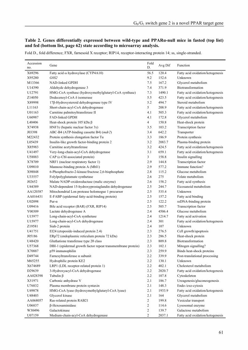
G0/G1 switch gene 2 is a novel PPAR target gene
61
Table 2. Genes differentially expressed between wild-type and PPARα-null mice in fasted (top list) and fed (bottom list, page 62) state according to microarray analysis. Fold D., fold difference; FXR, farnesoid X receptor; RIP14, receptor-interacting protein 14; ss, single-stranded.
Accession no. Gene Fold
D. Avg Dif Function
X69296 Fatty acid ω hydroxylase (CYP4A10) 56.5 120.4 Fatty acid oxidation/ketogenesis X95280 G0S2 9.2 152.6 Unknown M13366 NAD-linked GPDH 7.5 167.2 Glycerol metabolism U14390 Aldehyde dehydrogenase 3 7.4 371.9 Biotransformation U12791 HMG-CoA synthase (hydroxymethylglutaryl-CoA synthase) 7.3 1490.1 Fatty acid oxidation/ketogenesis Z14050 Dodecenoyl-CoA δ isomerase 5.5 423.5 Fatty acid oxidation/ketogenesis X89998 17β-Hydroxysteroid dehydrogenase type IV 5.2 494.7 Steroid metabolism L11163 Short-chain-acyl-CoA dehydrogenase 5 208.9 Fatty acid oxidation/ketogenesis U01163 Carnitine palmitoyltransferase II 4.1 505.3 Fatty acid oxidation/ketogenesis U60987 FAD-linked GPDH 4.1 172.8 Glycerol metabolism L40406 Heat-shock protein 105 kDa β 4 150.8 Heat-shock protein X74938 HNF3γ (hepatic nuclear factor 3γ) 3.5 103.2 Transcription factor J03398 ABC-B4 (ATP-binding cassette B4) (mdr2) 3.4 642.2 Transporter M22432 Protein synthesis elongation factor Tu 3.3 186.9 Protein synthesis L05439 Insulin-like growth factor-binding protein 2 3.2 2083.7 Plasma-binding protein X85983 Carnitine acetyltransferase 3.2 424.5 Fatty acid oxidation/ketogenesis U41497 Very-long-chain-acyl-CoA dehydrogenase 3.1 659.1 Fatty acid oxidation/ketogenesis U58883 CAP (c-Cbl-associated protein) 3 158.8 Insulin signalling X78709 NRF1 (nuclear respiratory factor 1) 2.9 144.8 Transcription factor U09010 Mannose-binding protein A (Mbl1) 2.9 577.2 Immune function? X98848 6-Phosphofructo-2-kinase/fructose 2,6-bisphosphate 2.8 115.2 Glucose metabolism U33557 Folylpolyglutamate synthetase 2.6 275 Folate metabolism J02652 Malate NADP-oxidoreductase (malic enzyme) 2.6 158.2 Fatty acid synthesis U44389 NAD-dependent 15-hydroxyprostaglandin dehydrogenase 2.5 244.7 Eicosanoid metabolism AA120387 Mitochondrial Lon protease holomogue 1 precursor 2.5 535.4 Unknown AA016431 E-FABP (epidermal fatty-acid-binding protein) 2.5 157.2 Fatty acid binding U02098 Pur-α 2.5 122.2 ssDNA-binding protein U09416 Bile acid receptor (BAR) (FXR, RIP14) 2.5 505.7 Transcription factor Y00309 Lactate dehydrogenase A 2.4 4506.4 Glucose metabolism U15977 Long-chain-acyl-CoA synthetase 2.4 1214.7 Fatty acid activation U15977 Long-chain-acyl-CoA dehydrogenase 2.4 301 Fatty acid oxidation/ketogenesis Z19581 Siah-2 protein 2.4 107 Unknown U41751 EI24 (etoposide-induced protein 2.4) 2.3 276.5 Cell growth/apoptosis J05186 ERp72 (endoplasmic reticulum protein 72 kDa) 2.3 286.5 Heat-shock protein U48420 Gluthatione transferase type 2θ class 2.3 809.8 Biotransformation U57368 DBI-1 (epidermal growth factor repeat transmembrane protein) 2.3 102.1 Mitogen signalling? X70887 p59 immunophilin 2.3 259.9 Binds heat-shock proteins D49744 Farnesyltransferase α subunit 2.2 339.9 Post-translational processing M65255 Hydrophilic protein KE2 2.2 130.1 Unknown X674689 LRP1 (LDL receptor-related protein 1) 2.2 402.1 Cholesterol metabolism D29639 3-Hydroxyacyl-CoA dehydrogenase 2.2 2020.7 Fatty acid oxidation/ketogenesis AA028398 Tubulin β 2.2 107.8 Cytoskeleton X51971 Carbonic anhydrase V 2.1 186.7 Ureagenesis/gluconeogenesis U76832 Plasma membrane protein syntaxin 2.1 148.3 Endo-/exo-cytosis U49878 HMG-CoA lyase (hydroxymethylglutaryl-CoA lyase) 2.1 1933.9 Fatty acid oxidation/ketogenesis U48403 Glycerol kinase 2.1 164 Glycerol metabolism AA068057 Ras-related protein RAB21 2 199.8 Vesicular transport U06837 β-Hexosaminidase 2 114.6 Lysosomal enzyme W30496 Galactokinase 2 139.7 Galactose metabolism U07159 Medium-chain-acyl-CoA dehydrogenase 2 2037.1 Fatty acid oxidation/ketogenesis
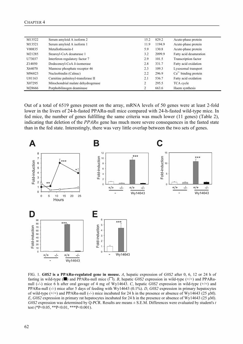
CHAPTER 4
62
M13522 Serum amyloid A isoform 2 15.2 829.2 Acute-phase protein M13521 Serum amyloid A isoform 1 11.9 1194.9 Acute-phase protein V00835 Metallothionein-I 5.9 130.8 Acute-phase protein M21285 Stearoyl-CoA desaturase 1 3.2 2099.9 Fatty acid desaturation U73037 Interferon regulatory factor 7 2.9 101.5 Transcription factor Z14050 Dodecenoyl-CoA δ-isomerase 2.8 331.7 Fatty acid oxidation X64070 Mannose phosphate receptor 46 2.3 109.3 Lysosomal transport M96823 Nucleobindin (Calnuc) 2.2 296.9 Ca2+ binding protein U01163 Carnitine palmitoyl-transferase II 2.1 536.7 Fatty acid oxidation X07295 Mitochondrial malate dehydrogenase 2 295.5 TCA cycle M28666 Porphobilinogen deaminase 2 663.6 Haem synthesis
Out of a total of 6519 genes present on the array, mRNA levels of 50 genes were at least 2-fold lower in the livers of 24-h-fasted PPARα-null mice compared with 24-h-fasted wild-type mice. In fed mice, the number of genes fulfilling the same criteria was much lower (11 genes) (Table 2), indicating that deletion of the PPARα gene has much more severe consequences in the fasted state than in the fed state. Interestingly, there was very little overlap between the two sets of genes.
FIG. 1. G0S2 is a PPARα-regulated gene in mouse. A, hepatic expression of G0S2 after 0, 6, 12 or 24 h of fasting in wild-type ( ) and PPARα-null mice ( ). B, hepatic G0S2 expression in wild-type (+/+) and PPARα-null (-/-) mice 6 h after oral gavage of 4 mg of Wy14643. C, hepatic G0S2 expression in wild-type (+/+) and PPARα-null (-/-) mice after 5 days of feeding with Wy14643 (0.1%). D, G0S2 expression in primary hepatocytes of wild-type (+/+) and PPARα-null (-/-) mice incubated for 24 h in the presence or absence of Wy14643 (25 µM). E, G0S2 expression in primary rat hepatocytes incubated for 24 h in the presence or absence of Wy14643 (25 µM). G0S2 expression was determined by Q-PCR. Results are means ± S.E.M. Differences were evaluated by student's t test (*P<0.05, **P<0.01, ***P<0.001).
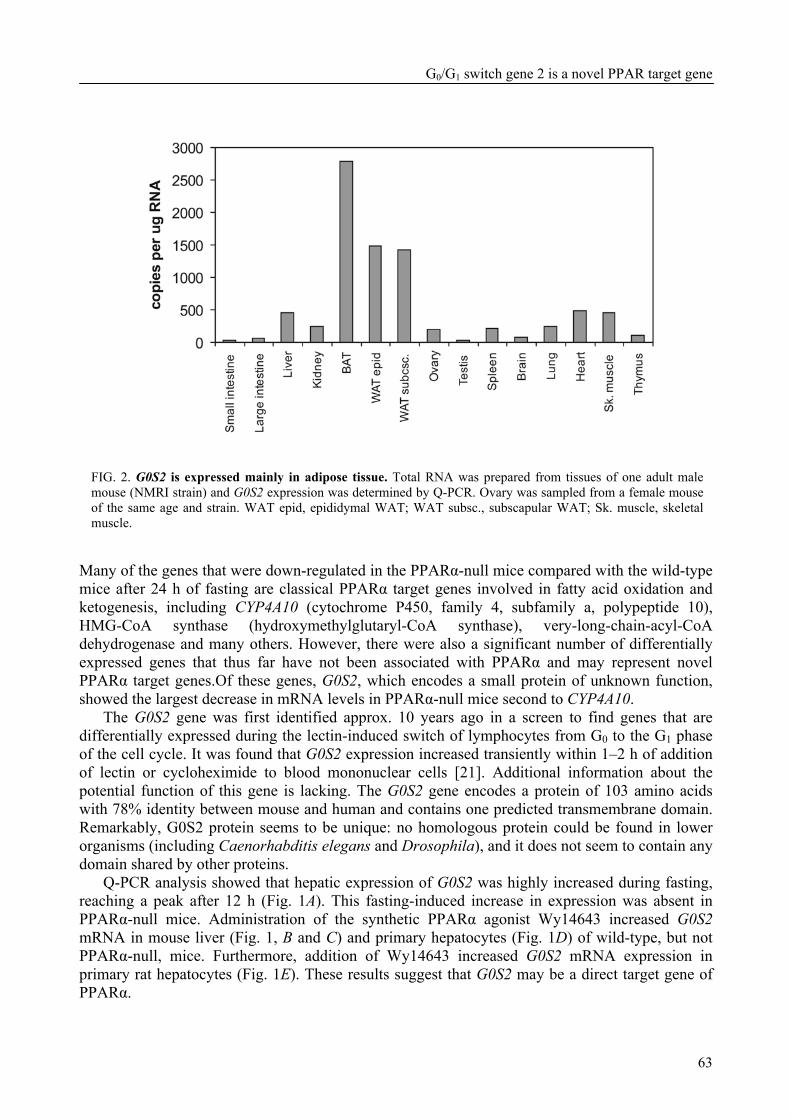
G0/G1 switch gene 2 is a novel PPAR target gene
63
FIG. 2. G0S2 is expressed mainly in adipose tissue. Total RNA was prepared from tissues of one adult male mouse (NMRI strain) and G0S2 expression was determined by Q-PCR. Ovary was sampled from a female mouse of the same age and strain. WAT epid, epididymal WAT; WAT subsc., subscapular WAT; Sk. muscle, skeletal muscle.
Many of the genes that were down-regulated in the PPARα-null mice compared with the wild-type mice after 24 h of fasting are classical PPARα target genes involved in fatty acid oxidation and ketogenesis, including CYP4A10 (cytochrome P450, family 4, subfamily a, polypeptide 10), HMG-CoA synthase (hydroxymethylglutaryl-CoA synthase), very-long-chain-acyl-CoA dehydrogenase and many others. However, there were also a significant number of differentially expressed genes that thus far have not been associated with PPARα and may represent novel PPARα target genes.Of these genes, G0S2, which encodes a small protein of unknown function, showed the largest decrease in mRNA levels in PPARα-null mice second to CYP4A10.
The G0S2 gene was first identified approx. 10 years ago in a screen to find genes that are differentially expressed during the lectin-induced switch of lymphocytes from G0 to the G1 phase of the cell cycle. It was found that G0S2 expression increased transiently within 1–2 h of addition of lectin or cycloheximide to blood mononuclear cells [21]. Additional information about the potential function of this gene is lacking. The G0S2 gene encodes a protein of 103 amino acids with 78% identity between mouse and human and contains one predicted transmembrane domain. Remarkably, G0S2 protein seems to be unique: no homologous protein could be found in lower organisms (including Caenorhabditis elegans and Drosophila), and it does not seem to contain any domain shared by other proteins.
Q-PCR analysis showed that hepatic expression of G0S2 was highly increased during fasting, reaching a peak after 12 h (Fig. 1A). This fasting-induced increase in expression was absent in PPARα-null mice. Administration of the synthetic PPARα agonist Wy14643 increased G0S2 mRNA in mouse liver (Fig. 1, B and C) and primary hepatocytes (Fig. 1D) of wild-type, but not PPARα-null, mice. Furthermore, addition of Wy14643 increased G0S2 mRNA expression in primary rat hepatocytes (Fig. 1E). These results suggest that G0S2 may be a direct target gene of PPARα.
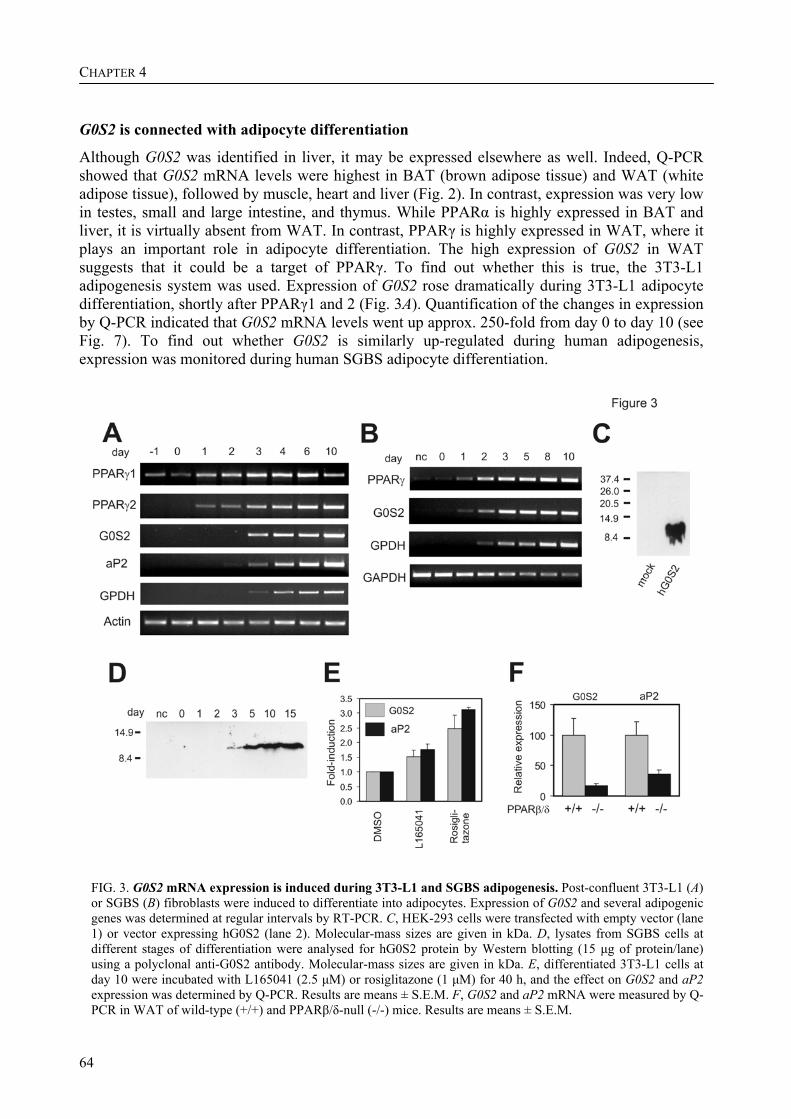
CHAPTER 4
64
G0S2 is connected with adipocyte differentiation
Although G0S2 was identified in liver, it may be expressed elsewhere as well. Indeed, Q-PCR showed that G0S2 mRNA levels were highest in BAT (brown adipose tissue) and WAT (white adipose tissue), followed by muscle, heart and liver (Fig. 2). In contrast, expression was very low in testes, small and large intestine, and thymus. While PPARα is highly expressed in BAT and liver, it is virtually absent from WAT. In contrast, PPARγ is highly expressed in WAT, where it plays an important role in adipocyte differentiation. The high expression of G0S2 in WAT suggests that it could be a target of PPARγ. To find out whether this is true, the 3T3-L1 adipogenesis system was used. Expression of G0S2 rose dramatically during 3T3-L1 adipocyte differentiation, shortly after PPARγ1 and 2 (Fig. 3A). Quantification of the changes in expression by Q-PCR indicated that G0S2 mRNA levels went up approx. 250-fold from day 0 to day 10 (see Fig. 7). To find out whether G0S2 is similarly up-regulated during human adipogenesis, expression was monitored during human SGBS adipocyte differentiation.
FIG. 3. G0S2 mRNA expression is induced during 3T3-L1 and SGBS adipogenesis. Post-confluent 3T3-L1 (A) or SGBS (B) fibroblasts were induced to differentiate into adipocytes. Expression of G0S2 and several adipogenic genes was determined at regular intervals by RT-PCR. C, HEK-293 cells were transfected with empty vector (lane 1) or vector expressing hG0S2 (lane 2). Molecular-mass sizes are given in kDa. D, lysates from SGBS cells at different stages of differentiation were analysed for hG0S2 protein by Western blotting (15 µg of protein/lane) using a polyclonal anti-G0S2 antibody. Molecular-mass sizes are given in kDa. E, differentiated 3T3-L1 cells at day 10 were incubated with L165041 (2.5 µM) or rosiglitazone (1 µM) for 40 h, and the effect on G0S2 and aP2 expression was determined by Q-PCR. Results are means ± S.E.M. F, G0S2 and aP2 mRNA were measured by Q-PCR in WAT of wild-type (+/+) and PPARβ/δ-null (-/-) mice. Results are means ± S.E.M.
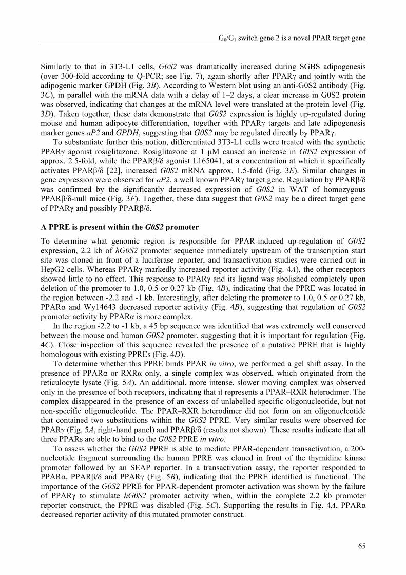
G0/G1 switch gene 2 is a novel PPAR target gene
65
Similarly to that in 3T3-L1 cells, G0S2 was dramatically increased during SGBS adipogenesis (over 300-fold according to Q-PCR; see Fig. 7), again shortly after PPARγ and jointly with the adipogenic marker GPDH (Fig. 3B). According to Western blot using an anti-G0S2 antibody (Fig. 3C), in parallel with the mRNA data with a delay of 1–2 days, a clear increase in G0S2 protein was observed, indicating that changes at the mRNA level were translated at the protein level (Fig. 3D). Taken together, these data demonstrate that G0S2 expression is highly up-regulated during mouse and human adipocyte differentiation, together with PPARγ targets and late adipogenesis marker genes aP2 and GPDH, suggesting that G0S2 may be regulated directly by PPARγ.
To substantiate further this notion, differentiated 3T3-L1 cells were treated with the synthetic PPARγ agonist rosiglitazone. Rosiglitazone at 1 µM caused an increase in G0S2 expression of approx. 2.5-fold, while the PPARβ/δ agonist L165041, at a concentration at which it specifically activates PPARβ/δ [22], increased G0S2 mRNA approx. 1.5-fold (Fig. 3E). Similar changes in gene expression were observed for aP2, a well known PPARγ target gene. Regulation by PPARβ/δ was confirmed by the significantly decreased expression of G0S2 in WAT of homozygous PPARβ/δ-null mice (Fig. 3F). Together, these data suggest that G0S2 may be a direct target gene of PPARγ and possibly PPARβ/δ. A PPRE is present within the G0S2 promoter
To determine what genomic region is responsible for PPAR-induced up-regulation of G0S2 expression, 2.2 kb of hG0S2 promoter sequence immediately upstream of the transcription start site was cloned in front of a luciferase reporter, and transactivation studies were carried out in HepG2 cells. Whereas PPARγ markedly increased reporter activity (Fig. 4A), the other receptors showed little to no effect. This response to PPARγ and its ligand was abolished completely upon deletion of the promoter to 1.0, 0.5 or 0.27 kb (Fig. 4B), indicating that the PPRE was located in the region between -2.2 and -1 kb. Interestingly, after deleting the promoter to 1.0, 0.5 or 0.27 kb, PPARα and Wy14643 decreased reporter activity (Fig. 4B), suggesting that regulation of G0S2 promoter activity by PPARα is more complex.
In the region -2.2 to -1 kb, a 45 bp sequence was identified that was extremely well conserved between the mouse and human G0S2 promoter, suggesting that it is important for regulation (Fig. 4C). Close inspection of this sequence revealed the presence of a putative PPRE that is highly homologous with existing PPREs (Fig. 4D).
To determine whether this PPRE binds PPAR in vitro, we performed a gel shift assay. In the presence of PPARα or RXRα only, a single complex was observed, which originated from the reticulocyte lysate (Fig. 5A). An additional, more intense, slower moving complex was observed only in the presence of both receptors, indicating that it represents a PPAR–RXR heterodimer. The complex disappeared in the presence of an excess of unlabelled specific oligonucleotide, but not non-specific oligonucleotide. The PPAR–RXR heterodimer did not form on an oligonucleotide that contained two substitutions within the G0S2 PPRE. Very similar results were observed for PPARγ (Fig. 5A, right-hand panel) and PPARβ/δ (results not shown). These results indicate that all three PPARs are able to bind to the G0S2 PPRE in vitro.
To assess whether the G0S2 PPRE is able to mediate PPAR-dependent transactivation, a 200-nucleotide fragment surrounding the human PPRE was cloned in front of the thymidine kinase promoter followed by an SEAP reporter. In a transactivation assay, the reporter responded to PPARα, PPARβ/δ and PPARγ (Fig. 5B), indicating that the PPRE identified is functional. The importance of the G0S2 PPRE for PPAR-dependent promoter activation was shown by the failure of PPARγ to stimulate hG0S2 promoter activity when, within the complete 2.2 kb promoter reporter construct, the PPRE was disabled (Fig. 5C). Supporting the results in Fig. 4A, PPARα decreased reporter activity of this mutated promoter construct.
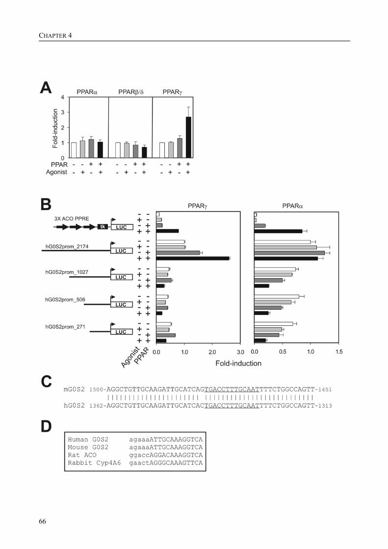
CHAPTER 4
66
G0/G1 switch gene 2 is a novel PPAR target gene
67
FIG. 4. G0S2 promoter is regulated by PPARs. A, hG0S2 reporter construct containing 2174 bp of immediate upstream promoter region was transfected into HepG2 cells together with a PPAR expression vector. Transfected cells were incubated for 24 h in the presence or absence of ligand. Normalized luciferase activity in the absence of PPAR and ligand was set at 1. Results are means ± S.E.M. for at least three independent experiments. B, hG0S2 reporter constructs containing 2174, 1027, 506 or 271 bp of immediate upstream promoter region were transfected into HepG2 cells together with an mPPARγ1 or mPPARα expression vector. Transfected cells were incubated for 24 h in the presence or absence of ligand. A luciferase reporter containing three copies of the acyl-CoA oxidase PPRE was used as a positive control. Normalized luciferase activity of the 2174 bp reporter in the absence of PPAR and ligand was set at 1. Results are means ± S.E.M. C, alignment of a putative regulatory region within mG0S2 and hG0S2 promoter about 1.5 kb upstream of transcription start site. The putative PPRE is underlined. D, alignment of a putative PPRE present within G0S2 promoter with established PPREs. Lower-case letters indicate the DNA base-pair sequence preceding the PPRE, which is represented by upper-case letters.
Finally, to investigate whether PPARα is bound to the G0S2 PPRE in mouse liver, in vivo ChIP was performed using an anti-PPARα antibody. In mice, treatment with Wy14643 enhanced binding of PPARα to the PPRE sequence in liver, which was not observed in PPARα-null mice (Fig. 5D). Similarly, fasting enhanced binding of PPARα to the PPRE sequence, which was not observed in the PPARα null mice (Fig. 5E). No detectable immunoprecipitation was observed with pre-immune serum and no amplification was observed for a control sequence. Furthermore, using ChIP, we observed binding of PPARγ (Fig. 5F) and PPARβ/δ (Fig. 5G) to the PPRE sequence in differentiated 3T3-L1 adipocytes, but not in pre-adipocytes. These results demonstrate that PPARα, PPARγ, and PPARβ/δ bind to the PPRE identified within the G0S2 promoter in vivo. Thus G0S2 can be formally classified as a direct PPAR target gene in human and mouse. G0S2 protein can be localized to the ER
To get a better understanding of the function of G0S2, it is important to determine its intracellular localization. The presence of a single transmembrane helix indicated that G0S2 was probably anchored in a (sub)cellular membrane. To determine the precise intracellular localization of G0S2, a fusion construct was created between G0S2 and GFP (green fluorescent protein), which was transfected into HEK-293 cells. Because the Internet-based program PSORTII predicted G0S2 to be present in the ER, co-transfection was carried out with a marker vector for ER (pDsRed2-ER). Confocal fluorescence microscopy showed that GFP fluorescence was present in discrete regions within the cytoplasm, and that it perfectly overlapped with the DsRed (Discosoma sp. red fluorescent protein) fluorescence (Fig. 6A), indicating that G0S2 protein is probably present in the ER.
Unfortunately, our anti-G0S2 antibody was not functional in immunohistochemistry, which precluded localization of endogenous G0S2 protein in differentiated adipocytes. To examine whether G0S2 protein might be associated with lipid droplets, which originate from the ER, and/or to study the effect of lipid droplets on the intracellular localization of G0S2, undifferentiated 3T3-L1 cells were transfected with fusion constructs of G0S2 to GFP (Fig. 6B) and DsRed (Fig. 6C) and were loaded with lipids by incubation with Tween 80. Lipid droplets were visualized with Oil Red O and BODIPY® 493/503, a green fluorescent dye that is compatible with DsRed. Fluorescence microscopy showed that G0S2 protein was present in distinct structures outside the nucleus, corresponding to the ER, and was not associated with lipid droplets. G0S2 up-regulation is specifically associated with adipogenesis
Although our results indicate that G0S2 is highly up-regulated during 3T3-L1 and SGBS adipogenesis, it is unclear whether this effect is specific to adipocyte differentiation or whether it may extend to cell differentiation in general.
CHAPTER 4
68
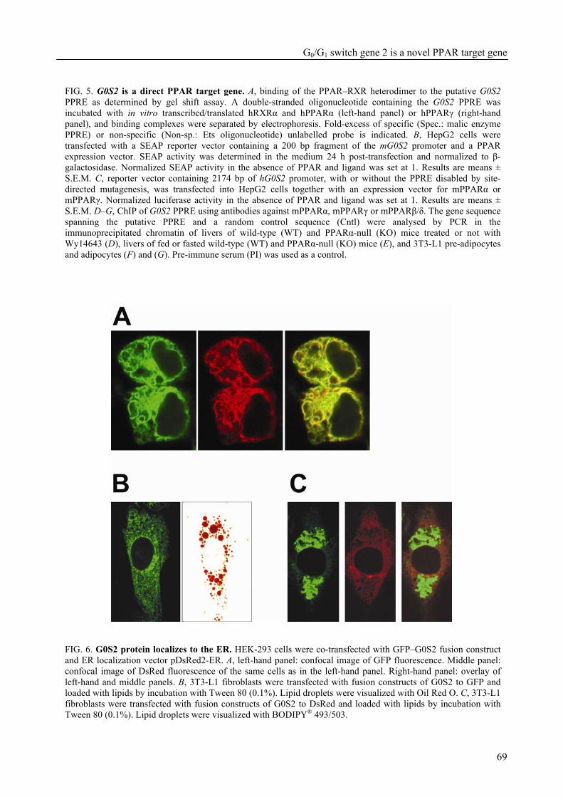
G0/G1 switch gene 2 is a novel PPAR target gene
69
FIG. 5. G0S2 is a direct PPAR target gene. A, binding of the PPAR–RXR heterodimer to the putative G0S2 PPRE as determined by gel shift assay. A double-stranded oligonucleotide containing the G0S2 PPRE was incubated with in vitro transcribed/translated hRXRα and hPPARα (left-hand panel) or hPPARγ (right-hand panel), and binding complexes were separated by electrophoresis. Fold-excess of specific (Spec.: malic enzyme PPRE) or non-specific (Non-sp.: Ets oligonucleotide) unlabelled probe is indicated. B, HepG2 cells were transfected with a SEAP reporter vector containing a 200 bp fragment of the mG0S2 promoter and a PPAR expression vector. SEAP activity was determined in the medium 24 h post-transfection and normalized to β-galactosidase. Normalized SEAP activity in the absence of PPAR and ligand was set at 1. Results are means ± S.E.M. C, reporter vector containing 2174 bp of hG0S2 promoter, with or without the PPRE disabled by site-directed mutagenesis, was transfected into HepG2 cells together with an expression vector for mPPARα or mPPARγ. Normalized luciferase activity in the absence of PPAR and ligand was set at 1. Results are means ± S.E.M. D–G, ChIP of G0S2 PPRE using antibodies against mPPARα, mPPARγ or mPPARβ/δ. The gene sequence spanning the putative PPRE and a random control sequence (Cntl) were analysed by PCR in the immunoprecipitated chromatin of livers of wild-type (WT) and PPARα-null (KO) mice treated or not with Wy14643 (D), livers of fed or fasted wild-type (WT) and PPARα-null (KO) mice (E), and 3T3-L1 pre-adipocytes and adipocytes (F) and (G). Pre-immune serum (PI) was used as a control.
FIG. 6. G0S2 protein localizes to the ER. HEK-293 cells were co-transfected with GFP–G0S2 fusion construct and ER localization vector pDsRed2-ER. A, left-hand panel: confocal image of GFP fluorescence. Middle panel: confocal image of DsRed fluorescence of the same cells as in the left-hand panel. Right-hand panel: overlay of left-hand and middle panels. B, 3T3-L1 fibroblasts were transfected with fusion constructs of G0S2 to GFP and loaded with lipids by incubation with Tween 80 (0.1%). Lipid droplets were visualized with Oil Red O. C, 3T3-L1 fibroblasts were transfected with fusion constructs of G0S2 to DsRed and loaded with lipids by incubation with Tween 80 (0.1%). Lipid droplets were visualized with BODIPY® 493/503.
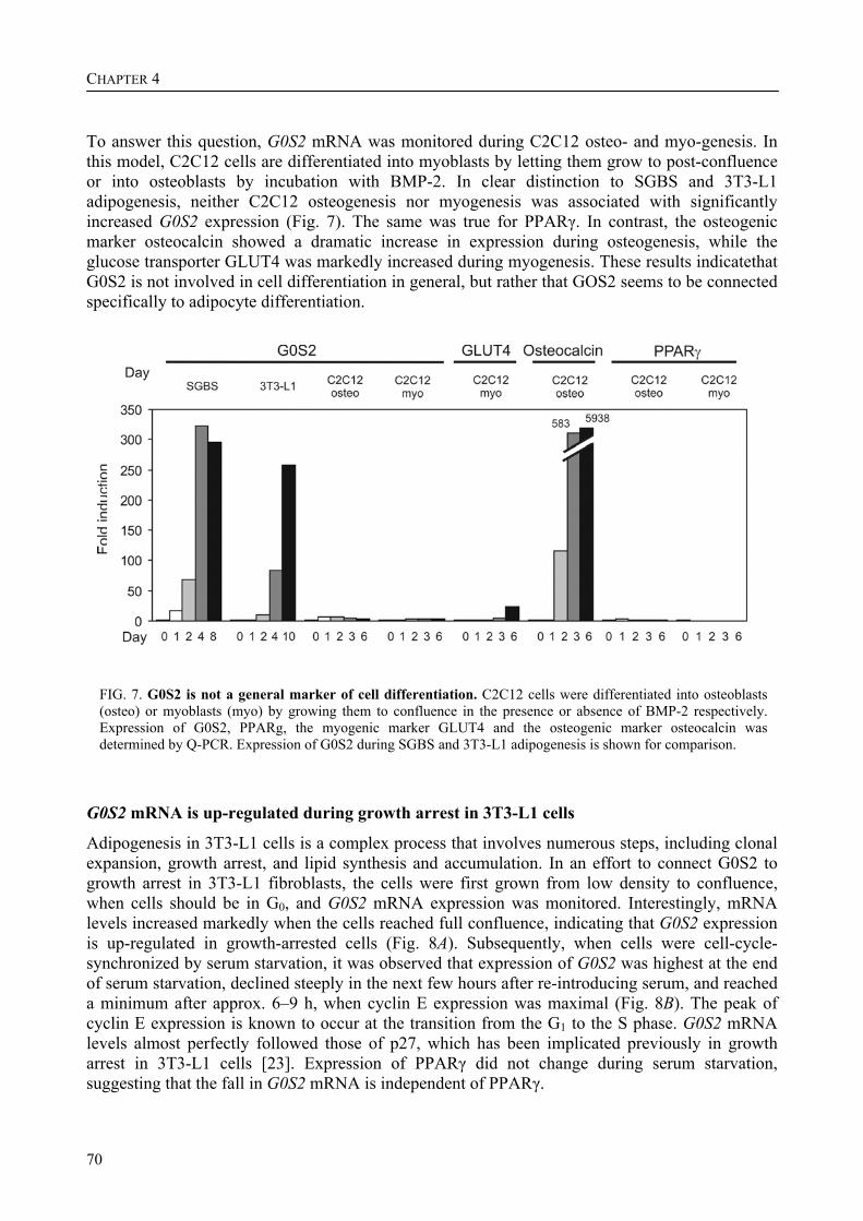
CHAPTER 4
70
To answer this question, G0S2 mRNA was monitored during C2C12 osteo- and myo-genesis. In this model, C2C12 cells are differentiated into myoblasts by letting them grow to post-confluence or into osteoblasts by incubation with BMP-2. In clear distinction to SGBS and 3T3-L1 adipogenesis, neither C2C12 osteogenesis nor myogenesis was associated with significantly increased G0S2 expression (Fig. 7). The same was true for PPARγ. In contrast, the osteogenic marker osteocalcin showed a dramatic increase in expression during osteogenesis, while the glucose transporter GLUT4 was markedly increased during myogenesis. These results indicatethat G0S2 is not involved in cell differentiation in general, but rather that GOS2 seems to be connected specifically to adipocyte differentiation.
FIG. 7. G0S2 is not a general marker of cell differentiation. C2C12 cells were differentiated into osteoblasts (osteo) or myoblasts (myo) by growing them to confluence in the presence or absence of BMP-2 respectively. Expression of G0S2, PPARg, the myogenic marker GLUT4 and the osteogenic marker osteocalcin was determined by Q-PCR. Expression of G0S2 during SGBS and 3T3-L1 adipogenesis is shown for comparison.
G0S2 mRNA is up-regulated during growth arrest in 3T3-L1 cells
Adipogenesis in 3T3-L1 cells is a complex process that involves numerous steps, including clonal expansion, growth arrest, and lipid synthesis and accumulation. In an effort to connect G0S2 to growth arrest in 3T3-L1 fibroblasts, the cells were first grown from low density to confluence, when cells should be in G0, and G0S2 mRNA expression was monitored. Interestingly, mRNA levels increased markedly when the cells reached full confluence, indicating that G0S2 expression is up-regulated in growth-arrested cells (Fig. 8A). Subsequently, when cells were cell-cycle-synchronized by serum starvation, it was observed that expression of G0S2 was highest at the end of serum starvation, declined steeply in the next few hours after re-introducing serum, and reached a minimum after approx. 6–9 h, when cyclin E expression was maximal (Fig. 8B). The peak of cyclin E expression is known to occur at the transition from the G1 to the S phase. G0S2 mRNA levels almost perfectly followed those of p27, which has been implicated previously in growth arrest in 3T3-L1 cells [23]. Expression of PPARγ did not change during serum starvation, suggesting that the fall in G0S2 mRNA is independent of PPARγ.
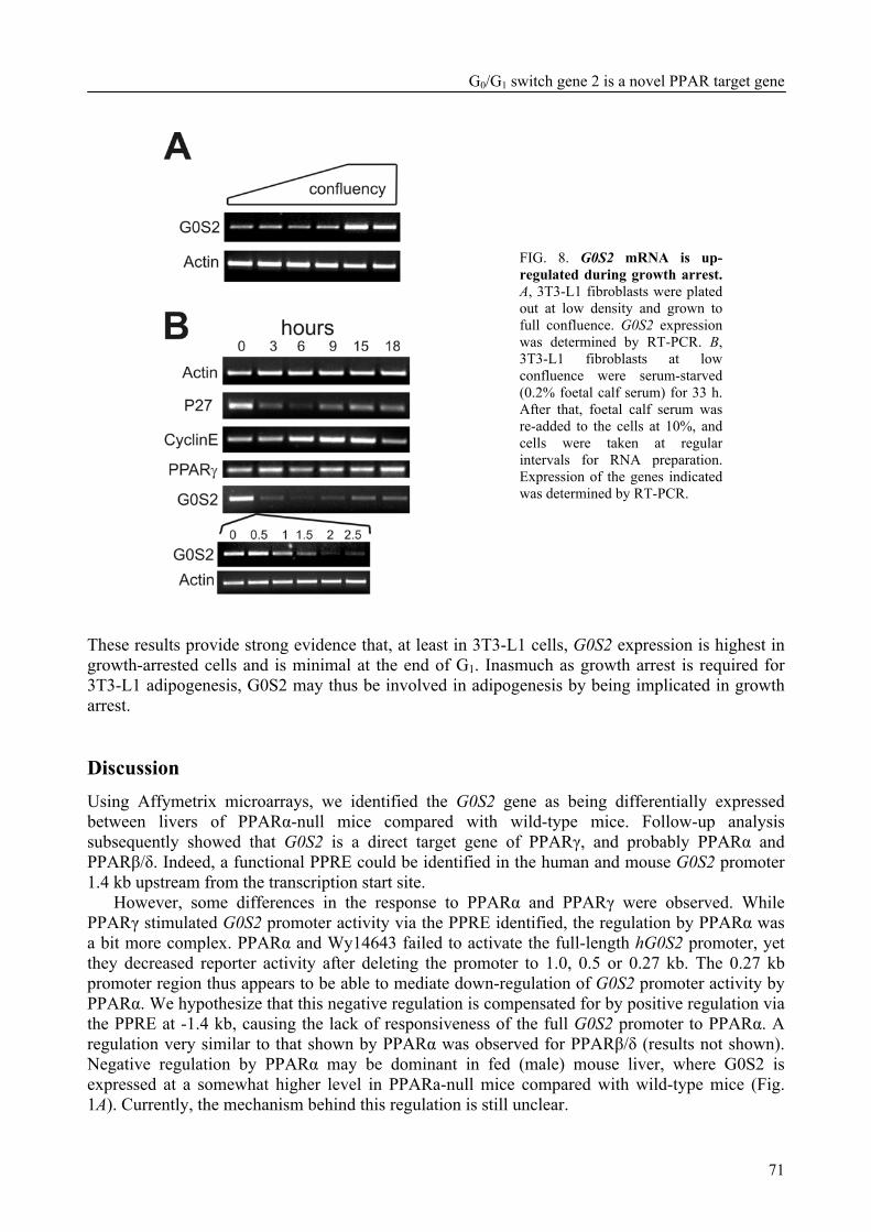
G0/G1 switch gene 2 is a novel PPAR target gene
71
These results provide strong evidence that, at least in 3T3-L1 cells, G0S2 expression is highest in growth-arrested cells and is minimal at the end of G1. Inasmuch as growth arrest is required for 3T3-L1 adipogenesis, G0S2 may thus be involved in adipogenesis by being implicated in growth arrest. Discussion Using Affymetrix microarrays, we identified the G0S2 gene as being differentially expressed between livers of PPARα-null mice compared with wild-type mice. Follow-up analysis subsequently showed that G0S2 is a direct target gene of PPARγ, and probably PPARα and PPARβ/δ. Indeed, a functional PPRE could be identified in the human and mouse G0S2 promoter 1.4 kb upstream from the transcription start site.
However, some differences in the response to PPARα and PPARγ were observed. While PPARγ stimulated G0S2 promoter activity via the PPRE identified, the regulation by PPARα was a bit more complex. PPARα and Wy14643 failed to activate the full-length hG0S2 promoter, yet they decreased reporter activity after deleting the promoter to 1.0, 0.5 or 0.27 kb. The 0.27 kb promoter region thus appears to be able to mediate down-regulation of G0S2 promoter activity by PPARα. We hypothesize that this negative regulation is compensated for by positive regulation via the PPRE at -1.4 kb, causing the lack of responsiveness of the full G0S2 promoter to PPARα. A regulation very similar to that shown by PPARα was observed for PPARβ/δ (results not shown). Negative regulation by PPARα may be dominant in fed (male) mouse liver, where G0S2 is expressed at a somewhat higher level in PPARa-null mice compared with wild-type mice (Fig. 1A). Currently, the mechanism behind this regulation is still unclear.
FIG. 8. G0S2 mRNA is up-regulated during growth arrest. A, 3T3-L1 fibroblasts were plated out at low density and grown to full confluence. G0S2 expression was determined by RT-PCR. B, 3T3-L1 fibroblasts at low confluence were serum-starved (0.2% foetal calf serum) for 33 h. After that, foetal calf serum was re-added to the cells at 10%, and cells were taken at regular intervals for RNA preparation. Expression of the genes indicated was determined by RT-PCR.

CHAPTER 4
72
In the absence of PPARα, G0S2 expression declines during fasting. The mechanism behind this decrease is unclear, but may be due to decreased insulin signalling or increased glucagon or other hormonal changes during fasting, which are compensated for by PPARα.
Several lines of evidence suggest that G0S2 is also a target gene of PPARβ/δ in WAT. However, since the function of PPARβ/δ in WAT is debatable [7,24], the functional implications of this regulation remain unclear.
A limited number of genes are known to be dual targets of PPARα in liver and of PPARγ in adipose tissue. These include lipoprotein lipase, fatty acid transport protein, acyl-CoA synthase, FIAF (fasting-induced adipose factor)/ANGPTL4 (angiopoietin-like 4)/PGAR (PPARγ angiopoietin-related gene) and cytosolic GPDH [14,25-27]. As the roles of PPARα in liver and PPARγ in adipose tissue are almost completely opposite (PPARα: fatty acid oxidation=catabolism compared with PPARγ: adipo/lipo-genesis=anabolism), the pathways supported by the target genes in the respective organs are also likely to be different. This is true for cytosolic GPDH, fatty acid transport protein, acyl-CoA synthase and, to a lesser extent, lipoprotein lipase, which are part of different pathways in the two tissues. Accordingly, it is not unreasonable to suggest that G0S2, as a dual or even triple PPAR target, might participate in different pathways in liver and adipose tissue.
The dominant expression of G0S2 in BAT and WAT, combined with the dramatic (specific) up-regulation of G0S2 during mouse and human adipogenesis and the up-regulation of G0S2 during growth arrest in 3T3-L1 cells, which is required for 3T3-L1 adipogenesis, suggest that G0S2 may play a role in adipogenesis.
Adipogenesis describes the differentiation of pre-adipocytes into mature fat cells and has been extensively studied in vitro using 3T3-L1, 3T3-F442A and NIH-3T3 mouse fibroblasts. These studies have led to a generally accepted model of adipocyte differentiation in 3T3 cells, in which a sequential up- or down-regulation of several transcription factors, including E2Fs, GATAs and C/EBPs (CCAAT/enhancer-binding proteins) [28-30], brings about the emergence of an adipose phenotype via up-regulation of a large number of adipose-specific target genes. Perhaps the most important transcription factor is PPARγ, which was demonstrated to be both necessary and sufficient for induction of an adipose phenotype [31]. Up-regulation of target genes of PPARγ is connected with the acquisition of functions specific to adipocytes, such as fatty acid and triacylglycerol synthesis, insulin-dependent glucose transport and the synthesis of secreted factors such as resistin and adiponectin [32,33]. The differentiation of 3T3-L1 cells into adipocytes follows a well-studied sequence of events, each of which is essential for final differentiation and development of the adipocyte phenotype. One important event is cell-cycle withdrawal/growth arrest. According to our results, G0S2 may be associated with 3T3-L1 adipogenesis by its involvement in growth arrest.
Currently, the role of G0S2 in non-adipose tissues, such as liver, is not clear and, based on the previous argument, may diverge from its function in adipose tissue. Highest expression of G0S2 is found in adipose tissue, but mRNA levels are also reasonably high in liver, heart and other tissues. The very low expression of G0S2 in rapidly proliferating hepatoma cell lines (HepG2, FAO, Hepa1-6) in comparison with growth-arrested mouse liver suggests that the possible role of G0S2 in growth arrest/differentiation may extend beyond adipose tissue. At the same time, our studies in C2C12 cells clearly indicate that G0S2 is not a general marker of cell differentiation. Further studies are necessary to determine the role of G0S2 in non-adipose tissues.
G0S2 was initially discovered using differential hybridization in blood mononuclear cells as a gene that is very transiently induced after treatment with concanavalin A (a lectin), cycloheximide (a protein synthesis inhibitor) and the combination of PMA (a phorbol ester) and ionomycin (a calcium ionophore) [21]. This rapid and transient increase in expression was inhibited by cyclosporin A. These results led the authors to conclude that G0S2 expression is transiently
G0/G1 switch gene 2 is a novel PPAR target gene
73
induced upon re-entry of cells into the G1 phase of the cell cycle and would be required to commit cells to enter G1 [21]. In contrast, our results indicate that up-regulation of G0S2 is associated with cell-cycle withdrawal. The reason for this discrepancy is not clear, but it may point to a cell-type-specific function for G0S2. Alternatively, transient up-regulation of G0S2 in blood mononuclear cells by any of the compounds mentioned above may reflect a different event from re-entry into the cell cycle.
The limited information available about G0S2 before the present study included an in situ hybridization analysis of G0S2 expression in mice embryos. It was found that, at day 18.5, G0S2 expression is restricted to BAT and WAT [34]. The present study confirms that G0S2 is mainly expressed in WAT and BAT, but also indicates that G0S2 mRNA is reasonably well expressed in other tissues, such as lung, liver and heart. The reason for this discrepancy is not exactly clear, but it may be due to a difference in sensitivity between the techniques used to detect G0S2 mRNA (in situ hybridization compared with Q-PCR) or a difference in the age of the animal (embryonic day 18.5 compared with adult animal). The latter explanation would support a role for G0S2 in growth arrest, since, at the embryonic stage, tissues such as muscle and liver still display a high rate of cell proliferation, whereas, in the adult stage, liver and muscle cells are highly differentiated and arrested in G0, which would result in increased G0S2 expression.
In the present study, G0S2 protein was localized to the ER. Analysis of the primary sequence by the Internet-based PSORT II program predicted the N-terminal domain comprising amino acids 1–26 to be protruding into the cytoplasm, whereas the C-terminal domain comprising amino acids 43–103 is expected to be in the ER lumen. The molecular mechanism by which G0S2 may influence growth arrest and, accordingly, adipogenesis would probably involve some kind of protein–protein interaction via either of these domains. Future studies will have to address this in more detail.
Finally, our microarray experiment corroborated perfectly the concept that PPARα is an important regulator of fatty acid oxidation and ketogenesis, and that the function of PPARα becomes mainly evident during fasting. Possible new target genes of PPARα that emerged from our microarray screen include those for insulin-like growth factor-binding protein 2, folylpolyglutamate synthetase and LDL (low-density lipoprotein)-receptor-related protein 1. These results underscore the utility of microarray analysis in finding and characterizing novel potential target genes of nuclear hormone receptors.
In conclusion, we have identified the G0S2 as a novel direct target gene of PPARγ, and probably PPARα and PPARβ/δ, and present results suggesting that it is involved in adipocyte differentiation. We are grateful to Dr Wilma Steegenga for the RT samples from C2C12 cells, and Dr M. Wabitsch for the gift of the SGBS cell line. We thank Erika Ferguson for excellent technical assistance, Yixin Wang for his support in microarray data analysis, Ken (Gang) Hu for his advice in GeneSpring program, Gary McMaster and Steven Hunt for their support to carry out the microarray study. This study was financed by the Netherlands Organization for Scientific Research (NWO), with additional support from the Royal Netherlands Academy of Art and Sciences (KNAW), the Wageningen Center for Food Sciences, the Swiss National Science Foundation and the Human Frontier Science Program (HFSP). We declare no conflict of interest.
CHAPTER 4
74
References 1 Kersten, S., Desvergne, B., Wahli, W. (2000) Roles of PPARs in health and disease. Nature 405, 421-
424 2 Tontonoz, P., Hu, E., Spiegelman, B. M. (1994) Stimulation of adipogenesis in fibroblasts by PPAR
gamma 2, a lipid-activated transcription factor. Cell 79, 1147-1156 3 Barak, Y., Nelson, M. C., Ong, E. S., et al. (1999) PPAR gamma is required for placental, cardiac,
and adipose tissue development. Mol. Cell 4, 585-595 4 Ren, D., Collingwood, T. N., Rebar, E. J., Wolffe, A. P., Camp, H. S. (2002) PPARgamma
knockdown by engineered transcription factors: exogenous PPARgamma2 but not PPARgamma1 reactivates adipogenesis. Genes Dev. 16, 27-32
5 Rosen, E. D., Hsu, C. H., Wang, X., et al. (2002) C/EBPalpha induces adipogenesis through PPARgamma: a unified pathway. Genes Dev. 16, 22-26
6 Way, J. M., Harrington, W. W., Brown, K. K., et al. (2001) Comprehensive messenger ribonucleic acid profiling reveals that peroxisome proliferator-activated receptor gamma activation has coordinate effects on gene expression in multiple insulin-sensitive tissues. Endocrinology 142, 1269-1277
7 Wang, Y. X., Lee, C. H., Tiep, S., et al. (2003) Peroxisome-proliferator-activated receptor delta activates fat metabolism to prevent obesity. Cell 113, 159-170
8 Wang, Y. X., Zhang, C. L., Yu, R. T., et al. (2004) Regulation of Muscle Fiber Type and Running Endurance by PPARdelta. PLoS Biol. 2, E294
9 Akiyama, T. E., Lambert, G., Nicol, C. J., et al. (2004) Peroxisome proliferator-activated receptor beta/delta regulates very low density lipoprotein production and catabolism in mice on a Western diet. J. Biol. Chem. 279, 20874-20881
10 Di Poi, N., Tan, N. S., Michalik, L., Wahli, W., Desvergne, B. (2002) Antiapoptotic role of PPARbeta in keratinocytes via transcriptional control of the Akt1 signaling pathway. Mol. Cell 10, 721-733
11 Harman, F. S., Nicol, C. J., Marin, H. E., Ward, J. M., Gonzalez, F. J., Peters, J. M. (2004) Peroxisome proliferator-activated receptor-delta attenuates colon carcinogenesis. Nat. Med. 10, 481-483
12 Gupta, R. A., Wang, D., Katkuri, S., Wang, H., Dey, S. K., DuBois, R. N. (2004) Activation of nuclear hormone receptor peroxisome proliferator-activated receptor-delta accelerates intestinal adenoma growth. Nat. Med. 10, 245-247
13 Mandard, S., Muller, M., Kersten, S. (2004) Peroxisome proliferator-activated receptor alpha target genes. Cell. Mol. Life Sci. 61, 393-416
14 Patsouris, D., Mandard, S., Voshol, P. J., et al. (2004) PPARalpha governs glycerol metabolism. J Clin Invest 114, 94-103
15 Gervois, P., Kleemann, R., Pilon, A., et al. (2004) Global suppression of IL-6-induced acute phase response gene expression after chronic in vivo treatment with the peroxisome proliferator-activated receptor-alpha activator fenofibrate. J. Biol. Chem. 279, 16154-16160
16 Delerive, P., Fruchart, J. C., Staels, B. (2001) Peroxisome proliferator-activated receptors in inflammation control. J. Endocrinol. 169, 453-459
17 Beigneux, A. P., Moser, A. H., Shigenaga, J. K., Grunfeld, C., Feingold, K. R. (2000) The acute phase response is associated with retinoid X receptor repression in rodent liver. J. Biol. Chem. 275, 16390-16399
18 Moshage, H., Casini, A., Lieber, C. S. (1990) Acetaldehyde selectively stimulates collagen production in cultured rat liver fat-storing cells but not in hepatocytes. Hepatology 12, 511-518
19 Wabitsch, M., Brenner, R. E., Melzner, I., et al. (2001) Characterization of a human preadipocyte cell strain with high capacity for adipose differentiation. Int. J. Obes. Relat. Metab. Disord. 25, 8-15
20 Kersten, S., Mandard, S., Tan, N. S., et al. (2000) Characterization of the fasting-induced adipose factor FIAF, a novel peroxisome proliferator-activated receptor target gene. J. Biol. Chem. 275, 28488-28493
G0/G1 switch gene 2 is a novel PPAR target gene
75
21 Russell, L., Forsdyke, D. R. (1991) A human putative lymphocyte G0/G1 switch gene containing a CpG-rich island encodes a small basic protein with the potential to be phosphorylated. DNA Cell Biol. 10, 581-591
22 Berger, J., Leibowitz, M. D., Doebber, T. W., et al. (1999) Novel peroxisome proliferator-activated receptor (PPAR) gamma and PPARdelta ligands produce distinct biological effects. J. Biol. Chem. 274, 6718-6725
23 Morrison, R. F., Farmer, S. R. (1999) Role of PPARgamma in regulating a cascade expression of cyclin-dependent kinase inhibitors, p18(INK4c) and p21(Waf1/Cip1), during adipogenesis. J. Biol. Chem. 274, 17088-17097
24 Matsusue, K., Peters, J. M., Gonzalez, F. J. (2004) PPARbeta/delta potentiates PPARgamma-stimulated adipocyte differentiation. FASEB J. 18, 1477-1479
25 Martin, G., Schoonjans, K., Lefebvre, A. M., Staels, B., Auwerx, J. (1997) Coordinate regulation of the expression of the fatty acid transport protein and acyl-CoA synthetase genes by PPARalpha and PPARgamma activators. J. Biol. Chem. 272, 28210-28217
26 Schoonjans, K., Peinado-Onsurbe, J., Lefebvre, A. M., et al. (1996) PPARalpha and PPARgamma activators direct a distinct tissue-specific transcriptional response via a PPRE in the lipoprotein lipase gene. EMBO J. 15, 5336-5348
27 Mandard, S., Zandbergen, F., Tan, N. S., et al. (2004) The direct peroxisome proliferator-activated receptor target fasting-induced adipose factor (FIAF/PGAR/ANGPTL4) is present in blood plasma as a truncated protein that is increased by fenofibrate treatment. J. Biol. Chem. 279, 34411-34420
28 Fajas, L., Landsberg, R. L., Huss-Garcia, Y., Sardet, C., Lees, J. A., Auwerx, J. (2002) E2Fs regulate adipocyte differentiation. Dev. Cell 3, 39-49
29 Tong, Q., Dalgin, G., Xu, H., Ting, C. N., Leiden, J. M., Hotamisligil, G. S. (2000) Function of GATA transcription factors in preadipocyte-adipocyte transition. Science 290, 134-138
30 Christy, R. J., Yang, V. W., Ntambi, J. M., et al. (1989) Differentiation-induced gene expression in 3T3-L1 preadipocytes: CCAAT/enhancer binding protein interacts with and activates the promoters of two adipocyte-specific genes. Genes Dev. 3, 1323-1335
31 Rosen, E. D., Spiegelman, B. M. (2001) PPARgamma: a nuclear regulator of metabolism, differentiation, and cell growth. J. Biol. Chem. 276, 37731-37734
32 Steppan, C. M., Bailey, S. T., Bhat, S., et al. (2001) The hormone resistin links obesity to diabetes. Nature 409, 307-312
33 Hu, E., Liang, P., Spiegelman, B. M. (1996) AdipoQ is a novel adipose-specific gene dysregulated in obesity. J. Biol. Chem. 271, 10697-10703
34 Bachner, D., Ahrens, M., Schroder, D., et al. (1998) Bmp-2 downstream targets in mesenchymal development identified by subtractive cloning from recombinant mesenchymal progenitors (C3H10T1/2). Dev. Dyn. 213, 398-411

76
FIAF governs plasma lipid levels and adiposity
77
CHAPTER 5 The fasting-induced adipose factor/angiopoietin-like protein 4 is physically associated with lipoproteins and governs plasma lipid levels and adiposity Stéphane Mandard, Fokko Zandbergen, Esther van Straten, Walter Wahli, Folkert Kuipers, Michael Müller, and Sander Kersten Abstract Proteins secreted from adipose tissue are increasingly recognized to play an important role in the regulation of glucose metabolism. However, much less is known about their effect on lipid metabolism. The fasting-induced adipose factor (FIAF/angiopoietin-like protein 4/peroxisome proliferator-activated receptor γ angiopoietin-related protein) was previously identified as a target of hypolipidemic fibrate drugs and insulin-sensitizing thiazolidinediones. Using transgenic mice that mildly overexpress FIAF in peripheral tissues we show that FIAF is an extremely powerful regulator of lipid metabolism and adiposity. FIAF overexpression caused a 50% reduction in adipose tissue weight, partly by stimulating fatty acid oxidation and uncoupling in fat. In addition, FIAF overexpression increased plasma levels of triglycerides, free fatty acids, glycerol, total cholesterol, and high density lipoprotein (HDL)-cholesterol. Functional tests indicated that FIAF overexpression severely impaired plasma triglyceride clearance but had no effect on very low density lipoprotein production. The effects of FIAF overexpression were amplified by a high fat diet, resulting in markedly elevated plasma and liver triglycerides, plasma free fatty acids, and plasma glycerol levels, and impaired glucose tolerance in FIAF transgenic mice fed a high fat diet. Remarkably, in mice the full-length form of FIAF was physically associated with HDL, whereas truncated FIAF was associated with low density lipoprotein. In human both full-length and truncated FIAF were associated with HDL. The composite data suggest that via physical association with plasma lipoproteins, FIAF acts as a powerful signal from fat and other tissues to prevent fat storage and stimulate fat mobilization. Our data indicate that disturbances in FIAF signaling might be involved in dyslipidemia. This chapter has been published in the Journal of Biological Chemistry 281, 934-944 (2006)

CHAPTER 5
78
Introduction Obesity and associated diabetes mellitus type 2 have become major health concerns throughout the world. These ailments are intimately linked as excess weight greatly increases the likelihood of developing diabetes. Obesity also increases the risk for other clinical abnormalities such as hypertension and dyslipidemia. Although the positive association between obesity and several co-morbidities has been well established at the epidemiological level, the mechanisms behind these associations are much less clear. Much attention has been given to the role of plasma free fatty acids (FFAs),1 which are elevated in the obese and able to disrupt cellular metabolism in several organs. However, it also has become evident that a variety of hormonal factors produced by adipose tissue can greatly affect organ functioning, especially at the metabolic and immunological levels [1]. Indeed, these so-called adipocytokines or adipokines are now known to affect diverse biological processes, ranging from energy intake, insulin sensitivity, and hepatic glucose production, to reproductive and immunological function [2-5]. Whereas the effects of adipocytokines on glucose homeostasis are becoming increasingly transparent, much less is known about how they regulate plasma lipid metabolism.
A relatively poorly characterized adipocytokine that may be involved in regulation of plasma lipid metabolism is the fasting-induced adipose factor (FIAF), also known as PPARγ angiopoietin-related protein, angiopoietin-like protein 4, or hepatic fibrinogen/angiopoietin-related protein [6,8-10]. FIAF is a glycoprotein of ~50 kDa that belongs to the family of fibrinogen/angiopoietin-like proteins. In mice FIAF is most highly expressed in white and brown adipose tissue, and to a much lesser extent in other tissues such as heart, skeletal muscle, and liver [6,8].
The most compelling data to date link FIAF with regulation of lipid metabolism. FIAF was first identified as a target gene of the nuclear receptors PPARα and PPARγ, which govern lipid metabolism in liver and white adipose tissue, respectively [6,8]. Subsequent studies employing adenoviral-mediated overexpression of FIAF or injection of recombinant FIAF have shown that FIAF potently elevates plasma triglyceride (TG) levels [10-12]. This is possibly achieved by inhibition of lipoprotein lipase, the enzyme that is rate-limiting for plasma TG hydrolysis [10,11,13]. According to a recent report, FIAF may also potently lower plasma glucose levels. In wild-type C57/B6 as well as in db/db mice, adenoviral-mediated overexpression of FIAF was found to improve hyperglycemia, hyperinsulinemia, and glucose tolerance [12].
In addition to lipid metabolism, FIAF has also been associated with angiogenesis. Expression of FIAF is up-regulated during hypoxia in both endothelial cells and cardiomyocytes, which
probably occurs via the hypoxia-inducible factor 1α (HIF-1α) [14,15]. Furthermore, FIAF is expressed in certain tumors, especially in the hypoxic area surrounding necrotic cells, and has been
suggested as a marker for conventional renal cell carcinoma. In the chicken chorioallantoic membrane assay, FIAF is able to induce a strong pro-angiogenic response [15]. In contrast, others have ascribed a potent anti-angiogenic function to FIAF [16]. Thus, the role of FIAF in angiogenesis remains ambiguous.
To determine the physiological role of FIAF, we studied the effect of FIAF overexpression in a transgenic mouse model. Our data indicate that FIAF, which is physically associated with plasma lipoproteins, is an important determinant of plasma TG concentration and clearance at physiological levels of expression. In addition, it stimulates adipose tissue lipolysis. In contrast to a recent study [12], in our model FIAF overexpression was associated with deterioration of glucose 1 The abbreviations used are: FFA, free fatty acid; FIAF, fasting-induced adipose factor; PPAR, peroxisome proliferator-activated receptor; TG, triglyceride; FIAF-Tg, FIAF transgenic; VLDL, very low density lipoprotein; HDL, high density lipoprotein; FPLC, fast protein liquid chromatography; LPL, lipoprotein lipase; WAT, white adipose tissue; BAT, brown adipose tissue; HSL, hormone-sensitive lipase; Q-PCR, quantitative real time PCR; HFD, high fat diet; LFD, low fat diet; ASP, acylation-stimulating protein.
FIAF governs plasma lipid levels and adiposity
79
tolerance. Finally, we observed that the effects of FIAF overexpression were clearly amplified by feeding a high fat diet. Experimental procedures Generation of FIAF transgenic (FIAF-Tg) mice
The complete murine FIAF gene was amplified by PCR (High fidelity Taq polymerase, Roche Applied Science) from genomic DNA of mouse ES cells (Sv129) using primers CGGGCTCCAGATCTTCTTCTGCACCAG and GTCAGAGGCGGCATTGGACCCCCTTGAA-GTA and subcloned into the pGEM-Teasy vector (Promega, Leiden, the Netherlands). The proper sequence of the exons was verified by DNA sequencing. The mFIAF gene was subsequently cut out with NotI and placed behind the murine aP2 (adipocyte fatty acid-binding protein) promoter within pBS-SKII+ (kind gift of Dr. Bruce Spiegelman). The promoter plus the mFIAF gene were excised with ClaI and SacII and gel purified before being injected into fertilized oocytes (strain FVB-NIco, Eurogentec Transgenic Production Service, Seraing, Belgium).
Genotyping was performed on genomic DNA from mouse ears by Q-PCR (Sybr Green) using primers GCCCCCATTGGTCACTCCTACAG and CGGCTCAGACTTAGACTTGCTC, which anneal within the aP2 promoter and the mFIAF gene, respectively, and control primers GCTGCTGGAGAATGAGTTGAATGC and CTCGGCTGAGTTTGAAGTTGAAGATG for the apoB gene. Animal experiments
All mice were on a pure-bred FVB-NIco background. Male animals were kept in normal cages with food and water ad libitum. Mice in the fed state were sacrificed at the beginning of the light cycle. Mice in the fasted state were deprived of food for 6 h starting at the beginning of the light cycle. At the time of sacrifice animals were between 2 and 4 months of age. For the diet intervention, 2-month-old male mice were fed with a low or high fat diet for 10 weeks. The respective diets provided either 10 or 45% energy percent in the form of lard fat (D12450B or D12451 (GenBank), Research Diets, New Brunswick, NJ). Blood was collected via orbital puncture into EDTA tubes. Tissues were excised, weighed, and immediately frozen in liquid nitrogen. Intragastric lipid loading test
Mice fasted for 16 h were administered 350 µl of olive oil (Bertolli, Extra Virgin) by intragastric gavage. Blood was collected by tail bleeding every 2 h for plasma TG measurement. VLDL production test
Mice fasted for 16 h were injected via the tail vein with 500 mg/kg bodyweight Triton WR1339 (Tyloxapol) under general anesthesia. Blood was collected by tail bleeding at several time points during 2.5 h for plasma TG measurement.
Intraperitoneal glucose tolerance test
After a 5-h fast mice were injected intraperitoneally with glucose (2 g/kg bodyweight on chow, 1 g/kg bodyweight on low fat/high fat diet). Blood was collected by tail bleeding after 0, 20, 40, 60, 90, and 150 min, and glucose was measured using Accucheck compact (Roche Diagnostics, Almere, the Netherlands).
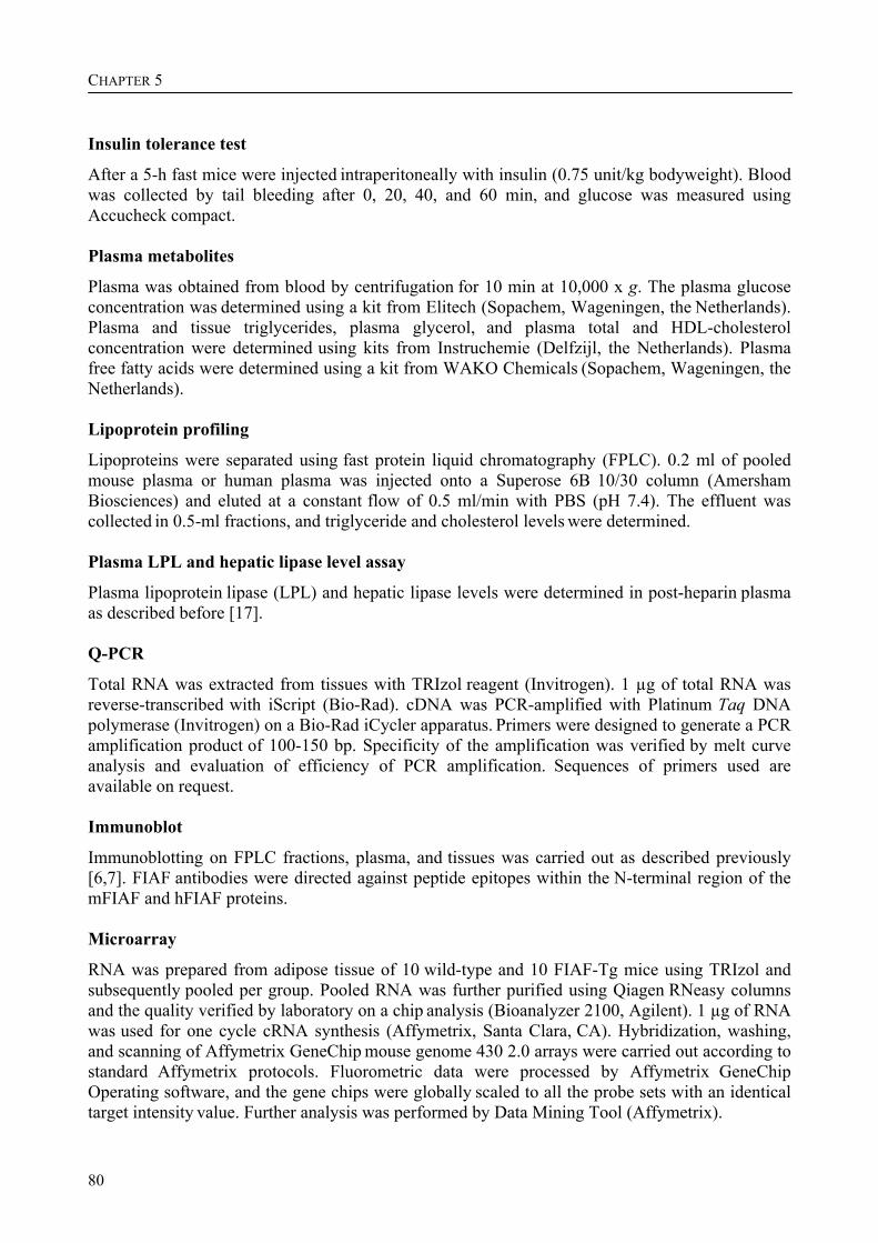
CHAPTER 5
80
Insulin tolerance test
After a 5-h fast mice were injected intraperitoneally with insulin (0.75 unit/kg bodyweight). Blood
was collected by tail bleeding after 0, 20, 40, and 60 min, and glucose was measured using Accucheck compact. Plasma metabolites
Plasma was obtained from blood by centrifugation for 10 min at 10,000 x g. The plasma glucose concentration was determined using a kit from Elitech (Sopachem, Wageningen, the Netherlands). Plasma and tissue triglycerides, plasma glycerol, and plasma total and HDL-cholesterol concentration were determined using kits from Instruchemie (Delfzijl, the Netherlands). Plasma
free fatty acids were determined using a kit from WAKO Chemicals (Sopachem, Wageningen, the Netherlands). Lipoprotein profiling
Lipoproteins were separated using fast protein liquid chromatography (FPLC). 0.2 ml of pooled
mouse plasma or human plasma was injected onto a Superose 6B 10/30 column (Amersham Biosciences) and eluted at a constant flow of 0.5 ml/min with PBS (pH 7.4). The effluent was collected in 0.5-ml fractions, and triglyceride and cholesterol levels were determined. Plasma LPL and hepatic lipase level assay
Plasma lipoprotein lipase (LPL) and hepatic lipase levels were determined in post-heparin plasma as described before [17]. Q-PCR
Total RNA was extracted from tissues with TRIzol reagent (Invitrogen). 1 µg of total RNA was reverse-transcribed with iScript (Bio-Rad). cDNA was PCR-amplified with Platinum Taq DNA polymerase (Invitrogen) on a Bio-Rad iCycler apparatus. Primers were designed to generate a PCR amplification product of 100-150 bp. Specificity of the amplification was verified by melt curve analysis and evaluation of efficiency of PCR amplification. Sequences of primers used are available on request. Immunoblot
Immunoblotting on FPLC fractions, plasma, and tissues was carried out as described previously [6,7]. FIAF antibodies were directed against peptide epitopes within the N-terminal region of the mFIAF and hFIAF proteins. Microarray
RNA was prepared from adipose tissue of 10 wild-type and 10 FIAF-Tg mice using TRIzol and subsequently pooled per group. Pooled RNA was further purified using Qiagen RNeasy columns and the quality verified by laboratory on a chip analysis (Bioanalyzer 2100, Agilent). 1 µg of RNA was used for one cycle cRNA synthesis (Affymetrix, Santa Clara, CA). Hybridization, washing, and scanning of Affymetrix GeneChip mouse genome 430 2.0 arrays were carried out according to standard Affymetrix protocols. Fluorometric data were processed by Affymetrix GeneChip Operating software, and the gene chips were globally scaled to all the probe sets with an identical target intensity value. Further analysis was performed by Data Mining Tool (Affymetrix).
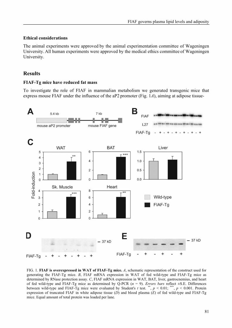
FIAF governs plasma lipid levels and adiposity
81
Ethical considerations
The animal experiments were approved by the animal experimentation committee of Wageningen University. All human experiments were approved by the medical ethics committee of Wageningen University. Results FIAF-Tg mice have reduced fat mass
To investigate the role of FIAF in mammalian metabolism we generated transgenic mice that express mouse FIAF under the influence of the aP2 promoter (Fig. 1A), aiming at adipose tissue-
FIG. 1. FIAF is overexpressed in WAT of FIAF-Tg mice. A, schematic representation of the construct used for generating the FIAF-Tg mice. B, FIAF mRNA expression in WAT of fed wild-type and FIAF-Tg mice as determined by RNase protection assay. C, FIAF mRNA expression in WAT, BAT, liver, gastrocnemius, and heart of fed wild-type and FIAF-Tg mice as determined by Q-PCR (n = 9). Errors bars reflect ±S.E. Differences between wild-type and FIAF-Tg mice were evaluated by Student's t test. **, p < 0.01; ***, p < 0.001. Protein expression of truncated FIAF in white adipose tissue (D) and blood plasma (E) of fed wild-type and FIAF-Tg mice. Equal amount of total protein was loaded per lane.
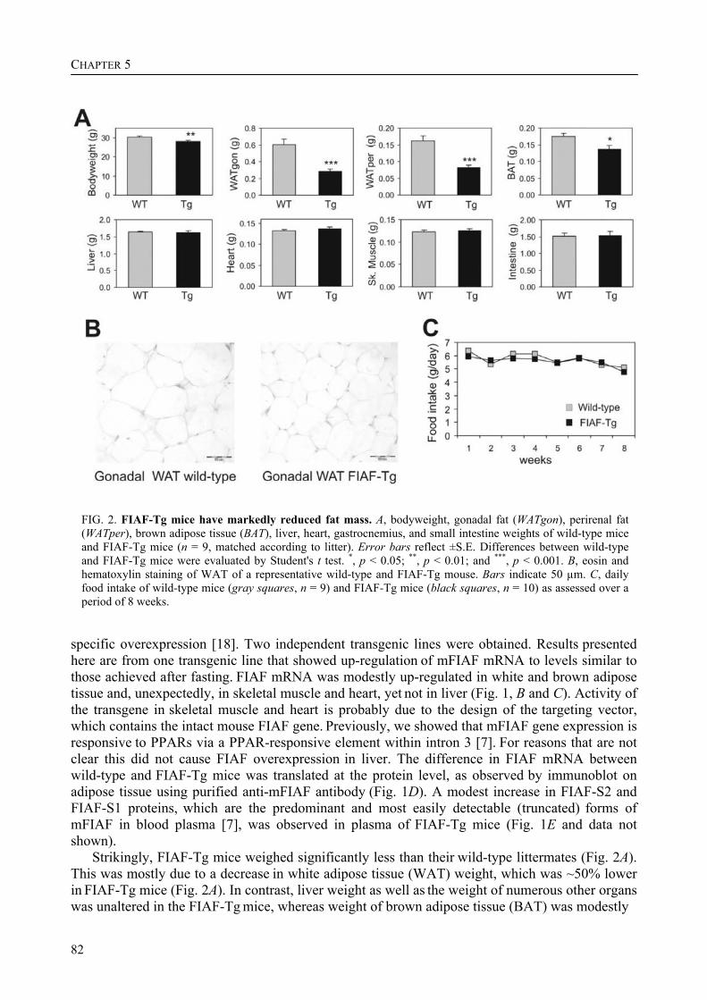
CHAPTER 5
82
FIG. 2. FIAF-Tg mice have markedly reduced fat mass. A, bodyweight, gonadal fat (WATgon), perirenal fat (WATper), brown adipose tissue (BAT), liver, heart, gastrocnemius, and small intestine weights of wild-type mice and FIAF-Tg mice (n = 9, matched according to litter). Error bars reflect ±S.E. Differences between wild-type and FIAF-Tg mice were evaluated by Student's t test. *, p < 0.05; **, p < 0.01; and ***, p < 0.001. B, eosin and hematoxylin staining of WAT of a representative wild-type and FIAF-Tg mouse. Bars indicate 50 µm. C, daily food intake of wild-type mice (gray squares, n = 9) and FIAF-Tg mice (black squares, n = 10) as assessed over a period of 8 weeks.
specific overexpression [18]. Two independent transgenic lines were obtained. Results presented here are from one transgenic line that showed up-regulation of mFIAF mRNA to levels similar to those achieved after fasting. FIAF mRNA was modestly up-regulated in white and brown adipose
tissue and, unexpectedly, in skeletal muscle and heart, yet not in liver (Fig. 1, B and C). Activity of the transgene in skeletal muscle and heart is probably due to the design of the targeting vector, which contains the intact mouse FIAF gene. Previously, we showed that mFIAF gene expression is responsive to PPARs via a PPAR-responsive element within intron 3 [7]. For reasons that are not clear this did not cause FIAF overexpression in liver. The difference in FIAF mRNA between wild-type and FIAF-Tg mice was translated at the protein level, as observed by immunoblot on adipose tissue using purified anti-mFIAF antibody (Fig. 1D). A modest increase in FIAF-S2 and FIAF-S1 proteins, which are the predominant and most easily detectable (truncated) forms of mFIAF in blood plasma [7], was observed in plasma of FIAF-Tg mice (Fig. 1E and data not shown).
Strikingly, FIAF-Tg mice weighed significantly less than their wild-type littermates (Fig. 2A). This was mostly due to a decrease in white adipose tissue (WAT) weight, which was ~50% lower in FIAF-Tg mice (Fig. 2A). In contrast, liver weight as well as the weight of numerous other organs was unaltered in the FIAF-Tg mice, whereas weight of brown adipose tissue (BAT) was modestly
FIAF governs plasma lipid levels and adiposity
83
FIG. 3. Fasting plasma triglycerides, free fatty acids, glycerol, HDL-cholesterol, and total cholesterol concentration are elevated in FIAF-Tg mice. Plasma TG (A), FFAs (B), glycerol (C), total cholesterol (D), HDL-cholesterol (E), and glucose (F), were determined in EDTA plasma of fed (n = 9) and 6-h-fasted (n = 10) wild-type mice (gray bars) and FIAF-Tg mice (black bars). Error bars reflect ±S.E. Differences between wild-type and FIAF-Tg mice were evaluated by Student's t test. *, p < 0.05; **, p < 0.01; and ***, p < 0.001.
decreased. Reduced WAT weight was due to a decrease in adipocyte size (Fig. 2B). Reduced adiposity was not the result of diminished food intake, which was identical between the two sets of mice (Fig. 2C). These data indicate that FIAF overexpression results in loss of body fat. FIAF-Tg mice display hypertriglyceridemia and hypercholesterolemia
To get a better understanding of the potential cause of the reduced adiposity, plasma lipid parameters were assessed in both fed and 6-h-fasted mice. A marked increase in plasma TG levels was observed in the FIAF-Tg mice, which was independent of their feeding status (Fig. 3A). In addition, plasma FFAs, glycerol, total cholesterol, and HDL-cholesterol concentrations were significantly elevated in the FIAF-Tg mice in the fasted state, whereas glucose was unchanged (Fig. 3, B - F). Thus, FIAF overexpression has profound effects on plasma lipid concentrations. Profiling of lipoproteins using FPLC analysis revealed that the increase in plasma TG in the 6-h-fasted animals was attributable to the VLDL fractions (Fig. 4A), whereas the increase in cholesterol was found in both VLDL and HDL fractions (Fig. 4B). Immunoblotting demonstrated a marked increase in apoB48 and apoB100 protein content in the VLDL fractions in FIAF-Tg mice, indicating that the number of VLDL particles in plasma is increased in these mice (Fig. 4C). It should be emphasized that in mice apoB48 accounts for approximately two-thirds of total apoB production in liver. Similarly, we found markedly increased apoAI and apoAII protein content in
the HDL fractions in FIAF-Tg mice, pointing toward a pronounced increase in the number of HDL particles (Fig. 4D).
Elevated plasma TG levels in 6-h-fasted mice can either be due to increased VLDL production or impaired clearance. To discriminate between these two possibilities, a VLDL production test and an oral lipid-loading test were performed.
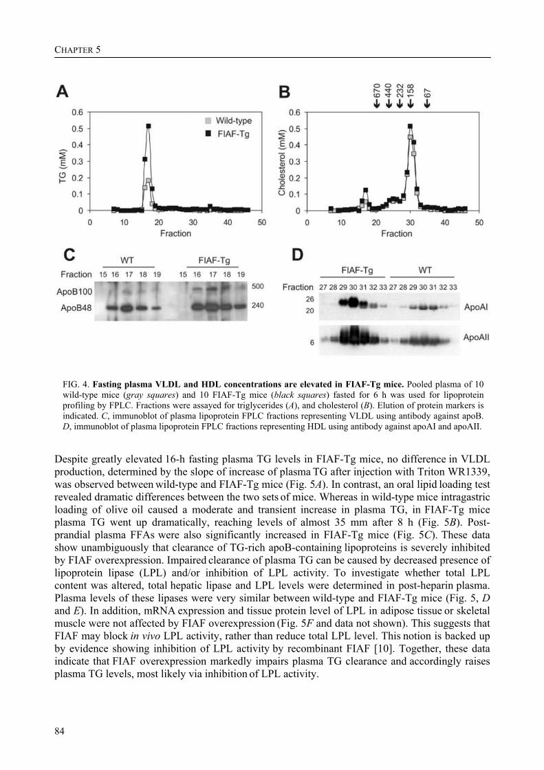
CHAPTER 5
84
FIG. 4. Fasting plasma VLDL and HDL concentrations are elevated in FIAF-Tg mice. Pooled plasma of 10 wild-type mice (gray squares) and 10 FIAF-Tg mice (black squares) fasted for 6 h was used for lipoprotein profiling by FPLC. Fractions were assayed for triglycerides (A), and cholesterol (B). Elution of protein markers is indicated. C, immunoblot of plasma lipoprotein FPLC fractions representing VLDL using antibody against apoB. D, immunoblot of plasma lipoprotein FPLC fractions representing HDL using antibody against apoAI and apoAII.
Despite greatly elevated 16-h fasting plasma TG levels in FIAF-Tg mice, no difference in VLDL production, determined by the slope of increase of plasma TG after injection with Triton WR1339, was observed between wild-type and FIAF-Tg mice (Fig. 5A). In contrast, an oral lipid loading test revealed dramatic differences between the two sets of mice. Whereas in wild-type mice intragastric loading of olive oil caused a moderate and transient increase in plasma TG, in FIAF-Tg mice plasma TG went up dramatically, reaching levels of almost 35 mm after 8 h (Fig. 5B). Post-prandial plasma FFAs were also significantly increased in FIAF-Tg mice (Fig. 5C). These data show unambiguously that clearance of TG-rich apoB-containing lipoproteins is severely inhibited by FIAF overexpression. Impaired clearance of plasma TG can be caused by decreased presence of
lipoprotein lipase (LPL) and/or inhibition of LPL activity. To investigate whether total LPL content was altered, total hepatic lipase and LPL levels were determined in post-heparin plasma. Plasma levels of these lipases were very similar between wild-type and FIAF-Tg mice (Fig. 5, D and E). In addition, mRNA expression and tissue protein level of LPL in adipose tissue or skeletal muscle were not affected by FIAF overexpression (Fig. 5F and data not shown). This suggests that FIAF may block in vivo LPL activity, rather than reduce total LPL level. This notion is backed up by evidence showing inhibition of LPL activity by recombinant FIAF [10]. Together, these data indicate that FIAF overexpression markedly impairs plasma TG clearance and accordingly raises plasma TG levels, most likely via inhibition of LPL activity.
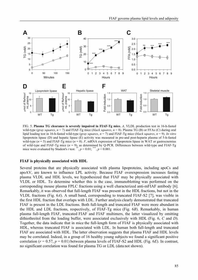
FIAF governs plasma lipid levels and adiposity
85
FIG. 5. Plasma TG clearance is severely impaired in FIAF-Tg mice. A, VLDL production test in 16-h-fasted wild-type (gray squares, n = 7) and FIAF-Tg mice (black squares, n = 8). Plasma TG (B) or FFAs (C) during oral lipid loading test in 16-h-fasted wild-type (gray squares, n = 7) and FIAF-Tg mice (black squares, n = 9). In vitro lipoprotein lipase (D) and hepatic lipase (E) activity was measured in pre-and post-heparin plasma of 5-h-fasted wild-type (n = 5) and FIAF-Tg mice (n = 8). F, mRNA expression of lipoprotein lipase in WAT or gastrocnemius of wild-type and FIAF-Tg mice (n = 9), as determined by Q-PCR. Differences between wild-type and FIAF-Tg mice were evaluated by Student's t test. **, p < 0.01; ***, p < 0.001.
FIAF is physically associated with HDL
Several proteins that are physically associated with plasma lipoproteins, including apoCs and apoAV, are known to influence LPL activity. Because FIAF overexpression increases fasting plasma VLDL and HDL levels, we hypothesized that FIAF may be physically associated with
VLDL or HDL. To determine whether this is the case, immunoblotting was performed on the corresponding mouse plasma FPLC fractions using a well characterized anti-mFIAF antibody [6]. Remarkably, it was observed that full-length FIAF was present in the HDL fractions, but not in the VLDL fractions (Fig. 6A). A small band, corresponding to truncated FIAF-S2 [7], was visible in
the first HDL fraction that overlaps with LDL. Further analysis clearly demonstrated that truncated FIAF is present in the LDL fractions. Both full-length and truncated FIAF were more abundant in the HDL and LDL fractions, respectively, of FIAF-Tg mice (Fig. 6B). Remarkably, in human plasma full-length FIAF, truncated FIAF and FIAF multimers, the latter visualized by omitting dithiothreitol from the loading buffer, were associated exclusively with HDL (Fig. 6, C and D). Together, the data indicate that in mice the full-length form of FIAF is physically associated with HDL, whereas truncated FIAF is associated with LDL. In human both full-length and truncated FIAF are associated with HDL. The latter observation suggests that plasma FIAF and HDL levels
may be correlated. Indeed, in a group of 16 healthy young subjects we found a significant positive correlation (r = 0.57, p = 0.01) between plasma levels of FIAF-S2 and HDL (Fig. 6E). In contrast, no significant correlation was found for plasma TG or LDL (data not shown).
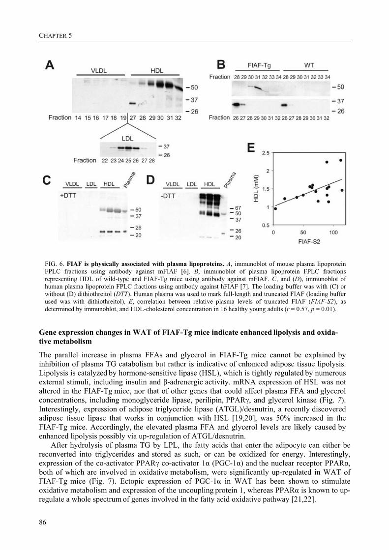
CHAPTER 5
86
FIG. 6. FIAF is physically associated with plasma lipoproteins. A, immunoblot of mouse plasma lipoprotein FPLC fractions using antibody against mFIAF [6]. B, immunoblot of plasma lipoprotein FPLC fractions representing HDL of wild-type and FIAF-Tg mice using antibody against mFIAF. C, and (D), immunoblot of human plasma lipoprotein FPLC fractions using antibody against hFIAF [7]. The loading buffer was with (C) or without (D) dithiothreitol (DTT). Human plasma was used to mark full-length and truncated FIAF (loading buffer used was with dithiothreitol). E, correlation between relative plasma levels of truncated FIAF (FIAF-S2), as determined by immunoblot, and HDL-cholesterol concentration in 16 healthy young adults (r = 0.57, p = 0.01).
Gene expression changes in WAT of FIAF-Tg mice indicate enhanced lipolysis and oxida- tive metabolism
The parallel increase in plasma FFAs and glycerol in FIAF-Tg mice cannot be explained by inhibition of plasma TG catabolism but rather is indicative of enhanced adipose tissue lipolysis. Lipolysis is catalyzed by hormone-sensitive lipase (HSL), which is tightly regulated by numerous external stimuli, including insulin and β-adrenergic activity. mRNA expression of HSL was not altered in the FIAF-Tg mice, nor that of other genes that could affect plasma FFA and glycerol concentrations, including monoglyceride lipase, perilipin, PPARγ, and glycerol kinase (Fig. 7). Interestingly, expression of adipose triglyceride lipase (ATGL)/desnutrin, a recently discovered adipose tissue lipase that works in conjunction with HSL [19,20], was 50% increased in the FIAF-Tg mice. Accordingly, the elevated plasma FFA and glycerol levels are likely caused by enhanced lipolysis possibly via up-regulation of ATGL/desnutrin.
After hydrolysis of plasma TG by LPL, the fatty acids that enter the adipocyte can either be reconverted into triglycerides and stored as such, or can be oxidized for energy. Interestingly, expression of the co-activator PPARγ co-activator 1α (PGC-1α) and the nuclear receptor PPARα, both of which are involved in oxidative metabolism, were significantly up-regulated in WAT of FIAF-Tg mice (Fig. 7). Ectopic expression of PGC-1α in WAT has been shown to stimulate oxidative metabolism and expression of the uncoupling protein 1, whereas PPARα is known to up-regulate a whole spectrum of genes involved in the fatty acid oxidative pathway [21,22].
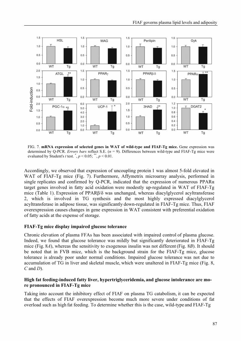
FIAF governs plasma lipid levels and adiposity
87
FIG. 7. mRNA expression of selected genes in WAT of wild-type and FIAF-Tg mice. Gene expression was determined by Q-PCR. Errors bars reflect S.E. (n = 9). Differences between wild-type and FIAF-Tg mice were evaluated by Student's t test. *, p < 0.05; **, p < 0.01.
Accordingly, we observed that expression of uncoupling protein 1 was almost 5-fold elevated in WAT of FIAF-Tg mice (Fig. 7). Furthermore, Affymetrix microarray analysis, performed in single replicates and confirmed by Q-PCR, indicated that the expression of numerous PPARα target genes involved in fatty acid oxidation were modestly up-regulated in WAT of FIAF-Tg mice (Table 1). Expression of PPARβ/δ was unchanged, whereas diacylglycerol acyltransferase
2, which is involved in TG synthesis and the most highly expressed diacylglycerol acyltransferase in adipose tissue, was significantly down-regulated in FIAF-Tg mice. Thus, FIAF overexpression causes changes in gene expression in WAT consistent with preferential oxidation of fatty acids at the expense of storage. FIAF-Tg mice display impaired glucose tolerance
Chronic elevation of plasma FFAs has been associated with impaired control of plasma glucose. Indeed, we found that glucose tolerance was mildly but significantly deteriorated in FIAF-Tg mice (Fig. 8A), whereas the sensitivity to exogenous insulin was not different (Fig. 8B). It should be noted that in FVB mice, which is the background strain for the FIAF-Tg mice, glucose tolerance is already poor under normal conditions. Impaired glucose tolerance was not due to accumulation of TG in liver and skeletal muscle, which were unaltered in FIAF-Tg mice (Fig. 8, C and D). High fat feeding-induced fatty liver, hypertriglyceridemia, and glucose intolerance are mo- re pronounced in FIAF-Tg mice
Taking into account the inhibitory effect of FIAF on plasma TG catabolism, it can be expected that the effects of FIAF overexpression become much more severe under conditions of fat overload such as high fat feeding. To determine whether this is the case, wild-type and FIAF-Tg
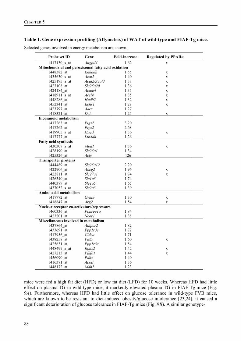
CHAPTER 5
88
Table 1. Gene expression profiling (Affymetrix) of WAT of wild-type and FIAF-Tg mice. Selected genes involved in energy metabolism are shown.
Probe set ID Gene Fold-increase Regulated by PPARα 1417130_s_at Angptl4 1.62 x Mitochondrial and peroxisomal fatty acid oxidation
1448382_at Ehhadh 1.55 x 1435630_s_at Acat2 1.40 x 1425195_a_at Acat2/Acat3 1.38 x 1423108_at Slc25a20 1.36 x 1424184_at Acadvl 1.35 x 1418911_s_at Acsl4 1.35 x 1448286_at Hadh2 1.32 x 1452341_at Echs1 1.28 x 1423797_at Aacs 1.27 1418321_at Dci 1.25 x Eicosanoid metabolism
1417263_at Ptgs2 3.20 1417262_at Ptgs2 2.68 1419905_s_at Hpgd 1.36 x 1417777_at Ltb4dh 1.26 Fatty acid synthesis
1430307_a_at Mod1 1.36 x 1428190_at Slc25a1 1.34 1425326_at Acly 126 Transporter proteins
1444489_at Slc25a12 2.20 1422906_at Abcg2 1.96 x 1422811_at Slc27a1 1.74 x 1426340_at Slc1a3 1.74 1440379_at Slc1a5 1.65 1437052_s_at Slc2a3 1.39 Amino acid metabolism
1417772_at Grhpr 1.30 x 1418847_at Arg2 1.54 x Nuclear receptor co-activators/repressors
1460336_at Ppargc1a 1.84 1423201_at Ncor1 1.38 Miscellaneous involved in metabolism
1437864_at Adipor2 1.82 1433691_at Ppp1r3c 1.72 1417956_at Cidea 1.71 1438258_at Vldlr 1.60 x 1425631_at Ppp1r3c 1.54 1448499_a_at Ephx2 1.42 x 1427213_at Pfkfb1 1.44 x 1456090_at Pdhx 1.40 1416371_at Apod 1.36 1448172_at Mdh1 1.23
mice were fed a high fat diet (HFD) or low fat diet (LFD) for 10 weeks. Whereas HFD had little effect on plasma TG in wild-type mice, it markedly elevated plasma TG in FIAF-Tg mice (Fig. 9A). Furthermore, whereas HFD had little effect on glucose tolerance in wild-type FVB mice, which are known to be resistant to diet-induced obesity/glucose intolerance [23,24], it caused a significant deterioration of glucose tolerance in FIAF-Tg mice (Fig. 9B). A similar genotype-
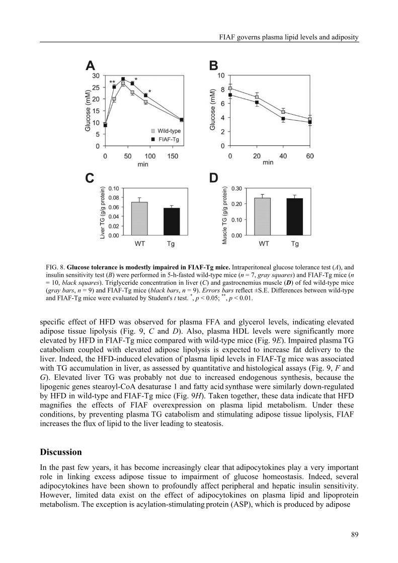
FIAF governs plasma lipid levels and adiposity
89
FIG. 8. Glucose tolerance is modestly impaired in FIAF-Tg mice. Intraperitoneal glucose tolerance test (A), and insulin sensitivity test (B) were performed in 5-h-fasted wild-type mice (n = 7, gray squares) and FIAF-Tg mice (n = 10, black squares). Triglyceride concentration in liver (C) and gastrocnemius muscle (D) of fed wild-type mice (gray bars, n = 9) and FIAF-Tg mice (black bars, n = 9). Errors bars reflect ±S.E. Differences between wild-type and FIAF-Tg mice were evaluated by Student's t test. *, p < 0.05; **, p < 0.01.
specific effect of HFD was observed for plasma FFA and glycerol levels, indicating elevated adipose tissue lipolysis (Fig. 9, C and D). Also, plasma HDL levels were significantly more elevated by HFD in FIAF-Tg mice compared with wild-type mice (Fig. 9E). Impaired plasma TG catabolism coupled with elevated adipose lipolysis is expected to increase fat delivery to the liver. Indeed, the HFD-induced elevation of plasma lipid levels in FIAF-Tg mice was associated
with TG accumulation in liver, as assessed by quantitative and histological assays (Fig. 9, F and G). Elevated liver TG was probably not due to increased endogenous synthesis, because the lipogenic genes stearoyl-CoA desaturase 1 and fatty acid synthase were similarly down-regulated by HFD in wild-type and FIAF-Tg mice (Fig. 9H). Taken together, these data indicate that HFD magnifies the effects of FIAF overexpression on plasma lipid metabolism. Under these conditions, by preventing plasma TG catabolism and stimulating adipose tissue lipolysis, FIAF
increases the flux of lipid to the liver leading to steatosis. Discussion In the past few years, it has become increasingly clear that adipocytokines play a very important role in linking excess adipose tissue to impairment of glucose homeostasis. Indeed, several adipocytokines have been shown to profoundly affect peripheral and hepatic insulin sensitivity. However, limited data exist on the effect of adipocytokines on plasma lipid and lipoprotein metabolism. The exception is acylation-stimulating protein (ASP), which is produced by adipose
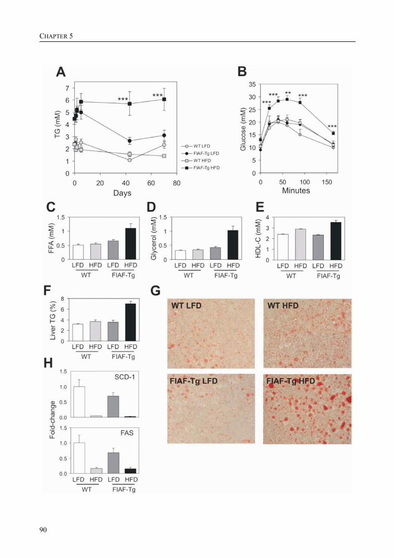
CHAPTER 5
90

FIAF governs plasma lipid levels and adiposity
91
FIG. 9. High fat diet magnifies the effect of FIAF-overexpression on lipid metabolism. A, time course of plasma TG in wild-type and FIAF-Tg mice during high fat (HFD) or low fat (LFD) feeding. B, intraperitoneal glucose tolerance test in wild-type and FIAF-Tg after 9 weeks of high fat or low fat feeding. Differences between wild-type and FIAF-Tg mice on HFD were evaluated by Student's t test. **, p < 0.01; ***, p < 0.001. Plasma FFA (C), glycerol (D), HDL-C (E), and liver TG (F) in wild-type and FIAF-Tg mice fed a LFD or HFD for 10 weeks. Significant effects were observed by two-way analysis of variance for diet (FFA, p < 0.05; glycerol, p < 0.001; HDL, p < 0.0001; liver TG, p < 0.0001), for genotype (FFA, p < 0.0001; glycerol, p < 0.0001; liver TG, p < 0.0001), and for the interaction between the two parameters (FFA, p < 0.01; glycerol, p < 0.0001; HDL, p < 0.01; liver TG, p < 0.0001). Error bars represent ±S.E. G, histological sections of liver stained for lipids using Oil Red O. H, mRNA expression of lipogenic genes stearoyl-CoA desaturase (SCD-1) and fatty acid synthase (FAS) in liver, as determined by Q-PCR. The numbers of animals per group were: WT_LFD: 9; WT_HFD: 11; FIAF-Tg_LFD: 9; and FIAF-Tg_HFD: 7.
tissue and influences plasma TG metabolism. Studies in mice lacking the complement component 3 gene C3, which is the precursor for ASP, as well as studies with mice injected with recombinant ASP have shown that ASP stimulates uptake and storage of plasma TG in fat cells [25,26]. As opposed to ASP, our and other data show that in mice FIAF is a powerful inhibitor of plasma TG clearance. In addition, in adipose tissue FIAF stimulates lipolysis, resulting in elevated plasma FFA and glycerol levels, and may promote fatty acid oxidative metabolism and uncoupling. One major consequence of this action is diminished fat stores.
As the rate-controlling step in plasma TG clearance is catalyzed by LPL, FIAF most likely increases plasma TG levels in mice by blocking LPL, which is supported by in vitro studies using
purified LPL and FIAF [10]. The inhibitory effect of FIAF on LPL is similar to that of apoCI and apoCIII, and opposes that of apoCII and apoAV, the latter of which, interestingly, is also physically associated with HDL. Recent data suggest that apoAV lowers plasma TG by directing TG-rich lipoproteins to proteoglycans-bound LPL [27]. However, it is not clear whether the physical association of FIAF with HDL is required for its hypertriglyceridemic effect. It can be speculated that inhibition of LPL is actually mediated by truncated FIAF bound to LDL. A mechanism opposite to apoCII but having the same net effect can be envisioned, where FIAF, rather than dissociating from VLDL as it decreases in size, associates with IDL/LDL and inhibits
LPL activity to prevent complete TG hydrolysis. If inhibition of LPL by FIAF and thus its hypertriglyceridemic effect is truly dependent on its binding to LDL, FIAF may function differently in human, as in human FIAF is associated exclusively with HDL. An additional possible explanation for the impaired LPL activity in FIAF-Tg mice may be elevated plasma FFAs, which have been shown to potently inhibit LPL activity via numerous mechanisms [28]. In the FIAF-Tg mice, plasma FFAs were elevated in fed and fasted state, as well as during the lipid loading test. Taken together, the inhibitory effect of FIAF on plasma TG clearance is likely explained by inhibition of LPL, although the one or more molecular and structural mechanisms behind this inhibition remain somewhat ambiguous.
Inhibition of LPL cannot be the cause of the elevated HDL-cholesterol levels in FIAF-Tg mice. Because LPL is structurally highly similar to hepatic lipase and endothelial lipase, and because impaired activity of these lipases causes elevation of plasma HDL [29-32], it is conceivable that FIAF may target hepatic lipase and endothelial lipase as well. Additional research is necessary to better define the molecular mechanisms behind the modestly elevated plasma HDL-cholesterol and markedly elevated plasma apoAI and apoAII levels in FIAF-Tg mice.
The increased levels of plasma FFAs and glycerol in FIAF-Tg mice are in agreement with the acute elevation in plasma FFAs observed after FIAF injection [10], indicating that FIAF stimulates
adipose tissue lipolysis. We did not find any effect of FIAF on HSL mRNA expression, although we cannot rule out activation of HSL activity via an alternative mechanism. In contrast, the mRNA

CHAPTER 5
92
level of the recently described adipose triglyceride lipase (ATGL) was elevated by 50% in FIAF-Tg mice [19,20]. Thus, FIAF may stimulate lipolysis via up-regulation of ATGL.
Previous studies have shown that adenoviral-mediated overexpression of FIAF or injection of recombinant FIAF potently elevates plasma triglyceride levels [10,11]. Although these studies established FIAF as a modulator of plasma TG levels in mice, they did not allow appraisal of the impact of physiological changes in FIAF expression. What is remarkable about the present study is that a modest increase in FIAF mRNA in peripheral tissues is sufficient to cause fasting plasma TG to go up to 2.5- to 8-fold, depending on the duration of fasting. Coupled with the severely impaired
plasma TG clearance after lipid loading, this indicates that FIAF is an extremely powerful regulator of plasma TG levels in mice.
In the FIAF-Tg mice impaired plasma TG clearance can be expected to diminish adipose stores [33]. However, the fate of the excess TG remains unclear, because they do not appear to be stored
in non-adipose tissues, at least under conditions of low fat diet. Based on our gene expression data, which show increased expression of a variety of genes involved in fatty acid oxidative metabolism and uncoupling, including PGC-1α and uncoupling protein 1, we speculate that these extra TG were actually oxidized. Detailed indirect calorimetric studies will be necessary to clarify this issue.
The present study clearly demonstrates that the effects of FIAF overexpression are much more pronounced in mice fed a high fat diet. In wild-type FVB mice, which are known to be resistant to high fat diet-induced obesity/glucose intolerance, high fat feeding had little effect on plasma TG, FFAs, and glycerol concentration, as well as on glucose intolerance, but markedly augmented these
parameters in mice overexpressing FIAF. Previously we showed that high fat feeding down-regulated plasma FIAF levels in mice [6], which in light of the present findings may serve to facilitate the clearance of plasma TG into peripheral tissues, and thus minimize their hepatic and plasma accumulation. FIAF overexpression overruled the down-regulation, resulting in markedly elevated plasma TG levels and a fatty liver.
Recently, Xu et al. [12] reported that adenoviral-mediated overexpression of FIAF causes a pronounced decrease in plasma glucose level and improves glucose tolerance. In contrast, we show that FIAF overexpression has no effect on plasma glucose levels and impairs glucose tolerance, particularly after feeding a high fat diet. The reason for this discrepancy is not clear but may be related to the level of overexpression, which was modest in our experiments, as well as the site of overexpression (liver versus peripheral tissues). We previously demonstrated that human liver produces the truncated form of FIAF, whereas human adipose tissue only produces the full-length form [7]. It can be hypothesized that these forms may have different activities toward glucose and
lipid metabolism, similar to what has been demonstrated for globular and full-length adiponectin [34]. It should be mentioned that, while our manuscript was in revision, Köster et al. [35] showed no effect of liver-specific hFIAF overexpression or general FIAF deletion on plasma glucose levels. In agreement with Xu et al. we find accumulation of hepatic triglycerides by FIAF overexpression, yet this is only observed after a high fat diet. The hepatic steatosis together with the markedly elevated plasma FFA levels may contribute to the impaired glucose tolerance in FIAF-Tg mice on a high fat diet.
An important question that is still not fully resolved is whether FIAF only acts locally in the tissue where it is produced, exerting an autocrine or paracrine action, or whether it also targets
distant organs. Another key question is whether the effects of FIAF on lipid and glucose metabolism are exclusively mediated by inhibition of LPL or involve an additional mechanism. Blocking LPL is expected to result in a decreased plasma FFA level, rather than an increase as observed here and in another study [10], suggesting that FIAF acts in multiple ways. It can be hypothesized that in analogy to numerous other adipocytokines FIAF is able to bind a specific cell-surface receptor, although this remains to be demonstrated.
FIAF governs plasma lipid levels and adiposity
93
Although FIAF is most highly expressed in adipose tissue, it may exert important effects in other tissues as well. Recently, it was reported that down-regulation of FIAF expression in the intestine is essential for the increase in adipose mass induced by intestinal microbiota [36]. The data suggested that FIAF is an important mediator of the physiological effects of the intestinal microbiota.
Despite bearing a name that belies any link with metabolism, in recent years it has become apparent that angiopoietin-like proteins are a group of proteins that play an important role in the regulation of lipid and glucose metabolism. Recent studies indicate that overexpression or disruption of Angptl3 and angiopoietin-related growth factor (Angptl6), which are mainly produced by the liver, result in major disturbances at the level of lipid metabolism and energy homeostasis [37-39]. Accordingly, angiopoietin-like proteins have become very interesting targets for the pharmacological treatment of dyslipidemia and obesity.
In conclusion, FIAF represents a powerful signaling molecule from fat and other tissues that prevents plasma TG clearance and stimulates adipose TG mobilization. Further studies, including
investigation of the connection between human plasma FIAF level and post-prandial TG response, are necessary to better define the role of FIAF in lipid metabolism in humans. Nevertheless, it can be speculated that FIAF may be involved in connecting adipose tissue to regulation of plasma lipid levels, and that changes in FIAF signaling may contribute to development of (diabetic)
dyslipidemia. Acknowledgements We thank René Bakker, Frits van der Hoeven, Karin Mudde, Shohreh Keshtar, Jolanda van der Meijde, and Vincent Bloks for excellent technical support. References 1. Ahima, R. S.and Flier, J. S. (2000) Adipose tissue as an endocrine organ. Trends Endocrinol. Metab.
11, 327-332 2. Zhang, Y., Proenca, R., Maffei, M., Barone, M., Leopold, L.and Friedman, J. M. (1994) Positional
cloning of the mouse obese gene and its human homologue. Nature 372, 425-432 3. Berg, A. H., Combs, T. P., Du, X., Brownlee, M.and Scherer, P. E. (2001) The adipocyte-secreted
protein Acrp30 enhances hepatic insulin action. Nat. Med. 7, 947-953 4. Steppan, C. M., Bailey, S. T., Bhat, S. et al. (2001) The hormone resistin links obesity to diabetes.
Nature 409, 307-312 5. Hotamisligil, G. S., Shargill, N. S.and Spiegelman, B. M. (1993) Adipose expression of tumor
necrosis factor-alpha: direct role in obesity-linked insulin resistance. Science 259, 87-91 6. Kersten, S., Mandard, S., Tan, N. S. et al. (2000) Characterization of the fasting-induced adipose
factor FIAF, a novel peroxisome proliferator-activated receptor target gene. J. Biol. Chem. 275, 28488-28493
7. Mandard, S., Zandbergen, F., Tan, N. S. et al. (2004) The direct peroxisome proliferator-activated receptor target fasting-induced adipose factor (FIAF/PGAR/ANGPTL4) is present in blood plasma as a truncated protein that is increased by fenofibrate treatment. J. Biol. Chem. 279, 34411-34420
8. Yoon, J. C., Chickering, T. W., Rosen, E. D. et al. (2000) Peroxisome proliferator-activated receptor gamma target gene encoding a novel angiopoietin-related protein associated with adipose differentiation. Mol. Cell. Biol. 20, 5343-5349
9. Kim, I., Kim, H. G., Kim, H. et al. (2000) Hepatic expression, synthesis and secretion of a novel fibrinogen/angiopoietin-related protein that prevents endothelial-cell apoptosis. Biochem. J. 346 Pt 3, 603-610
CHAPTER 5
94
10. Yoshida, K., Shimizugawa, T., Ono, M.and Furukawa, H. (2002) Angiopoietin-like protein 4 is a potent hyperlipidemia-inducing factor in mice and inhibitor of lipoprotein lipase. J. Lipid Res. 43, 1770-1772
11. Ge, H., Yang, G., Yu, X., Pourbahrami, T.and Li, C. (2004) Oligomerization state-dependent hyperlipidemic effect of angiopoietin-like protein 4. J Lipid Res 45, 2071-2079
12. Xu, A., Lam, M. C., Chan, K. W. et al. (2005) Angiopoietin-like protein 4 decreases blood glucose and improves glucose tolerance but induces hyperlipidemia and hepatic steatosis in mice. Proc. Natl. Acad. Sci. U.S.A. 102, 6086-6091
13. Yu, X., Burgess, S. C., Ge, H. et al. (2005) Inhibition of cardiac lipoprotein utilization by transgenic overexpression of Angptl4 in the heart. Proc. Natl. Acad. Sci. U.S. A. 102, 1767-1772
14. Belanger, A. J., Lu, H., Date, T. et al. (2002) Hypoxia up-regulates expression of peroxisome proliferator-activated receptor gamma angiopoietin-related gene (PGAR) in cardiomyocytes: role of hypoxia inducible factor 1alpha. J. Mol. Cell. Cardiol. 34, 765-774
15. Le Jan, S., Amy, C., Cazes, A. et al. (2003) Angiopoietin-like 4 is a proangiogenic factor produced during ischemia and in conventional renal cell carcinoma. Am. J. Pathol. 162, 1521-1528
16. Ito, Y., Oike, Y., Yasunaga, K. et al. (2003) Inhibition of angiogenesis and vascular leakiness by angiopoietin-related protein 4. Cancer Res. 63, 6651-6657
17. Berbee, J. F., van der Hoogt, C. C., Sundararaman, D., Havekes, L. M.and Rensen, P. C. (2005) Severe hypertriglyceridemia in human APOC1 transgenic mice is caused by apoC-I-induced inhibition of LPL. J. Lipid Res. 46, 297-306
18. Ross, S. R., Graves, R. A., Greenstein, A. et al. (1990) A fat-specific enhancer is the primary determinant of gene expression for adipocyte P2 in vivo. Proc. Natl. Acad. Sci. U.S.A. 87, 9590-9594
19. Villena, J. A., Roy, S., Sarkadi-Nagy, E., Kim, K. H.and Sul, H. S. (2004) Desnutrin, an adipocyte gene encoding a novel patatin domain-containing protein, is induced by fasting and glucocorticoids: ectopic expression of desnutrin increases triglyceride hydrolysis. J. Biol. Chem. 279, 47066-47075
20. Zimmermann, R., Strauss, J. G., Haemmerle, G. et al. (2004) Fat mobilization in adipose tissue is promoted by adipose triglyceride lipase. Science 306, 1383-1386
21. Mandard, S., Muller, M.and Kersten, S. (2004) Peroxisome proliferator-activated receptor alpha target genes. Cell. Mol. Life Sci. 61, 393-416
22. Puigserver, P., Wu, Z., Park, C. W., Graves, R., Wright, M.and Spiegelman, B. M. (1998) A cold-inducible coactivator of nuclear receptors linked to adaptive thermogenesis. Cell 92, 829-839
23. Hu, C. C., Qing, K.and Chen, Y. (2004) Diet-induced changes in stearoyl-CoA desaturase 1 expression in obesity-prone and -resistant mice. Obes. Res. 12, 1264-1270
24. Le Lay, S., Boucher, J., Rey, A. et al. (2001) Decreased resistin expression in mice with different sensitivities to a high-fat diet. Biochem. Biophys. Res. Commun. 289, 564-567
25. Murray, I., Sniderman, A. D.and Cianflone, K. (1999) Enhanced triglyceride clearance with intraperitoneal human acylation stimulating protein in C57BL/6 mice. Am. J. Physiol. 277, E474-480
26. Murray, I., Sniderman, A. D., Havel, P. J.and Cianflone, K. (1999) Acylation stimulating protein (ASP) deficiency alters postprandial and adipose tissue metabolism in male mice. J. Biol. Chem. 274, 36219-36225
27. Merkel, M., Loeffler, B., Kluger, M. et al. (2005) Apolipoprotein AV accelerates plasma hydrolysis of triglyceride-rich lipoproteins by interaction with proteoglycan-bound lipoprotein lipase. J. Biol. Chem. 280, 21553-21560
28. Goudriaan, J. R., den Boer, M. A., Rensen, P. C. et al. (2005) CD36 deficiency in mice impairs lipoprotein lipase-mediated triglyceride clearance. J. Lipid Res. 46, 2175-2181
29. Busch, S. J., Barnhart, R. L., Martin, G. A. et al. (1994) Human hepatic triglyceride lipase expression reduces high density lipoprotein and aortic cholesterol in cholesterol-fed transgenic mice. J. Biol. Chem. 269, 16376-16382
30. Homanics, G. E., de Silva, H. V., Osada, J. et al. (1995) Mild dyslipidemia in mice following targeted inactivation of the hepatic lipase gene. J. Biol. Chem. 270, 2974-2980
31. Jin, W., Millar, J. S., Broedl, U., Glick, J. M.and Rader, D. J. (2003) Inhibition of endothelial lipase causes increased HDL cholesterol levels in vivo. J. Clin. Invest. 111, 357-362
FIAF governs plasma lipid levels and adiposity
95
32. Ma, K., Cilingiroglu, M., Otvos, J. D., Ballantyne, C. M., Marian, A. J.and Chan, L. (2003) Endothelial lipase is a major genetic determinant for high-density lipoprotein concentration, structure, and metabolism. Proc. Natl. Acad. Sci. U.S.A. 100, 2748-2753
33. Weinstock, P. H., Bisgaier, C. L., Aalto-Setala, K. et al. (1995) Severe hypertriglyceridemia, reduced high density lipoprotein, and neonatal death in lipoprotein lipase knockout mice. Mild hypertriglyceridemia with impaired very low density lipoprotein clearance in heterozygotes. J. Clin. Invest. 96, 2555-2568
34. Xu, J., Chang, V., Joseph, S. B. et al. (2004) Peroxisomal proliferator-activated receptor alpha deficiency diminishes insulin-responsiveness of gluconeogenic/glycolytic/pentose gene expression and substrate cycle flux. Endocrinology 145, 1087-1095
35. Koster, A., Chao, Y. B., Mosior, M. et al. (2005) Transgenic angiopoietin-like (angptl)4 overexpression and targeted disruption of angptl4 and angptl3: regulation of triglyceride metabolism. Endocrinology 146, 4943-4950
36. Backhed, F., Ding, H., Wang, T. et al. (2004) The gut microbiota as an environmental factor that regulates fat storage. Proc. Natl. Acad. Sci. U.S.A. 101, 15718-15723
37. Oike, Y., Akao, M., Yasunaga, K. et al. (2005) Angiopoietin-related growth factor antagonizes obesity and insulin resistance. Nat. Med. 11, 400-408
38. Koishi, R., Ando, Y., Ono, M. et al. (2002) Angptl3 regulates lipid metabolism in mice. Nat. Genet. 30, 151-157
39. Inaba, T., Matsuda, M., Shimamura, M. et al. (2003) Angiopoietin-like protein 3 mediates hypertriglyceridemia induced by the liver X receptor. J. Biol. Chem. 278, 21344-21351

96
Recombinant expression and purification of FIAF
97
CHAPTER 6 Recombinant FIAF: Expression in and purification from insect cells Fokko Zandbergen, Tessa Steevels, Michael Müller, and Sander Kersten Abstract The Fasting-induced adipose factor (FIAF) is a secreted and glycosylated protein that has been implicated in lipid metabolism and angiogenesis. In an effort to facilitate the study of the function and structure of FIAF in more detail, we investigated the possibility of obtaining purified FIAF in quantities that are large enough to perform such studies. Recombinant expression and purification of FIAF were pursued using several expression systems and purification techniques. Expression in E. coli in our hands failed to result in the production of native FIAF, as did attempts to fold and renature the protein to sufficient amounts following denaturing FIAF from inclusion bodies. Furthermore, transient expression in eukaryotic HEK293 cells did not yield sufficient amounts of FIAF. However, these problems were partially overcome by expressing FIAF in insect cells using baculovirus as a vector. Whereas in the cytoplasm of the insect cells FIAF was mostly present as a denatured protein, which was likely the result of improper folding linked to different or inadequate glycosylation of FIAF, an approach in which FIAF was directed to the secretory pathway yielded native FIAF that could be purified from the cell culture medium. The production of FIAF in insect cells, using the baculovirus system, is promising for obtaining pure and relatively large quantities of FIAF that can be used to further study its function and structure.
CHAPTER 6
98
Introduction The Fasting-induced adipose factor (FIAF), also known as PGAR (PPARγ angiopoietin-related), HFARP (hepatic fibrinogen/angiopoietin-related protein), ANGPTL4 (angiopoietin-like protein 4) [1-4], is a secreted and glycosylated protein of approximately 50 kDa that has been reported to play roles in lipid metabolism and angiogenesis (see chapter 2 for a review). Our planning of future experiments to elucidate the function and structure of FIAF in more detail encompassed the utilization of techniques like incubation of cultured cells, prolonged in vivo injections into mice, and X-ray diffraction. Because these techniques require substantial amounts of protein (µg–mg), we explored the possibility of producing and purifying FIAF in amounts that are large enough to be able to conduct this kind of studies.
In order to obtain pure protein in substantial amounts, one of the options is to isolate it from a source in which it is already abundant. This requires the determination of properties of the protein like its molecular mass, hydrophobicity, and iso-electric point (i.e. the pH at which the collective positive and negative charges on the protein equal zero), as those characteristics can be used to separate the protein of interest from other proteins. An approach that is less dependent on the properties of the protein is to attach a tag that has a high affinity for a particular substrate. This method evades the problem that the purification can be complicated when properties of the protein to be purified are similar to that of other proteins in the source material. Examples of tags are GST (Glutathion-S-transferase), which has a high affinity for glutathione, and the amino acid histidine (His), which binds positively charged divalent metal ions like Cu2+, Zn2+ and Fe2+. These tags are applied by cloning a nucleotide sequence encoding the GST- or His-tag in frame with the sequence encoding the protein of interest into a vector. In the case of a His-tag, a sequence encoding multiple consecutive histidine residues is usually applied. The divalent metal ions that form the substrate for the tags are in general attached to a polymer, enabling immobilization and hence improved manipulation, like washing and concentration of the proteins during the purification process. This system is also known as immobilized metal affinity chromatography (IMAC).
The production of recombinant proteins in E. coli is relatively inexpensive as the medium is cheap and the laboratory infrastructure required is minimal. However, mammalian proteins often need extended post-translational modifications that prokaryotes are not able to perform. Depending on the properties of the protein, in E. coli this may be causing improper folding, resulting in inactive protein. This problem can be circumvented by the more expensive production of the protein of interest in mammalian cells. In contrast to prokaryotic E. coli, eukaryotic cells perform the post-translational manipulations and modifications such as recognition of the signal sequence and glycosylation of the protein, which might be needed for the proper folding and functionality of the protein [5]. On the other hand, expression in mammalian cells can result in production levels that are not sufficient for use in experiments requiring relatively large amounts of recombinant protein. Although equally expensive, the insect cell expression system combines the advantages of the high expression levels that can be reached in E. coli with the more sophisticated protein folding pathways of eukaryotic cells. Insect cells perform post-translational modifications of proteins in a similar manner as mammalian cells and in combination with the use of insect viruses as a vector, the production yield can approach that of E. coli [6].
So far, several groups have already reported the small scale production and purification of recombinant FIAF from prokaryotic E. coli and from eukaryotic cells [3,4,7,8]. The investigators that were the first to purify FIAF expressed the protein with a His-tag in eukaryotic COS-7 cells. They found that recombinant FIAF inhibited the apoptosis of vascular endothelial cells, the cells that make up the inner lining of blood vessels, and concluded that FIAF might have a protective effect on endothelial cells [3]. Using recombinant mouse FIAF that was produced in E. coli and purified using the attached GST-tag, it was discovered that upon injection into mice FIAF rapidly
Recombinant expression and purification of FIAF
99
increases plasma levels of triglycerides (TG), free fatty acids (FFA) and non-HDL cholesterol. Furthermore, FIAF was able to dose-dependently inhibit the activity of lipoprotein lipase (LPL) in vitro [4]. One group generated polyclonal antibodies against rat FIAF using several recombinant FIAF products. These researchers used full length GST-tagged FIAF and purified a His-tagged carboxyl-terminal part of FIAF from bacteria. In addition, they partially purified His-tagged FIAF from mammalian HEK293 cells [7]. Another group produced a recombinant His-tagged human FIAF amino-terminal fragment in bacteria in order to develop an enzyme-linked immuno-sorbent assay (ELISA). With this ELISA, the concentration of FIAF in plasma of healthy humans was determined to be approx. 350 ng/ml [9]. Finally, using purified recombinant FIAF from medium of Chinese hamster ovary cells (CHO), it was shown that LPL activity is also inhibited by the mouse version of FIAF [8]. Together with the inhibition of LPL by the human version of FIAF, which was already shown before [4], this indicates that FIAF shares functions in human and mouse.
No additional experiments with recombinant FIAF have been reported at the time of writing this chapter. This might reflect difficulties with producing or purifying FIAF in larger quantities. It has indeed been reported that members of the angiopoietin-like proteins, the family to which FIAF belongs, exhibit the tendency to form aggregates [10], complicating their experimental use. However, it was not clear whether FIAF also has the propensity to aggregate and precipitate. We have evaluated the expression of FIAF in E. coli, HEK293 cells, and insect cells. Expression in E. coli resulted in FIAF being present only in aggregates of inactive, improperly folded protein. Attempts to refold FIAF to sufficient amounts from these inclusion bodies failed in our hands. Furthermore, transient expression in the eukaryotic HEK293 cell line resulted in the production of insufficient amounts of FIAF. However, expression of FIAF in insect cells, using baculovirus as a vector, resulted in the secretion of native FIAF into the cell culture medium in amounts that could be easily visualized by Coomassie staining. In addition, by comparing several purification methods, we determined the optimal conditions for isolation of recombinant FIAF. Methods General materials
Vectors pFastBac-HT version A and pFastBac-1, Spodoptera frugiperda Sf21, High Five™, and BTI-TN-5B1-4 (a High Five™ strain optimized for growth in suspension) cells, and the Grace’s insect cell medium and Express Five SFM (serum free medium) were from Invitrogen. Vector pAcSecG2T and BaculoGold™ viral DNA were from BD Biosciences. Grace’s insect cell culture unsupplemented was from Sigma. HisTrap™ HP columns were from Amersham Biosciences. Foetal Bovine Serum was from Cambrex. 50 x Complete EDTA-free protease inhibitor was from Roche. Unless stated otherwise, all other chemicals were from Sigma. Constructs and cloning
The cDNA sequence encoding full length FIAF as well as that encoding the first 306 amino acids of FIAF was amplified by PCR from a vector containing the complete FIAF cDNA sequence. The forward primer (5’ - CGGGATCCTGCACAGCCAGAGCCAC - 3’) contained a 5’-end BamHI endonuclease restriction site (restriction sites underlined) and was designed to anneal downstream of the first 63 bp that encode the signal sequence directing FIAF to the secretory pathway, thus resulting in PCR products without the signal sequence (also see Table 1 for primer sequences). Both the reverse primer for the amplification of full length FIAF (5’ – ACCGCTCGAGCTAAGA-GGCTGCTGTAGC - 3’) and the one for amplifying the truncated FIAF product (5’ – ACCGCT-CGAGTCAAGTAGAGAAGGGCAGGG - 3’) contained a stop codon and a 5’-end XhoI
CHAPTER 6
100
endonuclease restriction site. The resulting PCR products were cloned between the BamHI and XhoI endonuclease restriction sites downstream of the 6xHis-tag sequence (His6) in pFastBac-HT version A. Table 1: primer sequences used for amplification of the sequences used for the vector constructs
primer name orientation nucleotide sequence mFIAF-s-Kzk-BamHI forward 5’ - CGGGATCCACCATGCGCTGCGCTCCGAC - 3’mFIAF-as-FastBac-1-EcoRI reverse 5’ - CCGGAATTCTAGAAGGCACAGTCGAGGCTG - 3’mFIAF-s-FastBac-HT-A-BamHI forward 5’ - CGGGATCCTGCACAGCCAGAGCCAC - 3’ mFIAF-ssRev reverse 5’ - ACCGCTCGAGCTAAGAGGCTGCTGTAGC - 3’mFIAF-R306 reverse 5’ - ACCGCTCGAGTCAAGTAGAGAAGGGCAGGG - 3’mFIAF-s-pAcSecG2T-BamHI forward 5’ - CGGGATCCGCACAGCCAGAGCCACCG - 3’mFIAF-FL-as-pAcSecG2T-EcoRI reverse 5’ - CCGGAATTCTAAGAGGCTGCTGTAGCCTCCA - 3’mFIAF-TR2-as-pAcSecG2T-EcoRI reverse 5’ - CCGGAATTCAAACATTGGTGGCACCCAGC - 3’
The new constructs were expected to express a full length and a truncated version of FIAF, both containing a His6 tag at their N-termini and being non-secreted. In addition, the complete FIAF cDNA sequence and the sequence encoding the first 306 amino acids, both with their signal sequence, were amplified by PCR from pcDNA3.1 vectors together with a His6-tag sequence attached to the C-terminal ends of the FIAF sequences. The forward primer (5’ - CGGGATCCACCATGCGCTGCGCTCCGAC - 3’) contained a 5’-end BamHI and the reverse primer (5’ - CCGGAATTCTAGAAGGCACAGTCGAGGCTG - 3’) a 5’-end EcoRI endonuclease restriction site. The obtained PCR products were cloned between the BamHI and EcoRI endonuclease restriction sites in pFastBac-1, expected to result in the production of a secreted full length and truncated version of FIAF, both with a His6-tag at their C-terminal extremities. Attempts to clone FIAF downstream of the polyhedrin promoter of the pFastBac-D/GFP vector failed. In addition to the polyhedrin promoter, this pFastBac-Dual vector contains a multiple cloning site downstream of the p10 promoter, in which the sequence for (enhanced) green fluorescent protein (GFP) has been cloned. Expression of GFP enables easy screening for infected cells, which are readily made visible using fluorescent microscopy [11]. The pFastBac-D/GFP vector was used to monitor the stage of insect cell infection.
The four different FIAF-containing pFastBac vectors, as well as the empty pFastBac-HT and pFastBac-1, and pFastBac-D/GFP were used to transform DH10Bac, an E. coli strain in which modified baculovirus DNA of the Autographa californica multiple capsid nucleopolyhedrovirus (AcMNPV) genome is propagated as a bacmid. Upon transformation, a part of the pFastBac vector, including the inserted FIAF-sequence, transposes into the baculovirus DNA with the assistance of a helper plasmid that is also present in the DH10Bac cells. The 2 pFastBac-1 vectors containing the full length and truncated FIAF sequences with their signal sequence, as well as empty pFastBac-1 vector, were used to transform a second strain of DH10Bac, in which the chitinase and v-cathepsin gene have been deleted from the standard baculovirus DNA [12]. The translation of chitinase from standard baculovirus DNA probably competes with recombinant secretory proteins for entering the secretory pathway [13,14] and activation of v-cathepsin may result in proteolysis of recombinant proteins [15]. Therefore, translation of FIAF from the modified baculovirus DNA, called AcBac∆CC, is expected to result in increased amounts and improved integrity of the recombinant protein that is produced [12].
In addition to the His6-tag containing constructs, FIAF was also cloned between the BamHI and EcoRI endonuclease restriction sites in frame with and downstream of a GST encoding sequence into the pAcSecG2T vector. The GST-tag in this vector has been cloned downstream of
Recombinant expression and purification of FIAF
101
an insect-virus signal sequence for secretion. The cDNA sequences encoding full length FIAF as well as that encoding the first 294 amino acids of FIAF were amplified by PCR from a vector containing the complete FIAF cDNA sequence. The forward primer (5’ – CGGGATCCGCACAG-CCAGAGCCACCG - 3’) contained a 5’-end BamHI endonuclease restriction site and annealed 81 bp downstream of the translation initiation start site to exclude amplification of the signal sequence of FIAF. The reverse primers (5’ - CCGGAATTCTAAGAGGCTGCTGTAGCCTCCA - 3’ for full length and 5’ - CCGGAATTCAAACATTGGTGGCACCCAGC - 3’ for truncated FIAF) contained a stop codon and a 5’-end EcoRI endonuclease restriction site. Expression from the polyhedrin promoter of the vector constructs, should result in the production and secretion of full length and truncated FIAF respectively, both with a GST-tag fused to their N-termini.
Blue-white screening was used to identify DH10Bac colonies containing baculovirus bacmid in which transposition of an insert had occurred. Baculovirus DNA was isolated from the white colonies, kept at 4 °C, and screened for the presence of FIAF by PCR using primers amplifying FIAF. A second M13 primer-set was also used, amplifying FIAF together with surrounding sequences or only the surrounding sequences in case empty pFastBac vector was used in the transformation. Generation of recombinant baculoviruses
Baculovirus DNA that was determined to be positive for the presence of inserts of expected length was used to transfect Sf21 Spodoptera frugiperda insect cells, using Cellfectin® reagent (Invitrogen) according to the manufacturer’s protocol.
The empty vector pAcSecG2T as well as the pAcSecG2T vectors encoding full length FIAF without the signal sequence and the first 294 amino acids without the signal sequence were used to transfect Sf21 cells together with linearized baculovirus DNA (BaculoGold™ viral DNA). The protocol of the manufacturer of the linearized DNA was followed, except for the transfection reagents, which were replaced by Cellfectin® reagent. After co-transfection, selection was applied on recombinant baculovirus in which the insert from pAcSecG2T together with a complementing part of the vector sequence have been inserted by homologous recombination, rescuing a lethal deletion on the linearized baculovirus DNA.
A few days after transfection, recombinant baculoviruses were harvested by collecting the medium of the transfected cells. Part of the cell lysate or medium was analyzed for the presence of FIAF by Western blotting (see below). Remaining medium of the samples containing FIAF was used to infect fresh cells in order to amplify the recombinant viruses. Alternatively, viruses were plaque-purified before amplification. The viruses from baculovirus DNA in which inserts from the empty pFastBac vectors had been transposed, and that were judged to be recombinant based on the size of amplified PCR-fragments, were amplified in order to be used as negative control for future production of FIAF. The virus derived from the empty pAcSecG2T vector was amplified too. Plaque purification
In order to get pure recombinant viruses of the His6-tag-containing constructs, they were isolated from single virus colonies by plaque-purification. Per construct, Sf21 cells were seeded in quadruplicate in six-well plates at a density of 50 %. The cells were infected with 4 different 10-fold dilutions of virus, ranging from 10-3 – 10-8, in 1 ml of Grace’s insect cell medium per well. After incubation for 1 hr at 25 °C, medium was replaced with 2 ml of plaquing medium (Grace’s insect cell medium containing 1 % low melting point agarose). The plaquing medium had been cooled to 37 °C and had been prepared according to the manufacturer’s protocol (Invitrogen). After cooling down at 25 °C for 15-20 min and subsequent incubation at 27 °C for 7-10 days, single plaques were isolated from the gel with a sterile pipette tip and transferred to 500 µl
CHAPTER 6
102
unsupplemented Grace’s insect cell medium in a micro-centrifuge tube. The tubes were vigorously vortexed to release the virus into the medium and the solution was used to infect fresh insect cells with an estimated multiplicity of infection (M.O.I., = number of virus particles per cell) of 0.1. The amount of virus-containing medium required for a desired M.O.I. was calculated using the following formula: Inoculum required (ml) = (M.O.I. (pfu)/cell) x number of cells) / titer of virus stock (pfu/ml), where pfu stands for plaque forming units.
After a few days, the cell lysate was collected and analyzed for the presence of FIAF by Western blotting (see below). Remaining medium of the samples containing FIAF was used to infect cells again in order to amplify the recombinant viruses. Western blotting
Transfected or infected insect cells to be analyzed for the presence of FIAF were lysed with lysis buffer (50 mM TrisHCl pH 8.0, 0.1 M NaCl, 5 mM EDTA, 0.1 % (w/v) SDS, and 1 % (v/v) NP40) and lysate was stored at -20 °C. If medium of the cells was to be analyzed, it was centrifuged to pellet cell debris and the supernatant was stored at -20 °C. Protein from the supernatant was precipitated by adding an equal volume of 40% trichloroacetic acid (TCA) and incubation on ice for 1-2 hrs. Protein was pelleted by centrifugation, washed twice with cold acetone, and redissolved in PBS. SDS-Page loading buffer was added to the cell lysate and proteins concentrated from the medium.
The protein samples were subjected to SDS-Page according to standard procedures. The separated proteins were transferred to Immobilon-P transfer membrane (Millipore) and subsequently analyzed for the presence of FIAF protein by incubation with a well-characterized polyclonal antibody directed against the epitope CQGPKGKDAPFKDSE, which is located in the N-terminal part of the mouse FIAF protein. The immunodetection was carried out as described before [1], except that the primary antibody was used at a dilution between 1:2000 and 1:5000.
Presence of the His-tag was verified too by using an anti-His antibody, with horseradish peroxidase conjugated to it, at a dilution of 1:4000. Time-course and M.O.I. determination for adherent insect cells
In order to increase the amounts of recombinant viruses, the virus amplification round was repeated once or twice, resulting in virus stocks P2 and P3, respectively. To amplify viruses, an M.O.I. of 0.1 was usually applied for the infection.
To determine the optimal M.O.I. and duration of infection for optimal production of FIAF, small-scale infections were performed with the amplified viruses. Cells were seeded in 24-well plates and grown to a density of 50–60 %. The cells were infected with the His-tagged FIAF-encoding recombinant baculoviruses at M.O.I.’s of 1, 2, 5 and 10. For calculation of the volumes of virus solution needed, an estimated virus titer of 108 pfu/ml, based on the values generally observed for P2 and P3 virus stocks, was used. Cell lysate or medium was harvested every day from day 1–4 after infection. The samples were compared for the amount of FIAF by Western blotting or Coomassie Brilliant Blue protein staining of SDS-Page gels. Large-scale infection of adherent insect cells and harvest of cells and medium
To obtain large amounts of FIAF for purification, Sf21 cells were grown in 150 or 175 cm2 cell culture flasks to a density of 70 % and infected with recombinant baculoviruses at an M.O.I. of 1.0–2.5. After infection for 3 to 3.5 days, the medium or the cells, depending on the virus construct, was harvested.
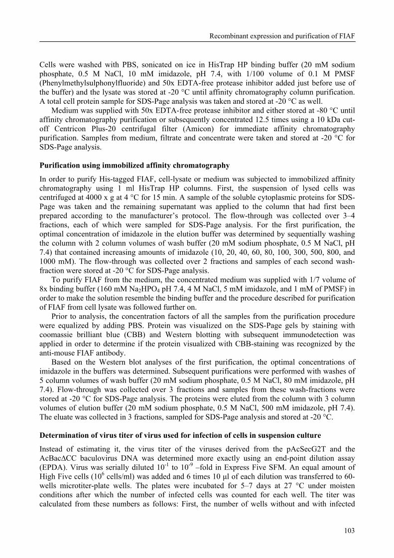
Recombinant expression and purification of FIAF
103
Cells were washed with PBS, sonicated on ice in HisTrap HP binding buffer (20 mM sodium phosphate, 0.5 M NaCl, 10 mM imidazole, pH 7.4, with 1/100 volume of 0.1 M PMSF (Phenylmethylsulphonylfluoride) and 50x EDTA-free protease inhibitor added just before use of the buffer) and the lysate was stored at -20 °C until affinity chromatography column purification. A total cell protein sample for SDS-Page analysis was taken and stored at -20 °C as well.
Medium was supplied with 50x EDTA-free protease inhibitor and either stored at -80 °C until affinity chromatography purification or subsequently concentrated 12.5 times using a 10 kDa cut-off Centricon Plus-20 centrifugal filter (Amicon) for immediate affinity chromatography purification. Samples from medium, filtrate and concentrate were taken and stored at -20 °C for SDS-Page analysis. Purification using immobilized affinity chromatography
In order to purify His-tagged FIAF, cell-lysate or medium was subjected to immobilized affinity chromatography using 1 ml HisTrap HP columns. First, the suspension of lysed cells was centrifuged at 4000 x g at 4 °C for 15 min. A sample of the soluble cytoplasmic proteins for SDS-Page was taken and the remaining supernatant was applied to the column that had first been prepared according to the manufacturer’s protocol. The flow-through was collected over 3–4 fractions, each of which were sampled for SDS-Page analysis. For the first purification, the optimal concentration of imidazole in the elution buffer was determined by sequentially washing the column with 2 column volumes of wash buffer (20 mM sodium phosphate, 0.5 M NaCl, pH 7.4) that contained increasing amounts of imidazole (10, 20, 40, 60, 80, 100, 300, 500, 800, and 1000 mM). The flow-through was collected over 2 fractions and samples of each second wash-fraction were stored at -20 °C for SDS-Page analysis.
To purify FIAF from the medium, the concentrated medium was supplied with 1/7 volume of 8x binding buffer (160 mM Na2HPO4 pH 7.4, 4 M NaCl, 5 mM imidazole, and 1 mM of PMSF) in order to make the solution resemble the binding buffer and the procedure described for purification of FIAF from cell lysate was followed further on.
Prior to analysis, the concentration factors of all the samples from the purification procedure were equalized by adding PBS. Protein was visualized on the SDS-Page gels by staining with coomassie brilliant blue (CBB) and Western blotting with subsequent immunodetection was applied in order to determine if the protein visualized with CBB-staining was recognized by the anti-mouse FIAF antibody.
Based on the Western blot analyses of the first purification, the optimal concentrations of imidazole in the buffers was determined. Subsequent purifications were performed with washes of 5 column volumes of wash buffer (20 mM sodium phosphate, 0.5 M NaCl, 80 mM imidazole, pH 7.4). Flow-through was collected over 3 fractions and samples from these wash-fractions were stored at -20 °C for SDS-Page analysis. The proteins were eluted from the column with 3 column volumes of elution buffer (20 mM sodium phosphate, 0.5 M NaCl, 500 mM imidazole, pH 7.4). The eluate was collected in 3 fractions, sampled for SDS-Page analysis and stored at -20 °C. Determination of virus titer of virus used for infection of cells in suspension culture
Instead of estimating it, the virus titer of the viruses derived from the pAcSecG2T and the AcBac∆CC baculovirus DNA was determined more exactly using an end-point dilution assay (EPDA). Virus was serially diluted 10-1 to 10-9 –fold in Express Five SFM. An equal amount of High Five cells (106 cells/ml) was added and 6 times 10 µl of each dilution was transferred to 60-wells microtiter-plate wells. The plates were incubated for 5–7 days at 27 °C under moisten conditions after which the number of infected cells was counted for each well. The titer was calculated from these numbers as follows: First, the number of wells without and with infected
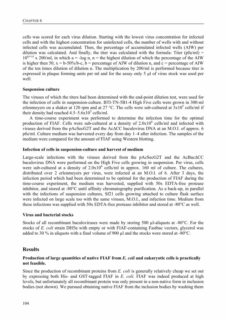
CHAPTER 6
104
cells was scored for each virus dilution. Starting with the lowest virus concentration for infected cells and with the highest concentration for uninfected cells, the number of wells with and without infected cells was accumulated. Then, the percentage of accumulated infected wells (AIW) per dilution was calculated. And finally, the titer was calculated with the formula: Titer (pfu/ml) = 10(a+x) x 200/ml, in which a = -log n, n = the highest dilution of which the percentage of the AIW is higher then 50, x = b-50%/b-c, b = percentage of AIW of dilution n, and c = percentage of AIW of the ten times dilution of dilution n. The multiplication by 200/ml is performed because titer is expressed in plaque forming units per ml and for the assay only 5 µl of virus stock was used per well. Suspension culture
The viruses of which the titers had been determined with the end-point dilution test, were used for the infection of cells in suspension-culture. BTI-TN-5B1-4 High Five cells were grown in 300-ml erlenmeyers on a shaker at 120 rpm and at 27 °C. The cells were sub-cultured at 3x10⁵ cells/ml if their density had reached 4.5–5.6x10⁵ cells/ml.
A time-course experiment was performed to determine the infection time for the optimal production of FIAF. Cells were sub-cultured at a density of 2.0x10⁶ cells/ml and infected with viruses derived from the pAcSecG2T and the Ac∆CC baculovirus DNA at an M.O.I. of approx. 6 pfu/ml. Culture medium was harvested every day from day 1–4 after infection. The samples of the medium were compared for the amount of FIAF using Western blotting. Infection of cells in suspension-culture and harvest of medium
Large-scale infections with the viruses derived from the pAcSecG2T and the AcBac∆CC baculovirus DNA were performed on the High Five cells growing in suspension. Per virus, cells were sub-cultured at a density of 2.0x10⁶ cells/ml in approx. 160 ml of culture. The cultures, distributed over 2 erlenmeyers per virus, were infected at an M.O.I. of 6. After 3 days, the infection period which had been determined to be optimal for the production of FIAF during the time-course experiment, the medium was harvested, supplied with 50x EDTA-free protease inhibitor, and stored at -80°C until affinity chromatography purification. As a back-up, in parallel with the infections of suspension cultures, Sf21 cells growing attached to culture flask surface were infected on large scale too with the same viruses, M.O.I., and infection time. Medium from these infections was supplied with 50x EDTA-free protease inhibitor and stored at -80°C as well. Virus and bacterial stocks
Stocks of all recombinant baculoviruses were made by storing 500 µl-aliquots at -80°C. For the stocks of E. coli strain DH5α with empty or with FIAF-containing Fastbac vectors, glycerol was added to 30 % in aliquots with a final volume of 900 µl and the stocks were stored at -80°C. Results Production of large quantities of native FIAF from E. coli and eukaryotic cells is practically not feasible.
Since the production of recombinant proteins from E. coli is generally relatively cheap we set out by expressing both His- and GST-tagged FIAF in E. coli. FIAF was indeed produced at high levels, but unfortunately all recombinant protein was only present in a non-native form in inclusion bodies (not shown). We pursued obtaining native FIAF from the inclusion bodies by washing them
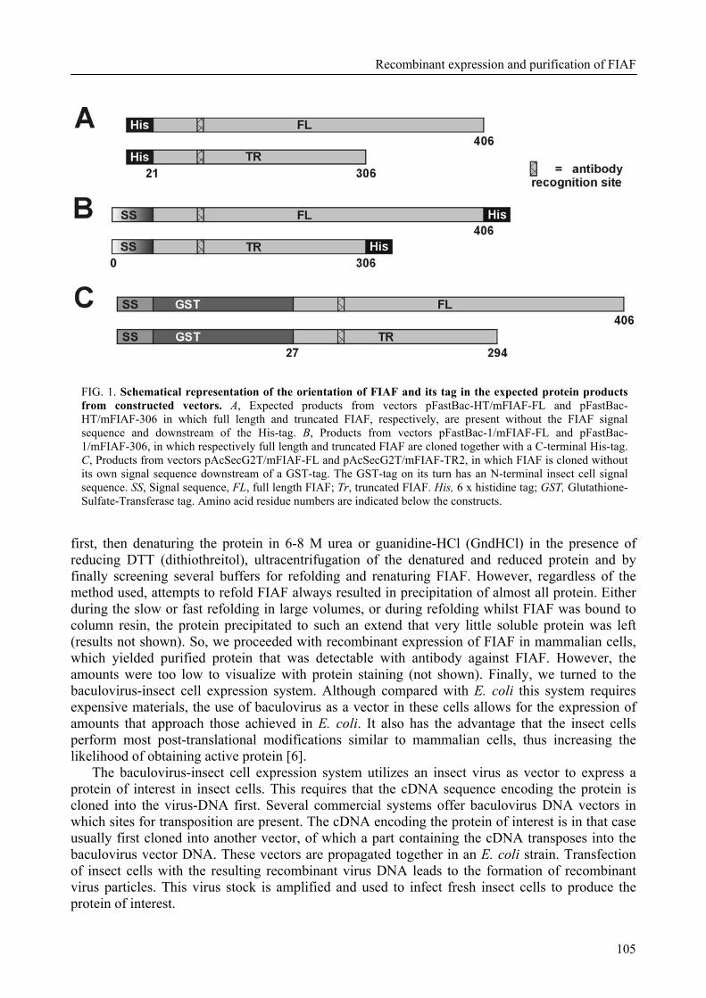
Recombinant expression and purification of FIAF
105
FIG. 1. Schematical representation of the orientation of FIAF and its tag in the expected protein products from constructed vectors. A, Expected products from vectors pFastBac-HT/mFIAF-FL and pFastBac-HT/mFIAF-306 in which full length and truncated FIAF, respectively, are present without the FIAF signal sequence and downstream of the His-tag. B, Products from vectors pFastBac-1/mFIAF-FL and pFastBac-1/mFIAF-306, in which respectively full length and truncated FIAF are cloned together with a C-terminal His-tag. C, Products from vectors pAcSecG2T/mFIAF-FL and pAcSecG2T/mFIAF-TR2, in which FIAF is cloned without its own signal sequence downstream of a GST-tag. The GST-tag on its turn has an N-terminal insect cell signal sequence. SS, Signal sequence, FL, full length FIAF; Tr, truncated FIAF. His, 6 x histidine tag; GST, Glutathione-Sulfate-Transferase tag. Amino acid residue numbers are indicated below the constructs.
first, then denaturing the protein in 6-8 M urea or guanidine-HCl (GndHCl) in the presence of reducing DTT (dithiothreitol), ultracentrifugation of the denatured and reduced protein and by finally screening several buffers for refolding and renaturing FIAF. However, regardless of the method used, attempts to refold FIAF always resulted in precipitation of almost all protein. Either during the slow or fast refolding in large volumes, or during refolding whilst FIAF was bound to column resin, the protein precipitated to such an extend that very little soluble protein was left (results not shown). So, we proceeded with recombinant expression of FIAF in mammalian cells, which yielded purified protein that was detectable with antibody against FIAF. However, the amounts were too low to visualize with protein staining (not shown). Finally, we turned to the baculovirus-insect cell expression system. Although compared with E. coli this system requires expensive materials, the use of baculovirus as a vector in these cells allows for the expression of amounts that approach those achieved in E. coli. It also has the advantage that the insect cells perform most post-translational modifications similar to mammalian cells, thus increasing the likelihood of obtaining active protein [6].
The baculovirus-insect cell expression system utilizes an insect virus as vector to express a protein of interest in insect cells. This requires that the cDNA sequence encoding the protein is cloned into the virus-DNA first. Several commercial systems offer baculovirus DNA vectors in which sites for transposition are present. The cDNA encoding the protein of interest is in that case usually first cloned into another vector, of which a part containing the cDNA transposes into the baculovirus vector DNA. These vectors are propagated together in an E. coli strain. Transfection of insect cells with the resulting recombinant virus DNA leads to the formation of recombinant virus particles. This virus stock is amplified and used to infect fresh insect cells to produce the protein of interest.
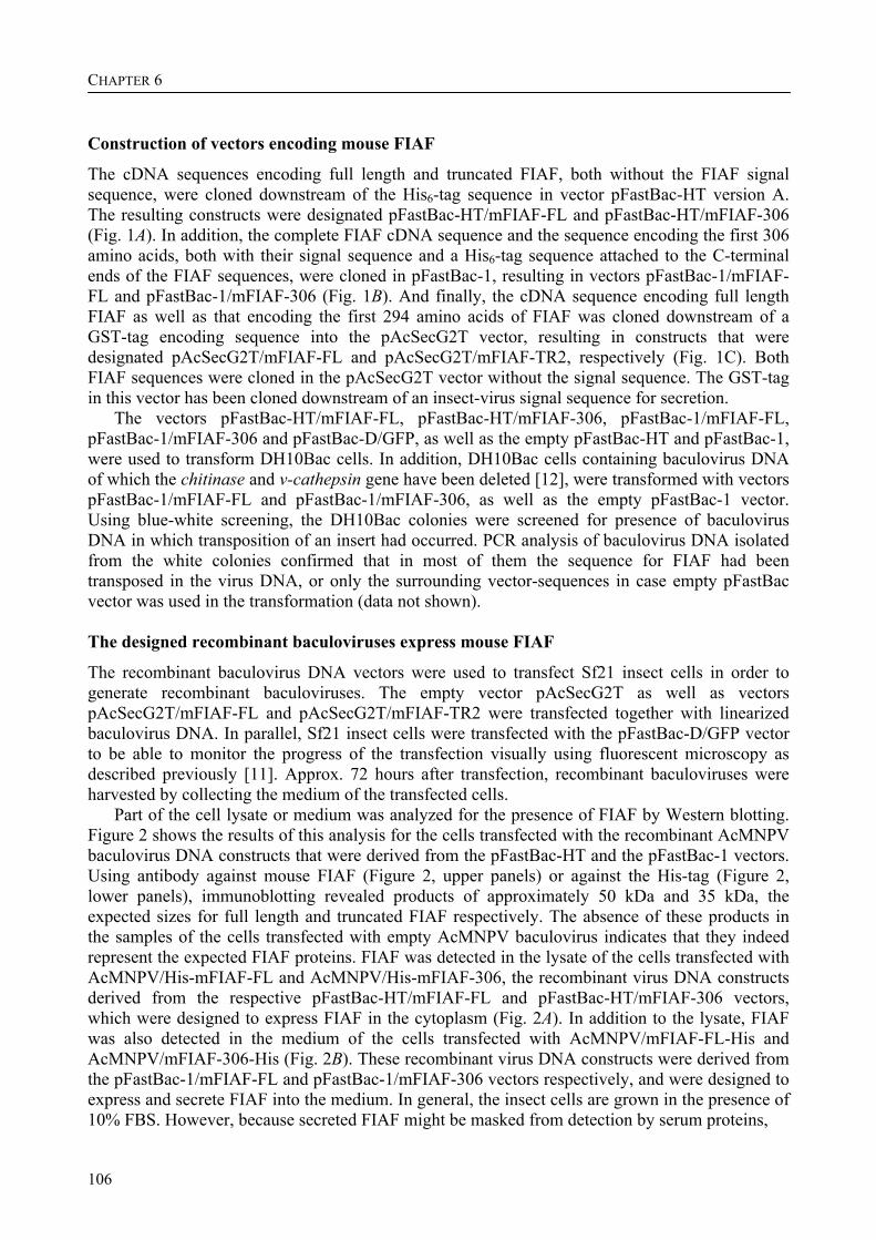
CHAPTER 6
106
Construction of vectors encoding mouse FIAF
The cDNA sequences encoding full length and truncated FIAF, both without the FIAF signal sequence, were cloned downstream of the His6-tag sequence in vector pFastBac-HT version A. The resulting constructs were designated pFastBac-HT/mFIAF-FL and pFastBac-HT/mFIAF-306 (Fig. 1A). In addition, the complete FIAF cDNA sequence and the sequence encoding the first 306 amino acids, both with their signal sequence and a His6-tag sequence attached to the C-terminal ends of the FIAF sequences, were cloned in pFastBac-1, resulting in vectors pFastBac-1/mFIAF-FL and pFastBac-1/mFIAF-306 (Fig. 1B). And finally, the cDNA sequence encoding full length FIAF as well as that encoding the first 294 amino acids of FIAF was cloned downstream of a GST-tag encoding sequence into the pAcSecG2T vector, resulting in constructs that were designated pAcSecG2T/mFIAF-FL and pAcSecG2T/mFIAF-TR2, respectively (Fig. 1C). Both FIAF sequences were cloned in the pAcSecG2T vector without the signal sequence. The GST-tag in this vector has been cloned downstream of an insect-virus signal sequence for secretion.
The vectors pFastBac-HT/mFIAF-FL, pFastBac-HT/mFIAF-306, pFastBac-1/mFIAF-FL, pFastBac-1/mFIAF-306 and pFastBac-D/GFP, as well as the empty pFastBac-HT and pFastBac-1, were used to transform DH10Bac cells. In addition, DH10Bac cells containing baculovirus DNA of which the chitinase and v-cathepsin gene have been deleted [12], were transformed with vectors pFastBac-1/mFIAF-FL and pFastBac-1/mFIAF-306, as well as the empty pFastBac-1 vector. Using blue-white screening, the DH10Bac colonies were screened for presence of baculovirus DNA in which transposition of an insert had occurred. PCR analysis of baculovirus DNA isolated from the white colonies confirmed that in most of them the sequence for FIAF had been transposed in the virus DNA, or only the surrounding vector-sequences in case empty pFastBac vector was used in the transformation (data not shown). The designed recombinant baculoviruses express mouse FIAF
The recombinant baculovirus DNA vectors were used to transfect Sf21 insect cells in order to generate recombinant baculoviruses. The empty vector pAcSecG2T as well as vectors pAcSecG2T/mFIAF-FL and pAcSecG2T/mFIAF-TR2 were transfected together with linearized baculovirus DNA. In parallel, Sf21 insect cells were transfected with the pFastBac-D/GFP vector to be able to monitor the progress of the transfection visually using fluorescent microscopy as described previously [11]. Approx. 72 hours after transfection, recombinant baculoviruses were harvested by collecting the medium of the transfected cells.
Part of the cell lysate or medium was analyzed for the presence of FIAF by Western blotting. Figure 2 shows the results of this analysis for the cells transfected with the recombinant AcMNPV baculovirus DNA constructs that were derived from the pFastBac-HT and the pFastBac-1 vectors. Using antibody against mouse FIAF (Figure 2, upper panels) or against the His-tag (Figure 2, lower panels), immunoblotting revealed products of approximately 50 kDa and 35 kDa, the expected sizes for full length and truncated FIAF respectively. The absence of these products in the samples of the cells transfected with empty AcMNPV baculovirus indicates that they indeed represent the expected FIAF proteins. FIAF was detected in the lysate of the cells transfected with AcMNPV/His-mFIAF-FL and AcMNPV/His-mFIAF-306, the recombinant virus DNA constructs derived from the respective pFastBac-HT/mFIAF-FL and pFastBac-HT/mFIAF-306 vectors, which were designed to express FIAF in the cytoplasm (Fig. 2A). In addition to the lysate, FIAF was also detected in the medium of the cells transfected with AcMNPV/mFIAF-FL-His and AcMNPV/mFIAF-306-His (Fig. 2B). These recombinant virus DNA constructs were derived from the pFastBac-1/mFIAF-FL and pFastBac-1/mFIAF-306 vectors respectively, and were designed to express and secrete FIAF into the medium. In general, the insect cells are grown in the presence of 10% FBS. However, because secreted FIAF might be masked from detection by serum proteins,
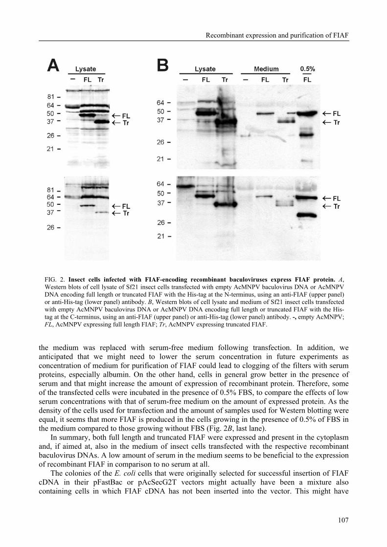
Recombinant expression and purification of FIAF
107
FIG. 2. Insect cells infected with FIAF-encoding recombinant baculoviruses express FIAF protein. A, Western blots of cell lysate of Sf21 insect cells transfected with empty AcMNPV baculovirus DNA or AcMNPV DNA encoding full length or truncated FIAF with the His-tag at the N-terminus, using an anti-FIAF (upper panel) or anti-His-tag (lower panel) antibody. B, Western blots of cell lysate and medium of Sf21 insect cells transfected with empty AcMNPV baculovirus DNA or AcMNPV DNA encoding full length or truncated FIAF with the His-tag at the C-terminus, using an anti-FIAF (upper panel) or anti-His-tag (lower panel) antibody. -, empty AcMNPV; FL, AcMNPV expressing full length FIAF; Tr, AcMNPV expressing truncated FIAF.
the medium was replaced with serum-free medium following transfection. In addition, we anticipated that we might need to lower the serum concentration in future experiments as concentration of medium for purification of FIAF could lead to clogging of the filters with serum proteins, especially albumin. On the other hand, cells in general grow better in the presence of serum and that might increase the amount of expression of recombinant protein. Therefore, some of the transfected cells were incubated in the presence of 0.5% FBS, to compare the effects of low serum concentrations with that of serum-free medium on the amount of expressed protein. As the density of the cells used for transfection and the amount of samples used for Western blotting were equal, it seems that more FIAF is produced in the cells growing in the presence of 0.5% of FBS in the medium compared to those growing without FBS (Fig. 2B, last lane).
In summary, both full length and truncated FIAF were expressed and present in the cytoplasm and, if aimed at, also in the medium of insect cells transfected with the respective recombinant baculovirus DNAs. A low amount of serum in the medium seems to be beneficial to the expression of recombinant FIAF in comparison to no serum at all.
The colonies of the E. coli cells that were originally selected for successful insertion of FIAF cDNA in their pFastBac or pAcSecG2T vectors might actually have been a mixture also containing cells in which FIAF cDNA has not been inserted into the vector. This might have
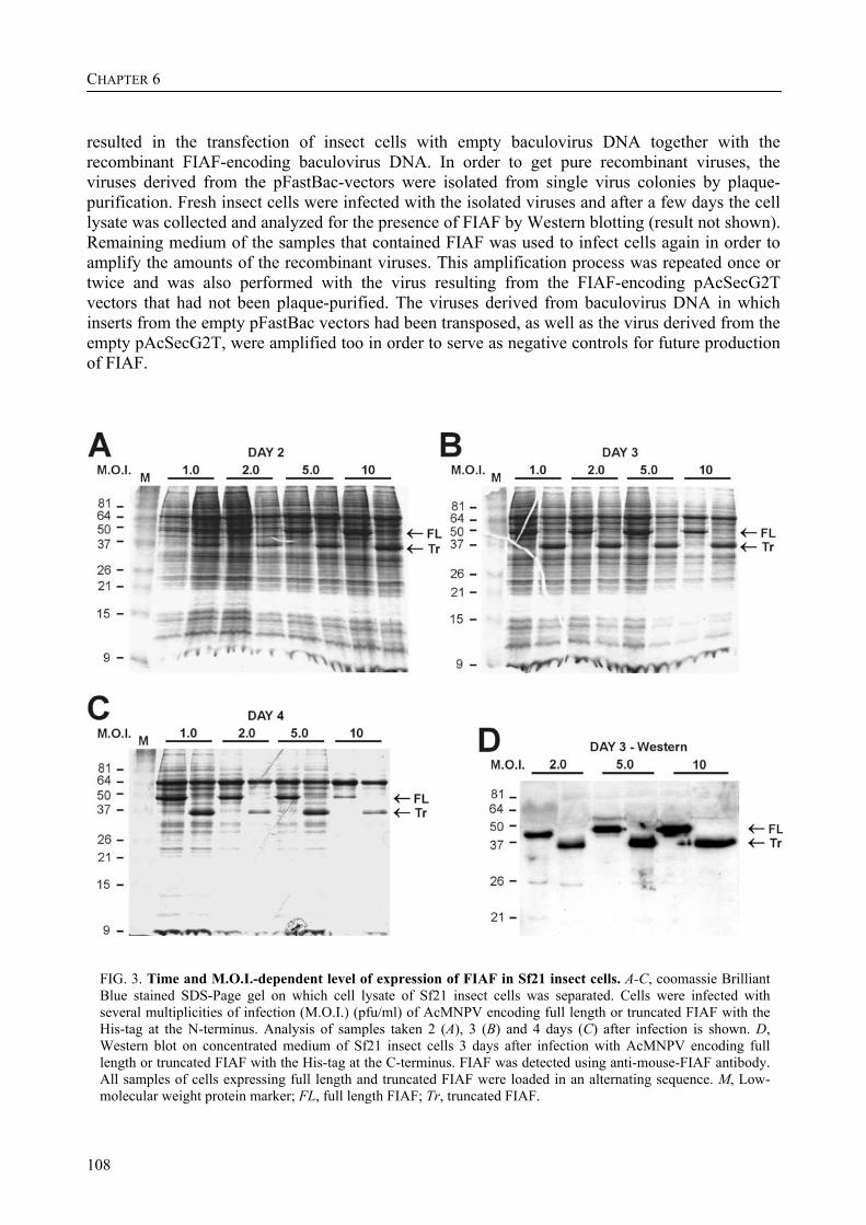
CHAPTER 6
108
resulted in the transfection of insect cells with empty baculovirus DNA together with the recombinant FIAF-encoding baculovirus DNA. In order to get pure recombinant viruses, the viruses derived from the pFastBac-vectors were isolated from single virus colonies by plaque-purification. Fresh insect cells were infected with the isolated viruses and after a few days the cell lysate was collected and analyzed for the presence of FIAF by Western blotting (result not shown). Remaining medium of the samples that contained FIAF was used to infect cells again in order to amplify the amounts of the recombinant viruses. This amplification process was repeated once or twice and was also performed with the virus resulting from the FIAF-encoding pAcSecG2T vectors that had not been plaque-purified. The viruses derived from baculovirus DNA in which inserts from the empty pFastBac vectors had been transposed, as well as the virus derived from the empty pAcSecG2T, were amplified too in order to serve as negative controls for future production of FIAF.
FIG. 3. Time and M.O.I.-dependent level of expression of FIAF in Sf21 insect cells. A-C, coomassie Brilliant Blue stained SDS-Page gel on which cell lysate of Sf21 insect cells was separated. Cells were infected with several multiplicities of infection (M.O.I.) (pfu/ml) of AcMNPV encoding full length or truncated FIAF with the His-tag at the N-terminus. Analysis of samples taken 2 (A), 3 (B) and 4 days (C) after infection is shown. D, Western blot on concentrated medium of Sf21 insect cells 3 days after infection with AcMNPV encoding full length or truncated FIAF with the His-tag at the C-terminus. FIAF was detected using anti-mouse-FIAF antibody. All samples of cells expressing full length and truncated FIAF were loaded in an alternating sequence. M, Low-molecular weight protein marker; FL, full length FIAF; Tr, truncated FIAF.
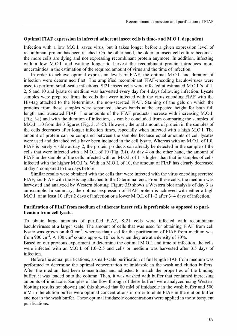
Recombinant expression and purification of FIAF
109
Optimal FIAF expression in infected adherent insect cells is time- and M.O.I. dependent
Infection with a low M.O.I. saves virus, but it takes longer before a given expression level of recombinant protein has been reached. On the other hand, the older an insect cell culture becomes, the more cells are dying and not expressing recombinant protein anymore. In addition, infecting with a low M.O.I. and waiting longer to harvest the recombinant protein introduces more uncertainties in the estimation of the required amount of virus and the time of infection.
In order to achieve optimal expression levels of FIAF, the optimal M.O.I. and duration of infection were determined first. The amplified recombinant FIAF-encoding baculoviruses were used to perform small-scale infections. Sf21 insect cells were infected at estimated M.O.I.’s of 1, 2, 5 and 10 and lysate or medium was harvested every day for 4 days following infection. Lysate samples were prepared from the cells that were infected with the virus encoding FIAF with the His-tag attached to the N-terminus, the non-secreted FIAF. Staining of the gels on which the proteins from these samples were separated, shows bands at the expected height for both full length and truncated FIAF. The amounts of the FIAF products increase with increasing M.O.I. (Fig. 3A) and with the duration of infection, as can be concluded from comparing the samples of M.O.I. 1.0 from the 3 figures (Fig. 3, A–C). However, the total amount of protein in the samples of the cells decreases after longer infection times, especially when infected with a high M.O.I. The amount of protein can be compared between the samples because equal amounts of cell lysates were used and detached cells have been included in the cell lysate. Whereas with an M.O.I. of 1.0, FIAF is barely visible at day 2, the protein products can already be detected in the sample of the cells that were infected with a M.O.I. of 10 (Fig. 3A). At day 4 on the other hand, the amount of FIAF in the sample of the cells infected with an M.O.I. of 1 is higher than that in samples of cells infected with the higher M.O.I.’s. With an M.O.I. of 10, the amount of FIAF has clearly decreased at day 4 compared to the days before.
Similar results were obtained with the cells that were infected with the virus encoding secreted FIAF, i.e. FIAF with the His-tag attached to the C-terminal end. From these cells, the medium was harvested and analyzed by Western blotting. Figure 3D shows a Western blot analysis of day 3 as an example. In summary, the optimal expression of FIAF protein is achieved with either a high M.O.I. of at least 10 after 2 days of infection or a lower M.O.I. of 1–2 after 3–4 days of infection. Purification of FIAF from medium of adherent insect cells is preferable as opposed to puri- fication from cell lysate.
To obtain large amounts of purified FIAF, Sf21 cells were infected with recombinant baculoviruses at a larger scale. The amount of cells that was used for obtaining FIAF from cell lysate was grown on 400 cm2, whereas that used for the purification of FIAF from medium was from 900 cm2. A 100 cm2 counts approx. 107 cells when they are at a density of 70%. Based on our previous experiment to determine the optimal M.O.I. and time of infection, the cells were infected with an M.O.I. of 1.0–2.5 and cells or medium was harvested after 3.5 days of infection.
Before the actual purifications, a small-scale purification of full length FIAF from medium was performed to determine the optimal concentration of imidazole in the wash and elution buffers. After the medium had been concentrated and adjusted to match the properties of the binding buffer, it was loaded onto the column. Then, it was washed with buffer that contained increasing amounts of imidazole. Samples of the flow-through of these buffers were analyzed using Western blotting (results not shown) and this showed that 80 mM of imidazole in the wash buffer and 500 mM in the elution buffer were optimal concentrations in order to elute FIAF in the elution buffer and not in the wash buffer. These optimal imidazole concentrations were applied in the subsequent purifications.
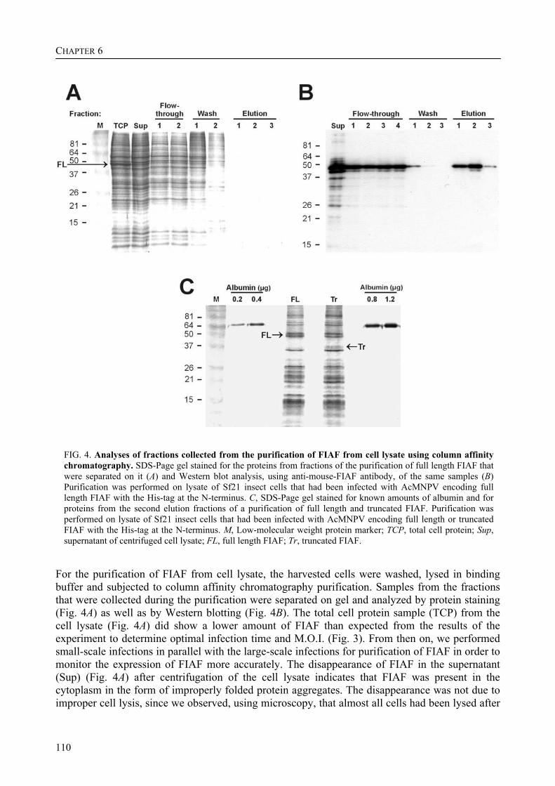
CHAPTER 6
110
FIG. 4. Analyses of fractions collected from the purification of FIAF from cell lysate using column affinity chromatography. SDS-Page gel stained for the proteins from fractions of the purification of full length FIAF that were separated on it (A) and Western blot analysis, using anti-mouse-FIAF antibody, of the same samples (B) Purification was performed on lysate of Sf21 insect cells that had been infected with AcMNPV encoding full length FIAF with the His-tag at the N-terminus. C, SDS-Page gel stained for known amounts of albumin and for proteins from the second elution fractions of a purification of full length and truncated FIAF. Purification was performed on lysate of Sf21 insect cells that had been infected with AcMNPV encoding full length or truncated FIAF with the His-tag at the N-terminus. M, Low-molecular weight protein marker; TCP, total cell protein; Sup, supernatant of centrifuged cell lysate; FL, full length FIAF; Tr, truncated FIAF.
For the purification of FIAF from cell lysate, the harvested cells were washed, lysed in binding buffer and subjected to column affinity chromatography purification. Samples from the fractions that were collected during the purification were separated on gel and analyzed by protein staining (Fig. 4A) as well as by Western blotting (Fig. 4B). The total cell protein sample (TCP) from the cell lysate (Fig. 4A) did show a lower amount of FIAF than expected from the results of the experiment to determine optimal infection time and M.O.I. (Fig. 3). From then on, we performed small-scale infections in parallel with the large-scale infections for purification of FIAF in order to monitor the expression of FIAF more accurately. The disappearance of FIAF in the supernatant (Sup) (Fig. 4A) after centrifugation of the cell lysate indicates that FIAF was present in the cytoplasm in the form of improperly folded protein aggregates. The disappearance was not due to improper cell lysis, since we observed, using microscopy, that almost all cells had been lysed after
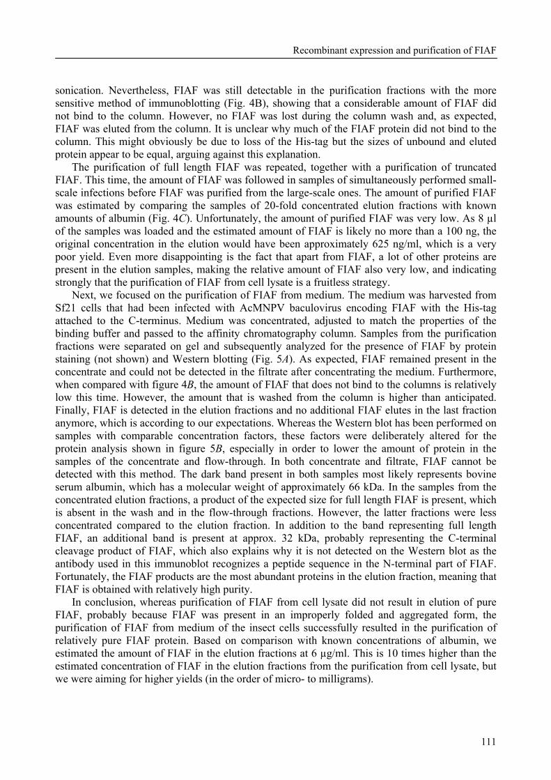
Recombinant expression and purification of FIAF
111
sonication. Nevertheless, FIAF was still detectable in the purification fractions with the more sensitive method of immunoblotting (Fig. 4B), showing that a considerable amount of FIAF did not bind to the column. However, no FIAF was lost during the column wash and, as expected, FIAF was eluted from the column. It is unclear why much of the FIAF protein did not bind to the column. This might obviously be due to loss of the His-tag but the sizes of unbound and eluted protein appear to be equal, arguing against this explanation.
The purification of full length FIAF was repeated, together with a purification of truncated FIAF. This time, the amount of FIAF was followed in samples of simultaneously performed small-scale infections before FIAF was purified from the large-scale ones. The amount of purified FIAF was estimated by comparing the samples of 20-fold concentrated elution fractions with known amounts of albumin (Fig. 4C). Unfortunately, the amount of purified FIAF was very low. As 8 µl of the samples was loaded and the estimated amount of FIAF is likely no more than a 100 ng, the original concentration in the elution would have been approximately 625 ng/ml, which is a very poor yield. Even more disappointing is the fact that apart from FIAF, a lot of other proteins are present in the elution samples, making the relative amount of FIAF also very low, and indicating strongly that the purification of FIAF from cell lysate is a fruitless strategy.
Next, we focused on the purification of FIAF from medium. The medium was harvested from Sf21 cells that had been infected with AcMNPV baculovirus encoding FIAF with the His-tag attached to the C-terminus. Medium was concentrated, adjusted to match the properties of the binding buffer and passed to the affinity chromatography column. Samples from the purification fractions were separated on gel and subsequently analyzed for the presence of FIAF by protein staining (not shown) and Western blotting (Fig. 5A). As expected, FIAF remained present in the concentrate and could not be detected in the filtrate after concentrating the medium. Furthermore, when compared with figure 4B, the amount of FIAF that does not bind to the columns is relatively low this time. However, the amount that is washed from the column is higher than anticipated. Finally, FIAF is detected in the elution fractions and no additional FIAF elutes in the last fraction anymore, which is according to our expectations. Whereas the Western blot has been performed on samples with comparable concentration factors, these factors were deliberately altered for the protein analysis shown in figure 5B, especially in order to lower the amount of protein in the samples of the concentrate and flow-through. In both concentrate and filtrate, FIAF cannot be detected with this method. The dark band present in both samples most likely represents bovine serum albumin, which has a molecular weight of approximately 66 kDa. In the samples from the concentrated elution fractions, a product of the expected size for full length FIAF is present, which is absent in the wash and in the flow-through fractions. However, the latter fractions were less concentrated compared to the elution fraction. In addition to the band representing full length FIAF, an additional band is present at approx. 32 kDa, probably representing the C-terminal cleavage product of FIAF, which also explains why it is not detected on the Western blot as the antibody used in this immunoblot recognizes a peptide sequence in the N-terminal part of FIAF. Fortunately, the FIAF products are the most abundant proteins in the elution fraction, meaning that FIAF is obtained with relatively high purity.
In conclusion, whereas purification of FIAF from cell lysate did not result in elution of pure FIAF, probably because FIAF was present in an improperly folded and aggregated form, the purification of FIAF from medium of the insect cells successfully resulted in the purification of relatively pure FIAF protein. Based on comparison with known concentrations of albumin, we estimated the amount of FIAF in the elution fractions at 6 µg/ml. This is 10 times higher than the estimated concentration of FIAF in the elution fractions from the purification from cell lysate, but we were aiming for higher yields (in the order of micro- to milligrams).

CHAPTER 6
112
FIG. 5. Analyses of fractions collected from the purification of FIAF from medium using column affinity chromatography. Western blot analysis, using anti-mouse-FIAF antibody, of the fractions from the purification of full length FIAF from medium (A), and protein-stained SDS-Page gel (B) on which samples of the same fractions as in A, but with different concentration factors, were separated. Purification was performed on medium of Sf21 insect cells that had been infected with AcMNPV encoding full length FIAF with the His-tag at the C-terminus. M, Low-molecular weight protein marker; Med, medium; Conc, concentrated medium; Filtr, filtrate of concentrated medium; Conc. Factor, Concentration factor.
Titers of viruses to be used for the infection of cells in suspension culture are in the normal range.
In order to achieve higher concentrations of purified FIAF, we investigated the possibility of producing FIAF in suspension cultures, which reach a higher density of cells and are hence expected to result in higher concentrations of recombinant protein. For this purpose, we first determined the titer of the recombinant baculoviruses that are derived from the pAcSecG2T and the Ac∆CC vectors using an end-point dilution assay (EPDA) (Table 2), instead of estimating it like before.
Table 2: Titers of the viruses derived from the pAcSecG2T and the Ac∆CC baculovirus DNA vectors.
virus name protein product titer (pfu/ml) Ac∆CC/-His none, negative control 4.9 x 10⁷ Ac∆CC/mFIAF-FL-His secreted full length FIAF with C-terminal His-tag 4.4 x 10⁷ AcMNPV/GST- none, negative control 2.4 x 10⁸ AcMNPV/GST-mFIAF-FL secreted full length FIAF with N-terminal GST-tag 3.5 x 10⁸
We shifted our focus towards the Ac∆CC viruses because the expression level of FIAF from this virus was expected to be higher. The pAcSecG2T virus was also used from now on, because the substitution of the His-tag by a GST-tag could increase the solubility of the fusion product and thus result in lower sensitivity to aggregation and precipitation. In general, the titers of virus stocks that have been amplified 2 to 3 times are between 107 and 108 pfu/ml. This means that the titers of the Ac∆CC viruses are as expected and those of the GST viruses are even a little higher.
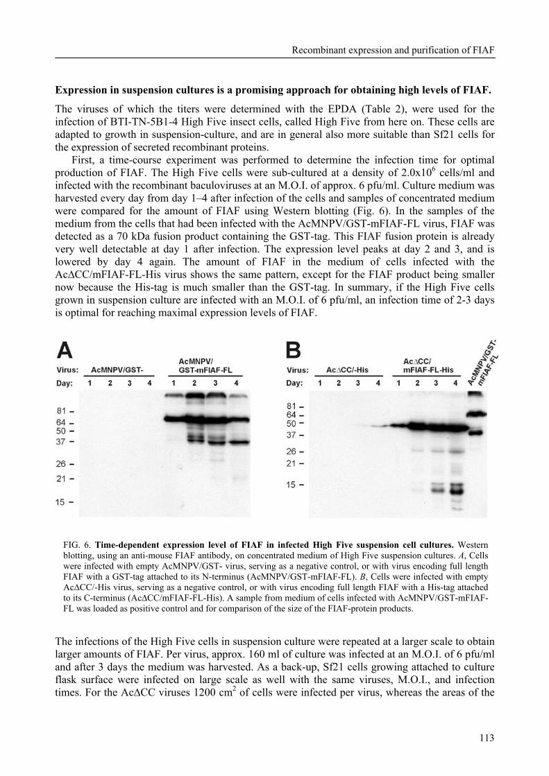
Recombinant expression and purification of FIAF
113
Expression in suspension cultures is a promising approach for obtaining high levels of FIAF.
The viruses of which the titers were determined with the EPDA (Table 2), were used for the infection of BTI-TN-5B1-4 High Five insect cells, called High Five from here on. These cells are adapted to growth in suspension-culture, and are in general also more suitable than Sf21 cells for the expression of secreted recombinant proteins.
First, a time-course experiment was performed to determine the infection time for optimal production of FIAF. The High Five cells were sub-cultured at a density of 2.0x10⁶ cells/ml and infected with the recombinant baculoviruses at an M.O.I. of approx. 6 pfu/ml. Culture medium was harvested every day from day 1–4 after infection of the cells and samples of concentrated medium were compared for the amount of FIAF using Western blotting (Fig. 6). In the samples of the medium from the cells that had been infected with the AcMNPV/GST-mFIAF-FL virus, FIAF was detected as a 70 kDa fusion product containing the GST-tag. This FIAF fusion protein is already very well detectable at day 1 after infection. The expression level peaks at day 2 and 3, and is lowered by day 4 again. The amount of FIAF in the medium of cells infected with the Ac∆CC/mFIAF-FL-His virus shows the same pattern, except for the FIAF product being smaller now because the His-tag is much smaller than the GST-tag. In summary, if the High Five cells grown in suspension culture are infected with an M.O.I. of 6 pfu/ml, an infection time of 2-3 days is optimal for reaching maximal expression levels of FIAF.
FIG. 6. Time-dependent expression level of FIAF in infected High Five suspension cell cultures. Western blotting, using an anti-mouse FIAF antibody, on concentrated medium of High Five suspension cultures. A, Cells were infected with empty AcMNPV/GST- virus, serving as a negative control, or with virus encoding full length FIAF with a GST-tag attached to its N-terminus (AcMNPV/GST-mFIAF-FL). B, Cells were infected with empty Ac∆CC/-His virus, serving as a negative control, or with virus encoding full length FIAF with a His-tag attached to its C-terminus (Ac∆CC/mFIAF-FL-His). A sample from medium of cells infected with AcMNPV/GST-mFIAF-FL was loaded as positive control and for comparison of the size of the FIAF-protein products.
The infections of the High Five cells in suspension culture were repeated at a larger scale to obtain larger amounts of FIAF. Per virus, approx. 160 ml of culture was infected at an M.O.I. of 6 pfu/ml and after 3 days the medium was harvested. As a back-up, Sf21 cells growing attached to culture flask surface were infected on large scale as well with the same viruses, M.O.I., and infection times. For the Ac∆CC viruses 1200 cm2 of cells were infected per virus, whereas the areas of the
CHAPTER 6
114
cells infected with the AcMNPV/GST viruses were 750 cm2 each. The medium from the infected High Five as well as that from the Sf21 cells was supplied with 50x EDTA-free protease inhibitor and stored at -80°C for future affinity chromatography purification of FIAF. Smaller samples for the determination of the amount of FIAF were stored separately. Discussion The availability of a protein in a pure form and in relatively large quantities is in general very useful for studying its function and for determining the 3-dimensional structure of the protein. FIAF is a glycosylated protein of approx. 50 kDa that has been reported to play roles in lipid metabolism, angiogenesis and possibly carcinogenesis (reviewed in chapter 2). FIAF is part of the family of angiopoietin-like proteins, several members of which have been implicated in lipid metabolism and angiogenesis [16,17]. Several reports on FIAF describe its recombinant expression and the subsequent use in experiments that in general require relatively small quantities of protein. However, none of them focused on large-scale expression systems in order to obtain amounts of recombinant FIAF in the range of micro- to milligrams, which would be sufficient for administration to mice. The lack of reports describing large-scale expression and purification of FIAF might reflect difficulties with production of recombinant FIAF, which might in turn be the result of FIAF having a propensity to aggregate and precipitate, as has been reported to occur for members of the angiopoietin-like protein family [10]. In fact, reports on large-scale expression and purification of other members than FIAF are also lacking to date.
We investigated the possibility of expressing and purifying FIAF on a larger scale. FIAF was produced at high levels in E. coli, but unfortunately all recombinant protein was only present in a non-native form in inclusion bodies (data not shown). Attempts to obtain native FIAF from the inclusion bodies by denaturing and reducing the protein and subsequent refolding and renaturing in a variety of buffers consistently resulted in precipitation of almost all protein. Prokaryotes like E. coli do not perform the post-translational modifications that are usually performed in eukaryotic organisms. The lack of for instance glycosylation might result in reduced solubility of the protein and might be a reason for the aggregation and precipitation of FIAF expressed in E. coli. Therefore, we proceeded with the expression of FIAF in eukaryotic cells. This strategy resulted in purified protein that was detectable with antibody against FIAF but the amounts were too low to visualize with protein staining.
The baculovirus-insect cell expression system combines the advantages of expression in E. coli and that of mammalian cells. The use of baculovirus as a vector in the insect cells allows for the expression of amounts that approach those achieved in E. coli. It also has the advantage that the insect cells perform most post-translational modifications similar to those in mammalian cells, thus increasing the likelihood of obtaining functional protein [6]. For these reasons, we focused our effort on expressing FIAF in insect cells using baculovirus as a vector.
First, we generated vectors in which the cDNA sequences encoding full length and truncated mouse FIAF were cloned either upstream or, without the signal sequence for secretion, downstream of a His-tag sequence. In addition, cDNA sequences encoding full length and truncated FIAF were cloned without their signal sequence downstream of a GST-tag encoding sequence. From all these vectors (Fig. 1), the respective AcMNPV baculovirus vectors were constructed. Additional AcBac∆CC baculovirus vectors, in which two genes have been deleted in order to achieve higher expression levels of recombinant proteins [12], were constructed from the vectors in which FIAF was cloned upstream of the His-tag. Insect cells that were transfected with the viruses all expressed the desired FIAF product (Fig. 2).
Recombinant expression and purification of FIAF
115
After amplification of the amounts of the viruses, and following determination of the optimal M.O.I. and time of infection in order to reach maximal expression levels of FIAF (Fig. 3), large-scale infections were performed to express FIAF for purification using column affinity chromatography. Unfortunately, centrifugation of cell lysate resulted in the loss of most of the FIAF protein and the finally obtained amount of FIAF was impure and too small; only 2 ml of an estimated concentration of 0.6 µg/ml (Fig. 4). The reason for the loss of FIAF during centrifugation is not clear, but incomplete cell lysis can be ruled out as a cause, as observation using a microscope showed that almost all cells had lysed following sonication. Another explanation is that FIAF has been improperly folded and has subsequently aggregated and precipitated. In this strategy, FIAF is expressed in the cytosol and is not secreted, because the signal sequence of FIAF has been excluded from cloning into the vector construct. Usually, if a protein’s signal sequence is detected during translation, the protein is transferred to the endoplasmatic reticulum (ER) for further processing. In the ER, chaperone proteins assist in proper folding and initial glycosylation occurs [5]. Extensive control on correct folding also takes place and improperly folded proteins are targeted for destruction. A correctly folded protein proceeds to the Golgi apparatus, where final glycosylation and protein targeting, partly mediated by specific glycan groups, takes place [5]. Proteins without a signal sequence, like FIAF in this approach, are not directed to the ER, will therefore not be glycosylated and targeted to another cellular location, and thus remain in the cytosol. Folding also takes place in the cytosol, but control on proper folding is less extensive than in the ER. Therefore, the majority of FIAF in the cytosol might be present in the form of improperly folded protein.
In contrast, purification of FIAF from medium of infected cells was more successful. The concentration was at least 10 times higher compared to FIAF purified from the cytosol and, also very important; the protein was of high purity (Fig. 5). In this approach, FIAF contains its own signal sequence and is secreted, confirming the recognition of the mammalian signal sequence by the insect cells. This means that FIAF is in this case also directed to the ER where it is subject to the more rigid control on proper folding as compared to the cytosol. In addition, FIAF expressed with its signal sequence, might be N-glycosylated, like it probably is in mammalian cells [1,3,7]. As N-glycan groups play an important role in the correct folding, oligomerization, quality control, sorting and transport of a protein [5], its possible glycosylation, resulting from the strategy to express FIAF as a secreted protein, might be responsible for the successful purification of FIAF from the medium. N-glycosylation is less complex in insect cells than in mammalian cells and is usually limited to the production of simple oligomannose-type oligosaccharides [6]. Because the glycosylation status may be of great importance for the functioning of a protein, the biological activity of purified FIAF should be verified, e.g. by measuring its ability to inhibit LPL activity. If FIAF purified from medium would prove to be non-functional because of inadequate glycosylation, an alternative strategy could be to use engineered insect cell lines that are modified to perform more complex N-glycosylation [18].
In addition to the full length FIAF, a smaller product of approx. 32 kDa was detectable with protein staining in the elution fraction. This product likely consists of the C-terminal cleavage product of FIAF. It is not detected with the antibody, which recognizes an epitope located in the N-terminal region of FIAF. Future experiments should clarify whether this product is recognized by an antibody that is directed against the C-terminally located His-tag. Although protease activity and protease recognition sites in insect cells might differ from those in mammalian cells, N-terminal sequencing of the C-terminal cleavage product might give a clue about the cleavage site as it occurs in mice, in which truncated forms of FIAF can be detected in plasma [19].
We figured that higher yields of FIAF expression might be obtained with insect cells grown in suspension cultures, which reach a higher density. Preliminary results of experiments indeed indicate that the expression of FIAF in suspension cultures is an approach that could result in
CHAPTER 6
116
higher yields (Fig. 6), but additional experiments are needed to quantify the amount of produced recombinant FIAF and to compare it with the amount produced in adherent cell cultures. In addition, affinity chromatography purification needs to be performed on the medium of the suspension cultures in order to determine if this strategy ultimately results in higher yields of purified FIAF.
Although we already did some experiments to optimize the purification of FIAF, it could possibly be improved in future experiments by purifying FIAF in the presence of a hydrophobic carrier. We recently reported the finding that FIAF is specifically present in HDL and LDL-containing FPLC fractions of blood plasma [20]. Other proteins present in lipoprotein particles, like apoAI, are also known to be difficult to express as recombinant proteins and to purify, mostly due to their amphiphilic character, autoaggregation, and degradation [21]. For apoAI, the problems this posed for expression in E. coli were tackled by changing the codons to ones that still encode the same amino acids but to that are more generally used in E. coli [22], resulting in improved folding and less aggregation and precipitation.
In addition to the experiments performed with recombinant FIAF so far, it could for example be used to study the nature of the interaction between FIAF and LPL, as well as other lipases like endothelial lipase (EL) and hepatic lipase (HL). We previously showed that FIAF is present in blood plasma in several forms [19]. Furthermore, we recently discovered that in FPLC fractions of mouse plasma, the full length form of FIAF is specifically associated with the HDL-containing fractions, whereas the more abundant truncated form is specifically present in the LDL-containing fractions [20]. In contrast, in humans both the full length and the truncated form of FIAF were only present in the HDL-containing fraction. Recombinant FIAF could be used to investigate the nature of this physical association of FIAF with lipoprotein particles. It could also be employed in investigating whether the full length and the truncated forms of FIAF have different functions. Furthermore, recombinant FIAF could be utilised to determine its 3-dimensional structure and to screen for possible receptors which might be responsible for the protective effect on apoptosis of vascular endothelial cells [3] and for the stimulation of lipolysis that appears to occur in transgenic mice that over-express FIAF [20].
In conclusion, since in E. coli, we experienced folding problems with FIAF and because the production level of FIAF was insufficient in mammalian cells, we ultimately investigated the expression of recombinant FIAF in insect cells and the purification of FIAF from the cell lysate as well as from the medium. In addition, we optimized the purification of FIAF. The production of FIAF in insect cells, using the baculovirus system, is a promising approach for obtaining pure and relatively large quantities of FIAF that can be used to further study its function and structure. Acknowledgements We thank Els de Roode for technical assistance and Monique van Oers and Just Vlak for the gift of pFastBac-D/GFP vector and for their advice on experiments. The research on FIAF in the author’s laboratory is supported by the Netherlands Organization for Scientific Research (NWO), the Royal Netherlands Academy of Art and Sciences (KNAW), and the Wageningen Center for Food Sciences (WCFS). References
1 Kersten, S., Mandard, S., Tan, N. S., et al. (2000) Characterization of the fasting-induced adipose factor FIAF, a novel peroxisome proliferator-activated receptor target gene. J Biol Chem 275, 28488-28493
Recombinant expression and purification of FIAF
117
2 Yoon, J. C., Chickering, T. W., Rosen, E. D., et al. (2000) Peroxisome proliferator-activated receptor gamma target gene encoding a novel angiopoietin-related protein associated with adipose differentiation. Mol Cell Biol 20, 5343-5349
3 Kim, I., Kim, H. G., Kim, H., et al. (2000) Hepatic expression, synthesis and secretion of a novel fibrinogen/angiopoietin-related protein that prevents endothelial-cell apoptosis. Biochem J 346 Pt 3, 603-610
4 Yoshida, K., Shimizugawa, T., Ono, M., Furukawa, H. (2002) Angiopoietin-like protein 4 is a potent hyperlipidemia-inducing factor in mice and inhibitor of lipoprotein lipase. J Lipid Res JID - 0376606 43, 1770-1772
5 Helenius, A., Aebi, M. (2001) Intracellular functions of N-linked glycans. Science 291, 2364-2369 6 Altmann, F., Staudacher, E., Wilson, I. B., Marz, L. (1999) Insect cells as hosts for the expression of
recombinant glycoproteins. Glycoconj J 16, 109-123 7 Ge, H., Yang, G., Huang, L., Motola, D. L., Pourbahrami, T., Li, C. (2004) Oligomerization and
regulated proteolytic processing of angiopoietin-like protein 4. J Biol Chem 279, 2038-2045 8 Koster, A., Chao, Y. B., Mosior, M., et al. (2005) Transgenic angiopoietin-like (angptl)4
overexpression and targeted disruption of angptl4 and angptl3: regulation of triglyceride metabolism. Endocrinology 146, 4943-4950
9 Xu, A., Lam, M. C., Chan, K. W., et al. (2005) Angiopoietin-like protein 4 decreases blood glucose and improves glucose tolerance but induces hyperlipidemia and hepatic steatosis in mice. Proc Natl Acad Sci U S A 102, 6086-6091
10 Oike, Y., Yasunaga, K., Suda, T. (2004) Angiopoietin-related/angiopoietin-like proteins regulate angiogenesis. Int J Hematol 80, 21-28
11 van Hulten, M. C., Westenberg, M., Goodall, S. D., Vlak, J. M. (2000) Identification of two major virion protein genes of white spot syndrome virus of shrimp. Virology 266, 227-236
12 Kaba, S. A., Salcedo, A. M., Wafula, P. O., Vlak, J. M., van Oers, M. M. (2004) Development of a chitinase and v-cathepsin negative bacmid for improved integrity of secreted recombinant proteins. J Virol Methods 122, 113-118
13 Thomas, C. J., Brown, H. L., Hawes, C. R., et al. (1998) Localization of a baculovirus-induced chitinase in the insect cell endoplasmic reticulum. J Virol 72, 10207-10212
14 Possee, R. D., Thomas, C. J., King, L. A. (1999) The use of baculovirus vectors for the production of membrane proteins in insect cells. Biochem Soc Trans 27, 928-932
15 Hom, L. G., Volkman, L. E. (1998) Preventing proteolytic artifacts in the baculovirus expression system. Biotechniques 25, 18-20
16 Koishi, R., Ando, Y., Ono, M., et al. (2002) Angptl3 regulates lipid metabolism in mice. Nat.Genet. 30, 151-157
17 Oike, Y., Akao, M., Yasunaga, K., et al. (2005) Angiopoietin-related growth factor antagonizes obesity and insulin resistance. Nat Med 11, 400-408
18 Tomiya, N., Narang, S., Lee, Y. C., Betenbaugh, M. J. (2004) Comparing N-glycan processing in mammalian cell lines to native and engineered lepidopteran insect cell lines. Glycoconj J 21, 343-360
19 Mandard, S., Zandbergen, F., Tan, N. S., et al. (2004) The direct peroxisome proliferator-activated receptor target fasting-induced adipose factor (FIAF/PGAR/ANGPTL4) is present in blood plasma as a truncated protein that is increased by fenofibrate treatment. J Biol Chem 279, 34411-34420
20 Mandard, S., Zandbergen, F., van Straten, E., et al. (2005) The fasting-induced adipose factor/angiopoietin-like protein 4 is physically associated with lipoproteins and governs plasma lipid levels and adiposity. J Biol Chem
21 Schmidt, H. H., Genschel, J., Haas, R., Buttner, C., Manns, M. P. (1997) Expression and purification of recombinant human apolipoprotein A-I in Chinese hamster ovary cells. Protein Expr Purif 10, 226-236
22 Ryan, R. O., Forte, T. M., Oda, M. N. (2003) Optimized bacterial expression of human apolipoprotein A-I. Protein Expr Purif 27, 98-103

118
General discussion
119
CHAPTER 7 General discussion The aim of the research described in this thesis was to characterize target genes of the peroxisome proliferator-activated receptors (PPARs) and to elucidate the function of the protein products of these target genes in energy metabolism. PPARs are nuclear hormone receptors that are activated by small lipophylic compounds such as polyunsaturated fatty acids and various fatty-acid derived molecules. Activation of PPARs leads to the transcription of a large set of target genes and has been implicated in a variety of functions [1-4]. PPARs are also targeted by a number of pharmacological interventions to treat atherogenic dyslipidemia and insulin resistance. Previously, fasting was shown to be an effective way of triggering the activation of the PPARα isotype and to study its role under physiological circumstances [5], which has expanded our appreciation of the impact of PPARα on energy metabolism [6,7]. Comparison of gene expression during fasting in liver of wild-type mice and mice that lack functional PPARα resulted in the identification of a variety of genes that were differentially expressed and thus directly or indirectly regulated by PPARα. Among the genes that showed the highest up-regulation in such a screening of wild-type mice compared with PPARα-null mice were the G0/G1 switch gene 2 (G0S2) and a novel gene that was named the fasting-induced adipose factor (FIAF). This thesis describes more detailed studies on the molecular characterization of these genes and their potential function in metabolism. The results of these studies and their implications will be discussed in this chapter and some recommendations for future studies will be offered. FIAF A PPAR target gene present in blood plasma in a truncated form
Previous studies had already identified FIAF, also known as PPARγ angiopoietin-related protein (PGAR), hepatic fibrinogen/angiopoietin-related protein (HFARP), and angiopoietin-like protein 4 (ANGPTL4) [8-11] (reviewed in chapter 2), as regulated by PPARα and PPARγ [8,9]. These initial studies also demonstrated that FIAF is a secreted glycoprotein of ~50 kDa that belongs to the family of fibrinogen/angiopoietin-like proteins. FIAF was found to be most highly expressed in white and brown adipose tissue, and to a lesser extent in other tissues such as heart, skeletal muscle, and liver of mice [8,9] (and chapter 2). Furthermore, FIAF was shown to be up-regulated by fasting and during adipogenesis. Together, these findings indicated that FIAF might be a novel hormone from adipose tissue that could be implicated in the regulation of lipid metabolism.
To further characterize FIAF, we studied the regulation of FIAF mRNA and protein in liver and adipose cell lines as well as in human and mouse plasma (Chapter 3). The expression of FIAF mRNA was up-regulated in response to PPAR agonists in liver cell lines and increased during adipogenesis, the differentiation of pre-adipocytes into mature adipose cells. Transactivation, chromatin immunoprecipitation, and gel shift experiments identified a functional PPRE within intron 3 of the FIAF gene, establishing FIAF as being a direct PPAR target gene.
Interestingly, an additional protein product that followed the level of native FIAF during adipogenesis was observed in the mouse adipose cell line, but not during human adipogenesis. Further investigation showed that in human and mouse blood plasma, FIAF was present both as the native protein and as a truncated form. The ratio in which these forms could be detected varied
CHAPTER 7
120
per tissue and between mouse and human. Whereas FIAF was previously found to be elevated in blood plasma of fasted mice [8], plasma levels of both truncated and native FIAF were not influenced in humans that had received only a third of their normal energy intake for 5 days. However, the levels of FIAF in plasma from humans that were fasted overnight already showed a large inter-individual variation. These levels were not associated with body mass index. Interestingly, only truncated FIAF was detectable in human liver and treatment with the synthetic PPARα agonist fenofibrate markedly increased plasma levels of truncated FIAF, but not of native FIAF, in humans. Together, these data suggest that FIAF may partially exert its function through a truncated form. Regulation of triglycerides metabolism and adiposity by FIAF
In order to study the function of FIAF in vivo, we determined the effect of over-expression of FIAF in a transgenic mouse model (FIAF-tg mice, see chapter 5), in which FIAF over-expression was targeted to adipose tissue. The FIAF-tg mice had elevated levels of FIAF protein in blood plasma. The mildly elevated FIAF mRNA expression (about 3-fold elevated) in the peripheral tissues heart, skeletal muscle and adipose tissues in the transgenic mice was comparable to the increase in FIAF mRNA observed after 24 hrs fasting, making this a useful model to study the physiological function of FIAF. The up-regulation of FIAF expression in peripheral tissues is probably the result of the nature of the construct that was used to make the transgenic mice. That construct contained the entire gene sequence including the PPRE in intron 3.
The transgenic mice had a marked reduction of 50 % of their adipose tissue stores compared to their wild-type littermates, despite similar food intake. The FIAF-tg mice also had elevated plasma levels of triglycerides (TG), glycerol, free fatty acids (FFA), and high-density lipoprotein (HDL) as well as very low-density lipoprotein (VLDL) cholesterol. Separation of blood plasma using FPLC showed that the elevated TG level was mainly associated with the VLDL plasma fractions.
The elevated levels of VLDL (cholesterol and ApoB) and of plasma TG in the FIAF-tg mice could be the result of increased production of VLDL, a decrease in the amount of LPL or a decreased clearance of plasma TG by lipoprotein lipase (LPL). LPL is a key regulatory enzyme in the clearance of TG from the circulation. Hydrolysis of TG from TG-rich chylomicrons and VLDL particles by LPL makes the resulting FFA available for uptake by tissues like muscle and adipose tissue, the major sites of LPL production. In adipose tissue, the FFA are re-esterified into TG, which are in turn stored in lipid droplets in this tissue whereas in muscle the FFA are preferentially oxidized to generate energy. As the production rate of VLDL did not differ between the FIAF-tg and the wild type mice and since the amount of LPL that could be released into plasma also did not differ between FIAF-tg and wild type mice, the elevated levels of VLDL and TG are most likely caused by a decreased rate of TG clearance from plasma. In addition, oral lipid loading showed that the FIAF-tg mice had severely impaired plasma TG clearance. These observations strongly support a role for inhibition of LPL activity by FIAF in vivo. Additional support for this explanation comes from other research groups that observed dose-dependent inhibition of LPL activity by FIAF in vitro [11,12]. In addition, administration of recombinant FIAF protein or FIAF-encoding adenovirus to mice was reported to potently elevate their plasma TG levels [11,13,14]. Physiological relevance of FIAF function in lipid metabolism in mice
The up-regulation of FIAF during fasting, at least in mice, and its ability to inhibit plasma TG clearance, probably by inhibiting the activity of LPL, indicate that FIAF might play an important role in repartitioning TG from adipose tissue to other tissues under circumstances of energy shortage. Under these conditions, it would make sense to inhibit the hydrolysis of VLDL-TG for

General discussion
121
subsequent uptake of FFA by adipose tissue, since under fasting conditions the TG present in VLDL are synthesized from FFAs derived from adipose tissue lipolysis [15]. However, this raises the question whether the activity of LPL in muscle is equally affected by FIAF, which seems an undesirable consequence under fasting conditions. The latter effect might be overcome by changes in other factors that affect the activity or expression of LPL, antagonizing the effect of FIAF locally. We did not detect a difference in adipose tissue LPL mRNA levels or heparin-releasable plasma abundance of HL and LPL in FIAF-tg compared to wild-type mice. However, it is conceivable that the amount of LPL itself is locally regulated to overcome the inhibitory effect of FIAF in those tissues that need to be supplied with TG under fasting conditions. Indeed, the expression of LPL in adipose tissue appears to be reciprocal to that in cardiac and skeletal muscle in response to nutritional status, with LPL being increased in the muscle tissues under fasting conditions [16]. Alternatively, the energy source of these tissues could be redirected from plasma TG to plasma FFA under these conditions, a mechanism that would fit the observation that levels of FFA and glycerol are elevated in plasma of FIAF-tg mice, which could be the result of stimulation of TG hydrolysis in adipose tissue of these mice. Increased lipolysis in adipose tissue of FIAF-tg mice was supported by the increased expression level of adipose triglyceride lipase (ATGL). Additional genes that were differentially expressed are involved in oxidative metabolism and uncoupling, which might explain the decreased adipose tissue weight of the FIAF-tg mice while their food intake was similar to that of their wild-type littermates. Comparison of the oxidative metabolism of the FIAF-tg mice with that of the wild-type mice by utilizing metabolic cages may clarify this subject further. FIAF and cholesterol in mice an human
The elevated plasma HDL (apoAI and apoAII as well as cholesterol) levels in FIAF-tg mice can be speculated to result from the inhibition of hepatic lipase (HL) and endothelial lipase (EL) by FIAF, in addition to the inhibition of the activity of LPL. EL and HL have higher phospholipase than triglyceride lipase activity and EL more efficiently hydrolyzes HDL than other lipoprotein particles [17]. Indeed, FIAF might be important in mice mainly in the regulation of plasma TG levels, whereas in humans it might be more important for regulating plasma cholesterol levels. The fact that in humans we did not observe a correlation between the plasma levels of FIAF and TG whereas the plasma levels of HDL-C and FIAF were positively correlated is in agreement with this hypothesis. So, it would be interesting to investigate whether FIAF also inhibits EL and/or HL and if so, to compare FIAF-mediated inhibition of human EL/HL with that of LPL and compare the relative levels of inhibition with that of the same lipases from mouse.
Interestingly, after fractionation of mouse plasma by FPLC, the full length form of FIAF was present specifically in the HDL-containing fractions, whereas the truncated form of FIAF was specifically present in the LDL-containing fractions. In human plasma, both full length and truncated FIAF were only present in the HDL-containing fractions. Combined with our finding that the plasma levels of FIAF and HDL correlated positively, and the observation that fibrate treatment increases the plasma levels of truncated FIAF, the physical association of FIAF with HDL particles raises the possibility that FIAF might be involved in the mechanism by which PPARα agonist treatment increases HDL-C levels in humans, which has a protective effect on atherosclerosis. Up-regulation through FIAF-mediated inhibition of e.g. EL could then be speculated to be an additional mechanism, next to the up-regulation of ApoA-I and ApoA-II, by which PPARα activation increases HDL. Since we found that full length FIAF was produced by adipose tissue in humans and because PPARγ is highly expressed in this tissue, it would be interesting to determine the effect of PPARγ activation on levels of full-length FIAF in plasma of humans treated with PPARγ agonists. If there is a causal relationship between FIAF and HDL,
CHAPTER 7
122
PPARγ activation could then also be speculated to increase HDL through the full length form of FIAF from adipose tissue. To further investigate if there is a causal relationship between FIAF and HDL, additional studies in which either FIAF or HDL is increased would probably return valuable data. For instance, FIAF might respond or not to other HDL-elevating therapeutic treatments with e.g. niacin, providing more data on whether HDL or FIAF is a causal factor in the relationship between the two. In addition, identification of polymorphisms linked to changes in FIAF expression, could be of great aid in linking FIAF with its physiological effects in humans. FIAF and glucose metabolism
Chronic elevated FFA levels are often linked to decreased insulin sensitivity. In our transgenic mice FIAF over-expression was indeed associated with deterioration of glucose tolerance. On a normal diet, the reduced glucose tolerance was not due to accumulation of TG in muscle or liver. A high-fat diet (HFD) however caused significant worsening of the glucose intolerance and an increase in liver TG in the FIAF-tg mice, whereas these effects were absent in the wild-type mice. In fact, all effects of FIAF on lipid levels in FIAF-tg mice were clearly amplified by feeding a high fat diet. The effects on lipid levels are in agreement with a recent study in which adenoviral-mediated over-expression of FIAF in mouse models was associated with hepatic steatosis and (transient) hypertriglyceridemia [14]. In contrast to our findings, FIAF markedly lowered blood glucose and improved glucose tolerance in this adenoviral FIAF expression model. Additional experiments showed that FIAF decreased glucose production and enhanced insulin-mediated inhibition of gluconeogenesis in primary rat hepatocytes, and that in obese patients with Type 2 diabetes, serum levels of FIAF were lower than those in healthy subjects with or without obesity [14]. In other studies however, over-expression of FIAF in liver or general deletion of FIAF did not result in altered plasma glucose levels [12]. The reason for these discrepancies is not clear but may be related to the level of over-expression, which was modest in our FIAF experiments, as well as the site of over-expression (liver versus peripheral tissues). It can be hypothesized that in addition to different effects of the sites of expression, the different forms of FIAF differ in their activities toward glucose and lipid metabolism. Currently the effects of FIAF on glucose homeostasis are equivocal. Other functions of FIAF
In addition to lipid metabolism, FIAF has also been associated with angiogenesis [18,19]. However, its role in angiogenesis remains ambiguous as both pro-angiogenic and anti-angiogenic effects have been observed for FIAF both in vivo and in vitro [19,20]. FIAF might indeed have both properties, with the ultimate functional implications being dependent on the circumstances defined by the surrounding cells and/or tissue. An angiogenic effect of FIAF may be particularly relevant in the context of tumorigenesis and metastasis. One of the first reports on FIAF showed that FIAF protects vascular endothelial cells from apoptosis [10]. HDL is also known to protect endothelial cells from apoptosis and has been reported to increase angiogenesis [21,22]. The physical association of FIAF with HDL might be one of the reasons for the protection of vascular endothelial cells from apoptosis by HDL. Recombinant FIAF
Chapter 6 describes the efforts of producing and purifying recombinant FIAF protein. Although several publications have reported the expression and purification of recombinant FIAF in a variety of cell types, these experiments yielded only small quantities. The availability of FIAF protein in a pure form in larger quantities could greatly facilitate further studies on the function of
General discussion
123
FIAF, e.g. by enabling the determination of the 3-dimensional structure of FIAF. However, our attempts to express FIAF in eukaryotic cells and E. coli resulted in low quantities of FIAF and inactive protein, respectively. The latter is in agreement with the remarks in a review on angiopoietins and angiopoietin-like proteins, stating that the recombinant expression and purification of members of these family is often frustrated by their proneness to aggregation [23]. In contrast, expression in insect cells using recombinant insect viruses resulted in sufficient quantities for visualization on coomassie brilliant blue stained protein electrophoresis gel. The recombinant insect virus-mediated expression in insect cells is a promising approach with regard to expression of larger and probably active recombinant FIAF protein. It remains to be tested, e.g. in LPL-activity assays, if the protein obtained through this strategy is still active. G0S2 – a PPAR target gene involved in adipogenesis Before embarking on the functional characterization of the G0S2 gene (Chapter 4) virtually no information was available on its function and no homologous genes could be found. Transactivation, gel shift and chromatin immunoprecipitation assays indicated that G0S2 is a direct PPARγ and probable PPARα target gene with a functional PPRE (PPAR-responsive element) in its promoter. We found that G0S2 mRNA level was highest in brown and white adipose tissue and was greatly up-regulated during adipogenesis. G0S2 is localized to the endoplasmic reticulum (ER), which plays an important role in the initiation of the formation of lipid droplet in adipocytes. However, we did not observe localization of G0S2 to the membrane surrounding lipid droplets. Further investigations showed that up-regulation of G0S2 mRNA during the differentiation of adipocytes seemed to be specific for adipogenesis, as no up-regulation of G0S2 mRNA was observed during osteogenesis or myogenesis. In 3T3-L1 pre-adipocytes, expression of G0S2 was associated with growth arrest, which is required for 3T3-L1 adipogenesis. Together, the data described in Chapter 4 indicate that G0S2 is a novel target gene of PPARs that may be involved in adipocyte differentiation. Conclusion The work described in this thesis adds both G0S2 and FIAF to the direct PPAR target genes (Chapter 3 and 4). Furthermore, G0S2 might be involved in cell cycle arrest specifically during adipogenesis (Chapter 4). Further experiments should determine whether G0S2 is dispensable for adipogenesis or not and if G0S2 is truly not up-regulated in other cell types during differentiation. Furthermore, additional studies should determine more precisely what the role of G0S2 is in the cell cycle.
In mice, FIAF has a major impact on plasma TG levels by suppressing clearance of plasma VLDL and chylomicrons, most likely via inhibition of LPL activity. FIAF probably stimulates adipose tissue lipolysis, thereby elevating plasma FFA and glycerol levels. In addition, FIAF may diminish fat stores by stimulating fatty acid oxidation and uncoupling in adipose tissue. FIAF thus represents an important signalling molecule from fat tissue that prevents the storage and stimulates the mobilization of adipose triglycerides. FIAF appears to be an important determinant of adipose tissue size as well. These properties of FIAF could especially be of physiological relevance under conditions of fasting, during which hydrolysis of TG in adipose tissue generates FFA for utilization in other tissues for the supply of energy. Being physically associated with plasma lipoproteins, FIAF also seems to be a modulator of plasma HDL levels and metabolism. In our
CHAPTER 7
124
transgenic model, FIAF over-expression was associated with deterioration of glucose tolerance. The effects of FIAF over-expression were clearly amplified by feeding a high fat diet.
FIAF might have different functions in human and mice. In addition, the functions of the full length form might differ from that of truncated FIAF. We could for example not find an increase in levels of FIAF by prolonged semistarvation in plasma of humans, nor was the plasma level of FIAF associated with that of TG. However, similar as in mice, the plasma level of FIAF was correlated with that of HDL. The physical association of truncated FIAF with HDL together with the increase of both HDL and the truncated form of FIAF in plasma of humans treated with synthetic PPARα ligand raises the possibility that FIAF might be involved in the mechanism by which PPARα ligand treatment increases HDL-C levels in humans, which has a protective effect on atherosclerosis.
The recombinant insect virus-mediated expression in insect cells did not yield quantities of recombinant FIAF protein that are sufficient for experiments such as determining its 3-dimensional structure. It also remains to be tested, e.g. in LPL-activity assays, if the protein obtained via this approach is still active, and can be used in functional experiments. However, this study provided important leads on how to optimize the production of recombinant FIAF.
Finally, the exact role of FIAF in angiogenesis is currently ambiguous and deserves further elucidation.
Together, these data show that FIAF is a direct PPAR target gene that might, similar to other adipocytokines, exert part of its functions through a truncated form. Alterations in FIAF signalling might be involved in dyslipidemia. In humans, FIAF forms an interesting candidate for targeting dyslipidemia. References 1. Mandard, S., Muller, M.and Kersten, S. (2004) Peroxisome proliferator-activated receptor alpha
target genes. Cell. Mol. Life Sci. 61, 393-416 2. Wang, Y. X., Lee, C. H., Tiep, S. et al. (2003) Peroxisome-proliferator-activated receptor delta
activates fat metabolism to prevent obesity. Cell 113, 159-170 3. Akiyama, T. E., Lambert, G., Nicol, C. J. et al. (2004) Peroxisome proliferator-activated receptor
beta/delta regulates very low density lipoprotein production and catabolism in mice on a Western diet. J. Biol. Chem. 279, 20874-20881
4. Way, J. M., Harrington, W. W., Brown, K. K. et al. (2001) Comprehensive messenger ribonucleic acid profiling reveals that peroxisome proliferator-activated receptor gamma activation has coordinate effects on gene expression in multiple insulin-sensitive tissues. Endocrinology 142, 1269-1277
5. Kersten, S., Seydoux, J., Peters, J. M., Gonzalez, F. J., Desvergne, B.and Wahli, W. (1999) Peroxisome proliferator-activated receptor alpha mediates the adaptive response to fasting. J. Clin. Invest. 103, 1489-1498
6. Patsouris, D., Mandard, S., Voshol, P. J. et al. (2004) PPARalpha governs glycerol metabolism. J. Clin. Invest. 114, 94-103
7. Kersten, S., Mandard, S., Escher, P. et al. (2001) The peroxisome proliferator-activated receptor alpha regulates amino acid metabolism. Faseb J. 15, 1971-1978
8. Kersten, S., Mandard, S., Tan, N. S. et al. (2000) Characterization of the fasting-induced adipose factor FIAF, a novel peroxisome proliferator-activated receptor target gene. J. Biol. Chem. 275, 28488-28493
9. Yoon, J. C., Chickering, T. W., Rosen, E. D. et al. (2000) Peroxisome proliferator-activated receptor gamma target gene encoding a novel angiopoietin-related protein associated with adipose differentiation. Mol. Cell. Biol. 20, 5343-5349
General discussion
125
10. Kim, I., Kim, H. G., Kim, H. et al. (2000) Hepatic expression, synthesis and secretion of a novel fibrinogen/angiopoietin-related protein that prevents endothelial-cell apoptosis. Biochem. J. 346 Pt 3, 603-610
11. Yoshida, K., Shimizugawa, T., Ono, M.and Furukawa, H. (2002) Angiopoietin-like protein 4 is a potent hyperlipidemia-inducing factor in mice and inhibitor of lipoprotein lipase. J. Lipid Res. 43, 1770-1772
12. Koster, A., Chao, Y. B., Mosior, M. et al. (2005) Transgenic angiopoietin-like (angptl)4 overexpression and targeted disruption of angptl4 and angptl3: regulation of triglyceride metabolism. Endocrinology 146, 4943-4950
13. Ge, H., Yang, G., Yu, X., Pourbahrami, T.and Li, C. (2004) Oligomerization state-dependent hyperlipidemic effect of angiopoietin-like protein 4. J. Lipid Res. 45, 2071-2079
14. Xu, A., Lam, M. C., Chan, K. W. et al. (2005) Angiopoietin-like protein 4 decreases blood glucose and improves glucose tolerance but induces hyperlipidemia and hepatic steatosis in mice. Proc. Natl. Acad. Sci. U.S.A. 102, 6086-6091
15. Barrows, B. R.and Parks, E. J. (2006) Contributions of different fatty acid sources to VLDL-triacylglycerol in the fasted and fed-states. J. Clin. Endocrinol. Metab.
16. Fielding, B. A.and Frayn, K. N. (1998) Lipoprotein lipase and the disposition of dietary fatty acids. Br. J. Nutr. 80, 495-502
17. McCoy, M. G., Sun, G. S., Marchadier, D., Maugeais, C., Glick, J. M.and Rader, D. J. (2002) Characterization of the lipolytic activity of endothelial lipase. J. Lipid Res. 43, 921-929
18. Belanger, A. J., Lu, H., Date, T. et al. (2002) Hypoxia up-regulates expression of peroxisome proliferator-activated receptor gamma angiopoietin-related gene (PGAR) in cardiomyocytes: role of hypoxia inducible factor 1alpha. J. Mol. Cell. Cardiol. 34, 765-774
19. Le Jan, S., Amy, C., Cazes, A. et al. (2003) Angiopoietin-like 4 is a proangiogenic factor produced during ischemia and in conventional renal cell carcinoma. Am. J. Pathol. 162, 1521-1528
20. Ito, Y., Oike, Y., Yasunaga, K. et al. (2003) Inhibition of angiogenesis and vascular leakiness by angiopoietin-related protein 4. Cancer Res. 63, 6651-6657
21. Sugano, M., Tsuchida, K.and Makino, N. (2000) High-density lipoproteins protect endothelial cells from tumor necrosis factor-alpha-induced apoptosis. Biochem. Biophys. Res. Commun. 272, 872-876
22. Miura, S., Fujino, M., Matsuo, Y. et al. (2003) High density lipoprotein-induced angiogenesis requires the activation of Ras/MAP kinase in human coronary artery endothelial cells. Arterioscler. Thromb. Vasc. Biol. 23, 802-808
23. Oike, Y., Yasunaga, K.and Suda, T. (2004) Angiopoietin-related/angiopoietin-like proteins regulate angiogenesis. Int. J. Hematol. 80, 21-28

126
Samenvatting
127
Samenvatting Obesitas, diabetes type 2 en hart- en vaatziekten
In dit proefschrift heb ik het onderzoek waar ik aan heb deelgenomen en wat ik heb uitgevoerd samengebracht. In bredere zin zou je kunnen zeggen dat dat onderzoek zich richt op hoe het lichaam omgaat met vetten uit de voeding. Dit is een interessant onderwerp met het oog op de toename van mensen met obesitas, en de gezondheidsproblemen die dat met zich meebrengt. Onder obesitas wordt het hebben van een teveel aan vet verstaan. Iemand kan obesitas ontwikkelen wanneer de energie-inname hoger is dan de hoeveelheid energie die die persoon verbruikt. Een teveel aan vet, vooral rond de buik, verhoogt het risico op het krijgen van hart- en vaatziekten en diabetes type 2 (ouderdomssuikerziekte). Deze ziekten zijn belangrijke doodsoorzaken in voornamelijk de Westerse landen. Het is niet helemaal duidelijk waarom mensen met obesitas een verhoogde kans hebben op het krijgen van suikerziekte en hart- en vaatziekten, maar het is zo goed als zeker dat een verstoorde balans van vetten en cholesterol in het bloed hierin een belangrijke schakel vormt. De rol van transcriptiefactoren, genen en eiwitten
Een aantal van de medicijnen die worden voorgeschreven om de vroege fasen van hart- en vaatziekten en van diabetes, nl. atherosclerose (ook vaak aderverkalking genoemd) en ongevoeligheid voor insuline, te bestrijden, activeert de PPAR transcriptiefactoren. Transcriptiefactoren zijn eiwitten die kunnen binden op verschillende plaatsen aan de erfelijke informatie, het DNA. Op de plaatsen waar PPARs aan het DNA binden worden de nabijgelegen genen afgelezen en vertaald in eiwitten. Die eiwitten hebben vaak een functie in het verbranden of opslaan van vetten. Dit heeft tot gevolg dat de hoeveelheid vetten en het LDL cholesterol (ook wel vaak het ‘slechte cholesterol’ genoemd) in het bloed dalen. Daarnaast stijgt de concentratie van het HDL cholesterol (ook wel bekend als het ‘goede cholesterol’). Een grotere hoeveelheid HDL in het bloed gaat meestal samen met minder atherosclerose en met een verlaagde kans op hartaanvallen.
Om beter te begrijpen hoe het komt dat het activeren van PPARs een gunstig effect heeft op het vetmetabolisme heb ik de functie van een tweetal eiwitten daarin bestudeerd. Die twee eiwitten zijn het G0/G1 switch gen 2 (G0S2) en de fasting-induced adipose factor (FIAF). Voor beide hebben we aangetoond dat de PPARs ook direct binden aan het DNA in de nabijheid of zelfs middenin de genen die voor de FIAF and G0S2 eiwitten coderen. Die binding resulteert ook in een toename van de hoeveelheid FIAF en G0S2. G0S2
Het bleek dat G0S2 vooral aangemaakt wordt in vetcellen. Verder wordt er meer van het eiwit aangemaakt op het moment dat cellen gestopt zijn met delen. Cellen stoppen vaak met delen als ze gaan differentiëren; zich gaan specialiseren in een bepaalde taak. We vonden dat de hoeveelheid van G0S2 in vetcellen inderdaad toenam wanneer voorlopers van deze cellen zich gaan ontwikkelen tot vetcellen, een proces dat gestimuleerd wordt door een van de PPARs transcriptiefactoren. Dit vonden we niet in andere cellen die we lieten differentiëren tot spier- of botcellen. Mogelijk speelt G0S2 specifiek in vetcellen een belangrijke rol in de controle op het delen van deze cellen tijdens de differentiatie. Dat is een interessant onderwerp voor verder onderzoek, zeker wanneer een verandering van de hoeveelheid G0S2 in vetcellen ook de hoeveelheid vetten die opgeslagen kan worden in vetweefsel zou beïnvloeden.
Samenvatting
128
FIAF
FIAF, wat staat voor fasting-induced adipose factor, ofwel een eiwit waarvan de aanmaak tijdens vasten toeneemt in vetweefsel, werd voor het eerst beschreven in het jaar 2000. FIAF wordt door cellen uitgescheiden en circuleert in het bloed.
Uit ons onderzoek aan muizen die extra FIAF aanmaken, FIAF transgene (FIAF-tg) muizen genoemd, bleek dat de FIAF-tg muizen maar half zoveel vet hadden als hun normale broers en zussen, ondanks dat ze evenveel aten. Dit zou verklaard kunnen worden door een verhoogde verbranding, met daaraan gekoppeld een groter energieverlies in de vorm van warmte. Veranderingen in de activiteit van een aantal genen in het vet van de FIAF-tg muizen wijzen hier inderdaad op, maar er is verder onderzoek nodig om het te kunnen bevestigen.
Verder bleek dat FIAF een rol speelt in het controleren van de hoeveelheid vetten (triglyceriden) die uit het bloed worden opgenomen in de weefsels die veel vetten opslaan of verbranden. FIAF remt onder bepaalde omstandigheden dit proces, waarschijnlijk door de activiteit te remmen van het eiwit lipoproteine lipase (LPL), dat aan de bloedvatwanden van vetweefsel en spierweefsel verankerd is. LPL splitst daar de triglyceriden in vrije vetzuren en glycerol, waardoor de vrije vetzuren opgenomen kunnen worden in spieren of vetweefsel. Het remmen van dat proces kan bijvoorbeeld van nut zijn tijdens perioden waarin het lichaam een tekort aan energie heeft. Onder die omstandigheid maakt het lichaam vetten vrij uit vetweefsel om te voorzien in de energiebehoefte van andere organen in het lichaam. FIAF zou er dan voor kunnen zorgen dat het vet niet opnieuw wordt opgenomen in vetweefsel, maar in het bloed blijft tot het wordt opgenomen in bijvoorbeeld de lever. Samen met een verhoogde splitsing van triglyceriden (lipolyse) in het vetweefsel, iets dat ondersteund wordt door verhoogde concentraties vrije vetzuren en glycerol in het bloedplasma, zou dit ervoor zorgen dat organen en weefsels voor hun energievoorziening omschakelen van het gebruik van triglyceriden naar dat van vrije vetzuren.
Naast een verhoogde hoeveelheid vetten, en daaraan gekoppeld een verhoogde hoeveelheid van het VLDL-cholesterol, waarin zich de meeste triglyceriden in het bloed bevinden, hadden de FIAF-tg muizen ook meer van het ‘goede’ HDL-cholesterol in hun bloed. Dit zou het resultaat kunnen zijn van het remmen van andere lipasen die lijken op LPL, een punt van verder onderzoek.
Chronisch verhoogde concentraties vrije vetzuren zijn vaak in verband gebracht met insulineongevoeligheid en dus met het ontstaan van diabetes. We vonden dat de FIAF-tg muizen inderdaad meer moeite hadden met het verlagen van hun glucosespiegel in het bloed, een teken van insulineongevoeligheid. De verhoogde hoeveelheid glucose verergerde, net als de veranderingen in de concentraties vet en cholesterol in het bloed van de FIAF-tg muizen, wanneer die een dieet met veel vet kregen. Daarnaast hoopte zich dan ook vet op in de lever.
We vonden in bloedplasma van zowel muizen als mensen naast de volledige vorm van FIAF ook nog een kleiner fragment. In mensenlever vonden we vrijwel enkel en alleen het fragment terwijl we in het vetweefsel de volledige versie van FIAF vonden. Het lijkt er dus op dat het FIAF-fragment in het bloedplasma afkomstig is van de lever. Dat idee werd ondersteund door de vondst dat in mensen die een medicijn hadden gekregen dat aangrijpt op de versie van PPAR die vooral in de lever aanwezig is, de concentratie steeg van het FIAF-fragment, maar niet van het volledige FIAF eiwit.
Na scheiding van de verschillende cholesterol deeltjes uit het bloed, vonden we dat in muizen het FIAF-fragment aanwezig was in het LDL-cholesterol, terwijl de volledige vorm aanwezig was in de HDL-cholesterol fractie. In mensen waren beide vormen van FIAF alleen aanwezig in de HDL-cholesterol fractie van het bloedplasma.
De functie van FIAF zou in mensen wel eens iets anders kunnen zijn dan in muizen. Het zou goed kunnen dat in mensen FIAF een groter effect heeft op het goede HDL cholesterol dan op de
Samenvatting
129
vetten in het bloedplasma. We vonden in mensen namelijk geen verband tussen de hoeveelheid vetweefsel of de concentratie van vetten in het bloedplasma en de FIAF-concentratie. De hoeveelheid FIAF werd ook niet beïnvloed door een dieet waarbij mensen 5 dagen lang maar een derde van hun normale energie-inname kregen.
In bloedplasma van mensen vonden we wel dat naarmate de hoeveelheid FIAF groter is, de concentratie HDL-cholesterol ook hoger is. Omdat we ook al een verhoging van de hoeveelheid van het FIAF-fragment zagen in mensen die het PPAR-activerende medicijn hadden gekregen zou het mogelijk kunnen zijn dat FIAF in mensen een rol speelt in het mechanisme dat verantwoordelijk is voor de stijging van het HDL-cholesterol dat optreedt na activering van PPAR, misschien dus door de activiteit van LPL-gelijkende lipasen te remmen, een effect dat atherosclerose tegen zou gaan.
Omdat het in handen hebben van zuiver FIAF eiwit van dienst zou kunnen zijn in het verder bestuderen van de functie van FIAF, werden een aantal pogingen ondernomen om FIAF eiwit te produceren en op te zuiveren. Dat lukte niet in bacteriën omdat het geproduceerde FIAF niet meer actief was. En in mensencellen was de hoeveelheid niet hoog genoeg. Maar met gebruik van insectenvirus zijn in insectencellen veelbelovende resultaten geboekt, waarvan verder onderzoek nog zal moeten uitwijzen of het op die wijze verkregen FIAF eiwit nog actief is.
Uit het in dit proefschrift beschreven onderzoek blijkt dat FIAF een belangrijke signaalfunctie
heeft in het vetmetabolisme en mogelijk ook een effect op het cholesterolniveau. Het is mogelijk dat verstoringen hierin een gevolg zijn van een verstoorde signaalfunctie van FIAF. FIAF vormt dan ook een interessante kandidaat voor het verbeteren van abnormale vetconcentraties in het bloed, in mensen mogelijk in het bijzonder voor dat van HDL-cholesterol. Toekomstig onderzoek aan mensen, bijvoorbeeld mensen die een wijziging in hun FIAF-gen en in de hoeveelheid FIAF-eiwit hebben, zal meer helderheid moeten verschaffen over de precieze functie van FIAF in mensen en zal moeten uitwijzen of FIAF inderdaad als aangrijpingspunt voor medicatie/therapie gebruikt kan worden.

130
Dankwoord
131
Dankwoord Sander, jij bent voor mij een voorbeeld in het beoefenen van de wetenschap. Ik heb veel geleerd van de efficiëntie waarmee jij het doen van experimenten combineert met het bijhouden van de literatuur op je vakgebied en het houden van goede presentaties. Ik bewonder je eigenschap dat je ondanks toenemende ‘bureau’verantwoordelijkheden de tijd neemt om te doen wat je denk ik het leukste aan het beoefenen van de wetenschap vindt: het doen van experimenten, vooral voor de kick van een geslaagd experiment en het bevredigen van de nieuwsgierigheid natuurlijk. Ik denk dat dat ook een van onze grootste gemeenschappelijke drijfveren is voor het doen van onderzoek. Bedankt voor het geduld dat je met me had en dat je me af en toe, figuurlijk, een schop onder mijn achterste gaf, als ik het even niet meer zag zitten. Zonder jou zou ik nu geen onderzoek doen aan Harvard. Ik denk af en toe een beetje met heimwee terug aan de grappen (een e-mail vanuit ‘Lausanne’: geen eerste auteur!) en de lol die we gehad hebben. Ik deel ook jouw passie voor fitness, en ondanks dat dat niet geldt voor het racefietsen, hoop ik dat we in figuurlijke zin nog af en toe samen op een fiets stappen; de tandem: Ik hoop in de toekomst nog af en toe te kunnen samenwerken.
Stéphane, Stef, part of the things I wrote about Sander also apply to you. You learnt a lot of Dutch words during the time we were colleagues in Wageningen, and you may have understood the above, but I enjoyed talking in French with you a lot and will therefore give it my best shot: Stef, ‘c’est toujours la même avec toi!’ J’ai beaucoup aimé les conneries que nous avons faites et qui nous ont fait bien marré. ‘Die Schande!’ Tout de même, tu as une très bonne connaissance de la littérature des PPARs et une grande expérience en biologie moléculaire. Je te suis très reconnaissant pour tout ce que tu m’as appris et je suis heureux que tu as trouvé une bonne position en France. Merci encore pour ton travail pour les articles. J’espère que cela va être toujours la même avec toi!
Michael, ik kon altijd zo bij je binnenvallen, voor als ik een handtekening nodig had of voor het lenen van een boek. Je was altijd geïnteresseerd in hoe het ervoor stond met het onderzoek, want ondanks alle administratieve verantwoordelijkheden die er komen kijken bij het hoogleraarschap, daar hebben we je tijdens het feest na je inauguratie nog flink mee geplaagd, ligt wat er in het lab gebeurt je na aan het hart. Je was dan ook altijd bereid om me te ondersteunen met commentaar en advies, en niet alleen aangaande de experimenten, maar ook op het vlak van theorie en presentaties. Ontzettend bedankt daarvoor!
Heleen, mijn collega-aio in de NMG-groep van het eerste uur. Bedankt voor de gesprekken die we hebben gehad, vooral in de tijd dat we nog op een kamer zaten in het Biotechnion. Ik denk dat ik ook het vaakst samen met jou bij Unitas gegeten heb als er weer eens ’s avonds doorgewerkt moest worden. Dat heb ik altijd als een aangename en gezellige pauze ervaren. Veel succes met het afronden van je onderzoek!
Toen de NMG-groep nog niet zo groot was trok ik veel op met aio’s van de andere leerstoelgroepen van Humane Voeding. Ik denk met veel plezier terug aan de keren dat ik even een kopje koffie ging drinken bij de buren op de 6e verdieping van het Biotechnion of bij de collega’s in het John Snow gebouw of later op het Agrotechnion. Vooral de contacten met Mark Boekschoten en Guus Jansen, op dat moment twee van de schaarse mannelijke mede-aio’s bij Humane Voeding, heb ik altijd erg gewaardeerd. Bedankt voor de gezelligheid!
Ook wil ik graag mensen van het lab en het secretariaat op de 3e verdieping van het Biotechnion bedanken voor hun hulpvaardigheid. Ik kon er altijd terecht voor hulp bij dingen uiteenlopend van het opsturen van pakketjes met samples tot het lenen van chemicaliën. Bedankt!
Dankwoord
132
Er kwamen al gauw meer aio’s bij binnen de NMG-groep; Meike, Rinke, Elke, David, en nog wat later Anand, Linda en Mark. En ook kwamen er naast Michael, Sander en Annelies al gauw meer niet-aio collega’s bij; Guido, Stéphane, Jolanda, Elgin, Wilma, Lydia, Philip, Nicole, Mechteld, Hanneke, Shohreh, Caroline, Karin en Anita. Al met al is het een flinke, gezellige groep geworden die zich naar ik begrepen heb nog steeds uitbreidt. Het was er afgezien van af en toe wat logistieke probleempjes, niet vreemd voor een snelgroeiend lab, zeer goed werken, met beschikking over de nieuwste apparatuur! Bedankt voor de discussies, de samenwerking en de gezellige dagjes uit.
Studenten, waarvan ik een aantal (deels) heb begeleid; Rinke, Koen, Frank, Paul, Annelieke, allen ontzettend bedankt voor jullie bijdragen. Tessa, omdat ik tijdens jouw hele afstudeervak je coach ben geweest, noem ik jou apart. Het was een lastig project, maar het is vast leerzaam geweest en je hebt het prima gedaan. Bedankt voor je inzet en veel succes met je aio-baan in Utrecht. Ik weet zeker dat je daar geschikt voor bent!
Laeticia, you are my successor. You’ve got a great project and with the new mouse-strain and findings that we had with the transgenic mice, I am sure that you will already soon have nice results for publications. Bonne chance!
Dit is ook een mooie gelegenheid om al mijn afdelingsgenoten die ik had tijdens het wonen op afdeling 3C van de studentenflat de Rijnsteeg te bedanken voor de gezellige tijd die ik daar heb gehad. De meesten van jullie waren altijd wel benieuwd naar hoe het met mijn onderzoek ging en het was vaak erg prettig om nog even met iemand te kunnen kletsen en een biertje te drinken als ik laat thuis kwam van het lab. De ene keer kon ik mijn enthousiasme delen als ik leuke onderzoeksresultaten had, een andere keer hielpen jullie me relativeren als ik eens een baaldag had. Allemaal heel erg bedankt!
In het bijzonder wil ik mijn ouders, Luut en Jetty Zandbergen, maar ook de rest van de familie, mijn broers, ooms en tantes, neven en nichten, bedanken. En naast mijn familie ook die van mijn lieve vriendin Linnea, zij waren altijd nieuwsgierig naar mijn nieuwste bevindingen in het lab, en deden hun best om mijn soms ‘lab-jargon’ in voor hen begrijpelijk Frysk of Nederlands uitgelegd te krijgen. Iets dat heel goed was omdat het me dwong mijn onderzoek ook voor niet-wetenschappers uit te leggen. Bedankt voor alle steun, zorg en belangstelling!
Last but not least, wil ik ook jou ontzettend bedanken Linnea, voor je geduld en begrip, als ik soms vanwege uitlopende werkzaamheden in het lab later op kwam dagen dan afgesproken. Soms na het eten ’s avonds nog even terug moest of in het weekend. Nu ik in Boston zit heeft dat wel de meest extreme vorm aangenomen. Maar gelukkig ziet de toekomst er zonnig uit nu jij bent toegelaten tot de onderzoeksschool biologie van Tufts voor een aio-baan. En gelukkig staat de techniek tegenwoordig voor niets en kunnen we elkaar met behulp van het Internet totdat je naar Boston toe komt toch veel spreken en zien. Ik zie erg uit naar het moment dat je hier naartoe komt!

133
CHAPTER 7
134
About the author Fokko Jouke Zandbergen was born on the 15th of November, 1977, in Leeuwarden, the Netherlands. After his first year in the Frisian capital, he moved to Kollum, a village close to the nature reverve ‘het Lauwersmeergebied’. He could often be found outside there, fishing or drawing and studying plants and birds. He continued to do so during his secondary school period, at the ‘Lauwerscollege’ in the neighbouring village Buitenpost and also after moving to Sneek, where in 1996 he finished the last 2 years of secondary school at the ‘Bogerman College’. Given his interest in nature, it did not come as a surprise that he started to study Biology that same year, which he did at Wageningen University. He specialized in cell and microbiology and performed 3 study projects as part of that. The first project was performed at the Laboratory for Microbiology, where he studied the effect of growth substrates on the regulation of 3 genes in Pyrococcus furiosus. The second study was conducted at the Laboratory for Virology, where he transferred shrimp virus genes into bacteria and insect cell viruses, enabling the production and study of the proteins encoded by the genes. Via this laboratory he went to the company Galapagos Genomics in Leiden where he worked on the modification of the DNA from adenovirus in order to optimize the production of proteins in human cells in culture, which is used as a tool to screen proteins for functions that might make them suitable new drug targets. After receiving his MSc degree in Biology with specializations in micro and cell biology in 2000, in June 2001 he started his PhD-project at the Division of Human Nutrition at Wageningen University, where he was the first PhD-student to start at the new chair of Nutrition, Metabolism & Genomics. The research that he conducted during this PhD-study is described in this thesis. This project with the title ‘Determination of the function of the Fasting-induced Adipose Factor (FIAF) a novel hormone implicated in energy metabolism’ was funded by the NWO, the Netherlands Organisation for Scientific Research. He joined the educational program of the graduate school VLAG, which offers advanced courses in food technology, agrobiotechnology, nutrition and health sciences. And he was a member of the editorial board of the newsletter of the Wageningen Postgraduate Programme in Human Nutrition & Epidemiology. The PhD-project ended in October 2005. At present, Fokko Zandbergen is affiliated to Harvard Medical School and working as a research fellow at the Cardiovascular Division of the Brigham en Women’s Hospital in Boston, U.S.A. His current research focuses on elucidating the mechanisms by which the transcription factors PPARs affect the expression of genes that are involved in atherosclerosis and inflammation in cells of the vasculature.
Education statement of the Graduate school VLAG
135
Education statement of the Graduate school VLAG
Discipline specific activities
• VLAG Masterclass: From Nutrigenomics to Healthy Food, 2001 • VLAG course Bio-information Technology I (BIT I), 2002 • Symposia and days of CHN (Centre for Human Nutrition), 2001-2004 • Nutrition meetings, NWO (Netherlands Organisation for Scientific Research), 2001-2005 • PPAR-congress, Florence, Italy, Lorenzini Foundation, 2003 • PPAR-congress, Monte Carlo (Monaco), Lorenzini Foundation, 2005 • Congress on Triglycerides and HDL, New York, U.S.A., Lorenzini Foundation, 2005
General courses
• Course on laboratory animal science (Proefdierkunde), Utrecht University, 2002 • Organizing and supervising MSc thesis work, Wageningen University, 2002 • VLAG PhD week, 2002
Optional courses and activities
• Preparation PhD research proposal, 2001 • PhD study tour Switzerland, Italy and Germany, Division of Human Nutrition,
Wageningen University, 2001 • Journal club, Division of Human Nutrition, Wageningen University, 2001-2004 • Journal club and work in progress-meetings, NMG-group (Nutrition, Metabolism &
Genomics Group, Wageningen University, 2001-2005

136
The studies described in this thesis were supported by a grant from the Netherlands Organisation for Scientific Research (grant no. 903-39-188) Printing: Grafisch Bedrijf Ponsen & Looijen BV, Wageningen, The Netherlands