SH* rz} - WUR eDepot
136
m/ SH* rz} Mushroom virus disease in the Netherlands: symptoms, etiology, electron microscopy, spread and control Annemarie Dieleman-van Zaayen NN08201,523
-
Upload
khangminh22 -
Category
Documents
-
view
1 -
download
0
Transcript of SH* rz} - WUR eDepot
m/ SH* rz}
Mushroom virus disease in the Netherlands:
symptoms, etiology, electron microscopy,
spread and control
Annemarie Dieleman-van Zaayen
NN08201,523

Mushroom virus disease in the Netherlands: symptoms, etiology,
electron microscopy, spread and control

Dit proefschrift met stellingen van Annemarie Dieleman-van Zaayen, landbouw-kundig ingenieur, geboren te Utrecht op 31 januari 1940, is goedgekeurd door de promotor, dr.ir. J.P.H. van der Want, hoogleraar in de virologie.
De Rector Magnificus van de Landbouwhogeschool J.M. Polak
Wageningen, 23 juni 1972
Annemarie Dieleman-van Zaayen
Mushroom virus disease in the Netherlands: symptoms, etiology, electron microscopy, spread and control
Proefschrift
ter verkrijging van de graad van
doctor in de landbouwwetenschappen,
op gezag van de rector magnificus, dr. ir. H.A. Leniger,
hoogleraar in de technologie,
in het openbaar te verdedigen
dp vrijdag 15 September 1972 te 16.00 uur
in de Aula van de Landbouwhogeschool te Wageningen
Centre for Agricultural Publishing and Documentation
Wageningen - 1972

ISBN 90 220 C407 4
This thesis will also be published as Agricultural Research Reports 782.
©Centre for Agricultural Publishing and Documentation, Wageningen, 1972.
No part of this book may be reproduced and/or published in any form, by print, photoprint, microfilm or any other means without written permission from the publishers.
Stellingen
i Door enkele mycovirologen worden termen als 'postulate!! van Koch1, 'meer-
componenten-systean' en zelfs 'virus' op lichtzinnige wijze gehanteerd.
M. Hollings & O.M. Stone. Viruses that infect fungi. A. Rev. Phytopathol. 9 (1971): 93-118. G. Ratti & K.W. Buck. Virus particles in Aspergillus foetidusi a multicomponent system. J. gen. Virol. 14 (1972): 165-175.
II
Ue hypothese, dat er gedurende de laatste vijf jaar een verschuiving heeft
plaatsgevonden in de aanwezigheid van de verschillende typen champignon-
virusdeeltjes in het Verenigd Koninkrijk, zou wel eens kunnen berusten op
een verandering in toetsmethode.
M. Hollings & O.M. Stone. Viruses that infect fungi. A. Rev. Phytopathol. 9 (1971): 93-118.
Ill
Het door Schisler en zijn medewerkers gesignaleerde verschil in breedte
tussen sporen van viruszieke en van gezonde champignons (Agarious bisporus,
witte varieteit) berust op een tekortkoming in nun methode van onderzoek.
L.C. Schisler, J.W. Sinden & E.M. Sigel. Etiology, symptomatology, and epidemiology of a virus disease of cultivated mushrooms. Phytopathology 57 (1967): 519-526.
IV
Het lijkt voorbarig om 'broad bean stain virus' als een afzonderlijk virus
te beschrijven voordat de verwantschappen tussen dit virus en 'red clover
mottle virus' grondig zijn bestudeerd.
A.J. Gibbs & H.G. Smith. Broad bean stain virus. CMl/AAB Descriptions of Plant Viruses No. 29 (1970).

V
Het welslagen van de meristeemcultuur voor het verkrijgen van virusvrije
klonen van volledig besmette rassen is niet te danken aan de afwezigheid
van virus in het meristeem, maar aan de samenstelling van de voedingsbodem.
F. Quak. Review of heat treatment and meristem tip culture as methods to obtain virus-free plants. Proc. Symp. Production of healthy plants, Tel-Aviv, 1970 (in press).
VI
Het in de Nederlandse champignonteeIt gestandaardiseerde eenzone-systeem
biedt onder de hier te lande geldende omstandigheden meer voordelen dan het
in andere landen veelal geprefereerde meerzone-systeem. Het is ook uit een
oogpunt van hygiene aantrekkelijker.
VII
Het in cultuur brengen van Boletus edulis zal moeten geschieden volgens een
model dat afwijkt van bestaande modellen voor het telen van paddestoelen.
VIII
De door demons & Sisler aangevoerde gegevens zijn onvoldoende cm aanneme-
lijk te maken, dat de werking van een fungitoxisch benomylderivaat berust
op een verstoring van de DNA-synthese of een aanverwant proces als kern- of
celdeling.
G.P. Clemons & H.D. Sisler. Localization of the site of action of a fungitoxic benomyl derivative. Pestic. Biochemy Physiol. 1 (1971): 32-43.
IX
De tegenstrijdigheid in de resultaten verkregen met cycloheximide enerzijds
en deuteriumoxyde anderzijds ten aanzien van de nieuwvorming van ribonuclease
na mechanische beschadiging van aardappelweefsel hoeft geen verwondering te
wekken.
D. Pitt & M. Galpin. Increase in ribonuclease activity following mechanical damage to leaf and tuber tissues of Solanum tuberosum L. Planta 101 (1971): 317-332.

X
Bij begassingsproeven ter bepaling van de gevoeligheid van planten voor
luchtverontreiniging verdient het aanbeveling cm, zelfs in Wageningen, de
aangezogen lucht door koolfilters te leiden om schade door oxidantia te
voorkomen.
XI
Het woord 'bommelding' suggereert ten onrechte een verband met heer Olivier
B. Bommel.
XII
Ten onrechte wordt aangenomen, dat wijziging van de spelling betere resul-
taten bij het spellingonderwijs zou geven. Bovendien leidt een spellingher-
vorming alleen gebaseerd op het criterium van uitspraak tot taal- en cul-
tuurverarming. In de eerste plaats is een betere spellingdidactiek noodza-
kelijk, waarvoor meer wetenschappelijk onderzoek verricht dient te worden.
R. Kuitert. Eerst bezinning over de spelling. NRC-Handelsblad, 3 maart 1972. H. Mulisch. Soep lepelen met een vork. De Bezige Bij, Amsterdam (1972).
Proefschrift van Annemarie Dieleman-van Zaayen Wageningen, 15 September 1972

Abstract
Dieleman-van Zaayen, Annemarie, 1972. Mushroom virus disease in the Netherlands: symptoms, etiology, electron microscopy, spread and control. Doctoral thesis, Wageningen. ISBN 90 220 0407 A, (xii) + 130 p., 21 tbs, 56 figs, Eng. and Dutch summaries. Also: Agric. Res. Rep. (Versl. landbouwk. Onderz.) 782.
During the 1960s, Dutch mushroom farmers suffered severe losses from an infectious disease. Three types of virus particles were associated with the disease: isometric particles 25 and 34 nm in diameter and bacilliform particles 19 nm wide and 50 nm long. Symptoms were highly variable. Two, possibly three types of particle, were demonstrated in ultrathin sections of diseased fruiting bodies; one type, the 34-nm particle, was observed in sections of virus-infected mycelium from a nutrient medium and of basidio-spores from diseased mushrooms.
The disease spread with viable mycelium and spores from infected mushrooms. The time of infection governed loss of yield: earlier infection considerably reduced yield, whereas later infection did not. Results of the trials were used in drawing up control measures, which have been implemented among Dutch growers and have considerably reduced national losses, as shown by annual returns from the growers.
Preface
The research described here was initiated in 1966 in the hope of find
ing ways of preventing, controlling or curing a disease of mushrooms that
has threatened the livelihood of Dutch growers. My thanks are due to many
for the success of this quest,
To my promotor, Dr Ir J.P.H. van der Want, Professor of Virology, for his
continual interest, suggestion and criticism during investigations and while
preparing the report, and for granting the use of an electron-microscope.
To the staff of the Institute of Phytopathological Research (IPO) at
Wageningen, where I did most of the work, especially to the director, Prof.
Dr J.G. ten Houten, and to the head of the Virus Department, Drs Frederika
Quak, and other members of the Department.
To the director, Ir P.H. van de Pol, and other staff of the Mushroom
Experimental Station at Horst in Limburg, where the practical trials were
done, and to Mr P.J. van Tilburg for skilful technical assistance.
To the Board of the Mushroom Experimental Station and the Cooperative
Dutch Mushroom Growers Association for providing an isolated growing-room.
To the mushroom growers, of whom over 100 took part in tests reported
in Section 4.2.
To the Netherlands Organization for the Advancement of Pure Research
(ZWO) for a research grant.
To Mr J. Groenewegen of the University Department of Virology for help
ing in the handling of the electron-microscope.
To Dr J.T. Finch (Cambridge, England) for helpful discussion of the
structure of mushroom virus particles 25 nm in diameter.
To all those who assisted during the preparation of the manuscript, in
particular: Mr J.C. Rigg (Pudoc) for correcting the English text, Mr R.J.P.
Aalpol (Pudoc) for the editing, Mrs 0. Krechting-Janssen (IPO) for typing
the manuscript, Mr C.F. Scheffel (IPO) for preparing the drawings, Mr C.A.
Koedam (IPO) for the photographs, Mr H.G. Elerie (Technical and Physical
Engineering Research Service, TFDL, Wageningen) for the prints of the elec
tron micrographs.
To the publishers of MGA Bull., Mushr. Sci., Nature (Lond.), Neth. J.
agric. Sci., Neth. J. PI. Pathol, and Virology for permission to reprint
the articles incorporated in this report.

Curriculum vitae
De auteur behaalde in 1958 het einddiploma gymnasium-a aan het Stedelijk
Gymnasium te Arnhem. In hetzelfde jaar werd een begin gemaakt met de studie
aan de Landbouwhogeschocl te Wageningen. Nadat in 1964 het kanuidaatsexamen
richting Planteziektenkunde was behaald, werd de praktijktijd deels in
Bergerac, Frankrijk, deels op het Laboratcrium voor Biochemie te Leiden
doorgebracht. In 1966 werd het ingenieursexamen afgelegd (hoefdvak virologie;
bijvakken fytopathologie, biochemie, en pedagogiek en algemene didaktiek).
Vanaf oktober 1966 was zij als wetenschappelijk ambtenaar in tijdelijke
dienst werkzaam bij de volgende instellingen:
- Instituut voor Plantenziektenkundig Onderzoek (IPO), tot januari 1969;
- Nederlandse Organisatie voor Zuiver-Wetenschappelijk Onderzoek (ZWO),
tot juni 1971;
- IPO, tot december 1971;
- Proefstation voor de Champignoncultuur te Horst (L.)> tot op heden.

Samenvatting
De afstervingsziekte vormde een ernstige bedreiging voor de champignon-
teelt in Nederland. Bij dit onderzoek konden uit zieke champignons [Agaricus
bisporus (Lange) Sing.) drie typen virusdeeltjes worden gelsoleerd die vaak
samen werden aangetroffen: isometrische deeltjes met een diameter van 25 nm,
isometrische deeltjes met een diameter van 34 nm en langwerpige deeltjes met
afgeronde einden (bacilliforme deeltjes) met een diameter van 19 nm en 50 nm
lang. Injectie van jonge champignons met een celvrij preparaat waarin zich
de drie soorten virusdeeltjes bevonden, veroorzaakte karakteristieke symp-
tomen. Her-isolatie van de drie soorten virusdeeltjes uit champignons van
deze kunstmatig gelnfecteerde culture bleek mogelijk te zijn. Hiermee was
het bewijs geleverd, dat de uit zieke champignons gelsoleerde virusdeeltjes
inderdaad de oorzaak waren van de afstervingsziekte (paragraaf 2.1).
De diverse symptomen van deze ziekte worden ook beschreven in paragraaf
2.1. Zij zijn zeer variabel en dikwijls moeilijk te herkennen. In twijfel-
gevallen kan slechts de electronenmicroscoop uitsluitsel geven (paragraaf
2.2).
In paragraaf 3.1 wordt de ultrastructuur van virusziek champignonweef-
sel en sporen behandeld. In ultradunne coupes van champignonmycelium op agar
werden aggregaten gevonden van dicht opeengepakte virusdeeltjes, vaak vlak
bij een septum of in de buurt van een kern. (Uit later onderzoek bleken dit
de deeltjes met een diameter van 34 nm te zijn). Voorkleuring van het weef-
sel, direct na fixatie, met uranylacetaat in water bleek noodzakelijk te
zijn cm de champignon-virusdeeltjes duidelijk te kunnen waarnemen. In ultra
dunne coupes van vruchtlichamen kwamen virusdeeltjes van 34 nm, vaak in
grote hoeveelheden, voor in het cytoplasma, zowel in losse aggregaten als
verspreid, en soms in vacuolen. Deze deeltjes werden ook waargenomen in doli-
poren, wat zou kunnen betekenen dat ze zich verspreiden van de ene eel naar
de andere. Virusdeeltjes van 19 x 50 nm waren moeilijker waarneembaar en
werden slechts enkele malen aangetroffen in weefsel van de steel. Virusdeel
tjes met een diameter van 25 nm kwamen waarschijnlijk voor in cellen van de
hoed van een monster champignons, waarvan uit een celvrij preparaat bekend
was dat ze onder andere veel virusdeelties van 25 nm bevatten. In het weef-
sel waren de mogelijke 25-nm deeltjes veelal samengeklonterd en dan omgeven
door een membraan. Aangezien deze deeltjes van dezelfde grootte zijn als ri-
bosomen, kan niet met zekerheid worden vastgesteld of het inderdaad virus
deeltjes met een diameter van 25 nm zijn. In ultradunne coupes van basidio-
sporen, afkomstig van zieke champignons, werden groepjes 34-nm virusdeeltjes
waargenomen in kleine vacuolen en sons in het cytoplasma van sporen. Per
coupe van een spore werden aantallen tot enkele honderden virusdeeltjes ge-
constateerd. In weefsel of sporen van gezonde champignons werden geen virus-
achtige deeltjes aangetroffen (paragraaf 3.1).
De morfologie van 25-nm champignon-virusdeeltjes bleek gelijk te zijn
aan die van het knolle-geelmozaiekvirus (turnip yellow mosaic virus), dus met
een T = 3 structuur, waarbij de subeenheden tot hexameren of pentameren ge-
groepeerd zijn (3.2). Een soortgelijke evenredigheid werd gevonden tussen
staafvormige virusachtige deeltjes, die dikwijls gelsoleerd konden worden
uit apotheciSn van de Ascomyceet Peziza ostracoderma Korf, en tabaksmozaiek-
virus (TMV) • P. ostracoderma (syn. Pliaaria fulva R. Schneider) is een on-
kruidschismel in de champignoncultuur. De negatief gecontrasteerde virus
achtige deeltjes waren gemiddeld 17 nm in doorsnede en 350 nm lang en ver-
toonden een duidelijk waarneembare centrale holte. Soms werden dergelijke
deeltjes aangetroffen in celvrije preparaten van zieke champignons, naast
champignon-virusdeeltjes (paragraaf 3.2 en 3.3). Apothecien van P. ostraco-
derma waarin virusachtige deeltjes werden gevonden verschilden op het oog
niet van apotheciSn zonder virusachtige deeltjes (paragraaf 3.3). In ultra
dunne coupes van apothecien, waarvan bekend was dat ze virusachtige deeltjes
bevatten, bleken kristalvormige aggregaten van de deeltjes voor te komen in
de cellaag net onder de asci. Deze aggregaten bevonden zich vaak in vacuolen
en bestonden uit kruiselings gerangschikte staven; ze deden sterk denken aan
de door sommige stammen van TMV in planteweefsel gevormde aggregaten. Naast
dergelijke overeenkomsten met TMV waren er echter ook verschillen, onder
andere in structuur van de deeltjes. Afgezien van een oppervlakkige gelijke-
nis bleken de virusachtige deeltjes uit de schimmel toch wel duidelijk van
TOV te verschillen (paragraaf 3.3). De relatie van deze deeltjes tot champig
nons is nog duister.
Hoofdstuk 4 behandelt praktische aspekten. In paragraaf 4.1 wordt aan-
gegeven, hoe groot de schade in 1967 was tengevolge van afstervingsziekte in
Nederland, waarna wordt uiteengezet hoe onderzoek werd verricht in een ge-
Isoleerde kweekruimte van het Proefstation voor de Champignoncultuur in
Horst (L.). Hieruit bleek dat verspreiding van de ziekte plaats vond via le-
vend mycelium en sporen van zieke champignons, hetgeen een bevestiging was
van enkele literatuurgegevens. Proeven toonden aan, dat het tijdstip van in-
fectie van veel belang was voor de opbrengst: vroegtijdige inoculatie had
een grote oogstderving tengevolge, terwijl latere inoculatie aanzienlijk min
der schadelijke gevolgen had. Fust (plukdozen), dat meermalen gebruikt werd,
droeg bij in de verspreiding van de ziekte door het gehele land. Als resul*-
taat van dit onderzoek werd een lijst samengesteld met maatregelen ter voor-
koming, c.q. bestrijding van de afstervingsziekte. Deze werd in november 1968
onder de kwekers verspreid. De maatregelen zijn voornamelijk gebaseerd op
volstrekte hygiSne: het verhinderen van verspreiding van de ziekte via myce
lium en sporen van zieke champignons.
Een onderzoek van broed, waaraan meer dan 100 kwekers gedurende ruim
een jaar meewerkten, leverde geen duidelijke resultaten op, mede doordat er
geen goede methode voorhanden was om broed op virus te toetsen. Broed zal
echter wel virus kunnen bevatten, zij het in lage concentraties, maar andere
verspreidingswijzen worden van veel groter belang geacht (paragraaf 4.2).
Begassing met methylbromide bleek niet afdoende te zijn om de afster
vingsziekte te bestrijden. Nog afgezien van de gevaren bij eventuele toepas-
sing, bleek het slechts champignonsporen vrij snel te kunnen doden, maar de
resultaten van behandeling van champignonmycelium en broed waren zeer varia-
bel, respectievelijk teleurstellend. Vernietiging van sporen kan reeds be-
reikt worden met aanzienlijk minder gevaarlijke middelen (4.3). Gebruik van
dit gas in de champignoncultuur moet ten sterkste worden ontraden.
In paragraaf 4.4 worden nogmaals de proeven in de geisoleerde kweek-
ruimte beschreven, onder andere over wijzen van verspreiding en het belang
van het infectietijdstip, nu met meer details, en wordt de uiteindelijke ver-
sie gegeven van de lijst van maatregelen ter voorkoming en bestrijding van
de afstervingsziekte. Algemene toepassing van deze maatregelen leidde tot
een vermindering van de totale schade, door deze ziekte veroorzaakt, zoals
bleek uit jaarlijkse enquStes over 1967 t/m 1970 onder de Nederlandse champig-
nontelers. Virusziek mycelium bleek noch door warmtebehandeling bij 33 C,
noch door herhaaldelijk overenten van de uiterste hyfentoppen, virusvrij ge-
maakt te kunnen worden. Wat betreft virusvrij basismateriaal en bestrijding
wordt in de toekomst echter veel verwacht van rassen van een andere Agaricus-
soort, die tekenen van resistentie tegen champignonvirus vertoont.

List of incorporated papers already published or in press:
I Dieleman-van Zaayen, A. & J.H.M. Temmink, 1968. A virus disease of cultivated mushrooms in the Netherlands. Neth. J. Fl. Pathol. 74: 48-51.
II Dieleman-van Zaayen, A., 1969. A virus disease of cultivated mushrooms in the Netherlands. Mushr. Sci. 7: 213-220.
III Dieleman-van Zaayen, A. & 0. Igesz, 1969. Intracellular appearance of mushroom virus. Virology 39: 147-152.
IV Dieleman-van Zaayen, A., 1972. Intracellular appearance of mushroom virus in fruiting bodies and basidiospores of Agarious bisporus. Virology 47: 94-104.
V Dieleman-van Zaayen, A., 1967. Virus-like particles in a weed mould growing on mushroom trays. Nature, Lond. 216: 595-596.
VI Dieleman-van Zaayen, A., 0. Igesz & J.T. Finch, 1970. Intracellular appearance and some morphological features of viruslike particles in an ascomycete fungus. Virology 42: 534-537.
VII Dieleman-van Zaayen, A., 1970. Means by which virus disease in cultivated mushrooms is spread, and methods to prevent and control it. MGA (Mushroom Growers Association) Bull. 244: 158-178.
VIII Dieleman-van Zaayen, A., 1971. Methyl bromide fumigation versus other ways to prevent the spread of mushroom virus disease. Neth. J. agric. Sci. 19: 154-167.
IX Dieleman-van Zaayen, A., 1972. Spread, prevention and control of mushroom virus disease. Mushr. Sci. 8: in press.
Contents
1 Introduction 3
2 A virus disease of cultivated mushrooms in the Netherlands 7
2.1 Etiology and symptomatology 9
Paper I 9
Paper II 13
2.2 Additional information and discussion 21
3 Electron microscopy 25
3.1 Intracellular appearance of mushroom virus 27
Paper III 27
Paper IV 33
3.2 Morphological resemblances of fungus viruses to those
of higher plants 44
3.3 Virus-like particles in an ascomycete 47
Paper V 49
Paper VI 52
3.4 Additional information and discussion 56
4 Epidemiology and control of mushroom virus disease 61
4.1 Trials on epidemiology and control 63
Paper VII 63
4.2 An investigation of spawn 76
4.3 Methyl bromide fumigation and control 79
Paper VIII 81
4.4 Various aspects of control, and practical results 95
Paper IX 96
4.5 Additional information and discussion 121
5 General discussion 124
Summary 128
1 Introduction
Mushroom cultivation has expanded ernormously in the Netherlands during the
last twenty years: the number of growers increased from fifty in 19S0 with
a total production of about 250 000 kg mushrooms (Bels, 1962) to over a
thousand, with an annual production of nearly 30 million kg mushrooms, in
1970 (van de Pol, 1971). Mushrooms used to be grown in caves in the South '
of the country (Bels-Koning & Bels, 1958), but are nowadays grown in spe
cially constructed mushroom-houses.
In the 1960s the Dutch mushroom farmers suffered severe losses from a
highly infectious disease, which proved to be due to virus. Growers could
not eradicate the disease once their farms were contaminated. Yield was
seriously reduced and the crop was of poor quality. A survey we held among
Dutch growers showed that in 1967 and the first half of 1968, one in three
mushroom farms was contaminated; on these farms average loss of yield was
151. Thus in 1967 in the Netherlands, 4.51 or about 800 000 kg of mushrooms
were lost, total yield being 17.5 million kg.
Little was known about spread of the disease, though English and Amer
ican literature gave some hints (Gandy, 1960; Schisler et al., 1967). Re
ports on control were unsatisfactory (Last et al., 1967).
Most results of my research have been published in nine articles and
are here collected by subject rather than in chronological order. Literature
on mushroom virus disease has been reviewed in each paper.
Virus disease of cultivated mushrooms was the first clear example of a
fungus infected by virus (Gandy & Hollings, 1962); later Blattny & Kralik
(1968) described a virus disease of the wild basidiomycete Laocaria laaoata
(Scop, ex Fr.) Cooke. There is considerable presumptive evidence that other
fungi are subject to attack by viruses. Many reports describe virus-like
particles in the fungi Alternaria tenuis Nees (Isaac & Gupta, 1964), Peni-
aillium etoloniferum Thorn (Ellis & Kleinschmidt, 1967), other Penicillivan
spp. (e.g. Banks et al., 1968, 1969), Aspergillus foetidus Thorn & Raper
(Banks et al., 1970), Ophiobolus graminis Sacc. (Lapierre et al., 1970),
Sclerotium oepivorum Berk. (Lapierre et al., 1971), Pirioularia oryzae
Briosi et Cav. (Ferault et al., 1971), and a phycomycete, kphelidiwn sp.
(Schnepf et al., 1970). Lhoas (1971) has recently reported successful infec
tion of isolated protoplasts of P. stoloniferum with such particles.
Fungi are associated with viruses too as vectors of plant viruses (re
viewed by Grogan & Campbell, 1966; Gibbs, 1969). Until recently, these vec
tors were believed to be Phycomycetes only, but Yarwood (1971) detected an
association between tobacco mosaic virus (TMV) and some Erysiphaceae (Asco-
mycetes).
I have attempted to place some of the viruses infecting fungi and to
determine their relationship to plant viruses.
OUTLINE
Section 2.1 comprises two papers. The first briefly describes the isolation
of three types of virus particle from diseased mushrooms, and furnishes ev
idence that these particles cause the disease. The partial purification de
scribed was used in all further studies. Since establishment of the associ
ation of the virus particles with the disease was an important step, this
aspect preceeds symptomatology. The second paper deals with visual symptoms
and, since it is the text of a lecture presented at the 7th International
Congress on Mushroom Science, again mentions the detection of virus particles
and proof that these particles cause the disease. Chap. 2 is concluded with
Section 2.2, giving some new information and discussing the results.
Chap. 3 deals with electron-microscopy. Section 3.1 comprises two pa
pers about the intracellular appearance of a type of mushroom virus parti
cle in vegetative mycelium, three types in fruiting bodies, and one type in
basidiospores of Agarious bieporus.
Section 3.2 gives detailed information on the morphological similarity
between the 25 nm mushroom virus particles and turnip yellow mosaic virus,
already mentioned in Section 3.1. This resemblance between a virus of a fun
gus and of a higher plant leads on to a diversion from mushroom virus to
virus-like particles associated with the ascomycete Peziza ostraooderma
Korf (syn. Plioaria fulva R. Schneider) (Section 3.3). From this fungus, a
common contaminant in mushroom nurseries, I isolated rod-shaped virus-like
particles resembling TMV. Similar particles sometimes occurred in cell-free
mushroom virus preparations (Sections 3.2 and 3.3). Section 3.3 also deals
with the ultrastructure of apothecia of P. ostraooderma containing the rod-
shaped virus-like particles, and with a morphological study of the particles

and comparison with TMV. Section 3.4 gives some additional information and
discusses the results.
Chap. 4 describes practical aspects of mushroom virus disease. Section
4.1 deals with trials on spread and control. The paper represented in this
section contains the first tentative list of measures to prevent or control
the disease, as recommended to Dutch growers in November 1968.
Section 4.2 describes an extensive investigation on spawn with the co
operation of more than 100 mushroom growers.
Section 4.3 covers trials with methyl bromide fumigant. The gas has
been recommended in some countries to control mushroom virus disease; re
search was needed on its efficiency as an after-crop sterilant. The section
also briefly outlines the pattern of mushroom growing in the Netherlands.
Section 4.4, like Section 4.1, deals with spread and control of the
disease, but more fully. The section is the elaborated text of a lecture
presented at the 8th International Congress on Mushroom Science in 1971 and
will be published in Mushroom Science 8. Besides research on spread and con
trol, it describes attempts to free mycelial cultures from virus, gives a
definitive list of control measures, and shows from growers' returns the re
sults of general implementation of the measures. Chap. 4 concludes with ad
ditional information and a discussion (Section 4.5).
Chap. 5 analyses and discusses all the results, and indicates lines
for further research.
REFERENCES
Banks, G.T., K.W. Buck, E.B. Chain, F. Himmelweit, J.E. Marks, J.M. Tyler, M. Hollings, F.T. Last & O.M. Stone, 1968. Viruses in fungi and interferon stimulation. Nature, Lond. 218: 542-545
Banks, G.T., K.W. Buck, E.B. Chain, J.E. Darbyshire & F. Himmelweit, 1969. Penicillium cyaneo-fulvum virus and interferon stimulation. Nature, Lond. 223: 155-158.
Banks, G.T., K.W. Buck, E.B. Chain, J.E. Darbyshire, F. Himmelweit, G. Ratti, T.J. Sharpe & D.N. Planterose, 1970. Antiviral activity of double stranded RNA from a virus isolated from Aspergillus foetidus. Nature, Lond. 227: 505-507.
Bels, P.J., 1962. Mushroom growing in Holland. Mushr. Sci. 5: 561-565. Bels-Koning, H.C. & P.J. Bels, 1958. Handleiding voor de champignoncultuur.
Proefstation voor de Champignoncultuur, Horst (L.). Blattny, C. & 0. Kralik, 1968. A virus disease of Laccaria laccata (Scop, ex
Fr.) Cooke and some other fungi. Ceska Mykol. 22: 161-166. Ellis, L.F., & W.J. Kleinschmidt, 1967. Virus-like particles of a fraction
of statolon, a mould product. Nature, Lond. 215: 649-650. Ferault, A T C , D. Spire, F. Rapilly, J. Bertrandy, M. Skajennikoff & P.
Bernaux, 1971. Observation de particules virales dans des souches de Piricularia oryzae Briosi et Cav. Annls Phytopathol. 3: 267-269.
Gandy, D.G., I960. 'Watery stipe1 of cultivated mushrooms. Nature, Lond. 185: 482-483.
Gandy, D.G. & M. Hoilings, 1962. Die-back of mushrooms: a disease associated with a virus. Rep. Glasshouse Crops Res. Inst. 1961: 103-108.
Gibbs, A.J., 1969. Plant virus classification. Advan. Virus Res. 14: 263-328. Grogan, R.G. & R.N. Campbell, 1966. Fungi as vectors and hosts of viruses.
A. Rev. Phytopathol. 4: 29-52. Isaac, P.K. & S.K. Gupta, 1964. A virus-like infection of Alternaria tenuis.
Abstr. 10th Int. Bot. Congr., Edinburgh 1964: 390-391. Lapierre, H., JTM. Lemaire, B. Jouan & G. Molin, 1970. Mise en evidence de
particules virales associees a une perte de pathogenicity chez le Pietin-echaudage des cereales, Ophiobolus graminis Sacc. C. r. hebd. Seanc. Acad. Sci., Paris 271: 1833-1836.
Lapierre, H., J. Albouy, A. Faivre-Amiot & G. Molin, 1971. Mise en evidence de particules virales dans divers champignons du genre Sclerotium. C. r. hebd. Seanc. Acad. Sci., Paris 272: 2848-2851.
Last, F.T., M. Hollings & O.M. Stone, 1967. Some effects of cultural treatments on virus diseases of cultivated mushroom, Agaricus bisporus. Ann. appl. Biol. 59: 451-462.
Lhoas, P., 1971. Infection of protoplasts from Penicillium stoloniferum with double-stranded RNA viruses. J. gen. Virol. 13: 365-367.
Pol, P.H. van de, 1971. Aspecten van de champignoncultuur die van belang zijn voor de bedrijfsontwikkeling. Bedrijfsontwikkeling, editie Tuin-bouw 2: 27-31.
Schisler, L.C., J.W. Sinden & E.M. Sigel, 1967. Etiology, symptomatology, and epidemiology of a virus disease of cultivated mushrooms. Phytopathology 57: 519-526.
Schnepf, E., C.J. Soeder & E. Hegewald, 1970. Polyhedral viruslike particles lysing the aquatic phycomycete Aphelidium sp., a parasite of the green alga Scenedesmus armatus. Virology 42: 482-487.
Yarwood, C.E., 1971. Erysiphaceae transmit virus to Chenopodium. PI. Dis. Rep. 55: 342-344.

2 A virus disease of cultivated mushrooms in the Netherlands
2.1 Etiology and symptomatology
Papers:
I A virus disease of cultivated mushrooms in the Netherlands
Neth. J. PI. Pathol. 74: 48-S1 (1968).
II A virus disease of cultivated mushrooms in the Netherlands
Mushr. Sci. 7: 213-220 (1969).
Reprint from Neth. J. PI. Path. 74 (1968) 48-51
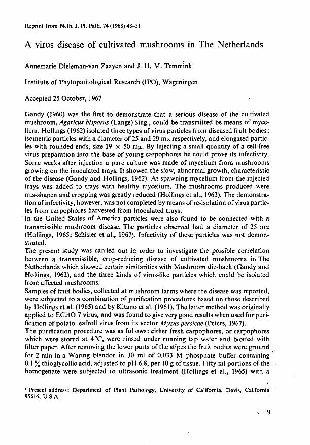
A virus disease of cultivated mushrooms in The Netherlands
Annemarie Dieleman-van Zaayen and J. H. M. Temmink1
Institute of Phytopathological Research (IPO), Wageningen
Accepted 25 October, 1967
Gandy (1960) was the first to demonstrate that a serious disease of the cultivated mushroom, Agaricus bisporus (Lange) Sing., could be transmitted be means of mycelium. Hollings (1962) isolated three types of virus particles from diseased fruit bodies; isometric particles with a diameter of 25 and 29 m\L respectively, and elongated particles with rounded ends, size 19 x 50 mji. By injecting a small quantity of a cell-free virus preparation into the base of young carpophores he could prove its infectivity. Some weeks after injection a pure culture was made of mycelium from mushrooms growing on the inoculated trays. It showed the slow, abnormal growth, characteristic of the disease (Gandy and Hollings, 1962). At spawning mycelium from the injected trays was added to trays with healthy mycelium. The mushrooms produced were mis-shapen and cropping was greatly reduced (Hollings et al., 1963). The demonstration of infectivity, however, was not completed by means of re-iso!ation of virus particles from carpophores harvested from inoculated trays. In the United States of America particles were also found to be connected with a transmissible mushroom disease. The particles observed had a diameter of 25 m\L (Hollings, 1965; Schisler et al., 1967). Infectivity of these particles was not demonstrated. The present study was carried out in order to investigate the possible correlation between a transmissible, crop-reducing disease of cultivated mushrooms in The Netherlands which showed certain similarities with Mushroom die-back (Gandy and Hollings, 1962), and the three kinds of virus-like particles which could be isolated from affected mushrooms. Samples of fruit bodies, collected at mushroom farms where the disease was reported, were subjected to a combination of purification procedures based on those described by Hollings et al. (1965) and by Kitano et al. (1961). The latter method was originally applied to ECHO 7 virus, and was found to give very good results when used for purification of potato leafroll virus from its vector Myzus persicae (Peters, 1967). The purification procedure was as follows: either fresh carpophores, or carpophores which were stored at 4°C, were rinsed under running tap water and blotted with filter paper. After removing the lower parts of the stipes the fruit bodies were ground for 2 min in a Waring blendor in 30 ml of 0.033 M phosphate buffer containing 0.1 % thioglycollic acid, adjusted to pH 6.8, per 10 g of tissue. Fifty ml portions of the homogenate were subjected to ultrasonic treatment (Hollings et al., 1965) with a
1 Present address: Department of Plant Pathology, University of California, Davis, California 95616, U.S.A.
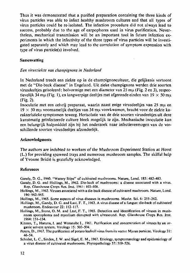
Thus it was demonstrated that a purified preparation containing the three kinds of virus particles was able to infect healthy mushroom cultures and that all types of virus particles could be re-isolated. The infection procedure did not always lead to success, probably due to the age of carpophores used in virus purification. Nevertheless, mechanical transmission will be an important tool in future infection experiments in which the infectivity of the three types of virus particles will be investigated separately and which may lead to the correlation of symptom expression with type of virus particle(s) involved.
Samenvatting
Een virusziekte van champignons in Nederland
In Nederland treedt een ziekte op in de champignoncultuur, die gelijkenis vertoont met de "Die-back disease" in Engeland. Uit zieke champignons werden drie soorten virusdeeltjes gei'soleerd: bolvormige met een diameter van 25 mjx (Fig. 2 en 3), respec-tievelijk 34 mjx (Fig. 1), en langwerpige deeltjes met afgeronde einden van 19 X 50 mjx (Fig. 2). Inoculatie met een celvrij preparaat, waarin naast enige virusdeeltjes van 25 my. en 19 x 50 m[i voornamelijk deeltjes van 34 mjx voorkwamen, bracht voor de ziekte ka-rakteristieke symptomen teweeg. Herisolatie van de drie soorten virusdeeltjes uit deze kunstmatig geinfecteerde culture bleek mogelijk te zijn. Mechanische inoculatie kan een belangrijk hulpmiddel zijn bij het onderzoek naar infectievermogen van de ver-schillende soorten virusdeeltjes afzonderlijk.
Acknowledgments
The authors are indebted to workers of the Mushroom Experiment Station at Horst (L.) for providing spawned trays and numerous mushroom samples. The skilful help of Yvonne Bridie is gratefully acknowledged.
References
Gandy, D. G., 1960. "Watery Stipe" of cultivated mushrooms. Nature, Lond. 185: 482-483. Gandy, D. G. and Hollings, M., 1962. Die-back of mushrooms: a disease associated with a virus.
Rep. Glasshouse Crops Res. Inst. 1961: 103-108. Hollings, M., 1962. Viruses associated with a die-back disease of cultivated mushroom. Nature, Lond.
196:962-965. Hollings, M., 1965. Some aspects of virus diseases in mushrooms. Mushr. Sci. 6: 255-262. Hollings, M., Gandy, D. G. and Last, F. T., 1963. A virus disease of a fungus: die-back of cultivated
mushroom. Endeavour 22: 112-117. Hollings, M., Stone, O. M. and Last, F. T., 1965. Detection and identification of viruses in mush
room sporophores and mycelium disrupted with ultrasound. Rep. Glasshouse Crops Res. Inst. 1964:151-154.
Kitano, T., Haruna, I. and Watanabe I., 1961. Purification and concentration of viruses by an organic solvent system. Virology 15: 503-504.
Peters, D., 1967. The purification of potato Ieafroll virus from its vector Myzus persicae. Virology 31: 46-54.
Schisler, L. C , Sinden, J. W. and Sigel, E. M., 1967. Etiology, symptomatology and epidemiology of a virus disease of cultivated mushrooms. Phytopathology 57: 519-526.
12

A virus disease of cultivated mushrooms in the Netherlands
Annemarie Dieleman-van Zaayen
Instituut voor Plantenziektenkundig Onderzoek, Wageningen, Niederlande
Abstract
For the last couple of years the yield on many Dutch mushroom farms was reduced by a mysterious disease which showed certain similarities to 'Die-back disease' in England. Various symptoms of this disease are described.
The disease-causing agent was transmissible to healthy mushroom cultures by means of diseased mycelium, which showed a very slow and declining growth as compared to healthy mycelium. A great number of samples was taken from various mushroom farms where the unknown disease had been reported. A special purification method was applied and led to the observation of three types of virus particles in the samples under study: 1. 'spherical' particles, diameter 25 nm (Hollings's Mushroom virus 1?); 2. elongated particles with rounded ends, size 19 x 50 nm (Hollings's Mushroom virus 3?); 3. 'spherical' particles, diameter 34 nm, with a distinct hexagonal outline. These particles differ considerably from Hollings's Mushroom virus 2 (diameter 29 nm).
No virus particles were observed in samples of apparently healthy mushrooms. Mostly we found a combination of the three types of virus particles in varying
concentration ratios. The mixture has been shown to be infectious by mechanical inoculation of healthy mushrooms using a cell-free virus preparation.
Introduction
In 1948 a very serious infectious disease of cultivated mushrooms (Agaricus bisporus (Lange) Sing.) was observed in the United States of America; this disorder of unknown cause was called 'La France disease' (Sinden & Hauser, 1950). During the following years a wide variety of names was given to mushroom disorders having some characteristics in common, but all of uncertain origin (Brown disease, Watery Stipe, X-disease). In 1957 devastating crop losses occurred in Britain due to a similar disease. Gandy (1960) found that it was caused by an infectious agent, which could be transmitted by hyphal anastomosis. Experiments by Gandy & Hollings (1962) demonstrated the presence of three types of virus particles associated with the disease under investigation. It was named 'Die-back disease' to distinguish it from any other possible disorder and to refer to the loss of crop and the degeneration of the mycelium, which phenomenon appeared to be more characteristic than the symptoms of the fruit bodies. These symptoms are highly variable, probably depending upon environmental conditions mainly.
Disorders of this type were not reported in the Netherlands until 1964,
13.

when a heavy outbreak occurred. Considerable losses of crop were incurred. In 1966 investigations into this disease were started.
Symptoms
The various symptoms going with the disease under investigation have been extensively described by several authors, and on the whole we can agree with them (Gandy, 1962; Schisler et al., 1967). Under Dutch conditions the following symptoms are observed, often in combination:
1. Locally the mycelium does not permeate, or hardly permeates, the casing layer, or it disappears from the casing layer after initial normal growth. In these areas no fruiting occurs (bare zones, fig. 1); the immediate result is, of course, a serious loss of crop. All around these areas mushrooms are found in dense clusters, maturing too early. The barren zones are often marked by such competitors as Botrytis crystallina (Bon.) Sacc. or Sporendonema purpurascens Bon.
2. Mycelium isolated from diseased sporophores on agar shows a slow and degenerated growth as compared with healthy mycelium, as was already reported by Gandy in 1960. In Petri dishes with peak-heated ground, and sterilized compost similar differences are very clearly visible after the relatively short time of one week or less (A.P.A. Oversteyns, pers. comm.).
IP Fig. 1. Bare zone (photograph: P. J. C. Vedder).
14

3. The delayed appearance of the pinheads of the first flush can be an important indication of the disease, as well as the formation of the fruiting pri-mordia below the surface of the casing layer. As soon as these mushrooms appear above the casing soil, their pilei are already opened.
4. Symptoms of sporophores are highly variable. The following abnor-
l ig. 2. A. I lungated slipes with small, early maturing caps. B: 'Drumsticks' (photographs: P. J.C. Vedder).

Fig. 3. A: Thickened, barrel-shaped stipes with small, flat pilei. B: Brown, slimy caps. The stipes are tapering downwards (photographs: P. J. C. Vedder).
malities can be found, separately or together: — off-white colour of the caps; early maturity. — slow development of the pinheads; dwarfing. — elongated, slightly bent stipes; sometimes with small, early maturing pileus 16
(fig. 2A). The stipes can be very thin ('drumsticks', fig. 2B). — the mushrooms are loosely attached to the substrate: at the slightest touch they are pushed over. — watery stipes; streaking in the stipes. — stipes are spongy. They quickly turn brown on cutting and show an abnormal structure. — thickened, barrel-shaped stipes; the veil is attached to the thickest part of the stipe, thus lower than usually. Pilei are small and flat (fig. 3A). — brown, slimy caps occur owing to a secondary bacterial rot; stipes are sometimes tapering downwards (fig. 3B); during the first flush sometimes a few light-brown caps can be observed. — abnormal or absent veils; 'hard gill'.
5. A specific, musty smell can be perceived in a diseased growing room.
Isolation of virus particles
Samples of fruit bodies, collected at mushroom farms where the disease was reported, were subjected to a combination of purification procedures described by Hollings et al. (1965) and by Kitano etal. (1961). The former method includes ultrasonic treatment, the latter is based on an organic solvent phase system. The virus particles are retained in the interphase, from which they can be se-
&£ 1-.". HP"""!*
V*. * •*•-
•***' V M "/fas,WfcMi'aa*J c
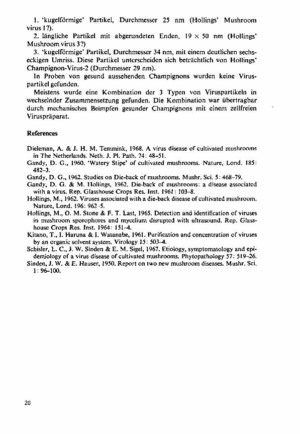
Fig. 4. Mushroom virus particles negatively stained with phosphotungstic acid (electron micrographs: TFDL, Wageningen). A: isometric particles, diameter 25 nm. B: elongated particles with rounded ends, size 19 x 50 nm. C: isometric particles, diameter 34 nm.
17
parated by ultracentrifugation. The particles could be observed, after negative staining with phosphotungstic acid, in an electron microscope.
In most samples of diseased mushrooms three types of virus particles were detected in varying concentrations (Dieleman & Temmink, 1968):
1. isometric particles, diameter about 25 nm (identical to Hollings's Mushroom virus 1 ?) (fig. 4A).
2. elongated particles with rounded ends, size 19 x 50 nm (identical to Hollings's Mushroom virus 3?) (fig. 4B).
3. isometric particles with a distinct hexagonal outline, diameter about 34 nm. The diameter of these particles differed markedly from that of Hollings's Mushroom virus 2 (diameter 29 nm) (fig. 4C).
Usually no virus particles were present in seemingly healthy mushrooms; rarely some 25 nm particles were observed.
Infection experiments; re-isolation of virus particles
The disease is easily transmitted with the aid of infected mycelium. The supply of a small quantity of diseased mycelium, which was grown on sterilized compost, to a healthy mushroom tray leads to the development of symptoms of the disease.
Infection experiments with cell-free virus preparations are far more difficult to perform. Gandy & Hollings (1962) proved the infectivity of a cell-free virus preparation by injecting a small quantity into the base of sporophores. The demonstration of infectivity, however, was not completed by means of re-isolation of virus particles from sporophores harvested from inoculated trays.
We have tested the infectivity of the cell-free preparations containing the three types of particles mentioned before. For that purpose mushrooms were grown in steam-sterilized 30 x 30 x 25 cm trays on an approximately 20 cm high layer of horse manure-straw compost which was covered with a casing layer of soil with a depth of 5 cm. The trays were kept at 15 to 17r'C; the relative humidity was about 80°,,. The first flush appeared five weeks after spawning.
Very young mushrooms of the first flush were, according to Gandy & Hollings (1962), inoculated with a purified preparation by injection into the bases of the stipes with a hypodermic syringe and fine needle. The inoculated trays were observed daily and compared with the control trays; once a week samples were collected from the trays which were tested for the presence of virus particles, by means of the previously indicated purification procedure.
A few days after injection no particles could be detected in the inoculated mushrooms. Fruit bodies of the third flush, however, appearing two weeks after inoculation, showed long stipes and the off-white colour typical of the disease. These mushrooms contained the three types of virus particles, predominantly the 34 nm 'sphere' which also prevailed in the inoculum used in this experiment. Cropping ceased almost completely in the infected tray. Sporophores of the uninoculated control trays did not show any symptoms or contain any particles.
However, this infection procedure did not always lead to success.
Experiments to investigate the importance of the three types of virus particles observed are being performed. This research is greatly hampered by the lack of a simple and reliable test method. The want of such method is also felt in the study of the epidemiological aspects of the disease. In recent years, however, our knowledge of this disease has made considerable progress.
Resume Une maladie a virus chez le champignon de couche cultive aux Pays-Bas
Depuis quelques annees, on constatait dans beaucoup de champignon-nieres hollandaises une diminution de rendement causee par une maladie inconnue qui avait une certaine ressemblance avec le 'Die-back disease' en Angleterre. Differents symptomes sont decrits.
Cette maladie pouvait etre transmise aux cultures saines de champignons de couche au moyen de mycelium infecte, qui presentait une croissance ties lente et retardee par rapport au mycelium sain. De nombreux echantillons furent preleves dans des champignonnieres oil la maladie inconnue s'etait manifestee. Une methode de purification speciale a ete employee et a conduit a l'observation de trois types de particules de virus dans les echantillons etudies:
1. des particules 'spheriques', diametre 25 nm (Hollings' Mushroom virus 1 ?); 2. des particules allongees avec des bouts arrondis, 19 x 50 nm (Hollings"
Mushroom virus 3?)' 3. des particules 'spheriques', diametre 34 nm, avec des contours hexagonaux
distincts. Ces particules se distinguent considerablement de Hollings' Mushroom virus 2 (diametre 29 nm).
Aucune particule de virus n'a ete observee dans des champignons apparem-ment sains.
Le plus souvent, on a trouve les trois especes de particules de virus presentes ensemble et a des concentrations variees. Le melange s'est montre transmissible par l'inoculation mecanique de champignons sains en utilisant une preparation de virus acellulaire.
Zusammenfassung Eine V'ruskrankheit beim kultivierten Champignon in den Nieaerlanden
Seit einigen Jahren wurden in vielen hollandischen Champignonbetrieben die Ertrage verringert durch eine unbekannte Krankheit, die gewisse Ahnlichkeit mit 'Die-back disease' in England zeigte. Verschiedene Symptome werden beschrieben und illustriert.
Diese Krankheit wurde durch verseuchtes Mycel in gesunde Champignon-kulturen iibertragen. Das kranke Mycel zeigte im Vergleich zu gesundem Mycel ein sehr langsames und verzogertes Wachstum.
Eine grosse Anzahl Proben wurde aus verschiedenen Betrieben genommen, bei denen diese unbekannte Krankheit aufgetreten war. Es wurde eine spezielle Reinigungsmethode angewendet, die zur Feststellung von 3 Arten von Virus-partikeln in den untersuchten Proben fiihrte:
19
1. 'kugelformige' Partikel, Durchmesser 25 nm (Hollings' Mushroom virus 1 ?).
2. langliche Partikel mit abgerundeten Enden, 19 x 50 nm (Hollings' Mushroom virus 3?)
3. 'kugelformige' Partikel, Durchmesser 34 nm, mit einem deutlichen sechs-eckigen Umriss. Diese Partikel unterscheiden sich betrachtlich von Hollings' Champignon-Virus-2 (Durchmesser 29 nm).
In Proben von gesund aussehenden Champignons wurden keine Virus-partikel gefunden.
Meistens wurde eine {Combination der 3 Typen von Viruspartikeln in wechselnder Zusammensetzung gefunden. Die Kombination war ubertragbar durch mechanisches Beimpfen gesunder Champignons mit einem zellfreien Viruspraparat.
References
Dieleman, A. & J. H. M. Temmink, 1968. A virus disease of cultivated mushrooms in The Netherlands. Neth. J. PI. Path. 74: 48-51.
Gandy, D. G., 1960. 'Watery Stipe' of cultivated mushrooms. Nature, Lond. 185: 482-3.
Gandy, D. G., 1962. Studies on Die-back of mushrooms. Mushr. Sci. 5: 468-79. Gandy, D. G. & M. Hollings, 1962. Die-back of mushrooms: a disease associated
with a virus. Rep. Glasshouse Crops Res. Inst. 1961: 103-8. Hollings, M., 1962. Viruses associated with a die-back disease of cultivated mushroom.
Nature, Lond. 196: 962-5. Hollings, M., O. M. Stone & F. T. Last, 1965. Detection and identification of viruses
in mushroom sporophores and mycelium disrupted with ultrasound. Rep. Glasshouse Crops Res. Inst. 1964. 151-4.
Kitano, T., I. Haruna & I. Watanabe, 1961. Purification and concentration of viruses by an organic solvent system. Virology 15: 503^.
Schisler, L. C , J. W. Sinden & E. M. Sigel, 1967. Etiology, symptomatology and epidemiology of a virus disease of cultivated mushrooms. Phytopathology 57: 519-26.
Sinden, J. W. & E. Hauser, 1950. Report on two new mushroom diseases. Mushr. Sci. 1: 96-100.
20
2.2 Additional information and discussion
Mushroom virus disease has now been recognized in many mushroom-growing
countries all over the world, and has probably been present as long as mush
rooms were grown. It was successively reported from the United States
(Sinden & Hauser, 1950), England (Anonymous, 1957), Denmark (Hansen & Block,
1967), the Netherlands (Section 2.1, 1968) and Australia (Paterson, 1968).
The most plausible explanation of its late detection (1964 in the Neth
erlands) is that the disease had been present for a long time but was not
recognized earlier because of the variable and often unclear symptoms; in
poor mushroom cultures, any bare zone or misshapen fruiting body is not con
spicuous. The occurrence of virus disease was established, by isolation of
virus particles from suspected mushrooms, in caves of the St Pietersberg
near Maastricht, a rather isolated former centre of mushroom growing. Local
growers did not consider these infected mushrooms abnormal, although they
had long stipes and were very loosely attached to the substrate. Virus-in
fected spores may have been present in the caves for years; once established,
it would be extremely difficult to control the disease.
Symptoms varied. Usually there were bare zones, where the casing soil
was devoid of mycelium, surrounded by mushrooms with long stipes and small,
early maturing and off-white caps. The fruiting bodies were often loosely
attached to the substrate and inferior. But bare zones and long stipes may
have other causes. Isolation of virus particles from suspected mushrooms is
decisive, if mushrooms are correctly sampled, e.g. from around bare zones.
Usually characteristic, but likewise variable, is the weaker growth on
agar of mycelium isolated from infected fruiting bodies than of healthy
mycelium. Some isolates hardly grow, whereas others show moderate to almost
normal growth rates. The individual growth rates are maintained even after
repetitive transfer to fresh media, although some, especially moderately
growing cultures sometimes recover slowly. Because of the wide variation
and because some infected isolates grow almost normally, this interesting
feature is no reliable test. Moreover, an aberrant growth on agar may be
caused by other factors such as composition of the nutrient medium (Fritsche,
1969).
Apart from the method of Kitano et al. (1961) at pH 6.8, none of the
many clarification procedures tested yielded cell-free virus preparations
21

clean enough to be examined by electron microscope. At higher pH virus par
ticles were severely damaged; at lower pH virus particles were even complete
ly lost, especially particles 34 nm in diameter. Hence the butanol-clarifica-
tion method, which Hollings (1962) used, was unsatisfactory: it did not yield
clean virus preparations and obviously destroyed the 34-nm particles. Hollings
& Stone (1971) had similar experience.
The change in incidence of the different types of mushroom virus par
ticle in the United Kingdom in the past five years, as suggested by Hollings
et al. (1971), is unlikely. They state that the 34-nm virus particles are
now the most prevalent, whereas these particles were seldom observed in 1967.
By then, however, they had introduced another test for mushroom virus (in
crude juice) instead of those used previously, which probably caused more
damage (Hollings et al., 1967). With crude juice, the unstable 34-nm par
ticles could now survive.
In the method of Hollings et al. (1967), mushrooms are squeezed with a
hand-press through cheese cloth, and the expressed juice is mixed with phos-
photungstic acid and viewed directly in the electron microscope. Though very
convenient, I found that the method was not a good test for virus; it often
yielded preparations with too much debris, in which virus particles could
not easily be detected. Although more laborious, the method of Kitano et al.
(1961) at pH 6.8 proved more satisfactory. As a rule portions of 30 g of
mushrooms were ground; a quantity of 50 ml was treated with ultrasound and
further processed. Several steps of the procedure are critical, for instance
the molarity of the potassium phosphate buffer must be precisely 2.5, or
else it does not work. The method is limited because of the extremely high,
perhaps damaging, salt concentration and the 'corrosiveness1 of the organic
solvents, and of the ultrasonic treatment before clearing. For complete pu
rification and isolation of the different types of mushroom virus particles,
probably a much milder procedure is required for clarification.
Although Hollings et al. (1971) stated that the mushroom virus particles
34 nm in diameter were seldom observed in 1967, I have isolated such parti
cles from most samples of diseased mushrooms since 1966, usually with one
or two of the other types in varying concentrations. Cell-free preparations
from severely diseased fruiting bodies from a highly contaminated farm some
times only contained a few virus particles, whereas almost normal mushrooms
now and then contained vast numbers of particles; the reverse sometimes oc
curred too. So far, no relationship has been encountered between symptoms
and types or amount of virus particles. Mushrooms were considered infected
22

if electron-microscopic preparations contained either a few clusters of 34-
nm particles, or many 25-nm particles. Both 34-nm and 25-nm particles have
been found separately, but the other particle types may not have been noticed
or may have been absent from the cell-free preparations through poor per
formance of the clarification technique or through minute concentrations in
the fruiting bodies. In cell-free preparations, there could be more of virus
particles 34 nm in diameter than of the other two particle types.
The concentration of the individual particle types might be affected
by factors like climatic conditions (temperature; relative humidity; nutri
tional value or condition of the compost), pathogenicity of viral strains,
and genetic properties of the mushroom host. Further research is needed to
elucidate these aspects. Another question is, the frequent occurrence of a
few particles 25 nm in diameter in cell-free preparations from apparently
healthy mushrooms; such mushrooms were taken to be healthy.
So far I have not managed to isolate virus particles from the tiny bit
of material (some dozens of milligrams) that mushrocm mycelium, and especial
ly infected mycelium, provides on agar. Hollings et al. (1965) suggested
direct electron-microscopy of mycelium treated with ultrasound. I found this
method unsatisfactory. Attempts to isolate virus particles from larger a-
mounts (up to a few grams) of infected mycelium, grown in a shaken liquid
malt medium, by ultrasonic treatment followed by the method of Kitano et al.
(1961) at pH 6.8, failed. The resulting cell-free preparations were extreme
ly dirty. Probably virus particles must be isolated from mycelium by another
clarification method than for fruiting bodies.
As the three types of mushroom virus particles often occur together, a
mutual relationship, though unlikely, could not be excluded. To test this
possibility, however, the three types of virus particles must be completely
purified and isolated from each other, in order to inoculate them separately
into mushroom cultures. After disappointing results for purification, par
ticularly with the 34-nm particles, we considered whether the virus particles
might be membrane-bound or attached to seme cell constituents, rather than
free in the cytoplasm of mushroom cells. Consequently more information was
needed on the intracellular appearance of mushroom virus. Because of the
special character of virus-infected mycelium, research started with a very
slowly growing infected culture. Chapter 3 deals with this and other aspects
of ultrastructure.
23

REFERENCES
Anonymous, 1957. It is 'La France'. MGA (Mushroom Growers Association) Bull. 96: 407-409.
Fritsche, G., 1969. Untersuchungen des Nahrbodeneinflusses auf verschiedene Myzelformen des Kulturchampignons. Mushr. Sci. 7: 515-529.
Hansen, H.P. & P. Block, 1967. Champignon-virose i Danmark. Horticultura 21: 179-181.
Hollings, M., 1962. Viruses associated with a die-back disease of cultivated mushroom. Nature, Lond. 196: 962-965.
Hollings, M., O.M. Stone & F.T. Last, 1965. Detection and identification of viruses in mushroom sporophores and mycelium disrupted with ultrasound. Rep. Glasshouse Crops Res. Inst. 1964: 151-154.
Hollings, M., O.M. Stone & F.T. Last, 1967. Mushroom viruses. Rep. Glasshouse Crops Res. Inst. 1966: 97.
Hollings, M., O.M. Stone 4 P.T. Atkey, 1971. Mushrooms. Rep. Glasshouse Crops Res. Inst. 1970: 157-159.
Hollings, M. & O.M. Stone, 1971. Viruses that infect fungi. A. Rev. Phyto-pathol. 9: 93-J18.
Kitano, T., I. Haruna & I. Watanabe, 1961. Purification and concentration of viruses by an organic solvent system. Virology 15: 503-504.
Paterson, R., 1968. The bad news for mushroom growers: Dieback appears in Australia. J. Agric. Vict. Dep. Agric. 66: 312-313.
Sinden, J.W. & E. Hauser, 1950. Report on two new mushroom diseases. Mushr. Sci. 1: 96-100.
24

3 Electron microscopy
3.1 Intracellular appearance of mushroom virus
Papers:
III Intracellular appearance of mushroom virus
Virology 39: 147-152 (1969).
IV Intracellular appearance of mushroom virus in fruiting bodies and
basidiospores of Agaricus bispcrus
Virology 47: 94-104 (1972).
25

Reprinted from ViHotoaY, Volume 30, No. 1, September 1969 Copyright © 1969 by Academic Press Inc. Printed in U.S.A.
Intracellular Appearance of Mushroom
Virus
Virus particles have been found associated with "Die-back disease" of cultivated mushroom, Agaricus bisporus (Lange) Sing. (1, 2). Recently, it has been proved that this disease is caused by one or more viruses (S). In cell-free preparations extracted from diseased mushrooms and stained with phos-photungstic acid, usually three types of virus particles were observed: isometric particles with a diameter of 25 m/i and 34 m/i, and elongated particles with rounded ends, 19 X 50 m;* (S).
Since the disease under investigation is the first clear case of a fungus attacked by virus, it was interesting to know whether the particles could be detected in the fungus
cells with the aid of common procedures, and in which part of the fungus cell mushroom virus occurs. Moreover such an approach might elucidate the role of the three kinds of virus particles associated with "Die-back disease." The present report deals with the detection of mushroom virus in diseased mycelium of Agaricus bisporus.
Preliminary attempts to demonstrate virus particles in tissue of mycelium and fruit bodies, embedded in styrene metha-crylate resin, were unsuccessful. Therefore a suitable working procedure was developed by comparing several methods with pelleted virus. For this purpose virus-diseased mushrooms were ground in 0.033 M phosphate buffer containing 0.1% thioglycollic acid, pH 6.8, and subjected to a combination of two purification procedure
27

SHORT COMMUNICATIONS
Kir.. 1. Ultrnthin section of pelleted mushroom virus. A. Without pre-staining. B. Pre-stained with 0 .7 , uranyl aeelate overnight after fixation. Note the dark centre in some of the particles. Electron micrograph: Laboratory of Virology, State Agricultural University, Wageningen.
including ultrasonic treatment (4), the other band on an organic solvent phase system (o), as was described previously (S). The virus particles are retained in the interphase, from which they can be separated by ultracentrifugation at 105,000 g for 60 min. Immediately after removing the super-nut ant liquid the pellets were pre-fixed in the centrifuge tubes for 1 hour in 6 % (v/v) glutaraldehyde buffered to pH 6.8 with 0.1 M phosphate buffer. The pellets could then easily be detached from the tube wall. They were washed in three changes of the buffer and post-fixed for 1 hour in 1% (w/v) osmium tetroxide in the same buffer. The whole fixation procedure was carried out at 4°. After washing, one pellet was left overnight in 0.5% uranyl acetate in water at 4° (6, 7). The other pellet immediately after fixation, was subjected to dehydration in a graded scries of ethyl alcohol and propylene oxide, and embedded in a 1:3 mixture of
Kpon 812 and Araldite 6005 (8). The following day the pre-stained pellet was treated in the same way. Sections were cut with a glass knife on an LKB Ultrotome I II and picked up on Formvar-coated 150 mesh copper grids. The sections were post-stained for \^ hour in 2 % uranyl acetate and for 5 min in Reynolds's lead citrate (9), and examined with a Siemens Elmiskop 1 or a Philips EM-300 electron microscope. Figure 1 shows that pre-staining of the material overnight in uranyl acetate was essential for clearly demonstrating the mushroom virus particles. Consequently this method was applied to the fungus tissue.
Mycelium, isolated from healthy and from virus-diseased mushrooms, was grown on 2% Biomals agar in petridishes for 10-14 days at 25°; virus-diseased mycelium usually showed a slow and degenerate growth as compared to healthy mycelium (10, 11).
28

M ' nuclear pore
^
' H
N
at*
" ^ | 3
nucleus
9 s A
NU Abbreviations used in Figures: as endoplasmic retieuiiun nucleolus NM nuclear membrane M mitochondrion
Fn; 2. Section of virus-diseased mushroom mycelium on Agar. A Fungus cell with an aggregate of virus particles close t<> a nucleus. B. Enlargement of the virus area ('. Enlargement of some particles. Note the dark centre. Electron micrograph: Technical and Physical Engineering Research Service (Ti l >L), Wageningen.
29
SHORT COMMUNICATION'S
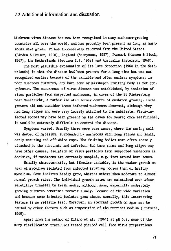
I N 1}L
Fia. 3. A. Lougil itdiiial sect ion of ;i virus diseased hypha wii li MM aggregate of vims pari ides (arrow) near theaepUim. IV Kiilargemenl of ih*> virus aggregate. C. Knlargemeul of some particles. Note again the <l:irk centre. Klectron micrograph: TFDL, Wageningen.
30

SHOUT COMMUNICATIONS
* * * * * «L
iE
Fii;. 4. Section of • virus-diseased mycelium coll, showing Humorous virus pari icles close to a nucleus, membranes of the endoplasmic reticulum system, and clearly visible nuclear pores. V* represents most probably • virnplasm. Electron micrograph: Laboratory of Virology, State Agricultural University, Wageningcn.
Blocks of 1.5 X 1.5 mm were cut out of the agar near the centre of the colony. Thaw were fixed, pre-stained and embedded in the way described above. Thin sections were post-stained and examined with an electron microscope.
In virus-diseased mycelium cells, virus particle! were found in aggregates, often near a septum or close to the nucleus (Fig. 2-4). The aggregates were partly surrounded by membranes of the endoplasmic reticulum system. Most probably viroplasms were observed (Fig. 4). So far only one type of virus particle has been found, with a diameter of approximately 25 n\p. Some pat tides had a dark centre, as was also noticed in the pelleted virus (Fig. 1).
No virus-like particles were detected in healthy mycelium. A further study of the
three types of virus particles and of virus in tissue of fruitbodics is in progress.
ACKNOWLEDGMENTS The authors thank Ir. T.S. Ie for his interest
and advice. This work was supported, in part , by the
Netherlands Organization fur the Advancement of Pure Research (Z.W.O.).
REFERENCES
1. CiANDY, I). (1., and SOLUMQS, M., Rep. (Glasshouse Crops Res. Inst. 1961, 103-108 (1982),
2. HOU.IN<;S , M., Nmturt 1%. 989-968 (1962). 5. DIELEMAN-VAS ZAAYEN, A., and TEMMINK,
J . H. M., Nelh. J. PI. Path. 74, 48-51 (10G8). 4. HoLLIMQe, M., STUNK, O. M., and LAST, F . T. ,
Rep. Glasshouse Crops Res. Inst. 1964, 151-154 (1965).
5. K ITANO, T. , HAKUNA, I., and WATANAHE, I.,
Virol,,</!/ !.'>, 503-504 (1961).
31

SHORT COMMUNICATIONS
6. HESS , W. M., Stain Techn. 41, 27-35 (19(10). 11. DIELEMAN-VAN ZAAYEN, A., Mushr. Sci. 7, 7. ARNOTT, H. J., and SMITH, K. M., Virology in press (1969).
34, 25-35 (1968). 8. TEMMINK, J . H. M., and CAMPKELL, R. N., ANNEMARIE DIELEMAN-VAN ZAAYEN
Can. J. Botany 46, 951-956 (1968). r . , „ ? N ,GfZ. , 9. REYNOLDS, E. S., / . Cell Biol. 17, 208-212 Institute oj Phylopalhological Research (I.P.O.)
/,«/.,•) Wagemngen, The Netherlands
10. GANDY, I). G., Nature 185, 482-483 (1960). Accepted July 1, 1969
32
Reprinted from VIROLOGY, Volume 47, No. 1, January 1972 Copyright © 1972 by Academic Press, Inc. Printed in I'.S.A.
VIROLOGY 47, 94-104 (1972)
Intracellular Appearance of Mushroom Virus in Fruiting Bodies and
Basidiospores of Agaricus bisporus
ANXEMARIE DIELEMAX-VAX ZAAYEX
Institute of Phytopathvlogical Research (I.P.O.), Wageningen, The Xetherlanih
Virus particles of 34 nm diameter were detected in ultrathin sections of cap, stipe, and basidiospores of Agaricus bisporus (Lange) .Sing. These particles formed dense aggregates in vegetative mycelium, but occurred dispersed and often abundantly in the cytoplasm and sometimes in vacuoles of fruiting bodies. In basidiospores 34 nm virus particles were found grouped together in small vacuoles and occasionally in the spore cytoplasm. Virus particles of 19 X 50 nm were seldom observed in cytoplasm of the stipe. Isometric particles of 25 nm, resembling ribosomes, were found in disarranged cells of the cap of mushrooms known to contain many 25 nm virus particles. These isometric particles occurred in vacuoles, free or clumped together and membrane bound, or were aggregated into membrane-limited electron-dense bodies. In such cells many 34 nm particles were also observed. In cap and stipe cells of the same mushrooms helices, thought to consist of ribosomes, were often found.
Virus particles of 34 nm diameter were observed in dolipores, which implies cell to cell translocation. The means of spread of mushroom virus disease by hyphal anastomosis and by spores from diseased mushrooms were confirmed.
No viruslike particles, electron-dense bodies, or helices were found in tissue or spores from healthy mushrooms.
IXTKODITTION'
The virus nature of an infectious disease of the cultivated mushroom, Agaricus bisporus (Lange) Sing., was established by Holiings (19(52), when lie isolated virus particles of 2">, 29, and 19 X •>() nm from diseased mushrooms. The disease, first noticed in 194S (Sinden and Hauser, 19.>0) and having various names including "La France disease" and "Die-back disease," is spread by viable mycelium (Gaudy, 19(10) and by spores from infected mushrooms (Schisler et al, 1967). Symptoms are reviewed by several authors (dandy, 1902; Schisler et al., 1967; Dieleman-van Zaayen, 19G9).
In cell-free preparations from diseased mushrooms, stained with phosphotungstic acid, usually three types of virus particles were observed (Dieleman-van Zaayen and Temmink, 1908): isometric particles with diameters of 2o and 34 nm, and elongated particles with rounded ends, 19 X ;">0 nm. The 34 nm particles had a distinct hexag
onal outline. When a cell-free virus preparation was injected into the stipes of young mushrooms on a growing-tray, symptoms developed a few weeks later in mushrooms of a subsequent crop; from these mushrooms, the three types of virus particles, predominantly the 34 nm particles, could be reisolated (Dieleman-van Zaayen and Temmink, 190.S). Then Holiings (190S) also observed the 34 nm virus particles, so far overlooked probably because of their unstable nature.
Mushroom virus was earlier detected in-ultrathin sections of virus-diseased vegetative mycelium of .4. bisporus on agar (Dieleman-van Zaayen and Igesz, 1969).
The ultrastmeture of healthy tissue of this basidiomycete fungus was briefly reviewed by Manocha (196.)); Scannerini (1967) and Thielke (1967, 1969) described some aspects-such as lomasomes, and basidia, in greater detail.
The present report deals with the detection of two, possibly three, types of mush-
C..|,M-inl,l
33
ULTRASTRUCTURE OF INFECTED MUSHROOMS
room virus particles in ultrathin sections of fruiting bodies, and of one particle type in basidiospores."
MATERIALS AND METHODS
Diseased fruiting bodies were collected from contaminated mushroomfarms. Healthy mushrooms were obtained from the Mushroom Experiment Station at Horst (L.), the Netherlands.
Fruiting bodies. Cap and stipe tissue of fresh, young mushrooms was prepared and sectioned as described by Dieleman-van Zaayen and Igesz (1909). As they stated, prestaining en bloc with 0.5 CA uranyl acetate in water overnight after fixation (Strugger, 1956; Hess, 1906) is essential for clearly demonstrating the mushroom virus particles. This method was employed throughout the present work.
Mushroom virus in pellets obtained by ultracentrifugation was treated as indicated by Dieleman-van Zaayen and Igesz (1969).
Basidiospores. Mushroom spores are about 7 X 5/im in diameter, more or less ovoid, and have a thick, hardly permeable wall. To bypass the difficulties encountered in fixing dormant spores (Hawker, 1965; Bracker, 1967) these were induced to germinate prior to fixation. The techniques used in spore collection, germination, and preparation of ultrathin sections are described elsewhere (manuscript in preparation).
RESULTS "
Fruiting Bodies
Sporophore tissue consists of loosely arranged hyphae. Most of our observations are pertinent to cap cells, which are usually smaller than cells of the stipe and appear more or less circular in cross section; they are rich in cytoplasm.
Occurrence of 34 nm diameter particles. Rather high concentrations of virus particles were observed throughout the fruiting body, in cytoplasm of base, stipe, and cap. The particles were of the same size as those occurring in vegetative mycelium and also showed a dark center. The particles, however, were not 25 nm in diameter as mentioned before (Dieleman-van Zaayen and Igesz, 1969), but represented the larger
mushroom virus particles of 34 nm, as will be clear from comparison of sectioned virus pellets (Fig. IB) with negatively stained virus particles (Fig. 1A). In Fig. IB the dark center is clearly visible in the large (34 nm) virus particles. Measuring these particles in ultrathin tissue sections yielded an average diameter of 30 nm.
As was indicated earlier, in vegetative mycelium the virus particles usually occur in dense aggregates, often near a septum (Fig. 2) or close to a nucleus. In the mushroom cap, virus particles of 34 nm are observed either in loose aggregates in some part of the cell (Fig. 4), or throughout the cell (Fig. 5) in the cytoplasm. Although the cap tissue mainly consists of loosely arranged hyphae, the cells in the lamellar trama of the gills are arranged in a regular fashion (Manocha, 1965) and are closely connected. Septal pores are abundant in this region. The septal pore apparatus of . 1 . bisporus is of the dolipore type, described by Girbardt (1958) and by Moore and McAlear (1962). Dolipores connect the cytoplasm of two adjacent cells. Figure 6 shows a median section through a septum, revealing virus particles on either side of the dolipore; a movement of the virus particles in a certain direction is suggested. Figure 7 shows 34 nm particles in a dolipore, which implies cell-to-cell translocation.
Occasionally virus particles were found in vacuoles, in both cap and stipe tissue (Fig. 8). Particles were sometimes observed in association with an osmiophilic substance which nearly filled the vacuole (Fig. 9).
Cells invaded with 34 nm particles showed no definite signs of deterioration as compared to cells from healthy mushrooms.
Occurrence of 19 X 50 nm particles. In cytoplasm of the stipe occasionally some particles of 19 X 50 nm were observed (Fig. 3). Detection of these virus particles is difficult for the following reasons: (1) Their amount in diseased mushrooms generally seems to be minute as judged by amounts in cell-free virus preparations; (2) these particles are often inadequately stained in ultrathin sections (see Fig. IB, the pelleted virus); (3) they are difficult to distinguish in cross section, since their diameter of 19 nm
34

DIELEMAN-YAN ZAAYEN
resembles thai of small ribosomea. The large amounl of 25nm virus particles in particle- may very well be present in cap addition to virus particles of M and 1!> X tissue but remain undetected. 5 0um, particles with the same diameter as
Occurrence of 25 nm ftarticlet. In cap tissue ribosomea (about 22 nm) were obHerved in of a mushroom sample, known to contain a vacuoles, either dispersed or clumped to-
. o-' M .
KlH. I Virus particle* c\i raeted from di>eased muahroonia: isometric partirlea with diameters <>f 25 ;u id 34 nm. and elongated partirlea with rounded cm Is. Ill 50 um. (A) Cell-free preparation, negatively stained with phoaphotungstir arid. (B) IHt rat bin seet ion of pelleted mushroom virus, stained with nranyl acetate and lead H rate, Arrows indicate the deeply stained 25 Um particles. Note the dark center in the 34 nm pa r t i c les .
In . _' rjltrathin section ol diacaaed vegetative mycelium, grown on 2*'-< Biomalsagar. Dense aggregate of 34 nm virtu part idea near a septum. PA . septal pore apparatus (dolipore). .Marker indicates .VKI • I m
Fio. .5. Section of mushroom atipe tisaue. Three virus particles of l'.i • -r>o nm, aggregated side n> side, in the cytoplasm.
35
• • > • '
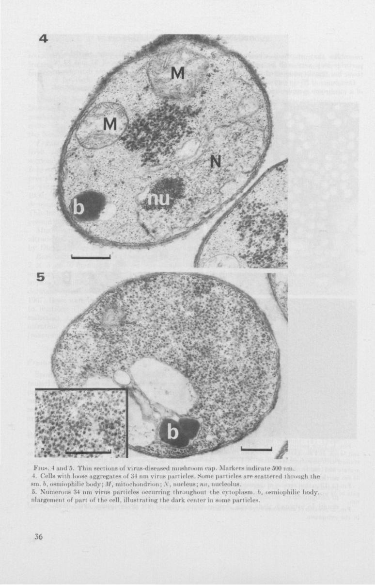
Fics. I and 5. Th in sect ions of v i rus diseased mushroom cap. Markets indicate "Ml Mi l . I Cells w i t h loose aggregates of 34 nm virus part ic le*. Some pari idea are seat tered through the
sm. h, osmiophilic body; M, m i tochondr ion; A . nucleus; nu, nucleolus ."). Numerous :w nm virus part idea occurring throughout the cytoplasm. '/. osmiophilic l>ody. nlargement of part of the cell, i l l ust rat ing the dark center in some particles.
36

TJf^ • * a
t *'f
• • •
• !fe *
V?*
-
FIGS , ti and 7. Ultrathin sections of virus-diseased mushroom cap, lamellar trama of the Rills. Markers indicate 500 nm
F IG . 6. Septum with dolipore and 34 nm virus particles on either side of the pore C, septal pore cap; P, central pore; 5 , septal swelling.
I hi. 7. Dolipore. Virus particles of 34 nm are seen in the pore. F I G . 8- Ultrathin sect inn of diseased mushroom stipe tissue, with 34 nm virus particles occurring in a
vacuole; some 34 nm particles are scattered through the cytoplasm. Marker indicates 500 nm.
37
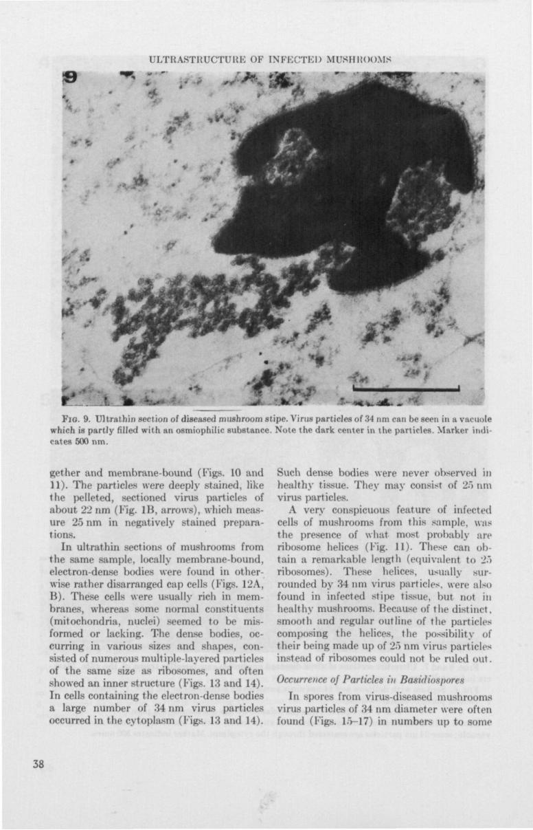
ULTKASTRUCTUKK OF INFECTED MU8HHOOMS
t •• •* * »
FJG. 9. UHnithin section of diseased mushroom stipe. Virus particles of M DOB run be seen in a vacuole which is partly filled with mi osmiophilic substance. Note the dark center in the particles. Marker Indi cates 500 nm.
get her and membrane-bound (Figs. 10 and 11). The particles were deeply .stained, like the pelleted, sectioned virus particles of about 22 nm (Fig. Hi, arrows), which meas ure 25 nm in negatively stained preparn-tions.
In ultrathin sections of mushrooms from the same sample, locally membrane-bound, electron-dense bodies were found in otherwise rather disarranged cap cells (Figs. 12A, B). These cells were usually rich in membranes, whereas some normal constituents (mitochondria, nuclei) seemed to be mis-formed or lacking. The dense bodies, occurring in various sizes and shapes, consisted of numerous multiple layered particles of the same six.' as ribosomes, and often showed an inner struct tire (Figs. 13 and 14). In cells containing the electron-dense bodies a large number of 34 nm virus particles occurred in the cytoplasm (Figs. 13 and 14).
Such dense bodies were never observed in healthy tissue. They may consist of 2.1 tint virus particle-.
A very conspicuous feat tire of infected cells of mushrooms from this sample, was the presence of what most probably are ribosome helices (Fig. 11). These can ob tain a remarkable length (equivalent to 2"> ribosomes). These helices, usually sur rounded by 34 am virus particles, were also found in infected stipe tissue, but not in healthy mushrooms. Because of the distinct, smooth and regular outline of the particle-composing the helices, the possibility of their being made up of 25 nm virus particle* itistead of ribosomes could not be ruled out.
Occurrence of Particles in Basirfiospores
In spores from virus-diseased mushrooms virus particles of 34 nm diameter were often found (Figs. 1">-17) in numbers up to some
38

FM;S 10 12. UhrHthin sectionsof diwNW'd imismroom cup, fn>m a mushroom sample known to contain a Largi amouni of 2Snm virus particles in addition to 'A t nm and l!t x S) nm particles.
I 'n. 10. Cap cell with many 34 ntn vims particles, and 25 una particles in vacuoles, either freely dis-tributed »r dumped together and membrane bound. Marker indicates "><)<) nm.
FIG. 11. Particles of 25 nm dumped together and membrane bound in a vacuole. A belieal si rueture is visible in the cytoplasm.
F IG . 12. (A) Membrane-limited, electron dense bodies of various si/es and shapes, possibly composed of 26 nm virus particles, occurring in a rather disarranged cap cell. (B.I Enlargement of part of the same cell. Note the presence of many membranes. Markers indicate 500 nm, 39
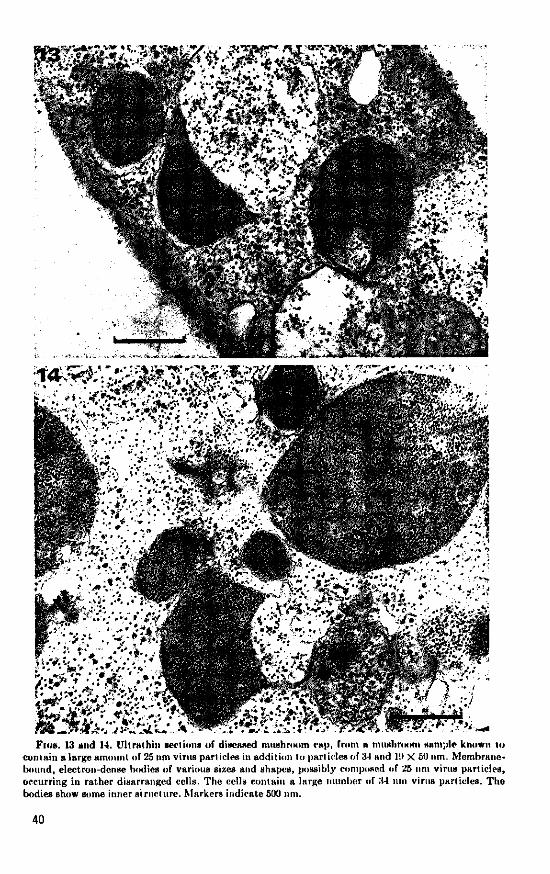

Flos. 15-17. UUrsthin sections of basidinspures from virus-diseased mushrooms. Virus particles of 34 nm are grouped together in small vacuoles. The spores are enveloped by a thick wall. Markers indicate 500 nm.
Fio. 15. Section of a spore containing lipid bodies (/,) and virus particles (V). FIG. 10. Detail of hasidiospore with 34 nm virus particles.
Fio. 17. Germinating basidiospore with 34 nm virus particles (I ') , ribosomes and mitochondria, and a "typical membrane complex" (MR), which is often found in fungus spores. GT, germ tube.
41
ULTKASTKUCTUKE OF INFECTED MUKHIiOOMS
hundreds per section. They usually were grouped together in small vacuoles, and sometimes occurred in the spore cytoplasm.
The germinating spores otherwise showed normal spore constituents (Fig. 17). Spores from healthy mushrooms did not possess viruslike particles in their small vacuoles. Further differences between spores from diseased and healthy individuals were not revealed. The walls of "healthy" spores may be slightly thicker than those of "diseased" spores, as suggested by Schisler et al. (19(17). However, the observed differences in wall thickness could also be due to changes during germination.
In "diseased" spores, no other types of virus particles were observed. They could, however, readily be missed or overlooked.
In germ tubes from "diseased" spores, large numbers of 34 ran virus particles were found. They occurred in dense aggregates, as in the vegetative mycelium. In germ tubes from "healthy" spores no viruslike particles were detected.
DISCUSSION
Virus particles of 34 rnn diameter have been shown to occur in large amounts in all kinds of mushroom tissue and in basidio-spores. The means of spread of the disease via hyphal anastomosis (Gandy, 1900) and by spores from diseased mushrooms (Schisler et al., 19(57), have now been confirmed. Methods to prevent or control the disease, based on the means of spread, are described elsewhere (Dieleman-van Zaayen, 1970).
The micromorphology of fungus cells closely resembles that of higher plant cells. Similarly, the ways of spread of mushroom virus resemble those of some plant viruses transmissible by grafting or seed, and the process of cell to cell translocation is comparable. Passage from cell to cell of 34 nm mushroom virus particles most probably takes place through dolipores, as that which occurs in higher plants through plasmodes-mata (Esau et al., 1967; DeZoeten and Gaard, 19(59).
Germinating spores of A. bisporus sometimes appear to be partly empty. This can be caused by consumption of storage products during germination. Another explana
tion may be that the principal storage product in fungi, glycogen (Zalokar, 19(i">; Wells, 198")) has been washed out by the procedure employed, i.e., en bloc staining with aqueous uranyl acetate. This technique implies disappearance of the greater part of glycogen, and clumping of the residual glycogen into irregular masses (Manasek, 19(H); Vye and Fischman, 1970). The method was required to detect 34 nm virus particles (Dieleman-van Zaayen and Igesz, 1909).
The application of this en bloc prestaining with aqueous uranyl acetate implies that the electron-dense, membrane-bound bodies consisting of numerous particles cannot be made up of glycogen. The particles composing the dense bodies definitely do not represent ferritin, which was seldom found as small, regularly arranged particles in healthy germinating tubes. The dense bodies may be made up of ribosomes, which are very deeply stained by the procedure employed. However, aggregation of ribosomes into membrane-hound bodies is improbable. Electron-dense bodies similar to those consisting of 2") nm particles have been reported by Gerola el al. (19<i(i) to occur in the young sieve elements of the leaf secondary veins from Brassica cliinensis L. infected with turnip yellow mosaic virus (TYMV). The roundish bodies contained a number of osmiophilic particles, believed to be virus particles. This similarity is accompanied by a remarkable morphological resemblance; the structure of 2."> nm mushroom virus particles is the same as that of TYMV, i.e., a T = 3 structure with hexamer-pentamer clustering of subunits (J. T. Finch, personal communication).
Unidentified, membrane-bound, dense bodies were reported by Thielke (1907) to occur in the maturing basidium of . 1 . bisporus. In a later publication (Thielke, 19(59), the presence of vacuoles with an electron-dense material which was membrane-bound, was mentioned instead of the dense bodies. A probable analogy with secondary vacuoles in growing cells of the stipe was assumed. In that case, the phenomenon described by Thielke could be related to the vacuole containing an osmiophilic substance, shown in Fig. 9.
42

DIELEMAN-VAN ZAAYEN
ACKNOWLEDGMENTS
The author thanks Mr. H. A. J. I. Waterreus, who prepared some of the tiltrathiu sections. This work was supported, in part, by the Netherlands Organization for the Advancement of Pure He-search (Z.W.O.). The Siemens Elmiskop 1 electron microscope of the Laboratory of Virology, State Agricultural University at Wageningen, was used during part of this s tudy. The prints of the electron micrographs were prepared by Mr. H. G. Elerie from the Technical and Physical Engineering Hesenrch Service (TFDL) at Wageningen.
REFERENCES
HRACKKK, C. E. (1967). infrastructure of fungi. Annu. Her. Phytopathnl. 5 , 343-374.
DKZOKTKN, (1. A., and G.YAKD, ( i . (1969). Possibilities for inter- and intra-cellular translocation of some icosahedral plant viruses. J. Cell Hiol. 40 , 814-823.
DIKLKMAN-VAN ZAAYKN, A. (1969). A virus disease of cultivated mushrooms in the Netherlands. Mushroom Sri. 7. 213-220.
DIKLKMAN-VAN ZAAYKN, A. (1970). Means by which virus disease in cultivated mushrooms is spread, and methods to prevent and control it. MGA Hull. 244,158-178.
DIKLKMAN-VAN ZAAYKN, A., and IGKSZ, O. (1969).
Intracellular appearance of mushroom virus. Virology 39, 147-152.
DIKLKMAN-VAN ZAAYKN, A., and TEMMINK,
J. H. M. (1968). A virus disease of cultivated mushrooms in the Netherlands. Neth. J. Plant Palhol. 7 4 ,48-51 .
EKAU, K., CRONSHAW, J . , and HOKFFKHT, L. L.
(1967). Relation of beet yellows virus to the phloem and to movement in the s ieve tube. J. Cell Biol. 32 , 71-87.
OANDY, D . G. (1960). 'Watery Stipe' of cultivated mushrooms. Nature (Ixmdon) 185,482-483.
OANDY, I ) . G. (1962). Studies on die-back of mushrooms. Mushroom Sri. 5 , 468-479.
GKROLA, F . M. , BASSI , M., and GIURSANI, G.
(1966). Some observations on the shape and localization of different viruses in experimentally infected plants, and on the fine structure of the host cells. I II . Turnip yellow mosaic virus in Brassica chinensis L. Caryologia 19, 457-479.
GIRMAKDT, M. (1958). Cber die Substruktur von Polystictus versicolor L. Arch. Mikrobiol. 28 , 255-269.
HAXVKKR, L. E. (1965). Fine structure of fungi as revealed by electron microscopy. Biol. Rev. Cambridge Phil. Soc. 40, 52-92.
I IKSS, W. M. (1966). Fixation and staining of fungus hyphae and host plant root tissues for electron microscopy. Stain Techno!. 4 1 , 27-35.
Hou. iMis , M. (1962). Viruses associated with a die-back disease of cultivated mushroom. Nature (hmilon) 196, 962-965.
IIoLUNus, M. (1968). Mushroom viruses. Rep. Glasshouse Crops Res. Inst. 1967, 101-102.
MANASKK, F. J . (1969). Myocardial cell death in the embryonic chick ventricle. J. Embryol. Exp. MorphoL2\, 271-284.
MANOCHA, M. S. (1965). Fine structure of the Ayaricus carpophore. Can. J. Bol. 43, 1329-1333.
MOOKK, 11. T . , and M<;ALKAH, J . H. (1962). Fine structure of Mycota. 7. Observations on septa of Aseomycetes and Basidiomycetes. Amer. J. Bol. 49, 86-94.
HCASKKKINI, S. (1967). Un particolare s istema di membrane e lomasomi in Atjaricus campester Fr. var. bisporus. Caryologia 20,115-133.
SCHISLKR, L. C , S INDKN, J. W., and SIGKL, E. M.
(1967). Etiology, symptomatology, and epidemiology of a virus disease of cult ivated mushrooms. Phytopathology 57, 519-526.
S I N I > K N , J . W., and Hu'SKit, E. (1950). Report on two new mushroom diseases. Mushroom Sri. 1, 96-100.
STRUOOKR, S. (1956). Die Uranylacetat-Koiitrastierung fur die elektronenmikrosko-pische Untersuchung von Pflanzenzelleu. Xaturwissenschajlen 43, 357-358.
THIKLKK, C. (1967). Die Feinstrukturder Basidieu des Kulturchampignons. Arch. Mikrobiol. 5 9 , 405-407.
THIKLKK, C. (1969). D ie Substruktur der Zellen im Fruchtkorper von Psalliota bispora. Mushroom Sci. 7 , 23 -30 .
VYK, M. V. and FISCHMAN, D . A. (1970). The morphological alteration of particulate glycogen by en bloc s taining with uranyl acetate. J. Ultraslruct. Res. 33 , 278-291.
W E L L S , K. (1965). Ultrastructural features of developing and mature basidia and basidiospores of Schizophyllum commune. Mycologia 57, 236-261.
ZALOKAR, M. (1965). Integration of cellular metabolism. In "The Fungi" (G. C. Ainsworth and A. S. Sussman, eds . ) , Vol. I; pp. 377-426. Academic Press, New York.
43

3.2 Morphological resemblances of fungus viruses to those of higher plants
Mushroom virus particles 25 ran in diameter are remarkably similar in struc
ture to particles of turnip yellow mosaic virus (TYMV). Centrifuging in a
sucrose (10-40$,w/v) density gradient often yielded a top component, most
probably consisting of empty protein shells.
T ... *:• ,-»•*:.V".fs,W.••• * * . * V V T
Fig. 3.2.1. Mushroom virus particles 25 ran in diameter, clarified, centri-fuged in a sucrose density gradient (10-40%) and negatively stained with phosphotungstic acid. Some particles show 2-fold views (2), others are close to 3-fold views (3), 5-fold views (5) or are close to the local 2-fold direction (L). The marked scale is 100 nm.
44

Fig. 3.2.2. Mushroom virus particles 25 ran in diameter, enlarged from Fig. 3.2.1, showing a 2-fold view, being close to a 3-fold view, to the local 2-fold direction and to a 5-fold view (from top to bottom, left row). The right row is a sketch of corresponding views of a model (after Finch & Klug, 1966).
Figs 3.2.1 and 3.2.2. show that the particle is the same in structure
as TYMV, which is composed of 32 large morphological units arranged in the
T = 3 icosahedral surface lattice, where T is triangulation number, with
hexamer-pentamer clusters of 180 structure units (Finch & Klug, 1966). Some
mushroom virus particles obviously show 2-fold views (a view of the particle
down a 2-fold axis, labelled 2 ) , others are close to 3-fold views (3) or 5-
fold views (5). Particles marked L are close to the local 2-fold direction.
Thus the 25-nm particles have 180 protein subunits in obvious hexamer-
pentamer clusters, with accessory particles which are probably empty protein
shells, so that they look closely related to viruses of the turnip yellow
mosaic virus group, described by Harrison et al. (1971).
Cell-free preparations with 25-nm mushroom virus particles did not
react with antiserum against TYMV. No beetles are known to feed on culti
vated mushrooms, so that a beetle vector of the 25-nm particles is highly
improbable. Although the virus does not necessarily have to share all prop
erties with the type member of the turnip yellow mosaic virus group to merit
inclusion in the group, some properties will be decisive. For instance, the
nature of the viral nucleic acid will be of importance for classification.
So far this factor is not known.
Similar striking resemblances and differences between a fungal virus
and a plant virus were encountered after isolating rod-shaped virus-like
particles from apothecia of Peziza ostraaoderma Korf (syn. Pliccwia fulva
R. Schneider). The particles closely resembled those of tobacco mosaic virus.
During research on mushroom virus disease they were observed several times
45

Fig. 3.2.3. Cell-free preparation from diseased cultivated mushrooms, with mushroom virus particles 25 and 34 nm in diameter, and with rod-shaped viruslike particles similar to particles from apothecia of Peziza ostracoderma Korf, and resembling tobacco mosaic virus particles. Negative stain with phosphotungstic acid. The marked scale is 100 nm.
in cell-free mushroom virus preparations (Fig. 3.2.3). Section 3.3 is con
cerned with this aspect.
REFERENCES
Finch, J.T. & A. Klug, 1966. Arrangement of protein subunits and the distribution of nucleic acid in turnip yellow mosaic virus. II. Electron microscopic studies. J. molec. Biol. 15: 344-364.
Harrison, B.D., J.T. Finch, A.J. Gibbs, M. Hollings, R.J. Shepherd, V. Valenta & C. Wetter, 1971. Sixteen groups of plant viruses. Virology 45: 356-363.

3.3 Virus-like particles in an ascomycete
Papers:
V Virus-like particles in a weed mould growing on mushroom trays
Nature, Lond. 216: 595-596 (1967).
VI Intracellular appearance and some morphological features of viruslike
particles in an ascomycete fungus
Virology 42: 534-537 (1970).
47
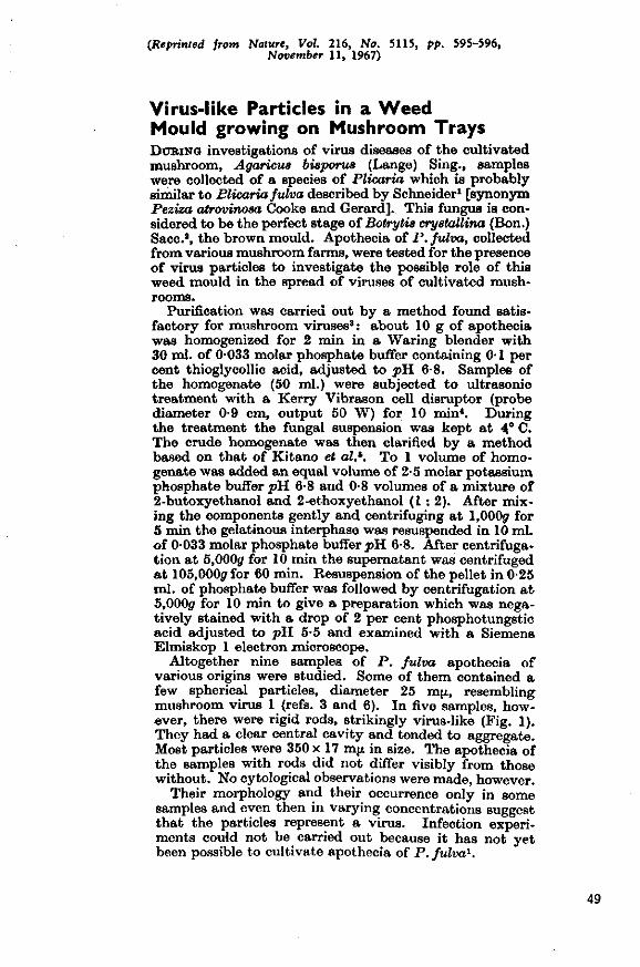
(Reprinted from Nature, Vol. 216, No. 5115, pp. 595-596, November 11, 1967)
Virus-like Particles in a Weed Mould growing on Mushroom Trays DtJBiNG investigations of virus diseases of the cultivated mushroom, Agaricus bisporua (Lange) Sing., samples were collected of a species of Plicaria which is probably similar to Plicaria fulva described by Schneider1 [synonym Peziza atrovinosa Cooke and Gerard]. This fungus is considered to be the perfect stage of Botrytis crystallina (Bon.) Sacc.*, the brown mould. Apothecia of P. fulva, collected from various mushroom farms, were tested for the presence of virus particles to investigate the possible role of this weed mould in the spread of viruses of cultivated mushrooms.
Purification was carried out by a method found satisfactory for mushroom viruses3: about 10 g of apothecia was homogenized for 2 min in a Waring blender with 30 ml. of 0-033 molar phosphate buffer containing 0-1 per cent thioglycollic acid, adjusted to pH 6-8. Samples of the homogenate (50 ml.) were subjected to ultrasonic treatment with a Kerry Vibrason cell disruptor (probe diameter 0-9 cm, output 50 W) for 10 min4. During the treatment the fungal suspension was kept a t 4° C. The crude homogenate was then clarified by a method based on that of Kitano et al.h. To 1 volume of homogenate was added an equal volume of 2-5 molar potassium phosphate buffer pH. 6-8 and 0-8 volumes of a mixture of 2-butoxyethanol and 2-ethoxyethanol (1 : 2). After mixing the components gently and centrifuging at l,000jr for 5 min the gelatinous interphase was resuspended in 10 ml. of 0033 molar phosphate buffer ^ H 6-8. After centrifuga-tion at 5,000(7 f ° r 10 m ' n the supernatant was centrifuged a t 105,000</ for 60 min. Resuspension of the pellet in 0-25 ml. of phosphate buffer was followed by centrifugation a t 5,000<7 for 10 min to give a preparation which was negatively stained with a drop of 2 per cent phosphotungstic acid adjusted to pH 5-5 and examined with a Siemens Elmiskop 1 electron .microscope.
Altogether nine samples of P. fulva apothecia of various origins were studied. Some of them contained a few spherical particles, diameter 25 m(x, resembling mushroom virus 1 (refs. 3 and 6). In five samples, however, there were rigid rods, strikingly virus-like (Fig. 1). They had a clear central cavity and tended to aggregate. Most particles were 350 x 17 mjx in size. The apothecia of the samples with rods did not differ visibly from those without. No cytological observations were made, however.
Their morphology and their occurrence only in some samples and even then in varying concentrations suggest that the particles represent a virus. Infection experiments could not be carried out because it has not yet been possible to cultivate apothecia of P. fulva1.
49

Fig. 1. Rod-shaped particles from Plicaria fulm R. Schneider.
Fig. 2. Some rod-shaped particles among 'spherical' mushroom virus from Agaricus bisporw (Lange) Sing.
50

In some samples of cultivated mushrooms similar rod-shaped particles were observed although in minute concentrations (Fig. 2). Attempts are being made to infect cultivated mushrooms with the rods. The known mushroom viruses are easily transmitted by hyphal anastomosis, but inoculation of mushroom cultures with cell-free virus preparations has been successful only in a few cases'-6. For the rod-shaped virus-like particles this might be even more difficult, for their concentration in cultivated mushrooms is much lower.
Inoculation of purified preparations from apothecia of P.fulva, containing large numbers of rods, onto Nicotiana glutinosa L., N. tabacum L. var. White Burley, Phaseolus vulgaris L. var. Bataaf, Gomphrena globosa L. and Cheno-podium amaranticolor Coste and Reyn did not induce any visible reaction in these plants, thus eliminating the possibility of contamination of the fungal material with a strain of tobacco mosaic virus.
Electron micrographs were prepared by the Service Institute for Applied Mechanics and Technical Physics in Agriculture, Wageningen.
ANNEMARIE DIELEMAN-VAN ZAAYEK The Institute of Phytopathological Research, Wageningen. Received July 20; revised September 25,1967.
> Schneider, K., Zbl. Batter. II, 108, 147 (1954/56). * Bels-Koning, H. C, and Bels, P. J., Handleiding voor de Champignon-
eulluur, 186 (Uitgave Proefstation voor de Champignoncultuur, Horst (Limburg), 1958).
* Dleleman, A., and Temmink, J. H. M. (in the press). 4 HoUings, M., Stone, O. M., and Last, F. T., Rep. Glatshoute Crop* Ret.
In*., 1964,151 (1965). 1 Kitano, T., Haruna, I., and Watanabe, I., Virology, 15, 503 (1961). * Hollings, M., Nature, 196, 962 (1962).
Printed in Great Britain by Fisher. Knight ft Co., Ltd., St. Albans.
51

Reprinted from VtaoMMT, Volume 42, No. 2, October 1970 Copyright] ' l!i7(l liy Academic Press, Inc. I'rinted in U.S.A.
VIROLOGY 42, 5:14-5:17 (1070)
Intracellular Appearance and Some Morphological Features of Viruslike
Particles in an Ascomycete Fungus
Peziza oslracoderma Korf [synonym Pli-caria fulva R. Schneider] is a fungus frequently occurring as ;i contaminant in mushroom nurseries, Fruitbodies (apothecia) of this Ascomycete collected from nurseries occasionally eontain rod-shaped viruslike particles which closely resemble tobacco mosaic virus (TMV) (/). From such apothe-
rod-shaped viruslike particles present in a fungus, it seemetl worthwhile to find where these particles were in the cells of the apothecia of P. ostracoderma. The way of preparing and sectioning the tissue was identical to that applied to mycelium of the cultivated mushroom Agaricus bisponu (Lange) Sing. (•>, .'f, 5, 6). Ultrathin sections were ex-
Pio. 1. Rod-shaped viroslike particles isolated from l'<:iz« oetraeoderma Korf. Negative stain with phosphotimgstic acid (|>H 5.6). Electron micrograph: Technical and Physical Engineering Research (Service (TFDL), Wageningen.
fia, cell-free preparations with the rod-shaped particles were obtained by a purification method devised for isolating virus from cultivated mushrooms (/, 2). Particles negatively stained with pliosphotungstic acid, pH 0.0, averaged 17 X 350 nm in size and had a clear axial canal (Fig. 1). Apothecia that contained such particles did not differ macroscoptcally from those without particles (/). Since this is the first reported case of
amined with a Siemens Elmiskop 1 or a Philips EM-300 electron microscope.
In apothecia from samples known to contain tiie viruslike particles, crystallike arrangements of the particles were found in cells just below the asci. They were often observed in vacuoles (Fig. 2) and sometimes in tlie cytoplasm. The crystallike aggregates consisted of rods arranged crosswise (Fig. 3). Sectioning these aggregates at different an-
52

SHOUT COMMUNICATIONS
glr- and at different levels, revealed par-tide- alternately in transverse and in longitudinal array, as well as in crosshatched patterns (Fig. 2).
Similar crystalline arrangements have been demonstrated to occur in plant cells infected with a strain of TMV (7).
Cross sections of the rods occasionally showed a central core. In cells of apothecia from samples without viruslike particles, no particles or particle aggregates as illustrated in Fig. 2 and A were found. Vacuoles have been observed in some cells of these fruit-bodies, but the vacuoles containing particles typically also contained cell debris (Fig. 2).
I t was not possible to inoculate preparations of the rod-shaped viruslike particles into P. ostracwlennn, because apothecia of the fungus could not be cultivated. Low concentrations of similar particles occasionally have been found in cell-free preparations from cultivated mushrooms (/). Attempts to infect mushrooms with the rods failed. Mechanical inoculation of cultivated mush rooms, however, is difficult even with the mushroom virus (2, 8).
To check for possible identity of the observed particles with TMV, indicator plants for this virus were inoculated with prepara
FIG. 2. Ultrathin section of fruitbody of Peziza ostracoderma. Fungus cell with aggregates of viruslike panicles in vacuole. Electron micrograph: Laboratory of Virology, State Agricultural University, Wageningen, and TFDL, Wageningen.
53

SHOUT COMMl-N'K ATIOXS
^ ^
I
o.i H
!•'[(;. 3. Cromwise arrangement nf rod shaped viriialike particle* in vacuole of ;i cell uf I'vziza imlraro-derma.
Fi«. I. Optical diffraction pattern «»f viritslike particle**, negatively stained with uratiyl formate. Near-meridional intensity occurs 011 a layer line a1 0.037 A ' corresponding to ;i periodicity of alwul 27 A in the particle image. Traces of a layer line :it 0.011 A ' ran also be s i. Photograph MH<' Labors lory of Molecular Biology, Cambridge,
54
SHOUT COMMUNICATIONS
tions of the viruslike particles (1), but no symptoms developed.
Cell-free preparations containing the rods did not react with antiserum against TMV in microprecipitation tests. Xor did the particles in a preparation that had been incubated with TMV-antiserum, mounted in negative stain and examined in the electron microscope, differ in appearance either from other particles that had been incubated with normal serum, or from particles that had been incubated in saline. TMV particles treated with antiserum became clumped and covered with a layer of antibody molecules. These results, and lack of symptoms in inoculated indicator plants indicate that the observed particles are not identical with TMV. Moreover, optical diffraction patterns of electron micrographs of the particles, negatively stained with uraiiyl formate (9), revealed that their structure differed appreciably from that of TMV (10). In Fig. 4 the near-meridional intensity on the layer line at 0.0:57 A - 1 shows that the particle structure is helical with a basic pitch of about 27 A which is larger than the 23 A pitch of TMV. The layer line at 0.014 also is not exactly one-third of 0.037 as it would be in TMV, thus the structure does not repeat after three turns as precisely as that of TMV. Apart from a superficial resemblance, the fungal viruslike particles differ distinctlv lYom TMV.
Whether I'. imlrarmUriiia can be artificially infected and whether the fungus will show symptoms after infection, is currently
under study, as well as the significance of the rods in A. bisporus.
ACKNOWLEDGMENTS
The authors thank Mr. It. Woods, who did part of the serological experiments, and Dr. A. J. Gibbs, who established the mutual contacts. The critical reading of the manuscript and the advice of Dr. F. Quak and Professor J. P . H. van der Want is gratefully acknowledged. This work was supported, in part, by the Netherlands Organisation for the Advancement of Pure Research (Z.W.O.).
/
HKFKUKNCKS
DIELEMAX-VAX ZAAYEX, A., Xature (London)
216, 505 (1007). 2. DIELEMAX-VAX ZAAYEX, A., and TEMMIXK,
J. II. M., Xeth.J. Plant Pathol. 74, 48 (1%8). 3. DIELEMAX-VAX ZAAVEX, A., and IKESZ, O.,
Virology 39. 147 (1%9). 4. AKXOTT, II. J., and SMITH, K. M., Virology
31, 25 (19(1S). 5. TEMMIXK, J. If. M., and CAMTHELL, R. N.,
Can. J. Bot. 16, 051 (1008). 6. liEYXOLiis, K. S.,./. Cell liiol. IT, 208 (10(13;. 7. WAIIMKE. II. K., Virology 34, 149 (l!Xi8). 8. Hoi.i.ixcis, M., Xature (London) 196, 0(12
(UM12). 9. FIXCII, J . T. , . / . Vol. liiol. B, 872 (1904).
10. FIXCII, J. T., Virology 38. 182 (19(39). AXXEMAUIE DIELEMAX-VAX ZAAYEX
Oxxo IGESZ Institute of I'hijtojMthological- Research (/.P.O.) W'uyeninyen, The Xetherlnnds
i. T. FINCH
\(UC f.ahoralory of Molecular liiology Hills ]!oad, Cambridge, Engluiul
Accepted July 7,1970
55

3.4 Additional information and discussion
Two, possibly three types of mushroom virus particles: 25 and 34 ran in dia
meter and 19 x 50 nm, have now been observed in ultrathin sections of in
fected mushroom tissue.
Particles 34 nm in diameter may be important in mushroom virus disease,
as this part of the study suggested; the number of 34-nm particles observed
in ultrathin sections of diseased fruiting bodies is often much larger than
was expected from the purification experiments. Purification and separation
of mushroom virus particles, however, are greatly hampered by difficulties
in breaking the fungal cell walls and eliminating the contaminating cell
constituents, and by lack of a simple infectivity test. Moreover, the 34-
nm particles proved to be unstable. Hence complete purification and isolation
from the other particle types did not succeed. Apparently this difficulty
was not due to their being surrounded by membranes or attached to cell con
stituents.
Particles 25 nm in diameter taken to be virus were often membrane-
bound, but could be purified: rather high yields of these particles were ob
tained in a rate-zonal sucrose density gradient. Virus particles 19 x 50 nm
were seldom observed in cytoplasm of the mushroom stipe; they may have been
present in other tissues but were difficult to detect. Also there are usual
ly too few such particles in cell-free preparations from diseased mushrooms
for success in purification trials. Rarely many 19 x 50-nm particles were
isolated from infected fruiting bodies; they were always associated with one
or two of the other particle types.
Although most mushroom scientists would now agree, despite the variety
of names and symptoms, that there is only one disease, there is still little
agreement on the number of particle types involved. In England, five kinds
of particle have been mentioned ('mushroom viruses 1 to 5'), including the
three particle types described here: particles 25 nm in diameter and of
19 x 50 nm ('mushroom viruses 1 and 3') both first mentioned by Hollings
(1962); particles 34 nm in diameter ('mushroom virus 4') first described hy
Dieleman-van Zaayen & Temmink (1968; Section 2.1), and later confirmed by
Hollings et al. (1968). I have found these three types of particle in cell-
free preparations of diseased mushrooms from Belgium, Bulgaria, Germany,
Italy, Switzerland, United States and the Netherlands (so far no diseased
56
mushrooms were obtained from other countries). Also their infectivity has
been demonstrated (Section 2.1) and their presence in ultrathin sections of
mushroom tissue (Section 3.1).
The English workers also mention particle types of 29 nm ('mushroom
virus 2'; Hollings, 1962) and 50 nm ('mushroom virus 51; Hollings et al.,
1968). Particles of about SO nm can be observed in ultrathin sections of
fruiting bodies and basidiospores, but they vary widely in shape and size,
and definitely do not look virus-like; before accepting them as 'mushroom
virus 5', their virus nature must be proved by mechanical inoculation and
reisolation of particles which was not done.
It would be difficult to distinguish between particles 25 nm and 29 nm
in diameter. Differences in penetration into the negative stain might al
ready result in a 4 nm variation, and the particle sizes may differ slightly
from preparation to preparation. Particles 29 nm in diameter were described
as angular, usually hexagonal in outline (Hollings, 1962). Fig. 3.4.1 re
presents a cell-free mushroom virus preparation, negatively stained with
phosphotungstic acid; most virus particles have a diameter of 25 nm, and some
34-nm particles and a few rod-shaped virus-like particles of 17 x 350 nm are
visible. After mushroom homogenate had been partially purified by the common
procedure, the cell-free preparation was filtered through a Millipore filter
(pore size 450 nm). During centrifuging and filtration, most of the unstable
34-nm particles were lost but many 25-nm particles were retained. Probably
because of their relatively high concentration, they formed aggregates as
was sometimes noticed in electron-micrographs; the 25-nm particles composing
the pattern definitely look angular (Fig. 3.4.1). The picture is very similar
to that given by Hollings et al. (1963, Fig. 10) as an electron-microgram of
mushroom virus particles 29 nm in diameter. Besides, if the scale in that
micrograph indeed indicates 200 nm, the particle size is 22 rather than 29
nm. Fig. 3.4.1 provides conclusive evidence that mushroom virus particles
25 nm in diameter, if present in profusion and if subjected to certain clar
ification procedures, aggregate in patterns similar to those formed by the
presumed 29-nm particles. The existence of 29-nm mushroom virus particles
('mushroom virus 2') in addition to mushroom virus particles 25 nm in diameter,
is therefore improbable.
So far, in ultrathin sections of basidiospores, only virus particles 34
nm in diameter have been observed (Section 3.1), but these spores were col
lected from diseased mushrooms known to contain mainly an abundance of 34-
nm particles. Infection trials showed, however, that virus particles of 25
57
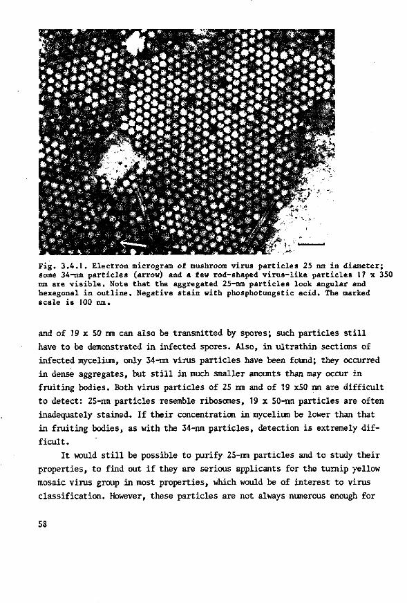
Fig. 3.4.1. Electron microgram of mushroom virus particles 25 nm in diameter; some 34-nm particles (arrow) and a few rod-shaped virus-like particles 17 x 350 nm are visible. Note that the aggregated 25-nm particles look angular and hexagonal in outline. Negative stain with phosphotungstic acid. The marked scale is 100 nm.
and of 19 x SO nm can also be transmitted by spores; such particles still
have to be demonstrated in infected spores. Also, in ultrathin sections of
infected mycelium, only 34-nm virus particles have been found; they occurred
in dense aggregates, but still in much smaller amounts than may occur in
fruiting bodies. Both virus particles of 25 nm and of 19 x50 nm are difficult
to detect: 25-nm particles resemble ribosomes, 19 x 50-nm particles are often
inadequately stained. If their concentration in mycelium be lower than that
in fruiting bodies, as with the 34-nm particles, detection is extremely dif
ficult.
It would still be possible to purify 25-nm particles and to study their
properties, to find out if they are serious applicants for the turnip yellow
mosaic virus group in most properties, which would be of interest to virus
classification. However, these particles are not always numerous enough for
58

purification trials. Moreover, cultivation of virus-infected material (fruit
ing bodies) has proved to be complicated: either the cultures do not produce
any mushrooms, or the mushrooms formed contain too few particles or none of
the desired type at all. For purification trials, I usually obtained diseased
mushrooms from commercial farms.
The role played in mushroom virus disease by the rod-shaped virus-like
particles, frequently isolated from Peziza ostraaoderma, should be examined.
Unpublished later studies indicate, that they differ distinctly from TMV,
not only from the common strain but also from the aucuba strain, which forms
the angled-layer aggregates in Turkish tobacco (Warmke, 1967, 1968). Recently
the structure of this strain, kindly provided by Dr M.K. Corbett (Maryland,
United States) was examined by Dr J.T. Finch (Cambridge, England). Optical
diffraction patterns and X-ray diffraction studies pointed out that its
structure is very like that of the common TMV-strain both in pitch and in
helical parameters (3-tum repeat). Aggregation of particles into a specific
arrangement apparently has nothing to do with the construction and the pitch
of their helix; in fact, similar angled-layer aggregates have been reported
to be formed by detached bacterial pili of 7 x 500-2000 nm with a rigid heli
cal structure and a pitch of 24 A (Brinton, 1965).
The rod-shaped particles associated with P. ostraaoderma and sometimes
observed in cell-free mushroom virus preparations, definitely differ from
the TMV-particles found associated with some Erysiphaceae (Yarwood, 1971;
Nienhaus, 1971). Their low concentration in P. ostraaoderma and especially
in A . bisporus is a great hindrance for further research.
REFERENCES
Brinton, C.C., 1965. The structure, function,synthesis and genetic control of bacterial pili and a molecular model for DNA and RNA transport in gram negative bacteria. Trans. N. Y. Acad. Sci. (2), 27: 1003-1054.
Hollings, M., 1962. Viruses associated with a die-back disease of cultivated mushroom. Nature, Lond. 196: 962-965.
Hollings, M., D.G. Gandy & F.T. Last, 1963. A virus disease of a fungus: die-back of cultivated mushroom. Endeavour 22: 112-117.
Hollings, M., O.M. Stone & P.T. Atkey, 1968. Mushroom viruses. Rep. Glasshouse Crops Res. Inst. 1967: 101-102.
Nienhaus, F., 1971. Tobacco mosaic virus strains extracted from conidia of powdery mildews. Virology 46: 504-505.
Warmke, H.E., 1967. Aucuba strain of tobacco mosaic virus: an unusual aggregate. Science, H.Y. 156: 262-263.
Warmke, H.E., 1968. Fine structure of inclusions formed by the aucuba strain of tobacco mosaic virus. Virology 34: 149-159.
Yarwood, C.E., 1971. Erysiphaceae transmit virus to Chenopodium. PI. Dis. Rep. 55: 342-344.
59

4 Epidemiology and control of mushroom virus disease
4.1 Trials on epidemiology and control
Paper:
VII Means by which virus disease in cultivated mushrooms is spread, and
methods to prevent and control it
MGA (Mushroom Growers Association) Bull. 244: 158-178 (1970).
61

Means by which virus disease in cultivated mushrooms is spread, and methods to prevent and control it by Annemarie Dieleman-van Zaayen INSTITUTE OF PHYTOPATHOLOGICAL RESEARCH (I.P.O.), WAGENINGEN, T H E NETHERLANDS.
History A virus disease in cultivated mushrooms, later having various names
including 'La France disease' and 'Die-back disease', was first noticed in 1948 in the United States of America (Sinden and Hauser, 1950). It was subsequently reported in England, where in 1957 t n e disease caused wide-spread losses. In the Netherlands the first definite signs of the disease were not observed until 1964, although the disease had probably been present before this time.
It became evident in 1962 that one or more viruses were involved: Hollings isolated three types of virus particles from diseased mushrooms. A few years later in the Netherlands, three types of virus particles were also isolated from diseased mushrooms, two of which corresponded with the particles discovered by Hollings; the third, however, had a diameter of 34 mfx instead of the 29 m\x recorded by Hollings.
When a cell-free, purified virus preparation was injected into the stipes of young mushrooms on a growing-tray, this produced symptoms a few weeks later in mushrooms of a subsequent flush; from these mushrooms, virus particles could be re-isolated. This provided the final proof that the disease to be investigated was indeed caused by virus particles isolated from diseased mushrooms (Dieleman-van Zaayen and Temmink, 1968).
Symptoms Reviews of the most common symptoms have already been given by
Gandy (1962) and Schisler et al. (1967). The symptoms most frequently met with in the Netherlands have also been reported (Dieleman-van Zaayen, 1969). Symptoms of virus disease are, however, highly variable. Sometimes even too low a yield will indicate that something is amiss. Frequently occurring symptoms are bare zones where the mycelium will not grow into the casing soil, or only very slightly, and mushrooms of poor quality standing loose in the casing soil, often having a long stipe and an off-white cap that matures too early. If, however, the presence of virus particles is established in mushrooms from a suspected crop, this can provide a decisive answer as to virus impairment.
Losses A diminished yield as a result of the virus disease will bring about
losses. An idea of the exact extent of such losses was obtained from an official inquiry and random test held among more than 1,100 Dutch growers. The following information was obtained: in 1967 and the first
63

half of 1968, one out of three mushroom farms was affected by the disease; the affected farms suffered an average production loss of 15%. That means that in 1967, there was an overall loss of 4-5% of the annual production of 17 million kg. mushrooms, (750,000 kg. or 1,653,465 lb.).
What is known about the means of spread? Little was known in this country of the ways in which the virus disease
was spread. English authors revealed at an early stage that viable mycelium could
transmit the 'Die-back disease' (Gandy, 1960a). Viable diseased mycelium would remain behind in trays after a crop, and after inadequate disinfection, would anastomose with healthy mycelium in a following crop and thus transmit the virus. Cooking-out of the unemptied trays for at least two hours at a temperature of 66°C. (i5i°F.) or higher would sufficiently disinfect the trays (Last et ah, 1967). In the Netherlands, however, where it was customary to cook-out a growing-room for 24 hours at 65-70°C. (149-158^.) before emptying, and in the event of virus disease, to repeat this for another 24 hours at the same temperature after emptying, it was often found that even this was not effective in ousting the disease. Suspending operations for a period of several months did not always lead to virus-free premises.
According to American authors, however,, the disease could also be spread by means of spores of diseased mushrooms (Schisler et al., 1967).
64
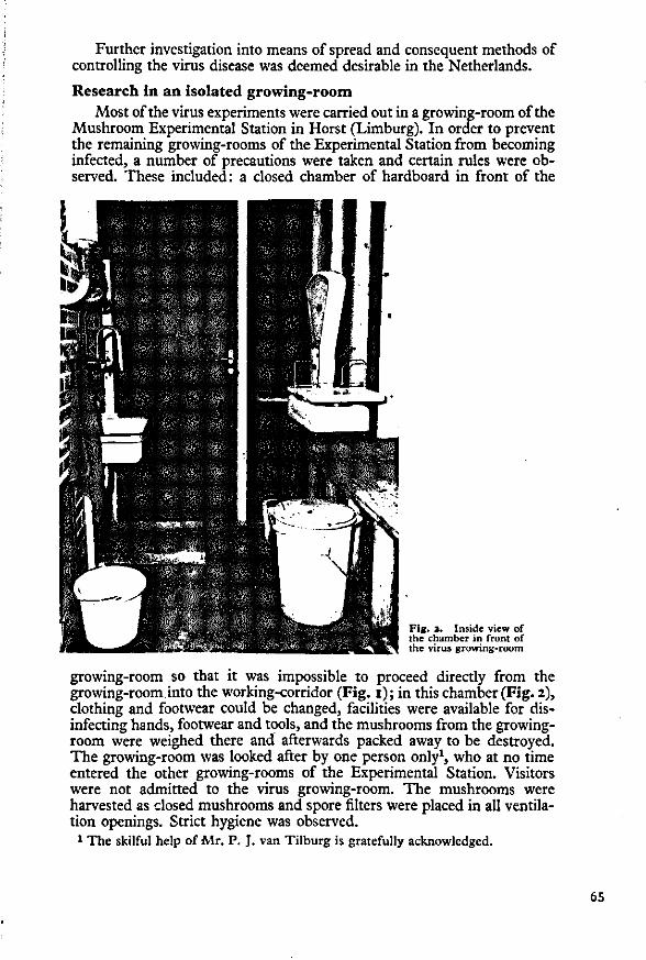
Further investigation into means of spread and consequent methods of controlling the virus disease was deemed desirable in the Netherlands.
Research in an isolated growing-room Most of the virus experiments were carried out in a growing-room of the
Mushroom Experimental Station in Horst (Limburg). In order to prevent the remaining growing-rooms of the Experimental Station from becoming infected, a number of precautions were taken and certain rules were observed. These included: a closed chamber of hardboard in front of the
IV
f/\ f **—*»"̂ \**« Fig. *• Inside view of Jt\. ^Jt-'•* ' F *•..'"*• . •; - * ' v X T | the chamber in front of ™*^^J^™J-***-- -- --•* -*»x*'>»* - - •y^ J^_^\^ the virus growing-room
growing-room so that it was impossible to proceed directly from the growing-room into the working-corridor (Fig. i ) ; in this chamber (Fig. 2), clothing and footwear could be changed, facilities were available for disinfecting hands, footwear and tools, and the mushrooms from the growing-room were weighed there and afterwards packed away to be destroyed. The growing-room was looked after by one person only1, who at no time entered the other growing-rooms of the Experimental Station. Visitors were not admitted to the virus growing-room. The mushrooms were harvested as closed mushrooms and spore filters were placed in all ventilation openings. Strict hygiene was observed.
1 The skilful help of Mr. P. J. van Tilburg is gratefully acknowledged.
65
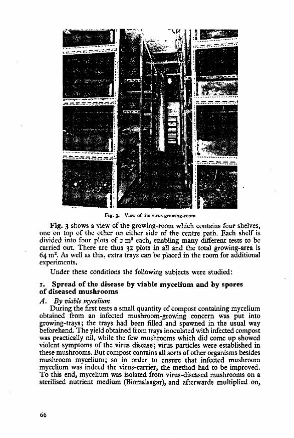
.1 ?**M8a
Fig. 3. View of the virus growing-room
Fig. 3 shows a view of the growing-room which contains four shelves, one on top of the other on either side of the centre path. Each shelf is divided into four plots of 2 m2 each, enabling many different tests to be carried out. There arc thus 32 plots in all and the total growing-area is 64 m2. As well as this, extra trays can be placed in the room for additional experiments.
Under these conditions the following subjects were studied:
1. Spread of the disease by viable mycelium and by spores of diseased mushrooms A. By viable mycelium
During the first tests a small quantity of compost containing mycelium obtained from an infected mushroom-growing concern was put into growing-trays; the trays had been filled and spawned in the usual way beforehand. The yield obtained from trays inoculated with infected compost was practically nil, while the few mushrooms which did come up showed violent symptoms of the virus disease; virus particles were established in these mushrooms. But compost contains all sorts of other organisms besides mushroom mycelium; so in order to ensure that infected mushroom mycelium was indeed the virus-carrier, the method had to be improved. To this end, mycelium was isolated from virus-diseased mushrooms on a sterilised nutrient medium (Biomalsagar), and afterwards multiplied on,
66
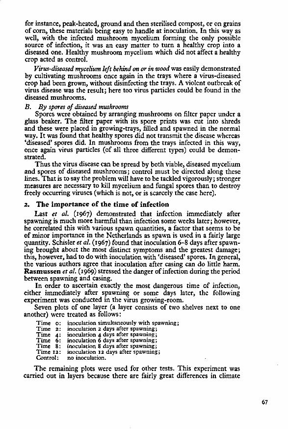
for instance, peak-heated, ground and then sterilised compost, or on grains of corn, these materials being easy to handle at inoculation. In this way as well, with the infected mushroom mycelium forming the only possible source of infection, it was an easy matter to turn a healthy crop into a diseased one. Healthy mushroom mycelium which did not affect a healthy crop acted as control.
Virus-diseased mycelium left behind on or in wood was easily demonstrated by cultivating mushrooms once again in the trays where a virus-diseased crop had been grown, without disinfecting the trays. A violent outbreak of virus disease was the result; here too virus particles could be found in the diseased mushrooms. B. By spores of diseased mushrooms
Spores were obtained by arranging mushrooms on filter paper under a glass beaker. The filter paper with its spore prints was cut into shreds and these were placed in growing-trays, filled and spawned in the normal way. It was found that healthy spores did not transmit the disease whereas 'diseased' spores did. In mushrooms from the trays infected in this way, once again virus particles (of all three different types) could be demonstrated.
Thus the virus disease can be spread by both viable, diseased mycelium and spores of diseased mushrooms; control must be directed along these lines. That is to say the problem will have to be tackled vigorously; stronger measures are necessary to kill mycelium and fungal spores than to destroy freely occurring viruses (which is not, or is scarcely the case here).
2. The importance of the time of infection Last et al. (1967) demonstrated that infection immediately after
spawning is much more harmful than infection some weeks later; however, he correlated this with various spawn quantities, a factor that seems to be of minor importance in the Netherlands as spawn is used in a fairly large quantity. Schisler et al. (1967) found that inoculation 6-8 days after spawning brought about the most distinct symptoms and the greatest damage; this, however, had to do with inoculation with 'diseased' spores. In general, the various authors agree that inoculation after casing can do little harm. Rasmussen et al. (1969) stressed the danger of infection during the period between spawning and casing.
In order to ascertain exactly the most dangerous time of infection, either immediately after spawning or some days later, the following experiment was conducted in the virus growing-room.
Seven plots of one layer (a layer consists of two shelves next to one another) were treated as follows:
Time o: inoculation simultaneously with spawning; Time 2: inoculation 2 days after spawning; Time 4: inoculation 4 days after spawning; Time 6: inoculation 6 days after spawning; Time 8: inoculation 8 days after spawning; Time 12: inoculation 12 days after spawning; Control: no inoculation.
The remaining plots were used for other tests. This experiment was carried out in layers because there are fairly great differences in climate
67
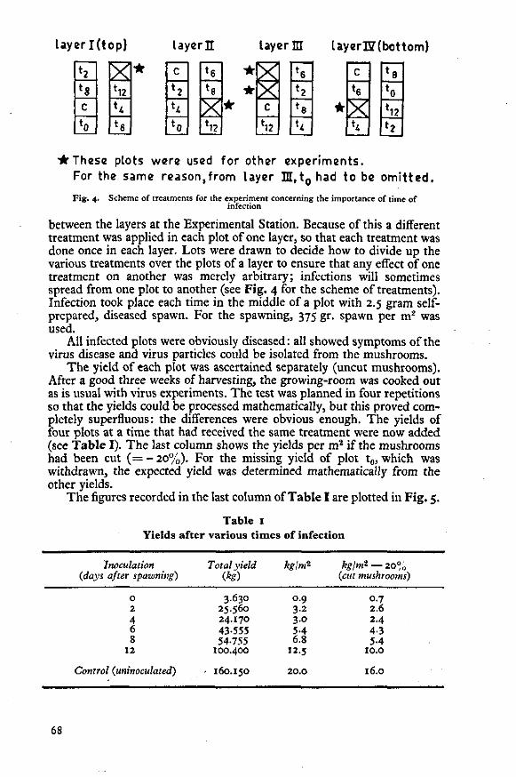
layerl(top) Uyer l l layer HI layerUfbottom)
*2
*8 C
*0
X *12
**
*6
• C
*2
*4
*0
*6
*8
X *12
• •
• S
C
*12
*6
*2
*e h
•
c
*6
X 1*4
*8
*0
*12
*2
* These plots were used for other experiments. For the same reason,from layer H , t 0 had to be omitted.
Fig. 4. Scheme of treatments for the experiment concerning the importance of time of infection
between the layers at the Experimental Station. Because of this a different treatment was applied in each plot of one layer, so that each treatment was done once in each layer. Lots were drawn to decide how to divide up the various treatments over the plots of a layer to ensure that any effect of one treatment on another was merely arbitrary; infections will sometimes spread from one plot to another (see Fig. 4 for the scheme of treatments). Infection took place each time in the middle of a plot with 2.5 gram self-prepared, diseased spawn. For the spawning, 375 gr. spawn per m2 was used.
All infected plots were obviously diseased: all showed symptoms of the virus disease and virus particles could be isolated from the mushrooms.
The yield of each plot was ascertained separately (uncut mushrooms). After a good three weeks of harvesting, the growing-room was cooked out as is usual with virus experiments. The test was planned in four repetitions so that the yields could be processed mathematically, but this proved completely superfluous: the differences were obvious enough. The yields of four plots at a time that had received the same treatment were now added (see Table I). The last column shows the yields per m2 if the mushrooms had been cut ( = - 20%). For the missing yield of plot t0, which was withdrawn, the expected yield was determined mathematically from the other yields.
The figures recorded in the last column of Table I are plotted in Fig. 5.
Table 1 Yields after various t imes of infection
Inoculation (days after spawning)
0 2 4 6 8
12
Control (uninoculated)
Total yield (kg)
3.630 25.560 24.170 43-555 54-755
100.400
160.150
kgjnfi
0.9 3.2 3.0 5-4 6.8
12.5
20.0
kgjnfi — 20% (cut mushrooms)
0.7 2.6 2.4 4-3 5-4
10.0
16.0
68

Mushroom ylolds (kg/m2)
16.0
10.0
5.4 4.3
2.6 2.4 0-7
12 Inoculation UNINOCULATED (days after spawning) CONTROL
Fig. 5. Mushroom yields in kg/m2 (cut mushrooms) at various times of infection
Note that the control plots showed a yield of 16 kg/m2 (cut mushrooms) in a good three weeks of picking, which may certainly be considered a very high yield. From the remaining figures and from the graph, it is obvious that,
(a) using a constant quantity of infectious matter, the t ime of infection plays a very important part in the virus disease;
(b) infection during or shortly after spawning has the most serious effect;
(c) even 8 days after spawning, however, infection will cause a 66% loss in yield;
(d) early infection will have even more disastrous consequences if it takes place in more than one spot at once, as will be the case in practice, and not in one spot only, in the middle of the plot such as here.
Infection during spawning or shortly afterwards can happen, for instance, because the spawning machine has been inadequately disinfected, but more likely because there is still viable, diseased mycelium present in the wood of shelves and trays. Another possible source of infection is the presence of 'diseased' spores on the premises, which fall on the compost during or after spawning and germinate there. 'Diseased' spores often germinate better and faster than healthy ones, as was noted by Schisler et al. (1967), and which can be read also from Table 2. The percentages in this table were obtained in the following way: Spores of diseased and healthy mushrooms, respectively, were collected on filter paper and afterwards transferred to a nutrient medium (2% Biomalsagar) in test tubes. For Experiment I, 48 tubes were provided with 'diseased' spores, and 48 with healthy ones; for Experiment II, the number was 60 in each case. It was decided each day, by a visual inspection, whether the spores had germinated. Spore germination is expressed in percentage of test tubes in which germination was observed.
As can be seen from the table, spores of diseased mushrooms had already germinated for the greater part after eight days on agar; in the spawned compost this will probably happen even more quickly and completely.
69
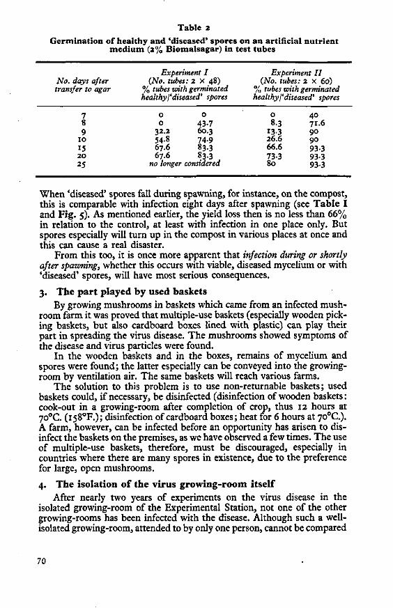
Table 2
Germination of healthy and 'diseased' spores on an artificial nutrient medium (2% Biomalsagar) in test tubes
No. days after transfer to agar
Experiment I (No. tubes: 2 x 48)
% tubes with germinated healthy/'diseased' spores
Experiment II (No. tubes: 2 x 60)
% tubes with germinated healthy I'diseased' spores
7 8 9
10
15 2 0
25
0 0 0 43-7
32.2 60.3 54.8 74.9 67.6 83.3 67.6 83.3
no longer considered
o 40 8.3 71.6
133 90 26.6 90 66.6 93.3 73-3 93-3 80 93.3
When 'diseased' spores fall during spawning, for instance, on the compost, this is comparable with infection eight days after spawning (see Table I and Fig. 5). As mentioned earlier, the yield loss then is no less than 66% in relation to the control, at least with infection in one place only. But spores especially will turn up in the compost in various places at once and this can cause a real disaster.
From this too, it is once more apparent that infection during or shortly after spawning, whether this occurs with viable, diseased mycelium or with 'diseased' spores, will have most serious consequences.
3. The part played by used baskets By growing mushrooms in baskets which came from an infected mush
room farm it was proved that multiple-use baskets (especially wooden picking baskets, but also cardboard boxes lined with plastic) can play their part in spreading the virus disease. The mushrooms showed symptoms of the disease and virus particles were found.
In the wooden baskets and in the boxes, remains of mycelium and spores were found; the latter especially can be conveyed into the growing-room by ventilation air. The same baskets will reach various farms.
The solution to this problem is to use non-returnable baskets; used baskets could, if necessary, be disinfected (disinfection of wooden baskets: cook-out in a growing-room after completion of crop, thus 12 hours at 70°C. (i58°F.); disinfection of cardboard boxes; heat for 6 hours at 70°C). A farm, however, can be infected before an opportunity has arisen to disinfect the baskets on the premises, as we have observed a few times. The use of multiple-use baskets, therefore, must be discouraged, especially in countries where there are many spores in existence, due to the preference for large, open mushrooms.
4. The isolation of the virus growing-room itself After nearly two years of experiments on the virus disease in the
isolated growing-room of the Experimental Station, not one of the other growing-rooms has been infected with the disease. Although such a well-isolated growing-room, attended to by only one person, cannot be compared
70

with a mushroom-growing business, it still gives an indication of the measures to be taken in practice to control the disease.
Control A. The effect of sodium pentachlorophenate*
One of the principal ways in which the virus disease is spread is by viable, diseased mycelium. When the wood of trays or shelves is cooked out before emptying, it is highly probable that even at a temperature of 70°C. in the compost for a period of 12 hours, not all parts of the wood will reach a sufficiently high temperature to destroy the remains of mycelium contained in it. New wood is usually impregnated so that mushroom mycelium cannot grow into it, but this is not repeated later.
Sodium pentachlorophenate (SPCP) was tested on its qualities as a means of preserving wood, partly because of the favourable results which seemed to have been obtained abroad (principally in Denmark). A preliminary test with several trays in the virus growing-room of the Experimental Station showed early promising results. A test on a larger scale was carried out at a commercial mushroom farm, in which 159 (not disinfected) trays were immersed in a solution of 2% SPCP, to which soda (Na2C03) had been added to enable the solution to spread more efficiently over the wood. Ten trays were left untreated as control. The immersion consisted of wetting the trays thoroughly for some seconds. After drying the trays were hosed down with a considerable quantity of water to prevent possible subsequent damage to the mushrooms. After that, mushrooms were grown in the trays.
The previous crop from these trays had shown violent symptoms of the virus disease; in mushrooms from untreated trays now too, many virus particles were to be found. This, however, was not the case with mushrooms from xh.t trays which had been treated. A visual judgement of the trays led to a percentage of 40% healthy for the ten untreated trays, and 86% healthy for the 159 trays treated with SPCP. The latter percentage could have been higher, but a number of the trays were attacked by Verticillium sp.; only a very small number of the treated trays was perhaps affected by the virus. Immersion in 4% SPCP would certainly have resulted in a completely healthy crop. Through this treatment the yield from this farm rose by 37.5%, which is by no means the maximum possible increase.
SPCP is now being used throughout the country with very good results. Applying this method means that growing-rooms need never be cooked out after emptying any more (but still beforel), not even if virus disease is present. Mushroom mycelium only grows to within a few mm. of the wood treated with SPCP and can thus not penetrate into it (Fig. 5), which means that the wood can be cleaned much more simply after the crop.
It only remains to say that ten other trays from this same grower underwent gassing with methyl bromide (600, 700 and 800 CTP), but the results were very unsatisfactory. After a great number of experiments with methyl bromide and both virus-diseased and healthy mushroom material, it is my opinion that this gas is inadequate as an after-crop sterilant (Dieleman-van Zaayen, in preparation).
•This chemical is available in the United Kingdom as SANTOBRITE or DOWICIDE G.
71

Fig. 6. The effect of sodium pemachlorophenate The upper tray is the untreated control (mycelium grows on the wood), the lower tray was dipped in a 4% solution of pentachlorophenate: mycelium does not grow on the
wood
B. Measures to prevent\to control the virus disease Summarizing with regard to the virus disease, the following important
points have been brought up: 1. The disease is spread by viable mycelium and spores of diseased
mushrooms. 2. Early infection is dangerous, especially an infection simul
taneous with or shortly after spawning. Up to the time of casing compost and mycelium must be protected.
With these facts in mind, and on the basis of the information obtained with the aid of the isolated virus growing-room, a meeting was held at the Experimental Station, attended by scientists, extension officers and educational staff. During this meeting, a number of measures to prevent or if necessary, to control the disease were drawn up. This led to a list being compiled, which was issued to Dutch growers in November 1968. This list is shown in Table 3, the left column giving preventive measures, and the right showing ways and means of controlling the disease after it has been diagnosed on the premises. The methods are not set out in their order of importance, but in chronological order according to the growth of the crop. They are, of course, applicable to Dutch conditions, which means to small farms where cultivation takes place on shelves following the one-zone system. In the majority of such farms, growing-rooms are situated side by side and they lead out into a covered working-corridor that can be a gathering-place for spores (which explains the measure concerning plastic
72
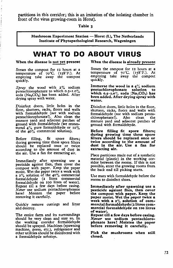
partitions in this corridor; this is an imitation of the isolating chamber in front of the virus growing-room in Horst).
Table 3
Mushroom Experiment Station — Horst (L), The Netherlands Institute of Phytopathological Research, Wageningen
WHAT TO DO ABOUT VIRUS When the disease is not yet present
Steam the compost for 12 hours at a temperature of 70°C. (i58"F.). At emptying take away the compost quickly.
Spray the wood with 2% sodium pentachlorophenate to which 0.5-1.0% soda (Na2C03) has been added. After drying spray with water.
Disinfect doors, little holes in the floor, shutters, racks, floors and walls with formaldehyde (not with sodium pentachlorophenate!). Also clean the manure yard and adjacent patches of ground with formaldehyde (we recommend 4% pure formaldehyde or 10% of the 40% commercial solution).
Before filling, fit spore filters; during growing time these spore filters should be replaced once or twice according to the amount of dust in the air. Use a fan for extracting air.
Immediately after spawning use a pesticide against flies, then cover the compost with paper. Keep the paper moist. Wet the paper twice a week with a 2% solution of the 40% commercial formaldehyde (2 litres commercial formaldehyde on 100 litres of water). Repeat till a few days before casing. Never use sodium pentachlorophenate here! Moisten the paper before removing it carefully.
Quickly remove cuttings and litter and destroy.
The entire farm and its surroundings should be very clean and stay so. In the working corridor formaldehyde should be sprayed. Machines (spawning machine, press, etc.), refrigerator and other utilities should be disinfected with a formaldehyde solution.
When the disease is already present
Steam the compost for 12 hours at a temperature of 70°C. (i58°F.). At emptying take away the compost quickly.
Immerse the wood in a 4% sodium pentachlorophenate solution to which 0.5-1.0% soda (Na2C03) has been added. After drying spray with water.
Disinfect doors, little holes in the floor, shutters, racks, floors and walls with formaldehyde (not with sodium pentachlorophenate!). Also clean the manure yard and adjacent patches of ground with formaldehyde.
Before filling fit spore filters; during growing t ime these spore niters should be replaced once or twice according to the amount of dust in the air. Use a fan for extracting air.
Place partitions made out of a synthetic material (plastic) in the working corridor between the rooms. If this is not possible, enter the growing rooms from the back end till picking starts.
Use mats with formaldehyde before the rooms to disinfect shoes.
Immediately after spawning use a pesticide against flies, then cover the compost with paper. Keep the paper moist. Wet the paper twice a week with a 2% solution of c ommercial formaldehyde (2 litres c ommercial formaldehyde on 100 litres of water). Repeat till a few days before casing. Never use sodium pentachlorophenate here! Moisten the paper before removing it carefully.
Pick the mushrooms when still closed.
73

(contd. from previous page)
At a first contamination the disease can be controlled best by i m mediately steaming out the concerned room. After that, act according to right column.
(contd. from opp. column) spawn too quickly, actually only when you cannot pick anything anymore. The entire farm and its surroundings should be very clean and stay so. In the working corridor formaldehyde should be sprayed twice a week. Machines (spawning machine, press, etc.), refrigerator and other utilities should be disinfected with a formaldehyde solution.
Quickly remove cuttings and litter and destroy.
Keep each room as a separate entity with separate clothes, shoes, steps, buckets, picking knives, picking racks, fans, etc. Kill off diseased patches with salt and cover with plastic; make the limits of the patches rather big. First pick from the healthy parts, then from the diseased patches. Wash hands often.
Admit as few visitors in the diseased rooms as possible and keep the door towards the working corridor closed. Kill off pests, etc., in particular. Have a short picking period only (not more than 4 weeks). Do not change over to brown strains of
(contd. in opp. column)
Dutch concerns which had been infected with the disease since 1964 were found to be completely free of the virus within a few months (checks for virus particles!) after applying these methods. The yield of one growing-room from such a concern rose from ± 0.5 kg/m2 to ± 13 kg/m2 in the following crop.
For English conditions, only slight adaptation of these measures will be needed. Stress must be laid once again on the following points:
(a) at the first sign of the virus disease, the mushrooms should be harvested as closed mushrooms as much as possible;
(b) spore filters should be used (2 cm.-thick glass fibre is suitable for this purpose);
(c) spawn quickly with a disinfected spawning machine; then cover the compost immediately with paper and spray this twice a week with a diluted formaldehyde solution;
(d) treat wood after emptying with SPCP; before emptying, cook-out for 12 hours at 70°C.;
(e) apply strict hygiene (e.g. treat working-corridor with f ormaldehyde, etc.).
It is not sufficient to perform one or two control measures only; the whole lot of measures should be applied to free a farm completely of the virus disease.
Is there a cure? The measures described are first and foremost intended to prevent and
protect and are a matter of strict hygiene. Are there any possible measures that might be taken which would have a curative effect? As far as chemical means are concerned, the answer at present is 'No'.
Gandy (1960b) did report that treatment of virus-diseased mushroom mycelium — which grows slowly and badly on an artificial nutrient medium such as agar — for a period of two weeks at 33°C. (9i°F.) resulted in most cases in a better and faster growth. It was presumed that the temperature treatment freed the fungus from the virus.
From mushrooms supplied by many different Dutch growers, mycelium was isolated on agar; in this way a collection of more than 60 different virus-diseased mycelia was made. The complete collection, together with
74

several healthy cultures, was subjected to the above-mentioned temperature treatment, half the collection even more than once. The results were very indecisive; some of the mycelia, however, did show a distinct improvement in growth. Nevertheless, some of the controls (healthy mycelium) showed a similar trend. Inoculation material was prepared from some of the most promising mycelia from both treated colonies (2 weeks at 33°C.) and untreated ones. Small growing trays, filled and spawned in the usual way, were inoculated with this material. The trays containing the controls, consisting of treated and untreated healthy mycelia respectively, remained healthy; the other inocula, which included the temperature-treated cultures, produced virus-diseased mushrooms. It is superfluous to say that mushrooms from all trays, as far as any came up, were tested for the presence of virus particles. Only in one case was there any doubt as to whether or not the treated culture was diseased; this is being investigated further. The temperature treatment, however, is not very promising; improvement in the growth of a treated colony certainly does not mean that it has become virus-free or carried virus beforehand. Such a method, for that matter, even if it was found to be effective, would not be of direct interest to mushroom growers.
REFERENCES
Dieleman-van Zaayen, A. and Temmink, J. H. M., 1968. A virus disease of cultivated mushrooms in the Netherlands. Neth.J. PI. Path., 74: 48-51.
Dieleman-van Zaayen, A., 1969. A virus disease of cultivated mushrooms in the Netherlands. Mushr. Sci., 7 : 213-20.
Gandy, D. G., 1960a. 'Watery stipe' of cultivated mushrooms. Nature, Lond., 185: 482-3.
Gandy, D. G., 1960b. A transmissible disease of cultivated mushrooms ('Watery stipe'). Ann. appl. Biol., 48: 427-30.
Gandy, D. G., 1962. Studies on die-back of mushrooms. Mushr. Sci., 5 : 468-79. Hollings, M., 1962. Viruses associated with a die-back disease of cultivated
mushrooms. Nature, Lond., 196: 962-5. Last, F. T., Hollings, M. and Stone, O. M., 1967. Some effects of cultural
treatments on virus diseases of cultivated mushrooms, Agaricus bisporus. Ann. appl. Biol., 59: 451-62.
Rasmussen, C. Riber, Mitchell, R. E. and Slack, C. I., 1969. Investigations into the infectivity of X-disease in mushroom culture. Mushr. Sci., 7 : 385-98.
Schisler, L. C , Sinden, J. W. and Sigel, E. M., 1967. Etiology, symptomatology, and epidemiology of a virus disease of cultivated mushrooms. Phytopathology, 57: 519-26.
Sinden, J. W. and Hauser, E., 1950. Report on two new mushroom diseases. Mushr. Sci., 1: 96-100.
75
4.2 An investigation of spawn
Spawn usually consists of mushroom mycelium aseptically grown on sterilized
cereal grains. Dutch growers use imported spawn. In theory, spawn could be
virus-contaminated, but severe infection would soon be detected by the slow
growth and deformity of the mycelium.
Spawn could hardly be examined directly because of the tiny bit of my
celium on the grains. First trials indicated, that the mycelium scraped from
grains yielded too little material to be tested for the presence of virus.
Another solution was, to grind grains and mycelium together, in a Waring
Blendor in 30 ml of 0.033 M phosphate buffer containing 0.U (v/v) thio-
glycollic acid, final pH 6.8, per 10 g of spawn. Occasionally the mixture
had to be diluted. The crude homogenate was then treated in the same way
as fruiting bodies (Section 2.1). However this procedure often resulted in
preparations that contained too much debris, so that virus particles could
hardly be seen with the electron-microscope. Thus the method of detecting
virus was poor. Besides, the chance of finding virus-infected spawn may be
very small; thousands of litres of spawn were be tested in the laboratory
for the presence of virus. Therefore, it was decided to handle the investi
gation differently. Help was sought from mushroom growers, of whom over 100
assisted. Only virus-free farms were allowed to take part in the test, which
began in January 1968.
Samples were collected from the farms in the following way. The growers
were asked to hold back a little from each bag of spawn at spawning; the
bags were not mixed and spawned per growing-room, but per shelf. Growers
could spawn their usual variety. The portions of spawn held back from all
bags used for one shelf, were collected into a plastic bag. The bags were
labelled with name and address of grower, date of spawning, spawn variety
and number of shelf. Thus at Wageningen we received a parcel with 10 bags
from each standard growing-room with 10 shelves. The parcels were stored in
a cold room at about 4 C.
If disease symptoms were observed in a growing-room, the grower informed
us. A sample of mushrooms from that growing-room was then tested for virus
particles in the usual way (Section 2.1). If virus disease was diagnosed,
the corresponding stored spawn was indirectly examined for virus in the fol
lowing way.
76
A small sterilized tray of 26 x 22 x 5.5 an was filled to a depth of
about 4 an with spawn (the spawn to be tested sometimes had to be supplemented
with fresh commercial spawn of the same variety) and covered with a 3-4 cm
casing layer of soil. The tray was kept in an incubator at 25°C and occa
sionally moistened. Once the casing soil was permeated with mycelium, the
temperature was slowly reduced to 15-17 C and usually after a few weeks some
mushrooms appeared. They were tested for virus particles in the usual way
(Section 2.1). Control trays only contained fresh commercial spawn.
Presumably because of the long storage of the spawn, some trays got contam
inated with, for instance, Triohoderma spp., and mushrooms did not always
form. Spawn was then tested for virus as before.
The results did not yield any clear indication on possible virus in
fection of spawn. Such an infection, however, cannot be excluded: the my
celium grown on the grains usually originates from spores; both spores and
mycelium have been found to spread the virus. Virus-infected mycelial cul
tures do not always show a slow and degenerated growth, but they sometimes
have almost normal growth rates so that they may look uninfected (Section
2.2). Anyhow, to answer the question whether spawn can be virus-contaminated
or not, detection methods for virus in spawn need improvement.
Although some spawn may be virus-infected, spread of mushroom virus in
other ways, for instance with basidiospores, seems more important, especial
ly in the Netherlands where some mushroom farms are built close together,
and where returnable, non-disinfected picking boxes may be used. For these
reasons, further cooperation with mushroom farms in routine testing of spawn
seems unadvisable.
77
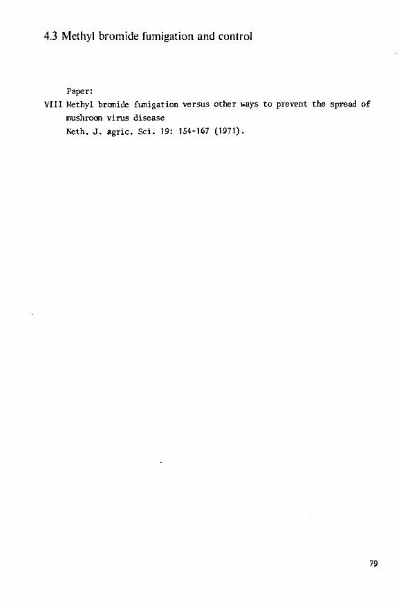
4.3 Methyl bromide fumigation and control
Paper:
VIII Methyl bromide fumigation versus other vvays to prevent the spread of
mushroom virus disease
Neth. J. agric. Sci. 19: 154-167 (1971).
79

Reprint Neth. J. agric. Sci. 19 (1971) 154—167
Methyl bromide fumigation versus other ways to prevent the spread of mushroom virus disease
Annemarie Dieleman-van Zaayen
Institute of Phytopathological Research (IPO), Wageningen, the Netherlands
Received: 20 April 1971
Summary
A virus disease has been a considerable threat to the rapidly growing mushroom industry in the Netherlands. Intensive research has been done to control this disease, which spreads by mycelium and spores from infected mushrooms.
The action of methyl bromide in the gas phase was studied, and compared with current chemicals and control methods. Mushroom spores appeared to be destroyed at relatively low dosages of methyl bromide; they were also readily killed, however, by a short treatment with formaldehyde vapour. It was already known that this can be achieved by a short heat treatment (1 h at 70°C). The results of methyl bromide fumigation of mycelium, spawn, contaminated growing trays and compost permeated with mycelium were disappointing. Various mycelial isolates differed in sensitivity to methyl bromide.
Thus methyl bromide fumigation cannot replace the traditional after-crop cooking-out of mushroom houses with live steam, followed by treatment of the wood with sodium pentachlorophenate.
Introduction
A virus disease of cultivated mushrooms In recent years the Dutch mushroom farming has suffered severe losses. They are due to a virus disease of the cultivated mushroom (Agaricus bisporus (Lange) Sing.), first recognized in this country in 1964. Crop yield is seriously reduced and the few mushrooms formed are misshapen (Dieleman-van Zaayen, 1969).
A survey among the more than 1,000 Dutch growers showed that in 1967 and the first half of 1968, one out of three mushroom farms was contaminated; on these farms average crop loss was 15 %. Thus in 1967, in total 4.5 % (or about 790,000 kg) of the production, which amounted to 17.5 million kg mushrooms, was lost.
The disease concerned was first observed in America in 1948 (Sinden and Hauser, 1950) und further described in England by Gandy (1960). Later it was named 'Die-back disease'. Hollings (1962) showed that different types of virus particles can be isolated and are always associated with the disease. In the Netherlands, the disease is caused by the following three types of virus particles, either alone or in combination: isometric particles, diameter 25 and 34 nm, respectively, and elongated particles with rounded ends, size 19 x 50 nm (Dieleman-van Zaayen and Temmink, 1968). Recently, one particle type has- been observed in ultrathin sections of diseased mycelium (Dieleman-van Zaayen and Igesz, 1969).
81

METHYL BROMIDE FUMIGATION AND MUSHROOM VIRUS DISEASE
The disease is spread by viable mycelium (Gandy, 1960) and by spores from diseased mushrooms (Schisler et al., 1967; Dieleman-van Zaayen, 1968).
Mushroom growing in the Netherlands Mushrooms are grown on compost of horse manure and straw, which was subjected to outdoor composting for two weeks. After a mushroom growing room is filled with about 100 kg/Jft compost, the compost is pasteurized (up to 60°C) for about 10 days. Cooling to 25 °C is followed by spawning (spawn is mushroom mycelium grown on sterilized grains). After two weeks of mycelial growth in the compost at 25 °C, a casing layer of soil (3-4 cm) is added. The temperature is then slowly reduced to 150C and after three weeks the first mushrooms can be harvested. These appear in flushes (weekly peaks). For five to six weeks the mushrooms are picked. The crop is then finished, the growing room emptied and after some days refilled with fresh compost. One crop takes 12 to 13 weeks.
The problem Until recently, mushroom growers were advised to cook out the growing rooms at the end of a crop for at least 24 h at 70°C to destroy all the mycelium and spores; in case of virus disease they were advised to repeat this treatment after the room had been emptied. Such a prolonged heat treatment, however, is very detrimental to mushroom houses. Besides, it is highly questionable whether the mycelium, grown into the wood of trays and shelves, will be killed (Spradling Chidester, 1939). If not destroyed it will anastomose with mycelium of the following crop and may thus cause early virus infection and severe damage (Last et al., 1967; Dieleman-van Zaayen, 1970).
This problem has recently been solved by the application, after every crop, of wood preservatives based on sodium pentachlorophenate. Cooking-out can then be reduced to 12 h at 70°C, before emptying the room, merely to kill mushroom mycelium and harmful organisms in the compost which is afterwards used for other horticultural purposes.
The growing houses, however, still suffer from this 12-h heat treatment, and steam installations are not everywhere available. In England, good results were claimed with methyl bromide in the gas phase, either during the final stages of compost preparation to eradicate pests and harmful micro-organisms (Hayes and Randle, 1968), or as a substitute for cooking-out with live steam at the end of a crop to disinfect growing houses and to kill mushroom mycelium, other micro-organisms and pests in the compost (Hussey et al., 1962; Flegg, 1968).
According to unpublished work by Dr F. T. Last a CTP (Concentration Time Product) of 600 g.h^.m-3 would kill virus-infected mushroom mycelium (Flegg, 1968).
Hayes (1969) described a number of experiments on methyl bromide fumigation of spores, fruiting bodies and mycelium of cultivated mushroom and of mycelium and fruitbody tissues of Verticillium malthousei Ware, a pathogenic fungus in mushroom culture. With respect to virus disease, he stressed the necessity to kill viable mushroom spores and mycelial fragments. His results suggested, that methyl bromide fumigation can, with advantage, replace the traditional cooking-out with steam. However he tested only healthy mushroom tissue, mycelium and spores.
From these data it was decided worthwhile to see whether methyl bromide, although very poisonous, could be used in the Dutch mushroom growing industry, particularly to control the virus disease. A second possible application was the central disinfection of picking boxes or baskets. These baskets, used a number of times by different
Neth. J. agric. Sci. 19 (1971)
82

ANNEMAR1E DIELEMAN-VAN ZAAYEN
growers, had earlier been shown to contribute in spreading the virus disease all over the country (Dieleman-van Zaayen and Van Tilburg, 1968, 1969).
A comparison was made with formaldehyde vapour, which is easier to handle and less poisonous. It is used in the Netherlands as a general disinfectant for control of the mushroom virus disease.
Co-operation between the Mushroom Experiment Station, the Plant Protection Service and the Institute of Phytopathological Research allowed a comprehensive experiment.
Material and methods
The trials were done in an experimental fumigation chamber of the Plant Protection Service at Wageningen. It is made of iron, with oil-painted walls, of 3 m8 capacity and with a gas circulation system charging 120 ms/h. With a heating unit a temperature of 20°C was maintained throughout the experiments. Liquid methyl bromide stored under pressure in a cylinder was led into a glass measure whence by opening a tap it passed through a volatilizer via the circulation system into the fumigation chamber. The fan in the circulation system was continuously operated to maintain a homogeneous gas/air mixture.
Several objects (indicated 1-6) were successively subjected to three methyl bromide fumigation trials (Experiments I - III). Experiment IV concerns a formaldehyde treatment.
The fumigated objects will be discussed in the following sequence: 1. Wooden trays of 57 X 110 X 15 cm, in which virus-diseased mushrooms had been grown commercially. At the end of the crop, the trays were emptied but not disinfected (Exp. I). 2. Compost, permeated with virus-diseased mushroom mycelium, obtained from a contaminated mushroom farm (Exp. I). 3. Spores from virus-diseased and healthy mushrooms collected on filter paper (Exp. II). 4. Various virus-diseased and healthy mycelial isolates on 2 % Biomals agar in test tubes (Exp. II and HI). 5. Self-prepared spawn from various virus-diseased and one healthy mycelial isolates in glass tubes or flasks (Exp. II and III). 6. Corn weevils, to ascertain whether methyl bromide can penetrate glass tubes sealed wilh filter paper instead of cottonwool plugs, as employed in fumigation of objects 4 and 5 (Exp. II).
Each treatment included non-fumigated controls. Experiment I was at atmospheric pressure; immediately before the methyl bromide
dosages of Experiments II and III were applied, the absolute pressure in the chamber was reduced by a vacuum pump to about 70 cm of mercury, to stimulate penetration of the gas into the test tubes with mycelium. After dosage of methyl bromide the atmospheric pressure in the chamber was restored by allowing air to enter through a small aperture.
During Experiment I (trays and compost) the concentration of the 'free' methyl bromide in the chamber was regularly measured by means of a thermal conductivity meter ('Gow-Mac Gasmaster'), that previously had been stamped and verified for this purpose, using data of van de Pol and Mathot (1961). The CTP's were calculated by
Neth. J. agric. Sci. 19 (1971)
83

METHYL BROMIDE FUMIGATION AND MUSHROOM VIRUS DISEASE
multiplication of the measured concentration of 'free' methyl bromide (in g/m3) and the exposure time (in h), according to the EPPO-report on fumigation standards (1961).
In Experiments II (mushroom mycelium on agar, spawn, and spores on filter paper) and III (mycelium and spawn) the initial dosages, necessary for a given CTP, were calculated. Taken into account was a certain loss by sorption, which was gathered from earlier experimentation (Van de Pol and Mathot, 1961). In fumigation of mycelium, spawn and spores, losses due to sorption were considerably smaller than in fumigation of wooden trays and compost (about 6 and 24 % respectively).
Methyl bromide fumigation was always carried out for about 24 (21 - 27) h. After every treatment the fumigation chamber was ventilated for about 30 min.
In the same fumigation chamber at atmospheric pressure a comparison was made with the effect of formaldehyde vapour on virus-diseased and healthy mushroom mycelium and on spores from diseased mushrooms (Experiment IV).
Further technical details will be given when describing the trials.
Experiments and results
1 Fumigation of contaminated trays (Exp. 1) The trays, obtained from a contaminated farm, had previously contained a crop with sr/ere symptoms of virus disease. Before and immediately after fumigation the nine treys to be examined were enveloped separately in plastic to prevent mutual contamination, and were then stored at 4°C. After they had been restored to the mushroom farm concerned, the trays were filled with compost of horse manure and straw. After 12 days of pasteurization the compost was spawned with commercial mushroom spawn (Sinden). For some weeks there was mycelial growth in the compost and then a casing layer of soil was added.
About three weeks later the first mushrooms were harvested. From the same farm 159 similar, non-disinfected and non-fumigated trays were treated with a 2 % solution of the wood preservative sodium pentachlorophenate (SPCP), with sodium carbonate as a wetting agent. Ten other trays served as controls.
Table 1. Visual judgment of crop on fumigated (Exp. I), untreated and SPCP-treated trays, and detection of virus particles in the mushrooms.
Number of trays
10
3" 3 3
159
Treatment
— (control)
Methyl bromide: 612 CTP 712 CTP 801 CTP
SPCP 2 %
Estimated number of 'healthy' trays
136'
Virus particles'
Many 25, 34, and 19 X 50 nm
Relatively few 25, 34, 19 X 50 nm (Fig. 1) No particles observed Relatively few 25, 34, 19 X 50 nm
No particles observed
• The figures in italics indicate the particle type(s) prevailing in that virus preparation. » This number could have been higher, but a number of the trays were infected by Verticillium sp.
Neth. J. agric. Sci. 19 (1971)
84
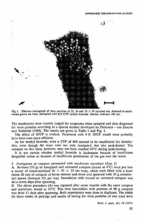
ANNEMARIE DIELEMAN-VAN ZAAYEN
\ .
• ' ' / • • •
*•• " -
< ': -
i . ' ) •
a
'*
.ImLfadUl
1
.* » ' "i
•
=.._.. .--_y.
* *
Fig. 1. Electron micrograph of virus particles of 25, 34 and 19 X 50 (arrows) nm, detected in mushrooms grown on trays, fumigated with 612 CTP methyl bromide. Marker indicates 100 nm.
The mushrooms were visually judged for symptoms when sampled and then diagnosed for virus particles according to a special method developed by Dieleman - van Zaayen and Temmink (1968). The results are given in Table 1 and Fig. 1.
The effect of SPCP is evident. Treatment with 4 % SPCP would most probably have been even more efficient.
As for methyl bromide, even a CTP of 800 seemed to be insufficient for disinfection, even though the trays were not only fumigated, but also peak-heated. The compost on this farm, however, may not have reached 60°C during peak-heating.
11 is not certain whether methyl bromide is inadequate because of insufficient fungicidal action or because of insufficient penetration of the gas into the wood.
2 Fumigation of compost permeated with mushroom mycelium (Exp. I) A. Portions (70 g) of fumigated and untreated compost (stored at 4°C) were put into a corner of steam-sterilized 30 X 30 X 25 cm trays, which were filled with a layer (some 20 cm) of compost of horse manure and straw and spawned with 35 g commercial spawn (Somycel 32) per tray. Inoculation with treated or untreated compost was done seven days after spawning. B. The above procedure (A) was repeated after some months with the same compost and mycelium, stored at 10°C. This time inoculation with portions of 80 g compost was done 11 days after spawning. Both experiments were done in duplicate. The yields (in three weeks of picking) and results of testing for virus particles of two trays were
Neth. J. agric. Sci. 19 (1971)
85

METHYL BROMIDE FUMIGATION AND MUSHROOM VIRUS DISEASE
Table 2. Yields of and virus particles in crop on small trays, inoculated with fumigated (Exp. I) compost (averages of two trays).
Treatment A B
Yields (g) Virus particles ' Yields (g) Virus particles '
— (inoculated 40 control)
299 CTP
Many 25 and 34 nm 40
400 CTP 499 CTP 612 CTP 712 CTP 801 CTP
193 45 78 85
105 190
Relatively many 25, 34, 19 X 50 nm 63 Rel. few 25 nm 180 Rel. few 25 and 34 nm 350 Rel. many 25, 34, 19 X 50 mm (Fig. 4) 540 Rel. many 25 nm 495 Many 25 nm 350
Many 25, 34 and 19 X 50 nm
Many 25 and 34 nm Few 25 nm Few 25 and 34 nm Few 25 and 34 nm Rel. many 25, 34, 19 X 50 nm Rel. few 25, 34, 19 X 50 nm (Fig. 5)
1 The figures in italics indicate the particle type(s) prevailing in that virus preparation.
averaged. Mushrooms growing on trays were tested for virus at least twice. The results are given in Table 2. These experiments were carried out at the Mushroom Experiment Station at Horst (L).
Yields of B were higher than those of A because of more favourable climatic conditions in the isolated virus growing room at that time. Yields were variable, however. Yet the higher CTP's of methyl bromide led to better yields than the lower CTP's. Obviously even a CTP of 800 cannot free compost from virus, that is kill the virus-
\ t - • -**> -; h > .> • ' - •• •.'••J
L *l ' - • f - "* U
Fig. 2. Trays inoculated with untreated, infected compost (ngnt) ana wun am) i_ir-iumigatea compost (left). The right tray yielded a virus-diseased culture; the tray on the left seemed healthy.
Neth. J. agric. Sci. 19 (1971)
86
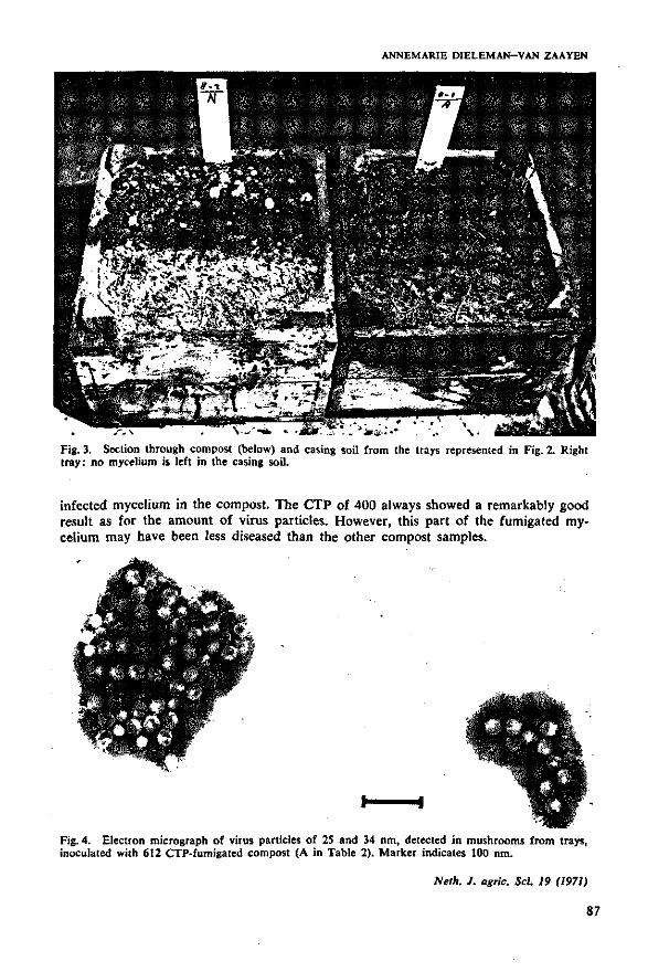
ANNEMARIE DIELEMAN-VAN ZAAYEN
Fig. 3. Section through compost (below) and casing soil from the trays represented in Fig. 2. Right tray: no mycelium is left in the casing soil.
infected mycelium in the compost. The CTP of 400 always showed a remarkably good result as for the amount of virus particles. However, this part of the fumigated mycelium may have been less diseased than the other compost samples.
JM£*A.
Fig. 4. Electron micrograph of virus particles of 25 and 34 nm, detected in mushrooms from trays, inoculated with 612 CTP-fumigated compost (A in Table 2). Marker indicates 100 nm.
Neth. J. agric. Sci. 19 (1971)
87
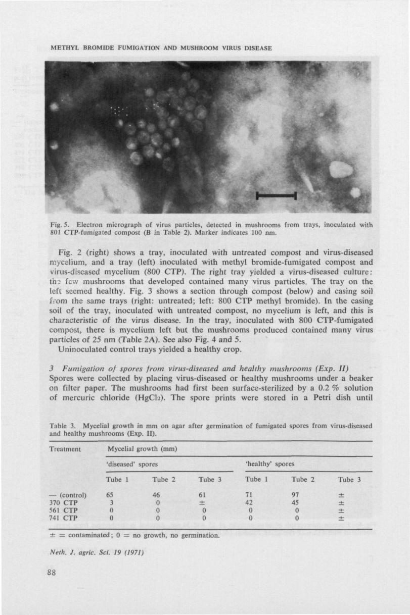
METHYL BROMIDE FUMIGATION AND MUSHROOM VIRUS DISEASE
m
Fig. 5. Electron micrograph of virus particles, detected in mushrooms from trays, inoculated with 801 CTP-fumigated compost (B in Table 2). Marker indicates 100 nm.
Fig. 2 (right) shows a tray, inoculated with untreated compost and virus-diseased mycelium, and a t ray (left) inoculated with methyl bromide-fumigated compost and virus-diseased mycelium (800 CTP) . The right tray yielded a virus-diseased cu l ture : t h : few mushrooms that developed contained many virus particles. The tray on the left seemed healthy. Fig. 3 shows a section through compost (below) and casing soil from the same trays (right: untreated; left: 800 CTP methyl bromide). In the casing soil of the t ray, inoculated with untreated compost, no mycelium is left, and this is characteristic of the virus disease. In the tray, inoculated with 800 CTP-fumigated compost, there is mycelium left but the mushrooms produced contained many virus particles of 25 nm (Table 2A). See also Fig. 4 and 5.
Uninoculated control trays yielded a healthy crop.
3 Fumigation of spores from virus-diseased and healthy mushrooms (Exp. II) Spores were collected by placing virus-diseased or healthy mushrooms under a beaker on filter paper. The mushrooms had first been surface-sterilized by a 0.2 % solution of mercuric chloride (HgCL>). The spore prints were stored in a Petri dish until
Table 3. Mycelial growth in mm on agar after germination of fumigated spores from virus-diseased and healthy mushrooms (Exp. II).
Treatment Mycelial growth (mm)
— (control) 370 CTP 561 CTP 741 CTP
'diseased' spores
Tube 1
65 3 0 0
Tube 2
46 0 0 0
Tube 3
61 ± 0 0
'healthy' spores
Tube 1
71 42 0 0
Tube 2
97 45 0 0
Tube 3
± s contaminated; 0 = no growth, no germination.
Neth. J. agric. Sci. 19 (1971)
88
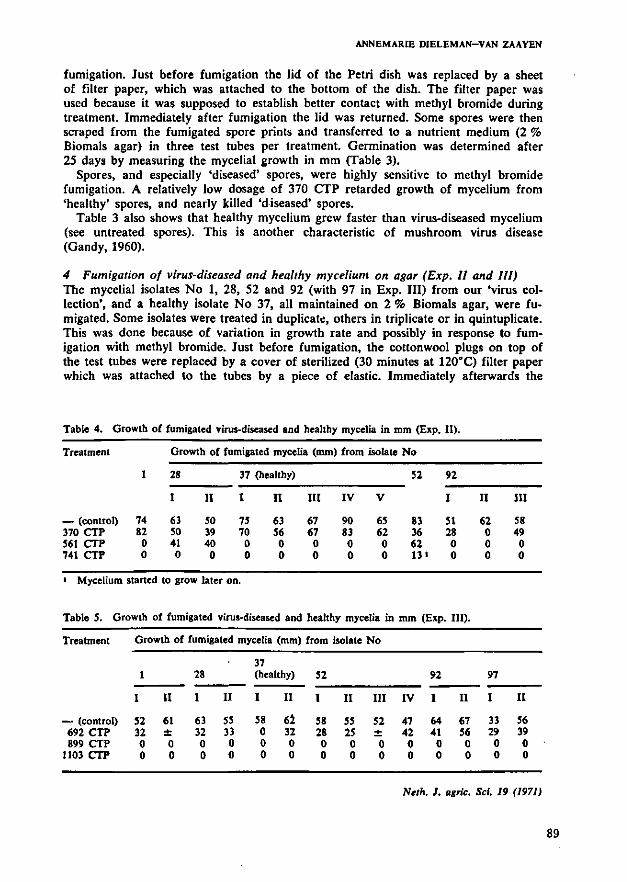
ANNEMARIE DIELEMAN-VAN ZAAYEN
fumigation. Just before fumigation the lid of the Petri dish was replaced by a sheet of filter paper, which was attached to the bottom of the dish. The filter paper was used because it was supposed to establish better contact with methyl bromide during treatment. Immediately after fumigation the lid was returned. Some spores were then scraped from the fumigated spore prints and transferred to a nutrient medium (2 % Biomals agar) in three test tubes per treatment. Germination was determined after 25 days by measuring the mycelial growth in mm (Table 3).
Spores, and especially 'diseased' spores, were highly sensitive to methyl bromide fumigation. A relatively low dosage of 370 CTP retarded growth of mycelium from 'healthy' spores, and nearly killed 'diseased' spores.
Table 3 also shows that healthy mycelium grew faster than virus-diseased mycelium (see untreated spores). This is another characteristic of mushroom virus disease (Gandy, 1960).
4 Fumigation of virus-diseased and healthy mycelium on agar (Exp. II and III) The mycelial isolates No 1, 28, 52 and 92 (with 97 in Exp. Ill) from our 'virus collection', and a healthy isolate No 37, all maintained on 2 % Biomals agar, were fumigated. Some isolates were treated in duplicate, others in triplicate or in quintuplicate. This was done because of variation in growth rate and possibly in response to fumigation with methyl bromide. Just before fumigation, the cottonwool plugs on top of the test tubes were replaced by a cover of sterilized (30 minutes at 120°C) filter paper which was attached to the tubes by a piece of elastic. Immediately afterwards the
Table 4. Growth of fumigated virus-diseased and healthy mycelia in mm (Exp. II).
Treatment
— (control) 370 CTP 561 CTP 741 CTP
1
74 82 0 0
Growth of fumigated myceli
28
I
63 50 41 0
II
50 39 40 0
37 (healthy)
I II
75 70 0 0
63 56 0 0
a (mm) from isolate No
HI
67 67 0 0
IV
90 83 0 0
V
65 62 0 0
52
83 36 62 13 •
92
I
51 28 0 0
II
62 0 0 0
III
58 49 0 0
> Mycelium started to grow later on.
Table 5. Growth of fumigated virus-diseased and healthy mycelia in mm (Exp. III).
Treatment
— (control) 692 CTP 899 CTP
1103 CTP
Growth of fumigated mycelia (mm) from isolate No
1
I
52 32 0 0
II
61 ± 0 0
28
I
63 32 0 0
II
55 33 0 0
37 (healthy)
I II
58 0 0 0
62 32 0 0
52
I
58 28 0 0
II
55 25 0 0
III
52 -*-0 0
IV
47 42 0 0
92
I
64 41 0 0
II
67 56 0 0
97
I
33 29 0 0
II
56 39 0 0
Neth. J. agric. Sci. 19 (1971)
89
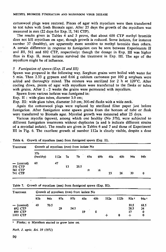
METHYL BROMIDE FUMIGATION AND MUSHROOM VIRUS DISEASE
cottonwool plugs were restored. Pieces of agar with mycelium were then transferred to test tubes with fresh Biomals agar. After 25 days the growth of the mycelium was measured in mm (22 days for Exp. II, 741 CTP).
The results given in Tables 4 and 5 prove, that about 600 CTP methyl bromide does not kill mycelium on agar, though growth is reduced. Some isolates, for instance number 37 (healthy), are apparently more sensitive to methyl bromide than others. A certain difference in response to fumigation can be seen between Experiments II and III, 561 and 692 CTP, respectively: though the dosage in Exp. I l l was higher than in Exp. II, more isolates survived the treatment in Exp. III. The age of the mycelium might be of influence.
5 Fumigation of spawn (Exp. II and III) Spawn was prepared in the following way. Sorghum grains were boiled with water for 5 min. Then 2.33 g gypsum and 0.66 g calcium carbonate per 100 g sorghum were added and thoroughly mixed. The mixture was sterilized for 2 h at 120°C. After cooling down, pieces of agar with mycelium were transferred to the flasks or tubes with grains. After 1 - 2 weeks the grains were permeated with mycelium.
Spawn from various isolates was fumigated in: Exp. II : wide glass tubes, diameter 3.0 cm; Exp. I l l : wide glass tubes, diameter 3.0 cm; 300-ml flasks with a wide neck.
Again the cottonwool plugs were replaced by sterilized filter paper just before fumigation. After fumigation some spawn grains from the bottom of tube or flask were transferred to Biomals agar. Mycelial growth was measured after 25 days.
Various mycelia (spawn), among which one healthy (No 37b), were subjected to different fumigation treatments without duplicates (a and b indicate different strains of a mycelial isolate). The results are given in Tables 6 and 7 and those of Experiment III in Fig. 6. The excellent growth of number 112a is clearly visible, despite a dose
Table 6. Growth of mycelium (mm) from fumigated spawn (Exp. II).
Treatment Growth of mycelium (mm) from isolate No
37b (healthy) 112a 7a 7b 69a 69b 40a 40b 94a 94b
— (control) 60 47 370 CTP 13 20.5 561 CTP ± ± 741 CTP 23 26 30 0
Table 7. Growth of mycelium (mm) from fumigated spawn (Exp. III).
Treatment Growth of mycelium (mm) from isolate No
92b 94b 97a 97b 40a 40b 112a 112b 92a i 94a'
— (control) 49 70.5 30.5 68.5 692 CTP 29 54.5 28.5 34 899 CTP 19 0 17 0
1103 CTP + 0 + 0
« Flasks; -f Mycelium started to grow later on.
Neth. J. agric. Sci. 19 (1971)
90

ANNEMARIE DIELEMAN-VAN ZAAYEN
rt ' i-7"
11
* .< t
| - I • c • . ^ . , 4 • I"'' *' . '- '• .• • . ' , | ^ I I * - I ; . • - 1 - I ;••:-•.*". - i Z u l f - * i
- • i - * y ^ v ~ - > ' • *•-• • - ' , > * * 1 t ,
Fig. 6. Mycelium from fumigated spawn. CTP's are rounded off downwards. For explanation, see text.
of over 1000 CTP. It is obvious from these results that 600 CTP is not sufficient to kill mycDlium in the form of spawn. Even a CTP of 1103 cannot destroy all types of mycelia on grains of sorghum.
6 Fumigation of corn weevils (Exp. 11) During Experiment II, five tubes each with 50 corn weevils, were added to every treatment except that with 741 CTP. Each tube had a diameter of 3.0 cm and was sealed with filter paper. When counted immediately after fumigation, all 5 X 50 controls were alive, but none of the fumigated corn weevils (5 X 50 at 370 CTP, 5 X 50 at 561 CTP) survived. This indicates penetration of the gas through the cover of filter paper into the tubes.
The effect of formaldehyde vapour on spores and mycelium (Exp. IV) For comparison various virus-diseased (No 40, 52, 92 and 112) and healthy (a 'Sinden' isolate) mycelia and spores from virus-diseased mushrooms were exposed to formaldehyde vapour in the fumigation chamber. Each isolate, growing on 2 % Biomals agar in test tubes, was treated in duplicate. Cottonwool plugs were replaced by sterilized filter paper as described before. After formaldehyde fumigation, pieces of agar with mycelium were transferred to tubes with fresh agar. Growth of the mycelium was determined after 24 days. For each treatment one spore print was fumigated in a Petri dish, the lid of which had been replaced by a cover of filter paper. After fumigation some spores were transferred to 5 Petri dishes with agar per treatment.. Fumigation took place for 1, 2, 3 and 24 h. Formaldehyde vapour was produced inside the fumigation chamber by mixing 52.5 g potassium permanganate with 105 ml
Neth. J. agric. Sci. 19 (1971)
91
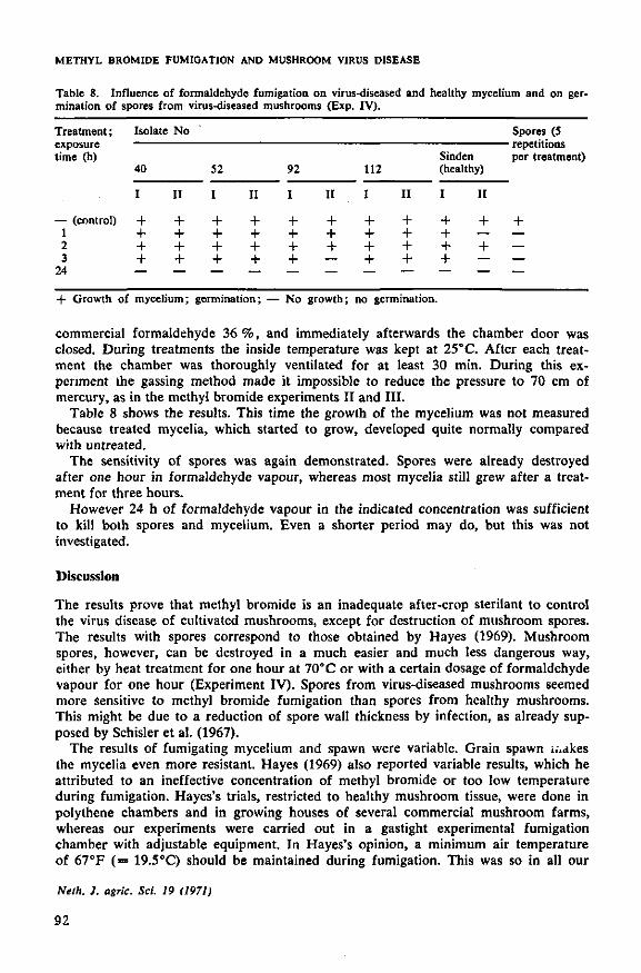
METHYL BROMIDE FUMIGATION AND MUSHROOM VIRUS DISEASE
Table 8. Influence of formaldehyde fumigation on virus-diseased and healthy mycelium and on germination of spores from virus-diseased mushrooms (Exp. IV).
Treatment;
time (h)
— (control) 1 2 3
24
Isolate No
40
I
+ + + +
II
+ + + +
52
I
+ + + +
II
+ + + +
92
I
+ + + +
II
+ + + —
112
I
+ + + +
II
+ + + +
Sinden (healthy)
I II
+ + + +
+ — + —
Spores (5
per treatment)
+ — — —
+ Growth of mycelium; germination; — No growth; no germination.
commercial formaldehyde 36 %, and immediately afterwards the chamber door was closed. During treatments the inside temperature was kept at 25°C. After each treatment the chamber was thoroughly ventilated for at least 30 min. During this experiment the gassing method made it impossible to reduce the pressure to 70 cm of mercury, as in the methyl bromide experiments II and III.
Table 8 shows the results. This time the growth of the mycelium was not measured because treated mycelia, which started to grow, developed quite normally compared with untreated.
The sensitivity of spores was again demonstrated. Spores were already destroyed after one hour in formaldehyde vapour, whereas most mycelia still grew after a treatment for three hours.
However 24 h of formaldehyde vapour in the indicated concentration was sufficient to kill both spores and mycelium. Even a shorter period may do, but this was not investigated.
Discussion
The results prove that methyl bromide is an inadequate after-crop sterilant to control the virus disease of cultivated mushrooms, except for destruction of mushroom spores. The results with spores correspond to those obtained by Hayes (1969). Mushroom spores, however, can be destroyed in a much easier and much less dangerous way, either by heat treatment for one hour at 70" C or with a certain dosage of formaldehyde vapour for one hour (Experiment IV). Spores from virus-diseased mushrooms seemed more sensitive to methyl bromide fumigation than spores from healthy mushrooms. This might be due to a reduction of spore wall thickness by infection, as already supposed by Schisler et al. (1967).
The results of fumigating mycelium and spawn were variable. Grain spawn i..akes the mycelia even more resistant. Hayes (1969) also reported variable results, which he attributed to an ineffective concentration of methyl bromide or too low temperature during fumigation. Hayes's trials, restricted to healthy mushroom tissue, were done in polythene chambers and in growing houses of several commercial mushroom farms, whereas our experiments were carried out in a gastight experimental fumigation chamber with adjustable equipment. In Hayes's opinion, a minimum air temperature of 67°F (= 19.5°C) should be maintained during fumigation. This was so in all our
Neth. J. agric. Sci. 19 (1971)
92
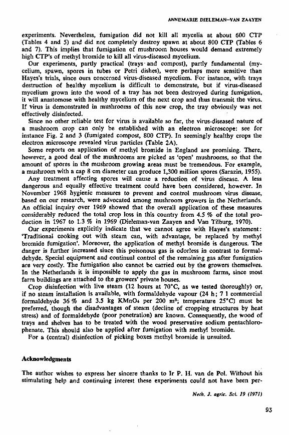
ANNEMARIE DIELEMAN-VAN ZAAYEN
experiments. Nevertheless, fumigation did not kill all mycelia at about 600 CTP (Tables 4 and 5) and did not completely destroy spawn at about 800 CTP (Tables 6 and 7). This implies that fumigation of mushroom houses would demand extremely high CTP's of methyl bromide to kill all virus-diseased mycelium.
Our experiments, partly practical (trays and compost), partly fundamental (mycelium, spawn, spores in tubes or Petri dishes), were perhaps more sensitive than Hayes's trials, since ours concerned virus-diseased mycelium. For instance, with trays destruction of healthy mycelium is difficult to demonstrate, but if virus-diseased mycelium grown into the wood of a tray has not been destroyed during fumigation, it will anastomose with healthy mycelium of the next crop and thus transmit the virus. If virus is demonstrated in mushrooms of this new crop, the tray obviously was not effectively disinfected.
Since no other reliable test for virus is available so far, the virus-diseased nature of a mushroom crop can only be established with an electron microscope: see for instance Fig. 2 and 3 (fumigated compost, 800 CTP). In seemingly healthy crops the electron microscope revealed virus particles (Table 2A).
Some reports on application of methyl bromide in England are promising. There, however, a good deal of the mushrooms are picked as 'open' mushrooms, so that the amount of spores in the mushroom growing areas must be tremendous. For example, a mushroom with a cap 8 cm diameter can produce 1,300 million spores (Sarazin, 1955).
Any treatment affecting spores will cause a reduction of virus disease. A less dangerous and equally effective treatment could have been considered, however. In November 1968 hygienic measures to prevent and control mushroom virus disease, based on our research, were advocated among mushroom growers in the Netherlands. An official inquiry over 1969 showed that the overall application of these measures considerably reduced the total crop loss in this country from 4.5 % of the total production in 1967 to 1.3 % in 1969 (Dieleman-van Zaayen and Van Tilburg, 1970).
Our experiments explicitly indicate that we cannot agree with Hayes's statement: 'Traditional cooking out with steam can, with advantage, be replaced by methyl bromide fumigation'. Moreover, the application of methyl bromide is dangerous. The danger is further increased since this poisonous gas is odorless in contrast to formaldehyde. Special equipment and continual control of the remaining gas after fumigation are very costly. The fumigation also cannot be carried out by the growers themselves. In the Netherlands it is impossible to apply the gas in mushroom farms, since most farm buildings are attached to the growers' private houses.
Crop disinfection with live steam (12 hours at 70°C, as we tested thoroughly) or, if no steam installation is available, with formaldehyde vapour (24 h; 7 1 commercial formaldehyde 36 % and 3.5 kg KMnCU per 200 m3; temperature 25°C) must be preferred, though the disadvantages of steam (decline of cropping structures by heat stress) and of formaldehyde (poor penetration) are known. Consequently, the wood of trays and shelves has to be treated with the wood preservative sodium pentachloro-phenate. This should also be applied after fumigation with methyl bromide.
For a (central) disinfection of picking boxes methyl bromide is unsuited.
Acknowledgments
The author wishes to express her sincere thanks to Ir P. H. van de Pol. Without his stimulating help and continuing interest these experiments could not have been per-
Neth. J. agric. Sci. 19 (1971)
93
METHYL BROMIDE FUMIGATION AND MUSHROOM VIRUS DISEASE
formed. The skilful technical assistance of Miss W. Buter, who carried out the fumigations, and of P. J. van Tilburg, who did the mushroom growing and mycelial work, are gratefully acknowledged.
The electron micrographs are from the Laboratory of Virology, Agricultural University, Wageningen, and the photographs from the Institute of Phytopathological Research, Wageningen, the Netherlands.
References
Dieleman-van Zaayen, A., 1968. Sporen van zieke champignons verspreiden de afstervingsziekte. Cham-pignoncuhuur 12 (6): 192.
Dieleman-van Zaayen, A., 1969. A virus disease of cultivated mushrooms in the Netherlands. Mushroom Sci. 7: 213-220.
Dieleman-van Zaayen, A., 1970. Means by which virus disease in cultivated mushrooms is spread, and methods to prevent and control it. MGA Bull. 244: 158-178.
Dieleman-van Zaayen, A. & J. H. M. Temmink, 1968. A virus disease of cultivated mushrooms in the Netherlands. Neth. J. PI. Path. 74: 48-51.
Dieleman-van Zaayen, A. & P. J. van Tilburg, 1968. De rol van gebruikt fust bij de verspreiding van de afstervingsziekte. Champignoncultuur 12 (10): 336-337.
Dieleman-van Zaayen, A. & P. J. van Tilburg, 1969. Gebruikt fust speelt nog steeds een rol bij de verspreiding van de afstervingsziekte. Champignoncultuur 13 (7): 264-265.
Dieleman-van Zaayen, A. & O. Igesz, 1969. Intracellular appearance of mushroom virus. Virology 39: 147-152.
Dieleman-van Zaayen, A. & P. J. van Tilburg, 1970. Afstervingsziekte: Enquete 1969. Champignoncultuur 14 (9): 293-294.
European and Mediterranean Plant Protection Organisation, 1961. Report of the working party on fumigation standards. HemmerI6, Petit & Cie, Paris.
Flegg, P. B., 1968. Methyl bromide and mushrooms. MGA Bull. 228: 582-584. Gandy, D. G., 1960. 'Watery Stipe' of cultivated mushrooms. Nature, Lond. 185: 482-483. Hayes, W. A., 1969. Methyl bromide as an after-crop sterilant. MGA Bull. 234: 240-243. Hayes, W. A. & P. E. Randle, 1968. The use of water soluble carbohydrates and methyl bromide in
the preparation of mushroom composts. MGA Bull. 218: 81-102. HoIIings, M., 1962. Viruses associated with a die-back disease of cultivated mushrooms. Nature, Lond.
196: 962-965. Hussey, N. W., I. J. Wyatt & J. J. Hesling, 1962. Entomology department report. Rep. Glasshouse
Crops Res. Inst. 1961: 63-64. Last, F. T., M. Hollings, & O. M. Stone, 1967. Some effects of cultural treatments on virus diseases
of cultivated mushroom, Agaricus bisporus. Ann. appl. Biol. 59: 451-462. Pol, P. H. van de & A. M. Mathot, 1961. The influence of commodity, packing material and the
properties of the fumigation chamber on the concentration of methyl bromide (MeBr) in the gas phase. Neth. J. agric. Sci. 9: 159-162.
Sarazin, A., 1955. The cultivated mushroom. Mushroom Growers' Association, London. Schisler, L. C , J. W. Sinden & E. M. Sigel, 1967. Etiology, symptomatology, and epidemiology of
a virus disease of cultivated mushrooms. Phytopathology 57: 519-526. Sinden, J. W. & E. Hauser, 1950. Report on two new mushroom diseases. Mushroom Sci. 1: 96-100. Spradling Chidester, M., 1939. Further studies on temperatures necessary to kill fungi in wood. Proc.
Am. Wood Preserv. Ass. 35: 319-324.
Neth. J. agric. Sci. 19 (1971)
94
4.4 Various aspects of control, and practical results
Paper:
IX Spread, prevention and control of mushroom virus disease
Mushr. Sci. 8: in press (1972).
95
Spread, prevention and control of mushroom virus disease
SUMMARY
In an isolated growing-room, mushroom virus disease was spread by viable mycelium and by spores from diseased mushrooms, as reported earlier by several authors. The trials demonstrated the importance of time of infection and the relative unimportance of the amount of inoculum. Intensive research into this disease resulted in a list of measures to prevent or control it, applicable to Dutch mushroom farms but, with slight adaptation, also suited for more-zone systems. General implementation of the measures in the Netherlands reduced the total crop loss considerably.
Trials to obtain virus-free material by various techniques (heat treatment at 33 C, repetitive transfer from the outermost periphery of mycelial cultures) failed. Better results may be expected from a wild mushroom with a high tolerance, or even resistance, to virus disease.
INTRODUCTION
In recent years the Dutch mushroom fanning has suffered severe losses from a
virus disease of the cultivated mushroom, Agaricua b-ispovua (Lange) Sing..
Hollings (1962) detected virus particles in association with a similar dis
ease in England.
Several authors (Gandy, 1962; Schisler et al., 1967; Dieleman-van
Zaayen, 1969) have reviewed the various types of symptoms. Frequent symptoms
are bare zones where the mycelium grows only slightly, if at all, into the
casing soil, and mushrooms of poor quality standing loose in the casing soil,
often with a long stipe and an off-white cap that matures too early. Usually
crop yield is seriously reduced.
Since Dutch research into the disease started in 1966, usually three
types of virus particles, often in combination, have been observed in cell-
free preparations from diseased mushrooms: isometric particles with diame
ters of 25 and 34 nm, and elongated particles with rounded ends, 19 x 50 run
(Dieleman-van Zaayen and Temmink, 1968). We have isolated these particle
types also from samples of diseased mushrooms from Belgium, Bulgaria, Germa
ny, Italy, Switzerland and the United States. When a cell-free virus prepa
ration was injected into the stipes of young mushrooms on a growing tray,
96

symptoms developed a few weeks later in mushrooms of a subsequent flush;
from these fruiting bodies, the three types of virus particles could be re-
isolated, predominantly the 34-nm particles, which also prevailed in the in
oculum (Dieleman-van Zaayen and Temmink, 1968). This provided the final proof
that the disease was indeed caused by virus particles isolated from diseased
mushrooms. In 1968 Hollings also observed the 34-nm virus particles, so far
overlooked most probably because of their instability.
A survey among the more than 1 000 Dutch growers showed that in 1967
and the first half of 1968, one out of three mushroom farms was contaminated;
on these farms average yield loss was 15$. Thus in 1967 in the Netherlands,
4.51 or about 790 000 kg of mushrooms were lost, total yield being 17.5 mil
lion kg.
The disease is spread by viable mycelium (Gandy, 1960a) and by spores
from diseased mushrooms (Schisler et al., 1967). Spread with spores was con
firmed by infection experiments (Dieleman-van Zaayen, 1970) and by the detec
tion of virus particles in ultrathin sections of spores from infected mush
rooms (Dieleman-van Zaayen, 1972).
Intensive research has been done to control the disease. Two aspects of
control are dealt with here:
a. of interest to mushroom growers, the drawing up from research data of a
list of control measures;
b. of interest to spawnmakers, preparation of virus-free material, for in
stance by heat treatment.
MATERIAL AND METHODS
Research in an isolated growing-room
Most of the virus growing trials were in a growing-room of the former build
ing of the Mushroom Experimental Station at Horst (L.). To prevent the other
growing-rooms from becoming infected, certain precautions and rules were
kept. These included: a closed chamber of hardboard in front of the growing-
room so that one could not proceed directly from the growing-room into the
working-corridor. In this chamber, where clothing and footwear could be
changed, facilities were available for disinfecting hands, footwear and
tools, and for weighing the mushrooms picked in the growing-room. The mush
rooms were afterwards packed for destruction. The growing-room was tended
by one person, who never entered the other growing-rooms of the Experimental
97

Station. Generally visitors were not admitted to the virus-contaminated
growing-room. The mushrooms were harvested as closed mushrooms and spore
filters were placed in all ventilation openings. Strict hygiene was observed.
Picking was restricted to a maximum of three flushes.
The growing-room contained on either side of the centre path four
shelves, one above the other. Each shelf was divided into four plots, each 2
of 2 m . There were thus 32 plots in all and the total growing-area was 64 2
m . Trays could be placed in the room for extra trials.
Mushrooms were grown by the single-zone system. Shelves and trays were 2
always filled with compost, about 100 kg/m , from the Co-operative Composting Enterprise at Ottersum. Compost was pasteurized (up to 60°C) for about
2 10 days. The spawning rate was 375 g/m of commercial spawn (snowwhite
strains) for the plots and about 40 g per tray, unless otherwise stated.
Inoculation with virus-infected material
Isolation of mycelium; the Virus Collection
Various virus-infected and some uninfected mycelial cultures were obtained
by isolating tissue fragments from samples of fruiting bodies sent in by
growers to be tested for virus (Section: Detection of virus particles, page
99). The fragments were plated on 2% Biomals agar (a malt product) at 25°C.
After transfer to tubes, the mycelial cultures were stored at 10-12UC, and
transferred to a fresh nutrient medium every four months.
In that way a collection was established of virus-infected and some
uninfected mycelia, about 70 cultures in all. About 50 were used frequently
in several experiments. The mycelia were numbered serially by date of arriv
al of the mushroom samples to be tested; from the corresponding mushroom
samples, the number of virus particles was known. The collection of these
mycelial cultures is referred to here as 'Virus Collection1.
Preparation of inoculum
Preliminary trials are described elsewhere (Dieleman-van Zaayen, 1970). They
used compost with virus-infected mycelium from a contaminated mushroom farm
as inoculum. This dubious procedure was gradually improved. At first several
virus-infected mycelial cultures from the Virus Collection were transferred
to and multiplied on peak-heated, ground and sterilized compost in Petri
dishes. Mushroom mycelium grew well on this medium. On the day of inocula
tion, mycelium was carefully scraped from the compost medium; inevitably the
98

inoculum contained some nutrient medium.
Soon this inoculum was replaced by 'infected spawn' (Schisler et al.,
1967). The material was easy to handle at inoculation. It was prepared in
the following way:
to 1 kg sorghum grains, boiled in 1 litre water for 5-10 min, 5.3 g chalk
(CaCO,) and 21.2 g gypsum (CaSO.) were added (Stoller, 1962). The mixture was
thoroughly stirred and divided among small flasks. These were closed with
cotton-wool plugs and sterilized for 2 h at 120°C. After cooling, the flasks
were vigorously shaken and a piece of agar with infected mycelium was trans
ferred to the grains. Spawn running was at 25°C for about 3 weeks; twice dur
ing this period the flasks were vigorously shaken. The 'infected spawn1 was
then ready for use.
Inoculation
On the day of inoculation the desired amount of inoculum (ground compost with
mycelium, or grains with mycelium) was weighed out and put in the centre of a
plot or tray in the isolated growing-room, at a depth of 5 cm in the compost.
Inoculation was always done before casing.
Although inoculation with spores from diseased mushrooms is certainly
possible (Schisler et al., 1967; Dieleman-van Zaayen, 1970), it was usually
done with infected mycelium, which was easier to handle and gave more reliable
results.
Detection of virus particles
Fruiting bodies were diagnosed for virus particles by a method developed
by Dieleman-van Zaayen and Temmink (1968). Mushrooms, stored at 4°C for
3-10 days, were rinsed under running tap-water and blotted with filter
paper. After removal of the lower parts of the stipes, the fruiting bodies
were ground for 2 min in a Waring Blendor in 30 ml 0.033 M phosphate buffer
containing 0.11 thioglycollic acid, final pH 6.8, per 10 g tissue. Homoge-
nate in 50-ml portions was subjected to ultrasonic treatment (Hollings et al.,
1965) with a Kerry Vibrason cell disrupter (probe diam. 0.9 cm, output 50 W)
for 10 min. During treatment, the thick-walled glass tube (internal diam.
3.4 cm), which contained the homogenate, was kept at 4°C. A procedure modi
fied from Kitano et al. (1961) was used on the sonicated homogenate. To one
volume of homogenate, an equal volume of 2.5 M potassium phosphate buffer
pH 6.8 and 0.8 volume of a mixture (1:2) of 2-butoxyethanol and 2-ethoxy-
99
ethanol, were added. The phosphate buffer was prepared by mixing hot solu
tions of 2.5 M KH-PO. and 2.5 M KJiPO. until pH reached 6.8. The components
were mixed gently by hand. Centrifugation at 1000 x g for 5 min resulted in
partition of an aqueous bottom phase, an organic top phase and a gelatinous
interphase. After decantation of the liquids the interphase was resuspended
in 25-30 ml 0.033 M phosphate buffer, pH 6.8, per 100 ml sonicated homoge-
nate. Centrifugation at 5000 x g for 10 min gave a clear supernatant, which
was ultracentrifuged at 105 000 x g for 60 min. As much as possible of the
procedure was at 4°C.
The pellet resulting from ultracentrifugation was resuspended in 0.5 ml
0.033 M phosphate buffer pH 6.8 per 100 ml crude homogenate. The suspension
was kept for some hours at 4 C and sometimes finally centrifuged at 5000 x g
for 5 min but not in later trials because of undesired precipitation of vi
rus.
After negative staining with 21 phosphotungstic acid pH 6.0, the virus
particles were observed in a Siemens Elmiskop 1 or a Philips EM-300 electron
microscope.
Material and methods will be detailed under the individual trials.
EXPERIMENTS AND RESULTS
The isolation of the virus growing-room
During trials lasting about 3j years in the isolated growing-room of the for
mer building of the Experimental Station, not one of the other growing-rooms
became infected with the disease. Although such a well isolated growing-room,
tended by only one person, cannot be compared with a mushroom farm, it still
indicates what control measures are possible.
Infection experiments
Earlier trials on infection in the isolated growing-room had already confirm
ed that the disease is very easily spread by viable mycelium or by spores
from diseased mushrooms (Dieleman-van Zaayen, 1970). Virus-infected mycelium,
left behind on or in wood after inadequate disinfection at the end of a crop,
could anastomose with healthy mycelium in a following crop and thus transmit
the*- virus.
Various authors, for instance Rasmussen et al. (1969), stressed the
100

danger of infection between spawning and casing. Below are described two of
the trials to find the most dangerous time of infection and the influence of
amount of inoculum.
Trial 7: time of infection
A constant amount of inoculum was introduced in seven plots of one layer (a
layer consists of two shelves next to one another) on the day of spawning
(tQ), every 2 days after spawning(t2> t^, t&, tg) or 12 days after spawning
(t 1 2 ). A control (C) was not inoculated.
The remaining plots were used for other tests. This trial was carried
out in layers since there were rather great differences in climate between
the layers at the former building of the Experimental Station. Because of
this, each plot of one layer was treated differently, so that each treat
ment occurred once in each layer. The various treatments were divided ran
domly over the plots of a layer. The layout is shown in Fig. 1. The plots
were inoculated in the centre with 2.5 g 'infected spawn* consisting of a
mixture of Virus Collection No 92 and 94.
All inoculated plots showed symptoms of the disease, and virus parti
cles, of 25 and 34 nm mainly, could be isolated from the mushrooms. The con
trols remained healthy.
Uncut mushrooms from each plot were weighed separately. After 25 days
of harvesting (three flushes), the growing-room was cooked out, as is common
procedure in virus experiments. The test was in quadruplicate so that the re
sults could be processed by variance analysis, but this proved completely
superfluous: the differences were striking. The yields of four plots that 2
had received a treatment at one time were now added, and yields/m were adjusted (-201) as if the mushrooms had been cut. For the missing treatment t in layer III, which plot was used for other trials, the expected yield was extrapolated from all other yields by the missing-plot technique.
Results are given in Table 1 and plotted in Fig. 2. Note that the con-
0
layer I (top)
ISI UyerU layer in layer ET(bottom)
c
«0
«6
X ' ,2
ii! c
X
f 8
•o
«12
<2
* These plots were used for other experiments. For the same reason,from layer H, t 0had to be omitted.
Fig. 1. Layout of Trial 1 on time of infection.
101
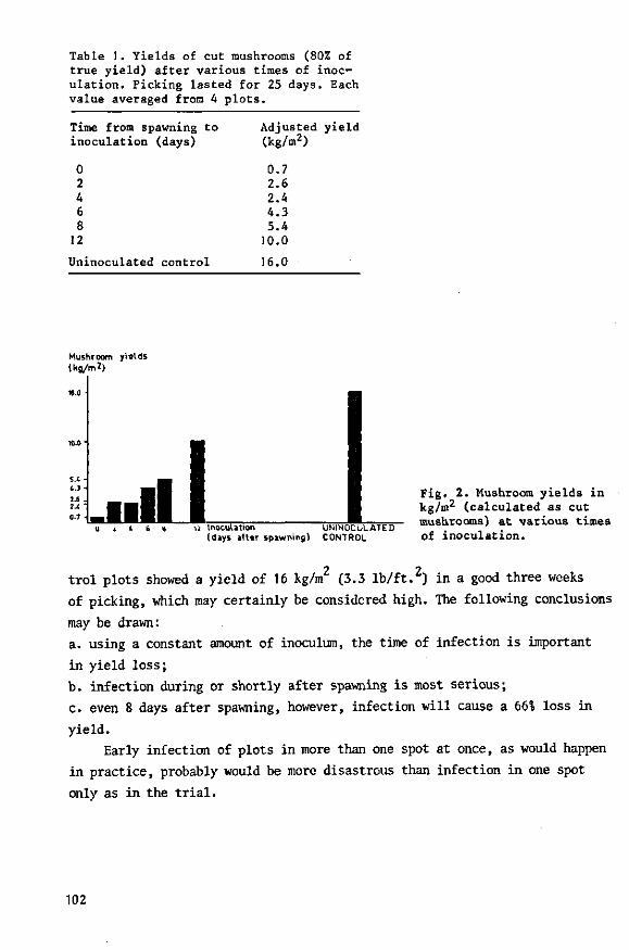
Table 1. Yields of cut mushrooms (80% of true yield) after various times of inoculation. Picking lasted for 25 days. Each value averaged from 4 plots.
Time from spawning to inoculation (days)
0 2 4 6 8
12
Uninoculated control
Adjusted yield (kg/m2)
0.7 2.6 2.4 4.3 5.4
10.0
16.0
Mushroom yialds (kg/rr.2)
2 i 6 » 12 Inoculation UNINOCULATED (days after spawning) CONTROL
Fig. 2. Mushroom yields in kg/m2 (calculated as cut mushrooms) at various times of inoculation.
trol plots showed a yield of 16 kg/m2 (3.3 lb/ft.2) in a good three weeks
of picking, which may certainly be considered high. The following conclusions
may be drawn:
a. using a constant amount of inoculum, the time of infection is important
in yield loss;
b. infection during or shortly after spawning is most serious;
c. even 8 days after spawning, however, infection will cause a 66% loss in
yield.
Early infection of plots in more than one spot at once, as would happen
in practice, probably would be more disastrous than infection in one spot
only as in the trial.
102
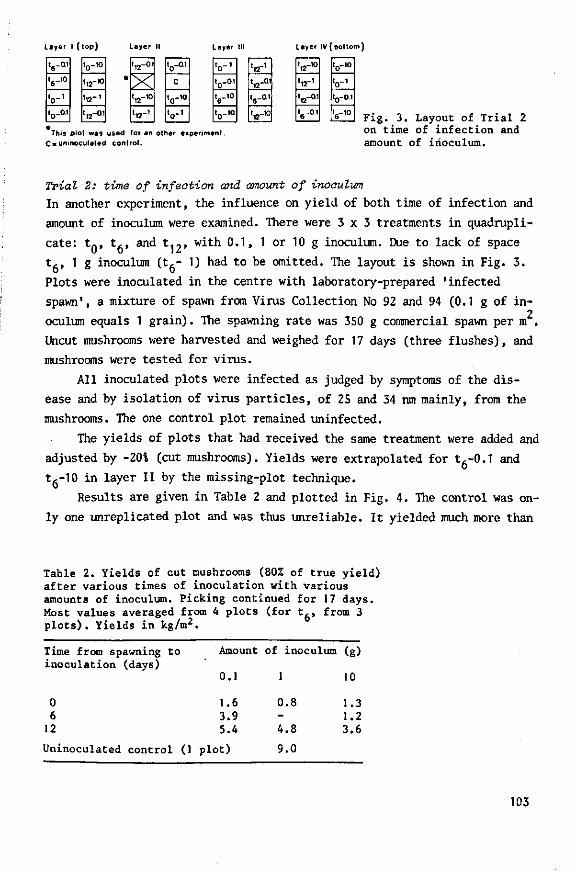
Layer I ( l o p ) Layer II Layer III
This plot was used for an other experiment.
C = uninoculated control .
Layer IV ( b o t t o m )
1,-0.1
1,-10
V 1
t 0 -at
t 0 - io
t12-10
'12-'
t„-Q1
t12-o.i
X t,2-io
t1 2 - i
' o - 0 1
c
t0-1O
V 1
V 1
t0-o.i
Sr10
t0-1O
'12-' t,2-01
t 6 - a .
tc-10
.12-io
t12-i
to -01
.,-0.1
'o-'° V t0-o.i
t5-.o Fig. 3. Layout of Trial 2 on time of infection and amount of inoculum.
Trial 2: time of infection and amount of inoculum
In another experiment, the influence on yield of both time of infection and
amount of inoculum were examined. There were 3 x 3 treatments in quadrupli
cate: tn, t,, and t12, with 0.1, 1 or 10 g inoculum. Due to lack of space
tj., 1 g inoculum (t,- 1) had to be omitted. The layout is shown in Fig. 3.
Plots were inoculated in the centre with laboratory-prepared 'infected
spawn', a mixture of spawn from Virus Collection No 92 and 94 (0.1 g of in-2
oculum equals 1 grain). The spawning rate was 350 g commercial spawn per m .
Uncut mushrooms were harvested and weighed for 17 days (three flushes), and
mushrooms were tested for virus.
All inoculated plots were infected as judged by symptoms of the dis
ease and by isolation of virus particles, of 25 and 34 nm mainly, from the
mushrooms. The one control plot remained uninfected.
The yields of plots that had received the same treatment were added and
adjusted by -20% (cut mushrooms). Yields were extrapolated for tg-0.1 and
t6-10 in layer II by the missing-plot technique.
Results are given in Table 2 and plotted in Fig. 4. The control was on
ly one unreplicated plot and was thus unreliable. It yielded much more than
Table 2. Yields of cut mushrooms (80% of true yield) after various times of inoculation with various amounts of inoculum. Picking continued for 17 days. Most values averaged from 4 plots (for t,, from 3 plots). Yields in kg/m2.
Time from spawning t o i nocu l a t i on (days)
0 6
12
Uninoculated con t ro l (1
Amount
0.1
1.6 3.9 5.4
p l o t )
of inoculum
1
0 .8 -4 .8
9.0
(g)
10
1.3 1.2 3.6
103
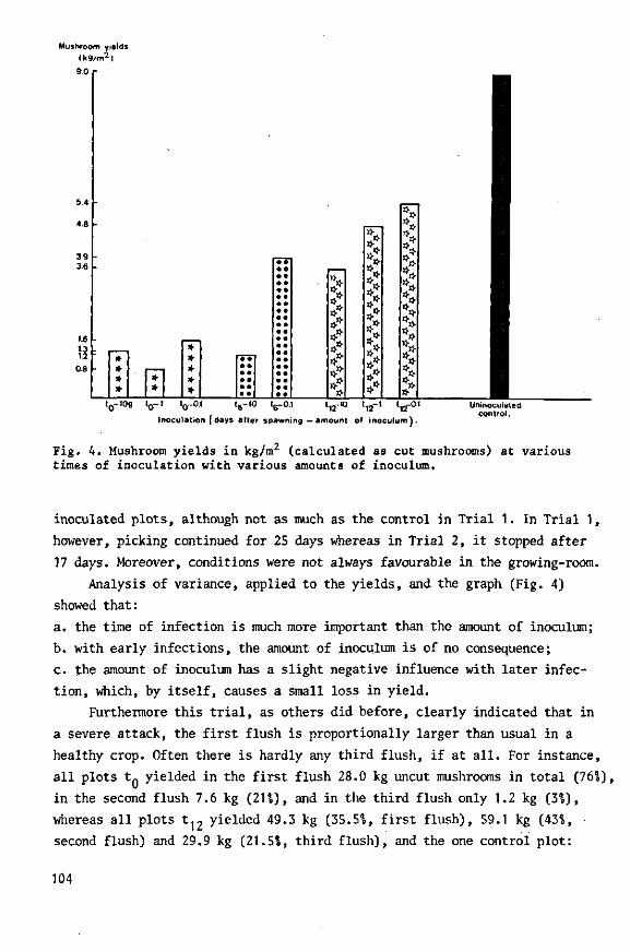
Mushroom yields <k9/m2)
5 4
48
39 3.6
* * * I 1 *
* * * * * * I.-lOg t n - l U-0.1 tR-10 tR-0.t t„-K) 0 - i .0-u.i iR- iu iR-u.i i,2-iu i ,2-l t12-01
Inoculation (days after spawning —amount of inoculum).
Uninoculated control.
Fig. 4. Mushroom yields in kg/m2 (calculated as cut mushrooms) at various times of inoculation with various amounts of inoculum.
inoculated plots, although not as much as the control in Trial 1. In Trial 1,
however, picking continued for 25 days whereas in Trial 2, it stopped after
17 days. Moreover, conditions were not always favourable in the growing-room.
Analysis of variance, applied to the yields, and the graph (Fig. 4)
showed that:
a. the time of infection is much more important than the amount of inoculum;
b. with early infections, the amount of inoculum is of no consequence;
c. the amount of inoculum has a slight negative influence with later infec
tion, which, by itself, causes a small loss in yield.
Furthermore this trial, as others did before, clearly indicated that in
a severe attack, the first flush is proportionally larger than usual in a
healthy crop. Often there is hardly any third flush, if at all. For instance,
all plots tQ yielded in the first flush 28.0 kg uncut mushrooms in total (76%),
in the second flush 7.6 kg (21%), and in the third flush only 1.2 kg (3%),
whereas all plots t 1 2 yielded 49.3 kg (35.5%, first flush), 59.1 kg (43%,
second flush) and 29,9 kg (21.5%, third flush), and the one control plot:
104

9.4 kg (411), 8.1 kg C36%3, and 5.2 kg (231) respectively.
Interpretation of the results Infection during spawning or shortly afterwards can happen, for instance,
when the spawning machine has been inadequately disinfected, but more likely
because there is still viable, infected mycelium present in or on the wood
of shelves and trays. Both cooking-out with live steam for extended periods
and fumigation with 600 CTP (or more) methyl bromide (Dieleman-van Zaayen,
1971) were found to be inadequate to destroy the virus-infected mushroom
mycelium, grown into the wood of trays and shelves. If not destroyed, it
anastomoses with healthy mycelium of the next crop and thus causes early
virus infection and severe damage.
Another possible source of infection is the presence of virus-infected
spores on the farms, which fall on the compost during or after spawning and
germinate there. Electron-microscopy of ultrathin sections of spores re
vealed up to some hundred virus particles per section (Dieleman-van Zaayen,
1972). According to Sarazin (1955), a (healthy) mushroom with a cap 8 cm
diameter can produce 1 300 million spores. The number of virus particles in
spores from one fruiting body must be vast! One of the most characteristic
symptoms of virus-diseased mushrooms is early maturity; as soon as the caps
open, spores can be liberated. Moreover spores from diseased mushrooms often
germinate better and faster than uninfected spores, as was noted by Schisler
et al. in 1967, and confirmed later (Dieleman-van Zaayen, 1970). In ultra-
thin sections of germ tubes from infected spores, many virus particles were
observed. These germ tubes can anastomose with healthy mycelium and thus vi
rus can be transmitted.
If virus-infected spores fall on the compost during spawning, this can
be compared with infection about 8 days after spawning since most spores
have germinated by then. The loss of yield may then amount to no less than
66?0 in relation to the uninoculated control (Table 1; Fig. 2 ) , at least af
ter inoculation in one place only. But spores in particular can turn up in
the compost in various places at once and this may cause a real disaster.
From this too, it is again apparent that infection during or shortly
after spawning, whether this occurs with viable infected mycelium or with
infected spores, will have serious consequences.
105

Table 3. Measures to prevent or control mushroom virus disease1
If the disease is not yet present If the disease is already present
Cook out (growing-room and) compost at the end of a crop for 12 h at 70 C. After emptying take away the compost quickly.
Spray the wood with 2% sodium penta- Dip the wood into 4% sodium penta-chlorophenate to which 0.5-1.0% chlorophenate to which 0.5-1.0% Na2C03 (wetting agent) has been Na2C03 (wetting agent) has been added. After drying for about 5 h, added. After drying for about 5 h, spray with a large amount of water. spray with a large amount of water.
Before filling, insert spore filters (e.g. glass fibre 2 cm thick). After casing, these filters should be renewed once or twice according to the amount of dust in the air. Use a fan for extracting air.
Place plastic partitions in the working corridor between the rooms. If this is not possible, enter the growing-rooms from the back until picking starts.
Just before spawning and thereafter twice a week, disinfect the working corridor with a 2% solution of commercial formaldehyde (2 litres comm. formaldehyde 36% in 100 litres of water).
Immediately after spawning use a pesticide against flies, then cover the compost with paper. Wet the paper twice a week with a 0.5% formaldehyde solution (0.5 litre comm. formaldehyde 36% in 100 litre water per 180 m2
growing area). Repeat till a few days before casing. Moisten the paper before removing it carefully.
Pick the mushrooms when still closed.
Use" new or thoroughly disinfected picking baskets only.
Quickly remove cuttings and litter and destroy.
Keep each room as a separate entity with separate equipment (e.g. clothes, shoes, steps, buckets, picking knives); in brief, practise hygiene. Keep doors of contaminated growing-rooms onto the working corridor closed.
Have a short picking period only (not more than A weeks).
The entire farm and its surroundings should be clean and stay so. Clean the manure yard with formaldehyde before the arrival of manure or casing soil. Clean the working corridor thoroughly, especially the service pipes (heating, steam pipes).
Keep mats with formaldehyde outside the rooms to disinfect shoes.
Machines (e.g. spawning machine, press) should be disinfected with a 2% solution of comm. formaldehyde.
1. Issued by: Mushroom Experimental Station, Horst (L.); and Institute of Phytopathological Research, Wageningen, the Netherlands.
106

Prevention and control
After the spread of mushroom virus disease by viable infected mycelium and
by spores from diseased mushrooms had been confirmed, and after it had been
demonstrated that early infection is most dangerous, measures to prevent or,
if necessary, to control the disease were drawn up, and recommended to Dutch
growers in November 1968. The measures were based mainly on the information
obtained from the trials in the isolated growing-room. The measures are
listed in Table 3, the left column giving preventive, and the right control
measures. The methods are not listed in order of importance, but in chro
nological order.
The measures are, of course, applicable to Dutch conditions, where cul
tivation takes place on shelves by the single-zone system. In most farms,
growing-rooms are adjoining and leading into a covered working corridor that
can be a gathering place for spores (hence the measure on plastic partitions
in the corridor; this is an imitation of the isolating chamber in front of
the virus growing-room of the Experimental Station). The crops in the dif
ferent growing-rooms are not synchronic. A growing-room with (diseased
open) mushrooms to be harvested may be situated next to a growing-room to
be spawned. Therefore, once a farm is contaminated, control of the disease
is not easy though feasible. The larger the farm, the harder is control.
The measures are intended:
a. to kill viable infected mycelium, left behind in or on the wood of
shelves and trays after a crop. Research into this problem (Dieleman-van
Zaayen, 1970) led to the measure of applying, after every crop, wood preserv
atives based on sodium pentachlorophenate (SPCP, e.g. Santobrite). Cooking-
out can then be reduced to 12 h at 70 C (158 F ) , before emptying the room,
merely to kill mushroom mycelium, spores and harmful organisms in the com
post which is afterwards used for other horticultural purposes. Needless to
say, the temperature in the compost should be 70°C for 12 h. Tests proved
these to be the right exposure time and temperature. Other tests showed,
that it is essential to treat the wood with 44 SPCP in case of virus disease;
24 SPCP is merely adequate as a preventive treatment.
b. to prevent spread of the disease by spores. Measures include:
1. picking of mushrooms as much as possible in closed condition;
2. use of spore filters (2 cm-thick layer of glass fibre is suitable;
3. covering of the compost, immediately after quick spawning with a disin
fected spawning machine, with paper, which is sprayed twice a week with a
107

0.5% solution of commercial formaldehyde until casing. This percentage is
sufficient to kill mushroom spores, but the mycelium growing in the compost
underneath is not harmed, as tests indicated;
4. strict hygiene.
It is not sufficient to implement one or two control measures only;
the whole list of measures must be implemented to free a farm from virus.
For more-zone systems, only slight adaptation of these measures would
be needed.
Reduction of crop loss
In the Netherlands, an official enquiry for 1969 in which 971 of the growers
participated, showed that implementation of all the measures considerably
reduced crop loss: from 4.5% of the total production in 1967 to 1.31 in 1969
(Table 4 ) . Although the enquiry over 1970 is only partly processed (up to
801), preliminary results indicate exactly the same figure, 1.3%, for total
crop loss; the annual production amounted to 30 million kg cut mushrooms.
The total crop loss due to virus disease has already decreased from 4.5% to
1.3%, but in future this figure might fall even lower when the new return
able picking boxes are centrally disinfected. Picking boxes used a number
of times by different growers and redistributed to others by regional auc
tions have earlier been shown to contribute to spread of the disease through
out the country.
The conclusion is: prevention and control of virus disease is certainly
possible. The measures have proved to be effective. It should be realized,
however, that this requires continuous effort from every grower.
Table 4. Commercial losses from mushroom virus disease in the Netherlands. Data from official enquiries and a random test among more than 1 000 Dutch growers.
Year
1967 1968
Contaminated farms (%)
>30 25.1
Avg crop loss per Total crop contaminated farm loss (%) (%)
15.0 16.7
Control measures (November 1968) 1969 11.2 11.4
4.5 4.2
1.3
Annual production (kg)
Crop loss (kg)
17.5 million 787,500 20 million 840,000
23 million 299,000
108

Trials to obtain virus-free material
Heat treatment of mycelium
Heat treatment Virus-infected mushroom mycelium often grows slowly and de
generates on a nutrient medium but Gandy (1960b) reported that treatment at
33 C resulted in most cases in better and faster growth. Presumably this
treatment, which lasted two weeks (Gandy and Hollings, 1962), freed the fun
gus from virus. However heat-treated mycelium was never tested for virus
particles.
To verify this assumption the Virus Collection, including some healthy
mycelia, was kept in an incubator:
a. for 3 weeks at 33 C: 36 different mycelia including 10 uninfected cultures;
b. for 3 weeks at 33 C: other material from the same 36 mycelial cultures;
a. for 2 weeks at 34 C: the same 36 cultures plus 22 virus-infected and 8
uninfected mycelia (including 2 wild strains).
growth increase of heal — treated mycelium
50 -
20 I 1 1 1-4 0 50
gtowth of
Fig. 5. Differences in growth (mm) between heat-treated and untreated mycelial cultures against growth rate of the untreated cultures. Heat treatment was for 3 weeks at 33°C or for 2 weeks at 34°C. Regression equation: y - -0.46a; + 34.6, with
• .• r « -0.61 (significant at 1% level). Means:
, • , growth increase • 8.2 mm; so so growth rate of untreated
untreated mycelium C u l t u r e m 5 7 . 7 mm.
109
Fig. 6. Heat treatment (2 weeks at 30 C) of virus-infected mycelial cultures No 29, 53 and 54 on 2% Biomals agar. Left untreated cultures; right the same cultures after treatment. All transfers were made on the same day.
For this purpose transfers of the mycelial cultures were made to 4 test
tubes with 1% Biomals agar per isolate; the tubes were kept at 25 C. Five
days later 2 tubes were incubated at 33°C or 34°C for three or two weeks. The
other 2 tubes remained at 25°C as controls. After incubation, all tubes were
again kept at 25°C, and about 5 days later all cultures were transferred to
a fresh nutrient medium in tubes. Then the growth in length (mm) of the co
lonies was measured after 20 days. The two replicates were averaged and
growth increase or decrease in relation to control was calculated.
The results are shown in Fig. 5. The negative relationship between
growth increase of heat-treated mycelia and their growth rate when untreated,
can be expressed by the regression equation y = -0.46a; + 34.6 (r • -0.61;
significant at 11 level), represented by the regression line in the graph.
The equation was obtained by averaging the regression equations of the in
dividual treatments. Although a certain relationship could be demonstrated,
it is clear that the results had a wide spread. Replicates often differed
widely. In general, slowly growing cultures displayed a much better and
faster growth after heat treatment (Fig. 6 ) , whereas mycelia with a good or
excellent growth did not react or deteriorated with the treatment. This
110

Table 5. Difference in growth of mycelial cultures, after various treatments (d), from controls (25 C, last column). Averages of two colonies on 2% Biomals agar in test tubes.
Culture No Growth
2 wk1
-20°C
Virus—infeoted I 7
92 123 135 154
Healthy 37 43
130 152
1. wk= weeks
-10 t2
-23 - 1 - 3
t
+ 2
t + 4 -13
increase or decrease
2 wk 28°C
- 4 + 1 - 2 -11 + 5 - 7
- 8 0
+ 3 + 1
2 wk 30°C
- 2 + 15 + 16 + 2 + 14 + 18
-11 + 11 - 4
0
2. t» culture did not survive
2 wk 33°C
+ 10 + 17 +24 + 2 +24 + 18
+ 19 + 15 + 7 + 3
(mm)
3 wk 33°C
+ 10 +20 + 19 +22 + 14 +31
+ 8 + 8 + 1 + 14
2 wk 34°C
- 3 + 7 +39 + 16 + 12 +27
-18 -
+ 8 - 9
2 wk 35°C
0
+ + 3 + 7 + 5 + 9
+ t -
-22
Growth of control (mm)
60 49 43 39 47 32
63 80 58 44
applies to uninfected and infected cultures, and to wild strains.
Further treatments:
d. Material from 6 infected and 4 uninfected mycelial cultures was subjected
to one of the following treatments in duplicate: for 2 weeks at -20°C; 2
weeks at 28°C; 2 weeks at 30°C; 2 weeks at 33°C; 3 weeks at 33°C; 2 weeks at
34°C; and 2 weeks at 35°C. Controls were kept at 25°C.
After the treatments, the cultures were again kept at 25 C and 5 days
later they were transferred to a fresh medium. Growth rates were measured
on agar in test tubes 20 days after transfer. Averages were calculated; the
growth increases or decreases of the treated mycelia in relation to the con
trols are given in Table 5. Treatments at -20°C and 28°C did not influence
the mycelial growth rate; -20°C even killed some cultures, as did 35°C.
Some isolates (No 7, No 43) seemed to be more sensitive than others. Incu
bation at 30-34 C, however, often resulted in growth increase although with
great irregularity. Note that some of the uninfected mycelia showed a simi
lar trend.
Tests for virus particles To test for virus particles in mycelium,
Hollings et al.* (1965) suggested direct observation of sonicated mycelium
with the electron microscope. In our experience, this method is unsatisfac-
111
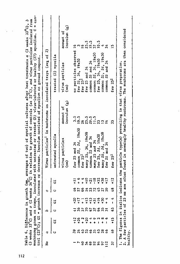
I a
-Q a o o CJ
• u y-* *H II CJ
O T> O oo tu CO U '
to fi
10 I-I 3
0) CO i-t cj - H o> 3 if
to >, <n » g 53 -H r -
4 J a CO 0) 0)
43 « ^^ M O a>o
<H tN to
to to 0) ^ ^ u 3 co
. - I o 3 M CJ 4J
a t - l o cfl CJ
•r-t i-l >M a) o o ss CM O O l-i
00 o o 3 u .u
a <M O O - H
•U CO a) CD i-l 00 01 to u u oi a > •* ct)
»c_> Eo B -*
* w CO
J3 CO t! •* 3 a) 0 HJ
K > co
CM a ^
••H
O cj - a a a o> at u oi ^ -> <w o < M O • H co P co
» to vo ^
ai
01 0)
CD CO
43 4J
01 T l
- c 3 O M 00
v ^ Q)
0)
0
0)
U 0) CO
0) 43 M U CJ
• i - l 01 3 -a
TJ H 01 O
01 o
S
T3 0) 4J
01 M
O
3 O o o
cu o >^ s •o CU
• u CO 0) 14 * J
c 3
M CO O.
to 3 -M i
> -
<*-t
o
u to
t o 4 3
>% -u to 3 M o 4 J U
oo c O II
1 o o v< • » o o , - v
o too B m O CM
o v^ 1-1
4 3 . - 1 CO O 3 M B u
r O
«
o a
v o c o v o t n o x r - o - a ' - * i n —. _ CM - . CO - - CO —
•o <u > M Ol O CO LO
43 X 0 o»
CO 01 «
I-I <r o CO
o o UO t o X X
o \ O CT» -a- — m — •* co x co • . CTl •*
- * -O <J- < - "^ -o CO C CO M C
co » CO TJ •» -3" •* C t l i LfJ CO tQ UJ CO C^ eg C\3 Cv]
U t o CO eg Q,
3 O a) C tw
t o i n CM <N
C tn O tN o o
3 3 « E 3 B tt oi a) o o a) o o
tw CH cj o CM o o
t / i m t o i n i n
C O O » v O c O O » ~ f O C O - » CO — t N c S C S " < N — — co
o i n
X o> ~~
M
• * M I
CO t<3
•o -a to CO CM
OJ
o o m o m X -3- m x
cy> co K e n — - » a i —
T3 CO — ~ * * c; * co
- 3 - t o T > " V f CO C * « 1 )
i n t o c j g * CM " C O
i n i n •> t o in c m t N c j t N i n c M t o C-4 o
3 I 0) o I H CJ
CM
3 0)
«-(
O < N CM
a B c 3 a 3 CO O CO 0) CO 0) B CJ S CH S <H
m CM
~ v £ > - t f O — CO N CO r s - . — CM —. CO CO — — + + + + + 1 + + +
v O r ^ ^ o - 3 - c o c M i n r ^ o
o r ^ — > m i n — v o o ~ * CM ^^ — * - CO «-" CO + + + + + + + + +
« o r ^ o r - ~ r ^ < f " i n - ~ ^ • c o i n r ^ t o c o t o c o c o
C M v O C N O - a - C y i C M C O v O —< CM CM
+ + + + + + I + I
t r i < i - i n c o v O L n c o o o ^ o a\ n c N - t f i n ^ t o j - c M < t ^ •
r ~ o - 3 - a \ C M - t f r - C M • j i n >o o i en en -
01 u tu
TJ
o cj
C o)
3 0)
"> H . 4 J CO
•u o
C J3 •r-l CO
3 oo B c
•H > , r-l 43 • H 4J
tO i-H > * 0) 0) ^ 43 a.
00
a H a> 0)
3 0)
I H
CM i — •
+
0 0
-* - 3 -
1
CO v O
S § O. 0
M-l 0)
4 3 13 u 0 )
• u 01 CM U O CO O 0)
•rt U • O CO
c •H 0
(3 CO o i n
••- I CM i-H CO <l-l
u o • r 4
CO C at
3 ao
• •H CM
CD 43 H
• —
t o CU
3 01
t H
< • CM
• >, 43 i-i i-H CO 01
4 3
112
tory. Therefore, to verify whether the heat treatment indeed freed the myce
lial cultures from virus, we prepared inoculum from nine of the most promis
ing infected mycelia plus one uninfected, from both treated colonies (3
weeks at 33°C, treatment, fcl and untreated ones. The inoculum consisted of
mycelium on ground compost; some inoculum was lost by contamination of the
Petri dishes; so the amounts of inoculum varied. Snail growing trays of
30 x 30 x 25 cm , filled and spawned in the usual way, were inoculated with
this material 13 days after spawning. Some uninoculated healthy controls
were present. The experiment was in duplicate.
Mushrooms were tested for virus. The mean results are presented in
Table 6; the reactions of the 10 cultures to treatments a, b and o plus
growth of untreated controls (at 25 C) are also shown. Because yields from
small growing trays usually are unreliable, they have been omitted.
Most heat-treated mycelial cultures were far away from being virus-
free. No 37 and 40, both treated and untreated mycelia, yielded a few 25-nm
particles and were thus counted as healthy. No 1 alone seemed to be virus-free
after the treatment, but a later trial on shelves, which are more reliable,
proved this tentative conclusion to be wrong (Table 7). The inoculum in the
later experiment amounted to 10 g 'infected spawn1 per plot; inoculation
was at spawning. Both plots inoculated with heat-treated culture No 1 showed
better yields than the plot inoculated with untreated No 1, whereas the
yields from the treated culture were reasonable in relation to the control
plots in that trial (10.5 to 11 kg cut mushrooms/m in 22 days). However the
presence of virus particles was established in mushrooms from plots inocu
lated with No 1, treated or untreated mycelium.
Table 7. Test for the presence of virus particles in mushrooms, grown in plots inoculated with heat-treated (HT) mycelial culture No 1. Heat treatment was for 3 weeks at 33 C (b). U = untreated.
Plot
1 2
3
Inoculum
No 1, HT No 1, HT
No 1, U
Amount of inoculum1
(g/plot)
10 10
10
Symptoms
none loose mushrooms
low yield in 3rd flush
1. Inoculum consisted of laboratory-prepare 2. 80% of true yield
Adjusted yield2
(kg/m2)
9.3 9.1
7.4
id spawn.
Virus particles (nm)
fairly few 25 and 34 few 25 and 34
few 25 and 34
113
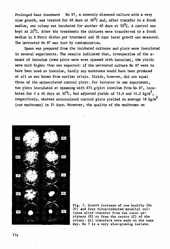
Prolonged heat treatment No 97, a severely diseased culture with a very
slow growth, was treated for 60 days at 30 C and, after transfer to a fresh
medium, one colony was incubated for another 60 days at 30°C. A control was
kept at 25 C. After the treatments the cultures were transferred to a fresh
medium in 5 Petri dishes per treatment and 20 days later growth was measured.
The untreated No 97 was lost by contamination.
Spawn was prepared from the incubated cultures and plots were inoculated
in several experiments. The results indicated that, irrespective of the a-
mount of inoculum (some plots were even spawned with inoculum), the yields
were much higher than was expected: if the untreated culture No 97 were to
have been used as inoculum, hardly any mushrooms would have been produced
at all as was known from earlier trials. Yields, however, did not equal
those of the uninoculated control plots. For instance in one experiment,
two plots inoculated at spawning with 475 g/plot inoculum from No 97, incu
bated for 2 x 60 days at 30°C, had adjusted yields of 13.4 and 14.2 kg/m2,
respectively, whereas uninoculated control plots yielded on average 18 kg/m
(cut mushrooms) in 31 days. Moreover, the quality of the mushrooms on
Fig. 7. Growth increase of one healthy (No 37) and four virus-infected mycelial cultures after transfer from the outer periphery (P) or from the centre (C) of the colony. All transfers were made on the same day. No 7 is a very slow-growing isolate.
114
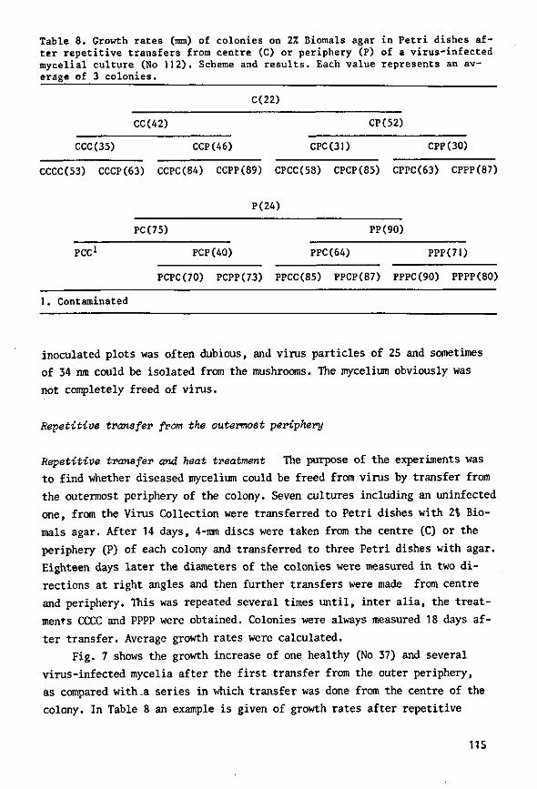
Table 8. Growth rates (mm) of colonies on 2% Biomals agar in Petri dishes after repetitive transfers from centre (C) or periphery (P) of a virus-infected mycelial culture (No 112). Scheme and results. Each value represents an average of 3 colonies.
C(22)
CC(42) CP(52)
CCC(35) CCP(46) CPC(31) CPP(30)
CCCC(53) CCCP(63) CCPC(84) CCPP(89) CPCC(58) CPCP(85) CPPC(63) CPPP(87)
P(24)
PC(75) PP(90)
PCC1 PCP(40) PPC(64) PPP(71)
PCPC(70) PCPP(73) PPCC(85) PPCP(87) PPPC(90) PPPP(80)
1. Contaminated
inoculated plots was often dubious, and virus particles of 25 and sometimes
of 34 nm could be isolated from the mushrooms. The mycelium obviously was
not completely freed of virus.
Repetitive transfer from the outermost periphery
Repetitive transfer and heat treatment The purpose of the experiments was
to find whether diseased mycelium could be freed from virus by transfer from
the outermost periphery of the colony. Seven cultures including an uninfected
one, from the Virus Collection were transferred to Petri dishes with 2% Bio
mals agar. After 14 days, 4-mm discs were taken from the centre (C) or the
periphery (P) of each colony and transferred to three Petri dishes with agar.
Eighteen days later the diameters of the colonies were measured in two di
rections at right angles and then further transfers were made from centre
and periphery. This was repeated several times until, inter alia, the treat
ments CCCC and PPPP were obtained. Colonies were always measured 18 days af
ter transfer. Average growth rates were calculated.
Fig. 7 shows the growth increase of one healthy (No 37) and several
virus-infected mycelia after the first transfer from the outer periphery,
as compared with.a series in which transfer was done from the centre of the
colony. In Table 8 an example is given of growth rates after repetitive
115

transfer from centre or periphery of a virus-infected mycelial culture (No
112). Although a certain growth increase after transfer from the centre can
also be noticed, the increase is more pronounced with peripheral transfer.
The other cultures reacted similarly; however the results were variable as
with heat treatment, although most replicates varied only slightly.
A number of resulting cultures from the last transfer of the seven dif
ferent mycelia under treatment were incubated for two weeks at 34 C. Con
trols remained at 25°C. Twenty days after transfer to fresh medium, growth
was measured (Table 9 ) . Tests were on 21 Biomals agar in test tubes in dupli -
cate. Expectedly, the results were variable but followed the general trend
of heat treatment: growth increase of the slowly growing cultures, and no
or negative reaction of the faster growing mycelia.
Tests for virus particles Spawn inoculum was prepared from No 52, CCCC
and CCCC heat-treated (HT), 52 PPPP and PPPP-HT. Small growing trays were
inoculated with 5 g laboratory-prepared spawn each. The trial was in duplicate.
The results indicated that No 52 PPPP, PPPP-HT and CCCC-HT were not freed
from virus particles: two or three particle types could be isolated from
mushrooms on the trays. Earlier similar tests with No 92 failed because of
Table 9. Growth increase or decrease (mm) of mycelial cultures, earlier subjected to repetitive transfer from centre or periphery, after heat treatment (2 weeks at 34°C). Each value represents an average of two cultures in test tubes. C= growth of untreated control (25°C); GI= difference in growth from control.
Culture
CCCC CPCC CCPP CPPP PCCC PCPC PPCC PCPP PPPP
No
C
58
55 67 67
67
70
1
GI
+ 15
+ 20 + 16 + 21
+ 12
-13
No
C
50
50 55
55
51
28
GI
- 2
- 1 + 2
+ 6
+ 12
No
C
69
67 76 76
66 72
371
GI
+2
0 -3 -7
+5 *
No
C
68
58 68 63
62
72
52
GI
-2
+4 0
+ 1
+3
-7
No
C
45
56 30 32
56
92
GI
+20
+ 3 +25 +20
+ 13
No
C
63
62
94
GI
- 3
+ 13
No
C
55
70 71
61 76
84
112
GI
- 5
- 4 + 5
+ 10
*
- 7
*. Contaminated
116

Table 10. Yields of, and presence of virus particles in, mushrooms grown in plots inoculated (Trial I) or spawned (Trial II) with spawn from virus-infected No 112. This mycelial culture was subjected to repetitive transfer from centre (C) or periphery (P), and sometimes heat-treated (HT, 2 weeks at 34 C) . Averages of two plots.
Treatment
Trial J CCCC CCCC-HT PPPP PPPP-HT
Trial II no 112
(untreated) PPPP PPPP-HT
Inoculum (g/plot)
2.5 2.5 2.5 2.5
450
750 750
Adjusted1
(kg/m2)
0.26 10.1 10.6 9.7
7.4
19.3 17.6
yield2 Virus particles3 (nm)
abundant 25 and 24 few 25, 34, 19x50 few 25, and probably 34 few 25 and 34
many 25, 34, 19x50
common 25 and 34 common 25, 19x50 and probably 34
1. 80% of true yield. 2. Yields are for 22 and 31 days of picking in Trials I and II, respectively 3. The figures in italics indicate the particle type(s) prevailing in that virus preparation.
several unfavourable circumstances.
The results of two trials in duplicate with No 112 in plots are shown
in Table 10. In the first trial (Trial I ) , plots were inoculated at spawning
with 2.5 g spawn each, prepared from No 112 CCCC, CCCC-HT, PPPP or PPPP-HT.
In Trial II, plots were spawned with inoculum (grain spawn) prepared from
No 112 PPPP (375 g/m2), PPPP-HT (375 g/m2) and No 112 (450 g 'infected
spawn' and 300 g commercial spawn per plot). Virus Collection No 112 is sim
ilar to No 112 CCCC. Yields were estimated in 22 days (Trial I) and 31 days 2
(Trial II) of harvesting. Control plots yielded 10.5 to 11 kg/m (Trial I) 2
and 18 kg/m (Trial II) cut mushrooms. Table 10 shows that repetitive trans
fer from the outer periphery of the colony increased yields as does heat
treatment; combination of both, however, seemed slightly less favourable.
Although the yields of the treated cultures were quite as good as controls,
the mycelia certainly were not virus-free. Whether the virus, still present
after treatment, may be an attenuated strain, or may later cause a new out
break, has not yet been investigated; hence treated mycelium should be
handled cautiously.
There is no distinct difference between heat treatment and repetitive
transfer from the outer periphery; the effect observed after heat treatment,
better growth of the mycelium, may even have been due'to transfer, after
117
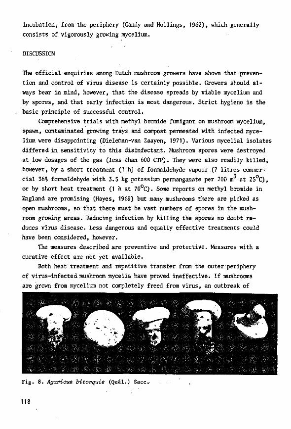
incubation, from the periphery (Gandy and Hollings, 1962), which generally
consists of vigorously growing mycelium.
DISCUSSION
The official enquiries among Dutch mushroom growers have shown that preven
tion and control of virus disease is certainly possible. Growers should al
ways bear in mind, however, that the disease spreads by viable mycelium and
by spores, and that early infection is most dangerous. Strict hygiene is the
basic principle of successful control.
Comprehensive trials with methyl bromide fumigant on mushroom mycelium,
spawn, contaminated growing trays and compost permeated with infected myce
lium were disappointing (Dieleitian-van Zaayen, 1971). Various mycelial isolates
differed in sensitivity to this disinfectant. Mushroom spores were destroyed
at low dosages of the gas (less than 600 CTP). They were also readily killed,
however, by a short treatment (1 h) of formaldehyde vapour (7 litres commer
cial 36*o formaldehyde with 3.5 kg potassium permanganate per 200 m at 25°C),
or by short heat treatment (1 h at 70°C). Some reports on methyl bromide in
England are promising (Hayes, 1969) but many mushrooms there are picked as
open mushrooms, so that there must be vast numbers of spores in the mush
room growing areas. Reducing infection by killing the spores no doubt re
duces virus disease. Less dangerous and equally effective treatments could
have been considered, however.
The measures described are preventive and protective. Measures with a
curative effect are not yet available.
Both heat treatment and repetitive transfer from the outer periphery
of virus-infected mushroom mycelia have proved ineffective. If mushrooms
are grown from mycelium not completely freed from virus, an outbreak of
5 •'•'J'-wfir'- •.. /V*.
Fig. 8. Agarious bitorquis (Quel.) Sacc.
118

virus disease may be expected sooner or later. However better results may
be obtained in future with resistant or tolerant varieties. Trials showed
that most other strains (cream, brown) of Agariaus bisporus could become as
diseased as snowwhite strains after some time, as confirmed on commercial
mushroom farms, but the wild mushroom Agariaus bitorquis (Quel.) Sacc.
{Psalliota edulis Vitt.), represented in Fig. 8, has a remarkably high tol
erance, possibly even resistance to virus. Preliminary results were most
premising. This aspect is under study.
ACKNOWLEDGMENTS
The author thanks Mr P.J. van Tilburg for skilful technical assistance,
Drs J.P.G. Gerrits for statistical advice and Mr J.C. Rigg for correcting
the English text.
119
REFERENCES
Dieleman-van Zaayen, A., 1969. A virus disease of cultivated mushrooms in the Netherlands. Mushr. Sci. 7: 213-220.
Dieleman-van Zaayen, A., 1970. Means by which virus disease in cultivated mushrooms is spread, and methods to prevent and control it. MGA Bull. 244: 158-178.
Dieleman-van Zaayen, A., 1971. Methyl bromide fumigation versus other ways to prevent the spread of mushroom virus disease. Neth. J. agric. Sci. 19: 154-167.
Dieleman-van Zaayen, A., 1972. Intracellular appearance of mushroom virus in fruiting bodies and basidiospores of Agaricus bisporus. Virology 47: 94-104.
Dieleman-van Zaayen, A. and J.H.M. Temmink, 1968. A virus disease of cultivated mushrooms in the Netherlands. Neth. J. PI. Path. 74: 48-51.
Gandy, D.G., 1960a. 'Watery stipe1 of cultivated mushrooms. Nature, Lond. 185: 482-483.
Gandy, D.G., 1960b. A transmissible disease of cultivated mushrooms ('Watery stipe1). Ann. appl. Biol. 48: 427-430.
Gandy, D.G., 1962. Studies on die-back of mushrooms. Mushr. Sci. 5: 468-479. Gandy, D.G. and M. Hollings, 1962. Die-back of mushrooms: a disease associated
with a virus. Rep. Glasshouse Crops Res. Inst. 1961: 103-108. Hayes, W.A., 1969. Methyl bromide as an after-crop sterilant. MGA Bull. 234:
240-243. Hollings, M., 1962. Viruses associated with a die-back disease of cultivated
mushrooms. Nature, Lond. 196: 962-965. Hollings, M., 1968. Mushroom viruses. Rep. Glasshouse Crops Res. Inst. 1967:
101-102. Hollings, M., O.M. Stone and F.T. Last, 1965. Detection and identification
of viruses in mushroom sporophores and mycelium disrupted with ultrasound. Rep. Glasshouse Crops Res. Inst. 1964: 151-154.
Kitano, T., I. Haruna and I. Watanabe, 1961. Purification and concentration of viruses by an organic solvent system. Virology 15: 503-504.
Rasmussen, C.R., R.E. Mitchell and C.I. Slack, 1969. Investigations into the infectivity of X-disease in mushroom culture. Mushr. Sci. 7: 385-398.
Sarazin, A., 1955. The cultivated mushroom. Mushroom Growers' Association, London.
Schisler, L.C., J.W. Sinden and E.M. Sigel, 1967. Etiology, symptomatology, and epidemiology of a virus disease of cultivated mushrooms. Phytopathology 57: 519-526.
Stoller, B.B., 1962. Some practical aspects of making mushroom spawn. Mushr. Sci. 5: 170-184.
120
4.5 Additional information and discussion
Mushroom virus disease is spread by viable infected mycelium and by virus-
infected spores, as has been reported by different authors and is confirmed
by the trials described in this Chapter. Spread by mycelium can be complete
ly prevented, chiefly by applying the wood-preservative sodium pentachloro-
phenate. Spread by spores, however, is harder to control. This means of
spread is very effective: infected mushrooms usually mature too early (Sec
tion 2.1) so that growers cannot pick them all before they open and before
spores are released; virus-infected spores germinate better and earlier
than uninfected spores (Section 4.1) and may contain many virus particles
(Section 3.1). Moreover, single spores are invisible and growers can Only
suspect their presence; spores are light in weight and can easily be carried
by air-currents or wind. Schisler et al. (1967) mention a longevity of more
than 30 years for healthy mushroom spores, under undefined conditions. Our "
data indicate that virus-infected spores still germinated after storage for
Z{ years (the storage period did not yet exceed 3j years) on filter paper
in a refrigerator at 4°C. In less favourable conditions as occur in nature
(moister; higher temperatures), their life may be shortened. The danger of
spores will be obvious, especially in areas where farms are built close
together as in some parts of the Netherlands. But within one contaminated
mushroom farm, spores can already cause much trouble.
Spawn has not proved an important means of spread, at least in this
country.
Spread of mushroom virus disease by an insect vector is highly improb
able: phorid flies, for instance, only seem to lick mushrooms'since their
mouth-parts are suctorial; the larvae feed on the mycelium and do not tunnel
in the stems and caps of fruiting bodies (Hussey, 1959); moreover, no Diptera
with suctorial mouth-parts are known that are vectors of viruses. An insect
vector is also not necessary, since the rapid spread of mushroom virus dis
ease can be explained by spores. But British workers evidently did not rate
spread by spores at its true value and explained the rapid spread of the
disease, even to control trays of their trials, from virus transmission by
the phorid fly Megaselia halterata Wood (Hollings, 1967). The mushroom build
ing where these trials were done had no spore filters (Hollings et'al., 1971).
Experiments on virus transmission by these flies had no controls and are
121 -

unconvincing (Boilings, 1967; Hollings & Stone, 1971). So there is no real
evidence that phorid flies play any role in virus transmission in mushroom
cultures, except indirectly by carrying spores.
Control of virus disease in modern mushroom houses has proved to be
feasible in the Netherlands. Data from our inquiries among growers are fair
ly Teliable, since most growers participated through the Netherlands Mush
room Growers Association (owner of the Co-operative Composting Enterprise,
from which most growers order their compost) and since additional data, if
necessary, were obtained from mushroom consultants or from the growers them
selves, if they sent mushroom samples to be tested for virus. Calculations 2
of crop loss took into account the mean annual yields (12 kg/m in 1967;
12.7 kg/m2 in 1968; 13.5 kg/m2 in 1969 and 13.7 kg/m2 in 1970) and the mean
number of crops per year (3 in 1967; 3.2 in later years).
Attempts are now being made to wash and sterilize returnable picking
boxes in some centres in the Netherlands. In specially constructed machines,
resembling cleansing machines for cheese-vats in dairying, the boxes are
treated with hot water (70-80 C) and detergent, and with a chlorine solution
containing 250-300 ppm (w/v) active chlorine (temperature below 25 C ) . We
did mushroom-growing trials and experiments in the laboratory to find suit
able exposure times and concentrations.
However, mushroom virus disease may never be completely eradicated, as
most growers will be careless in prosperous times and ignore sanitary meas
ures. A virus-resistant strain would be the best solution, also for a virus-
free start in the spawn laboratories, since heat treatment and repetitive
transfer from the outermost periphery of the colony did not result in cul
tures freed from virus. However, yields were improved and the amount of vi
rus particles had been decreased (Section 4.4). Although neither of the two
techniques can be used to free mycelium from virus, they certainly indicate
an interesting but still inexplicable aspect of virus infection. Why does
infected mycelium on agar usually maintain its abnormal, weak growth? The
virus hardly ever kills the mycelium. But, if used as inoculum ('infected
spawn') for plots or trays, virus multiplication probably was not limited.
It is presumed that, in infected mushroom cultures, a certain virus concen
tration is built up in the mycelium permeating the compost during the weeks
of mycelial growth. The concentration of virus in this mycelium most proba
bly determines whether or not fruiting primordia.will be formed (F.T. Last,
pers. commun.).
Heat treatment of mycelium and repetitive transfer from the outer peri-
122

phery of the hyphae seemed to decrease the number of virus particles in the
mycelium. It would be interesting to know whether the virus concentration
could easily be built up again if spores were collected from mushrooms grown
from treated mycelium, and would be used to prepare inoculum for mushroom
cultures, and if this was repeated several times. Such trials have still to
be done.
A related problem is the variability in virus concentration in diseased
mushrooms (Section 2.2). So far, no relationship has been noticed between
symptoms and types or amount of virus particles. In fact, reproduction of
symptoms in fruiting bodies by inoculation with 'infected spawn' did not al
ways succeed, unlike the production of bare zones and crop loss.
REFERENCES
Hollings, M., 1967. Some aspects of virus diseases in mushrooms. Mushr. Sci. 6: 255-260.
Hollings, M., O.M. Stone & P.T. Atkey, 1971. Mushrooms. Rep. Glasshouse Crops Res. Inst. 1970: 157-159.
Hollings, M. & O.M. Stone, 1971. Viruses that infect fungi. A. Rev. Phy-topathol. 9: 93-118.
Hussey, N.W., 1959. Biology of mushroom phorids. Mushr. Sci. 4: 260-270. Schisler, L.C., J.W. Sinden & E.M. Sigel, 1967. Etiology, symptomatology,
and epidemiology of a virus disease of cultivated mushrooms. Phytopathology 57: 519-526.
123
5 General discussion
Over the years, mushroom virus disease has acquired various names, probably
because of the great diversity of symptoms: La France disease (United States),
watery stipe (Great Britain), brown disease (Great Britain), X-disease
(United States), and mushroom die-back (Great Britain). Mushroom die-back
also covered mummy disease, which is certainly of different nature, being
caused by an intracellular bacterium (Schisler et al., 1968). The name mush
room die-back or die-back disease was chosen after the presumed dying-back
and disappearing from the compost of mushroom mycelium (Gandy & Hollings,
3962). Since we never noticed a dying-back of mycelium in the compost (in
severe attacks, the mycelium in the casing soil may retract slightly or not
grow at all into the casing soil), and since growers usually associate the
name with dying mushrooms, a rarely observed symptom, 'die-back' is not a
good name. Better would be to use the antecedent name, La France (Dr J.W.
Sinden first noticed the disease on the farm of the La France brothers in
Chester County, Pennsylvania; Sinden & Hauser, 1950). However such a name
could mistakenly suggest that the disease is of French origin. X-disease
(Kneebone et al., 1962; Hager, 1969) is hardly descriptive and has not caught
on but could be expanded into mushroom virus X disease. For the moment mush
room virus disease is sufficient but this would have to be reviewed if an
other mushroom virus is discovered or the three types of particle prove to
be distinct viruses. The Dutch name "Afstervingsziekte", a translation of
'die-back disease', has similar disadvantages, but is now familiar to the
growers and has been recommended by the Nomenclature Committee of the Neth
erlands Society of Plant Pathology (Bos, 1971).
Symptoms may vary with unknown factors. Time of infection, conditions
or type(s) of virus particles might influence symptoms. There are not always
distinctive visible symptoms; isolation of virus particles from suspected
mushrooms is decisive. In cell-free preparations from diseased mushrooms,
negatively stained with phosphotungstic acid, three types of virus particles
were usually observed: isometric particles 25 and 34 nm in diameter, and
bacilliform particles, 19 x 50 nm. For extraction, a certain clarification
124
procedure was used, which gave rather good virus preparations if conditions
were carefully controlled. However, an electron-microscope was essential.
Since clarification and electron-microscopy are rather laborious and cannot
be used for routine tests by growers, a simple test for the virus would be
extremely useful. Attempts to find cytological abnormalities of diagnostic
value for the disease by using vital stains did not succeed, in confirmation
of tests by Norrish (1964) with 80 different stains.
Much information was obtained on the intracellular appearance of mush
room virus particles. Two, possibly three, types of particles could be de
tected in ultrathin sections of mushroom tissue; hence the types are not
artefacts formed during clarification but really do exist in the tissue.
Demonstration of virus particles in basidiospores confirmed spread of the
disease by spores, as shown by Schisler et al. (1967), and has convinced
English mushroom scientists of spore transmission (Gandy, 1971). Apart from
hyphal anastomosis they used to attribute virus transmission to an insect
vector (Hollings, 1967), which is most unlikely.
Spread of the disease is by viable infected mycelium and by spores from
infected mushrooms; the time of infection proved significant in yield loss.
Research in an isolated growing-room resulted in a list of sanitary measures.
The measures are based on general hygiene and prevent the spread of viable
mycelium and spores. They were implemented among Dutch growers in November
1968, and annual inquiries showed that since then total crop loss due to
virus disease considerably decreased: from 4.54 in 1967, to 1.34 in 1969
and 1970. Not only did the percentage of contaminated farms decrease (from
more than 30 in 1967 to 11.2 in 1969), but also the average crop loss per
contaminated farm (from 15.0 in 1967 to 11.4 in 1969). Once a farm is con
taminated, the disease can now be eradicated within a few weeks or months
(according to severity of attack and size of farm). The reduction in crop
loss evidently helped to improve yields per farm, and to raise production
of mushrooms in the Netherlands.
Picking boxes proved to play an important role in spread of the disease
throughout the country. All returnable boxes are now to be centrally washed
and disinfected. Total crop loss due to virus disease may then be reduced
even further. A resistant strain would probably solve all problems; breeding
from such a strain is envisaged, but the apparently resistant strain needs
improvement in some secondary characteristics.
The use of methyl bromide fumigant, recommended in some countries to
control mushroom virus disease, should be discouraged. It proved effective
125

only in the destruction of spores; but spores can be killed in easier and
safer ways, e.g. by brief heat treatment or brief exposure to formaldehyde
vapour. Mycelium and especially spawn were found to be moderately resistant
to the fumigant, and various uninfected and infected mycelial cultures dif
fered in sensitivity to methyl bromide.
This variability was also noticed in attempts to free mycelium from vi
rus. Heat treatment and repetitive transfer from the outermost hyphal tips
did not result in virus-free cultures. Yet growth of mycelium often improved,
but with wide variation, and mushroom yields usually increased after treat
ment. Nothing is yet known about what happens during treatment, nor about the
effects on multiplication of virus in the mycelium.
Because of the difficulties encountered during purification, I have not
yet succeeded in preparing specific antisera against the three different
types of virus particle. Although serology may be an important tool in es
tablishing relationships between the particle types, satisfactory antisera
can only be prepared after purification and complete isolation of each kind
of particle. It will probably never provide a sensitive test method because
of the extremely low virus concentrations sometimes observed in cell-free
preparations from obviously diseased mushrooms (low, as compared to virus
concentrations in many flowering plants).
The diversity of virus concentrations in mushrooms, the virus multipli
cation and the build-up of virus concentrations in mushroom cultures are in
teresting problems. A simple infectivity test would facilitate further re
search on purification. Improved purification and separation techniques are
indispensable for elucidation of any mutual relationship between the three
types of virus particle, their properties and the role of each type in caus
ing the disease and in symptoms.
Since properties and relationships of the mushroom virus particles are
not yet known, it seems premature to name or number the different particle
types as British workers have done (Hollings & Stone, 1971).
A mutual relationship between the three types cannot be excluded, since
they are usually found together. From Penicillium stoloniferum, three types
of virus-like particles were isolated, two of which were electrophoretically
and serologically distinct (Buck & Kempson-Jones, 1970; Bozarth et al., 1971).
From Aspergillus foetidus , two types of virus-like particles were recently
isolated, similar in diameter but different in double-stranded RJNA compo
nents. They were probably serologically unrelated. Ratti & Buck (1972) have
suggested a multicomponent system of these virus-like particles.
126
A similar system could exist for mushroom virus particles. The possibili
ty of satellite viruses, or mutual helper viruses cannot be excluded. But the
particle types may alternatively represent distinct viruses. Comparisons with
the double-stranded RNA-containing virus-like particles in Penicillium and
Aspergillus spp. should be considered carefully, since those particles have
not yet been proved to be pathogens or infectious agents; the work of Lhoas
(1971) is not conclusive.
The mushroom virus particles will have to be purified and isolated. It
would then be possible to investigate such questions as the nature of the
nucleic acid, and perhaps to make more comparisons with viruses of higher
plants.
REFERENCES
Bos, L.f 1971. Nederlandse namen van plantevirussen. Gewasbescherming 2: 115-123.
Bozarth, R.F., H.A. Wood & A. Mandelbrot, 1971. The Penicillium stoloniferum virus complex: two similar double-stranded RNA virus-like particles in a single cell. Virology 45: 516-523.
Buck, K.W. & G.F. Kempson-Jones, 1970. Three types of virus particle in Penicillium stoloniferum. Nature, Lond. 225: 945-946.
Gandy, D.G., 1971. Basidiospore concentrations in the air of mushroom cropping houses and ancillary buildings. Rep. Glasshouse Crops Res. Inst. 1970: J 79-185.
Gandy, D.G. & M. Hollings, 1962. Die-back of mushrooms: a disease associated with a virus. Rep. Glasshouse Crops Res. Inst. 1961: 103-108.
Hager, R.A., 1969. An investigation of X-disease. Mushr. Sci. 7: 205-211. Hollings, M., 1967. Some aspects of virus diseases in mushrooms. Mushr. Sci.
6: 255-260. Hollings, M. & O.M. Stone, 1971. Viruses that infect fungi. A. Rev. Phyto-
pathol. 9: 93-118. Kneebone, L.R., J.D. Lockard & R.A. Hager, 1962. Infectivity studies with
"X-disease". Mushr. Sci. 5: 461-467. Lhoas, P., 1971. Infection of protoplasts from Penicillium stoloniferum with
double-stranded RNA viruses. J. gen. Virol. 13: 365-367. Norrish, M.E., 1964. Intracellular inclusions. Rep. Glasshouse Crops Res.
Inst. 1963: 92. Ratti, G. & K.W. Buck, 1972. Virus particles in Aspergillus foetidus: a mul-
ticomponent system. J. gen. Virol. 14: 165-175. Schisler, L.C., J.W. Sinden & E.M. Sigel, 1967. Etiology, symptomatology,
and epidemiology of a virus disease of cultivated mushrooms. Phytopathology 57: 519-526.
Schisler, L.C., J.W. Sinden & E.M. Sigel, 1968. Etiology of mummy disease of cultivated mushrooms. Phytopathology 58: 944-948.
Sinden, J.W. & E. Hauser, 1950. Report on two new mushroom diseases. Mushr. Sci. 1: 96-100.
127
Summary
Diseased cultivated mushrooms, Agaricus bispoms (Lange) Sing., usually
yielded three types of virus particle, often in combination: isometric par
ticles 25 and 34 nm in diameter, and bacilliform particles 19 nm in diameter
and 50 nm long. Injection of a cell-free virus preparation into young mush
rooms on a growing-tray, and subsequent reisolation of the three types of
virus particles, in a later flush, from fruiting bodies showing symptoms,
proved that the particles caused the disease (Section 2.1).
Symptoms were highly variable (Section 2.1). However isolation of virus
particles from suspect mushrooms is decisive (Section 2.2).
In ultrathin sections of vegetative mycelium, dense aggregates of virus
particles were detected often near a septum or close to a nucleus. Prestain-
ing en bloc with aqueous uranyl acetate proved essential to demonstrate the
mushroom virus particles (Section 3.1). Later studies revealed the particles
in mycelium to be 34 nm in diameter. In ultrathin sections of fruiting bod
ies, they were often abundant in the cytoplasm, in loose aggregates or dis
persed, and sometimes in vacuoles. They were observed in dolipores; hence
34-nm particles seem to pass from cell to cell. Virus particles 19 nm x 50
nm were seldom found in sections of the stipe; they were difficult to detect.
Isometric particles 25 nm in diameter, resembling ribosomes, occurred in de
ranged cells of the cap of mushrooms from a sample, known to contain many
25-nm virus particles. These isometric particles were often clumped together
and membrane-bound. In ultrathin sections of basidiospores from diseased
mushrooms, virus particles 34 nm in diameter were grouped together in small
vacuoles and occasionally in spore cytoplasm, up to some hundreds of parti
cles per section of one spore. No virus-like particles were found in tissue
or spores from healthy mushrooms (Section 3.1).
The 25-nm mushroom virus particles turned out to be identical in ap
pearance to turnip yellow mosaic virus, i.e. a 1 - 3 structure with hexamer-
pentamer clustering of subunits (Section 3.2). A parallel resemblance was
observed between rod-shaped virus-like particles, frequently isolated from
apothecia of Peziza ostraooderma Korf, and tobacco mosaic virus, p. ostra-
128
aoderma (syn. Pliaaria fulva R. Schneider) often occurs as a contaminant in
mushroom farms. Negatively stained virus-like particles were on average 17
nm in diameter x 350 nm long and had a clear axial canal. Occasionally such
particles were observed in cell-free preparations from diseased cultivated
mushrooms (Sections 3.2 and 3.3). Apothecia of P. ostraoodexma that contained
particles did not differ macroscopically from those without particles (Sec
tion 3.3). Ultrathin sections of apothecia, from samples known to contain
the virus-like particles, revealed the presence of crystal-like aggregates
of the particles in cells just below the asci. The aggregates were often
found in vacuoles and consisted of rods arranged in angled layers. However
similarities as in aggregation, to some strains of tobacco mosaic virus,
were accompanied by distinct differences, for instance in structure (Section
3.3). So far the significance of the rods in A . bisporus is unknown.
In Section 4.1, practical aspects were tackled. Trials in an isolated
growing-room of the Mushroom Experimental Station confirmed that the disease
spreads with viable mycelium and with spores from diseased mushrooms, as re
ported earlier by different authors, and demonstrated the importance of time
of infection: early infection considerably reduced crop yield, whereas later
infection did not. Returnable picking boxes contributed to the spread of the
disease all over the country. Intensive research resulted in a list of meas
ures to prevent or control the disease. It was implemented among Dutch grow
ers in November 1968. The measures are based mainly on strict hygiene and
are meant to prevent the spread of infected mycelium and spores.
Extensive investigations on spawn, in which more than 100 growers par
ticipated for over a year, were not decisive, partly through lack of a re
liable test for virus in spawn. Virus may spread with spawn, but other means
are considered more important (Section 4.2).
, Methyl bromide fumigant does not control mushroom virus disease, except
partially by destroying mushroom spores. Spores, however, can be destroyed
easier and less dangerously. The results of fumigating mushroom mycelium
were variable, and of spawn disappointing (Section 4.3). The use of this
chemical should be discouraged..
Section 4.4 describes trials in the isolated growing-room on, inter
alia, the importance of time of infection and the inconsequence of amount
of inoculum. It gives the definitive version of the list of measures to pre
vent or control disease. General implementation of the measures in the Neth
erlands considerably reduced crop loss, as appeared from annual inquiries
among all mushroom"growers. Attempts to free mycelium from virus by various
129
techniques (heat treatment at 33 C, repetitive transfer fron the outermost
periphery of hyphae) failed. For a virus-free start, and for control, better
results may be expected by breeding from a mushroom strain that seems re
sistant to virus disease.
130