Decision-Making Groups Attenuate the Discussion Bias in Favor of Shared Information: A Meta-Analysis
Upload
independentCategory
view
0download
0
Culture-Modified Bone Marrow Cells Attenuate Cardiacand Renal Injury in a Chronic Kidney Disease Rat Modelvia a Novel Antifibrotic MechanismDarren A. Yuen1., Kim A. Connelly1., Andrew Advani1, Christine Liao1, Michael A. Kuliszewski1, Judy
Trogadis1, Kerri Thai1, Suzanne L. Advani1, Yuan Zhang2, Darren J. Kelly2, Howard Leong-Poi1, Armand
Keating3, Philip A. Marsden1, Duncan J. Stewart4, Richard E. Gilbert1*
1 Keenan Research Centre of the Li Ka Shing Knowledge Institute, St. Michael’s Hospital, Department of Medicine, University of Toronto, Toronto, Ontario, Canada,
2 Department of Medicine, St. Vincent’s Hospital, University of Melbourne, Melbourne, Victoria, Australia, 3 Department of Medicine, Princess Margaret Hospital, University
of Toronto, Toronto, Ontario, Canada, 4 Ottawa Health Research Institute, University of Ottawa, Ottawa, Ontario, Canada
Abstract
Background: Most forms of chronic kidney disease are characterized by progressive renal and cardiac fibrosis leading todysfunction. Preliminary evidence suggests that various bone marrow-derived cell populations have antifibrotic effects. Inexploring the therapeutic potential of bone marrow derived cells in chronic cardio-renal disease, we examined the anti-fibrotic effects of bone marrow-derived culture modified cells (CMCs) and stromal cells (SCs).
Methodology/Principal Findings: In vitro, CMC-conditioned medium, but not SC-conditioned medium, inhibited fibroblastcollagen production and cell signalling in response to transforming growth factor-ß. The antifibrotic effects of CMCs and SCswere then evaluated in the 5/6 nephrectomy model of chronic cardio-renal disease. While intravascular infusion of 106 SCshad no effect, 106 CMCs reduced renal fibrosis compared to saline in the glomeruli (glomerulosclerosis index: 0.860.1 v1.960.2 arbitrary units) and the tubulointersitium (% area type IV collagen: 1.260.3 v 8.462.0, p,0.05 for both). Similarly,106 CMCs reduced cardiac fibrosis compared to saline (% area stained with picrosirius red: 3.260.3 v 5.160.4, p,0.05),whereas 106 SCs had no effect. Structural changes induced by CMC therapy were accompanied by improved function, asreflected by reductions in plasma creatinine (5863 v 81611 mmol/L), urinary protein excretion (96/41 v 646/41 mg/day),and diastolic cardiac stiffness (left ventricular end-diastolic pressure-volume relationship: 0.03060.003 v 0.05860.011 mmHg/mL, p,0.05 for all). Despite substantial improvements in structure and function, only rare CMCs were present in thekidney and heart, whereas abundant CMCs were detected in the liver and spleen.
Conclusions/Significance: Together, these findings provide the first evidence suggesting that CMCs, but not SCs, exert aprotective action in cardio-renal disease and that these effects may be mediated by the secretion of diffusible anti-fibroticfactor(s).
Citation: Yuen DA, Connelly KA, Advani A, Liao C, Kuliszewski MA, et al. (2010) Culture-Modified Bone Marrow Cells Attenuate Cardiac and Renal Injury in aChronic Kidney Disease Rat Model via a Novel Antifibrotic Mechanism. PLoS ONE 5(3): e9543. doi:10.1371/journal.pone.0009543
Editor: Arnold Schwartz, University of Cincinnati, United States of America
Received December 21, 2009; Accepted February 10, 2010; Published March 4, 2010
Copyright: � 2010 Yuen et al. This is an open-access article distributed under the terms of the Creative Commons Attribution License, which permitsunrestricted use, distribution, and reproduction in any medium, provided the original author and source are credited.
Funding: This work was supported by grants from the Canadian Institutes of Health Research (CIHR) and the Physicians’ Services Incorporated Foundation ofOntario. Dr. Yuen is supported by a KRESCENT postdoctoral fellowship. Dr. Gilbert is supported by a generous donation from George and Diane Mara. Dr. Connellyis supported by a Clinician Scientist award from the Heart and Stroke Foundation of Ontario. Dr. Advani is supported by a Clinician Scientist award from theCanadian Diabetes Association. Dr. Leong-Poi is supported by a Phase II Clinician Scientist Award from the Heart and Stroke Foundation of Ontario and an EarlyResearcher Award from the Ministry of Research and Innovation, Ontario, Canada. Dr. Keating holds the Gloria and Seymour Epstein Chair in Cell Therapy andTransplantation at the University Health Network and the University of Toronto. The funders had no role in study design, data collection and analysis, decision topublish, or preparation of the manuscript.
Competing Interests: The authors have declared that no competing interests exist.
* E-mail: [email protected]
. These authors contributed equally to this work.
Introduction
Repair by connective tissue formation is a fundamental response
to acute injury. If unchecked, however, the resulting fibrotic response
leads to parenchymal replacement and organ dysfunction, estimated
to account for nearly 45% of all deaths in the industrialized world
[1]. Chronic kidney disease (CKD), for instance, now estimated to
affect almost 20 million adults in the United States alone, is
characterized by progressive renal fibrosis with attendant reduction
in glomerular filtration that ultimately results in the need for dialysis
or transplantation to preserve life [2]. Moreover, in CKD, the
fibrotic pathology is not confined to the kidney but is also present in
the heart [3], where even mild degrees of renal impairment are
associated with ventricular stiffening, impaired relaxation and
diastolic dysfunction that increase as kidney function worsens [4].
Importantly, diastolic dysfunction is a primary contributor to
cardiovascular morbidity and mortality in CKD patients, where its
presence portends a particularly poor prognosis [5].
PLoS ONE | www.plosone.org 1 March 2010 | Volume 5 | Issue 3 | e9543
Studies conducted over almost two decades have consistently
implicated transforming growth factor-ß (TGF-ß) as a key mediator
of pathological fibrosis [6]. Moreover, in addition to its profibrotic
effects, TGF-ß has also been implicated in microvascular loss [7],
cardiomyocyte hypertrophy [8], and podocyte dysfunction [9] that
characterize cardio-renal disease. Indeed, inhibition of TGF-ß has
been a major target for drug discovery with several small molecules,
antibodies and nucleic acid-based strategies in development [10].
While most experimental and clinical studies of bone marrow
derived cell (BMDC) therapy have been undertaken for the
treatment of large vessel angio-occlusive disease [11,12,13], others
have demonstrated that various BMDC populations may exert
beneficial effects in other settings also, including fibrosis in the liver
[14], an organ in which a dual blood supply renders it ischemia-
resistant. With this in mind, we speculated as to whether this effect
might be common to the two types of BMDCs that are currently
under investigation as potential therapeutic agents: bone marrow-
derived culture-modified cells (CMCs) and stromal cells (SCs).
Here we show that CMCs reduce collagen formation, inhibit
TGF-ß signaling and also effectively reduce renal and cardiac
fibrosis in a rodent model of chronic cardio-renal disease that
mimics human disease. Importantly, these effects were associated
with improvements in kidney and heart function, an effect not seen
with current clinically available therapies.
Results
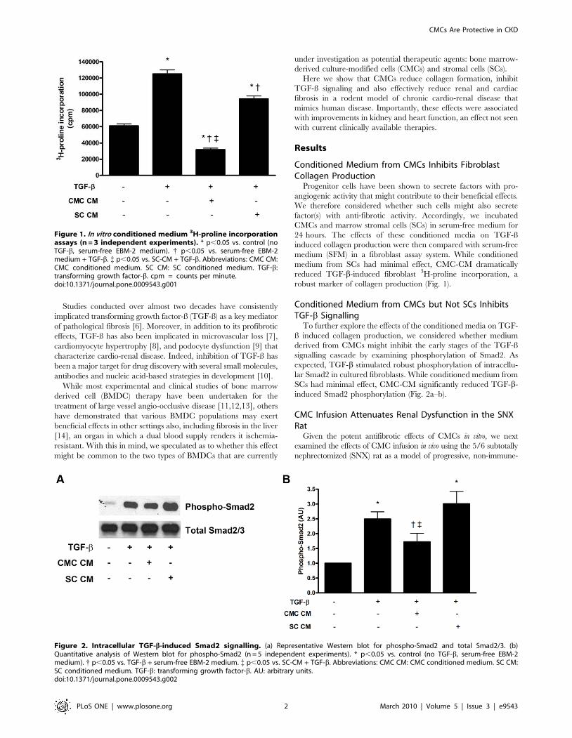
Conditioned Medium from CMCs Inhibits FibroblastCollagen Production
Progenitor cells have been shown to secrete factors with pro-
angiogenic activity that might contribute to their beneficial effects.
We therefore considered whether such cells might also secrete
factor(s) with anti-fibrotic activity. Accordingly, we incubated
CMCs and marrow stromal cells (SCs) in serum-free medium for
24 hours. The effects of these conditioned media on TGF-ß
induced collagen production were then compared with serum-free
medium (SFM) in a fibroblast assay system. While conditioned
medium from SCs had minimal effect, CMC-CM dramatically
reduced TGF-b-induced fibroblast 3H-proline incorporation, a
robust marker of collagen production (Fig. 1).
Conditioned Medium from CMCs but Not SCs InhibitsTGF-b Signalling
To further explore the effects of the conditioned media on TGF-
ß induced collagen production, we considered whether medium
derived from CMCs might inhibit the early stages of the TGF-ß
signalling cascade by examining phosphorylation of Smad2. As
expected, TGF-b stimulated robust phosphorylation of intracellu-
lar Smad2 in cultured fibroblasts. While conditioned medium from
SCs had minimal effect, CMC-CM significantly reduced TGF-b-
induced Smad2 phosphorylation (Fig. 2a–b).
CMC Infusion Attenuates Renal Dysfunction in the SNXRat
Given the potent antifibrotic effects of CMCs in vitro, we next
examined the effects of CMC infusion in vivo using the 5/6 subtotally
nephrectomized (SNX) rat as a model of progressive, non-immune-
Figure 1. In vitro conditioned medium 3H-proline incorporationassays (n = 3 independent experiments). * p,0.05 vs. control (noTGF-b, serum-free EBM-2 medium). { p,0.05 vs. serum-free EBM-2medium + TGF-b. { p,0.05 vs. SC-CM + TGF-b. Abbreviations: CMC CM:CMC conditioned medium. SC CM: SC conditioned medium. TGF-b:transforming growth factor-b. cpm = counts per minute.doi:10.1371/journal.pone.0009543.g001
Figure 2. Intracellular TGF-b-induced Smad2 signalling. (a) Representative Western blot for phospho-Smad2 and total Smad2/3. (b)Quantitative analysis of Western blot for phospho-Smad2 (n = 5 independent experiments). * p,0.05 vs. control (no TGF-b, serum-free EBM-2medium). { p,0.05 vs. TGF-b + serum-free EBM-2 medium. { p,0.05 vs. SC-CM + TGF-b. Abbreviations: CMC CM: CMC conditioned medium. SC CM:SC conditioned medium. TGF-b: transforming growth factor-b. AU: arbitrary units.doi:10.1371/journal.pone.0009543.g002
CMCs Are Protective in CKD
PLoS ONE | www.plosone.org 2 March 2010 | Volume 5 | Issue 3 | e9543

mediated chronic kidney disease that also develops heart failure. In
this model, the kidney and heart develop progressive fibrosis similar
to many patients with CKD. Systolic blood pressure (SBP), urinary
protein excretion and plasma creatinine were all elevated in SNX
when compared with sham-operated animals at 8 weeks (Table 1).
The kidneys receive approximately 20% of cardiac output such that
infusing cells intra-arterially would be expected to deliver a greater
number of cells to this site of injury than might be anticipated from
intravenous administration. We therefore compared intra-arterial
with intravenous routes of cell administration, with an a priori decision
that if similar reductions in proteinuria were apparent, then the two
groups would be considered as one, enabling greater statistical power
for comparison with control animals. Administration of 106 CMCs
attenuated the magnitude and progression of proteinuria in SNX rats
to a near-identical extent whether infused by an intravenous or intra-
arterial route. Indeed, for a range of functional and structural
parameters in both the kidney and heart, outcomes were nearly
identical for both routes of administration (Fig. S1). In addition to
reducing proteinuria, cell therapy attenuated the increase in plasma
creatinine in SNX rats, though it remained elevated in comparison to
sham animals (Table 1). No mortality was seen in any treatment
group after CMC or saline infusion.
CMC Infusion Ameliorates Fibrosis and MicrovascularRarefaction in the SNX Rat
To determine the structural correlates of the improved kidney
function seen with CMC therapy, we examined the extent of
glomerulosclerosis and tubulointerstitial fibrosis in SNX animals
treated with syngeneic CMCs. Tubulointerstitial fibrosis, a major
correlate of declining kidney function, was substantially increased
in SNX rats, as was glomerulosclerosis. CMC-treated rats showed
substantial reductions in the extent of fibrosis, both in the
tubulointerstitium (Fig. 3) and in the glomerulus (Fig. 4).
Progressive fibrosis replaces normal renal parenchyma, includ-
ing its constituent capillaries. Accordingly, we examined glomer-
ular capillary density using immunolabelling for a glomerular
endothelial cell antigen in SNX and CMC-treated SNX-rats. As
expected, SNX rats experienced a significant reduction in
glomerular capillary density, while CMC-treated SNX rats
demonstrated preservation of glomerular capillaries (Fig. 5). With
the consideration that the increased abundance of cells showing
positive endothelial cell immunolabelling in CMC-treated SNX
rats may reflect augmentation in antigenic expression rather than
an increase in capillarity, a subset of SNX and CMC-treated SNX
animals underwent fluorescence microangiography (FMA), dem-
onstrating loss of both glomerular and peritubular capillaries
(Fig. 5), along with their amelioration by CMC administration.
Chronic Diastolic Cardiac Dysfunction Is Improved byCMC Administration
Given the close association between kidney disease and heart
failure, and the importance of chronic heart failure as a poor
prognostic factor in CKD patients, we next examined pre-terminal
(8 weeks post-surgery) cardiac function in SNX rats with and
without CMC therapy, focussing particularly on diastolic function.
SNX rats demonstrated chronic impairment of diastolic function
as demonstrated by an elevated left ventricular end-diastolic
pressure volume relationship (LV EDPVR), a marker of impaired
left ventricular compliance (Fig. 6). When compared with
untreated SNX animals, CMC-treated animals showed improve-
ment in passive diastolic filling, as manifested by a reduction in LV
EDPVR (Fig. 6).
CMC Infusion Ameliorates Cardiac Structural Damage inthe Remnant Kidney Rat
One of the primary determinants of impaired left ventricular
relaxation is the development of cardiac fibrosis. Accordingly, we
examined the degree of cardiac matrix accumulation at 8 weeks
post-surgery. As in the kidney, SNX lead to significant interstitial
fibrosis in the heart. CMC treatment significantly attenuated the
extent of cardiac fibrosis (Fig. 7). In addition, myocyte cross
sectional area, indicative of cellular hypertrophy and thus
worsened cardiac afterload, was increased in SNX rats and
reduced by CMC administration (Fig. 8).
SCs Were without Effect on Cardio-Renal Function andStructure in the Remnant Kidney Rat
To ascertain whether the beneficial actions of CMCs in the
SNX model tracked with their in vitro anti-fibrotic effects or
whether it reflected a more generalizable attribute of bone marrow
derived cells, we next examined the in vivo efficacy of marrow
stromal cells in the SNX model. In contrast to CMCs,
intravascular infusion of 106 SCs did not attenuate the structural
or functional abnormalities in either the heart or kidney of SNX
rats (Table S1 and Fig. S2).
In Vivo Tissue Localization of Infused CMCsGiven that both intravenous and intra-arterial CMC delivery had
equivalent effects, and our in vitro results suggesting that CMCs
release soluble antifibrotic factor(s) that inhibit TGF-b signalling in
fibroblasts, we next sought to examine the abundance of the
administered cells in the kidney and heart. Accordingly, CMCs
were loaded with the vital cytoplasmic fluorophore CMTMR, and
infused by tail vein injection into rats 4 weeks after 5/6 subtotal
nephrectomy. While only rare CMTMR-labelled CMCs were
Table 1. Renal function at 8 weeks post-surgery.
Sham SNX SNX–CMC iv SNX–CMC ia SNX - CMC
N 6 15 9 8 17
Urine protein (mg/day) 3.16/41.1 64.06/41.4* 9.36/41.4{ 8.86/41.7{ 9.06/41.3{
Plasma creatinine (mmol/L) 3262 81611* 5563{ 6265* 5863*{
Systolic BP (mm Hg) 12362 184610* 17266* 17866* 17564*
Abbreviations: SNX, SNX animal treated with phosphate-buffered saline; SNX–iv, SNX animal treated with intravenous CMC infusion; SNX–ia, SNX animal treated withintra-arterial CMC infusion; SNX–CMC, SNX animal treated with either intravenous or intra-arterial CMC infusion. Urine protein excretion is presented as geometric mean6/4 tolerance factors.*p,0.05 versus sham-operated animals.{p,0.05 versus SNX animals.doi:10.1371/journal.pone.0009543.t001
CMCs Are Protective in CKD
PLoS ONE | www.plosone.org 3 March 2010 | Volume 5 | Issue 3 | e9543

detected in the kidneys and hearts of infused SNX animals at either
early (1 day) or later (14 day) time points, CMTMR-labelled CMCs
were abundant in the liver and spleen with a notable peak 4 days
after administration but persistence throughout the time course of
the study (Fig. 9).
Discussion
Despite current treatment, the incidence of end-stage renal
disease continues to rise, with no major therapeutic advances to
stall the progression of CKD since the introduction of renin-
angiotensin system antagonists. In the present study, we show that
declining GFR and worsening proteinuria, two of the cardinal
laboratory indicators of progression towards end-stage kidney
disease, were substantially attenuated by the administration of
culture modified bone marrow derived cells in a widely used model
of non-immune chronic kidney disease. Moreover, chronic heart
failure, a common comorbidity in CKD that portends a poor
prognosis, was also improved by cell therapy.
While CMCs have therapeutic effects in a range of diseases, the
mechanisms by which they do so remain unclear. In the present
study we explored their anti-fibrotic activity, focusing on the
dominant prosclerotic factor TGF-ß, and the cells’ in vivo efficacy
in a model of chronic kidney disease that is characterized by
clinically significant progressive scarring of both the kidney and
the heart. CMCs, but not SCs, were shown to secrete factor(s) that
inhibit TGF-ß receptor signalling and collagen production in
cultured cells. Intriguingly, the beneficial effects seen in vivo with
CMC infusion occurred despite minimal CMC localization to the
kidney or heart, in either the acute or later settings, suggesting that
diffusible factor(s), such as those identified in culture, may also be
active in the in vivo setting. Indeed, rather than homing to the site
of injury, exogenous CMCs were present in large numbers in the
liver and spleen, suggesting that they may be preferentially
sequestered by the reticulo-endothelial system.
In line with our limited understanding of how non-embryonic
cell therapy might work, there has been considerable conjecture as
to whether the administered cells differentiate [15], fuse with
resident cells [16] or secrete paracrine factors [17]. Notwithstand-
ing this debate, their efficacy has nevertheless been assumed to be
dependent on cell homing to sites of injury [18]. As a consequence,
techniques for increasing local cell delivery have been used
increasingly in clinical trials in an attempt to maximize the
therapeutic gain [19]. However, two studies in acute kidney injury
models have suggested that circulating factors elaborated by bone
marrow derived cells may account for the beneficial effects
observed [20,21]. In the present study, we compared intravenous
with intra-arterial CMC delivery, noting similar efficacy in a range
of outcome measures with either mode of administration.
Moreover, despite the observed benefits, CMCs were rarely
present in the kidneys or hearts of treated SNX animals, whereas
high numbers were found in the liver and spleen, suggesting that at
least some of their effects may be mediated at a distance, possibly
through the elaboration of diffusible factor(s) that inhibit(s) the
actions of TGF-ß.
Glomerulonephritis, diabetes and hypertension account for the
majority of chronic kidney disease in the developed world.
However, despite this diverse range of etiologies, they are often
characterized histopathologically by chronic cardiorenal fibrosis
and microvascular rarefaction. Indeed, it is the extent of the
Figure 3. Tubulointerstitial renal cortical fibrosis. Kidney sections were immunolabelled for Type IV collagen 8 weeks post-surgery. (a–c)Representative cortical tubulointerstitial images. Original magnification 6 160. (a) Sham animal. (b) SNX animal. (c) SNX–CMC animal. (d) Type IVcollagen % positivity. * p,0.05 vs. sham operated animals. { p,0.05 vs. SNX animals.doi:10.1371/journal.pone.0009543.g003
CMCs Are Protective in CKD
PLoS ONE | www.plosone.org 4 March 2010 | Volume 5 | Issue 3 | e9543

fibrosis, particularly in the renal interstitium, that correlates most
closely with the decline in kidney function [22]. Similarly, in the
heart, interstitial fibrosis, along with myocyte hypertrophy and
capillary loss, are key components of adverse cardiac remodelling
that affect cardiac geometry and function, particularly during
diastole [23]. As in the kidney, fibrosis appears as a final common
pathway in most forms of chronic cardiac disease, being present in
diabetic [24] and uremic cardiomyopathies [25], ischaemic heart
disease [23], and even in valvular heart disease where the extent of
fibrosis correlates closely with left ventricular end-diastolic
pressure and inversely with ejection fraction [26].
Consistent with its spectrum of activity in stimulating fibrosis,
hypertrophy, podocyte injury, and capillary loss, elevated TGF-ß
expression has been repeatedly demonstrated in studies of cardiac
and renal disease in both humans and animal models [6], and in
particular the 5/6 subtotally nephrectomised rat [27]. The central
role of TGF-b in promoting fibrovascular pathology and
subsequent organ dysfunction has been reinforced by multiple
studies demonstrating the beneficial effects of blocking TGF-b for
either chronic renal [28,29] or cardiac disease [30,31]. In the
present study, we used 3H-proline incorporation as a bioassay of
fibroblast collagen production. In these studies, medium derived
from CMCs, but not that from SCs, was found to suppress TGF-ß
induced 3H-proline incorporation, suggesting that soluble factor(s)
released by CMCs, at least in part, mediate an anti-fibrotic effect.
To investigate how the culture modified medium was inhibiting
TGF-ß induced 3H-proline incorporation, we examined its well-
described signalling pathway. Binding of TGF-ß to its type 2
receptor (TßR2), permits recruitment and activation of the type 1
receptor (TßR1), which in turn continues the signalling relay by
phosphorylating and thereby activating the intermediate Smad2
[32]. In the present study, we show that conditioned medium from
cultured CMCs reduced TGF-b-induced Smad2 phosphorylation,
suggesting that factor(s) secreted into the medium by CMCs
inhibited the activity of the TGF-ß receptor complex. In contrast
to CMCs, conditioned medium from cultured SCs had compar-
atively minimal effects on TGF-ß induced 3H-proline incorpora-
tion and Smad2 phosphorylation, suggesting that CMCs, and not
SCs, are one of the principal BMDC populations that secrete these
inhibitory factor(s).
A wide range of methods have been used to isolate and identify
progenitor cells in bone marrow [33]. In the absence of specific and
unique cell surface markers, we compared two major types of bone
marrow derived cells based primarily on their in vitro adhesion
and growth characteristics. Bone marrow cells were cultured on
fibronectin-coated plates for 7 to 10 days, at which time they dis-
played many of the phenotypic and surface marker characteristics
that are commonly associated with endothelial progenitor cells [33].
However, given the ambiguity surrounding the nomenclature of such
cells, we used a more operational definition, referring to the cells that
we used as early outgrowth, bone marrow derived, culture-modified
cells (CMCs). The other broad category of bone marrow derived cells
under investigation, mesenchymal or marrow stromal cells (SCs) were
identified in this study by their plastic adherence and their ability to
differentiate into osteoblasts, adipocytes, and chondroblasts, criteria
used widely for their definition [34]. Since both the CMC and SC
populations used in our studies were heterogeneous, the generaliz-
ability of our findings to other cell doses and other types of bone
marrow derived cells, cultured under different conditions or
administered in other disease settings, is uncertain.
Clinically, perhaps the most well known link between TGF-ß,
fibrosis and renal dysfunction relates to angiotensin II, as agents
Figure 4. Glomerulosclerosis. PAS-stained kidney sections at 8 weeks post-surgery were used to assess the degree of glomerulosclerosis. (a–c)Representative glomerular images. Original magnification 6400. (a) Sham animal. (b) SNX animal. (c) SNX–CMC animal. (d) Glomerulosclerosis index(GSI). * p,0.05 vs. sham operated animals. { p,0.05 vs. SNX animals.doi:10.1371/journal.pone.0009543.g004
CMCs Are Protective in CKD
PLoS ONE | www.plosone.org 5 March 2010 | Volume 5 | Issue 3 | e9543

that block its formation and receptor binding are the cornerstones of
current CKD management. While angiotensin converting enzyme
(ACE) inhibitors and angiotensin receptor blockers (ARBs) were
initially viewed as exerting their beneficial effects through primarily
hemodynamic means, such as their ability to reduce intraglomerular
pressure, more recent studies have emphasized the ability of these
agents to reduce TGF-ß formation and attenuate fibrosis in the
tubulointerstitium as well as the glomerulus, in both humans [35,36]
and experimental animals [27,37]. Similarly, in the heart, beyond its
haemodynamic effects, the ability of angiotensin II to induce TGF-ß
expression [38] is thought to be a major contributor to the beneficial
effects of renin-angiotensin system (RAS) blockade in heart failure
[39,40].
To date, many of the studies showing beneficial effects of bone
marrow derived cells have focussed on their pro-angiogenic actions.
Consistent with these reports, the present study also showed that cell
Figure 5. Renal microvasculature. (a–c) Kidney sections were immunolabelled with JG-12 antibody, which recognizes a glomerular endothelialcell antigen. Representative JG-12 immunostained images. Original magnification 6400. (a) Sham animal. (b) SNX animal. (c) SNX–CMC animal. (d) %positive area for JG-12 immunostaining per glomerulus. (e–g) Representative fluorescence microangiography (FMA) images of glomeruli. Theautofluorescence of the surrounding renal cortex was captured and pseudocolourized red. The green fluorescence of the infused beads outlinesmicrovascular structures. Original magnification 6 40. (e) Sham animal. (f) SNX animal. (g) SNX–CMC animal. (h–j) Representative FMA images ofperitubular capillaries. Each image is a flattened Z-stack of a 100 mm section taken with a confocal microscope. Original magnification675. (h) Shamanimal. (i) SNX animal. (j) SNX–CMC animal. * p,0.05 vs. sham operated animals. { p,0.05 vs. SNX animals.doi:10.1371/journal.pone.0009543.g005
CMCs Are Protective in CKD
PLoS ONE | www.plosone.org 6 March 2010 | Volume 5 | Issue 3 | e9543

therapy improved the microvasculature of the kidney as assessed by
both immunohistochemical and angiographic techniques. While
loss of the capillary microvasculature is a feature of the SNX model,
as with CKD in humans, growth factors with primarily angiogenic
effects, such as vascular endothelial growth factor (VEGF), do not
attenuate proteinuria, hypertension or glomerulosclerosis despite
preventing microvascular loss [41]. In contrast to VEGF, agents
that target the pro-sclerotic factor TGF-b, either directly [29] or
indirectly [28], have been shown to attenuate not only fibrosis and
declining kidney function, but to also reduce proteinuria in models
of CKD. Similarly, in the heart, agents that inhibit TGF-ß activity
have also been shown to reduce fibrosis and cardiac dysfunction
[30,31,42].
Functioning in tandem, the kidney and heart provide the
physiological regulation of extracellular fluid volume, sodium
homeostasis, blood pressure, cardiac output and glomerular
filtration rate (GFR). In the setting of kidney disease, the resulting
dysregulation affects cardiac function with increases in both pre-
and afterload, along with the accumulation of waste products that
characterizes the uraemic state. At a histopathological level,
fibrosis and myocyte hypertrophy are features of cardiac tissue
from both humans and animals with impaired kidney function that
are thought to mediate the abnormal relaxation properties of the
left ventricle in CKD [25,43,44]. In the present study, we found
that CMC administration not only attenuated these pathological
changes in cardiac structure but also led to improvements in
diastolic filling. Importantly, these changes in cardiac function
were assessed using load-insensitive techniques, an important
consideration in the setting of the alterations in blood volume and
arterial pressure seen in CKD.
As commonly seen with other therapeutic interventions in both
the experimental and clinical settings, CMC administration led to
a more dramatic reduction in proteinuria than in serum
creatinine, reflecting the different pathogeneses of these two
disease manifestations as well as the non-linear relationship
between serum creatinine and GFR. Interestingly, while improve-
ments in kidney filtration and proteinuria were observed with
CMC treatment, systemic arterial pressure was unaffected,
remaining significantly elevated above that of sham animals, as
has been documented with other anti-TGF-b therapies in
experimental renal disease [28]. These data suggest that the
mechanisms underlying CMC-induced renal structural and
functional changes in the 5/6 nephrectomy model are mostly
independent of changes in systemic blood pressure. However, a
Figure 6. Invasive cardiac hemodynamic assessment at 8 weeks post-surgery. (a–c) Representative pressure-volume (PV) loops obtained 8weeks post-surgery. Each loop is generated by the simultaneous measurement of left ventricular pressure and volume at all points in the cardiaccycle, enabling the measurement of cardiac functional parameters. Left ventricular end diastolic pressure volume relationship (LV EDPVR), a measureof passive LV relaxation, is represented by the slope of the tangent of the base of each PV loop (green lines). (a) Sham animal. (b) SNX animal. (c) SNX–CMC animal. (d) Quantitative analysis. * p,0.05 vs. sham operated animals. { p,0.05 vs. SNX animals.doi:10.1371/journal.pone.0009543.g006
CMCs Are Protective in CKD
PLoS ONE | www.plosone.org 7 March 2010 | Volume 5 | Issue 3 | e9543

modest trend towards blood pressure reduction was seen in CMC-
treated animals at the end of the study, suggesting that this was a
consequence of improved glomerular filtration rather than a cause.
Importantly, given the well-recognized importance of systemic
blood pressure control in the attenuation of renal disease
progression [45], our data suggests that CMC infusion may
provide additive benefit when used in combination with anti-
hypertensive therapy.
In summary, to our knowledge, this study is the first to
document the beneficial effects of exogenous CMCs in reducing
the functional and structural manifestations of both renal and
cardiac injury in a well-established model of chronic progressive
cardio-renal disease. This study also suggests that CMCs may
exert therapeutic effects not solely through localization to injured
organs, but also from remote locations, and that specifically their
anti-fibrotic activity may be mediated by the elaboration of soluble
factor(s) that inhibit TGF-ß signalling.
Materials and Methods
Ethics StatementAll animal work was conducted according to the Canadian
Council on Animal Care guidelines [46]. The specific experimen-
tal protocols were approved by the Animal Care Committee of St.
Michael’s Hospital.
CMCs and SCs Were Cultured from Bone Marrow Cells ofSyngeneic Male F344 Rat Donors
CMCs were cultured as previously described [47]. Briefly, bone
marrow cells were flushed from the femora and tibiae of 4 week
old male Fischer 344 rats with sterile phosphate-buffered saline
(PBS). The collected cells were centrifuged at 360 rcf for 10 min at
22uC. The supernatant was discarded, and the cell pellet was
resuspended in differential endothelial cell culture medium (EGM-
2, Clonetics Corp, Walkersville, MD). The cells were plated on
human fibronectin-coated tissue culture flasks and incubated at
37uC with 5% CO2 for 7 to 10 days to produce CMCs. Medium
was changed every 2–3 days. Adherent CMCs adopted a typical
cobblestone appearance in culture, and displayed positivity for a
range of endothelial cell markers including binding of Bandeiraea
simplicifolia-1 lectin, uptake of fluorescently labelled acetylated-
LDL (DiI acLDL, Molecular Probes) and immunostaining for von
Willebrand Factor (vWF) and vascular endothelial growth factor
receptor type 2 (VEGFR-2), as previously described [47].
SCs were cultured as previously described [48]. Briefly, flushed
bone marrow cells harvested as above were centrifuged at 360 rcf for
10 minutes at 22uC. The supernatant was discarded, and the cell
pellet was resuspended in PBS supplemented with 0.5% bovine
serum albumin and 2 mM EDTA (pH 7.2). The cells were then
resuspended in Dulbecco’s Modified Eagle Medium (DMEM,
Figure 7. Cardiac fibrosis. (a–c) Representative picrosirius red stained heart sections at 8 weeks post-surgery. Original magnification 6 160. (a)Sham animal. (b) SNX animal. (c) SNX–CMC animal. (d) Quantitative cardiac fibrosis analysis. * p,0.05 vs. sham operated animals. { p,0.05 vs. SNXanimals. Abbreviations: PSR: picrosirius red.doi:10.1371/journal.pone.0009543.g007
CMCs Are Protective in CKD
PLoS ONE | www.plosone.org 8 March 2010 | Volume 5 | Issue 3 | e9543

GibcoTM; Invitrogen, Grand Island, NY), plated on plastic tissue
culture flasks, and incubated at 37uC with 5% CO2 to produce
mesenchymal stromal cells (SCs). Medium was changed every 2–3
days. Cells used for experiments were between passages 2 and 6. The
phenotype of the cells was confirmed through their plastic adherence
and their ability to differentiate into chondroblasts, osteoblasts, and
adipocytes [34].
Conditioned Medium GenerationCMC conditioned medium was generated by incubating
subconfluent 10 day cultured CMCs with serum-free endothelial
basal medium-2 (EBM-2) medium for 24 hr as previously described
[17]. The medium was then collected and used for in vitro
experiments. SC conditioned medium was generated similarly by
incubating subconfluent passage 2–6 SCs with DMEM. Serum-free
EBM-2 medium served as a control.
3H-Proline Incorporation AssayNeonatal cardiac fibroblasts were isolated from the hearts of 1-
day-old Sprague-Dawley rat pups as previously described [42].
Cells were passaged twice and then seeded at a density of 50,000/
well in DMEM. After 24 hr, fibroblasts were serum starved in
DMEM supplemented with 0.5% bovine serum albumin (BSA),
150 uM L-ascorbic acid (Sigma-Aldrich), and 1% antibiotic/
antimycotic mixture (GibcoTM, Invitrogen), and then incubated
with 0.5 mL of conditioned or unconditioned medium for 4 hr. For
TGF-b stimulation experiments, TGF-b was added at a concen-
tration of 20 ng/mL after the 4 hr pre-incubation with conditioned
or unconditioned medium. The fibroblasts were then incubated
with [3H]-proline (1 mCi/well, L-[2,3,4,5-3H]-proline; Amersham
Biosciences) for 44 hours. Fibroblasts were then harvested, washed
four times with PBS, solubilised in 0.75 ml of 1 M NaOH and then
neutralized with 0.5 ml 1 M HCl. Incorporation of exogenous
[3H]-proline was measured using a liquid scintillation counter (LS
6000 Beckman Instruments, Inc) as previously described [49].
Western Blot AnalysisNeonatal cardiac fibroblasts were isolated and cultured as
described above. After 24 hrs, fibroblasts were serum starved in
DMEM supplemented with 0.5% bovine serum albumin (BSA),
and 1% antibiotic/antimycotic mixture (GibcoTM, Invitrogen),
and then incubated with 0.5 mL of unconditioned medium, CMC
CM, or SC CM for 4 hours. The fibroblasts were then stimulated
with TGF-b 20 ng/mL for 2 hrs, and then lysed with Cell Lysis
Buffer (Cell Signaling Technology). Cell lysates were probed for
Smad2/3 (rabbit anti-rat, Catalog No. 3102) and phospho-Smad2
(rabbit anti-rat, Catalog No. 3104) using antibodies from Cells
Signaling Technology after SDS-PAGE.
Figure 8. Cardiomyocyte cross-sectional area 8 weeks post-surgery. Original magnification6400. (a) Sham animal. (b) SNX animal. (c) SNX–CMC animal. (d) Quantitative analysis of myocyte cross-sectional area. * p,0.05 vs. sham operated animals. { p,0.05 vs. SNX animals.doi:10.1371/journal.pone.0009543.g008
CMCs Are Protective in CKD
PLoS ONE | www.plosone.org 9 March 2010 | Volume 5 | Issue 3 | e9543

AnimalsRecipient (age 8–10 wks) and donor (age 3–4 wks) Fischer 344
rats were obtained (Charles River, Montreal, Quebec). Rats were
maintained at the St. Michael’s Hospital Animal Research
Vivarium in a temperature-controlled (22uC) room with ad libitum
access to commercial standard rat chow. All animal studies were
approved by the St. Michael’s Hospital Animal Ethics Committee
in accordance with the Guide for the Care and Use of Laboratory
Animals (NIH Publication No. 85-23, revised 1996).
Subtotal Nephrectomy Animal ModelFischer 344 rats (Charles River, Montreal, Quebec) of 8 weeks
of age were randomized to one-step subtotal 5/6 nephrectomy
(n = 43) or sham surgery (n = 6), as previously described [27].
Briefly, animals were anesthetized with inhaled 2.5% isoflurane.
The right kidney was removed via subcapsular nephrectomy.
Infarction of approximately two thirds of the left kidney was
achieved via selective ligation of 2 out of the 3 or 4 branches of the
renal artery. Sham surgery consisted of laparotomy and manip-
ulation of both kidneys before wound closure.
Cell InfusionFour weeks after SNX, animals were randomized in a 1:1:2
fashion via a masked protocol to receive an infusion of: (1) 16106
CMCs (intra-arterial injection), (2) 16106 CMCs (intravenous in-
jection), or (3) sterile phosphate-buffered saline (PBS, intravenous
Figure 9. In vivo CMC tissue distribution 4 days post-infusion. (a–c): Representative confocal microscopy images of kidney, heart, and liverrespectively at 4 days post-CMC infusion. Original magnification 6 20. (a) Kidney cortex. (b) Heart. (c) Liver. (d) Time course of CMC retention inkidney, heart, and liver. n = 3 animals per time point. * p,0.05 vs. kidney.doi:10.1371/journal.pone.0009543.g009
CMCs Are Protective in CKD
PLoS ONE | www.plosone.org 10 March 2010 | Volume 5 | Issue 3 | e9543

injection). For this procedure, animals were anesthetized with
inhaled 2.5% isoflurane and either the left internal carotid artery
(intra-arterial route) or right internal jugular vein (intravenous route)
was cannulated with a PE50 catheter with the tip placed at the
aortic arch or in the superior vena cava respectively. For PBS
controls, PBS was infused via a single tail vein injection. On the day
of infusion, CMCs were trypsinized and resuspended at a
concentration of 106 cells/mL in sterile PBS. The total volume of
infusion for each animal was 1 ml.
The in vivo study was predicated on the assumption that a
clinically relevant effect of CMCs would be similar to that of
angiotensin converting enzyme inhibitor therapy, with which an
approximate 50% reduction in proteinuria is seen in the SNX
model [27]. Having postulated that the intra-arterial and
intravenous routes would result in similar reductions in protein-
uria, we planned a priori should this be the case, the two
intervention groups would be considered as one.
In a second cell infusion study, a further group of animals
received 16106 SCs by intravenous injection, as described above,
using passage 2–6 SCs that had been trypsinized and resuspended
in 1 mL of sterile PBS prior to administration.
Renal Functional ParametersBody weight was determined weekly. Every two weeks, rats were
individually housed in metabolic cages for 24 hr for subsequent
determination of urine protein excretion using the benzethonium
chloride method. Plasma creatinine was determined at sacrifice by
the Jaffe method. Systolic blood pressure was measured in conscious
rats using an occlusive tail-cuff plethysmograph attached to a
pneumatic pulse transducer (Powerlab, ADInstruments, Colorado
Springs, CO), as previously described [50].
Cardiac CatheterizationCardiac catheterization and data analysis were performed as
previously published [51]. In brief, a 2F miniaturized combined
conductance catheter-micro-manometer (Model SPR-838 Millar
instruments, Houston, TX) was inserted into the carotid artery to
obtain aortic blood pressure, then advanced into the left ventricle
until stable PV loops were obtained. Data were then acquired
under steady state conditions and during preload reduction. The
following functional parameters were then calculated (Millar
analysis software PVAN 3.4): end diastolic volume, end diastolic
pressure, end systolic pressure, ejection fraction, the slope of the
end systolic pressure volume relationship, and the slope of the end
diastolic pressure volume relationship (EDPVR).
Tissue Preparation and HistochemistryAt study end, rats were anesthetized with inhaled isoflurane
2.5%. The left renal artery was clamped and the remnant kidney
removed, decapsulated, and sliced transversely before immersion
fixation in 10% neutral buffered formalin, embedding in cryostat
matrix (Tissue-Tek, Sakura, Kobe, Japan), or flash freezing in
liquid nitrogen. The heart, liver, and spleen were also excised and
stored in a similar manner. Formalin-fixed tissues were routinely
processed, embedded in paraffin, and sectioned before staining
with haematoxylin & eosin, Periodic acid-Schiff, and/or picrosir-
ius red stains.
ImmunohistochemistryGlomerular endothelial cells were immunostained using the
mouse anti-rat monoclonal antibody JG-12 (Bender Medsystems),
which binds to endothelial cells of blood vessels but not to
lymphatics in rat kidney [50]. Tubulointerstitial fibrosis was assessed
by examining the accumulation of collagen type IV within the renal
cortical tubulointerstitium. Type IV collagen deposition was
detected by a goat anti-rat type IV collagen polyclonal antibody
(Southern Biotech, Birmingham, AL), and quantified in six random
non-overlapping 20x fields for each animal in a blinded fashion as
previously described [52]. Immunohistochemistry was performed as
previously described [53]. Omission of primary antisera served as
the negative control.
Glomerulosclerosis IndexGlomerulosclerosis was assessed using a semi-quantitative
technique in a blinded fashion as described previously [27]. In
brief, the degree of sclerosis in each glomerulus was subjectively
graded on a scale of 0–4: Grade 0, normal; Grade 1, sclerotic area
up to 25% (minimal); Grade 2, sclerotic area 25–50% (moderate);
Grade 3, sclerotic area 50–75% (moderate to severe) and Grade 4,
sclerotic area 75–100% (severe). Glomerulosclerosis was defined as
glomerular basement membrane thickening, mesangial hypertro-
phy and capillary occlusion. A glomerulosclerotic index (GSI) was
then calculated using the formula:
4
GSI ~ SFi ið Þ
ið Þ~ 0
Where Fi is the percentage of glomeruli in the rat with a given
score (i).
Endothelial Cell DensityGlomerular capillary density was determined on JG-12 labelled
kidney sections by computer-assisted image analysis (AIS;
Analytical Imaging Station version 6.0, St. Catherines, ON,
Canada). The percentage glomerular area positive for JG-12
immunostaining was determined in 30 randomly selected
glomeruli from each rat in a blinded fashion as previously
described [50].
Cardiac Matrix DepositionThe accumulation of matrix was quantified on picrosirius red
stained heart sections using computer-assisted image analysis in a
blinded fashion, as previously reported [54]. Briefly, 5 random
non-overlapping fields from 6 animals per group were captured
from the subendocardium and digitized using a BX50 microscope
attached to a Fujix HC5000 digital camera. Digital images were
then loaded onto a Pentium III IBM computer. An area of red on
picrosirius red sections (for matrix) was selected for their color
ranges. Calculation of the proportional area stained was then
determined using image analysis, as described [54].
Myocyte HypertrophyThe extent of cardiac myocyte hypertrophy, as measured by
cross sectional area, was determined on haematoxylin-eosin
stained sections in a blinded fashion, as previously published [55].
Fluorescence MicroangiographyRenal (glomerular and tubulointerstitial) microvascular archi-
tecture was assessed 8 weeks post-surgery using a slightly modified
fluorescence microangiography technique [56]. In brief, animals
were anesthetized with inhaled isoflurane 2.5%. The right internal
jugular vein was cannulated with a PE50 catheter and 100 U of
unfractionated heparin was infused for systemic anticoagulation.
CMCs Are Protective in CKD
PLoS ONE | www.plosone.org 11 March 2010 | Volume 5 | Issue 3 | e9543

The abdominal aorta was cannulated with a 20G plastic catheter
with the tip advanced to a point just distal to the renal artery origins.
Whole body perfusion-exsanguination commenced at systolic blood
pressure (SNX animals: 170–220 mm Hg, sham animals: 120 mm
Hg) via the abdominal aorta with PBS (pH 7.4, 100 mL) to remove
circulating blood, and the right internal jugular vein was
simultaneously severed, allowing free flow of perfusate. The aorta
proximal and distal to the renal artery origins was then clamped
such that kidney perfusion was isolated. 5 ml of a pre-warmed
(50uC) agarose (1%)–fluorescent microbead (10%) (0.02 mm
diameter, Invitrogen) mixture was then infused via the 20G catheter
into the aorta. After microbead infusion, the animal was cooled with
ice for 10 mins, and then the kidney was harvested and immersed in
neutral buffered formalin for 24 hrs at 4uC. 200 mm sections were
then viewed using a confocal microscope at 20x optical zoom (Leica
TCS SL, Leica, Richmond Hill, CA). Neurolucida software (MBF
Bioscience, Willitson, VT) was used to digitally reconstruct
glomerular and peritubular microvasculature.
CMTMR Labelling and CMC TrackingCMCs cultured for 10 days as described were incubated with a
5 mM solution of 5-(and-6)-4-chloromethyl-benzoyl-amino-tetra-
methylrhodamine (CMTMR, Invitrogen, Carlsbad, CA) for
30 min. 106 CMTMR- cells were infused via tail vein injection
into SNX rats 4 weeks after surgery and animals were sacrificed at
the time points described. The presence of CMTMR positive cells
was determined on a Zeiss LSM510-META confocal microscope
by counting CMTMR positive cells in 10 randomly selected 406magnification fields in a 40 mm frozen section embedded in
cryostat matrix (Tissue-Tek, Sakura, Kobe, Japan) [57].
Statistical AnalysisAll data are shown as mean 6 SEM unless otherwise stated. A
minimum number of 3 independent experiments were performed
for all in vitro experiments. Urine protein excretion is presented as
geometric mean 6/4 tolerance factors. Differences between
groups were analyzed by ANOVA with post-hoc Fisher’s Least
Significant Difference. All statistics were performed using
GraphPad Prism 4.00 for Windows (GraphPad Software, San
Diego, CA) or SPSS 15.0 for Windows (SPSS, Chicago, IL). A
change was considered statistically significant if P,0.05.
Supporting Information
Figure S1 Intravenous and intra-arterial CMC therapy resulted
in similar renal and cardiac protection. (a) Proteinuria. (b) Systolic
blood pressure. (c) Glomerulosclerosis. (d) Glomerular endothelial
(JG-12) immunostaining. (e) Tubulointerstitial type IV collagen
immunostaining. (f) Left ventricular end diastolic pressure-volume
relationship. (g) Myocyte cross-sectional area. (h) Cardiac interstitial
fibrosis. Abbreviations: SNX - iv: 5/6 nephrectomy (SNX) animal
treated with intravenous CMC infusion. SNX - ia: SNX animal
treated with intra-arterial CMC infusion. BP: blood pressure. LV
EDPVR: left ventricular end diastolic pressure-volume relationship.
PSR: picrosirius red.
Found at: doi:10.1371/journal.pone.0009543.s001 (0.38 MB TIF)
Figure S2 Renal and cardiac parameters 8 weeks post-surgery in
SNX and SNX - SC animals. (a) Tubulointerstitial type IV collagen
immunostaining. (b) Glomerulosclerosis index. (c) Left ventricular
end diastolic pressure-volume relationship. (d) Cardiac interstitial
fibrosis. (e) Myocyte cross-sectional area. Abbreviations: LV
EDPVR: left ventricular end diastolic pressure-volume relationship.
PSR: picrosirius red. SNX: 5/6 subtotal nephrectomy animal
treated with phosphate-buffered saline. SNX - SC: SNX animal
treated with bone marrow-derived stromal cells.
Found at: doi:10.1371/journal.pone.0009543.s002 (11.17 MB
TIF)
Table S1
Found at: doi:10.1371/journal.pone.0009543.s003 (0.05 MB
DOC)
Acknowledgments
The authors would like to thank Mr. C. Johnson, Ms. C. Botelho, and Ms.
B. Stead for their technical support of the animal studies.
Author Contributions
Conceived and designed the experiments: DY KAC AA AK PAM DJS
RG. Performed the experiments: DY KAC CL KT SLA YZ. Analyzed the
data: DY KAC AA SLA YZ DJK. Contributed reagents/materials/
analysis tools: AA CL MAK JT DJK HLP AK PAM DJS. Wrote the
paper: DY KAC AA RG.
References
1. Wynn TA (2007) Common and unique mechanisms regulate fibrosis in various
fibroproliferative diseases. J Clin Invest 117: 524–529.
2. Eddy AA (2005) Progression in chronic kidney disease. Adv Chronic Kidney Dis
12: 353–365.
3. Lopez B, Gonzalez A, Hermida N, Laviades C, Diez J (2008) Myocardial fibrosis in
chronic kidney disease: potential benefits of torasemide. Kidney Int Suppl. pp S19–23.
4. Fried LF, Shlipak MG, Crump C, Bleyer AJ, Gottdiener JS, et al. (2003) Renal
insufficiency as a predictor of cardiovascular outcomes and mortality in elderly
individuals. J Am Coll Cardiol 41: 1364–1372.
5. Parfrey PS, Harnett JD, Barre PE (1991) The natural history of myocardial
disease in dialysis patients. J Am Soc Nephrol 2: 2–12.
6. Border WA, Noble NA (1994) Transforming growth factor beta in tissue fibrosis.
N Engl J Med 331: 1286–1292.
7. Lebrin F, Deckers M, Bertolino P, Ten Dijke P (2005) TGF-beta receptor
function in the endothelium. Cardiovasc Res 65: 599–608.
8. Schultz Jel J, Witt SA, Glascock BJ, Nieman ML, Reiser PJ, et al. (2002) TGF-
beta1 mediates the hypertrophic cardiomyocyte growth induced by angiotensin
II. J Clin Invest 109: 787–796.
9. Lai KN, Leung JC, Chan LY, Saleem MA, Mathieson PW, et al. (2009)
Podocyte injury induced by mesangial-derived cytokines in IgA nephropathy.
Nephrol Dial Transplant 24: 62–72.
10. Yingling JM, Blanchard KL, Sawyer JS (2004) Development of TGF-beta
signalling inhibitors for cancer therapy. Nat Rev Drug Discov 3: 1011–1022.
11. Burt RK, Loh Y, Pearce W, Beohar N, Barr WG, et al. (2008) Clinical
applications of blood-derived and marrow-derived stem cells for nonmalignant
diseases. JAMA 299: 925–936.
12. Kocher AA, Schuster MD, Szabolcs MJ, Takuma S, Burkhoff D, et al. (2001)
Neovascularization of ischemic myocardium by human bone-marrow-derived
angioblasts prevents cardiomyocyte apoptosis, reduces remodeling and improves
cardiac function. Nat Med 7: 430–436.
13. Tateishi-Yuyama E, Matsubara H, Murohara T, Ikeda U, Shintani S, et al.
(2002) Therapeutic angiogenesis for patients with limb ischaemia by autologous
transplantation of bone-marrow cells: a pilot study and a randomised controlled
trial. Lancet 360: 427–435.
14. Nakamura T, Torimura T, Sakamoto M, Hashimoto O, Taniguchi E, et al.
(2007) Significance and therapeutic potential of endothelial progenitor cell
transplantation in a cirrhotic liver rat model. Gastroenterology 133: 91–107
e101.
15. Orlic D, Kajstura J, Chimenti S, Bodine DM, Leri A, et al. (2001) Transplanted
adult bone marrow cells repair myocardial infarcts in mice. Ann N Y Acad Sci
938: 221–229; discussion 229–230.
16. Nygren JM, Jovinge S, Breitbach M, Sawen P, Roll W, et al. (2004) Bone
marrow-derived hematopoietic cells generate cardiomyocytes at a low frequency
through cell fusion, but not transdifferentiation. Nat Med 10: 494–501.
17. Gnecchi M, Zhang Z, Ni A, Dzau VJ (2008) Paracrine mechanisms in adult stem
cell signaling and therapy. Circ Res 103: 1204–1219.
18. Vandervelde S, van Luyn MJ, Tio RA, Harmsen MC (2005) Signaling factors in
stem cell-mediated repair of infarcted myocardium. J Mol Cell Cardiol 39:
363–376.
19. Caplice NM, Gersh BJ, Alegria JR (2005) Cell therapy for cardiovascular
disease: what cells, what diseases and for whom? Nat Clin Pract Cardiovasc Med
2: 37–43.
CMCs Are Protective in CKD
PLoS ONE | www.plosone.org 12 March 2010 | Volume 5 | Issue 3 | e9543

20. Bi B, Schmitt R, Israilova M, Nishio H, Cantley LG (2007) Stromal cells protect
against acute tubular injury via an endocrine effect. J Am Soc Nephrol 18:2486–2496.
21. Bruno S, Grange C, Deregibus MC, Calogero RA, Saviozzi S, et al. (2009)
Mesenchymal stem cell-derived microvesicles protect against acute tubularinjury. J Am Soc Nephrol 20: 1053–1067.
22. Nath KA (1992) Tubulointerstitial changes as a major determinant in theprogression of renal damage. Am J Kidney Dis 20: 1–17.
23. Spinale FG (2007) Myocardial matrix remodeling and the matrix metallopro-
teinases: influence on cardiac form and function. Physiol Rev 87: 1285–1342.24. Frustaci A, Kajstura J, Chimenti C, Jakoniuk I, Leri A, et al. (2000) Myocardial
cell death in human diabetes. Circ Res 87: 1123–1132.25. Amann K, Breitbach M, Ritz E, Mall G (1998) Myocyte/capillary mismatch in
the heart of uremic patients. J Am Soc Nephrol 9: 1018–1022.26. Hein S, Arnon E, Kostin S, Schonburg M, Elsasser A, et al. (2003) Progression
from compensated hypertrophy to failure in the pressure-overloaded human
heart: structural deterioration and compensatory mechanisms. Circulation 107:984–991.
27. Wu LL, Cox A, Roe CJ, Dziadek M, Cooper ME, et al. (1997) Transforminggrowth factor beta 1 and renal injury following subtotal nephrectomy in the rat:
role of the renin-angiotensin system. Kidney Int 51: 1553–1567.
28. Kelly DJ, Zhang Y, Gow R, Gilbert RE (2004) Tranilast attenuates structuraland functional aspects of renal injury in the remnant kidney model. J Am Soc
Nephrol 15: 2619–2629.29. Ziyadeh FN, Hoffman BB, Han DC, Iglesias-De La Cruz MC, Hong SW, et al.
(2000) Long-term prevention of renal insufficiency, excess matrix geneexpression, and glomerular mesangial matrix expansion by treatment with
monoclonal antitransforming growth factor-beta antibody in db/db diabetic
mice. Proc Natl Acad Sci U S A 97: 8015–8020.30. Kelly DJ, Zhang Y, Connelly K, Cox AJ, Martin J, et al. (2007) Tranilast
attenuates diastolic dysfunction and structural injury in experimental diabeticcardiomyopathy. Am J Physiol Heart Circ Physiol 293: H2860–2869.
31. Kuwahara F, Kai H, Tokuda K, Kai M, Takeshita A, et al. (2002) Transforming
growth factor-beta function blocking prevents myocardial fibrosis and diastolicdysfunction in pressure-overloaded rats. Circulation 106: 130–135.
32. Leask A, Abraham DJ (2004) TGF-beta signaling and the fibrotic response.Faseb J 18: 816–827.
33. Hirschi KK, Ingram DA, Yoder MC (2008) Assessing identity, phenotype, andfate of endothelial progenitor cells. Arterioscler Thromb Vasc Biol 28:
1584–1595.
34. Dominici M, Le Blanc K, Mueller I, Slaper-Cortenbach I, Marini F, et al. (2006)Minimal criteria for defining multipotent mesenchymal stromal cells. The
International Society for Cellular Therapy position statement. Cytotherapy 8:315–317.
35. Langham RG, Kelly DJ, Gow RM, Zhang Y, Cordonnier DJ, et al. (2006)
Transforming growth factor-beta in human diabetic nephropathy: effects ofACE inhibition. Diabetes Care 29: 2670–2675.
36. Sakharova OV, Taal MW, Brenner BM (2001) Pathogenesis of diabeticnephropathy: focus on transforming growth factor-beta and connective tissue
growth factor. Curr Opin Nephrol Hypertens 10: 727–738.37. Border WA, Noble NA (1998) Interactions of transforming growth factor-beta
and angiotensin II in renal fibrosis. Hypertension 31: 181–188.
38. Leask A (2007) TGFbeta, cardiac fibroblasts, and the fibrotic response.Cardiovasc Res 74: 207–212.
39. Kupfahl C, Pink D, Friedrich K, Zurbrugg HR, Neuss M, et al. (2000)Angiotensin II directly increases transforming growth factor beta1 and
osteopontin and indirectly affects collagen mRNA expression in the human
heart. Cardiovasc Res 46: 463–475.40. Lapointe N, Blais C, Jr., Adam A, Parker T, Sirois MG, et al. (2002)
Comparison of the effects of an angiotensin-converting enzyme inhibitor and a
vasopeptidase inhibitor after myocardial infarction in the rat. J Am Coll Cardiol
39: 1692–1698.
41. Kang DH, Hughes J, Mazzali M, Schreiner GF, Johnson RJ (2001) Impaired
angiogenesis in the remnant kidney model: II. Vascular endothelial growth
factor administration reduces renal fibrosis and stabilizes renal function. J Am
Soc Nephrol 12: 1448–1457.
42. Martin J, Kelly DJ, Mifsud SA, Zhang Y, Cox AJ, et al. (2005) Tranilast
attenuates cardiac matrix deposition in experimental diabetes: role of
transforming growth factor-beta. Cardiovasc Res 65: 694–701.
43. Amann K, Wiest G, Zimmer G, Gretz N, Ritz E, et al. (1992) Reduced capillary
density in the myocardium of uremic rats–a stereological study. Kidney Int 42:
1079–1085.
44. Gross ML, Ritz E (2008) Hypertrophy and fibrosis in the cardiomyopathy of
uremia–beyond coronary heart disease. Semin Dial 21: 308–318.
45. Klahr S, Levey AS, Beck GJ, Caggiula AW, Hunsicker L, et al. (1994) The
effects of dietary protein restriction and blood-pressure control on the
progression of chronic renal disease. Modification of Diet in Renal Disease
Study Group. N Engl J Med 330: 877–884.
46. Olfert ED, Cross BM, McWilliam AA (1993) Guide to the Care and Use of
Experimental Animals. Canadian Council on Animal Care.
47. Zhao YD, Courtman DW, Deng Y, Kugathasan L, Zhang Q, et al. (2005)
Rescue of monocrotaline-induced pulmonary arterial hypertension using bone
marrow-derived endothelial-like progenitor cells: efficacy of combined cell and
eNOS gene therapy in established disease. Circ Res 96: 442–450.
48. Ortiz LA, Gambelli F, McBride C, Gaupp D, Baddoo M, et al. (2003)
Mesenchymal stem cell engraftment in lung is enhanced in response to
bleomycin exposure and ameliorates its fibrotic effects. Proc Natl Acad Sci U S A
100: 8407–8411.
49. Advani A, Gilbert RE, Thai K, Gow RM, Langham RG, et al. (2009)
Expression, localization, and function of the thioredoxin system in diabetic
nephropathy. J Am Soc Nephrol 20: 730–741.
50. Advani A, Kelly DJ, Advani SL, Cox AJ, Thai K, et al. (2007) Role of VEGF in
maintaining renal structure and function under normotensive and hypertensive
conditions. Proc Natl Acad Sci U S A 104: 14448–14453.
51. Connelly KA, Prior DL, Kelly DJ, Feneley MP, Krum H, et al. (2006) Load-
sensitive measures may overestimate global systolic function in the presence of
left ventricular hypertrophy: a comparison with load-insensitive measures.
Am J Physiol Heart Circ Physiol 290: H1699–1705.
52. Kelly DJ, Chanty A, Gow RM, Zhang Y, Gilbert RE (2005) Protein kinase
Cbeta inhibition attenuates osteopontin expression, macrophage recruitment,
and tubulointerstitial injury in advanced experimental diabetic nephropathy.
J Am Soc Nephrol 16: 1654–1660.
53. Advani A, Kelly DJ, Cox AJ, White KE, Advani SL, et al. (2009) The (Pro)renin
receptor: site-specific and functional linkage to the vacuolar H+-ATPase in the
kidney. Hypertension 54: 261–269.
54. Lehr HA, van der Loos CM, Teeling P, Gown AM (1999) Complete chromogen
separation and analysis in double immunohistochemical stains using Photoshop-
based image analysis. J Histochem Cytochem 47: 119–126.
55. Connelly KA, Kelly DJ, Zhang Y, Prior DL, Martin J, et al. (2007) Functional,
structural and molecular aspects of diastolic heart failure in the diabetic (mRen-
2)27 rat. Cardiovasc Res 76: 280–291.
56. Dutly AE, Kugathasan L, Trogadis JE, Keshavjee SH, Stewart DJ, et al. (2006)
Fluorescent microangiography (FMA): an improved tool to visualize the
pulmonary microvasculature. Lab Invest 86: 409–416.
57. Campbell AI, Kuliszewski MA, Stewart DJ (1999) Cell-based gene transfer to the
pulmonary vasculature: Endothelial nitric oxide synthase overexpression inhibits
monocrotaline-induced pulmonary hypertension. Am J Respir Cell Mol Biol 21:
567–575.
CMCs Are Protective in CKD
PLoS ONE | www.plosone.org 13 March 2010 | Volume 5 | Issue 3 | e9543
Copyright © 2022 FDOKUMEN


![[Immunohistochemical studies of cultured cells from bone marrow and aseptic inflammation]](https://static.fdokumen.com/doc/165x107/63360d7764d291d2a302b9a2/immunohistochemical-studies-of-cultured-cells-from-bone-marrow-and-aseptic-inflammation.jpg)