Evaluation of sericin/collagen membranes as prospective wound dressing biomaterial
Upload
independentCategory
view
0download
0
The Antifibrotic Drug Halofuginone Inhibits Proliferation andCollagen Production by Human Leiomyoma and MyometrialSmooth Muscle Cells
Meagan M. Grudzien, MS1, Philip Low, PhD2, Peter C. Manning, MD2, Melissa Arredondo,BS1, Robert J. Belton Jr, PhD1, and Romana A. Nowak, PhD1,21Department of Animal Sciences, University of Illinois, Urbana, IL2Center for Uterine Fibroids, Brigham & Women’s Hospital, Boston, MA
Structured AbstractObjective—To investigate the effects of the antifibrotic drug halofuginone on extracellularmatrix production, cell proliferation and apoptosis of cultured myometrial and leiomyoma smoothmuscle cells.
Design—Comparative and controlled experimental research study.
Setting—University research laboratory.
Patients—Leiomyoma and myometrial tissues were obtained from 8 different patients at the timeof elective hysterectomy.
Main Outcome Measures—The effects of halofuginone on cell proliferation were assessed bytritiated thymidine uptake assays and cell count assays. Effects on TGFβ1, collagen type I, andcollagen type III mRNA levels were assessed by quantitative real-time PCR. Effects on apoptosiswere assayed using a chemiluminescent assay to measure changes in caspase 3 and 7.
Results—Halofuginone inhibited cell proliferation of both leiomyoma and autologousmyometrial cells in a dose-dependent manner by inhibiting DNA synthesis within 24 hrs and laterinducing apoptosis (as measured by increased caspase 3/7) by 48-72 hrs. Halofuginone alsosignificantly reduced collagen type I (α1) and collagen type III (α1) mRNA levels, as well as theprofibrotic factor TGFβ1 mRNA levels in both cell types.
Conclusions—These results provide evidence to support the use of the antifibrotic drughalofuginone as a novel drug treatment for uterine leiomyomas.
Keywordshalofuginone; leiomyoma; myometrium; collagen; antifibrotic
© 2009 American Society for Reproductive Medicine. Published by Elsevier Inc. All rights reserved.Please address all correspondence to: Romana A. Nowak, 1207 W. Gregory Drive, Urbana, IL 61801, Tel: 217-244-3902, Fax:217-333-8286, [email protected]'s Disclaimer: This is a PDF file of an unedited manuscript that has been accepted for publication. As a service to ourcustomers we are providing this early version of the manuscript. The manuscript will undergo copyediting, typesetting, and review ofthe resulting proof before it is published in its final citable form. Please note that during the production process errors may bediscovered which could affect the content, and all legal disclaimers that apply to the journal pertain.
NIH Public AccessAuthor ManuscriptFertil Steril. Author manuscript; available in PMC 2011 March 1.
Published in final edited form as:Fertil Steril. 2010 March 1; 93(4): 1290–1298. doi:10.1016/j.fertnstert.2008.11.018.
NIH
-PA Author Manuscript
NIH
-PA Author Manuscript
NIH
-PA Author Manuscript
INTRODUCTIONUterine leiomyomas are benign tumors originating from a single neoplastic uterine smoothmuscle cell (SMC) and are characterized by an excess of extracellular matrix (ECM)production and increased cell proliferation. Although benign, these tumors present asignificant health problem to women of reproductive age. Leiomyomas have a reportedsymptomatic prevalence of up to 25%, but the overall incidence of these tumors inpremenopausal women has been reported to be as high as 77% (1-3). Nearly $1.5 billionhealthcare dollars are spent yearly towards treatment of these benign tumors that causeabnormal uterine bleeding, severe abdominal pain and pressure, and infertility (4). Availabletreatments for leiomyomas are limited. Hysterectomy is considered the standard surgicaltreatment, but less-invasive treatments such as vaginal hysterectomy, laparoscopic excision,uterine artery embolization, and thermoblative therapy have become more common.Unfortunately, these are often followed by a high rate of recurrence and other complications(1,5,6). The only FDA-approved drug therapy for treatment of leiomyomas is gonadotropin-releasing hormone agonist treatment (GnRHα). Although GnRHα treatment is effective inreducing leiomyoma size, this treatment cannot be used as a long term therapy since womenexperience irreversible bone loss and other painful menopausal side effects (2). In spite ofthe high morbidity and economic impact of this disease, little is known about the etiology ofthese tumors. Genetic, endocrine, autocrine, and environmental factors all seem to contributeto leiomyoma tumor initiation and growth, but the precise cause is still not well understood(1).
Two essential features of all leiomyoma tumors are an increase in SMC proliferation andexcessive ECM deposition. It is not known exactly why leiomyoma SMCs have a higher rateof proliferation, but one growth factor that appears to play an important role in regulatingproliferation of these cells is transforming growth factor-β (TGFβ). TGFβ is a member ofthe TGFβ superfamily which is comprised of TGFβ I, II and III, bone morphogenic proteins,and activins (7-9). TGFβs have diverse biological activities within the cell includingregulation of cell proliferation and extracellular matrix production. Human myometrial andleiomyoma SMCs have been shown to express the mRNA and proteins for TGFβI, -βII, and−βIII as well as their receptors (10-12). Studies have shown that leiomyoma SMCs expresshigher levels of TGFβ3 mRNA and protein than corresponding myometrium (12,13). Thesesame studies also showed that treatment with exogenous TGFβI and TGFβIII inhibitedmyometrial SMC proliferation, while TGFβIII increased leiomyoma SMC proliferation (12).In addition to regulating cell proliferation TGFβs have also been shown to increase collagenand fibronectin synthesis by leiomyoma SMCs (12,13).
Antifibrotic drugs are attractive candidates for treatment of uterine leiomyomas. Oneantifibrotic agent is halofuginone, a small alkaloid molecule isolated from the plant Dichorafebrifuga, that has been used as a coccidiostat in chickens since the 1960s (14).Coccidiostats are commonly added to the feed or water of poultry to prevent infection of thegastrointestinal tract by coccidium protozoa. Halofuginone is considered an antifibrotic drugsince it has been shown to inhibit collagen type I production, TGFβI signaling, and cellproliferation in a number of mesenchymal cell types (15,16). This drug has recently beenshown to be effective in treating fibrotic conditions including scleroderma, radiation-induced fibrosis, pulmonary fibrosis, fibrotic adhesions surrounding surgical implants, SMCproliferation in arteries following angioplasty in rabbits, as well as the growth of prostatetumors in mice (17-21).
We hypothesized that halofuginone would inhibit both proliferation and collagen productionin cultured leiomyoma and myometrial SMCs. The effects of increasing concentrations ofhalofuginone on cell proliferation and apoptosis were measured using DNA synthesis assays
Grudzien et al. Page 2
Fertil Steril. Author manuscript; available in PMC 2011 March 1.
NIH
-PA Author Manuscript
NIH
-PA Author Manuscript
NIH
-PA Author Manuscript
and measurement of caspase activity. Real-Time PCR was used to assess effects on TGFβand collagen mRNA levels. Our results demonstrated that halofuginone can reversiblyinhibit proliferation of both myometrial and leiomyoma SMCs, and this inhibition initiallyinvolves an inhibition of DNA synthesis followed later by induction of apoptosis. Ourresults also demonstrate that halofuginone reduced TGFβI, collagen type I(αI) and typeIII(αI) mRNA production, in both myometrial and leiomyoma SMCs. These results provideevidence to support the use of halofuginone as a novel drug treatment for uterineleiomyomas.
MATERIALS AND METHODSTissue Collection
Leiomyoma and myometrial samples were obtained from premenopausal women withsymptomatic leiomyomas at the time of elective hysterectomy at either Carle FoundationHospital (Urbana, Illinois) or the Brigham & Women’s Hospital (Boston, MA). All tissueswere collected under a consent for use of discarded human tissue that was approved by theInstitutional Review Board at the University of Illinois Urbana-Champaign and CarleFoundation Hospital or by the Brigham & Woman’s Hospital and Harvard Medical School.Each tissue sample was assigned an arbitrary identification number on the day it wasreceived and patient information was known only to the physician. However, the physiciansdid provide information about the day of the menstrual cycle for all patients. The patientsthat were consented for use of discarded tissue were all premenopausal and had not been onany hormonal active medications for three months prior to hysterectomy. Cells used in thisstudy were obtained from 8 different patients. Five of the patients were in the proliferativephase of the cycle and three were in the mid to late secretory phase.
Tissue samples were minced manually and then digested gently overnight in DMEMmedium supplemented with 5% fetal calf serum (FBS) and 5% bovine calf serum (BCS)(Atlanta Biologicals, Atlanta, GA) and containing 100 U/mL of collagenase (cat#17018-029,Invitrogen, Carlsbad, CA) at 37°C in a 5% CO2 incubator. Once digested, cells were platedinto 75cm2 flasks and grown in this same medium as described in our earlier study (12).Cells were used in experiments at passages 2-8. All cell culture maintenance andexperiments were conducted at 37°C and 5% CO2.
Effects of Halofuginone on DNA SynthesisLeiomyoma and autologous myometrial SMCs from eight different patients were plated at2,000 cells per well in 96-well plates (Nalge Nunc International, Rochester, NY) andallowed to reach 80% confluency. Six wells each of the leiomyoma and myometrial SMCswere then treated with DMEM containing 10% serum plus 0, 10, 25, 50, 75, 100, 150, or200 ng/mL of halofuginone (Hoechst-Maion-Roussel, Strasbourg, France) dissolved indimethyl sulfoxide (DMSO) (Sigma, St. Louis, MO) vehicle for 24h. 0.4μCi [3H] thymidinewas added per well in the last 6 hours of treatment. At the end of the treatment period, cellsharvested and counted in a MicroBeta liquid scintillation counter. A total of eightexperiments were carried out using six wells per treatment group per experiment.
Effects of Halofuginone Treatment on Collagen and TGFβLeiomyoma and myometrial SMCs established from 3 different patients were plated in60cm2 dishes and allowed to reach 80% confluency. Once cells reached 80% confluencythey were treated with DMEM containing 10% serum plus 0, 25, 100, or 200 ng/mL ofhalofuginone for 24h. After treatment medium was removed and cells were harvested inTRIzol™ Reagent (Invitrogen, Carlsbad, CA) for RNA isolation and Real-Time PCR (RT-PCR). A total of three experiments were performed.
Grudzien et al. Page 3
Fertil Steril. Author manuscript; available in PMC 2011 March 1.
NIH
-PA Author Manuscript
NIH
-PA Author Manuscript
NIH
-PA Author Manuscript

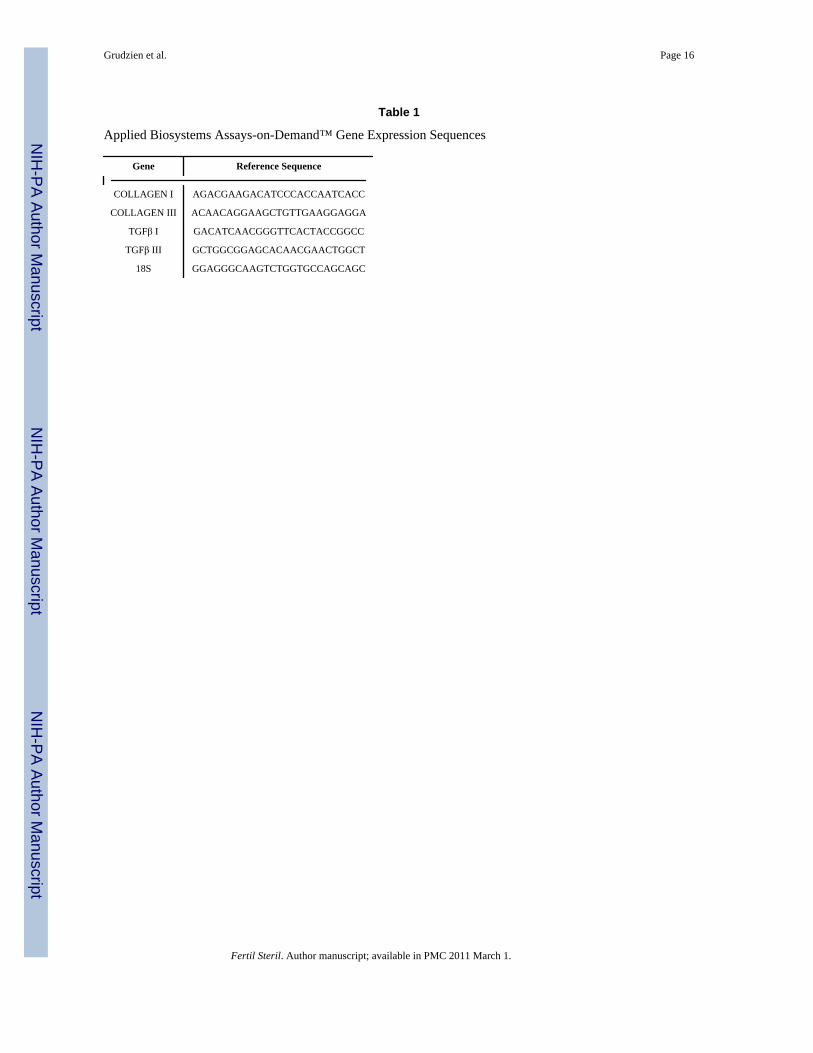
cDNA Synthesis and Quantitative Reverse-Transcriptase PCRTotal RNA was extracted from cells using TRIzol™ and transcribed into cDNA using theiScript™ cDNA Synthesis Kit following the manufacturer’s instructions (cat#170-8890,Bio-Rad, Hercules, CA). Synthesized cDNA was then used for TaqMan® Gene ExpressionAssays real-time PCR analysis (Applied Biosystems, Foster City, CA) using the 20XAssays-on-Demand™ Gene Expression Assays (Applied Biosystems) for COLLAGEN I(Hs00164004_m1), COLLAGEN III (Hs00164103_m1), TGFβ1 (Hs99999918_m1), TGFβ3(Hs00234245_m1) and 18S (Hs99999901_s1). Reference sequences are listed in Table 1.Real-time PCR amplification and detection were performed in ABI Prism 384-well ClearOptical Reaction Plates (Applied Biosystems). Amplification conditions included: hold 10min at 95°C, 40 thermal cycles of denaturing 15 sec at 95°C and anneal/extend for 1 min at60°C. Relative fold induction levels were calculated using the Comparative CT Method asoutlined in the ABI Sequence Detector User Bulletin #2 (www.appliedbiosystems.com). 18Sribosomal RNA gene expression served as an endogenous control.
Effect of Halofuginone on Caspase 3/7 ProductionLeiomyoma and corresponding myometrial SMC lines from three different patients wereused between passages 3-5. Cells were plated at 2,000 per well and allowed to attach for24h. Both cell types where then treated for 0, 12, 24, 48, and 72 hours with 100μL ofmedium containing 0, 25, 100, or 200ng/mL of halofuginone in DMSO vehicle. Controlcells were treated with the same amount of DMSO vehicle alone. A 1μM Staurosporine(Sigma) treatment for 20h was used as a positive control as recommended by the CaspaseGlo® 3/7 Assay Technical Bulletin (Promega, Madison, Wisconsin). After halofuginonetreatment, cells were treated with 100μL of the Caspase Glo® 3/7 Reagent according to themanufacturer’s instructions. Caspase 3/7 activity was detected in quadruplicate wells foreach treatment in relative luminescence units (RLU) using the Bio-Tek Synergy HT Reader(Bio-Tek, Winooski, Vermont). A total of three experiments were carried out.
Effect of Halofuginone on Cell ProliferationLeiomyoma and myometrial SMC lines from three different patients were plated at 200,000cells/dish and allowed to attach for 48h in DMEM supplemented with 10% serum. On day 1,cells were treated with 75ng/mL of halofuginone in 10% serum-containing DMEM, and twodishes were harvested to obtain an initial cell count. On day 3, one pair of dishes wascounted and the remaining dishes were given fresh medium containing 75ng/mLhalofuginone. On day 5, one pair of dishes was counted and all remaining dishes were givenfresh 10% serum DMEM without halofuginone to see if the cells could resume growth afterthe removal of halofuginone. On day7 and day 9, one pair of dishes was counted while theremaining dishes were again given fresh 10% serum DMEM without halofuginone.Leiomyoma and myometrial SMCs that were not treated with halofuginone are not shown inthe results as these cells reached 100% confluency by day 3 (data not shown). A total of fiveexperiments were carried out for leiomyoma cells and three for myometrial cells.
Statistical Analysis for PCR, Cell Proliferation and Caspase AssaysAll PCR experiments were carried out in quadruplicate with each of 3 different cell linesfrom 3 different patients used as replicates. The difference between the threshold cycle ofthe target gene and 18S was used to determine statistical significance (ΔCT) betweentreatments. Threshold cycle (CT) is defined as the cycle number where transcripts are in thelinear phase of amplification. All data were first transformed (2-ΔCT) and normalized to thecontrol treatment expression so that they could be expressed as relative fold differences.Standard error for each treatment was determined using individual transformed 2-ΔCT valuesthat had been normalized to the control treatment for each of the 3 replicates. Data were
Grudzien et al. Page 4
Fertil Steril. Author manuscript; available in PMC 2011 March 1.
NIH
-PA Author Manuscript
NIH
-PA Author Manuscript
NIH
-PA Author Manuscript
transformed using a log10 transformation, and then all points were compared using ANOVA(α=0.05).
Analysis of the proliferation assays was performed using Kruskal-Wallis ANOVA withDunn’s test using the STATA software program (CRC, College Station, TX). An α=0.05was considered statistically significant. All caspase experiments were carried out intriplicate for each of the three cell lines, with results representing the average RLU from all3 experiments. Caspase data were transformed using a log10 transformation, and thenmultiple analyses were carried out using ANOVA (α=0.05). Cell count results represent theaverage of 3-5 experiments using three different leiomyoma or myometrial SMC lines. Cellcount numbers were transformed using a log10 transformation and all time points werecompared using an ANOVA (α=0.05) test. All statistical analyses in this manuscript wereanalyzed using SAS v.9.1 (SAS Institute, Inc, Cary, NC). The statistical significancebetween treatments is indicated in the figures by different letters above treatments.
RESULTSEffect of Halofuginone on Myometrial and Leiomyoma SMC Proliferation
To assess the effects of halofuginone on leiomyoma and myometrial SMC proliferation,cells were treated with increasing concentrations of halofuginone for 24h and analyzed forchanges in DNA synthesis. There was a dose-dependent decrease in DNA synthesis atconcentrations of halofuginone above 25 ng/mL for both myometrial and leiomyoma SMCs(Figure 1). We next tested whether the inhibitory effect of halofuginone on proliferation ofleiomyoma or myometrial SMCs was reversible and whether treated cells could recover andresume proliferating once halofuginone was removed. Cell proliferation experiments werecarried out using leiomyoma and myometrial SMCs from several different patients treatedwith 75ng/mL of halofuginone. This concentration of halofuginone was chosen because itcaused an approximately 50-60% inhibition of DNA synthesis in the thymidine uptakeassays. Cell numbers did not change between days 1-3 in cultures treated with halofuginone,in contrast to untreated cells that had already reached confluence at this time (data notshown). This confirmed the results of the tritiated thymidine uptake assays that showedinhibition of DNA synthesis within the first 24 hrs of treatment. Cell numbers of both typesof treated cells were markedly decreased by day 5 of treatment, suggesting that by this timethere was probably a significant effect on apoptosis (Figure 2). Cells were then placed infresh, halofuginone-free medium on day 5, and cell populations were increased on days 7and 9 showing that the inhibitory effect of halofuginone on cell proliferation was reversible.Cell morphology appeared normal throughout the 5 days of halofuginone treatment and the4 day recovery period (Figure 2).
Effect of Halofuginone on Apoptosis of Myometrial and Leiomyoma SMCsTo determine whether extended halofuginone treatment reduced cell number throughapoptosis, we performed caspase 3/7 activity assays on leiomyoma and autologousmyometrial SMCs treated with halofuginone for up to 72h. Results showed that there wereno differences in caspase 3/7 activity levels between leiomyoma and autologous myometrialSMCs for any of the concentrations of halofuginone tested at any of the time points.Treatment with a low dose (25ng/mL) of halofuginone did not increase caspase 3/7 levels atany point during the 72-hour treatment period (Figure 3a). However, treatment with 100ng/mL of halofuginone increased caspase 3/7 activity levels after 48h (Figure 3b). Treatmentwith a high dose of 200 ng/mL of halofuginone increased caspase 3/7 levels at both the 48hand 72h time points, and to a greater extent than the 100ng/mL dose. The levels of caspase3/7 measured in response to 25, 100, or 200ng/mL halofuginone were significantly different(p=0.05) from each other. Overall, caspase 3/7 activity levels were increased in response to
Grudzien et al. Page 5
Fertil Steril. Author manuscript; available in PMC 2011 March 1.
NIH
-PA Author Manuscript
NIH
-PA Author Manuscript
NIH
-PA Author Manuscript
halofuginone treatment in a dose-dependent manner but the increases were only apparentafter 48h of treatment.
Halofuginone Alters TGFβ and Collagen mRNA Levels in Leiomyoma and MyometrialSMCs
Autologous leiomyoma and myometrial SMC cell lines from four different patients treatedwith 0, 25, 100, or 200ng/mL of halofuginone for 24h were analyzed for differences incollagen type I(αI), collagen type III(αI), TGFβI, and TGFβIII mRNA by Real Time-PCR.Halofuginone decreased not only collagen type I(αI), but also collagen type III(αI) mRNAlevels in both leiomyoma and myometrial SMCs (Figure 4). However, the decrease incollagen mRNA levels in response to halofuginone was dose-dependent only in leiomyomaSMCs. TGFβI mRNA levels were also decreased in both leiomyoma and myometrial SMCsin response to halofuginone (Figure 5) but halofuginone treatment did not significantlydecrease TGFβIII mRNA levels in either leiomyoma or myometrial SMCs.
DISCUSSIONIn the present study we demonstrated that halofuginone inhibits both myometrial andleiomyoma SMC proliferation. Halofuginone inhibited DNA synthesis within 24 hours oftreatment in both leiomyoma and myometrial SMCs in a dose-dependent manner atconcentrations above 25ng/mL. Halofuginone did not increase caspase 3/7 levels, a markerof apoptosis, in either cell type until after 48h of treatment and only at higher doses of 100or 200 ng/ml. The inhibition of DNA synthesis at low concentrations, such as 25ng/mL,coupled with a modest increase in caspase activity (apoptosis) after 72h of treatment,suggests that halofuginone reduces cell proliferation through both mechanisms in uterineSMCs. These results are consistent with the findings of Gavish et al. (21) who observed highlevels of apoptosis in prostate cancer cell xenografts after exposure to high doses ofhalofuginone for 2 months. The increase in apoptosis observed in this study and by Gavishet al. (21) is different from another recent study that reported a decrease in apoptosis in Tcells treated with halofuginone. Leiba et al. (22) showed that halofuginone inhibitedapoptosis in normal, activated T cells. These results suggest that the induction of apoptosisby halofuginone may be a cell type-specific response. Our results showed that increasedlevels of caspase 3/7 activity, and therefore apoptosis, occurred only at higherconcentrations of halofuginone and after 48-72 h of exposure.
This delayed increase in apoptosis was also evident in our longer term cell proliferationexperiments. Leiomyoma and myometrial SMCs treated with halofuginone for 5 days didnot show a decrease in cell number until after 5 days of treatment. Reversal of the inhibitoryeffect of halofuginone on cell growth was delayed as there was no increase in cell numberuntil 4 days after removal of halofuginone. Interestingly, cells treated with halofuginone didnot appear necrotic or unhealthy during the course of the treatment period (Figure 2), andonce halofuginone was removed the cells regained their ability to proliferate as indicated bythe significant increase in cell number by day 9 (Figure 2). Halofuginone inhibited DNAsynthesis in leiomyoma and myometrial SMCs within 24 h of treatment, but increases incaspase 3/7 levels, and therefore apoptosis, appear to require a longer time of exposure tohalofuginone. Other studies have observed a similar inhibition of cell proliferation inresponse to halofuginone. Haran et al. (23) reported that halofuginone inhibited rat renalfibroblast proliferation in a reversible and dose-dependent manner after 48-192 h oftreatment. Another study by Nagler et al. (24) observed a decrease in cultured hepatocellularcarcinoma cell proliferation after one week of treatment with halofuginone, and also did notobserve any toxic effects to the cells or changes in cell viability.
Grudzien et al. Page 6
Fertil Steril. Author manuscript; available in PMC 2011 March 1.
NIH
-PA Author Manuscript
NIH
-PA Author Manuscript
NIH
-PA Author Manuscript
In addition to inhibiting fibroblast and SMC proliferation, halofuginone has also been shownto be a specific inhibitor of collagen type I synthesis and TGFβ signaling (15,16). Weexamined the effects of halofuginone on TGFβ I and III mRNA levels in leiomyoma andmyometrial SMCs because TGFβI and III have direct positive effects on collagen productionin both cell types, and TGFβIII has been shown to increase DNA synthesis in leiomyomaSMCs (12,25). Our results showed that halofuginone specifically decreased TGFβIexpression in both leiomyoma and myometrial SMCs. This is in contrast to a recentlypublished study by Popov et al. (26) who found no change in TGFβI mRNA expression inhalofuginone-treated rat hepatic satellite cells. However, halofuginone has been shown toinhibit TGFβI signaling in embryonic fibroblasts by interfering with Smad3 activation (16),and as TGFβI production is often dependent upon autocrine stimulation (27), halofuginonecould reduce TFGβI production by interfering with the Smad activation pathway. However,halofuginone did not alter TGFβIII mRNA levels in either leiomyoma or myometrial SMCssuggesting that TGFβI and TGFβIII are not regulated in the same manner in these cells.
Real-time PCR analysis of leiomyoma and autologous myometrial SMCs showed ananticipated decrease in collagen type I(αI) mRNA levels in response to treatment withhalofuginone. Since collagen type III is another ECM protein that is upregulated in uterineleiomyomas (28,29), we also examined the effect of halofuginone on collagen type IIImRNA levels. The results showed that halofuginone also markedly reduced collagen type IIImRNA levels in both leiomyoma and myometrial SMCs, and this inhibitory effect oncollagen type III mRNA occurred in a dose-dependent manner in leiomyoma SMCs. Thesuppression of collagen type III mRNA is in agreement with recent findings by Papov et al.(26) who showed a similar decrease in collagen III mRNA in response to halofuginone in rathepatic satellite cells. While these rapid decreases in collagen mRNA levels would notimmediately be reflected by a decrease in collagen protein expression in the ECM, the levelsof newly synthesized, monomeric collagen would be decreased fairly rapidly.
The suppressive effect of halofuginone on collagen production could have multiple effectsin leiomyoma and myometrial SMCs. New collagen production is essential for cellproliferation, not only as a scaffold to support cell growth but also as part of a functionalsignaling pathway. Collagens can signal through the β1 family of integrins and members ofthe discoidin-domain receptor (DDR) family to convey migration, proliferation, and celladhesion signals (33-39). Collagens can also bind various signaling molecules such asmembers of the fibroblast growth factor (FGF) and TGFβ families, and can inhibit orenhance the effects of these growth factors (33-39). The suppression of collagen productionby halofuginone could work in two ways. First, it would limit the protein matrix available toleiomyoma SMCs to support growth and migration. Second, it would also reduce theproliferation signals initiated by interactions of collagen with integrins, DDRs, and throughbinding to members of the FGF and TGFβ growth factor families.
Our studies showed that halofuginone had similar effects on leiomyoma and myometrialSMCs in culture. These results are not very surprising in light of other recent studies thathave shown that myometrial and leiomyoma SMCs behave very similarly in culture. A studyby Zaitseva et al. (30) examined the gene expression patterns of leiomyoma and myometrialcells in vivo and in vitro using microarray gene analysis. Their results showed that while thegene expression patterns of leiomyoma and myometrial SMCs were quite different in vivo,there were relatively small differences in gene expression between the cell types cultured invitro. We have observed similar levels of expression of collagen type I and type III mRNAsin cultured leiomyoma and myometrial cells but this is most likely due to the fact that thesecells upregulate collagen production significantly in culture in response to being plated onplastic surfaces. In addition, the production of TGFβ-1 was also similar for the two celltypes (150-180 pg/ml/24 hrs, (12)). A study by Gross et al (31) showed that while
Grudzien et al. Page 7
Fertil Steril. Author manuscript; available in PMC 2011 March 1.
NIH
-PA Author Manuscript
NIH
-PA Author Manuscript
NIH
-PA Author Manuscript
leiomyomas express much higher levels of HMGA2 mRNA in vivo when compared toautologous myometrium, the levels of HMGA2 are quite similar in cultured myometrial andleiomyoma SMCs. Another recent study by Loy et al. (32) examined the effects of theperoxisome proliferator-activated receptor ligand pioglitazone on cultured leiomyoma andmyometrial cell proliferation. These investigators also reported similar anti-proliferativeeffects of pioglitazone on cultured myometrial and leiomyoma SMCs. It will not be possibleto determine definitively whether there are differences in the effects of halofuginone onleiomyoma and myometrial cells in vivo until the drug can be tested in an in vivo animalmodel or in a clinical pilot study.
In conclusion, we have shown that halofuginone inhibits both leiomyoma and myometrialSMC proliferation by rapidly inhibiting DNA synthesis and later inducing apoptosis. Inaddition, halofuginone significantly suppressed TGFβI mRNA production. The suppressionof TGFβ by halofuginone may be one of the mechanisms by which halofuginone decreasescollagen type I(αI) and collagen type III(αI) mRNA levels in uterine SMCs. These resultssupport our hypothesis that halofuginone could be a novel and effective medical therapy fortreatment of uterine leiomyomas. Future studies will focus on clarifying the signalingpathways used by halofuginone in exerting its anti-fibrotic and anti-proliferative effects inleiomyoma SMCs.
AcknowledgmentsThis work was supported by NIH HD046227 (RAN) and a grant from the LAM Foundation (RAN).
References1. Walker CL, Stewart EA. Uterine fibroids: the elephant in the room. Science 2005;308:1589–1592.
[PubMed: 15947177]2. Chavez NF, Stewart EA. Medical treatment of uterine fibroids. Clin Obstet Gynecol 2001;44:372–
384. [PubMed: 11347559]3. Buttram VC Jr, Reiter RC. Uterine leiomyomata: etiology, symptomatology, and management.
Fertil Steril 1981;36:433–445. [PubMed: 7026295]4. Zhao SZ, Wong JM, Arguelles LM. Hospitalization costs associated with leiomyoma. Clin Therap
1999;21:563–575. [PubMed: 10321423]5. Nowak RA. Identification of new therapies for leiomyomas: what in vitro studies can tell us. Clin
Obstet Gynecol 2001;44:327–334. [PubMed: 11344996]6. Stewart EA. Uterine fibroids. Lancet 2001;357:293–298. [PubMed: 11214143]7. Derynck R, Zhang YE. Smad-dependent and Smad-independent pathways in TGF-beta family
signaling. Nature 2003;425:577–584. [PubMed: 14534577]8. Derynck R, Feng XH. TGF-beta receptor signaling. Biochim Biophys Acta 1997;1333:F105–150.
[PubMed: 9395284]9. Massague J. How cells read TGF-beta signals. Nat Rev Mol Cell Biol 2000;1:169–178. [PubMed:
11252892]10. Murphy, LJ.; Ballejo, G. Growth factor and cytokine expression in the endometrium. In: Findlay,
JK., editor. Molecular biology of the female reproductive system. Academic Press; San Diego:1994. p. 457
11. Tang XM, Dou Q, Zhao Y, McLean F, Davis J, Chegini N. The expression of transforming growthfactor-beta s and TGF-beta receptor mRNA and protein and the effect of TGF-betas on humanmyometrial smooth muscle cells in vitro. Mol Hum Reprod 1997;3:233–240. [PubMed: 9237249]
12. Lee BS, Nowak RA. Human leiomyoma smooth muscle cells show increased expression oftransforming growth factor-beta 3 (TGF beta 3) and altered responses to the antiproliferativeeffects of TGF beta. J Clin Endocrinol Metab 2001;86:913–920. [PubMed: 11158066]
Grudzien et al. Page 8
Fertil Steril. Author manuscript; available in PMC 2011 March 1.
NIH
-PA Author Manuscript
NIH
-PA Author Manuscript
NIH
-PA Author Manuscript
13. Arici A, Sozen I. Transforming growth factor-β3 is expressed at high levels in leiomyoma where itstimulates fibronectin expression and cell proliferation. Fertil Steril 2000;73:1006–1011.[PubMed: 10785229]
14. Pines M, Vlodavsky I, Nagler A. Halofuginone: From veterinary use to human therapy. DrugDevelopment Research 2000;50:371–378.
15. Granot I, Halevy O, Hurwitz S, Pines M. Halofuginone: an inhibitor of collagen type I synthesis.Biochim Biophys Acta 1993;1156:107–112. [PubMed: 8427869]
16. McGaha TL, Phelps RG, Spiera H, Bona C. Halofuginone, an inhibitor of type-I collagen synthesisand skin sclerosis, blocks transforming-growth-factor-beta-mediated Smad3 activation infibroblasts. J Invest Dermatol 2002;118:461–470. [PubMed: 11874485]
17. Nagler A, Miao HQ, Aingorn H, Pines M, Genina O, Vlodavsky I. Inhibition of collagen synthesis,smooth muscle cell proliferation, and injury-induced intimal hyperplasia by halofuginone.Arterioscler Thromb Vasc Biol 1997;17:194–202. [PubMed: 9012656]
18. Pines M, Snyder D, Yarkoni S, Nagler A. Halofuginone to treat fibrosis in chronic graft-versus-host disease and scleroderma. Biol Blood Marrow Transplant 2003;9:417–425. [PubMed:12869955]
19. Xavier S, Piek E, Fujii M, Javelaud D, Mauviel A, Flanders KC, et al. Amelioration of radiation-induced fibrosis: inhibition of transforming growth factor-beta signaling by halofuginone. J BiolChem 2004;279:15167–15176. [PubMed: 14732719]
20. Nagler A, Firman N, Feferman R, Cotev S, Pines M, Shoshan S. Reduction in pulmonary fibrosisin vivo by halofuginone. Am J Respir Crit Care Med 1996;154:1082–1086. [PubMed: 8887611]
21. Gavish Z, Pinthus JH, Barak V, Ramon J, Nagler A, Eshhar Z, et al. Growth inhibition of prostatecancer xenografts by halofuginone. Prostate 2002;51:73–83. [PubMed: 11948962]
22. Leiba M, Cahalon L, Shimoni A, Lider O, Zanin-Zhorov A, Hecht I, et al. Halofuginone inhibitsNF-{kappa}B and p38 MAPK in activated T cells. J Leukoc Biol 2006;80:399–406. [PubMed:16769768]
23. Haran N, Leschinski L, Pines M, Rapoport J. Inhibition of rat renal fibroblast proliferation byhalofuginone. Nephron Exp Nephrol 2006;104:e35–e40. [PubMed: 16735800]
24. Nagler A, Ohana M, Shibolet O, Shapira MY, Alper R, Vlodavsky I, et al. Suppression ofhepatocellular carcinoma growth in mice by the alkaloid coccidiostat halofuginone. Eur J Cancer2004;40:1397–1403. [PubMed: 15177499]
25. Chegini N, Luo X, Ding L, Ripley D. The expression of Smads and transforming growth factorbeta receptors in leiomyoma and myometrium and the effect of gonadotropin releasing hormoneanalogue therapy. Mol Cell Endocrinol 2003;209:9–16. [PubMed: 14604812]
26. Popov Y, Patsenker E, Bauer M, Niedobitek E, Schulze-Krebs A, Schuppan D. Halofuginoneinduces matrix metalloproteinases in rat hepatic stellate cells via activation of p38 and NFkappaB.J Biol Chem 2006;281:15090–15098. [PubMed: 16489207]
27. Clark DA, Coker R. Transforming growth factor-beta (TGF-beta). Int J Biochem Cell Biol1998;30:293–298. [PubMed: 9611771]
28. Fujita M. Histological and biochemical studies of collagen in human uterine leiomyomas.Hokkaido Igaku Zasshi 1985;60:602–615. [PubMed: 4054826]
29. Stewart EA, Friedman AJ, Peck K, Nowak RA. Relative overexpression of collagen type I andcollagen type III messenger ribonucleic acids by uterine leiomyomas during the proliferative phaseof the menstrual cycle. J Clin Endocrinol Metab 1994;79:900–906. [PubMed: 8077380]
30. Zaitseva M, Vollenhoven BJ, Rogers PA. In vitro culture significantly alters gene expressionprofiles and reduces differences between myometrial and leiomyoma smooth muscle cells. MolHum Reprod 2006;12:187–207. [PubMed: 16524927]
31. Gross KL, Neskey DM, Manchanda N, Weremowicz S, Kleinman MS, Nowak RA, et al. HMGA2expression in uterine leiomyomata and myometrium: quantitative analysis and tissue culturestudies. Genes Chromosomes Cancer 2003;38:68–79. [PubMed: 12874787]
32. Loy CJ, Evelyn S, Lim FK, Liu MH, Yong EL. Growth dynamics of human leiomyoma cells andinhibitory effects of the peroxisome proliferators-activated receptor ligand, pioglitazone. Mol HumReprod 2005;11:561–566. [PubMed: 16051682]
Grudzien et al. Page 9
Fertil Steril. Author manuscript; available in PMC 2011 March 1.
NIH
-PA Author Manuscript
NIH
-PA Author Manuscript
NIH
-PA Author Manuscript
33. Brooke BS, Karnik SK, Li DY. Extracellular matrix in vascular morphogenesis and disease:structure versus signal. Trends Cell Biol 2003;13:51–56. [PubMed: 12480340]
34. Henriet P, Zhong ZD, Brooks PC, Weinberg KI, DeClerck YA. Contact with fibrillar collageninhibits melanoma cell proliferation by up-regulating p27KIP1. Proc Natl Acad Sci U S A2000;97:10026–10031. [PubMed: 10944199]
35. Koyama H, Raines EW, Bornfeldt KE, Roberts JM, Ross R. Fibrillar collagen inhibits arterialsmooth muscle proliferation through regulation of Cdk2 inhibitors. Cell 1996;87:1069–78.[PubMed: 8978611]
36. Pozzi A, Wary KK, Giancotti FG, Gardner HA. Integrin alpha1beta1 mediates a unique collagen-dependent proliferation pathway in vivo. J Cell Biol 1998;142:587–594. [PubMed: 9679154]
37. Vogel WF, Aszodi A, Alves F, Pawson T. Discoidin domain receptor 1 tyrosine kinase has anessential role in mammary gland development. Mol Cell Biol 2001;21:2906–2917. [PubMed:11283268]
38. Hou G, Vogel WF, Bendeck MP. Tyrosine kinase activity of discoidin domain receptor 1 isnecessary for smooth muscle cell migration and matrix metalloproteinase expression. Circ Res2002;90:1147–1149. [PubMed: 12065315]
39. Vogel W, Gish GD, Alves F, Pawson T. The discoidin domain receptor tyrosine kinases areactivated by collagen. Mol Cell 1997;1:13–23. [PubMed: 9659899]
Grudzien et al. Page 10
Fertil Steril. Author manuscript; available in PMC 2011 March 1.
NIH
-PA Author Manuscript
NIH
-PA Author Manuscript
NIH
-PA Author Manuscript

Figure 1. Halofuginone inhibits DNA synthesis in leiomyoma and myometrial cellsLeiomyoma (black bars) and myometrial (grey bars) SMCs were treated with increasingconcentrations of halofuginone for 24 hrs and tritiated thymidine incorporation wasmeasured by liquid scintillation. Tritiated thymidine incorporation is reported as counts perminute (CPM). Eight experiments were carried out with six samples per treatment for eachexperiment. Statistically significant differences between treatments are indicated bydifferent lower case letters (p≤0.05).
Grudzien et al. Page 11
Fertil Steril. Author manuscript; available in PMC 2011 March 1.
NIH
-PA Author Manuscript
NIH
-PA Author Manuscript
NIH
-PA Author Manuscript
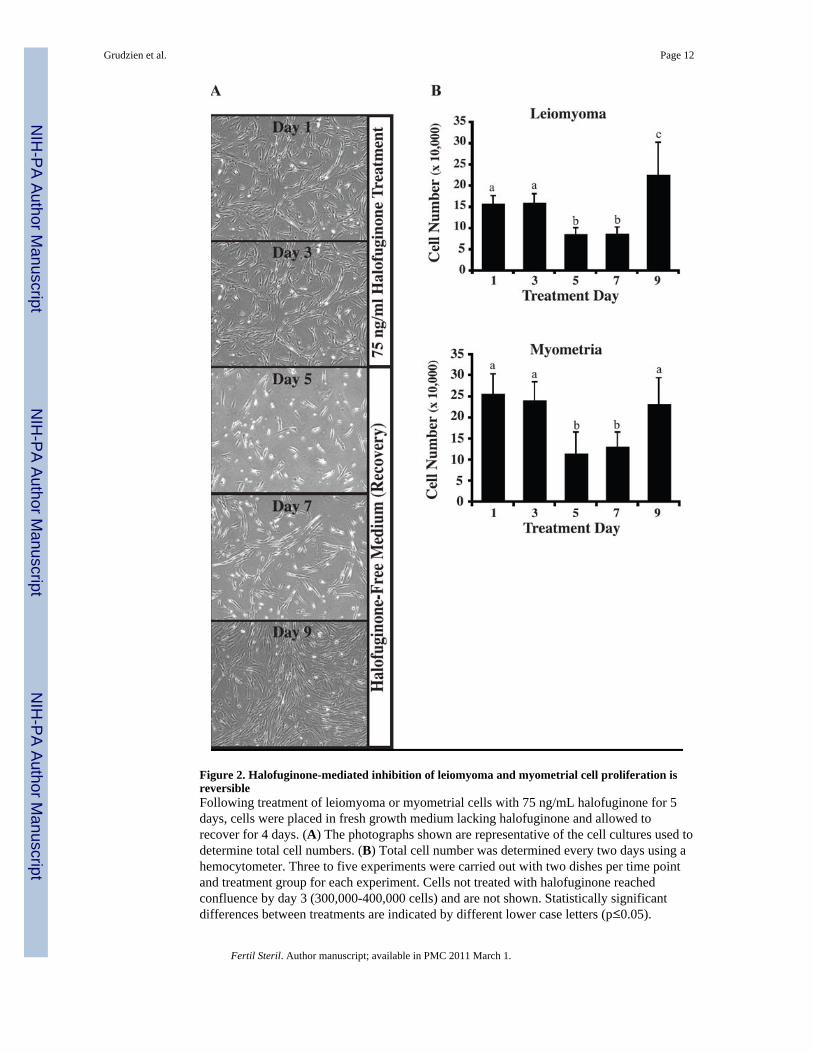
Figure 2. Halofuginone-mediated inhibition of leiomyoma and myometrial cell proliferation isreversibleFollowing treatment of leiomyoma or myometrial cells with 75 ng/mL halofuginone for 5days, cells were placed in fresh growth medium lacking halofuginone and allowed torecover for 4 days. (A) The photographs shown are representative of the cell cultures used todetermine total cell numbers. (B) Total cell number was determined every two days using ahemocytometer. Three to five experiments were carried out with two dishes per time pointand treatment group for each experiment. Cells not treated with halofuginone reachedconfluence by day 3 (300,000-400,000 cells) and are not shown. Statistically significantdifferences between treatments are indicated by different lower case letters (p≤0.05).
Grudzien et al. Page 12
Fertil Steril. Author manuscript; available in PMC 2011 March 1.
NIH
-PA Author Manuscript
NIH
-PA Author Manuscript
NIH
-PA Author Manuscript
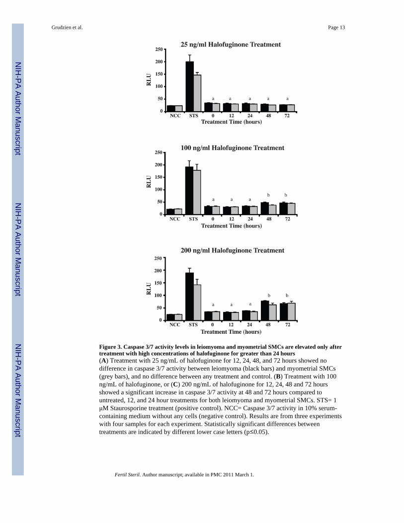
Figure 3. Caspase 3/7 activity levels in leiomyoma and myometrial SMCs are elevated only aftertreatment with high concentrations of halofuginone for greater than 24 hours(A) Treatment with 25 ng/mL of halofuginone for 12, 24, 48, and 72 hours showed nodifference in caspase 3/7 activity between leiomyoma (black bars) and myometrial SMCs(grey bars), and no difference between any treatment and control. (B) Treatment with 100ng/mL of halofuginone, or (C) 200 ng/mL of halofuginone for 12, 24, 48 and 72 hoursshowed a significant increase in caspase 3/7 activity at 48 and 72 hours compared tountreated, 12, and 24 hour treatments for both leiomyoma and myometrial SMCs. STS= 1μM Staurosporine treatment (positive control). NCC= Caspase 3/7 activity in 10% serum-containing medium without any cells (negative control). Results are from three experimentswith four samples for each experiment. Statistically significant differences betweentreatments are indicated by different lower case letters (p≤0.05).
Grudzien et al. Page 13
Fertil Steril. Author manuscript; available in PMC 2011 March 1.
NIH
-PA Author Manuscript
NIH
-PA Author Manuscript
NIH
-PA Author Manuscript
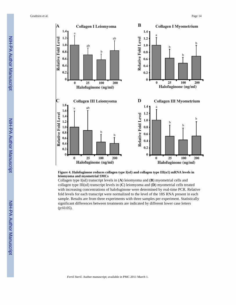
Figure 4. Halofuginone reduces collagen type I(αI) and collagen type III(α1) mRNA levels inleiomyoma and myometrial SMCsCollagen type I(αI) transcript levels in (A) leiomyoma and (B) myometrial cells andcollagen type III(αI) transcript levels in (C) leiomyoma and (D) myometrial cells treatedwith increasing concentrations of halofuginone were determined by real-time PCR. Relativefold levels for each transcript were normalized to the level of the 18S RNA present in eachsample. Results are from three experiments with three samples per experiment. Statisticallysignificant differences between treatments are indicated by different lower case letters(p≤0.05).
Grudzien et al. Page 14
Fertil Steril. Author manuscript; available in PMC 2011 March 1.
NIH
-PA Author Manuscript
NIH
-PA Author Manuscript
NIH
-PA Author Manuscript
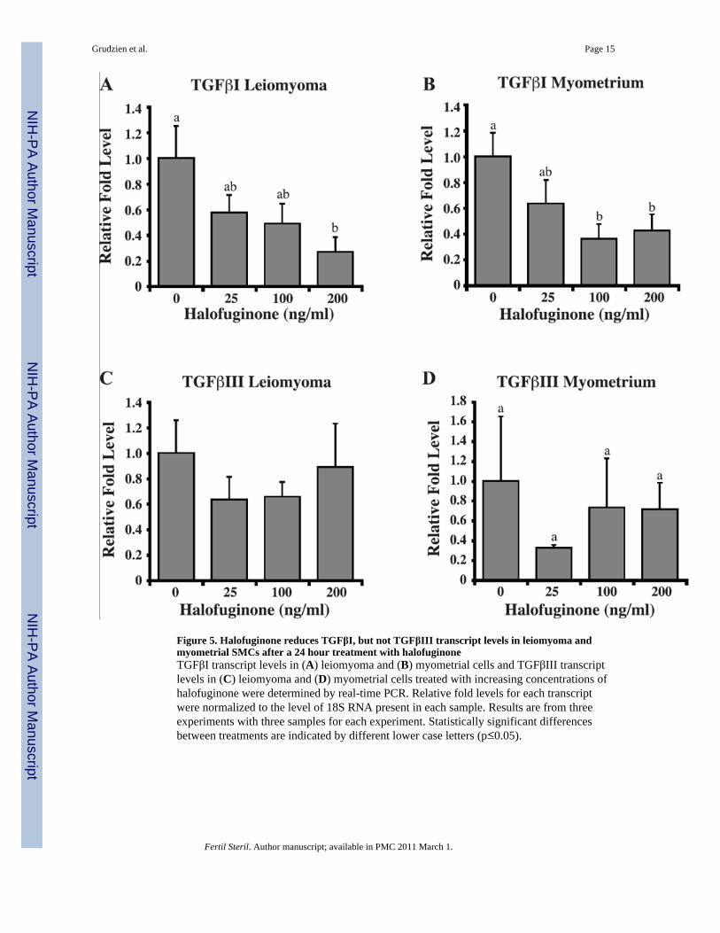
Figure 5. Halofuginone reduces TGFβI, but not TGFβIII transcript levels in leiomyoma andmyometrial SMCs after a 24 hour treatment with halofuginoneTGFβI transcript levels in (A) leiomyoma and (B) myometrial cells and TGFβIII transcriptlevels in (C) leiomyoma and (D) myometrial cells treated with increasing concentrations ofhalofuginone were determined by real-time PCR. Relative fold levels for each transcriptwere normalized to the level of 18S RNA present in each sample. Results are from threeexperiments with three samples for each experiment. Statistically significant differencesbetween treatments are indicated by different lower case letters (p≤0.05).
Grudzien et al. Page 15
Fertil Steril. Author manuscript; available in PMC 2011 March 1.
NIH
-PA Author Manuscript
NIH
-PA Author Manuscript
NIH
-PA Author Manuscript
NIH
-PA Author Manuscript
NIH
-PA Author Manuscript
NIH
-PA Author Manuscript
Grudzien et al. Page 16
Table 1
Applied Biosystems Assays-on-Demand™ Gene Expression Sequences
Gene Reference Sequence
COLLAGEN I AGACGAAGACATCCCACCAATCACC
COLLAGEN III ACAACAGGAAGCTGTTGAAGGAGGA
TGFβ I GACATCAACGGGTTCACTACCGGCC
TGFβ III GCTGGCGGAGCACAACGAACTGGCT
18S GGAGGGCAAGTCTGGTGCCAGCAGC
Fertil Steril. Author manuscript; available in PMC 2011 March 1.
Copyright © 2022 FDOKUMEN