Close-loop dynamic nanohybrids on collagen-ark with in situ ...
9
This journal is © The Royal Society of Chemistry 2019 Mater. Horiz., 2019, 6, 385--393 | 385 Cite this: Mater. Horiz., 2019, 6, 385 Close-loop dynamic nanohybrids on collagen-ark with in situ gelling transformation capability for biomimetic stage-specific diabetic wound healing† Zehua Liu,‡ a Yunzhan Li,‡ bc Wei Li,* a Wenhua Lian, bc Marianna Kemell, d Sami Hietala, d Patrı ´ cia Figueiredo, a Li Li, bc Ermei Ma ¨ kila ¨ , e Ming Ma, f Jarno Salonen, e Jouni T. Hirvonen, a Dongfei Liu, ag Hongbo Zhang,* hi Xianming Deng* bc and He ´ lder A. Santos * ag Here, an oxidation/acid dual-responsive nanohybrids/ark system was produced. The microfluidics-produced nanohybrids endow the system with an orchestrated cascade from wound detection, reactive oxygen species scavenging, drug release to hydrogel formation. The drug release behavior imitates the dynamic wound healing process, thus rendering an enhanced bio-mimetic regeneration. Intelligent materials that adapt and respond to their environ- ment are termed as dynamic materials, 1 and have recently drawn attention for tissue engineering and regeneration. Conventional materials often fail to apprehend the dynamic and hetero- geneous nature of an in vivo environment during the regeneration process. In contrast, strategically designed dynamic materials can bio-mimetically alter their physiochemical properties when encountering a specific biological irritant, further executing pro- grammed functions, and thus holding great clinical value. 1–4 Diabetic foot ulcer (DFU) is a critical non-healing complication for diabetic patients. 5 Over the past decades, great efforts have been made to overcome this challenge. However, despite the reporting of several bioactive substances (e.g., small molecule drugs, growth factors, and cytokines) used to accelerate the healing process, the combination of a ‘‘smart’’ delivery system and tailored release behavior is equally indispensable. As for tissue engineering, long-term or un-regulated drug delivery may cause aberrant regeneration and even formation of carcinoma-like structure. 6,7 Inspired by the meticulous self-regulating system within the human body, where regenerative enhancement is dynamically altered during different healing stages, 8–10 prospec- tive materials for fabricating the envisioned bio-mimetic delivery system should be able to sense and respond to the healing process, and concurrently alter their drug releasing behavior. a Drug Research Program, Division of Pharmaceutical Chemistry and Technology, Faculty of Pharmacy, University of Helsinki, FI-00014, Helsinki, Finland. E-mail: [email protected], [email protected] b State Key Laboratory of Cellular Stress Biology, Innovation Center for Cell Signaling Network, School of Life Sciences, Xiamen University, 361102, Fujian, China. E-mail: [email protected] c State-Province Joint Engineering Laboratory of Targeted Drugs from Natural Products, School of Life Sciences, Xiamen University, 361102, Fujian, China d Department of Chemistry, University of Helsinki, FI-00014, Helsinki, Finland e Laboratory of Industrial Physics, Department of Physics, University of Turku, FI-20014, Turku, Finland f State Key Laboratory of High Performance Ceramics and Superfine Microstructures, Shanghai Institute of Ceramics, Chinese Academy of Sciences, Shanghai 200050, China g Helsinki Institute of Life Science (HiLIFE), University of Helsinki, FI-00014, Helsinki, Finland h Department of Pharmaceutical Science, Åbo Akademi University, FI-20520, Turku, Finland. E-mail: [email protected] i Turku Center of Biotechnology, University of Turku and Åbo Akademi University, FI-20520, Turku, Finland † Electronic supplementary information (ESI) available. See DOI: 10.1039/c8mh01145a ‡ These authors contributed to this work equally. Received 14th September 2018, Accepted 31st October 2018 DOI: 10.1039/c8mh01145a rsc.li/materials-horizons Conceptual insights We have developed here a self-regulated dynamic nanohybrid that can sensitively respond to a hyperglycemic microenvironment. The nanohybrid is subsequently embedded in a collagen patch for further in vivo diabetic wound healing applications. The nanohybrid with a core/shell structure was produced through a single-step microfluidics-assisted nanoprecipitation method, where drug-loaded porous silicon (PSi) nanoparticles were encapsulated by a H 2 O 2 responsive polymer, 4-(hydroxymethyl)-phenyl- boronic acid pinacol ester conjugated with oxidized dextran (POD). POD encapsulation can effectively block accessible pores of PSi, thus preventing a potent burst release of the drug. Upon exposure to high levels of glucose, GOx can initiate glucose oxidation, generating gluconic acid and H 2 O 2 . Then, POD can consume the produced H 2 O 2 to undergo degradation and further release the encapsulated drug. The degradation product of POD, oxidized dextran (ODEX), can in situ form a hydrogel with the collagen patch. This in situ nanoparticle/hydrogel transformation creates a moist environment around the wound bed, altering the stiffness of the patch to better mimic elastic properties of the natural soft tissues. Glucose content within the wound bed also gradually decreased as a result of less bleeding and interstitial fluid exudation, thus a stage/temporal-specific drug release can also be achieved. Materials Horizons COMMUNICATION Open Access Article. Published on 31 October 2018. Downloaded on 6/3/2022 6:25:40 AM. This article is licensed under a Creative Commons Attribution-NonCommercial 3.0 Unported Licence. View Article Online View Journal | View Issue
-
Upload
khangminh22 -
Category
Documents
-
view
0 -
download
0
Transcript of Close-loop dynamic nanohybrids on collagen-ark with in situ ...

This journal is©The Royal Society of Chemistry 2019 Mater. Horiz., 2019, 6, 385--393 | 385
Cite this:Mater. Horiz., 2019,
6, 385
Close-loop dynamic nanohybrids on collagen-arkwith in situ gelling transformation capability forbiomimetic stage-specific diabetic woundhealing†
Zehua Liu,‡a Yunzhan Li,‡bc Wei Li,*a Wenhua Lian,bc Marianna Kemell, d
Sami Hietala, d Patrıcia Figueiredo,a Li Li,bc Ermei Makila,e Ming Ma, f
Jarno Salonen,e Jouni T. Hirvonen,a Dongfei Liu, ag Hongbo Zhang,*hi
Xianming Deng*bc and Helder A. Santos *ag
Here, an oxidation/acid dual-responsive nanohybrids/ark system
was produced. The microfluidics-produced nanohybrids endow the
system with an orchestrated cascade from wound detection, reactive
oxygen species scavenging, drug release to hydrogel formation. The
drug release behavior imitates the dynamic wound healing process,
thus rendering an enhanced bio-mimetic regeneration.
Intelligent materials that adapt and respond to their environ-ment are termed as dynamic materials,1 and have recently drawnattention for tissue engineering and regeneration. Conventionalmaterials often fail to apprehend the dynamic and hetero-geneous nature of an in vivo environment during the regenerationprocess. In contrast, strategically designed dynamic materialscan bio-mimetically alter their physiochemical properties whenencountering a specific biological irritant, further executing pro-grammed functions, and thus holding great clinical value.1–4
Diabetic foot ulcer (DFU) is a critical non-healing complicationfor diabetic patients.5 Over the past decades, great efforts havebeen made to overcome this challenge. However, despite thereporting of several bioactive substances (e.g., small moleculedrugs, growth factors, and cytokines) used to accelerate thehealing process, the combination of a ‘‘smart’’ delivery systemand tailored release behavior is equally indispensable. As fortissue engineering, long-term or un-regulated drug delivery maycause aberrant regeneration and even formation of carcinoma-likestructure.6,7 Inspired by the meticulous self-regulating systemwithin the human body, where regenerative enhancement isdynamically altered during different healing stages,8–10 prospec-tive materials for fabricating the envisioned bio-mimetic deliverysystem should be able to sense and respond to the healingprocess, and concurrently alter their drug releasing behavior.
a Drug Research Program, Division of Pharmaceutical Chemistry and Technology,
Faculty of Pharmacy, University of Helsinki, FI-00014, Helsinki, Finland.
E-mail: [email protected], [email protected] State Key Laboratory of Cellular Stress Biology, Innovation Center for Cell
Signaling Network, School of Life Sciences, Xiamen University, 361102, Fujian,
China. E-mail: [email protected] State-Province Joint Engineering Laboratory of Targeted Drugs from Natural
Products, School of Life Sciences, Xiamen University, 361102, Fujian, Chinad Department of Chemistry, University of Helsinki, FI-00014, Helsinki, Finlande Laboratory of Industrial Physics, Department of Physics, University of Turku,
FI-20014, Turku, Finlandf State Key Laboratory of High Performance Ceramics and Superfine
Microstructures, Shanghai Institute of Ceramics, Chinese Academy of Sciences,
Shanghai 200050, Chinag Helsinki Institute of Life Science (HiLIFE), University of Helsinki, FI-00014,
Helsinki, Finlandh Department of Pharmaceutical Science, Åbo Akademi University, FI-20520, Turku,
Finland. E-mail: [email protected] Turku Center of Biotechnology, University of Turku and Åbo Akademi University,
FI-20520, Turku, Finland
† Electronic supplementary information (ESI) available. See DOI: 10.1039/c8mh01145a‡ These authors contributed to this work equally.
Received 14th September 2018,Accepted 31st October 2018
DOI: 10.1039/c8mh01145a
rsc.li/materials-horizons
Conceptual insightsWe have developed here a self-regulated dynamic nanohybrid that cansensitively respond to a hyperglycemic microenvironment. The nanohybridis subsequently embedded in a collagen patch for further in vivo diabeticwound healing applications. The nanohybrid with a core/shell structure wasproduced through a single-step microfluidics-assisted nanoprecipitationmethod, where drug-loaded porous silicon (PSi) nanoparticles wereencapsulated by a H2O2 responsive polymer, 4-(hydroxymethyl)-phenyl-boronic acid pinacol ester conjugated with oxidized dextran (POD). PODencapsulation can effectively block accessible pores of PSi, thus preventinga potent burst release of the drug. Upon exposure to high levels of glucose,GOx can initiate glucose oxidation, generating gluconic acid and H2O2.Then, POD can consume the produced H2O2 to undergo degradation andfurther release the encapsulated drug. The degradation product of POD,oxidized dextran (ODEX), can in situ form a hydrogel with the collagenpatch. This in situ nanoparticle/hydrogel transformation creates a moistenvironment around the wound bed, altering the stiffness of the patch tobetter mimic elastic properties of the natural soft tissues. Glucose contentwithin the wound bed also gradually decreased as a result of less bleedingand interstitial fluid exudation, thus a stage/temporal-specific drug releasecan also be achieved.
MaterialsHorizons
COMMUNICATION
Ope
n A
cces
s A
rtic
le. P
ublis
hed
on 3
1 O
ctob
er 2
018.
Dow
nloa
ded
on 6
/3/2
022
6:25
:40
AM
. T
his
artic
le is
lice
nsed
und
er a
Cre
ativ
e C
omm
ons
Attr
ibut
ion-
Non
Com
mer
cial
3.0
Unp
orte
d L
icen
ce.
View Article OnlineView Journal | View Issue

386 | Mater. Horiz., 2019, 6, 385--393 This journal is©The Royal Society of Chemistry 2019
Phenylboronic acid ester (PAE) is an important building blockfor constructing a series of oxidation responsive materials.11–14
This unique feature renders it suitable for application in chronicwounds due to elevated concentrations of common reactiveoxygen species (ROS) within the wound bed.11 However, despitePAE’s ROS scavenging profile and boronate degradation pro-ducts, which favor augmentation of the wound healing rate, fewpapers have applied PAE based materials for DFU caring.15,16
This may be explained by selective oxidation behavior of PAE,which can only be oxidized by H2O2 and not by other ROS,such as superoxide, hypochlorous acid or hydroxyl radical17,18 –resulting in a sensitivity concern when applied in vivo. The lowsolubility of PAE containing polymers in various solvents furtherconstrains the productions of its electrospun nanofibrousmembrane or hydrogels.13 Therefore, a more deliberately designedsystem should be conceived for further applications.
High level of glucose is another pivotal factor in diabetes andmay impact the delivery system construction for DFU repair.Among the reported glucose responsive materials so far, theintroduction of glucose oxidase (GOx) is especially advantageousfor diabetic wound healing, as it can effectively decrease theglucose level that is likely of great importance for diabeticwound healing management.19,20 However, GOx-containingbulk hydrogels exhibit an insensitive response to glucose.21,22
GOx-Containing membranes suffer from low mechanical strength,which can result in premature leakage of the drug.21,22 Moreover,the side products, especially of hydrogen peroxide that is generatedby this oxidation process, may further increase concerns of itsbiocompatibility.23,24 A viable approach to circumvent thisissue is to fabricate a closed-loop nanocarrier that can expendits side products, and sequentially generate the required down-stream actions.
Based on above considerations, herein, we have developed aself-regulated dynamic nanohybrid that could sensitivelyrespond to the hyperglycaemic microenvironment. The nano-hybrid with a core/shell structure was produced through asingle-step microfluidics-assisted nano-precipitation method,25,26
where drug-loaded porous silicon (PSi) nanoparticles were encap-sulated by a H2O2/acid dual-responsive polymer formed by4-(hydroxymethyl)-phenylboronic acid pinacol ester conjugatedwith oxidized dextran (POD). PSi has been widely investigatedfor biomedical applications due to its biocompatibility andflexibility in loading different types of cargos.27,28 Moreover,its main degradation product, orthosilicic acid, has also beenknown to favor the cutaneous traumatic healing,29–31 thusmaking PSi an attractive candidate for DFU application. PODencapsulation can effectively block accessible pores of PSi, thuspreventing a potent burst release of the drug. Furthermore, theredundant aldehyde groups on the outer surface of POD canboth immobilize GOx through the formation of a pH-responsivedynamic covalent bond (imine),32,33 and/or simultaneouslyanchor other amine containing drugs. The benefits of such adelivery system arise from the interaction between the oxidation/acid dual-responsive POD and GOx, which confers control overthe drug release property in a glucose concentration dependentmanner. To facilitate in vivo application, the nanohybrids were
feasibly loaded onto a collagen ark. Interestingly, the degrada-tion product of POD, oxidized dextran (ODEX), could in situform a hydrogel with the collagen patch. This in situ nano-particle/hydrogel transformation can create a moisture environ-ment around the wound bed, altering the stiffness of the patchto better mimic the elastic properties of natural soft tissues.34,35
More importantly, along with dermis regeneration and cutaneousgap closure during the treatment, the glucose content within thewound bed will gradually decrease as a result of less bleeding andinterstitial fluid exudation – thus, a stage/temporal-specific drugrelease schedule can be achieved.36 Herein, the physicochemicalproperties of the nanohybrids were characterized, and theglucose triggered orchestrated cascade and its potent bioactivitywas evaluated both in vitro and in vivo through diabetic woundmice model.
Fig. 1A schematically illustrates the fabrication and dynamictransformation of the nanohybrids during the treatment process.We modified oxidized dextran (ODEX) with phenylboronicpinacol ester to produce POD. The synthesis procedure andits degradation under H2O2 irritation is briefly described inFig. S1 (ESI†). Successful oxidation of dextran was first con-firmed by fourier transform infrared spectroscopy (FTIR), as anew band was observed at 1720 cm�1, which was attributed tothe aldehyde groups (Fig. S2, ESI†).37 Oxidation degree wasfurther determined as 40.2% by 1H NMR by the carbazoneformation method (ESI†). 4-(Hydroxymethyl)phenylboronicacid pinacol ester (PAPE) was then conjugated to the residuehydroxyl groups of oxidized dextran via a carbonate linkage.The substitution degree of hydroxyl groups was ca. 31%, asdetermined from the 1H NMR peak area with the peak ofdextran used as the standard (Fig. S3, ESI†).13 The conjugationof PAPE enhanced the hydrophobicity of polymer, as the newproduct precipitated out from an aqueous solution. A micro-fluidic assisted nanoprecipitation method was used to con-struct PSi@POD, and the nanoprecipitation process wastriggered by a passive microfluidic mixing in a co-flow glasscapillary microtube.25,38 The outer POD layer within the micro-tube was formed through nucleation and nanoprecipitationdriven by diffusion and mixing between the solvent (EtOH/dd-H2O = 8 : 2, v/v) and anti-solvent (dd-H2O) streams across theinterface. It should be noted that no extra surfactant wasneeded to obtain particles with satisfactory polydispersity index(PdI) during this process, as the unmodified hydroxyl groups onODEX function as stabilizers.39 Transmission electron micro-scopy (TEM) images indicated the successful encapsulationof PSi nanoparticles (Fig. 1B(a)) in the POD matrix. As shownin Fig. 1B(b), after encapsulation, the irregular shape and theporous structure of PSi were fully covered and new nanohybridswith spherical morphology were formed. Energy dispersiveX-ray microscopy (EDX) was carried out to further confirm thesuccessful encapsulation (Fig. 1B(c)). At physiological condi-tions, free aldehyde groups on the surface steadily formedSchiff-bases with primary amine groups,40 which were furtherutilized for surface immobilization of both GOx and deferoxamine(DFO) without causing obvious morphology changes (as observedfrom TEM) (Fig. 1B(d)). Scanning electron microscopy (SEM)
Communication Materials Horizons
Ope
n A
cces
s A
rtic
le. P
ublis
hed
on 3
1 O
ctob
er 2
018.
Dow
nloa
ded
on 6
/3/2
022
6:25
:40
AM
. T
his
artic
le is
lice
nsed
und
er a
Cre
ativ
e C
omm
ons
Attr
ibut
ion-
Non
Com
mer
cial
3.0
Unp
orte
d L
icen
ce.
View Article Online

This journal is©The Royal Society of Chemistry 2019 Mater. Horiz., 2019, 6, 385--393 | 387
images of the prepared particles embedded in the collagenpatch illustrated a uniformly formed nano-network (Fig. 1B(e)),confirming it to be a facile method for the preparation of atopical patch. The successful encapsulation and Schiff-baseformation were verified by FTIR (Fig. 1C). After encapsulation,the band at 1045 cm�1 for Si–O, which is a typical band for PSi,disappeared, and was replaced by the band at 1010 cm�1 due topolysaccharide encapsulation. The typical band from Schiff-base(CQN) was observed at 1637 cm�1 after DFO conjugation.25,41
After the encapsulation, the particle size changed from185 � 2 (PSi) to 226 � 5 nm (PdI: 0.12 � 0.01 vs. 0.06 � 0.03)and the zeta (z)-potential values shifted from �21.4 � 0.9 to�48.1 � 2.0 mV, while after Schiff-base formation both size(284 � 7) and PdI (0.26 � 0.04) increased and the zeta potentialchanged to �23.9 � 1.2 mV due to the surface drug/proteinimmobilization (Fig. 1D).
The introduction of GOx in the nanohybrids can endow thesystem with an ability to respond to different glucose levels, as
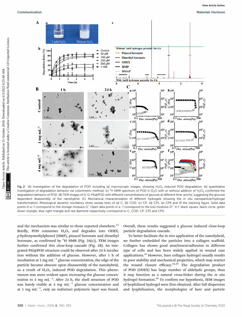
glucose can be catalyzed by GOx to generate gluconic acid andhydrogen peroxide.42 The glucose-dependent H2O2 generationand pH-drop were separately confirmed through the 20,7 0-dichlorodihydrofluorescein diacetate (DCFH-DA) and phenol-red assisted colorimetric methods (Fig. S4, ESI†). It was notedthat at glucose concentration of 4 mg mL�1, which is the typicalglucose level at hyperglycemia, the H2O2 yield was rather high(380 � 7 mM). This concentration may bring potent damage tothe surrounding environment when applied for biomedicalapplications. We hypothesized that the co-existing POD couldconsume H2O2 and initiate the downstream action, thereforeH2O2 induced degradation behavior of PAPE–ODEX was furtherinvestigated. Macroscopic images confirmed the H2O2 triggeredpolymer degradation (Fig. 2A(a)). Bare POD particles weresuspended in H2O with different concentrations of H2O2 andthe degradation process was observed by measuring lightscattering at 500 nm (Fig. 2A(b)). The results suggested a highlysensitive H2O2 concentration-dependent degradation of POD
Fig. 1 Preparation and characterization of DFO, GOx conjugated, ATO loaded nanohybrid (D-G-PSi@POD). (A) Schematic illustration of theconstruction of glucose responsive delivery system and the orchestrated cascade for diabetic wound care. The detrimental side product of H2O2,was utilized and consumed, further aiding the close-loop system. (B) TEM/SEM images showing morphologies of (a) PSi, (b) PSi@POD and (c) theirrespective EDX spectra at particle site and the background, (d) D-G-PSi@POD and (e) D-G-PSi@POD embedded on the collagen patch without obviousmorphology changes. (C) FTIR spectra of PSi, PSi@POD, D-PSi@POD and DFO, suggesting a successful encapsulation and Schiff-base formation. (D) Size,PdI and zeta-potential shift during each step of synthesis, suggesting a successful modification in each step.
Materials Horizons Communication
Ope
n A
cces
s A
rtic
le. P
ublis
hed
on 3
1 O
ctob
er 2
018.
Dow
nloa
ded
on 6
/3/2
022
6:25
:40
AM
. T
his
artic
le is
lice
nsed
und
er a
Cre
ativ
e C
omm
ons
Attr
ibut
ion-
Non
Com
mer
cial
3.0
Unp
orte
d L
icen
ce.
View Article Online
388 | Mater. Horiz., 2019, 6, 385--393 This journal is©The Royal Society of Chemistry 2019
and the mechanism was similar to those reported elsewhere.13
Briefly, POD consumes H2O2 and degrades into ODEX,p-hydroxymethylphenol (HMP), pinacol boronate and dimethylboronate, as confirmed by 1H NMR (Fig. 2A(c)). TEM imagesfurther confirmed this close-loop cascade (Fig. 2B). An inte-grated PSi@POD structure could be observed after 24 h incuba-tion without the addition of glucose. However, after 1 h ofincubation at 1 mg mL�1 glucose concentration, the edge of theparticle became obscure upon disassembly of the nanohybrid,as a result of H2O2 induced POD degradation. This pheno-menon was more evident upon increasing the glucose concen-tration to 4 mg mL�1. After 24 h, the shell structure of PODwas barely visible at 4 mg mL�1 glucose concentration andat 1 mg mL�1, only an indistinct polymeric layer was found.
Overall, these results suggested a glucose induced close-loopparticle degradation cascade.
To better facilitate the in vivo application of the nanohybrid,we further embedded the particles into a collagen scaffold.Collagen has shown good attachment/adhesion to differenttype of cells and has been widely applied in wound careapplications.43 However, bare collagen hydrogel usually resultsin poor stability and mechanical properties, which may restrictthe wound closure efficacy.44,45 The degradation productof POD (ODEX) has large number of aldehyde groups, thusit may function as a natural cross-linker during the in situhydrogel formation.44 To confirm our hypothesis, SEM imagesof lyophilized hydrogel were first obtained. After full dispersionand lyophilization, the morphologies of bare and particle
Fig. 2 (A) Investigation of the degradation of POD including (a) macroscopic images, showing H2O2 induced POD degradation; (b) quantitativeinvestigation of degradation behavior via colorimetric method; (c) 1H NMR spectrum of POD in D2O with or without addition of H2O2 confirmed thedegradation behavior of POD. (B) TEM images of D-G-PSi@POD with different concentrations of glucose at different time-points, suggesting the glucosedependent disassembly of the nanohybrid. (C) Mechanical characterization of different hydrogels showing the in situ nanoparticle/hydrogeltransformation. Rheological dynamic oscillatory stress sweep tests of (a) C, (b) COD, (c) CP, (d) CP1, (e) CP4 and (f) the stacking figure. Solid datapoints in a–f correspond to the storage modulus G0. Open data points in a- f correspond to the loss modulus G00. In f, black square, black circle, greendown-triangle, blue right-triangle and red diamond respectively correspond to C, COD, CP, CP1 and CP4.
Communication Materials Horizons
Ope
n A
cces
s A
rtic
le. P
ublis
hed
on 3
1 O
ctob
er 2
018.
Dow
nloa
ded
on 6
/3/2
022
6:25
:40
AM
. T
his
artic
le is
lice
nsed
und
er a
Cre
ativ
e C
omm
ons
Attr
ibut
ion-
Non
Com
mer
cial
3.0
Unp
orte
d L
icen
ce.
View Article Online

This journal is©The Royal Society of Chemistry 2019 Mater. Horiz., 2019, 6, 385--393 | 389
embedded collagen hydrogels were observed to be different:The pores of the latter hydrogel were smaller (Fig. S5, ESI†),which is favorable for improving the strength of hydrogel. Theoscillatory stress sweep tests (Fig. 2C) and angular frequencysweep tests (Fig. S6, ESI†) of different hydrogels, including barecollagen (C), collagen + ODEX (COD), collagen + PSi@POD(containing the same concentration of ODEX, CP), collagen +PSi@POD + 1 mg mL�1 glucose (CP1) and collagen + PSi@POD +4 mg mL�1 glucose (CP4) were performed and the corres-ponding storage moduli (G0) and loss moduli (G00) wererecorded. Viscoelastic properties of the different hydrogelswere cumulatively characterized by the viscoelastic figureof merit (VFOM), calculated as VFOM = |G*|tan d, whereG* = (G02 + G002)0.5 and tan d = G00/G0.46 Fig. 2C clearly suggestsa structural breakdown occurring at higher shear stresswhen adding ODEX or PSi@POD, consequently, the CP4 groupshowed the highest stiffness enhancement. Similar tendencywas also observed from VFOM value, where CP4 gave a value of1668 Pa at 8 Hz compared to 322 Pa of C (Table S1, ESI†). Thereason for this enhanced viscoelastic property of CP4 comparedto COD, despite the same cross-linker concentration, is relatedto the decreased local pH value caused by the synchronouslyproduced gluconic acid. It has been previously suggested thatinsoluble collagen charges will become positive within an acidicenvironment, allowing the collagen fibers to repel each otherand de-aggregate – further facilitating the interactions betweencollagen and the cross-linker,47 and imparting a higher struc-tural stiffness for the CP4 group.
We further investigated the drug loading and release profileof D-G-PSi@POD in vitro. In this study, small molecule drugsare chosen as model drugs since they are more attractive fortheir lower product development costs and higher commercia-lization potential. Thus, both hydrophobic (atorvastatin, ATO)and hydrophilic (DFO) drugs were loaded in the nanohybrid.Topical delivery of DFO can accelerate angiogenesis by promotingthe expression of hypoxia inducible factor 1a (Hif-1a) andvascular endothelial growth factor (VEGF), which furtherpromote the wound healing process;48,49 while the positiveeffect of ATO and DFU due to their anti-inflammatory effecthas been previously reported.50,51 Herein, the hydrophobicdrug ATO was loaded into PSi, because PSi has displayedimprovement of the in vivo drug bioavailability, especially forhydrophobic drugs.52 The hydrophilic drug DFO was anchoredon the outer surface of the nanohybrid through dynamiccovalent bond during Schiff-base formation. The final loadingdegrees of ATO and DFO were 2.4 � 0.2% and 4.7 � 0.5%,respectively. Due to the coverage of POD layer, ATO showeda clear H2O2 concentration dependent release. However,maximized ATO release was achieved at relatively high H2O2
concentration, while at common physiological conditions thedrug release was barely observed (Fig. S7A, ESI†). As illustratedabove, the introduction of GOx may not only solve this problem,but the synchronously generated gluconic acid can also decreasethe surrounding pH value and trigger the release of DFO. TheATO/DFO release behavior from both free particles (Fig. S7Band C, ESI†) and particles loaded collagen patch (Fig. 3A) were
Fig. 3 (A) Transwell assisted in vitro (a) ATO and (b) DFO release curves for different formulations embedded on collagen patches at different glucoseconcentrations (0, 1 or 4 mg mL�1) in phosphate buffer saline (PBS) within 72 h. (B) Biocompatibility studies of different components. Cell viability offibroblast cells at 24 h (a) and 48 h (b). Different components with series of concentrations were incubated with fibroblast cells for 24 h (a) and 48 h (b).Concentrations indicate the amount of PSi within the system or the corresponding concentrations in D-G-PSi@POD with constant PSi concentration.Data are shown as mean � SD (n Z 3).
Materials Horizons Communication
Ope
n A
cces
s A
rtic
le. P
ublis
hed
on 3
1 O
ctob
er 2
018.
Dow
nloa
ded
on 6
/3/2
022
6:25
:40
AM
. T
his
artic
le is
lice
nsed
und
er a
Cre
ativ
e C
omm
ons
Attr
ibut
ion-
Non
Com
mer
cial
3.0
Unp
orte
d L
icen
ce.
View Article Online

390 | Mater. Horiz., 2019, 6, 385--393 This journal is©The Royal Society of Chemistry 2019
investigated at different glucose concentrations (0, 1 and4 mg mL�1). Sustained and glucose-triggered ATO/DFO releaseswere observed when the nanohybrid was employed. Only9.5 � 1.4% of ATO and 35 � 2.9% of DFO were released after72 h without glucose addition, whereas for free drugs loadedcollagen patch, over 97% of the drugs were released withinthe first 3 h. Cumulative ATO/DFO release was significantlyhigher when increasing the glucose concentration, whereinafter 72 h, the cumulative release of ATO/DFO was 57 � 2.7%and 71 � 22%, respectively, at 1 mg mL�1 glucose concen-tration and this value increased to more than 90% at glucoseconcentration of 4 mg mL�1. This glucose dependent releasemay further lead to stage specific, temporal drug release fromthe patch. This is important as the local glucose content indiabetic wounds is highly correlated to the diabetes conditionand tissue repairing process.36 With the closure of the dermisgap, fewer blood and tissue exudates can infiltrate to the woundbed, thus altering the release behavior of both drugs to benefitthe therapeutic efficiency and rehabilitation.
Biocompatibility of the different components in the nano-system was further evaluated using fibroblast cells. After 24 and48 h co-incubation, most of the components did not cause anobvious decrease in cellular viability, preliminarily indicatinglow cytotoxicity of the drugs and the materials (Fig. 3B). Notice-ably, a sharp cell viability decrease was observed for the GOxgroup (decreased to 1%), due to the generation of H2O2 uponaddition of GOx to the high glucose DMEM medium (4 mg mL�1
glucose). However, POD particles conjugated with GOx showed atotally reversed effect on the cytotoxicity with a cell viability of83 � 2.0% at the highest concentration (48 h), suggesting in situclose-loop H2O2 consumption.
In vitro angiogenesis was also evaluated on human umbilicalvascular cells (HUVECs) through a typical transwell facilitatedtube-formation assay. It has been previously suggested that theangiogenesis-promoting efficacy of DFO can interfere with theactivity of prolylhydroxylases (PHDs), which is a key factor inthe degradation process of Hif-1a. This excess degradationof Hif-1a will further promote the downstream vascularendothelial growth factor (VEGF) expression, thus activatingthe angiogenesis process.49 As can be seen in Fig. S8 (ESI†), theHUVEC cells merely showed tubulization without the additionof drugs after 12 h. The tube-formation efficacy of free drugsand D-G-PSi@POD was further compared, and the HUVEC cellscultured with free drugs were observed to show more vasculartube formation compared with those cultured with D-G-PSi@PODat 6 h. However, after 12 h, the HUVEC cells displayed similarsprouting when administrated with free drugs, whereas forD-G-PSi@POD group, longer tubules were subsequently observed.This suggests a delayed yet sustained drug release from thenanohybrid, thus confirming the in vitro drug release.
Streptozotocin (STZ)-induced diabetic mice with full thick-ness wounds were used to evaluate the in vivo performance ofour system for diabetic wound closure. The mice were randomlydivided into four groups (n = 5) and topically administrated withdifferent samples, including (1) blank PSi@POD embeddedin collagen patch (CP), (2) free agents (ATO, DFO and GOx)
embedded in collagen patch (CF), (3) ATO and GOx loadedPSi@POD embedded in collagen patch (PA), and (4) ATO, DFOand GOx loaded PSi@POD embedded in collagen patch (PAD).The administered dosage can be feasibly tuned by altering theamount of particles within the collagen patch. Herein, we used200 mg (corresponding to the amount of PSi) per patch for thein vivo study, changing the patch every 3 days. A sharp debride-ment, a standardized protocol for diabetic wound management,53
was conducted before administrating a new patch.Images of wounds on the animals were taken at different
time intervals and the wound area was quantified by tracing itsmargins and calculating percentage of new area to the originalwound size, using ImageJ software. As can be seen in Fig. 4Aand Fig. S9 (ESI†), significant lessening of wound area wasobserved during the first 3 d for the PAD group, and a statisticaldifference between PAD and PA suggests the positive effect ofDFO. It has previously been suggested that the angiogenesisamelioration effect of DFO, via the activation of vascularendothelial cell function, and early stage angiogenesis are keyfactors for accelerating the healing process.35,49 Moreover, therecovery rate was also significantly faster for PAD compared tofree drugs group (CF), which may be attributed to the sustainedrelease behavior of the system, as shown above. At day 6,the wound closing ratio for PAD was 70 � 4.7% compared to52� 7.2% for the CF group (P o 0.01) and 44� 3.9% (P o 0.01)for the PA group. However, after the 6th day, the healing rate ofPAD group was highly impaired, whereas the closing rate ofwound area of the CF groups became faster. The retardedhealing process may be caused by the dermis development,and thus, less tissue exudate and bleeding limited the release ofdrugs from the nanosystem, whereas the release of free drugswas not affected.
To confirm our hypothesis, full scanning hematoxylin-eosin(H&E) staining images of different groups at day 7 (Fig. 4B) andday 14 (Fig. S10, ESI†) were obtained and analyzed. Cross-sectional analysis of histological wound sections allowed detec-tion of the crawling distance of new skin tissues. Both CF andPAD showed considerable wound closure compared to the PAand CP groups. However, more obvious epidermis regenerationwas observed for the PAD group, suggesting the advancedhealing stage of PAD group compared to the free drugs group.The epidermis functions as barrier to regulate the fluid contentaround the wound bed, and a more intact epidermis canrestrain the blood and tissue exudate infiltration, thus, therelease of drug was thereby correlated to the healing stage.
Immunofluorescence staining for CD31 was studied toinvestigate the distribution of microvessels in the regenerateddermis near the wound bed, and the microvessel density (MVD)was semi-quantitatively evaluated using an established protocol.35
As shown in Fig. 4C, CD31 staining was significantly increasedin the dermis treated PAD (181 � 33 microvessels/hotspot) compared to the CP (30 � 6 microvessels/hot spot), PA(56 � 31 microvessels/hot spot) and CF (128 � 28 microvessels/hot spot) groups at day 7 (Fig. S11, ESI†). An overall subsidedMVD was observed at 14 d at the final healing stage.8,9,54
However, the MVD of CF was only moderately reduced, whereas
Communication Materials Horizons
Ope
n A
cces
s A
rtic
le. P
ublis
hed
on 3
1 O
ctob
er 2
018.
Dow
nloa
ded
on 6
/3/2
022
6:25
:40
AM
. T
his
artic
le is
lice
nsed
und
er a
Cre
ativ
e C
omm
ons
Attr
ibut
ion-
Non
Com
mer
cial
3.0
Unp
orte
d L
icen
ce.
View Article Online

This journal is©The Royal Society of Chemistry 2019 Mater. Horiz., 2019, 6, 385--393 | 391
the endothelial regression for PAD was more obvious, resultingin a higher MVD of CF compared to PAD (124 � 25 vs. 69 � 25microvessels/hot spot, respectively, *P o 0.05, Fig. S11, ESI†).Similar tendency was also observed from immunohistochemistryanalysis of the fraction of Ki-67 expressing cells within thegranulation tissue, which indicates the existence of cellularproliferation within the wound (Fig. S12, ESI†). Western blotanalysis also semi-quantitatively confirmed this stage specificregeneration enhancement: For day 7, the Hif-1a expressionof PAD group was higher than that of CF, but a reversedphenomenon was observed at day 14 (Fig. 4D and Fig. S13,ESI†). Consequently, the production of VEGF exhibited a similar
tendency (Fig. S14, ESI†).55 Stage specific controlled drug releaseis a crucial factor in regenerative medicine. For example, topicaladministration of VEGF can significantly enhance the woundhealing process in diabetic mice, however, it has been shownthat the long-term expression or administration of VEGF in bothischemic and non-ischemic tissues may cause aberrant vesselsformation and even angioma-like vascular tumors.56,57 Theapplication of our nanosystem can mimic the healing stage bymonitoring the implicated factor, further altering the releasecontent accordingly, and thereby enhancing the treatment.
One advantage of applying nanoparticles as a deliverysystem is their feasibility in achieving combination therapy by
Fig. 4 (A) Digital photographs of the wounds at days 0, 6, 9, and 14 after injury, along with quantification of the wound area using ImageJ software. Errorbars in (A) indicate the SD value (n = 5). *P o 0.05 and **P o 0.01. (B) H&E stained full-thickness wound beds on days 7 suggesting the different recoverydegrees of different groups and less exudate caused by potent epidermis regeneration. Scale bars are 1 mm. (C) Immunofluorescence staining of CD31suggesting the different angiogenesis degrees of different groups, showing the superior treatment efficacy of D-G-PSi@POD compared to the free drugat an early stage (7 d) and the impaired augmented angiogenesis at 14 d due to the stage-specific drug release. All scale bars are 200 mm. (D) Western blotanalysis of Hif-1a at different healing stages, suggesting a mechanism for the treatment regimen and the stage-specific regeneration enhancement. Dataare shown as mean� SD (n = 5), *P o 0.05. (E) Quantitative analysis of 3 major pro-inflammatory cytokines (IL-1b, IL-6, and TNF-a) within the wound bedat day 7, suggesting the advantage of co-delivery strategy using single particle. Data are shown as mean � SEM (n = 5), *P o 0.05.
Materials Horizons Communication
Ope
n A
cces
s A
rtic
le. P
ublis
hed
on 3
1 O
ctob
er 2
018.
Dow
nloa
ded
on 6
/3/2
022
6:25
:40
AM
. T
his
artic
le is
lice
nsed
und
er a
Cre
ativ
e C
omm
ons
Attr
ibut
ion-
Non
Com
mer
cial
3.0
Unp
orte
d L
icen
ce.
View Article Online
392 | Mater. Horiz., 2019, 6, 385--393 This journal is©The Royal Society of Chemistry 2019
simply co-loading multiple therapeutic agents into a single carrier.However, simultaneous loading of therapeutics with differentphysicochemical properties can present challenges. Herein, bothhydrophobic ATO and hydrophilic DFO were co-loaded in [email protected] the positive effect of DFO on angiogenesis, it was also beenshown to stimulate inflammation, however, the combination appli-cation of statins may retard this phenomenon.58–60 To evaluate theinflammation status of different groups, the secretion of three majorpro-inflammatory cytokines (interleukin 1b (IL-1b), interleukin 6(IL-6) and tumor necrosis factor a (TNF-a)) within wound bed wereevaluated at day 7 (Fig. 4E) and day 14 (Fig. S15, ESI†). Both particlesinvolved groups (PA, PAD) showed significantly lower inflammationresponse compared to free drugs group (CF), as the generated H2O2
was effectively consumed by POD. Furthermore, in spite of differentrehabilitation degrees of PA and PAD, the inflammation status of PAwas equal to or even superior than PAD, suggesting the positiveeffect of ATO and the rationale of treatment regime.
Conclusions
In summary, due to the simple design and application of a PAEcontaining oxidation responsive polymer, we have successfullyprepared a nanohybrid embedded collagen patch for diabeticwound healing. The feasible combination of nanohybrid andcollagen patch integrated both the sensitive response abilityand multi-functionality of the nanohybrids and the woundcaring compliance by the collagen patch. Meanwhile, theco-existence of these two components was necessary forfulfilling the orchestrated cascade, including identifying spe-cial pathological character in diabetic wound, triggered dual-drugs release in a close-loop manner and in situ hydrogeltransformation. The collagen ark provided a framework forthe nanohybrid to exert its designed functionality withoutcausing unnecessary irritation, making it more suitable forwound caring. Meanwhile, the degradation product of thenanohybrid can prominently improve the mechanical propertyof the collagen patch. More importantly, the newly developedsystem showed a healing stage-specific proliferative enhance-ment. Hence, the tissue regeneration was biomimeticallyaccelerated without any potent side effects. Both in vitro andin vivo results confirmed feasibility of the proposed cascade,and the superior treatment effect and better prognosis of thisnewly developed system compared to the free drugs. Overall, theresults presented here support the development of a nanoparticle/patch system for diabetic wound management, and potentially forother tissue engineering applications.
Experimental
Details on the experimental procedures are provided inthe ESI.†
Conflicts of interest
The authors declare no competing financial interests.
Acknowledgements
Z. Liu and Y. Li contributed equally to this work. Z. Liu acknowl-edges the Chinese Scholarship Council for financial support.Dr W. Li acknowledges the Orion Research Foundation for financialsupport. Prof. H. Zhang acknowledges Jane and Aatos Erkko Foun-dation (Decision No. 4704010), Academy of Finland (Grant No.297580) and Sigrid Juselius Foundation (Decision No. 28001830K1)for financial support. Prof. X. Deng acknowledges financial supportfrom the Ministry of Science and Technology (Grants No.2017YFA0504504 and 2016YFA0502001) and the National NaturalScience Foundation of China (Grants No. 81422045, U1405223 and81661138005), the Fundamental Research Funds for the CentralUniversities of China (Grant No. 20720160064), and the Program ofIntroducing Talents of Discipline to Universities (111 Project,B12001). Prof. H. A. Santos acknowledges financial support fromthe University of Helsinki Funds, the Sigrid Juselius Foundation(Decision No. 4704580), the HiLIFE Research Funds, and theEuropean Research Council under the European Union’s SeventhFramework Programme (FP/2007-2013, Grant No. 310892).
Notes and references
1 J. S. Mohammed and W. L. Murphy, Adv. Mater., 2010, 21,2361–2374.
2 K. F. Bruggeman, R. J. Williams and D. R. Nisbet, Adv.Healthcare Mater., 2018, 7, 1700836.
3 A. Tian, B. Wang and J. Jiang, Proc. Natl. Acad. Sci. U. S. A.,2017, 114, E2699.
4 J. Yi, Y. Wang, Y. Jiang, I. Jung, W. Liu, V. Andrade, R. Xu,R. Parameswaran, I. Peters and R. Divan, Nat. Commun.,2017, 8, 509.
5 V. Falanga, Lancet, 2005, 366, 1736–1743.6 R. J. Lee, M. L. Springer, W. E. Blanco-Bose, R. Shaw,
P. C. Ursell and H. M. Blau, Circulation, 2000, 102, 898–901.7 L. L. Wang and J. A. Burdick, Adv. Healthcare Mater., 2016,
6, 1601041.8 A. Rege, N. V. Thakor, K. Rhie and A. P. Pathak, Angiogenesis,
2012, 15, 87–98.9 H. Hashimoto and R. L. Prewitt, Int. J. Microcirc.: Clin. Exp.,
1987, 5, 303.10 S. A. Eming, B. Brachvogel, T. Odorisio and M. Koch, Prog.
Histochem. Cytochem., 2008, 42, 115–170.11 J. Martin, C. Nelson, M. Gupta, F. Yu, S. Sarett, K. Hocking,
A. Pollins, L. Nanney, J. Davidson and S. Guelcher, Adv.Healthcare Mater., 2016, 5, 2751–2757.
12 D. Zhang, Y. Wei, K. Chen, X. Zhang, X. Xu, Q. Shi, S. Han,X. Chen, H. Gong and X. Li, Adv. Healthcare Mater., 2015, 4,69–76.
13 K. Broaders, S. Grandhe and J. Frechet, J. Am. Chem. Soc.,2010, 133, 756–758.
14 C. de Gracia Lux, S. Joshi-Barr, T. Nguyen, E. Mahmoud,E. Schopf, N. Fomina and A. Almutairi, J. Am. Chem. Soc.,2012, 134, 15758–15764.
15 R. M. Nzietchueng, B. Dousset, P. Franck, M. Benderdour,P. Nabet and K. Hess, J. Trace Elem. Med. Biol., 2002, 16, 239–244.
Communication Materials Horizons
Ope
n A
cces
s A
rtic
le. P
ublis
hed
on 3
1 O
ctob
er 2
018.
Dow
nloa
ded
on 6
/3/2
022
6:25
:40
AM
. T
his
artic
le is
lice
nsed
und
er a
Cre
ativ
e C
omm
ons
Attr
ibut
ion-
Non
Com
mer
cial
3.0
Unp
orte
d L
icen
ce.
View Article Online

This journal is©The Royal Society of Chemistry 2019 Mater. Horiz., 2019, 6, 385--393 | 393
16 S. Demirci, A. Dogan, S. Aydın, E. Ç. Dulger and F. S- ahin,Mol. Cell. Biochem., 2016, 417, 119–133.
17 C. Song, F. Du and Z. Li, J. Mater. Chem. B, 2014, 2,3413–3426.
18 C. Tapeinos and A. Pandit, Adv. Mater., 2016, 28, 5334.19 V. Arul, J. G. Masilamoni, E. P. Jesudason, P. J. Jaji,
M. Inayathullah, D. G. Dicky John, S. Vignesh andR. Jayakumar, J. Biomater. Appl., 2012, 26, 917.
20 M. M. Jr, Surg. Clin. North Am., 1984, 64, 769–778.21 Z. Gu, A. Aimetti, Q. Wang, T. Dang, Y. Zhang, O. Veiseh,
H. Cheng, R. S. Langer and D. G. Anderson, ACS Nano, 2013,7, 4194–4201.
22 V. Ravaine, C. Ancla and B. Catargi, J. Controlled Release,2008, 132, 2–11.
23 J. Wang, Y. Ye, J. Yu, A. R. Kahkoska, X. Zhang, C. Wang,W. Sun, R. D. Corder, Z. Chen and S. A. Khan, ACS Nano,2018, 12, 2466–2473.
24 Y. Zhang, J. Wang, J. Yu, D. Wen, A. R. Kahkoska, Y. Lu,X. Zhang, J. B. Buse and Z. Gu, Small, 2018, 14, 1704181.
25 Z. Liu, Y. Li, W. Li, C. Xiao, D. Liu, C. Dong, M. Zhang, E. Makila,M. Kemell and J. Salonen, Adv. Mater., 2018, 30, 1703393.
26 D. Liu, H. Zhang, E. Makila, J. Fan, B. Herranz-Blanco, C. F.Wang, R. Rosa, A. J. Ribeiro, J. Salonen and J. Hirvonen,Biomaterials, 2015, 39, 249–259.
27 W. Li, Z. Liu, F. Fontana, Y. Ding, D. Liu, J. T. Hirvonen andH. A. Santos, Adv. Mater., 2018, 30, 1703740.
28 Z. Liu, V. Balasubramanian, C. Bhat, M. Vahermo, E. Makila,M. Kemell, F. Fontana, A. Janoniene, V. Petrikaite andJ. Salonen, Adv. Healthcare Mater., 2018, 6, 1601009.
29 L. M. Bimbo, M. Sarparanta, H. A. Santos, A. J. Airaksinen,E. Makila, T. Laaksonen, L. Peltonen, V. P. Lehto,J. Hirvonen and J. Salonen, ACS Nano, 2010, 4, 3023–3032.
30 V. Grotheer, M. Goergens, P. C. Fuchs, S. Dunda, N. Pallua,J. Windolf and C. V. Suschek, Biomaterials, 2013, 34, 7314–7327.
31 S. Quignard, T. Coradin, J. J. Powell and R. Jugdaohsingh,Colloids Surf., B, 2017, 155, 530.
32 J. Hu, Y. Quan, Y. Lai, Z. Zheng, Z. Hu, X. Wang, T. Dai,Q. Zhang and Y. Cheng, J. Controlled Release, 2017, 247, 145.
33 Z. Wei, D. M. Lewis, Y. Xu and S. Gerecht, Adv. HealthcareMater., 2017, 6, 1700523.
34 Y. Zhou, P. Damasceno, B. Somashekar, M. Engel, F. Tian,J. Zhu, R. Huang, K. Johnson, C. Mcintyre and K. Sun, Nat.Commun., 2018, 9, 54.
35 X. Sun, Q. Lang, H. Zhang, L. Cheng, Y. Zhang, G. Pan,X. Zhao, H. Yang, Y. Zhang and H. A. Santos, Adv. Funct.Mater., 2016, 27, 1604617.
36 S. L. Lu, L. Qiao, T. Xie, Y. Yang, S. Jin and C. Qing, Natl.Med. J. China, 2005, 85, 1899–1902.
37 X. Geng, X. Mo, L. Fan, A. Yin and J. Fang, J. Mater. Chem.,2012, 22, 25130–25139.
38 D. Liu, H. Zhang, E. Makila, J. Fan, B. Herranz-Blanco,C. Wang, R. Rosa, A. J. Ribeiro, J. Salonen and J. Hirvonen,Biomaterials, 2015, 39, 249–259.
39 I. Ydens, D. Rutot, P. Degee, J. Six, E. Dellacherie andP. Dubois, Macromolecules, 2000, 33, 6713–6721.
40 S. Yang, Z. Tang, D. Zhang, M. Deng and X. Chen, Biomater.Sci., 2017, 5, 2169–2178.
41 T. Jiao, J. Zhou, J. Zhou, L. Gao, Y. Xing and X. Li, Iran.Polym. J., 2011, 20, 123–136.
42 X. Hu, J. Yu, C. Qian, L. Yue, A. R. Kahkoska, Z. Xie, X. Jing,J. B. Buse and G. Zhen, ACS Nano, 2017, 11, 613.
43 M. Sheikholeslam, W. Mee, M. G. Jeschke and S. Amini-Nik,Adv. Healthcare Mater., 2018, 7, 1700897.
44 X. Zhang, Y. Yang, J. Yao, Z. Shao and X. Chen, ACSSustainable Chem. Eng., 2014, 2, 1318–1324.
45 R. Attalla, C. Ling and P. R. Selvaganapathy, Adv. HealthcareMater., 2018, 7, 1701385.
46 Y. Zhou, P. F. Damasceno, B. S. Somashekar, M. Engel,F. Tian, J. Zhu, R. Huang, K. Johnson, C. Mcintyre andK. Sun, Nat. Commun., 2018, 9, 54.
47 W. Friess, G. Lee and M. J. Groves, Pharm. Dev. Technol.,1996, 1, 185–193.
48 D. Duscher, E. Neofytou, V. W. Wong, Z. N. Maan, R. C.Rennert, M. Inayathullah, M. Januszyk, M. Rodrigues,A. V. Malkovskiy and A. J. Whitmore, Proc. Natl. Acad. Sci.U. S. A., 2015, 112, 94–99.
49 H. Chen, P. Jia, H. Kang, H. Zhang, Y. Liu, P. Yang, Y. Yan,G. Zuo, L. Guo and M. Jiang, Adv. Healthcare Mater., 2016, 5,907–918.
50 S. Farsaei, H. Khalili and E. S. Farboud, Int. Wound J., 2012,9, 238–247.
51 J. J. Akershoek, K. M. Brouwer, M. Vlig, B. Bkhl, B. Rhj,E. Middelkoop and U. Mmw, PLoS One, 2017, 12, e0179350.
52 M. Tahvanainen, T. Rotko, E. Makila, H. A. Santos, D. Neves,T. Laaksonen, A. Kallonen, K. Hamalainen, M. Peura andR. Serimaa, Int. J. Pharm., 2012, 422, 125.
53 G. F. Pierce, Am. J. Physiol., 2001, 159, 399–403.54 V. Kant, D. Kumar, D. Kumar, R. Prasad, A. Gopal,
N. N. Pathak, P. Kumar and S. K. Tandan, Cytokine, 2015,73, 144–155.
55 I. R. Botusan, V. G. Sunkari, O. Savu, A. I. Catrina, J. Grunler,S. Lindberg, T. Pereira, S. Yla-Herttuala, L. Poellinger andK. Brismar, Proc. Natl. Acad. Sci. U. S. A., 2008, 105,19426–19431.
56 H. Karvinen, E. Pasanen, T. T. Rissanen, P. Korpisalo,E. Vahakangas, A. Jazwa, M. Giacca and S. Ylaherttuala,Gene Ther., 2011, 18, 1166–1172.
57 P. Carmeliet, Nat. Med., 2000, 6, 1102–1103.58 T. A. Markel, P. R. Crisostomo, M. Wang, C. M. Herring,
T. Lahm, K. K. Meldrum, K. D. Lillemoe, F. J. Rescorla andD. R. Meldrum, Am. J. Physiol., 2007, 292, G958–G963.
59 C. Ritter, A. A. Cunha, I. C. Echer, M. Andrades, A. Reinke,N. Lucchiari, J. Rocha, E. L. Streck, S. Menna-Barreto andJ. C. Moreira, Crit. Care Med., 2006, 34, 471–477.
60 H. Chun, D. Kim, H. Yi, Y. Kim, E. Kim, S. Hwang, C. Jwa,Y. Lee and H. Ryou, Neurol. Sci., 2012, 33, 289.
Materials Horizons Communication
Ope
n A
cces
s A
rtic
le. P
ublis
hed
on 3
1 O
ctob
er 2
018.
Dow
nloa
ded
on 6
/3/2
022
6:25
:40
AM
. T
his
artic
le is
lice
nsed
und
er a
Cre
ativ
e C
omm
ons
Attr
ibut
ion-
Non
Com
mer
cial
3.0
Unp
orte
d L
icen
ce.
View Article Online