Periostin regulates collagen fibrillogenesis and the biomechanical properties of …
25
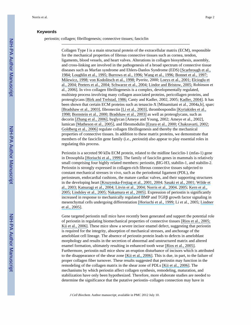
Periostin Regulates Collagen Fibrillogenesis and the Biomechanical Properties of Connective Tissues Russell A. Norris 1,* , Brook Damon 2 , Vladimir Mironov 1 , Vladimir Kasyanov 3 , Anand Ramamurthi 1,4 , Ricardo Moreno-Rodriguez 1 , Thomas Trusk 1 , Jay D. Potts 5 , Richard L. Goodwin 5 , Jeff Davis 5 , Stanley Hoffman 1 , Xuejun Wen 1 , Yukiko Sugi 1 , Christine B. Kern 1 , Corey H. Mjaatvedt 1 , Debi K. Turner 1 , Toru Oka 6 , Simon J. Conway 7 , Jeffery D. Molkentin 6 , Gabor Forgacs 2 , and Roger R. Markwald 1 1 Department of Cell Biology and Anatomy, Medical University of South Carolina, Charleston, South Carolina 2 Department of Physics and Astronomy, University of Missouri-Columbia, Columbia 3 Riga Stradins University, Riga LV-1007, Latvia 4 Department of Biomedical Engineering, Clemson University, Clemson, South Carolina 5 Department of Cell and Developmental Biology and Anatomy, University of South Carolina, Columbia, South Carolina 6 Department of Molecular Cardiovascular Biology, Children's Hospital Medical Center, Cincinnati, Ohio 7 Wells Center for Pediatric Research, Indiana University School of Medicine, Indianapolis, Indiana Abstract Periostin is predominantly expressed in collagen-rich fibrous connective tissues that are subjected to constant mechanical stresses including: heart valves, tendons, perichondrium, cornea, and the periodontal ligament (PDL). Based on these data we hypothesize that periostin can regulate collagen I fibrillogenesis and thereby affect the biomechanical properties of connective tissues. Immunoprecipitation and immunogold transmission electron microscopy experiments demonstrate that periostin is capable of directly interacting with collagen I. To analyze the potential role of periostin in collagen I fibrillogenesis, gene targeted mice were generated. Transmission electron microscopy and morphometric analyses demonstrated reduced collagen fibril diameters in skin dermis of periostin knockout mice, an indication of aberrant collagen I fibrillogenesis. In addition, differential scanning calorimetry (DSC) demonstrated a lower collagen denaturing temperature in periostin knockout mice, reflecting a reduced level of collagen cross-linking. Functional biomechanical properties of periostin null skin specimens and atrioventricular (AV) valve explant experiments provided direct evidence of the role that periostin plays in regulating the viscoelastic properties of connective tissues. Collectively, these data demonstrate for the first time that periostin can regulate collagen I fibrillogenesis and thereby serves as an important mediator of the biomechanical properties of fibrous connective tissues. © 2007 Wiley-Liss, Inc. * Correspondence to: Russell A. Norris, Department of Cell Biology and Anatomy, Medical University of South Carolina, Rm 604F CRI, 173 Ashley Avenue, Charleston, SC 29425. [email protected]. Russell A. Norris and Brook Damon have contributed equally to this work. NIH Public Access Author Manuscript J Cell Biochem. Author manuscript; available in PMC 2012 July 10. Published in final edited form as: J Cell Biochem. 2007 June 1; 101(3): 695–711. doi:10.1002/jcb.21224. NIH-PA Author Manuscript NIH-PA Author Manuscript NIH-PA Author Manuscript
Transcript of Periostin regulates collagen fibrillogenesis and the biomechanical properties of …
Periostin Regulates Collagen Fibrillogenesis and theBiomechanical Properties of Connective Tissues
Russell A. Norris1,*, Brook Damon2, Vladimir Mironov1, Vladimir Kasyanov3, AnandRamamurthi1,4, Ricardo Moreno-Rodriguez1, Thomas Trusk1, Jay D. Potts5, Richard L.Goodwin5, Jeff Davis5, Stanley Hoffman1, Xuejun Wen1, Yukiko Sugi1, Christine B. Kern1,Corey H. Mjaatvedt1, Debi K. Turner1, Toru Oka6, Simon J. Conway7, Jeffery D. Molkentin6,Gabor Forgacs2, and Roger R. Markwald1
1Department of Cell Biology and Anatomy, Medical University of South Carolina, Charleston,South Carolina2Department of Physics and Astronomy, University of Missouri-Columbia, Columbia3Riga Stradins University, Riga LV-1007, Latvia4Department of Biomedical Engineering, Clemson University, Clemson, South Carolina5Department of Cell and Developmental Biology and Anatomy, University of South Carolina,Columbia, South Carolina6Department of Molecular Cardiovascular Biology, Children's Hospital Medical Center, Cincinnati,Ohio7Wells Center for Pediatric Research, Indiana University School of Medicine, Indianapolis,Indiana
AbstractPeriostin is predominantly expressed in collagen-rich fibrous connective tissues that are subjectedto constant mechanical stresses including: heart valves, tendons, perichondrium, cornea, and theperiodontal ligament (PDL). Based on these data we hypothesize that periostin can regulatecollagen I fibrillogenesis and thereby affect the biomechanical properties of connective tissues.Immunoprecipitation and immunogold transmission electron microscopy experiments demonstratethat periostin is capable of directly interacting with collagen I. To analyze the potential role ofperiostin in collagen I fibrillogenesis, gene targeted mice were generated. Transmission electronmicroscopy and morphometric analyses demonstrated reduced collagen fibril diameters in skindermis of periostin knockout mice, an indication of aberrant collagen I fibrillogenesis. In addition,differential scanning calorimetry (DSC) demonstrated a lower collagen denaturing temperature inperiostin knockout mice, reflecting a reduced level of collagen cross-linking. Functionalbiomechanical properties of periostin null skin specimens and atrioventricular (AV) valve explantexperiments provided direct evidence of the role that periostin plays in regulating the viscoelasticproperties of connective tissues. Collectively, these data demonstrate for the first time thatperiostin can regulate collagen I fibrillogenesis and thereby serves as an important mediator of thebiomechanical properties of fibrous connective tissues.
© 2007 Wiley-Liss, Inc.*Correspondence to: Russell A. Norris, Department of Cell Biology and Anatomy, Medical University of South Carolina, Rm 604FCRI, 173 Ashley Avenue, Charleston, SC 29425. [email protected].
Russell A. Norris and Brook Damon have contributed equally to this work.
NIH Public AccessAuthor ManuscriptJ Cell Biochem. Author manuscript; available in PMC 2012 July 10.
Published in final edited form as:J Cell Biochem. 2007 June 1; 101(3): 695–711. doi:10.1002/jcb.21224.
NIH
-PA Author Manuscript
NIH
-PA Author Manuscript
NIH
-PA Author Manuscript
Keywordsperiostin; collagen; fibrillogenesis; connective tissues; fasciclin
Collagen Type I is a main structural protein of the extracellular matrix (ECM), responsiblefor the mechanical properties of fibrous connective tissues such as cornea, tendon,ligaments, blood vessels, and heart valves. Alterations in collagen biosynthesis, assembly,and cross-linking are involved in the pathogenesis of a broad spectrum of connective tissuediseases such as Marfan syndrome and Ehlers-Danlos Syndrome (EDS) [Scarbrough et al.,1984; Loughlin et al., 1995; Burrows et al., 1996; Wang et al., 1996; Bonnet et al., 1997;Milewicz, 1998; von Kodolitsch et al., 1998; Pyeritz, 2000; Loeys et al., 2001; Elcioglu etal., 2004; Peeters et al., 2004; Schwarze et al., 2004; Lindor and Bristow, 2005; Robinson etal., 2006]. In vivo collagen fibrillogenesis is a complex, developmentally regulated,multistep process involving many collagen associated proteins, pericollagen proteins, andproteoglycans [Birk and Trelstad, 1986; Canty and Kadler, 2002, 2005; Kadler, 2004]. It hasbeen shown that certain ECM proteins such as tenascin-X [Minamitani et al., 2004a,b], sparc[Bradshaw et al., 2003], fibronectin [Li et al., 2003], thrombospondin [Kyriakides et al.,1998; Bornstein et al., 2000; Bradshaw et al., 2003] as well as proteoglycans, such asdecorin [Zhang et al., 2006], byglycan [Ameye and Young, 2002; Ameye et al., 2002],lumican [Matheson et al., 2005], and fibromodulin [Ezura et al., 2000; Chakravarti, 2002;Goldberg et al., 2006] regulate collagen fibrillogenesis and thereby the mechanicalproperties of connective tissues. In addition to these matrix proteins, we demonstrate thatmembers of the fasciclin gene family (i.e., periostin) also appear to play essential roles inregulating this process.
Periostin is a secreted 90 kDa ECM protein, related to the midline fasciclin-1 (mfas-1) genein Drosophila [Horiuchi et al., 1999]. The family of fasciclin genes in mammals is relativelysmall comprising four highly related members: periostin, βIG-H3, stabilin-1, and stabilin-2.Periostin is strongly expressed in collagen-rich fibrous connective tissues subjected toconstant mechanical stresses in vivo, such as the periodontal ligament (PDL), theperiosteum, endocardial cushions, the mature cardiac valves, and their supporting structuresin the developing heart [Kruzynska-Frejtag et al., 2001, 2004; Sasaki et al., 2001; Wilde etal., 2003; Katsuragi et al., 2004; Litvin et al., 2004; Norris et al., 2004, 2005; Kern et al.,2005; Lindsley et al., 2005; Nakamura et al., 2005]. Expression of periostin is significantlyincreased in response to mechanically regulated BMP and TGFβ growth factor signaling inmesenchymal cells undergoing differentiation [Horiuchi et al., 1999; Li et al., 2005; Lindneret al., 2005].
Gene targeted periostin null mice have recently been generated and support the potential roleof periostin in regulating biomechanical properties of connective tissues [Rios et al., 2005;Kii et al., 2006]. These mice show a severe incisor enamel defect, suggesting that periostinis required for the integrity, absorption of mechanical stresses, and anchorage of theameloblast cell lineage. The absence of periostin protein leads to defects in ameloblastmorphology and results in the secretion of abnormal and unstructured matrix and alteredenamel formation, ultimately resulting in enhanced tooth wear [Rios et al., 2005].Furthermore, periostin null mice show an eruption disturbance of incisors which is attributedto the disappearance of the shear zone [Kii et al., 2006]. This is due, in part, to the failure ofproper collagen fiber turnover. These results suggested that periostin may function in theremodeling of the collagen matrix in the shear zone of PDLs [Kii et al., 2006]. Themechanisms by which periostin affect collagen synthesis, remodeling, maturation, andstabilization have only been hypothesized. Therefore, more elaborate studies are needed todetermine the significance that the putative periostin–collagen connection may have in
Norris et al. Page 2
J Cell Biochem. Author manuscript; available in PMC 2012 July 10.
NIH
-PA Author Manuscript
NIH
-PA Author Manuscript
NIH
-PA Author Manuscript
developing and maintaining functional connective tissues. Recent evidence has supported aninteraction of periostin with other ECM molecules such as fibronectin, tenascin-C, andcollagen V [Takayama et al., 2006]. However, no detailed investigations have been reportedon putative interactions between periostin and collagen I, as well as how this molecularinteraction may significantly affect the biomechanical properties of connective tissues.
In this report, we demonstrate for the first time that periostin is co-localized with collagenType I and directly binds to collagen Type I. How this interaction is of functionalsignificance was further evaluated in the context of periostin null mice and in periostinoverexpression experiments. An evaluation of collagen I fibrillogenesis in the skin ofperiostin null mice demonstrated a reduction in collagen fibril diameter resulting in adecrease in overall stiffness. In addition, differential scanning calorimetry (DSC)experiments demonstrated a significant reduction in collagen cross-linking, a defectassociated with improper collagen fibril formation. On the other hand, when periostin wasoverexpressed in cardiac valvulogenic tissue, the overall viscosity, a measure of collagencross-linking was increased. Collectively, these data indicate that periostin is essential forproper collagen fibril formation and maturation. This is the first report on the molecularinteractions of periostin with collagen Type I and validates periostin as an importantregulator of collagen fibrillogenesis. More importantly, proper biomechanical function ofconnective tissues, such as heart valves, skin, and tendon are dependent on this collagen I-periostin interaction.
MATERIALS AND METHODSAnimals: Mouse
Breeding pairs of 3-month-old periostin −/− knockout and wild-type mice derived fromsv129 line (manuscript in preparation) were housed in a fully accredited AmericanAssociation for Accreditation of Laboratory Animal Care (AAALAC) facility and theMedical University of South Carolina' institutional animal care and use committee approvedall experiments. All animals were classified as either periostin knockout or wild-type byPCR genotyping of genomic DNA isolated from tail clips. Primers used in genotyping were:upper: 5…-ccttgccagtctcaatgaagg-3…, lower WT- 5…-tgacagagtgaacacatgcc, lower KO-5…-ggaagacaatagcaggcatg. Cycling conditions were: 100°C (2 min), 96°C (2 min), 30cycles of 96°C (35 s), 56°C (35 s), 72°C (75 s) with a final extension of 72°C (5 min).Periostin null and wild-type age matched mice (3 months of age) were used for analysis.Only male mice were used in these experiments.
Animals: ChickenOne hundred twenty fertilized viral-free chicken eggs (Spafas) were incubated for 6 days ina humidity controlled 37°C incubator. After 6 days, HH27 embryos were removed, washedin PBS, and hearts were isolated. Further dissection of the hearts was performed to isolateatrioventricular (AV) valve tissues, which were used as explants for adenoviral infectionexperiments as described below.
HistochemistryLeft AV valve leaflets were isolated from adult mouse hearts. The leaflets were fixed with3.5% formaldehyde in PBS for 2 h, then methanol for 2 h, rehydrated and stained forcollagen using picrosirius red as previously reported in Whittaker et al. [1994].
Immunohistochemistry (IHC)Three-month-old mouse tissue was fixed in 4% paraformaldehyde (skin) or 100% coldmethanol (hearts), embedded in paraffin, and sectioned at 5 μm. Deparaffinized sections
Norris et al. Page 3
J Cell Biochem. Author manuscript; available in PMC 2012 July 10.
NIH
-PA Author Manuscript
NIH
-PA Author Manuscript
NIH
-PA Author Manuscript
were rehydrated through a graded series of ethanol to PBS. IHC on skin sections wasperformed as previously described [Kern et al., 2005]. IHC on heart valve sections wasperformed similarly except that blocking was performed for 15 min in 10% NGS/1% BSA/PBS. Sections were incubated with the primary antibody (rabbit anti-mouse periostin-1:100dilution) and washed with 1% BSA/PBS. Negative controls for IHC were performed byincubation with normal rabbit IgG in place of the primary antibody. The specificity of thisantibody has previously been evaluated by us and [Rios et al., 2005] with no detectableexpression or non-specific cross-reactivity present in the context of the periostin nullbackground (data not shown). Heart sections were double immunostained with the musclemarker, MF20 (1:1 antisera). No appreciable staining was observed in negative controls forperiostin (data not shown). Immunostained sections were viewed with a Leica TCS SP2AOBS Confocal Microscope System.
Immunogold Transmission Electron MicroscopyHearts were dissected from adult C57Bl6 mice and sectioned transversely. Sectionscontaining AV valve leaflets were then processed for TEM. Samples were fixed in 2%paraformaldehyde/0.1% glutaraldehyde (PBS) overnight at 4°C. Samples were thendehydrated in increasing concentrations of EtOH at −20°C and embedded in Lowicryl K4M(EMS) with UV polymerization for 48 h. Further curing was carried out under ambientfluorescence lighting until clear (~2 weeks). Cured blocks were cut on a Leica UltraCut Rmicrotome and 110 nm sections were collected on nickel mesh grids. Grids were blockedwith PBS solutions containing 0.05 M glycine, 1% CWFS gelatin, and 5% BSA. Grids werethen incubated overnight in 1:50 dilution of the rabbit anti-mouse periostin antibody (660ng/ml) at 4°C. No primary antibody controls were incubated in a solution of 1% BSA/0.05%Tween-20/PBS. After rinsing, these grids were incubated for 2 h with a 1:25 dilution of thegoat anti-rabbit 10 nm gold conjugate anti sera (EMS) in 1% BSA (PBS). The grids werethen fixed with 1% glutaraldehyde (PBS), and stained with 0.25% OsO4 (aq). Grids werestained with 2% uranyl acetate and Hanaichi lead stain prior to viewing on a JEOL 200CXtransmission electron microscope at 120 kV.
Adenoviral GenerationA full-length mouse periostin cDNA was obtained from Dr. Simon Conway (IndianaUniversity-Purdue University Indianapolis) and cloned into the pDNR-CMV shuttle vector(Clontech). To facilitate detection of the virally produced protein, a hemagluttinin (HA) tag(YPYDVPDYA) was inserted at the carboxylterminus through standard PCR techniques.The mouse periostin cDNA insert and the proximal CMV promoter were moved into theAdeno-X Acceptor Vector through a Cre/Lox mediated recombination. DNA from positivetransformants was isolated and transfected into HEK293 cells. Infectious viral particles werepurified using the Adeno-X Viral Purification Kit (Clontech). Titers for both periostinviruses were determined by plaque assays and spectrophotometry to be 1–2×10⁹ pfu/ml.Two control viral constructs expressing the β-galactosidase (LacZ) gene and GFP were alsogenerated in this same manner.
Cell Culture and Western Analysis1×105 HEK293 cells were plated on tissue culture plastic and infected with either theperiostin or LacZ adenoviruses at an MOI of five. After 2 days the cells and supernatantwere removed. The cell and supernatant mixture was spun at 1,000× rpm for 10 min. topellet the cells. Supernatant was removed and the cells were lysed in a 1× RIPA buffer (withand without 100 mM β-mercaptoethanol reducing agent) with mild sonication. Bothsupernatant and cell lysates were stored at 20°C. Total protein from the cell lysate wasquantified (Pierce-Commassie Protein Assay). Twenty microgram of cell lysate and 20 μl ofinfected media were loaded on a 4–15% SDS–PAGE. Proteins were electroblotted onto
Norris et al. Page 4
J Cell Biochem. Author manuscript; available in PMC 2012 July 10.
NIH
-PA Author Manuscript
NIH
-PA Author Manuscript
NIH
-PA Author Manuscript
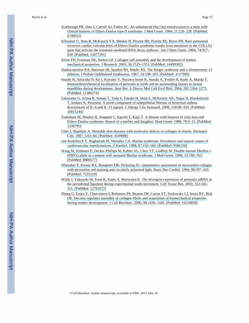
nitrocellulose membranes and immuno-probed for periostin expression using an α-HAantibody (1:1,000 in 5% milk-Sigma HA-7 clone) followed by a goat α-mouse-HRP(1:7,500 in 5% milk) secondary antibody (Sigma). Immunopositive bands were detected byECL (Pierce) and autoradiography. Western analysis of periostin expression in wild-type,heterozygous, and homozygous skin samples were accomplished similarly as describedabove with minor differences (Fig. 1). Skin samples were dissected and snap frozen in liquidnitrogen. Frozen samples were fractured and ground up by mortar and pestle. This processwas repeated and followed by the addition of a 1× RIPA buffer. Western analysis ofperiostin expression was accomplished as described previously. In house rabbit α-mouseperiostin antibody was used at a 1:2,500 dilution
Transmission Electron Microscopy and Fibril Diameter AnalysisThe dorsal skin from age-matched wild-type and periostin−/− mice was dissected out. Thedorsal skin was immersed in 2% glutaraldehyde (TAAB, UK) and processed fortransmission electron microscopy. Briefly, samples were rinsed and post-fixed in 1%osmium tetroxide (Merck, NJ) in 0.1 M cocadylate buffer, and thereafter dehydrated ingraded acetones and embedded in Epon (Electron Microscopic Sciences). Ultrathin sectionswere stained with 3% uranyl acetate and 0.2% lead citrate, and were examined under aJEOL electron microscope (JEOL, Japan). At least four mice of each genotype were used formeasurement of collagen fibril diameter and distribution. Micrographs (four per group) fromnon-overlapping regions of the dorsal skin were taken from cross-sections. The distributionof collagen fibril diameters was calculated using NIH image. Eight areas in a square werechosen from dorsal skin of each genotype for determination of distribution.
Co-Immunoprecipitation and Western Blotting of Collagen and PeriostinThirty microliters Protein-A agarose beads (Sigma) bound with an antibody to rat tailcollagen I (Abcam) were blocked for 1 h at 37°C in 1% BSA/1XTBST. Block was removedand the complexes were incubated overnight at 4°C with 5 μg of purified rat tail collagen I(BD Biosciences). Collagen complexes were spun (3 min at 6,000g), washed (5× in TBST),and resuspended in 500 μl periostin infected HEK 293 serum-free conditioned media.Complexes were incubated at RT for 2 h, then spun, and washed 5× in TBST. Bindingcomplexes were dissociated by addition of either a 1× SDS non-reducing loading buffer or a1× SDS denaturing loading buffer (containing 100 mm β-mercaptoethanol) and boiled at95°C for 5 min. Eluates were run on a 4–20% SDS–PAGE and immunoblotted using an α-HA antibody (Sigma HA-7 Clone) 1:500 in 5% milk, 1× TBST. A rabbit α-mouse-HRPsecondary (Sigma 1:5,000) was used followed by ECL detection (Pierce). As a control,subsequent staining of the IP plot was performed with an α-rabbit collagen I antibody(1:1,000 Abcam).
AV Valve Explant Adenoviral InfectionsFertilized white leghorn eggs (Spafas, Inc., Roanoke, IL) were incubated at 38.5°C with80% humidity until the desired Hamburger Hamilton developmental stage was reached,HH27~5 days [Hamburger and Hamilton, 1951]. Chicken AV valve explants were isolatedfrom HH27 embryos and incubated overnight in hanging drop cultures (DMEM/10% FBS/100 UPen/Strep) with 6×10⁶ PFUs of mouse periostin virus. A portion of these hangingdrops were used in Western analysis to verify viral infection whereas others were used formeasuring tissue surface tension (described below).
Measurement of Tissue Surface TensionTissue surface tension (a quantitative measure of the apparent liquid properties of embryonictissues) of periostin infected and LacZ control infected chicken valve tissues was determined
Norris et al. Page 5
J Cell Biochem. Author manuscript; available in PMC 2012 July 10.
NIH
-PA Author Manuscript
NIH
-PA Author Manuscript
NIH
-PA Author Manuscript
as described previously [Foty et al., 1996; Forgacs et al., 1998]. Briefly, isolated AVchicken embryo HH27 valve explants were rounded overnight and placed into a tensiometer(Fig. 9A). The compression apparatus (modified from previously used similar devices[Forgacs et al., 1998] used in this work to measure the liquid tissue properties is shown inFigure 9. A typical measurement was performed as follows. Spheroidal aggregates, rangingin diameter from 200 to 300 μm, were placed on the lower plate of the apparatus in CO2independent medium (with 100 U pen/strep; GIBCO/BRL) at 37°C, and rapidly compressedwith the help of a stepping motor to produce a deformation of a definite magnitude. Tominimize adhesion of the aggregate to the compression plates, these were coated with poly-2hydroxyethylmethacrylate (polyHEMA). To avoid irreversible damage to the cells,aggregates were compressed a maximum of 30% of their original diameter. The timevariation of the force exerted by the explant upon the upper compression plate was measuredusing a Cahn/Ventron (model 2000, Cerritos, CA) electrobalance (the upper compressionplate was connected to the arm of the balance). The force relaxation process was recordedby Labview software (National Instruments, Austin, TX) until the compressive forcereached a constant equilibrium value (45–60 min), at which point the plates were separated,and the aggregate was allowed to regain its original shape. Measurements in the rare caseswhen the aggregate did not regain its pre-compressed shape were discarded. The shape ofthe aggregate before, during, and after compression, was recorded by a Spot Insight CCDcamera (Diagnostic Instruments, Sterling Heights, MI) fitted to a horizontally positioneddissecting microscope (SZ60, Olympus). The surface tension of the model tissue was
evaluated using the Laplace equation [Forgacs et al., 1998]: .Here σ is the tissue's apparent surface tension (i.e., interfacial tension with the surroundingtissue culture medium), Feq is the equilibrium value of the compressive force, R3 is theradius of the circular contact area of the compressed aggregate with the plates. R1 and R2 arethe radii of curvature of the aggregate's surface, respectively, along its equatorial plane, andits peripheral contour, which is assumed to be circular. The geometric parameters weredetermined by an in-house built tracking program, with a precision of 3 μm. The programevaluates the aggregate's recorded contour on the basis of variation in gray scale values in itsvicinity. To check tissue liquidity (i.e., the independence of σ on the compressive force)aggregates were compressed 2–3 times with varying force with 60 min recovery inuncompressed state. Average σ values were calculated from at least six independentmeasurements.
Valve Tissue Explant Fusion AssaysThe viscosity of cushion tissue was determined from the time (t) variation of the circularinterfacial area (of instantaneous radius R) between two similar-size fusing roundupexplants. According to the theory of viscous liquids the length parameter L=(2/3)π(R2/R0)varies as L=(σ/η)t, where R0 and η are, respectively, the radius of the unfused explants andtissue viscosity [Frenkel, 1945].
DSC to Determine Levels of Collagen Cross-LinkingFreshly isolated tendon samples from periostin knockout (n=5) and wild-type mice (n=5),weight ranging from 5 to 8 μg, were sealed, respectively, in aluminum pans and their DSCthermograms were recorded on Mettler Toledo DSC 822e calorimeter with temperatureincrements of 5°C/min).
Biomechanical AnalysisTo investigate the influence of periostin on collagen fiber cross-linking, pieces of skin fromthe dorsal side of wild-type males (n=6) and periostin null male (n=3) mice were used asexperimental material. Prior to skin biopsies, hair from the dorsal part of mice was removed
Norris et al. Page 6
J Cell Biochem. Author manuscript; available in PMC 2012 July 10.
NIH
-PA Author Manuscript
NIH
-PA Author Manuscript
NIH
-PA Author Manuscript
by a hair remover gel “Nair” (Carter-Horner Corp., Mississauga, ON, Canada). In order toperform tensile tests, rectangular shaped dorsal skin samples (50 mm long and 4.5 mm widein the middle) oriented parallel to the spine was dissected with the aid of a dual-bladedsurgical knife. The specimens were kept in M199 medium (Sigma) at room temperature(20°C) and tested within 1 h of sacrifice. Before measurements were obtained, skin sampleswere placed between two microscope slides, and overall thickness of the specimens wasmeasured with a micrometer device “Ultra Digital Mark IV”(Fowler, Swiss) with accuracyof±0.001 mm. Specimens were gripped between specially designed “alligator” clamps toprevent slippage of the tissue, and tensile tests were performed using MTS materials testingsystem (Synergie 100) with a load cell of 50 N. Force-elongation curves were recorded at aconstant elongation rate of 5 mm/min until failure. During experimentations, the skinspecimens were kept continuously moist with room temperature M199 media. Stress wascalculated from force in Newton (N) divided by the initial cross-section area of thespecimen. Incremental modulus of elasticity between the levels of stress 0.25/0.3 MPa ofsamples was calculated also using MTS provided software.
Statistical AnalysisResults are displayed as the mean±standard deviation. One-factor analysis of variance(ANOVA) was utilized to evaluate mechanical properties and intergroup comparison bymeans of a Student's t-test using a difference of P<0.05.
RESULTSPeriostin is expressed in collagen rich fibrous connective tissues subjected to high levels ofmechanical loading [Horiuchi et al., 1999; Kruzynska-Frejtag et al., 2001, 2004; Oshima etal., 2002; Beck et al., 2003; Wilde et al., 2003; Katsuragi et al., 2004; Kudo et al., 2004;Litvin et al., 2004, 2005, 2006; Nakazawa et al., 2004; Norris et al., 2004, 2005; Suzuki etal., 2004; Kern et al., 2005; Lindsley et al., 2005; Nakamura et al., 2005; Rios et al., 2005;Kii et al., 2006]. In this report, we utilize newly developed reagents: (i) periostin knockoutmice, (ii) periostin specific antibodies, and (iii) periostin adenoviruses to examine thepossibility that collagen I fibrillogenesis and mechanical properties of connective tissues aredependent on the presence of periostin protein.
Colocalization of Periostin and CollagenIHC demonstrates strong expression of periostin in adult mice valve leaflets and theirsuspensory apparatus such as chordae tendineae (Fig. 2B,C). Not surprisingly, these samestructures express high levels of collagen (Fig. 2A). Similar co-expression patterns areevident in other fibrous connective tissue subjected to mechanical stress such as skin (Figs. 1and 2D), tendon and PDLs (not shown). Thus, at the microscopic level, IHC demonstratescolocalization of periostin and collagen Type I. In order to further investigate periostin andcollagen I co-localization at the ultrastructural level we performed immunogold IHC usingspecific α-mouse periostin antibodies. TEM studies of adult mouse AV valve leaflets foundthat periostin decorates collagen fibrils. Note the electron dense gold conjugates in Figure3B. These conjugates were not observed in the no primary, negative controls (Fig. 3A).
Periostin Binds to Collagen Type ITo further elucidate whether periostin is able to directly interact with collagen I, assuggested by the immunogold TEMs, biochemical co-immunoprecipitation experimentswere performed using immobilized collagen I. Eukaryotically expressed periostin was foundto co-precipitate with collagen Type I indicating a direct, specific protein–protein interaction(Fig. 4A-arrows, B–asterisks). Six negative controls and one positive control experiment
Norris et al. Page 7
J Cell Biochem. Author manuscript; available in PMC 2012 July 10.
NIH
-PA Author Manuscript
NIH
-PA Author Manuscript
NIH
-PA Author Manuscript
were performed to confirm the specificity of the interaction (Fig. 4A). This is the firstdemonstration of direct binding of periostin to collagen Type I.
Reduced Diameter of Collagen Fibrils in Periostin Knockout MiceTo determine if periostin has an effect on collagen fibrillogenesis, collagen fibril diameterand distribution in skin and tendon of periostin knockout and wild-type mice wereinvestigated. Alterations or redistribution of collagen fibrils is used as an indicator ofaberrant collagen fibrillogenesis [Christiansen et al., 2000]. Figure 5A, B shows thatperiostin knockout mice have a significant reduction in collagen fibril diameter indicatingaberrant collagen fibril organization. In addition, the knockout mice exhibited a substantialreduction in the thickness of the collagenous dermal layer of the skin (Fig. 5C–E).
Decreased Collagen Cross-Linking in Periostin Knockout MiceTo determine if the level of collagen cross-linking was also altered in periostin knockoutmice, DSC was performed. This system evaluates denaturation of tissue samples in 5°Cincrements and therefore is a direct measure of collagen cross-linking. Freshly isolatedtendon from five periostin knockout mice and five wild-type mice were used. Periostinknockout mice exhibited a reduction in the denaturation temperature of collagen (Fig. 6).Representative DSC profiles of tendon samples from wild-type and periostin knockout miceare presented in Figure 6A. Thermal denaturing temperatures were statistically differentfrom wild-type mice as shown in Figure 6B. Since thermal denaturation temperature reflectsthe level of collagen cross-linking [Miles et al., 2005; Pietrucha, 2005], these data stronglyindicate a reduced level of collagen cross-linking in periostin knockout mice and therefore isconsistent with periostin regulating collagen fibril assembly and maturation.
Characterization of Mechanical Properties of Skin in Periostin Knockout MiceA typical experiment derived from stretching fresh dorsal skin from two animals is shown inFigure 7A. The stress–strain relationship for all skin specimens was non-linear. However,skin from the knockout mouse exhibited lower tensile strength in contrast with the skin fromthe wild-type. Incremental modulus of elasticity (at the stress level 0.25/0.3 MPa) of dorsalskin samples of the wild-type and knockout mice were statistically different from eachanother: 1.8 ± 0.32 and 1.21 ± 0.19 MPa, respectively (P<0.05). In addition, skin fromknockout mice exhibited a substantially lower ultimate stress (Fig. 7B) than that seen withthe wild-type mice: 0.63 ± 0.10 and 1.77 ± 0.55 MPa, respectively (P<0.05). Therefore, theskin from wild-type mice is stiffer than that from knockout mice, demonstrating the absenceof periostin causes an increase in skin compliance.
Infection With Periostin Adenoviruses Alters the Visco-Elastic Properties of MesenchymalValve Tissue
If periostin has a direct effect on the mechanical properties of connective tissues, thenaltering levels of periostin expression in these tissues should result in biomechanicalchanges. In our preliminary studies it was shown that mechanical properties of cushiontissue (tissue surface tension and viscosity) are regulated during development (Damon et al.unpublished observations). At HH27, a time point of intense periostin expression, the valvemesenchymal tissue is sufficient in size to perform tensiometric assays and tissue fusionassays. Data generated from these assays were used to calculate the visco-elastic propertiesof embryonic prevalvular mesenchymal tissues.
Tensiometric StudiesIn order to assay the affect periostin has on the biomechanics of connective tissues, aperiostin overexpressing adenovirus was constructed as shown in Figure 8A. This virus (and
Norris et al. Page 8
J Cell Biochem. Author manuscript; available in PMC 2012 July 10.
NIH
-PA Author Manuscript
NIH
-PA Author Manuscript
NIH
-PA Author Manuscript
a LacZ control adenovirus) was functionally evaluated in HEK293 cells to ensureextracellular secretion (Fig. 8B). Western blot analysis confirms the ability for periostin tobe secreted as well as the ability to homodimerize. These adenoviruses were then used intensiometry experiments with cardiac valve tissue.
Tensiometry measurements on isolated rounded cushion explants were performed using theapparatus shown in Figure 10A. Infectability of AV cushions in hanging drops was initiallyassessed by infecting the tissue with GFP and periostin adenoviruses. These tissues are ableto be infected as demonstrated by immunofluorescence and Western blotting (Fig. 9A,B).Following infection of the mouse periostin adenovirus, surface tension was not appreciablyaffected (Fig. 10B). In order to estimate tissue viscosity, tissue fusion assays wereperformed (Fig. 10C,D). Kinetics of tissue fusion of two opposing rounded mesenchymaltissue aggregates allowed calculation of the ratio of tissue surface tension to tissue viscosity(Fig. 10C). This ratio, combined with data on direct measurements of tissue surface tension(estimated by tensiometry) was further used to calculate tissue viscosity. Data demonstratethat overexpression of periostin increased tissue viscosity (Fig. 10D). To validate that theseresults were specific to periostin and not a non-specific adenovirus affect, tissue aggregateswere also infected with a LacZ control adenovirus. Incubation with the LacZ adenovirusgenerated statistically similar values as observed with the non-infected controls (Fig. 10B–D). This demonstrates that the differences obtained in tissue fusion and viscosity are specificfor periostin overexpression and not due to non-specific adenovirus affects. To the best ofour knowledge, this is the first report providing direct evidence that the viscosity ofmesenchymal connective tissues is regulated in part by periostin.
DISCUSSIONConnective tissues such as skin, tendon, and heart valves are rich in collagens and fasciclinproteins (periostin and βIG-H3). The primary structural component conferring mostbiomechanical and material properties to connective tissues is collagen Type I. Therefore,we aimed to examine the potential molecular interaction of periostin with collagen Type I,and the impact it might have on the material properties of connective tissues. In this study,we report the co-localization of periostin and collagen Type I as seen by IHC andtransmission electron microscopy in murine skin and heart valves. Most importantly, webiochemically defined a direct interaction between periostin and collagen Type I. This is thefirst known report demonstrating periostin–collagen Type I interactions. This interactionwas also observed at the electron microscopic level. Immunogold transmission electronmicroscopy of adult mouse heart valves demonstrated the presence of periostin on collagenfibrils. This coincides with previously reported co-localization of periostin and collagen inthe PDL [Suzuki et al., 2004; Kii et al., 2006]. Taken together, these data strongly indicate amolecular interaction between periostin and collagen Type I. Recently, additional ECMcomponents such as tenascin-C, fibronectin, and collagen V have been shown to interactwith periostin [Takayama et al., 2006]. How these interactions are functionally importantremains to be seen. More importantly, the identification of specific collagen I, tenascin-C,fibronectin, and collagen V binding domains within the periostin molecule will be anessential step in understanding how these proteins organize in the ECM.
Although we demonstrate, through molecular and biochemical approaches, that periostinbinds specifically to collagen I both in vivo and in vitro, it is important to link theseinteractions to mechanistic function at the tissue and organ level. In this report we presentnovel bio-mechanical data that demonstrates the importance of periostin in collagenfibrillogenesis, ultimately dictating structural integrity and functional consequences inconnective tissues such as skin, tendon, and heart valves.
Norris et al. Page 9
J Cell Biochem. Author manuscript; available in PMC 2012 July 10.
NIH
-PA Author Manuscript
NIH
-PA Author Manuscript
NIH
-PA Author Manuscript
Collagen fibrillogenesis is a multistep process which involves linear and accelerative growthfollowed by lateral growth and subsequent collagen fiber fusion [Kadler et al., 1996; Berisioet al., 2002; Canty and Kadler, 2002, 2005; Hulmes, 2002; Silver et al., 2003; Kadler, 2004].Based on studies of collagen fibrillogenesis in vitro it has been suggested that there is acorrelation between collagen fibril diameter and mechanical properties of collagen-basedconnective tissues [Christiansen et al., 2000]. By altering collagen diameter, the structuraland functional integrity of the connective tissue is compromised. Morphometric studies oftransmission electron micrographs of ultrathin skin sections demonstrate reduced diameterof collagen fibrils in periostin knockout mice compared to wild-type mice. Thus, thisalteration reflects defects in collagen fibril maturation and assembly. Mechanistically thismay occur through specific periostin–collagen interactions which bridge and stabilizeadjacent collagen fibrils during fibril fusion. This theory is supported by three key pieces ofdata: (i) periostin binds directly to collagen Type I fibrils, (ii) periostin null mice exhibit areduction in fibril diameter, and (iii) periostin null mice have a reduction in collagen crosslinking as determined by DSC. Similar results have been obtained in the context of Sparc(secreted protein acidic rich cystein, also known as osteonectin and BM-40) deficientanimals [Bradshaw et al., 2003]. Collagen fibril diameter and collagen cross-linking aredecreased in these animals and result in alterations in the biomechanical properties ofconnective tissues. The mechanisms by which Sparc effects collagen fibril diameter may bethrough the direct binding to collagen Type I. Additionally, Sparc is a substrate fortransglutaminase, an enzyme that establishes cross-links between proteins [Aeschlimann etal., 1995]. It is unclear at the present whether periostin is a substrate for this enzyme or otherenzymes involved in cross-linking collagen moieties (such as lysyl oxidase). However, it isnot unreasonable to hypothesize that periostin may be a substrate for one of these cross-linking enzymes, thus mechanistically explaining the duality of periostin function duringcollagen fibrillogenesis.
The extrapolation of mechanical properties of connective tissues from the analysis ofcollagen diameter distribution is an important initial step in understanding the contributionof periostin to collagen fibrillogenesis. However, to fully appreciate the significanceperiostin may play in regulating and maintaining proper biomechanics of connective tissues,additional assays must be employed which examine specific biomechanical parameterswhen periostin expression is altered. To address these concerns, two main biomechanicalexperiments were developed and applied to cell and tissue specific assays. First,overexpression of periostin in isolated rounded embryonic chick heart mesenchymal valvetissue explants resulted in increased tissue viscosity as measured by using surfacetensiometry and tissue fusion assays. Mechanisms by which this occurs are currently beingevaluated and may involve induction of procollagen expression, enhanced fibril maturationand stabilization, and/or resistance to the natural process of collagen fibril turnover. Inaddition, we have recently shown that periostin, through integrin signaling, enhancescollagen contraction in embryonic mesenchymal valve tissue [Butcher et al., 2006], furthersubstantiating a role for periostin in the regulation of matrix strength and integrity. Second,an analysis of skin compliance in the context of the periostin null mouse would provideimportant information on the role that periostin may play in responding to increasingamounts of biomechanical forces (i.e., stress and strain relationship). These experimentsdemonstrate that the skin of wild-type mice is stiffer (less compliant) than skin of theperiostin knockout mouse—incremental modulus of elasticity of dorsal skin samples of thewild-type mice is higher than the skin from knockout mice. The skin specimen from thewild-type mice exhibited higher ultimate stress in contrast with the skin from the knockouttype. In addition, the data generated from these skin samples suggest that periostin knockoutmice develop a skin hyperextensive syndrome or “cutis laxa” similar to what is seen inpatients with the collagen associated EDS [Judd, 1984; Uitto and Shamban, 1987; Tsukaharaet al., 1988; Gorlin and Cohen, 1989].
Norris et al. Page 10
J Cell Biochem. Author manuscript; available in PMC 2012 July 10.
NIH
-PA Author Manuscript
NIH
-PA Author Manuscript
NIH
-PA Author Manuscript
Collectively, the molecular, biochemical, and biomechanical data provide important insightsinto the mechanisms of periostin function at the tissue level. In light of this data and due tostrong expression of periostin in heart valves and their tendinous supporting structures(chordae tendineae) it will be important to evaluate the periostin null mice for cardiovasculardefects. These potential defects would stem from altered formation and remodeling of thecardiac valves, which would compromise their ability to withstand extensive hemodynamicstresses during the cardiac contraction cycle. Developmental defects in the ECM can havemajor implications that over time greatly affect valve function. Thus, even small changes inthe composition (and alignment) of the valvular ECM can, over time, compromise valveintegrity and result in valvular and cardiovascular diseases (i.e., ECM changes over time =pathology). This is evident in Marfan Syndrome (Fibrillin-1 mutations) [Wang et al., 1996;Bonnet et al., 1997; Milewicz, 1998; von Kodolitsch et al., 1998; Pyeritz, 2000; Loeys et al.,2001; Elcioglu et al., 2004] and EDS (COL5A1, COL5A2, and COL1A2 mutations withinvolvement of tenascin-X, and COL1A1) [Scarbrough et al., 1984; Tsukahara et al., 1988;Loughlin et al., 1995; Burrows et al., 1996; Bonnet et al., 1997; Milewicz, 1998; Peeters etal., 2004; Schwarze et al., 2004; Lindor and Bristow, 2005]. As is the case for both MarfanSyndrome and EDS, valve abnormalities result in mitral, tricuspid and/or aortic valveprolapse, regurgitation, aortic aneurism, and a host of secondary cardiac defects (ventricularhypertrophy, cardiac arrhythmia's, and heart failure). Of potential clinical significance, EDStype II syndrome has been associated with an unbalanced (6q;13q) translocation whichincludes the locus for periostin at 13q13.3 [Scarbrough et al., 1984]. In addition, patientswith Rieger syndrome type II, (who have assorted cardiovascular malformations such as:aortic valvular stenosis, inter-atrial defects, congenital tricuspid valve anomaly, bicuspidaortic valve, etc.) have been assigned to chromosomal break points near the periostin locus[Stathacopoulos et al., 1987; Phillips et al., 1996; Mammi et al., 1998].
In conclusion, the data presented in this report define, for the first time, that periostin canregulate collagen fibrillogenesis and is essential to the biomechanical properties ofconnective tissues such as skin, tendon, and the heart valves. In addition, the periostin nullmouse may be a useful model for understanding molecular, cellular, and biomechanicalmechanisms of various connective tissue diseases in humans.
AcknowledgmentsThese studies were supported in part by the National Institutes of Health: RO1 HL33756 to RRM, PPG HL52813 toRRM, RO1 HL66231 to CHM, RO1 HL072958 to JDP, COBRE P20RR016434 to RLG, SC INBRE5MO1RR001070-28 to AR; and the National Sciences Foundation: FIBRE EF0526854 to GF and VM.
Grant sponsor: National Institutes of Health; Grant numbers: RO1 HL33756, PPG HL52813, RO1 HL66231, RO1HL072958, COBRE P20RR016434, SC INBRE 5MO1RR001070-28; Grant sponsor: National SciencesFoundation; Grant number: FIBRE EF0526854.
REFERENCESAeschlimann D, Kaupp O, Paulsson M. Transglutaminase—Catalyzed matrix cross-linking in
differentiating cartilage: Identification of osteonectin as a major glutaminyl substrate. J Cell Biol.1995; 129:881–892. [PubMed: 7730416]
Ameye L, Young MF. Mice deficient in small leucine-rich proteoglycans: Novel in vivo models forosteoporosis, osteoarthritis, Ehlers-Danlos syndrome, muscular dystrophy, and corneal diseases.Glycobiology. 2002; 12:107R–116R.
Ameye L, Aria D, Jepsen K, Oldberg A, Xu T, Young MF. Abnormal collagen fibrils in tendons ofbiglycan/ fibromodulin-deficient mice lead to gait impairment, ectopic ossification, andosteoarthritis. FASEB J. 2002; 16:673–680. [PubMed: 11978731]
Beck GR Jr. Moran E, Knecht N. Inorganic phosphate regulates multiple genes during osteoblastdifferentiation, including Nrf2. Exp Cell Res. 2003; 288:288–300. [PubMed: 12915120]
Norris et al. Page 11
J Cell Biochem. Author manuscript; available in PMC 2012 July 10.
NIH
-PA Author Manuscript
NIH
-PA Author Manuscript
NIH
-PA Author Manuscript
Berisio R, Vitagliano L, Mazzarella L, Zagari A. Recent progress on collagen triple helix structure,stability and assembly. Protein Pept Lett. 2002; 9:107–116. [PubMed: 12141907]
Birk DE, Trelstad RL. Extracellular compartments in tendon morphogenesis: Collagen fibril, bundle,and macroaggregate formation. J Cell Biol. 1986; 103:231–240. [PubMed: 3722266]
Bonnet D, Saygili A, Bonhoeffer P, Fermont L, Sidi D, Kachaner J. Atrio-ventricular valve dysplasiain 22 newborn infants. Int J Cardiol. 1997; 59:113–118. [PubMed: 9158161]
Bornstein P, Armstrong LC, Hankenson KD, Kyriakides TR, Yang Z. Thrombospondin 2, amatricellular protein with diverse functions. Matrix Biol. 2000; 19:557–568. [PubMed: 11102746]
Bradshaw AD, Puolakkainen P, Dasgupta J, Davidson JM, Wight TN, Helene Sage E. SPARC-nullmice display abnormalities in the dermis characterized by decreased collagen fibril diameter andreduced tensile strength. J Invest Dermatol. 2003; 120:949–955. [PubMed: 12787119]
Burrows NP, Nicholls AC, Yates JR, Gatward G, Sarathachandra P, Richards A, Pope FM. The geneencoding collagen alpha1(V)(COL5A1) is linked to mixed Ehlers-Danlos syndrome type I/II. JInvest Dermatol. 1996; 106:1273–1276. [PubMed: 8752669]
Butcher JT, Norris RA, Hoffman S, Mjaatvedt CH, Markwald RR. Periostin promotes atrioventricularmesenchyme matrix invasion and remodeling mediated by integrin signaling through Rho/PI 3-Kinase. Dev Biol. 2006 [Epub ahead of print].
Canty EG, Kadler KE. Collagen fibril biosynthesis in tendon: A review and recent insights. CompBiochem Physiol A Mol Integr Physiol. 2002; 133:979–985. [PubMed: 12485687]
Canty EG, Kadler KE. Procollagen trafficking, processing and fibrillogenesis. J Cell Sci. 2005;118:1341–1353. [PubMed: 15788652]
Chakravarti S. Functions of lumican and fibromodulin: Lessons from knockout mice. Glycoconj J.2002; 19:287–293. [PubMed: 12975607]
Christiansen DL, Huang EK, Silver FH. Assembly of type I collagen: Fusion of fibril subunits and theinfluence of fibril diameter on mechanical properties. Matrix Biol. 2000; 19:409–420. [PubMed:10980417]
Elcioglu NH, Akalin F, Elcioglu M, Comeglio P, Child AH. Neonatal Marfan syndrome caused by anexon 25 mutation of the fibrillin-1 gene. Genet Couns. 2004; 15:219–225. [PubMed: 15287423]
Ezura Y, Chakravarti S, Oldberg A, Chervoneva I, Birk DE. Differential expression of lumican andfibromodulin regulate collagen fibrillogenesis in developing mouse tendons. J Cell Biol. 2000;151:779–788. [PubMed: 11076963]
Forgacs G, Foty RA, Shafrir Y, Steinberg MS. Viscoelastic properties of living embryonic tissues: Aquantitative study. Biophys J. 1998; 74:2227–2234. [PubMed: 9591650]
Foty RA, Pfleger CM, Forgacs G, Steinberg MS. Surface tensions of embryonic tissues predict theirmutual envelopment behavior. Development. 1996; 122:1611–1620. [PubMed: 8625847]
Frenkel J. Viscous flow of crystalline bodies under the action of surface tension. J Phys (Moscow).1945; 9:385–391.
Goldberg M, Septier D, Oldberg A, Young MF, Ameye LG. Fibromodulin-deficient mice displayimpaired collagen fibrillogenesis in predentin as well as altered dentin mineralization and enamelformation. J Histochem Cytochem. 2006; 54:525–537. [PubMed: 16344330]
Gorlin RJ, Cohen MM Jr. Craniofacial manifestations of Ehlers-Danlos syndromes, cutis laxasyndromes, and cutis laxa-like syndromes. Birth Defects Orig Artic Ser. 1989; 25:39–71.[PubMed: 2697382]
Hamburger V, Hamilton HL. A series of normal stages in the development of the chick. J Morphol.1951; 8:49–92.
Horiuchi K, Amizuka N, Takeshita S, Takamatsu H, Katsuura M, Ozawa H, Toyama Y, Bonewald LF,Kudo A. Identification and characterization of a novel protein, periostin, with restricted expressionto periosteum and periodontal ligament and increased expression by transforming growth factorbeta. J Bone Miner Res. 1999; 14:1239–1249. [PubMed: 10404027]
Hulmes DJ. Building collagen molecules, fibrils, and suprafibrillar structures. J Struct Biol. 2002;137:2–10. [PubMed: 12064927]
Judd KP. Hyperelasticity syndromes. Cutis. 1984; 33:494–496. [PubMed: 6383732]
Norris et al. Page 12
J Cell Biochem. Author manuscript; available in PMC 2012 July 10.
NIH
-PA Author Manuscript
NIH
-PA Author Manuscript
NIH
-PA Author Manuscript
Kadler K. Matrix loading: Assembly of extracellular matrix collagen fibrils during embryogenesis.Birth Defects Res C Embryo Today. 2004; 72:1–11. [PubMed: 15054900]
Kadler KE, Holmes DF, Trotter JA, Chapman JA. Collagen fibril formation. Biochem J. 1996; 316(Pt1):1–11. [PubMed: 8645190]
Katsuragi N, Morishita R, Nakamura N, Ochiai T, Taniyama Y, Hasegawa Y, Kawashima K, KanedaY, Ogihara T, Sugimura K. Periostin as a novel factor responsible for ventricular dilation.Circulation. 2004; 110:1806–1813. [PubMed: 15381649]
Kern CB, Hoffman S, Moreno R, Damon BJ, Norris RA, Krug EL, Markwald RR, Mjaatvedt CH.Immuno-localization of chick periostin protein in the developing heart. Anat Rec A Discov MolCell Evol Biol. 2005; 284:415–423. [PubMed: 15803479]
Kii I, Amizuka N, Minqi L, Kitajima S, Saga Y, Kudo A. Periostin is an extracellular matrix proteinrequired for eruption of incisors in mice. Biochem Biophys Res Commun. 2006; 342:766–772.[PubMed: 16497272]
Kruzynska-Frejtag A, Machnicki M, Rogers R, Markwald RR, Conway SJ. Periostin (anosteoblastspecific factor) is expressed within the embryonic mouse heart during valve formation.Mech Dev. 2001; 103:183–188. [PubMed: 11335131]
Kruzynska-Frejtag A, Wang J, Maeda M, Rogers R, Krug E, Hoffman S, Markwald RR, Conway SJ.Periostin is expressed within the developing teeth at the sites of epithelial-mesenchymalinteraction. Dev Dyn. 2004; 229:857–868. [PubMed: 15042709]
Kudo H, Amizuka N, Araki K, Inohaya K, Kudo A. Zebrafish periostin is required for the adhesion ofmuscle fiber bundles to the myoseptum and for the differentiation of muscle fibers. Dev Biol.2004; 267:473–487. [PubMed: 15013807]
Kyriakides TR, Zhu YH, Smith LT, Bain SD, Yang Z, Lin MT, Danielson KG, Iozzo RV, LaMarca M,McKinney CE, Ginns EI, Bornstein P. Mice that lack thrombospondin 2 display connective tissueabnormalities that are associated with disordered collagen fibrillogenesis, an increased vasculardensity, and a bleeding diathesis. J Cell Biol. 1998; 140:419–430. [PubMed: 9442117]
Li S, Van Den Diepstraten C, D'Souza SJ, Chan BM, Pickering JG. Vascular smooth muscle cellsorchestrate the assembly of type I collagen via alpha2-beta1 integrin, RhoA, and fibronectinpolymerization. Am J Pathol. 2003; 163:1045–1056. [PubMed: 12937145]
Li G, Oparil S, Sanders JM, Zhang L, Dai M, Chen LB, Conway SJ, McNamara CA, Sarembock IJ.Phosphatidylinositol-3-kinase signaling mediates vascular smooth muscle cell expression ofperiostin in vivo and in vitro. Atherosclerosis. 2005; 188:292–300. [PubMed: 16325820]
Lindner V, Wang Q, Conley BA, Friesel RE, Vary CP. Vascular injury induces expression ofperiostin: Implications for vascular cell differentiation and migration. Arterioscler Thromb VascBiol. 2005; 25:77–83. [PubMed: 15514205]
Lindor NM, Bristow J. Tenascin-X deficiency in autosomal recessive Ehlers-Danlos syndrome. Am JMed Genet Part A. 2005; 135A:75–80. [PubMed: 15793839]
Lindsley A, Li W, Wang J, Maeda N, Rogers R, Conway SJ. Comparison of the four mouse fasciclin-containing genes expression patterns during valvuloseptal morphogenesis. Gene Expr Patterns.2005; 5:593–600. [PubMed: 15907457]
Litvin J, Selim AH, Montgomery MO, Lehmann K, Rico MC, Devlin H, Bednarik DP, Safadi FF.Expression and function of periostin-isoforms in bone. J Cell Biochem. 2004; 92:1044–1061.[PubMed: 15258926]
Litvin J, Zhu S, Norris R, Markwald R. Periostin family of proteins: Therapeutic targets for heartdisease. Anat Rec A Discov Mol Cell Evol Biol. 2005; 287:1205–1212. [PubMed: 16240445]
Litvin J, Blagg A, Mu A, Matiwala S, Montgomery M, Berretta R, Houser S, Margulies K. Periostinand periostin-like factor in the human heart: Possible therapeutic targets. Cardiovasc Pathol. 2006;15:24–32. [PubMed: 16414453]
Loeys B, Nuytinck L, Delvaux I, De Bie S, De Paepe A. Genotype and phenotype analysis of 171patients referred for molecular study of the fibrillin-1 gene FBN1 because of suspected Marfansyndrome. Arch Intern Med. 2001; 161:2447–2454. [PubMed: 11700157]
Loughlin J, Irven C, Hardwick LJ, Butcher S, Walsh S, Wordsworth P, Sykes B. Linkage of the genethat encodes the alpha 1 chain of type V collagen (COL5A1) to type II Ehlers-Danlos syndrome(EDS II). Hum Mol Genet. 1995; 4:1649–1651. [PubMed: 8541855]
Norris et al. Page 13
J Cell Biochem. Author manuscript; available in PMC 2012 July 10.
NIH
-PA Author Manuscript
NIH
-PA Author Manuscript
NIH
-PA Author Manuscript
Mammi I, De Giorgio P, Clementi M, Tenconi R. Cardiovascular anomaly in Rieger Syndrome:Heterogeneity or contiguity? Acta Ophthalmol Scand. 1998; 76:509–512. [PubMed: 9716345]
Matheson S, Larjava H, Hakkinen L. Distinctive localization and function for lumican, fibromodulinand decorin to regulate collagen fibril organization in periodontal tissues. J Periodontal Res. 2005;40:312–324. [PubMed: 15966909]
Miles CA, Avery NC, Rodin VV, Bailey AJ. The increase in denaturation temperature following cross-linking of collagen is caused by dehydration of the fibres. J Mol Biol. 2005; 346:551–556.[PubMed: 15670603]
Milewicz DM. Molecular genetics of Marfan syndrome and Ehlers-Danlos type IV. Curr Opin Cardiol.1998; 13:198–204. [PubMed: 9649943]
Minamitani T, Ariga H, Matsumoto K. Deficiency of tenascin-X causes a decrease in the level ofexpression of type VI collagen. Exp Cell Res. 2004a; 297:49–60. [PubMed: 15194424]
Minamitani T, Ikuta T, Saito Y, Takebe G, Sato M, Sawa H, Nishimura T, Nakamura F, Takahashi K,Ariga H, Matsumoto K. Modulation of collagen fibrillogenesis by tenascin-X and type VIcollagen. Exp Cell Res. 2004b; 298:305–315. [PubMed: 15242785]
Nakamura S, Terashima T, Yoshida T, Iseki S, Takano Y, Ishikawa I, Shinomura T. Identification ofgenes preferentially expressed in periodontal ligament: Specific expression of a novel secretedprotein, FDC-SP. Biochem Biophys Res Commun. 2005; 338:1197–1203. [PubMed: 16259954]
Nakazawa T, Nakajima A, Seki N, Okawa A, Kato M, Moriya H, Amizuka N, Einhorn TA, YamazakiM. Gene expression of periostin in the early stage of fracture healing detected by cDNAmicroarray analysis. J Orthop Res. 2004; 22:520–525. [PubMed: 15099630]
Norris RA, Kern CB, Wessels A, Moralez EI, Markwald RR, Mjaatvedt CH. Identification anddetection of the periostin gene in cardiac development. Anat Rec A Discov Mol Cell Evol Biol.2004; 281:1227–1233. [PubMed: 15532025]
Norris RA, Kern CB, Wessels A, Wirrig EE, Markwald RR, Mjaatvedt CH. Detection of betaig-H3, aTGFbeta induced gene, during cardiac development and its complementary pattern with periostin.Anat Embryol (Berl). 2005; 210:13–23. [PubMed: 16034610]
Oshima A, Tanabe H, Yan T, Lowe GN, Glackin CA, Kudo A. A novel mechanism for the regulationof osteoblast differentiation: Transcription of periostin, a member of the fasciclin I family, isregulated by the bHLH transcription factor, twist. J Cell Biochem. 2002; 86:792–804. [PubMed:12210745]
Peeters AC, Kucharekova M, Timmermans J, van den Berkmortel FW, Boers GH, Novakova IR,Egging D, den Heijer M, Schalkwijk J. A clinical and cardiovascular survey of Ehlers-Danlossyndrome patients with complete deficiency of tenascin-X. Neth J Med. 2004; 62:160–162.[PubMed: 15366699]
Phillips JC, del Bono EA, Haines JL, Pralea AM, Cohen JS, Greff LJ, Wiggs JL. A second locus forRieger syndrome maps to chromosome 13q14. Am J Hum Genet. 1996; 59:613–619. [PubMed:8751862]
Pietrucha K. Changes in denaturation and rheological properties of collagen-hyaluronic acid scaffoldsas a result of temperature dependencies. Int J Biol Macromol. 2005; 36:299–304. [PubMed:16102806]
Pyeritz RE. The Marfan syndrome. Annu Rev Med. 2000; 51:481–510. [PubMed: 10774478]
Rios H, Koushik SV, Wang H, Wang J, Zhou HM, Lindsley A, Rogers R, Chen Z, Maeda M,Kruzynska-Frejtag A, Feng JQ, Conway SJ. Periostin null mice exhibit dwarfism, incisor enameldefects, and an early-onset periodontal disease-like phenotype. Mol Cell Biol. 2005; 25:11131–11144. [PubMed: 16314533]
Robinson PN, Arteaga-Solis E, Baldock C, Collod-Beroud G, Booms P, De Paepe A, Dietz HC, GuoG, Handford PA, Judge DP, Kielty CM, Loeys B, Milewicz DM, Ney A, Ramirez F, ReinhardtDP, Tiedemann K, Whiteman P, Godfrey M. The molecular genetics of Marfan syndrome andrelated disorders. J Med Genet. 2006; 43:769–787. [PubMed: 16571647]
Sasaki H, Lo KM, Chen LB, Auclair D, Nakashima Y, Moriyama S, Fukai I, Tam C, Loda M, Fujii Y.Expression of Periostin, homologous with an insect cell adhesion molecule, as a prognostic markerin non-small cell lung cancers. Jpn J Cancer Res. 2001; 92:869–873. [PubMed: 11509119]
Norris et al. Page 14
J Cell Biochem. Author manuscript; available in PMC 2012 July 10.
NIH
-PA Author Manuscript
NIH
-PA Author Manuscript
NIH
-PA Author Manuscript
Scarbrough PR, Daw J, Carroll AJ, Finley SC. An unbalanced (6q;13q) translocation in a male withclinical features of Ehlers-Danlos type II syndrome. J Med Genet. 1984; 21:226–228. [PubMed:6748022]
Schwarze U, Hata R, McKusick VA, Shinkai H, Hoyme HE, Pyeritz RE, Byers PH. Rare autosomalrecessive cardiac valvular form of Ehlers-Danlos syndrome results from mutations in the COL1A2gene that activate the nonsense-mediated RNA decay pathway. Am J Hum Genet. 2004; 74:917–930. [PubMed: 15077201]
Silver FH, Freeman JW, Seehra GP. Collagen self-assembly and the development of tendonmechanical properties. J Biomech. 2003; 36:1529–1553. [PubMed: 14499302]
Stathacopoulos RA, Bateman JB, Sparkes RS, Hepler RS. The Rieger syndrome and a chromosome 13deletion. J Pediatr Ophthalmol Strabismus. 1987; 24:198–203. [PubMed: 3117999]
Suzuki H, Amizuka N, Kii I, Kawano Y, Nozawa-Inoue K, Suzuki A, Yoshie H, Kudo A, Maeda T.Immunohistochemical localization of periostin in tooth and its surrounding tissues in mousemandibles during development. Anat Rec A Discov Mol Cell Evol Biol. 2004; 281:1264–1275.[PubMed: 15386274]
Takayama G, Arima K, Kanaji T, Toda S, Tanaka H, Shoji S, McKenzie AN, Nagai H, HotokebuchiT, Izuhara K. Periostin: A novel component of subepithelial fibrosis of bronchial asthmadownstream of IL-4 and IL-13 signals. J Allergy Clin Immunol. 2006; 118:98–104. [PubMed:16815144]
Tsukahara M, Shinkai H, Asagami C, Eguchi T, Kajii T. A disease with features of cutis laxa andEhlers-Danlos syndrome. Report of a mother and daughter. Hum Genet. 1988; 78:9–12. [PubMed:3338795]
Uitto J, Shamban A. Heritable skin diseases with molecular defects in collagen or elastin. DermatolClin. 1987; 5:63–84. [PubMed: 3549080]
von Kodolitsch Y, Raghunath M, Nienaber CA. Marfan syndrome: Prevalence and natural course ofcardiovascular manifestations. Z Kardiol. 1998; 87:150–160. [PubMed: 9586150]
Wang M, Kishnani P, Decker-Phillips M, Kahler SG, Chen YT, Godfrey M. Double mutant fibrillin-1(FBN1) allele in a patient with neonatal Marfan syndrome. J Med Genet. 1996; 33:760–763.[PubMed: 8880577]
Whittaker P, Kloner RA, Boughner DR, Pickering JG. Quantitative assessment of myocardial collagenwith picrosirius red staining and circularly polarized light. Basic Res Cardiol. 1994; 89:397–410.[PubMed: 7535519]
Wilde J, Yokozeki M, Terai K, Kudo A, Moriyama K. The divergent expression of periostin mRNA inthe periodontal ligament during experimental tooth movement. Cell Tissue Res. 2003; 312:345–351. [PubMed: 12761672]
Zhang G, Ezura Y, Chervoneva I, Robinson PS, Beason DP, Carine ET, Soslowsky LJ, Iozzo RV, BirkDE. Decorin regulates assembly of collagen fibrils and acquisition of biomechanical propertiesduring tendon development. J Cell Biochem. 2006; 98:1436–1449. [PubMed: 16518859]
Norris et al. Page 15
J Cell Biochem. Author manuscript; available in PMC 2012 July 10.
NIH
-PA Author Manuscript
NIH
-PA Author Manuscript
NIH
-PA Author Manuscript
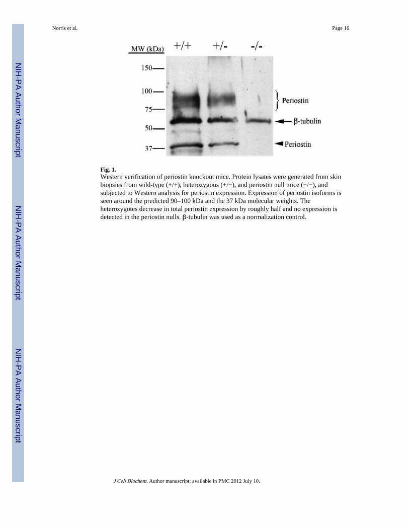
Fig. 1.Western verification of periostin knockout mice. Protein lysates were generated from skinbiopsies from wild-type (+/+), heterozygous (+/−), and periostin null mice (−/−), andsubjected to Western analysis for periostin expression. Expression of periostin isoforms isseen around the predicted 90–100 kDa and the 37 kDa molecular weights. Theheterozygotes decrease in total periostin expression by roughly half and no expression isdetected in the periostin nulls. β-tubulin was used as a normalization control.
Norris et al. Page 16
J Cell Biochem. Author manuscript; available in PMC 2012 July 10.
NIH
-PA Author Manuscript
NIH
-PA Author Manuscript
NIH
-PA Author Manuscript
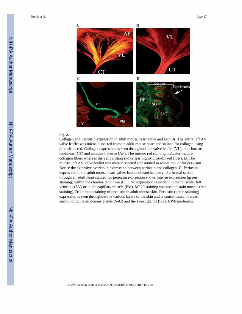
Fig. 2.Collagen and Periostin expression in adult mouse heart valve and skin. A: The entire left AVvalve leaflet was micro-dissected from an adult mouse heart and stained for collagen usingpicrosirius red. Collagen expression is seen throughout the valve leaflet (VL), the chordaetendineae (CT) and annulus fibrosae (AF). The intense red staining indicates maturecollagen fibers whereas the yellow stain shows less highly cross-linked fibers. B: Themurine left AV valve leaflet was microdissected and stained in whole mount for periostin.Notice the extensive overlap in expression between periostin and collagen. C: Periostinexpression in the adult mouse heart valve. Immunohistochemistry of a frontal sectionthrough an adult heart stained for periostin expression shows intense expression (greenstaining) within the chordae tendineae (CT). No expression is evident in the muscular leftventricle (LV) or in the papillary muscle (PM). MF20 staining was used to stain muscle (redstaining). D: Immunostaining of periostin in adult mouse skin. Periostin (green staining)expression is seen throughout the various layers of the skin and is concentrated in areassurrounding the sebaceous glands (SeG) and the sweat glands (SG); HP-hypodermis.
Norris et al. Page 17
J Cell Biochem. Author manuscript; available in PMC 2012 July 10.
NIH
-PA Author Manuscript
NIH
-PA Author Manuscript
NIH
-PA Author Manuscript
Fig. 3.Immunogold transmission electron microscopic localization of periostin along collagenfibrils. Transverse sections of the left anterior AV valve leaflet from an adult mouse wereassayed for the presence of periostin using immunogold TEM. A: No primary antibodycontrol immunogold TEM showing no gold particles on the collagen fibers. B:Electrondense gold particles (immunogold positive dots) were localized to collagen fibrilsusing the anti-mouse periostin anti-sera; (Bar=500 nm).
Norris et al. Page 18
J Cell Biochem. Author manuscript; available in PMC 2012 July 10.
NIH
-PA Author Manuscript
NIH
-PA Author Manuscript
NIH
-PA Author Manuscript
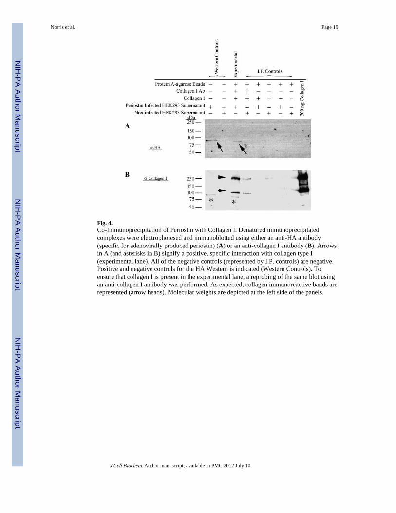
Fig. 4.Co-Immunoprecipitation of Periostin with Collagen I. Denatured immunoprecipitatedcomplexes were electrophoresed and immunoblotted using either an anti-HA antibody(specific for adenovirally produced periostin) (A) or an anti-collagen I antibody (B). Arrowsin A (and asterisks in B) signify a positive, specific interaction with collagen type I(experimental lane). All of the negative controls (represented by I.P. controls) are negative.Positive and negative controls for the HA Western is indicated (Western Controls). Toensure that collagen I is present in the experimental lane, a reprobing of the same blot usingan anti-collagen I antibody was performed. As expected, collagen immunoreactive bands arerepresented (arrow heads). Molecular weights are depicted at the left side of the panels.
Norris et al. Page 19
J Cell Biochem. Author manuscript; available in PMC 2012 July 10.
NIH
-PA Author Manuscript
NIH
-PA Author Manuscript
NIH
-PA Author Manuscript
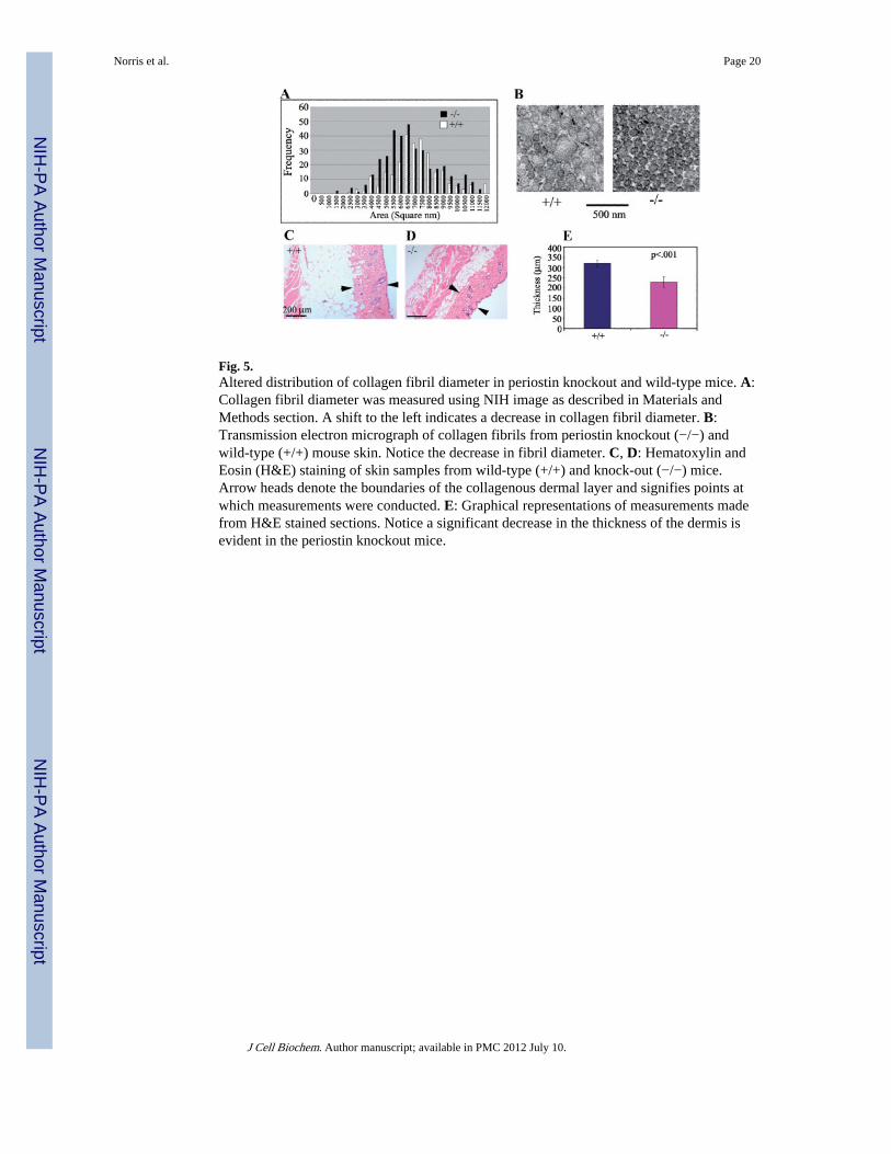
Fig. 5.Altered distribution of collagen fibril diameter in periostin knockout and wild-type mice. A:Collagen fibril diameter was measured using NIH image as described in Materials andMethods section. A shift to the left indicates a decrease in collagen fibril diameter. B:Transmission electron micrograph of collagen fibrils from periostin knockout (−/−) andwild-type (+/+) mouse skin. Notice the decrease in fibril diameter. C, D: Hematoxylin andEosin (H&E) staining of skin samples from wild-type (+/+) and knock-out (−/−) mice.Arrow heads denote the boundaries of the collagenous dermal layer and signifies points atwhich measurements were conducted. E: Graphical representations of measurements madefrom H&E stained sections. Notice a significant decrease in the thickness of the dermis isevident in the periostin knockout mice.
Norris et al. Page 20
J Cell Biochem. Author manuscript; available in PMC 2012 July 10.
NIH
-PA Author Manuscript
NIH
-PA Author Manuscript
NIH
-PA Author Manuscript
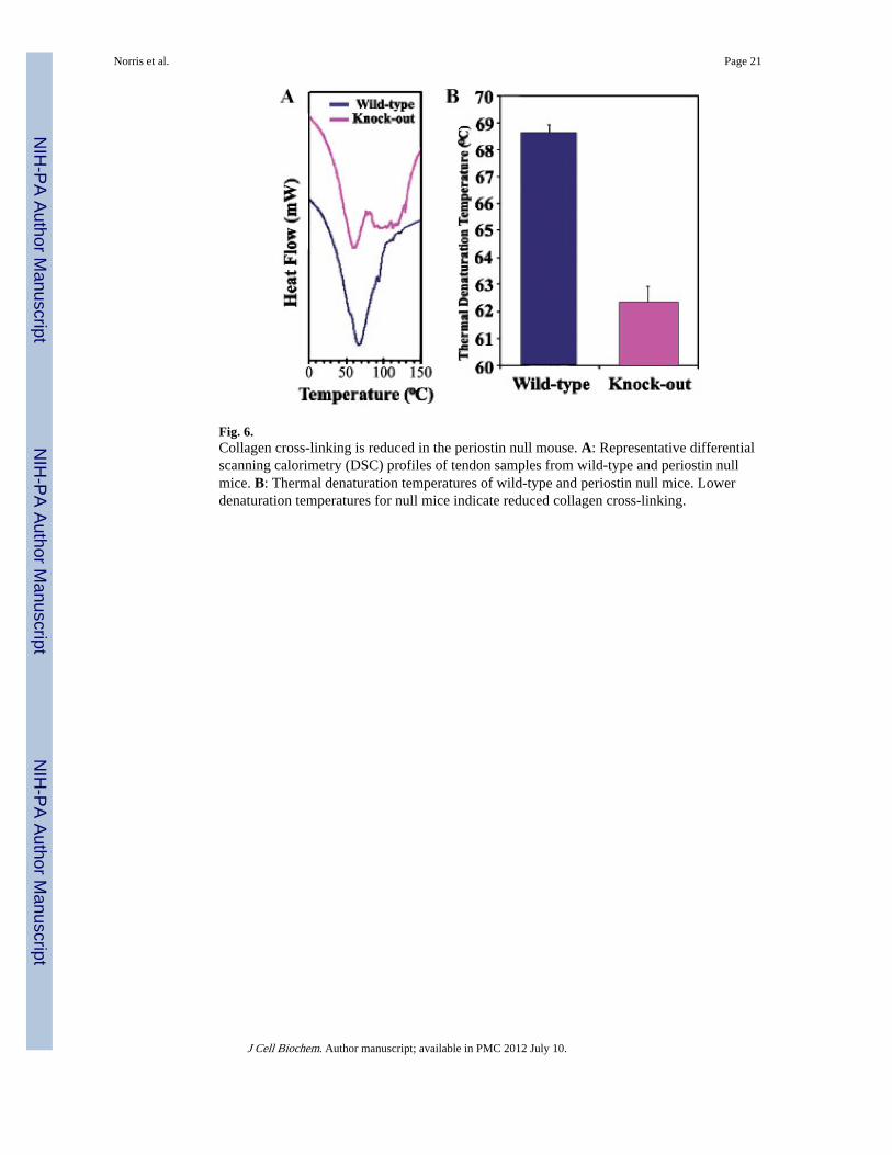
Fig. 6.Collagen cross-linking is reduced in the periostin null mouse. A: Representative differentialscanning calorimetry (DSC) profiles of tendon samples from wild-type and periostin nullmice. B: Thermal denaturation temperatures of wild-type and periostin null mice. Lowerdenaturation temperatures for null mice indicate reduced collagen cross-linking.
Norris et al. Page 21
J Cell Biochem. Author manuscript; available in PMC 2012 July 10.
NIH
-PA Author Manuscript
NIH
-PA Author Manuscript
NIH
-PA Author Manuscript
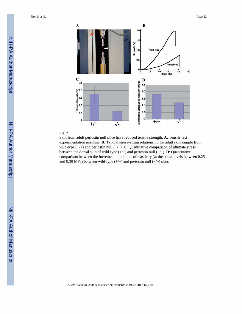
Fig. 7.Skin from adult periostin null mice have reduced tensile strength. A: Tensile testexperimentation machine. B: Typical stress–strain relationship for adult skin sample fromwild-type (+/+) and periostin null (−/−). C: Quantitative comparison of ultimate stressbetween the dorsal skin of wild-type (+/+) and periostin null (−/−). D: Quantitativecomparison between the incremental modulus of elasticity (at the stress levels between 0.25and 0.30 MPa) between wild-type (+/+) and periostin null (−/−) skin.
Norris et al. Page 22
J Cell Biochem. Author manuscript; available in PMC 2012 July 10.
NIH
-PA Author Manuscript
NIH
-PA Author Manuscript
NIH
-PA Author Manuscript
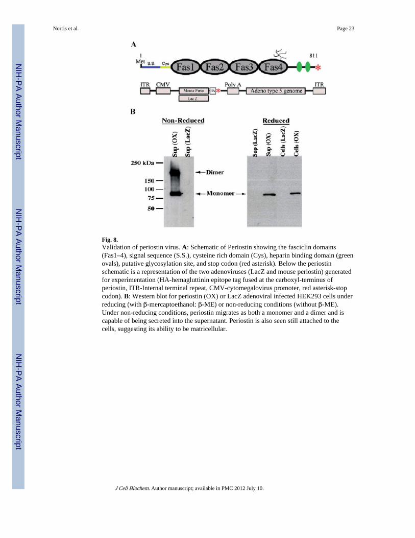
Fig. 8.Validation of periostin virus. A: Schematic of Periostin showing the fasciclin domains(Fas1–4), signal sequence (S.S.), cysteine rich domain (Cys), heparin binding domain (greenovals), putative glycosylation site, and stop codon (red asterisk). Below the periostinschematic is a representation of the two adenoviruses (LacZ and mouse periostin) generatedfor experimentation (HA-hemagluttinin epitope tag fused at the carboxyl-terminus ofperiostin, ITR-Internal terminal repeat, CMV-cytomegalovirus promoter, red asterisk-stopcodon). B: Western blot for periostin (OX) or LacZ adenoviral infected HEK293 cells underreducing (with β-mercaptoethanol: β-ME) or non-reducing conditions (without β-ME).Under non-reducing conditions, periostin migrates as both a monomer and a dimer and iscapable of being secreted into the supernatant. Periostin is also seen still attached to thecells, suggesting its ability to be matricellular.
Norris et al. Page 23
J Cell Biochem. Author manuscript; available in PMC 2012 July 10.
NIH
-PA Author Manuscript
NIH
-PA Author Manuscript
NIH
-PA Author Manuscript
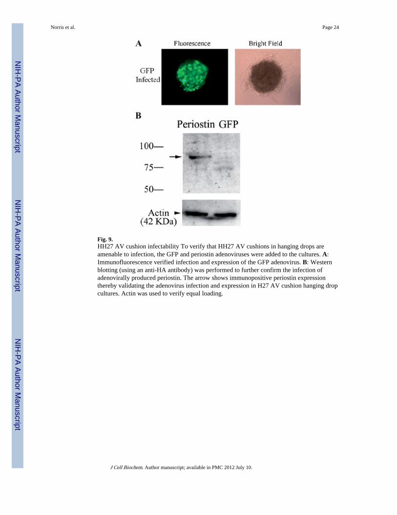
Fig. 9.HH27 AV cushion infectability To verify that HH27 AV cushions in hanging drops areamenable to infection, the GFP and periostin adenoviruses were added to the cultures. A:Immunofluorescence verified infection and expression of the GFP adenovirus. B: Westernblotting (using an anti-HA antibody) was performed to further confirm the infection ofadenovirally produced periostin. The arrow shows immunopositive periostin expressionthereby validating the adenovirus infection and expression in H27 AV cushion hanging dropcultures. Actin was used to verify equal loading.
Norris et al. Page 24
J Cell Biochem. Author manuscript; available in PMC 2012 July 10.
NIH
-PA Author Manuscript
NIH
-PA Author Manuscript
NIH
-PA Author Manuscript
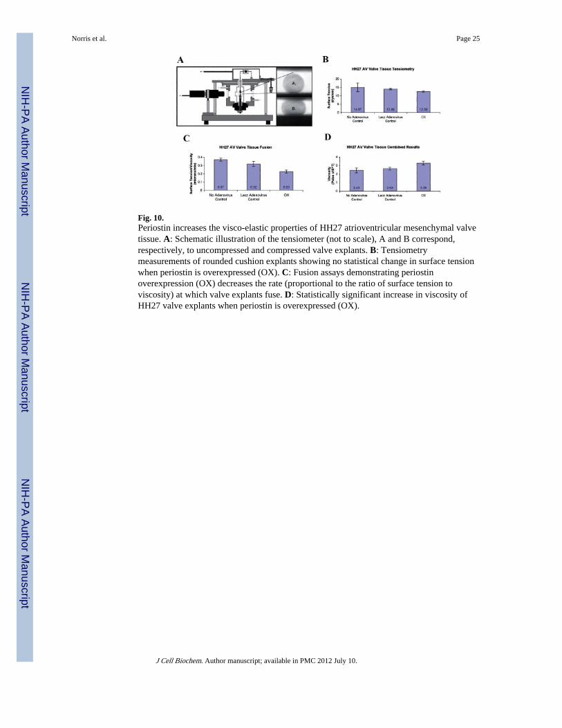
Fig. 10.Periostin increases the visco-elastic properties of HH27 atrioventricular mesenchymal valvetissue. A: Schematic illustration of the tensiometer (not to scale), A and B correspond,respectively, to uncompressed and compressed valve explants. B: Tensiometrymeasurements of rounded cushion explants showing no statistical change in surface tensionwhen periostin is overexpressed (OX). C: Fusion assays demonstrating periostinoverexpression (OX) decreases the rate (proportional to the ratio of surface tension toviscosity) at which valve explants fuse. D: Statistically significant increase in viscosity ofHH27 valve explants when periostin is overexpressed (OX).
Norris et al. Page 25
J Cell Biochem. Author manuscript; available in PMC 2012 July 10.
NIH
-PA Author Manuscript
NIH
-PA Author Manuscript
NIH
-PA Author Manuscript