Iodine Atoms: A New Molecular Feature for the Design of Potent Transthyretin Fibrillogenesis...
13
Iodine Atoms: A New Molecular Feature for the Design of Potent Transthyretin Fibrillogenesis Inhibitors Teresa Mairal 1. , Joan Nieto 2. , Marta Pinto 3 , Maria Rosa ´ rio Almeida 4 , Luis Gales 4 , Alfredo Ballesteros 5 , Jose ´ Barluenga 5 , Juan J. Pe ´ rez 3 , Jesu ´s T. Va ´ zquez 6 , Nuria B. Centeno 7 , Maria Joao Saraiva 4 , Ana M. Damas 4 , Antoni Planas 2 , Gemma Arsequell 1 , Gregorio Valencia 1 * 1 Unit of Glycoconjugate Chemistry, Institut de Quı ´mica Avanc ¸ada de Catalunya, I.Q.A.C.-C.S.I.C., Barcelona, Spain, 2 Laboratory of Biochemistry, Institut Quı ´mic de Sarria `, Universitat Ramon Llull, Barcelona, Spain, 3 Departamento de Ingenieria Quı ´mica, ETSEIB-Universitat Polite `cnica de Barcelona, Barcelona, Spain, 4 IBMC - Instituto de Biologia Molecular e Celular, Universidade do Porto, and ICBAS - Instituto de Cie ˆncias Biome ´dicas Abel Salazar, Universidade do Porto, Porto, Portugal, 5 Instituto Universitario de Quı ´mica Organometa ´lica ‘‘Enrique Moles’’, Unidad Asociada al C.S.I.C. Universidad de Oviedo, Oviedo, Spain, 6 Instituto Universitario de Bio-Orga ´nica ‘‘Antonio Gonza ´lez’’, Universidad de La Laguna, La Laguna, Tenerife, Spain, 7 Computer-Assisted Drug Design Laboratory, Research Group on Biomedical Informatics (GRIB) IMIM-Universitat Pompeu Fabra, Barcelona, Spain Abstract The thyroid hormone and retinol transporter protein known as transthyretin (TTR) is in the origin of one of the 20 or so known amyloid diseases. TTR self assembles as a homotetramer leaving a central hydrophobic channel with two symmetrical binding sites. The aggregation pathway of TTR into amiloid fibrils is not yet well characterized but in vitro binding of thyroid hormones and other small organic molecules to TTR binding channel results in tetramer stabilization which prevents amyloid formation in an extent which is proportional to the binding constant. Up to now, TTR aggregation inhibitors have been designed looking at various structural features of this binding channel others than its ability to host iodine atoms. In the present work, greatly improved inhibitors have been designed and tested by taking into account that thyroid hormones are unique in human biochemistry owing to the presence of multiple iodine atoms in their molecules which are probed to interact with specific halogen binding domains sitting at the TTR binding channel. The new TTR fibrillogenesis inhibitors are based on the diflunisal core structure because diflunisal is a registered salicylate drug with NSAID activity now undergoing clinical trials for TTR amyloid diseases. Biochemical and biophysical evidence confirms that iodine atoms can be an important design feature in the search for candidate drugs for TTR related amyloidosis. Citation: Mairal T, Nieto J, Pinto M, Almeida MR, Gales L, et al. (2009) Iodine Atoms: A New Molecular Feature for the Design of Potent Transthyretin Fibrillogenesis Inhibitors. PLoS ONE 4(1): e4124. doi:10.1371/journal.pone.0004124 Editor: Hilal Lashuel, Swiss Federal Institute of Technology Lausanne, Switzerland Received June 20, 2008; Accepted October 29, 2008; Published January 6, 2009 Copyright: ß 2009 Mairal et al. This is an open-access article distributed under the terms of the Creative Commons Attribution License, which permits unrestricted use, distribution, and reproduction in any medium, provided the original author and source are credited. Funding: This work was supported by grants: EET2002 05157 CO5 (to G.V., J.B., A.P.) from MCYT (Spain), CTQ2004 08077 CO2 (to J.B.) and BIO2007 67904 (to A.P.) from MEC (Spain), IB05 136 (to J.B.) from CEC Principado de Asturias, 2005SGR00883 (to A.P.) from Generalitat de Catalunya, DR06/0009 (to N.B.C.) from red Heracles ISCIII (Spain). Funding from Fundac ¸a ˜o Cie ˆncia e Tecnologia (FCT) (Portugal) through POCTI and POCI programmes (Programa Operacional Cie ˆncia e Inovac ¸a ˜o 2010) (to M.J.S.) and FEDER (Project POCI/SAU-MMO/57321/ 2004 to M.R.A.). POCTI/SAU NEU/58735/2004 (to A.M.D.), PTDC/SAU NEU/69123/2006 (to A.M.D.) and FEDER funds (to A.M.D.) are acknowledged. Diffraction data were collected at ESRF beam line ID14 1. Competing Interests: The authors have declared that no competing interests exist. * E-mail: [email protected] . These authors contributed equally to this work. Introduction Alzheimer’s disease is the best example of the 20 or so known amyloid diseases, in which protein or peptidic aggregates are considered to be the direct or indirect origin of the pathological conditions of the disease [1,2,3]. A distinctive group of diseases where amyloid deposition does not mainly occur in the central nervous system but rather in several organs in the periphery is associated to the plasma protein transthyretin (TTR). Amyloidosis linked to wild type TTR appears to cause senile systemic amyloidosis (SSA) [4,5], whereas most of the one hundred TTR mutants, already identified, result in familial amyloidotic polyneu- ropathy (FAP) [6,7]. TTR binds and transports 15–20% of serum thyroxine (T 4 ) and up to 80% of thyroxine in central nervous system [8]. In addition, TTR is the main carrier of vitamin A by forming a complex with retinol-binding protein (RBP) [9]. To physiologically function, the TTR molecule is self-assembled as a homotetramer, leaving a central hydrophobic channel with two symmetrical binding sites [10,11]. Recent studies on the aggregation pathway of TTR into amyloid fibrils point to a fibrillogenesis model which involves several steps such as dissociation of the tetramer, changes on monomer conformation, aggregation of conformationally modi- fied monomers into non-fibrillar oligomers that latter form protofibrils and further elongate into mature fibrils [12–15]. This mechanism along with the fact that binding of thyroid hormones to TTR results in tetramer stabilization, suggests that inhibition of amyloid fibril formation can be accomplished by small molecule compounds [16–26] sharing structural similarities with T 4 . Indeed this hypothesis has been confirmed by the identification of several families of compounds that, by binding to TTR, stabilize the ground state of the protein to an extent which is proportional to the dissociation constants [27]. The most common molecular features on this range of inhibitors [28–43] is that they are composed of two aromatic rings bearing halogen substituents in PLoS ONE | www.plosone.org 1 January 2009 | Volume 4 | Issue 1 | e4124
-
Upload
michiganstate -
Category
Documents
-
view
0 -
download
0
Transcript of Iodine Atoms: A New Molecular Feature for the Design of Potent Transthyretin Fibrillogenesis...
Iodine Atoms: A New Molecular Feature for the Design ofPotent Transthyretin Fibrillogenesis InhibitorsTeresa Mairal1., Joan Nieto2., Marta Pinto3, Maria Rosario Almeida4, Luis Gales4, Alfredo Ballesteros5,
Jose Barluenga5, Juan J. Perez3, Jesus T. Vazquez6, Nuria B. Centeno7, Maria Joao Saraiva4, Ana M.
Damas4, Antoni Planas2, Gemma Arsequell1, Gregorio Valencia1*
1 Unit of Glycoconjugate Chemistry, Institut de Quımica Avancada de Catalunya, I.Q.A.C.-C.S.I.C., Barcelona, Spain, 2 Laboratory of Biochemistry, Institut Quımic de Sarria,
Universitat Ramon Llull, Barcelona, Spain, 3 Departamento de Ingenieria Quımica, ETSEIB-Universitat Politecnica de Barcelona, Barcelona, Spain, 4 IBMC - Instituto de
Biologia Molecular e Celular, Universidade do Porto, and ICBAS - Instituto de Ciencias Biomedicas Abel Salazar, Universidade do Porto, Porto, Portugal, 5 Instituto
Universitario de Quımica Organometalica ‘‘Enrique Moles’’, Unidad Asociada al C.S.I.C. Universidad de Oviedo, Oviedo, Spain, 6 Instituto Universitario de Bio-Organica
‘‘Antonio Gonzalez’’, Universidad de La Laguna, La Laguna, Tenerife, Spain, 7 Computer-Assisted Drug Design Laboratory, Research Group on Biomedical Informatics
(GRIB) IMIM-Universitat Pompeu Fabra, Barcelona, Spain
Abstract
The thyroid hormone and retinol transporter protein known as transthyretin (TTR) is in the origin of one of the 20 or soknown amyloid diseases. TTR self assembles as a homotetramer leaving a central hydrophobic channel with twosymmetrical binding sites. The aggregation pathway of TTR into amiloid fibrils is not yet well characterized but in vitrobinding of thyroid hormones and other small organic molecules to TTR binding channel results in tetramer stabilizationwhich prevents amyloid formation in an extent which is proportional to the binding constant. Up to now, TTR aggregationinhibitors have been designed looking at various structural features of this binding channel others than its ability to hostiodine atoms. In the present work, greatly improved inhibitors have been designed and tested by taking into account thatthyroid hormones are unique in human biochemistry owing to the presence of multiple iodine atoms in their moleculeswhich are probed to interact with specific halogen binding domains sitting at the TTR binding channel. The new TTRfibrillogenesis inhibitors are based on the diflunisal core structure because diflunisal is a registered salicylate drug withNSAID activity now undergoing clinical trials for TTR amyloid diseases. Biochemical and biophysical evidence confirms thatiodine atoms can be an important design feature in the search for candidate drugs for TTR related amyloidosis.
Citation: Mairal T, Nieto J, Pinto M, Almeida MR, Gales L, et al. (2009) Iodine Atoms: A New Molecular Feature for the Design of Potent TransthyretinFibrillogenesis Inhibitors. PLoS ONE 4(1): e4124. doi:10.1371/journal.pone.0004124
Editor: Hilal Lashuel, Swiss Federal Institute of Technology Lausanne, Switzerland
Received June 20, 2008; Accepted October 29, 2008; Published January 6, 2009
Copyright: � 2009 Mairal et al. This is an open-access article distributed under the terms of the Creative Commons Attribution License, which permitsunrestricted use, distribution, and reproduction in any medium, provided the original author and source are credited.
Funding: This work was supported by grants: EET2002 05157 CO5 (to G.V., J.B., A.P.) from MCYT (Spain), CTQ2004 08077 CO2 (to J.B.) and BIO2007 67904 (to A.P.)from MEC (Spain), IB05 136 (to J.B.) from CEC Principado de Asturias, 2005SGR00883 (to A.P.) from Generalitat de Catalunya, DR06/0009 (to N.B.C.) from redHeracles ISCIII (Spain). Funding from Fundacao Ciencia e Tecnologia (FCT) (Portugal) through POCTI and POCI programmes (Programa Operacional Ciencia eInovacao 2010) (to M.J.S.) and FEDER (Project POCI/SAU-MMO/57321/ 2004 to M.R.A.). POCTI/SAU NEU/58735/2004 (to A.M.D.), PTDC/SAU NEU/69123/2006 (toA.M.D.) and FEDER funds (to A.M.D.) are acknowledged. Diffraction data were collected at ESRF beam line ID14 1.
Competing Interests: The authors have declared that no competing interests exist.
* E-mail: [email protected]
. These authors contributed equally to this work.
Introduction
Alzheimer’s disease is the best example of the 20 or so known
amyloid diseases, in which protein or peptidic aggregates are
considered to be the direct or indirect origin of the pathological
conditions of the disease [1,2,3]. A distinctive group of diseases
where amyloid deposition does not mainly occur in the central
nervous system but rather in several organs in the periphery is
associated to the plasma protein transthyretin (TTR). Amyloidosis
linked to wild type TTR appears to cause senile systemic
amyloidosis (SSA) [4,5], whereas most of the one hundred TTR
mutants, already identified, result in familial amyloidotic polyneu-
ropathy (FAP) [6,7].
TTR binds and transports 15–20% of serum thyroxine (T4) and
up to 80% of thyroxine in central nervous system [8]. In addition,
TTR is the main carrier of vitamin A by forming a complex with
retinol-binding protein (RBP) [9]. To physiologically function, the
TTR molecule is self-assembled as a homotetramer, leaving a
central hydrophobic channel with two symmetrical binding sites
[10,11].
Recent studies on the aggregation pathway of TTR into
amyloid fibrils point to a fibrillogenesis model which involves
several steps such as dissociation of the tetramer, changes on
monomer conformation, aggregation of conformationally modi-
fied monomers into non-fibrillar oligomers that latter form
protofibrils and further elongate into mature fibrils [12–15]. This
mechanism along with the fact that binding of thyroid hormones
to TTR results in tetramer stabilization, suggests that inhibition of
amyloid fibril formation can be accomplished by small molecule
compounds [16–26] sharing structural similarities with T4. Indeed
this hypothesis has been confirmed by the identification of several
families of compounds that, by binding to TTR, stabilize the
ground state of the protein to an extent which is proportional to
the dissociation constants [27]. The most common molecular
features on this range of inhibitors [28–43] is that they are
composed of two aromatic rings bearing halogen substituents in
PLoS ONE | www.plosone.org 1 January 2009 | Volume 4 | Issue 1 | e4124

one moiety and hydrophilic functions in the second which give rise
to structures as diverse as tetrahydroquinolines, dihydropyridines,
benzodiazepines, phenoxazines, stilbenes and benzoxazoles
[44,45].
Thyroid hormones are the only human biochemicals presenting
multiple iodine atoms in their molecules. Blake and co-workers were
the first to describe that in each TTR binding site there are six
pockets capable of accomodate an iodine atom (Figure 1a). Indeed,
when T4 binds TTR, four of these six pockets become occupied by
the iodine atoms of the hormone molecule resulting in a close steric
fit between the ligand and the binding site (Figure 1b). Therefore,
iodine atoms are crucial for the binding mode of thyroid hormones
to TTR, making an important contribution to the protein-hormone
interactions that stabilise the complex [46]. In spite of this evidence,
up to our knowledge, none of the potential newly designed TTR
amyloid inhibitors have taken advantage of the potential benefits of
incorporating iodine atoms to mimick the iodine-assisted binding
mode of thyroid hormones. Accordingly, the aim of the present
investigation was to provide initial evidences for the hypothesis that
iodine atom addition to already known TTR inhibitors could
produce more potent TTR fibrillogenesis inhibitors (hereafter
referred to as the iodination hypothesis).
Salicylates look particularly interesting as drug candidates due
to their long therapeutic tradition and wide clinical applications.
Owing that a number of salicylate analogues have also been
postulated as good TTR amyloid inhibitors and because the
salicylic core is amenable to electrophilic iodination, a salicylate
was chosen as a model template to test this hypothesis. Among the
many posible analogues a difluorophenyl derivative, namely,
diflunisal (29,49-difluoro-4-hydroxy-[1,19-biphenyl]-3-carboxylic
acid) was selected since it is an already registered drug [47]
having a biphenyl core structure which complies with the two-ring
model of TTR inhibitors shows a good TTR amyloid inhibitory
profile [48–52], and is under clinical trials for TTR-related
amyloidosis [53].
Results and Discussion
Computational GRID studies of TTR binding siteNaturally occurring TTR is composed of four chemically
identical monomers folded in a ß sandwich arquitecture leaving a
central channel where two ligand molecules may bind simulta-
neously (Figure 1). Owing to the two fold crystallographic axis that
runs through this channel there are two symmetry related positions
for the ligand at both ends of the channel. As already said, three
symmetry related pairs of HBPs capable to accomodate iodine
atoms is the most prominent structural feature of this channel.
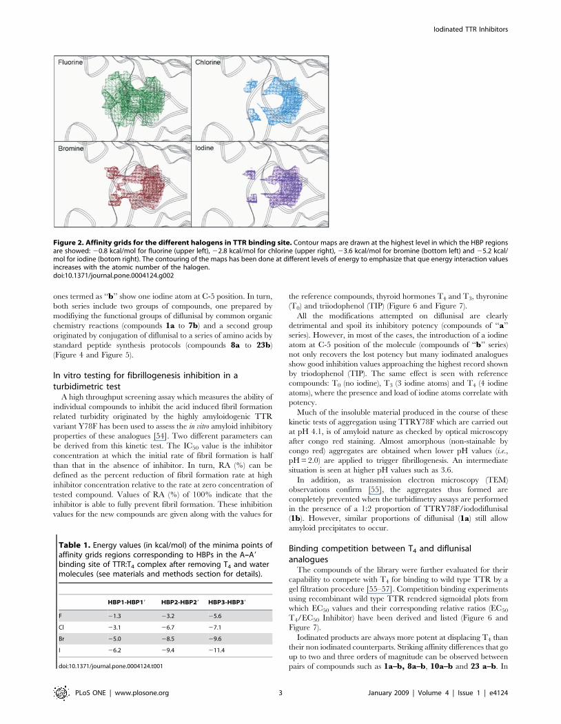
To computationally analyze these HBPs we have performed
calculations for imaging the grids of affinity between different
halogen atom probes and the surfaces of the binding channel. The
contour maps of Figure 2 show specific regions with high affinity
for all the halogen atoms. The situation of these areas perfectly
agrees with the initial geometrical description of HBPs. Their
extension is almost identical for every halogen although the close
proximity of HBP2 and HBP3 results in a continuous zone with
two optimal affinity points matching HBP2 and HBP3. In spite of
sharing the same regions, the energy of interaction for every
halogen atom is different and its magnitude increases with the
atomic number up to a maximum value for iodine (Table 1).
Synthesis of diflunisal analoguesAccording to GRID studies, iodine atoms placed at strategic
positions of the structure of TTR ligand may maximize their
potency by stablishing positive energetic interactions with these
high affinity halogen binding regions on the TTR binding
channel. To test the iodination hypothesis here proposed, a
number of iodinated analogues of already known inhibitors such
as, i.e., flufenamic, 4-phenyl and 4-phenoxy benzoic acids have
been prepared and tested in our fibrillogenesis inhibition assay.
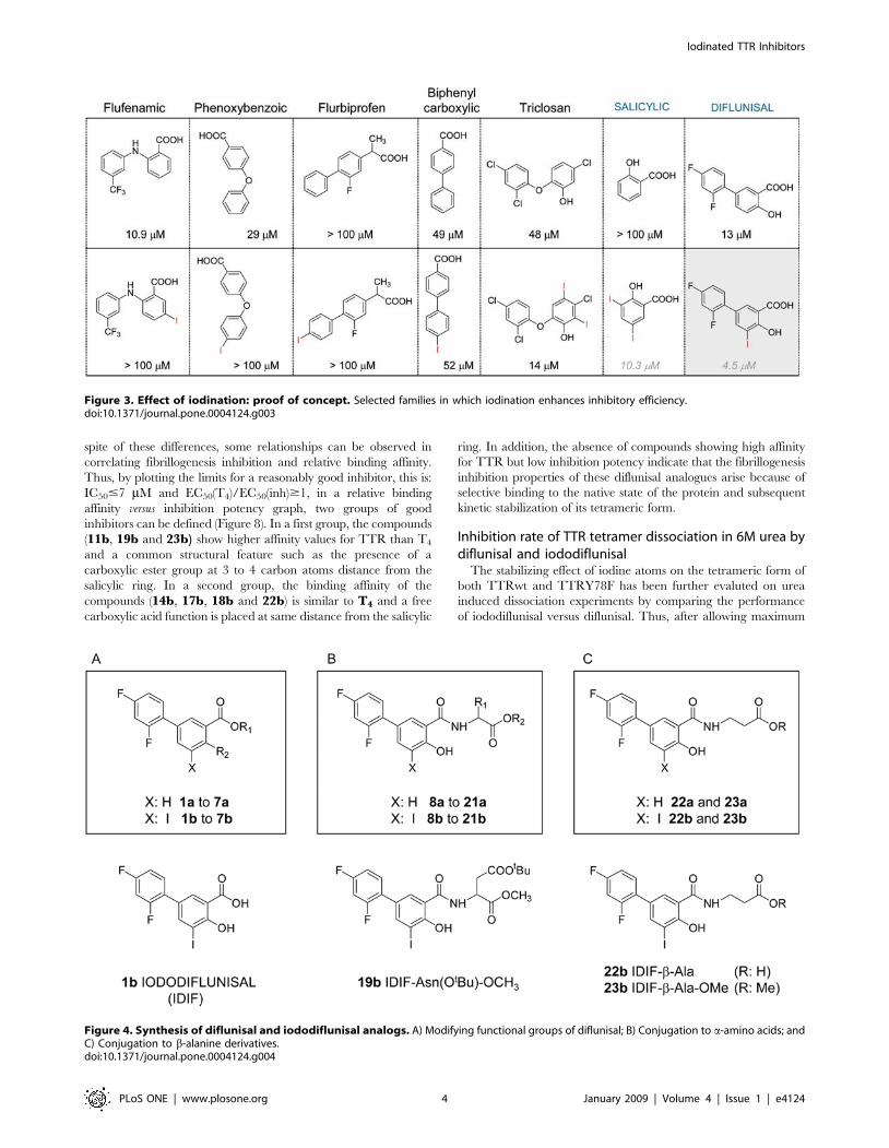
Results from this rough screening (Figure 3) have shown that most
striking positive effects on inhibitory potency were found for
diflunisal, a FDA-approved cyclooxygenase inhibitor with well
documented clinical records as NSAID.
To further examine how general was this effect on diflunisal
analogues, a library of 40 derivatives has been designed and
prepared. Two subsets of twin compounds have been synthesized
(Figure 4). The products labeled as ‘‘a’’ lack iodine atoms while the
Figure 1. A) Ribbon diagram of the quaternary structure of TTR with aschematic representation of the three-related pairs of pockets capableof accommodate an iodine atom in each binding site located at theinterface of monomers A–A9 and B–B9. These pockets are named in theliterature HBP1-HBP19 (green spheres), HBP2-HBP29 (pink spheres) andHBP3-HBP39 (blue spheres). B) Detailed view of one of the binding sitesfor the TTR:T4 complex, showing the occupation of four of the six HBPsby the iodine atoms of T4 .doi:10.1371/journal.pone.0004124.g001
Iodinated TTR Inhibitors
PLoS ONE | www.plosone.org 2 January 2009 | Volume 4 | Issue 1 | e4124
ones termed as ‘‘b’’ show one iodine atom at C-5 position. In turn,
both series include two groups of compounds, one prepared by
modifiying the functional groups of diflunisal by common organic
chemistry reactions (compounds 1a to 7b) and a second group
originated by conjugation of diflunisal to a series of amino acids by
standard peptide synthesis protocols (compounds 8a to 23b)
(Figure 4 and Figure 5).
In vitro testing for fibrillogenesis inhibition in aturbidimetric test
A high throughput screening assay which measures the ability of
individual compounds to inhibit the acid induced fibril formation
related turbidity originated by the highly amyloidogenic TTR
variant Y78F has been used to assess the in vitro amyloid inhibitory
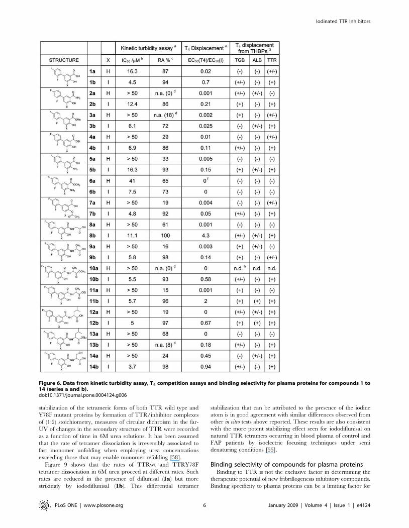
properties of these analogues [54]. Two different parameters can
be derived from this kinetic test. The IC50 value is the inhibitor
concentration at which the initial rate of fibril formation is half
than that in the absence of inhibitor. In turn, RA (%) can be
defined as the percent reduction of fibril formation rate at high
inhibitor concentration relative to the rate at zero concentration of
tested compound. Values of RA (%) of 100% indicate that the
inhibitor is able to fully prevent fibril formation. These inhibition
values for the new compounds are given along with the values for
the reference compounds, thyroid hormones T4 and T3, thyronine
(T0) and triiodophenol (TIP) (Figure 6 and Figure 7).
All the modifications attempted on diflunisal are clearly
detrimental and spoil its inhibitory potency (compounds of ‘‘a’’
series). However, in most of the cases, the introduction of a iodine
atom at C-5 position of the molecule (compounds of ‘‘b’’ series)
not only recovers the lost potency but many iodinated analogues
show good inhibition values approaching the highest record shown
by triodophenol (TIP). The same effect is seen with reference
compounds: T0 (no iodine), T3 (3 iodine atoms) and T4 (4 iodine
atoms), where the presence and load of iodine atoms correlate with
potency.
Much of the insoluble material produced in the course of these
kinetic tests of aggregation using TTRY78F which are carried out
at pH 4.1, is of amyloid nature as checked by optical microscopy
after congo red staining. Almost amorphous (non-stainable by
congo red) aggregates are obtained when lower pH values (i.e.,
pH = 2.0) are applied to trigger fibrillogenesis. An intermediate
situation is seen at higher pH values such as 3.6.
In addition, as transmission electron microscopy (TEM)
observations confirm [55], the aggregates thus formed are
completely prevented when the turbidimetry assays are performed
in the presence of a 1:2 proportion of TTRY78F/iododiflunisal
(1b). However, similar proportions of diflunisal (1a) still allow
amyloid precipitates to occur.
Binding competition between T4 and diflunisalanalogues
The compounds of the library were further evaluated for their
capability to compete with T4 for binding to wild type TTR by a
gel filtration procedure [55–57]. Competition binding experiments
using recombinant wild type TTR rendered sigmoidal plots from
which EC50 values and their corresponding relative ratios (EC50
T4/EC50 Inhibitor) have been derived and listed (Figure 6 and
Figure 7).
Iodinated products are always more potent at displacing T4 than
their non iodinated counterparts. Striking affinity differences that go
up to two and three orders of magnitude can be observed between
pairs of compounds such as 1a–b, 8a–b, 10a–b and 23 a–b. In
Figure 2. Affinity grids for the different halogens in TTR binding site. Contour maps are drawn at the highest level in which the HBP regionsare showed: 20.8 kcal/mol for fluorine (upper left), 22.8 kcal/mol for chlorine (upper right), 23.6 kcal/mol for bromine (bottom left) and 25.2 kcal/mol for iodine (botom right). The contouring of the maps has been done at different levels of energy to emphasize that que energy interaction valuesincreases with the atomic number of the halogen.doi:10.1371/journal.pone.0004124.g002
Table 1. Energy values (in kcal/mol) of the minima points ofaffinity grids regions corresponding to HBPs in the A–A9
binding site of TTR:T4 complex after removing T4 and watermolecules (see materials and methods section for details).
HBP1-HBP19 HBP2-HBP29 HBP3-HBP39
F 21.3 23.2 25.6
Cl 23.1 26.7 27.1
Br 25.0 28.5 29.6
I 26.2 29.4 211.4
doi:10.1371/journal.pone.0004124.t001
Iodinated TTR Inhibitors
PLoS ONE | www.plosone.org 3 January 2009 | Volume 4 | Issue 1 | e4124
spite of these differences, some relationships can be observed in
correlating fibrillogenesis inhibition and relative binding affinity.
Thus, by plotting the limits for a reasonably good inhibitor, this is:
IC50#7 mM and EC50(T4)/EC50(inh)$1, in a relative binding
affinity versus inhibition potency graph, two groups of good
inhibitors can be defined (Figure 8). In a first group, the compounds
(11b, 19b and 23b) show higher affinity values for TTR than T4
and a common structural feature such as the presence of a
carboxylic ester group at 3 to 4 carbon atoms distance from the
salicylic ring. In a second group, the binding affinity of the
compounds (14b, 17b, 18b and 22b) is similar to T4 and a free
carboxylic acid function is placed at same distance from the salicylic
ring. In addition, the absence of compounds showing high affinity
for TTR but low inhibition potency indicate that the fibrillogenesis
inhibition properties of these diflunisal analogues arise because of
selective binding to the native state of the protein and subsequent
kinetic stabilization of its tetrameric form.
Inhibition rate of TTR tetramer dissociation in 6M urea bydiflunisal and iododiflunisal
The stabilizing effect of iodine atoms on the tetrameric form of
both TTRwt and TTRY78F has been further evaluted on urea
induced dissociation experiments by comparing the performance
of iododiflunisal versus diflunisal. Thus, after allowing maximum
Figure 3. Effect of iodination: proof of concept. Selected families in which iodination enhances inhibitory efficiency.doi:10.1371/journal.pone.0004124.g003
Figure 4. Synthesis of diflunisal and iododiflunisal analogs. A) Modifying functional groups of diflunisal; B) Conjugation to a-amino acids; andC) Conjugation to b-alanine derivatives.doi:10.1371/journal.pone.0004124.g004
Iodinated TTR Inhibitors
PLoS ONE | www.plosone.org 4 January 2009 | Volume 4 | Issue 1 | e4124
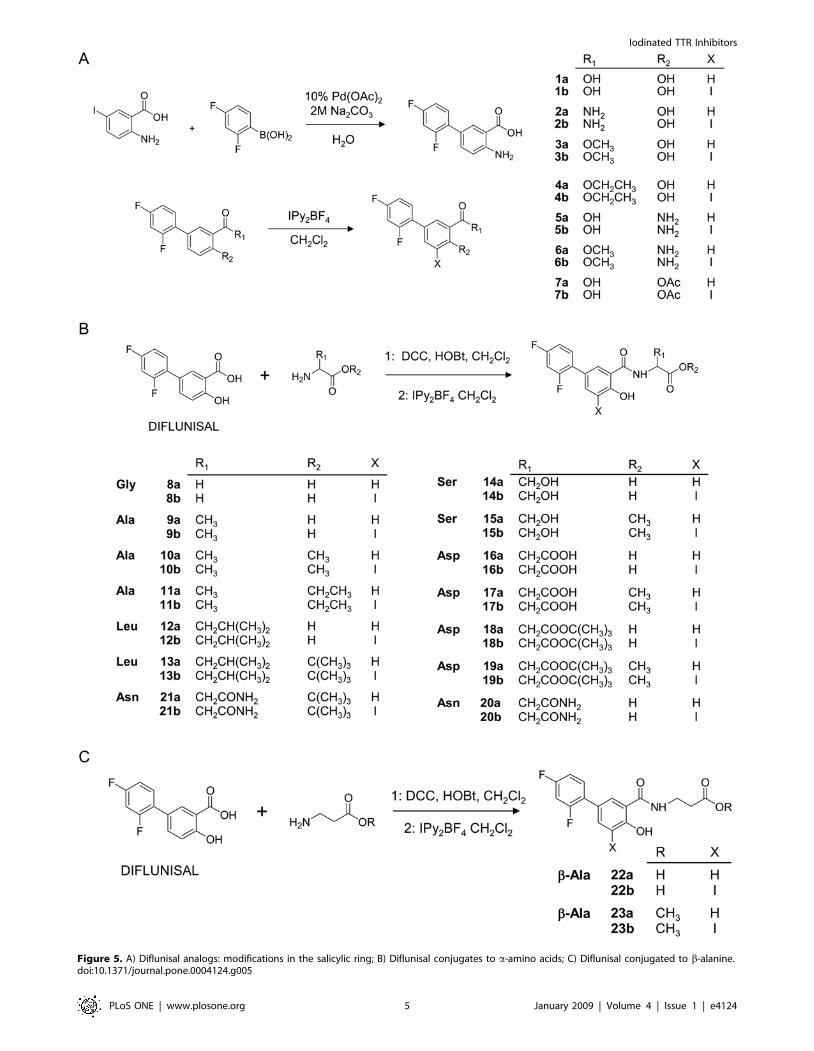
Figure 5. A) Diflunisal analogs: modifications in the salicylic ring; B) Diflunisal conjugates to a-amino acids; C) Diflunisal conjugated to b-alanine.doi:10.1371/journal.pone.0004124.g005
Iodinated TTR Inhibitors
PLoS ONE | www.plosone.org 5 January 2009 | Volume 4 | Issue 1 | e4124
stabilization of the tetrameric forms of both TTR wild type and
Y78F mutant proteins by formation of TTR/inhibitor complexes
of (1:2) stoichiometry, measures of circular dichroism in the far-
UV of changes in the secondary structure of TTR were recorded
as a function of time in 6M urea solutions. It has been assumed
that the rate of tetramer dissociation is irreversibly associated to
fast monomer unfolding when employing urea concentrations
exceeding those that may enable monomer refolding [58].
Figure 9 shows that the rates of TTRwt and TTRY78F
tetramer dissociation in 6M urea proceed at different rates. Such
rates are reduced in the presence of diflunisal (1a) but more
strikingly by iododiflunisal (1b). This differential tetramer
stabilization that can be attributed to the presence of the iodine
atom is in good agreement with similar differences observed from
other in vitro tests above reported. These results are also consistent
with the more potent stabilizing effect seen for iododiflunisal on
natural TTR tetramers occurring in blood plasma of control and
FAP patients by isoelectric focusing techniques under semi
denaturing conditions [55].
Binding selectivity of compounds for plasma proteinsBinding to TTR is not the exclusive factor in determining the
therapeutic potential of new fribrillogenesis inhibitory compounds.
Binding specificity to plasma proteins can be a limiting factor for
Figure 6. Data from kinetic turbidity assay, T4 competition assays and binding selectivity for plasma proteins for compounds 1 to14 (series a and b).doi:10.1371/journal.pone.0004124.g006
Iodinated TTR Inhibitors
PLoS ONE | www.plosone.org 6 January 2009 | Volume 4 | Issue 1 | e4124

Figure 7. Part II: Data from kinetic turbidity assay, T4 competition assays and binding selectivity for plasma proteins forcompounds 15 to 23 (series a and b) and reference compounds T4, T3, T0, and TIP.doi:10.1371/journal.pone.0004124.g007
Iodinated TTR Inhibitors
PLoS ONE | www.plosone.org 7 January 2009 | Volume 4 | Issue 1 | e4124

biodistribution, metabolism, activity and toxicity profiles of any
potential drug [59]. This is specially crucial in this case because
very strong plasma protein competitors of TTR include thyroid
binding globulin (TBG), which has an order of magnitude high
affinity for thyroxine [60], and albumin (ALB) which is at
concentrations of two orders of magnitude higher than TTR in
plasma. From our already reported results [55] of binding
competition experiments with plasma of transgenic mice and
FAP patients, iododiflunisal but not diflunisal has been shown to
be a very selective ligand for TTR which is another indication of
the important role of the iodine atom. This has been further
confirmed by qualitatively assessing the ability of the new
compounds to bind to the three T4 binding plasma proteins
(THBPs), thyroid binding globulin (TBG), albumin (ALB) and
TTR. The procedure has been essentially a T4 binding
displacement experiment but performed using whole human
plasma followed by a separation step of the serum proteins by
polyacrylamide gel electrophoresis (PAGE).
The two most selective compounds which exclusively bind to
TTR (20b and 22b) are iodinated (Figure 6 and Figure 7). One of
them (22b) is also a very potent inhibitor with equal affinity for
TTR than T4. A total of 17 less selective compounds show good
affinity for TTR (+) and some binding (+/2) for only one of the
two proteins TBG or ALB, among them, 13 compounds are
iodinated.
Crystal structures of TTR of two new complexes of TTRwith diflunisal analogs (23b and 22b)
Additional evidence for the iodination hypothesis has been
sought by elucidating at the atomic level how structural features
such as iodination and presence of a carboxyl group enhance the
binding affinity of these analogues. Two compounds: 23b and
22b were selected for structural studies (Figure 6, Figure 7, and
Text S1). One of them, 23b has been chosen because is one of the
most effective T4 displacing compounds with a high fibrillogenesis
inhibitory potency. The second compound, 22b is a very closely
related analogue lacking a methyl ester group which is also a very
potent fibril inhibitor which retains the same binding affinity as T4
[EC50 (T4)/EC50 (22b) = 0.95]. In addition 22b is one of the two
most selective binders of this series of inhibitors when tested in
human plasma. Both show very close molecular structures and
each one is representative of one of the two differential binding
affinity groups discussed above.
The crystal structures of their complexes with TTR could be
determined and refined to 1.85 and 1.80 A and are here
compared to the ones of their parent compounds diflunisal and
iododiflunisal. While the iododiflunisal-TTR complex has been
elucidated by us [61], the diflunisal parameters, here discussed, are
taken from published descriptions [52].
Figure 8. TTR binding vs. aggregation inhibition. TTR binding(from T4 displacement) vs. fibrillogenesis inhibition (from turbidimetricassay) plot for the series of iododiflunisal derivatives 1b–23b. Group 1and group 2 inhibitors are marked (see text).doi:10.1371/journal.pone.0004124.g008
Figure 9. TTR tetramer stabilization by inhibitors monitored by circular dichroism in 6M urea. Influence of small molecule inhibitorsdiflunisal (1a, 7,2 mM DIF) and iododiflunisal (1b, 7,2 mM IDIF) on the rate of wtTTR (3,6 mM) (left) and mutant Y78FTTR (3,6 mM) (right) dissociation in6M urea measured by Far-UV CD circular dichroism as a function of time.doi:10.1371/journal.pone.0004124.g009
Iodinated TTR Inhibitors
PLoS ONE | www.plosone.org 8 January 2009 | Volume 4 | Issue 1 | e4124

The positioning of iododiflunisal in the TTR channel is
exclusively in the forward mode, this is, with the difluorophenyl
ring occupying the inner part of the cavity and the salicylic ring
the outer part. This is a common feature among other inhibitors
having a biphenyl core molecule [62]. The same forward mode is
also the single disposition that is seen in both 23b and 22bstructures which show almost coincident spatial ring disposition
(Figure 10 and Figure 11a). In both cases, the compounds are
located further inside in the cavity than iododiflunisal. In sharp
contrast, diflunisal is observed in the pocket sharing two
orientations with equal probabilities, the one described as forward
and a totally opposite where the rings swap positions that is called
reverse mode.
The iodine atom in the iododiflunisal complex establishes close
hydrophobic interactions with Leu17, Thr106, Ala108, Thr119
and Val121, thus, occupying the HBP1 pocket which is the
outermost and more hydrophobic HBP. The innermost HBP
pockets, HBP3 and HBP39, in turn, closely interact with the
fluorine atoms of the difluorophenyl ring. A further stabilizing
interaction is found between the carbonyl group of Thr106 and
iodine which closely resembles an halogen bond [63]. Similar but
more optimized interactions than in the iododiflunisal complex are
observed for the iodine atom in both crystal structures of 23b and
22b complexes. Thus, the iodine atom of these analogues interact
with residues Leu17 and Ala 108 at distances ranging from 3.8 to
4.9 A but it is more efficiently accommodated to the HBP1
because of a new hydrophobic interaction with Met13 and
reinforcement of all the others. This fact is also in good agreement
with GRID calculations. Interestingly, by superimposition of the
conformations seen for 23b, 22a and T4 in their crystal
complexes, the position of the iodine atom of diflunisal analogues
is identical to the iodine at C-3 in the thyroid hormone T4
(Figure 12). This suggests that iodinated diflunisal analogues
mimick some of the features of thyroid hormones. GRID also
correctly predicted the interactions of the fluorine atoms. Thus,
while fluorine at C-29 is located in HBP3, the other, fluorine at C-
49, is placed in the most inner part of the binding cavity where
GRID predicts a high energy binding site for fluorine (Figure 13).
Also important are the hydrophilic interactions detected in the
iododiflunisal complex between the phenol and the carboxylic acid
functions with the side chain of Lys15, a residue which is
positioned at the entrance of the channel within close proximity to
HBP1. This feature fixes the positions of the two Lys15 residues
which in diflunisal are described as more disordered. These same
additional hydrophilic interactions are also seen in 23b and 22bcomplexes but, here, are even more hardened owing to a new
bifurcated hydrogen bond between the phenol group and both
Lys15 and Lys159 with distances in the range of 2.7 to 3.1 A. A
distinctive feature between 23b and 22b complexes comes from
the location of the carboxyl group, thus, while the carboxylic acid
of 22b is asymmetrically positioned between Lys 15 (3.0 A) and
Lys 159 (3.4 A), the carbonyl of the ester of 23b forms a water
mediated hydrogen bond with Thr106. Moreover, additional
hydrophobic interactions are build between the methyl group of
this ester function and residues Pro24 and Ser52. All of them
contribute to fix the ligand in the forward mode and to stabilize
the tetramer explaining the superior binding affinity of compound
23b over 22b ([EC50 (T4)/EC50 (23b) = 3.4] vs. [EC50 (T4)/EC50
(22b) = 0.95] , see Figure 6 and Figure 7).
Other monomer-monomer interactions not reported in difluni-
sal but seen in iododiflunisal are a direct hydrogen bond between
the two Ser117 residues. This motive is also evident in 23b and
22b where the binding of these analogues induce slight changes in
the protein structure resulting in Ser117 reorientation as to form a
strong hydrogen bond connecting the two monomers at a distance
of 2.6 A.
Thyroid hormonal activityTo gain further insight on the therapeutic potential of these
iodinated TTR fibrillogenesis inhibitors, in vitro binding tests of
idodiflunisal to thyroid hormone receptors alfa and beta were
carried out (data not shown). The almost negligible values of the
Figure 10. Ribbon diagram of the quaternary structure of TTR in complex with iododiflunisal-betaAlaOMe (23b) (A) and betaAlaOH(22b) (B) conjugates. Because of the two-fold symmetry axis running along the hormone-binding channel there are two-symmetry related bindingpositions of the two compounds in each binding site.doi:10.1371/journal.pone.0004124.g010
Iodinated TTR Inhibitors
PLoS ONE | www.plosone.org 9 January 2009 | Volume 4 | Issue 1 | e4124

binding constants suggest a possible lack of hormonal activity. This
has been further confirmed by preclinical animal studies using a
TTRV30M transgenic mice strain receiving 2.8 mg of iododi-
flunisal per day during 3 months. The animals did not show
significant metabolic disfunctions. However, further preclinical
tests are needed to validate these compounds as potential drugs for
TTR related amyloidosis.
In conclusion, by mimicking the natural interactions between
thyroid hormones and TTR and by using diflunisal as a model
compound, the biochemical and biophysical data above discussed
supports the hypothesis that iodine atoms inserted in TTR binding
compounds is a crucial factor for the design of novel highly potent
TTR fibrillogenesis inhibitors that one day become effective drugs
for the treatment of TTR-related amyloidosis.
Figure 11. Crystal structures of the TTR-22b and TTR-23bcomplexes. A) Close-view of the TTR hormone binding sites AA9 of theTTR:iododiflunisal-betaAlaOMe (23b) (in grey) and TTR:iododiflunisal-betaAlaOH (22b) (in blue) superposed. Only one of the two-symmetryrelated binding positions of these two iododiflunisal conjugates isshown. B) Superposition of the crystal structures of the TTR:iododi-flunisal-betaAlaOMe (23b) (in grey), TTR:iododiflunisal-betaAlaOH (22b)(in blue), and TTR:iododiflunisal (1b) complex (in yellow).doi:10.1371/journal.pone.0004124.g011
Figure 12. Superposition of TTR:iododiflunisal-betaAlaOH (22b) (blue) and TTR:iododiflunisal-betaAlaOMe (23b) (yellow) andTTR:T4 complexes (green). Iodine in iododiflunisal derivates is in the same region as I3 of T4.doi:10.1371/journal.pone.0004124.g012
Figure 13. Affinity grids maps for TTR:iododiflunisal-betaA-laOH (22b) (A) and TTR:iododiflunisal-betaAlaOMe (23b) (B)complexes. Contour maps are drawn at 20.9 kcal/mol for fluorine and26.3 kcal/mol for iodine. For clarity, only contour surrounding iodineand fluor substituents are shown and TTR structure is hidden. Iodine’scontour is coloured in purple, fluorine countour coincident with HPB3pocket is coloured in green, an the additional region located in themost inner part of the cavity along the two-fold symmetry axis iscoloured in red.doi:10.1371/journal.pone.0004124.g013
Iodinated TTR Inhibitors
PLoS ONE | www.plosone.org 10 January 2009 | Volume 4 | Issue 1 | e4124
Materials and Methods
More detailed methods are included as Methods S1.
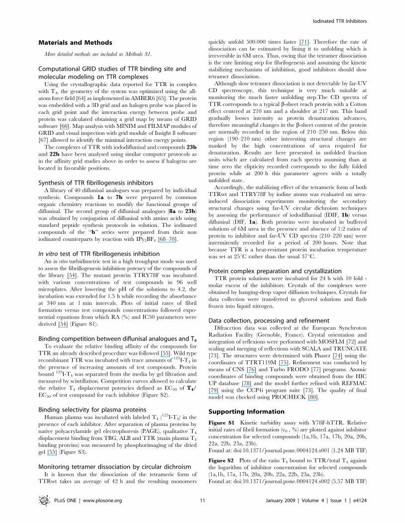
Computational GRID studies of TTR binding site andmolecular modeling on TTR complexes
Using the crystallographic data reported for TTR in complex
with T4, the geometry of the system was optimized using the all-
atom force field [64] as implemented in AMBER6 [65]. The protein
was embedded with a 3D grid and an halogen probe was placed in
each grid point and the interaction energy between probe and
protein was calculated obtaining a grid map by means of GRID
software [66]. Map analysis with MINIM and FILMAP modules of
GRID and visual inspection with grid module of Insight ll software
[67] allowed to identify the minimal interaction energy points.
The complexes of TTR with iododiflunisal and compounds 23band 22b have been analysed using similar computer protocols as
in the affinity grid studies above in order to assess if halogens are
located in favorable positions.
Synthesis of TTR fibrillogenesis inhibitorsA library of 40 diflunisal analogues was prepared by individual
synthesis. Compounds 1a to 7b were prepared by common
organic chemistry reactions to modify the functional groups of
diflunisal. The second group of diflunisal analogues (8a to 23b)
was obtained by conjugation of diflunisal with amino acids using
standard peptide synthesis protocols in solution. The iodinated
compounds of the ‘‘b’’ series were prepared from their non
iodinated counterparts by reaction with IPy2BF4 [68–70].
In vitro test of TTR fibrillogenesis inhibitionAn in vitro turbidimetric test in a high troughput mode was used
to assess the fibrillogenesis inhibition potency of the compounds of
the library [54]. The mutant protein TTRY78F was incubated
with various concentrations of test compounds in 96 well
microplates. After lowering the pH of the solutions to 4.2, the
incubation was extended for 1.5 h while recording the absorbance
at 340 nm at 1 min intervals. Plots of initial rates of fibril
formation versus test compounds concentrations followed expo-
nential equations from which RA (%) and IC50 parameters were
derived [54] (Figure S1).
Binding competition between diflunisal analogues and T4
To evaluate the relative binding affinity of the compounds for
TTR an already described procedure was followed [55]. Wild type
recombinant TTR was incubated with trace amounts of 125I-T4 in
the presence of increasing amounts of test compounds. Protein
bound 125I-T4 was separated from the media by gel filtration and
measured by scintillation. Competition curves allowed to calculate
the relative T4 displacement potencies defined as EC50 of T4/
EC50 of test compound for each inhibitor (Figure S2).
Binding selectivity for plasma proteinsHuman plasma was incubated with labeled T4 (125I-T4) in the
presence of each inhibitor. After separation of plasma proteins by
native polyacrylamide gel electrophoresis (PAGE), qualitative T4
displacement binding from TBG, ALB and TTR (main plasma T4
binding proteins) was measured by phosphorimaging of the dried
gel [55] (Figure S3).
Monitoring tetramer dissociation by circular dichroismIt is known that the dissociation of the tetrameric form of
TTRwt takes an average of 42 h and the resulting monomers
quickly unfold 500.000 times faster [71]. Therefore the rate of
dissociation can be estimated by lining it to unfolding which is
irreversible in 6M urea. Thus, owing that the tetramer dissociation
is the rate limiting step for fibrilogenesis and assuming the kinetic
stabilizing mechanism of inhibition, good inhibitors should slow
tetramer dissociation.
Although slow tetramer dissociation is not detectable by far-UV
CD spectroscopy, this technique is very much suitable at
monitoring the much faster unfolding step.The CD spectra of
TTR corresponds to a typical b-sheet reach protein with a Cotton
effect centered at 210 nm and a shoulder at 217 nm. This band
gradually looses intensity as protein denaturation advances,
therefore meaningful changes in the b-sheet content of the protein
are normally recorded in the region of 210–250 nm. Below this
region (190–210 nm) other interesting structural changes are
masked by the high concentrations of urea required for
denaturation. Results are here presented in unfolded fraction
units which are calculated from each spectra assuming than at
time zero the elipticity recorded corresponds to the fully folded
protein while at 200 h this parameter agrees with a totally
unfolded state.
Accordingly, the stabilizing effect of the tetrameric form of both
TTRwt and TTRY78F by iodine atoms was evaluated on urea-
induced dissociation experiments monitoring the secondary
structural changes using far-UV circular dichroism techniques
by assessing the performance of iododiflunisal (IDIF, 1b) versus
diflunisal (DIF, 1a). Both proteins were incubated in buffered
solutions of 6M urea in the presence and absence of 1:2 ratios of
protein to inhibitor and far-UV CD spectra (210–220 nm) were
intermitently recorded for a period of 200 hours. Note that
because TTR is a heat-resistant protein incubation temperature
was set at 25uC rather than the usual 37uC.
Protein complex preparation and crystallizationTTR protein solutions were incubated for 24 h with 10 fold -
molar excess of the inhibitors. Crystals of the complexes were
obtained by hanging-drop vapor diffusion techniques. Crystals for
data collection were transferred to glycerol solutions and flash
frozen into liquid nitrogen.
Data collection, processing and refinementDifracction data was collected at the European Synchroton
Radiation Facility (Grenoble, France). Crystal orientation and
integration of reflexions were performed with MOSFLM [72] and
scaling and merging of reflections with SCALA and TRUNCATE
[73]. The structures were determined with Phaser [74] using the
coordinates of TTRT119M [75]. Refinement was conducted by
means of CNS [76] and Turbo FRODO [77] programs. Atomic
coordinates of binding compounds were obtained from the HIC
UP database [78] and the model further refined with REFMAC
[79] using the CCP4i program suite [73]. The quality of final
model was checked using PROCHECK [80].
Supporting Information
Figure S1 Kinetic turbidity assay with Y78F-hTTR. Relative
initial rates of fibril formation (v0 , %) are plotted against inhibitor
concentration for selected compounds (1a,1b, 17a, 17b, 20a, 20b,
22a, 22b, 23a, 23b).
Found at: doi:10.1371/journal.pone.0004124.s001 (1.24 MB TIF)
Figure S2 Plots of the ratio T4 bound to TTR/total T4 against
the logarithm of inhibitor concentration for selected compounds
(1a,1b, 17a, 17b, 20a, 20b, 22a, 22b, 23a, 23b).
Found at: doi:10.1371/journal.pone.0004124.s002 (5.57 MB TIF)
Iodinated TTR Inhibitors
PLoS ONE | www.plosone.org 11 January 2009 | Volume 4 | Issue 1 | e4124

Figure S3 Polyacrylamide gel electrophoresis of plasma proteins.
The plasma was incubated with 125I-T4 in the presence of the
indicated compounds. C: plasma incubated with 125I-T4 with no
competitor. The migration of the main T4 binding plasma
proteins, TBG, Albumin, and TTR is indicated.
Found at: doi:10.1371/journal.pone.0004124.s003 (2.55 MB TIF)
Text S1 Data collection and refinement statistics
Found at: doi:10.1371/journal.pone.0004124.s004 (0.21 MB
DOC)
Methods S1 Iodinated TTR inhibitors
Found at: doi:10.1371/journal.pone.0004124.s005 (1.00 MB
DOC)
Author Contributions
Conceived and designed the experiments: JJP NBC MJS AD AP GA GV.
Performed the experiments: TM JN MP MdRA LG JTV. Analyzed the
data: MdRA AB JB NBC MJS AD AP GA GV. Contributed reagents/
materials/analysis tools: AB JB. Wrote the paper: MdRA NBC MJS AD
AP GA GV.
References
1. Dobson CM (2001) The structural basis of protein folding and its links withhuman disease. Phil Trans R Soc Lond B Biol Sci 356: 133–145.
2. Chiti F, Dobson CM (2006) Protein misfolding, functional amyloid, and human
disease. Annu Rev Biochem 75: 333–366.
3. Forman MS, Trojanowski JQ, Lee VM (2004) Neurodegenerative diseases: a
decade of discoveries paves the way for therapeutic breakthroughs. Nat Med 10:
1055–1063.
4. Westermark P, Sletten K, Johansson B, Cornwell GG 3rd (1990) Fibril in senile
systemic amyloidosis is derived from normal transthyretin. Proc Natl Acad SciUSA 87: 2843–2845.
5. McCarthy RE, Kasper EK (1998) A review of amyloidoses that infiltrate theheart. Clin Cardiol 21: 547–552.
6. Saraiva MJ (2001) Transthyretin mutations in hyperthyroxinemia and amyloid
diseases. Hum Mutat 17: 493–503.
7. Benson MD, Kincaid JC (2007) The molecular biology and clinical features of
amyloid neuropathy. Muscle Nerve 36: 411–423.
8. Hagen GA, Elliot WJ (1973) Transport of thyroid hormones in serum and
cerebrospinal fluid. J Clin Endocrinol 37: 415–422.
9. Kanai M, Raz A, Goodman DS (1968) Retinol-binding protein: the transport
protein of vitamin A in human plasma. J Clin Invest 47: 2025–2044.
10. Blake CC, Swan ID, Rerat C, Berthou J, Laurent A, et al. (1971) An X-ray study
of the subunit structure of prealbumin. J Mol Biol 61: 217–224.
11. Blake CC, Geisow MJ, Oatley SJ, Rerat B, Rerat C (1978) Structure of
prealbumin: secondary, tertiary and quaternary interactions determined byFourier refinement at 1.8 A. J Mol Biol 121: 339–356.
12. Wiseman RL, Powers ET, Kelly JW (2005) Partitioning conformationalintermediates between competing refolding and aggregation pathways: insights
into transthyretin amyloid disease. Biochemistry 44: 16612–16623.
13. Foss TR, Wiseman RL, Kelly JW (2005) The pathway by which the tetrameric
protein transthyretin dissociates. Biochemistry 44: 15525–33.
14. Hurshman AR, White JT, Powers ET, Kelly JW (2004) Transthyretin
aggregation under partially denaturing conditions is a downhill polymerization.Biochemistry 43: 7365–7381.
15. Quintas A, Vaz DC, Cardoso I, Saraiva MJC, Brito RMM (2001) Tetramerdissociation and monomer partial unfolding precedes protofibril formation in
amyloidogenic transthyretin variants. J Biol Chem 276: 27207–27213.
16. Palaninathan SK, Mohamedmohaideen NN, Snee WC, Kelly JW,
Sacchettini JC (2008) Structural insight into pH-induced conformationalchanges within the native human transthyretin tetramer. J Mol Biol 382:
1157–1167.
17. Hurshman Babbes AR, Powers ET, Kelly JW (2008) Quantification of the
thermodynamically linked quaternary and tertiary structural stabilities of
transthyretin and its disease-associated variants: the relationship betweenstability and amyloidosis. Biochemistry 47: 6969–6984.
18. Liu F, Du D, Fuller AA, Davoren JE, Wipf P, et al. (2008) An experimentalsurvey of the transition between two-state and downhill protein folding scenarios.
Proc Natl Acad Sci U S A 105: 2369–2374.
19. Reixach N, Foss TR, Santelli E, Pascual J, Kelly JW, et al. (2008) Human-
murine transthyretin heterotetramers are kinetically stable and non-amyloido-genic. A lesson in the generation of transgenic models of diseases involving
oligomeric proteins. J Biol Chem 283: 2098–2107.
20. Cordeiro Y, Kraineva J, Suarez MC, Tempesta AG, Kelly JW, et al. (2006)
Fourier transform infrared spectroscopy provides a fingerprint for the tetramerand for the aggregates of transthyretin. Biophys J 91: 957–967.
21. Wiseman RL, Green NS, Kelly JW (2005) Kinetic stabilization of an oligomericprotein under physiological conditions demonstrated by a lack of subunit
exchange: implications for transthyretin amyloidosis. Biochemistry 44: 9265–74.
22. Wiseman RL, Johnson SM, Kelker MS, Foss T, Wilson IA, et al. (2005) Kinetic
stabilization of an oligomeric protein by a single ligand binding event. J AmChem Soc 127: 5540–5551.
23. Foss TR, Kelker MS, Wiseman RL, Wilson IA, Kelly JW (2005) Kineticstabilization of the native state by protein engineering: implications for inhibition
of transthyretin amyloidogenesis. J Mol Biol 347: 841–854.
24. Miller SR, Sekijima Y, Kelly JW (2004) Native state stabilization by NSAIDs
inhibits transthyretin amyloidogenesis from the most common familial disease
variants. Lab Invest 84: 545–552.
25. Damas AM, Saraiva MJ (2000) Review: TTR amyloidosis – structural features
leading to protein aggregation and their implications on therapeutic strategies.
J Struct Biol 130: 290–299.
26. Miroy GJ, Lai Z, Lashuel HA, Peterson SA, Strang C, et al. (1996) Inhibiting
transthyretin amyloid fibril formation via protein stabilization. Proc Natl Acad
Sci U S A 93: 15051–15056.
27. Hammarstrom P, Wiseman RL, Powers ET, Kelly JW (2003) Prevention of
transthyretin amyloid disease by changing protein misfolding energetics. Science
299: 713–716.
28. Johnson SM, Wiseman RL, Sekijima Y, Green NS, Adamski-Werner SL, et al.
(2005) Native state kinetic stabilization as a strategy to ameliorate protein
misfolding diseases: a focus on the transthyretin amyloidoses. Acc Chem Res 38:
911–921.
29. Johnson SM, Connelly S, Wilson IA, Kelly JW (2008) Toward Optimization of
the Linker Substructure Common to Transthyretin Amyloidogenesis Inhibitors
Using Biochemical and Structural Studies. J Med Chem 51: 6348–6358.
30. Julius RL, Farha OK, Chiang J, Perry LJ, Hawthorne MF (2007) Synthesis and
evaluation of transthyretin amyloidosis inhibitors containing carborane phar-
macophores. Proc Natl Acad Sci U S A 104: 4808–13.
31. Reixach N, Adamski-Werner SL, Kelly JW, Koziol J, Buxbaum JN (2006) Cell
based screening of inhibitors of transthyretin aggregation. Biochem Biophys Res
Commun 348: 889–897.
32. Green NS, Foss TR, Kelly JW (2005) Genistein, a natural product from soy, is a
potent inhibitor of transthyretin amyloidosis. Proc Natl Acad Sci U S A 102:
14545–14550.
33. Petrassi HM, Johnson SM, Purkey HE, Chiang KP, Walkup T, et al. (2005)
Potent and selective structure-based dibenzofuran inhibitors of transthyretin
amyloidogenesis: kinetic stabilization of the native state. J Am Chem Soc 127:
6662–6671.
34. Johnson SM, Petrassi HM, Palaninathan SK, Mohamedmohaideen NN,
Purkey HE, et al. (2005) Bisaryloxime ethers as potent inhibitors of transthyretin
amyloid fibril formation. J Med Chem 48: 1576–1587.
35. Razavi H, Powers ET, Purkey HE, Adamski-Werner SL, Chiang KP, et al.
(2005) Design, synthesis, and evaluation of oxazole transthyretin amyloidogen-
esis inhibitors. Bioorg Med Chem Lett 15: 1075–1078.
36. Purkey HE, Palaninathan SK, Kent KC, Smith C, Safe SH, et al. (2004)
Hydroxylated polychlorinated biphenyls selectively bind transthyretin in blood
and inhibit amyloidogenesis: rationalizing rodent PCB toxicity. Chem Biol 11:
1719–1728.
37. Miller SR, Sekijima Y, Kelly JW (2004) Native state stabilization by NSAIDs
inhibits transthyretin amyloidogenesis from the most common familial disease
variants. Lab Invest 84: 545–552.
38. Green NS, Palaninathan SK, Sacchettini JC, Kelly JW (2003) Synthesis and
characterization of potent bivalent amyloidosis inhibitors that bind prior to
transthyretin tetramerization. J Am Chem Soc 125: 13404–13414.
39. Oza VB, Smith C, Raman P, Koepf EK, Lashuel HA, et al. (2002) Synthesis,
structure, and activity of diclofenac analogues as transthyretin amyloid fibril
formation inhibitors. J Med Chem 45: 321–332.
40. Klabunde T, Petrassi HM, Oza VB, Raman P, Kelly JW, et al. (2000) Rational
design of potent human transthyretin amyloid disease inhibitors. Nat Struct Biol
7: 312–321.
41. Baures PW, Oza VB, Peterson SA, Kelly JW (1999) Synthesis and evaluation of
inhibitors of transthyretin amyloid formation based on the non-steroidal
antiinflammatory drug, flufenamic acid. Bioorg Med Chem 7: 1339–1347.
42. Oza VB, Petrassi HM, Purkey HE, Kelly JW (1999) Synthesis and evaluation of
anthranilic acid-based transthyretin amyloid fibril inhibitors. Bioorg Med Chem
Lett 9: 1–6.
43. Baures PW, Peterson SA, Kelly JW (1998) Discovering transthyretin amyloid
fibril inhibitors by limited screening. Bioorg Med Chem 6: 1389–1401.
44. Johnson SM, Connelly S, Wilson IA, Kelly JW (2008) Biochemical and
structural evaluation of highly selective 2-arylbenzoxazole-based transthyretin
amyloidogenesis inhibitors. J Med Chem 51: 260–270.
45. Razavi H, Palaninathan SK, Powers ET, Wiseman RL, Purkey HE, et al. (2003)
Benzoxazoles as transthyretin amyloid fibril inhibitors: synthesis, evaluation, and
mechanism of action. Angew Chem Int Ed 42: 2758–2761.
Iodinated TTR Inhibitors
PLoS ONE | www.plosone.org 12 January 2009 | Volume 4 | Issue 1 | e4124

46. De la Paz P, Burridge JM, Oatley SJ, Blake CCF (1992) Multiple modes of
binding of thyroid hormones and other iodothyronines to human plasmatransthyretin. In: Beddell CR, ed (1992) The Design of Drugs to Macromolec-
ular Targets. Chichester: Wiley. pp 119–172.
47. Steelman SL, Cirillo VJ, Tempero KF (1978) The chemistry, pharmacology andclinical pharmacology of diflunisal. Curr Med Res Opin 5: 506–514.
48. Kingsbury JS, Laue TM, Klimtchuk ES, Theberge R, Costello CE, et al. (2008)The modulation of transthyretin tetramer stability by cysteine 10 adducts and
the drug diflunisal. Direct analysis by fluorescence-detected analytical ultracen-
trifugation. J Biol Chem 283: 11887–11896.49. Tagoe CE, Reixach N, Friske L, Mustra D, French D, et al. (2007) In vivo
stabilization of mutant human transthyretin in transgenic mice. Amyloid 14:227–236.
50. Sekijima Y, Dendle MA, Kelly JW (2006) Orally administered diflunisalstabilizes transthyretin against dissociation required for amyloidogenesis.
Amyloid 13: 236–249.
51. Tojo K, Sekijima Y, Kelly JW, Ikeda S (2006) Diflunisal stabilizes familialamyloid polyneuropathy-associated transthyretin variant tetramers in serum
against dissociation required for amyloidogenesis. Neurosci Res 56: 441–449.52. Adamski-Werner SL, Palaninathan SK, Sacchettini JC, Kelly JW (2004)
Diflunisal analogues stabilize the native state of transthyretin. Potent inhibition
of amyloidogenesis. J Med Chem 47: 355–374.53. Boston University, Food and Drug Administration (FDA) and National Institute
of Neurological Disorders and Stroke (NINDS): The effect of diflunisal onfamilial amyloidosis, ClinicalTrials.gov identifier: NCT00294671.
54. Dolado I, Nieto J, Saraiva MJ, Arsequell G, Valencia G, et al. (2005) Kineticassay for high-throughput screening of in vitro transthyretin amyloid fibrillogen-
esis inhibitors. J Comb Chem 7: 246–252.
55. Almeida MR, Macedo B, Cardoso I, Alves I, Valencia G, et al. (2004) Selectivebinding to transthyretin and tetramer stabilization in serum from patients with
familial amyloidotic polyneuropathy by an iodinated diflunisal derivative.Biochem J 381: 351–356.
56. Almeida MR, Damas AM, Lans MC, Brouwer A, Saraiva MJ (1997) Thyroxine
binding to transthyretin Met 119. Comparative studies of different heterozygoticcarriers and structural analysis. Endocrine 6: 309–315.
57. Lans MC, Klasson-Wehler E, Willemsen M, Meussen E, Safe S, et al. (1993)Structure-dependent, competitive interaction of hydroxy-polychlorobiphenyls -
dibenzo-p-dioxins and -dibenzofurans with human transthyretin. Chem BiolInteract 88: 7–21.
58. Hammarstrom P, Jiang X, Hurshman AR, Powers ET, Kelly JW (2002)
Sequence-dependent denaturation energetics: A major determinant in amyloiddisease diversity. Proc Natl Acad Sci U S A 25: 16427–16432.
59. Oravcova J, Boehs B, Lindner W (1996) Drug-protein binding studies newtrends in analytical and experimental methodology. J Chromatogr B Biomed Sci
Appl 677: 1–28.
60. Nilsson SF, Rask L, Peterson PA (1975) Studies on thyroid hormone-bindingproteins. II. Binding of thyroid hormones, retinol-binding protein, and
fluorescent probes to prealbumin and effects of thyroxine on prealbuminsubunit self association. J Biol Chem 250: 8554–8563.
61. Gales L, Macedo-Ribeiro S, Arsequell G, Valencia G, Saraiva MJ, et al. (2005)Human transthyretin in complex with iododiflunisal: structural features
associated with a potent amyloid inhibitor. Biochem J 388: 615–621.
62. Almeida MR, Gales L, Damas AM, Cardoso I, Saraiva MJ (2005) Small
transthyretin (TTR) ligands as possible therapeutic agents in TTR amyloidoses.
Curr. Drug Targets CNS Neurol Disord 4: 587–596.
63. Aufinger P, Hays FA, Westhof E, Ho PS (2004) Halogen bonds in biological
molecules. Proc Natl Acad Sci USA 101: 16789–16794.
64. Cornell WD, Cieplak P, Bayly CI, Gould IR, Merz KM, et al. (1995) A second
generation force field for the simulation fo proteins, nucleic acids, and organic
molecules. J Am Chem Soc 117: 5179–5197.
65. Case DA, Pearlman DA, Caldwell JW, et al. (1999) AMBER Version 6.
University of California, San Francisco .
66. Goodford PJ (1985) A computational procedure for determining energetic
favourable binding site of biological important macromolecules. J Med Chem
28: 849–857.
67. Insight II, Version 2000. San Diego: Simulations IM; 2000.
68. Barluenga J, Gonzalez JM, Garcıa-Martın MA, Campos PJ, Asensio G (1992)
An expeditious and general aromatic iodination procedure. J Chem Soc Chem
Commun 14: 1016–1017.
69. Barluenga J, Gonzalez JM (1999) Iodonium Chemistry: More than a Simple
Mimicry of Some Transition Metal Based Organic Transformations. In: Current
Trends in Scolastico C, Nicotra F, eds (1999) Organic Synthesis. New York:
Academic/Plenum Publishers. pp 145–151.
70. Barluenga J (1999) Recent advances in selective organic synthesis mediated bytransition metal complexes. Pure Appl Chem 71: 431–436.
71. Hammarstrom P, Jiang X, Deechongkit S, Kelly JW (2001) Anion shielding of
electrostatic repulsions in transthyretin modulates stability and amyloidosis:
insight into the chaotrope unfolding dichotomy. Biochemistry 40: 11453–11459.
72. Leslie AGW In: Moras D, Podjarny AD, Thierri JC, eds (1992) Crystallographic
Computing 5: From Chemistry to Biology. Oxford: Oxford University Press. pp
50–61.
73. Collaborative Computational Project, Number 4 (1994) The CCP4 suite:
programs for protein crystallography. Acta Crystallogr D Biol Crystallogr 50:
760–3.
74. McCoy AJ, Grosse-Kunstleve RW, Storoni LC, Read RJ (2005) Likelihood-
enhanced fast translation functions. Acta Crystallogr D Biol Crystallogr 6:
458–464.
75. Sebastiao MP, Lamzin V, Saraiva MJ, Damas AM (2001) Transthyretin stability
as a key factor in amyloidogenesis: X-ray analysis at atomic resolution. J Mol
Biol 306: 733–744.
76. Brunger AT, Adams PD, Rice LM (1998) Recent developments for the efficient
crystallographic refinement of macromolecular structures. Curr Opin Struct Biol
8: 606–611.
77. Roussel A, Cambillau C (1991) TurboFRODO in Silicon Graphics Geometry.
Partner Directory, Silicon Graphics, Mountain View, CA.
78. Kleywegt GJ, Jones TA (1998) Databases in protein crystallography. Acta
Crystallogr D Biol Crystallogr 54: 1119–31.
79. Murshudov GN, Vagin AA, Dodson EJ (1997) Refinement of macromolecular
structures by the maximum-likelihood method. Acta Crystallogr D Biol Crystal-
logr 53: 240–55.
80. Laskowski RA, MacArthur MW, Moss DS, Thornton JM (1993) PROCHECK
– A program to check the stereochemical quality of protein structures. J Appl
Crystall 6: 238–291.
Iodinated TTR Inhibitors
PLoS ONE | www.plosone.org 13 January 2009 | Volume 4 | Issue 1 | e4124