Monte Carlo iodine brachytherapy dosimetry: study for a clinical application

Tumor Resection Cavity Administered Iodine-131-Labeled Anti-tenascin 81C6 Radioimmunotherapy in Patients with MalignantGlioma: Neuropathology Aspects
Roger E. McLendona, Gamal Akabanib, Henry S. Friedmanc, David A. Reardond, LindaClevelanda, Ilkcan Cokgorc, James E. Herndon IIe, Carol Wikstranda, Susan T. Boultonf,Allan H. Friedmanf, Darell D. Bignera, and Michael R. Zalutskyba Department of Pathology, Duke University Medical Center, Durham, North Carolina 27710 USA
b Department of Radiology, Duke University Medical Center, Durham, North Carolina 27710 USA
c Department of Pediatrics, Duke University Medical Center, Durham, North Carolina 27710 USA
d Department of Medicine, Duke University Medical Center, Durham, North Carolina 27710 USA
e Department of Biostatistics and Bioinformatics, Duke University Medical Center, Durham, North Carolina27710 USA
f Department of Surgery, Duke University Medical Center, Durham, North Carolina 27710 USA
AbstractIntroduction— The neurohistological findings in patients treated with targeted β emitters suchas 131I are poorly described. We report a histopathologic analysis from patients treated with combinedexternal beam therapy and a brachytherapy consisting of a 131I-labeled monoclonal antibody (mAb)injected into surgically created resection cavities during brain tumor resections.
Methods— Directed tissue samples of the cavity walls were obtained because of suspected tumorrecurrence from 28 patients. Samples and clinical follow-up were evaluated on all patients (GroupA) based on total radiation dose received and a subset of these (n=18; Group B, proximal therapysubset) who had received external beam therapy within ≤3 months of mAb therapy, and undergoing26 biopsies over 37 months. Histologic outcomes were “proliferative glioma”, “quiescent glioma”and negative for neoplasm. Statistical analysis was used to assess the casual relation between totalabsorbed dose (131I-mAb + external beam) and histologic diagnosis.
Results— The lesions observed after 131I-mAb therapy were qualitatively similar to those reportedfor other types of radiation therapy; however, the high localized dose rate and absorbed dosesproduced by the short range of 131I β particles seem to have resulted in an earlier necrotic reactionin the tumor bed. Among all 28 (Group A) patients, median survival from tissue analysis post mAbtherapy depended on histopathology and total radiation absorbed dose. Median survival for patientswith tissue classified as proliferative glioma, quiescent glioma, and negative for neoplasm were 3.5,15, and 27.5 months, respectively. Without categorization, total dose was a significant predictor ofsurvival (p < 0.002) where patients with higher doses had better prognoses. For example, median
Corresponding author: Roger E. McLendon, DUMC 3712, Durham, N.C. 27710, Telephone (919)684-6940, Facsimile: (919)681-7634,E-mail: [email protected]'s Disclaimer: This is a PDF file of an unedited manuscript that has been accepted for publication. As a service to our customerswe are providing this early version of the manuscript. The manuscript will undergo copyediting, typesetting, and review of the resultingproof before it is published in its final citable form. Please note that during the production process errors may be discovered which couldaffect the content, and all legal disclaimers that apply to the journal pertain.
NIH Public AccessAuthor ManuscriptNucl Med Biol. Author manuscript; available in PMC 2008 May 1.
Published in final edited form as:Nucl Med Biol. 2007 May ; 34(4): 405–413.
NIH
-PA Author Manuscript
NIH
-PA Author Manuscript
NIH
-PA Author Manuscript

survival in patients receiving a total radiation dose greater than 86 Gy was 19 months compared with7 months for those receiving less than 86 Gy.
Conclusions— Histopathologic analysis correlated with prognosis. Among all patients (Group A)there was a significant correlation between biopsy outcome, survival, and total radiation absorbeddose. Among the Group B proximal therapy patients, the neuropathologic changes were qualitativelysimilar to those described for external beam therapy and interstitial brachytherapy.
KeywordsRadioimmunotherapy; Neuropathology; 131I; Brain Tumor; Monoclonal antibody; Radiation therapy
1. IntroductionTargeted radiotherapy has great promise for increasing the selectivity of radiation treatmentcompared to conventional approaches such as external beam radiation. The histopathologiceffects of external beam radiation are well established in cancer treatment and, as such, theconsequences of its application are understood. On the other hand, the effects of targeted βemitters such as 131I on treated tissues have largely been unexplored. Administration of targetedradiotherapeutics via loco-regional approaches is appealing because this tactic should act notonly to increase tumor radiation dose but also to help confine normal tissue effects to regionsclose to the site of injection. For example, patients with malignant brain tumors have beentreated with 131I-labeled monoclonal antibodies (mAbs) administered directly into surgicallycreated tumor resection cavities [1].
What is known to date concerning the safety and efficacy of treating human gliomas withradiation has come from animal models and clinical biopsy and autopsy studies [2,3]. Radiationsensitivity of benign central nervous system (CNS) tissue at 60 Gy absorbed dose is the majorfactor limiting external beam therapy with standard fractionation [4,5]. Interstitialbrachytherapy and stereotactic radiosurgery represent alternative methods of radiation deliverywhich focus the dose of therapy locally to the tumor [6–8] and their histopathologic effects arebecoming known. These reports indicate that radiation necrosis is not only delayed 3 to 6months following treatment but also may be seen years after therapy and is associated withvascular hyalinization, intravascular thromboses, and coagulative necrosis.
Iodine-131 labeled anti-tenascin 81C6 mAb delivered directly into the surgically createdresection cavity of malignant brain tumor patients has been shown to deliver absorbed dosesof 2000 Gy to the cavity interface, 90 Gy to the 1-cm cavity margins and 34 Gy to the 2-cmcavity margins [9]. We have established an optimal absorbed dose of 44 Gy to the 2-cm cavitymargin limiting the damage of normal brain tissue [10].
In order to better understand the neuropathology of tissues biopsied following this treatment,we undertook a systematic histologic review of the biopsies obtained from the first 28malignant glioma patients receiving 131I-labeled 81C6 at Duke University Medical Center.Qualitatively, the lesions associated with 131I irradiation were similar to those described forother forms of radiation therapy. Extensive radiation necrosis resulting from excessivepermeation of mAb into brain tissue was not found. The high localized dose rate and highabsorbed doses produced by the short range of 131I β particles seem to have resulted in anearlier necrotic reaction in the tumor bed.
McLendon et al. Page 2
Nucl Med Biol. Author manuscript; available in PMC 2008 May 1.
NIH
-PA Author Manuscript
NIH
-PA Author Manuscript
NIH
-PA Author Manuscript
2. Material and Methods2.1. Patients
Our study group consisted of the first 28 patients biopsied at Duke following 131I-labeled 81C6treatment. These 28 patients were selected because they developed one or more enhancingnodularities around the surgically resected cavities and developed some delayed clinicalsymptoms suggestive of recurrent glioma during the follow-up period. The FDG PET scanspresented hypermetabolic activity in each of the cases, rendering a distinction betweenradiation necrosis and recurrent tumor impossible. The Karnofsky Performance Status for thesepatients at the time of treatment ranged from 70–100 and their ages ranged from 19 to 64 yr;20 were male and 8 were female, 27 patients were Caucasian and one was African-American.Twenty-two patients were initially diagnosed with glioblastoma multiforme, two patients withanaplastic astrocytoma, three patients with anaplastic oligodendroglioma, and one patient witha well-differentiated oligodendroglioma that recurred as an anaplastic oligodendroglioma andwas treated. This last patient had received radiation therapy more than 8 years prior to mAbtherapy. The patients were treated with 131I-labeled 81C6 at doses between 20 and 160 mCi.Table 1 presents a summary of 131I-labeled 81C6 administered activities; 3 patients weretreated twice.
2.1 Treatment81C6 murine mAb reacts with tenascin-C, an extracellular matrix glycoprotein that is overexpressed in human gliomas but not normal brain [11–13]. The mAb was labeled with 131Iusing Iodogen and administered as described [14]. All patients received external beam radiationwithin one year of labeled mAb, except for one who refused after 131I-labeled 81C6 mAbtherapy. Patients were evaluated by FDG PET and MRI before and after treatment on a monthlybasis for a minimum of one year or until death. Unless there was clear evidence of tumorprogression, no additional radiotherapy was allowed in the first 3 months after mAb therapyand no additional chemotherapy could be administered for 6 wk. However, patients could havereceived either external beam radiation or chemotherapy before mAb therapy.
A total of 39 samples were obtained, 23 were by stereotactic biopsy and 16 by open craniotomy.Although stereotactic needle biopsy is limited with respect to volume sampling, it remains thestate of the art for diagnosis of focal, small, brain lesions. All of the biopsies were performedby one of us (AHF). The patients were studied with regard to histopathologic effects relatedto total absorbed dose of irradiation and time interval between mAb therapy and biopsy.
2.3. DosimetryAbsorbed dose calculations were carried out for the 2-cm cavity margins based on previouslydescribed methods [9,10]. Briefly, for every patient, contrast enhanced T1-weighted MRimages were obtained to estimate the volume of the cavity, and whole-body scintigraphyimages or head probe counts were used to estimate the effective residence time of 131I-labeled81C6 mAb in the cavity. From these two measurements, it was possible to estimate the averageabsorbed dose to the 2-cm cavity margins. The cavity sizes among these patients rangedbetween 2.4 cm3 and 43.9 cm3. The average absorbed dose to the 2-cm cavity marginsfrom 131I-labeled 81C6 mAb was then added to that received during external beam radiotherapy(if any). A summary of total absorbed doses is given in Table 1; one patient refused externalbeam therapy.
2.4. HistologyAntibodies used in the histopathologic evaluation of the tissue samples included HAM-56(1:1000 dilution; Enzo Diagnostics, New York, NY, USA; anti-macrophage and anti-
McLendon et al. Page 3
Nucl Med Biol. Author manuscript; available in PMC 2008 May 1.
NIH
-PA Author Manuscript
NIH
-PA Author Manuscript
NIH
-PA Author Manuscript

endothelial cell reactivity) [15]; Mac 387 (1:500 dilution; DAKO Corp., Santa Barbara, CA,USA; anti-macrophage reactivity) [16]; anti-GFAP antibody (1:1000 dilution, monoclonalcocktail; glioma cell reactivity) and anti-Ki-67 (1:50; DAKO Corp. anti-proliferating nuclearantigen). The technique for staining brain tissues has been previously reported in detailelsewhere [17].
2.5. Histologic analysisAll patient samples were initially studied using routine H&E stains. At the time of surgery,intraoperative frozen section evaluations were performed and all of the biopsies were evaluatedfor the presence of viable, abnormal tissue and the operative procedure was continued untilsuch tissue was obtained. Permanent sections of these biopsies were evaluated for the presenceof tumor and necrosis. Those exhibiting only necrosis (negative for tumor in the biopsy tissues)were diagnosed as "gliosis, necrosis and macrophages". Tumor recurrences, werecharacterized as either “proliferative” or “quiescent” accordingly: “quiescent glioma” wasdesignated for those exhibiting mild hypercellularity with nuclear pleomorphism but noevidence of vascular proliferation, macrophage infiltrates, or mitotic activity (and had an anti-Ki67 labeling index of 2% or less.); “proliferative gliomas” were those tumors that weredensely cellular, mitotically active or briskly labeled with anti-Ki67 (>3%) and lacked acomponent of HAM56 labeling (microglia/macrophage) cells.
A Cox’s proportional hazards model was used to examine the relationship between the statusof the glioma and survival from the time of the first biopsy. To allow for changes in status dueto re-biopsies, status was incorporated into the statistical analysis as a time-dependentcovariable.
2.6. Histopathologic examinationHistologically, the biopsies from all 28 patients (designated Group A) were evaluated for thefollowing histologic findings: neoplasia and its status, necrosis, macrophage infiltrates,reactive gliosis, and vascular change including hyaline vascular wall change and intraluminalthrombi. In order to better understand the onset of histologic findings as related to the timeinterval between mAb treatment and biopsy, a subset of 18 patients (who had undergone 26biopsies) from the Group A patients, was designated as the proximal therapy group, or GroupB, with the defining characteristic being that they had either received chemotherapy alone orexternal beam therapy within three months or less of 131I-labeled 81C6 mAb treatment. Sixteenof these eighteen (89%) patients were newly diagnosed with no prior chemotherapy or externalbeam radiation. Their age range was 19–57 yr, and 11 of 18 were male. Sixteen patients werediagnosed with glioblastoma and two with anaplastic astrocytoma. One patient had a recurrentbrain tumor following external beam radiation and chemotherapy. The dose breakdown of these18 Group B patients was: 20 mCi (n=2), 60 mCi (n=2), 80 mCi (n=3), 100 mCi (n=1), 116.5mCi (n=1), 120 mCi (n=4), 140 mCi (n=3) and 160 mCi (n=2). Two patients (11%) were treateda second time, one was treated at 120 mCi following an initial treatment with 20 mCi and theother received 100 mCi following a 60-mCi treatment. Both patients had recurred prior tosecond treatment. Five patients treated at doses equal or higher than 120 mCi developed acuteor delayed toxicities, including two 160 mCi and one at 140 mCi developing dose limitingneurological toxicities. Biopsy samples were obtained from all of these patients due toincreased enhancement around their cavity, with increased or irregular nodularity, suggestiveof recurrent tumor. In order to illustrate the appearance and persistence of these histologicfindings, the results for each category from all 26 biopsies from these 18 proximally treated(Group B) patients were plotted according to their post treatment date on a time-lineafter 131I-labeled 81C6 mAb treatment (Table 2).
McLendon et al. Page 4
Nucl Med Biol. Author manuscript; available in PMC 2008 May 1.
NIH
-PA Author Manuscript
NIH
-PA Author Manuscript
NIH
-PA Author Manuscript

3. ResultsThe biopsies on eight patients showed recurrences of their malignant gliomas mixed withnecrosis. Four patients revealed only quiescent glioma mixed with reactive effects. Of thesefirst 28 patients treated and subsequently biopsied at Duke, six (21.4%) patients underwentopen craniotomies with debulking surgery due to apparent clinical progression and the tissuefrom all 6 patients revealed proliferative glioma. Sixteen patients presented with changes onlyof radiation necrosis effects characterized by chronic inflammation, gliosis and macrophageinfiltration with no evidence of glioma in the biopsy. Eight (28.6%) patients developed chronicirreversible neurological toxicity from the mass effect caused either by their tumor or radiationnecrosis.
Tumor recurrence was typically proliferative (Fig. 1) and the tumors manifested typicalfindings of high grade glioma including mitotic activity, vascular proliferation, and necrosispseudopalisaded by tumor cells. Regions in these tumors in which few macrophages wereidentified by the anti-macrophage mAbs HAM-56 and/or Mac-387 were helpful incharacterizing these proliferative gliomas. Necrosis lacking pseudopalisading was commonlyfound, even within obvious tumors. Such instances of necrosis suggested an obfuscation of theWorld Health Organization (2000) guidelines for grading these gliomas induced by thetherapeutic intervention and in such instances, we opted for the diagnosis of “proliferativeglioma with therapeutic effects”.
Quiescent tumor recurrence was manifested by the presence of a modestly cellular collectionof glial cells with mild nuclear pleomorphism, lacking in mitotic activity, vascular proliferationor tumor associated necrosis. Cellular regions in these tumors, which exhibited GFAP reactiveneoplastic glial cells not associated with a cellular mAb HAM-56 and mAb Mac-387 reactivemacrophage population, were used to identify quiescent gliomas. Patients with quiescentgliomas were not retreated.
In order to determine the prognostic significance of distinguishing between proliferative andquiescent gliomas, a Cox’s proportional hazards model was used to describe the survival as afunction of time-dependent measure of grade. By a factor of 2.0 (1.3–3.0 95% confidenceinterval), the risk of dying among patients with proliferative glioma was significantly higherthan the risk among patients with “necrosis, gliosis and macrophages” (p < 0.001). Nodifference in the risk of survival was noted between the quiescent glioma and the “necrosis,gliosis and macrophages” diagnostic categories. These findings support the use of the needlebiopsy method to characterize status in recurrent gliomas in concurrence with the findings ofForsyth [18].
3.1.1 Proximal therapy histopathologic study (Group B patients)A subset of 18 patients were chosen for further study (Group B patients) as these patients hadundergone external beam therapy within 3 months of mAb therapy (2 prior to mAb therapyand 16 after mAb therapy). These 18 patients underwent 26 biopsies over a period of 37 monthsfollowing therapy. Necrotic tissue was identified in 23/26 biopsies and was found in all post-treatment intervals. In all 23 of these biopsies, coagulative type necrosis was found and smallareas in 15 biopsies also lacked cell structural outlines suggesting liquifactive type necrosis.These patterns of necrosis were not different from those typically found after surgery andexternal beam therapy. In particular, the small foci of devitalized brain tissue surrounding asmall vessel and associated with minimal reactive gliosis ascribed to radiation effects followingexternal beam therapy were also found in these tissues (Fig. 2A & B).
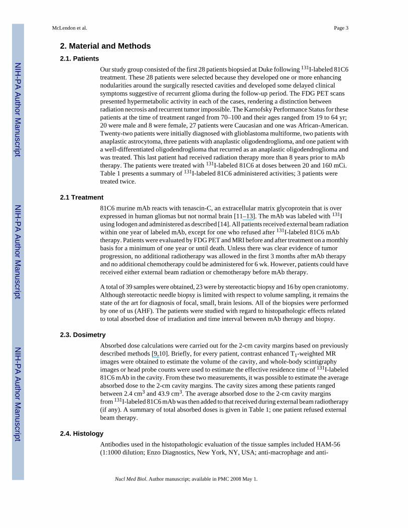
Intramural vascular hyalinization of small intraparenchymal vessels (Fig. 3) was found in 15biopsies, becoming evident in those biopsies taken 6 months or more following 131I-labeled
McLendon et al. Page 5
Nucl Med Biol. Author manuscript; available in PMC 2008 May 1.
NIH
-PA Author Manuscript
NIH
-PA Author Manuscript
NIH
-PA Author Manuscript

mAb treatment. The hyalinization of the vessel wall was circumferential and eosinophilic andexpanded the vascular wall, typical in every way to that described previously in patientsundergoing external beam therapy. Blood vessel thrombosis was identified in smallparenchymal vessels in 12 biopsies.
No evidence of glioma was noted in 15 biopsies; typically, reactive type gliosis manifest bycells with prominent eosinophilic processes that were densely knitted together formed by cellswith abundant cytoplasm and enlarged nuclei. Bizarre nuclear change could be found inoccasional reactive astrocytes as identified by their sparse distribution among otherwise benignastrocytes and macrophages (Fig. 4). A dense collection of gemistocytic cells with significantnuclear pleomorphism was a useful clue for tumor recurrence. Mitotic figures in these cellswere considered to be a sign of proliferative glioma. Concurrent use of theimmunohistochemically based proliferation marker, Ki-67 [17], clearly delineated malignant,proliferating glioma populations from benign, non-proliferating, reactive populations of glialcells (although macrophage populations were also noted occasionally to be proliferative). So-called “granular mitoses”, characterized by glial cells with nuclei exhibiting chromatin inmembrane bound, irregularly sized packets, could be found in tissues diagnosed as “gliosis,necrosis, and macrophages.”
Although highly variable in number and density, macrophage infiltrates were found in everybiopsy and were most commonly associated with obviously necrotic foci (Fig. 5). Occasionally,the macrophage infiltrates, when compared against the background edematous neuropil,exhibited an appearance such that a distinction from infiltrating tumor could not be made byroutine H&E staining (Fig. 6A). Therefore, immunohistochemical stains using the macrophagerelated markers HAM-56 and Mac-387, and glial marker GFAP, were evaluated for histologicphenotyping. Infiltrating microglia (Fig. 6B) often mimicked infiltrating gliomas as they lackedthe typical foamy, net-like cytoplasm of the activated macrophage and exhibited round, regularnuclei in contrast to the typical elongated nuclei of microglia in resting brain. Macrophagesoccasionally accounted for a brisk (>3%) Ki-67 labeling index.
Five patients were biopsied within four months of mAb therapy (n= 5 biopsies). Tumor wasidentified in two biopsies. Four of the five biopsies revealed necrosis with macrophageinfiltrates in all four and associated vascular thrombosis in two. Five patients coming to biopsywere long-term survivors (26 to 37 months post mAb treatment). Biopsies in two patientsmanifested proliferative glioma recurrence. Obvious necrosis was found in three long termsurvivors associated with macrophage infiltrates which indicated that necrosis was ongoing inthe treated fields in these samples.
3.2. Total absorbed dose versus histologic findings (Group A studies)A Kaplan-Meier plot (Fig. 7A) graphically displays survival of all 28 patients (Group A)stratified by last histologic diagnosis (negative for neoplasm, quiescent glioma, andproliferative glioma). Median survival for patients with tissue classified as proliferative glioma,quiescent glioma, and negative for neoplasm were 3.5, 15, and 27.5 months, respectively. Atwo-degree of freedom log-rank test showed significant differences in the pattern of survivalamong the three diagnoses (p < 0.0001). Patients with proliferative gliomas had the poorersurvival. The relationship between total absorbed radiation dose and survival was examinedusing Cox’s proportional hazards model. Without categorization, total dose was a significantpredictor of survival (p < 0.002); patients with higher doses had better prognoses. The averagetotal absorbed dose to the 2-cm cavity margins was categorized at higher and lower than 86Gy, and the relationship between total dose and survival was examined. Again, categorizedtotal dose was a significant predictor of survival (p < 0.002). A Kaplan-Meier survival curve(Fig. 7B) again graphically displays survival stratified by absorbed dose. Median survival inpatients receiving a total radiation dose greater than 86 Gy was 19 months compared with 7
McLendon et al. Page 6
Nucl Med Biol. Author manuscript; available in PMC 2008 May 1.
NIH
-PA Author Manuscript
NIH
-PA Author Manuscript
NIH
-PA Author Manuscript

months for those receiving less than 86 Gy. The relationship between total dose and thediagnosis of radiation necrosis was examined using logistic regression. This analysis, whichconsidered uncategorized total dose as a predictor of a “negative for neoplasm” histologicfinding (i.e. “gliosis, necrosis and macrophages”), showed a statistically significantrelationship (p < 0.003). Higher absorbed doses were more commonly associated with a“negative for neoplasm” histologic finding. If one were to dichotomize dose by its median, therelationship between dose and a “negative for neoplasm” biopsy again revealed that higherdoses were associated with radiation necrosis, as expected.
4. DiscussionLoco-regional administration of a targeted radiotherapeutic in the CNS such as into a surgicallycreated resection cavity is an attractive alternative to intravenous injection because of thepotential for minimizing radiation dose to normal organs and for overcoming the blood brainbarrier. On the other hand, radiation dose deposition to normal tissues in contact with a cavitywill be increased. The nature of the effect will depend on several factors including the range-energy deposition profile of the radiation, cross reactivity of the targeting vehicle with normalcell populations and the diffusion of the labeled drug beyond the cavity interface. With regardto 131I-labeled 81C6, 131I is a relatively low energy β-emitter, tenascin is not present in normalbrain, and because of their large size, intact mAbs diffuse slowly in tissue [11,12,19].
The present study incorporates the ten patients previously reported by Marriott [20] who werefound by 18FDG-PET to be hypermetabolic along the surgically created rim after treatmentwith 131I-labeled 81C6. That study noted that 18FDG accumulation along the rim correlatedwith macrophage infiltrates while nodular accumulations beyond the rim correlated withrecurrent glioma. The present study undertook to evaluate these tissues and those from biopsiesof additional patients more thoroughly from a histopathological standpoint.
To our knowledge the only other studies evaluating brain tissue from a patient treatedwith 131I-labeled mAbs to glioma-associated antigens were by Papanastassiou and colleagues[21,22] and by Riva and colleagues [23,24]. The former mentions a biopsy that was performed7 wk after 131I-ERIC-1 therapy on one patient who required further surgery; the tissue wasreported as necrotic without further description [21,22]. In a report on 17 patients treated withintra-cavitary 131I-labeled anti-tenascin mAb, all patients exhibited necrosis, with neoplasticcells found in 14 patients [23,24].
Iodine-131 is a useful radionuclide for labeling mAbs because of its low cost, simple labelingchemistry and short-range β radiation. Iodine-125, the most common radionuclide used inbrachytherapy and one of the more thoroughly studied radionuclides from the standpoint ofCNS sensitivity, differs from 131I in both its radiotoxicity and in its biological effects [10].External beam radiation differs substantially from the β emissions of 131I. The 90th percentilerange of 131I β particles in tissue is approximately 1 mm resulting in a steep dose gradientwithin this range. Based on cavity size and the limited diffusion of mAb in tissues, we wouldexpect the radiotoxic effects of 131I confined to be between a 2 to 5-mm shell of tissue adjacentto the cavity interface. Thus, for a 10-cm3 cavity, 131I would deliver a localized absorbed doseto the cavity interface in the range of 4000 Gy and at 1-cm depth, an absorbed dose of 39 Gy.The resulting average absorbed dose over the 2-cm cavity margins will be approximately 48Gy. Such high-absorbed doses and dose rate at the cavity interface will lead to immediate DNAdamage and have cytotoxic effects that can be observed within three months post mAb therapy,in contrast to external beam irradiation in which early toxicity is considered controversial[25].
McLendon et al. Page 7
Nucl Med Biol. Author manuscript; available in PMC 2008 May 1.
NIH
-PA Author Manuscript
NIH
-PA Author Manuscript
NIH
-PA Author Manuscript

Coagulative necrosis and liquefactive necrosis were found in the biopsies of 131I-labeled 81C6treated patients in the first month following therapy (Table 2). This evidence of early necrosismust be interpreted with caution, recognizing that surgically induced changes may also beactive at this time. Early-delayed and late-delayed effects of radiation from any source arelargely attributed to vascular damage, specifically, breakdown of the blood brain barrier inearly-delayed effects, and hyaline thickening of vessels and fibrin emboli in late delayedeffects. Although all of the regions sampled in these biopsies demonstrated gadoliniumenhancement on magnetic resonance imaging, the present study could not evaluate thefunctional effectiveness of the blood brain barrier. However, both hyaline thickening and fibrinemboli were present in tissues obtained from our patients undergoing biopsies beyond 3 monthspost mAb therapy. In contrast to the findings of 125I brachytherapy experimental animals[26] or children exposed to external beam irradiation [27], no vascular calcifications were notedin our specimens.
Reactive benign gliosis has been consistently noted in experimental models and in humanbiopsy and autopsy studies of post-radiation CNS damage. The reactive gliosis has been notedto be proliferative in one BUdR study following gamma radiation in rats [28]; however, theproliferative index, as determined with the anti-Ki-67 mAb, indicates the index is less than 1%in benign post-radiation reactive gliosis exclusive of proliferative endothelial cells andmacrophages.
Selection of the present specimens was keyed into radiographic images demonstrating regionsof enhancement and increased metabolic activity by PET. By selecting regions thatdemonstrated uptake of 18FDG, our tissues would, by inference, be hypercellular. The presentstudy, therefore, supports the earlier contentions of Marriott and colleagues [20] who suggestedthat bright PET regions related to radiation necrosis could be related to macrophage infiltratesand/or recurrent/residual glioma.
5. ConclusionThe neuropathologic features of intracavitary 131I-labeled mAb irradiation are qualitativelysimilar to those described for other radiation therapy modalities; however, the high dose ratesand high absorbed doses produced by the short range of 131I β particles seem to have resultedin an earlier necrotic reaction in the tumor bed than that reported for external beam irradiation[29]. Similar to 125I interstitial brachytherapy, 131I-labeled mAb treatment induced localizedradionecrois; however, toxic effects were less widespread, consistent with the shorter tissuerange of 131I βparticles. This study demonstrated that there was a significant correlationbetween total absorbed dose and biopsy outcome, and total absorbed dose and survival,pointing out that patients whose biopsy revealed quiescent glioma or radionecrosis havestatistically a higher median survival than those biopsies suggestive of proliferative glioma.Also, those patients that received total absorbed dose doses higher than 86 Gy had a mediansurvival that was significantly higher than those with total absorbed doses less than 86 Gy.
Acknowledgements
This work was supported in part by Grants NS2003, CA42324, CA108786 and CA70164 from the National Institutesof Health. The authors gratefully acknowledge the immunohistochemical contributions of Mr. James Burchette HT.
References1. Zalutsky MR. Current status of therapy of solid tumors: brain tumor therapy. J Nucl Med 2005;46
(suppl):151S–156S. [PubMed: 15653663]
McLendon et al. Page 8
Nucl Med Biol. Author manuscript; available in PMC 2008 May 1.
NIH
-PA Author Manuscript
NIH
-PA Author Manuscript
NIH
-PA Author Manuscript

2. Burger, PC.; Boyko, OB. The pathology of central nervous system radiation injury. In: Gutin, PH.;Leibel, SA.; Sheline, GE., editors. Radiation Injury to the Central Nervous System. New York: RavenPress; 1991. p. 191-208.
3. Burger PC, Mahley MS Jr, Dudka L, Vogel FS. The morphologic effects of radiation administeredtherapeutically for intracranial gliomas: a postmortem study of 25 cases. Cancer 1979;44:1256–72.[PubMed: 387205]
4. Baglan RJ, Marks JE. Soft-tissue reactions following irradiation of primary brain and pituitary tumors.Int J Radiat Oncol Biol Phys 1981;7:455–9. [PubMed: 7251415]
5. Marks JE, Wong J. The risk of cerebral radionecrosis in relation to dose, time and fractionation. Afollow-up study. Prog Exp Tumor Res 1985;29:210–8. [PubMed: 4070632]
6. Coffey RJ, Lunsford LD, Flickinger JC. The role of radiosurgery in the treatment of malignant braintumors. Neurosurg Clin North Am 1992;3:231–44.
7. Loeffler JS, Alexander E 3rd, Wen PY, Shea WM, Coleman CN, Kooy HM, et al. Results of stereotacticbrachytherapy used in the initial management of patients with glioblastoma. J Natl Cancer Inst1990;82:1918–21. [PubMed: 2250312]
8. Malkin MG. Interstitial brachytherapy of malignant gliomas: the Memorial Sloan-Kettering CancerCenter experience Recent Results. Cancer Res 1994;13:117–25.
9. Akabani G, Reist CJ, Cokgor I, Friedman AH, Friedman HS, Coleman RE, et al. Dosimetry of 131I-labeled 81C6 monoclonal antibody administered into surgically created resection cavities in patientswith malignant brain tumors. J Nucl Med 1999;40:631–8. [PubMed: 10210222]
10. Akabani G, Cokgor I, Coleman RE, Gonzalez Trotter D, Wong TZ, Friedman HS, et al. Dosimetryand dose- response relationships in newly diagnosed patients with malignant gliomas treated withiodine-131-labeled anti-tenascin monoclonal antibody 81C6 therapy. Int J Radiat Oncol Biol Phys2000;46:947–58. [PubMed: 10705017]
11. Bourdon MA, Wikstrand CJ, Furthmayr H, Matthews TJ, Bigner DD. Human glioma-mesenchymalextracellular matrix antigen defined by monoclonal antibody. Cancer Res 1983;43:2796–805.[PubMed: 6342760]
12. Bourdon MA, Matthews TJ, Pizzo SV, Bigner DD. Immunochemical and biochemicalcharacterization of a glioma-associated extracellular matrix glycoprotein. J Cell Biochem1985;28:183–95. [PubMed: 4066774]
13. Ventimiglia JB, Wikstrand CJ, Ostrowski LE, Bourdon MA, Lightner VA, Bigner DD. Tenascinexpression in human glioma cell lines and normal tissues. J Neuroimmunol 1992;36:41–55.[PubMed: 1370958]
14. Bigner DD, Brown MT, Friedman AH, Coleman RE, Akabani G, Friedman HS, et al. Iodine-131-labeled antitenascin monoclonal antibody 81C6 treatment of patients with recurrent malignantgliomas: phase I trial results. J Clin Oncol 1998;16:2202–12. [PubMed: 9626222]
15. Hulette CM, Downey BT, Burger PC. Macrophage markers in diagnostic neuropathology. Am J SurgPathol 1992;16:493–9. [PubMed: 1376020]
16. Brandtzaeg P, Jones DB, Flavell DJ, Fagerhol MK. Mac 387 antibody and detection of formalinresistant myelomonocytic L1 antigen. J Clin Pathol 1988;41:963–70. [PubMed: 3056988]
17. Wikstrand, CJ.; Fung, KM.; Trajanowski, JQ.; McLendon, RE.; Bigner, DD. Russell and Rubinstein’sPathology of Tumors of the Nervous System. Bigner, DD.; Rosemblum, M.; Brunner, JM., editors.London: Edward Arnold; 1998. p. 251-306.
18. Forsyth PA, Kelly PJ, Cascino TL, Scheithauer BW, Shaw EG, Dinapoli RP, et al. Radiation necrosisor glioma recurrence: is computer-assisted stereotactic biopsy useful? J Neurosurg 1995;82:436–44.[PubMed: 7861222]
19. Zalutsky, MR. Radionuclide therapy. In: Roesch, F., editor. Handbook of Nuclear Chemistry Volume4: Radiochemistry and Radiopharmaceutical Chemistry in Life Sciences. Dordrecht, Netherlands:Kluwer Academic; 2003. p. 315-348.
20. Marriott CJ, Thorstad W, Akabani G, Brown MT, McLendon RE, Hanson MW, et al. Locallyincreased uptake of fluorine-18-fluorodeoxyglucose after intracavitary administration of iodine-131-labeled antibody for primary brain tumors. J Nucl Med 1998;39:1376–80. [PubMed: 9708511]
McLendon et al. Page 9
Nucl Med Biol. Author manuscript; available in PMC 2008 May 1.
NIH
-PA Author Manuscript
NIH
-PA Author Manuscript
NIH
-PA Author Manuscript

21. Papanastassiou V, Pizer BL, Coakham HB, Bullimore J, Zananiri T, Kemshead JT. Treatment ofrecurrent and cystic malignant gliomas by a single intracavity injection of 131I monoclonal antibody:feasibility, pharmacokinetics and dosimetry. Br J Cancer 1993;67:144–51. [PubMed: 8427774]
22. Hopkins K, Papanastassiou V, Kemshead JT. The treatment of patients with recurrent malignantgliomas with intratumoral radioimmunoconjugates. Recent Results Cancer Res 1996;141:159–75.[PubMed: 8722426]
23. Riva P, Arista A, Sturiale C, Moscatelli G, Tison V, Mariani M, et al. Treatment of intracranial humanglioblastoma by direct intratumoral administration of 131I-labelled anti-tenascin monoclonalantibody BC-2. Int J Cancer 1992;51:7–13. [PubMed: 1373410]
24. Riva P, Franceschi G, Frattarelli M, Lazzari S, Riva N, Giuliani G, et al. Loco-regionalradioimmunotherapy of high-grade malignant gliomas using specific monoclonal antibodies labeledwith 90Y: a phase I study. Clin Cancer Res 1999;5:3275s–3280s. [PubMed: 10541375]
25. Miot E, Hoffschir D, Alapetite C, Gaboriaud G, Pontvert D, Fetissof F, et al. Experimental MR studyof cerebral radiation injury: quantitative T2 changes over time and histopathologic correlation. AJNRAm J Neuroradiol 1995;16:79–85. [PubMed: 7900606]
26. Bernstein M, Marotta T, Stewart P, Glen J, Resch L, Henkelman M. Brain damage from 125Ibrachytherapy evaluated by MR imaging, a blood-brain barrier tracer, and light and electronmicroscopy in a rat model. J Neurosurg 1990;73:585–593. [PubMed: 2398389]
27. Price RA, Birdwell DA. The central nervous system in childhood leukemia: III. Mineralizingmicroangiopathy and dystrophic mineralization. Cancer 1978;42:717–728. [PubMed: 277284]
28. Matsumoto K, Nakagawa M, Higashi H, Maeshiro T, Tsuno K, Mishima N, et al. Preliminary resultsof interstitial 192Ir brachytherapy for malignant gliomas. Neurol Med Chir (Tokyo) 1992;32:739–46. [PubMed: 1280775]
29. Fajardo, LF.; Berthrong, M.; Anderson, RE. Radiation Pathology. New York: Oxford UniversityPress; 2001. p. 351-361.2001
McLendon et al. Page 10
Nucl Med Biol. Author manuscript; available in PMC 2008 May 1.
NIH
-PA Author Manuscript
NIH
-PA Author Manuscript
NIH
-PA Author Manuscript

Figure 1.Tumor recurrence in these active malignant neoplasms was characterized by densehypercellularity associated with nuclear pleomorphism and vascular proliferation. Althoughareas of necrosis were easily found, the influence of therapeutic effect was not often readilydiscernible; thus, these recurrences were characterized as “active malignant gliomas” (H&E;250×).
McLendon et al. Page 11
Nucl Med Biol. Author manuscript; available in PMC 2008 May 1.
NIH
-PA Author Manuscript
NIH
-PA Author Manuscript
NIH
-PA Author Manuscript

McLendon et al. Page 12
Nucl Med Biol. Author manuscript; available in PMC 2008 May 1.
NIH
-PA Author Manuscript
NIH
-PA Author Manuscript
NIH
-PA Author Manuscript

Figure 2.(A) Punctate areas of rarefied cerebral white matter, often associated with a central vessel.Similar lesions are reported to represent an early finding of external beam radiation necrosis.(B) These regions of rarefaction often revealed little if any reaction in the surrounding brain,further lending support to an early manifestation of radionecrosis. (A: H&E 100×; B: H&E170×).
McLendon et al. Page 13
Nucl Med Biol. Author manuscript; available in PMC 2008 May 1.
NIH
-PA Author Manuscript
NIH
-PA Author Manuscript
NIH
-PA Author Manuscript
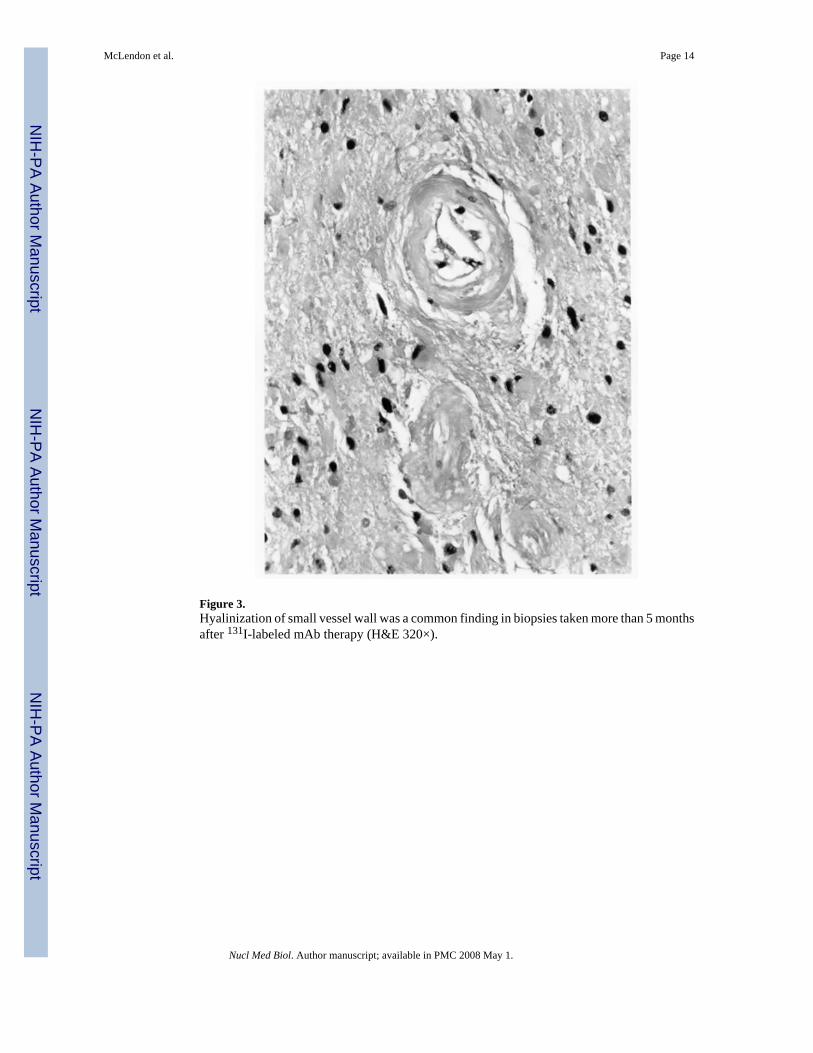
Figure 3.Hyalinization of small vessel wall was a common finding in biopsies taken more than 5 monthsafter 131I-labeled mAb therapy (H&E 320×).
McLendon et al. Page 14
Nucl Med Biol. Author manuscript; available in PMC 2008 May 1.
NIH
-PA Author Manuscript
NIH
-PA Author Manuscript
NIH
-PA Author Manuscript

Figure 4.Bizarre nuclear change could be found in occasional reactive astrocytes as identified by theirsparse distribution among otherwise benign astrocytes and macrophages (H&E 325×).
McLendon et al. Page 15
Nucl Med Biol. Author manuscript; available in PMC 2008 May 1.
NIH
-PA Author Manuscript
NIH
-PA Author Manuscript
NIH
-PA Author Manuscript

Figure 5.Within areas of necrosis, macrophages could be easily identified by their foamy cytoplasm,round cytoplasmic outlines, and bland nuclei (H&E 400×).
McLendon et al. Page 16
Nucl Med Biol. Author manuscript; available in PMC 2008 May 1.
NIH
-PA Author Manuscript
NIH
-PA Author Manuscript
NIH
-PA Author Manuscript

McLendon et al. Page 17
Nucl Med Biol. Author manuscript; available in PMC 2008 May 1.
NIH
-PA Author Manuscript
NIH
-PA Author Manuscript
NIH
-PA Author Manuscript

Figure 6.(A) Macrophages occasionally exhibit an appearance against the background edematousneuropil and made a distinction from infiltrating tumor difficult on H&E stain. (B) Here, thevast majority of cells present are infiltrating macrophages as confirmed by staining with theHAM 56 (A:H&E 325×; B:mAb HAM56 325×).
McLendon et al. Page 18
Nucl Med Biol. Author manuscript; available in PMC 2008 May 1.
NIH
-PA Author Manuscript
NIH
-PA Author Manuscript
NIH
-PA Author Manuscript

Figure 7.(A) Kaplan-Meier plot displaying median survival stratified by last tissue diagnosis as radiationnecrosis (“negative for neoplasm”), quiescent glioma, and active glioma. (B) Kaplan-Meierplot demonstrating median survival stratified by average total absorbed dose. Both plots displaymedian survival since last tissue histopathological diagnosis post mAb therapy.
McLendon et al. Page 19
Nucl Med Biol. Author manuscript; available in PMC 2008 May 1.
NIH
-PA Author Manuscript
NIH
-PA Author Manuscript
NIH
-PA Author Manuscript
NIH
-PA Author Manuscript
NIH
-PA Author Manuscript
NIH
-PA Author Manuscript
McLendon et al. Page 20Ta
ble
1H
isto
logi
cal d
iagn
osis
, rad
iatio
n ab
sorb
ed d
ose
and
surv
ival
dat
a fo
r the
28
patie
nts i
n th
is st
udy
Ori
gina
l Dia
gnos
is*
131 I-
labe
led
mA
bD
ose
(mC
i)
Abs
orbe
d D
ose
(Gy)
Res
ults
of L
ast
Bio
psy
Post
131 I-
mA
b tr
eatm
ent
Surv
ival
(mon
ths)
Aliv
e13
1 I-m
Ab
XR
TT
otal
From
Bio
psy
From
131 I-
mA
bT
reat
men
tFr
om O
rigi
nal D
iagn
osis
GB
M10
073
6013
3R
adia
tion
Nec
rosi
s12
2125
NA
O80
6862
130
Rad
iatio
n N
ecro
sis
4374
74N
GB
M12
032
5991
Qui
esce
nt G
liom
a16
2727
NG
BM
100
4960
109
Rad
iatio
n N
ecro
sis
9811
712
7Y
GB
M16
046
6010
6R
adia
tion
Nec
rosi
s41
5153
NG
BM
140
4360
103
Act
ive
Glio
ma
316
19N
GB
M80
1760
77R
adia
tion
Nec
rosi
s4
1113
NG
BM
120
4561
106
Rad
iatio
n N
ecro
sis
2629
31N
GB
M12
052
5911
1R
adia
tion
Nec
rosi
s56
7172
NA
A80
1966
85A
ctiv
e G
liom
a3
547
NG
BM
8034
5993
Rad
iatio
n N
ecro
sis
1928
29N
GB
M12
033
6194
Act
ive
Glio
ma
418
24N
GB
M16
017
6077
Act
ive
Glio
ma
312
14N
AO
204
6064
Rad
iatio
n N
ecro
sis
1214
15N
GB
M80
5060
110
Act
ive
Glio
ma
26
15N
GB
M14
047
6010
7R
adia
tion
Nec
rosi
s29
4243
NA
O10
036
--36
Act
ive
Glio
ma
9211
911
9Y
GB
M60
,101
4346
89A
ctiv
e G
liom
a9
4445
NA
A21
660
66A
ctiv
e G
liom
a1
414
NG
BM
140
560
65A
ctiv
e G
liom
a10
1014
NG
BM
100
2560
85Q
uies
cent
Glio
ma
1430
40N
AA
15,2
0N
AN
A--
Act
ive
Glio
ma
1114
14N
AA
6023
5679
Act
ive
Glio
ma
1023
26N
GB
M20
2058
78Q
uies
cent
Glio
ma
1235
36N
AA
207
5966
Act
ive
Glio
ma
214
15N
GB
M20
461
65A
ctiv
e G
liom
a3
512
NA
A12
038
6410
2A
ctiv
e G
liom
a17
2122
NG
BM
20,1
2026
6187
Qui
esce
nt G
liom
a12
315
615
8Y
* GB
M, g
liobl
asto
ma
mul
tifor
me;
AO
, ana
plas
tic o
ligod
endr
oglio
ma;
AA
, ana
plas
tic a
stro
cyto
ma
Nucl Med Biol. Author manuscript; available in PMC 2008 May 1.

NIH
-PA Author Manuscript
NIH
-PA Author Manuscript
NIH
-PA Author Manuscript
McLendon et al. Page 21Ta
ble
2Ti
me-
line
refle
ctin
g th
e oc
curr
ence
of h
isto
logi
c fin
ding
s am
ong
biop
sies
take
n fo
llow
ing
131 I
-labe
led
81C
6 m
Ab
ther
apy.
Tim
e (m
onth
s)
13
45
67
89
1011
1213
1517
2326
–37
Tum
or P
rese
nt− −
+−
++
−− −
+ +
+ +
−−
− +
+ +
−−
−− −
+ +
Coa
gula
tive
Nec
rosi
s−
+ +
++
++
+ +
+ +
+ +
++
+ +
+ +
++
−−
+ +
+Li
quifa
ctiv
e N
ecro
sis
− +
+−
−+
++
++
++
++
−−
+−
++
−−
− −
+ +
Rea
ctiv
e G
liosi
s− −
++
+−
++
+++
− +
++
+ +
− −
−+
+− − −
+B
lood
Ves
sel H
yalin
izat
ion
− −
+−
−−
++
++
++
++
−+
++
++
+−
− − −
+B
lood
Ves
sel T
hrom
bosi
s−
+ +
−−
−−
− +
± +
+ +
−−
− +
− +
++
−− −
+ +
Mac
roph
age
Infil
trate
− +
++
++
++
++
++
++
++
++
++
++
+ +
+ +
Each
mar
k (+
or −
) rep
rese
nts a
sing
le b
iops
y ob
tain
ed fr
om a
sing
le p
atie
nt a
t eac
h tim
e in
terv
al a
nd th
e m
ark
repr
esen
ts th
e pr
esen
ce (+
) or a
bsen
ce (−
) of t
he h
isto
logi
c fin
ding
.
Nucl Med Biol. Author manuscript; available in PMC 2008 May 1.
Copyright © 2022 FDOKUMEN