The neuropathology of CAG repeat diseases: review and update of genetic and molecular features
Upload
khangminh22Category
view
0download
0
NeuropathologyReview
Richard A. Prayson, MD
Humana PressHumana Press

Contents
NEUROPATHOLOGY REVIEW

2 Contents
Contents
HUMANA PRESSTOTOWA, NEW JERSEY
NEUROPATHOLOGYREVIEW
By
RICHARD A. PRAYSON, MDDepartment of Anatomic PathologyCleveland Clinic Foundation, Cleveland, OH

4 Contents
© 2001 Humana Press Inc.999 Riverview Drive, Suite 208Totowa, New Jersey 07512
For additional copies, pricing for bulk purchases, and/or information about other Humana titles, contact Humana at the above address or at any ofthe following numbers: Tel.: 973-256-1699; Fax: 973-256-8341; E-mail:[email protected]; Website: http://humanapress.com
All rights reserved.
No part of this book may be reproduced, stored in a retrieval system, or transmitted in any form or by any means, electronic, mechanical, photocopying,microfilming, recording, or otherwise without written permission from the Publisher.
Due diligence has been taken by the publishers, editors, and authors of this book to assure the accuracy of the information published and to describegenerally accepted practices. The contributors herein have carefully checked to ensure that the drug selections and dosages set forth in this text areaccurate and in accord with the standards accepted at the time of publication. Notwithstanding, as new research, changes in government regulations, andknowledge from clinical experience relating to drug therapy and drug reactions constantly occurs, the reader is advised to check the product informationprovided by the manufacturer of each drug for any change in dosages or for additional warnings and contraindications. This is of utmost importancewhen the recommended drug herein is a new or infrequently used drug. It is the responsibility of the treating physician to determine dosages and treatmentstrategies for individual patients. Further it is the responsibility of the health care provider to ascertain the Food and Drug Administration status of eachdrug or device used in their clinical practice. The publisher, editors, and authors are not responsible for errors or omissions or for any consequencesfrom the application of the information presented in this book and make no warranty, express or implied, with respect to the contents in this publication.
Cover illustrations: Inset gross picture depicts a brain with lissencephaly. The background microscopic picture is of subacute sclerosing panencephalitis.Artwork courtesy of Richard A. Prayson, MD.Cover design by Patricia F. Cleary.
This publication is printed on acid-free paper. ∞ANSI Z39.48-1984 (American National Standards Institute) Permanence of Paper for Printed Library Materials.
Photocopy Authorization Policy:Authorization to photocopy items for internal or personal use, or the internal or personal use of specific clients, is granted by Humana Press Inc.,provided that the base fee of US $10.00 per copy, plus US $00.25 per page, is paid directly to the Copyright Clearance Center at 222 Rosewood Drive,Danvers, MA 01923. For those organizations that have been granted a photocopy license from the CCC, a separate system of payment has beenarranged and is acceptable to Humana Press Inc. The fee code for users of the Transactional Reporting Service is: [1-58829-024-7/01 $10.00 + $00.25].
Printed in the United States of America. 10 9 8 7 6 5 4 3 2 1
Library of Congress Cataloging-in-Publication Data
Prayson, Richard A.Neuropathology review / by Richard A. Prayson.
p. ;cm.Includes bibliographical references and index.ISBN 1-58829-024-7 (alk. paper)1. Nervous system—Diseases—Outline, syllabi, etc. . 2. Nervous system—Diseases—Examinations, questions, etc. I. Title.[DNLM: 1. Nervous System Diseases—Outlines. WL 18.2 P921n 2001]RC347.P724 2001616.8’0076—dc21 2001016673

Contents
PREFACE
The scope of neuropathology continues to expand, as evidenced by increasing numbers ofmultivolume and specialty texts that have been published in recent years. For those in the neurosciencedisciplines, the ever increasing amount of information one needs to assimilate and master can be challeng-ing and even at times daunting.
Neuropathology Review attempts to summarize, in outline form, the essentials of neuropathology.The objective is twofold: (1) to provide an overview of neuropathology, for those initially encounteringthe discipline and, (2) to provide a framework for review for those preparing for in-service and boardexaminations in the disciplines of neurology, neurosurgery, and pathology that require some knowledgeof neuropathology.
The text is divided into three main sections. The first part (Chapters 1–11) presents, in outline form,basic information on the spectrum of neurologic-related disease. The second part (Chapters 12 and 13)will present pertinent pictorial examples of a spectrum of neuropathologic conditions in a question formatwith answers and brief explanation provided. The final section (Chapters 14 and 15) will include a seriesof written questions provided with answers and explanations in order to review the material presented inthe first section of the text.
The author would like to thank Ms. Denise Egleton for her help in preparing this manuscript and toacknowledge Dr. Caroline Abramovich for her review of the text.
Richard A. Prayson, MD
v

6 Contents

ContentsCONTENTS
Preface .................................................................................................................................... v1 Normal Histology .............................................................................................................12 Vascular Lesions .............................................................................................................. 73 Tumors ............................................................................................................................ 154 Trauma............................................................................................................................ 355 Congenital Malformations, Perinatal Disease, and Phacomatoses ............................... 416 Demyelinating and Dysmyelinating Diseases ............................................................... 517 Metabolic and Toxic Diseases ....................................................................................... 558 Degenerative Diseases ................................................................................................... 599 CNS Infection ................................................................................................................. 6510 Skeletal Muscle .............................................................................................................. 7511 Peripheral Nerve............................................................................................................. 8512 Figures with Questions................................................................................................... 9113 Answers to Figures with Questions ............................................................................. 18514 Written Self-Assessment Questions............................................................................. 19315 Answers to Written Self-Assessment Questions ......................................................... 211Bibliography ....................................................................................................................... 217Index ................................................................................................................................... 219
vii

8 Contents

1 Normal Histology
• Synaptophysin immunostain positive—mem-I. Central Nervous Systembrane protein of synaptic vesiclesA. Neurons
• Lipofuscin (aging pigment, lipochrome)• 5–100 µm in size◦ Increases with age• Variety of shapes: stellate, oval, round, pyra-◦midal, bipolar and unipolar Intracytoplasmic
• Nucleus ◦ Light yellow-brown color on routinehematoxylin and eosin staining◦ Vesicular with prominent nucleolus
• Hyaline (colloid inclusion)◦ Scalloped—Purkinje cells and pyramidal◦cells in cortical layer V Cytoplasmic
• Cytoplasm ◦ Endoplasmic reticulum dilated cisternae◦ Mitochondria prominent • Marinesco bodies
◦ ◦Nissl substance—granular basophilic mate- Eosinophilic intranuclear inclusionsrial, rough endoplasmic reticulum ◦ May be multiple within one nucleus
◦ Neurofilaments (60–100 A, transport ◦ Prominently seen in pigmented neuronsfunction)
◦ Increases with age◦ May have pigments such as lipofuscin • A variety of other inclusions within neuronsand melaninmay be seen in association with certain dis-• Cell processes ease processes (e.g., Lewy bodies, tangles,
◦ Axon—conducts away from cell body, Hirano bodies, Pick bodies, granulovacuolararises from axon hillock which contains degeneration, viral inclusions, etc.)no Nissl substance; axoplasm with mito- • Ischemic changes—“red and dead”—chondria, neurotubules, and neurofila- shrunken cell body with dark nucleus, indis-ments tinguishable nucleolus, and eosinophilic cyto-
◦ plasm.Dendrite—conducts toward cell body, con-tains Nissl substance • Central chromatolysis—related to axonal or
retrograde degeneration, cytoplasmic swell-• Pigmented (neuromelanin) neurons—coarse,ing with loss of Nissl substance, and periph-dark brown granuleseralization/flattening of the nucleus, may be◦ Substantia nigra (midbrain)reversible to a point
◦ Locus ceruleus (pons) • Ferruginization of neurons (fossilized neu-◦ Dorsal motor nucleus of the vagus nerve rons)—encrusted with calcium and/or iron,
may be seen in old infarcts.(medulla)
1

2 NEUROPATHOLOGY REVIEW
• Neuronophagia—phagocytosis of cell body • Alzheimer type II astrocyte
◦• Axonal spheroids—focal dilatation of axon Nuclear swelling and chromatin clearingwith neurofilaments and organelles, reaction ◦ Prominent nucleolationto axon injury, Purkinje cell axonal spher-
◦oids called torpedoes Cell cytoplasm not readily apparent
B. Astrocytes ◦ Prominently seen in globus pallidus inhepatic diseases that produce elevated• Largest of glial cellsammonia levels• Vesicular nucleus without nucleolus
• Viral inclusions in astrocytes• Cytoplasm highlighted with GFAP (glial◦fibrillary acidic protein) antibody Cowdry A—intranuclear, eosinophilic, or
basophilic viral inclusion; displaces the• Processes often invest vesselsnucleolus (e.g., subacute sclerosing• A variety of shapespanencephalitis (SSPE), progressive multi-
◦ Star-shaped fibrillary (white matter) and focal leukoencepholopathy (PML), herpes,protoplasmic (gray matter) cells cytomegalovirus (CMV)
◦ ◦Pilocytic astrocytes of periventricular Cowdry B—intranuclear, eosinophilicregion and cerebellum viral inclusion; often multiple and smaller
than Cowdry A inclusions, generally do◦ Bergmann astrocytes of cerebellumnot displace the nucleolus (e.g., polio)• Corpora amylacea
C. Oligodendrocytes◦ Laminated, basophilic polyglucosan • Most numerous cells in the central nervousbodiessystem (CNS)
◦ Associated with astrocytic foot processes • Small cells with no cytoplasmic staining◦ with hematoxylin and eosinIncrease with age
• Thinner and fewer cell processes than astro-◦ Frequently seen in subpial regionscytes• Reactive astrocytes (gemistocytes)
• Tend to arrange themselves around neurons◦ Cell hypertrophy—increased cell cyto- (neuronal satellitosis)plasm • “Fried egg” appearance is a delayed post-◦ Gliosis may be associated with increased formalin-fixation artifact (will not be seen atcell number frozen section)
• Rosenthal fiber • Cells often arranged in small rows in the◦ white matter between myelinated fibersCytoplasmic inclusion
• Involved in the formation and maintenance◦ Brightly eosinophilic, elongated structuresof myelin◦ Observed in a number of entities includ- • Only weak immunostaining with GFAP, posi-ing gliosis, Alexander’s disease, pilocytictive staining with S-100 protein antibodyastrocytoma
• May contain intranuclear inclusions in PML• Creutzfeldt astrocyteand SSPE.
◦ Reactive astrocytes with multiple small D. Ependymanuclei • Epithelioid cells that line ventricular walls◦ Seen in demyelinative processes and central canal of the spinal cord
• Alzheimer type I astrocyte • Columnar/cuboidal cells with cilia◦ Large cells often with multiple nuclei or • Cilia attached to cell body by blepharoplast
irregularly lobulated nuclei • Ependyma-lined central canal is patent in◦ Seen in progressive multifocal leukoence- childhood but generally becomes obliterated
at pubertyphalopathy

CHAPTER 1 / NORMAL HISTOLOGY 3
• Ependymal loss with injury accompanied by II. External granular—small neuronswith short axonsa proliferation of subependymal glia (granu-
lar ependymitis) III. Outer pyramidal—medium and largeE. Microglia neurons
• Small, dark, elongated nuclei with scant cyto- IV. Internal granular—small stellateplasm neurons
• HAM56 positive V. Inner pyramidal—medium size neu-rons, Betz cells• May proliferate in a diffuse pattern (micro-
gliosis) or nodular pattern (microglial nod- VI. Polymorphic layer—innermost layerule)—commonly seen with viral encephalitis • Cerebellum
• With destruction of parenchymal tissue, will ◦ Three layerssee a macrophage response (gitter cells)� Molecular layer—outermost layer• Macrophages seen in a variety of patholo-� Purkinje cell layer—single layer ofgies (e.g., infarct, demyelinating disease,
neuronsradiation, abscess)� Granular cell layer—hypercellular,F. Choroid plexus
small cells• Produces cerebrospinal fluid (CSF), usually◦intraventricular in location Outer granular cell layer in infant overlies
molecular layer, disappears by age 1 yr• May see choroid plexus in subarachnoidspace in cerebellopontine angle region • White matter
• Fibrovascular cores lined by epithelial cells, ◦ Fibers run at right angles to corticalcobblestoned contour surface
• May see small nests of arachnoidal (men- ◦ Includes centrum semiovaleingothelial) cells intermixed
◦ Two bundles in gray matter parallel to• May be focally calcifiedsurface
G. Arachnoid cap cells� Inner line of Baillarger between layers• Seen principally in the arachnoid membrane 3 and 4
• Associated with psammoma body formation� Stria of Gennari between layers 5 and
(laminated calcifications)6 in occipital lobe
• Dorsal leptomeninges of cord may contain • Basal gangliawhite firm plaques (arachnoiditis ossifi-
◦ Includes caudate, putamen, globus pal-cans)—laminated dense hyalinized fibrouslidus, and amygdalatissue
◦• May see melanocytes in the meninges, more Gray matter tissueprominent in darker skinned individuals ◦ Caudate and putamen contain thin fasci-• Dura overlying the leptomeninges comprised cles of myelinated fibers (pencil bundlesprimarily of dense fibrous connective tissue of Wilson)
H. Architecture • Hippocampus• Cortex ◦ Consists of dentate gyrus, Ammon’s horn,
◦ Six layers, parallel to surface and subiculum
◦ ◦Neurons within the layers have their Ammon’s horn (CA = cornu Ammon’s)processes oriented perpendicular to the divided into four regionssurface
� CA4 endplate region within the hilus◦ of the dentate gyrusLayers
I. Molecular—few small neurons, glia, � CA3 connects endplate to resistancesector (CA2)outermost layer

4 NEUROPATHOLOGY REVIEW
� CA1 Sommer sector (most sensitive ◦ Perifascicular atrophy—ischemia related,area of brain to anoxic damage) characteristic of dermatomyositis
• Spinal cord ◦ Fascicular atrophy—spinal muscular atro-◦ phy, hereditary motor and sensory neurop-Enlargement of anterior (ventral) horns in
athies, vasculitiscervical and lumbosacral region
◦◦ Type I atrophy—myotonic dystrophy, con-Central canal in middlegenital myopathies, spinocerebellar degen-◦ Terminates with filum terminale eration
• Pineal gland ◦ Type II atrophy—disuse, steroids (type◦ Vaguely nodular architectural arrangement IIB), collagen vascular disease, myasthe-
of cells with glial background nia gravis, cachexia, paraneoplastic neuro-myopathy◦ Corpora arenacea—calcifications, present
after puberty • Hypertrophy
◦ ◦Pineal cysts—gliotic cysts often with Normal in athletes (type II fibers)Rosenthal fibers ◦ Often associated with central nuclei and
• Pituitary gland split fibers.
◦ Adenohypophysis—nests of epithelioid E. Muscle fiber types (type I, type IIA, type IIB)cells separated by delicate fibrovascular generally distributed in a mosaic/checkerboardseptae, cell types in a given nest are patternmixed type, geographic predominance of • Type I fibers—high oxidative, low glyco-certain cell types lytic activity
◦ Pars intermedia—at interface between • Type II fibers—low oxidative, high glyco-adenohypophysis and neurohypophysis, lytic activityglandular formations may be seen • Different muscles contain different propor-
◦ Neurohypophysis—spindled cells loosely tions of the three fiber typesarranged • Loss of the mosaic distribution of fiber
II. Skeletal Muscle types—fiber type grouping, indicative ofchronic denervation and reinnervationA. Muscle fibers arranged in bundles (fascicles)
surrounded by perimysium connective tissue; F. Muscle cells of variable length—run from ori-endomysium is connective tissue between mus- gin to insertioncle fibers G. Muscle cells made up of myofilaments
B. Nuclei multiple, peripherally arranged • Two main types: thick myosin filaments and• Central nuclei abnormal if seen in >3–5% of thin actin filaments
myofibers; • Sarcomere segment is the functional contrac-• Increased central nuclei in myotonic dystro- tile unit of muscle formed by the orderly
phy, myotubular myopathy, Charcot-Marie- arrangement of myofilamentsTooth disease • Sarcomeric banding patterns
C. At muscle–tendon junction, fibers get smaller ◦ Dark central A (anisotropic) band consist-and often contain internalized nuclei.ing of mostly thick myosin filaments
D. In cross section, fibers are generally the same ◦ A band crossed at midpoint by a dark nar-sizerow transverse M line consisting of cross-• Atrophy bridges which link adjacent, myosin fila-
◦ ments togetherScattered versus grouped (neurogenic),tend to have angulated contours ◦ M band surrounded by paler H band
◦ Rounded fiber atrophy—more likely myo- which varies in width as the sarcomericlength changespathic

CHAPTER 1 / NORMAL HISTOLOGY 5
◦ K. Neuromuscular junction—interface between thePale I (isotropic) band situated on eithernerve and muscle, acetylcholine is released byside of the A band consisting mostly ofthe nerve to activate the muscle; the electricalthin, actin filaments in combination withimpulse reaches the sarcoplasmic reticulum viatroponin and tropomyosin proteinsthe T system.◦ Z band divides I band at midpoint and
III. Peripheral Nervemarks the longitudinal boundary of eachA. Connective tissue layerssarcomere
• Epineurium—binds fascicles together, contin-• During contraction of the fiber, I filamentsuous with dura mater at junction of spinalslide toward the center of the A band withnerves and spinal nerve rootsshortening of the I and H bands
• Perineurium—concentric layers of flattened• Desmin intermediate filaments link Z discscells which are epithelial membrane antigentogether and join them to the plasmalemma(EMA) positive, separated by collagen that
H. Sarcoplasmsurrounds individual fascicles, forms part of
• Represents the cytoplasm of the myofiber the blood–nerve barrier, continuous with thepia arachnoid• Consists of a variety of structures/organelles
• Endoneurium—collagen compartment that◦ Mitochondria—type I > type IIsurrounds individual axons, Schwann cells,◦ T system (transverse tubular system)—net- and fibroblasts
work of tubules which is continuous withB. Mast cells also present in endoneurium,the muscle cell plasma membrane, allows
increase in number with axonal degenerationfor the rapid passage of depolarizationand neurofibromainto the interior of the myofiber
C. Renaut bodies–cylindrical hyaline bodies◦ Sarcoplasmic reticulum—a series of flat- attached to inner aspect of the perineurium;tened sacs between and around myofbrils composed of collagen fibers, fibroblasts and epi-
◦ neurial cells; function not known, increase inLipid—droplets lie between myofibrilsnumber in compressive neuropathies.and adjacent to mitochondria, type I >
type II D. Blood supply comes from vasa nervorum
◦ E. Nerve fibersGlycogen—more prominent in region of Iband than A band, type II > type I • Either myelinated or unmyelinated
◦ • Class A fibersRibosomes, Golgi membranes, intermedi-ate filaments, microtubules, and lipofuscin ◦ Myelinatedalso seen. ◦ 1–20 µm in diameter; conduction velocity
I. Muscle spindle 12–100 m/s• Collections of small intrafusal fibers (3–14) • Class B fibers
enclosed in a connective tissue capsule◦ Myelinated preganglionic autonomic• Center of spindle is swollen, ends are
fiberstapered
◦ Up to 3 µm in diameter; conduction veloc-• Located between muscle fascicles in perimy-ity 3–15 m/ssial connective tissue
• Class C fibers• Consists of two types of intrafusal fibers:◦ Unmyelinatednuclear bag fibers and nuclear chain fibers
◦• More numerous in muscles involved in deli- Small fibers (0.2–1.5 µm diameter); con-cate movements duction velocity 0.3–1.6 m/s
• Associated with sensory nerve endings F. Myelin
• Can be highlighted with certain stainsJ. Motor unit—consists of anterior horn cell,nerve fiber arising from it and the muscle fibers (Luxol fast blue, Loyez, osmium, periodic
acid schiff (PAS), Masson trichrome)supplied by it.

6 NEUROPATHOLOGY REVIEW
• Formed by fusion of Schwann cell mem- • Distance between nodes is proportional tomyelin thicknessbranes
• 75% lipid and 25% protein • Conduction of impulses along myelinatedfibers proceeds in a discontinuous manner• Major lipids comprising myelin: cholesterol,from node to node (saltatory conduction)sphingomyelin, galactolipid
• One Schwann cell per myelinated axon (in I. Myelinated axonsCNS, one oligodendrocyte supplies myelin • Axon delimited by axolemma membraneto multiple axons) • Axolemma separated from adjacent Schwann• Schwann cells S-100 positive by immunohis- cell by periaxonal space of Klebstochemistry • Axonal cytoplasm contains mitochondria,• Pi granules of Reich—lamellated structures,
smooth endoplasmic reticulum, glycogen,cytoplasm accumulates along with lipofuscinribosomes, peroxisomes, neurotransmitterwith increasing agevesicles, and filaments/tubules• Corpuscles of Erzholz—spherical bodies in • Filaments include actin, neurofilaments, andSchwann cell cytoplasm.microtubules
G. Schmidt-Lanterman cleftsJ. Unmyelinated axons• Cleft splits the cytoplasmic membranes and
• Best evaluated and studied by electronserves as a route of passage for substancesmicroscopyfrom the outer cytoplasmic layer through the
myelin sheath to the inner cytoplasm • More numerous than myelinated axons:• The number of clefts correlate with the diam- 3–4 : 1 unmyelinated : myelinated axons
eter of the axon • Frequently associated with Schwann cells;H. Node of Ranvier may be more than one axon per Schwann
cell• Gaps in the myelin sheath

2 Vascular Lesions
ties exist; the entire brain not uniformlyI. Anoxiainvolved (selective vulnerability)
A. Anoxia observed in a wide variety of clinical • Neurons most sensitive to anoxiaconditions and forms◦ Hippocampal, Sommer sector (CA1)• Anoxic anoxia—insufficient oxygen
most sensitivereaches blood (e.g., drowning)◦ Cerebral cortex, layers III, V, and VI• Anemic anoxia—insufficient oxygen con-
(which contain larger-size neurons)tent in blood (e.g., carbon monoxide poi-◦soning) Cerebellar Purkinje cells (if patient sur-
vives for a period of time, may see• Histotoxic anoxia—poisons interfere withBergmann gliosis)oxygen utilization (e.g., cyanide, sulfide)
◦ Caudate and putamen• Stagnant anoxia—most common,• Accentuated in the boundary zonesdecreased cerebral perfusion (e.g., cardiac
between vascular distributionsarrest); factors that determine amount ofbrain damage after cardiac arrest include D. Gross pathologyduration of ischemia, degree of ischemia, • Swollen, softtemperature during the event and blood glu- • Gray matter is duskycose levels
• Areas of cavitation in a laminar patternB. Precise mechanisms not completely eluci-may be observeddated; however, a number of consequences◦ Pseudolaminar is used to describeoccur which have a negative effect on the
involvement of more than one corticalCNSlayer• Edema
◦ Laminar describes involvement of a sin-• Lactic acid accumulation, decreased pHgle cortical layer• Increase in free fatty acids
E. Microscopic pathology• Increase in extracellular potassium and • Dendrite and astrocyte swelling (i.e., spong-
ammonia iness of the neuropil)• Abnormalities in calcium flux • Ischemic (homogenized) neurons (i.e., “red
and dead”)• Reperfusion problems
• Endothelial hyperplasiaC. Morphology of ischemia highly variable andunpredictable. • Microglial reaction
• Dissolution of neurons after several days• Numerous regional and cellular vulnerabili-
7

8 NEUROPATHOLOGY REVIEW
II. Infarction (2) interstitial edema, (3) pyknosis, (4)hypereosinophilia of neurons, (5) microva-A. Generalcuolization of neurons (swollen mito-• Cerebral blood flow 20% of cardiac out-chondria)
put, 15% of oxygen consumption of body.• 24 h: macrophage infiltration begins, axo-• Main anterior flow (70%) through internal
nal swelling, may see neutrophilic infiltra-carotid arteries; posterior flow via vertebral
tion (ceases by d 5)arteries—these systems anastomose via the
• 3–4 d: prominent macrophage infiltrationanterior and posterior communicating arter-ies to form the circle of Willis • 7–10 d: astrocytic proliferation and hyper-
trophy evident• Disruption in flow can result in infarct(stroke) • 30 d: intense gliosis
• Stroke is a clinical term referring to • May have a hemorrhagic component (18–“abrupt onset of focal or global neurologi- 48%); most hemorrhagic infarcts arecal symptoms caused by ischemia or hem- embolic artery events; hemorrhage inorrhage”—symptoms >24 h; if symptoms infarcts due to reperfusion of necrotic ves-resolve in less than 24 h = transient isch- sels or occlusion of venous drainageemic attack (TIA) • May encounter degeneration of tracts distal
• Risk of stroke increased with increasing to infarctage, male > female, smoking, hyperten- E. Lacunar infarctsion, atrial fibrillation, carotid artery steno- • The infarcts range in size from 3–4 mmsis, hyperlipidemia, diabetes, heart surgery,
up to 1.5 cmantiphospholipid antibodies, high-estrogen• Most common sites: putamen, caudate, thal-oral contraceptives
amus, pons, internal capsule, and convolu-• Venous thrombosis associated with preg-tional white matternancy and oral contraceptive use
• Causes: (1) lipohyalinosis, (2) occlusion of◦ Most common sites of venous thrombo-small penetrating vessels, (3) dissection,sis in superior sagittal sinus, lateral(4) embolisinuses, and straight sinus
• Small perivascular cavities common in◦ Septic thrombosis most common in cav- basal ganglia and deep white matter; etaternous sinus, often due to contiguous lacunaire (gray matter) and etat criblespread from soft tissue/sinus infection (deep white matter)
B. Determining the age of an infarct either F. Respirator braingrossly or microscopically can only be done • Also known as (AKA) diffuse anoxicwithin a wide range. The times given are
encephalopathyguidelines rather than absolutes.• Permanent global ischemia due to nonper-C. Gross pathology
fusion of entire brain• Unequivocal alterations require up to 24 h; • Brain perfusion depends on mean arterialearly changes include edema, congestion,blood pressure exceeding the intracranialsofteningpressure (cerebral perfusion pressure =• 48 h: “cracking”—separation of the mean arterial blood pressure − intracranial
necrotic tissue from intact tissue pressure)• 72 h: infarcted area usually clearly deline- • The most extreme form of stagnant anoxia
ated; cortex friable and soft• Grossly—dusky brown discoloration of cor-• A 1-cm cavity takes 2–3 months to form
tex, blurring of gray–white junction, gen-• Once cavitation begins, it is difficult to eral friability of tissue (brain often does
determine the age of the infarct. not fix well in formalin)D. Microscopic pathology • Microscopically—ischemic neurons every-
where ± infarcts• Earliest changes: (1) astrocytic swelling,

CHAPTER 2 / VASCULAR LESIONS 9
G. Carbon monoxide (CO) poisoning • Risk of rupture, greatest in aneurysms > 1cm in size• A form of anemic anoxia due to displace-
ment of oxygen from its binding site on • “Giant aneurysm” defined as > 2.5 cm, usu-hemoglobin ally causes compressive or embolic
symptoms• CO binds irreversibly to hemoglobin,reducing its oxygen-carrying capacity • Etiology
• CO directly binds to iron-rich areas of ◦ Many theoriesbrain (globus pallidus and pars reticulata ◦ Congenital defects of part or all of theof substantia nigra)
media at the arterial bifurcation, does• Pathologically marked by necrosis of glo- not explain why most arise in adulthoodbus pallidus and substantia nigra. ◦ Remnants of embryonic vessels• May see demyelination and cerebral white
◦ Focal destruction of the internal elasticmatter destruction (Grenker’s myelin-membrane due to hemodynamic alter-opathy)ations, about 3–9% of patients with arte-H. Air embolismriovenous malformation (AVM) have• May be related to decompression sicknessaneurysms
“the bends”—nitrogen gas in blood, can◦ Abnormalities in specific collagencause spinal cord microinfarcts
subsets• May also occur related to cardiac bypass◦surgery Associated conditions:
I. Hypoglycemic brain damage � Polycystic kidney disease, Potter type3• Causes selective neuronal necrosis-like
ischemia � Ehlers–Danlos syndrome (types IVand VI)• Different mechanism of injury than
ischemia � Coarctation of the aorta
◦ � Marfan’s syndromeDecreased lactate and pyruvate
◦ � Pseudoxanthoma elasticumTissue alkalosis• Pathology• Neuronal necrosis in cerebral cortex super-
ficial layers, hippocampus (CA1 and den- ◦ Media usually defectivetate) and caudate; no Purkinje cell necrosis ◦ Intimal hyperplasia and sometimes a
III. Aneurysms gap in the internal elastic membraneA. Saccular aneurysms seen
• AKA: “berry,” congenital or medical ◦ Aneurysm wall usually contains fibrousdefect tissue
• Incidence in general population, 2–5%; ◦ Atherosclerotic changes occasionallyfemale : male 3 : 2.seen• Usually present at arterial bifurcation; ◦ Phagocytosis and hemosiderin deposi-most asymptomatiction may be present◦ 85% in anterior circle of Willis
B. Infectious (septic) aneurysms◦ 25% multiple • Most related to bacterial endocarditis◦ 20% bilateral ◦ Arterial wall weakened by pyogenic bac-• Most common sites teria which usually reach the wall by an◦ infected embolusMiddle cerebral artery trifurcation
◦◦ May be multipleAnterior communicating artery junction
◦ ◦ Located on distal branches of the mid-Internal carotid artery—posterior com-municating artery junction dle cerebral artery

10 NEUROPATHOLOGY REVIEW
• Organisms usually of low virulence, partic- • Less compact than AVM or cavernous mal-formationularly Streptococcus viridans and Staphylo-
coccus aureus • May have a large central draining vein• Mycotic aneurysms typically refer to fun- • Varix is a single dilated/large vein
gal aneurysms; Aspergillus the most com-D. Cavernous malformation (angioma)mon organism responsible
• Large, sinusoidal-type vessels in appositionC. Fusiform aneurysmsto each other• Related to dolichoectasia (elongation, wid- • Little or no intervening parenchymaening, and tortuosity of a cerebral artery)
• Compact malformations• Supraclinoid segment of internal carotidartery and basilar artery most common • Mineralization and ossification common;sites occasionally massive
• Fusiform aneurysm refers to dilated seg- • May bleedment of artery E. Capillary telangiectasia
• Seen commonly with advanced atheroscle- • Capillary sized vesselsrosis • Separated by normal neural parenchyma
IV. Vascular Malformations• Common in the striate ponsA. General information• Often an “incidental” finding at autopsy• True malformations result from the embry-
V. Hemorrhageonic vascular networkA. Common causes• Some increase in size by incorporating
adjacent vessels—“recruitment” • Spontaneous intracerebral hemorrhage,• Clinical symptoms include seizures, “steal” often ganglionic (caudate, putamen) related
phenomenon, hemorrhage to hypertension—most common cause ofnontraumatic hemorrhage• In children, excessive shunting may lead to
cardiac decompensation (especially vein of • Ruptured saccular aneurysmGalen malformations). • Vascular malformation, particularly AVM
• True incidence of hemorrhage unknown, and cavernous angiomaestimates = 5–10% overall • Coagulopathy associated, may have both
• A subset of malformations is of mixed platelet and prothrombin time abnormal-type. ities
B. Arteriovenous malformation (AVM) • Congophilic (amyloid) angiopathy• Admixture of arteries, veins, and intermedi- ◦ Lobar hemorrhage
ate size vessels◦ Elderly• Vessels are separated by gliotic neural◦parenchyma Amyloid deposition in vessel walls both
in meninges and cortex• Foci of mineralization and hemosiderindeposition common ◦ Most commonly β-amyloid type (chro-
• Typically superficial, wedge-shaped with mosome 21)the apex directed toward the ventricle ◦ Associated with Down’s syndrome and• Commonly found in surgical series; the Alzheimer’s diseasemost common vascular malformation asso- ◦ Highlighted on Congo red (apple greenciated with hemorrhage; peak presentation
birefringence with polarized light), thi-2nd–4th decadesoflavin S or T, and crystal violet stainsC. Venous malformation (angioma)
• Neoplasms• Veins of varying sizes• Blood dyscrasias (i.e., sickle cell anemia)• Vessels separated by mostly “normal”
parenchyma • Vasculitis

CHAPTER 2 / VASCULAR LESIONS 11
B. Pathology ◦ Associated with polymyalgia rheumatica• Soft, gelatinous clot ◦ May be a focal process (skip lesions)• Small vessels with thrombosis may project ◦ Lymphoplasmacytic inflammation ±
into the hemorrhage granulomas, fibrous scarring with• Petechial hemorrhage may be seen around healing
the large hemorrhage • Takayasu’s arteritis• Ischemic necrosis seen in associated brain ◦ Aortic arch and branches and descend-
tissue ing aorta• Clot edge often has hemosiderin-laden mac- ◦ Younger patients (15–40 yr), Orientalrophages
females at higher risk• Astrocytic proliferation often prominent at ◦ Lymphoplasmacytic inflammation of thethe edgemedia with fibrosis, granulomas/giant• Amyloid may be seen in adjacent vessels cells
if congophilic angiopathy is the cause • Thromboangiitis obliterans• Venous hemorrhage—usually the infarcts • Vasculitis associated with connective tis-are larger and not confined to any arterialsue disease, (i.e., systemic lupus erythema-distribution, often over the convexitytosus [SLE], Sjogren’s syndrome, Behcet’s
VI. Vasculitis syndromeA. Common causes • Churg-Strauss—necrotizing vasculitis with
eosinophilia• Polyarteritis nodosa (PAN) and its variants• Primary angiitis of central nervous system◦ Systemic disease
◦ Headaches, multifocal deficits, diffuse◦ Male : female 2 : 1encephalopathy◦ Any age, peak 40–50 yr ◦ Adults (ages 30–50 yr)◦ Necrotizing vasculitis with fibrinoid ◦ ESR normal or mildly elevatednecrosis of medium-sized vessels
◦ Biopsy of nondominant temporal tip,◦ May represent immune-complex-including leptomeninges (highest yield),
mediated vasculitis (subset of patientscortex, and white matter recommended
are hepatitis B or C positive)◦ Arteries involved more than veins• Hypersensitivity angiitis◦ May be granulomatous or nongranulo-• Wegener’s granulomatosis
matous◦ Systemic disease often with respiratory ◦ May be complicated by infarct or hem-tract componentorrhage◦ Most patients 40–60 yr • Other causes
◦ ANCA (anti-neutrophil cytoplasmic anti- ◦ A variety of other conditions can resultbodies)—directed against proteinase 3 in a “vasculitic” pattern of injury patho-
• Lymphomatoid granulomatosis logically• Giant cell arteritis ◦ Drugs (e.g., cocaine, amphetamines)
◦ ◦The most common of the granuloma- Infection (e.g., Treponema, Borrelia,tous vasculitides Herpes, HIV, Aspergillus, Mucor)
◦ ◦Older patients >50 yr Lymphoproliferative disorders
◦ ◦Primary target is extracranial arteries of Other (demyelinating disease, atheroscle-head, but may involve cerebral vessels rosis, amyloid, etc.)
B. Pathology◦ Headache, blindness, increased erythro-cyte sedimentation rate (ESR) • Segmental inflammation and necrosis of

12 NEUROPATHOLOGY REVIEW
vascular walls are the common features of C. Atherosclerosis/hypertensive angiopathythe above-listed vasculitides. • Risk factors: dyslipidemia, hypertension,
smoking, diabetes• Vasculitis associated with SLE and lymph-omatoid granulomatosis are the only two • Initial atherosclerotic lesion is fatty streakwhich typically involve brain parenchyma. marked by foam cells filled with low-
density lipoprotein (LDL) cholesterol.• PAN, Wegener’s granulomatosis, and giant• Fatty streaks may develop into fibrouscell arteritis involve vessels in the sub-
plaques (especially develop at outerarachnoid space, peripheral nervous sys-aspects of arterial bifurcation where lami-tem, or the extracranial vessels.nar flow is disturbed)—connective tissue,• Neurologic dysfunction in all entities issmooth muscle cells, foam cells, lympho-related to ischemia.cytes, necrotic cell debris, and extracellular
VII. Miscellaneous lipids/cholesterolA. Binswanger’s disease • Turbulence caused by plaque may cause
further disease progression.• Rare condition generally presenting• Complicated plaque results from disruptionbetween ages 50–60 yr; evolves over 3–5
of endothelium resulting in thrombus for-yrmation (risk for occlusion and emboli).• Also called subcortical arteriosclerotic • Hypertensive angiopathy shifts the autoregu-encephalopathylation (maintains cerebral blood flow at a• Moderate intermittent hypertension and pro- constant level between mean arterial pres-
gressive, often profound dementia are fea- sures of 50–150 mm Hg) curve to the right,tures. raising the lower limit of regulation at which
• Widespread vascular alterations and white adequate cerebral flow can be maintained.matter changes are seen and readily demon- • Malignant hypertension–acute hypertensionstrated with neuroimaging. ◦ Diffuse cerebral dysfunction, headache,
• Ventricular dilatation, often secondary to nausea, vomiting, altered consciousnesshydrocephalus ex vacuo ◦ May be the result of pheochromocytoma
• Focal and diffuse myelin loss with associ- (release of catecholamine), disseminatedated reactive astrocytosis in the deep hemi- vasculitis, eclampsia, rebound drugspheric white matter effect
• Subcortical arcuate fibers are spared. ◦ Brain edema, focal ischemia, and intra-cerebral hemorrhage• Changes most severe in the temporal and
occipital lobes. • Chronic hypertension
◦• Demyelination is thought to be secondary Further worsens atherosclerotic changesto reduced perfusion due to arteriosclerosis in large arteriesof small penetrating vessels. ◦ Causes small vessel disease with disrup-
B. CADASIL (Cerebral autosomal dominant arte- tion of blood-brain barrier resulting inriopathy with subcortical infarcts and leu- basal lamina thickening and reduplica-koencephalopathy) tion, smooth-muscle degeneration, fibri-
noid change (necrosis), and increased• Mutation of notch 3 gene on chromosomecollagen deposition (lipohyalinosis—col-19q12lagenous fibrosis)• Strokes and vascular dementia ◦ May form Charcot-Bouchard microane-• Systemic disorder urysms (miliary aneurysms)—dilata-tions/outpouchings of vessel wall• Thickened vessel walls (media and adventi-caused by fibrinoid changetia) with basophilic, PAS-positive granules
within smooth muscle cells D. Moyamoya syndrome
• Defined angiographically by spontaneous• Variable degrees of perivascular atrophy

CHAPTER 2 / VASCULAR LESIONS 13
occlusion of the circle of Willis and the • Hemiparesis and hemorrhagepresence of abnormal collateralization • Intimal fibroplasia usually without athero-
• Two peaks of incidence—1st and 4th sclerotic changes, no inflammationdecades
• Most patients young, <20 yr, females >males


3 Tumors
• >90% derived from fibrillary astrocytesI. Astrocytic Neoplasms• Arise in white matterA. Classification schemas (grading) for astrocy-• Frontal lobe>parietal>temporal>occipitaltomas• Noncontrast enhancing, ill-defined region• WHO classification
of low density on CT scan◦ Grade I: pilocytic astrocytoma, subepen- • Seizures, sensorimotor deficits, increaseddymal giant cell astrocytomaintracranial pressure, altered mentation
◦ Grade II: well-differentiated diffuse • Grossly a solid tumor, infiltrative growthastrocytoma, pleomorphic xanthoastro- pattern, obliterate gray–white junction, sub-cytoma tle color change (yellow/tan or gray)
◦ Grade III: anaplastic astrocytoma • Microscopic
◦ ◦Grade IV: glioblastoma multiforme Uneven cellularity• Ringertz classification ◦ Rare mitoses
◦ ◦Low-grade astrocytoma Irregular nuclear contours, hyperchro-matic nuclei◦ Anaplastic astrocytoma
◦ Mild pleomorphism◦ Glioblastoma multiforme• May see microcystic change (do not see• Daumas-Duport classification (St. Anne-
typically with gliosis)Mayo)• Calcification (15%)Grade Criteria • Should not see vascular or endothelial pro-
1 0/4 liferation or necrosis2 1/4 • GFAP (glial fibrillary acidic protein) pos-
itive3 2/4
◦4 3–4/4 Intermediate cytoplasmic filaments(Criteria: mitoses, necrosis, nuclear atypia, ◦ Also stains ependymomas and choroidendothelial proliferation) plexus tumors
B. Low-grade astrocytoma ◦ Decreased staining in higher-gradetumors• 20% of all gliomas, 10–15% of all astro-
cytic tumors • Electron microscopy• Incidence: 1.4 new cases/1 million popula- ◦ Astrocytoma cells marked by cyto-
tion/yr plasmic intermediate filaments (7–11nm)• 4th-decade peak
15

16 NEUROPATHOLOGY REVIEW
◦ ◦ Hypertrophic cells (reactive astrocytes)Generally not useful for diagnosisprominent• DNA/cell proliferation
◦ Hypercellularity with even distribution◦ Most diploid◦ Lack nuclear hyperchromasia and angu-◦ Ki-67 (MIB-1) proliferation indices
larityroughly correlate with tumor grade◦ Absent calcification◦ Tumor heterogeneity in proliferation—◦different rates of cell proliferation and Rosenthal fibers—intensely eosino-
varying histologic appearance in differ- philic, elongate, or corkscrew-shapedent regions of the tumor structures
◦• Molecular genetics Collagen and hemosiderin deposition
◦ ◦TP53 mutations in >60% of diffuse Lack microcystic changeastrocytomas (chromosome 17)— ◦ No satellitosisappear to progress more frequently
E. Anaplastic astrocytoma◦ Increased platelet-derived growth factor • 4th and 5th decadesreceptor (PDGFR) alpha mRNA• Can arise from low-grade astrocytomaexpression• Clinical presentation similar to low-grade◦ Other abnormalities which are variably
astrocytomapresent include gain 7q, amplification8q, LOH on 10p, and LOH on 22q. • Same gross pathologic features as low-
grade astrocytoma• May remain well differentiated or progress• Microscopic pathologyto a higher-grade tumor over time
◦• Average postoperative survival 6–8 yr Hypercellularity with more prominentnuclear pleomorphism versus low-grade• Young age and gross total resection doastrocytomabetter
◦ Mitotic figures more readily identifiableC. Gemistocytic astrocytoma
◦• Cells with plump, eosinophilic cell bodies Vascular or endothelial proliferationand short processes may be present (not required)—if prom-
inently seen in the tumor, may warrant• Nuclei usually eccentric with smallWHO grade IV designationnucleoli
◦ No necrosis• Perivascular lymphocytes common
◦• Lots of gemistocytes associated with more Secondary structures of Sherer (helpful,aggressive behavior (20% or more gemisto- not always present—satellitosis ofcytic component) tumor cells around pre-existing element
such as neurons or vessels)• Gemistocytes usually with lower rate ofcell proliferation than background tumor • Cell proliferation rate greater than low-cells grade astrocytoma
• P16 deletion (30%), RB alterations (25%),D. Gliosis versus gliomaP19 deletion (15%), CDK4 amplification• Brain tissue adjacent to other lesions may(10%)have gliosis
• Some with LOH 10q, 19q, and 22q• Limited biopsy makes it difficult to distin-• Postoperative survival slightly > 2 yrguish reactive gliosis from glioma
• Treatment same as glioblastoma multi-• Biopsy for recurrent tumor will show reac-forme (radiation)tive gliosis
F. Glioblastoma multiforme (GBM)• History of head trauma—may see gliosis
◦ Several possible origins• Reactive gliosis

CHAPTER 3 / TUMORS 17
◦ � Polymorphous cell proliferation—Astrocytic dedifferentiation (associatedsmooth muscle cells, pericytes,with a loss of chromosome 10)adventitia, endothelial cells◦ Nonastrocytic glioma differentiation—
� Also seen in anaplastic oligodendro-some use the term to denote high-gradegliomas, anaplastic ependymoma,ependymomas, oligodendrogliomas andpilocytic astrocytoma, abscessmixed gliomas
◦◦ A number of histiologic variantsDe novo—two genetic groupsexist—small cell, metaplastic, epitheli-� Loss of heterozygosity on chromo-oid, granular cell, spongioblastomatous,some 17p, mutations of p53 gene, nogiant cell (monstrocellular sarcoma ofamplification of epidermal growthZulch)factor receptor (EGFR) gene
• Most tumors focally GFAP positive,� Loss of heterozygosity on chromo-S-100 positive, vimentin positive; negativesome 10 and amplification of EGFRfor cytokeratin markers (note: some cross-gene; no loss of chromosome 17pimmunoreactivity with certain keratin• Tumor suppressor gene loci for astrocy-immunostains such as AE1/3)
toma on chromosomes 10, 17p, 9p, 19q,• Differential diagnosisand 22
◦• Most common glioma, 50–60% of all Radiation changeastrocytic neoplasms, 12–15% of all intra-
� Lack of perinecrotic pseudopali-cranial neoplasms sading
• 5th and 6th decades� Microcalcifications in necrosis• Rare < age 30 yr� Vascular changes (hyalinized, scle-• Male : female 3 : 2 rotic vessel walls)
• Increased risks with hydrocarbon exposure� Bizarre cytologic atypia and cyto-
and radiation plasmic vacuolization• Ring enhancing, central hypodense area
� Macrophages associated with(necrosis)—also seen with abscesses, meta-
necrosisstases, evolving infarcts
◦ Metastatic carcinoma• Short duration of pre-operative symptoms� Discrete gross lesion• Grossly, may appear somewhat discrete
with variegated color but often widely � No fibrillary backgroundinfiltrative and frequently crosses midline
� Collagenous stromastructures (“butterfly glioma”)
� Discrete cell borders• Microscopic pathology� No perinecrotic pseudopalisading◦ Geographic necrosis� Generally no vascular proliferation◦ Perinecrotic pseudopalisading� Epithelial appearing cells◦ Usually areas of prominent cellularity� Epithelial membrane antigen (EMA)and nuclear pleomorphism
and cytokeratin positive (some cyto-◦ Readily identifiable mitotic figures keratin markers such as AE1/3 cross-◦ react with gliomas), most GFAP neg-Vascular or endothelial proliferation
ative� Proliferation and piling up of cellsaround vascular lumina • More favorable prognosis
� Capillary and glomeruloid appear- ◦ Young age (<45 yr)ances ◦ Long duration of preoperative
� Some cells participating in the vascu-symptomslar proliferation are Factor VIII neg-
◦ Favorable neurologic conditionative

18 NEUROPATHOLOGY REVIEW
◦ • Altered behavior and mentation, seizures,Presence of a substantial, better differ-headaches, focal deficitsentiated component
• Most diagnosed postmortem (difficult to◦ Extent of resectiondiagnose on biopsy alone)◦ Prominent giant-cell component (contro- • Microscopic pathologyversial)◦ Undifferentiated cells with foci of◦ Lower rate of cell proliferation
higher-grade tumor• Clinical course◦ Peripheral margin cells are elongated◦ Without therapy: 14–16 wk and slender
◦ With therapy: about 1 yr ◦ Mitotic activity variable◦ Chemotherapy generally not useful, ◦ GFAP positivetreated with radiation
◦ Secondary structures of Sherer◦ Metastases• Cell of origin not known
� #1 lung• Poor prognosis� #2 bone
J. Pilocytic astrocytomaG. Multifocality in glial tumors• WHO grade I• GBM is most common• Most common glioma in children, associ-• 2–5%
ated with neurofibromatosis type I• Increased recognition related to degree of• Locations—cerebellum, wall third ventri-tissue sampling
cle, optic nerve, temporal lobe, thalamus,• No age differencebasal ganglia
H. Gliosarcoma • Presentation most commonly includes• Feigin tumor focal neurological deficits or increased• Considered by many to be a GBM variant, intracranial pressure symptoms
genetic changes similar to GBM • Discrete cystic mass; radiographically a• <2% of GBMs cystic tumor with enhancing mural
nodule(s)• Similar presenting features, age, distribu-tion and prognosis as GBM • Microscopic pathology
• Temporal lobe most common site ◦ Nuclear pleomorphism—a degenerative• Sarcoma and GBM microscopically change• Increased metastases—sarcoma part ◦ Mitoses rare• Sarcoma may be fibrosarcoma, malignant ◦ Vascular proliferation and sclerosisfibrous histiocytoma (MFH); rarely chon-
◦ Biphasic appearancedrosarcoma, rhabdomyosarcoma, or angio-sarcoma � Microcystic, loose and reticular areas
• In some tumors, identical genetic abnor- � Compact areas with more spindledmalities present in both components of the cellstumor, supporting the notion that these
� Granular eosinophilic bodies or cellscomponents arise from a single cell ofwith protein drops, seen in looseoriginareas
I. Gliomatosis cerebri� Rosenthal fibers more common in• Diffusely infiltrative glioma (WHO grade compact areas
III) ◦ Microcysts• Peak incidence age 40-50 yr◦ Rarely lobular without cysts• Diffuse, symmetric enlargement of brain,
loss of normal architectural landmarks • Clinical course

CHAPTER 3 / TUMORS 19
◦ • Origin still debated, most classify as anGrow slowly, low rates of cell prolifer-astrocytoma (GFAP positive); Scatteredationcells in tumor stain with neuronal mark-◦ Better survival with gross total or radi- ers, recently described association with cor-
cal subtotal excision tical dysplasia implies possible maldevel-◦ 5 yr survival: 85%; 10 yr survival: opmental origin
79% • Temporal or parietal lobes◦ Malignant transformation rare • Cystic with mural nodule(s) configuration◦ Infiltration of the meninges does not • Nodules—superficial, in contact with lepto-
adversely affect prognosis. meningesK. Protoplasmic astrocytoma • Abrupt interface with adjacent paren-
chyma• Young patients (males > females)
• Mixtures of protoplasmic and fibrillary • Microscopic pathologycells ◦ Densely cellular central areas
• Superficial ◦ Periphery less cellular• Cysts grossly and microscopically◦ Pleomorphic cells• Fairly well demarcated◦ Lipidization of astrocytes• Short processes, fibril poor
◦ Perivascular lymphocytes• GFAP immunostaining variable
• Favorable prognosis? ◦ Reticulin richL. Subependymal giant-cell astrocytoma ◦ Necrosis absent
• Increased intracranial pressure (obstruction ◦ Mitoses rare, generally low rates of cellof foramina of Monro)proliferation• Associated with tuberous sclerosis
◦ Rare anaplastic lesions with increased(<20 yr), can occur later in lifemitoses and necrosis exist• Males = females
◦ GBM in differential diagnosis—necro-• WHO grade Isis, mitoses, older age, vascular prolifer-• Elevated nodule, wall of anterior lateralation, reticulin poorventricle
• Generally favorable prognosis• Often associated with smaller hamartomas,• Rare anaplastic change (increased mitoseswhich have been likened to “candle drip-
and necrosis), malignant transformation orpings”recurrence in 10–25%• Discrete grossly and microscopically
N. Brain stem glioma• Microscopic pathology• Childhood, rarely adults◦ Giant cells with glassy eosinophilic• Generally unresectablecytoplasm
• Cranial nerve palsies, long tract signs, gait◦ Smaller elongated cells in backgrounddisturbances, vomiting, and cerebellar◦ Coarsely fibrillar background dysfunction
◦ Rare mitoses • Grossly may present as enlargement of◦ brain stem without a discrete massGFAP positive
• Bilateral or unilateral• Good prognosis, low recurrence rate
• May be lobulated mass, may encircleM. Pleomorphic xanthoastrocytomabasilar artery• First two decades most commonly
• Microscopic pathology• Seizures 1st decade of life
◦ Fasciculated and elongated cells• WHO grade II tumor

20 NEUROPATHOLOGY REVIEW
◦ • Disfigurement, obstruction, CSF rhinor-May see anaplastic astrocytoma orrhea, meningitisGBM
• Disorganized mixture of neuroglia and◦ Most fibrillary, diffuse astrocytomasfibrovascular tissue◦ Some pilocytic (10–15%)—better cir- • Treat by excision
cumscribed, less infiltrative • Not truly a glioma, more akin to hetero-O. Optic nerve glioma topic or ectopic neuroglial tissue
• 1st decade • Other sites of glial heterotopia include• Neurofibromatosis type I associated— occipital bone, nasopharynx, scalp, subcu-
bilateral taneous tissue of temporal and parietalregions• Adults—anterior optic pathway,
aggressive Q. Astroblastoma
• Visual loss • Young adults
• Variable prognosis, tumors with high-• Grow within optic sheath—enlarged opticgrade histological features tend to behavenervemore aggressively• Fusiform enlargement
• Cerebral hemispheres• Posterior lesion—exophytic suprasellar chi-• Discrete massasmal mass• Microscopic pathology• Microscopic pathology
◦ Solid, elongated cells that radiate from◦ Two patternsvasculature
� Lacy, low cellularity, lacking polar-◦ Slight or moderate nuclear pleomor-ity, cystic foci with mucoid material
phism� Fibrillated cells in bundles delineated◦by nerve septae Rare mitoses
◦ ◦Rosenthal fibers (if pilocytic) GFAP and S-100 positive
◦◦ Overlap with ordinary astrocytomasNuclei with little anaplasia
R. Desmoplastic cerebral astrocytoma of infancy◦ Adults—anaplastic and GBM forms• Superficial, frequently dural based◦ Rare mitoses, necrosis, endothelial pro-• <1 yr of ageliferation• Desmoplasia◦ Evaluate margin• Derived from subpial astrocytes• Clinical course• Frontal or parietal lobe◦ Slow growing—pilocytic types in • Cysticchildren• No giant cells, no lipidization, no ganglion◦ Long survival—anterior to optic chiasm cells (if ganglion cells present—desmo-
◦ Optic nerve and chiasm—location, age, plastic infantile ganglioglioma)extent of resection important factors • GFAP positive
◦ 5% recur after total excision • Favorable prognosis
◦ S. GliofibromaRadiation therapy for incompleteexcision • Rare
P. Nasal glioma • Tumors with malignant glial componentand abundant collagen• Congenital anterior displacement of non-
neoplastic glial tissue • Generally well-circumscribed, firm mass
• Islands, cords, or single glial cells in a col-• No connection to brain (encephalocele if aconnection is present) lagen stroma

CHAPTER 3 / TUMORS 21
• Behavior seems to be dictated by grade of II. Oligodendroglial Neoplasmsglioma A. Oligodendroglioma
T. Chordoid glioma • 5–15% of glial neoplasms• Rare, slow growing • Long history of signs and symptoms
(mean 1–5 yr) prior to diagnosis• Most arise in 3rd ventricle in adults
• Seizures, headaches most common symp-• Solid tumors composed of clusters ortoms; about 20% hemorrhagecords of epithelioid cells with a variably
mucinous stroma • Adults > children; mean age 35–45 yr• GFAP positive • WHO grade II lesion
• Mitoses rare • White matter
• Stromal lymphoplasmacytic infiltrate often • Frontal lobe>parietal>temporal>occipitalwith numerous Russell bodies • Microscopic pathology
• Tends to recur if incompletely excised ◦ Sheets of cells, distinct cytoplasmicU. Ganglioglioma borders
• WHO grade I tumor ◦ Vacuolated “fried egg” appearance—formalin-fixation artifact• May be confused with astrocytoma
◦• Most common in temporal lobe Central nucleus, minimal pleomorphism
• Children > adults ◦ Arcuate or “chicken wire” vascularpattern• Most patients present with chronic epi-
lepsy ◦ Calcification—prominent at periphery,• Cystic tumor with calcified mural nodule seen in 90% of cases
◦• A glioneuronal tumor; microscopically Cortical invasionneed to identify both atypical ganglionic ◦ May see meningeal invasion with(neuronal) cell component and a glioma
desmoplasiacomponent◦ Subpial and perineuronal arrangement• Atypical neurons may be binucleated
of tumor cells• Calcifications common◦ May see microcystic change• Granular bodies◦ Rare mitoses at most• Rare or absent mitoses◦ Occasional signet-ring cell variant• Perivascular lymphocytes
(degenerative phenomenon), no clinical• Associated with coexistent cortical dyspla-significance
sia, possible maldevelopmental origin• GFAP negative or focally positive; S-100,• Generally good prognosis with complete A2B5, and Leu-7 positive
excision; rare aggressive tumors reported • Cell proliferation marker labeling indices• Desmoplastic infantile ganglioglioma— lowvery young patients, large size, superficial • Molecular geneticslocation
◦ Most frequent genetic alteration present• Gangliocytoma—tumor composed ofis LOH chromosome 19qmature ganglion cells
◦ Other abnormalities include LOH 1p• Dysplastic gangliocytoma of the cerebel-and 4qlum (Lhermitte-Duclos)—occurs in setting
of Cowden syndrome caused by phospha- ◦ TP53 mutations relatively raretase and tensin homolog deleted on chro- • Generally difficult to totally resectmosome 10 (PTEN) germline mutation,hamartomatous lesion of ganglion cells • Most do not survive beyond 10 yr

22 NEUROPATHOLOGY REVIEW
• Radiation therapy of debated use, more • Mitoses rare, low rates of cell prolifer-ationlikely to be chemoresponsive
• Good prognosis, potentially curable with• May dedifferentiate with timecomplete excisionB. Anaplastic oligodendroglioma
E. Central neurocytoma• WHO grade III tumor• WHO grade II tumor• Pleomorphic nuclei• Another oligodendroglioma look-alike• Mitoses, often > 5 per high-power fields• Discrete, often partly calcified• Vascular proliferation• Intraventricular near septum pellucidum or• Necrosis
in 3rd ventricle• Increased cellularity • Rare examples of extraventricular neurocy-• Worse prognosis than low-grade tumor, toma have been reported including the pap-median survival 3–4 yr illary glioneuronal tumor and cerebellar
• Rarely can see “glioblastoma multiforme” liponeurocytomaderived from oligodendroglial tumor • Most frequent in young adults
• 1p and 19q chromosome deletions associ- • Uniform nuclei with speckled chromatinated with increased likelihood of chemo- • Perivascular pattern, pseudorosettesresponsiveness
• Fibrillary backgroundC. Mixed glioma• May be focally calcified• Clinically and radiographically similar to• Mitoses generally rareastrocytomas and oligodendrogliomas
• Generally GFAP negative, synaptophysin• Most frequently a mixture of astrocytomapositiveand oligodendroglioma (oligoastrocytoma),
20–30% minor component • Dense core granules on electron micros-copy (evidence of neuronal differentiation)• May have anaplastic tumor components
present (malignant mixed glioma) • Surgical cure may be possible with com-plete excision• A subset of tumors have genetic alter-
ations similar to oligodendroglioma III. Ependymal Neoplasms• Prognosis may be intermediate between A. Rosettes
pure oligodendroglioma and astrocytoma • True rosettes (tumor cells themselves form• Rarely see mixtures of other glioma types a lumen)
(oligoependymoma and ependymoma— ◦ Flexner Wintersteinerastrocytoma)� Small lumenD. Dysembryoplastic neuroepithelial tumor� Cuboidal lining• WHO grade I tumor� Retinoblastoma• An oligodendroglioma look-alike
◦ Ependymal• Young patients, childhood, chronic epi-lepsy history � Ependymal lining
• Multinodular, multicystic, cortical based � Ciliary attachment (blepharoplast)
• Associated with cortical dysplasia (malde- � Cilia in 9 : 1 orientationvelopmental lesion) � Canal/channel with long lumen
• Mixture of predominantly oligodendroglial • Pseudorosettes (tumor cells arrange them-cells with fewer numbers of neurons and selves around something)astrocytes arranged against a microcystic ◦ Perivascularbackground, no significant cytologicatypia � Uniformly arranged cells
� Vessel center• Temporal lobe most common site

CHAPTER 3 / TUMORS 23
◦ C. Anaplastic ependymomaHomer Wright• WHO grade III tumor� Fibrillary core• Rare� Neuroblastic differentiation• Precise histologic criteria to distinguish� Primitive neuroectodermal tumor
from ordinary ependymoma is not well(PNET)definedB. Ependymoma
• Increased mitoses• WHO grade II tumor• Nuclear and cellular pleomorphism• 5–10% of tumors, children• Hypercellularity• 1–2% of brain tumors, adult• Endothelial proliferation• Any ventricle (especially 4th) and spinal• Focal necrosis frequentcord (intramedullary)
D. Subependymoma• 71% infratentorial• WHO grade I tumor• Obstruction of CSF-related signs and
symptoms at presentation • Adults; males > females
• Asymptomatic, most cases are an inciden-• Grossly: soft lobular, discrete mass, welltal finding at autopsydemarcated
• Small, usually <0.5 cm• Microscopic pathology
• Nodular mass; soft, tan, solid◦ Extreme fibrillarity• Septum pellucidum, floor or lateral recess◦ Nuclei round to oval, “carrot-shaped”
of the 4th ventricle◦ Dense nuclear chromatin • Microscopic pathology◦ Slight nuclear pleomorphism ◦ “Islands of blue (nuclei) in a sea of◦ Infrequent mitoses pink (fibrillary background)”
◦ ◦Perivascular pseudorosettes Nuclei-like ependymoma
◦ ◦True ependymal rosettes Mitoses rare, low cell proliferationmarker indices◦ Blepharoplasts—intracytoplasmic, basal
◦ Calcification common, microcysticbodies of cilia, best seen ultrastruc-turally change common
◦◦ GFAP positiveVariants: papillary, clear cell, tanycytic,melanotic, lipomatous, giant cell, sig- ◦ No rosettes or pseudorosettesnet-ring cell; no difference in prognosis ◦ Microcystic change commonassociated with these variants
• Excellent prognosis, surgical removal usu-◦ May see heavy calcification/ossifica-ally curativetion/cartilage
• More likely to recur if larger, hard to• GFAP and EMA positiveresect, mixed with true ependymoma• Low cell proliferation labeling indices
E. Myxopapillary ependymoma• Clinical course • WHO grade I tumor◦ Better prognosis in adults, supraten- • Males > females
torial tumors, gross total resection • Average age of diagnosis: 30–40 yr◦ Radiation to CNS axis • Lower back pain history◦ Grade is not as important as location • Slow growing
and extent of resection • May be locally infiltrative, hemorrhagic,◦ Better differentiated lesions do better or lobular
◦ Spinal better than intracranial tumors • 25% encapsulated

24 NEUROPATHOLOGY REVIEW
• Filum terminale • A variety of benign histologic subtypesdefined (WHO grade I tumors)• Rarely subcutaneous sacrococcygeal◦region Meningothelial (syncytial) cells
arranged in lobules divided by collage-• Microscopic pathologynous septae, whorling of cells around◦ Mucoid stroma, mucicarmine, and vessels or psammoma bodies (calcifica-
alcian blue positive tions), eosinophilic cytoplasmic protru-◦ Intracellular mucin sions into the nuclei prominent (nuclear
pseudoinclusions)◦ Recapitulate normal filum terminale◦ Fibrous (fibroblastic)—spindle cells◦ Papillary appearance with central vessel
with abundant collagen or reticulin◦ GFAP positive between cells
◦ ◦Some atypia and rare mitoses Transitional (mixed)—features betweenmeningothelial and fibrous types• Clinical course
◦ Psammomatous—abundant calcifica-◦ Recurrence 10–15%tions◦ Good prognosis ◦ Angiomatous—marked by prominent◦ Radiation therapy (if not completely vasculature, not to be confused with
excised) hemangiopericytoma or hemangio-◦ blastomaMean survival 19 yr after total excision
in one large series ◦ Microcystic—loose, mucinous back-groundIV. Dural-Based Tumors
◦A. Meningioma Secretory (pseudopsammomatous)—for-mation of intracellular lumina contain-• 13–26% of primary intracranial tumors,ing PAS positive, eosinophilic materialincidence 6/100,000
◦ Lymphoplasmacyte rich—marked by• Associated with neurofibromatosis type IIextensive chronic inflammation• Any age, most common in middle-aged
◦and elderly Metaplastic—demonstrates mesenchy-mal differentiation (bone, cartilage, fat)• Females > males
• Marked by low rate of cell proliferation• Associated with breast carcinoma• Immunohistochemistry: most EMA posi-• May express progesterone > estrogen
tive and vimentin positive; variable positi-receptorsvity with S-100 protein and cytokeratins• Most arise proximal to meninges from • Electron microscopy shows prominentarachnoidal cap cells; falx cerebri is singleinterdigitated cell processes and desmoso-most common site of originmal junctions• Rarely arise in unusual sites such as intra- • May be induced by prior radiationventricular region, head and neck region,
• Most common cytogenetic abnormality isor lungdeletion of chromosome 22; other less fre-• Most are slow growing and present withquent abnormalities are deletion/loss onsigns and symptoms related to compres-1p, 6q, 9q, 10q, 14q, 17p, and 18qsion of adjacent structures
• Potentially curable with gross total resec-• Grossly, most are well circumscribed; com-tion, a subset recurs over timepress adjacent parenchyma
B. Atypical and malignant meningiomas• May invade skull and produce hyperos-• Four histologic subtypes associated withtosis
more aggressive behavior• Gross appearance may be variable, depend-◦ Chordoid—areas similar to chordomaing on histologic type

CHAPTER 3 / TUMORS 25
with trabeculae of eosinophilic and vac- ◦ Derived from notochordal tissue rem-uolated cells in a myxoid background, nants (ecchordosis physaliphora)WHO grade II tumor ◦ Most arise in spheno-occipital region or
◦ Clear cell—cells with clear, glycogen- clivus and sacrococcygeal regionrich cytoplasm, WHO grade II tumor ◦ Bone-based tumors, lobulated, may
◦ appear grossly mucoidPapillary—perivascular pseudopapillarypattern, WHO grade III tumor ◦ May contain cartilage (chondroid
chordoma)◦ Rhabdoid—rhabdoid-like cells witheccentric nuclei, prominent nucleoli and ◦ Microscopically consists of epithelioidcytoplasmic inclusions of intermediate cells arrayed against a mucoid matrixfilaments, WHO grade III ◦ Some cells with marked vacuolation• Atypical meningioma marked by increased (physaliphorous or bubble cells)
mitoses (4 or more mitotic figures/10 high ◦ Immunoreactive for vimentin, cytokera-power fields or 0.16 mm2) or 3 or more oftin, EMA, S-100 proteinthe following features: increased cellular-
◦ity, small cells, prominent nucleoli, sheet- Locally recur, occasionally metastasizelike patternless growth pattern, and foci of ◦ Gross total resection and young agenecrosis; WHO grade II tumor, more com- (<40 yr) do bettermon in males; 4.7–7.2% of meningiomas,
D. Hemangiopericytomamore likely to recur than grade I tumors.• Relatively rare sarcoma of CNS• Malignant (anaplastic meningioma)• Men > womenmarked by histological features of frank
malignancy, 20 or more mitotic figures • Mean age of diagnosis 40–50 yrper 10 high power fields (0.16 mm2), brain • May cause lytic destruction of adjacentinvasion (rarely seen in lower-grade bone, no hyperostosislesions), or metastases; WHO grade III • Grossly a solid, well-demarcated mass,tumor, median survival less than 2 yr
tends to bleed during removalC. Mesenchymal, nonmeningothelial tumors • WHO grade II or III tumors
• Includes a variety of benign and malignant • Histologically, highly cellularlesions, histologically resembling their • Cells randomly orientedextra CNS counterparts
• Numerous staghorn contoured vessels• Benign lesions one can see include lipoma • Reticulin rich(adipose), angiolipoma (adipose and promi-• Calcification/psammoma bodies not seennent vasculature), hibernoma (brown fat),
fibromatosis, solitary fibrous tumor (CD34 • Variably see mitotic figures, necrosispositive), benign fibrous histiocytoma • Immunohistochemistry: most stain with(fibrous xanthoma), leiomyoma (smooth
vimentin, CD34; EMA negativemuscle, AIDS associated), rhabdomyoma • May be pericytic in origin(striated muscle), chondroma (cartilage),
• Decreased survival has been associatedosteoma (bone), osteochondroma (bone),with increased mitosis (5 or more mitotichemangioma (capillary or cavernous) andfigures/10 high-power fields), high cellular-epithelioid hemangioendotheliomaity, nuclear pleomorphism, hemorrhage• Malignant lesions one can encounterand necrosisinclude a variety of sarcomas (lipo-
• Treated by surgery and radiation, mostsarcoma, fibrosarcoma, malignant fibrouseventually recur, 60–70% eventuallyhistiocytoma, leiomyosarcoma, rhabdomyo-metastasizesarcoma, osteosarcoma, chondrosarcoma,
angiosarcoma, and Kaposi’s sarcoma) E. Melanocytic lesions
• Melanocytoma• Chordoma

26 NEUROPATHOLOGY REVIEW
◦ V. Choroid Plexus Tumors0.06–0.1% of brain tumorsA. Choroid plexus papilloma◦ Any age, females > males
• Most present in the 1st two decades of life◦ Monomorphic spindled or epithelioid • Lateral ventricle in childrencells rich in melanin• 4th ventricle more common in adults◦ Mitotic activity low, minimal necrosis • Presentation often related to CSF obstruc-◦ May recur locally tion symptoms
• Diffuse melanocytosis • Circumscribed, papillary mass
◦ • WHO grade I tumorChildren
• Pathologically marked by a hyperplasia of◦ Leptomeningeal based proliferation ofbland epithelial-type cells on collagen–nevoid cellsvascular cores◦ Associated with neurocutaneous melano- • Not much atypia, mitotic activity, necro-
sis syndromesis, or brain invasion
◦ Poor prognosis • May rarely contain metaplastic elements(bone, cartilage) or demonstrate oncocytic• Primary meningeal melanomachange or mucinous degeneration◦ Very rare • Most stain with antibodies to cytokeratins,◦ Marked by cytologic atypia, high vimentin, S-100 protein; up to half stain
mitotic counts, necrosis, hemorrhage focally with GFAP
◦ Poor prognosis • Can seed cells in the CSF space
• Low rates of cell proliferationF. Hemangioblastoma
• Associated with SV40 DNA sequences• May not necessarily be dural based
• Associated with LiFraumeni syndrome• Young adults• Potentially curable by surgery with 5-yr• Cerebellum most common site, less com-
survival rate of up to 100%monly brain stem and spinal cord• Occasional tumors with worrisome histol-• Cyst with enhancing mural nodule appear-
ogy (not enough to call carcinoma) desig-ance radiographicallynated as atypical choroid plexus papilloma• Associated with von Hippel-Lindau dis-
B. Choroid plexus carcinomaease (chromosome 3p)• About 80% arise in children• Associated with secondary polycythemia• Same locations as papillomavera (produces erythropoietin)• WHO grade III tumor• Uncertain histogenesis• Grossly invasive, solid, and necrotic• WHO grade I tumor• Histologically demonstrates features of• Histologically marked by rich capillary net-
frank malignancy—atypia, increased mito-work and intermixed vacuolated stromalses, solid areas, necrosis, and braincellsinvasion• May show cystic changes and nuclear pleo- • High rates of cell proliferationmorphism
• 5-yr survival rate of about 40%• Mitoses and necrosis not prominentVI. Embryonal Tumors• EMA negative by immunohistochemistry
A. Myoepithelioma(to distinguish from EMA-positive meta-static renal cell carcinoma in von Hippel- • Rare, children 6 mo–5 yrLindau setting) • Most commonly arise in periventricular
region in cerebral hemispheres• Prognosis generally good

CHAPTER 3 / TUMORS 27
• Often large in size and well circumscribed • Large cell variant marked by large, pleo-morphic cells with prominent nucleoliat presentation
• Medullomyoblastoma marked by focal• WHO grade IV tumormyogenic differentiation• Mimics histologically the embryonic neu-
• Melanotic medulloblastoma marked byral tubemelanotic cells, often epithelial in appear-• Papillary, tubular, or trabecular arrange-ance and forming tubulesments of neuroepithelial cells which may
• Many stain with synaptophysin antibody;demonstrate neural, glial, or mesenchymalsubset GFAP positivedifferentiation
• Cytogenetics: about half with isochromo-• Mitoses abundant, necrosis commonsome 17q, loss of part of 17p in 30–45%,• Generally poor prognosis, rapidly grow-loss on 1q and 10q in 20–40%ing, radiation may provide some benefit
• 5-yr survival—50–70%B. Ependymoblastoma• Poor prognosis associated with young age• Rare, young children
(<3 yr), metastasis at presentation, subtotal• Most supratentorial, large in sizeresection, large cell variant, melanotic
• WHO grade IV tumor variant• Dense cellularity D. Cerebral neuroblastoma• Multilayered rosettes, outer cells of • AKA: Supratentorial primitive neuroecto-
rosettes merge with adjacent undifferenti- dermal tumor (PNET)ated embryonal tumor • Most arise in 1st decade
• Prominent mitoses • Presentation with seizures, disturbances of• Grow rapidly, disseminate readily, poor consciousness, focal motor deficits
prognosis • WHO grade IV tumorC. Medulloblastoma • Histologically similar in appearance to
• Peak in 1st decade, 70% occur in children medulloblastoma<16 yr • More than half calcified
• Males > females • May contain areas with terminally differen-• Most arise in vermis and project into 4th tiated cells (ganglion cells)—ganglioneuro-
ventricle blastoma• Clinical presentation most commonly • 5-yr survival (30–40%)
ataxia, gait disturbance, headache, vom- E. Atypical teratoid/rhabdoid tumoriting • Most arise in infants or children• WHO grade IV tumor • Slightly more common in males• Typical histopathology marked by densely • About half arise in posterior fossapacked cells with high nuclear-to-cyto-
• About a third present with metastasesplasmic ratiothroughout CSF axis• Homer Wright pseudorosettes <40% of
• WHO grade IV tumorcases• Histologically marked by rhabdoid cells• Mitoses frequent, apoptosis frequent
with eosinophilic cytoplasm, eccentric• Necrosis commonnucleus, prominent nucleolus• Desmoplastic variant marked by nodular, • Cytoplasmic “pink body”—corresponds toreticulin-free zones (pale islands) sur-collections of intermediate filamentsrounded by more densely cellular areas
• May contain a small cell embryonal com-(reticulin rich)ponent or mesenchymal component• Nodular variant marked by intranodular
• Mitoses and necrosis commonnuclear uniformity, cell streaming, neuro-cytic-like cells • Rhabdoid cells by immunohistochemistry

28 NEUROPATHOLOGY REVIEW
express EMA and vimentin; variably • Painful massesexpress neurofilament protein, GFAP, • Associated with neurofibromatosis type Ismooth-muscle actin and cytokeratin • WHO grade I tumorimmunoreactivity
• Grossly may present as a cutaneous nod-• 90% demonstrate monosomy or deletionule or as a diffuse lesion; plexiformon chromosome 22tumors are multinodular “bag of worms”• Most patients die within 1 yr of diagnosis • Histologically composed of an admixture
VII. Peripheral Nerve Tumorsof Schwann cells, perineurial-like cells
A. Schwannoma and fibroblasts in a mucoid/loose matrix• AKA: neurilemmoma, neurinoma, acoustic • Mitoses rare, atypia usually not prominent,
neuroma cellularity typically low• Arises from peripheral nerve • May contain tactile-like structures• 8% of intracranial and 29% of primary spi- (Wagner-Meissner-like corpuscles)
nal cord tumors • Scattered S-100 positivity• Associated with neurofibromatosis type I • Plexiform tumors and those arising in• Any age, peak 4th–6th decades large nerves may give rise to malignant
peripheral nerve sheath tumors (MPNST)• WHO grade I lesion
• Many are encapsulated, globoid masses ◦ AKA: neurogenic sarcoma, neurofibro-sarcoma, malignant schwannoma• Composed microscopically of spindled
cells with alternating areas of compact ◦ About half of MPNST arise in neurofi-cells (Antoni A pattern) and less cellular bromatosis Ilooser regions (Antoni B pattern)
◦ Histologically marked by hypercellular-• Cell nuclei with tapered endsity, necrosis, increased mitoses• May demonstrate nuclear pleomorphism
◦ Frequently invasive(ancient change)
◦• May show nuclear palisading Verocay Variant types include epithelioid, glan-bodies dular, and Triton tumor (rhabdomyo-
sarcomatous differentiation)• May contain lipid-like cells or undergocystic degeneration ◦ Poor prognosis, 5-yr survival about
• Frequently has thickened vessel walls 35%
• Cellular schwannoma variant– C. Perineuriomahypercellular, mostly Antoni A pattern • Rare benign tumor comprised of perineu-
• Melanotic schwannoma–grossly and micro- rial cellsscopically pigmented (melanin), about • Presents in adolescence or young adults50% of tumors with psammoma bodies • Progressive muscle weakness (intraneuralassociated with Carney complex (autoso-
tumors) and mass effect (soft tissuemal dominant disorder marked by cardiactumors)myxomas, endocrine abnormalities and pig-
mented skin lesions), about 10% follow a • Intraneural tumors present with segmentalmalignant course nerve enlargement
• Plexiform schwannoma—associated with • Microscopically see proliferating perineu-neurofibromatosis type II rial cells in endoneurium around nerve
fibers (pseudo-onion bulbs)• Immunohistochemistry—strong and dif-fuse staining with S-100 protein • Soft-tissue tumors comprised of spindled,
wavy cells• Slow growing, benign tumors
• EMA positive by immunohistochemistryB. Neurofibroma
• Any age • Benign clinical course

CHAPTER 3 / TUMORS 29
D. Neuromas • Gene rearrangement studies by polymerasechain reaction may help in identifying• Traumatic or amputation neuroma markedtumors where the number of cells presentby fibrosis and disorderly arrangement ofis lownerve twigs/fibers
• Favorable prognosis in patients with single• Morton’s neuroma thickening and degener-lesion, absence of meningeal or periven-ation of interdigital nerve of foot, presentstricular tumor, immunocompetent, age <60with pain, histologically digital artery wallyr, and preoperative Karnofsky score >70thickened ± occluded with fibrosis of
nerve • 85% response rate, median survival 17–45moVIII. Hematopoietic Tumors
A. Lymphoma • Very steroid-responsive initially
• Increasing incidence in recent years B. Histiocytic Tumors
• Associated with AIDS • Langerhan’s cell histiocytosis• All ages, peak 6th–7th decades in immuno- ◦ Children
competent persons◦ Multifocal involvement of bone and• Males > females
brain (Hand-Schuller-Christian disease)• 60% supratentorial or skin, nodes, viscera, and rarely CNS• 25–50% multiple (60–85% in AIDS and (Letterer-Siwe disease)
posttransplant patients) ◦ Grossly appear as yellow or white dural• Most present with focal neurological defi- nodules or granular parenchymal infil-cits or neuropsychiatric symptoms trate
• CSF cytology pleocytosis observed in ◦ Granulomatous infiltrates marked by35–60% of patients, cytology diagnostic inLangerhan’s cell histiocytes (eccentric5–30% of primary CNS lymphomas, andnuclei, linear grooved nucleus, incon-70–95% of metastatic lymphomas (whichspicuous nucleoli, CD1a positive,tend to be leptomeningeal based)Birbeck granules by electron micros-• Histologically marked by angiocentric, copy—34-nm-wide rod-shaped or
atypical lymphoid cell infiltrate tennis-racket-shaped intracytoplasmic• Most primary CNS lymphomas are non pentalaminar structures), lymphocytes,
Hodgkin’s lymphomas, diffuse large cell plasma cells and eosinophilstype, B-cell immunophenotype (CD20, ◦ Overall survival 88% at 5 yrCD79a positive)
• Non-Langerhans cell histiocytosis• Tumor cells may be admixed with vari-able numbers of smaller tumor infiltrating ◦ Macrophage differentiation, absence oflymphocytes (T-cells) features of dendritic Langerhans cell
• About 2% are T-cell lymphomas ◦ Rosai-Dorfman disease—adults, dural• Other rare lymphoma types to involve based, sheets or nodules of histiocytes
CNS include angiotropic large cell lym- (CD1a negative, CD11c positive, CD68phoma or intravascular lymphoma (cells positive)are confined primarily to vascular lumens, ◦ Erdheim-Chester disease—adults, lipid-large B-cells), Hodgkin’s lymphoma (Reed
laden macrophages (CD1a negative,Sternberg cells—CD30 and CD15 posi-CD68 positive, S-100 protein negative)tive) and mucosa-associated lymphoid tis-and multinucleated Touton giant cellssue (MALT) lymphoma of dura (low-
grade B-cell lymphoma; CD20 and CD79a ◦ Hemophagocytic lymphohistiocytosis—positive; CD5, CD10, and CD23 negative) autosomal recessive, infants; infiltrates
of lymphocytes and macrophages with• Epstein-Barr virus genome present in mosttumors in immunocompromised patients hemophagocytosis and variable necrosis

30 NEUROPATHOLOGY REVIEW
IX. Cysts • Fibrous capsule containing gelatinousfluid, no epithelial liningA. Colloid Cyst
H. Dermoid and epidermoid cysts• Third ventricle• Arise from embryologically misplaced epi-• Young to middle-aged adults
thelial cells either in meninges, ventricles,• Headaches, transient paralysis of lower or brain parenchymaextremities, incontinence, personality • Epidermoid: cerebellopontine angle mostchanges
common site, may occur in other sites• Histologically marked by a single layer of (i.e., suprasellar)columnar epithelial cells, may contain gob- • Dermoid: posterior fossa, midline lesions,let and ciliated cells
4th ventricle• Amorphous, brightly eosinophilic cyst • Macroscopic pathologyfluid◦ Epidermoid: smooth, translucent cyst,• May see filamentous-like masses of nucleo-
when open has flaky contentsprotein
◦ Dermoid: thicker capsule with piloseba-• Positive for EMA and cytokeratin markersceous contentsby immunohistochemistry
• Microscopic pathologyB. Rathke’s cleft cyst
◦• Intrasellar or suprasellar Epidermoid: keratin debris, keratohya-line granules, and delicate epithelial• Often incidental finding, larger ones mayliningbe symptomatic causing visual distur-
bances or abnormalities of hypothalamic ◦ Dermoid: epithelial-lined cyst withpituitary function sweat and sebaceous glands
• Histologically lined by ciliated columnar ◦ Cysts can rupture and elicit a foreign-epithelium, may see squamous metaplasia body giant-cell reaction to their con-
C. Endodermal cyst tents• Any age, many are intraspinal X. Miscellaneous Tumors• AKA: enterogenous, neurenteric, entero- A. Paraganglioma
genic cysts • Most present as spinal intradural tumors in• Histologically consists of cuboidal to the cauda equina region
columnar epithelium lying on fibrovascu- • Intracranial tumors usually represent exten-lar capsule sions of jugulotympanic tumors
D. Ependymal cyst—intraparenchymal or peri- • Mean age 40–50 yr, males > femalesventricular cyst lined by ependymal type • Often present with lower back paincells
• WHO grade I tumorE. Choroid plexus cyst—epithelial-lined cyst of• Composed of chief cells arranged in neststhe choroid plexus
or lobules (zellballen) surrounded by a sin-F. Arachnoid cystgle layer of sustentacular cells• Subarachnoid space in the temporal lobe • May encounter mild pleomorphism, occa-region is favored sitesional mitotic figures, and rarely foci of• Delicate, translucent wall filled with clear hemorrhagic necrosis
fluid • Chief cells stain with synaptophysin, chro-• EMA positive by immunohistochemistry mogranin, and neurofilament proteinsG. Synovial cysts of spine • Sustentacular cells stain with S-100
• AKA: ganglion cyst protein
• Most in cauda equina are slow growing• Intradural, extramedullary lesions associ-ated with degenerative joint disease and potentially curable by total excision

CHAPTER 3 / TUMORS 31
• Cannot predict behavior of tumor based ◦ Cut section: necrotic debris, calcium,on histology “cystic oil” often with cholesterol
B. Pineocytoma ◦ Spillage of the oil may result in chemi-• Most common in adolescence and cal meningitis
adulthood • Microscopic pathology• Less prone than pineoblastoma to seed the ◦ Cells form compact masses or line
CSF cysts• Survival information scanty ◦ Assume an adamantinomatous pattern:• Microscopic pathology peripheral palisading of epithelial cells
◦ admixed with a loose “stellate retic-Lobular, small cellsulum”◦ Largely hypocellular zones of fibrillar-
◦ Keratin-containing cellsity that resemble Homer-Wrightrosettes (pineocytomatous rosettes) ◦ Histiocytes, calcium, and cholesterol
◦ clefts seenFibrillary zones have neural nature (syn-aptophysin, neuron-specific enolase, ◦ Surrounding brain has intense gliosisand neurofilament protein positive) and Rosenthal fiber formation
C. Germ cell neoplasms E. Pituitary adenoma• Occur along the midline, most commonly • About 10–20% of all intracranial neo-
suprasellar region and pineal plasms• Suprasellar tumors equally divided among • Most common in adults
males and females, pineal tumors with • Women most often affectedmale predominance• Seen incidentally in approximately 25% of• Most occur in 1st three decades
autopsies• Common signs and symptoms: headache, • Microadenoma <1 cm in diameterpapilledema, mental status changes, gaze • Symptoms: related to endocrine dysfunc-palsies, precocious puberty
tion or visual disturbance• Diffuse dissemination occasionally seen • Most common secreting adenoma—pro-• Histologically the same as germ celllactinoma
tumors of the ovary or testis (germinoma, • Microscopic pathologyteratoma, yolk sac tumor, embryonal carci-◦noma, choriocarcinoma) Heterogeneous histologic appearance
• Germinoma is the most common CNS ◦ Patterned sheets of uniform cells with agerm cell tumor delicate vascular network
D. Craniopharyngioma ◦ Nuclei are round and have a delicate• Rare tumors of the sellar region chromatin pattern
◦ Two peaks: 1st two decades and the ◦ Cells often have granular cytoplasm6th–7th decades ◦ Lacks the normal acinar or lobular pat-
◦ Local recurrence tern of the pituitary◦ Presents with disturbances in the hypo- ◦ Monotonous cell types
thalamic–pituitary axis, visual symp- ◦ Immunohistochemical staining for pitu-toms, CSF symptoms (obstruction)itary hormones will identify the secre-
◦ May originate from squamous cells at tory productsthe base of the infundibular stalk • Rare examples of pituitary carcinoma
• Macroscopic pathology (defined by noncontiguous spread)
◦ Fill suprasellar region, displace or • Apoplexy—sudden enlargement of ade-noma related to infarct/hemorrhageattach to cranial nerves and vessels

32 NEUROPATHOLOGY REVIEW
F. Metastatic disease • Ependymoma (near ventricle)
• Meningioma/sarcoma (dural based)• Common
• Lymphoma• Typically found in patients 5th–7thdecades • Neuroblastoma
• Often multiple, may be ring-enhancing B. Corpus callosumlesions radiographically • Astrocytoma
• Associated with prominent edema • Oligodendroglioma• Most common in descending order of inci- • Lipoma
dence: lung, breast, melanoma, renal cell,C. Lateral ventriclegastrointestinal tumors
• Ependymoma• Common in all areas of brain• Choroid plexus papilloma• 70% of patients have more than one metas-• Subependymomatasis• Central neurocytoma• Distributed in arterial watershed zones
D. Third ventricle• Associated with edema, which may be out• Ependymomaof proportion to tumor size
• Colloid cyst• Frequently at the gray–white junction
• Pilocytic astrocytoma (adjacent)• Discrete, sharply demarcated from adja-cent brain grossly E. Fourth ventricle
• Microscopic pathology • Ependymoma
◦ • Choroid plexus papillomaDistinctive nuclear characteristics,resembling the primary tumor • Meningioma
◦ Vascular proliferation uncommon F. Optic nerve/chiasm
• Astrocytoma◦ Immunohistochemical staining withcytokeratins often positive and dis- • Meningiomatinctive G. Pituitary/sella
◦ Cells often cohesive • AdenomaG. Lipoma • Craniopharyngioma
• Usually midline: corpus callosum, 3rd ven- • Germ cell tumortricular region, cerebellopontine (CP) • Meningiomaangle, quadrigeminal area • Rathke’s cleft cyst• May be associated with developmental
H. Cerebellumabnormalities (i.e., agenesis of the corpus• Hemangioblastomacallosum)• Medulloblastoma• Usually incidental autopsy findings; rarely
associated with seizures • Astrocytoma
• Metastasis• Color (yellow) is essential feature and cal-cification may be seen peripherally • Dermoid cyst
• Composed of mature fat and blood vessels I. Pineal glandXI. Common Tumors by Location • Germ cell tumor (especially germinoma)
A. Cerebral hemisphere • Pineocytoma• Astrocytoma (diffuse, fibrillary type) • Pineoblastoma• Oligodendroglioma J. Cerebellopontine angle
• Metastatic carcinoma • Schwannoma
• Meningioma• Glioneuronal tumors
CHAPTER 3 / TUMORS 33
• Epidermoid cyst • Intradural, extramedullary
◦• Ependymoma Schwannoma• Choroid plexus papilloma ◦ Meningioma
K. Brain stem ◦ Myxopapillary ependymoma• Astrocytoma—diffuse, fibrillary type ver- ◦ Paraganglioma
sus pilocytic◦ ChordomaL. Spinal cord
• Extradural• Intramedullary◦ Metastasis◦ Ependymoma◦ Lymphoma◦ Astrocytoma◦ Myeloma


4 Trauma
◦ Enlarged extracellular spaces with peri-I. Herniation Syndromesvascular astrocyte foot process swellingA. General information
• Cytotoxic• Herniation is a significant cause of CNS◦ Result of impairment of cell Na+-K+ mem-morbidity and mortality.
brane pump• Alterations in intracranial dynamics causing◦increased intracranial pressure (ICP) operate Intracellular swelling mainly in gray
by a variety of mechanisms matter
◦ ◦Rapid growth, rather than size of growth Hypoxia most common causeoften the more critical factor ◦ No enlargement of extracellular spaces
◦ Location of the mass relative to CSF and D. Lateral transtentorial herniationvenous outflow is important • AKA: parahippocampal/uncal herniation
◦ Increased ICP also related to tissue • Usually occurs with lateral supratentorialedema masses
B. Generalized brain swelling (edema)—increase • Earliest change is displacement of the uncusin brain volume because of increased tissue and parahippocampal gyrus beneath the ten-water content torial notch• Gyri flattened ◦ Eventually hemorrhagic necrosis occurs• Narrow sulci ◦ Herniating tissue alters the relationship of• Prominent pacchionian granulations structures to the tentorium• In chronic cases, bony erosion may result ◦ Kernohan’s notch—damage to the contra-• Increased brain weight lateral cerebral peduncle• Obstruction of subarachnoid space and ◦ Compression of the ispilateral 3rd nerve
decreased ventricular size. ◦ Posterior cerebral artery is trapped by the• May be associated with congestive brainparahippocampal gyrus and the ipsilateral
swelling—increase in cerebral blood vol-cerebral peduncle, leading to infarction in
ume in capillary and postcapillary bedsthe calcarine cortex
C. Types of cerebral edema ◦ Midline brain stem hemorrhage, Duret• Vasogenic hemorrhage◦ Result of blood-brain barrier breakdown E. Central herniation
◦ Around tumors or trauma, diffuse in • Occurs with parenchymal lesions in the fron-tal and parietal lobeswhite matter
35

36 NEUROPATHOLOGY REVIEW
• Extracerebral lesions near the vertex may geal artery; secondary to a fracture (in 80–85%)also be responsible
• In children, may be the result of impact• Downward displacement of the hemispheresalone without skull fractureresults in herniation of the thalamus and
midbrain • Amount of bleeding results from a varietyof factors◦ Compression of the anterior choroidal
artery with subsequent infarction of the ◦ Adherence of the dura to the inner tableglobus pallidus and optic tract of the skull
F. Subfalcial herniation ◦ Depth of the meningeal arterial groove• AKA: cingulate or supracallosal herniation ◦ Relationship of the fracture to the vessels• Usually associated with mass lesions in the • One-third of patients with epidural hema-
anterior portion of the cerebrum (frontal or toma have other traumatic lesionsparietal lobe) • Epidural hematomas may deform the under-• Displacement of the cingulate gyrus under lying brain with subsequent mass effectthe falx cerebri • Majority are acute and potentially lethal if• Anterior cerebral artery may be displaced not evacuatedwith resultant infarction
B. Subdural hematomaG. Tonsillar herniation • More common than epidural hematoma;• Cerebellar tonsils are displaced downward may produce symptoms referable to mass
into the foramen magnum effect or may present with fluctuating neuro-• Tonsillar tips are elongated and flattened logic signs and headache
against the medulla • Result of tearing of bridging veins whichtransverse through the subarachnoid space• Prolonged tonsillar compression leads to
hemorrhagic necrosis of the tonsils • Acute subdural hemorrhage associated withskull fracture (50%) and diffuse axonalH. Upward herniationinjury; high mortality (40–60%) if subdural• Result of an expanding posterior fossableed and diffuse axonal injury combinedlesion
• Amount of blood that is significant depen-• Upward protrusion of the cerebellumdent on age and rapidity of accumulationthrough the tentorial notch with grooving of◦the anterior vermis and the lateral lobes Infant—a few milliliters
◦ ◦May cause compression of the superior Children (1–3 yr)–30 mLcerebellar artery with infarction ◦ Adult–100–150 mL
◦ May displace the overlying vein of Galen • May be categorized based on the intervalbetween the hematoma and the traumaticI. External herniationevent• Displacement of brain tissue through a◦defect in skull (often caused by trauma or Acute: within 3 d of trauma
surgery) ◦ Subacute: between 3 d and 3 wk post-• Hemorrhagic infarct at edge of herniated trauma
tissue ◦ Chronic: develops after 3 wkII. Traumatic Hemorrhage • Microscopic dating of subdural hematoma
A. Epidural hematoma ◦ 1 wk—lysis of clot• Commonly seen post-trauma, occurs in◦ 2 wk—growth of fibroblasts from theabout 3% of significant head injuries
dural surface into the hematoma• Most are frontal or temporal and associated◦ 1–3 mos—early development of hyalin-with a laceration of one of the meningeal
arteries, most commonly the middle menin- ized connective tissue

CHAPTER 4 / TRAUMA 37
C. Subarachnoid hemorrhage based on the presence or absence of anoverlying scalp laceration• May be related to trauma
• Most common in the frontal or parietal• Nontraumatic causes most commonly theregionresult of saccular aneurysm and vascular
• Rule of thumb: If the fracture is below themalformationhat brim, it is more likely to be secondary• May be secondary to contiguous rupture ofto a fall; a fracture above the hat brim levelintraparenchymal hemorrhageis more likely secondary to traumatic force• Traumatic basilar subarachnoid hemorrhage • Two special types of depressed skull frac-following a blow to the face, jaw, orture: orbital blow-in and blow-out fracturesneck—result of tear of vertebral artery at◦C1 Occur in elderly people who have
sustained a blow to the back or top ofIII. Skull Fracturesthe head
A. Classification◦ May represent contrecoup movement of• Will be present, by definition, in an open or
the frontal lobes over the thin bones inpenetrating head woundthe frontal fossa• May be present in up to 80% of fatal closed
D. Basilar skull fractureshead injuries• Common, difficult to diagnose by x-ray• Fractures should be located accurately (i.e.,• Must strip dura from the skull base to visu-calvarium, base of skull, facial bones) and
alize, when basilar fracture is suspected atdefined as to the typeautopsy◦ Linear • Result of significant force from falls, motor
◦ Depressed (displaced) vehicle accidents, or assaults
◦ Diastatic • Subtypes include hinge, ring, and contra-coup fractures◦ Open or closed
• Battle sign—soft tissue swelling and discol-◦ Comminuted—bone fragmented oration in the area of the mastoid, second-◦ ary to blood in the mastoid air cellsCompound fracture with skull laceration
overlying the fracture • Raccoon eyes—represents periorbital ecchy-moses, may be seen with basilar skull frac-B. Linear skull fracturestures• AKA bursting fractures
E. Hinge fractures• Make up the majority of skull fractures• Special type of fracture• Secondary to the “outbending” of the skull• Occur with crushing injuries, such as com-at a point distant from the traumatic impact
pression of the head between the ground• Frequently seen in falls and traffic accidentsand a heavy object (i.e., a car tire)• Diastatic fractures—subset of linear frac- • May be either longitudinal or transverse
tures occurring in young people, defined as• Transverse hinge fracture extends across thelinear fractures that cause separation of or
dorsum sellae of the skullacross cranial suturesIV. ContusionsC. Depressed skull fractures
A. General characteristics• Secondary to low-velocity local traumatic• Most characteristic lesions of traumaforces, also called bending fractures• Contusions are parenchymal brain injuries• Subdivided into stellate and comminuted
◦• Depressed skull fracture present if the inner Typically wedge shaped and preferen-table of the skull is depressed by a distance tially affect the crown of the gyriequal to the thickness of the skull or greater ◦ Most common sites frontal and temporal◦ Further subdivided into open or closed poles

38 NEUROPATHOLOGY REVIEW
◦ ◦ CavitationIn many contusions, the pial–glial mem-brane disrupted; by definition, the contu- ◦ Secondary missile production—pieces ofsion then accompanied by a laceration bone break off
• A fracture contusion may develop at site of • Gunshot woundsfracture ◦ Wounding capacity related to velocity of• Hernation contusion represents bruising of
missilethe uncus and/or parahippocampal gyrus as
◦ Tissue damage resulting from track ofit impacts on the dura of the tentorial notch;bullet and temporary cavity that followsproduced by sudden, forceful herniation ofthe bullet (damage from stretching inbrain tissueadjacent tissue)• Gliding contusion—focal hemorrhage in cor-
◦tex and underlying white matter at the supe- Entrance wound through skull will haverior margins of the cerebral hemispheres, a broader inner table bevel; exit willusually bilateral but asymmetrical have a broader outer table bevel
B. Blunt head injuryB. Coup and contrecoup contusions
• Static loading—forces are applied to the• Coup lesions—contusions immediatelyhead gradually (more than 200 ms tobeneath and associated with direct traumadevelop) (e.g., earthquakes or landslides)◦ Stationary head impacted by object that
• Dynamic loading—forces are applied < 200deforms the skull and impacts enoughmsenergy to damage the underlying brain
surface ◦ Impulsive loading—head is set in motionor moving head is stopped without being◦ Skull must be intact (i.e., no fractures)struck (blows to face and thorax)• Contrecoup lesions—contusions at a dis-
◦ Impact loading—blunt object strikestance from and frequently roughly oppositehead, injury often related to both contactto the point of traumaand inertia factors◦ Often represent rotational brain move-
◦ Strain caused by loading is the cause ofment and deceleration injurytissue injury as a result of compression,◦ Thought to be related to the irregularitiestension, or shear
of the skull opposite the point of impact • May be accompanied by skin lesions◦ “Burst lobe”—pulped frontal or temporal ◦ Abrasions—scraping away of skin sur-pole resulting from massive contrecoupface (scratches, brush burns)contusion, associated with subdural hem-
◦orrhage Contusions—bruise (extravasation ofblood into soft tissue)V. Other Lesions
◦ Ecchymoses—extravasation of bloodA. Penetrating versus perforation woundsfrom one site to another• Penetrating brain wounds—the object enters� Periorbital (raccoon eyes)—fracturesbut does not completely transverse the brain
of orbital plates of the anterior cranial(stab wounds, low-velocity gunshot)fossa• Perforating brain wounds—wounds in
� Mastoid (battle sign) fracture ofwhich the object passes through the brainpetrous bone lacerating the internaland exitscarotid artery• Factors to keep in mind related to brain
◦ Laceration—splitting or tearing of thewounds:tissue◦ Entrance wound (marginal abrasion, muz-
• Crushing head injury—stationary headzle imprint, soot, stippling)impacted by massive force that crushes the◦ Exit wound skull, brain often lacerated by bone frag-
◦ Internal ricochet—lower-velocity bullets ments and fracture contusions

CHAPTER 4 / TRAUMA 39
• Infant brains • Punctate or streak-like hemorrhages
◦ • Axonal injury—retraction balls or spheroidsSmooth skull base and pliable skull(seen on silver stain), seen as early as 4–12◦ Contusion tears—separation of cortexh after injuryfrom subcortical white matter
• May get avulsion of the pontomedullary or◦ Coup and contrecoup contusions seldomcervicomedullary junction with severe hyper-seen in children before age 3–4 yrextension of neck
C. Diffuse axonal injury • May result from angular acceleration• AKA: intermediary coup lesions, StrichD. Fat embolilesions
• Fracture of long bones• Shearing injury of nerve fibers
• Associated with traffic accidents (frontal, • Fat globules in circulationoccipital or vertex impacts) and shaken • Often presents with sudden onset tachypnea,infants dyspnea, and tachycardia 1–3 d after injury
• Deformation of brain in the anterior– • Petechial white matter hemorrhagesposterior direction • Need to process tissue fresh to stain with• Damage to corpus callosum, fornix, corona
oil-red-O or lipid stainradiata, superior cerebellar peduncles


5 Congenital Malformations,Perinatal Disease, and Phacomatoses
◦ Protrusion of brain and meninges throughI. Congenital Malformationscongenital opening of skullA. Disorders of closure
◦ Result of failure of overlying mesenchy-• AKA: dysraphismmal tissue to develop• Closure of neural tube (4th wk)
◦ Posterior type (midoccipital, occipitocervi-• Craniorachischisis totalis—total failure ofcal) most common in fatal casesclosure
◦ Occipital encephalocele is part of◦ Brain and cord exposed to amniotic fluidMeckel-Gruber syndrome—autosomal◦ Necrosis and degeneration of brain and recessive condition with sloping forehead,
cord polydactyly, polycystic kidneys, and• Anencephaly hepatic fibrosis
◦ AKA: exencephalia acrania • Spina bifida
◦ ◦Defective closure of anterior end of neu- Meningocele—subcutaneous herniation ofral tube meninges through defective closure of ver-
tebral arch◦ Most aborted prematurely◦ Meningomyelocele—herniation of both◦ Females > males, high incidence in Ire-
leptomeninges and cord through a verte-land and Walesbral defect◦ Associated cranial vault and scalp defects� Often associated with hydrocephalus,
◦ Base of skull thickened, flattened; sphe- Chiari type II malformation, and othernoid wing abnormalities, shallow orbits spinal cord anomalies (syringomyelia,
hydromyelia, diastematomyelia)◦ Brain absent or rudimentary (cystic masswith vessels—area cerebrovasculosa) � Lesions above T12 level more likely
associated with other malformations◦ Eyes well formed (optic vesicles formand more common in femalesvery early)
◦ Myelocystocele—myelomeningocele with◦ Cerebellum and brain stem may be pres-central canal dilatation and thin posteriorent, but usually absentcord◦ Absence or hypoplasia of pituitary (50%) ◦ Myeloschisis—cleft cord with interposed◦ Amyelia—total absence of cord vascular tissue with remnants of neuraltissue• Cranium bifidum with encephalocele
41

42 NEUROPATHOLOGY REVIEW
◦ ◦ Associated craniofacial malformationsDiastematomyelia—cord segments sepa-rated by collagen or bone, 50% associated � Cyclopiawith myeloschisis and myelocystocele,
� Nasal protuberance or proboscis aboverarely extends above the mid-thoracic
orbitslevels
� Agnathia (absence of jaw)◦ Diplomyelia—duplication of the spinal� Fusion of earscord� Situs inversus• Aqueductal malformations� Cleft lip/palate◦ Stenosis without gliosis� Trigonocephaly◦ Forking ◦ Three types
◦ Septum (neuroglial membrane)—obstruct-� Alobar
ing caudal end� Most severe form◦ Gliosis—narrowing or subdivision of� Monoventricular small cerebrum
aqueductremains undivided into lobes
◦ Acquired destructive process � Thin membrane forms a portion of• Hydromyelia ventricle roof
◦ � Absent olfactory bulbs and tractsCongenital dilatation of central canal� Associated facionasal malformations◦ Associated with meningomyelocele and� Brain stem and cerebellum generallyChiari malformation
normal◦ Rarely symptomatic� Semilobar• Syringomyelia
� Incomplete lobe formation◦ Slit or tubular cavitation of cord� Partial formation of a shallow inter-(syrinx = tube)
hemispheric fissure◦ Most commonly cervical or thoracic cord
� Absent olfactory bulb tracts◦ Cavity may be multiple, irregular � Associated facionasal malformation◦ May be filled with fluid associated with � Lobar
compression and damage to surrounding � Lobes and interhemispheric fissurestissue well formed
◦ Histology—variable, cavity resembles � Ocular abnormalitiestear, may see astrocytosis
� Usually normal karyotype, 13–15◦ trisomyAssociated with intramedullary tumors
• Chiari malformations• Syringobulbia
◦◦ Type I (adult)Cavity in medulla� Downward herniation of cerebellar◦ Often associated with cervical syringo-
tonsilsmyelia� No elongation of medulla and 4th ven-B. Disorders of diverticulation
tricle• Holoprosencephaly� No associated spina bifida◦ AKA: holotelencephaly (failure of ante-� Often asymptomatic, may cause late
rior telencephalon to divide [i.e. incom-onset hydrocephalus
plete separation of the cerebral hemi-� Arachnoidal adhesionsspheres across the midline])� Associated with syringomyelia (50%)◦ Associated with trisomy 13 and increased
◦ Type II (infantile)risk in diabetic mother

CHAPTER 5 / CONGENITAL MALFORMATIONS, PERINATAL DISEASE AND PHACOMATOSES 43
� Malformation of basocranial bones ◦ Most develop by the 16th wk of gestation� Platybasia (flattened) ◦ Miller-Dieker syndrome—del 17p13.3� Occipito-atlantal assimulation (LIS-1 gene, seizures, mental retardation,
(fusion) and agyria)� Small foramen magnum ◦ Gross patterns include agyria, pachygyria
polymicrogyria, hemimegalencephaly, nod-� Cervical spina bifidaular and laminar heterotopias� Klippel-Feil abnormality (vertebral
◦fusion) All marked by an abnormal arrangementof neurons in the cortex� Craniolacunia—irregular patches of
cranial vault thinning or erosion ◦ Several histologic types� Cerebellar malformations � Laminar architecture abnormalities� Persistence of embryonic cervical � Neuronal disorientation within cortex
flexure� Hypercellular molecular layer
� Foreshortening of hindbrain� Balloon cells
� Elongation and downward displace-� Neuronal dysmorphism
ment of inferior vermis, medulla,� Pial–arachnoidal glioneuronal hetero-and cervical cord
topia� Associated features
� Neuronal cytomegaly� Lumbar meningocele or meningomy- • Association with tuberous sclerosis, neurofi-elocele (95%)
bromatosis type I, epidermal nevus syn-� Hydrocephalus (95%) drome� Microgyria (55%) • Association with neoplasia (ganglioglioma,� Tectal malformation (75%) dysembryoplastic neuroepithelial tumor
[DNT])� Aqueductal atresia (>35%)• Neuronal heterotopia◦ Type III
◦ White matter neurons� Very rare
◦� Occipitocervical or high cervical bony Frequently seen in epilepsy patientsdefect with herniation of cerebellum ◦ Significance uncertainthrough the defect
D. Disorders of sulcation◦ Dandy-Walker syndrome • Normal development� Hypoplasia or aplasia of cerebellar ◦ 5th mo—first fissures formvermis
◦ 3rd trimester—secondary sulci form� Cystic dilatation of 4th ventricle
◦ 3rd trimester to 6 mo of age—tertiary� Enlargement of posterior fossasulci form� Hydrocephalus symptoms early, promi-
• Lissencephaly (agyria) and pachygyrianent occiput
◦� Variable agenesis of corpus callosum, Agyria—absence of convolutionneuronal heterotopia, spinal dysplasia, ◦ Pachygyria—few broad gyrirenal defects, polydactylism, pachygy-
◦ Bilateral or unilateralria, polymicrogyria, microcephaly,aqueduct stenosis, syringomyelia, men- ◦ Associated with neuronal migration abnor-ingocele, spina bifida malities between 11th and 13th wk of ges-
� Compatible with normal life tationC. Disorders of neuronal migration ◦ Brain small, underweight (microcephalus
vera)• Cortical dysplasia

44 NEUROPATHOLOGY REVIEW
◦ ◦ Excessive weight (1600–2850 g), brainLateral ventricles larger (colpocephaly)weight 2.5 standard deviations above the◦ Decrease in number of secondary gyrimean for age and gender
◦ Depth of gray matter underlying the ◦ Heavy brain with associated anomaliessmooth part of cortex increased◦ Three types◦ Calvaria usually small, misshaped and
� Diffuse blastomatous glial over-thickgrowth—associated with malignancy◦ Microscopic cortical dysplasia, four-layer and dysplasia
cortex� Increased size without glial prolifer-◦ Often with heterotopias ation
◦ Associated with small jaw, severe mental � Increased size secondary to other dis-and motor retardation, failure to thrive, ease (tuberous sclerosis, Tay-Sachs,seizures leukodystrophies such as Alexander’s
disease)◦ Walker-Warburg syndrome (HARD + E◦syndrome) Megalocephaly—enlargement of head
� Hydrocephalus, agyria, retinal dyspla- • Microencephalysia, encephalocele ◦ < 900 g weight in adults
� Psychomotor retardation severe, death ◦ Associated microcephaly (small head)in infancy◦ Most the result of perinatal destructive◦ Cerebro-ocular dysplasia—muscular dys-
processestrophy syndrome◦ Familial type (500–600 g), recessive� Similar to Walker-Warburg syndrome
defectplus muscle disease• Schizencephaly� Muscular dystrophy
◦ Symmetrical clefts in line of primary fis-◦ Neu-Laxova syndromesures and involve whole depth of cerebral
� Agyria with microcephalywall
� Autosomal recessive ◦ Two types� May also see intrauterine growth retar-
� Clefts intactdation, skin dysplasia, organ hypopla-sia, facial anomalies � Internal hydrocephalus, clefts covered
by membrane• Micropolygyria (polymicrogyria)◦ Clefts resulting from localized failure of◦ Miniature, incomplete convolution
growth and differentiation in cerebral◦ Abnormal cortical architecture (four-layer wall before end of 2nd mo of gestationcortex, absent molecular layer), thin corti-
F. Disorders of commissurationcal ribbon• Agenesis of corpus callosum◦ Associated with cerebellar microgyria
◦ 12–22 wk of gestation◦ Associated with neuronal migration abnor-◦ Asymptomatic or associated with mentalmalities between 20th and 24th wk of ges-
retardation, seizures, speech disturbancestation.
◦◦ Associated with holoprosencephaly,Zellweger’s syndromepachygyria, schizencephaly
� Microgyria and polymicrogyria◦ Associated with webbed toes, funnel� Peroxisome abnormality
chest, spina bifida, persistent fontanelles,� Chromosomes 1, 7, and 8
facial asymmetry, high arched palate,E. Disorders of proliferation agenesis of cerebellar vermis, ventricular
septal defect• Megalencephaly

CHAPTER 5 / CONGENITAL MALFORMATIONS, PERINATAL DISEASE AND PHACOMATOSES 45
• Abnormalities of septum pellucidum � Cerebral hemispheres largely unaf-fected◦ Agenesis/hypoplasia
� 4th ventricle dilated� Associated with agenesis of corpus cal-
• Pontoneocerebellar hypoplasialosum, holoprosencephaly, defects oflateral ventricles and optic nerve ◦ Microcephaly
◦ Cavum septi pellucidi ◦ Hypoplastic cerebellar hemispheres� Normal in newborn, separation of the ◦ Dentate nuclei disorganized
leaflets of the septum pellucidum◦ Death by age 2 yr
� Asymptomatic• Granular cell aplasiaG. Trisomies• Cerebellar heterotopia—ectopic gray matter• Down’s syndrome (Trisomy 21)
in cerebellar white matter, more common in◦ Brain more rounded (reduced frontal hemispheres
lobe) • Cerebellar cortical dysplasia—disorganized◦ cerebellar cortical architectureUsually decreased brain weight
I. Brain stem malformations◦ Narrowing of superior temporal gyrus;bilateral in half of cases • Olivary heterotopia
◦ • Olivary and dentate dysplasiaBrain stem and cerebellum small versuscerebrum J. Hippocampal sclerosis (mesial temporal scle-
rosis)◦ Loss of neurons focally (cortical layer 3),poor myelination • Associated with chronic epilepsy
◦ • Neuronal cell loss and gliosis in SommerAlzheimer’s disease-like changes in mid-sector region (CA1), dentate and endplatedle-aged patients(CA4) of hippocampus◦ May see pathology in brain (abscess, • Etiology uncertainembolic disease) related to congenital
II. Perinatal Pathologyheart abnormalities that can be seen inDown’s syndrome A. Perinatal lesions of CNS
• Patau syndrome (Trisomy 13) • “Birth injuries”—may be misnomer◦ Holoprosencephaly and arhinencephaly • Uncertainty regarding time of insult
< 2/3 of cases • Types◦ May see cyclopia, microcephaly, and cere- ◦ Anoxic/ischemic
bellar dysplasia ◦ Trauma• Edward’s syndrome (Trisomy 18)◦ Infection◦ Microcephaly, dysplasia, disorganization◦ Toxic/metabolicof the lateral geniculate nucleus
B. Anoxia/ischemiaH. Cerebellar malformations• Prenatal causes include maternal anemia,• Total agenesis rare, often associated with
heart disease, pneumonia, shock, placentalhypoplastic/dysplastic pontine nuclei andabnormality (umbilical cord knots, infarcts,inferior olives; may see with large occipitalinfection), multiple gestations (twins, trip-encephaloceleslets, etc.)• Aplasia of vermis
• Oversedation (improper anesthesia at◦ Seen in Dandy-Walker syndrome delivery)◦ Joubert syndrome • Postnatal causes include aspiration, unre-
lieved airway obstruction, respiratory dis-� Episodic hyperpnea, abnormal eyemovements, ataxia, mental retardation tress syndrome, pneumonia

46 NEUROPATHOLOGY REVIEW
C. Ulegyria thy–hypertrophic astrocytes, perivascularglobules in white matter, Gram-negative• Related to cortical ischemiaendotoxin effect on myelin formation• Shrunken sclerotic gyri
H. Hydranencephaly• Loss of tissue at sulci depth • Related to ischemic change• Watershed areas • Cerebral hemispheres (carotid distribu-• Flanked by gyri with less neuronal loss andtion)—membranous sacs of leptomeninges
irregular patches of myelinated axonsand gliosis
D. Diffuse lobar sclerosis • Porencephaly—continuation with ventricular• Related to cortical ischemia system and subarachnoid space• Evenly distributed atrophy • Spastic quadriplegia, failure to develop• Bilateral, symmetrical I. Germinal matrix hemorrhage• “Walnut kernel brain” • Cause: perinatal anoxia• Secondary white matter atrophy • Prematurity increased risk, <32 wk
E. Basal gray matter necrosis • Rupture of thin-walled veins related to• Related to ischemia congestion
• Cystic necrosis • Intraventricular hemorrhage
• Partial necrosis • Cavitary lesions in healed cases
◦ • May see choroid plexus hemorrhageIrregular neuronal loss and astrocytosis(full term)◦ Overgrowth of perivascular myelinated
J. Subarachnoid hemorrhagenerve fibers (status marmoratus, etat mar-• Originble), ferruginization of residual neurons
◦F. Multilocular cystic encephalomalacia Related to germinal matrix bleed intoventricle• Related to ischemia
◦• Infancy De novo related to anoxia
• Cystic cavities with gliotic white matter rem- ◦ Traumatic venous tearnants • Progressive hydrocephalus
G. Periventricular leukomalacia • May see in cerebellum• Related to ischemia K. Perinatal vascular lesions• Multifocal, sharply circumscribed foci of • Arterial
necrosis, typically adjacent to lateral ven-◦ Thrombotic occlusions rare (middle cere-tricle
bral artery most common site)• Coagulative necrosis starting 3–8 hr after◦insult, marked by nuclear pyknosis, edema, Embolic—congenital heart disease
eosinophilia, swollen axons (spheroids) • Venous• After 12–15 hr, marginal astrocytic prolifera- ◦ Circulatory failure and leptomeningitis—
tion and capillary hyperplasia seen stasis thrombosis• Microglial proliferation and macrophages ◦ Premature—deep internal venous systemwithin 1 wk
◦ Term—superficial cerebral veins and• Later astrocytosis, mineralized axons, cavi-sinusestation
• Arteriovenous malformation• Associated with prematurity (greatest risk28–32 wk), respiratory distress syndrome, L. Subdural hemorrhageapnea, cardiac arrest • Trauma related, full term with prolonged
labor• Petechiae, hemorrhage in white matter
• Perinatal telencephalic leukoencephalopa- • Laceration of falx or tentorium

CHAPTER 5 / CONGENITAL MALFORMATIONS, PERINATAL DISEASE AND PHACOMATOSES 47
• Damage to bridging veins, straight sinus, • Cortical dysplasiavein of Galen III. Phacomatoses
• Posterior fossa rare, more serious than con- A. Neurofibromatosis type I (classical type NF-I,vexity von Recklinghausen’s disease)
• Subdural hygroma—hematoma encased by • Autosomal dominant, 1 : 3000–1 : 5000neomembrane • 50% of cases are sporadic
M. Ponto-subicular necrosis • Variable expressivity and penetrance• Neuronal necrosis in basis pontis ± • Gene located chromosome 17q11.2subiculum• Gene product: neurofibromin, a GTP-ase• Pathogenesis may be function of disordered
activating proteinflow through vertebrobasilar circulation• Diagnostic criteria for NF-I (need at leastN. Mobius syndrome
two of the following)• Necrosis of selected cranial nerve nuclei◦ Six or more cafe-au-lait macules >5 mm(especially VI and VII)
in diameter in children and >15 mm in• Destruction may occur in utero or in neona-teenagers and adults (usually present attal periodbirth or early childhood, flat and hyperpig-• Masked facies (facial diplegia) and stra-mented macules)
bismus◦ At least two neurofibromas of any type or• Skeletal abnormalities and mental retar-
one plexiform neurofibromadation◦ Axillary or inguinal freckling (intertrigi-O. Bilirubin encephalopathy
nous hyperpigmentation)• AKA: kernicterus, nuclear jaundice◦ Optic nerve glioma (12% of NF-I• Associated with erythroblastosis fetalis,
patients, 50% of childhood tumors are inhemolytic anemiaspatients with NF-I)• Two patterns
◦ Two or more hamartomas of the iris◦ Symmetrical; pallidum, substantia nigra,(Lisch nodules) (mostly bilateral, appearAmmon’s horn, cranial nerve nuclei (III,by age 2–5 yr by slit-lamp exam)VIII, XII), inferior olive, dentate
◦ A distinct osseous lesion (sphenoid dys-◦ Diffuse, associated with periventricularplasia, thinning of long-bone cortex withleukomalaciaor without pseudoarthrosis).• Damage to blood-brain barrier with necrosis
◦ A first-degree relative (parent, sibling or• Discoloration of neurons (lost over time),child) with NF-Ipyknosis, eosinophilic neuronal degenera-
• Other findings may include malignant periph-tion, glial scarring, atrophyeral nerve sheath tumor, meningioangio-• Increased risk with low albumin, respiratorymatosis, pheochromocytoma, cranial anddistress, acidosisrenal artery stenosis, skull defects, rhabdo-
P. Alcohol (fetal alcohol syndrome) myosarcoma, Wilm’s tumor and nonlympho-• Dysraphism cytic leukemia, glioma, dural ectasia, macro-
cephaly, cortical dysplasia/hamartomas,• Holoprosencephalyscoliosis/kyphosis• Agenesis of corpus callosum
B. Neurofibromatosis type II (central type, NF-II)• Porencephaly• Autosomal dominant• Microcephaly• 1 : 50,000• Hydrocephalus• Mutation rare• Cerebellar malformations
• Gene located chromosome 22q• Optic nerve hypoplasia
• Retinal ganglion cell loss • Gene product: merlin (a cytoskeletal protein

48 NEUROPATHOLOGY REVIEW
and tumor suppressor)—moesin–ezrin– ◦ Shagreen patches—lumbar and intertrigi-radixin-like protein nous areas of leathery macular patches,
20% of patients• Diagnostic criteria (need one of following)
◦◦ Ash-leaf patches—hypopigmentedBilateral cranial nerve VIII masses seenpatcheswith appropriate imaging (schwannoma)
◦◦ Periungual fibromas—5–50% of patientsA first-degree relative with NF-II andeither a unilateral cranial nerve VIII mass ◦ Angiomyolipomaor two of the following: ◦ Polycystic kidneys in children� Neurofibroma
◦ Cardiac rhabdomyomas� Meningioma◦ Lymphangioleiomyomatosis� Glioma
◦ Retinal hamartoma� Schwannoma
D. Sturge-Weber disease� Juvenile posterior subcapsular lenticu-lar opacity • AKA: encephalotrigeminal angiomatosis
C. Tuberous sclerosis • Absence of familial predisposition• “Tuber”-potato-like growth, lesions often • Facial port wine stain (nevus flammeus)—
calcify with age—“sclerotic” especially trigeminal region• Autosomal dominant with incomplete pene- • Seizures, retardation
trance; 6 : 100,000 incidence • Leptomeningeal venous angioma• Frequently sporadic (50–80%) • Cortical calcification (tram-line type of calci-• Forme-fruste cases common fication radiologically) and atrophy• Decreased life expectancy • Hemihypertrophy of skull—reaction to brain• Chromosome 9q34 (TSC1 gene) and 16 atrophy, elevation of sphenoid wing and
p13.3 (TSC2 gene) petrous ridges, enlarged paranasal sinuses• Classic triad: adenoma sebaceum, seizures, • Congenital buphthalmus and glaucoma
mental retardation • Hemiatrophy of body—contralateral to• Tuberous sclerosis findings in central ner- facial nevus, result of hemiparesis
vous system E. von Hippel-Lindau disease◦ Subependymal giant-cell astrocytoma, usu- • 1/31,000–1/53,000 incidence
ally near foramen of Monro • 20% of cases with familial incidence, males◦ Astrocytic hamartomas—small nodules, > females
“candle guttering,” lateral ventricles • Autosomal dominant with incomplete pene-◦ trance, chromosome 3p“Tubers”—firm, ill-defined, widened gyri,
disorganized cortical architecture, can • Cerebellar hemangioblastoma (Lindau’scalcify, sheaf-like bundles of neuroglial tumor) and retinal hemangioblastoma (vonfibers in superficial part of tuber Hippel’s disease)
◦ “Idiopathic” ventricular dilatation • Renal cell carcinoma and renal cysts
◦ • Hepatic adenomas and cystsCortical dysplasia• Pancreatic adenoma◦ Seizures in 90% of patients• Epididymal cystadenoma◦ Mental retardation in a minority of• Pheochromocytomapatients• Polycythemia associated with hemangio-• Tuberous sclerosis findings in other organ
blastomasystems• Paragangliomas◦ Adenoma sebaceum (angiofibroma)—
60–90% of patients over 4 yr of age • Aggressive papillary tumor of middle ear

CHAPTER 5 / CONGENITAL MALFORMATIONS, PERINATAL DISEASE AND PHACOMATOSES 49
F. Ataxia—telangiectasia benign tumors of the skin and other organs,thyroid carcinoma• 1/40,000 incidence
I. Retinoblastoma syndrome• Autosomal recessive• Prone to developing osteosarcomas,• Chromosome 11q22–23
pineoblastomas, malignant gliomas• Progressive cerebellar ataxia (usually pres- • Chromosome 13q14, RB1 gene
ents in infancy), conjunctival and facial tel- • Gene product causes cell cycle arrestangiectasias, immunoglobulin abnormalities• Both normal alleles of Rb locus must be[especially IgA], hypersensitivity to radia-
inactivated (“two hits”) for development oftion, predisposition to lymphoma, thymustumor. In familial cases, the child is bornabsent or rudimentary, hypoplastic gonads)with one normal and one defective copy of• Abnormality of DNA processing and repairthe Rb gene. The intact gene copy is lost
• Cerebellar cortical atrophy as a result of through a somatic mutation.defective maturation of Purkinje cells and J. Li-Fraumeni syndromegranular neurons, posterior column degenera- • Malignant gliomas of CNStion, nuclear pleomorphism and cytoplasmic • Also predisposed to breast carcinoma, softenlargement in Schwann cells and gangli-
tissue sarcoma, osteosarcoma, leukemia,onic satellite cellslymphoma, and adrenocortical carcinoma
G. Neurocutaneous melanosis (Touraine syn- • p53 gene on chromosome 17p13drome)K. Turcot syndrome• Autosomal dominant • Medulloblastoma and malignant glioma• Melanocytic proliferation of leptomeninges, • Also develop polyposis coliskin, and eye • Chromosome 5q21, APC gene• Some cases sporadicL. Gorlen syndrome• Rarely associated with neurofibromatosis • Medulloblastoma
H. Cowden syndrome • AKA: basal cell nevus syndrome (basal cell• Dysplastic gangliocytoma of cerebellum carcinoma), keratocysts of the jaw, ovarian
(Lhermitte-Duclos) fibromas• Chromosome 9q31• Also develops trichilemmomas, hamartomas,


6 Demyelinating and DysmyelinatingDiseases
• Two-thirds with continuous deteriorationI. Demyelinating Disease• Subgroups are either rapidly fatal orA. Definition
“benign”• AKA: myelinoclastic disease• Genetic predisposition• Disease of central and peripheral nervous sys-
◦ 15–20× risk in immediate relativestem characterized by a selective loss ofmyelin with relative sparing of axons ◦ 533× risk for identical twins
• To be distinguished from dysmyelinating dis- ◦ 259× risk for fraternal twinsease (i.e., leukodystrophies), which are char-
◦ Northern European HLA-DR15acterized by loss of myelin with an accumula-tion of abnormal myelin breakdown products ◦ Association with M3 antitrypsin alleleand axonal involvement ◦ Geographical distribution
B. Myelin sheath◦ High-frequency zones: northern Europe• Formed by modification of oligodendroglial
and United States, Southern Canada, Newcell membraneZealand, southeast Australia; infrequent in• 75% lipid, 25% protein Orientals
• Myelin basic protein ◦ Multiple sclerosis factor operates during• Membrane lipids—cholesterol (43%), phos- adolescencepholipids (40%), sphingolipids (17%) • Classic clinical presentation
C. Causes/mechanisms of demyelination ◦ Peak age 20–40 yr, females > males• Oligodendroglial cell affected◦ Clinical course highly variable◦ Viral◦ Paresthesias, limb weakness, ataxia, blad-◦ Chemical
der dysfunction, nystagmus, optic neuritis,• Myelin affected rarely clinically silent◦ Immune ◦ Recovery from acute relapse may be the◦ result of loss of edema, decreased astro-Viral
cytic swelling, disappearance of locally◦ Chemicalproduced substances that interfere with
◦ Physical nerve conductionD. Multiple sclerosis ◦ CSF fluid with elevated protein; increased
γ-globulin and oligoclonal bands• Relapses and remissions
51

52 NEUROPATHOLOGY REVIEW
• Gross pathology ◦ Blindness with paraplegia (demyelinationof optic nerve and spinal cord)◦ May see atrophy
◦ Most with acute onset, 50% die within◦ Plaques firm, deep white matter, often mul-months of clinical onsettiple
E. Tumor-like demyelinating lesion� Diffuse distribution especially periven-
• Most present with a solitary lesion, radio-tricular region, optic nerve, spinal cordgraphically suggesting tumor
� Cervical cord most common spinal site • Subset of patients are older (i.e., >55 yr)• Lesions often of different histologic age • Most improve with steroid therapy, most do• Acute lesion—myelin fragmentation, relative not develop additional lesionsaxonal preservation, microglial proliferation • Pathology marked by demyelination (white• Subacute plaque—Loss of myelin and oligo- matter macrophages, reactive astrocytosis,dendrocytes, macrophages, free neutral fat, perivascular chronic inflammation)astrocytosis, perivascular lymphocyte
F. Acute disseminated encephalomyelitisinflammation (T-cells)• Postvaccination, postinfectious (measles,• Remote/inactive lesion—loss of myelin,
mumps, chicken pox, rubella, whoopingsome axons and astrocytic processes remain,
cough, herpes simplex)meningeal inflammation, iron deposits at
• Sudden onset 5–14 d after viral infection oredge, adjacent white matter abnormalimmunization• Shadow plaques—border between normal • Generally good prognosis, death 10%and affected white matter where there are
• Immunological mechanism (resembles experi-abnormally thin myelin sheaths, may repre-mental allergic encephalitis).sent either partial/incomplete myelin loss or
remyelination by surviving oligodendrocytes • Pathology marked by perivascular demyelin-ation, perivascular inflammation (acute and• May occasionally see plaques in the cerebralchronic), microglial proliferationcortex
• In acute phase, considerable edema (with her-• Marburg typeniation) and vascular congestion possible◦ Acute, fulminant multiple sclerosis
G. Acute hemorrhagic leukoencephalitis◦ Death, often between 1 and 6 mo after • AKA: Weston-Hurst disease
onset of symptoms • Less than age 30 yr, males > females◦ Most plaques histologically acute/subacute • Incidence highest in spring and fall
(hypercellular) • Abrupt onset, pyrexia, neck stiffness, hemi-• Balo type plegia, seizure, coma, death in a few days◦ ”Concentric sclerosis” • Preceded by upper respiratory infection in
about 50% of cases◦ Acute onset, typically 2nd and 3rd decades• Increased CSF protein, neutrophils◦ Most patients die in less than 2 yr from • Etiology: ? immune complex mediated, peri-presentation
vascular immunoglobulin and complement◦ Concentric demyelination • Pathology marked by swollen, soft brain,• Schilder type symmetrical white matter hemorrhages, necro-
sis of vessel wall, perivascular edema, neutro-◦ Acute/subacute form with one or morephils, perivascular demyelination, ball andlarge plaques involving cerebral hemi-ring hemorrhages (thrombosed capillary insphere and measuring >2 × 3 cmcenter)◦ Seen predominantly in childhood
H. Central pontine myelinolysis• Devic type • Pons (central part of upper and middle pons),
◦ “Neuromyelitis optica” other sites may be affected

CHAPTER 6 / DEMYELINATING AND DYSMYELINATING DISEASES 53
• Loss of myelin, preservation of neurons tain dyes there is a shift in the absorbancespectrum of the dye• Oligodendrocytes lost
• Electron microscopy• Reactive astrocytosis◦ Cytoplasmic bodies in oligodendrocytes,• Associated with abnormalities of sodium
astrocytes, Schwann cells, 3 types(rapid increase from hyponatremic state)� Concentrically lamellar structures (tuff-I. Marchiafava-Bignami disease
stone inclusions)• Corpus callosum and genu demyelination,� Herringbone pattern or hexagonalmay see lesions elsewhere
arrangement• “Drinkers of crude red wine,” associated with� Laminated structures or zebra-likealcohol use
bodies• Degeneration of anterior commissure and� Also seen in skin, kidney, liver, gall-middle cerebellar peduncles
bladder, and urine• Total loss of oligodendrocytes, macrophageB. Globoid cell leukodystrophyand astrocyte response, vessels in involved
regions may proliferate and show hyalinized • AKA: Krabbe’s diseasewalls • Autosomal recessive
• Pathogenesis not known, toxic? • Deficiency of galactocerebroside-B galacto-J. Toxic sidase
• Chronic cyanide • Diagnose with enzyme assay using serum,white blood cells, cultured fibroblasts• Chronic methyl alcohol
• Onset 3–5 mo• Amphotericin B
• Seizures, feeding problems, hyperirritability,• Many otherspsychomotor retardation, cortical blindnessII. Dysmyelinating Diseases
• Death by age 2 yrA. Metachromatic leukodystrophy• Cerebral atrophy• Autosomal recessive• Gray discoloration of centrum ovale and• Arylsulfatase A deficiency, chromosome 22q
corona radiata• Sulfatide accumulation • Spares arcuate fibers• Most common form late—infantile • “Globoid” cell proliferation along vessels,• Diagnosis: amniotic fluid fibroblast assay, macrophage originleukocytes, urine assay • Cells PAS positive, weakly sudan positive• Electromyography (EMG) of peripheral nerve • Pyramidal tracts most severely affectedabnormal—loss of myelinated axons and thin
• Reactive astrocytosismyelin sheaths
• Loss of large myelinated axons with segmen-• Onset age 1–2 yr with flaccid weakness, psy-tal demyelinationchomotor retardation, quadriplegia, blindness
• Electron microscopy: inclusions—straight or• Death in 2 yrcurved tubular profiles with longitudinal stria-• Adult type presents with psychosis andtions within globoid cellsdementia, may die of venous thrombosis
C. Adrenoleukodystrophy• Chalky discoloration of white matter, macro-• Adrenomyeloneuropathy (spastic paraparesis,phages containing cerebroside sulfate
distal symmetrical polyneuropathy)• Preservation of arcuate fibers• Neonatal• Severe axonal damage and loss• Symptomatic heterozygote• Metachromatic granular bodies in macro-• Most X-linked (Xq28)phages, neurons, Schwann cells
• Metachromasia—when sulfatide binds to cer- • Classical form with onset in 1st decade

54 NEUROPATHOLOGY REVIEW
• Visual symptoms early, motor signs later • White cortical ribbon, discolored, loosenedwhite matter• Steady deterioration with death in 5 yr
• Diffuse demyelination• One-third with adrenocortical failure• Arcuate fibers not spared• Cutaneous pigmentation • Rosenthal fibers• Some juvenile forms with long survival
E. Canavan’s disease• Adults may present with psychiatric • AKA: van Bogart and Bertrand spongysymptomsdegeneration• Defect in long-chain fatty acid metabolism • Deficiency in aspartoacylase
(carbon chain length >22) • Subcortical arcuate fibers involved• Firm, gray colored white matter; occipital, • Spongy change (spongy degeneration)parietal, temporal lobes; atrophy symmetric• European Jews, autosomal recessive• Relative sparing of subcortical arcuate fibers, • Most die in infancy, onset first 6 moband of Gennari, frontal lobe, brain stem,• Severe mental retardation, head enlargement,and spinal cord
hypotonia followed by spastic paralysis• Microscopically three zones• Demyelination, Alzheimer II astrocytes◦ Destruction of myelin, axonal sparing, • Occipital lobe more involvedoccasional PAS-positive sudanophilic mac-
F. Pelizaeus-Merzbacher diseaserophages• X-linked—most cases (Xq 21)◦ Myelinated and demyelinated axons, • Defect in gene for phospholipid proteininflammation, and macrophages• Classic (type I)◦ Dense gliosis, loss of oligodendrocytes, • Onset infantilemyelin, and axons• Dementia• Degeneration of geniculate bodies, hippo-• Death within 5 yrcampal dentate nucleus• Atrophic brain• Electron microscopy: curvilinear inclusions• Brain stem and cerebellum severely affected(macrophages)• Tigroid demyelination—irregular islands of• Adrenals may show
myelin (perivascular) preserved (some degen-◦ Cortical atrophy, medulla spared erating fibers)◦ Relative sparing of zona glomerulosa • Astrocytosis
G. Cockayne’s disease◦ Ballooned epithelial cells, lymphs• Onset late infancy, autosomal recessive◦ Inclusions—seen in testis (Leydig cells),• Sunken orbits, dwarfism, deafness, mentalSchwann cells, nodes, liver, spleen
deficiency, prominent straight noseD. Alexander’s disease • Completely shrunken white matter• Presents in infancy or childhood • Calcification• Progressive dementia, paralysis, epilepsy • Small brain, atrophic optic nerves, fibrosed
• Megalencephalic meninges• Sporadic
7 Metabolic and Toxic Diseases
• Death age 2–3 yrI. Metabolic Disorders• Late infantile form (onset after 18 mo) andA. Lysosomal disorders
rare adult forms exist• Lysosomal enzyme deficiency resulting inC. GM1 gangliosidosisaccumulation of material in secondary lyso-
• Infantile form (type I)—failure to thrive, hep-somes.atosplenomegaly, facial dysmorphisms, car-• Includes glycogenosis—type II, sphingolipi-diomyopathy, psychomotor retardation, sei-doses, mucopolysaccharidoses, mucoli-zures, cherry red spotspidoses, gangliosidoses
• Late infantile/juvenile (type II)—progressive• Othersmental retardation and motor retardation, sei-
B. Tay-Sachs disease zures, death age 3–10 yr, organs normal size• GM2 gangliosidosis • Rare adult form (type III)• Autosomal recessive • Defect in β-galactosidase (chromosome 3)• Ashkenazi Jewish population • Brains generally appear grossly normal or• Hexosaminidase A deficiency (lack of syn- atrophic
thesis of α-subunit of enzyme)—type B • Balloon neuronal cytoplasm with foamy qual-• Hexosaminidase A and B deficiency ity and white matter gliosis
(Sandhoff)—type O, infantile onset D. Niemann-Pick disease• Activator protein deficiency, hexosaminidase • Autosomal recessive, chromosome 11p
levels generally normal—Type AB, infantile • Mutations common in Ashkenazi Jewish pop-onset ulation
• Accumulation of GM2 ganglioside • Sphingomyelinase deficiency (a subset of• Normal at birth, psychomotor retardation patients do not have a sphingomyelinase
starts after 5–6 mo deficiency)• Later hypotonia and spasticity develop, • Accumulation of sphingomyelin and choles-
seizures terol• Brain weight normal or decreased/increased • Five phenotypes (A–E)
• Balloon neurons with cytoplasmic vacuoles ◦ Type A—75–80% of cases (severe infan-tile neurovisceral form)• Visceral organs generally do not show much
evidence of ganglionic accumulation ◦ Type B—visceral disease only, normal• Electron microscopy: whorled membranes or CNS function
zebra bodies • Electron microscopy: membranous cyto-plasmic bodies, zebra bodies• Cherry red spot in macula, blindness
55

56 NEUROPATHOLOGY REVIEW
• Brain weight decreased, balloon neurons and age 30 yr with myoclonic epilepsy,dementia, ataxiaglial cells
• Multiorgan involvement with type A, includ- ◦ Finnish type—late infantile onset (4–7ing hepatosplenomegaly, cherry red spots of yr); chromosome 13qmacula, lung infiltrates • Accumulation of autofluorescent lipopig-
• May see ballooned ganglion cells in the gas- ments in neuronstrointestinal tract (Auerbach and Meissner • Brain usually atrophic, skull thickenedplexuses) • Neuronal loss, gliosis, loss of myelin includ-• Nieman-Pick cell–foamy phagocyte storage ing subcortical fiberscell seen in extra-CNS organs (sea blue his- • Electron microscopy: granular, curvilinear,tiocytes)
fingerprint patterns• Death age 1–2 yr • Biopsy: skin, rectum, or brain to diagnose;E. Gaucher’s disease can evaluate lymphocytes in some cases
• Autosomal recessive G. Mucopolysaccharidoses• Glucocerebrosidase deficiency, chromosome • Classification
1q◦ Type I Hurler-Scheie; α-l-iduronidase• Accumulation of glucocerebroside in reticulo-
deficiency; chromosome 4pendothelial cells and neurons◦ Type II Hunter; iduronate sulfatase defi-• Three clinical types (I: adult, noncerebral; II:
ciency; chromosome Xqinfantile, acute cerebral pattern; III: interme-◦diate) Type III Sanfilippo; heparan N-sulfatase,
α-N-acetylglucosamindase, acetyl CoA :• Brain gross appearance generally normalα-glucosaminide acetyl transferase defi-• Hepatosplenomegaly and lymphadenopathyciency; subset with chromosome 12q• Gaucher cells (RE cells) rarely vacuolated, abnormalities
fibrillary cytoplasm, “crumpled tissue ◦ Type IV Morquio; β-galactosidase defi-paper,” cells often perivascular in distri-ciency; chromosome 16q in subsetbution
◦ Type VI Maroteaux-Lamy; N-acetylgalac-• Electron microscopy: distended, elongatedtosaminase-4-sulfatase deficiency; chromo-lysosomes with stacks of lipid bilayerssome 5q• Neurons shriveled and destroyed, especially
◦cortical layers III and V, Purkinje cells, den- Type VII Sly; β-glucuronidase deficiency;tate nucleus chromosome 7q
F. Ceroid-lipofuscinoses (Batten’s disease) • Accumulation of dermatan sulfate or hep-aran sulfates• Most autosomal recessive
• Storage of mucopolysaccharides in lyso-• Five main clinical typessomes in multiple organ systems◦ Infantile type—psychomotor regression • Brains usually grossly normalstarting at about 8 mo, visual loss, hypoto-
• Perivascular collections of foamy cells, peri-nia, ataxia, microcephaly, death age 3–10vascular pitting in white matteryr, chromosome 1p
• Electron microscopy: zebra bodies, mem-◦ Late infantile type—seizures startbrane-bound lamellar stacksbetween 18 mo and 4 yr, death between 4
and 10 yr H. Sialidosis
◦ • Neuraminidase deficiency; chromosome 10pJuvenile type—retinopathy presentationbetween ages 4 and 9 yr, seizures, gait • Cherry red spot of the macula, myoclonus,disturbance, hallucinations, death late coarse facial features, skeletal dysplasia,teens; chromosome 16p deafness, mental retardation
◦ Adult type (Kuf’s disease)—onset around • Variable degrees of ballooned neurons, vacu-

CHAPTER 7 / METABOLIC AND TOXIC DISEASES 57
olation of neuronal cytoplasm, and lipo- M. Cystinosisfuscin accumulation • Associated with renal disease, Fanconi’s syn-
drome, photophobia, hypothyroidism• May see extra-CNS pathology—GI tract, kid-ney, liver • Widespread deposition of cystine including
brain (especially choroid plexus)• A group of disorders (galactosialidoses)marked by a combined neuraminidase/β- • Crystals are soluble, should process tissuegalactosidase deficiency fresh/frozen or in alcohol to see crystals on
sectioningI. MucolipidosesN. Lafora body disease• Defect in N-acetylglucosamine-1-phospho-
transferase; chromosome 4q • Abnormality of carbohydrate metabolismresulting in an accumulation of polyglucosan• Skull/skeletal deformities, short stature, psy-(glucose polymers)chomotor retardation, joint contractures, hep-
• Autosomal recessive; chromosome 6qatosplenomegaly (type IV)
• Seizures with myoclonus• Vacuolated lymphocytes in peripheral blood• Microscopically, cytoplasmic neuronal inclu-• Brain usually normal grossly; may see some
sion (Lafora body) with basophilic core andneuronal loss and gliosis in type IVconcentric target-like appearance and pale
J. Mannosidosesouter zone (PAS positive)
• Defects in lysosomal α- or β-mannosidase O. Mitochondrial encephalomyopathies• Defect in α-mannosidase in autosomal reces- • Leigh’s disease
sive condition, chromosome 19 ◦ AKA: subacute necrotizing encephalomye-• Psychomotor retardation, coarse facies, gingi- lopathyval hyperplasia, deafness, hepatosplenomeg-
◦ Autosomal recessive disorderaly, corneal opacities
◦ Lactic acidemia, arrest of psychomotor• Vacuolation of nerve cells in brain, PAS pos-development, feeding problems, seizures,itiveextraocular palsies, weaknessK. Fucosidosis
◦ Associated with a point mutation at posi-• Defect in α-fucosidase; chromosome 1tion 8993 of mitochondrial DNA• Brain may be large or small
◦ Most die within 1–2 yr• Neuronal ballooning and some neuronal◦ Result of biochemical defects in mitochon-loss/gliosis
drial pathway• May see many Rosenthal fibers◦ Multifocal, asymmetric destruction ofL. Fabry’s disease
brain tissue• Defect in α-galactosidase A; chromosome ◦ Vessel proliferation in periventricular grayXqmatter of midbrain, tegmentum of pons,• Skin lesions—telangiectasias (angiokeratoma thalamus, and hypothalamus
corporis diffusum) • MERRF• Renal insufficiency ◦ Myoclonic epilepsy with ragged red fibers• Retinal abnormalities, autonomic dysfunc-◦ Maternally transmitted mitochondrialtion, hypertrophic cardiomyopathy
defect (DNA mutation)• PAS-positive deposits in certain neurons• MELAS(amygdala, hypothalamus, brain stem)
◦ Mitochondrial encephalopathy, lactic aci-• Vascular changes in endothelial cells in thedosis, and stroke-like episodesbrain, infarcts relatively common
◦ Mitochondrial abnormalities in cerebral• Electron microscopy: lipid lamellae inclu-sions vessels and basal ganglia calcification

58 NEUROPATHOLOGY REVIEW
P. Phenylketonuria • Swelling of myelin with vacuolations of tho-racic cord with degeneration of posterior col-• Impaired cognitive developmentumns and descending pyramidal tracts• Can identify patients with neonatal urine
U. Hepatic encephalopathyscreening• Alzheimer II astrocytes in cortex and basal• Microcephaly if untreated
ganglia• White matter spongiosis and gliosis • Alzheimer II astrocytes—enlarged, pale cen-Q. Maple syrup urine disease tral chromatin, prominent nuclear membrane
• Vomiting, lethargy, convulsions, urine and nucleussmells like maple syrup • In Wilson’s disease (autosomal recessive
condition related to copper accumulation),• Caused by a variety of defects in α-ketoaciddehydrogenase neuronal degeneration in the striatum with
Alzheimer II astrocytes• In untreated cases, spongiosis and gliosis ofII. Toxic disorderswhite matter develop
A. Carbon monoxideR. Menke’s disease• Selective injury of neurons of layers III and• X-linked recessive disease of copper metab-
V of cortex, Sommer sector and Purkinjeolismcells• Mental retardation, hypotonia, hypothermia,
• Bilateral globus pallidus necrosisvisual disturbance, seizures, failure to thriveB. Methanol• Atrophic brain (especially cerebellum),
• Edema and petechial hemorrhagescerebral arteries thin walled and tortuous(because of altered elastic fibers) • Degeneration of retinal ganglion cells caus-
ing blindness• Neuronal loss and gliosis, hypomyelination,abnormal Purkinje cell dendritic arborization • Bilateral putamenal necrosis and focal white
matter necrosisS. Thiamine deficiencyC. Ethanol• Beriberi—associated with a symmetrical
• Anterior vermal atrophy associated withperipheral neuropathy (axonal degeneration)ataxia and nystagmus• Wernicke encephalopathy—abrupt onset of
• At risk for developing expressive Wernickepsychotic symptoms, if untreated mayencephalopathy, coagulopathies related todevelop into Korsakoff syndrome marked byliver disease, head trauma, fetal alcohol syn-memory disturbances and confabulationdrome, myelopathy, peripheral neuropathy,• May be caused by any disorder that resultscentral pontine myelinolysis, Marchiafava-in thiamine deficiency (e.g., alcoholism, gas-Bignami diseasetric abnormalites)
D. Metallic intoxications• Wernicke encephalopathy marked pathologi-• Aluminum—abnormal cytoskeletal derange-cally by hemorrhage and necrosis of mamil-
ments, neurofibrillary tangles (structurallylary bodies, periventricular regions, hypothal-different than those of Alzheimer’s disease)amic nuclei and thalamus; eventually see
• Arsenic—used in weed killers (pesticides),gliosis with hemosiderin deposition and mac-peripheral neuropathy with segmental demye-rophageslination, microvascular hemorrhagic encepha-T. Vitamin B12 deficiencylopathy in pons and midbrain• Subacute combined degeneration of the spi- • Lead—cerebral edema and vascular conges-
nal cord tion, petechial white matter hemorrhages• Manganese—basal ganglia degeneration• Ataxia, numbness and tingling of extremi-
ties, spastic weakness/paraplegia of extrem- (especially medial globus pallidus and sub-stantia nigra)ities

8 Degenerative Disease
B. Proposed causative mechanismsI. Brain Changes in “Normal” Aging• Genetic—chromosome 4 (presenilin-1), 21• Loss of mass (10–20%) with neuronal loss
(amyloid), 19 (apolipoprotein E), 1 (preseni-• Ventricular dilatationlin-2)• Thickened leptomeninges • Vascular (amyloidosis)• Vascular changes
• Toxic (aluminum)• Neurofibrillary tangles, neuritic plaques,• Traumatic (head trauma—risk factor)Lewy bodies, granulovacuolar degeneration,
Hirano bodies in small numbers • Inflammatory (acute-phase reactants)• Corpora amylacea • Metabolic (mitochondrial abnormality)• Lipofuscin accumulation • Infection (atypical agent)• Increase in astrocyte number and size • Degenerative (age as risk factor)• Decrease in neurotransmitters C. Gross pathology
II. Normal Pressure Hydrocephalus • Normal size or atrophic brain• Normal CSF pressure • Atrophy fronto-temporal predisposition (nar-• Elderly rowing of gyri and widening of sulci)• Triad: dementia, gait disturbance, urinary • Ventricular dilatation
incontinenceD. Neuronal loss• Ventricular dilatation, no cortical atrophy
• Acetylcholine deficiency• ? impaired CSF absorption• Nucleus basalis of Meynert—neurons lost,• At autopsy, ventricular size usually
tanglesdecreased (terminal edema, formalin)• Locus ceruleus, nucleus raphe dorsalis,III. Alzheimer’s Disease
pontine tegmentum, hippocampus andA. General informationamygdaloid nucleus with variable neuronal• Familial/hereditary cases (5–10%)—begin loss
early, progress rapidlyE. Neurofibrillary tangles• Atrophy (temporal, frontal) variable finding • Non-membrane-bound masses of filaments• Impaired higher intellectual function, affect
• Silver positivedisturbances• Neuronal cytoplasm• Death usually from infection, inanition, dehy-
dration • Phosphorylated tau protein (i.e., A68 [tau isa microtubule protein that promotes tubule• Identical pathologic changes in trisomy 21
patients stability and assembly])
59

60 NEUROPATHOLOGY REVIEW
• 10–20-nm filaments in paired helix configu- • Amorphous/acellular eosinophilic appear-ance by light microscopyration, crossover every 80 nm
• β-Amyloid encoded on chromosome 21• Tangles may be seen in a whole variety ofother pathologies, including normal aging, J. There are several published pathologic criteriasubacute sclerosing panencephalitis, Nie- for the diagnosis of Alzheimer’s diseasemann Pick disease, Down’s syndrome, myo- • Khachaturian criteria based on age-relatedtonic dystrophy, dementia pugilistica, pro- minimum number of senile plaques of neo-gressive supranuclear palsy, Kuf’s disease, cortex per 1 mm2 at ×200 magnificationand Cockayne’s syndrome • CERAD (Consortium to Establish a Registry
F. Neuritic plaques for Alzheimer’s Disease) criteria—semi-• Spherical structures, 5–200 µm in diameter quantitative assessment of plaque frequency
in three neocortical regions (superior tempo-• Swollen neurites (dystrophic), contain degen-ral gyrus, prefrontal cortex, inferior parietalerating clusters of mitochondria, lysosomes,lobule) correlated with age and clinical his-and filamentstory of dementia• Difficult to see with routine hematoxylin • Braak and Braak staging—expansion of theand eosin stainingCERAD to also evaluate entorhinal cortex• The neurites highlighted by silver staining and hippocampus, also looks at neurofibril-
• β-amyloid core (Congo red, thioflavin S, lary tanglescrystal violet positive), β-amyloid encoded K. Mixed dementiaon chromosome 21 • One may encounter Alzheimer’s disease• Microglial cells and astrocytosis with amy- changes along with other dementia causingloid nearby pathologies (e.g., diffuse Lewy body dis-
• Three types ease, Parkinson’s disease, infarcts)
• Anywhere between 7% and 25% of Alzhei-◦ Classic/typicalmer’s disease patients will have evidence of◦ Premature—small cerebrovascular disease
◦ Burnt out—stellate amyloid with few neu- • Multi-infarct dementia: >100 mL of infarct,rites clinically need to be able to temporally
relate onset of dementia with strokeG. Granulovacuolar degenerationIV. Other Degenerative Disorders• Basophilic granules with halo (3–5 µm)
A. Pick’s disease• Pyramidal neurons of hippocampus• AKA: lobar atrophy• May represent degenerating autophagocytic• Peak age of onset 45–65 yr, uncommon >75process involving cytoskeletal proteins
yrH. Hirano bodies• Less than 2% of dementia cases• Rod-like or oval eosinophilic cytoplasmic• Subset of patients (minority) have autosomalinclusions (10–30 µm in length) within
dominant inheritance patternneurons• Frontal and temporal lobes preferentially• May represent abnormal microfilament con-
atrophicfigurations, by electron microscopy they con-• May see atrophy in basal gray nuclei, sub-sist of parallel filaments and sheet-like
stantia nigra, outer three layers of cortexmaterial
• Neuronal loss, astrocytosis, spongiosisI. Amyloid angiopathy• “Knife-blade atrophy” or “walnut brain”• Presenilin mutations can cause increased
β-amyloid production • Posterior two-thirds of superior temporalgyrus—spared• β-Amyloid deposits in cerebral vessel wall
(especially in small arteries/arterioles in the • Characteristic pathology marked by ballooncell neurons (Pick cells, globose cells) andleptomeninges and cortex)
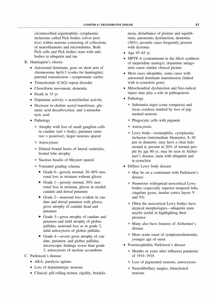
CHAPTER 8 / DEGENERATIVE DISEASE 61
circumscribed argentophilic cytoplasmic nesia, disturbance of posture and equilib-rium, autonomic dysfunction, dementiainclusions called Pick bodies (silver posi-
tive) within neurons consisting of collections (30%); juvenile cases frequently presentwith dystoniaof neurofilaments and microtubules. Both
Pick cells and Pick bodies stain with anti- • Age 45–65 yrbodies to ubiquitin and tau • MPTP-A (contaminant in the illicit synthesis
B. Huntington’s chorea of meperidine analogs); dopamine antago-nists cause similar clinical picture• Autosomal dominant, gene on short arm of
chromosome 4p16.3 (codes for huntingtin); • Most cases idiopathic, some cases withpaternal transmission—symptomatic earlier autosomal dominant transmission (linked
with α-synuclein gene)• Trinucleotide (CAG) repeat disorder
• Mitochondrial dysfunction and free-radical• Choreiform movement, dementiainjury may play a role in pathogenesis• Death in 15 yr
• Pathology• Dopamine activity > acetylcholine activity◦ Substantia nigra (zona compacta) and• Decrease in choline acetyl-transferase, glu-
locus ceruleus marked by loss of pig-tamic acid decarboxylase, and γ-aminobu-mented neuronstyric acid
◦ Phagocytic cells with pigment• Pathology
◦◦ AstrocytosisAtrophy with loss of small ganglion cellsin caudate (tail > body), putamen (ante- ◦ Lewy body—eosinophilic, cytoplasmicrior > posterior), larger neurons spared inclusion (intermediate filaments), 8–30
◦ µm in diameter, may have a clear haloAstrocytosisaround it, present in 20% of normal peo-◦ Dilated frontal horns of lateral ventricles,ple by age 80 yr, may be seen in Alzhei-frontal lobe atrophymer’s disease, stain with ubiquitin and
◦ Nucleus basalis of Meynert spared α-synuclein
◦ • Diffuse Lewy body diseaseVonsattel grading schema
◦� Grade 0—grossly normal, 30–40% neu- May be on a continuum with Parkinson’sronal loss in striatum without gliosis disease
� Grade 1—grossly normal, 50% neu- ◦ Numerous widespread neocortical Lewyronal loss in striatum, gliosis in medial bodies (especially superior temporal lobe,caudate and dorsal putamen cingulate gyrus, insular cortex layers V
and VI)� Grade 2—neuronal loss evident in cau-date and dorsal putamen with gliosis, ◦ Often the neocortical Lewy bodies havegross atrophy of caudate head and atypical morphologies—ubiquitin stainputamen maybe useful in highlighting their
� Grade 3—gross atrophy of caudate and presenceputamen and mild atrophy of globus ◦ Many also have features of Alzheimer’spallidus, neuronal loss as in grade 2,
diseasemild astrocytosis of globus pallidus
◦ More acute onset of symptoms/dementia,� Grade 4—severe gross atrophy of cau-
younger age of onsetdate, putamen, and globus pallidus,• Postencephalitic Parkinson’s diseasemicroscopic findings worse than grade
3, astrocytosis of nucleus accumbens ◦ Months or years after influenza pandemicof 1914–1918C. Parkinson’s disease
◦• AKA: paralysis agitans Loss of pigmented neurons, astrocytosis• Loss of dopaminergic neurons ◦ Neurofibrillary tangles, binucleated
neurons• Clinical: pill rolling tremor, rigidity, bradyki-

62 NEUROPATHOLOGY REVIEW
◦ ◦ Neuronal loss, axonal swellingFew, if any, Lewy bodies
D. Parkinsonism–dementia complex of Guam ◦ Demyelination• Chamorro natives of Guam and Marina ◦ Astrocytosis
IslandsG. Striatonigral degeneration• Rigidity, bradykinesia, dementia • Clinically similar to Parkinson’s disease• Onset age 50–60 yr • Resistant to levodopa• Death in 5 yr • Atrophy of caudate, putamen, zona com-• Generalized cerebral atrophy pacta of substantia nigra
• Tangles, neuron loss; no plaques, Lewy bod- • Neuron loss, astrocytosisies, or inflammatory reaction • No Lewy bodies, no tangles• Hippocampus, amygdaloid nucleus, temporal • Cytoplasmic inclusions (tubular structures)and frontal cortex, basal ganglia, substantia
in oligodendroglia and some neurons,nigranuclear inclusions in neurons and glial cells,• Anterior horn cell disease in some patients neuropil threads—these findings also noted
(amyotrophic lateral sclerosis-like picture)— in Shy-Drager syndrome and olivopontocere-longer clinical course bellar atrophy
E. Progressive supranuclear palsy • Part of multisystem atrophy syndrome• AKA: Steele-Richardson-Olszewski syn- H. Shy-Drager syndrome
drome • Autonomic system failure with parkinsonism• Ophthalmoplegia (loss of vertical gaze), • Loss of intermediolateral column neuronsataxia, expressionless facies, rigidity (hyper-extension of head—posterior cervical mus- • Pathology variable—between Parkinson’scle), mental disorders/dementia disease and striatonigral degeneration
• Onset 5th–7th decades • Part of multisystem atrophy syndrome
• Pathology I. Olivopontocerebellar atrophy
◦ • Ataxia, eye and somatic movement abnor-Neuronal loss with tau positive tanglesmalities, dysarthria, rigidity(straight filaments with a diameter of 15
nm), gliosis, and neuropil threads • Most autosomal dominant
◦ Basal ganglia and brain stem (substantia • Gross atrophy of basis pontisnigra, subthalamic nucleus, superior colli- • Depletion of Purkinje cells, loss of neuronsculi, periaqueductal gray, pontine tegmen- in inferior olivestum, dentate) the most severely involved • Variable degeneration of spinal cord longareas, may see atrophy in these regions
tracts and neuronal loss/gliosis in putamen◦ Astrocytes may also stain with tau protein and substantia nigra
F. Hallervorden-Spatz disease • Part of multisystem atrophy syndrome• Autosomal recessive J. Corticobasal degeneration• Early childhood onset • 6th–7th decade• Dystonic posturing, choreoathetotic or tremu- • Apraxia, limb dystonia, akinesia, rigidity,
lous movements, muscular rigidity and spas- impaired ocular movement, dementiaticity, dementia • Frontoparietal atrophy• Death in 10–12 yr • Ballooned neurons• Pathology • Degeneration of subcortical nuclei, espe-◦ Rust brown discoloration of globus pal- cially substantia nigra
lidus and substantia nigra • Neuronal inclusions (neurofibrillary tangle-◦ Mineralization (iron) of vessel walls like)—tau positive; ubiquitin positive

CHAPTER 8 / DEGENERATIVE DISEASE 63
K. Friedreich’s ataxia • Asymmetrical → symmetrical extremityinvolvement• Familial form, onset 1st two decades
• Muscle wasting, fasciculation, sensation• Autosomal recessiveintact• Chromosome 9q13 (frataxin gene)
• CSF normal• GAA trinucleotide repeat expansion• 2–3 yr to death• Ataxia, sensory disturbances (lower part of• Cause of death—respiratory failure andbody—loss of joint position sense, vibration
pneumoniasense, two-point discrimination)• Atrophy of anterior spinal nerve roots and• Other neurologic disturbances—dysarthria,
affected musclesoptic atrophy, deafness, dementia, epilepsy• Precentral gyrus atrophic• Skeletal deformities—pes cavus, kyphosco-
liosis • Loss of anterior horn cells (lumbar, cervicalcord)• Heart abnormal—enlarged, pericardial adhe-
sions, myocarditis • Several types of inclusions may be seen inmotor neurons• Peak age of onset 10 yr
◦ Bunina bodies—small eosinophilic inclu-• Duration of disease 5–50 yrsions (2–5 µm)• Death most commonly in 3rd and 4th
decades (heart disease) ◦ Hyaline inclusions• Degeneration of sensory fibers, posterior ◦ Basophilic large inclusions
roots, and ganglion cells in lower segment ◦ Aggregates of thread-like structures calledof cordskeins (seen on ubiquitin staining)• Posterior column degeneration, gracilis > • Neuronal loss of hypoglossal nuclei andcuneatus
other cranial nerve nuclei (facial, trigeminal)• Degeneration of dorsal nuclei (Clarke’s col- • Pyramidal tract degenerationumn) and posterior > anterior spinocerebel-• Neuronophagia and gliosislar tract
N. Spinal muscular atrophies• Cranial nerve tracts variably involved• Variety of types• Cerebellar afferents (accessory cuneate
nucleus) and outflow from dentate nucleus ◦ Werdnig-Hoffmann—prototypicalalso involved ◦ Kugelberg-Welander
L. Dentorubropallidoluysial atrophy◦ Adult onset forms• CAG trinucleotide repeat expansion◦ Later onset—more favorable prognosis• Chromosome 12p
• Werdnig-Hoffman disease• Ataxia, chorea, myoclonic epilepsy,◦dementia Autosomal recessive
• Severe neuronal loss and gliosis in dentate ◦ Chromosome 5q11–13nucleus, globus pallidus, red nucleus, and ◦ 1 : 20,000subthalamic nucleus
◦ Death before 2 yr• Atrophy of superior cerebellar peduncles◦ Proximal > distal onset of weaknessM. Amyotrophic lateral sclerosis
◦• Male : Female 1.7 : 1 Hypotonia, “floppy baby”• Middle-aged and elderly ◦ Decreased fetal movement—3rd trimester• 5-10% familial autosomal dominant, genetic ◦ Facial muscles spared
locus for some of this group on SOD1 gene◦ Fasciculations of tongueon chromosome 21q◦ Loss of anterior horn cell neurons of spi-• Weakness and pain—in extremities or bul-
bar musculature nal cord

64 NEUROPATHOLOGY REVIEW
◦ ◦ Loss of neurons in nuclei ambigui, hypo-Atrophy of anterior spinal rootsglossal and facial nuclei◦ Neurogenic muscle disease—rounded
◦ Onuf’s nucleus sparedfiber atrophy

9 CNS Infection
• Associated with prior surgery or traumaI. General Information• Most common organisms are StreptococcusA. CNS protection
followed by Staphylococcus• CSF lacks host defense• Intracranial• Meninges
◦ Most often related to extension from a• Blood-brain barrierfrontal or mastoid sinus infection or focus
B. Routes of infectionof osteomyelitis
• Direct implantation ◦ Circumscribed, flattened• Shunt◦ Adhesion of dura to bone confines• Local spread
abscess• Hematogenous ◦ May cause subdural abscess, leptomen-• Septic emboliingitis
C. Factors in infection • Spinal• Number of organisms ◦ Thoracic and lumbar regions• Host immune system◦ More common than intracranial epidural• Type of organism
abscessesD. Pathophysiology of infection
◦ Compression of cord and roots• Injury to blood-brain barrier◦ Staphylococcus aureus• Separation of tight junctions
B. Subdural abscess• Increased albumin in CSF, increased ICP• Secondary to bone/sinus infection (espe-• Edema (vasogenic and inflammatory medi-
cially paranasal sinuses, middle ear, cra-ators)nium) via thrombophlebitis• Spinal nerve root and nerve (neck stiffness)
• Dorsolateral aspect of cerebrum, frontal• Vasculitis; infarctionpolar region, interhemispheric fissure• Direct cytopathic/toxic effect • Adhesions, loculation
II. Suppurative Lesions • Compression of brain, increased ICPA. Epidural abscess • Streptococcus (#1) and Staphylococcus (#2)• Extension from bones (trauma, osteomyeli- • Spinal subdural raretis, otitis)
C. Leptomeningitis• Less often extension from soft tissue(venous) • Neutrophils, fibrin in the leptomeninges
65

66 NEUROPATHOLOGY REVIEW
• With treatment, the inflammatory infiltrate • Early: central—pus; middle—granulation tis-sue; outer—astrocytosis/edemamay switch to more lymphocytes, macro-
phages, and plasma cells • Later: central—liquefaction; middle—collag-enous wall; outer—astrocytosis• Arterial subintimal infiltration
• Granulation tissue—contrast enhances on• Thrombosis, vasculitis, infarct, hemorrhage,CT scan as a result of hypervascularitycerebral edema, vascular congestion
• Hematogenous (metastatic) abscesses—often• Virchow-Robin space extensionmultiple• May extend to cause ependymitis, ventri-
• Damage a result of local tissue destruction,culitisedema (mass effect)• Subarachnoid fibrosis, CSF obstruction, sube-
• Consequencespendymal gliosis with healing
◦ Organization• Neonatal (maternal genital flora)—commu-nity acquired ◦ Formation of daughter abscess◦ Strep. agalactiae (group B)—exudate ◦ Ependymitis
over vertex ◦ Leptomeningitis◦ E. coli—hemorrhage, necrotizing arteritis,E. Septic embolismnecrosis, roughly same incidence in this
• Occlusion—infarctionage group as Strep. agalactiae• Infection—arteritis, septic aneurysm, hemor-◦ Proteus
rhage, septic infarcts◦ Listeria—microabscesses • Small parenchymal arteries—microabscess,
• Childhood—community acquired petechiae
◦ • Septicemia—capillaries, microabscessNeisseria meningitidis—basal, youngadults, army recruits, most epidemic out- • Leptomeninges—septic infarct, hemorrhage,breaks related to group A, associated with meningitisWaterhouse-Friderichsen syndrome [dis- • Large artery—infarctsoninated intravascular coagulopathy • Trunk artery base of brain—subarachnoid(DIC) picture with hemorrhagic adrenal
bleedglands]F. Tuberculosis (TB)◦ H. influenza—vertex, cortical softening,
• Epiduralvenous thrombosis◦ Extension from vertebral infection (Pott’s◦ Strep. pneumoniae—vertex
disease)• Adults/elderly—community acquired◦ Compression of cord◦ Strep. pneumoniae◦ Rarely causes meningitis◦ Listeria • Subdural◦ Any organism is possible with hospital- ◦ Miliary or tuberculoma-en-plaqueacquired infections
• Tuberculoma = abscessD. Abscess• Granulomas with caseous necrosis• Localized suppuration within brain sub-• Usually small and multiplestance• May act as mass• Streptococcus (#1), Gram-negative bacilli
(#2) Staphylococcus (#3), may see anaer- • Enhancing on CT scanobes and aerobes • TB meningitis—base of brain; neutrophils,
• Frequently multiple organisms fibrin, endarteritis, hemorrhage, caseousnecrosis, granulomatous inflammation.• Focal septic encephalitis (cerebritis), periph-
eral edema • CSF studies: lymphocyte predominance with

CHAPTER 9 / CNS INFECTION 67
pleocytosis, elevated protein, decreased ening of the intima and thinning of themedia), which may lead to infarctglucose
• Culture still remains gold standard for diag- ◦ Pachymeningitis cervicalis hypertrophi-nosis, molecular biologic techniques exist to cans—cervical fibrosis of dura to piaidentify the organisms • General paresis
• Use of special stains (such as Ziehl-Neelsen, ◦ Brain invasion—subacute encephalitisfite) on tissue sections has a relatively low◦ Prolonged secondary stage—failure to pro-yield (<50%)
duce neutralizing/destructive antibody or• Other nontuberculous mycobacteria mayfailure of antibody to reach spirocheteinfect CNS
◦ Early—brain grossly normal◦ Mycobacterium avium intracellulare typi-cally marked by perivascular macro- ◦ Later—frontal atrophyphages with organism; rarely presents as ◦ Loss of neurons, astrocytosis, capillarya tumor-like nodule with fibrosis
proliferation, perivascular inflammation,G. Sarcoidosis microglia with iron
• Noncaseating granulomas ◦ Spirochetes may be hard to identify, 3-yr• Base of brain, posterior fossa course• Near vessels in meninges • Lissauer’s dementia• Optic nerve and chiasm involvement ◦ Variant of general paresis
common◦ Epileptic or apoplectiform attacks• Course usually slow, may be rapidly fatal◦ Focal signs• Hydrocephalus, cranial nerve defects, hypo-◦thalamic symptoms Average 7 to 8-yr course
• May have no apparent systemic disease ◦ Temporal atrophy with pseudolaminardegenerationH. Whipple’s disesase
• Caused by Tropheryma whippelii, a Gram- • Tabes dorsalispositive actinomycete ◦ Degeneration of spinal dorsal roots and
• PAS-positive organisms in macrophages that columnsare often arranged in perivascular distri- ◦ Lumbosacral and lower thoracic cordbution
◦ Dorsal root ganglia unaffectedI. Syphilis◦ May see meningovascular disease• Caused by Treponema pallidum
• Optic atrophy• Highlighted with special stains (e.g., Steiner,Warthin-Starry, Dieterle) ◦ Chronic pial inflammation, degeneration
• Subacute secondary lymphocytic meningitis of nerve
• Meningovascular ◦ Peripheral > central
◦ Lymphs, plasma cells ± gummata (central ◦ Associated with tabes dorsalisnecrosis surrounded by epithelioid histio- • Congenitalcytes, fibroblasts, and multinucleated
◦ Meningovascular with obstruction, hydro-giant cells)cephalus◦ Do not usually see organisms with
◦ General paresis—especially cerebellumgumma because it represents a hyperim-mune form of tissue necrosis ◦ Rarely tabes dorsalis
◦ Perivascular parenchymal inflammation J. Lyme disease
◦ Arteritis, intimal proliferation, Heubner’s • Caused by Borrelia burgdorferi (a spiro-chete transmitted by ioxodid ticks)endarteritis obliterans (collagenous thick-

68 NEUROPATHOLOGY REVIEW
• Associated skin lesions (erythema chron- or rarely direct extension from an orbital orparanasal sinus infectionicum migrans)
• Pathology• Lymphoplasmacytic leptomeningitis
◦III. Fungal Infection Granuloma—abscess ± basal meningitisA. General ◦ Septic infarcts secondary to vascular infil-
• Immunocompromised persons at risk tration, hemorrhage
• Secondary spread common ◦ Focal cerebritis, especially white matter• Hematogenous spread > contiguous E. Mucormycosis
spread • Broad branching, nonseptate hyphae (10–15• Granulomatous inflammation and abscess µm)
are common patterns of injury • Genera of Mucoraceae family, including Rhi-• Septic emboli, may result in mycotic zopus, Absidia, Mucor, Rhizomucor, Apo-
aneurysm physomyces, and ChlamydoabsidiaB. Cryptococcosis • Entry—nasal sinus, extension into orbit, aspi-
ration into lung• Budding yeast, thick mucopolysaccharidewall • Pathology
• Pigeon excrement ◦ Acute meningeal inflammation• Entry—pulmonary > skin ◦ Hemorrhagic coagulative necrosis of• Pathology brain (ventral)
◦ Leptomeningitis, gelatinous “bubbly” ◦ Vascular luminal and mural invasionexudate F. Coccidioidomycosis
◦ Macrophages • Semiarid areas (South America and south-west United States)◦ Perivascular extension, may be quite
extensive and give the brain a honeycomb • Double-contoured, refractile capsules withlook grossly endospores
◦ • Self-limited respiratory infectionGranulomas, “torulomas” (abscesses)• 0.05% cases disseminate, CNS disease in◦ Organisms highlighted with PAS, muci-
10–25% of these casescarmine stains• PathologyC. Candidiasis
◦ Basilar meningitis—neutrophils, fibrinous• Budding cells, branching pseudohyphaeexudate• Entry—skin, GI tract
◦ Later—granulomas, fibrosis• CNS disease as a result of systemic infec-tion or endocarditis ◦ Cranial osteomyelitis
• Pathology ◦ Parenchymal granulomas rare◦ Chronic granulomas G. Blastomycosis◦ Microabscesses • Males > females, agricultural workers
◦ • Acquired from contaminated soil, southeast-Rarely arteritis, basal leptomeningitisern United States◦ Organisms highlighted on Gomori • Broad-based buddingmethenamine silver (GMS) or PAS stains
• Protoplasmic body (10–15 µm in diameter)D. Aspergillosis• No hyphae in tissue sections• Dichotomously branching septate hyphae• Lung and skin entry portals• Entry—airborne lung infection followed by
hematogenous spread, may also get dissemi- • Leptomeningitis and granulomas rare, oftenpresents as abscessnated from other organ system involvement

CHAPTER 9 / CNS INFECTION 69
H. Histoplasmosis ◦ Degeneration—chromatolysis, eosino-philia, nuclear changes, neuronophagia• Entry—inhalation of dust containing spores
◦ Eosinophilic nuclear inclusions (Cowdry)• Histoplasma capsulatum consists of smallovoid and budding bodies, PAS and GMS � Type A—intranuclear, large spherical,positive halo (e.g., CMV, herpes, PML)
• Only rarely disseminates to involve the CNS � Type B—small, multiple, no nucleolardisplacement (e.g., polio)• Necrosis with macrophages (containing
organisms), lymphocytes, and plasma cells � Intracytoplasmic
◦I. Actinomycosis Reactive endothelial changes• Bacteria with fungal-like morphology ◦ Tissue necrosis or demyelination with cer-
tain viruses• Thin (<1 µm pseudohyphae) filaments
◦• Gram-positive, acid-fast negative Inflammatory cell exudate� Microglia—diffuse or focal (neurono-• Sulfur granules
phagia, glial nodule, glial shrub, glial• Entry—cervicofacialscar)• Pathology
� Chronic inflammatory cells (lympho-◦ Granuloma—abscess, multiple cytes, plasma cells and macrophages),often perivascular distribution◦ Basilar meningitis
B. PolioJ. Nocardiosis• Acute febrile disease, asymmetric paralysis• Bacteria with fungal-like morphology
(legs > arms > trunk)• Thin filaments • 5% mortality• Gram-positive, partial acid-fast positive • Pathology• No sulfur granules ◦ Large motor neurons lost (anterior horn• Entry—lung cells, motor neurons of cranial nerves)• Pathology ◦ Lymphocytic perivascular and meningeal
◦ inflammationGranulomas, small and mulitple
◦IV. Viral Infection Gliosis and microgliosis with recovery
A. General C. Arbovirus
• Arthropod (RNA viruses)—mosquito borne• Routes of spread• Serologic exam to make diagnosis◦ Hematogenous (polio, arbovirus)• In United States—St Louis, California virus,◦ Peripheral nerve (rabies)
Eastern and Western Equine encephalitis◦ Olfactory mucosa, trigeminal nerve • Pathology
(herpes) ◦ Brain congestion and edema• Morphologic features ◦ Widespread, gray matter◦ Little (edema, congestion) or no distinc- ◦ Most severe changes with Eastern equinetive gross alterations (except herpes, pro-◦ Neuronal degeneration, microglial nod-gressive multifocal leukoencephalopathy
ules, perivascular and meningeal chronic[PML])inflammation; abscess and neutrophils in◦ Diffuse distribution (except polio, herpessevere caseszoster)
D. “Aseptic Meningitis”◦ Primary neuronal involvement common • Refers to any culture-negative meningitis• Common histopathologic features • Many viruses—Coxsackie and Echovirus
◦ Nerve cell alterations most cases

70 NEUROPATHOLOGY REVIEW
• Low mortality lum)—eosinophilic cytoplasmic neuronalinclusion with basophilic inner formation• Meningeal and choroid plexus lymphs
◦ Lyssa body—lack inner formationE. Influenza
J. Herpes simplex• Onset respiratory
• 70% fatal• Abnormal behavior, disoriented, seizures,coma • No seasonal incidence
• Neuronal degeneration, inflammation • May see no obvious skin or visceral disease,F. Measles the virus often replicates at a peripheral site
before reaching the CNS• Mostly < age 10 yr• Virus often later peripherally located and• Low fatality
may be reactivated by a variety of factors• Perivascular inflammation, demyelination(stress, fever, sunlight, trauma, radiation)• Multinucleated giant cells, intranuclear and • Virus spreads from periphery to ganglionintracytoplasmic inclusionsvia retrograde axoplasmic transport
G. Subacute sclerosing panencephalitis (SSPE) • Pathology• Fatal, measles virus◦ Congenital—acute encephalitis with vis-• Under age 12 yr
ceral lesion, herpes type I• Course: months to a few years ◦ Adults—herpes type II, acute hemor-• Progressive dementia, myoclonus, seizures,rhagic necrotizing encephalitis (basal fron-
ataxia, dystoniatal, cingulate, temporal gyri, insula)• Pathology ◦ Eosinophilic nuclear inclusions in glial◦ Gray matter subacute encephalitis cells (oligodendrocytes > astrocytes)
◦ Eosinophilic nuclear and cytoplasmic K. Herpes zosterinclusions, neuronal loss, microglial prolif- • Skin lesions (shingles)—vesicles in a sen-eration, perivascular inflammation sory nerve dermatome distribution
◦ Demyelination, oligodendroglial inclu- • Acute cerebellar disease—nystagmus, dysar-sions, reactive astrocytosis thria, ataxia
◦ Cerebral atrophy—old cases • Pathology
◦ May see occasional neurofibrillary tangles ◦ Dorsal ganglia—inflammation, petechiae,degenerating neuronsH. Mumps
◦• 2–10 d after onset Eosinophilic intranuclear inclusions• Low mortality ◦ Meningeal inflammation, granulomatous• Perivenous demyelination angiitis
• 10–20% aseptic meningitis L. Cytomegalovirus
I. Rabies • Infant—transplacental transmission, still-birth, prematurity• Variable incubation (1–3 mo)
• Adult—rare, less extensive, immunocom-• Bite of infected animal—skunk, racoon, etc.promised states (18–33% of AIDS patients• Anxiety, psychomotor sensitivity, hydropho-have evidence of CMV in brain)bia, dysphagia, seizures
• Pathology• Death 2–7 d◦ Infants—necrosis, inclusions (neurons,• Pathology
glial cells, endothelial cells), chorioretini-◦ Encephalomyelitis (especially brain stem) tis; endstage—gliosis, calcification, hydro-◦ cephalus, microcephalyGlial nodules
◦ ◦ Adults—microglial aggregatesNegri bodies (hippocampus, cerebel-

CHAPTER 9 / CNS INFECTION 71
◦ O. Human T lymphotropic virus type 1 (HTLV-1)Large cells with intranuclear (Cowdry A)and cytoplasmic inclusions • Retrovirus causing tropical spastic parapa-
resis◦ May involve ependyma and subependy-• Myelin loss and axonal degeneration in spi-mal regions (hemorrhagic necrotizing ven-
nal cord white matter, leptomeningealtriculoencephalitis and choroid plexitis)chronic inflammation and fibrosis, gliosisM. Progressive multifocal leukoencephalopathy
• Thoracic and lumbar regions particularly sus-(PML)ceptible• Immunocompromised states • May see demyelination of optic and audi-• Progressive disturbances in motor and men- tory nerves
tal function and visionV. Parasitic Infections• Papova virus (JC, SV40)—DNA virus
A. Toxoplasmosis• Demyelination—multifocal, asymmetric • Caused by Toxoplasma gondii infection• Astrocytosis • Congenital form• Alzheimer I astrocytes—bizarre, atypical ◦ Occurs only if the maternal infection is
astrocytes; probable viral cytopathic effectacquired during pregnancy
• Intranuclear inclusions—oligodendrocytes ◦ Miliary granulomata(Cowdry A)◦ Necrosis• Paucity of lymphocytes◦ CalcificationN. HIV◦ Meningitis, chorioretinitis• RNA retrovirus
• Adult• 10% develop “aseptic” meningitis within1–2 wk of seroconversion ◦ May rarely encounter cysts in an incompe-
tent individual• Meningoencephalitis often presents withdementia, seizures, myoclonus and focal ◦ Space-occupying lesion(s)—abscesses,motor signs may demonstrate the same architectural
features of a bacterial abscess• Chronic inflammation with microglialnodules ◦ Scattered necrotic foci
• Microglial nodules often contain macro- ◦ Rarely microglial nodules/encephalitisphage-derived multinucleated giant cells picture
• Virus can be detected in macrophages, CD4- B. Amebiasispositive lymphs and microglia • Entamoeba histolytica or lodamoeba buet-• Areas most severely affected include deep schliicerebral white matter, basal ganglia, cerebral • Naegleria—via nasal cavity, often acquiredcortex, and brain stem
by swimming in contaminated water, puru-• Vacuolar myelopathy involving degeneration lent meningitis, rapidly progressive, severe
of spinal cord (worst in thoracic cord), espe- involvement of the olfactory area (often thecially posterior columns; presents as point of entry)progressive paraparesis, sensory ataxia, and • Acanthamoeba—diffuse necrotizing granulo-incontinence; swelling between myelin matous encephalitis and meningitissheaths, reactive astrocytosis, macrophage • Often see trophozoites in tissue sectioninfiltrate and occasional multinucleated giant
C. Malariacells.• 2% of cases of Plasmodium falciparum• AIDS in children frequently marked by calci-
fication of parenchymal vessels within basal • Presents with headache, drowsiness, confu-ganglia and deep cerebral white matter sion, coma, fevers
• Often see the organisms in peripheral blood• At risk for opportunistic infections

72 NEUROPATHOLOGY REVIEW
• Grossly, see opacity of meninges, dusky col- • Conformational change promotes further pro-duction of the abnormal prion proteinoration of the gray matter, edema, con-
gestion material
• Prion protein inactivated by autoclaving or• Capillaries filled with organismbleach• Small foci of ischemic necrosis rimmed by
B. Creutzfeldt-Jakob disease (CJD)microglia (Durcks nodes)
• 1/106 incidence• White matter petechiae• Subacute dementia• Can see granules of dark malarial pigment
in red cells • MyoclonusD. Cysticercosis • EEG—biphasic, triphasic spikes
• Taenia solium (pork tapeworm) • Onset > 30 yr; female : male 2 : 1
• Larvae encysts in brain • Duration < 1 yr
• Can evaluate CSF for 14-3-3 protein to sup-• Inflammation, granuloma, calcificationport diagnosisE. Schistosomiasis
• Pathology• S. japonicum—cerebrum lesions◦ Spongiform change• S. mansoni, S. hematobium—spinal cord
lesions ◦ Neuronal atrophy/loss• Necrotizing inflammation with neutrophils, ◦ Gliosis
eosinophils, giant cells ◦ Granular layer degeneration• Fibrosis and calcification with healing◦ Kuru plaque 10% (extracellular depositsF. Trypanosomiasis
of aggregated abnormal protein—Congo• African disease caused by T. brucei; red and PAS positive), prominent in vari-acquired by bite of tsetse fly; may present ant CJD, which may be related to inges-with fever, rash, edema, cardiac involve- tion of contaminated beef (mad cowment, anemia; meningoencephalitis occurs disease)during late stages of the disease ◦ Iatrogenic transmission documented (e.g.,• South American disease (Chagas disease) pooled pituitary hormone preparations,caused by T. cruzi; acquired from feces of the dura mater and corneal grafts/transplants)reduviid bug; brain with edema, congestion,
C. Gerstmann-Straussler-Scheinker diseasehemorrhages, microglial nodules, granulomas• AtaxiaG. A variety of other parasites have been• Behavioral changedescribed to cause CNS pathology, including
Echinococcus, Spirometria (Spanganosis), Para- • Late dementiagonimus (lung fluke), Trichinella spiralis, Loa • Autosomal dominant, onset > 30 yrLoa, Onchocerca, and Strongyloides • Duration—5 yr
VI. Transmissible Spongiform Encephalopathies • 1/10 million incidenceA. General • Corticospinal tract degeneration
• Includes Creutzfeldt-Jakob disease, • No/mild spongiform change and astrocytosisGerstmann-Straussler-Scheinker, Kuru, famil- • Cerebellar plaquesial insomia
D. Kuru• Related to prion protein abnormalties• Cannibalism: Papua New Guinea; Fore• Prion protein is a normal 30 Kd cellular pro-
tribe—women and childrentein present in neurons (chromosome 20)• Tremor, ataxia, dysphagia, euphoria• In disease, protein undergoes conformational• Duration of disease < 1 yrchange from normal α-helix isoform to an
abnormal β-pleated sheet isoform (PrPsc or • Severe cerebellum disease—kuru plaques,axon torpedoes, granular layer degenerationPrPres)

CHAPTER 9 / CNS INFECTION 73
E. Familial insomnia • Duration of disease < 2 yr
• Sleep disorder, bizarre behavior, autonomic • Severe anterior and medial thalamus gliosis,dysfunction inferior olive degeneration
• Adult onset, autosomal dominant


10 Skeletal Muscle
• Trichrome (Gomori)I. General Information◦ Nuclei redA. Muscle biopsy
◦• Do not biopsy muscle with needle in it Ragged red fibers (mitochondria)(EMG), injection site (gluteus, deltoid), ◦ Inclusion body myositis inclusionseverely diseased muscle
(rimmed vacuoles)• Commonly biopsied muscles: quadriceps ◦ Fibrosis(vastus lateralis), triceps, biceps, and gas-trocnemius • PAS
B. Clinical information useful when interpreting ◦ Normal muscle with glycogena muscle biopsy ◦ Type I > type II• Age
◦ Glycogen storage diseases• Gender• Oil-Red-O• Drugs/medications
◦ Normally evenly distributed• Medical/family history
◦ Type I > type II• EMG findings
C. A variety of enzyme histochemical stains and ◦ Lipid disordersspecial stains are used in the evaluation of • (SAB) Sulfonated alcian blue/Congo redbiopsy specimens. Many of these stains
◦ Amyloidrequire fresh or frozen tissue to work.Improper freezing of the specimen may result ◦ Mast cells (SAB positive—internalin a vacuolar artifact. control)
D. Staining of the muscle biopsy specimen • Nonspecific esterase• Routine hematoxylin and eosin ◦ Angular atrophic fibers (neurogenic)• Acid phosphatase ◦ Acetylcholinesterase◦ Lysosomal enzyme
◦ Type I > type II◦ Increased with degeneration◦ Acute denervation (6 mo)(red staining)
◦◦ Some inflammatory cellsInflammatory cells
• NADH• Alkaline phosphatase
◦◦ Oxidative enzymeNormal capillary basement stains
◦ ◦ Type I > type IIRegenerating fibers (black staining)
75

76 NEUROPATHOLOGY REVIEW
◦ tem and result in secondary changes in theMyofibrillary network inside musclemusclefiber
D. Neurogenic changes• Cytochrome oxidase• Angular atrophic fibers (esterase positive)◦ Mitochondrial enzyme• Group atrophy◦ Type I > type II• Fiber type grouping◦ Focal deficiencies exist • Target and targetoid fibers• ATPase (adenosine triphosphatase) • Nuclear bags◦ pH 4.6 (reverse) or pH 4.3 • No necrotic or basophilic fibers
� Type I dark • Minimal interstitial fibrosis� Type IIA light • No inflammation� Type IIB intermediate • No centralized nuclei� Type IIC light (pH 4.2)
III. Inflammatory Myopathies◦ pH 9.8 (standard)A. Polymyositis
� Type I light • 30–60% of inflammatory myopathies� Type II dark • Nonhereditary• Succinate dehydrogenase (SDH) • Adults (age 40–60 yr), second peak in
◦ Mitochondrial enzyme childhood
◦ Type I > type II • Male : female 1 : 2
• A variety of other stains may be selec- • Idiopathic versus connective tissue diseasetively performed including myophosphory- (SLE, rheumatoid arthritis associated)lase, myoadenylate deaminase, phospho- • An association with neoplasms in adultsfructokinase, etc. • Proximal muscle weakness, often painful
E. Tissue is often processed in glutaraldehyde or • Fatigue, fever, weight lossother fixative for potential electron micro-
• CPK and aldolase elevatedscopic evaluation• Most respond to steroids or other immuno-F. In cases of suspected metabolic disease, extra
suppressivesfrozen tissue for assay purposes is warranted.• EMG—fibrillation potential in acute stage,II. Myopathic Versus Neurogenic Changes
polyphasic short duration potentialsA. Myopathic processes include such disease• Histopathologyentities as inflammatory myopathies, muscu-
lar dystrophies, congenital myopathies and ◦ Increased variation of myofiber size,metabolic diseases central nuclei
B. Myopathic changes ◦ Inflammation (lymphocytes and macro-• Irregularity of fiber size phages), endomysial (surrounding indi-
vidual muscle fibers)• Rounded fibers
◦• Centralized nuclei CD4-positive lymphocytes more com-mon around vessels• Necrotic and basophilic fibers
◦ CD8-positive lymphocytes more com-• Cytoplasmic alterationsmon in endomysium• Interstitial fibrosis
◦ Degeneration• Absence of enzyme staining
◦• Inflammation Regeneration—basophilic staining cyto-plasm, nuclear enlargement with promi-• Storage of glycogen and lipidnent nucleolusC. Neurogenic diseases include those processes
◦ Vasculitis (lymphocytic, nonnecrotizing)that affect the peripheral/central nervous sys-

CHAPTER 10 / SKELETAL MUSCLE 77
◦ ◦ Rimmed vacuoles—autophagic vacu-Endomysial fibrosisoles, granular basophilic material on tri-◦ Motheaten fiberschrome stain
� Ill-defined zones of enzyme loss ◦ Rare intranuclear inclusions� Type I > type II
◦ Frequent neurogenic atrophy-like com-� Nonspecific findings in myopathies
ponentB. Dermatomyositis
D. Rheumatoid arthritis• Proximal muscle weakness • Type II atrophy• Heliotropic rash, Gottron’s papules • Moth-eaten fibers common• Dysphagia, arthralgias, constitutional • Severe disease—type I and type II atro-
symptoms phy, ring fibers, increased central nuclei• Childhood disease often associated with • Chronic inflammation, either polymyositis
vasculitis and calcinosis, not associated or focal nodular myositis pattern of injurywith malignancy • Vasculitis—usually non-necrotizing lymph-• Associated with connective tissue disease ocytic
• Associated with malignancy in 7–30% of E. Bacterial myositiscases, elderly patients (>55–60 yr); cancers • Often related to skin injuryof the breast, ovary, lung, prostate, colon • Gas gangrene (Clostridium)• Histopathology • Pyomyositis (Staph)◦ Inflammation—perivascular, may be • Neutrophils frequently present
scant, mixture of T- and B-lymphocytesF. Viral myositis(vasculopathic disease)
• Muscle pain and muscle weakness usually◦ May see non-necrotizing lymphocyticnot salient featuresvasculitis, thrombosis
• Influenza A and B◦ Degeneration • Enterovirus◦ Regeneration • HIV (AZT treatment)—AZT may also◦ Perifascicular atrophy—ischemia related result in mitochondrial abnormality
• Coxsackie B (epidemic pleurodynia,C. Inclusion body myositisBornholm’s disease)• Most > 50 yr, males > females
G. Parasitic myositis• Progressive, painless muscle weakness• Trichinosis (Trichinella spiralis)• Steroid resistant
◦ Encysted larvae• Enzymes sometimes normal◦ Granulomatous inflammation with eosin-• EMG—neurogenic picture
ophils• Etiology not known◦ Inadequately cooked meat• Tubulofilaments (15–18 nm) on electron
• Toxocariasis, Cysticercosis, Hydatidosis,microscopy, most readily found in fibersToxoplasmosis, Sarcosporidiosis, Trypano-with rimmed vacuolessomiasis• May see amyloid protein material
H. Fungal myositis• Mitochondrial abnormalities frequent con-• Very rarecommitant finding• Candida and cryptococcus the most• Histopathology
common◦ InflammationI. Vasculitis
◦ Degeneration • May see vasculitic component to another◦ Regeneration inflammatory myopathic process (generally

78 NEUROPATHOLOGY REVIEW
non-necrotizing lymphocytic vasculitis) or � Type grouping—10 fibers of thesame type contiguous along 1 edgemay be part of systemic vasculitis process
such as polyarteritis nodosa � Target fibers• Polyarteritis nodosa (PAN) affects medium � Type I > type II fibers
to small size arterial vessels causing� Three zones of staining: central
fibrinoid necrosis of vessel wallspale zone devoid of oxidative activ-• With PAN, will often see vessels at vari- ity; dark annular intermediate zone
ous stages of the disease process in the rich in oxidative enzymes; normalsame biopsy peripheral zone
• May be associated with denervation atro- � Targetoid fibers (core fibers)—nophy changes (presumably related to intermediate zone, type II predilec-involvement of peripheral nerves) tion, a less specific finding
IV. Denervation Atrophy B. Spinal muscular atrophiesA. General information • Includes those disorders that cause anterior
horn cell degeneration such as Werdnig-• Group of disorders that affect anterior horncells, nerve roots, or peripheral nerves Hoffman disease, Kugelberg-Welander dis-
ease, etc. (see Neurodegenerative diseases)causing secondary changes in the skeletalmuscle • Chromosome 5 (survival motor neuron
gene)• Types of neurogenic disease
◦ • EMG findings confirm denervationMotor neuron• CPK levels often normal or slightly ele-� Amyotropic lateral sclerosis (ALS)
vated� Spinal muscular atrophy (Werdnig-
• Fascicular atrophy, rounded fiber atrophyHoffmann, Kugelberg-Welander)in infantile variant◦ Motor endplate • In later onset varients, see angular atrophic
� Myasthenia gravis esterase positive fibers instead of rounded◦ Peripheral (motor) neuropathy fiber atrophy
◦ • Scattered type I hypertrophic fibersMetabolic• Increased endomysial fibrofatty tissue◦ Toxic• Increase in muscle spindle number◦ Vascular/connective tissue disease
C. Myasthenia gravis◦ Systemic disease (diabetes, uremia) • Immune-mediated loss of acetylcholine◦ Infection receptors with circulating antibodies to ace-◦ tylcholine receptorMalignancies
• Prevalence of about 3/100,000 persons• Histopathology• In young patients (<40 yr), women > men;◦ Angulated fiber atrophy (esterase pos-
in older patients, equal gender distributionitive)• Associated with thymic hyperplasia (65%)◦ Type I and type II fibers both involved
and thymoma (15%)◦ Increased NADH staining in angular • Normal nerve conduction studiesatrophic fibers • Decreasing motor response with repeated◦ Nest-like fascicular atrophy—two con- stimulationtiguous motor units involved • Presents with weakness of extraocular mus-
◦ Nuclear bags—endstage cle, fluctuating generalized weakness
◦ May see evidence of reinnervation • Improved strength in response to acetylcho-linesterase agents� Collateral sprouting of spared motor
neurons • Histopathology

CHAPTER 10 / SKELETAL MUSCLE 79
◦ • Both type I and type II fibers affectedType II atrophy• Nemaline rods not specific for nemaline◦ Ultrastructurally, may see simplification
myopathy—reported in a variety of otherof the postsynaptic membranemuscle disorders◦ Lymphorrhages (lymphoid aggregates) • Rods derived from Z-band material
in about 25% of cases(α-actinin)
◦ May see evidence of mild denervation C. Central core diseaseatrophy • Early-onset hypotonia and nonprogressive
D. Lambert-Eaton myasthenic syndrome muscle weakness• Usually develops as a paraneoplastic pro- • Joint deformities such as hip dislocations
cess (especially with small cell carcinoma commonof lung) • May develop malignant hyperthermia• Proximal muscle weakness with autonomic • Autosomal dominant condition related todysfunction
defect of ryanodine receptor-1 gene (chro-• Enhanced neurotransmission with repeated mosome 19q13.1)stimulation • CPK generally normal or slightly elevated,• Decrease in the number of vesicles especially in later-onset casesreleased in response to each presynaptic • Cytoplasmic cores devoid of enzyme activ-action potential
ity (NADH negative) in many fibers• Some patients with antibodies to presynap- • Cores may be eccentrically placed and mul-tic calcium channelstiple
V. Congential Myopathies • Cores generally confined to type I fibersA. General information • Ultrastructurally, cores are zones in which• Disorders of muscle fiber maturation and myofilaments are densely packed with
reorganization absence of mitochondria, glycogen, and• Nonprogressive/slowly progressive lipids
• Retarded motor development, muscle • Minicore or multicore disease is a distinctweakness autosomal recessive condition
• Frequently associated with skeletal dysm- D. Centronuclear (myotubular) myopathyorphism • Presents in infancy or childhood with
• May present as “floppy baby” due to hypo- involvement of extraocular and facial mus-tonia or with joint contractures (arthro- cles, hypotonia, and slowly progressivegryposis) limb muscle weakness
• Familial incidence • CPK may be normal or slightly elevated
• Specific structural abnormality of muscle • X-linked autosomal recessive or sporadicfibers variants
B. Nemaline myopathy • Increased numbers of centrally placednuclei (involving most myofibers)• Weakness, hypotonia, and delayed motor
development in childhood, may present in • Most fibers containing central nuclei areadulthood type I and tend to be somewhat atrophic
• Autosomal recessive, autosomal dominant • Small zones surrounding the nuclei thatand sporadic variants recognized are devoid of myofibrils and filled with
mitochondrial aggregates, lipid, and gly-• CPK often normal or mildly elevatedcogen• Intracytoplasmic nemaline rodlets (red on
E. Congenital fiber type disproportiontrichrome)• Autosomal recessive condition• Number of affected fibers has no correla-
tion with disease severity • Variable presentation clinically; hypotonia,

80 NEUROPATHOLOGY REVIEW
long thin face, contractures, congenital hip ◦ Retinitis pigmentosadislocation, respiratory problems ◦ Ophthalmoplegia• Mean fiber diameter of type I fibers at ◦ Atrial-ventricular (AV) blockleast 12% smaller than type II fibers
◦ Cerebellar ataxia• May also see increased numbers of central-ized nuclei, moth-eaten fibers and nema- ◦ Deafnessline rods ◦ Peripheral neuropathy
VI. Metabolic Myopathies ◦ DwarfismA. Mitochondrial myopathy ◦ Mitochondrial abnormalities in other• Many related to mutations in either nuclear
organs (liver, skin, brain)or mitochondrial genes
◦ Brain with calcification• Three categories of genetic alterationsB. Glycogenoses◦ Class I mutations: involve nuclear DNA • Abnormalities of glycogen metabolism(e.g., Leigh’s syndrome)
• The most common ones to affect skeletal◦ Class II mutations: point mutations of muscle are type II (acid maltase defi-mitochondrial DNA (e.g., MERFF and ciency) and type V (myophosphorylaseMELAS) deficiency, McArdle’s disease).
◦ Class III mutations: deletions or duplica- • Less commonly, other types may affecttions of mitochondrial DNA (e.g., muscle, including type III (amylo 1,6-glu-Kearns-Sayre syndrome) cosidase deficiency, debrancher enzyme),
type IV (branching enzyme) and type VII.• May be related to abnormalities of mito-(phosphofructokinase deficiency)chondrial transport, substrate utilization,
Krebs cycle, oxidative—phosphorylation • Type II glycogenosis (Pompe’s disease)coupling, or the respiratory chain (the most ◦ Acid maltase deficiencycommon group of mitochondrial myopa-
◦ Chromosome 17q autosomal recessivethies)condition• Myopathy only presentation in some
◦patients Neonatal form with hypotonia and cardi-omegaly, rapidly fatal• Cerebral dysfunction with myopathy in
some patients (mitochondrial encephalomy- ◦ Childhood and adult form with limb–opathies) girdle weakness and cardiac disease
• Progressive external ophthalmoplegia, prox- ◦ Elevated CPK in all formsimal muscle weakness ◦ Type I fibers with vacuoles• Ragged red fibers (Gomori trichrome
◦ Vacuoles PAS and acid phosphatasestain)—related to aggregates of often mor-(acid maltase is a lysosomal enzyme)phologically abnormal mitochondria situ-positiveated in a subsarcolemmal location)
◦ Glycogen accumulation on electron• Not all myofibers equally affectedmicroscopy• Often nonspecific increase in lipid and
• Type V glycogenosis (McArdle’s disease)glycogen
◦ Myophosphorylase deficiency• Sporadic and inherited forms
◦• Abnormal mitochondrial structure on elec- Autosomal recessive condition, chromo-tron microscopic studies, paracrystalline or some 11q“parking lot” inclusions ◦ Males > females
• Example: oculocraniosomatic syndrome ◦ Muscle cramps and stiffness on exertion(Kearns-Sayre syndrome, ophthalmoplegia◦ Abnormal ischemic exercise test—doplus)

CHAPTER 10 / SKELETAL MUSCLE 81
not get the expected elevation of blood • Most are autosomal dominant, males >femaleslactate levels following exercise
◦ • Myotonia, relapsing episodes of hypotonicVacuolar myopathy—typically subsarco-paralysis following exercise, exposure tolemmal blebscold or high-carbohydrate meal◦ Vacuolar PAS positive, acid phospha- • Variants recognized associated with variedtase negativeserum potassium levels (normokalemic,
C. Lipid storage myopathieshyperkalemic, or hypokalemic periodic• Resulting from deficiencies in the carnitine paralysis)
transport system or deficiencies in the mito- • Myotonia congenita form linked to thechondrial dehydrogenase enzyme systemchloride channel (chromosome 7)• Related to deficiencies of carnitine, acyl- • Hyperkalemic forms and paramyotonia con-CoA dehydrogenase, or carnitine palmitoyl-genita form (childhood disorder marked bytransferase (CPT)myotonia and hypotonia that increases• Carnitine deficiency may be either myo- with continued exercise, especially in the
pathic (limited to muscle) or systemic cold) linked to the sodium channel (chro-• Myopathic carnitine deficiency presents mosome 17)
with generalized weakness, usually starting • PAS-positive vacuoles resulting from dila-in childhood tation of the sarcoplasmic reticulum
• CPT deficiency often presents with myoglo- • Tubular aggregates, highlighted by NADHbinuria (rhabdomyolysis following stainexercise)
VII. Toxic Myopathies• Histologically marked by an accumulationA. Rhabdomyolysisof lipid within predominantly type I myo-
• Necrosis (degeneration)cytes (highlighted with oil-red-O or Sudanblack stain) • Regeneration
D. Myoadenylate deaminase deficiency • Scant inflammation• Intolerance to exercise with cramps, stiff- • Rarely calcification
ness, or pain • Seen with drugs, metabolic myopathy,• Autosomal recessive condition viral myositis, malignant hyperthermia,• CPK levels high with exertion potassium deficiency, trauma/ischemia,
idiopathic• Type II fibers normally contain more ofthe enzyme than type I fibers • Caused by drugs and toxic agents, include
alcohol, heroin, carbon monoxide, barbitu-• Can stain for myoadenylate deaminase torates, amphetamines, methadone, amphoter-demonstrate deficiencyicin BE. Malignant hyperthermia
B. Subacute necrotizing myopathy—alcohol,• Rapid and sustained rise in temperatureAZT, clofibrateduring general anesthesia• Ethanol ingestion• Generalized muscle rigidity, tachycardia,• May also cause rhabdomyolysiscyanosis• Acute muscle pain that may be generalized• Extensive myonecrosis with subsequent
or localizedrenal failure may developC. Inflammatory—cimetidine, procainamide,• Autosomal dominant condition
D-penicillamine• Caffeine or anesthetic stimulation test onD. Hypokalemic—diuretics, laxatives, licoricemuscle strips in vitro
E. Type IIb fiber atrophy—steroids• Muscle pathology nonspecific
F. Vacuolar chloroquine myopathyF. Channelopathies
• Ion channel myopathies • Proximal muscle weakness

82 NEUROPATHOLOGY REVIEW
• Autophagic membrane bound vacuoles con- • Death—early twenties in Duchenne dys-trophytaining membranous debris
• Curvilinear bodies • Becker dystrophy less severe with laterage of onset and slower rate of progression• Vacuoles more common in type I fiberswith normal life expectancy• Variable degrees of myocyte necrosis
• Dystrophin absent in Duchenne dystrophy;G. Hyperthyroid myopathydecreased or abnormal dystrophin with• Thyrotoxic myopathy—proximal muscle Becker dystrophy
weakness, biopsies often normal • Dystrophin gene (chromosome Xq) abnor-• Hypokalemic periodic paralysis rarely malites may be result of deletions, pointdevelops with vacuoles mutations, or frameshift changes
• Associated with myasthenia gravis and • Dystrophin—a 427 KD protein associatedexophthalmic ophthalmoplegia with the sarcolemmal membrane and local-
VIII. Muscular Dystrophies ized to I and M bandsA. Traditionally classified by pattern of inheri- • Histopathology overlapping between
tance Duchenne and Becker dystrophy• X-linked recessive ◦ Variable muscle fiber size—atrophy and
◦ Duchenne hypertrophy with central nuclei
◦ ◦Becker Large ballooned fiber and hypercon-tracted fibers◦ Emery-Dreifuss
◦ Nests of regeneration• Autosomal dominant
◦◦ Muscle fiber degenerationFacio-scapulo-humeral
◦ ◦Myotonic Endomysial fibrosis
◦ ◦Oculopharyngeal Type I predominance
◦ D. Emery-Dreyfuss dystrophyDistal dystrophic myopathy
• Autosomal recessive • Onset in childhood
◦ • Difficulty in walking, rigidity of neck orLimb–girdle (some autosomal dom-spine, elbow deformityinant)
• Slowly progressive◦ Congenital• Cardiac involvement commonB. Many of them are being reclassified based on
underlying defects • Histopathology marked by myopathicchanges such as variation in fiber size,C. Duchenne dystrophy/Becker dystrophyincreased central nuclei, degenerating• Malesfibers, fibrosis• Rarely spontaneous mutation (about one- • Absent emerin by immunohistochemistrythird of cases)
E. Facio-scapulo-humeral dystrophy• Early-onset pelvic limb–girdle (proximal),wheelchair dependence by age 10–12 yr in • Variable age of onset (usually betweenDuchenne dystrophy ages 10 and 30 yr)
• Pseudohypertrophic calves—related to • Weakness of muscles of face, neck, andincreased fat and connective tissue shoulder girdle
• CPK increased • A subgroup related to defect on chromo-some 4q35• Cardiac involvement (heart failure or
arrhythmias) • Histopathology characterized by fiber sizevariation, chronic inflammation, moth-• Subset of patients have structural abnormal-
ities in the CNS with mental retardation eaten and lobulated fibers

CHAPTER 10 / SKELETAL MUSCLE 83
F. Myotonic dystrophy • Difficulty swallowing
• Gene defect on chromosome 14q• Most patients young and middle-aged,most common of adult-onset dystrophies • Histopathology characterized by vacuoles
with tubulofilamentous structures by elec-• Facial, distal amyotrophy of limbs, myo-tron microscopy, variability in fiber size,tonic (sustained involuntary contractions ofincreased central nucleimuscles) syndrome
H. Limb–girdle dystrophy• Systemic involvement: frontoparietal bald-ing, cataracts, cardiac disease, hypoplasia • Wide variety of subtypes recognized asof genital organs related to different gene defects (caveolin
3, calpain 3, sarcoglycan)• An inherited neonatal form exists
• Onset of different subtypes at different• Gene located on chromosome 19q13agesencodes myotonin protein kinase
• Weakness of pelvi-femoral muscles and• Trinucleotide (CTG) repeat disordershoulder–girdle muscles• Histopathology
• Pathology marked by fiber splitting,◦ Type I atrophy and type II hypertrophyincreased central nuclei, variability in fiber◦ Chains of nuclei with increased internal size, degenerating fibers, fibrosis, regenerat-
nuclei ing fibers, moth-eaten fibers, whorledfibers, and ring fibers◦ Acid phosphatase hyperactivity
I. Congenital muscular dystrophy◦ Variable fibrosis, degeneration, regener-• Neonatal hypotonia, respiratory insuffi-ation
ciency, delayed motor milestones, con-◦ Ring fibers common—rim of fiber con-tractures
tains myofibrils that are oriented circum- • A subgroup is merosin negative (related toferentially around the longitudinally ori-mutation in the merosin laminin α II gene)ented fibrils in the rest of the fiber
• Pathology marked by variability in fiberG. Oculopharyngeal dystrophysize, fibrosis, degenerating and regenerat-• Onset in mid-adult lifeing myofibrers, increased central nuclei
• Ptosis and extraocular muscle weakness


11 Peripheral Nerve
• Crush trauma—causes axonal swelling, gapsI. General Backgroundin myelinA. Incidence of peripheral neuropathy-40/100,000
• Fixation artifacts—formalin fixation mayB. Clinical patterns include polyneuropathy, mono-cause “neurokeratin” or herringbone appear-neuropathy, mononeuropathy multiplex, andance of myelinated fibers, fixation in a hyper-plexopathyosmolar solution may cause shrinkage and
C. Etiologies quite variable loss of circular profile• Acquired toxic or metabolic (50%), includeII. Major Pathologic Processes
such entities as diabetes, alcoholism, vitaminA. Axonal (Wallerian) degenerationB12 deficiency, drug toxicities
• Result of primary destruction of the axon• Inflammatory/infectious (10–20%), includeswith secondary degeneration of myelinsuch entities as Guillian-Barre syndrome,
chronic inflammatory demyelinating poly- • Damage to axon may be focal (trauma, isch-emia) or more generalized because of abnor-radiculopathy, vasculitis, leprosymalities affecting the neuron cell body (neu-• Neoplastic or paraneoplastic (5–10%)ronopathy) or axon (axonopathy)• Genetic/inherited abnormalities (10–20%),
• Wallerian degeneration occurs as result ofincludes the hereditary motor and sensoryfocal lesion—traumatic transection of nerveneuropathies
• Distal to site of transection—acute nerve• Unknown (10–20%)ischemia with inflammation (macrophages toD. Evaluation of the patient with suspected periph-clean up myelin debris)eral nerve disease—history (onset, course,
• Axons undergo alterations of the cytoskele-other diseases, medications, family history,ton and organelles, watery axoplasmexposures), physical examination, electrodiag-
nostic testing with EMG/nerve conduction stud- • Myelin undergoes fragmentation formingies, therapeutic trial, biopsy myelin ovoids with paranodal retraction
E. Procedures involved in handling of the biopsy • With axonal regeneration, may see a prolifer-specimen ation of Schwann cells within pre-existing• Fixation in formalin (routine light micros- basal lamina—bands of Bungner
copy) and glutaraldehyde (electron • A regenerative cluster a result of the growthmicroscopy) of one or more growth cones into a band of
• Teased nerve preparation Bungner• Frozen tissue • Eventually, remyelination occurs—new
myelin thinner caliber, shortened internodalF. Artifacts associated with biopsy specimen pro-distancescessing
• Stretch trauma—causes myelin splitting • Regenerative process slow, limited by the
85

86 NEUROPATHOLOGY REVIEW
slowest component of axonal transport nerve sheath and the misalignment of fas-cicles(2 mm/d)
• Regrowth may result in a disorganized massB. Distal axonopathyof axons—traumatic or amputation neuroma• “Dying back” axonopathy
• Compression neuropathy related to carpal• Entire neuron diseasedtunnel syndrome (compression of the median• Initial changes seen in portion farthest away nerve at the level of the wrist)
from the cell body • In entrapment neuropathies, the compressed• Marked by axonal swelling, filamentous region narrowed with nerve expansion proxi-inclusions, axonal atrophy, organelle alter- mal to and distal to the region, myelinatedations, and Schwann cell networks fiber size decreased and may see evidence of
C. Demyelination (segmental) axonal degeneration and/or demyelination
• Other common sites of compression neuropa-• Primary demyelination related to Schwannthy the ulnar nerve at the elbow, peronealcell dysfunction (e.g. diphtheritic neuropa-nerve at the knee, radial nerve in the upperthy) or myelin sheath damagearm, and interdigital nerve of the foot at• Degenerating myelin engulfed by Schwannintermetatarsal sites (Morton’s neuroma)cells and macrophages
• Radiation may cause fiber loss and fibrosis• Injured Schwann cells may be replaced byB. Guillain-Barre syndromecells in the endoneurium
• AKA: acute inflammatory demyelinating• Axons surrounded by degenerating myelinpolyradiculopathyor denuded myelin
• Overall annual incidence of 1–3/100,000 in• Denuded axon provides stimulation for remy-United Stateselination
• Distal limb weakness, rapidly progressing to• Newly formed myelin of thinner calibermore proximal weakness (ascending• Shortened internodal distances with remyelin-paralysis)
ation• Majority of cases with antecedent viral• With repeated episodes of demyelination and
illnessremyelination, a layering of Schwann cell • Deep tendon reflexes diminishprocesses (concentric layers of Schwann cell
• Decreased nerve conduction velocitycytoplasm and basement membrane) arounda thinly myelinated axon forms onion bulb • Elevated CSF protein
• With chronicity, some degree of axonal • Mortality rate 2–5%, most commonly relatedinjury may occur to respiratory problem or autonomic insta-
bility• May get secondary demyelination related toaxonal injury • Pathology
• Rare defects of hypomyelination (myelin ◦ Endoneurial and perivascular chronicsheaths thin in all fibers [e.g., hereditary inflammation (lymphocytes, plasma cellsmotor and sensory neuropathy type III]) and and macrophages)hypermyelination (abnormal numbers of ◦ Segmental demyelinationmyelin lamellae often related to folding and
C. Chronic inflammatory demyelinating polyradicu-reversal of the myelin spiral)lopathy (CIDP)III. Neuropathy Types• A subacute or chronic course (at least 8 wk)A. Traumatic• Association with HLA-B8• May be caused by lacerations (cutting• Typically marked by relapses and remissionsinjury), avulsions (pulling apart), or traction• Most commonly a symmetric, mixed sensori-• Regeneration of the transected nerve may be
motor polyneuropathyslow, problems related to the discontinuityof the proximal and distal portions of the • May respond to steroids or plasmapheresis

CHAPTER 11 / PERIPHERAL NERVE 87
• Pathology marked by demyelination pattern • HIVof injury with some onion bulb formation ◦ May encounter acute and chronic polyneu-developing over time ropathies
D. Infectious neuropathies ◦ May resemble Guillian-Barre disease or• Leprosy CIDP◦ Infection by Mycobacterium leprae ◦ Most common HIV-related neuropathy:◦ painful symmetrical sensorimotor neuropa-Also affects primarily skin and mucous
thy (mixture of axonal degeneration andmembranesdemyelination)◦ Symmetric polyneuropathy especially
◦ May encounter CMV infectioninvolving pain fibers
◦ E. Malignancy—associated neuropathiesLepromatous leprosy marked by Schwanncell invasion by organisms, segmental • Direct infiltration or compression of nervedemyelination and remyelination, and loss by tumor (mononeuropathy)of axons • Paraneoplastic neuropathy
◦ With chronicity, fibrosis and perineurial ◦ Most often encountered with bronchialthickening occurs carcinoma (small cell) associated with
◦ IgG anti-Hu antibodyTuberculoid leprosy marked by nodulargranulomatous inflammation, particularly ◦ Subacute sensory neuronopathy or sensori-affecting cutaneous nerves, sparse num- motor neuropathybers of organisms ◦ Depletion of ganglion cells in dorsal root• Diphtheria
ganglion with a proliferation of capsule◦ Neuropathy related to the effects of an cells and perivascular chronic inflam-
exotoxin mation◦ Often presents with weakness and pares- • Dysproteinemic neuropathy
thesias, loss of proprioception and vibra- ◦ Monoclonal gammopathies of undeter-tory sensation mined significance
◦ Earliest changes observed in the sensory ◦ Most commonly associated with IgM para-ganglia proteins, usually with κ light chain
◦ Demyelination changes ◦ Males > females• Varicella-zoster ◦ Chronic distal sensorimotor neuropathy◦ Affected ganglia marked by neuronal with tremor and ataxia
destruction and loss ◦ Demyelinative neuropathy with hypertro-◦ Prominent chronic inflammation phic changes and widely spaced myelin◦ May encounter focal necrosis and hemor- ◦ Anti-myelin-associated protein (MAG)
rhage antibodies in 50–90% of cases◦ Axonal degeneration F. Hereditary motor and sensory neuropathies
(HMSN)• Lyme disease
◦ • Charcot-Marie-Tooth disease, hypertrophicCaused by infection by Borrelia burgdorf-type (HMSN I)eri spirochete
◦◦ Presents in childhood/early adulthood,Acquired from a tick biteslowly progressive◦ Rash (erythema chronicum migrans), car-
◦ Autosomal dominantditis, arthritis
◦◦ Peroneal muscular atrophy, pes cavusFrequently affects peripheral nerves
◦ ◦ Three subtypes: IA—duplication on chro-Perivascular chronic inflammation withaxonal degeneration mosome 17p11.2–p12 peripheral myelin

88 NEUROPATHOLOGY REVIEW
protein 22; IB—chromosome 1 myelin ◦ Segmental demyelination with onionprotein zero; IC—not known bulbs
◦ X-linked variety associated with mutation ◦ Linear inclusions in Schwann cellsof gap junction protein gene connexin-32 • Metachromatic leukodystrophy
◦ Demyelinating neuropathy with prominent ◦ Reduction in myelinated fiber numberonion bulb formation and hypertrophic
◦ Segmental demyelination and remyelin-changesation• HMSN II
◦ Perinuclear granules in Schwann cells,◦ Neuronal form of Charcot-Marie-Toothwhich stain metachromatically—mem-diseasebrane bound, lamellated inclusions (prism◦ No nerve enlargement as in HMSN I stacks, “tuff stone bodies,” and zebra
◦ bodies)Presents at a slightly later age thanHMSN I • Globoid cell leukodystrophy
◦ Chromosome 1p abnormality ◦ Moderate reduction in myelinated axons◦ Loss of axons ◦ Segmental demyelination and remyelin-
• Dejerine-Sottas disease (HMSN III) ation
◦ ◦Slowly progressive, autosomal recessive Schwann cells contain straight or curvedprism/tubular inclusions◦ Presents in early childhood with delayed
developmental milestones • Refsum’s disease
◦ ◦Trunk and limb muscle involved Phytanoyl CoA α-hydroxylase abnormal-ity (peroxisomal enzyme)◦ Nerve enlargement
◦ Autosomal recessive◦ Demyelination with onion bulb formation,◦some axonal loss Onset before age 20 yr
G. Other hereditary neuropathies ◦ Ataxia, night-blindness, retinitis pigmen-• Hereditary sensory and autonomic neuropa- tosa, ichthyosis
thies ◦ Hypertrophic nerves, mixed motor and◦ Type I—autosomal dominant, axonal sensory neuropathy
degeneration, sensory neuropathy present- ◦ Prominent onion bulb formationing in adults• Porphyria◦ Type II—autosomal recessive, sensory
◦ Peripheral nerve involvement with acuteneuropathy presenting in infancy, axonalintermittent and variegate coproporphyriadegenerationtypes◦ Type III—Riley-Day syndrome, autoso-
◦ Autosomal dominantmal recessive chromosome 9q, autonomicneuropathy of infancy, axonal degenera- ◦ Acute episodes of neurologic dysfunction,tion of unmyelinated axons, atrophy and psychiatric abnormalities, abdominal pain,loss of sensory and autonomic ganglia seizures, autonomic dysfunction, proximal
• Adrenoleukodystrophy weakness
◦ X-linked ◦ Distal axonopathy of “dying back” type
◦ Mixed motor and sensory neuropathy H. Nutritional deficiency/toxic neuropathies
◦ • Beriberi (thiamine deficiency)Adrenal insufficiency
◦◦ Distal, symmetrical sensorimotor neu-Spastic paraplegiaropathy◦ Axonal degeneration (both myelinated
◦ Axonal loss and degeneration distallyand unmyelinated axons)

CHAPTER 11 / PERIPHERAL NERVE 89
◦ thy accompanied by some segmentalSecondary segmental demyelination moredemyelinationproximally
◦ Arteriolar thickening◦ Alcoholic neuropathy has a similar patho-• Uremic neuropathylogic appearance
◦ Distal, symmetric neuropathy• Vitamin B12 deficiency
◦◦ May be asymptomatic or associated withSymmetrical sensory polyneuropathymuscle cramps, dysesthesias, and◦ May not necessarily be associated with decreased deep tendon reflexes
subacute combined degeneration of the ◦ Axonal degenerationcord◦ Regeneration and some recovery after◦ Axonal degeneration
dialysis• Vitamin E deficiency • Amyloid neuropathy◦ Spinocerebellar degeneration and sensory ◦ May encounter with myeloma, Walde-
neuropathy nstrom’s macroglobulinemia◦ Related to chronic intestinal fat malab- ◦ Hereditary syndromes exist—most com-
sorption monly related to mutations in transthy-◦ retin (prealbumin) geneLoss of myelinated axons
◦ Hereditary forms most commonly autoso-• Toxic neuropathiesmal dominant, often marked by sensory◦ A wide spectrum of toxic neuropathiesand autonomic dysfunction
◦ Diagnoses depend on excluding other ◦ Amyloid deposits primarily in vesselcauses of neuropathy walls with axonal degeneration
I. Miscellaneous neuropathies ◦ Neuropathy usually not associated with• Diabetic neuropathy secondary amyloidosis
◦ • Vasculitic neuropathyWide variety of diseases may be encoun-tered: distal symmetric or sensorimotor ◦ Axonal degenerationneuropathy, autonomic neuropathy (20– ◦ May be related to a variety of different40% of diabetics), focal/multifocal asym-
vasculitic processes: polyarteritis nodosametric neuropathy(necrotizing), Churg-Strauss, collagen–
◦ Decreased sensation in distal extremities vascular disease
◦ • SarcoidosisPrevalence of peripheral neuropathyrelated to duration of disease ◦ May be multifocal
◦ ◦Almost all patients with conduction abnor- Perivascular or endoneurial non-necrotiz-malities ing granulomas
◦◦ Predominant pathology: axonal neuropa- Nerve findings nonspecific


12 Figures with Questions
Question 1
1. The lesion shown here is almost exclusively seenin the setting of
A. Neurofibromatosis type I
B. Neurofibromatosis type II
C. Tuberous sclerosis
D. Neurocutaneous melanosis
E. Li-Fraumeni syndrome
91

92 NEUROPATHOLOGY REVIEW
Question 2
4. This lesion presented as a lateral ventricular mass2. The most common cytogenetic abnormality associ-ated with this lesion is related to chromosome in an 18-yr-old male with seizures and adenoma
sebaceum. The lesion represents aA. 1A. Cortical tuberB. 6B. Subependymal giant-cell astrocytomaC. 9C. HemangiomaD. 17D. NeurofibromaE. 22E. Hemangioblastoma3. The most likely diagnosis is
A. Metastatic carcinoma
B. Lymphoma
C. Glioblastoma multiforme
D. Meningioma
E. Medulloblastoma

CHAPTER 12 / FIGURES WITH QUESTIONS 93
Question 3
Question 4

94 NEUROPATHOLOGY REVIEW
Question 5
Question 6
6. The cells in the intravascular lesion stain posi-5. These brightly eosinophilic staining, irregularlyshaped structures in a pilocytic astrocytoma are tively with CD20. The diagnosisreferred to as A. Metastatic carcinomaA. Rosenthal fibers B. Metastatic pituitary adenomaB. Granular bodies C. VasculitisC. Marinesco bodies D. LymphomaD. Tangles E. Histiocytosis XE. None of the above

CHAPTER 12 / FIGURES WITH QUESTIONS 95
Question 7
Question 8
8. This right arm lesion in an area of previous7. This lesion represents a pineal gland mass in a17-yr-old male. trauma represents a
A. NeurofibromaA. Lymphoma
B. Pineocytoma B. Schwannoma
C. Morton’s neuromaC. Germinoma
D. Pineoblastoma D. Traumatic neuroma
E. ParagangliomaE. Epithelioid glioma

96 NEUROPATHOLOGY REVIEW
Question 9
Question 10
10. This lesion is best classified as a(n)9. The malformation illustrated is best denoted as
A. Lissencephaly A. Astrocytoma
B. EpendymomaB. Polymicrogyria
C. Schizencephaly C. Meningioma
D. Metastatic carcinomaD. Periventricular leukomalacia
E. Hydranencephaly E. Schwannoma

CHAPTER 12 / FIGURES WITH QUESTIONS 97
Question 11
Question 12
12. Collars of perivascular meningothelial cells mark11. The numerous round to oval calcified structuresthis lesion, which is most commonly seen associ-in this tumor are termedated withA. Calcified parasite eggsA. Neurofibromatosis type IB. Marinesco bodiesB. Neurofibromatosis type IIC. Hyaline bodiesC. Tuberous sclerosisD. Psammoma bodiesD. von Hippel-Lindau diseaseE. Corpora amylaceaE. Sturge-Weber disease

98 NEUROPATHOLOGY REVIEW
Question 13
Question 14
14. This lateral ventricular mass in a 32-yr-old male13. This lesion is extending into the dura from anadjacent sinus. The diagnosis is stains positively with synaptophysin and nega-
tively with GFAP. The diagnosis isA. Mucor infectionA. OligodendrogliomaB. Aspergillus infectionB. Clear cell ependymomaC. Squamous cell carcinomaC. NeurocytomaD. MeningiomaD. Subependymal giant-cell astrocytomaE. AdenocarcinomaE. Subependymoma

CHAPTER 12 / FIGURES WITH QUESTIONS 99
Question 15
Question 16
16. This bone-based, sacrococcygeal mass microscopi-15. The diagnosis iscally consists of vacuolated epithelioid cellsA. Anaplastic astrocytomaarranged against a mucoid matrix. The mostB. Schwannomalikely diagnosis is
C. EpendymomaA. Chordoma
D. NeurofibromaB. Paraganglioma
E. LeiomyomaC. Syncytial meningioma
D. Myxopapillary ependymoma
E. Teratoma

100 NEUROPATHOLOGY REVIEW
Question 17
Question 18
18. Cerebrospinal fluid cytology shows atypical cells17. The structure marked by an arrow in this cerebel-lar mass in a 6-yr-old is a from a 4-yr-old male’s cerebellar mass (synapto-
physin positive). The diagnosis isA. FleuretteA. EpendymomaB. Homer Wright rosetteB. Pilocytic astrocytomaC. Flexner Wintersteiner rosetteC. MedulloblastomaD. Perivascular pseudorosetteD. Choroid plexus papillomaE. None of the aboveE. Neurocytoma

CHAPTER 12 / FIGURES WITH QUESTIONS 101
Question 19
Question 20
20. The ultrastructural finding here is from a Tay-19. The lesion illustrated in this 4-yr-old with cere-bral palsy is most likely the result of a Sachs disease patient and is known as
A. Whorled membrane bodiesA. Storage disease
B. Vascular insult B. Granular bodies
C. Fingerprint bodiesC. Tumor
D. Hemorrhage D. Curvilinear bodies
E. Zebra bodiesE. Inherited syndrome

102 NEUROPATHOLOGY REVIEW
Question 21
Question 22
22. The most likely diagnosis for this well-circum-21. The defect seen here is best classified asscribed intramedullary spinal cord mass isA. Agenesis of the corpus callosumA. SchwannomaB. Partial agenesis of the corpus callosumB. MeningiomaC. PolymicrogyriaC. AstrocytomaD. Dandy-Walker syndromeD. EpendymomaE. SchizencephalyE. Oligodendroglioma

CHAPTER 12 / FIGURES WITH QUESTIONS 103
Question 23
Question 24
24. This intradural, chromogranin-positive mass in23. The most likely diagnosis in this 8-yr-old femalewith tuberous sclerosis is the cauda equina region represents a
A. ParagangliomaA. Cortical tuber
B. Cortical dysplasia B. Schwannoma
C. Myxopapillary ependymomaC. Subependymal giant-cell astrocytoma
D. Metastatic germ cell tumor D. Meningioma
E. ChordomaE. Angiomyolipoma

104 NEUROPATHOLOGY REVIEW
Question 25
27. What is the diagnosis?25. The most common location for this lesion is
A. Frontal lobe A. Metastatic carcinoma
B. CraniopharyngiomaB. Parietal lobe
C. Occipital lobe C. Pituitary adenoma
D. MeningiomaD. Temporal lobe
E. Cerebellum E. Astrocytoma
26. The best diagnosis for this lesion is
A. Herniation
B. Duret hemorrhage
C. Chiari malformation
D. Brain stem glioma
E. Dandy-Walker syndrome

CHAPTER 12 / FIGURES WITH QUESTIONS 105
Question 26
Question 27

106 NEUROPATHOLOGY REVIEW
Question 28
30. The most likely diagnosis for this tumor that is28. The most likely diagnosis iscytokeratin AE1/3 negative, S-100 protein posi-A. Colloid cysttive is
B. Rathke’s cleft cystA. Metastatic lung carcinoma
C. Neurenteric cystB. Glioblastoma multiforme
D. Arachnoid cystC. Melanoma
E. Endodermal cystD. Lymphoma
29. The most likely location for this tumor in aE. None of the above12-yr-old is
A. Frontal lobe
B. 4th ventricle
C. Cerebellum
D. Lateral ventricle
E. Temporal lobe

CHAPTER 12 / FIGURES WITH QUESTIONS 107
Question 29
Question 30

108 NEUROPATHOLOGY REVIEW
Question 31
33. The abnormality seen is most likely associated31. Which chromosome is affected in this disorder?with which of the following?A. 17A. Neu-Laxova syndromeB. 9B. Zellweger’s syndromeC. 16C. Walker-Warburg syndromeD. 3D. Cerebro-ocular dysplasiaE. 22E. Lissencephaly32. The most likely hormone secreted by this lesion
would be
A. Prolactin
B. Growth hormone
C. ACTH
D. TSH
E. FSH

CHAPTER 12 / FIGURES WITH QUESTIONS 109
Question 32
Question 33

110 NEUROPATHOLOGY REVIEW
Question 34
36. The spinal cord tumor seen here ultrastructurally34. The hemorrhage seen here in the periventricularregion is most likely to occur in a(n) is best classified as
A. AstrocytomaA. <32-wk-gestation premature infant
B. Full-term infant B. Ependymoma
C. MeningiomaC. 1-mo-old infant
D. 1-yr-old infant D. Schwannoma
E. ParagangliomaE. An adult
35. The lesion seen here represents
A. Multiple sclerosis
B. Huntington’s chorea
C. Multisystem atrophy
D. Agenesis of the septum pellucidum
E. Cavum septi pellucidi

CHAPTER 12 / FIGURES WITH QUESTIONS 111
Question 35
Question 36

112 NEUROPATHOLOGY REVIEW
Question 37
39. The changes seen here are most characteristic of37. This temporal lobe tumor arose in a 14-yr-oldwith a history of chronic epilepsy. The MIB-1 A. Dandy-Walker syndromelabeling index was <1%. The most likely diagno- B. Chiari malformationsis is C. Brain stem gliomaA. Pleomorphic xanthoastrocytoma D. Dentate dysplasiaB. Glioblastoma multiforme E. Down’s syndromeC. Dysembryoplastic neuroepithelial tumor
D. Pilocytic astrocytoma
E. Ependymoma
38. A reticulin stain highlights spindled cell areas ofthis tumor. Nonspindled areas stain with GFAP.The tumor demonstrates focal vascular prolifera-tion and necrosis. The best diagnosis is
A. Malignant oligodendroglioma
B. Malignant ependymoma
C. Gliosarcoma
D. Metastatic carcinoma
E. Lymphoma

CHAPTER 12 / FIGURES WITH QUESTIONS 113
Question 38
Question 39

114 NEUROPATHOLOGY REVIEW
Question 40
Question 41
41. This patient is likely to have all the following40. The ependymal-lined cavity in this section of spi-nal cord is best termed except
A. Lisch nodulesA. Syrinx
B. Syringobulbia B. Pheochromocytoma
C. Sphenoid dysplasiaC. Aqueductal malformation
D. Hydromyelia D. Neurofibroma
E. Subependymal giant cell astrocytomaE. Diplomyelia

CHAPTER 12 / FIGURES WITH QUESTIONS 115
Question 42
Question 43
43. Which tumor is most likely to present like this?42. The gross appearance is most typical for a(n)
A. Infiltrating low-grade glioma A. Low-grade astrocytoma
B. Low-grade oligodendrogliomaB. Encephalitis
C. Demyelinating disease C. Ependymoma
D. GangliogliomaD. Pick’s disease
E. Infarct E. Pilocytic astrocytoma

116 NEUROPATHOLOGY REVIEW
Question 44
Question 45
45. This GFAP-positive filum terminale mass repre-44. The best diagnosis for this lesion in a 77-yr-oldmale is sents a(n)
A. SubependymomaA. Remote infarct
B. Demyelinating disease B. Ependymoma
C. Myxopapillary ependymomaC. Glioblastoma multiforme
D. Hemorrhage D. Astrocytoma
E. SchwannomaE. Fat emboli

CHAPTER 12 / FIGURES WITH QUESTIONS 117
Question 46
Question 47
47. The infant has megalencephaly with diffuse white46. This 32-yr-old female has multiple white matterlesions that look like this. Her diagnosis is matter abnormalities. The most likely diagnosis is
A. Alexander’s diseaseA. Infarct
B. Multiple sclerosis B. Adrenoleukodystrophy
C. Krabbe’s diseaseC. Anaplastic astrocytoma
D. Vasculitis D. Metachromatic leukodystrophy
E. Cockayne’s diseaseE. Storage disease

118 NEUROPATHOLOGY REVIEW
Question 48
50. The region denoted by the arrow represents48. This mass arose in the floor of the 4th ventricleand was an incidental findings at autopsy. The A. Dentatebest diagnosis is B. CA4A. Neurocytoma C. CA3B. Ependymoma D. Sommer sectorC. Choroid plexus papilloma E. SubiculumD. Subependymoma
E. Meningioma
49. The most likely diagnosis in this 28-yr-old immu-nocompetent female is
A. Multiple infarcts
B. Vasculitis
C. Astrocytoma
D. Multiple sclerosis
E. Progressive multifocal leukoencephalopathy

CHAPTER 12 / FIGURES WITH QUESTIONS 119
Question 49
Question 50

120 NEUROPATHOLOGY REVIEW
Questions 51, 52, and 53
54. The cell denoted by the arrow represents a(n)51. The patient is an 82-yr-old female with dementia.The changes illustrated in this figure are character- A. Neuronistic of B. OligodendrocyteA. Malformation C. Ependymal cellB. Atrophy D. Endothelial cellC. Tumor E. Microglial cellD. Demyelinating disease 55. The organism denoted by the smaller arrow
representsE. Hemorrhage
52. (See Figure 51) The lesion denoted by the A. CMVsmaller arrows most likely represents a(n) B. PMLA. Infarct C. ToxoplasmosisB. Malformation D. AmebiasisC. Multiple sclerosis E. CysticercosisD. Low-grade astrocytoma 56. (See Figure 55) The larger arrow denotes an
aggregate of cells forming what structure?E. Microglial nodule
53. (See Figure 51) The lesion denoted by the larger A. Granulomaarrow is B. Durcks nodeA. Germinoma C. Microglial noduleB. Part of a malformation D. AbscessC. Colloid cyst E. Sulfur granuleD. Pineal gland
E. Pituitary gland

CHAPTER 12 / FIGURES WITH QUESTIONS 121
Question 54
Questions 55 and 56

122 NEUROPATHOLOGY REVIEW
Question 57
60. The structures seen here represent infection from57. This patient is a 62-yr-old male with a history ofnon-Hodgkin’s lymphoma who developed multi- A. Cryptococcusple white matter lesions in both the brain and B. Candidabrain stem. The most likely diagnosis is C. AspergillusA. SSPE D. MucormycosisB. PML E. ActinomycesC. Multiple sclerosis 61. (See Figure 60) All of the following organismsD. CMV may have hyphae which look like this, exceptE. Toxoplasmosis A. Rhizopus
58. The infectious agent represented in this photo- B. Absidiamicrograph is C. MucorA. Cryptococcus D. ApophysomycesB. Candida E. FusariumC. Blastomycosis
D. CMV
E. Papova virus
59. (See Figure 58) The process shown represents
A. Abscess
B. Ventriculitis
C. Meningitis
D. Infarct
E. Encephalitis

CHAPTER 12 / FIGURES WITH QUESTIONS 123
Questions 58 and 59
Questions 60 and 61

124 NEUROPATHOLOGY REVIEW
Questions 62 and 63
65. The structures located in the cytoplasm of the neu-62. The best diagnosis for this 48-yr-old male withron marked by the arrow representrapidly progressive dementia and myoclonus isA. Pick bodiesA. Alzheimer’s diseaseB. Granulovacuolar degenerationB. Pick’s diseaseC. Neurofibrillary tanglesC. Parkinson’s diseaseD. Hirano bodiesD. Progressive supranuclear palsyE. Lewy bodiesE. None of the above
66. (See Figure 65) The lesion depicted here is most63. (See Figure 62) The structure denoted by thelikely to be encountered in the setting ofarrow represents a(n)A. Alzheimer’s diseaseA. Neurofibrillary tangleB. Pick’s diseaseB. Neuritic plaqueC. Parkinson’s diseaseC. Kuru plaqueD. Corticobasal degenerationD. Axon torpedoE. Amyotrophic lateral sclerosisE. Granular body
64. The strucuture denoted by the arrow represents
A. Marinesco body
B. Hyaline body
C. Rosenthal fiber
D. Corpora amylacea
E. Alzheimer type I astrocyte

CHAPTER 12 / FIGURES WITH QUESTIONS 125
Question 64
Questions 65 and 66

126 NEUROPATHOLOGY REVIEW
Question 67
Question 68
68. The organism seen here represents67. This 29-yr-old female most likely has
A. Multiple sclerosis A. Candida
B. AspergillusB. Glioblastoma multiforme
C. Cortical dysplasia C. Mucormycosis
D. BlastomycosisD. Leukodystrophy
E. Cavum septum pellucidum E. Cryptococcus

CHAPTER 12 / FIGURES WITH QUESTIONS 127
Question 69
Questions 70 and 71
69. This GFAP-positive intramedullary spinal cord 71. (See Figure 70) The stains most useful to high-mass represents a(n) light these structures includeA. Astrocytoma A. Bodian and Congo redB. Ependymoma B. GMS and PASC. Meningioma C. GMS and Congo redD. Medulloblastoma D. Bodian and PASE. Myxopapillary ependymoma E. Ziehl-Neelson and Congo red
70. The structures seen here representA. Pick bodiesB. Granular bodiesC. Neuritic plaquesD. Neurofibrillary tanglesE. Hirano bodies

128 NEUROPATHOLOGY REVIEW
Question 72
Question 73
73. The organism present here represents72. The mucicarmine-positive organisms seen hererepresent A. Amebiasis
B. MalariaA. Candida
B. Cryptococcus C. Toxoplasmosis
D. CysticercosisC. Aspergillus
D. Nocardia E. Schistosomiasis
E. Actinomyces

CHAPTER 12 / FIGURES WITH QUESTIONS 129
Question 74
Question 75 and 76
76. (See Figure 75) The inclusions seen here are74. The organism denoted by the arrow in this menin-caused by the JC virus. Which of the following isgitis most likely representsa likely finding on the biopsy?A. MeningococcusA. Neutrophilic infiltrateB. E. coliB. Alzheimer type I astrocytesC. M. tuberculosisC. Alzheimer type II astrocytesD. NocardiaD. AbscessE. Actinomyces
75. The lesions denoted by the arrows represent E. Microglial nodulesA. Microglial nodulesB. Cowdry type A inclusionsC. Cowdry type B inclusionsD. Cowdry type C inclusionsE. Cytoplasmic viral inclusions

130 NEUROPATHOLOGY REVIEW
Question 77
Question 78
78. The viral inclusion seen in the neuron denoted by77. The cytoplasm of these neurons in this normalbrain section are filled with the arrow represents what infection?
A. Herpes simplexA. Neuromelanin
B. Lipofucsin B. Cytomegalovirus
C. Herpes zosterC. Hyaline bodies
D. Marinesco bodies D. SSPE
E. RabiesE. Hemosiderin
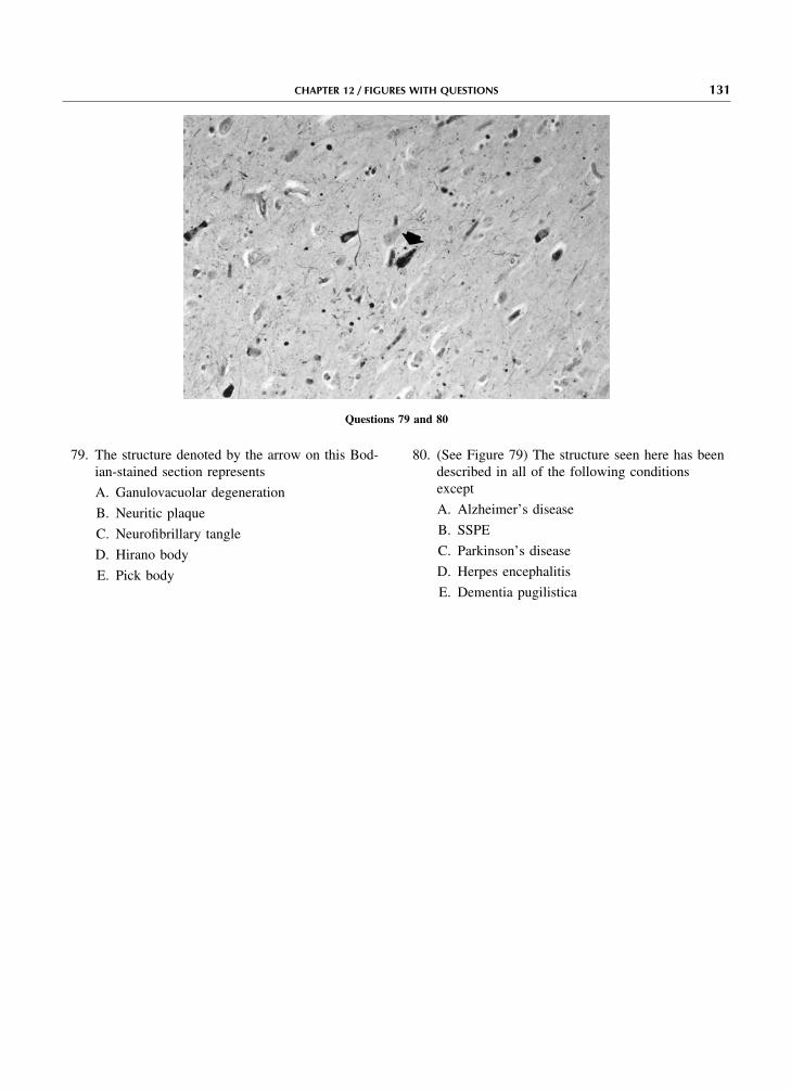
CHAPTER 12 / FIGURES WITH QUESTIONS 131
Questions 79 and 80
80. (See Figure 79) The structure seen here has been79. The structure denoted by the arrow on this Bod-ian-stained section represents described in all of the following conditions
exceptA. Ganulovacuolar degenerationA. Alzheimer’s diseaseB. Neuritic plaqueB. SSPEC. Neurofibrillary tangleC. Parkinson’s diseaseD. Hirano bodyD. Herpes encephalitisE. Pick bodyE. Dementia pugilistica

132 NEUROPATHOLOGY REVIEW
Questions 81, 82, and 83
84. The structure illustrated represents81. All of the following are abnormalities evident inthis photomicrograph except A. Choroid plexus
B. Choroid plexus papillomaA. Vascular congestion
B. Herniation C. Choroid plexus carcinoma
D. Papillary meningiomaC. Widening of the gyri
D. Narrowing of the sulci E. Papillary ependymoma
85. The cell denoted by the arrow represents a(n)E. Meningeal clouding
82. (See Figure 81) All are potential complications of A. Astrocytethe process seen here except B. OligodendrocyteA. Ventriculitis C. NeuronB. Infarct D. Microglial cellC. Hemorrhage E. Arachnoidal cap cellD. Secondary glioma development 86. (See Figure 85) This cell is most likely to stain
positively with which antibody?E. Hydrocephalus
83. (See Figure 81) The most likely causative organ- A. GFAPism in a 78-yr-old individual who has not been B. Synaptophysinrecently hospitalized would be C. Epithelial membrane antigenA. E. coli D. Common leukocyte antigenB. Proteus E. HAM 56C. Listeria
D. S. pneumoniae
E. N. meningitidis

CHAPTER 12 / FIGURES WITH QUESTIONS 133
Question 84
Questions 85 and 86

134 NEUROPATHOLOGY REVIEW
Question 87
89. (See Figure 88) The pattern of inheritance associ-87. The most likely cause of hemorrhage in this 58-ated with this condition is likely to beyr-old male isA. X-linked recessiveA. HypertensionB. X-linked dominantB. AmyloidC. Autosomal dominantC. VasculitisD. Autosomal recessiveD. AneurysmE. None of the aboveE. Oligodendroglioma
90. The illustration most accurately represents a(n)88. This leptomeningeal-based lesion is a commonCNS finding in A. Tumor
B. Embolized vascular formationA. Neurofibromatosis type I
B. Neurofibromatosis type II C. Artery with vasculitis
D. InfarctC. von Hippel-Lindau syndrome
D. Tuberous sclerosis E. Aneurysm
E. Sturge-Weber disease

CHAPTER 12 / FIGURES WITH QUESTIONS 135
Questions 88 and 89
Question 90

136 NEUROPATHOLOGY REVIEW
Question 91
94. (See Figure 92) This tumor is most likely to arise91. The large cell seen here in a patient with pro-in a(n)gressive multifocal leukoencephalopathy repre-
sents a(n) A. 59-yr-old male, frontal lobe
B. 60-yr-old female, temporal lobeA. Oligodendrocyte
B. Neuron C. 14-yr-old male, pineal gland
D. 15-yr-old female, pineal glandC. Alzheimer type I astrocyte
D. Alzheimer type II astrocyte E. 82-yr-old male, pituitary gland
95. The lesion seen here represents a(n)E. Cowdry type A inclusion
92. The lesion denoted by the arrows represents a(n) A. Arteriovenous malformation
B. Cavernous angiomaA. Microglial nodule
B. Abscess C. Venous angioma
D. VarixC. Secondary structure of Sherer
D. Granuloma E. Capillary telangiectasia
E. Vascular proliferation
93. (See Figure 92) The lesion in this case is associ-ated with what tumor?
A. Lymphoma
B. Germinoma
C. Meningioma
D. Metastatic carcinoma
E. Embryonal carcinoma

CHAPTER 12 / FIGURES WITH QUESTIONS 137
Questions 92, 93, and 94
Question 95

138 NEUROPATHOLOGY REVIEW
Questions 96, 97, and 98
99. The organism seen here represents96. The elongated structures (arrow) seen here rep-resent A. Amebiasis
B. CysticercosisA. Abnormal mitochondria
B. Cilia C. Schistosomiasis
D. TrypanosomiasisC. Blepharoplasts
D. Melanosomes E. Toxoplasmosis
100. (See Figure 99) The organism causing this lesionE. Birbeck granulesis97. (See Figure 96) The most likely diagnosis isA. Taenia soliumA. EpendymomaB. Trypanosoma bruceiB. Mitochondrial myopathyC. Entamoeba histolyticaC. MelanomaD. Trichonella spiralisD. Histiocytosis XE. Iodamoeba buetschliiE. Erdheim-Chester disease
101. The pathology illustrated here represents98. (See Figure 96) The cells that contain these struc-tures characteristically stain with antibody A. Oligodendroglioma
B. AstrocytomaA. GFAP
B. HMB-45 C. Ependymoma
D. Reactive astrocytosisC. CD3
D. CD1a E. Lymphoma
E. HAM 56

CHAPTER 12 / FIGURES WITH QUESTIONS 139
Questions 99 and 100
Question 101

140 NEUROPATHOLOGY REVIEW
Questions 102 and 103
105. The area seen here is consistent with a(n)102. The structure denoted by the arrow representsA. Caudate nucleusA. Secondary structure of SchererB. HippocampusB. Vascular proliferationC. Dentate nucleusC. NecrosisD. Olivary nucleusD. Vascular invasionE. None of the aboveE. None of the above
106. (See figure 105) The fascicles of myelinated103. (See Figure 102) The lesion seen here in a 76-yr-fibers seen here (arrow) are referred to asold male represents a(n)A. Lines of BaillargerA. WHO grade I astrocytomaB. Stria of GennariB. WHO grade II astrocytomaC. Bundles of WilsonC. WHO grade III astrocytomaD. Polymorphous bundlesD. WHO grade IV astrocytomaE. None of the aboveE. Ependymoma
104. The photomicrograph here represents what nor-mal structure?
A. Pineal gland
B. Leptomeninges
C. Pituitary adenohypophysis
D. Pituitary neurohypophysis
E. Pituitary pars intermedia

CHAPTER 12 / FIGURES WITH QUESTIONS 141
Question 104
Questions 105 and 106

142 NEUROPATHOLOGY REVIEW
Questions 107
Question 108
108. The photomicrograph here was most likely taken107. The age of the patient here most likely isfrom which location?A. 3 moA. Frontal lobe cortexB. 3 yrB. Frontal lobe white matterC. 13 yrC. CaudateD. 30 yrD. HippocampusE. 60 yrE. Pineal gland

CHAPTER 12 / FIGURES WITH QUESTIONS 143
Question 109
Question 110
110. The melanin pigmented neurons seen here belong109. The elastic stained section here represents a(n)to which neuronal group?A. Arteriovenous malformationA. Dentate nucleusB. Cavernous angiomaB. Substantia nigraC. Venous angiomaC. Hypoglossal nucleusD. HemangioblastomaD. Red nucleusE. Capillary telangiectasiaE. Olivary nucleus

144 NEUROPATHOLOGY REVIEW
Question 111
113. This 38-yr-old male has111. What normal structure is represented here?A. Non-Hodgkin’s lymphomaA. Pineal glandB. MalariaB. Arachnoidal cap cellsC. MedulloblastomaC. Choroid plexusD. AmebiasisD. Skeletal muscleE. ToxoplasmosisE. None of the above
112. The mass seen here represents a(n)
A. Astrocytoma
B. Infarct
C. Abscess
D. Granuloma
E. Demyelinating plaque

CHAPTER 12 / FIGURES WITH QUESTIONS 145
Question 112
Question 113

146 NEUROPATHOLOGY REVIEW
Questions 114 and 115
117. The findings in this lesion represent a(n)114. The organism seen here by electron microscopyrepresents A. Contusion
B. Acute infarctA. Cytomegalovirus
B. Toxoplasmosis C. Subacute infarct
D. Remote infarctC. Cryptococcus
D. Progressive multifocal leukoencephalopathy E. Tumor
118. (See Figure 117) The most likely age of thisE. E. colilesion is115. (See Figure 114) In an immunocompromised
adult, the most common histologic manifestation A. 24 hrof an infection by this organism is B. 48 hrA. Granuloma C. 72 hrB. Leptomeningitis D. 5–7 dC. Abscess E. 3 moD. Demyelination
E. Microglial nodule
116. This luxol-fast blue stained section of periventric-ular tissue shows areas of decreased white matterstaining in a 28-yr-old female. Cells that may beseen in these regions include all of the followingexcept
A. Reactive astrocytes
B. Macrophages
C. Neutrophils
D. Lymphocytes
E. Microglial cells

CHAPTER 12 / FIGURES WITH QUESTIONS 147
Question 116
Questions 117 and 118

148 NEUROPATHOLOGY REVIEW
Questions 119 and 120
122. (See Figure 121) The changes seen in the large119. The cell shown here represents a(n)neurons is the result ofA. NeuronA. IschemiaB. AstrocyteB. Central chromatolysisC. Microglial cellC. LipofuscinD. Oligodendroglial cellD. NeuronophagiaE. Endothelial cellE. Ferruginization120. (See Figure 119) The process shown in this cell
123. The accumulation of GM2 ganglioside material inrepresentsthese cells is characteristic ofA. Ischemic changesA. Gaucher’s diseaseB. Central chromatolysisB. Tay-Sachs diseaseC. FerruginizationC. Ceroid-lipofuscinosisD. NeuronophagiaD. Hunter’s diseaseE. Creutzfeldt astrocyteE. Leigh’s disease121. The section seen here is located where?
124. (See Figure 123) The cells in which the materialA. Frontal lobe cortexhas accumulated are
B. ThalamusA. Astrocytes
C. PonsB. Ependymal cells
D. MidbrainC. Microglial cells
E. CerebellumD. Neurons
E. Oligodendrocytes

CHAPTER 12 / FIGURES WITH QUESTIONS 149
Questions 121 and 122
Questions 123 and 124

150 NEUROPATHOLOGY REVIEW
Question 125
127. (See figure 126) All of the following conditions125. The structure most likely to be encountered inare likely to result in the pathology seen herethese neurons in a patient with Parkinson’s dis-exceptease isA. Chronic ischemic changeA. Neuritic plaqueB. Chronic epilepsyB. Hirano bodyC. AlcoholismC. Lewy bodyD. Infiltrating astrocytomaD. Neurofibrillary tangleE. Dilantin toxicityE. Granulovacuolar degeneration
128. The patient is a 12-yr-old female with a recent126. The paler staining cells denoted by the arrowmeasles infection. She now presents with seizuresrepresentand dystonia. The diagnosis on biopsy isA. Purkinje cellsA. OligodendrogliomaB. Granular cellsB. Progressive multifocal leukoencephalopathyC. LymphocytesC. Subacute sclerosing panencephalitisD. Bergmann astrocytesD. LymphomaE. Corpora amylaceaE. None of the above

CHAPTER 12 / FIGURES WITH QUESTIONS 151
Questions 126 and 127
Question 128

152 NEUROPATHOLOGY REVIEW
Question 129
132. (See Figure 130) The pattern of inheritance associ-129. The meningioma seen here is best classified asated with this condition isA. SyncytialA. X-linked recessiveB. FibrousB. Autosomal dominantC. InvasiveC. Autosomal recessiveD. MetaplasticD. SporadicE. RhabdoidE. X-linked dominant130. This infant has diffuse white matter abnormalities
133. The changes seen here are most likely related toon imaging studies. The diagnosis isA. Fat emboliA. Alexander’s diseaseB. Methanol ingestionB. Metachromatic leukodystrophyC. KernicterusC. Globoid cell leukodystrophyD. Ethanol toxicityD. AdrenoleukodystrophyE. Arsenic toxicityE. Canavan’s disease
131. (See Figure 130) The lesion seen is associatedwith a deficiency of
A. Arylsulfatase A
B. Galactocerebroside-B galactosidase
C. Aspartoacylase
D. Phospholipase
E. None of the above

CHAPTER 12 / FIGURES WITH QUESTIONS 153
Questions 130, 131, and 132
Question 133

154 NEUROPATHOLOGY REVIEW
Questions 134, 135, and 136
137. The lesion seen here most likely represents a(n)134. The lesion denoted by the arrow is a(n)
A. Arteriovenous malformation A. Vascular malformation
B. ContusionB. Air embolism
C. Aneurysm C. Infarct
D. Air embolismD. Venous angioma
E. Moyamoya disease E. Aneurysm
138. The cell denoted by the arrow represents a(n)135. (See Figure 134) Microscopically, the defect thatdefines this lesion is A. Reactive astrocyteA. A mixture of arterial and venous vessels B. GemistocyteB. Intimal fibroplasia C. Creutzfeldt astrocyteC. Atherosclerosis D. Alzheimer type I astrocyteD. Loss of arterial media E. Alzheimer type II astrocyteE. Fragmented elastic lamina of the vessel wall 139. (See Figure 138) This cell is most likely to be
encountered in the setting of136. (See Figure 134) The other gross pathologicabnormality evident in this picture is A. TumorA. Infarct B. Demylinating diseaseB. Ganglioglioma C. Diabetic encephalopathyC. Dandy-Walker syndrome D. Hepatic encephalopathyD. Subdural hematoma E. Progressive multifocal leukoencephalopathyE. Fat emboli

CHAPTER 12 / FIGURES WITH QUESTIONS 155
Question 137
Questions 138 and 139

156 NEUROPATHOLOGY REVIEW
Question 140
142. The lesion seen here represents a(n)140. The darker areas have a yellow coloration in thisautopsy specimen from a 2-wk-old baby with a A. Vascular malformationhistory of hemolytic anemia. The lesions seen B. Acute infarctrepresent
C. Remote infarctA. Multifocal infarcts
D. Multiple sclerosis plaqueB. Hemorrhage
E. Lacunar infarctC. Bilirubin encephalopathy
D. Fetal alcoholism syndrome
E. Ponto-subicular necrosis
141. The changes seen here are the result of
A. Atrophy
B. Edema
C. Polymicrogyria
D. Subarachnoid hemorrhage
E. Lissencephaly

CHAPTER 12 / FIGURES WITH QUESTIONS 157
Question 141
Question 142

158 NEUROPATHOLOGY REVIEW
Questions 143 and 144
146. (See Figure 145) The gene defect associated with143. The most accurate diagnosis here isthis lesion is located at chromosomeA. ContusionA. It is not associated with a gene defectB. Epidural hematomaB. 22C. Subdural hematomaC. 19D. Arteriovenous malformationD. 14E. MeningitisE. 3144. (See Figure 143) The lesion seen here is gener-
ally associated with the rupture of which vessel? 147. The lesion shown here in this 62-yr-old femalerepresents a(n)A. Middle meningeal arteryA. Demyelinative plaqueB. Bridging veinsB. Laminar infarctC. Vertebral arteryC. Pseudolaminar infarctD. Sagittal sinusD. AbscessE. Middle cerebral arteryE. Creutzfeldt-Jakob disease145. The changes seen here are most consistent with
A. Atherosclerotic vasculopathy
B. Moyamoya syndrome
C. Binswanger’s disease
D. CADASIL
E. Amyloid angiopathy

CHAPTER 12 / FIGURES WITH QUESTIONS 159
Questions 145 and 146
Question 147

160 NEUROPATHOLOGY REVIEW
Question 148
Question 149
149. The changes seen in this vessel are most likely148. The changes seen here are most likely related tothe result ofA. Brain stem gliomaA. LymphomaB. SyringobulbiaB. Polyarteritis nodosaC. Degeneration of corticospinal tractsC. Granulomatous angiitisD. Chiari malformationD. Hypersensitivity angiitisE. Olivopontocerebellar atrophyE. Amyloid angiopathy

CHAPTER 12 / FIGURES WITH QUESTIONS 161
Question 150
Question 151
151. The leptomeningeal organism denoted by the150. The organisms seen here representarrow representsA. TrichinosisA. SchistosomiasisB. CysticercosisB. MalariaC. TrypanosomiasisC. CysticercosisD. SarcocystisD. EchinococcusE. ToxoplasmosisE. Strongyloides

162 NEUROPATHOLOGY REVIEW
Questions 152, 153, and 154
154. (See Figure 152) The lesion seen here may be152. The lesion seen here represents a(n)encountered in all of the following conditionsA. GranulomaexceptB. Microglial noduleA. ListeriosisC. HamartomaB. SarcoidosisD. ThrombusC. TuberculosisE. Vascular proliferationD. Wegener’s vasculitis153. (See Figure 152) Stains that may be useful in fur-E. Germinomather delineating the etiology of this lesion would
include
A. GMS and PAS
B. GMS and fite
C. GFAP and GMS
D. GFAP and fite
E. GFAP and Ziehl-Neelson

CHAPTER 12 / FIGURES WITH QUESTIONS 163
Questions 155 and 156 Question 157
157. The abnormality shown in this cortical section155. The midbrain lesion seen here representsfrom a 14-yr-old with chronic epilepsy representsA. Vascular malformationA. No abnormalityB. Duret hemorrhageB. Cortical dysplasiaC. Diffuse axonal injuryC. GangliogliomaD. Fat emboliD. Dysembryoplastic neuroepithelial tumorE. Lacunar infarctE. Pleomorphic xanthoastrocytoma156. (See Figure 155) The midbrain lesion is often
seen associated with what other condition?
A. Uncal herniation
B. Central herniation
C. Subfalcial herniation
D. Tonsillar herniation
E. Upward herniation

164 NEUROPATHOLOGY REVIEW
Question 158
160. The findings seen here are consistent with158. This partial lobectomy specimen is from a 12-yr-old with tuberous sclerosis. The diagnosis is A. Infarct
B. Mesial temporal sclerosisA. Subependymal giant-cell astrocytoma
B. Cortical tuber C. Anaplastic astrocytoma
D. Carbon monoxide toxicityC. Metastatic rhabdomyoma
D. Lymphoma E. Wernicke encephalopathy
161. (See Figure 160) The underlying cause of theE. Hemangioblastomapathology seen here is159. The findings in this myelin-stained section of spi-
nal cord are most consistent with A. Poor cerebral perfusion
B. Not knownA. Thiamine deficiency
B. Wilson’s disease C. Vitamin deficiency
D. Possibly suicide relatedC. Menke’s disease
D. Subacute combined degeneration E. A risk from prior radiation therapy
E. Herpes myelitis

CHAPTER 12 / FIGURES WITH QUESTIONS 165
Question 159
Questions 160 and 161

166 NEUROPATHOLOGY REVIEW
Question 162
164. The GFAP immunostain seen here is highlighting162. The changes in the hippocampus of this 8-yr-oldwhat cells?with intractable seizures is the result ofA. Ependymal cellsA. Hippocampal sclerosisB. AstrocytesB. Cortical dysplasiaC. Microglial cellsC. GangliogliomaD. Oligodendroglial cellsD. Cortical tuberE. NeuronsE. Neuronal heterotopia
163. The changes seen in the leptomeninges in this68-yr-old male with bacterial meningitis represent
A. Granuloma formation
B. Abscess
C. Vasculitis with thrombosis
D. Infarct
E. Hemorrhage
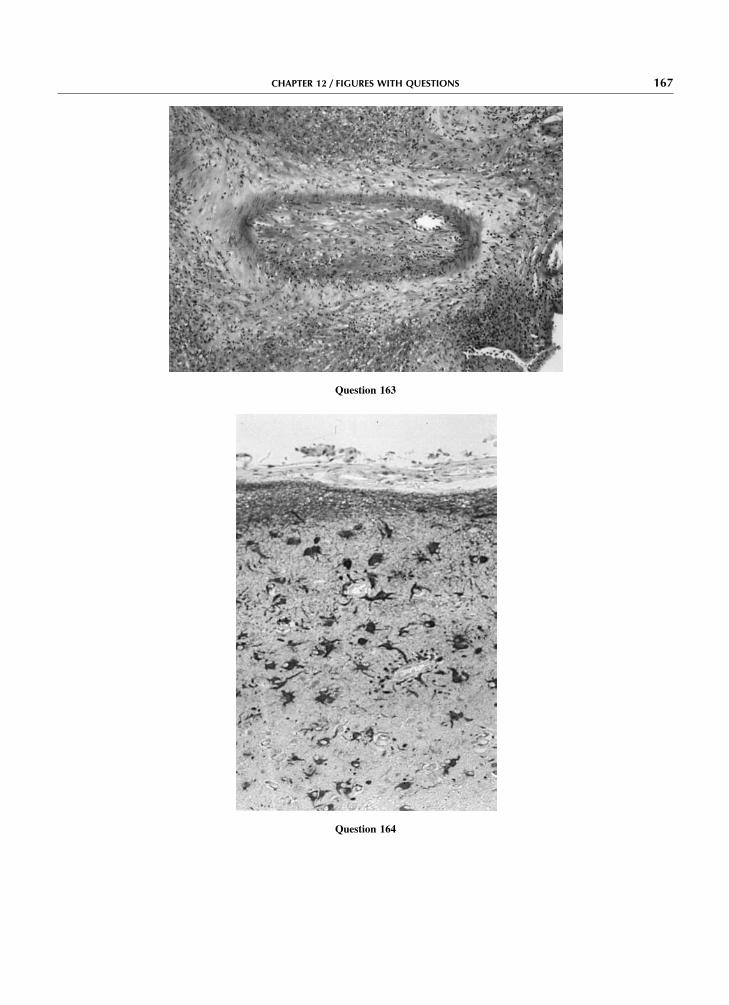
CHAPTER 12 / FIGURES WITH QUESTIONS 167
Question 163
Question 164

168 NEUROPATHOLOGY REVIEW
Question 165
Question 166
166. The pathology seen here in this 48-yr-old male165. The findings here are most likely related towith endocarditis and atrial fibrillation most likelyA. InfarctrepresentsB. EthanolA. Demyelinating diseaseC. Carbon monoxideB. Embolic acute infarctsD. Aluminum toxicityC. Air embolismE. Manganese toxicityD. Multiple organizing abscesses
E. Storage disease

CHAPTER 12 / FIGURES WITH QUESTIONS 169
Questions 167, 168, and 169
169. (See Figure 167) All of the following are likely167. The structure denoted by the arrow represents apathologic findings in this patient exceptA. Muscle fascicleA. Scattered type I hypertrophyB. Neuromuscular junctionB. Loss of neurons in Onuf’s nucleusC. Muscle spindleC. Increased muscle spindlesD. Blood vesselD. Atrophy of anterior spinal rootsE. Sarcomeric unitE. Fascicular atrophy of the muscle168. (See Figure 167) This biopsy is from a 3-mo-old
with hypotonia and tongue fasciculations. Thediagnosis is
A. Werdnig-Hoffmann disease
B. Central core disease
C. Duchenne dystrophy
D. Centronuclear myopathy
E. Pompe’s diease

170 NEUROPATHOLOGY REVIEW
Question 170
Question 171
171. The changes in the biopsy are most consistent170. The vessel seen here from a vastus lateralis mus-cle biopsy shows changes diagnostic of with a diagnosis of
A. PolymyositisA. Necrotizing vasculitis
B. Inclusion body myositis B. Dermatomyositis
C. Neurogenic atrophyC. Trichinosis
D. Dermatomyositis D. Vasculitis
E. Acid maltase deficiencyE. Polymyositis

CHAPTER 12 / FIGURES WITH QUESTIONS 171
Questions 172 and 173
173. (See Figure 172) All of the following are associ-172. The pathologic findings on the ATPase pH 4.6stain are suggestive of ated with this disease process except
A. ArthralgiaA. Dermatomyositis
B. Neurogenic atrophy B. Calcinosis
C. Steroid resistanceC. Muscular dystrophy
D. Inclusion body myositis D. Malignancy
E. Non-necrotizing vasculitisE. Spinal muscular atrophy

172 NEUROPATHOLOGY REVIEW
Questions 174 and 175
177. (See Figure 176) This change is a common fea-174. The structures observed in the myofiber denotedby the arrow on this Gomori trichrome stain ture observed inrepresent A. PolymyositisA. A freeze artifact B. Neurogenic atrophyB. Glycogen storage material C. Myotonic dystrophyC. Rimmed vacuoles D. Mitochondrial myopathyD. Nemaline rods E. Chloroquine myopathyE. Viral inclusions 178. On this ATPase-stained section, the dark staining
fibers are175. (See Figure 174) The presence of this finding inthe setting of an inflammatory myopathic biopsy A. Type Iis highly suggestive of B. Type IIAA. Angiopathic polymyositis C. Type IIBB. Viral myositis D. Type IICC. Fungal myositis E. Type IIID. Inclusion body myositis
E. Myasthenia gravis
176. The NADH-stained section shown reveals
A. Ring fibers
B. Moth-eaten fibers
C. Target fibers
D. Targetoid fibers
E. Core fibers
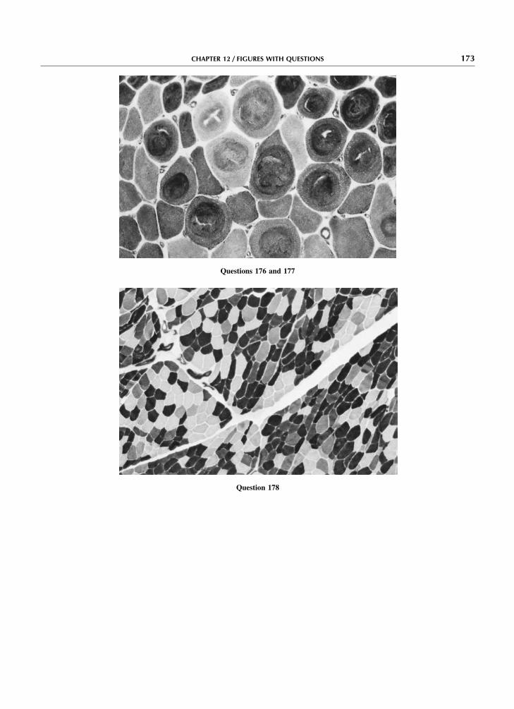
CHAPTER 12 / FIGURES WITH QUESTIONS 173
Questions 176 and 177
Question 178

174 NEUROPATHOLOGY REVIEW
Question 179
181. (See Figure 180) The structure denoted by the179. The positive staining on this alkaline phosphatasearrow represents a(n)stain representsA. I bandA. Degenerating fibersB. A bandB. Regenerating fibersC. H bandC. Acutely denervated fibersD. M bandD. Ragged red fibersE. Z bandE. A mitochondrial defect
182. The vacuolar structures seen here likely represent180. The fibrillary structures observed within thenucleus of this myocyte are characteristic of what A. Glycogen storage diseaseentity? B. Lipid storage diseaseA. Mitochondrial myopathy C. Mitochondrial degenerationB. Inclusion body myositis D. Core formationsC. Glycogen storage disease E. A freeze artifactD. Amyloid
E. A parasite

CHAPTER 12 / FIGURES WITH QUESTIONS 175
Questions 180 and 181
Question 182

176 NEUROPATHOLOGY REVIEW
Questions 183 and 184
185. The absence of staining on this cytochrome oxi-183. The abnormality seen ultrastructurally representsdase stain is indicative of a possiblean abnormality ofA. Glycogen storage diseaseA. T systemB. Lipid storage diseaseB. MitochondriaC. Viral infectionC. Sarcoplasmic reticulumD. Mitochondrial myopathyD. RibosomesE. None of the aboveE. Golgi
186. The curvilinear bodies seen here ultrastructurally184. (See Figure 183) The abnormality seen here isare most commonly associated withmost commonly associated withA. Mitochondrial encephalomyopathyA. Amyloid depositionB. Ceroid lipofuscinosisB. Ragged red fibersC. Lafora body diseaseC. Autophagic vacuolesD. Globoid cell leukodystrophyD. Core fibersE. Metachromatic leukodystrophyE. Nemaline rods

CHAPTER 12 / FIGURES WITH QUESTIONS 177
Question 185
Question 186

178 NEUROPATHOLOGY REVIEW
Questions 187 and 188
190. (See Figure 189) The most common mutation187. The cystalloid structures denoted by the smallassociated with the hereditary form of this condi-arrow representtion involves what gene?A. Renaut bodiesA. ThyroglobulinB. MyelinB. Notch 3C. Schmidt-Lanterman cleftsC. TransthyretinD. Pi granules of ReichD. Notch 5E. Corpuscles of ErzholzE. None of the above188. (See Figure 187) The structure denoted on the
191. The arrow corresponds to a(n)left by the larger arrow is a(n)A. Schwann cell nucleusA. Schwann cellB. Endothelial cellB. CollagenC. FibroblastC. Unmyelinated axonD. Mast cellD. Myelinated axonE. Renaut bodyE. Renaut body
192. (See Figure 191) The pathology seen here is most189. The finely fibrillar material (arrow) in the wall oflikely to be encountered in the setting ofthis epineurial vessel stains positively with Congo
red. The best diagnosis is A. Traumatic neuroma
B. Amyloid neuropathyA. Vasculitic neuropathy
B. CADASIL C. Vasculitic neuropathy
D. Charcot-Marie-Tooth diseaseC. Amyloid neuropathy
D. Adrenoleukodystrophy E. Thiamine deficiency neuropathy
E. Porphyria

CHAPTER 12 / FIGURES WITH QUESTIONS 179
Questions 189 and 190
Questions 191 and 192

180 NEUROPATHOLOGY REVIEW
Question 193
195. This muscle is from an 8-yr-old male who is193. The changes seen in this teased nerve preparationwheelchair bound. The most likely diagnosis ismost likely would be seen inA. PolymyositisA. Vasculitic neuropathyB. DermatomyostitisB. Charcot-Marie-Tooth diseaseC. Duchenne muscular dystrophyC. Guillain-Barre syndromeD. Myotonic muscular dystrophyD. CIDPE. Becker muscular dystrophyE. Diphtheritic neuropathy
196. (See Figure 195) Confirmation of the diagnosis in194. The change seen here representsthis case would involveA. Axonal sproutingA. T-cell subset analysisB. DemyelinationB. Serology testing for ANAC. RemyelinationC. Sedimentation rateD. Onion bulb formationD. Dystrophin analysisE. Renaut bodyE. Emerin analysis

CHAPTER 12 / FIGURES WITH QUESTIONS 181
Question 194
Questions 195 and 196

182 NEUROPATHOLOGY REVIEW
Questions 197 and 198
199. The pattern of atrophy observed here is sugges-197. The lesions seen on this NADH stain representtive ofA. Moth-eaten fibersA. DermatomyositisB. Rimmed vacuolesB. Spinal muscular atrophyC. Nemaline rodsC. Inclusion body myositisD. Target fibersD. Steroid myopathyE. Targetoid fibersE. Diabetic neuropathy-associated neurogenic198. (See Figure 197) This biopsy is most likely to be
atrophyseen in the setting of200. The vacuoles seen here stain with acid phospha-A. Myasthenia gravis
tase and are filled with PAS-positive material.B. Lambert-Eaton syndrome The diagnosis isC. Werdnig-Hoffman disease A. Carnitine deficiencyD. Neurogenic atrophy B. Myophosphorylase deficiencyE. Polymyositis C. Acid maltase deficiency
D. Succinate dehydrogenase deficiency
E. Phosphofructokinase deficiency

CHAPTER 12 / FIGURES WITH QUESTIONS 183
Question 199
Question 200


13 Answers to Figures with Questions
12. A (Chapter 5.III.A) Meningoangiomatosis is1. A (Chapter 5.III.A) The plexiform neurofibromacharacterized by a perivascular proliferation ofis almost exclusively seen in the setting of neuro-meningothelial cells and is associated with neuro-fibromatosis type I.fibromatosis type I.
2. E (Chapter 3.IV.A) Dural-based meningioma,13. C (Chapter 3.X.G) Squamous cell carcinomathe most commonly encountered cytogenetic
with large keratinized epithelial-appearing cellsabnormality is a deletion associated with chromo-infiltrating dura.some 22.
14. C (Chapter 3.II.E) The location, appearance, and3. C (Chapter 3.I.F) Perinecrotic pseudopalisading
immunohistochemical profile are classic for cen-in a glioblastoma multiforme.
tral neurocytoma.4. B(chapter 3 I L) Subependymal giant-cell astro- 15. B (Chapter 3.VII.A) The nuclear palisading
cytoma arising in the setting of tuberous sclerosis. (Verocay bodies) and mixed compact (Antoni A)5. A (Chapter 1.I.B) Rosenthal fibers in a pilocytic and loose regions (Antoni B) are characteristic of
astrocytoma. a schwannoma.
6. D (Chapter 3.VIII.A) Angiotropic large cell 16. A (Chapter 3.IV.B) The presence of vacuolatedlymphomas frequently are CD20 positive (B-cell epithelioid cells (physaliphorous) in a bone-basedimmunophenotype) and are typically confined to sacrococcygeal mass is suggestive of a chordoma.vascular lumina. 17. B (Chapter 3.VI.C) The structure denoted by the
arrow is a Homer Wright pseudorosette in a7. C (Chapter 3.X.C) The mixture of large “germ”medulloblastoma.cells and lymphocytes is characteristic of
germinoma. 18. C (Chapter 3.VI.C) The cells seen are from amedulloblastoma.8. D (Chapter 3.VII.D) The haphazard proliferation
of peripheral neural tissue with collagen in the 19. B (Chapter 4.II.H) The lesion shown here (pore-arm is best designated as traumatic neuroma. ncephaly) is usually the result of an ischemic/
infarct event.9. A (Chapter 5.I.D) The absence of convolutionseen in this brain is characteristic of lissencephaly 20. E (Chapter 7.I.B) The zebra bodies seen by elec-(agyria). tron microscopy are a common findings in Tay-
Sachs disease.10. C (Chapter 3.IV.A) The tumor shown is markedby cells arranged in a whorled configuration, char- 21. B (Chapter 5.I.F) The lesion seen represents aacteristic of a syncytial or meningotheliomatous partial agenesis of the corpus callosum.meningioma. 22. D (Chapter 3.III.B) The well-demarcated appear-
ance of this intramedullary spinal cord mass is11. D (Chapter 1.I.G) A meningioma with numer-ous laminated calcifications known as psammoma more commonly seen with ependymoma versus
an astrocytoma.bodies.
185

186 NEUROPATHOLOGY REVIEW
23. C (Chapter 5.III.C) The subependymal giant-cell 39. B (Chapter 5.I.B) The changes seen are mostcharacteristic of a Chiari malformation.astrocytoma seen here is associated with tuberous
sclerosis. 40. D (Chapter 5.I.A) Congenital dilatation of the24. A (Chapter 3.X.A) The nested architectural pat- central canal is termed hydromyelia.
tern (zellballen), chromogranin positivity, and 41. E (Chapter 5.III.A) The axillary freckling andlocation are classic for paraganglioma. cafe-au-lait macule are features of neurofibroma-
25. A (Chapter 3.II.A) The most common site of ori- tosis type I. All the listed lesions may be seen ingin for oligodendroglioma is frontal lobe (it has this setting except subependymal giant cell astro-the most white matter). cytoma (a feature of tuberous sclerosis).
26. D (Chapter 3.I.N) The lesion depicted here 42. A (Chapter 3.I.B) Obliteration of the gray–white(asymmetrial enlargement of the brain stem) is junction is a gross feature of infiltrating gliomasmost consistent with a brain stem glioma. (in this case astrocytoma).
27. B (Chapter 3.X.D) Adamantinomatous pattern 43. B (Chapter 3.II.A) About 2% of oligodendroglio-of a craniopharyngioma. mas present with evidence of hemorrhage.
28. A (Chapter 3.IX.A) The location and appear- 44. C (Chapter 3.I.F) The gross appearance is mostance of the mass is most consistent with a colloid consistent with a glioblastoma multiforme.cyst of the third ventricle.
45. C (Chapter 3.III.E) The lesion depicted repre-29. D (Chapter 3.V.A) The lateral ventricle is the sents a myxopapillary ependymoma.
most likely location of choroid plexus papillomas46. B (Chapter 6.I.D) Multiple white matter lesionsarising in childhood.
marked by reactive astrocytes, macrophages, and30. C (Chapter 3.X.G) The sharp interface between perivascular lymphocytes are suggestive of multi-
tumor and brain parenchyma and immunohisto- ple sclerosis.chemical profile is most consistent with mela-
47. A (Chapter 6.II.D) Numerous Rosenthal fibersnoma (most glioblastoma multiforme show cross-in the setting of diffuse demyelination and mega-immunoreactivity with cytokeratin AE1/3).lencephaly are suggestive of Alexander’s disease.
31. E (Chapter 5.III.B) Bilateral acoustic schwanno-48. D (Chapter 3.III.D) Subependymomas aremas are diagnostic of neurofibromatosis type II,
marked by loose clustering of cell nuclei arrangedwhich is related to a chromosome 22 abnor-against a fibrillary background. Microcysticmality.change is fairly common.
32. A (Chapter 3.X.E) The most common secreting49. D (Chapter 6.I.D) Periventricular white matterpituitary adenoma is a prolactinoma.
plaques are a characteristic feature of multiple33. B (Chapter 5.I.D) Polymicrogyria, seen in the sclerosis.
figure, is associated with Zellweger’s syndrome,50. D (Chapter 1.I.H) The Sommer sector (CA1)a peroxisome abnormality.
region is denoted by the arrow.34. A (Chapter 5.II.I) The germinal matrix hemor-
51. B (Chapter 8.III.C) The changes seen are charac-rhage is most likely to occur in the prematureteristic of atrophy—ventricular dilatation, narrow-infant.ing of gyri, widening of sulci. This patient had35. E (Chapter 5.I.F) The defect seen representsAlzheimer’s disease.cavum septi pellucidi.
52. A (Chapter 2.II.C) The partially cavitated lesion36. B (Chapter 3.III.B) The cilia and ciliary bodydenoted by the small arrow represents a remoteattachments are characteristic ultrastructural fea-infarct.tures of ependymoma.
53. D (Chapter 1.I.H) The large arrow is pointing at37. A (Chapter 3.I.M) The histologic appearancea normal appearing pineal gland.(pleomorphic astrocytic cells), clinical presenta-
54. E (Chapter 1.I.E) The cell characterized by antion, and low rate of cell proliferation are mostelongated nucleus and scant cytoplasm representsconsistent with a pleomorphic xanthoastrocytoma.a microglial cell.38. C (Chapter 3.I.H) The gliosarcoma consists of
reticulin-rich sarcomatous areas and GFAP-posi- 55. C (Chapter 9.V.A) The smaller arrow is point-ing at a Toxoplasma gondii cyst.tive glioblastomatous areas.

CHAPTER 13 / ANSWERS TO FIGURES WITH QUESTIONS 187
56. C (Chapter 9.V.A) Rarely, microglial nodules, 73. A (Chapter 9.V.B) The organism seen repre-sents Amebiasis in a background of necrosis.as seen here, are associated with Toxoplasmosis
infection. 74. A (Chapter 9.II.C) The cocci seen here most57. B (Chapter 9.IV.M) Multiple white matter likely represent Meningococcus.
lesions in an immunocompromised individual 75. B (Chapter 9.IV.A) The large intranuclear viralshould raise the possibility of PML as a diag- inclusions seen here represent Cowdry type Anosis. inclusions.
58. D (Chapter 9.IV.L) The Cowdry A nuclear inclu- 76. B (Chapter 9.IV.M) Alzheimer type I astrocytessions seen here are most characteristic of CMV are a common finding in progressive multifocalinfection. leukoencephaly (result of JC virus infection)
59. B (Chapter 9.IV.L) The viral inclusions are asso- 77. B (Chapter 1.I.A) The intracytoplasmic materialciated with the ependymal lining present and ven- in many of the neurons here represents lipofuscintricular space representing a ventriculitis. or aging pigment.
60. D (Chapter 9.III.E) The broad-branching, non- 78. E (Chapter 9 IV I ) The intraneuronal cytoplas-septate hyphae seen here are characteristic of mic Negri body is characteristic of rabies.Mucormycosis.
79. C (Chapter 8.III.E) The neuronal cytoplasmic sil-61. E (Chapter 9.III.E) Fusarium hyphae are mor- ver positive structure represents a neurofibrillary
phologically similar to Aspergillus in tissue sec- tangle.tions.
80. D (Chapter 8.III.E) Neurofibrillary tangles are62. E (Chapter 9.VI.B) The clinical history and not found in Herpes encephalitis.
pathology (cortical spongiform degeneration with81. B (Chapter 9.II.C) Herniation is not evident inneuronal loss) are characteristic of Creutzfeldt-
this case. The brain is edematous and demon-Jakob disease.strates vascular congestion and meningeal cloudi-
63. C (Chapter 9.VI.B) The star-burst-like structure ness consistent with a purulent meningitis.represents a kuru plaque.
82. D (Chapter 9.II.C) Secondary glioma develo-64. D (Chapter 1.I.B) The structure represents cor-
ment is not associated with purulent meningitis.pora amylacea, a laminated basophilic polygluco-
83. D (Chapter 9.II.C) Streptococcus pneumoniae issan body associated with astrocytic foot pro-the most likely cause of community acquired bac-cesses.terial meningitis in the elderly.
65. B (Chapter 8.III.G) The granules with a clear84. A (Chapter 1.I.F) The structure shown repre-halo represent granulovacuolar degeneration.
sents a normal choroid plexus, marked by fibro-66. A (Chapter 8.III.G) Granulovacuolar degenera-vascular cores lined by epithelioid cells.tion is most likely to be seen in the setting of Alz-
85. C (Chapter 1.I.A) The cell shown represents aheimer’s disease.neuron.67. A (Chapter 6.I.D) The periventricular areas of
86. B (Chapter 1.I.A) Neurons are most likely todemyelination (plaques) is characteristic of multi-stain with antibody to synaptophysin.ple sclerosis.
87. A (Chapter 2.V.A) Hypertension is the most68. B (Chapter 9.III.D) Dichotomously branchingcommon cause of nontraumatic intracerebral hem-septate hyphae is characteristic of Aspergillus.orrhage.69. B (Chapter 3.III.B) The presence of true ependy-
88. E (Chapter 5.III.D) Leptomeningeal venousmal rosettes and perivascular pseudorosettes inangioma is a common finding in Sturge-Weberthis tumor is characteristic of an ependymoma.disease.70. C (Chapter 8.III.F) The structures seen here rep-
89. E (Chapter 5.III.D) There is no pattern of inheri-resent neuritic plaques.tance associated with Sturge-Weber disease.71. A (Chapter 8.III.F) A Bodian stain will high-
90. B (Chapter 2.IV.B) The lesion illustrated repre-light the neuritic component of the plaque and asents an arteriovenous malformation that has beenCongo red will stain the amyloid core.embolized.72. B (Chapter 9.III.B) The thick mucopolysaccha-
ride wall of Cryptococcus stains with muci- 91. C (Chapter 9.IV.M) Large atypical astrocytes,Alzheimer type I astrocytes, are commonlycarmine.

188 NEUROPATHOLOGY REVIEW
encountered in the setting of progressive multifo- 109. A (Chapter 2.IV.B) The presence of arterial andvenous vessels with intervening neural paren-cal leukoencephalopathy.chyma is characteristic of an arteriovenous92. D (Chapter 3.X.C) The collection of epithelioidangioma.histocytes represents a non-necrotizing gran-
uloma. 110. B (Chapter 1.I.A) Melanin-pigmented neuronsare seen in substantia nigra, locus ceruleus, and93. B (Chapter 3.X.C) Granulomas may be seen inthe dorsal motor nucleus of the vagus nerve.germinomas. The large cells seen here represent
malignant germ cells. 111. E (Chapter 1.I.H) The nests of epithelioid cells94. C (Chapter 3.X.C) Germinomas are most likely separated by fibrovascular septae is characteristic
to arise in young males in the pineal gland region. of the pituitary adenohypophysis.
95. B (Chapter 2.IV.D) The sinusoidal-type vessels 112. C (Chapter 9.II.D) The central necrosis rimmedarranged in a back-to-back fashion is characteris- by a collagenous wall is characteristic of antic of a cavernous angioma. organizing abscess.
96. E (Chapter 3.VIII.B) The elongated “tennis- 113. B (Chapter 9.V.C) The granules of darkly pig-racket”-like structures seen here represent Birbeck mented material within red cells in blood vesselgranules. lumina is characteristic of malaria.
97. D (Chapter 3.VIII.B) Birbeck granules are a dis- 114. A (Chapter 9.IV.L) The organisms seen heretinctive feature of Langerhans cell histiocytes of have the characteristic ultrastructural appearancehistiocytosis X. of cytomegalovirus.
98. D (Chapter 3.VIII.B) Langerhan’s cell histio-115. E (Chapter 9.IV.L) In an immunocompromised
cytes stain with CD1a antibody.adult, cytomegalovirus infection most commonly
99. B (Chapter 9.V.D) The lesion seen here repre- manifests as microglial nodules.sents cysticercosis.
116. C (Chapter 6.I.D) Neutrophils are not a salient100. A (Chapter 9.V.D) The organism responsible feature microscopically of multiple sclerosis.
for cysticerosis is Taenia solium (the pork117. D (Chapter 2.II.C,D) Cavitary changes in thetapeworm).
cortex with surrounding gliosis and a relative spar-101. D (Chapter 3.I.D) The mildly increased cellular-
ing of the molecular layer is consistent with aity and larger cells with abundant eosinophilic
remote infarct.cytoplasm are characteristic of reactive astro-
118. E (Chapter 2.II.C,D) Remote infarct findingscytosis.are seen in association with the 3 mo of age102. B (Chapter 3.I.F) The lesion denoted here repre-parameter.sents glomeruloid vascular proliferation.
119. A (Chapter 1.I.A) The cell shown here repre-103. D (Chapter 3.I.A) The prominent vascular prolif-sents a neuron.eration in this astrocytoma is sufficient enough to
warrant a diagnosis of WHO grade IV astro- 120. B (Chapter 1.I.A) Cytoplasmic swelling withcytoma. loss and peripheralization of Nissl substance and
eccentric nucleus are salient features of central104. D (Chapter 1.I.H) The loose arrangement ofchromatolysis.spindled cells is characteristic of the pituitary neu-
rohypophysis. 121. E (Chapter 1.I.H) The section is from the cere-bellum with the molecular layer to the left, and105. A (Chapter 1.I.H) The area seen here may repre-two large Purkinje cells and smaller round granu-sent either the caudate or putamen.lar cells to the right.106. C (Chapter 1.I.H) The fascicles of myelinated
fibers seen in the caudate are known as the pencil 122. A (Chapter 1.I.A) The Purkinje cells are markedbundles of Wilson. by ischemic changes—shrunken cell bodies with
dark-staining nuclei and indistinguishable107. A (Chapter 1.I.H) The presence of the externalnucleoli.granular cell layer of the cerebellum is a finding
most consistent with an age of 3 mo. 123. B (Chapter 7.I.B) The accumulation of GM2ganglioside material is a feature of Tay-Sachs108. B (Chapter 1.I.H) The photomicrograph repre-
sents white matter parenchyma. disease.

CHAPTER 13 / ANSWERS TO FIGURES WITH QUESTIONS 189
124. D (Chapter 7.I.B) The distended cells repre- 141. B (Chapter 4.I.B) The changes seen (expansionof gyri and narrowing of sulci) are characteristicsented neurons filled with GM2 ganglioside
material. of cerebral edema.
142. C (Chapter 2.II.C) The cavitary defect repre-125. C (Chapter 8.IV.C) Lewy bodies are oftensents a remote infarct.encountered in substantia nigra (neuromelanin-pig-
mented) neurons. 143. C (Chapter 4.II.B) The hemorrhage seen hererepresents a subdural hematoma.126. D (Chapter 1.I.B) The proliferation of cells in
the region of the Purkinje cell layer are the 144. B (Chapter 4.II.B) Subdural hematomas are asso-Bergmann astrocytes. ciated with tearing of bridging veins which trans-
verse through the subarachnoid space.127. D (Chapter 1.I.B) Loss of Purkinje cells withastrocytosis is associated with all of the listed con- 145. D (Chapter 2.VII.B) The thickened vesselditions except infiltrating astrocytoma. walls with granularity is highly suggestive of
CADASIL.128. C (Chapter 9.IV.G) The antecedent measles146. C (Chapter 2.VII.B) The notch 3 gene responsi-infection with Cowdry A inclusions is typical of
ble for CADASIL is located on chromosomeSSPE (subacute sclerosing panencephalitis)19q12.129. C (Chapter 3.IV.B) This meningioma is brain
147. C (Chapter 2.II.D) The lesion seen here repre-invasive, a feature some associate with malig-sents an acute infarct surrounded by a zone ofnancy.edema in a pseudolaminar configuration (involv-130. C (Chapter 6.II.B) Collections of white mattering more than one cortical layer).globoid cells is characteristic of Krabbe’s disease
148. C (Chapter 2.II.D) The asymetrical loss of corti-or globoid cell leukodystrophy.cospinal tracts is likely related to an ipsilateral131. B (Chapter 6.II.B) Krabbe’s disease is the resultcerebral infarct.of a deficiency in galactocerebroside-B galacto-
149. C (Chapter 2.VI.A) The changes seen are thosesidase.of a granulomatous vasculitis.132. C (Chapter 6.II.B) Krabbe’s disease is inherited
150. D (Chapter 10.III.G) The organisms seen in theas an autosomal recessive condition.skeletal muscle represents Sarcocystis infection.
133. B (Chapter 7.II.B) The bilateral basal ganglia151. E (Chapter 9.V.G) The elongated organism rep-hemorrhagic necrosis is a feature of methanol
resents Strongyloides in a patient who had dissem-toxicity.inated disease.
134. C (Chapter 2.III.A) The lesion denoted by the152. A (Chapter 9.II.F) The collection of epithelioidarrow represents a saccular aneurysm arising
histiocytes and lymphocytes represents a gran-from the posterior inferior cerebellar arteryuloma.(PICA).
153. B (Chapter 9.II.F) The best combination of135. D (Chapter 2.III.A) Loss of the media definesstains to further evaluate this lesion would be athe saccular aneurysm.GMS (for fungal organisms) and fite stain (for a
136. A (Chapter 2.III.C) There is evidence of friable, mycobacterial organism).necrotic tissue consistent with infarct involving 154. A (Chapter 9.II.C) Listeria infection typicallythe cerebellum. presents with a suppurative leptomeningitis with
137. A (Chapter 2.IV.C) The lesion shown here repre- abscess formation, usually not accompanied bysents part of a vascular malformation (venous granulomas. All of the other conditions may beangioma). associated with granuloma formation.
138. E (Chapter 1.I.B) The nuclear swelling and chro- 155. B (Chapter 4.I.D) The midline brain stem hemor-matin clearing is characteristic of an Alzheimer rhage represents a Duret hemorrhage.type II astrocyte. 156. A (Chapter 4.I.D) The Duret hemorrhage is typi-
139. D (Chapter 1.I.B) Alzheimer type II astrocytes cally associated with uncal or lateral transtentorialare most likely encountered in hepatic encephalop- herniation.athy (elevated ammonia levels). 157. B (Chapter 5.I.C) The disorganized cortical
architecture is a common pattern of cortical dys-140. C (Chapter 5.II.O) The changes seen are charac-teristic of bilirubin encephalopathy (kernicterus). plasia.

190 NEUROPATHOLOGY REVIEW
158. B (Chapter 5.III.C) The enlarged gyrus repre- 175. D (Chapter 10.III.C) Rimmed vacuoles in thesetting of an inflammatory myopathy are highlysents a cortical tuber.suggestive of inclusion body myositis.159. D (Chapter 7.I.T) The findings seen here are
176. A (Chapter 10.VIII.F) Ring fibers are markedmost consistent with those of subacute combinedby a ring of myofibrils that is oriented circumfer-degeneration due to vitamin B12 deficiency.entially around the longitudinally oriented fibrils160. E (Chapter 7.I.S) Discoloration in the mamillaryin the rest of the fiber.bodies with gliosis due to hemorrhage/hemosid-
177. C (Chapter 10.VIII.F) Ring fibers are a rela-erin and loss of neurons is seen in the setting oftively common finding in patients with myotonicWernicke encephalopathy.dystrophy.
161. C (Chapter 7.I.S) Thiamine deficiency underlies178. A (Chapter 10.I.D) The dark fibers are type I onthe pathologic changes seen with Wernicke
ATPase pH 4.6.encephalopathy.179. B (Chapter 10.I.D) Regenerating myofibers are162. A (Chapter 5.I.J) Focal neuronal loss and gliosis
highlighted on the alkaline phosphatase stain.in the hippocampus (especially CA1, CA4, and180. B (Chapter 10.III.C) The tubulofilaments seendentate regions) characterizes hippocampal
within this myofiber nucleus are characteristic ofsclerosis.those seen in inclusion body myositis.163. C (Chapter 9.II.C) The leptomeningeal vessel
181. E (Chapter 1.II.G) The dark band marked by theshows evidence of vasculitis and organizingarrow represents the Z band.thrombus.
182. E (Chapter 10.I.C) The holes seen in all of the164. B (Chapter 1.I.B) The GFAP immunostain ismyofibers represent a freeze artifact sustained dur-highlighting astrocytic cells in the cortex.ing processing of the biopsy specimen.
165. B (Chapter 7.II.C) The cerebellar vermal atro-183. B (Chapter 10.VI.A) The paracrystalline inclu-phy seen here is most likely related to ethanol.
sions are observed in mitochondria.166. B (Chapter 2.II.C) The areas of darker discolor-
184. B (Chapter 10.VI.A) Ragged red fibers is a lightation represent multifocal acute infarcts, mostmicroscopic finding also associated with mito-likely embolic in origin.chondrial myopathies.
167. C (Chapter 1.II.I) The lesion denoted by the185. D (Chapter 10.I.D) The partial cytochrome oxi-arrow represents a muscle spindle (intrafusal
dase deficiency may be indicative of a mitochon-fibers enclosed by a connective tissue capsule).drial abnormality.
168. A (Chapter 10.IV.B and Chapter 8.IV.N) The186. B (Chapter 7.I.F) Ultrastructural evidence of cur-
rounded myofiber atrophy in this infant is charac-vilinear bodies supports a diagnosis of ceroid lipo-
teristic of Werdnig-Hoffman disease.fucinosis.
169. B (Chapter 10.IV.B and Chapter 8.IV.N) There 187. D (Chapter 1.III.F) The cystalloid structuresis often sparing of Onuf’s nucleus in Werdnig- within the Schwann cell cytoplasm are pi gran-Hoffman disease. ules of Reich.
170. A (Chapter 10.III.I) The changes seen in the ves- 188. C (Chapter 1.III.E) The structure denoted by thesel are those of a necrotizing vasculitis (polyarteri- larger arrow is an unmyelinated axon.tis nodosa).
189. C (Chapter 11.III.I) The congophilic material171. A (Chapter 10.III.A) Chronic endomysial represents amyloid deposition.
inflammation in the absence of perifascicular atro-190. C (Chapter 11.III.I) Mutations in the transthy-phy is consistent with polymyositis.
retin or prealbumin gene are associated with172. A (Chapter 10.III.B) The perifascicular pattern hereditary forms of amyloid neuropathy.
of injury is consistent with dermatomyositis. 191. A (Chapter 1.III.F) The cell nucleus is from a173. C (Chapter 10.III.B) Dermatomyositis is usually Schwann cell.
responsive to steroid therapy. 192. D (Chapter 11.III.F) The onion bulb structure ismost commonly associated with Charcot-Marie-174. C (Chapter 10.III.C) The vacuoles seen repre-
sent rimmed vacuoles or autophagic vacuoles. Tooth disease.

CHAPTER 13 / ANSWERS TO FIGURES WITH QUESTIONS 191
193. A (Chapter 11.III.I) The fragmentation of 197. D (Chapter 10.IV.A) The three zones of staining(central pale, dark intermediate, and normalmyelin is consistent with axonal degeneration, a
prominent feature of vasculitic neuropathy. peripheral) is characteristic of target fibers.
194. A (Chapter 11.II.A) The changes here are consis- 198. D (Chapter 10.IV.A) Target fibers are a com-tent with axonal regeneration (sprouting). mon feature of neurogenic atrophy.
195. C (Chapter 10.VIII.C) The marked variation in 199. B (Chapter 1.II.D) Fascicular atrophy is associ-fiber size and fibrosis are most suggestive of a ated with spinal muscular atrophy.dystrophic process. The clinical history favors
200. C (Chapter 10.VI.B) The lysosomal associatedDuchenne muscular dystrophy.(acid phosphatase positive) acid maltase defi-196. D (Chapter 10.VIII.C) Dystrophin analysisciency is associated with glycogen (PAS positive)would be most useful in confirming the diagnosisaccumulation in vacuoles.of Duchenne muscular dystrophy.


14 Written Self-Assessment Questions
5. Meckel-Gruber syndrome is associated with all of1. Nissl substance is composed ofthe following exceptA. Smooth endoplasmic reticulumA. Autosomal dominant pattern of inheritance
B. Rough endoplasmic reticulumB. Occipital encephalocele
C. Golgi structuresC. Polydactyly
D. NeuromelaninD. Polycystic kidneys
E. LipofuscinE. Hepatic fibrosis
2. Anoxic damage related to carbon monoxide6. Which HLA type is associated with the develop-poisoning would be best classified as
ment of multiple sclerosis?A. Anoxic anoxia
A. HLA B7B. Anemic anoxia
B. HLA D8C. Histotoxic anoxia
C. HLA DR15D. Stagnant anoxia
D. HLA DW6E. None of the above
E. HLA DR43. What WHO grade is pleomorphic xanthoastro- 7. All of the following are “normal” aging changes
cytoma? in the brain exceptA. Grade I A. Loss of brain weightB. Grade II B. Leptomeningeal thickeningC. Grade III C. Increased corpora amylaceaD. Grde IV D. Rare neurofibrillary tanglesE. Does not have WHO grade E. Decreased astrocyte number and size
4. Cerebral edema is marked by all of the following 8. The most common organism responsible for anexcept intracranial epidural abscess is
A. Increased brain weight A. E. coli
B. Narrowing of sulci B. Staphylococcus
C. Decreased ventricular size C. Streptococcus
D. Narrowing of gyri D. Listeria
E. Gram-negative anaerobesE. Prominent pacchionian granulations
193

194 NEUROPATHOLOGY REVIEW
9. The alkaline phosphatase stain is useful for identi- 15. The most common site of origin for low-gradefibrillary astrocytoma isfying
A. Degenerating myofibers A. Frontal lobe
B. Parietal lobeB. Regenerating myofibers
C. Acutely denervated fibers C. Temporal lobe
D. Occipital lobeD. Mitochondrial abnormalities
E. Amyloid deposition E. Spinal cord
16. Cerebral edema as a result of an impaired cell10. Congenital dilatation of the central canal isknown as Na-K membrane pump is referred to as
A. VasogenicA. Syrinx
B. Syringobulbia B. Cytotoxic
C. TumoralC. Hydromyelia
D. Diplomyelia D. Transient
E. None of the aboveE. Myeloschisis
11. Histologically, all of the following are features of 17. Area cerebrovasculosa is associated withmultiple sclerosis except A. AnencephalyA. Neutrophils B. MeningoceleB. Lymphocytes C. Chiari malformationC. Reactive astrocytes D. Edwards syndromeD. Macrophages E. SchizencephalyE. Myelin loss 18. Acute-onset multiple sclerosis with prominent
optic nerve and spinal cord involvement repre-12. All of the following features are characteristic ofneurogenic muscle disease except sents which subtype?
A. Marburg typeA. Angular atrophic fibers
B. Target fibers B. Balo type
C. Schilder typeC. Group atrophy
D. Fiber type grouping D. Devic type
E. Concentric sclerosis typeE. Ring fibers
13. Marinesco bodies are most likely going to be 19. Phosphorylated tau protein is associated with thedevelopment offound in which cell type?
A. Neuron A. Neurofibrillary tangles
B. Neuritic plaquesB. Astrocyte
C. Oligodendrocyte C. Amyloid angiopathy
D. Granulovacuolar degenerationD. Ependymal cell
E. Arachnoidal cap cell E. Hirano bodies
20. All of the following are potential complications14. The most senstive neurons to anoxic damage arein what region? of bacterial leptomeningitis except
A. InfarctA. Endplate of hippocampus
B. Sommer sector B. Hemorrhage
C. DemyelinationC. Cortex layer IV
D. Thalamus D. Vasculitis
E. HydrocephalusE. Pons

CHAPTER 14 / WRITTEN SELF-ASSESSMENT QUESTIONS 195
21. Which of the following is the most likely cause 27. All of the following are features of gliosis asopposed to diffuse astrocytoma exceptof meningitis in a 2-wk-old infant?
A. Rosenthal fibersA. Staph. epidermidis
B. Collagen depositionB. Staph. aureusC. Microcystic changeC. Strep. agalactiaeD. Absent calcificationD. Strep. pneumoniaeE. Reactive astrocytesE. Listeria
28. Kernohan’s notch is associated with which of the22. Cyclopia is most commonly associated with
following?A. Down’s syndrome A. Central herniationB. Holoprosencephaly B. Subfalcial herniationC. Chiari malformation, type II C. Uncal herniationD. Dandy-Walker syndrome D. Tonsillar herniation
E. None of the above E. Upward herniation
29. All of the following are commonly associated23. Myofibers with basophilic cytoplasm, nuclearwith the Chiari type I malformation exceptenlargement, and prominent nucleation are charac-
teristic of A. Cerebellar tonsillar herniationA. Targetoid fibers B. Adult presentation
B. Degenerating fibers C. Syringomyelia
D. Klippel-Feil anomalyC. Regenerating fibers
E. Arachnoidal adhesionsD. Nemaline rods30. Waterhouse-Friderichsen syndrome is most typi-E. Moth-eaten fibers
cally associated with which organism?24. All are characteristic of an axonal degenerative
A. Group B Strepprocess exceptB. E. coliA. Axonal sproutingC. ListeriaB. Myelin ovoidsD. N. meningitidis
C. Bands of BungnerE. H. influenza
D. Macrophages31. All of the following conditions are associated
E. Shortened internodal distances with granulomatous inflammation in the CNSexcept25. Bergmann astrocytes are found in the
A. TuberculosisA. Basal gangliaB. ListeriosisB. Cerebral cortexC. SarcoidosisC. Subcortical white matterD. Foreign body giant-cell reactionD. CerebellumE. Coccidioidomycosis
E. Hippocampus32. All of the following are features of acute hemor-
26. Hypoglycemic neuronal necrosis preferentially rhagic leukoencephalitis exceptaffects all of the following areas except
A. Cerebral edemaA. Cortex—layers II and III
B. Vessel wall necrosisB. CA1 region of hippocampus
C. White matter hemorrhagesC. Dentate D. Immunocompromised stateD. Caudate E. About half of cases present with antecedent
upper respiratory infectionE. Purkinje cells

196 NEUROPATHOLOGY REVIEW
33. Neurofibrillary cytoskeletal alterations are a fea- 39. All of the following are genetic alterations associ-ated with diffuse astrocytoma exceptture of which toxicity?
A. p53 mutationsA. Carbon monoxideB. Increased PDGFR alpha expressionB. AluminumC. Gain 7qC. ArsenicD. Trisomy 22D. LeadE. Loss of heterozygosity 10p
E. Manganese40. Which vessel may get compressed in lateral trans-
34. β-Amyloid is encoded for on chromosome tentorial herniation resulting in infarct of the cal-A. 1 carine cortex?
A. Basilar arteryB. 14B. Posterior cerebralC. 19C. Posterior inferior cerebellar arteryD. 21D. Anterior inferior cerebellar arteryE. 22E. None of the above
35. All of the following are features of polymyositis41. Hypoplasia of the cerebellar vermis with cysticexcept
dilatation of the 4th ventricle and agenesis of theA. May be associated with rheumatoid arthritis corpus callosum would be most accurately termedB. CPK often elevated A. Chiari type II malformationC. Endomysial chronic inflammation B. Dandy-Walker syndrome
D. Autophagic vacuoles C. Cerebellar dysplasia
D. HolotelencephalyE. More common in femalesE. Schizencephaly36. The most distinguishing feature pathologically of
dermatomyositis is 42. Which of the following stains is most useful inhighlighting mycobacterial organisms?A. Chronic inflammationA. Hematoxylin and eosinB. Myofiber degenerationB. PAS
C. Myofiber regenerationC. GMS
D. Perifascicular atrophyD. Ziehl-Neelsen
E. Fascicular atrophy E. Dieterle37. Rosenthal fibers may be encountered in all of the 43. Which of the following is true regarding meta-
following conditions except chromatic leukodystrophy?A. Alexander’s disease A. Chromosome 21q gene defectB. Reactive astrocytosis B. Autosomal dominant
C. Arcuate U fibers not sparedC. Fibrillary astrocytoma
D. White matter with macrophages filled withD. Pilocytic astrocytomaglucocerebrosides
E. Pineal gland cystE. Arylsulfatase A deficiency
38. Cerebral blood flow constitutes approximately44. CERAD criteria for the diagnosis of Alzheimer’swhat percentage of cardiac output?
disease rely on which pathologic feature?A. 5%
A. Neurofibrillary tanglesB. 10% B. Neuritic plaquesC. 15% C. Amyloid angiopathyD. 20% D. Lewy bodies
E. None of the aboveE. 30%
CHAPTER 14 / WRITTEN SELF-ASSESSMENT QUESTIONS 197
45. All are features of dermatomyositis except 51. The most common glioma type in the CNS is
A. Steroid refractory A. Oligodendroglioma
B. Heliotropic rash B. Ependymoma
C. Gottron’s papules C. Glioblastoma multiforme
D. Soft tissue calcinosis D. Low-grade diffuse astrocytoma
E. Association with malignancy E. Pilocytic astrocytoma46. Miller-Dieker syndrome is associated with agyria 52. Rupture of the middle meningeal artery is associ-
related to deletion on chromosome 17 involving ated withwhich gene?
A. Duret hemorrhageA. LIS-1
B. Epidural hemorrhageB. p53
C. Subdural hemorrhageC. N-myc
D. Subarachnoid hemorrhageD. Retinoblastoma gene
E. Parenchymal hemorrhageE. None of the above
53. Cortical dysplasia is associated with all of the fol-47. All of the following are common features of men- lowing except
ingovascular syphilitic CNS infection exceptA. Tuberous sclerosis
A. Prominent plasma cell infiltratesB. Dysembryoplastic neuroepithelial tumor
B. GummataC. Neurofibromatosis type I
C. Heubner’s endarteritis obliteransD. Neurofibromatosis type II
D. ArteritisE. Epidermal nevus syndrome
E. Demyelination54. All are true regarding globoid cell leukodystro-
48. All are true regarding or seen with inclusion bodyphy except
myositis exceptA. Deficiency of galactocerebrosidase
A. Steroid resistentB. Autosomal recessiveB. Caused by viral infectionC. Onset of symptoms in first year of lifeC. Rimmed vacuolesD. Sparing of arcuate subcortical fibersD. Tubulofilamentous inclusions ultrastructurallyE. Atrophy of brainE. Mitochondrial abnormalities
55. All of the following are pathologic features of49. Alzheimer’s II astrocytes are most commonlyAlzheimer’s disease exceptassociated with what condition?A. Granulovacuolar degenerationA. Hepatic encephalopathyB. Hirano bodiesB. Progressive multifocal leukoencephalopathyC. Bunina bodiesC. GliosisD. Lewy bodiesD. Pilocytic astrocytomaE. Amyloid angiopathyE. Alexander’s disease
56. Which fungus is a budding yeast with a thick50. Macrophages may be first microscopically evidentmucopolysaccharide wall?in an infarct at what time?
A. CryptococcusA. Within 12 h
B. CandidaB. 24 h
C. AspergillusC. 48 h
D. MucormycosisD. 1 wk
E. 3–4 wk E. Coccidioidomycosis

198 NEUROPATHOLOGY REVIEW
57. The diabetic patient with a periorbital infection 63. All are features of metastatic carcinoma exceptinvolving organisms with nonseptate hyphae is A. Discrete gross lesionmost likely infected with
B. Collagenous stromaA. Aspergillus
C. Discrete cell bordersB. Candida
D. Cytokeratin immunoreactivityC. Mucormycosis
E. Perinecrotic pseudopalisadingD. Blastomycosis
64. An acute subdural hematoma is associated with aE. Actinomyces
skull fracture (in)58. Lissencephaly results from defects in develop-
A. Neverment that occur during the
B. 25% of casesA. 5th–7th wk of gestationC. 50% of casesB. 8th–10th wk of gestationD. 75% of casesC. 11th–13th wk of gestationE. Almost alwaysD. 15th–17th wk of gestation
65. Agyria with hydrocephalus, retinal dysplasia,E. 20th–22nd wk of gestationencephalocele, no muscle disease, and death
59. A protoplasmic body is a salient feature of which in infancy is most characteristic ofof the following?
A. Cerebro-ocular dysplasiaA. Alzheimer’s disease
B. Neu-Laxova syndromeB. Demyelinating disease
C. Zellweger’s syndromeC. Fungal infection
D. Miller-Dieker syndromeD. Congenital myopathy
E. Walker-Warburg syndromeE. Tumor
66. Most forms of adrenoleukodystrophy are related60. An infarct of what minimum volume of brain tis-
to an abnormality on which chromosome?sue will almost invariably result in dementia
A. 1A. 10 mL
B. 22B. 25 mLC. 19C. 50 mLD. 6D. 80 mLE. None of the aboveE. 120 mL
67. Target fibers are most closely associated with61. The “fried egg” appearance of oligodendrocyteswhich process?representsA. PolymyositisA. Cell cytoplasmB. DermatomyositisB. Perinuclear edemaC. Myotonic dystrophyC. Artifact of delayed fixationD. Neurogenic atrophyD. Increased cytoplasmic lipidE. Mitochondrial myopathyE. None of the above
68. Diphtheritic neuropathy is best classified as being62. The least common site for a lacunar infarct is the
A. The result of Schwann cell dysfunctionA. Putamen
B. The result of myelin sheath damageB. Caudate
C. The result of vessel damageC. Pons
D. The result of axonal damageD. Mamillary bodies
E. Thalamus E. None of the above

CHAPTER 14 / WRITTEN SELF-ASSESSMENT QUESTIONS 199
69. Neurofibrillary tangles are best highlighted on 75. Eosinophilic granular bodies are a commonfeature ofwhich stain?
A. Glioblastoma multiformeA. PAS
B. Pilocytic astrocytomaB. Trichrome
C. Subependymal giant-cell astrocytomaC. Cytochrome oxidase
D. Nasal gliomaD. Bodian
E. None of the aboveE. Congo red76. Traumatic basilar subarachnoid hemorrhage is70. The Huntington gene is located on which chro-
related to the tear of what vessel?mosome?A. Basilar arteryA. 1B. Vertebral arteryB. 4C. Subclavian arteryC. 14D. AortaD. 19E. External carotid arteryE. 21
77. A brain weight of 2350 g is properly termed71. Infection with which organism is associated withA. Megalocephalysulfur granule formation?
B. SchizencephalyA. Coccidioidomycosis
C. MicroencephalyB. BlastomycosisD. MegaloencephalyC. ActinomycosisE. NormalD. Nocardiosis
78. Other organ systems that frequently contain inclu-E. None of the abovesions in adenoleukodystrophy include all of the
72. Cowdry type A inclusions are typical of all thefollowing except
following organisms exceptA. Testis
A. Polio virusB. Peripheral nerve
B. CMVC. Liver
C. Epstein-Barr virusD. Lymph nodes
D. Herpes virusE. Pancreas
E. JC virus79. All of the following pathologies are typical of a
73. Ciliary body attachments in ependymal cells are CNS viral infection exceptknown as
A. GranulomasA. Kinetoplasts
B. Microglial nodulesB. Chloroplasts
C. NeuronophagiaC. Microtubuloblasts
D. Chronic inflammationD. Blepharoplasts E. Central chromatolysisE. None of the above 80. All of the following are true regarding subacute
74. What percentage of saccular aneurysms are sclerosing panencephalitis (SSPE) exceptbilateral? A. Most present <15 yr of ageA. < 5% B. Most recover without serious sequelaeB. 10% C. The result of measles virus infectionC. 20% D. Causes demyelinationD. 50% E. Associated with neurofibrillary tangle
formationE. 85%

200 NEUROPATHOLOGY REVIEW
81. Fascicular muscle atrophy is most commonly asso- 87. All are features of the pleomorphic xanthoastro-cytoma exceptciated with
A. Temporal lobe locationA. Myasthenia gravisB. Cyst with enhancing mural nodule radiographi-B. Kugelberg-Welander disease
callyC. Amyotrophic lateral sclerosis
C. Reticulin richD. Polymyositis
D. Nuclear pleomorphismE. Mitochondrial myopathy
E. Increased mitotic activity82. Which of the following is a trinucleotide repeat
88. Langerhans cell histocytosis is characteristicallydisorder?
marked by which antibody?A. Progressive supranuclear palsy A. CD1aB. Diffuse Lewy body disease B. CD1bC. Corticobasal degeneration C. CD1cD. Hallervorden-Spatz disease D. CD4
E. Huntington’s chorea E. CD8
89. “Raccoon eyes” are associated with83. The most common site of a Morton’s neuroma isthe A. Hinge fractureA. Median nerve B. Basilar skull fracture
B. Peroneal nerve C. Diastatic fracture
D. Frontal depressed skull fractureC. Radial nerve
E. None of the aboveD. Intermetatarsal nerves90. All of the following are characteristic of Down’sE. Sural nerve
syndrome except84. All are true regarding Negri bodies except
A. Reduced frontal lobeA. Seen in Rabies
B. Narrowing of the inferior temporal gyrusB. Intraneuronal
C. Small cerebellumC. Intranuclear D. Poor myelinationD. Eosinophilic E. Alzheimer-like pathology in middle-agedE. Hippocamal neurons frequently involved adults
91. Which of the following is a characteristic feature85. Which of the following immunostains is a usefulof Alexander’s disease?marker for microglial cells?
A. MegalencephalyA. GFAPB. Arcuate subcortical fibers sparedB. SynaptophysinC. Eosinophilic granular bodiesC. HAM56D. Presents typically in the 2nd decade of lifeD. S-100 proteinE. Phospholipid protein defect
E. CLA92. A grossly normal brain with gliosis in the caudate
86. All of the following are conditions associated and putamen translates into what Vonsattel gradewith saccular aneurysms except for Huntington’s chorea?A. Tuberous sclerosis A. Grade 0B. Polycystic kidney disease B. Grade 1C. Coarctation of the aorta C. Grade 2D. Marfan’s syndrome D. Grade 3
E. Grade 4E. Ehler-Danlos syndrome

CHAPTER 14 / WRITTEN SELF-ASSESSMENT QUESTIONS 201
93. The spread of which organism from the periphery 99. All of the following are true regarding ganglio-glioma exceptto the central nervous system occurs primarily via
retrograde axoplasmic transport? A. Most commonly arise in temporal lobeA. Poliovirus B. Associated with cortical dysplasiaB. Arbovirus C. Associated with chronic epilepsyC. CMV D. Frequently contain small necrotic foci that do
not alter its good prognosisD. Herpes virusE. Calcifications commonE. PML
100. A zellballen architectural pattern microscopically94. Hippocampal sclerosis is characterized by markedcharacterizes which tumor?loss of neurons in which location?A. SchwannomaA. CaudateB. NeurofibromaB. AmygdalaC. Ependymoma
C. Sommer sectorD. Paraganglioma
D. CA2E. Chordoma
E. None of the above101. The wounding capacity of a gunshot wound is
95. Circulating antibodies to the acetylcholine recep- most related totor are seen in
A. Size of bulletA. McArdle’s disease
B. Shape of bulletB. MELAS syndrome C. Velocity of bulletC. Myasthenia gravis D. Weight of bulletD. Lambert-Eaton syndrome E. Size of gunE. None of the above 102. Focal coagulative necrosis adjacent to the lateral
ventricles in a 30-wk-old fetus is best termed96. All are true regarding Guillian-Barre syndromeexcept A. UlegyriaA. Associated with HLA B27 B. Multilocular cystic encephalomalacia
B. Ascending paralysis C. Periventricular leukomalacia
C. Decreased deep tendon reflexes D. Diffuse lobar sclerosis
E. Multiple sclerosisD. Endoneurial chronic inflammation
103. Congenital CMV infection is characterized by allE. Segmental demyelinationthe following except97. Layer III of the cerebral cortex is known as theA. ChorioretinitisA. External granular layerB. Gliosis
B. Internal granular layerC. Hydrocephalus
C. Polymorphic layerD. Megalencephaly
D. Outer pyramidal layerE. Microcephaly
E. Inner pyramidal layer104. All of the following characterize Lambert-Eaton
98. Dolichoectasia is associated with the development syndrome exceptof which of the following?
A. Proximal muscle weaknessA. Fusiform aneurysm
B. Decreased neurotransmission with repeatedB. Mycotic aneurysm stimulationC. Saccular aneurysm C. Autonomic dysfunction
D. Berry aneurysm D. Associated with small cell carcinoma of lung
E. Antibodies to presynaptic calcium channelsE. Charcot aneurysm

202 NEUROPATHOLOGY REVIEW
105. The “motor oil” appearance of fluid in a cystic 111. Chemoresponsiveness in anaplastic oligodendrogli-omas has been linked to deletions on which chro-craniopharyngioma is the result ofmosome?A. CholesterolA. 1B. KeratinB. 3
C. NecrosisC. 11
D. CalcificationD. 17
E. GlycogenE. 22
106. Leprosy-associated neuropathy may be marked by112. Static-loading blunt head injury is the result of
all of the following exceptforces applied gradually to the head, such as
A. Granulomas with a
B. Schwann cell invasion by organisms A. Car rolling over a head
B. Hammer blow to headC. Perineurial fibrosis
C. Gunshot to headD. Symmetric polyneuropathyD. Trauma sustained in a landslideE. Toxin-mediated responseE. Pedestrian hit by a car107. Autosomal dominant transmission of Parkinson’s
113. All of the following are common sites of bili-disease is linked with what gene?rubin deposition in kernicterus exceptA. Tau protein geneA. DentateB. AgitonianB. Inferior olive
C. UbiquitinC. Cranial nerve nucleus VIII
D. α-SynucleinD. Claustrum
E. SOD1E. Substantia nigra
108. Nemaline rods are composed of 114. Canavan’s disease is the result of a deficiency inA. α-Actinin A. Arylsulfatase BB. Dystrophin B. LipophosphorylaseC. Emerin C. AspartoacylaseD. Sarcoplasmic reticulum D. Proteolipid protein
E. None of the above E. Not known
115. All of the following are common features of the109. The pencil bundles of Wilson are a feature ofcongenital myopathies exceptwhich structure?
A. Disorders of myofiber maturation/reorgani-A. Substantia nigrazation
B. DentateB. Often fatal
C. PutamenC. Retarded motor development
D. Locus ceruleusD. Associated with skeletal dysmorphism
E. HippocampusE. Many show familial tendencies
110. The most common organism associated with the 116. All of the following are true regarding Lyme-development of mycotic aneurysms is associated neuropathy exceptA. Mucor A. Caused by Borrelia burgdorferiB. Aspergillus B. Acquired from a mosquito biteC. Candida C. May be associated with arthritisD. Cryptococcus D. Perivascular chronic inflammation
E. Axonal degenerationE. Blastomycosis

CHAPTER 14 / WRITTEN SELF-ASSESSMENT QUESTIONS 203
117. For those hard-to-identify Lewy bodies, which of 123. All are true regarding central neurocytoma exceptthe following stains would be most useful? A. Presents usually in young adultsA. PAS B. Most patients die within 2 yr of diagnosisB. Congo red C. Synaptophysin positiveC. Ubiquitin D. Intraventricular locationD. GFAP E. Dense secretory core granules ultrastructurallyE. Gomori methenamine silver 124. A “burst lobe” represents an example of a
118. Which of the following is true regarding progres- A. Skull fracturesive multifocal leukoencephalopathy? B. Gunshot woundA. Caused by RNA virus C. Coup contusionB. Most patients present <age 12 yr D. Contrecoup lesionC. Alzheimer’s II astrocytes E. Gliding contusionD. Gray matter microglial nodules 125. The neurofibromin gene is located on chro-E. Oligodendroglial cell inclusions mosome
A. 1119. Vacuolar myelopathy is a feature of
B. 22A. Syphilis
C. 3B. HIV
D. 9C. HTLV-I
E. 17D. PML
126. The salient pathologic features of Pelizaeus-E. None of the aboveMerzbacher disease include all of the following
120. Ragged red fibers may be seen in all the follow-except
ing exceptA. Perivascular demyelination
A. MELASB. Brain atrophy
B. MERFFC. Astrocytosis
C. Kearns-Sayre syndromeD. Prominent cerebellar and brain stem
D. Inclusion body myositis involvementE. Central core myopathy E. White matter macrophages
121. The endplate of the hippocampus (Ammon’s 127. Which of the following is not true regardinghorn) is referred to as HTLV-I infection?A. Dentate A. Caused by retrovirusB. CA1 B. Causes tropical spastic paraparesisC. CA2 C. Myelin loss in spinal cordD. CA3 D. Cowdry A viral inclusionsE. CA4 E. Demyelination of optic nerve
122. Which vascular malformation is characterized by 128. Paraneoplastic neuropathy is most commonly asso-back-to-back vessels? ciated with which tumor type?
A. AVM A. Breast carcinoma
B. Venous angioma B. Lung carcinoma
C. Cavernous angioma C. Melanoma
D. Capillary telangiectasia D. Lymphoma
E. Renal cell carcinomaE. Varix

204 NEUROPATHOLOGY REVIEW
129. Where should one look for Lewy bodies? 135. The most important feature in predicting the out-come in ependymomas isA. Neuronal nucleusA. Mitosis countsB. Neuronal cytoplasmB. Presence of necrosis
C. Astrocytic nucleusC. Cellularity
D. Astrocytic cytoplasmD. Vascular proliferation
E. Both neuronal and astrocytic cytoplasmE. Extent of resection
130. All of the following may be seen with Kearns-136. All of the following areas are common sites of
Sayre syndrome exceptdiffuse axonal injury except the
A. Deafness A. MidbrainB. Acromegaly B. Corpus callosumC. Ophthalmoplegia C. FornixD. Retinitis pigmentosa D. Corona radiata
E. Heart block E. Superior cerebellar peduncles
137. Which of the following is a major diagnostic cri-131. Straight filament (tau positive) tangles with glio-terion for neurofibromatosis type I?sis and neuropil threads in the basal ganglia and
brain stem region support a diagnosis of A. Six or more cafe-au-lait spots >15 mm in achildA. Alzheimer’s disease
B. One plexiform neurofibromaB. Progressive supranuclear palsyC. One Lisch noduleC. Striatonigral degenerationD. Cousin with neurofibromatosis type I.D. Corticobasal degenerationE. Facial frecklingE. None of the above
138. A distinguishing feature of myxopapillary ependy-132. Type II glycogenosis is related to a defect on moma versus ordinary ependymoma is
chromosomeA. Pseudorosettes
A. 1B. True rosettes
B. 12C. GFAP positivity
C. 17 D. Mucoid stromaD. 22 E. Papillary architectureE. X 139. The most common pathologic manifestation of
Toxoplasmosis infection in the CNS in an immu-133. Perifascicular atrophy is a feature ofnocompromised adult isA. DermatomyositisA. Abscess
B. PolymyositisB. Hemorrhage
C. Neurogenic atrophyC. Infarct
D. Myotonic dystrophyD. Leptomeningitis
E. Steroid useE. Granulomas
134. The most common cause of nontraumatic hemor- 140. Glycogen storage diseases are manifested typi-rhage is cally on a muscle biopsy byA. Vascular malformation A. MyonecrosisB. Aneurysm B. Vacuolar changesC. Coagulopathy C. Extensive fibrosisD. Hypertension D. Fascicular atrophy
E. Targetoid fibersE. Vasculitis
CHAPTER 14 / WRITTEN SELF-ASSESSMENT QUESTIONS 205
141. Durcks nodes are a feature of 147. Meningiomas with a high likelihood of recurrenceinclude all of the following exceptA. CMVA. MetaplasticB. ToxoplasmosisB. RhabdoidC. AmebiasisC. PapillaryD. MalariaD. Clear cellE. None of the aboveE. Atypical142. All are true regarding Hallervorden-Spatz disease
except 148. Neurofibromatosis type II is related to abnormal-ity on chromosomeA. Onset in childhoodA. 1B. Autosomal dominantB. 17C. May cause choreoathetosisC. 3D. Globus pallidus rust brown coloredD. 22E. Increased iron deposition in brainE. 9143. Cysticercosis is caused by
149. Tay-Sachs disease is caused by a deficiency inA. Pork tapewormA. β-GalactosidaseB. Beef tapewormB. ArylsulfataseC. Liver flukeC. Hexosaminidase AD. Lung flukeD. GlucocerebrosidaseE. NematodeE. Iduronidase144. Anti-MAG antibodies are most commonly associ-
ated with 150. Which of the following is true regarding type-IVglycogenosis?A. Paraneoplastic neuropathyA. Myophosphorylase deficiencyB. Myasthenia gravisB. Autosomal dominant conditionC. Dysproteinemic neuropathyC. Chromosome 17 defectD. Guillian-Barre diseaseD. Females > malesE. None of the aboveE. Normal ischemic exercise test145. The longitudinal boundaries defining the sarcom-
ere are the 151. An oil-red-O stain may be most helpful in diag-nosingA. A bandsA. McArdle’s diseaseB. I bandsB. Mitochondrial diseaseC. M linesC. Carnitine deficiencyD. Z bandsD. Myotonic dystrophyE. H bandsE. Central core myopathy146. All of the following are true regarding amyloid
angiopathy except 152. Chagas disease is caused by
A. S. japonicumA. Associated with Down’s syndrome
B. Cause of lobar hemorrhage B. T. gondii
C. T. bruceiC. Highlighted with a congo red stain
D. Chromosome 21 abnormality D. T. cruzi
E. T. gambienseE. Associated with Pick’s disease

206 NEUROPATHOLOGY REVIEW
153. All are true regarding Tay-Sachs disease except 159. The most common cytogenetic abnormality inmeningiomas involves chromosomeA. GM2 gangliosidosisA. 1B. Cherry red spot in maculaB. 2C. Balloon neuronsC. 10D. May see normal brain weightD. 17
E. HepatosplenomegalyE. 22
154. Multisystem atrophy includes which of the follow-160. Retraction balls or spheroids and punctate hemor-ing conditions?
rhages are most characteristic ofA. Olivopontocerebellar atrophy
A. ContusionB. Hallervorden-Spatz disease
B. InfarctC. Corticobasal degeneration
C. Diffuse axonal injuryD. Friedrich’s ataxia
D. Burst lobeE. Huntington’s disease
E. None of the above155. All are salient features of Creutzfeldt-Jakob
161. Which of the following is associated with tuber-pathology exceptous sclerosis?
A. Spongiform changeA. Pilocytic astrocytoma
B. Microglial nodulesB. Optic nerve glioma
C. GliosisC. Subependymal giant cell astrocytoma
D. Neuronal lossD. Brain stem glioma
E. Kuru plaquesE. Medulloblastoma
156. Charcot-Marie-Tooth disease may be caused by 162. Abnormal accumulation of sphingomyelin anddefects associated with which chromosomes? cholesterol is a feature ofA. 1 and 9 A. Tay-Sach diseaseB. 1 and 17 B. Niemann-Pick diseaseC. 9 and 17 C. Gaucher’s diseaseD. 17 and 22 D. Batten’s diseaseE. X and 22 E. Mucolipidoses
157. Increased myofiber central nuclei may be seen in 163. The most common cause of death in Friedrich’sall of the following except ataxia isA. Exercise-induced hypertrophy A. Brain infarct/strokeB. Neurogenic atrophy B. Heart diseaseC. Myotubular myopathy C. HerniationD. Myotonic dystrophy D. Coagulopathy related bleedE. Charcot-Marie-Tooth disease E. Associated neoplasm
158. Granulomatous vasculitis is a common feature of 164. All are true regarding Friedrich’s ataxia except
A. Polyarteritis nodosa A. Autosomal recessive
B. Wegener’s vasculitis B. Chromosome 9q defect
C. Lupus associated vasculitis C. GAA trinucleotide repeat disorder
D. Hypersensitivity angiitis D. Posterior columns spared
E. Ganglion cell/posterior root degenerationE. None of the above

CHAPTER 14 / WRITTEN SELF-ASSESSMENT QUESTIONS 207
165. The prion protein gene is located on which chro- 171. Solitary fibrous tumor can be distinguished frommeningioma by immunoreactivity tomosome?
A. 1 A. Vimentin
B. CD34B. 17
C. 20 C. S-100 protein
D. Cytokeratins AE1/3D. 22
E. None of the above E. Actin
172. A Duret hemorrhage is located in the166. Dilatation of muscle sarcoplasmic reticulum withtubular aggregates is a feature of A. Frontal lobeA. Myasthenia gravis B. Basal gangliaB. Myoadenylate deficiency C. Mamillary bodiesC. Malignant hyperthermia D. CerebellumD. Periodic paralysis E. PonsE. None of the above 173. All of the following are associated with tuberous
sclerosis except167. Which is true regarding Dejerine-Sottas disease?
A. Autosomal dominant A. Schwannoma
B. Adenoma sebaceumB. X-linked
C. Presents in young adulthood C. Angiomyolipoma
D. Cardiac rhabdomyomaD. Nerve enlargement
E. Primarily axonal degenerative disease E. Lymphangioleiomyomatosis
174. Cherry red spots in the macula is a prominent fea-168. Phytanoyl CoA α-hydroxylase deficiency causesture of all the following exceptA. Fabry’s diseaseA. Tay-Sachs diseaseB. Refsum’s diseseB. Niemann-Pick diseaseC. Riley-Day syndromeC. Gaucher’s diseaseD. Canavan’s diseaseD. SialodosisE. Metachromatic leukodystrophyE. They are seen in all of the above169. Intermediate filaments that link Z discs together
in muscle are known as 175. Type IIb muscle fiber atrophy is a feature of
A. Alcohol myopathyA. Actin
B. Desmin B. Steroid myopathy
C. Chloroquine myopathyC. Myosin
D. Dystrophin D. AZT myopathy
E. Cimetidine myopathyE. Vimentin
170. The highest yield area to look for vasculitis on a 176. All of the following are true regarding primaryCNS lymphoma exceptbiopsy would be
A. Leptomeninges A. More common in males
B. Most are B-cell typeB. Cerebral cortex
C. White matter C. Most are diffuse small cell type
D. Most are parenchymal basedD. Hippocampus
E. Basal ganglia E. Most are angiocentric histologically

208 NEUROPATHOLOGY REVIEW
177. von Hippel-Lindau disease is associated with all 183. Physaliphorous cells are a feature of whichtumor?of the following except
A. Pheochromocytoma A. Meningioma
B. Hemangiopericytoma B. HemangioblastomaC. Renal cell carcinoma C. HemangiopericytomaD. Secondary polycythemia vera D. ChondromaE. Autosomal dominant inheritance pattern E. None of the above
178. Spinocerebellar degeneration with sensory neurop- 184. All of the following are autosomal dominantathy as a result of chronic intestinal fat malabsorp- disorders excepttion is caused by deficiency of
A. Neurofibromatosis type IA. Vitamin A
B. Neurofibromatosis type IIB. Vitamin B
C. Neurocutaneous melanosisC. Thiamine
D. Tuberous sclerosisD. Vitamin EE. Sturge-Weber diseaseE. Vintamin K
185. A trigeminal nevus flammeus is a marker of179. A SOD1 gene defect on chromsome 21 is associ-A. Epidermal nevus syndromeated with what disorder?
A. Becker’s muscular dystrophy B. Cowden disease
B. Myotonic dystrophy C. Sturge-Weber disease
C. Refsum’s disease D. Gorlen syndromeD. Amyotrophic lateral sclerosis E. Tuberous sclerosisE. Werdnig-Hoffman disease 186. Sea blue histocytes in the bone marrow are a
feature of180. All of the following inclusions are seen fre-quently in amyotrophic lateral sclerosis except A. Tay-Sachs diseaseA. Hirano bodies B. Niemann-Pick diseaseB. Bunina bodies C. Ceroid-lipofucinosisC. Hyaline inclusions
D. CystinosisD. Basophilic inclusions
E. Lafora body diseaseE. Skeins
187. “Giant aneurysm” is used to denote a saccular181. Which of the following cells is a normal constit- aneurysm greater than
uent of the endoneurial compartmentA. 0.5 cm
A. LymphocytesB. 1.0 cm
B. EosinophilsC. 1.5 cmC. Mast cellsD. 2.0 cmD. BasophilsE. 2.5 cmE. Neutrophils
188. The most common site of origin of choroid182. CADASIL is associated with a mutation on theplexus papillomas in children isnotch 3 gene located on which chromosome?A. Lateral ventricleA. 1B. Third ventricleB. 17
C. Fourth ventricleC. 19
D. Brain stemD. 21
E. 22 E. Spinal cord

CHAPTER 14 / WRITTEN SELF-ASSESSMENT QUESTIONS 209
189. Cowden syndrome is associated with which of 195. Secondary polycythemia vera is associated withthe following? A. HemangiopericytomaA. Medulloblastoma B. HemangioblastomaB. Cerebellar gangliocytoma C. MeningiomaC. Melanoma D. Solitary fibrous tumor
E. Choroid plexus papillomaD. Lymphoma
196. The accumulation of autofluorescent lipopigmentsE. Melanotic schwannomain neurons resulting in granular, fingerprint, or190. Which of the following is the result of a defectcurvilinear inclusions by electron microscopy ison chromosome 1q?known as
A. Niemann-Pick diseaseA. Gaucher’s disease
B. Gaucher’s diseaseB. Batten’s disease
C. Morquio’s diseaseC. Mucopolysaccharidoses
D. Sialodosis D. CystinosisE. Mucolipidoses E. Fucosidosis
191. All of the following are characteristic of Leigh’s 197. Neuronal-PAS positive deposits, skin telangiecta-disease except sia, renal insufficiency, and autonomic dysfunc-A. Associated with mitochondrial defects tion are features ofB. Autosomal recessive A. Sialodosis
B. Tay-Sachs diseaseC. Most survive to early adulthood
C. FucosidosisD. Vascular proliferation in periventricular graymatter (brain stem) D. Fabry’s disese
E. Lactic acidosis E. Lafora body disease192. Ethanol is associated with all the following 198. All the following are related to gene defects on
except the X chromosome except
A. Fabry’s diseaseA. Anterior vermal atrophyB. Duchenne dystrophyB. Peripheral neuropathyC. AdrenoleukodystrophyC. Corpus callosum demyelinationD. Hunter’s mucopolysaccharidosisD. Thiamine deficiencyE. Lafora body diseaseE. Neurofibrillary tangles
199. Loss of anterior horn cells, tongue fasciculations,193. Lamellated cytoplasmic structures located withinand hypotonia in a 2-mo-old is most likely due tothe Schwann cell cytoplasm that increase witha disorder associated with what chromosome?age are referred to asA. 1A. Corpuscles of ErzholzB. 5B. LipofuscinC. 14
C. Schmidt-Lanterman cleftsD. 16
D. Pi granules of ReichE. 17
E. Space of Klebs200. The ratio of myelinated to unmyelinated axons in
194. Moyamoya is characterized by all of the follow- a sural nerve biopsy would be best approximateding except by which of the following?A. Amyloid deposition A. 1 : 2B. Peak incidence in the 1st and 4th decades B. 1 : 4C. Females > males C. 1 : 6D. May see hemorrhage D. 1 : 8
E. 1 : 10E. Vessel occlusion with intimal fibroplasia

210 NEUROPATHOLOGY REVIEW
201. The most common cause of “aseptic” meningitis 206. The single best immunomarker for medul-loblastoma is which of the following?is
A. CytokeratinA. BacteriaB. GFAPB. VirusC. S-100C. FungusD. SynaptophysinD. ParasiteE. ChromograninE. Prion protein disease
207. Poor prognosis in medulloblastoma is associated202. All are true regarding familial insomnia syndromewith all of the following exceptexceptA. Age >3 yr
A. Most patients survive 10-20 yearsB. Metastases at presentation
B. Autosomal dominantC. Subtotal resection
C. Associated autonomic dysfunctionD. Large cell variant
D. A prion-protein-associated diseaseE. Melanotic variant
E. Thalamic gliosis 208. Which of the following are true regarding pri-203. The saltatory conduction pattern in the peripheral mary angiitis of the CNS?
nerve is most closely related to which stucture? A. ESR may be normalA. Node of Ranvier B. Most common in older adults, typicallyB. Periaxonal space of Klebs >60 yr
C. Veins involved more than arteriesC. NeurofilamentsD. Marked by prominent eosinophiliaD. Schmidt-Lanterman cleftsE. Almost always granulomatosisE. Vasa vasorum
209. Melanotic schwannoma is associated with which204. The major defining histopathologic feature of aof the followingsaccular aneurysm isA. Turcot syndromeA. Breaks in the internal elastic membraneB. Cowden diseaseB. Absence of the mediaC. Neurofibromatosis type IC. InflammationD. Tuberous sclerosis
D. Atherosclerotic changeE. Carney’s compex
E. Intimal fibroplasia210. The most common form of hereditary amyloid-
205. Genetic alterations associated with medul- osis peripheral neuropathy is related to a mutationloblastoma most frequently involve chromosome of which gene?A. 1 A. κ Light chainB. 10 B. λ Light chainC. 17 C. β-AmyloidD. 19 D. Transthyretin
E. AlbuminE. 20

15 Answers to the WrittenSelf-Assessment Questions
15. A (Chapter 3.I.B) Frontal lobe (with the most1. B (Chapter 1.I.A) Nissl substance is composed ofwhite matter) is the most common site of originrough endoplasmic reticulum.for the low-grade fibrillary astrocytoma.
2. B (Chapter 2.I.A) Carbon monoxide poisoning16. B (Chapter 4.I.C) Cytotoxic edema is caused byrepresents an example of anemic anoxia (insuf-
impairment of the cell Na-K pump.ficent oxygen content in blood).17. A (Chapter 5.I.A) Area cerebrovasculosa (cystic3. B (Chapter 3.I.A) The pleomorphic xanthoastro-
mass with vessels) is associated with anence-cytoma is a WHO grade II tumor.phaly.4. D (Chapter 4.I.B) Cerebral edema causes expan-
18. D (Chapter 6.I.D) Devic-type multiple sclerosission of the gyri.is characterized by predominantly optic nerve and5. A (Chapter 5.I.A) Meckel-Gruber syndrome isspinal cord involvement.an autosomal recessive condition.
19. A (Chapter 8.III.E) Phosphorylated tau proteins6. C (Chapter 6.I.D) HLA-DR15 is associated withare associated with the development of neuro-the development of multiple sclerosis.fibrillary tangles.
7. E (Chapter 7.I.) Increased astrocyte number and20. C (Chapter 9.II.C) Demyelination is not a fre-size are seen with “normal” aging.
quent complication of bacterial meningitis.8. C (Chapter 9.II.A) Streptococcus is the most
21. C (Chapter 9.II.C) Streptococcus agalactiaecommon cause of an intracranial epidural abscess.(group B) is the most common cause of neonatal9. B (Chapter 10.I.D) Alkaline phosphatase stainsbacterial meningitis (along with E. coli).regenerating myofibers black.
22. B (Chapter 5.I.B) Cyclopia is most commonly10. C (Chapter 5.I.A) Hydromyelia is characterizedassociated with holoprosencephaly.by congenital dilatation of the central canal of the
spinal cord. 23. C (Chapter 10.III.A) Regenerating fibers aremarked by cytoplasmic basophilia, nuclear11. A (Chapter 6.I.D) Neutrophils are not a histo-enlargement, and nucleolation.logic feature of the multiple sclerosis plaque.
24. E (Chapter 11.II.A) Shortened internodal dis-12. E (Chapter 10.II.D) Ring fibers are more com-tances is a feature of remyelination.monly encountered in myopathic processes such
as muscular dystrophy. 25. D (Chapter 1.I.B). Bergmann astrocytes are foundin the cerebellum near the Purkinje cells.13. A (Chapter 1.I.A) Marinesco bodies are intra-
nuclear neuronal inclusions. 26. E (Chapter 2.II.I) The Purkinje cells of the cere-bellum are relatively spared in hypoglycemic neu-14. B (Chapter 2.I.C) Sommer sector (CA1) neurons
are the most sensitive to anoxic injury. ronal necrosis.
211

212 NEUROPATHOLOGY REVIEW
27. C (Chapter 3.I.D) True microcystic degeneration 44. B (Chapter 8.III.J) The CERAD criteria arebased on a semiquantitative assessment of plaqueis more commonly a feature of glioma than
gliosis. frequency in the neocortex.
45. A (Chapter 10.III.B) Most patients with dermato-28. C (Chapter 4.I.D) Uncal or lateral transtentorialmyositis respond well to steroid or immunosup-herniation may cause a Kernohan’s notch (dam-pressive therapy.age to contralateral cerebral peduncle).
46. A (Chapter 5.I.C) A LIS-1 gene defect is associ-29. D (Chapter 5.I.B) Klippel-Feil abnormalities areated with the development of Miller-Dieker syn-more typically a feature of Chiari type II malfor-drome agyria.mation.
47. E (Chapter 9.II.I) Demyelination is not a typical30. D (Chapter 9.II.C) Waterhouse-Friderichsenfeature of meningovascular syphilitic disease.(adrenal gland hemorrhage) is most commonly
48. B (Chapter 10.III.C) The etiology of inclusionassociated with Neisseria meningitidis.body myositis remains unknown.31. B (Chapter 9.II.A) Listeriosis does not usually
49. A (Chapter 1.I.B) Alzheimer type II astrocytesresult in granuloma formation—it more typicallymay be prominently seen in hepatic diseasesmanifests as acute leptomeningitis with abscesses.marked by elevated ammonia levels.32. D (Chapter 6.I.F) There is no predisposition of
50. B (Chapter 2.II.D) Macrophage infiltration in animmunocompromised individuals to develop acuteinfarct begins at about 24 hr.hemorrhagic leukoencephalitis.
51. C (Chapter 3.I.F) Glioblastoma multiforme is33. B (Chapter 7.II.D) Aluminum toxicity is associ-the most common glioma type.ated with neurofibrillary cytoskeletal alterations.
52. B (Chapter 4.II.A) Epidural hemorrhage is asso-34. D (Chapter 8.III.I) β-Amyloid in Alzheimer’sciated with rupture of the middle meningealdisease is encoded for on chromosome 21.artery.
35. D (Chapter 10.III.A) Autophagic (rimmed) vacu-53. D (Chapter 5.I.C) Cortical dysplasia is not asso-oles are not a salient feature of polymyositis but
ciated with neurofibromatosis type II.are more suggestive of inclusion body myositis.54. A (Chapter 6.II.B) Globoid cell leukodystrophy
36. D (Chapter 10.III.B) Perifascicular atrophy is is the result of a deficiency of galactocerebroside-the most salient pathologic feature of dermatomy- B galactosidase.ositis.
55. C (Chapter 8.IV.M) Bunina bodies are more typ-37. C (Chapter 1.I.B) Rosenthal fibers are fre- ically seen in amyotrophic lateral sclerosis. Lewy
quently seen in all the listed conditions except bodies may be encountered in a subset of Alzhei-fibrillary astrocytoma. mer’s patients.
38. D (Chapter 2.II.A) Cerebral blood flow consti- 56. A (Chapter 9.III.B) Cryptococcus is a buddingtutes about 20% of cardiac output. yeast with a thick mucopolysaccharide wall.
39. D (Chapter 3.I.B) Trisomy 22 is generally not a 57. C (Chapter 9.III.E) The features described arefeature of diffuse astrocytomas. most likely attributable to Mucormycosis.
40. B (Chapter 4.I.D) Compression of the posterior 58. C (Chapter 5.I.D) Lissencephaly or agyria is asso-cerebral artery in lateral transtentorial herniation ciated with developmental abnormalities whichmay result in an infarct of the calcarine cortex. occur during the 11th–13th wk of gestation.
59. C (Chapter 9.III.G) A protoplasmic body is seen41. B (Chapter 5.I.B) The constellation of findingsin Blastomycosis infection (fungal infection).listed is classic for Dandy-Walker syndrome.
60. E (Chapter 8.III.K) Infarcts of >100 mL volume42. D (Chapter 9.II.F) A Ziehl-Neelsen stain is theare associated with the clinical development ofmost useful one listed in identifying mycobacte-dementia.rial organisms.
61. C (Chapter 1.I.C) The “fried egg” appearance of43. E (Chapter 6.II.A) Metachromatic leukodystro-oligodendrocytes is related to delays in formalinphy is the result of an arylsulfatase A deficiencyfixation (an artifact).(autosomal recessive on chromosome 22q). Arcu-
ate fibers are preserved and white matter macro- 62. D (Chapter 2.II.E) The least common site for alacunar infarct is in the mamillary bodies.phages are filled with sulfatides.

CHAPTER 15 / ANSWERS TO THE WRITTEN SELF-ASSESSMENT QUESTIONS 213
63. E (Chapter 3.I.F) Perinecrotic pseudopalisading 83. D (Chapter 11.III.A) Morton’s neuroma arisesassociated with the interdigital nerve of the footis a feature more commonly observed in glio-
blastoma multiforme than metastatic carcinoma. at intermetatarsal sites.
64. C (Chapter 4.II.B) About 50% of acute subdural 84. C (Chapter 9.IV.I) Negri bodies are intraneuro-hematomas are associated with a skull fracture. nal cytoplasmic inclusions.
65. E (Chapter 5.I.D) The features described are 85. C (Chapter 1.I.E) Microglial cells are HAM56most characteristic of the Walker-Warburg syn- positive.drome.
86. A (Chapter 2.III.A) Tuberous sclerosis is not66. E (Chapter 6.II.C) Most forms of adrenoleuko- associated with the development of saccular aneu-
dystrophy are X-linked. rysms.67. D (Chapter 10.IV.A) Target fibers are associated
87. E (Chapter 3.I.M) Increased mitotic activity iswith neurogenic atrophy.not a feature of the typical pleomorphic xantho-
68. A (Chapter 11.II.C) Diphtheritic neuropathy is a astrocytoma.demyelinative neuropathy related primarily to
88. A (Chapter 3.VIII.B) The Langerhan’s cell his-Schwann cell dysfunction.tiocytes stain with CD1a.
69. D (Chapter 8.III.E) Silver stains (bodian) are89. B (Chapter 4.III.D) Raccoon eyes (periorbitaluseful in highlighting neurofibrillary tangles.
ecchymoses) are associated with basilar skull70. B (Chapter 8.IV.B) The huntington gene isfractures.located on chromosome 4p.
90. B (Chapter 5.I.G) Narrowing of the superior71. C (Chapter 9.III.I) Actinomycosis bacteria aretemporal gyrus is seen in Down’s syndrome.associated with sulfur granule formation.
91. A (Chapter 6.II.D) Megalencephaly is a com-72. A (Chapter 9.IV.A) Poliovirus more typicallymon feature of Alexander’s disease.results in Cowdry type B inclusions.
73. D (Chapter 1.I.D) Blepharoplasts are the ciliary 92. B (Chapter 8.IV.B) Vonsattel grade I lesions ofbody attachments in ependymal cells. Huntington’s chorea are grossly normal with up
to 50% neuronal loss in the striatum and gliosis74. C (Chapter 2.III.A) Approximately 20% of sac-in the caudate and putamen.cular aneurysms are bilateral.
93. D (Chapter 9.IV.J) Retrograde axoplasmic trans-75. B (Chapter 3.I.J) Eosinophilic granular bodiesport is associated with herpes virus.are observed in a majority of pilocytic astrocy-
tomas. 94. C (Chapter 5.I.J) Sommer sector (CA1), CA4,76. B (Chapter 4.II.C) Traumatic basilar subarach- and the dentate are the areas most severely
noid hemorrhages are related to vertebral artery affected (neuronal loss and gliosis ) in hippo-tears. campal sclerosis.
77. D (Chapter 5.I.E) A brain weight of 2350 g 95. C (Chapter 10.IV.C) Myasthenia gravis is char-would represent megaloencephaly. acterized by the presence of circulating antibodies
78. E (Chapter 6.II.C) All of the organs listed may to the acetylcholine receptor.contain inclusions in adrenoleukodystrophy 96. A (Chapter 11.III.B) Guillian-Barre syndrome isexcept for the pancreas. not associated with HLA B27.
79. A (Chapter 9.IV.A) Granulomatous inflamma-97. D (Chapter 1.I.H) Layer III of the cerebral cor-tion is not a typical feature of viral infections in
tex is known as the outer pyramidal layer.the CNS.98. A (Chapter 2.III.C) Dolichoectasia (elongation,80. B (Chapter 9.IV.G) Most patients with SSPE
widening, and tortuosity of the artery) is associ-die from the disease.ated with the formation of fusiform aneurysms.
81. B (Chapter 10.IV.B) Fascicular atrophy is com-99. D (Chapter 3.I.U) Necrosis is distinctly uncom-monly encountered in muscle biopsies of patients
mon in gangliogliomas.with spinal muscular atrophy, includingKugelberg-Welander disease. 100. D (Chapter 3.X.A) Paraganglioma is composed
of cells arranged in nests or lobules referred to as82. E (Chapter 8.IV.B) Huntington’s chorea is a tri-nucleotide (CAG) repeat disorder. zellballen.

214 NEUROPATHOLOGY REVIEW
101. C (Chapter 4.V.A) The wounding capacity of a 121. E (Chapter 1.I.H) The endplate of the hippo-campus is designated CA4.gunshot wound is most related to the velocity of
the bullet. 122. C (Chapter 2.IV.D) Cavernous angiomas are102. C (Chapter 5.II.G) Periventricular coagulative characterized by back-to-back vessels.
necrosis in a premature infant is termed periven- 123. B (Chapter 3.II.E) The prognosis of central neu-tricular leukomalacia. rocytoma is good. Most patients live well beyond
103. D (Chapter 9.IV.L) Congenital CMV infection 2 yr of diagnosis.does not result in megalencephaly. 124. D (Chapter 4.IV.B) A burst lobe represents a
104. B (Chapter 10.IV.D) Enhanced neurotransmis- form of contrecoup contusion.sion with repeated stimulation is seen with 125. E (Chapter 5.III.A) The neurofibromin geneLambert-Eaton syndrome. (neurofibromatosis type I) is located on chromo-
105. A (Chapter 3.X.D) Cholesterol accounts for the some 17q.“motor oil” quality of craniopharyngioma cyst
126. A (Chapter 6.II.F) A relative perivascular whitefluid.
matter sparing (tigroid demyelination) in charac-106. E (Chapter 11.III.D) Leprosy neuropathy is not teristic of Pelizaeus-Merzbacher disease.
a toxin-mediated process.127. D (Chapter 9.IV.O) Viral inclusions are not
107. D (Chapter 8.IV.C) Autosomal dominant trans- seen with HTLV-1 infection.mission of Parkinson’s disease is linked with the
128. B (Chapter 11.III.E) Paraneoplastic neuropathyα-synuclein gene.is most often encountered with lung carcinoma
108. A (Chapter 10.V.B) Nemaline rods are derived (especially small cell carcinoma).from α-actinin or Z-band material.
129. B (Chapter 8.IV.C) Lewy bodies are intraneuro-109. C (Chapter 1.I.H) The pencil bundles of Wilson
nal cytoplasmic inclusions.are thin fascicles of myelinated fibers seen in the
130. B (Chapter 10.VI.A) Acromegaly is not associ-caudate and putamen.ated with Kearns-Sayre syndrome.110. B (Chapter 2.III.B) Aspergillus is the most com-
131. B (Chapter 8.IV.E) The features described aremon cause of mycotic (fungal) aneurysms.characteristic of progressive supranuclear palsy.111. A (Chapter 3.II.B) Chromosome 1p and 19q
132. C (Chapter 10.VI.B) Type II glycogenosis (aciddeletions have been associated with chemorespon-maltase deficiency) is caused by a chromosomesiveness in oligodendrogliomas.17q defect.112. D (Chapter 4.V.B) Blunt head injury sustained
133. A (Chapter 1.II.D) Ischemia-related perifascicu-in a landslide is an example of static loading.lar atrophy is a feature of dermatomyositis.113. D (Chapter 5.II.O) The least likely site of biliru-
bin deposition in kernicterus is in the claustrum. 134. D (Chapter 2.V.A) Hypertension is the mostcommon cause of nontraumatic hemorrhage.114. C (Chapter 6.II.E) Canavan’s disease is caused
by a deficiency in aspartoacylase. 135. E (Chapter 3.III.B) Extent of surgical resectionis the most important factor in predicting out-115. B (Chapter 10.V.A) Most congenital myopathiescome in ependymomas.are nonprogressive or only slowly progressive.
136. A (Chapter 4.V.C) Midbrain is the least com-116. B (Chapter 11.III.D) Lyme disease is acquiredmon site (listed) of diffuse axonal injury.from a tick bite.
117. C (Chapter 8.IV.C) The ubiquitin immunostain 137. B (Chapter 5.III.A) One plexiform neurofibromacan be useful in identifying Lewy bodies. is a major criterion for the diagnosis of neuro-
fibromatosis type I.118. E (Chapter 9.IV.M) Oligodendroglial cellCowdry A inclusions are characteristic of PML. 138. D (Chapter 3.III.E) Mucoid stroma (mucin posi-
tive) is a distinguishing feature of myxopapillary119. B (Chapter 9.IV.N) The term “vacuolar myelopa-ependymoma. All of the other features may bethy” is used to described HIV-associated spinalobserved in ordinary ependymomas.cord changes.
120. E (Chapter 10.VI.A) Ragged red fibers are asso- 139. A (Chapter 9.V.A) Abscess is the most commonpathologic manifestation of Toxoplasmosis infec-ciated with mitochondrial abnormalities and are
generally not seen with central core disease. tion in adults who are immunocompromised.

CHAPTER 15 / ANSWERS TO THE WRITTEN SELF-ASSESSMENT QUESTIONS 215
140. B (Chapter 10.VI.B) Glycogen storage diseases 159. E (Chapter 3.IV.A) Deletions on chromosome22 are the most common cytogenetic abnormalityare typically vacuolar myopathies.in meningioma.141. D (Chapter 9.V.C) Dhrcks nodes (foci of necro-
sis rimmed by microglia) are a feature of malaria. 160. C (Chapter 4.V.C) Diffuse axonal injury is mani-fested by punctate or streak-like hemorrhages and142. B (Chapter 8.IV.F) Hallervorden-Spatz diseaseretraction balls or spheroids.is an autosomal recessive condition.
161. C (Chapter 5.III.C) The subependymal giant-cell143. A (Chapter 9.V.D) Cysticercosis is caused byastrocytoma is associated with tuberous sclerosis.the pork tapeworm (Taenia solium).
162. B (Chapter 7.I.D) An accumulation of sphingo-144. C (Chapter 11.III.E) Anti-MAG antibodies aremyelin and cholesterol as a result of sphingomye-encountered in 50–90% of cases of dysproteine-linase deficiency is a feature of Niemann-Pickmic neuropathy.disease.
145. D (Chapter 1.II.G) The sarcomere runs from Z163. B (Chapter 8.IV.K) The most common cause ofband to Z band.
death in Friedrich’s ataxia is related to heart146. E (Chapter 2.V.A) Amyloid angiopathy is not disease.
associated with Pick’s disease; it is associated164. D (Chapter 8.IV.K) Posterior column degenera-with Alzheimer’s disease.
tion is commonly seen in Friedrich’s ataxia.147. A (Chapter 3.IV.A) Metaplastic meningiomas
165. C (Chapter 9.VI.A) The prion protein gene isare less likely to recur than the other types listed.located on chromosome 20.
148. D (Chapter 5.III.B) The merlin gene of neurofi-166. D (Chapter 10.VI.F) Dilated sarcoplasmic reticu-bromatosis type II is located on chromosome 22.
lum and tubular aggregates are features of the149. C (Chapter 7.I.B) Tay-Sachs disease is a result
periodic paralyses (channelopathies).of a hexosaminidase A deficiency.
167. D (Chapter 11.III.F) Nerve enlargement is a150. A (Chapter 10.VI.B) Type V glycogenosis
common finding in Dejerine-Sottas disease(McArdle’s disease) is caused by myophosphory-
(HMSN III).lase deficiency.
168. B (Chapter 11.III.G) Refsum’s disease is due to151. C (Chapter 10.VI.C) An oil-red-O stain for lipidphytanoyl CoA α-hydroxylase deficiency.would be most useful in diagnosing carnitine
169. B (Chapter 1.II.G) Desmin filaments link Zdeficiency (a lipid-storage myopathy).discs together and join them to the plasmalemma.152. D (Chapter 9.V.F) Trypanosoma cruzi (T. cruzi)
170. A (Chapter 2.VI.A) The leptomeningeal vesselsis the causative agent of Chagas disease.are the highest yield area to look for vasculitis on153. E (Chapter 7.I.B) Visceral organ involvementa biopsy.including hepatosplenomegaly is not a prominent
171. B (Chapter 3.IV.B) Solitary fibrous tumors, infeature of Tay-Sachs disease.contrast to meningiomas, are generally CD34154. A (Chapter 8.IV.I) Multisystem atrophy includespositive.olivopontocerebellar atrophy, Shy-Drager syn-
172. E (Chapter 4.I.D) Duret hemorrhages are mid-drome, and striatonigral degeneration.line brain stem hemorrhages associated with lat-155. B (Chapter 9.VI.B) Microglial nodules are not aeral transtentorial herniation.feature of Creutzfeldt-Jakob disease.
173. A (Chapter 5.III.C) Schwannoma is not associ-156. B (Chapter 11.III.F) Abnormalities on chromo-ated with tuberous sclerosis.somes 1, 17, and X are associated with Charcot-
174. C (Chapter 7.I.E) Cherry red spots are not aMarie-Tooth disease.salient feature of Gaucher’s disease.157. B (Chapter 1.II.B) Increased central nuclei (nor-
175. B (Chapter 10.VII.E) Steroid myopathy ismally <3–5% of all fibers) is not typically seen inmarked by a type IIb muscle fiber atrophy.neurogenic atrophy.
158. B (Chapter 2.VI.A) Wegener’s vasculitis is com- 176. C (Chapter 3.VIII.A) Most primary CNS lymph-omas are diffuse large B-cell type (REAL classi-monly associated with granulomatous inflam-
mation. fication).

216 NEUROPATHOLOGY REVIEW
177. C (Chapter 5.III.E) von Hippel-Lindau is associ- 193. D (Chapter 1.III.F) The pi granules of Reich arelamellated structures that increase in number withated with hemangioblastoma, not hemangioperi-
cytoma. increasing age in the Schwann cell cytoplasm.194. A (Chapter 2.VII.D) There is no amyloid deposi-178. D (Chapter 11.III.H) Vitamin E deficiency may
tion associated with moya moya.manifest as spinocerebellar degeneration with a195. B (Chapter 3.IV.E) Hemangioblastoma may pro-sensory neuropathy caused by fat malabsorption.
duce erythropoietin resulting in a secondary poly-179. D (Chapter 8.IV.M) SOD1 gene defect on chro- cythemia vera.
mosome 21 is associated with amyotrophic lateral196. B (Chapter 7.I.F) The features described aresclerosis.
characteristic of Batten’s disease or the ceroid-180. A (Chapter 8.IV.M) Hirano bodies are not a fre- lipofuscinoses.
quent finding in amyotrophic lateral sclerosis. 197. E (Chapter 7.I.L) All the features listed aresalient features of Fabry’s disease.181. C (Chapter 1.III.B) Mast cells are normally pres-
ent in the endoneurium in small numbers. 198. E (Chapter 7.I.N) Lafora-body disease is associ-ated with a chromosome 6q defect.182. C (Chapter 2.VII.B) The notch 3 gene of
199. B (Chapter 8.IV.N) The presentation is consis-CADASIL is located on chromosome 19.tent with Werdnig-Hoffman disease associated
183. E (Chapter 3.IV.B) Physaliphorous cells are a with a chromosome 5 abnormality.feature of chordoma. 200. B (Chapter 1.III.J) The best approximation of a
184. E (Chapter 5.III.C) Sturge-Weber disease is not myelinated : unmyelinated axon ratio is 1 : 4.an autosomal dominant condition. 201. B (Chapter 9.IV.D) Viral infections account for
the majority of aseptic meningitis cases.185. C (Chapter 5.III.C) A nevus flammeus (port202. A (Chapter 9.VI.E) Most patients with familialwine stain) in the trigeminal region is associated
insomnia syndrome die within a few years ofwith Sturge-Weber disease.symptom onset.
186. B (Chapter 7.I.D) Sea blue histiocytes are a fea-203. A (Chapter 1.III.H) Discontinuous saltatory con-ture of Niemann-Pick disease.
duction is most related to the nodes of Ranvier.187. E (Chapter 2.III.A) Giant aneurysms are >2.5 204. B (Chapter 2.III.A) Absence of the media
cm in size. defines the saccular aneurysm.188. A (Chapter 3.V.A) The lateral ventricle is the 205. C (Chapter 3.VI.C) Medulloblastomas are associ-
most common site of origin of choroid plexus ated with chromosome 17 alterations.papilloma in children; 4th ventricle in adults. 206. D (Chapter 3.VI.C) Synaptophysin is the single
best marker for medulloblastoma.189. B (Chapter 5.III.H) The dysplastic cerebellar207. A (Chapter 3.VI.C) Age <3 yr is associatedgangliocytoma (Lhermette-Duclos) is associated
with a poor prognosis in medulloblastoma.with Cowden syndrome.208. A (Chapter 2.VI.A) Erythrocyte sedimentation190. B (Chapter 7.I.E) Gaucher’s disease is caused
rates are often normal or only slight elevated inby glucocerebrosidase deficiency related to aprimary angiitis of the CNS.defect on chromosome 1q.
209. E (Chapter 3.VII.A) Melanotic schwannoma191. C (Chapter 7.I.O) Most patients with Leigh’s (often with psammoma bodies) is associated with
disease die in the 1st decade of life. Carney’s complex.210. D (Chapter 11.III.I) Transthyretin (prealbumin)192. E (Chapter 7.II.C) Ethanol is not associated
with neurofibrillary tangle formation. is related to hereditary amyloid neuropathy.

Bibliography
Graham, D. I., Lantos, P. L. (eds.) (1997). Greenfield’sBirch, R., Bonney, G., Wynn Parry, C. B. (1998). SurgicalNeuropathology. Oxford University Press, New York.Disorders of the Peripheral Nerves. Churchill Living-
stone. Edinburgh. Kleihues, P., Cavenee, W. K. (eds.) (2000). Pathologyand Genetics: Tumours of the Nervous System. IARCBurger, P. C., Scheithauer, B. W. (1994). Tumors of thePress, Lyon, FranceCentral Nervous System. Armed Forces Institute of
Pathology, Washington, DC. Loughlin, M. (1993). Muscle Biopsy. A Laboratory Inves-tigation. Butterworth-Heinemann, Oxford.Burger, P. C., Scheithauer, B. W., Vogel, F. S. (1991).
Surgical Pathology of the Nervous System and Its Cov- Nelson, J. S., Parisi, J. E., Schochet, S. S., Jr. (eds.) (1993).Principles and Practice of Neuropathology. Mosby, St.erings. Churchill Livingstone, New York.Louis, MO.deGirolami, U., Anthony, D. C., Frosch, M. D. (1999).
Peripheral nerve and skeletal muscle and the central Ortiz-Hidalgo, C., Weller, R. O. (1997). Peripheral ner-vous system. In: Histology for Pathologists (Sternberg,nervous system. In: Pathologic Basis of Disease
(Cotran, R. S., Kumar, V., Collins, T., eds), W.B. Saun- S. S., ed.). Lippincott-Raven, Philadelphia.ders Co., Philadelphia. Prayson, R. A., Cohen, M. L. (2000). Practical Differential
Diagnosis in Surgical Neuropathology. Humana Press,Davis, R. L., Robertson, D. M. (eds.) (1999). Textbook ofNeuropathology. Williams & Wilkins, Baltimore, MD. Totowa, NJ.
Schochet, S. S. Jr. (1986). Diagnostic Pathology of Skele-Engel, A. G., Franzini-Armstrong, C. (eds.) (1993). Prin-ciples and Practice of Neuropathology. Mosby, St. tal Muscle and Nerve. Appleton-Century-Crofts, Nor-
walk, CT.Louis, MO.
Fuller, G. N., Burger, P. C. (1997). Central nervous sys- Vinters, H. V., Farrell, M. A., Mischel, P. S., Anders, K.H. (1998). Diagnostic Neuropathology. Marcel Dekker,tem. In: Histology for Pathologists (Sternberg, S. S.,
ed.), Lippincott-Raven, Philadelphia. Inc., New York.
217


Index 219
INDEX
219
AA band, 4A2B5, 21Abrasions, 38Abscess, 66, 68, 69, 71, 145 (Fig.)Absidia, 68Acanthamoeba, 71Acid maltase, 80, 183 (Fig.)Acid phosphatase, 75, 80, 81, 83Actin, 4Actinomyces, 69Activator protein deficiency, 55Acute disseminated encephalomyelitis, 52Acute hemorrhagic leukoencephalitis, 52Acyl-CoA dehydrogenase, 81Adenohypophysis, 4, 144 (Fig.)Adenoma sebaceum, 48Adrenoleukodystrophy, 53–54, 88Adrenomyeloneuropathy, 53Agenesis of corpus callosum, 32, 44,102 (Fig.)Agenesis of septum pellicidium, 45Aging brain, 59Agyria, 43, 96 (Fig.)AIDS, 25, 29, 70–71Air embolism, 9Alcian blue, 24Alcohol, 47, 58, 81, 89, 168 (Fig.)Aldolase, 76Alexander’s disease, 54, 117 (Fig.)Alkaline phosphatase, 75, 174 (Fig.)Alpha-actinin, 79Alpha-fucosidase, 57Alpha-galactosidase A, 57Alpha-ketoacid dehydrogenase, 58Alpha-L-iduronidase, 56Alpha-synuclein, 61Aluminum, 58Alzheimer type I astrocyte, 2, 71, 136 (Fig.)Alzheimer type II astrocyte, 2, 54, 58, 155 (Fig.)Alzheimer’s disease, 10, 59–60, 120 (Fig.)Amebiasis, 71, 128 (Fig.)Ammon’s horn, 3
Amputation neuroma, 86Amyelia, 41Amyloid, 59–60, 77, 179 (Fig.)Amyloid angiopathy, 10Amyloid neuropathy, 89, 179 (Fig.)Amyotrophic lateral sclerosis, 63Anaplastic astrocytoma, 16Anaplastic ependymoma, 23Anaplastic oligodendroglioma, 22ANCA , 11Anemic anoxia, 7, 9Anencephaly, 41Anesthetic stimulation test, 81Aneurysm (see saccular aneurysm)Angiokeratoma corporis diffusum, 57Angiomatous meningioma, 24Angiotropic large cell lymphoma, 29, 94 (Fig.)Anti-MAG antibody, 87Anoxia, 7, 45, 149 (Fig.)Anoxic anoxia, 7Anti-Hu antibody, 87Antoni A/B patterns, 28, 99 (Fig.)APC gene, 49Aplasia of vermis, 45Apolipoprotein E, 59Apophysomyces, 68Apoplexy, 31Aqueductal malformations, 42Arachnoid cap cells, 3, 24Arachnoid cyst, 30Arachnoiditis ossificans, 3Arbovirus, 69Area cerebrovasculosa, 41Arsenic, 58Arteriovenous malformation, 10, 135 (Fig.),
143 (Fig.)Arylsulfatase A, 53Aseptic meningitis, 69–71Ash-leaf patch, 48Aspartoacylase, 54Aspergillus, 10, 68, 126 (Fig.)Astroblastoma, 20

220 Index
Astrocyte, 2, 167 (Fig.)Astrocytoma, 15–20, 115 (Fig.)Ataxia–telangiectasia, 49Atherosclerotic angiopathy, 12ATPase, 76, 173 (Fig.)Atrophy (muscle), 4Atypical meningioma, 24–25Atypical teratoid tumor, 27Axillary freckling, 114 (Fig.)Axon, 1Axonal degeneration, 85–89, 180 (Fig.)Axonal regeneration, 85, 181 (Fig.)Axonal spheroid, 2AZT, 77, 81
BBacterial myositis, 77Balloon cells, 43Baló multiple sclerosis, 52Bands of Bungner, 85Basal cell nevus syndrome, 49Basal ganglia (architecture), 3, 141 (Fig.)Basal gray matter necrosis, 46Basilar fracture (skull), 37Batten’s disease, 56Battle sign, 37–38Becker dystrophy, 82Bergmann astrocytes (gliosis), 2, 7, 151 (Fig.)Beriberi, 58, 88–89Beta-amyloid, 10, 60Beta-galactosidase, 55, 56Beta-glucuronidase, 56Bilirubin encephalopathy, 47, 156 (Fig.)Binswanger’s disease, 12Birbeck granules, 29, 138 (Fig.)Blastomycosis, 68Blepharoplast, 2, 22, 23Borrelia burgdorferi, 67, 87Braak and Braak staging (Alzheimer’s
disease), 60Brain stem glioma, 19, 105 (Fig.)Bunina bodies, 63Burst lobe, 38Butterfly glioma, 17
CCADASIL, 12, 159 (Fig.)Café-au-lait macules, 47, 114 (Fig.)Caffeine stimulation test, 81Calcinosis, 77Calpain 3, 83Canavan’s disease, 54Candida, 68, 78Capillary telangiectasia, 10Carbon monoxide, 9, 58Carcinoma (see also metastatic disease), 98 (Fig.)Carney complex, 28Carnitine, 81Carnitine palmitoyltransferase, 81Caveolin 3, 83Cavernous malformation, 10, 137 (Fig.)Cavum septi pellucidi, 45, 111 (Fig.)CD1a, 29CD4, 71, 76CD5, 29CD8, 77CD10, 29CD11c, 29CD15, 29CD20, 29CD23, 29CD30, 29CD34, 25CD68, 29CD79a, 29CDK4, 16Cellular schwannoma, 28Central chromatolysis , 1, 148 (Fig.)Central core disesae, 79Central herniation, 35Central neurocytoma, 22, 98 (Fig.)Central pontine myelinolysis, 52–53, 58Centrinuclear myopathy, 79CERAD criteria (Alzheimer’s disease), 60Cerebellar cortical dysplasia, 45Cerebellar heterotopia, 45Cerebellar liponeurocytoma, 22Cerebellum (architecture), 3, 149 (Fig.)Cerebral neuroblastoma, 27Cerebral perfusion pressure, 8

Index 221
Cerebritis, 66, 68Cerebro-ocular dysplasia-muscular dystrophy
syndrome, 44Ceroid-lipofuscinoses, 56, 177 (Fig.)Chagas disease, 72Channelopathies, 81Charcot–Bouchard microaneurysms, 12Charcot–Marie–Tooth disease, 87Cherry red spot, 55, 56Chiari malformation, 42–43, 113 (Fig.)Chlamydoabsidia, 68Chloroquine, 82Chondroid chordoma, 25Chordoid glioma, 21Chordoid meningioma, 24Chordoma, 25, 99 (Fig.)Choroid plexus, 3, 133 (Fig.)Choroid plexus carcinoma, 26Choroid plexus cyst, 30Choroid plexus papilloma, 26, 107 (Fig.)Chromogranin , 30Chronic inflammatory demyelinating
polyradiculopathy, 86–87Churg–Strauss vasculitis, 11Class A nerve fibers, 5Class B nerve fibers, 5Class C nerve fibers, 5Clear cell meningioma, 25Clostridium, 77Coccidiomycosis, 68Cockayne’s disease, 54Colloid cyst, 29, 106 (Fig.)Colpocephaly, 44Comminuted fracture, 37Compound fracture, 37Compression neuropathy, 86Congenital fiber type disproportion, 80Congenital muscular dystrophy, 83Congenital myopathies, 79Congo red, 10, 60, 72, 75Congophilic angiopathy, 10Connexin-3.2, 88Contrecoup contusion, 38Contusion, 37–38Core fibers, 78
Corpora amylacea, 2, 59, 125 (Fig.)Corpora arenacea, 4Corpuscles of Erzholz, 6Cortex (architecture), 3Cortical dysplasia, 19, 21, 22, 43, 45, 48, 163 (Fig.)Corticobasal degeneration, 62Coup contusion, 38Cowden syndrome, 21, 49Cowdry A inclusion, 2, 69, 71, 123 (Fig.), 129
(Fig.), 151 (Fig.)Cowdry B inclusion, 2, 69Coxsackie virus, 69, 77CPK, 76, 78, 82Craniolacunia, 43Craniopharyngioma, 31, 105 (Fig.)Craniorachischisis totalis, 41Cranium bifudum, 41Creutzfeldt astrocyte, 2Creutzfeldt–Jakob disease, 72, 124 (Fig.)Cryptococcus, 68, 78, 128 (Fig.)Crystal violet, 10, 60Curvilinear bodies, 82, 177 (Fig.)Cyclopia, 42, 45Cysticercosis, 72, 139 (Fig.)Cystinosis, 57Cytochrome oxidase, 76Cytokeratin, 17, 24–27, 30, 32Cytomegalovirus, 70–71, 87, 123 (Fig.), 146 (Fig.)Cytotoxic edema, 35
DDandy–Walker syndrome, 43, 45Dejerine–Sottas disease, 88Demyelinating disease, 51–53Demyelinative neuropathy, 86–88Dendrite, 1Denervation atrophy, 78Dense core granules, 22Dentorubropallidoluysial atrophy, 63Depressed fracture (skull), 37Dermatomyositis, 77, 171 (Fig.)Dermoid cyst, 30Desmin, 5Desmoplastic infantile ganglioglioma, 20, 21Desmoplastic astrocytoma of infancy, 20

222 Index
Desmoplastic medulloblastoma, 27DJvic multiple sclerosis, 52Diabetic neuropathy, 89Diastatic fracture (skull), 37Diastematomyelia, 42Dieterle, 67Diffuse anoxic encephalopathy, 8Diffuse axonal injury, 36, 39Diffuse Lewy body disease, 61Diffuse lobar sclerosis, 46Diffuse melanocytosis, 26Diphtheria neuropathy, 87Diplomyelia, 42Distal axonopathy, 86Dolichoectasia, 10Down’s syndrome, 10, 45Duchenne dystrophy, 82, 181 (Fig.)Dürcks nodes, 72Duret hemorrhage, 35, 163 (Fig.)Dynamic loading, 38Dysembryoplastic neuroepithelial tumor, 22, 43Dysmyelinating disease, 51, 53–54Dysplastic gangliocytoma of the cerebellum, 1, 49Dysproteinemic neuropathy, 87Dystrophin, 82
EEastern equine encephalitis, 69Ecchordosis physaliphora, 25Ecchymoses, 38Echovirus, 69E. coli, 66Edema, 35, 157 (Fig.)Edward’s syndrome, 45Emerin, 82Emery–Dreyfuss dystrophy, 82Encephalitis, 66–67, 70, 71Encephalocele, 20Endocarditis, 9Endodermal cyst, 30Endomysium, 4Endoneurium, 5Endothelial proliferation, 15–17, 23Entamoeba histolytica, 71Enterovirus, 77
Entrapment neuropathy, 86Ependyma, 2Ependymal cyst, 30Ependymal rosettes, 22, 23, 127 (Fig.)Ependymitis, 66Ependymoblastoma, 27Ependymoma, 23, 102 (Fig.), 111 (Fig.), 127
(Fig.)Epidermal growth factor receptor (EGFR), 17Epidermal nevus syndrome, 43Epidermoid cyst, 30Epidural abscess, 65Epidural hematoma, 36Epineurium, 5Epithelial membrane antigen (EMA), 5, 17,
23–28, 30Epstein–Barr virus, 29Erdheim–Chester disease, 29Erythema chronicum migrans, 68, 87Esterase, 75–76, 78Estrogen receptors, 24État criblé, 8État lacunairé, 8État marblé, 46Ethanol (see alcohol)External herniation, 36
FFabry’s disease, 57Facio-scapulo-humeral dystrophy, 82–83Familial insomnia, 72–73Fascicular atrophy, 4, 78, 183 (Fig.)Fat emboli, 39Feigin tumor, 18Ferruginization, 1, 46Fetal alcohol syndrome, 47, 58Fiber type grouping, 4Fibrillary astrocyte, 2Fibrous meningioma, 24Flexner–Wintersteiner rosettes, 22Fracture (skull), 36–37Frataxin, 63Freckling (axillary or inguinal), 47Friedreich’s ataxia, 63

Index 223
Fucosidosis, 57Fungal myositis, 78Fusiform aneurysm, 10
GGalactocerebroside-B galactosidase, 53Galactosialidoses, 57Gangliocytoma, 21Ganglioglioma, 21, 43Ganglioneuroblastoma, 27Ganglionic hemorrhage, 10Gaucher cell, 56Gaucher’s disease, 56Gemistocytes, 2Gemistocytic astrocytoma, 16Germinal matrix hemorrhage, 46, 110 (Fig.)Germinoma, 31, 95 (Fig.), 137 (Fig.)Gerstmann–Straussler–Scheinker disease, 72Giant aneurysm, 9Giant cell arteritis, 11Gitter cell, 3Glial fibrillary acidic protein (GFAP), 2, 15,
17–24, 26, 27, 167 (Fig.)Glial heterotopia, 20Gliding contusion, 38Glioblastoma multiforme, 16–17, 19, 93 (Fig.),
116 (Fig.)Gliofibroma, 20Gliomatosis cerebri, 18Gliosarcoma, 18, 113 (Fig.)Gliosis, 16Globoid cell leukodystrophy, 53, 88, 153 (Fig.)Globose cells, 60Glucocerebrosidase, 56Glycogenoses, 80GM1 gangliosidosis, 55GM2 gangliosidosis, 55Gomori methenamine silver (GMS), 68, 69Gorlen syndrome, 49Gottron’s papules, 77Gram negative bacilli, 66Granular bodies, 18, 21Granular ependymitis, 3Granular layer (cerebellum), 3, 142 (Fig.)Granular layer (cortex), 3
Granuloma, 66–69, 71, 72, 77, 87, 137 (Fig.),162 (Fig.)
Granulovacuolar degeneration, 59, 125 (Fig.)Grenker’s myelinopathy, 9Grouped atrophy, 4Guillain–Barre syndrome, 86Gummata, 67Gunshot wound, 38
HHallervorden–Spatz disease, 62HAM 56, 3Hamartoma, 48Hand–Schüller–Christian disease, 29H band, 4Heliotropic rash, 77Hemangioblastoma, 26, 48Hemangiopericytoma, 25Hemophagocytic lymphohistiocytosis, 29Hemorrhage, 10, 134 (Fig.)Hemorrhagic infarct, 8Heparin N-sulfatase, 56Hepatic encephalopathy, 58, 155 (Fig.)Hereditary motor and sensory neuropathies,
87–88Hereditary sensory and autonomic
neuropathies, 88Herniation, 35Herniation contusion, 38Herpes simplex, 70Herpes zoster, 70Heubner’s endarteritis obliterans, 67Hexosaminidase, 55H. influenza, 66Hinge fracture, 37Hippocampal sclerosis, 45, 166 (Fig.)Hippocampus (architecture), 3Hirano body, 59Histoplasmosis, 69Histotoxic anoxia, 7HIV, 71, 77, 87Hodgkin’s lymphoma, 29Holoprosencephaly, 42, 45Holotelencephaly 42Homer Wright rosette, 23, 27, 100 (Fig.)

224 Index
HTLV-1, 71Hunter disease, 56Huntington, 61Huntington’s chorea, 61Hurler-Scheie disease, 56Hyaline inclusion, 1, 63Hydranencepaly, 46Hydromyelia, 42, 114 (Fig.)Hyperostosis, 24Hypertensive angiopathy, 12Hyperthryoid myopathy, 82Hypertrophy (muscle), 4Hyphae, 68Hypoglycemia, 9
I & JI band, 5Inclusion body myositis, 77, 172 (Fig.), 175 (Fig.)Iduronate sulfatase, 56Infarct (infarction), 8, 120 (Fig.), 147 (Fig.),
154 (Fig.), 157 (Fig.), 159 (Fig.), 160(Fig.), 168 (Fig.)
Infectious aneurysm, 9Influenza, 70, 77Intrafusal fibers, 5Iodamoeba butschlii, 71Ioxodid tick, 67Ischemia (see anoxia)Ischemic exercise test , 81JC virus, 71Joubert syndrome, 45
K & LKearns–Sayre syndrome, 80Kernicterus, 47Kernohan’s notch, 35Ki-67, 16Klippel–Feil abnormality, 43Khachaturian criteria (Alzheimer’s disease), 60Korsakoff syndrome, 58Krabbe’s disease, 53, 153 (Fig.)Kuf’s disease, 56Kugelberg–Welander disease, 63, 78Kuru, 72
Kuru plaque, 72, 124 (Fig.)
LLaceration, 38Lacunar infarct, 8Lafora body, 57Lafora body disease, 57Lambert–Eaton syndrome, 79Langerhan’s cell histiocytosis, 29, 138 (Fig.)Lateral transtentorial herniation, 35Lead, 58Leigh’s disease, 57, 80Leiomyoma, 25Lepromatous leprosy, 87Leprosy, 87Leptomeningitis, 65–69, 71, 129 (Fig.), 132 (Fig.)Letterer–Siwe disease, 29Leu-7, 21Lewy body, 59, 61Lhermitte–Duclos, 21, 49LiFraumeni syndrome, 26, 49Limb girdle dystrophy, 83Line of Baillarger, 3Linear fracture (skull), 37Lipid storage myopathy, 81Lipofuscin, 1, 59, 130 (Fig.)Lipoma, 32LIS-1 gene, 43Lisch nodule, 47Lissauer’s dementia, 67Lissencephaly, 43, 96 (Fig.)Listeria, 66Lobulated fiber, 83Low grade astrocytoma, 15Lyme disease, 67–68, 87Lymphoma, 29Lymphoplasmocyte rich meningioma, 24Lymphorrhages, 79Lyssa body, 70
MMad cow disease, 72Malaria, 71–72, 145 (Fig.)Malignant peripheral nerve sheath tumor, 28

Index 225
Malignant hypertension, 12Malignant hyperthermia, 79, 81Malignant meningioma, 24–25Malignant mixed glioma, 22MALT lymphoma of dura, 29Manganese, 58Mannosidase, 57Mannosidoses, 57Maple syrup urine disease, 58Marburg multiple sclerosis, 52Marchiafava-Bignami disease, 53, 58Marinesco bodies, 1Maroteaux–Lamy disease, 56Mast cells, 5M band, 4McArdle’s disease, 80Measles, 70Meckel Gruber syndrome, 41Medulloblastoma, 27, 49, 100 (Figs.)Medullomyoblastoma , 27Megalocephaly, 44Megalencephaly, 44, 54Melanin (neuromelanin), 1, 143 (Fig.), 150 (Fig.)Melanocyte, 3Melanocytoma, 25Melanoma, 26, 107 (Fig.)Melanotic medulloblastoma, 27Melanotic schwannoma, 28MELAS, 57, 80Meningioma, 24–25, 92 (Fig.), 96 (Fig.), 97
(Fig.), 152 (Fig.)Meningitis (see leptomeningitis)Meningoangiomatosis, 97 (Fig.)Meningocele, 41Meningomyelocele, 41Meningothelial meningioma, 24, 96 (Fig.)Menke’s disease, 58Merlin, 47Merosin, 83MERRF, 57, 80Mesial temporal sclerosis, 45Metachromasia, 53Metachromatic leukodystrophy, 53, 88Metaplastic meningioma, 24Metastatic disease, 17, 31–32Methanol, 58, 153 (Fig.)
MIB-1, 16Microcephalus vera, 43Microcephaly, 44, 45, 56, 58, 70Microcystic meningioma, 24Microencephaly, 44Microglia, 3, 69, 121 (Fig.)Microglial nodule, 3, 69, 71, 72, 121 (Fig.)Microgliosis, 3Micropolygyria, 44Miller–Dieker syndrome, 43Mitochondrial myopathy, 80, 176 (Fig.), 177 (Fig.)Mixed glioma,22Möbius syndrome, 47Molecular layer (cerebellum), 3Molecular layer (cortex), 3Morquios’s disease, 56Morton’s neuroma, 29, 86Motheaten fibers, 77, 80, 83Motor unit, 5Moyamoya syndrome, 12MPTP-A, 61Mucicarmine, 24, 68Mucolipidoses, 57Mucopolysaccharidoses, 56Mucormycosis, 68, 123 (Fig.)Multi-infarct dementia, 60Multilocular cystic encephalomalacia, 46Multiple sclerosis, 51–52, 117 (Fig.), 119
(Fig.), 126 (Fig.), 147 (Fig.)Multisystem atrophy syndrome, 62Mumps, 70Muscle spindle, 5, 169 (Fig.)Muscular dystrophy, 82Myasthenia gravis, 78–79, 82Mycobacterium avium intracellulare, 67Mycobacterium leprae, 87Mycotic aneurysm, 10Myelin, 5, 51Myelin ovoids, 85Myelin protein zero, 88Myelinated axons, 6Myelocystocele, 41Myeloschisis, 41Myoadenylate deaminase, 81Myoepithelioma, 26Myophosphorylase, 80–81

226 Index
Myosin, 4Myotonia congenita, 81Myotonic dystrophy, 83Myotonin protein kinase, 83Myotubular myopathy, 79Myxopapillary ependymoma, 23, 116 (Fig.),
118 (Fig.)
NN-acetylgalactosaminase-4-sulfatase, 56N-acetylglucosamine-1-phosphotransferase, 57NADH, 76, 78, 79, 81Naegleria, 71Nasal glioma, 20Negri body, 70, 131 (Fig.)Neisseria meningitidis, 66Nemaline myopathy, 79Nemaline rods, 79, 80Neu–Laxova syndrome, 44Neuraminidase, 56Neuritic plaques, 59, 60, 127 (Fig.)Neurocutaneous melanosis syndrome, 26, 49Neurofibrillary tangle , 58, 59–62, 70, 131 (Fig.)Neurofibroma,28, 47Neurofibromatosis type I, 18, 20, 28, 43, 47Neurofibromatosis type II, 24, 28, 47-48, 108 (Fig.)Neurofibromin, 47Neurofilament protein, 27, 30, 31Neurofilaments, 1Neurohypophysis, 4, 141 (Fig.)Neurokeratin, 85Neuroma, 28Neuromuscular junction, 5Neuron, 1, 133 (Fig.), 148 (Fig.)Neuron specific enolase, 31Neuronal heterotopia, 43Neuronophagia, 2Neuropil threads, 62Nevus flammeus, 48Niemann-Pick cell, 56Niemann-Pick disease, 55–56Nissl substance, 1Nocardiosis, 69Node of Ranvier, 6Normal pressure hydrocephalus, 59
Notch 3 gene, 12Nuclear bags, 78
O – POculopharyngeal dystrophy, 83Oil-red-O, 75, 81Oligoastrocytoma, 22Oligodendrocyte, 2Oligodendroglioma, 21, 104 (Fig.), 115 (Fig.)Olivopontocerebellar atrophy, 62Onion bulb, 86-88, 179 (Fig.)Optic nerve glioma, 20, 47P16, 16P19, 16P53 (see also TP53), 16, 17, 49Pachygyria, 43Pachymeningitis cervicalis hypertrophicans, 67Papillary glioneuronal tumor, 22Papillary meningioma, 25Papova virus, 71Paracrystalline inclusions (mitochondria), 80,
176 (Fig.)Paraganglioma, 30, 103 (Fig.)Paraneoplastic neuropathy, 87Parasitic myositis, 77Parkinson’s disease, 61Parkinsonism-dementia complex of Guam, 62Pars intermedia, 4PAS, 24, 53, 54, 57, 67, 68, 69, 72, 75, 80, 81Patau syndrome, 45PDGFR alpha, 16Pelizaeus–Merzbacher disease, 54Pencil bundles of Wilson, 3, 141 (Fig.)Penetrating wound, 38Perforating wound, 38Periaxonal space of Klebs, 6Perifascicular atrophy, 4, 77, 171 (Fig.)Perimysium, 4Perinatal telencephalic leukoencephalopathy, 46Perineurioma, 28Perineurium, 5Periodic paralysis, 81, 82Peripheral myelin protein, 88Peripheral nerve (histology), 5–6Periungual fibroma, 48

Index 227
Perivascular pseudorosette, 22, 23, 127 (Fig.)Periventricular leukomalacia, 46, 47Peroxisome, 44Phenylketonuria, 58Phospholipid protein, 54Physaliphorous cells, 25Phytanoyl CoA alpha hydroxylase, 88Pick bodies, 61Pick cells, 60Pick’s disease, 60–61Pi granules of Reich, 6, 178 (Fig.)Pilocytic astrocyte, 2Pilocytic astrocytoma, 18Pineal cyst, 4Pineal gland (architecture), 4, 120 (Fig.)Pineoblastoma, 49Pineocytoma, 30–31Pineocytomatous rosettes, 31Pituitary adenoma, 31, 109 (Fig.)Pituitary carcinoma, 31Pituitary gland (architecture), 4, 141 (Fig.), 144 (Fig.)Plaque (demyelination), 52Plasmodium falciparum, 71Platybasia, 43Pleomophoric xanthoastrocytoma, 19, 112 (Fig.)Plexiform neurofibroma, 28, 47, 91 (Fig.)Plexiform schwannoma, 28Polio, 69Polyarteritis nodosa, 11, 78, 170 (Fig.)Polycythemia vera, 26Polyglucosan, 2, 57Polymicrogyria, 44, 109 (Fig.)Polymorphic layer (cortex), 3Polymyositis, 76, 170 (Fig.)Pompe’s disease, 80Pontoneocerebellar hypoplasia, 45Ponto-subicular necrosis, 47Porencephaly, 46, 101 (Fig.)Porphyria, 88Postencephalitic Parkinson’s disease, 61–62Pott’s disease, 66Prealbumin, 89Presenilin-1, 59, 60Presenilin-2, 59, 60Primary angiitis of the central nervous system, 11
Primitive neuroectodermal tumor, 27Prion protein, 72Prism stacks, 88Progesterone receptors, 24Progressive multifocal leukoencephalopathy,
71, 122 (Fig.), 129 (Fig.)Progressive supranuclear palsy, 62Prolactinoma, 31, 109 (Fig.)Proteinase, 3Proteus, 66Protoplasmic astrocyte, 2Protoplasmic astrocytoma, 19Protoplasmic body, 68Psammoma body, 3, 24, 28, 97 (Fig.)Psammomatous meningioma, 24, 97 (Fig.)Pseudohyphae, 68, 69PTEN mutation, 21Purkinje cell layer (cerebellum), 3, 149 (Fig.)Pyramidal layer (cortex), 3
Q – RRabies, 70, 130 (Fig.)Raccoon eyes, 37–38Radiation, 17Ragged red fibers, 80Rathke’s cleft cyst, 30RB 1 gene, 16, 49Reactive astrocyte, 2, 16, 139 (Fig.)Reduviid bug, 72Reed Sternberg cell, 29Refsum’s disease, 88Renaut bodies, 5Resistance sector, 3Respirator brain, 8Reticulin, 19, 25Retinoblastoma syndrome, 49Rhabdoid meningioma, 25Rhabdoid tumor, 27Rhabdomyolysis, 81Rheumatoid arthritis, 77Rhizomucor, 68Rhizopus, 68Riley–Day syndrome, 88Rimmed vacuole 77,172 (Fig.)Ring fibers, 77, 83, 173 (Fig.)

228 Index
Rosai–Dorfman disease, 29Rosenthal fiber, 2, 4, 16, 18, 20, 31, 54, 57, 94
(Fig.), 117 (Fig.)Rosettes, 22Ryanodine receptor-1 gene, 79
SS-100 protein, 2, 6, 17, 20, 21, 24, 25, 26, 28-30Saccular aneurysm, 9, 154 (Fig.)Saltatory conduction, 6Sandoff disease, 55Sanfilippo disease, 56Sarcocystis, 161 (Fig.)Sarcoglycan, 83Sarcoidosis, 67, 89Sarcoma, 25Sarcomere, 4Sarcoplasm, 5Sarcoplasmic reticulum, 5Schilder multiple sclerosis, 52Schistosomiasis, 72Schizencephaly, 44Schmidt–Lanterman clefts, 6Schwann cell, 6, 179 (Fig.)Schwannoma, 28, 48, 99 (Fig.), 108 (Fig.)SDH (succinate dehydrogenase), 76Secondary structures of Scherer, 16, 28Secretory meningioma, 24Segmental demyelination, 86-89Selective vulnerability, 7Septic aneurysm, 9Septic embolism, 66, 68Septic thrombosis, 8Shadow plaque, 52Shagreen patch, 48Shingles, 70Sialodosis, 56–57Silver staining, 59–61Skein, 63Skeletal muscle (histology), 4–5Shy disease, 56Shy–Drager syndrome, 62Smooth muscle actin, 27SOD1, 63Solitary fibrous tumor, 25
Sommer sector, 4, 7, 119 (Fig.)Sphenoid dysplasia, 47Spheroids, 46Sphingomyelinase, 55Spina bifida, 41Spinal cord (architecture), 4Spinal muscular atrophy, 63–64, 78, 183 (Fig.)Stagnant anoxia, 7, 8Staphylococcus, 10, 65, 66, 77Static loading, 38Status marmoratus, 46Steiner, 67Steroid myopathy, 82Streptococcus, 10, 65, 66Stria of Gennari, 3Striatonigral degeneration, 62Stroke,8Strongyloides, 161 (Fig.)Sturge–Weber disease, 48, 135 (Fig.)Subacute combined degeneration, 58, 165 (Fig.)Subacute sclerosing panencephalitis, 70, 151 (Fig.)Subarachnoid hemorrhage, 37, 46Subdural abscess, 65Subdural hematoma, 36, 46, 158 (Fig.)Subdural hygroma, 47Subependymal giant cell astrocytoma, 19, 48,
93 (Fig.), 103 (Fig.)Subependymoma, 23Subfalcial herniation, 36Sudan (black), 53, 54, 81Sulfonated alcian blue, 75Sulfur granules, 69Survival motor neuron gene, 78Sustentacular cells, 30SV40 virus, 26, 71Synaptophysin, 1, 22, 27, 30, 31Synovial cyst, 30Syphilis, 67Syringobulbia, 42Syringomyelia, 4
TTabes dorsalis, 67Taenia solium, 72, 139 (Fig.)Takayasu’s arteritis, 11

Index 229
Target fibers, 78, 182 (Fig.)Targetoid fibers, 78Tau protein, 59, 61, 62Tay–Sachs disease, 55, 101 (Fig.), 149 (Fig.)T.brucei, 72T.cruzi, 72Thiamine, 58, 88, 165 (Fig.)Thioflavin S/T, 10, 60Thymic hyperplasia, 78Thymoma, 78Tigroid demyelination, 54Tonsillar herniation, 36Torpedoes, 2, 72Toruloma, 68Touraine syndrome, 49Toxoplasma gondii, 71, 121 (Fig.)Toxoplasmosis, 71, 121 (Fig.)TP53 mutations, 16, 21Transient ischemic attack, 8Transitional meningioma, 24Transthyretin, 89Traumatic neuroma, 86, 95 (Fig.)Traumatic neuropathy, 86Treponema pallidum, 67Trichinella spiralis, 77Trichinosis, 77Trichrome (Gomori), 75, 77, 79, 80Trinucleotide repeat disorder, 61, 63, 83Triton tumor, 28Tropheryma whippelli, 67Tropical spastic paraparesis, 71Trypanosomiasis, 72TSC1 gene, 48TSC2 gene, 48Tsetse fly, 72T system, 5Tuber, 48, 164 (Fig.)Tuberculoid leprosy, 87Tuberculoma, 66Tuberculosis, 66–67Tuberous sclerosis, 19, 43, 48Tubular aggregates, 81Tubulofilaments (inclusion body myositis), 77,
175 (Fig.)Tuffstone bodies, 88
Tumor-like demyelinating lesion, 52Turcot syndrome, 49Type grouping, 78Type I atrophy, 4Type II atrophy, 4
U – VUbiquitin, 61–63Ulegyria, 46Uncal herniation (see lateral transtentorial
herniation)Unmyelinated axons, 6, 178 (Fig.)Upward herniation, 36Uremic neuropathy, 89Vacuolar myelopathy, 71Vacuolar myopathy, 81Varicella zoster, 87Varix, 10Vascular malformation, 10Vascular proliferation, 15–18, 22, 140 (Fig.)Vasculitic neuropathy, 89Vasculitis, 11, 77, 78, 160 (Fig.), 167 (Fig.),
170 (Fig.)Vasogenic edema, 35Venous malformation, 10, 155 (Fig.)Venous thrombosis, 8Ventriculitis, 66, 123 (Fig.)Verocay bodies, 28, 99 (Fig.)Vimentin, 17, 24–27Viral myositis, 77Vitamin B12, 58, 89, 165 (Fig.)Vitamin E, 89von Hippel–Lindau disease, 26, 48Vonsattel grading (Huntington’s chorea), 61
WWagner–Meissner-like corpuscles, 28Walker–Warburg syndrome, 44Wallerian degeneration, 85–86Warthin–Starry, 67Waterhouse–Friderichson, 66Wegener’s granulomatosis, 11Werdnig–Hoffmann disease, 63–64, 78, 169 (Fig.)Wernicke encephalopathy, 58, 165 (Fig.)

230 Index
Weston–Hurst disease, 52Whipple’s disease, 67White matter (architecture), 3, 142 (Fig.)Whorled fibers, 83
X – Y – ZZ band, 5, 175 (Fig.)
Zebra bodies, 55, 56, 88, 101 (Fig.)Zellballen, 30, 103 (Fig.)Zellweger’s syndrome, 44Ziehl–Neelsen, 67
Copyright © 2022 FDOKUMEN