In Silico Biomechanical Evaluation of WE43 Magnesium ...
10
In Silico Biomechanical Evaluation of WE43 Magnesium Plates for Mandibular Fracture Fixation Vincenzo Orassi 1,2 , Heilwig Fischer 3 , Georg N. Duda 1 , Max Heiland 3 , Sara Checa 1 * † and Carsten Rendenbach 3† 1 Berlin Institute of Health at Charité—Universitätsmedizin Berlin, Julius Wolff Institute, Berlin, Germany, 2 Berlin-Brandenburg School for Regenerative Therapies, Berlin, Germany, 3 Department of Oral and Maxillofacial Surgery, Charité—Universitätsmedizin Berlin, Corporate Member of Freie Universität Berlin, Humboldt-Universität zu Berlin and Berlin Institute of Health, Berlin, Germany Titanium fixation devices are the gold standard for the treatment of mandibular fractures; however, they present serious limitations, such as non-degradability and generation of imaging artifacts. As an alternative, biodegradable magnesium alloys have lately drawn attention due to their biodegradability and biocompatibility. In addition, magnesium alloys offer a relatively high modulus of elasticity in comparison to biodegradable polymers, being a potential option to substitute titanium in highly loaded anatomical areas, such as the mandible. This study aimed to evaluate the biomechanical competence of magnesium alloy WE43 plates for mandibular fracture fixation in comparison to the clinical standard or even softer polymer solutions. A 3D finite element model of the human mandible was developed, and four different fracture scenarios were simulated, together with physiological post-operative loading and boundary conditions. In a systematic comparison, the material properties of titanium alloy Ti-6Al-4V, magnesium alloy WE43, and polylactic acid (PLA) were assigned to the fixation devices, and two different plate thicknesses were tested. No failure was predicted in the fixation devices for any of the tested materials. Moreover, the magnesium and titanium fixation devices induced a similar amount of strain within the healing regions. On the other hand, the PLA devices led to higher mechanical strains within the healing region. Plate thickness only slightly influenced the primary fixation stability. Therefore, magnesium alloy WE43 fixation devices seem to provide a suitable biomechanical environment to support mandibular fracture healing in the early stages of bone healing. Magnesium WE43 showed a biomechanical performance similar to clinically used titanium devices with the added advantages of biodegradability and radiopacity, and at the same time it showed a remarkably higher primary stability compared to PLA fixation devices, which appear to be too unstable, especially in the posterior and more loaded mandibular fracture cases. Keywords: finite element, mechanobiology, biomechanics, magnesium WE43, biodegradable, osteosynthesis Edited by: Pankaj, University of Edinburgh, United Kingdom Reviewed by: Shuqiao Xie, Imperial College London, United Kingdom Giulia Brunello, University Hospital of Düsseldorf, Germany *Correspondence: Sara Checa [email protected] † These authors share last authorship Specialty section: This article was submitted to Biomechanics, a section of the journal Frontiers in Bioengineering and Biotechnology Received: 27 October 2021 Accepted: 29 December 2021 Published: 10 February 2022 Citation: Orassi V, Fischer H, Duda GN, Heiland M, Checa S and Rendenbach C (2022) In Silico Biomechanical Evaluation of WE43 Magnesium Plates for Mandibular Fracture Fixation. Front. Bioeng. Biotechnol. 9:803103. doi: 10.3389/fbioe.2021.803103 Frontiers in Bioengineering and Biotechnology | www.frontiersin.org February 2022 | Volume 9 | Article 803103 1 ORIGINAL RESEARCH published: 10 February 2022 doi: 10.3389/fbioe.2021.803103
-
Upload
khangminh22 -
Category
Documents
-
view
1 -
download
0
Transcript of In Silico Biomechanical Evaluation of WE43 Magnesium ...
In Silico Biomechanical Evaluation ofWE43 Magnesium Plates forMandibular Fracture FixationVincenzo Orassi1,2, Heilwig Fischer3, Georg N. Duda1, Max Heiland3, Sara Checa1*† andCarsten Rendenbach3†
1Berlin Institute of Health at Charité—Universitätsmedizin Berlin, Julius Wolff Institute, Berlin, Germany, 2Berlin-BrandenburgSchool for Regenerative Therapies, Berlin, Germany, 3Department of Oral andMaxillofacial Surgery, Charité—UniversitätsmedizinBerlin, Corporate Member of Freie Universität Berlin, Humboldt-Universität zu Berlin and Berlin Institute of Health, Berlin, Germany
Titanium fixation devices are the gold standard for the treatment of mandibular fractures;however, they present serious limitations, such as non-degradability and generation ofimaging artifacts. As an alternative, biodegradable magnesium alloys have lately drawnattention due to their biodegradability and biocompatibility. In addition, magnesium alloysoffer a relatively high modulus of elasticity in comparison to biodegradable polymers, beinga potential option to substitute titanium in highly loaded anatomical areas, such as themandible. This study aimed to evaluate the biomechanical competence of magnesiumalloy WE43 plates for mandibular fracture fixation in comparison to the clinical standard oreven softer polymer solutions. A 3D finite element model of the human mandible wasdeveloped, and four different fracture scenarios were simulated, together withphysiological post-operative loading and boundary conditions. In a systematiccomparison, the material properties of titanium alloy Ti-6Al-4V, magnesium alloy WE43,and polylactic acid (PLA) were assigned to the fixation devices, and two different platethicknesses were tested. No failure was predicted in the fixation devices for any of thetested materials. Moreover, the magnesium and titanium fixation devices induced a similaramount of strain within the healing regions. On the other hand, the PLA devices led tohigher mechanical strains within the healing region. Plate thickness only slightly influencedthe primary fixation stability. Therefore, magnesium alloy WE43 fixation devices seem toprovide a suitable biomechanical environment to support mandibular fracture healing in theearly stages of bone healing. Magnesium WE43 showed a biomechanical performancesimilar to clinically used titanium devices with the added advantages of biodegradabilityand radiopacity, and at the same time it showed a remarkably higher primary stabilitycompared to PLA fixation devices, which appear to be too unstable, especially in theposterior and more loaded mandibular fracture cases.
Keywords: finite element, mechanobiology, biomechanics, magnesium WE43, biodegradable, osteosynthesis
Edited by:Pankaj,
University of Edinburgh,United Kingdom
Reviewed by:Shuqiao Xie,
Imperial College London,United KingdomGiulia Brunello,
University Hospital of Düsseldorf,Germany
*Correspondence:Sara Checa
†These authors share last authorship
Specialty section:This article was submitted to
Biomechanics,a section of the journal
Frontiers in Bioengineering andBiotechnology
Received: 27 October 2021Accepted: 29 December 2021Published: 10 February 2022
Citation:Orassi V, Fischer H, Duda GN,
Heiland M, Checa S andRendenbach C (2022) In Silico
Biomechanical Evaluation of WE43Magnesium Plates for Mandibular
Fracture Fixation.Front. Bioeng. Biotechnol. 9:803103.
doi: 10.3389/fbioe.2021.803103
Frontiers in Bioengineering and Biotechnology | www.frontiersin.org February 2022 | Volume 9 | Article 8031031
ORIGINAL RESEARCHpublished: 10 February 2022
doi: 10.3389/fbioe.2021.803103
INTRODUCTION
The therapeutic gold standard for the treatment of simplemandibular fractures is open reduction and internal fixationwith titanium miniplates and screws. Titanium alloys like Ti-6Al-4V are biocompatible, are resistant to corrosion, and providehigh mechanical strength (Riviş et al., 2020). However, the use oftitanium is associated with several drawbacks. The latter includesthe induction of metal imaging artifacts in computedtomography, cone beam computed tomography, and magneticresonance imaging (Radzi et al., 2014; Rendenbach et al., 2018;Demirturk Kocasarac et al., 2019), which reduces the diagnosticquality, e.g., in identifying malignancies and additionalpathologies or to assess the progress of healing. Due to therather high stiffness of such plate constructs, the undesiredstress-shielding effect in load-bearing implants cannot beexcluded and may impact union or induce even non-unions(Viljanen et al., 1995). Once the healing process is completedand implants are no longer functional (Rosa et al., 2016), theirpresence might interfere with facial growth (in adolescents),increase the infection risk, or be a source of metallic ionrelease (Katou et al., 1996; Armencea et al., 2019; Wang et al.,2019). Furthermore, when positioned in close proximity to themental nerve, chronic pain and paraesthesia can result(Hachleitner et al., 2014). These considerations lead to thegeneral strategy of plate removal in up to 70% of all cases,including both symptomatic and asymptomatic plates(Matthew and Frame, 1999; Bhatt et al., 2005; Van Bakelenet al., 2013; Sukegawa et al., 2020).
To overcome these challenges in metal plate fixation,alternative biomaterials for such implants, with moreadvantageous mechanical and biological features, have beenconsidered. To date, mainly polymeric biodegradable options,such as polylactic acid (PLA) and composites thereof, have beentested in fracture fixation. PLA-based fixation devices have beenproven to be effective in promoting bone healing in head andneck reconstructive surgeries (Suuronen et al., 1998; Sukegawaet al., 2016) and, generally, in minimally loaded anatomicallocations (Moe and Weisman, 2001; Lee et al., 2010).However, concerns about the biomechanical reliability of PLA-based fixation systems in regions of high loadings, such as in themandible, still remain. In fact, due to their low elastic modulus,these materials often lack sufficient primary fixation stability(Shetty et al., 1997; Buijs et al., 2006; Van Bakelen et al.,2013), and their use is generally limited to load-sharingindications in selected patients (Wang et al., 2016; On et al.,2020).
Alternatively, magnesium-based fixation systems have beenlong studied, thanks to their excellent biocompatibility andosteogenic properties (Witte et al., 2005), radiopacity, and afavorable elastic modulus closer to that of the human corticalbone than titanium. Nevertheless, the clinical use of magnesiumin fracture fixation was not possible for many years due to its highreactivity in vivo, resulting in rapid hydrogen gas formation inbone and soft tissues during degradation by corrosion and,consequently, wound healing disorders and fixation failure(Witte, 2010). Recently, however, less reactive alloys, like
WE43, and, more importantly, surface modification via plasmaelectrolytic oxidation (PEO) (Arrabal et al., 2008; Simchen et al.,2020; Hartjen et al., 2021; Rendenbach et al., 2021) have beenintroduced. In particular, PEO-coated WE43 alloy showedimproved cytocompatibility, cell viability, and corrosion resistancecompared to the corresponding non-coated alloys (Hartjen et al.,2021). Therefore, magnesium regained relevance for clinical use inload-bearing applications of reconstructive and trauma surgery.Several in vivo preclinical studies successfully observed new bonedeposition around WE43 plates and screws (Schaller et al., 2016;Naujokat et al., 2017; Byun et al., 2020; Imwinkelried et al., 2020; Onet al., 2020; Rendenbach et al., 2021; Torroni et al., 2021) and apreserved structural integrity in the first 12 weeks of the healingprocess (Marukawa et al., 2016; Levorova et al., 2018). However, todate, only Leonhardt et al. (Leonhardt et al., 2017; Leonhardt et al.,2021) reported a clinical application of magnesium-based lag screwsformandibular condylar head fracture fixation. No plate fixation of amidfacial or mandibular fracture has been performed in humans sofar. Therefore, it remains unknown if WE43 fixation plates canprovide sufficient mechanical primary stability for mandibularfracture healing.
This study aimed to investigate whether the fixation stabilityprovided by magnesium alloy WE43 devices results inconsiderable changes in the biomechanical environmentwithin mandibular fractures in comparison to the gold-standard titanium and alternative polylactide-based devicesin the early stages of the healing process. A computationalmodel of the human mandible was developed, and differentfracture scenarios were simulated in association withvariations in the design and material properties of thefixation devices. An evaluation of the biomechanicalperformance of biodegradable magnesium alloyWE43 versus traditional titanium alloy Ti-6Al-4V andbiodegradable PLA was performed in terms of bothassessment of implant failure risk and quantification of thebiomechanical strain provided at the healing site.
MATERIALS AND METHODS
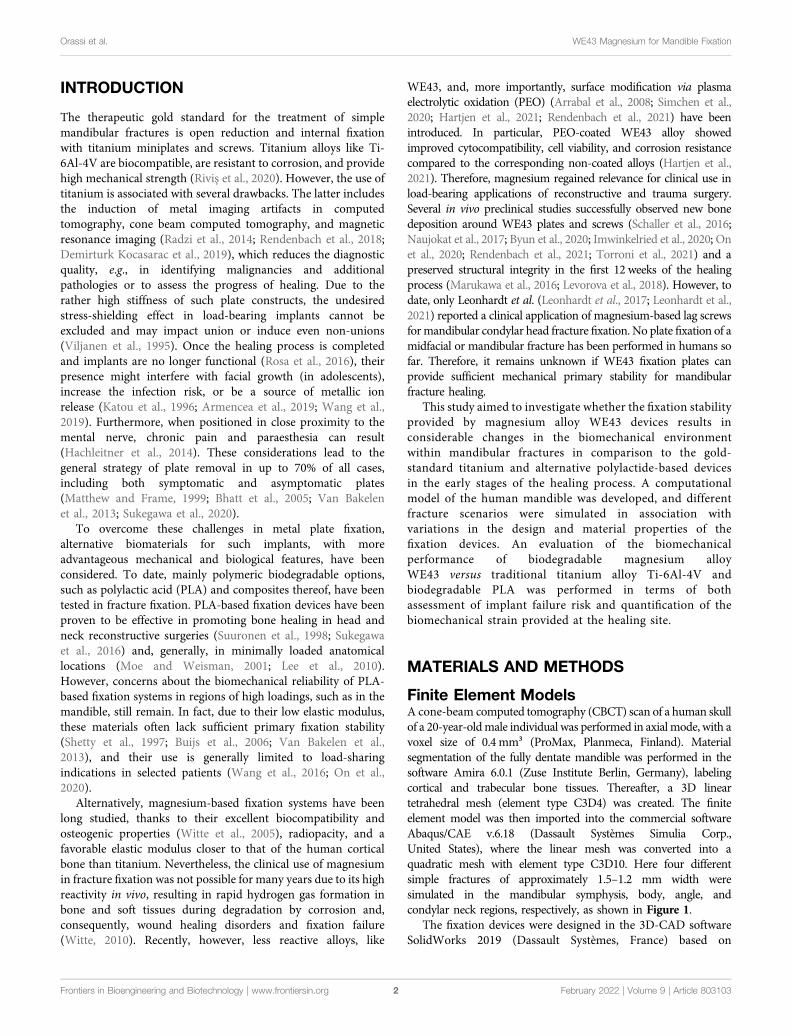
Finite Element ModelsA cone-beam computed tomography (CBCT) scan of a human skullof a 20-year-oldmale individual was performed in axialmode, with avoxel size of 0.4 mm³ (ProMax, Planmeca, Finland). Materialsegmentation of the fully dentate mandible was performed in thesoftware Amira 6.0.1 (Zuse Institute Berlin, Germany), labelingcortical and trabecular bone tissues. Thereafter, a 3D lineartetrahedral mesh (element type C3D4) was created. The finiteelement model was then imported into the commercial softwareAbaqus/CAE v.6.18 (Dassault Systèmes Simulia Corp.,United States), where the linear mesh was converted into aquadratic mesh with element type C3D10. Here four differentsimple fractures of approximately 1.5–1.2 mm width weresimulated in the mandibular symphysis, body, angle, andcondylar neck regions, respectively, as shown in Figure 1.
The fixation devices were designed in the 3D-CAD softwareSolidWorks 2019 (Dassault Systèmes, France) based on
Frontiers in Bioengineering and Biotechnology | www.frontiersin.org February 2022 | Volume 9 | Article 8031032
Orassi et al. WE43 Magnesium for Mandible Fixation
commercially available devices. Two different miniplate thicknesseswere tested: 1-mm-thick clinically used miniplates versus alternative1.5-mm-thick miniplates, in combination with simplified (withoutthread) monocortical 7-mm-long screws. The plates were adapted tothe mandibular bone surfaces and positioned according to the fourfracture scenarios (Figure 1). For the same fracture case, accurateoverlapping of the 1-mm- and 1.5-mm-thick miniplates wasachieved to exclude possible influences of plate positioning on
the results. As shown in Figure 1, according to the principles ofChampy et al. (1978) and the guidelines of AO Foundation ORIF(2021a) and followed by clinical advice, fracture fixation wasperformed with 2.0 miniplate systems. Specifically, two parallel 4-hole miniplates were used in the mandibular symphysis (AOFoundation ORIF, 2021d) and body regions (AO FoundationORIF, 2021c). In the mandibular angle region, a 6-hole miniplateand, in the mandibular condylar neck region, a 2- and a 4-holeminiplate, both with a center space (AO Foundation ORIF, 2021b),were used. The devices were imported into Abaqus andmeshed witha quadratic mesh (element type C3D10). Thereafter, tie constraintswere applied between plates and screws and between screws andmandibular bone tissues.
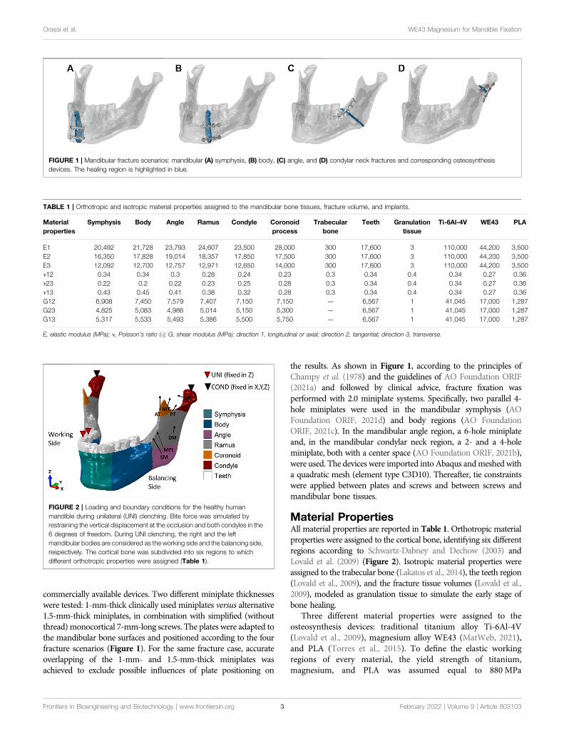
Material PropertiesAll material properties are reported in Table 1. Orthotropic materialproperties were assigned to the cortical bone, identifying six differentregions according to Schwartz-Dabney and Dechow (2003) andLovald et al. (2009) (Figure 2). Isotropic material properties wereassigned to the trabecular bone (Lakatos et al., 2014), the teeth region(Lovald et al., 2009), and the fracture tissue volumes (Lovald et al.,2009), modeled as granulation tissue to simulate the early stage ofbone healing.
Three different material properties were assigned to theosteosynthesis devices: traditional titanium alloy Ti-6Al-4V(Lovald et al., 2009), magnesium alloy WE43 (MatWeb, 2021),and PLA (Torres et al., 2015). To define the elastic workingregions of every material, the yield strength of titanium,magnesium, and PLA was assumed equal to 880 MPa
FIGURE 1 | Mandibular fracture scenarios: mandibular (A) symphysis, (B) body, (C) angle, and (D) condylar neck fractures and corresponding osteosynthesisdevices. The healing region is highlighted in blue.
TABLE 1 | Orthotropic and isotropic material properties assigned to the mandibular bone tissues, fracture volume, and implants.
Materialproperties
Symphysis Body Angle Ramus Condyle Coronoidprocess
Trabecularbone
Teeth Granulationtissue
Ti-6Al-4V WE43 PLA
E1 20,492 21,728 23,793 24,607 23,500 28,000 300 17,600 3 110,000 44,200 3,500E2 16,350 17,828 19,014 18,357 17,850 17,500 300 17,600 3 110,000 44,200 3,500E3 12,092 12,700 12,757 12,971 12,650 14,000 300 17,600 3 110,000 44,200 3,500]12 0.34 0.34 0.3 0.28 0.24 0.23 0.3 0.34 0.4 0.34 0.27 0.36]23 0.22 0.2 0.22 0.23 0.25 0.28 0.3 0.34 0.4 0.34 0.27 0.36]13 0.43 0.45 0.41 0.38 0.32 0.28 0.3 0.34 0.4 0.34 0.27 0.36G12 6,908 7,450 7,579 7,407 7,150 7,150 — 6,567 1 41,045 17,000 1,287G23 4,825 5,083 4,986 5,014 5,150 5,300 — 6,567 1 41,045 17,000 1,287G13 5,317 5,533 5,493 5,386 5,500 5,750 — 6,567 1 41,045 17,000 1,287
E, elastic modulus (MPa); ν, Poisson’s ratio (-); G, shear modulus (MPa); direction 1, longitudinal or axial; direction 2, tangential; direction 3, transverse.
FIGURE 2 | Loading and boundary conditions for the healthy humanmandible during unilateral (UNI) clenching. Bite force was simulated byrestraining the vertical displacement at the occlusion and both condyles in the6 degrees of freedom. During UNI clenching, the right and the leftmandibular bodies are considered as the working side and the balancing side,respectively. The cortical bone was subdivided into six regions to whichdifferent orthotropic properties were assigned (Table 1).
Frontiers in Bioengineering and Biotechnology | www.frontiersin.org February 2022 | Volume 9 | Article 8031033
Orassi et al. WE43 Magnesium for Mandible Fixation
(MatWeb, 2021), 162 MPa (MatWeb, 2021), and 70 MPa (Torreset al., 2015), respectively.
Loading and Boundary ConditionsSeveral studies found that contralateral molar occlusion inducedthe highest amount of mechanical solicitation and therefore couldrepresent a critical clenching task (Korioth et al., 1992; Lovaldet al., 2009; Hijazi et al., 2016). Therefore, unilateral clenching waschosen as the only and worst clenching task to be tested in thisstudy. Specifically, right unilateral clenching was simulated byrestraining the vertical displacement of the first molar—second
premolar teeth group (Figure 2). At the power stroke ofmastication, the condyles were assumed locked in the glenoidfossa and therefore restrained in all 6 degrees of freedom.
Post-operative loading conditions were applied by simulating thecontraction of superficial masseter (SM), deep masseter (DM),anterior temporalis (AT), medial temporalis (MT), posteriortemporalis (PT), medial pterygoid (MPt), and inferior lateralpterygoid (LPt) muscles (Figure 2). The magnitude of themuscular forces was reduced to 20% of the maximum muscleforces as reported by Nelson (1986) to adapt it to post-operativemuscle forces (Table 2). The muscle fiber activation patterns for
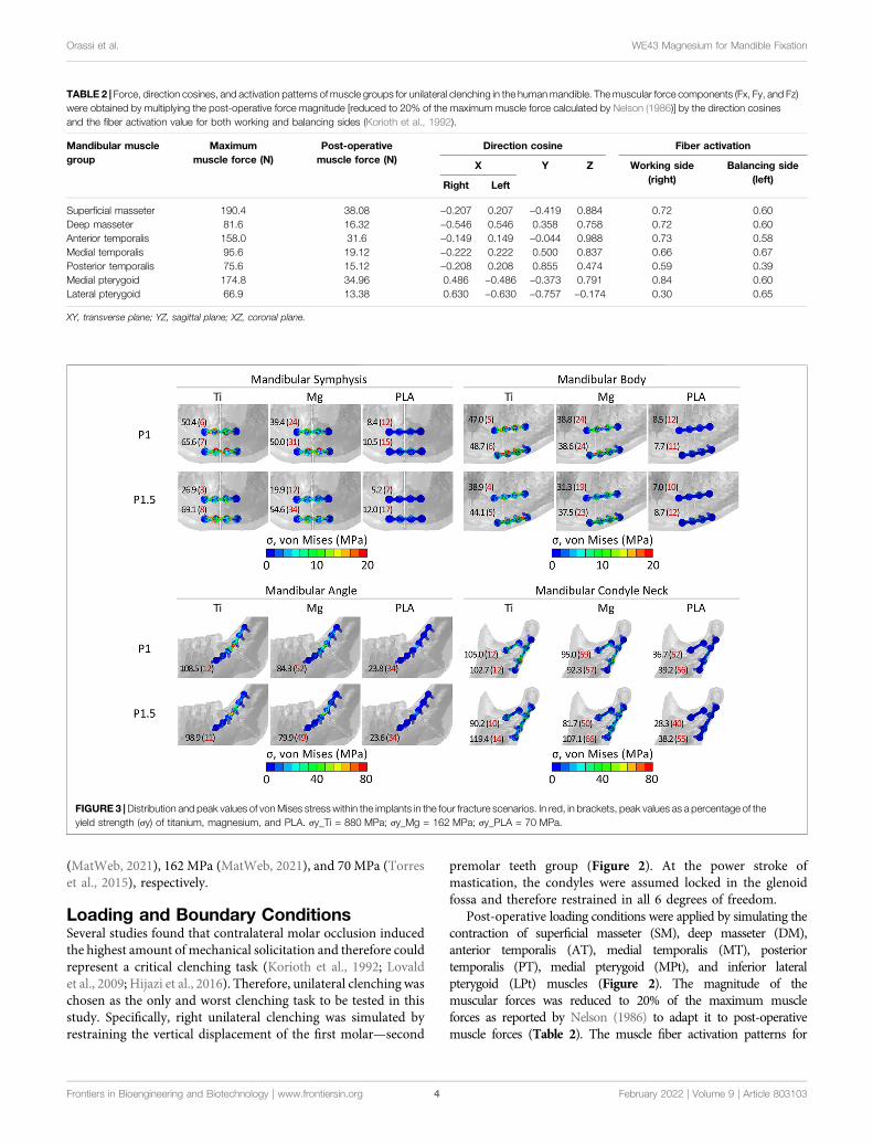
TABLE 2 | Force, direction cosines, and activation patterns of muscle groups for unilateral clenching in the humanmandible. Themuscular force components (Fx, Fy, and Fz)were obtained by multiplying the post-operative force magnitude [reduced to 20% of the maximum muscle force calculated by Nelson (1986)] by the direction cosinesand the fiber activation value for both working and balancing sides (Korioth et al., 1992).
Mandibular musclegroup
Maximummuscle force (N)
Post-operativemuscle force (N)
Direction cosine Fiber activation
X Y Z Working side(right)
Balancing side(left)Right Left
Superficial masseter 190.4 38.08 −0.207 0.207 −0.419 0.884 0.72 0.60Deep masseter 81.6 16.32 −0.546 0.546 0.358 0.758 0.72 0.60Anterior temporalis 158.0 31.6 −0.149 0.149 −0.044 0.988 0.73 0.58Medial temporalis 95.6 19.12 −0.222 0.222 0.500 0.837 0.66 0.67Posterior temporalis 75.6 15.12 −0.208 0.208 0.855 0.474 0.59 0.39Medial pterygoid 174.8 34.96 0.486 −0.486 −0.373 0.791 0.84 0.60Lateral pterygoid 66.9 13.38 0.630 −0.630 −0.757 −0.174 0.30 0.65
XY, transverse plane; YZ, sagittal plane; XZ, coronal plane.
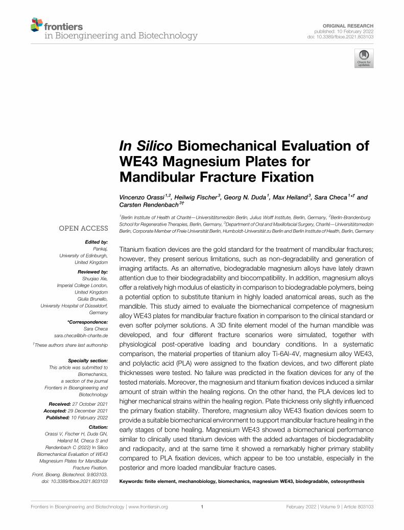
FIGURE 3 |Distribution and peak values of vonMises stress within the implants in the four fracture scenarios. In red, in brackets, peak values as a percentage of theyield strength (σy) of titanium, magnesium, and PLA. σy_Ti = 880 MPa; σy_Mg = 162 MPa; σy_PLA = 70 MPa.
Frontiers in Bioengineering and Biotechnology | www.frontiersin.org February 2022 | Volume 9 | Article 8031034
Orassi et al. WE43 Magnesium for Mandible Fixation
unilateral clenching used in this study have been described by Nelson(1986), while the directions of the force vectors are based on Koriothet al. (1992).
Mesh Convergence StudyFour different mesh sizes were used to perform a convergence study.Mesh (A) 1,099,247 elements (finest mesh), mesh (B) 645,150, mesh(C) 288,967, and mesh (D) 168,784 (coarsest mesh) were tested. Forthis analysis, isotropic material properties were assigned to the bonetissues. Specifically, the elastic moduli of cortical and trabecular bonewere chosen equal to 15,000 and 300MPa, respectively, and aPoisson’s ratio of 0.3 was defined (Orassi et al., 2021). The meshcreation workflow developed in this study only generates orphanmeshes (without geometry). Therefore, to avoid variability betweenthe four meshes in the definition of the muscular attachment areas,simplified and standardized loads were applied. Specifically, forceswith components Fx = 0, Fy = −50N, and Fz = 50N were applied atthe masseter attachment on a surface of 10-mm radius duringunilateral clenching. Average von Mises stresses and principalstrains were calculated in the symphysis region and comparedbetween the four cases. Mesh B, with element edges ca. 1 mm inlength, showed the best compromise between accuracy andcomputational costs, with a relative error inferior to 5%, and was,therefore, used in this study. Eventually, the same material propertiesused in this study were applied to mesh B, and the results werecompared to the ones obtained from the isotropic model.Comparable outcomes were observed.
AnalysisThe mechanical environment within the healing region is known toplay a key role on the healing outcome. Claes and Heigele (1999)quantified strain levels lower than 15% in regions where boneformation takes place during the healing process. Therefore, themaximum and minimum principal strains within the four healingregions were calculated in all fracture scenarios for all combinationsof plate thickness and material type. Specifically, the healing regionswere defined as thin gaps across the bone (thickness ofapproximately 1.2 mm) at the mandibular symphysis, body,angle, and condyle neck (Figure 1). Strains were computed atelement integration nodes to avoid possible discontinuities of thestrain field at the element edges. Moreover, at each integration node,only the largest, in absolute value, between the maximum andminimum principal strains was plotted to determine, respectively,the prevalent tensile or compressive solicitation of each specific node.
Similarly, von Mises stresses within plates and screws werecalculated at the integration nodes. To avoid stress singularitiesdue to constraints between plate and screws and between screwsand bone, the top 0.1% highest von Mises stress values wereexcluded, and the peak von Mises stresses were calculated byaveraging the values in the 10 nodes with the highest stress.
RESULTS
In the healthy mandible, a bite force of 100 N was obtained bycalculating the reaction force at the occlusion during unilateralclenching.
von Mises Stresses Within the FixationDevicesConcerning both plate thicknesses, the peak von Mises stressesnever exceeded the yield strength of the materials. Titaniumdevices showed the best mechanical performance since thepeak von Mises stresses ranged between 3 and 14% oftitanium yield strength (880 MPa). Higher peak stress valueswere found in PLA and magnesium, with the latter as theclosest to its yield strength but with both still working in theelastic region. Specifically, in magnesium, the peak von Misesstresses were 12–66% of magnesium yield strength (162 MPa),while in PLA, the peak von Mises stresses were 7–56% of PLAyield strength (70 MPa). Concerning the anatomical location, thehighest mechanical solicitation of the fixation devices waspredicted in mandibular angle and condyle neck fracturefixation (Figure 3).
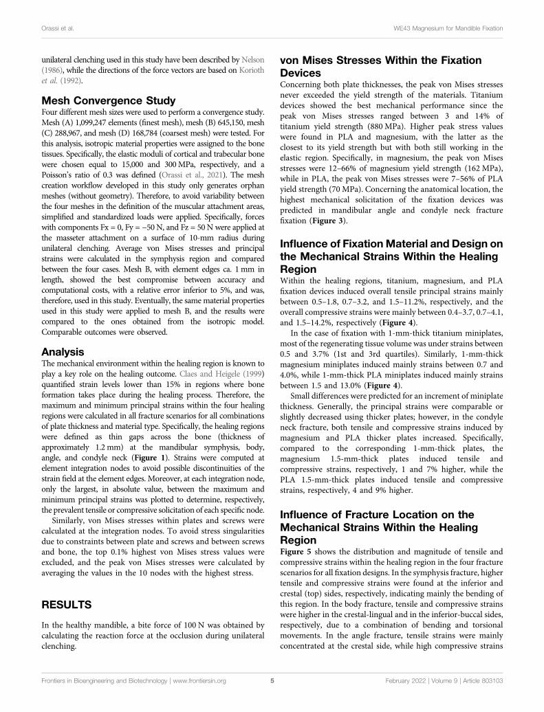
Influence of Fixation Material and Design onthe Mechanical Strains Within the HealingRegionWithin the healing regions, titanium, magnesium, and PLAfixation devices induced overall tensile principal strains mainlybetween 0.5–1.8, 0.7–3.2, and 1.5–11.2%, respectively, and theoverall compressive strains were mainly between 0.4–3.7, 0.7–4.1,and 1.5–14.2%, respectively (Figure 4).
In the case of fixation with 1-mm-thick titanium miniplates,most of the regenerating tissue volume was under strains between0.5 and 3.7% (1st and 3rd quartiles). Similarly, 1-mm-thickmagnesium miniplates induced mainly strains between 0.7 and4.0%, while 1-mm-thick PLA miniplates induced mainly strainsbetween 1.5 and 13.0% (Figure 4).
Small differences were predicted for an increment of miniplatethickness. Generally, the principal strains were comparable orslightly decreased using thicker plates; however, in the condyleneck fracture, both tensile and compressive strains induced bymagnesium and PLA thicker plates increased. Specifically,compared to the corresponding 1-mm-thick plates, themagnesium 1.5-mm-thick plates induced tensile andcompressive strains, respectively, 1 and 7% higher, while thePLA 1.5-mm-thick plates induced tensile and compressivestrains, respectively, 4 and 9% higher.
Influence of Fracture Location on theMechanical Strains Within the HealingRegionFigure 5 shows the distribution and magnitude of tensile andcompressive strains within the healing region in the four fracturescenarios for all fixation designs. In the symphysis fracture, highertensile and compressive strains were found at the inferior andcrestal (top) sides, respectively, indicating mainly the bending ofthis region. In the body fracture, tensile and compressive strainswere higher in the crestal-lingual and in the inferior-buccal sides,respectively, due to a combination of bending and torsionalmovements. In the angle fracture, tensile strains were mainlyconcentrated at the crestal side, while high compressive strains
Frontiers in Bioengineering and Biotechnology | www.frontiersin.org February 2022 | Volume 9 | Article 8031035
Orassi et al. WE43 Magnesium for Mandible Fixation
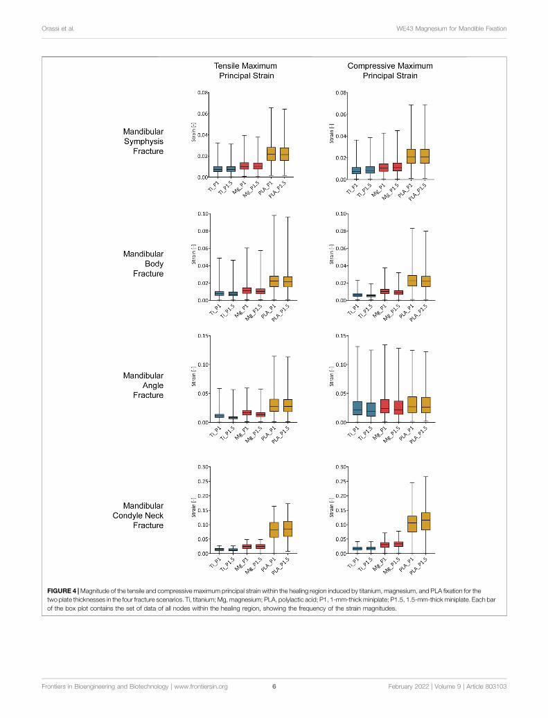
FIGURE 4 |Magnitude of the tensile and compressive maximum principal strain within the healing region induced by titanium, magnesium, and PLA fixation for thetwo plate thicknesses in the four fracture scenarios. Ti, titanium; Mg, magnesium; PLA, polylactic acid; P1, 1-mm-thick miniplate; P1.5, 1.5-mm-thick miniplate. Each barof the box plot contains the set of data of all nodes within the healing region, showing the frequency of the strain magnitudes.
Frontiers in Bioengineering and Biotechnology | www.frontiersin.org February 2022 | Volume 9 | Article 8031036
Orassi et al. WE43 Magnesium for Mandible Fixation
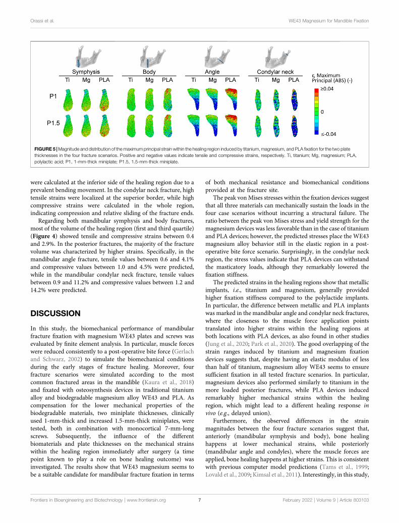
were calculated at the inferior side of the healing region due to aprevalent bending movement. In the condylar neck fracture, hightensile strains were localized at the superior border, while highcompressive strains were calculated in the whole region,indicating compression and relative sliding of the fracture ends.
Regarding both mandibular symphysis and body fractures,most of the volume of the healing region (first and third quartile)(Figure 4) showed tensile and compressive strains between 0.4and 2.9%. In the posterior fractures, the majority of the fracturevolume was characterized by higher strains. Specifically, in themandibular angle fracture, tensile values between 0.6 and 4.1%and compressive values between 1.0 and 4.5% were predicted,while in the mandibular condylar neck fracture, tensile valuesbetween 0.9 and 11.2% and compressive values between 1.2 and14.2% were predicted.
DISCUSSION
In this study, the biomechanical performance of mandibularfracture fixation with magnesium WE43 plates and screws wasevaluated by finite element analysis. In particular, muscle forceswere reduced consistently to a post-operative bite force (Gerlachand Schwarz, 2002) to simulate the biomechanical conditionsduring the early stages of fracture healing. Moreover, fourfracture scenarios were simulated according to the mostcommon fractured areas in the mandible (Kaura et al., 2018)and fixated with osteosynthesis devices in traditional titaniumalloy and biodegradable magnesium alloy WE43 and PLA. Ascompensation for the lower mechanical properties of thebiodegradable materials, two miniplate thicknesses, clinicallyused 1-mm-thick and increased 1.5-mm-thick miniplates, weretested, both in combination with monocortical 7-mm-longscrews. Subsequently, the influence of the differentbiomaterials and plate thicknesses on the mechanical strainswithin the healing region immediately after surgery (a timepoint known to play a role on bone healing outcome) wasinvestigated. The results show that WE43 magnesium seems tobe a suitable candidate for mandibular fracture fixation in terms
of both mechanical resistance and biomechanical conditionsprovided at the fracture site.
The peak vonMises stresses within the fixation devices suggestthat all three materials can mechanically sustain the loads in thefour case scenarios without incurring a structural failure. Theratio between the peak von Mises stress and yield strength for themagnesium devices was less favorable than in the case of titaniumand PLA devices; however, the predicted stresses place the WE43magnesium alloy behavior still in the elastic region in a post-operative bite force scenario. Surprisingly, in the condylar neckregion, the stress values indicate that PLA devices can withstandthe masticatory loads, although they remarkably lowered thefixation stiffness.
The predicted strains in the healing regions show that metallicimplants, i.e., titanium and magnesium, generally providedhigher fixation stiffness compared to the polylactide implants.In particular, the difference between metallic and PLA implantswas marked in the mandibular angle and condylar neck fractures,where the closeness to the muscle force application pointstranslated into higher strains within the healing regions atboth locations with PLA devices, as also found in other studies(Jung et al., 2020; Park et al., 2020). The good overlapping of thestrain ranges induced by titanium and magnesium fixationdevices suggests that, despite having an elastic modulus of lessthan half of titanium, magnesium alloy WE43 seems to ensuresufficient fixation in all tested fracture scenarios. In particular,magnesium devices also performed similarly to titanium in themore loaded posterior fractures, while PLA devices inducedremarkably higher mechanical strains within the healingregion, which might lead to a different healing response invivo (e.g., delayed union).
Furthermore, the observed differences in the strainmagnitudes between the four fracture scenarios suggest that,anteriorly (mandibular symphysis and body), bone healinghappens at lower mechanical strains, while posteriorly(mandibular angle and condyles), where the muscle forces areapplied, bone healing happens at higher strains. This is consistentwith previous computer model predictions (Tams et al., 1999;Lovald et al., 2009; Kimsal et al., 2011). Interestingly, in this study,
FIGURE 5 |Magnitude and distribution of the maximum principal strain within the healing region induced by titanium, magnesium, and PLA fixation for the two platethicknesses in the four fracture scenarios. Positive and negative values indicate tensile and compressive strains, respectively. Ti, titanium; Mg, magnesium; PLA,polylactic acid; P1, 1-mm-thick miniplate; P1.5, 1.5-mm-thick miniplate.
Frontiers in Bioengineering and Biotechnology | www.frontiersin.org February 2022 | Volume 9 | Article 8031037
Orassi et al. WE43 Magnesium for Mandible Fixation
the calculated overall mechanical strains at the fracture sitesinduced by clinically used titanium devices (-4 ÷ 2%) are inthe same range of strain values beneficial for bone healing in longbones (|ε|<15%) (Claes and Heigele, 1999). This suggests a similarmechano-regulation of bone healing between mandibular boneand long bones; however, this needs to be further investigated.
While changes in the implant material properties proved tohighly influence fixation stiffness, according to this study, a 0.5-mm increase of plate thickness did not substantially influenceeither the mechanical solicitation of the devices or thebiomechanical environment within the healing region.Specifically, at the mandibular body and angle fractures, arelatively small reduction of the strain level was predicted byincreasing the thickness. Interestingly, at the condyle neckfracture, increased plate thickness induced slightly increasedstrains, using magnesium and PLA devices. The reason forthis must be found in the combination of the plate thicknessand material properties of the devices at this specific location. Infact, increasing the plate thickness led to a reduction of the peakvon Mises stress in the 2-hole miniplate and an increase of thestresses in the 4-hole miniplate. This, in combination with thereduced stiffness of the biodegradable devices and the hightorsional forces at the condylar region, determined a differentand slightly higher mechanical solicitation of the healing region.To assess a possible influence of screw penetration within thebone tissues, an increased screw length (7.5 mm in length), incombination with the 1.5-mm-thick miniplates in mandibularsymphysis, body, and angle fractures, was also tested, but nosubstantial differences in the strain values at the fracture site wereinduced by the longer screws (supplementary data).
The finite element model developed in this study is based on aprevious finite element study of the healthy mandible (Orassiet al., 2021), which, in a maximum bite force condition, showedstrain magnitudes and distribution that are in good agreementwith previous computer studies (Korioth et al., 1992; Baek et al.,2012). Specifically, Korioth et al. (1992) found tensile andcompressive strains of ca. 250 and −400 με in the condylarneck, similar to ours, and of ca. 600 and −500 με in the bodyregion, slightly higher than the ones predicted in this study(400 and −300 με). However, as shown by Baek et al. (2012), ahigh variability (>100%) of mandibular deformation can beexpected between mandibles with different morphologicalfeatures. In addition, in this study, in the mandibular bodyfracture, fixated with two parallel, 1-mm-thick titaniumminiplates and a bite force of 100 N (20% of the maximumbite force), average maximum strain values of ca. 0.6–0.8% and apeak von Mises stress of 47–49 MPa were predicted. This is inagreement with the results found by Lovald et al. (2009), whosimulated a mandibular body fracture fixated with two paralleltitanium miniplates with a bite force of 320 N (60% of themaximum bite force). They observed average first principalstrain values within the healing regions of ca. 1.7% and a peakvon Mises stress of 87 MPa within the plates. Therefore, ourresults are consistent with the analysis performed by Lovald et al.(2009), considering that, in this study, a third of their bite forcewas used and small differences might exist in terms of themandibular morphology and plate design and positioning.
This study presents several limitations—for example,fixation device material properties were defined as isotropic,homogeneous, and elastic. PLA viscoelastic behavior was nottaken into account; however, cycling loading duringmastication might contribute to increasing the deformationof the PLA fixation devices cycle after cycle (Pepelnjak et al.,2020), thus further reducing the primary stability at the fracturesite. This implies that, in reality, PLA might provide lessstability than the one predicted in our study. Anotherlimitation of this study is the use of tie constraints forscrew–bone interface modeling, which does not allow therelative motion between screws and bone that might occurin reality. However, this limitation is unlikely to influence ourfindings since, as also shown in another study (MacLeod et al.,2012), only the local mechanical environment in thesurroundings of the screws is affected, while the globalload–displacement behavior of the model remains unvaried.Moreover, the analysis did not take into account theWE43 alloyand PLA degradation process. PEO-coated magnesium WE43has been shown to maintain up to ca. 80% of its volume at6 months after implantation (Schaller et al., 2016), and PLA-based devices can preserve up to 70% of their initial mechanicalstrength after 36 weeks (Wang et al., 2016). Complete bonehealing in mandibular fractures can take up to 6 months;however, functional loading is progressively restored startingfrom the fourth week after surgery (Friedman, 2014).Therefore, it is expected that material degradation does nothighly interfere with the healing process, and it should notinfluence the conclusions drawn in this study due to slowdegradation rates for both materials (Yerit et al., 2005;Rendenbach et al., 2021); however, this aspect remains to befurther investigated.
In summary, this study aimed to investigate the biomechanicalperformance of magnesium WE43 alloy in mandibular fracturefixation in contrast with both clinically used titanium and softerbiodegradable PLA devices. From a mechanical point of view,magnesium WE43 alloy was predicted to provide sufficientprimary stiffness at the fracture site to support mandibularbone healing. The effect of plate and screw resorption on thehealing process should be addressed in future studies.
DATA AVAILABILITY STATEMENT
The datasets presented in this article are not readily availablebecause they present some patented elements. Requests toaccess the datasets should be directed to SC, [email protected].
AUTHOR CONTRIBUTIONS
VO, CR, GD, MH, and SC designed the study. HF performed theCBCT and the anatomical observations. VO developed thecomputational models and collected the data. VO and SCinterpreted the data and drafted the manuscript. All authorsread and revised the manuscript and approved its content.
Frontiers in Bioengineering and Biotechnology | www.frontiersin.org February 2022 | Volume 9 | Article 8031038
Orassi et al. WE43 Magnesium for Mandible Fixation
ACKNOWLEDGMENTS
The authors acknowledge the support of European Union (EU)Horizon 2020 for the project InterLynk (grant agreement: H2020-NMBP-TR-IND-2020, project ID: 953169).
SUPPLEMENTARY MATERIAL
The SupplementaryMaterial for this article can be found online at:https://www.frontiersin.org/articles/10.3389/fbioe.2021.803103/full#supplementary-material
REFERENCES
AO Foundation ORIF (2021a). AO Foundation Mandible. Available at: https://surgeryreference.aofoundation.org/cmf/trauma/mandible (Accessed December15, 2021a).
AO Foundation ORIF (2021b). Two Load Sharing Plates for Symphysis andParasymphysis - Simple. Available at: https://surgeryreference.aofoundation.org/cmf/trauma/mandible/symphysis-and-parasymphysis-simple/orif-two-load-sharing-plates (Accessed December 15, 2021b).
AO Foundation ORIF (2021c). Two Load Sharing Plates for Body - Simple.Available at: https://surgeryreference.aofoundation.org/cmf/trauma/mandible/body-simple/orif-two-load-sharing-plates#examples-of-completed-osteosynthesis (Accessed December 15, 2021c).
AO Foundation ORIF (2021d). Plate Fixation with Two Plates for CondylarProcess and Head - Simple and Complex. Available at: https://surgeryreference.aofoundation.org/cmf/trauma/mandible/condylar-process-and-head-simple-and-complex/orif-plate-fixation-two-plates (AccessedDecember 15, 2021d).
Armencea, G., Gheban, D., Onisor, F., Mitre, I., Manea, A., Trombitas, V., et al.(2019). Histological Change in Soft Tissue Surrounding Titanium Plates afterJaw Surgery. Materials 12, 3205–3208. doi:10.3390/ma12193205
Arrabal, R., Matykina, E., Skeldon, P., Thompson, G. E., and Pardo, A. (2008).Transport of Species during Plasma Electrolytic Oxidation of WE43-T6Magnesium Alloy. J. Electrochem. Soc. 155, C101. doi:10.1149/1.2823374
Baek, S.-H., Cha, H.-S., Cha, J.-Y., Moon, Y.-S., and Sung, S.-J. (2012). Three-dimensional Finite Element Analysis of the Deformation of the HumanMandible: a Preliminary Study from the Perspective of Orthodontic Mini-Implant Stability. Korean J. Orthod. 42, 159. doi:10.4041/kjod.2012.42.4.159
Bhatt, V., Chhabra, P., and Dover, M. S. (2005). Removal of Miniplates inMaxillofacial Surgery: A Follow-Up Study. J. Oral Maxill. Surg. 63, 756–760.doi:10.1016/j.joms.2005.02.005
Buijs, G. J., Stegenga, B., and Bos, R. R. M. (2006). Efficacy and Safety ofBiodegradable Osteofixation Devices in Oral and Maxillofacial Surgery: ASystematic Review. J. Dent. Res. 85, 980–989. doi:10.1177/154405910608501102
Byun, S. H., Lim, H. K., Cheon, K. H., Lee, S. M., Kim, H. E., and Lee, J. H. (2020).Biodegradable Magnesium alloy (WE43) in Bone-fixation Plate and Screw.J. Biomed. Mater. Res. 108, 2505–2512. doi:10.1002/jbm.b.34582
Champy, M., Loddé, J. P., Schmitt, R., Jaeger, J. H., and Muster, D. (1978).Mandibular Osteosynthesis by Miniature Screwed Plates via a BuccalApproach. J. Maxill. Surg. 6, 14–21. doi:10.1016/S0301-0503(78)80062-9
Claes, L. E., and Heigele, C. A. (1999). Magnitudes of Local Stress and Strain alongBony Surfaces Predict the Course and Type of Fracture Healing. J. Biomech. 32,255–266. doi:10.1016/S0021-9290(98)00153-5
Demirturk Kocasarac, H., Ustaoglu, G., Bayrak, S., Katkar, R., Geha, H., Deahl, S.T., et al. (2019). Evaluation of Artifacts Generated by Titanium, Zirconium, andTitanium-Zirconium alloy Dental Implants on MRI, CT, and CBCT Images: APhantom Study. Oral Surg. Oral Med. Oral Pathol. Oral Radiol. 127, 535–544.doi:10.1016/j.oooo.2019.01.074
Friedman, C. D. (2009). “66 Basic Principles of Craniofacial Bone Healing andRepair,” in Facial Plastic and Reconstructive Surgery. I. D. Papel, J. L. Frodel,G. R. Holt, W. F. Larrabee, N. E. Nachlas, S. S. Park, et al. (Stuttgart: GeorgThieme Verlag). doi:10.1055/b-0034-73284
Gerlach, K. L., and Schwarz, A. (2002). Bite Forces in Patients after Treatment ofMandibular Angle Fractures with Miniplate Osteosynthesis According toChampy. Int. J. Oral Maxill. Surg. 31, 345–348. doi:10.1054/IJOM.2002.0290
Hachleitner, J., Enzinger, S., Brandtner, C., and Gaggl, A. (2014). The Role of theTitanium Functionally Dynamic Bridging Plate for the Treatment of theAtrophic Mandible Fractures. J. Craniomaxillofac. Surg. 42, 438–442. doi:10.1016/j.jcms.2013.05.037
Hartjen, P., Wegner, N., Ahmadi, P., Matthies, L., Nada, O., Fuest, S., et al. (2021).Toward Tailoring the Degradation Rate of Magnesium-Based Biomaterials forVarious Medical Applications: Assessing Corrosion, Cytocompatibility andImmunological Effects. Ijms 22, 971. doi:10.3390/ijms22020971
Hijazi, L., Hejazi, W., Darwich, M. A., and Darwich, K. (2016). Finite ElementAnalysis of Stress Distribution on the Mandible and Condylar FractureOsteosynthesis during Various Clenching Tasks. Oral Maxillofac. Surg. 20,359–367. doi:10.1007/s10006-016-0573-2
Imwinkelried, T., Beck, S., and Schaller, B. (2020). Pre-clinical Testing of HumanSize Magnesium Implants in Miniature Pigs: Implant Degradation and BoneFracture Healing at Multiple Implantation Sites.Mater. Sci. Eng. C 108, 110389.doi:10.1016/j.msec.2019.110389
Jung, B. T., Kim, W. H., Park, B., Lee, J.-H., Kim, B., and Lee, J.-H. (2020).Biomechanical Evaluation of Unilateral Subcondylar Fracture of the Mandibleon the Varying Materials: A Finite Element Analysis. PLoS One 15, e0240352.doi:10.1371/journal.pone.0240352
Katou, F., Andoh, N., Motegi, K., and Nagura, H. (1996). Immuno-inflammatoryResponses in the Tissue Adjacent to Titanium Miniplates Used in theTreatment of Mandibular Fractures. J. Craniomaxillofac. Surg. 24, 155–162.doi:10.1016/S1010-5182(96)80049-7
Kaur, P., Kaura, S., Bahl, R., Bansal, S., and Sangha, P. (2018). Retrospective Studyof Facial Fractures. Ann. Maxillofac. Surg. 8, 78–82. (Mumbai, India: MedknowPublications and Media Pvt. Ltd.). doi:10.4103/ams.ams_73_17
Kimsal, J., Baack, B., Candelaria, L., Khraishi, T., and Lovald, S. (2011).Biomechanical Analysis of Mandibular Angle Fractures. J. Oral Maxill. Surg.69, 3010–3014. doi:10.1016/j.joms.2010.12.042
Korioth, T. W. P., Romilly, D. P., and Hannam, A. G. (1992). Three-dimensionalFinite Element Stress Analysis of the Dentate Human Mandible. Am. J. Phys.Anthropol. 88, 69–96. doi:10.1002/ajpa.1330880107
Lakatos, É., Magyar, L., and Bojtár, I. (2014). Material Properties of theMandibularTrabecular Bone. J. Med. Eng. 2014, 1–7. doi:10.1155/2014/470539
Lee, H.-B., Oh, J.-S., Kim, S.-G., Kim, H.-K., Moon, S.-Y., Kim, Y.-K., et al. (2010).Comparison of Titanium and Biodegradable Miniplates for Fixation ofMandibular Fractures. J. Oral Maxill. Surg. 68, 2065–2069. doi:10.1016/j.joms.2009.08.004
Leonhardt, H., Franke, A., McLeod, N. M. H., Lauer, G., and Nowak, A. (2017).Fixation of Fractures of the Condylar Head of the Mandible with a NewMagnesium-alloy Biodegradable Cannulated Headless Bone Screw. Br. J. OralMaxill. Surg. 55, 623–625. doi:10.1016/j.bjoms.2017.04.007
Leonhardt, H., Ziegler, A., Lauer, G., and Franke, A. (2021). Osteosynthesis ofthe Mandibular Condyle with Magnesium-Based Biodegradable HeadlessCompression Screws Show Good Clinical Results during a 1-Year Follow-Up Period. J. Oral Maxill. Surg. 79, 637–643. doi:10.1016/j.joms.2020.02.025
Levorova, J., Duskova, J., Drahos, M., Vrbova, R., Vojtech, D., Kubasek, J., et al.(2018). In Vivo study on Biodegradable Magnesium Alloys: Bone HealingAround WE43 Screws. J. Biomater. Appl. 32, 886–895. doi:10.1177/0885328217743321
Lovald, S. T., Wagner, J. D., and Baack, B. (2009). Biomechanical Optimization ofBone Plates Used in Rigid Fixation of Mandibular Fractures. J. Oral Maxill.Surg. 67, 973–985. doi:10.1016/j.joms.2008.12.032
MacLeod, A. R., Pankaj, P., and Simpson, A. H. R. W. (2012). Does Screw-BoneInterface Modelling Matter in Finite Element Analyses? J. Biomech. 45,1712–1716. doi:10.1016/j.jbiomech.2012.04.008
Marukawa, E., Tamai, M., Takahashi, Y., Hatakeyama, I., Sato, M., Higuchi, Y.,et al. (2016). Comparison of Magnesium Alloys and Poly-L-Lactide Screws asDegradable Implants in a Canine Fracture Model. J. Biomed. Mater. Res. 104,1282–1289. doi:10.1002/jbm.b.33470
Matthew, I. R., and Frame, J. W. (1999). Policy of Consultant Oral andMaxillofacial Surgeons towards Removal of Miniplate Components after Jaw
Frontiers in Bioengineering and Biotechnology | www.frontiersin.org February 2022 | Volume 9 | Article 8031039
Orassi et al. WE43 Magnesium for Mandible Fixation
Fracture Fixation: Pilot Study. Br. J. OralMaxill. Surg. 37, 110–112. doi:10.1054/bjom.1997.0084
MatWeb L. L. C. (2021). Material Property Data. Available at: www.matweb.com(Accessed September 28, 2021).
Moe, K. S., and Weisman, R. A. (2001). Resorbable Fixation in Facial Plastic andHead and Neck Reconstructive Surgery: An Initial Report on Polylactic AcidImplants. The Laryngoscope 111, 1697–1701. doi:10.1097/00005537-200110000-00005
Naujokat, H., Seitz, J.-M., Açil, Y., Damm, T., Möller, I., Gülses, A., et al. (2017).Osteosynthesis of a Cranio-Osteoplasty with a Biodegradable Magnesium PlateSystem in Miniature Pigs. Acta Biomater. 62, 434–445. doi:10.1016/j.actbio.2017.08.031
Nelson, G. J. (1986). Three Dimensional Computer Modeling of HumanMandibular Biomechanics. MSc Dissertation. Vancouver, Canada:University of British Columbia. doi:10.14288/1.0096998
On, S.-W., Cho, S.-W., Byun, S.-H., and Yang, B.-E. (2020). BioabsorbableOsteofixation Materials for Maxillofacial Bone Surgery: A Review onPolymers and Magnesium-Based Materials. Biomedicines 8, 300. doi:10.3390/biomedicines8090300
Orassi, V., Duda, G. N., Heiland, M., Fischer, H., Rendenbach, C., and Checa, S.(2021). Biomechanical Assessment of the Validity of Sheep as a PreclinicalModel for Testing Mandibular Fracture Fixation Devices. Front. Bioeng.Biotechnol. 9. doi:10.3389/fbioe.2021.672176
Park, B., Jung, B. T., Kim, W. H., Lee, J.-H., Kim, B., and Lee, J.-H. (2020). TheStability of Hydroxyapatite/poly-L-Lactide Fixation for Unilateral AngleFracture of the Mandible Assessed Using a Finite Element Analysis Model.Materials 13, 228. doi:10.3390/ma13010228
Pepelnjak, T., Karimi, A., Maček, A., and Mole, N. (2020). Altering the ElasticProperties of 3D Printed Poly-Lactic Acid (Pla) Parts by Compressive CyclicLoading. Materials 13, 4456. doi:10.3390/ma13194456
Radzi, S., Cowin, G., Robinson, M., Pratap, J., Volp, A., Schuetz, M. A., et al. (2014).Metal Artifacts from Titanium and Steel Screws in CT, 1.5T and 3T MR Imagesof the Tibial Pilon: a Quantitative Assessment in 3D.Quant. Imaging Med. Surg.4, 163–172. doi:10.3978/j.issn.2223-4292.2014.03.06
Rendenbach, C., Schoellchen, M., Bueschel, J., Gauer, T., Sedlacik, J., Kutzner, D.,et al. (2018). Evaluation and Reduction of Magnetic Resonance ImagingArtefacts Induced by Distinct Plates for Osseous Fixation: An In VitroStudy @ 3 T. Dentomaxillofacial Radiol. 47, 20170361. doi:10.1259/dmfr.20170361
Rendenbach, C., Fischer, H., Kopp, A., Schmidt-Bleek, K., Kreiker, H., Stumpp, S.,et al. (2021). Improved In Vivo Osseointegration and Degradation Behavior ofPEO Surface-Modified WE43 Magnesium Plates and Screws after 6 and12 Months. Mater. Sci. Eng. C 129, 112380. doi:10.1016/j.msec.2021.112380
Riviș, M., Roi, C., Roi, A., Nica, D., Văleanu, A., and Rusu, L.-C. (2020). TheImplications of Titanium Alloys Applied in Maxillofacial Osteosynthesis. Appl.Sci. 10, 3203. doi:10.3390/app10093203
Rosa, J. H., Villanueva, N. L., Sanati-Mehrizy, P., Factor, S. H., and Taub, P. J.(2016). Review of Maxillofacial Hardware Complications and Indications forSalvage. Craniomaxillofacial Trauma & Reconstruction 9, 134–140. doi:10.1055/s-0035-1570074
Schaller, B., Saulacic, N., Imwinkelried, T., Beck, S., Liu, E. W. Y., Gralla, J., et al.(2016). In Vivo degradation of Magnesium Plate/screw Osteosynthesis ImplantSystems: Soft and Hard Tissue Response in a Calvarial Model in Miniature Pigs.J. Craniomaxillofac. Surg. 44, 309–317. doi:10.1016/j.jcms.2015.12.009
Schwartz-Dabney, C. L., and Dechow, P. C. (2003). Variations in Cortical MaterialProperties throughout the Human Dentate Mandible. Am. J. Phys. Anthropol.120, 252–277. doi:10.1002/ajpa.10121
Shetty, V., Caputo, A. A., and Kelso, I. (1997). Torsion-axial Force Characteristicsof SR-PLLA Screws. J. Craniomaxillofac. Surg. 25, 19–23. doi:10.1016/S1010-5182(97)80020-0
Simchen, F., Sieber, M., Kopp, A., and Lampke, T. (2020). Introduction to PlasmaElectrolytic Oxidation-An Overview of the Process and Applications. Coatings10, 628. doi:10.3390/coatings10070628
Sukegawa, S., Kanno, T., Nagano, D., Shibata, A., Sukegawa-Takahashi, Y., andFuruki, Y. (2016). The Clinical Feasibility of Newly Developed Thin Flat-type
Bioresorbable Osteosynthesis Devices for the Internal Fixation of ZygomaticFractures. J. Craniofac. Surg. 27, 2124–2129. doi:10.1097/SCS.0000000000003147
Sukegawa, S., Masui, M., Sukegawa-Takahashi, Y., Nakano, K., Takabatake, K.,Kawai, H., et al. (2020). Maxillofacial Trauma Surgery Patients with TitaniumOsteosynthesis Miniplates. J. Craniofac. Surg. 31, 1338–1342. Publish Ahead ofPrint. doi:10.1097/SCS.0000000000006352
Suuronen, R., Pohjonen, T., Hietanen, J., and Lindqvist, C. (1998). A 5-year InVitro and In Vivo Study of the Biodegradation of Polylactide Plates. J. OralMaxill. Surg. 56, 604–614. doi:10.1016/S0278-2391(98)90461-X
Tams, J., Otten, B., Loon, J.-P. v., and Bos, R. R. M. (1999). A Computer Study ofFracture Mobility and Strain on Biodegradable Plates Used for Fixation ofMandibular Fractures. J. Oral Maxill. Surg. 57, 973–981. doi:10.1016/S0278-2391(99)90020-4
Torres, J., Cotelo, J., Karl, J., and Gordon, A. P. (2015). Mechanical PropertyOptimization of FDM PLA in Shear with Multiple Objectives. JOM 67,1183–1193. doi:10.1007/s11837-015-1367-y
Torroni, A., Witek, L., Fahliogullari, H. P., Bortoli, J. P., Ibrahim, A., Hacquebord,J., et al. (2021). WE43 andWE43-T5 Mg Alloys Screws Tested In-Vitro CellularAdhesion and Differentiation Assay and In-VivoHistomorphologic Analysis inan Ovine Model. J. Biomater. Appl. 35, 901–911. doi:10.1177/0885328220956788
Van Bakelen, N. B., Buijs, G. J., Jansma, J., de Visscher, J. G. A. M., Hoppenreijs, T.J. M., Bergsma, J. E., et al. (2013). Comparison of Biodegradable and TitaniumFixation Systems in Maxillofacial Surgery. J. Dent. Res. 92, 1100–1105. doi:10.1177/0022034513508953
Viljanen, J., Kinnunen, J., Bondestam, S., Majola, A., Rokkanen, P., and Törmälä, P.(1995). Bone Changes after Experimental Osteotomies Fixed with AbsorbableSelf-Reinforced Poly-L-Lactide Screws or Metallic Screws Studied by plainRadiographs, Quantitative Computed Tomography and Magnetic ResonanceImaging. Biomaterials 16, 1353–1358. doi:10.1016/0142-9612(95)91052-Z
Wang, Z., Wang, Y., Ito, Y., Zhang, P., and Chen, X. (2016). A Comparative Studyon the In Vivo Degradation of poly(L-Lactide) Based Composite Implants forBone Fracture Fixation. Sci. Rep. 6, 1–12. doi:10.1038/srep20770
Wang, C., Zhang, G., Li, Z., Zeng, X., Xu, Y., Zhao, S., et al. (2019). TribologicalBehavior of Ti-6Al-4V against Cortical Bone in Different Biolubricants. J. Mech.Behav. Biomed. Mater. 90, 460–471. doi:10.1016/j.jmbbm.2018.10.031
Witte, F., Kaese, V., Haferkamp, H., Switzer, E., Meyer-Lindenberg, A., Wirth, C. J.,et al. (2005). In Vivo corrosion of Four Magnesium Alloys and the AssociatedBone Response. Biomaterials 26, 3557–3563. doi:10.1016/j.biomaterials.2004.09.049
Witte, F. (2010). The History of Biodegradable Magnesium Implants: A Reviewq.Acta Biomater. 6, 1680–1692. doi:10.1016/j.actbio.2010.02.028
Yerit, K. C., Hainich, S., Turhani, D., Klug, C., Wittwer, G., Ockher, M., et al.(2005). Stability of Biodegradable Implants in Treatment of MandibularFractures. Plast. Reconstr. Surg. 115, 1863–1870. doi:10.1097/01.PRS.0000165075.51898.6F
Conflict of Interest: The authors declare that the research was conducted in theabsence of any commercial or financial relationships that could be construed as apotential conflict of interest.
Publisher’s Note: All claims expressed in this article are solely those of the authorsand do not necessarily represent those of their affiliated organizations or those ofthe publisher, the editors, and the reviewers. Any product that may be evaluated inthis article or claim that may be made by its manufacturer is not guaranteed orendorsed by the publisher.
Copyright © 2022 Orassi, Fischer, Duda, Heiland, Checa and Rendenbach. This is anopen-access article distributed under the terms of the Creative Commons AttributionLicense (CC BY). The use, distribution or reproduction in other forums is permitted,provided the original author(s) and the copyright owner(s) are credited and that theoriginal publication in this journal is cited, in accordance with accepted academicpractice. No use, distribution or reproduction is permitted which does not complywith these terms.
Frontiers in Bioengineering and Biotechnology | www.frontiersin.org February 2022 | Volume 9 | Article 80310310
Orassi et al. WE43 Magnesium for Mandible Fixation