Hierarchical and non-hierarchical mineralisation of collagen
10
Hierarchical and non-hierarchical mineralisation of collagen Yan Liu a , Young-Kyung Kim b , Lin Dai c , Nan Li d , Sara O. Khan e , David H. Pashley f , Franklin R. Tay f, g, * a Department of Stomatology, Tongji Hospital, Tongji Medical College, Huazhong University of Science and Technology, Wuhan, China b Department of Conservative Dentistry, School of Dentistry, Kyungpook National University, Daegu, Republic of Korea c Department of Stomatology, The First Hospital of Wuhan, Wuhan, China d Department of Osteopedic & Traumatology, Fujian University of Traditional Chinese Medicine, Fujian, China e School of Dentistry, Medical College of Georgia, Augusta, GA, USA f Department of Oral Biology, School of Dentistry, Medical College of Georgia, Augusta, GA, USA g Department of Endodontics, School of Dentistry, Medical College of Georgia, Augusta, GA, USA article info Article history: Received 23 September 2010 Accepted 10 October 2010 Available online 30 October 2010 Keywords: Biomineralisation Non-protein molecules Hierarchical intrafibrillar mineralisation Crossbanding Non-hierarchical intrafibrillar mineralisation Extrafibrillar mineralisation abstract Biomineralisation of collagen involves functional motifs incorporated in extracellular matrix protein molecules to accomplish the objectives of stabilising amorphous calcium phosphate into nanoprecursors and directing the nucleation and growth of apatite within collagen fibrils. Here we report the use of small inorganic polyphosphate molecules to template hierarchical intrafibrillar apatite assembly in recon- stituted collagen in the presence of polyacrylic acid to sequester calcium and phosphate into transient amorphous nanophases. The use of polyphosphate without a sequestration analogue resulted only in randomly-oriented extrafibrillar precipitations along the fibrillar surface. Conversely, the use of poly- acrylic acid without a templating analogue resulted only in non-hierarchical intrafibrillar mineralisation with continuous apatite strands instead of discrete crystallites. The ability of using simple non-protein molecules to recapitulate different levels of structural hierarchy in mineralised collagen signifies the ultimate simplicity in Nature’s biomineralisation design principles and challenges the need for using more complex recombinant matrix proteins in bioengineering applications. Ó 2010 Elsevier Ltd. All rights reserved. 1. Introduction Nature utilises hierarchical structures in intriguing ways to create multifunctional materials. Type I collagen represents an example of Nature’s bottom-up approach [1] in self-assembling molecules at the nanoscopical scale to produce highly-ordered macromolecular structures at the microscopical and macroscopical dimensions. For organic-inorganic nanocomposites such as bone, this self-assembly process defines the framework and spatial constraints for nucleation and propagation of the reinforcing mineral phase [2]. During initial biomineralisation, no seed crys- tallites are available within the organic scaffold to serve as nidi for heterogeneous nucleation [3]. Type I collagen by itself is insufficient to induce nucleation of carbonated apatite from tran- sient amorphous calcium phosphate (ACP) phases [4]. Hence, alternative matrix-mediated mineralisation mechanisms that involve kinetically-driven steps are required to overcome the thermodynamically-unfavourable energy barrier in homogeneous nucleation [3,5]. Noncollagenous matrix macromolecules contribute to these kinetically-driven steps in biomineralisation by stabilising ACPs in the form of nanoprecursors (sequestration motif), and for initiating nucleation and hierarchical assembly of apatite within the collagen scaffold (templating motif) [6e8]. Because of the mineralisation potential of natural non- collagenous matrix proteins, recombinant versions of these mole- cules or their critical domains are employed in bioengineering [9e11]. Owing to their limited availability and high cost of production, others resort to using polyanionic electrolytes to mimic the functional domains of these proteins. Polycarboxylic acids such as polyacrylic acid or polyaspartic acid are often employed as biomimetic analogues to simulate the sequestration functional motif of matrix proteins [12e15]. When stabilised as ACP nano- particles, the latter demonstrate mouldable, fluid-like characteris- tics which enable them to infiltrate the internal water compartments of a collagen fibril [16]. However, the use of only a polycarboxylic acid sequestration analogue results in intrafibrillar mineralisation that lacks the hierarchy of apatite assembly (i.e. overlapping platelets that produce cross-banding patterns) in naturally mineralised collagen [14,15]. * Corresponding author. Department of Oral Biology, School of Dentistry, Medical College of Georgia, Augusta, Georgia; USA. Tel.: þ1 706 721 3145; fax: þ1 706 721 6252. E-mail address: [email protected] (F.R. Tay). Contents lists available at ScienceDirect Biomaterials journal homepage: www.elsevier.com/locate/biomaterials 0142-9612/$ e see front matter Ó 2010 Elsevier Ltd. All rights reserved. doi:10.1016/j.biomaterials.2010.10.018 Biomaterials 32 (2011) 1291e1300
-
Upload
independent -
Category
Documents
-
view
9 -
download
0
Transcript of Hierarchical and non-hierarchical mineralisation of collagen

lable at ScienceDirect
Biomaterials 32 (2011) 1291e1300
Contents lists avai
Biomaterials
journal homepage: www.elsevier .com/locate/biomater ia ls
Hierarchical and non-hierarchical mineralisation of collagen
Yan Liu a, Young-Kyung Kim b, Lin Dai c, Nan Li d, Sara O. Khan e, David H. Pashley f, Franklin R. Tay f,g,*
aDepartment of Stomatology, Tongji Hospital, Tongji Medical College, Huazhong University of Science and Technology, Wuhan, ChinabDepartment of Conservative Dentistry, School of Dentistry, Kyungpook National University, Daegu, Republic of KoreacDepartment of Stomatology, The First Hospital of Wuhan, Wuhan, ChinadDepartment of Osteopedic & Traumatology, Fujian University of Traditional Chinese Medicine, Fujian, Chinae School of Dentistry, Medical College of Georgia, Augusta, GA, USAfDepartment of Oral Biology, School of Dentistry, Medical College of Georgia, Augusta, GA, USAgDepartment of Endodontics, School of Dentistry, Medical College of Georgia, Augusta, GA, USA
a r t i c l e i n f o
Article history:Received 23 September 2010Accepted 10 October 2010Available online 30 October 2010
Keywords:BiomineralisationNon-protein moleculesHierarchical intrafibrillar mineralisationCrossbandingNon-hierarchical intrafibrillarmineralisationExtrafibrillar mineralisation
* Corresponding author. Department of Oral BiologyCollege of Georgia, Augusta, Georgia; USA. Tel.: þ1 706252.
E-mail address: [email protected] (F.R. Tay).
0142-9612/$ e see front matter � 2010 Elsevier Ltd.doi:10.1016/j.biomaterials.2010.10.018
a b s t r a c t
Biomineralisation of collagen involves functional motifs incorporated in extracellular matrix proteinmolecules to accomplish the objectives of stabilising amorphous calcium phosphate into nanoprecursorsand directing the nucleation and growth of apatite within collagen fibrils. Here we report the use of smallinorganic polyphosphate molecules to template hierarchical intrafibrillar apatite assembly in recon-stituted collagen in the presence of polyacrylic acid to sequester calcium and phosphate into transientamorphous nanophases. The use of polyphosphate without a sequestration analogue resulted only inrandomly-oriented extrafibrillar precipitations along the fibrillar surface. Conversely, the use of poly-acrylic acid without a templating analogue resulted only in non-hierarchical intrafibrillar mineralisationwith continuous apatite strands instead of discrete crystallites. The ability of using simple non-proteinmolecules to recapitulate different levels of structural hierarchy in mineralised collagen signifies theultimate simplicity in Nature’s biomineralisation design principles and challenges the need for usingmore complex recombinant matrix proteins in bioengineering applications.
� 2010 Elsevier Ltd. All rights reserved.
1. Introduction
Nature utilises hierarchical structures in intriguing ways tocreate multifunctional materials. Type I collagen represents anexample of Nature’s bottom-up approach [1] in self-assemblingmolecules at the nanoscopical scale to produce highly-orderedmacromolecular structures at the microscopical and macroscopicaldimensions. For organic-inorganic nanocomposites such as bone,this self-assembly process defines the framework and spatialconstraints for nucleation and propagation of the reinforcingmineral phase [2]. During initial biomineralisation, no seed crys-tallites are available within the organic scaffold to serve as nidifor heterogeneous nucleation [3]. Type I collagen by itself isinsufficient to induce nucleation of carbonated apatite from tran-sient amorphous calcium phosphate (ACP) phases [4]. Hence,alternative matrix-mediated mineralisation mechanisms thatinvolve kinetically-driven steps are required to overcome the
, School of Dentistry, Medical6 721 3145; fax: þ1 706 721
All rights reserved.
thermodynamically-unfavourable energy barrier in homogeneousnucleation [3,5]. Noncollagenous matrix macromoleculescontribute to these kinetically-driven steps in biomineralisation bystabilising ACPs in the form of nanoprecursors (sequestrationmotif), and for initiating nucleation and hierarchical assembly ofapatite within the collagen scaffold (templating motif) [6e8].
Because of the mineralisation potential of natural non-collagenous matrix proteins, recombinant versions of these mole-cules or their critical domains are employed in bioengineering[9e11]. Owing to their limited availability and high cost ofproduction, others resort to using polyanionic electrolytes tomimicthe functional domains of these proteins. Polycarboxylic acids suchas polyacrylic acid or polyaspartic acid are often employed asbiomimetic analogues to simulate the sequestration functionalmotif of matrix proteins [12e15]. When stabilised as ACP nano-particles, the latter demonstrate mouldable, fluid-like characteris-tics which enable them to infiltrate the internal watercompartments of a collagen fibril [16]. However, the use of onlya polycarboxylic acid sequestration analogue results in intrafibrillarmineralisation that lacks the hierarchy of apatite assembly (i.e.overlapping platelets that produce cross-banding patterns) innaturally mineralised collagen [14,15].
Y. Liu et al. / Biomaterials 32 (2011) 1291e13001292
Inorganic polyphosphates play important roles in bio-mineralisation [17,18]. Sodium trimetaphosphate (STMP) isa chemical phosphorylation agent for food proteins [19], carbox-ymethylcelluose [20] and type I collagen [21]. However, when thissimple cyclic polyphosphate molecule is used as a templatinganalogue without a sequestration agent to stabilise ACP in the formof nanoprecursors, either large extrafibrillar mineral spheres areproduced in the vicinity of the collagen fibrils [22] or extrafibrillarapatite crystals are precipitated in a non-orientedmanner along thefibrillar surface [23]. Extrafibrillar mineralisation with randomisedlarge crystals deposited over an organic matrix does not reproducethe mechanical properties exhibited by natural mineralised tissuesat the nanoscale level [24].
Recent studies on biomimetic collagen mineralisation appear toindicate that both the sequestration and templating functionalmotifs of matrix proteins involved in biomineralisation have to bereproduced before hierarchical intrafibrillar mineralisation ofa collagen matrix can be realised [25,26]. Here we report a poly-phosphate-based biomimetic collagen mineralisation strategyusing a low molecular weight polyacrylic acid to replicate thesequestration functional motif of the N-terminal fragment ofdentine matrix protein-1 (DMP-1) [6], and a small inorganic poly-phosphate to replicate the templating functional motif of theC-terminal fragment of DMP-1. Two polyphosphates wereemployed: sodium trimetaphosphate (STMP) and sodium tripoly-phosphate (TPP), a linear tripolyphosphate to demonstrate the
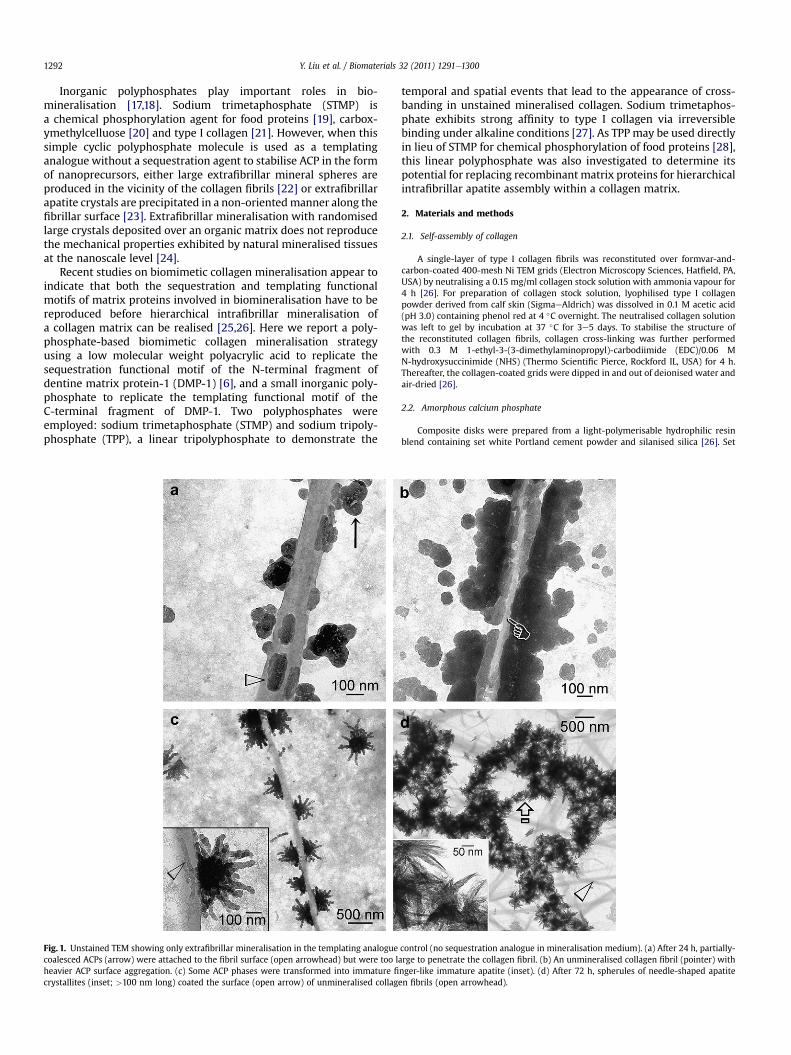
Fig. 1. Unstained TEM showing only extrafibrillar mineralisation in the templating analoguecoalesced ACPs (arrow) were attached to the fibril surface (open arrowhead) but were too lheavier ACP surface aggregation. (c) Some ACP phases were transformed into immature fi
crystallites (inset; >100 nm long) coated the surface (open arrow) of unmineralised collag
temporal and spatial events that lead to the appearance of cross-banding in unstained mineralised collagen. Sodium trimetaphos-phate exhibits strong affinity to type I collagen via irreversiblebinding under alkaline conditions [27]. As TPP may be used directlyin lieu of STMP for chemical phosphorylation of food proteins [28],this linear polyphosphate was also investigated to determine itspotential for replacing recombinant matrix proteins for hierarchicalintrafibrillar apatite assembly within a collagen matrix.
2. Materials and methods
2.1. Self-assembly of collagen
A single-layer of type I collagen fibrils was reconstituted over formvar-and-carbon-coated 400-mesh Ni TEM grids (Electron Microscopy Sciences, Hatfield, PA,USA) by neutralising a 0.15 mg/ml collagen stock solution with ammonia vapour for4 h [26]. For preparation of collagen stock solution, lyophilised type I collagenpowder derived from calf skin (SigmaeAldrich) was dissolved in 0.1 M acetic acid(pH 3.0) containing phenol red at 4 �C overnight. The neutralised collagen solutionwas left to gel by incubation at 37 �C for 3e5 days. To stabilise the structure ofthe reconstituted collagen fibrils, collagen cross-linking was further performedwith 0.3 M 1-ethyl-3-(3-dimethylaminopropyl)-carbodiimide (EDC)/0.06 MN-hydroxysuccinimide (NHS) (Thermo Scientific Pierce, Rockford IL, USA) for 4 h.Thereafter, the collagen-coated grids were dipped in and out of deionised water andair-dried [26].
2.2. Amorphous calcium phosphate
Composite disks were prepared from a light-polymerisable hydrophilic resinblend containing set white Portland cement powder and silanised silica [26]. Set
control (no sequestration analogue in mineralisation medium). (a) After 24 h, partially-arge to penetrate the collagen fibril. (b) An unmineralised collagen fibril (pointer) withnger-like immature apatite (inset). (d) After 72 h, spherules of needle-shaped apatiteen fibrils (open arrowhead).
Y. Liu et al. / Biomaterials 32 (2011) 1291e1300 1293
Portland cement releases calcium hydroxide (pH > 9.25) to facilitate trans-formation of amorphous calcium phosphate (ACP) to carbonated apatite. Thephosphate source was derived from NaN3-containing simulated body fluid (SBF):136.8 mM NaCl, 4.2 mM NaHCO3, 3.0 mM KCl, 1.0 mM K2HPO4$3H2O,1.5 mM MgCl2$6H2O, 2.5 mM CaCl2, 0.5 mM Na2SO4 and 3.08 mM Na3N. Polyacrylicacid (Mw 1800, SigmaeAldrich; 1000 mg/mL) was added to SBF to stabilise ACP asnanoprecursors [14,15].
2.3. Collagen mineralisation
Protein phosphorylationwith STMP is usually conducted at pH >11 to hydrolyseand open its closed ring structure [29]. To prevent collagen from denaturing, STMP(Mw 305.9; SigmaeAldrich) was hydrolysed at pH 12 for 5 h and neutralised to pH7.4 before use. Four STMP concentrations (wt %) were prepared: 2.5%, 1.25%, 0.625%and 0.313%. TEM grids containing cross-linked collagen (N ¼ 6) were immersed inthe respective STMP solution for 5 min, rinsed and air-dried. They were floatedupside-down over 30 mL of polyacrylic acid-containing SBF placed over a compositedisk inside a 100% humidity chamber [26]. Mineralisationwas performed initially for4e24 h to identify the optimal STMP concentration for further mineralisation for48e72 h. Grids were examined unstained using transmission electron microscopy(JEM-1230, JEOL) and selected area electron diffraction (SAED) at 110 kV. Theaforementioned experiments were repeated with TPP (Na5P3O10, Mw 367.8, Sig-maeAldrich) but omitting the alkaline hydrolysis step.
The collagen negative control (no sequestration and templating analogues)consisted of reconstituted collagen and SBF. The templating analogue control con-sisted of STMP/TPP-treated collagen and SBF (no sequestration analogue). Thesequestration analogue control consisted of reconstituted collagen and polyacrylicacid-containing SBF (no templating analogue).
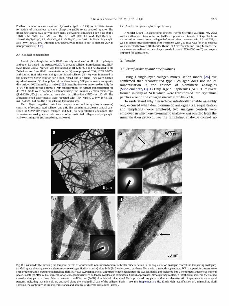
Fig. 2. Unstained TEM showing the temporal events associated with non-hierarchical intrafi(a) Grid space showing swollen electron-dense collagen fibrils (asterisk) after 24 h. (b) Swoseen predominantly around unmineralised fibrils (arrow). ACP nanoparticles appeared to haphase (inset). (c) After 72 h of mineralisation, collagen fibrils were no longer swollen and exhcross-banding patterns. Inset: Selected are electron diffraction (SAED) of individual minerapatterns indicating that minerals are arranged along the longitudinal axis of the collagenshowing the continuity of the mineral strands and absence of discrete crystallites (arrow).
2.4. Fourier transformeinfrared spectroscopy
A Nicolet 6700 FT-IR spectrophotometer (Thermo Scientific, Waltham, MA, USA)with an attenuated total reflection (ATR) setup was used to collect IR spectra fromvacuum-dried reconstituted collagen before and after treatmentwith 2.5 wt% TPP, aswell as competitive desorption after treatment with 250 mM NaCl for 24 h. Spectrawere collected between 4000 and 500 cm�1 at 4 cm�1 resolution using 32 scans. Thedata were normalised to the collagen amide I band (1715e1596 cm�1) and super-imposed for comparison.
3. Results
3.1. Extrafibrillar apatite precipitations
Using a single-layer collagen mineralisation model [26], weconfirmed that reconstituted type I collagen does not inducemineralisation in the absence of biomimetic analogues(Supplementary Fig. 1). Only large ACP spherules (ca. 1e3 mm) wereformed initially at 24 h which were transformed into crystallinepatches around the collagen matrix after 48e72 h.
To understand why hierarchical intrafibrillar apatite assemblyonly occurred when dual biomimetic analogues (i.e. sequestrationand templating) were employed, two analogue controls wereemployed inwhich one biomimetic analogue was omitted from themineralisation protocol. For the templating analogue control, no
brillar mineralisation in the sequestration analogue control (no templating analogue).llen, electron-dense fibrils with a smooth appearance. ACP nanoparticle clusters wereve penetrated the swollen fibrils and coalesced into a continuous amorphous mineralibited a fibrous appearance. Although they contained intrafibrillar mineral, they lackedlised fibrils produced ring patterns that are characteristic of apatite (note arc-shapedfibrils e see also Supplementary Fig. 4). (d) High magnification of a mineralised fibril

Y. Liu et al. / Biomaterials 32 (2011) 1291e13001294
sequestration analogue was included in the mineralisationmedium. Treatment of reconstituted collagen with 2.5 wt% TPP for5 min resulted in smaller ACP phases (ca. 50e100 nm) in thevicinity of the collagen fibrils after 24 h. These ACP phases were alsoattached to various extents on the fibrillar surface (Fig. 1a, b). Theywere transformed into blunted, finger-like immature apatite after48 h (Fig. 1c), and further into needle-shaped apatite that wererandomly precipitated over the surface of collagen fibrils after 72 h(Fig. 1d). No intrafibrillar mineralisation could be observed. Despitethe absence of a sequestration analogue, ACP phases were smallerthan those in the collagen negative control.
3.2. Non-hierarchical intrafibrillar mineralisation
In the sequestration analogue control, 1000 mg/mL polyacrylicacid was included in the mineralisation medium but did not treatthe reconstituted collagenwith STMP or TPP. Two ACP phases wereformed after 4 h (Supplementary Fig. 2). The relatively larger ACPphasewere 50e100 nm in diameter and the smaller ACP nanophasewas less than 10 nm in diameter. Only the smaller ACP nanophaseswere useful for intrafibrillar mineralisation (shown later). After24 h, collagen fibrils appeared swollen and were infiltrated bya highly electron-dense amorphousmineral phase (Fig. 2a). The ACPnanophases thatwere present in the vicinity of unmineralisedfibrilswere predominantly absent from the surface of those swollen,smooth, electron-dense fibrils (Fig. 2b). After 72 h, the swollen
Fig. 3. Unstained TEM showing the spatial effects of different templating analogue concepresence of a sequestration analogue. Sodium trimetaphosphate (a) 0.313 wt%, (b) 0.625 wtwere identified along the collagen fibril surfaces (open arrowheads). Large ACP phases (see
appearance of the fibrils disappeared and the coalesced ACP wastransformed into fibrous intrafibrillar mineral strands that lackedcross-banding patterns (Fig. 2c). Mineralised strands within thosefibrils appeared to be continuous at high magnification (Fig. 2d).
3.3. Hierarchical intrafibrillar mineralisation
Figs. 3 and 4 represent the temporal and spatial events associ-ated with the use of different templating analogue concentrations(0.313e2.5 wt%) for collagen mineralisation in the presence of1000 mg/mL polyacrylic acid as the sequestration analogue. Assimilar results were obtained for STMP and TPP, only one poly-phosphate templating analogue is illustrated for the early amor-phous (STMP; 4e12 h) and crystalline (TPP, 48e72 h) stages ofintrafibrillar mineralisation. In the amorphous stage (Fig. 3), lessand less ACP nanophases aggregated along the fibrillar surface withincreasing STMP concentrations, while increasingly apparent cross-banding corresponding to the D-spacing of collagen moleculescould be seen within the fibrils. Despite the appearance of cross-banding, SAED indicated that intrafibrillar minerals were amor-phous at this stage (not shown). In the crystalline stage (Fig. 4),progressively noticeable hierarchical apatite assembly could beidentified with increasing TPP concentrations. This ranged from theoccurrence of continuous microfibrillar strands without cross-banding at 0.313 wt%, to the appearance of vague cross-banding at0.625 wt%, to distinct cross-banding at 1.25 wt% and 2.5 wt% TPP.
ntrations on the amorphous stage of hierarchical intrafibrillar mineralisation in the%, (c) 1.25 wt%, (d) 2.5 wt% was used as the example. Only ACP nanoparticle aggregatesSupplementary Fig. 2) were not involved in intrafibrillar mineralisation.
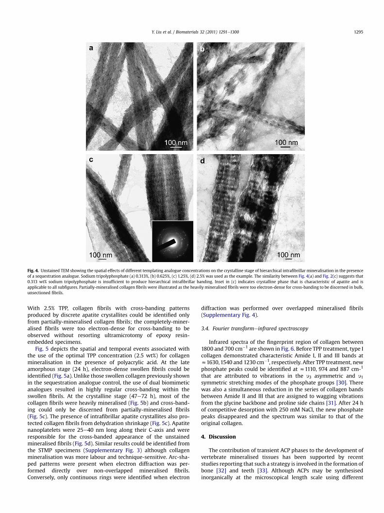
Fig. 4. Unstained TEM showing the spatial effects of different templating analogue concentrations on the crystalline stage of hierarchical intrafibrillar mineralisation in the presenceof a sequestration analogue. Sodium tripolyphosphate (a) 0.313%, (b) 0.625%, (c) 1.25%, (d) 2.5% was used as the example. The similarity between Fig. 4(a) and Fig. 2(c) suggests that0.313 wt% sodium tripolyphosphate is insufficient to produce hierarchical intrafibrillar banding. Inset in (c) indicates crystalline phase that is characteristic of apatite and isapplicable to all subfigures. Partially-mineralised collagen fibrils were illustrated as the heavily mineralised fibrils were too electron-dense for cross-banding to be discerned in bulk,unsectioned fibrils.
Y. Liu et al. / Biomaterials 32 (2011) 1291e1300 1295
With 2.5% TPP, collagen fibrils with cross-banding patternsproduced by discrete apatite crystallites could be identified onlyfrom partially-mineralised collagen fibrils; the completely-miner-alised fibrils were too electron-dense for cross-banding to beobserved without resorting ultramicrotomy of epoxy resin-embedded specimens.
Fig. 5 depicts the spatial and temporal events associated withthe use of the optimal TPP concentration (2.5 wt%) for collagenmineralisation in the presence of polyacrylic acid. At the lateamorphous stage (24 h), electron-dense swollen fibrils could beidentified (Fig. 5a). Unlike those swollen collagen previously shownin the sequestration analogue control, the use of dual biomimeticanalogues resulted in highly regular cross-banding within theswollen fibrils. At the crystalline stage (47e72 h), most of thecollagen fibrils were heavily mineralised (Fig. 5b) and cross-band-ing could only be discerned from partially-mineralised fibrils(Fig. 5c). The presence of intrafibrillar apatite crystallites also pro-tected collagen fibrils from dehydration shrinkage (Fig. 5c). Apatitenanoplatelets were 25e40 nm long along their C-axis and wereresponsible for the cross-banded appearance of the unstainedmineralised fibrils (Fig. 5d). Similar results could be identified fromthe STMP specimens (Supplementary Fig. 3) although collagenmineralisation was more labour and technique-sensitive. Arc-sha-ped patterns were present when electron diffraction was per-formed directly over non-overlapped mineralised fibrils.Conversely, only continuous rings were identified when electron
diffraction was performed over overlapped mineralised fibrils(Supplementary Fig. 4).
3.4. Fourier transformeinfrared spectroscopy
Infrared spectra of the fingerprint region of collagen between1800 and 700 cm�1 are shown in Fig. 6. Before TPP treatment, type Icollagen demonstrated characteristic Amide I, II and III bands atz1630,1540 and 1230 cm�1, respectively. After TPP treatment, newphosphate peaks could be identified at z1110, 974 and 887 cm-1
that are attributed to vibrations in the y3 asymmetric and y1symmetric stretching modes of the phosphate groups [30]. Therewas also a simultaneous reduction in the series of collagen bandsbetween Amide II and III that are assigned to wagging vibrationsfrom the glycine backbone and proline side chains [31]. After 24 hof competitive desorption with 250 mM NaCl, the new phosphatepeaks disappeared and the spectrum was similar to that of theoriginal collagen.
4. Discussion
The contribution of transient ACP phases to the development ofvertebrate mineralised tissues has been supported by recentstudies reporting that such a strategy is involved in the formation ofbone [32] and teeth [33]. Although ACPs may be synthesisedinorganically at the microscopical length scale using different

Fig. 5. Unstained TEM of collagen treated with 2.5 wt% sodium tripolyphosphate and mineralised in polyacrylic acid-containing SBF. (a) Swollen, cross-banded collagen fibrilscontaining amorphous electron-dense minerals (inset) after 24 h. (b) Grid space showing heavy mineralisation of the collagen fibrils after 72 h. A few fibrils contained unmin-eralised regions (open arrowheads). (c) Partially-mineralised collagen fibril with cross-banding created by intrafibrillar mineral assembly. Absence of minerals in the unmineralisedregion (between open arrows) resulted in extensive shrinkage during chemical dehydration for TEM. (d) Partially-mineralised collagen fibril showing hierarchical arrangement ofoverlapping nanoplatelets. Inset: SAEDwith ring patterns corresponding to those of poorly-crystalline apatite (note: arc-shaped patterns indicating that minerals are arranged alongthe longitudinal axis of the collagen fibrils e see also Supplementary Fig. 4).
Fig. 6. Infrared spectra (1800e700 cm�1) of type I collagen before and after treatmentwith 2.5 wt% sodium tripolyphosphate and after subjected to competitive desorptionwith 250 mM NaCl for 24 h. After tripolyphosphate treatment, new phosphate peakscould be identified (asterisks). There was also a simultaneous reduction in the series ofcollagen bands between Amide II and III (arrows). After 24 h of NaCl desorption, thespectrum was similar to that of the original type I collagen. This suggests that thesodium polyphosphate binding mechanism is exclusively electrostatic in nature,probably via the formation of ionic bridges.
Y. Liu et al. / Biomaterials 32 (2011) 1291e13001296
chemical systems [34], a size exclusion mechanism limits thedimension of ACP phases that can penetrate the internal watercompartments of a collagen fibril [35]. Molecules larger than40 kDa are completely excluded from these internal watercompartments, while molecules smaller than 6 kDa can diffuse intoall the water compartments within the collagen fibril. Thus, it islogical to assume that ACP nanophases have to be smaller than40 kDa in order for them to penetrate and displace water from atleast some internal compartments of a collagen fibril. This explainswhy the relatively larger ACP phases shown in Fig. 1 andSupplementary Fig. 2 are incapable of participating in intrafibrillarmineralisation. One of the functional motifs of matrix proteinsinvolved in biomineralisation is to act as a sequestration agent forstabilising ACP in the form of nanophases [36]. Dentin matrixprotein-1, an acidic matrix protein, is cleaved into a largerN-terminal and a smaller C-terminal fragment. The aspartic acid-rich N-terminal segment helps stabilise ACP into nanoparticles [6].A similar ACP stabilisation motif was also observed for a 20-kDcleavage product of amelogenin [37]. Thus, a sequestration biomi-metic analogue is required to simulate the function of these naturalprotein molecules.
Sodium tripolyphosphate has been used as an anionic surfactantto inhibit crystal growth in the synthesis of calcite nanocrystals[38]. Similar to STMP [27], a small amount of the collagen-boundTPP could have been displaced by anions derived from the

Y. Liu et al. / Biomaterials 32 (2011) 1291e1300 1297
mineralisation medium, resulting in the small ACP phases in thevicinity of the collagen fibrils. The remaining collagen-bound TPPcaused ACP phases to be attached directly to the fibrillar surface,which assumed different shapes (Fig. 1a) due to their initialmouldable characteristics [16]. Exclusive extrafibrillar precipita-tions identified from this control parallels the results achievedwhen STMP was used for mineralising collagen without a seques-tration analogue [15]. Taken together, these results indicate thatpolyphosphates such as STMP or TPP alone produces ACP phasesthat are too large to penetrate the internal water compartments offibrillar collagen and hence intrafibrillar mineralisation does notoccur.
As demonstrated in Figs. 2 and 4a, the use of a sequestrationanalogue alone or in combination with insufficient templatinganalogue does not result in the type of intrafibrillar mineralisationthat resembles what has been perfected by Nature. During theinitial mineralisation stage, it appears that the lateral spacingsbetween collagen molecules can be expanded temporarily toaccommodate the infiltrated ACP nanophases. The reason for thisswelling is unknown. A possible explanation is that ACP containsmorewater than apatite within its intercluster spaces [34,39]. Thus,a larger volume of ACP is required if apatite has to fully replace theoriginal volume occupied by water within a collagen fibril. Duringtransformation of ACP into more compactly-packed apatite, wateris displaced from the internal water compartments of the collagenfibril, resulting in progressive dehydration of the fibril [13]. Thisprovides a mechanism for the swollen fibril to return to its originaldimension.
In Fig. 2d, we observed that the mineralised fibrous strandsappeared to be continuous instead of existing as discrete crystal-lites. Although the results from the sequestration analogue controlsupport previous studies that intrafibrillar mineralisation occurswith the use of a templating analogue alone [14,15], the non-hier-archical mineralisation observed appears to suggest that the min-eralisation mechanism involved is considerably different from thatutilised by natural matrix proteins in biomineralisation of verte-brate collagenous tissues. In the absence of a templating analogueto direct the nucleation of discrete apatite nanocrystals, we suggest
Fig. 7. Schematic showing the different status of apatite deposition around and within a coanalogues.
that the interconnecting water-filled volume within the collagenfibril may be crystallised as a continuum and moulded into a singlecrystalline structure. Such a crystallisation mechanism producesunusual mineralised collagen entities that resemble the monolithicsingle crystal structure in sea urchin spines [40] or siliceous bio-skeletons [41], albeit within the nanoscale internal environment ofa collagen fibril. In this polymer-induced liquid precursor [16] orpolymer-assisted moulding process [42], single crystals withoutdefinitive crystalline planes may be created that bend aroundcurvatures and contain porosities. Although this mineralisationmechanism is important in amorphous calcium carbonate andamorphous silica-based biomineralisation systems, formation ofa single crystal with curvature and fenestrations is not what Naturehas intended apatite to be deposited within collagen fibrils invertebrates to achieve their load bearing function [24].
For the collagen that were mineralised in the presence of boththe sequestration and the templating analogues, visualisation ofcross-banding in only incompletely-mineralised fibrils is analogousto etching naturally mineralised collagen with mild acids to enablecollagen cross-banding patterns to be identified with atomic forcemicroscopy [43]. This hierarchical arrangement is identical to thatobserved in naturally mineralised type I collagen [2]. Matrixphosphoproteins serve as templates for apatite nucleation andgrowthwithin the gap zones of collagen fibrils [44].When bound tocollagen, these highly-phosphorylated polyanionic macromole-cules function as templates [45] by attracting calcium ions andinduce apatite nucleation within the gap zones of the collagenfibril. During this process, the comparative loose amorphousstructure in ACPs is transformed into a more closely-packed crys-talline structure [46]. The phosphoserine-rich C-terminal fractionof cleaved DMP-1 is thought to be responsible for apatite nucleation[6]. Phosphophoryn is another important initiator and modulatorfor apatite growth due to its excellent binding capacity for calciumions as well as its strong affinity to collagen [47]. Bone sialoproteinis another acidic phosphoprotein which is capable of binding tocollagen and nucleating apatite [48]. The polyphosphate analoguesappear to replicate the templating function of these phosphopro-teins well enough for hierarchical apatite assembly to be discerned
llagen fibril in the presence of different combinations of sequestration and templating

Fig. 8. Schematic depicting possible ionic cross-linking involved with binding of sodium tripolyphosphate anions to collagen molecules. Free hydroxyl ions that do not interact withcollagen molecules contribute to recruitment of polyacrylic acid-stabilised amorphous calcium phosphate nanoprecursors and induction of apatite nucleation.
Y. Liu et al. / Biomaterials 32 (2011) 1291e13001298
within the biomimetically-mineralised collagen fibrils. Togetherwith the controls, these results highlight the complementarycontribution of the two functional motifs of matrix proteinsinvolved in biomineralisation of collagen; failure to replicate eitherfunctional motif will result in exclusive extrafibrillar precipitationsor non-hierarchical intrafibrillar mineralisation of a collagenmatrix(Fig. 7).
It has been shown that STMP can bind irreversibly to collagen byforming new covalent bonds [22,23], We were also able to observeformation of new phosphate peaks when Fourier trans-formeinfrared spectroscopy was used to examine the interactionbetween TPP and reconstituted collagen (Fig. 6). However, thesenew phosphate peaks disappeared after the TPP-doped collagenwas immersed for 24 h in 250M NaCl, indicating that the cross-linking is reversible and non-covalent in nature. As TPP forms ioniccross-links (ionic bridges) between chitosan molecules via inter-acting with their amine groups [49], similar ionic bridges may beformedwhen TPP interacts with collagenmolecules. Although ioniccross-linking not as stable as covalent cross-linking, this cross-linking procedure is an important textile treatment process thatincreases the crease resistance of fabrics [50]. There are fivenegatively charged ionisable groups in TPP with different pKavalues (pKa1 ¼1, pKa2 ¼ 2, pKa3 ¼ 2.79, pKa4 ¼ 6.47, pKa5 ¼ 9.24)[51]. A 2.5 wt% TPP solution has a pH value of 8.5 with at least fourionisable OH� groups. Thus, TPP may form ionic cross-links withcollagen as a reversible binding mechanism, with the remainingfree OH� groups contributing to the attraction of ACP nanophases.Ionic cross-linking provides the rationale for displacement of
collagen-bound TPPs to form relatively large ACP phases even in theabsence of polyacrylic acid (Fig. 1b). Fig. 8 illustrates the possiblemechanism that may be involved in the binding of TPP to collagen.
5. Conclusions
We demonstrate here a highly hierarchical assembly of intra-fibrillar apatite crystallites in reconstituted collagenmineralised ex-situ with biomimetic analogues of extracellular matrix proteins.From a developmental biology perspective, the results of this workprovide important confirmation of the two distinct but comple-mentary functional motifs in matrix proteins involved in vertebratecollagen mineralisation. It is possible that a single sequestrationfunctional motif is probably what is required for biomineralisationof invertebrate systems that involve fusion of amorphous mineralprecursor phases into porous single crystalline structures. Thecreativity of evolution in which nature timelessly explores alloptions and finds the best solution is surprisingly instructive. Thenotions that these functional motifs are highly conserved in matrixproteins from different animal species and that they can be repli-cated using small, non-protein molecules imply that the bio-mineralisation principles perfected by Nature are, ultimately,simplistic and clear-cut in design. The complexity of differentproteins involved in the process probably represents the means bywhich the proteins incorporating these functional motifs areproduced within the intracellular milieu and delivered to theexternal environment. One of themain challenges of biomimetics isthat it demands creative solutions. Nature’s store of ideas is

Y. Liu et al. / Biomaterials 32 (2011) 1291e1300 1299
valuable only if it can be translated into usable technology. Thisstudy demonstrates that small non-protein biomimetic moleculesmay be substituted for complex recombinant matrix proteins ortheir functional domains for applications that involve the use ofmineralised collagen. The latter may include porous, three-dimensional mineralised collagen scaffolds to enhance stem cellproliferation and coating of orthopaedic implants with collagenpre-impregnated with hierarchical intrafibrillar nanoapatite.
Acknowledgements
This work was supported by grant R21 DE019213 from NIDCR(PI. Franklin Tay). We thank Michelle Barnes for secretarial support,Robert Smith for TEM assistance.
Appendix
Figures with essential color discrimination. Figs. 6 and 7 in thisarticle is difficult to interpret in black and white. The full colorimages can be found in the online version, at doi:10.1016/j.biomaterials.2010.10.018.
Appendix. Supporting material
The supplementary data associated with this article can befound in the online version at doi:10.1016/j.biomaterials.2010.10.018.
References
[1] Wong TS, Brough B, Ho CM. Creation of functional micro/nano systemsthrough top-down and bottom-up approaches. Mol Cell Biomech 2009;6:1e55.
[2] Traub W, Arad T, Weiner S. Three-dimensional ordered distribution of crystalsin turkey tendon collagen fibers. Proc Natl Acad Sci USA 1989;86:9822e6.
[3] Wang L, Nancollas GH. Pathways to biomineralization and biodemineraliza-tion of calcium phosphates: the thermodynamic and kinetic controls. DaltonTrans 2009;15:2665e72.
[4] Saito T, Arsenault AL, Yamauchi M, Kuboki Y, Crenshaw MA. Mineral inductionby immobilized phosphoproteins. Bone 1997;21:305e11.
[5] Cölfen H. Bio-inspired mineralization using hydrophilic polymers. Springer-Verlag, Berlin: Heidelberg 2007;271:1e77. Top Curr Chem.
[6] Gajjeraman S, Narayanan K, Hao J, Qin C, George A. Matrix macromolecules inhard tissues control the nucleation and hierarchical assembly of hydroxyap-atite. J Biol Chem 2007;282:1193e204.
[7] Weiner S. Biomineralization: a structural perspective. J Struct Biol 2008;163:229e34.
[8] Hao J, Ramachandran A, George A. Temporal and spatial localization of thedentin matrix proteins during dentin biomineralization. J Histochem Cyto-chem 2009;57:227e37.
[9] Sfeir C, Cambell P, Jadlowiex JA, Kumta P. Method of inducing bio-mineralization method of inducing bone regeneration and methods relatedthereof. US Patent Appl No. 20070087959, 2007.
[10] Kaplan DL, Huang J, Wong, CPF, Naik R, George A. Fibrous protein fusions anduse thereof in the formation of advanced organic/inorganic compositematerials. US Patent Appl No. 20080293919, 2008.
[11] Goldberg HA, Hunter GK, Tye CE. Bone sialoprotein collagen-binding peptides.US Patent Appl 20090005298, 2009.
[12] Girija EK, Yokogawa Y, Nagata F. Apatite formation on collagen fibrils in thepresence of polyacrylic acid. J Mater Sci Mater Med 2004;15:593e9.
[13] Chesnick IE, Mason JT, Giuseppetti AA, Eidelman N, Potter K. Magnetic reso-nance microscopy of collagen mineralization. Biophys J 2008;95:2017e26.
[14] Deshpande AS, Beniash E. Bio-inspired synthesis of mineralized collagenfibrils. Cryst Growth Des 2008;8:3084e90.
[15] Jee SS, Thula TT, Gower LB. Development of bone-like composites via thepolymer-induced liquid-precursor (PILP) process. Part 1: influence of polymermolecular weight. Acta Biomater 2010;6:3676e86.
[16] Gower LB. Biomimetic model systems for investigating the amorphousprecursor pathway and its role in biomineralization. Chem Rev 2008;108:4551e627.
[17] Omelon SJ, Grynpas MD. Relationships between polyphosphate chemistry,biochemistry and apatite biomineralization. Chem Rev 2008;108:4694e715.
[18] Usui Y, Uematsu T, Uchihashi T, Takahashi M, Takahashi M, Ishizuka M, et al.Inorganic polyphosphate induces osteoblastic differentiation. J Dent Res2010;89:504e9.
[19] Li Y, de Vries R, Slaghek T, Timmermans J, Cohen Stuart MA, Norde W.Preparation and characterization of oxidized starch polymer microgels forencapsulation and controlled release of functional ingredients. Bio-macromolecules 2009;10:1931e8.
[20] Leone G, Torricelli P, Giardino R, Barbucci R. New phosphorylated derivativesof carboxymethylcellulose with osteogenic activity. Polym Adv Technol2008;19:824e30.
[21] Gunasekaran S. Purifying type I collagen using two papain treatments andreducing and delipidation agents. US Patent Office No. 6548077, 2003.
[22] Li X, Chang J. Preparation of bone-like apatite-collagen nanocomposites bya biomimetic process with phosphorylated collagen. J Biomed Mater Res A2008;85:293e300.
[23] Xu Z, Neoh KG, Kishen A. A biomimetic strategy to form calcium phosphatecrystals on type I collagen substrate. Mat Sci Eng C 2010;30:822e6.
[24] Gupta HS, Seto J, Wagermaier W, Zaslansky P, Boesecke P, Fratzl P. Coopera-tive deformation of mineral and collagen in bone at the nanoscale. Proc NatlAcad Sci USA 2006;103:17741e6.
[25] Tay FR, Pashley DH. Guided tissue remineralisation of partially demineralisedhuman dentine. Biomaterials 2008;29:1127e37.
[26] Kim YK, Gu LS, Bryan TE, Kim JR, Chen L, Liu Y, et al. Mineralization ofreconstituted collagen using polyvinylphosphonic acid / polyacrylic acidtemplating matrix protein analogues in the presence of calcium, phosphateand hydroxyl ions. Biomaterials 2010;31:6618e27.
[27] Gu LS, Kim J, Kim YK, Liu Y, Dickens SH, Pashley DH, et al. A chemical phos-phorylation-inspired design for Type I collagen biomimetic remineralization.Dent Mater; 2010 [Epub ahead of print].
[28] Zhang K, Li Y, Ren Y. Research on the phosphorylation of soy protein isolatewith sodium tripolyphosphate. J Food Eng 2007;79:1233e7.
[29] Shen CY. Alkaline hydrolysis of sodium trimetaphosphate in concentratedsolutions and its role in built detergents. Ind Eng Chem Prod Res Dev1966;5:272e6.
[30] Arai Y, Sparks DL. ATR-FTIR spectroscopic investigation on phosphateadsorption mechanisms at the ferrihydrite-water interface. J Colloid InterfaceSci 2001;241:317e26.
[31] Jackson M, Choo L-P, Watson PH, Halliday WC, Mantsch HH. Beware ofconnective tissue proteins: assignment and implications of collagen absorp-tions in infrared spectra of human tissues. Biochim Biophy Acta 1995;1270:1e6.
[32] Mahamid J, Sharir A, Addadi L, Weiner S. Amorphous calcium phosphate isa major component of the forming fin bones of zebrafish: Indications foran amorphous precursor phase. Proc Natl Acad Sci USA 2008;105:12748e53.
[33] Deshpande AS, Fang PA, Simmer JP, Margolis HC, Beniash E. Amelogenin-collagen interactions regulate calcium phosphate mineralization in vitro. J BiolChem 2010;295:19277e87.
[34] Combes C, Rey C. Amorphous calcium phosphates: synthesis, properties anduses in biomaterials. Acta Biomater 2010;6:3362e78.
[35] Toroian D, Lim JE, Price PA. The size exclusion characteristics of type Icollagen: implications for the role of noncollagenous bone constituents inmineralization. J Biol Chem 2007;282:22437e47.
[36] He G, Gajjeraman S, Schultz D, Cookson D, Qin C, Butler WT, et al. Spatially andtemporally controlled biomineralization is facilitated by interaction betweenself-assembled dentin matrix protein 1 and calcium phosphate nuclei insolution. Biochemistry 2005;44:16140e8.
[37] Kwak SY, Wiedemann-Bidlack FB, Beniash E, Yamakoshi Y, Simmer JP,Litman A, et al. Role of 20-kDa amelogenin (P148) phosphorylation in calciumphosphate formation in vitro. J Biol Chem 2009;284:18972e9.
[38] Sonawane SH, Gumfekar SP, Meshram S, Deosarkar MP, Mahajan CM,Khanna P. Combined effect of surfactant and ultrasound on nano calciumcarbonate synthesized by crystallization process. Int J Chem React Eng2009;7. A47.
[39] Sedlak JM, Beebe RA. Temperature programmed dehydration of amorphouscalcium phosphate. J Colloid Interface Sci 1974;47:483e9.
[40] Müller WE, Wang X, Cui FZ, Jochum KP, Tremel W, Bill J, et al. Sponge spiculesas blueprints for the biofabrication of inorganic-organic composites andbiomaterials. Appl Microbiol Biotechnol 2009;83:397e413.
[41] Cölfen H. Single crystals with complex form via amorphous precursors.Angew Chem Int Ed Engl 2008;47:2351e3.
[42] Meldrum FC, Ludwigs S. Template-directed control of crystal morphologies.Macromol Biosci 2007;7:152e62.
[43] Balooch M, Habelitz S, Kinney JH, Marshall SJ, Marshall GW. Mechanicalproperties of mineralized collagen fibrils as influenced by demineralization.J Struct Biol 2008;162:404e10.
[44] George A, Veis A. Phosphorylated proteins and control over apatite nucleation,crystal growth, and inhibition. Chem Rev 2008;108:4670e93.
[45] Zhang TH, Liu XY. How does a transient amorphous precursor templatecrystallization. J Am Chem Soc 2007;129:13520e6.
[46] Tsuji T, Onuma K, Yamamoto A, Iijima M, Shiba K. Direct transformation fromamorphous to crystalline calcium phosphate facilitated by motif-programmedartificial proteins. Proc Natl Acad Sci USA 2008;105:16866e70.
[47] He G, Ramachandran A, Dahl T, George S, Schultz D, Cookson D, et al. Phos-phorylation of phosphophoryn is crucial for its function as a mediator ofbiomineralization. J Biol Chem 2005;280:33109e14.
[48] Baht GS, O’Young J, Borovina A, Chen H, Tye CE, Karttunen M, et al.Phosphorylation of Ser136 is critical for potent bone sialoprotein-

Y. Liu et al. / Biomaterials 32 (2011) 1291e13001300
mediated nucleation of hydroxyapatite crystals. Biochem J 2010;428:385e95.
[49] Hu M, Li Y, Decker EA, Xiao H, McClements DC. Influence of tripolyphosphatecross-linking on the physical stability and lipase digestibility of chitosan-coated lipid droplets. J Agric Food Chem 2010;58:1283e9.
[50] Hauser PJ, Smith CB, Hashem MM. Ionic crosslinking of cotton. Autex Res J2004;4:95e100.
[51] Shu XZ, Zhu KJ. The influence of multivalent phosphate structure on theproperties of ionically cross-linked chitosan films for controlled drug release.Eur J Pharm Biopharm 2002;54:235e43.