Extracellular Matrix of Porcine Pericardium: Biochemistry and Collagen Architecture
11
Extracellular Matrix of Porcine Pericardium: Biochemistry and Collagen Architecture Antonella Sachsida Braga-Vilela Edson Rosa Pimentel Sergio Marangoni Marcos Hikari Toyama Benedicto de Campos Vidal Received: 4 July 2007 / Accepted: 9 October 2007 / Published online: 3 December 2007 Ó Springer Science+Business Media, LLC 2007 Abstract Pericardial tissue has been used to construct bioprostheses employed in the repair of different kinds of injuries, mostly cardiac. However, calcification and mechanical failure have been the main causes of the lim- ited durability of cardiac bioprostheses constructed with bovine pericardium. In the course of this work, a study was conducted on porcine fibrous pericardium, its microscopic structure and biochemical nature. The general morphology and architecture of collagen were studied under conven- tional light and polarized light microscopy. The biochemical study of the pericardial matrix was conducted according to the following procedures: swelling test, hydroxyproline and collagen dosage, quantification of amino acids in soluble collagen, component extraction of the extracellular matrix of the right and left ventral regions of pericardium with different molarities of guanidine chloride, protein and glycosaminoglycan (GAG) dosage, sodium dodecyl sulfate-polyacrylamide gel electrophoresis and total GAG analysis. Microscopic analysis showed collagen fibers arranged in multidirectionally oriented layers forming a closely knit web, with a larger number of fibers obliquely oriented, initiating at the lower central region toward the upper left lateral relative to the heart. No qualitative differences were found between proteins extracted from the right and left regions. Likewise, no differences were found between fresh and frozen material. Protein dosages from left frontal and right frontal pericar- dium regions showed no significant differences. The quantities of extracted GAGs were too small for detection by the method used. Enzymatic digestion and electropho- retic analysis showed that the GAG found is possibly dermatan sulfate. The proteoglycan showed a running standard very similar to the small proteoglycan decorin. Keywords Extracellular matrix Á Porcine pericardium Á Collagen Á Proteoglycan glycosaminoglycan Á Bioprosthesis Introduction The pericardium is a fibrous-serous sac enveloping the heart and the commencement of the large vessels, com- posed mostly of fibrous connective tissue (Hollinshead, 1980: Moore & Dalley, 2001). Pericardial tissue has been used for bioprosthesis construction for decades, especially for the repair of cardiac injuries (Olmos et al., 1997; Bar- ros, Safatle & Rigueiro, 1999). Repair of the ventricular wall, cardiac valves and aortic wall for the correction of aneurysms has been successfully performed using peri- cardium bioprostheses (Pires, Saporito & Leao, 1997). Pericardium from different animals has been tested for the construction of bioprostheses, and good results have been attained using bovine and porcine pericardium. The fibrous layer of porcine pericardium possesses greater uniformity in its different regions and a thickness ranging between that of human and bovine pericardium. Thus, its use in the A. S. Braga-Vilela (&) Department of Biologic Sciences, UNIFAL, Rua Gabriel Monteiro Silva 714, 37 130 000 Alfenas, Minas Gerais, Brazil e-mail: [email protected] E. R. Pimentel Á B. de Campos Vidal Department of Cell Biology, UNICAMP, Sa ˜o Paulo, Brazil S. Marangoni Department of Biochemistry, UNICAMP, Sa ˜o Paulo, Brazil M. H. Toyama Department of Biochemistry, UNESP-CLP-SV, Sa ˜o Paulo, Brazil 123 J Membrane Biol (2008) 221:15–25 DOI 10.1007/s00232-007-9081-5
Transcript of Extracellular Matrix of Porcine Pericardium: Biochemistry and Collagen Architecture
Extracellular Matrix of Porcine Pericardium: Biochemistry andCollagen Architecture
Antonella Sachsida Braga-Vilela Æ Edson Rosa Pimentel ÆSergio Marangoni Æ Marcos Hikari Toyama ÆBenedicto de Campos Vidal
Received: 4 July 2007 / Accepted: 9 October 2007 / Published online: 3 December 2007
� Springer Science+Business Media, LLC 2007
Abstract Pericardial tissue has been used to construct
bioprostheses employed in the repair of different kinds of
injuries, mostly cardiac. However, calcification and
mechanical failure have been the main causes of the lim-
ited durability of cardiac bioprostheses constructed with
bovine pericardium. In the course of this work, a study was
conducted on porcine fibrous pericardium, its microscopic
structure and biochemical nature. The general morphology
and architecture of collagen were studied under conven-
tional light and polarized light microscopy. The
biochemical study of the pericardial matrix was conducted
according to the following procedures: swelling test,
hydroxyproline and collagen dosage, quantification of
amino acids in soluble collagen, component extraction of
the extracellular matrix of the right and left ventral regions
of pericardium with different molarities of guanidine
chloride, protein and glycosaminoglycan (GAG) dosage,
sodium dodecyl sulfate-polyacrylamide gel electrophoresis
and total GAG analysis. Microscopic analysis showed
collagen fibers arranged in multidirectionally oriented
layers forming a closely knit web, with a larger number of
fibers obliquely oriented, initiating at the lower central
region toward the upper left lateral relative to the heart. No
qualitative differences were found between proteins
extracted from the right and left regions. Likewise, no
differences were found between fresh and frozen material.
Protein dosages from left frontal and right frontal pericar-
dium regions showed no significant differences. The
quantities of extracted GAGs were too small for detection
by the method used. Enzymatic digestion and electropho-
retic analysis showed that the GAG found is possibly
dermatan sulfate. The proteoglycan showed a running
standard very similar to the small proteoglycan decorin.
Keywords Extracellular matrix � Porcine pericardium �Collagen � Proteoglycan glycosaminoglycan �Bioprosthesis
Introduction
The pericardium is a fibrous-serous sac enveloping the
heart and the commencement of the large vessels, com-
posed mostly of fibrous connective tissue (Hollinshead,
1980: Moore & Dalley, 2001). Pericardial tissue has been
used for bioprosthesis construction for decades, especially
for the repair of cardiac injuries (Olmos et al., 1997; Bar-
ros, Safatle & Rigueiro, 1999). Repair of the ventricular
wall, cardiac valves and aortic wall for the correction of
aneurysms has been successfully performed using peri-
cardium bioprostheses (Pires, Saporito & Leao, 1997).
Pericardium from different animals has been tested for the
construction of bioprostheses, and good results have been
attained using bovine and porcine pericardium. The fibrous
layer of porcine pericardium possesses greater uniformity
in its different regions and a thickness ranging between that
of human and bovine pericardium. Thus, its use in the
A. S. Braga-Vilela (&)
Department of Biologic Sciences, UNIFAL, Rua Gabriel
Monteiro Silva 714, 37 130 000 Alfenas, Minas Gerais, Brazil
e-mail: [email protected]
E. R. Pimentel � B. de Campos Vidal
Department of Cell Biology, UNICAMP, Sao Paulo, Brazil
S. Marangoni
Department of Biochemistry, UNICAMP, Sao Paulo, Brazil
M. H. Toyama
Department of Biochemistry, UNESP-CLP-SV, Sao Paulo,
Brazil
123
J Membrane Biol (2008) 221:15–25
DOI 10.1007/s00232-007-9081-5
construction of bioprostheses appears to be a distinct pos-
sibility (Fentie et al., 1986; Chanda, Kuribayashi & Abe,
1997).
The biomechanical properties of pericardium, as well as
other collagenous tissues, are directly related to the dis-
tribution and orientation of the collagen fiber bundles
(Sacks, Cuhong & More, 1994) and to the wave-like
structures (WLSs) or crimp of such fibers (Loke et al.,
1996; Langdon et al., 1999). Analysis of the WLS shows
the arrangement of type I collagen molecules in the col-
lagen bundle and possible direction of changes of these
fibrillar elements (Ault & Hoffman, 1992). The morpho-
logical variability of the collagenous bundle probably
reflects functional differentiation resulting from different
biomechanical properties of the tissues. Alterations in the
collagenous molecular organization can be observed and
quantified by polarized light microscopy (Whittaker et al.,
1987), which has been recommended as the most appro-
priate method for the detection, description and
interpretation of WLSs (Vidal, 2003; Gathercole & Keller,
1991).
The anisotropic optical properties, birefringence and
dichroism of the collagen bundle provide a statistical
model of its molecular organization and, consequently,
establish an important investigation mechanism of its
structural pattern (Vidal & Mello, 1972). While dichroism,
using toluidine blue (pH 3.5–4.0), reveals the molecular
arrangement of acid glycosaminoglycans (GAGs), bire-
fringence predominantly reveals the crystalline structure of
collagen polypeptide chains. Both phenomena are inti-
mately related, due to the fact that about 13% of the form
birefringence of collagen bundles is due to the GAGs
associated with them, as observed in young rat tendon
(Vidal, 1966, 1980).
The use of in totum tissue preparations provides addi-
tional subsidies for studies on and appraisal of the degree
of tridimensional grid formation of collagen. The archi-
tecture of collagen fibers establishes a choice and selection
parameter concerning the tissues to be used for the con-
struction of bioprostheses (Sacks et al., 1994).
Besides the fibrous components, other typical compo-
nents of the extracellular matrix (ECM), such as
proteoglycans, structural glycoproteins and collagenous
proteins, are part of the fibrous layer of the pericardium. A
small proteoglycan containing a single chain of dermatan
sulfate-type GAG was found in bovine pericardium (Sim-
ionescu, Iozzo & Kefalides, 1989). However, the literature
is still scarce concerning data related to porcine pericar-
dium. Proteoglycans, although comprising a small fraction
of the tissue mass (\1% of bovine flexor tendon net
weight), contribute significantly to the physicochemical
properties of connective tissues, such as the phenomena of
swelling and osmotic resistance to compression forces.
Tissues used for bioprosthesis construction are submit-
ted to several treatments in order to avoid implant
reabsorption, maintain their original structure and biome-
chanical integrity, minimize enzymatic degradation,
improve their biomechanical properties and reduce or even
neutralize their antigenic and immunogenic properties
(Khor, 1997; Petite et al., 1995). However, calcification
and mechanical failure have been the main causes of the
limited durability and loss of cardiac bioprostheses. Thus,
in order to obtain more adequate tissue characteristics for
bioprostheses, new procedures must be investigated and
tested (Jorge-Herrero et al., 1999).
The purpose of this work was to study the microscopic
structure and biochemical nature of porcine pericardium.
The surveyed data and the additional knowledge regarding
its molecular supraorganization will be useful for review-
ing pericardium preparation methods in order to obtain
membranes which are better prepared and free from anti-
genic matter.
Materials and Methods
Animals
Six-month-old pigs of the Large White lineage, with an
average weight of 95 kg, were used. The pericardia were
collected immediately after slaughter at the abattoir, fixed
kept fresh or frozen (3 days at -20�C) for the following
experimental procedures.
Morphology
The ventral surface of the pericardium (n = 10) was used,
with the regions identified as follows: upper central (UC),
lower central (LC), upper right lateral (URL), upper left
lateral (ULL), lower right lateral (LRL) and lower left
lateral (LLL). The fragments were fixed in 4% parafor-
maldehyde (in phosphate-buffered saline [pH 7.4] and 0.15
M NaCl). Part of the fragments was processed according to
the routine histological procedure for embedding in His-
tosec/Paraplast Plus (Merck, Darmstadt, Germany) and
microtomy with a thickness of 7 lm, and the remaining
part was submitted for in totum preparations. Part of the in
totum preparations and sections were stained with toluidine
blue (Merck) in McIlvaine buffer (pH 4.0), and the
remainder was kept without staining. The slides were
analyzed under polarized light microscopy, using a Zeiss
Polarizing Microscope, equipped with Planachromatic
objective, and a Zeiss Axiophot 2 microscope, equipped
with Pol-Neofluar infinitive objectives/infinitive focus
(Zeiss, Oberkochen, Germany).
16 A. S. Braga-Vilela et al.: ECM of Porcine Pericardium
123
Analysis of Collagen Amino Acids
Pericardial collagen (n = 5) was obtained by extraction in
5% acetic acid for 72 h at 4�C, precipitation in 2.5 M NaCl
for 24 h at 4�C and dialysis against water for 96 h at 4�C.
The amino acid analysis was realized by reverse-phase
high-performance liquid chromatography in order to sep-
arate phenylthiocarbamoyl derived from amino acids. The
amino acids were obtained from collagen samples sub-
mitted to hydrolysis with 6N HCl in the presence of 0.1%
phenol in vapor phase at 106�C for 24 h. The amino acid
content was evaluated by a PICO-TAG (Waters, Eschborn,
Germany) amino acid analyzer.
Swelling Test
Pericardium fragments (n = 5) were washed in water,
compression-dried between sheets of filter paper and
weighed. They were then immersed in water for 2 h and
again dried and weighed. They were subsequently
immersed in 3% acetic acid for 1 h and again dried and
weighed. The volumes of water and acetic acid that were
used corresponded to 500 times (v/v) the volume of the
pericardium fragment (Koob & Vogel, 1987).
Hydroxyproline Dosage
In order to quantify hydroxyproline, the fragments from
five pericardia were weighed and submitted to hydrolysis
in 6N HCl (1 ml/10 mg tissue) for 24 h at 106�C. The
hydrolysate was then treated with a chloramine-T solution
for 20 min at 20�C; perchloric/aldehyde acid was then
added, and the mixture was left in a water bath at 60�C for
15 min according to the method described by Stegemann &
Stalder (1967). Absorbance was read at 550 nm in a
Hewlett-Packard (Palo Alto, CA) 845A spectrophotometer.
Different concentrations of hydroxyproline (Sigma, St.
Louis, MO) were used for the standard curve.
Extraction of ECM Components
The samples of the left frontal and right frontal regions of
both fresh and frozen pericardia (n = 5) were cut into frag-
ments of approximately 1 x 1 mm and submitted to extraction
with 15 volumes of 3, 4, 5 or 6 M guanidine chloride (GuHCl)
containing 1 mM phenylmethylsulfonyl fluoride (PMSF), 20
mM ethylenediaminetetraacetic acid (EDTA) in 50 mM
sodium acetate buffer (pH 5.8) (Heinegard & Sommarin,
1987) at 4�C for 24 h. The mixture was then centrifuged
(39,000 x g, 4�C, 50 min), and the supernatant of each extract
was used for biochemical analysis.
Protein and Sulfated GAG Dosages
Proteins were quantified according to the Bradford method
(1976), using bovine serum albumin as standard. Sulfated
GAGs were quantified by the dimethylmethylene blue
method, according to Farndale, Buttle & Barret (1986),
using chondroitin sulfate (CS) as standard.
Ion Exchange Chromatography
The total extracts in GuHCl were dialyzed against 20
volumes of 7 M urea, 0.05 M Tris-acetate (pH 8.0) buffer.
After four changes of dialysis, 3 ml of dialyzed material
was applied to a diethylaminoethyl-Sephacel (DEAE-
Sephacel 1.5 x 2.7 cm) ion exchange column equilibrated
with 7 M urea, 0.05 M Tris-acetate (pH 8.0) buffer. The
fractions were eluted at a flow rate of 1.5 ml/min, using a
gradient of 0.1–1 M NaCl in the same buffer with 7 M urea.
Fractions of 2.8 ml were collected, and protein elution was
monitored by absorbance at 230 and 280 nm in a Hewlett-
Packard 8452 A spectrophotometer.
Sodium Dodecyl Sulfate-Polyacrylamide Gel
Electrophoresis
Sodium dodecyl sulfate-polyacrylamide gel electrophoresis
(SDS-PAGE) was performed according to Zingales (1984),
using a gradient of 4–16% of acrylamide in the presence of
SDS and stacking gel with 3.5% acrylamide and buffer
system according to Laemmli (1970). The proteins were
precipitated in a mixture of acetate-ethanol after 12 h at a
temperature of -4�C. The precipitate was suspended in a
sample buffer containing 0.05 M Tris-HCl (pH 6.8), 2%
SDS, 10% glycerol and 0.002% bromophenol blue. b-
Mercaptoethanol (5%) was used under reducing conditions.
Gel staining was realized with Coomassie brilliant blue or
by silver impregnation (Blum, Beier & Gross, 1987). The
relative molecular masses were deduced from the retenc-
tion factor (Rf) of molecular mass markers (Klaus &
Osborn, 1969).
b-Elimination
To release the GAG chains from the proteoglycans (PGs)
obtained by chromatography, samples of fractions con-
taining PGs were precipitated with acetate-ethanol and
incubated for 20 h in 0.5 M NaOH at 4�C, followed by
precipitation with ethanol and washing with acetone (Mi-
chelacci & Horton, 1989). The GAGs were analyzed in
agarose-propylene diamine (PDA) gel.
A. S. Braga-Vilela et al.: ECM of Porcine Pericardium 17
123
Enzymatic Treatment
Digestion with papain
For the extraction of GAGs from the tissue, pericardium
fragments were dehydrated in acetone overnight at 4�C,
dried at 37�C for 24 h and treated with papain (40 mg/g of
tissue) in 0.03 M sodium citrate buffer (pH 5.5) containing
0.04 M EDTA and 0.08 M b-mercaptoethanol (80 ul/1 ml)
and incubated at 50�C for 24 h (Michelacci & Horton,
1989). The GAGs obtained after precipitation with ethanol
were analyzed in agarose-PDA gel.
Digestion with chondroitinases ABC/AC
Samples containing GAGs obtained from b-elimination
and digestion with papain were treated with chondroitinase
ABC and AC (Seikagaku, Tokyo, Japan). For chondro-
itinase ABC (0.04 U), the sample was suspended in 10 ml
of 50 mM sodium acetate buffer, 10 mM EDTA and 50 mM
Tris (pH 6.0). For chondroitinase AC (0.08 U) the buffer
was the same but with pH 8.0 (Beeley, 1985). Digestion
lasted for 20 h at 37�C. After ethanolic precipitation, the
GAGs were analyzed in gel.
Electrophoresis in Agarose-Propylene Diamine Gel
The GAGs obtained by enzymatic digestion were analyzed
by electrophoresis in agarose gel in 50 mM acetate-pro-
pylene diamine buffer (pH 9.0) as described by Dietrich &
Dietrich (1976), using CS, dermatan sulfate (DS) and
heparan sulfate (HS) as standard.
Protein Elution from SDS-PAGE Gel
Bands corresponding to 75- and 68-kDa proteins in SDS-
PAGE gel were trimmed, chopped and placed in 0.5% SDS
with 50 mM Tris-HCl buffer (pH 7.4) for elution at ambient
temperature under constant agitation for 24 h. The super-
natant containing the eluted protein was then precipitated
in ethanol-acetate for 12 h in the freezer. After centrifu-
gation, the precipitate was suspended in a sample buffer in
the presence and absence of b-mercaptoethanol. The eluted
proteins were analyzed by SDS-PAGE.
Results
Morphological Analysis
The in totum preparations stained with toluidine blue at pH
4.0 revealed staining only in their polyanionic components.
Examining these preparations by polarized light micros-
copy enabled detection of collagen fibers due to their
birefringence, as well as their distribution, orientation and
aggregation.
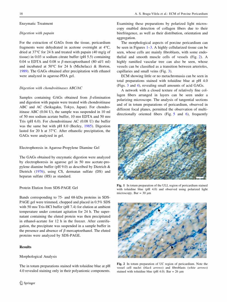
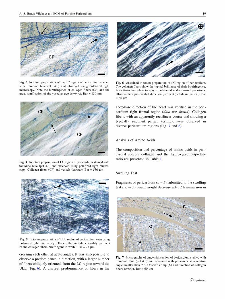
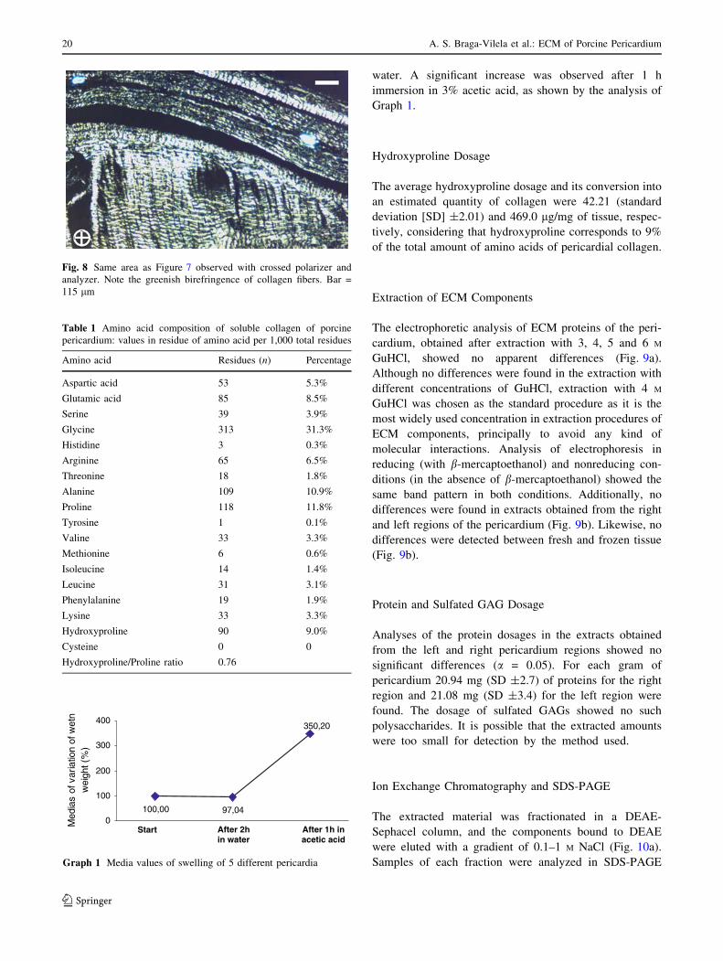
The morphological aspects of porcine pericardium can
be seen in Figures 1–3. A highly cellularized tissue can be
seen, whose cells are mainly fibroblasts, with some endo-
thelial and smooth muscle cells of vessels (Fig. 2). A
highly ramified vascular tree can also be seen, whose
vessels can be classified as a transition between arterioles,
capillaries and small veins (Fig. 3).
ECM showing little or no metachromasia can be seen in
total preparations stained with toluidine blue at pH 4.0
(Figs. 3 and 4), revealing small amounts of acid GAGs.
A network with a closed texture of relatively fine col-
lagen fibers arranged in layers can be seen under a
polarizing microscope. The analysis of tangential sections
and of in totum preparations of pericardium, observed in
different focal planes, permitted the observation of multi-
directionally oriented fibers (Fig. 5 and 6), frequently
Fig. 1 In totum preparation of the ULL region of pericardium stained
with toluidine blue (pH 4.0) and observed using polarized light
microscopy. Bar = 30 lm
Fig. 2 In totum preparation of UC region of pericardium. Note the
vessel cell nuclei (black arrows) and fibroblasts (white arrows)
stained with toluidine blue (pH 4.0). Bar = 26 lm
18 A. S. Braga-Vilela et al.: ECM of Porcine Pericardium
123
crossing each other at acute angles. It was also possible to
observe a predominance in direction, with a larger number
of fibers obliquely oriented, from the LC region toward the
ULL (Fig. 6). A discreet predominance of fibers in the
apex-base direction of the heart was verified in the peri-
cardium right frontal region (data not shown). Collagen
fibers, with an apparently rectilinear course and showing a
typically undulant pattern (crimp), were observed in
diverse pericardium regions (Fig. 7 and 8).
Analysis of Amino Acids
The composition and percentage of amino acids in peri-
cardial soluble collagen and the hydroxyproline/proline
ratio are presented in Table 1.
Swelling Test
Fragments of pericardium (n = 5) submitted to the swelling
test showed a small weight decrease after 2 h immersion in
Fig. 3 In totum preparation of the LC region of pericardium stained
with toluidine blue (pH 4.0) and observed using polarized light
microscopy. Note the birefringence of collagen fibers (CF) and the
great ramification of the vascular tree (arrows). Bar = 130 lm
Fig. 4 In totum preparation of LC region of pericardium stained with
toluidine blue (pH 4.0) and observed using polarized light micros-
copy. Collagen fibers (CF) and vessels (arrows). Bar = 550 lm
Fig. 5 In totum preparation of LLL region of pericardium seen using
polarized light microscopy. Observe the multidirectionality (arrows)
of the collagen fibers birefringent in white. Bar = 77 lm
Fig. 6 Unstained in totum preparation of LC region of pericardium.
The collagen fibers show the typical brilliance of their birefringence,
from first-class white to grayish, observed under crossed polarizers.
Observe their preferential direction (arrows) (details in the text). Bar
= 65 lm
Fig. 7 Micrography of tangential section of pericardium stained with
toluidine blue (pH 4.0) and observed with polarizers at a relative
angle smaller than 90�. Observe crimp (C) and direction of collagen
fibers (arrow). Bar = 60 lm
A. S. Braga-Vilela et al.: ECM of Porcine Pericardium 19
123
water. A significant increase was observed after 1 h
immersion in 3% acetic acid, as shown by the analysis of
Graph 1.
Hydroxyproline Dosage
The average hydroxyproline dosage and its conversion into
an estimated quantity of collagen were 42.21 (standard
deviation [SD] ±2.01) and 469.0 lg/mg of tissue, respec-
tively, considering that hydroxyproline corresponds to 9%
of the total amount of amino acids of pericardial collagen.
Extraction of ECM Components
The electrophoretic analysis of ECM proteins of the peri-
cardium, obtained after extraction with 3, 4, 5 and 6 M
GuHCl, showed no apparent differences (Fig. 9a).
Although no differences were found in the extraction with
different concentrations of GuHCl, extraction with 4 M
GuHCl was chosen as the standard procedure as it is the
most widely used concentration in extraction procedures of
ECM components, principally to avoid any kind of
molecular interactions. Analysis of electrophoresis in
reducing (with b-mercaptoethanol) and nonreducing con-
ditions (in the absence of b-mercaptoethanol) showed the
same band pattern in both conditions. Additionally, no
differences were found in extracts obtained from the right
and left regions of the pericardium (Fig. 9b). Likewise, no
differences were detected between fresh and frozen tissue
(Fig. 9b).
Protein and Sulfated GAG Dosage
Analyses of the protein dosages in the extracts obtained
from the left and right pericardium regions showed no
significant differences (a = 0.05). For each gram of
pericardium 20.94 mg (SD ±2.7) of proteins for the right
region and 21.08 mg (SD ±3.4) for the left region were
found. The dosage of sulfated GAGs showed no such
polysaccharides. It is possible that the extracted amounts
were too small for detection by the method used.
Ion Exchange Chromatography and SDS-PAGE
The extracted material was fractionated in a DEAE-
Sephacel column, and the components bound to DEAE
were eluted with a gradient of 0.1–1 M NaCl (Fig. 10a).
Samples of each fraction were analyzed in SDS-PAGE
Fig. 8 Same area as Figure 7 observed with crossed polarizer and
analyzer. Note the greenish birefringence of collagen fibers. Bar =
115 lm
Table 1 Amino acid composition of soluble collagen of porcine
pericardium: values in residue of amino acid per 1,000 total residues
Amino acid Residues (n) Percentage
Aspartic acid 53 5.3%
Glutamic acid 85 8.5%
Serine 39 3.9%
Glycine 313 31.3%
Histidine 3 0.3%
Arginine 65 6.5%
Threonine 18 1.8%
Alanine 109 10.9%
Proline 118 11.8%
Tyrosine 1 0.1%
Valine 33 3.3%
Methionine 6 0.6%
Isoleucine 14 1.4%
Leucine 31 3.1%
Phenylalanine 19 1.9%
Lysine 33 3.3%
Hydroxyproline 90 9.0%
Cysteine 0 0
Hydroxyproline/Proline ratio 0.76
100,00 97,04
350,20
0
100
200
300
400
Mide
sav fo ar
iitao
w fo ne
nte
wig
ht( %
)
After 1h inacetic acid
After 2hin water
Start
Graph 1 Media values of swelling of 5 different pericardia
20 A. S. Braga-Vilela et al.: ECM of Porcine Pericardium
123
gel. Analysis of the gel showed proteins with the fol-
lowing values of apparent molecular mass: 109, 105, 75,
71, 68, 63, 52, 48, 41, 35, 31, 22, 15 and 11 kDa. Two
polydisperse bands were found averaging 71 and 85 kDa
(Fig. 10b). The bands corresponding to the a1 and a2
collagen chains were found only in the material not
bound to DEAE-Sephacel.
Electrophoresis in Agarose-PDA Gel of GAGs
Obtained by Digestion with Papain
The analysis of GAGs in agarose-PDA gel after digestion of
the tissue with papain showed a large polydisperse band in an
intermediate position between HS and DS. After treatment
with chondroitinases AC and ABC, observation revealed that
the GAG was completely digested by chondroitinase ABC
but not by chondroitinase AC, indicating it was DS (Fig. 11).
The extensive polydisperse band seen in the control probably
contained some contaminant nucleic acid.
20,1
43
67
94
kDa
14,4
30
MM 2 3 4 5 6 7 8 9 col
-SH +SH
A B
kDa
94
67
43
30
right region left region
MM 2 3 4 5 6 7 8 9 10 11 col
α1α2
fresh frozen fresh frozen
Fig. 9 SDS-PAGE of extracts obtained from pericardium. a SDS-
PAGE of proteins extracted with GuHCl in conditions of 3 M (lanes 2
and 6), 4 M (lanes 3 and 7), 5 M (lanes 4 and 8) and 6 M (lanes 5 and 9).
–SH, without b-mercaptoethanol; +SH, with b-mercaptoethanol. b
SDS-PAGE of proteins extracted with 4 M GuHCl from the right and
left parts of fresh and frozen tissues. Observe that the band pattern
was the same in every case. MM, molecular mass markers; col, type I
collagen; a1 and a2, a chains; b, band resulting from two a chains
A
0
0,05
0,1
0,15
0,2
0,25
0,3
0,35
0,4
0,45
0,5
Am
n082
0 5 10 15 20 25 30 35 40
Fractions
MM 8 13 17 18 19 20 21 22 23 24 25 26 27 28 29 32 35 23 col
14,4
30
94
67
20,1
43
kDa
B
Fig. 10 Chromatography in DEAE-Sephacel and SDS-PAGE. aChromatography of material extracted from whole pericardium in 4
M GuHCl. G indicates the beginning of the gradient 0.1–1.0 M NaCl. bSDS-PAGE in the presence of b-mercaptoethanol of fractions eluted
from the column. The apparent molecular masses of proteins eluted
from the column are indicated by arrows. Arrowsheads, polydisperse
bands; MM, molecular mass markers; col, type I collagen
CS
DS
1 2 3
HS
Fig. 11 Electrophoresis in agarose-PDA of GAGs extracted from
pericardium with papain and treated with chondroitinases AC (lane 2)
and ABC (lane 3). The material not treated with chondroitinase
appears as an extensive polydisperse band (lane 1). On the left are the
GAG standards CS, DS and HS. Arrow indicates the direction of the
running
A. S. Braga-Vilela et al.: ECM of Porcine Pericardium 21
123
Electrophoresis in Agarose-PDA Gel of GAGs
Obtained by b-Elimination of Chromatography
Fractions
The polydisperse component with an apparent molecular
mass of 80–100 kDa, probably a small PG, eluted from
DEAE-Sephacel was submitted to b-elimination; and the
liberated GAG was analyzed in agarose-PDA gel (Fig. 12).
The band aligned in the same direction as the standard DS
and was completely digested by chondroitinase ABC but
not by chondroitinase AC, indicating that the polydisperse
component is a small PG containing DS.
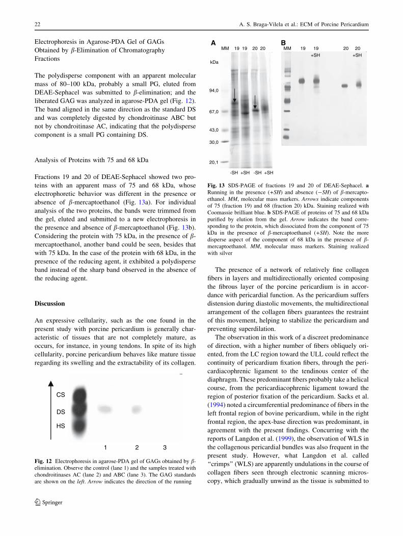
Analysis of Proteins with 75 and 68 kDa
Fractions 19 and 20 of DEAE-Sephacel showed two pro-
teins with an apparent mass of 75 and 68 kDa, whose
electrophoretic behavior was different in the presence or
absence of b-mercaptoethanol (Fig. 13a). For individual
analysis of the two proteins, the bands were trimmed from
the gel, eluted and submitted to a new electrophoresis in
the presence and absence of b-mercaptoethanol (Fig. 13b).
Considering the protein with 75 kDa, in the presence of b-
mercaptoethanol, another band could be seen, besides that
with 75 kDa. In the case of the protein with 68 kDa, in the
presence of the reducing agent, it exhibited a polydisperse
band instead of the sharp band observed in the absence of
the reducing agent.
Discussion
An expressive cellularity, such as the one found in the
present study with porcine pericardium is generally char-
acteristic of tissues that are not completely mature, as
occurs, for instance, in young tendons. In spite of its high
cellularity, porcine pericardium behaves like mature tissue
regarding its swelling and the extractability of its collagen.
The presence of a network of relatively fine collagen
fibers in layers and multidirectionally oriented composing
the fibrous layer of the porcine pericardium is in accor-
dance with pericardial function. As the pericardium suffers
distension during diastolic movements, the multidirectional
arrangement of the collagen fibers guarantees the restraint
of this movement, helping to stabilize the pericardium and
preventing superdilation.
The observation in this work of a discreet predominance
of direction, with a higher number of fibers obliquely ori-
ented, from the LC region toward the ULL could reflect the
continuity of pericardium fixation fibers, through the peri-
cardiacophrenic ligament to the tendinous center of the
diaphragm. These predominant fibers probably take a helical
course, from the pericardiacophrenic ligament toward the
region of posterior fixation of the pericardium. Sacks et al.
(1994) noted a circumferential predominance of fibers in the
left frontal region of bovine pericardium, while in the right
frontal region, the apex-base direction was predominant, in
agreement with the present findings. Concurring with the
reports of Langdon et al. (1999), the observation of WLS in
the collagenous pericardial bundles was also frequent in the
present study. However, what Langdon et al. called
‘‘crimps’’ (WLS) are apparently undulations in the course of
collagen fibers seen through electronic scanning micros-
copy, which gradually unwind as the tissue is submitted to
CS
1 2 3
DS
HS
Fig. 12 Electrophoresis in agarose-PDA gel of GAGs obtained by b-
elimination. Observe the control (lane 1) and the samples treated with
chondroitinases AC (lane 2) and ABC (lane 3). The GAG standards
are shown on the left. Arrow indicates the direction of the running
MM 19 19 20 20
94,0
67,0
43,0
30,0
20,1
kDa
-SH +SH -SH +SH
AMM 19 19 20 20
+SH +SH
B
Fig. 13 SDS-PAGE of fractions 19 and 20 of DEAE-Sephacel. aRunning in the presence (+SH) and absence (-SH) of b-mercapto-
ethanol. MM, molecular mass markers. Arrows indicate components
of 75 (fraction 19) and 68 (fraction 20) kDa. Staining realized with
Coomassie brilliant blue. b SDS-PAGE of proteins of 75 and 68 kDa
purified by elution from the gel. Arrow indicates the band corre-
sponding to the protein, which dissociated from the component of 75
kDa in the presence of b-mercaptoethanol (+SH). Note the more
disperse aspect of the component of 68 kDa in the presence of b-
mercaptoethanol. MM, molecular mass markers. Staining realized
with silver
22 A. S. Braga-Vilela et al.: ECM of Porcine Pericardium
123

tension forces. According to the present work, the occur-
rence of WLS is due to a change in the molecular orientation
in the collagen bundles, even though they may appear rec-
tilinear. These structures are detected by changes in
birefringence of the fibers under polarized light microscopy.
Even when tension forces are applied to the tissue, changes in
the molecular organization of the bundles can still be
detected by observation under polarized light. Sections or in
totum preparations of porcine pericardium stained with
toluidine blue at pH 4.0 showed a pale matrix with little or no
metachromasia, reflecting very small quantities of available
anionic groups. At pH 4.0, toluidine blue bound with car-
boxyl radicals and sulfate of acid GAGs, as well as phosphate
radicals, DNA and RNA, as indicated by the staining
observed in the nuclei of nucleic acid phosphate groups.
Staining with toluidine blue enables the analysis and quan-
tification of available anionic radicals and of electrostatic
combinations, which are important for supramolecular
organization and self-assembling (Vidal, 1995).
The toluidine blue molecule, which in solution behaves
like liquid crystal, is small, has a planar geometry and
possesses excellent optical anisotropic properties (Vidal,
1987). In the ECM, its molecules combine with GAGs,
creating orderly piles and showing metachromasia (Vidal,
1984). The present topochemical findings, which suggest a
low content of available anionic radicals that are important
in the production of metachromasia, are in agreement with
the behavior of pericardium swelling conditions. The PGs
contribute significantly to the physicochemical properties
of the ECM. This is especially due to the large number of
negatively charged groups, carboxyl and sulfate, which
form their chains of GAGs. Thus, the PGs in the cartilage,
e.g., exert swelling pressure on the collagenous matrix,
promoting osmotic resistance to compression loads (Urban
et al., 1979). A hypothesis that should be considered is the
possibility that water fixed by the acid radicals could
concomitantly create hydrostatic pressure. Swelling tests
performed with bovine flexor tendon showed that regions
with relatively low levels of GAGs lose weight in water
and swell in acetic acid (Koob & Vogel, 1987), a fact that
has also been verified with pericardium. In contrast, tendon
regions which swell more in water and less in acetic acid
have relatively high GAG levels. Low levels of PGs are
typical in tendons predominantly subjected to tension for-
ces (Koob & Vogel, 1987). However, the content of
negative charges is but one of the factors that contribute to
the swelling properties of the tissue. Therefore, the type
and ratio of collagen, size and organization of fibrils,
amount of intermolecular crosslinking and interfibrillary
interactions must still be considered, among other factors
(Grodzinsky, 1983). The behavior of pericardium during
the swelling test was typical of highly collagenous tissues
with response dominated, in acid pH, by the collagen
matrix, as described by Viswanadhan, Agrawd & Kramer
(1976) and Yannas & Grodzinsky (1973). The topochem-
ical and physicochemical findings presented here enable us
to state that the pericardial ECM shows small quantities of
PGs.
Despite the apparently small diameter of its bundles,
pericardial collagen proved clearly more insoluble in
solutions most frequently used for its extraction when
compared to collagen from rat tails and bovine flexor
tendons. It has been reported that crosslinks deriving from
hydroxylysine are more stable than those deriving from
lysine and that the great amount of these crosslinks might
be responsible for collagen’s high insolubility, which also
occurs in cardiac valves (Bashey, Bashey & Jimenez,
1978). These authors demonstrated that collagen in the
cardiac valves possesses higher quantities of hydroxylysine
compared to skin collagen, suggesting that the increase in
numbers and in the extension of crosslinks might represent
an evolutionary adaptation of collagen, making it capable
of withstanding the constant mechanical stress to which it
is submitted in the cardiac valves. This fact can also be
applied to pericardial collagen, which is similarly submit-
ted to continuous mechanical stress resulting from systolic
and diastolic movements.
About 47% of pericardium weight corresponds to col-
lagen, estimated by converting the amounts of
hydroxyproline obtained into amounts of sample collagen.
Variations concerning the percentage corresponding to
hydroxyproline in the molecule of type I collagen can be
found in the literature. Analysis of amino acids in collagen
I of porcine pericardium showed that around 9% corre-
spond to hydroxyproline, differing from the reports of
Bashey et al. (1978), Slack, Flint & Thompson (1984),
Riley et al. (1994), Reddy et al. (1998) and Stehno-Bittel
et al. (1998) concerning collagen I of other tissues. Such
findings may suggest a functional adaptation of the colla-
gen molecule, possibly with a consequent change in its
biomechanical properties, adapting them to the mechano-
physiological requirements of the pericardium. The
percentages of the remaining amino acids as well as the
hydroxyproline/proline ratio of 0.76 verified in the soluble
pericardial collagen are compatible with those of type I
collagen (Bashey et al., 1978).
The dosage of proteins and GAGs in the total extracts of
pericardium showed large amounts of proteins and only
traces of GAGs, in total agreement with the topochemical
and physicochemical findings that were obtained.
SDS-PAGE of the material eluted from DEAE-Sephacel
revealed 16 anionic proteins present in the pericardial
matrix, showing apparent molecular masses between 11
and 109 kDa. These proteins perform structural functions
and probably regulatory functions as they interact with the
various matrix components.
A. S. Braga-Vilela et al.: ECM of Porcine Pericardium 23
123
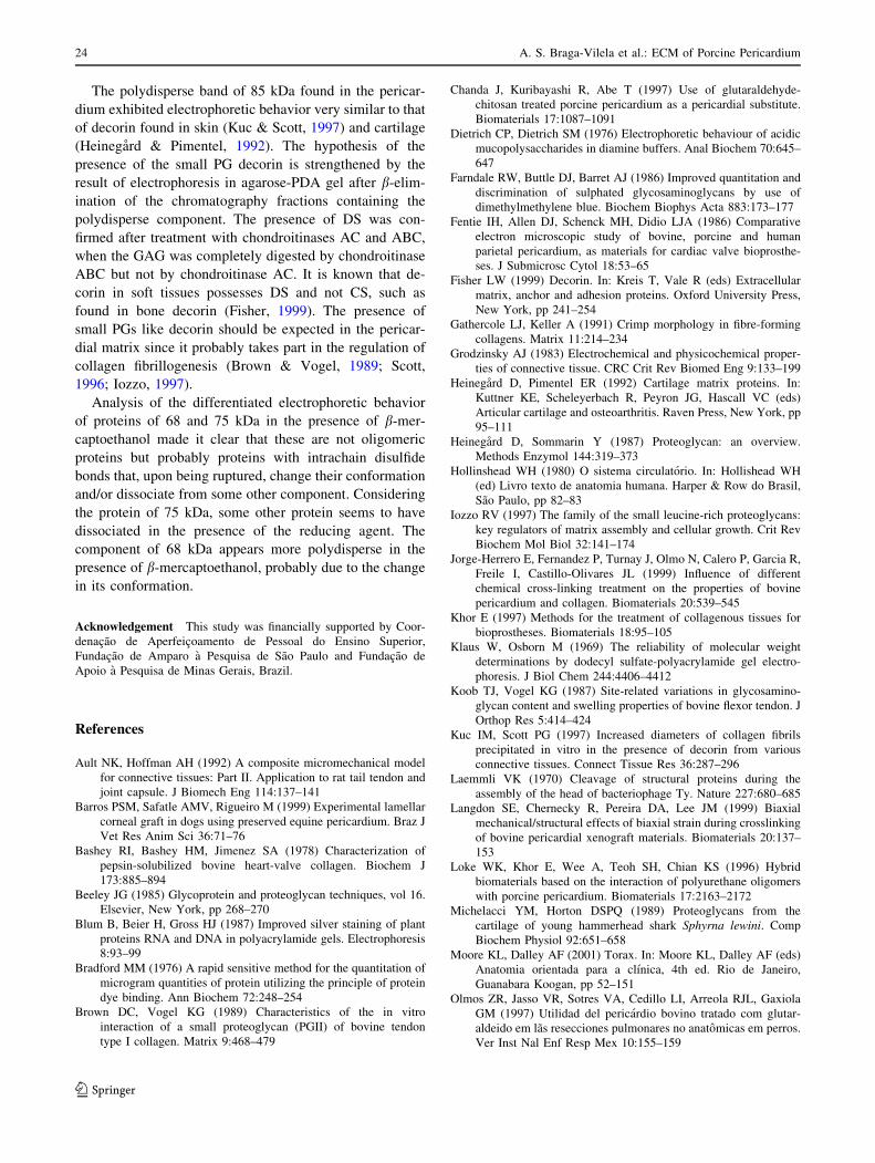
The polydisperse band of 85 kDa found in the pericar-
dium exhibited electrophoretic behavior very similar to that
of decorin found in skin (Kuc & Scott, 1997) and cartilage
(Heinegard & Pimentel, 1992). The hypothesis of the
presence of the small PG decorin is strengthened by the
result of electrophoresis in agarose-PDA gel after b-elim-
ination of the chromatography fractions containing the
polydisperse component. The presence of DS was con-
firmed after treatment with chondroitinases AC and ABC,
when the GAG was completely digested by chondroitinase
ABC but not by chondroitinase AC. It is known that de-
corin in soft tissues possesses DS and not CS, such as
found in bone decorin (Fisher, 1999). The presence of
small PGs like decorin should be expected in the pericar-
dial matrix since it probably takes part in the regulation of
collagen fibrillogenesis (Brown & Vogel, 1989; Scott,
1996; Iozzo, 1997).
Analysis of the differentiated electrophoretic behavior
of proteins of 68 and 75 kDa in the presence of b-mer-
captoethanol made it clear that these are not oligomeric
proteins but probably proteins with intrachain disulfide
bonds that, upon being ruptured, change their conformation
and/or dissociate from some other component. Considering
the protein of 75 kDa, some other protein seems to have
dissociated in the presence of the reducing agent. The
component of 68 kDa appears more polydisperse in the
presence of b-mercaptoethanol, probably due to the change
in its conformation.
Acknowledgement This study was financially supported by Coor-
denacao de Aperfeicoamento de Pessoal do Ensino Superior,
Fundacao de Amparo a Pesquisa de Sao Paulo and Fundacao de
Apoio a Pesquisa de Minas Gerais, Brazil.
References
Ault NK, Hoffman AH (1992) A composite micromechanical model
for connective tissues: Part II. Application to rat tail tendon and
joint capsule. J Biomech Eng 114:137–141
Barros PSM, Safatle AMV, Rigueiro M (1999) Experimental lamellar
corneal graft in dogs using preserved equine pericardium. Braz J
Vet Res Anim Sci 36:71–76
Bashey RI, Bashey HM, Jimenez SA (1978) Characterization of
pepsin-solubilized bovine heart-valve collagen. Biochem J
173:885–894
Beeley JG (1985) Glycoprotein and proteoglycan techniques, vol 16.
Elsevier, New York, pp 268–270
Blum B, Beier H, Gross HJ (1987) Improved silver staining of plant
proteins RNA and DNA in polyacrylamide gels. Electrophoresis
8:93–99
Bradford MM (1976) A rapid sensitive method for the quantitation of
microgram quantities of protein utilizing the principle of protein
dye binding. Ann Biochem 72:248–254
Brown DC, Vogel KG (1989) Characteristics of the in vitro
interaction of a small proteoglycan (PGII) of bovine tendon
type I collagen. Matrix 9:468–479
Chanda J, Kuribayashi R, Abe T (1997) Use of glutaraldehyde-
chitosan treated porcine pericardium as a pericardial substitute.
Biomaterials 17:1087–1091
Dietrich CP, Dietrich SM (1976) Electrophoretic behaviour of acidic
mucopolysaccharides in diamine buffers. Anal Biochem 70:645–
647
Farndale RW, Buttle DJ, Barret AJ (1986) Improved quantitation and
discrimination of sulphated glycosaminoglycans by use of
dimethylmethylene blue. Biochem Biophys Acta 883:173–177
Fentie IH, Allen DJ, Schenck MH, Didio LJA (1986) Comparative
electron microscopic study of bovine, porcine and human
parietal pericardium, as materials for cardiac valve bioprosthe-
ses. J Submicrosc Cytol 18:53–65
Fisher LW (1999) Decorin. In: Kreis T, Vale R (eds) Extracellular
matrix, anchor and adhesion proteins. Oxford University Press,
New York, pp 241–254
Gathercole LJ, Keller A (1991) Crimp morphology in fibre-forming
collagens. Matrix 11:214–234
Grodzinsky AJ (1983) Electrochemical and physicochemical proper-
ties of connective tissue. CRC Crit Rev Biomed Eng 9:133–199
Heinegard D, Pimentel ER (1992) Cartilage matrix proteins. In:
Kuttner KE, Scheleyerbach R, Peyron JG, Hascall VC (eds)
Articular cartilage and osteoarthritis. Raven Press, New York, pp
95–111
Heinegard D, Sommarin Y (1987) Proteoglycan: an overview.
Methods Enzymol 144:319–373
Hollinshead WH (1980) O sistema circulatorio. In: Hollishead WH
(ed) Livro texto de anatomia humana. Harper & Row do Brasil,
Sao Paulo, pp 82–83
Iozzo RV (1997) The family of the small leucine-rich proteoglycans:
key regulators of matrix assembly and cellular growth. Crit Rev
Biochem Mol Biol 32:141–174
Jorge-Herrero E, Fernandez P, Turnay J, Olmo N, Calero P, Garcia R,
Freile I, Castillo-Olivares JL (1999) Influence of different
chemical cross-linking treatment on the properties of bovine
pericardium and collagen. Biomaterials 20:539–545
Khor E (1997) Methods for the treatment of collagenous tissues for
bioprostheses. Biomaterials 18:95–105
Klaus W, Osborn M (1969) The reliability of molecular weight
determinations by dodecyl sulfate-polyacrylamide gel electro-
phoresis. J Biol Chem 244:4406–4412
Koob TJ, Vogel KG (1987) Site-related variations in glycosamino-
glycan content and swelling properties of bovine flexor tendon. J
Orthop Res 5:414–424
Kuc IM, Scott PG (1997) Increased diameters of collagen fibrils
precipitated in vitro in the presence of decorin from various
connective tissues. Connect Tissue Res 36:287–296
Laemmli VK (1970) Cleavage of structural proteins during the
assembly of the head of bacteriophage Ty. Nature 227:680–685
Langdon SE, Chernecky R, Pereira DA, Lee JM (1999) Biaxial
mechanical/structural effects of biaxial strain during crosslinking
of bovine pericardial xenograft materials. Biomaterials 20:137–
153
Loke WK, Khor E, Wee A, Teoh SH, Chian KS (1996) Hybrid
biomaterials based on the interaction of polyurethane oligomers
with porcine pericardium. Biomaterials 17:2163–2172
Michelacci YM, Horton DSPQ (1989) Proteoglycans from the
cartilage of young hammerhead shark Sphyrna lewini. Comp
Biochem Physiol 92:651–658
Moore KL, Dalley AF (2001) Torax. In: Moore KL, Dalley AF (eds)
Anatomia orientada para a clınica, 4th ed. Rio de Janeiro,
Guanabara Koogan, pp 52–151
Olmos ZR, Jasso VR, Sotres VA, Cedillo LI, Arreola RJL, Gaxiola
GM (1997) Utilidad del pericardio bovino tratado com glutar-
aldeido em las resecciones pulmonares no anatomicas em perros.
Ver Inst Nal Enf Resp Mex 10:155–159
24 A. S. Braga-Vilela et al.: ECM of Porcine Pericardium
123

Petite H, Duval J, Frei V, Abdul-Malak N, Sigot-Luizard M, Herbage
D (1995) Cytocompatibility of calf pericardium treated by
glutaraldehyde and by the acyl azide methods in an organotypic
culture model. Biomaterials 16:1003–1008
Pires AC, Saporito WF, Leao LEV (1997) Pericardio bovino utilizado
como remendo no sistema cardiovascular. Ver Bras Cir Cardi-
vasc 12:176–187
Reddy GK, Gum S, Stehno-Bittel L, Enwemeka CS (1998)
Biochemistry and biomechanics of healing tendon: Part I.
Effects of rigid plaster casts and functional casts. Med Sci
Sports Exerc 30:794–800
Riley GP, Harrall RL, Constant CR, Chard MD, Cawston TE,
Hazleman BL (1994) Tendon degeneration and chronic shoulder
pain: changes in the collagen composition of the human rotator
cuff tendons in rotator cuff tendinitis. Ann Rheum Dis 53:359–
366
Sacks MS, Chuong CJC, More R (1994) Collagen fiber architeture of
bovine pericardium. ASAIO J 40:M632–M637
Scott JE (1996) Proteodermatan and proteokeratan sulfate (decorin,
lumican/ fibromodulin) proteins are horse shoe shaped. Impli-
cations for their interactions with collagen. Biochemistry
35:8795–8799
Simionescu D, Iozzo R, Kefalides NA (1989) Bovine pericardial
proteoglycan: biochemical, immunochemical and ultrastructural
studies. Matrix 9:301–310
Slack C, Flint MH, Thompson BM (1984) The effect of tensional load
on isolated embryonic chick tendons in organ culture. Connect
Tissue Res 12:229–247
Stegemann H, Stalder K (1967) Determination of hydroxyproline.
Clin Chim Acta 18:267–273
Stehno-Bittel L, Reddy GK, Gum S, Enwemeka CS (1998)
Biochemistry and biomechanics of healing tendon: Part I.
Effects of rigid plaster casts and functional casts. Med Sci
Sports Exerc 30:788–793
Urban JPG, Maroudas A, Bayliss MT, Dillon MT (1979) Swelling
pressures of proteoglycans at concentrations found in cartilag-
inous tissues. Biorheology 16:447–464
Vidal BC (1966) Macromolecular disorientation in detached tendons.
Protoplasma Bd 62:121–131
Vidal BC (1980) The part played by proteoglycans and structural
glycoproteins in the macromolecular orientation of collagen
bundles. Cell Mol Biol 26:415–421
Vidal BC (1984) Ordem molecular y haces de colageno. Trab Inst
Cajal 75:19–27
Vidal BC (1987) Metodos em Biologia Celular. In: Vidal BC, Mello
MLS (eds) Biologia Celular. Sao Paulo, Atheneu, pp 5–34
Vidal BC (1995) From collagen type I solution to fibers with a helical
pattern: a self-assembly phenomenon. C R Acad Sci Paris
318:831–836
Vidal BC (2003) Image analysis of tendon helical superstructure
using interference and polarized light microscopy. Micron
34:423–432
Vidal BC, Mello MLS (1972) Anisotropic properties of toluidine
blue-stained collagen. Ann Histochim 18:106–122
Viswanadham RK, Agrawd DC, Kramer E (1976) Environmental
effects on the mechanical properties of reconstituted collagen
hollow fiber membranes. J Poly Sci Phys 14:2195–2209
Whittaker P, Boughner DR, Perkins DG, Canham PB (1987)
Quantitative structural analysis of collagen in chordae tendineae
and its relation to floppy mitral valves and proteoglycan
infiltration. Br Heart 57:264–269
Yannas IV, Grodzinsky AJ (1973) Electromechanical energy conver-
sion with collagen fibers in an aqueous medium. J Mechanochem
Cell Motility 2:113–125
Zingales B (1984) Analysis of protein sodium dodecyl sulphate-
polyacrylamide gel electrophoresis. In: Genes and antigens of
parasites. Rio de Janeiro, Fiocruz, pp 357–363
A. S. Braga-Vilela et al.: ECM of Porcine Pericardium 25
123