Principles of Biochemistry
93
Principles of Biochemistry Third Edition International Student Version Chapter 13 Biochemical Signaling Copyright © 2008 by John Wiley & Sons, Inc. Donald Voet • Judith G. Voet • Charlotte W. Pratt
-
Upload
khangminh22 -
Category
Documents
-
view
0 -
download
0
Transcript of Principles of Biochemistry
Principles of Biochemistry Third Edition
International Student Version
Chapter 13 Biochemical Signaling
Copyright © 2008 by John Wiley & Sons, Inc.
Donald Voet • Judith G. Voet • Charlotte W. Pratt

Figure 13-1
HORMONES
• Maintenir Homeostase • Répondre à des stimuli externes • Réguler des cycles (maturation, cycle mentruel, differentiation, etc)
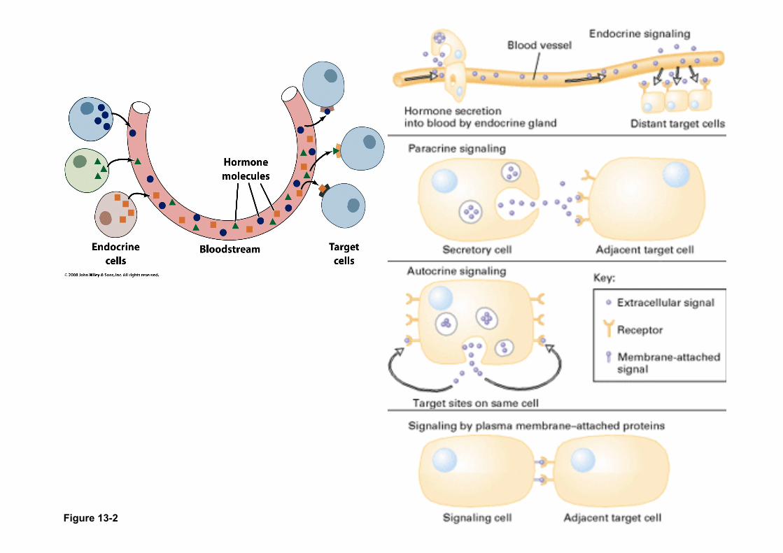
Figure 13-2

Receptors = Proteins that bind signals and initiate a signaling cascade
Cell membrane receptors -integral membrane proteins that bind an extracellular signal and start a signal cascade
Intracellular receptors -nuclear hormone receptors
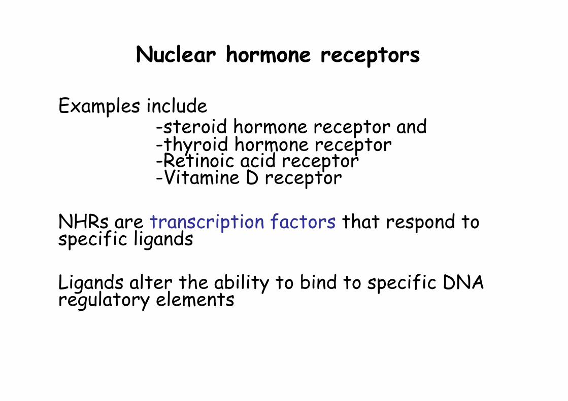
Nuclear hormone receptors
Examples include -steroid hormone receptor and -thyroid hormone receptor -Retinoic acid receptor -Vitamine D receptor
NHRs are transcription factors that respond to specific ligands
Ligands alter the ability to bind to specific DNA regulatory elements
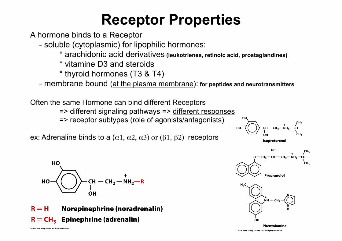
Receptor Properties A hormone binds to a Receptor - soluble (cytoplasmic) for lipophilic hormones: * arachidonic acid derivatives (leukotrienes, retinoic acid, prostaglandines) * vitamine D3 and steroids * thyroid hormones (T3 & T4) - membrane bound (at the plasma membrane): for peptides and neurotransmitters
Often the same Hormone can bind different Receptors => different signaling pathways => different responses => receptor subtypes (role of agonists/antagonists)
ex: Adrenaline binds to a (α1, α2, α3) or (β1, β2) receptors

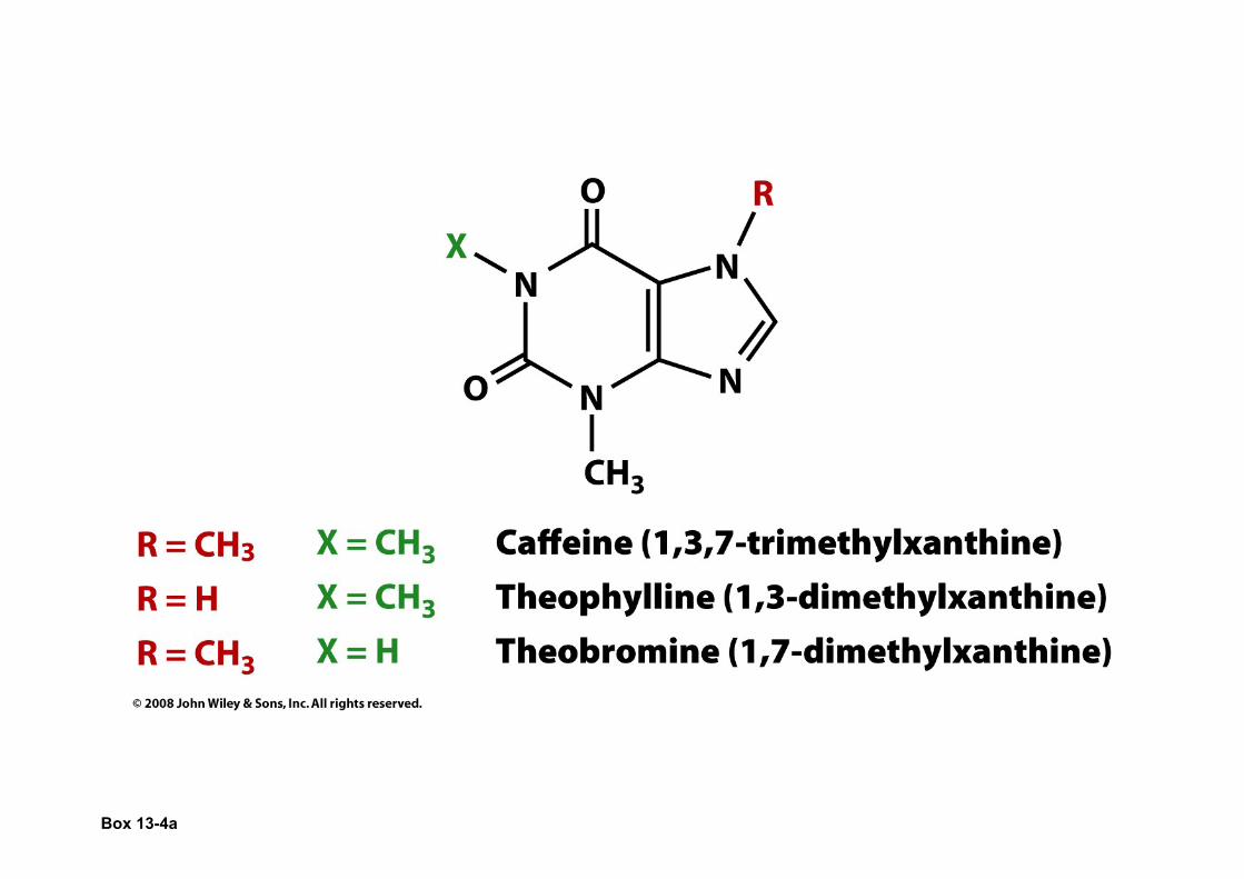
Box 13-2a
Liaison Ligand-Recepteur
Diagramme de Scatchard Diagramme hyperbolique
(R)(L) ((R)T – (R*L))(L) KL = __________ = _____________________
(R*L) (R*L)
B (Bmax – B) __ = _____________
F KL

General Principles of Signal Transduction 1. Communication usually
involves (i) a signaling molecule, (ii) a receptor, (iii) intracellular signal
transducers and (iv) targets
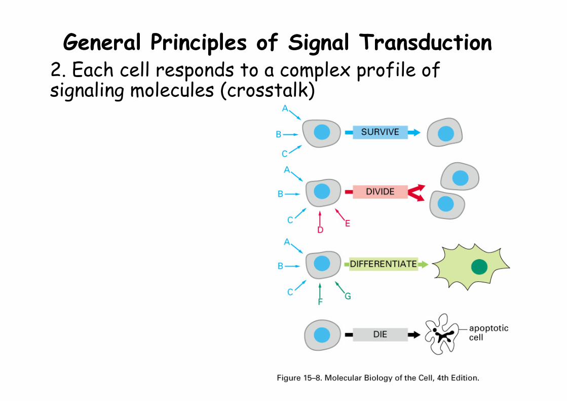
General Principles of Signal Transduction 2. Each cell responds to a complex profile of signaling molecules (crosstalk)
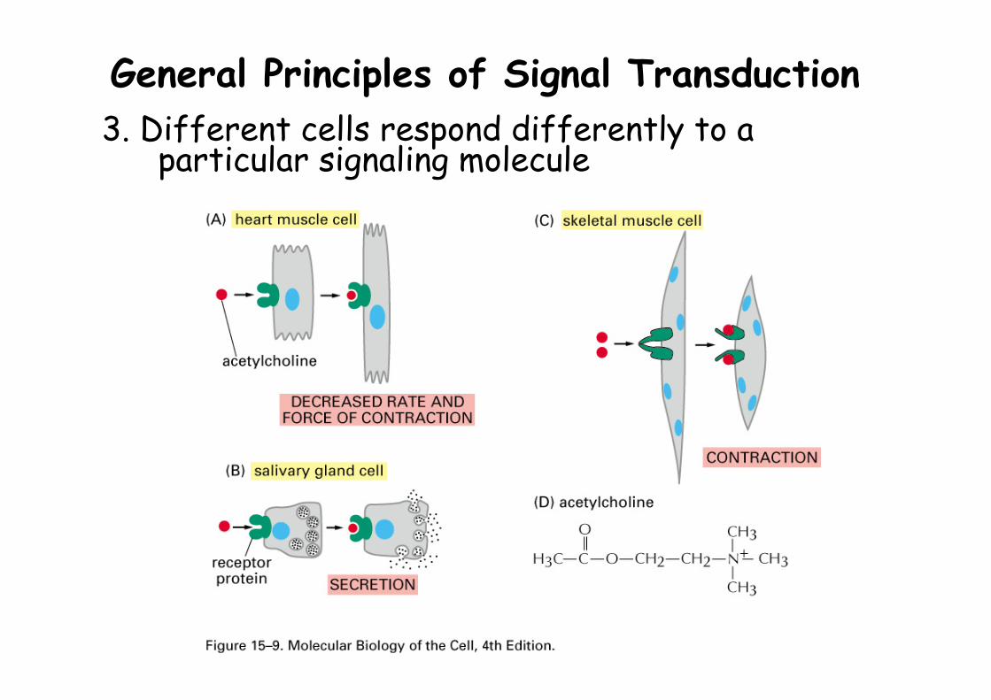
General Principles of Signal Transduction 3. Different cells respond differently to a
particular signaling molecule
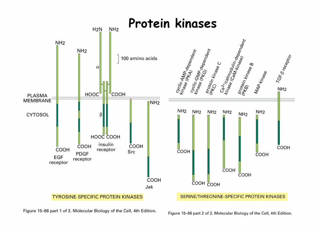
Protein kinases
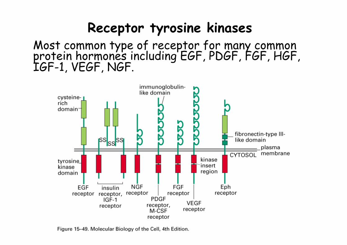
Receptor tyrosine kinases Most common type of receptor for many common protein hormones including EGF, PDGF, FGF, HGF, IGF-1, VEGF, NGF.
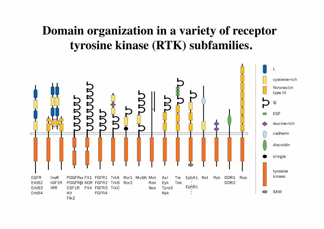
Domain organization in a variety of receptor tyrosine kinase (RTK) subfamilies.
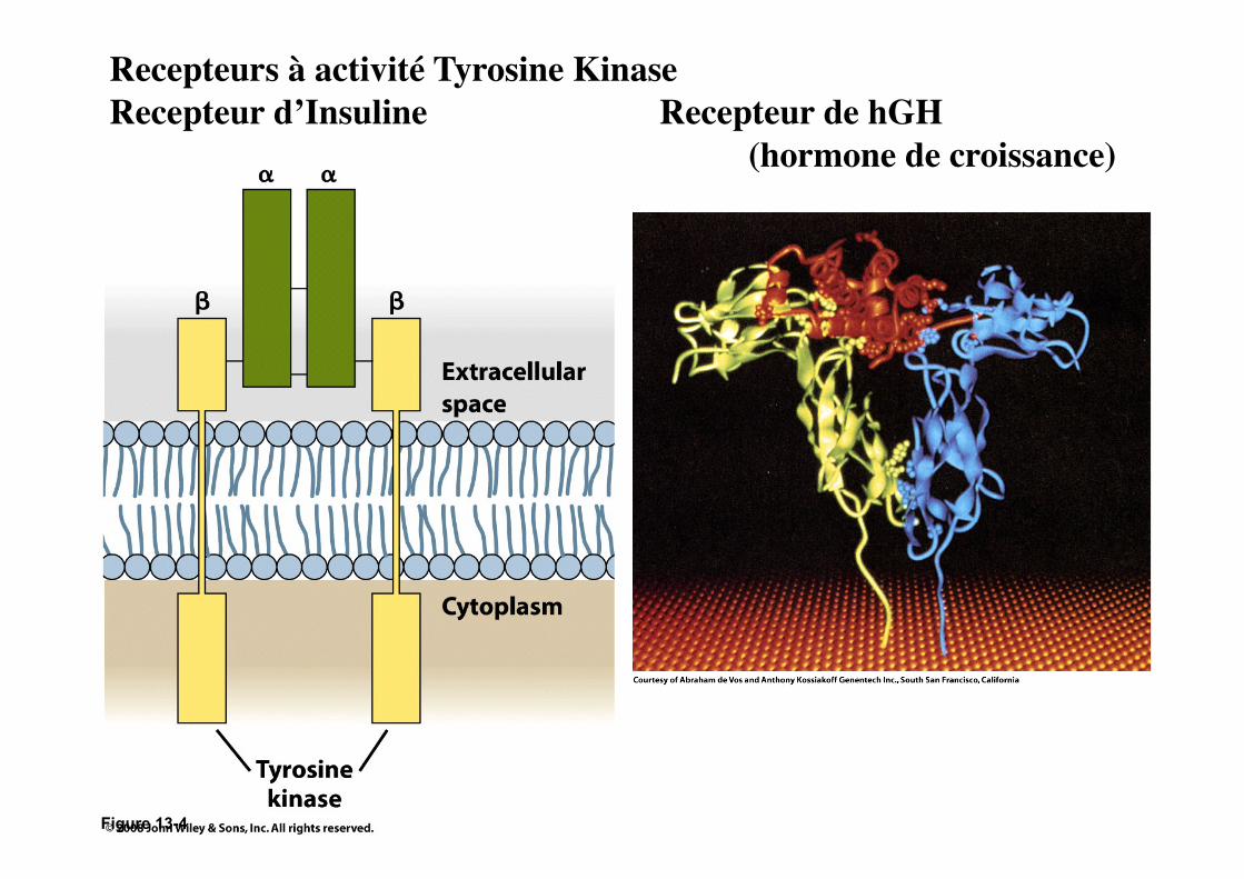
Figure 13-4
Recepteurs à activité Tyrosine Kinase Recepteur d’Insuline Recepteur de hGH (hormone de croissance)
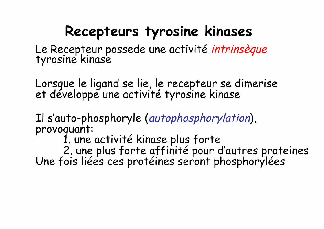
Recepteurs tyrosine kinases Le Recepteur possede une activité intrinsèque tyrosine kinase
Lorsque le ligand se lie, le recepteur se dimerise et développe une activité tyrosine kinase
Il s’auto-phosphoryle (autophosphorylation), provoquant: 1. une activité kinase plus forte 2. une plus forte affinité pour d’autres proteines Une fois liées ces protéines seront phosphorylées
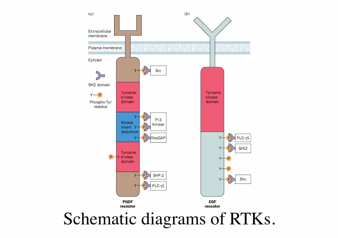
Schematic diagrams of RTKs.
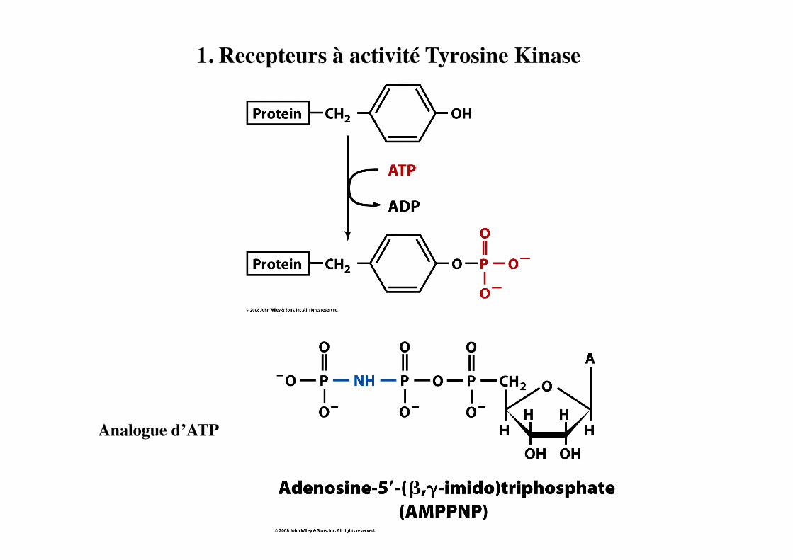
1. Recepteurs à activité Tyrosine Kinase
Analogue d’ATP

Structure du Domaine Tyrosine Kinase (Recepteur d’Insuline)
Jaune=déphosphorylé Vert=phophorylé
PTK Domain undergoes major conformation change & autophosphorylation (1 to 3 Tyr residues)
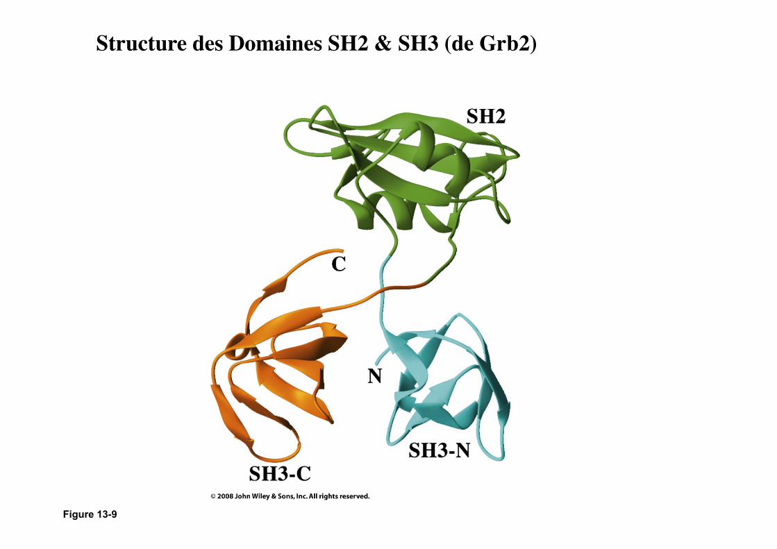
Figure 13-9
Structure des Domaines SH2 & SH3 (de Grb2)
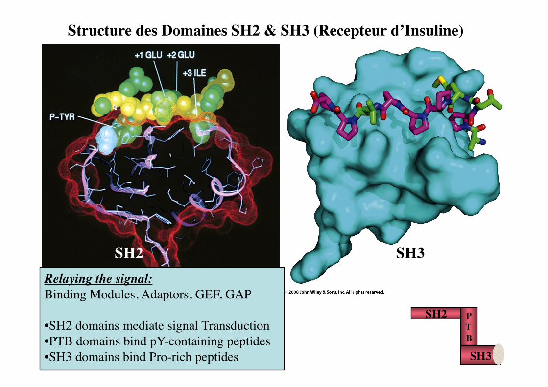
Structure des Domaines SH2 & SH3 (Recepteur d’Insuline)
SH2 SH3
SH2 P T B SH3
Relaying the signal: Binding Modules, Adaptors, GEF, GAP
• SH2 domains mediate signal Transduction • PTB domains bind pY-containing peptides • SH3 domains bind Pro-rich peptides

Voet Biochemistry 3e © 2004 John Wiley & Sons, Inc.
Structure du domaine SH3 de Abl dans le complexe le decapeptide Pro-rich (APTMPPPLPP).
Pag
e 69
3
SH3 domain: Molecular velcro: mediate interactions between kinases & regulatory proteins present in great variety of proteins • receptor Tyrosine Kinases • non-Receptor Tyrosine Kinases, • adaptor proteins (ex. Grb2) • structural proteins (myosin, spectrin) bind Pro-rich peptides
SH2
SH3 SOS Ras P
GF
P P P
GRB2
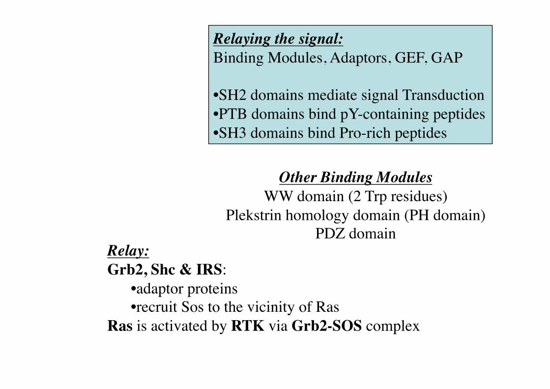
Other Binding Modules �WW domain (2 Trp residues)�
Plekstrin homology domain (PH domain)�PDZ domain
Relaying the signal: Binding Modules, Adaptors, GEF, GAP
• SH2 domains mediate signal Transduction • PTB domains bind pY-containing peptides • SH3 domains bind Pro-rich peptides
Relay: Grb2, Shc & IRS:
• adaptor proteins • recruit Sos to the vicinity of Ras
Ras is activated by RTK via Grb2-SOS complex
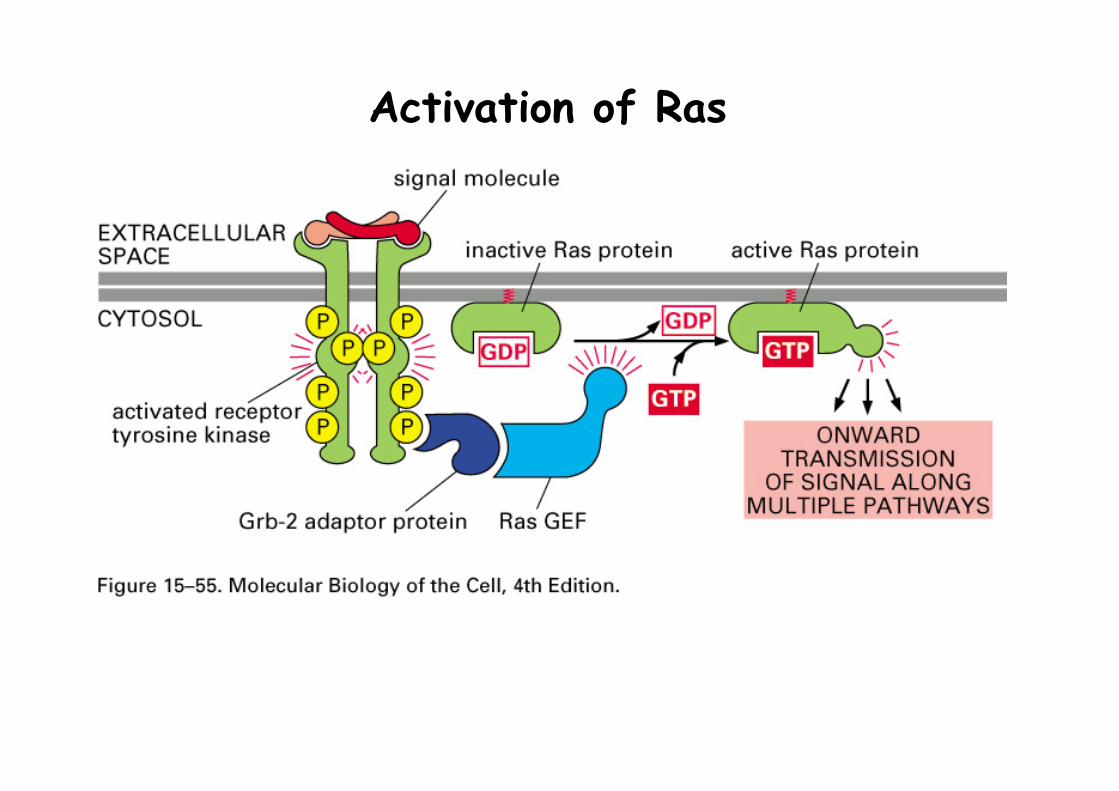
Activation of Ras
Voet Biochemistry 3e © 2004 John Wiley & Sons, Inc.
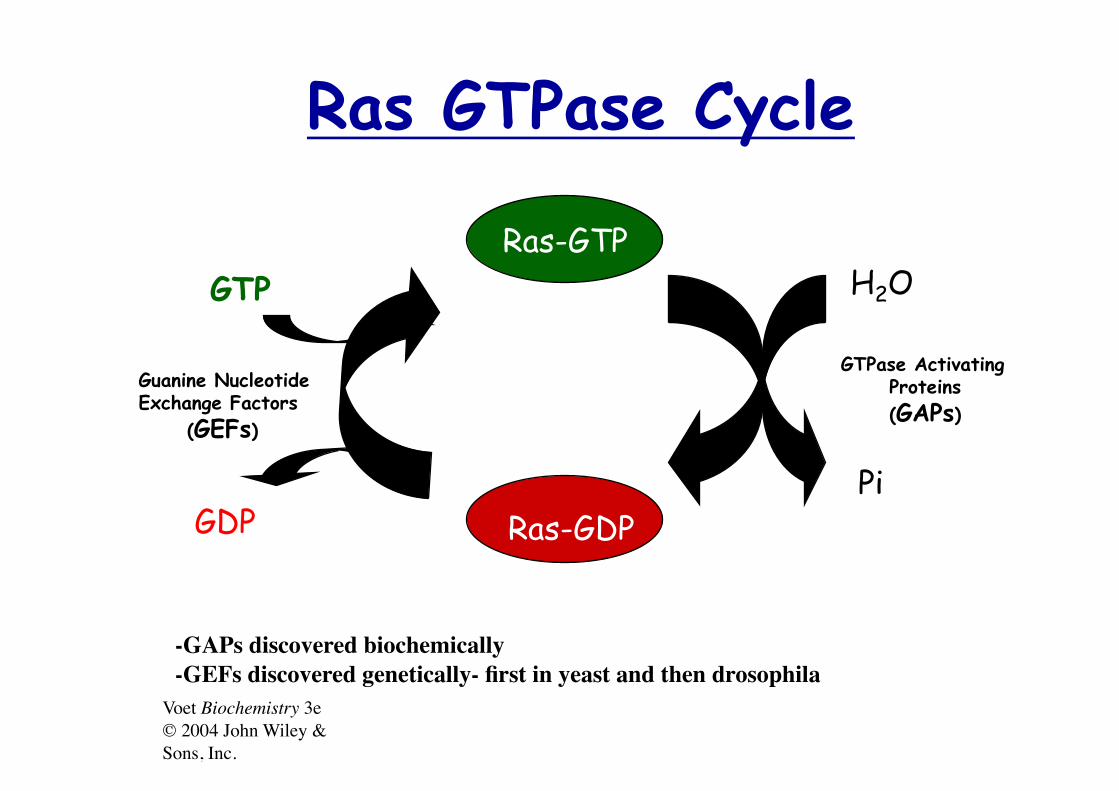
Ras GTPase Cycle
Ras-GTP
Ras-GDP
GTP
GDP
H2O
Pi
Guanine Nucleotide Exchange Factors (GEFs)
GTPase Activating Proteins (GAPs)
-GAPs discovered biochemically -GEFs discovered genetically- first in yeast and then drosophila

Voet Biochemistry 3e © 2004 John Wiley & Sons, Inc.
Ras Superfamily Ras Rho Rab Arf Ran
H-Ras N-Ras K-Ras
TC21
Rap1 Rap2
R-Ras
RalA RalB
RhoA RhoB RhoC
RhoG RhoE
CDC42
Rac1 Rac2
Rab1-N Arf1-6 Ran
Growth/ Cytoskeleton Vesicle sorting Differentiation NuclearTranslocation
Voet Biochemistry 3e © 2004 John Wiley & Sons, Inc.
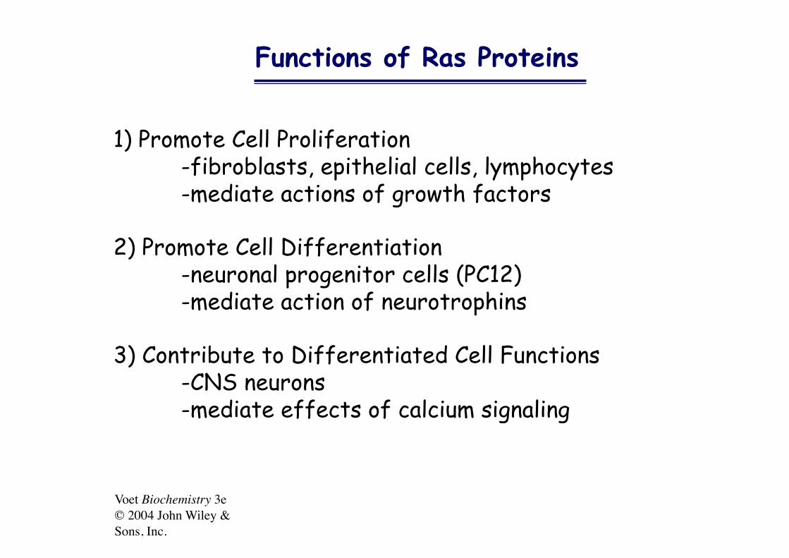
Functions of Ras Proteins
1) Promote Cell Proliferation -fibroblasts, epithelial cells, lymphocytes -mediate actions of growth factors
2) Promote Cell Differentiation -neuronal progenitor cells (PC12) -mediate action of neurotrophins
3) Contribute to Differentiated Cell Functions -CNS neurons -mediate effects of calcium signaling

Figure 13-10
Complexe Ras-GDP-GAP43-AlF3
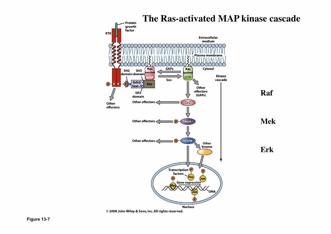
Figure 13-7
Raf
Mek
Erk
The Ras-activated MAP kinase cascade

MAP KINASE • The mitogen-activated protein kinase (MAPK) pathways
are typically comprised of a three-member protein kinase cascade.
• Specificity of MAPK responses is achieved by activation of different three-kinase modules.
• There are at least three sets of mammalian MAPK modules. – the extracellular-signal-regulated kinases (ERKs), – the Jun N-terminal kinases (JNKs) – the p38 kinases.
• As a group, the MAPKs are major players in mediating a variety of signals for cell proliferation and differentiation.

Figure 13-11
Cascade MAP-kinase
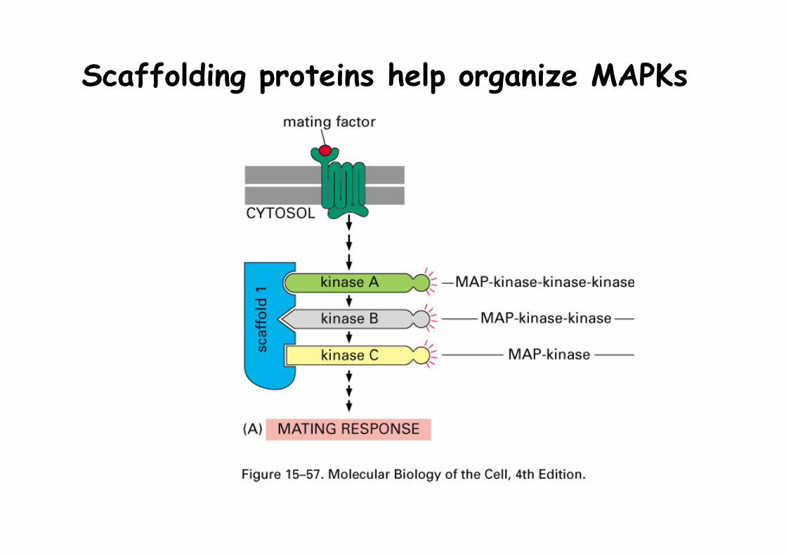
Scaffolding proteins help organize MAPKs
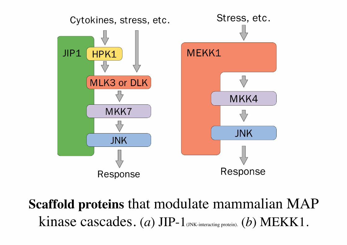
Scaffold proteins that modulate mammalian MAP kinase cascades. (a) JIP-1(JNK-interacting protein). (b) MEKK1.

Structure de Src (= non-Receptor Tyrosine kinase) • Many NRTK are activated by tyrosine kinase-associated receptors of the Scr family • Examples of TK-associated receptors: Src, Fyn, Lck
• Autoinhibitory mechanims of Src

Structure de Src (=non Receptor Tyrosine kinase) Modèle d’activation
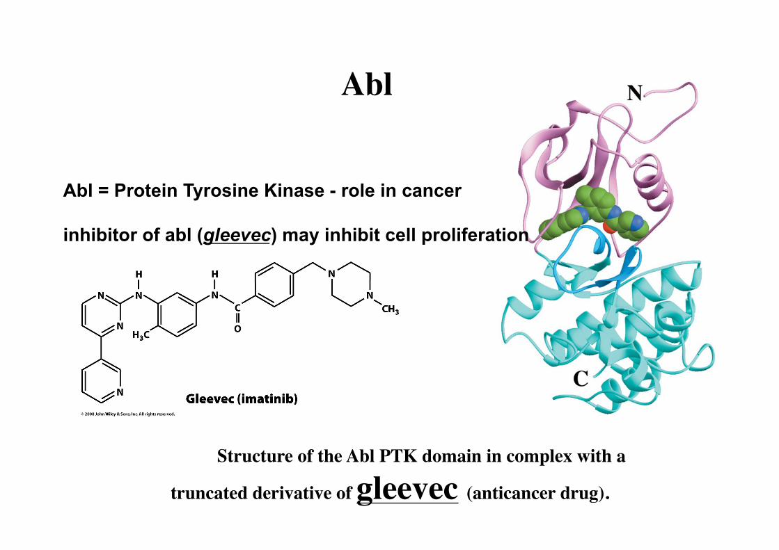
Structure of the Abl PTK domain in complex with a
truncated derivative of gleevec (anticancer drug).
Abl = Protein Tyrosine Kinase - role in cancer
inhibitor of abl (gleevec) may inhibit cell proliferation
Abl
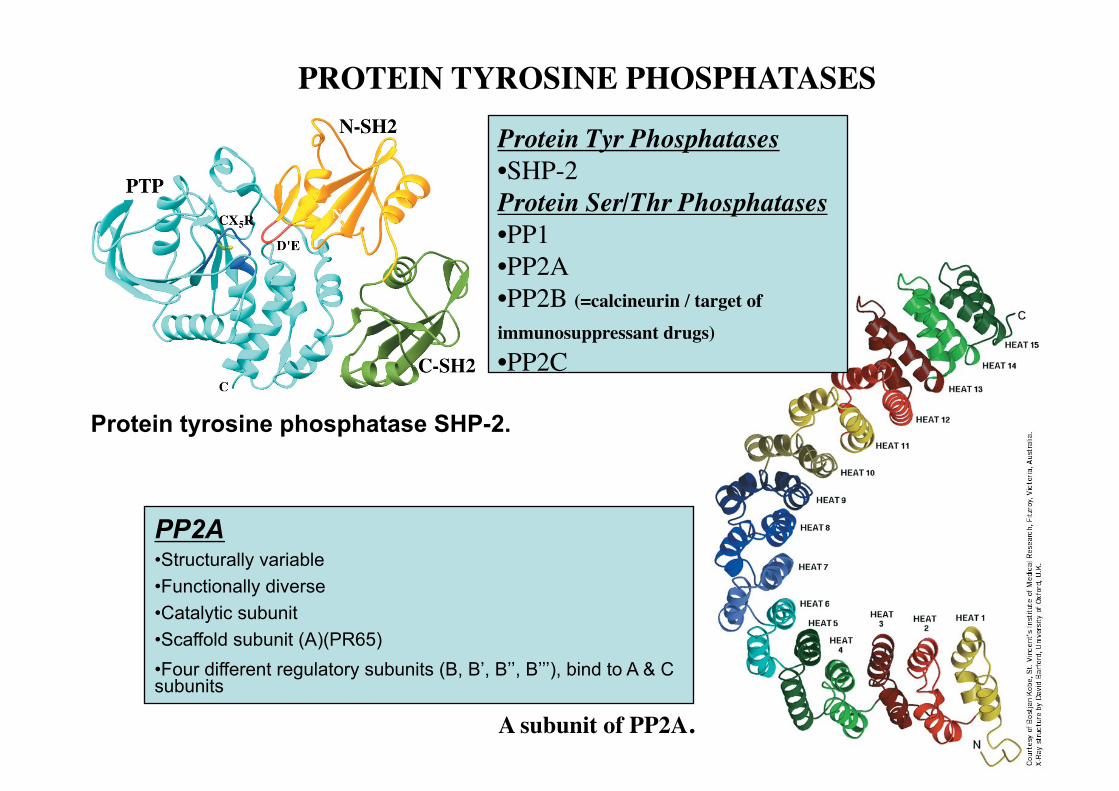
Protein tyrosine phosphatase SHP-2.
Protein Tyr Phosphatases • SHP-2 Protein Ser/Thr Phosphatases • PP1 • PP2A • PP2B (=calcineurin / target of
immunosuppressant drugs) • PP2C
PP2A • Structurally variable • Functionally diverse • Catalytic subunit • Scaffold subunit (A)(PR65) • Four different regulatory subunits (B, B’, B’’, B’’’), bind to A & C subunits
A subunit of PP2A.
PROTEIN TYROSINE PHOSPHATASES

PP2A
Scaffold subunit: HEAT repeats
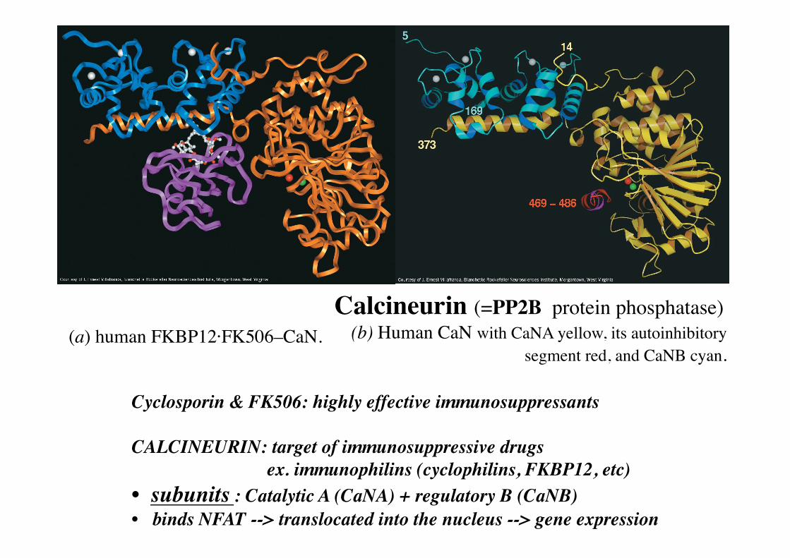
Calcineurin (=PP2B protein phosphatase) (b) Human CaN with CaNA yellow, its autoinhibitory
segment red, and CaNB cyan.
Cyclosporin & FK506: highly effective immunosuppressants
CALCINEURIN: target of immunosuppressive drugs ex. immunophilins (cyclophilins, FKBP12, etc) • subunits : Catalytic A (CaNA) + regulatory B (CaNB) • binds NFAT --> translocated into the nucleus --> gene expression
(a) human FKBP12·FK506–CaN.

JAK-STAT Pathway JAK: = Janus kinases - contiennent 2 domaines Tyrosine Kinase
STAT: Signal Transducers and Activators of Transduction = Transcription Factors, activated by JAK
1. - Ligand binding dimerizes the receptor (β1 and β2 subunits) 2. - Receptor dimers bind JAK and induce phosphorylation of JAK 3. - Phosphorylated JAK phosphorylate the Receptor Subunits 4. - Phophorylated Receptor can phosphorylate STAT 5. - Phophorylated STAT dimerizes 6. - Dimeric, phosphorylated STAT moves to the nucleus and act as
Transcription Factors 7. - ---> gene expression
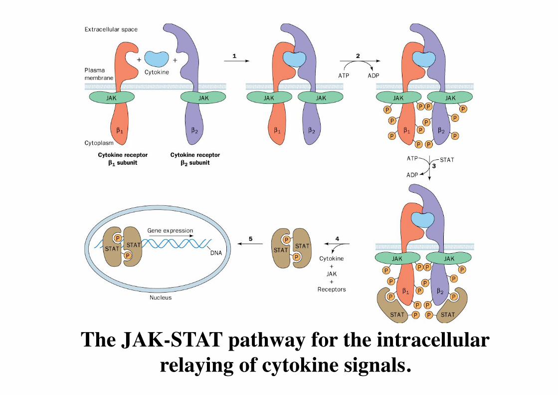
The JAK-STAT pathway for the intracellular relaying of cytokine signals.

JAK-STAT

JAK-STAT Pathway
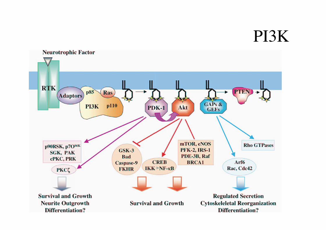
PI3K
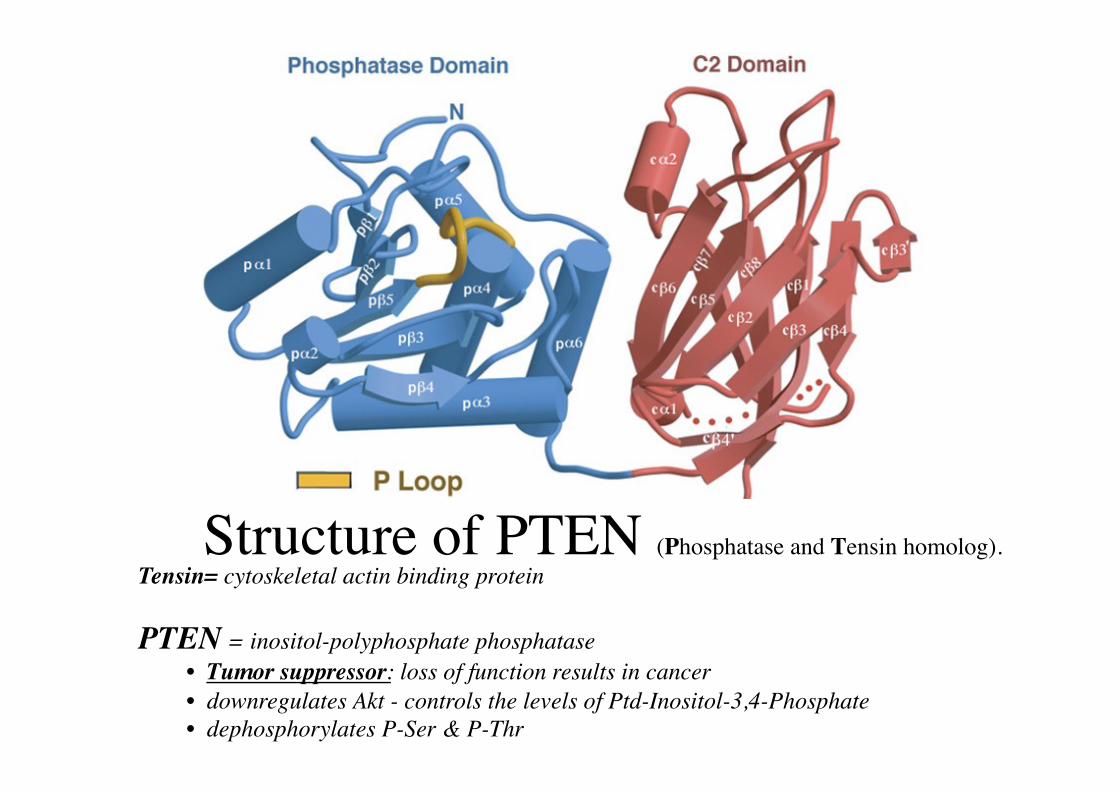
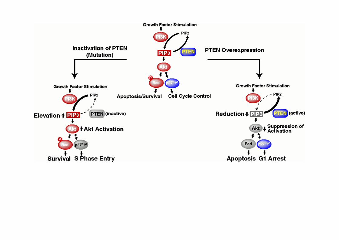
Structure of PTEN (Phosphatase and Tensin homolog). Tensin= cytoskeletal actin binding protein
PTEN = inositol-polyphosphate phosphatase • Tumor suppressor: loss of function results in cancer • downregulates Akt - controls the levels of Ptd-Inositol-3,4-Phosphate • dephosphorylates P-Ser & P-Thr
Heterotrimeric G-Proteins

Figure 13-17
GPCR = G-Protein Coupled Receptor
Très grande famille de récepteurs Recepteurs monomèriques 7 domaines transmembranaires (7TM)
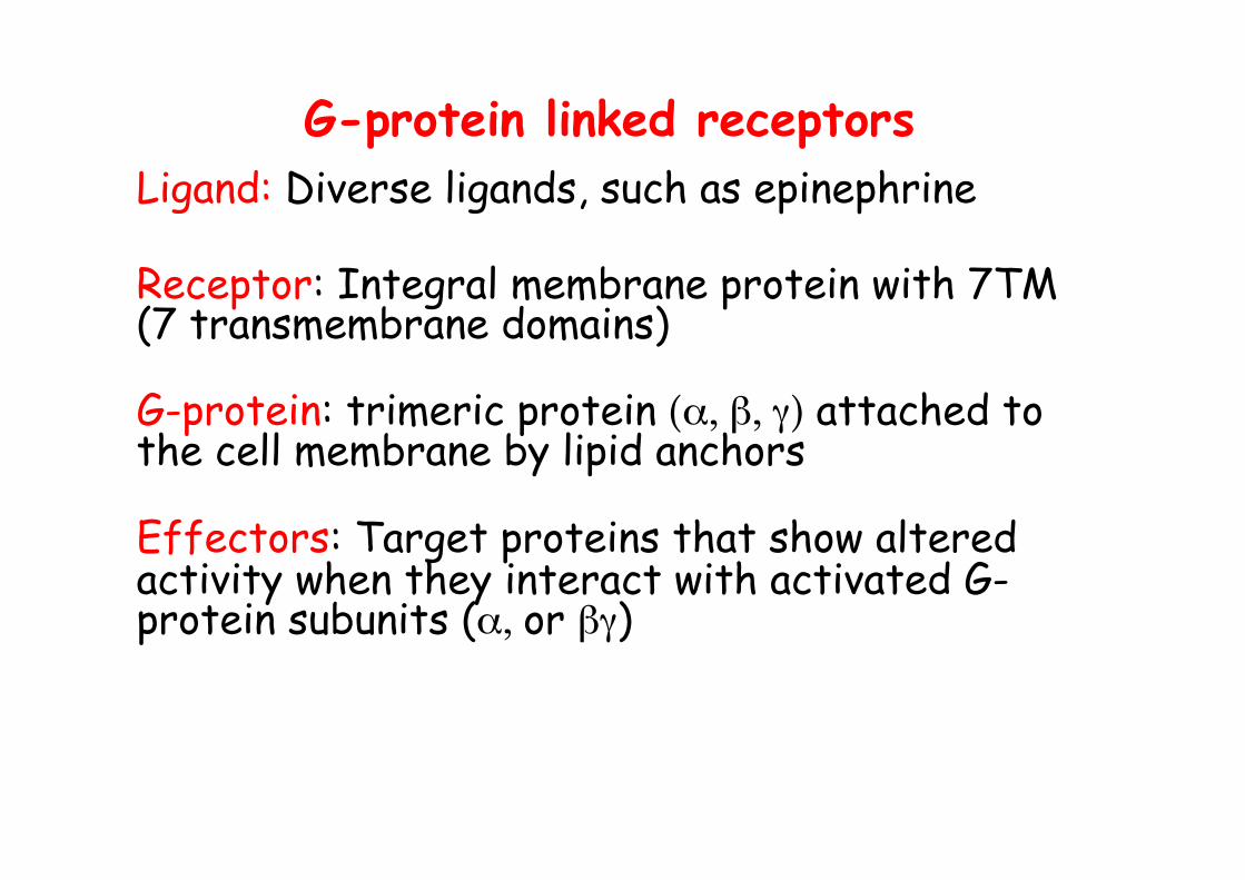
G-protein linked receptors Ligand: Diverse ligands, such as epinephrine
Receptor: Integral membrane protein with 7TM (7 transmembrane domains)
G-protein: trimeric protein (α, β, γ) attached to the cell membrane by lipid anchors
Effectors: Target proteins that show altered activity when they interact with activated G-protein subunits (α, or βγ)

G-protein coupled Receptor

Figure 13-18: Rhodopsine

G-protein linked receptors and G-proteins
Receptor
G-protein
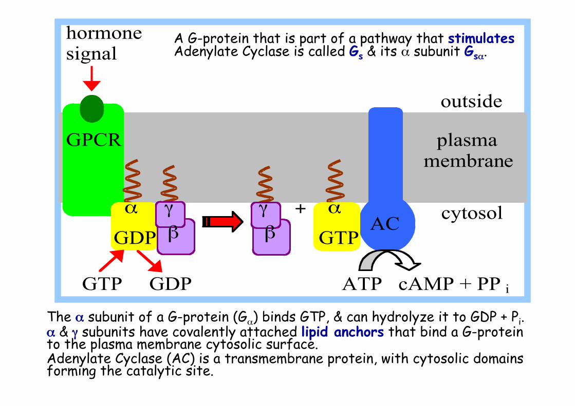
The α subunit of a G-protein (Gα) binds GTP, & can hydrolyze it to GDP + Pi. α & γ subunits have covalently attached lipid anchors that bind a G-protein to the plasma membrane cytosolic surface. Adenylate Cyclase (AC) is a transmembrane protein, with cytosolic domains forming the catalytic site.
AC
hormone signal outside GPCR plasma membrane
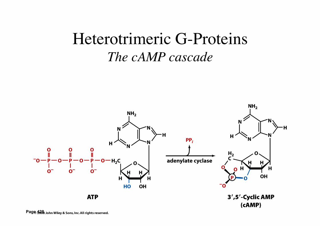
GTP GDP ATP cAMP + PP i
α γ γ + α cytosol GDP β β GTP
A G-protein that is part of a pathway that stimulates Adenylate Cyclase is called Gs & its α subunit Gsα.
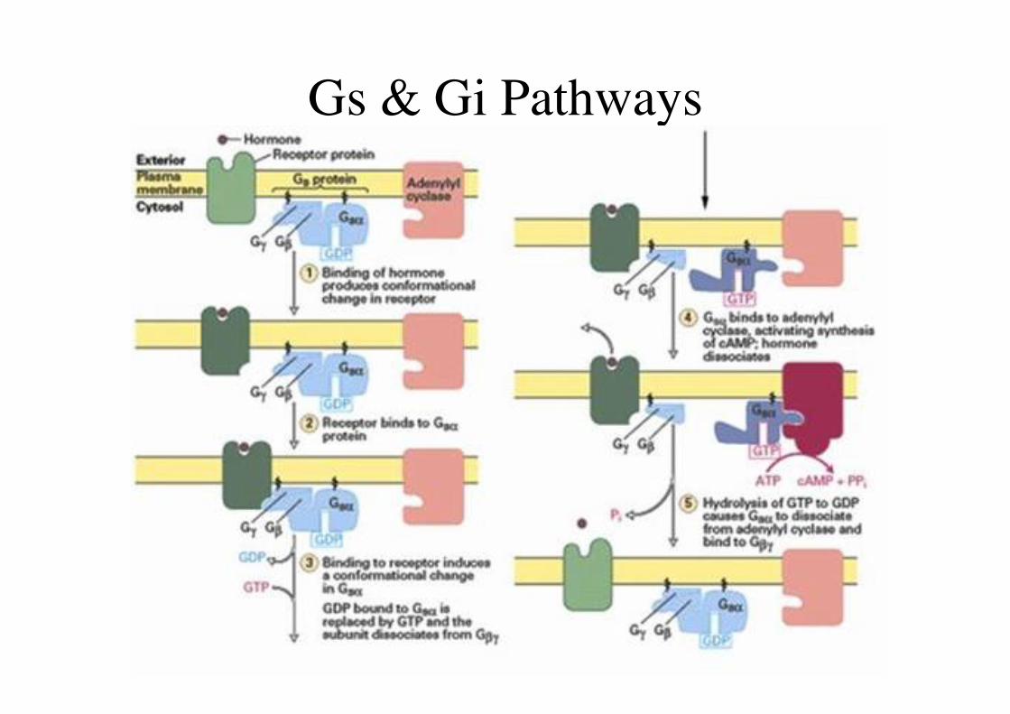
Gs & Gi Pathways
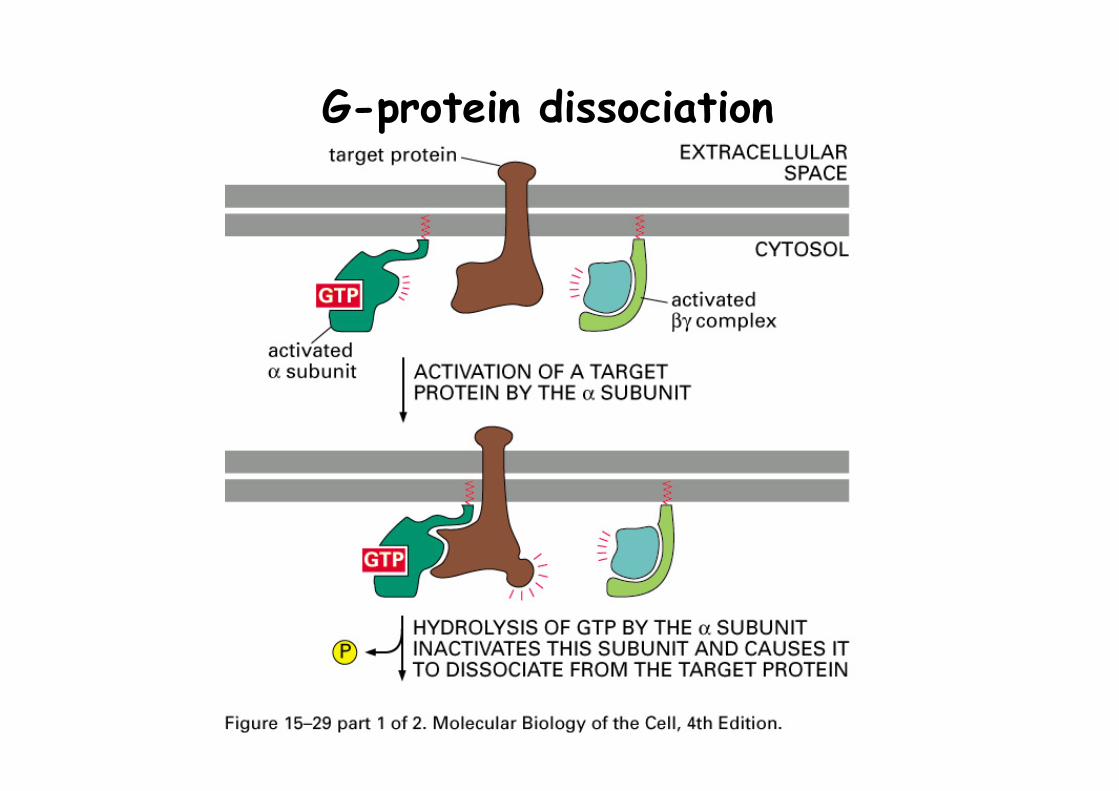
G-protein dissociation
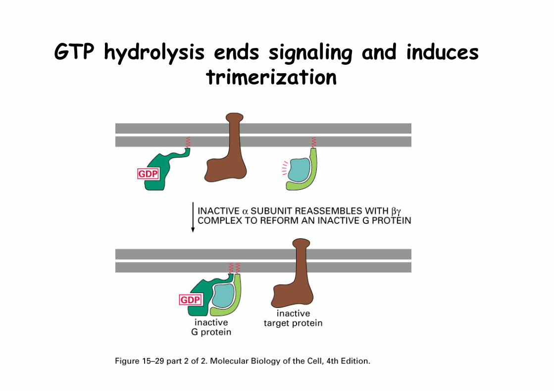
GTP hydrolysis ends signaling and induces trimerization
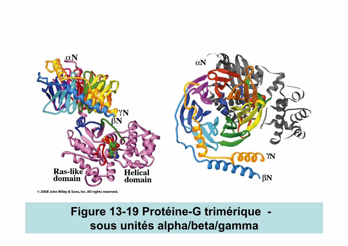
Figure 13-19 Protéine-G trimérique - sous unités alpha/beta/gamma
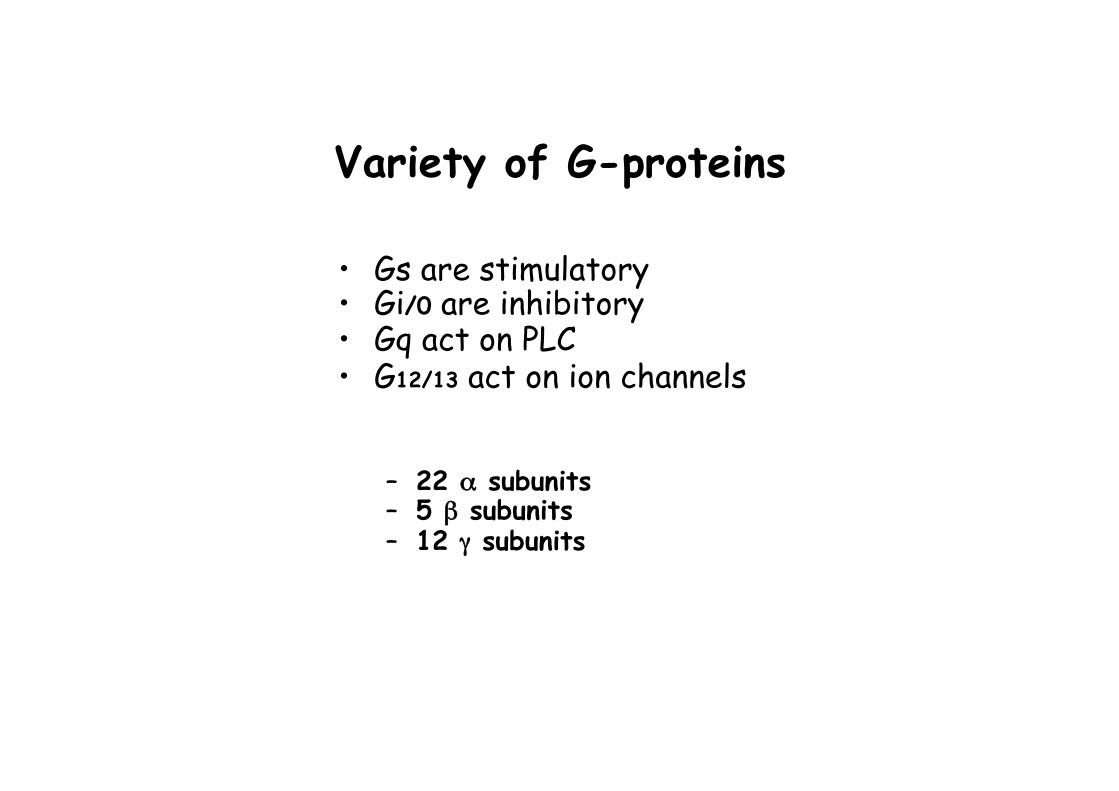
Variety of G-proteins
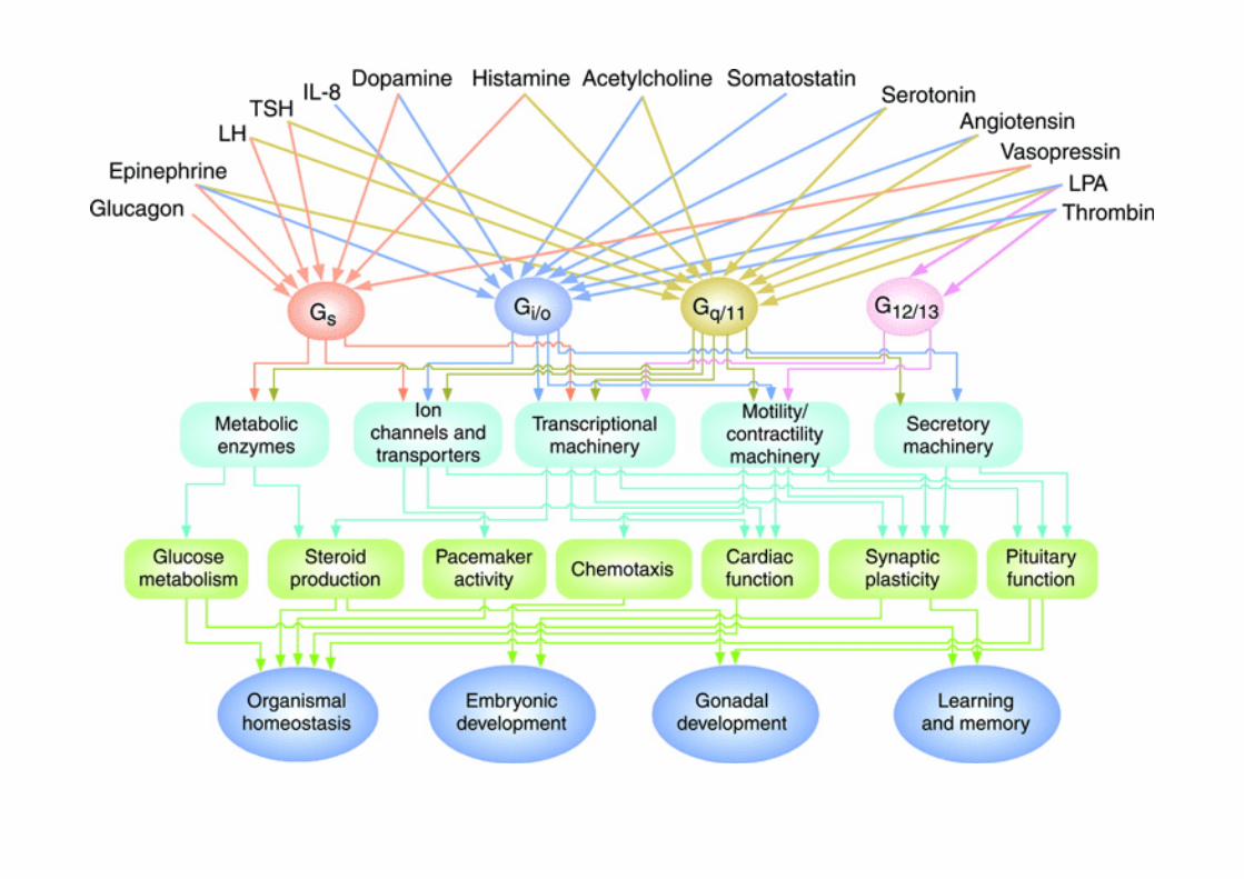
• Gs are stimulatory • Gi/0 are inhibitory • Gq act on PLC • G12/13 act on ion channels
– 22 α subunits – 5 β subunits – 12 γ subunits
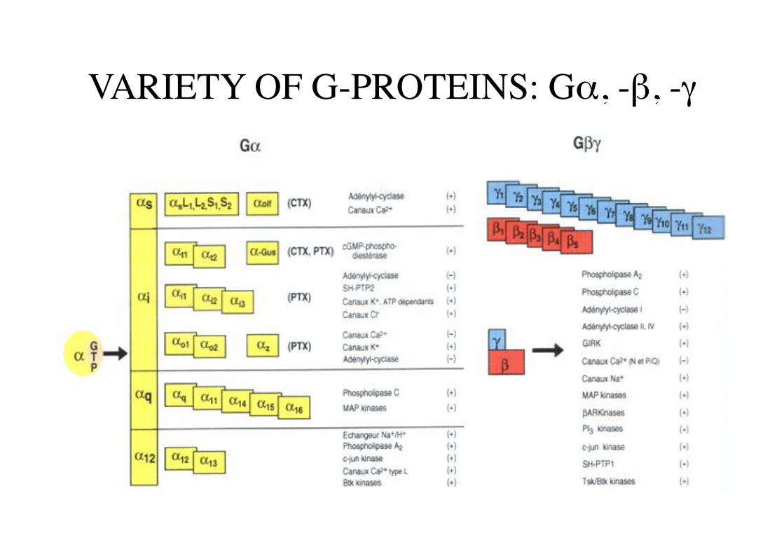
VARIETY OF G-PROTEINS: Gα, -β, -γ
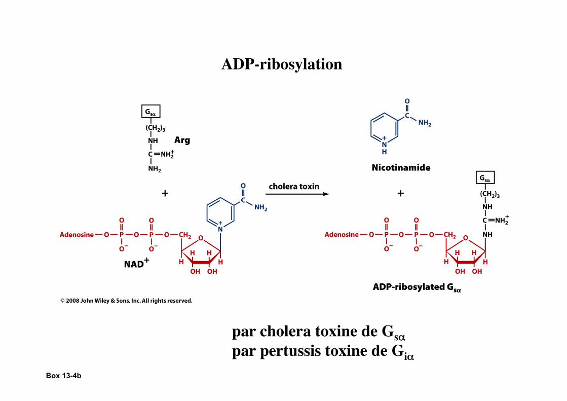
Box 13-4b
ADP-ribosylation
par cholera toxine de Gsα par pertussis toxine de Giα
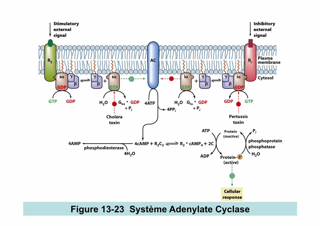
Figure 13-23 Système Adenylate Cyclase
Page 428
Heterotrimeric G-Proteins The cAMP cascade
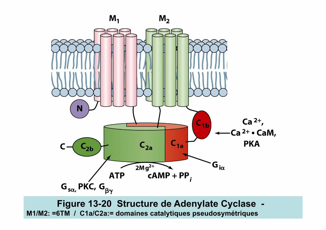
Figure 13-20 Structure de Adenylate Cyclase - M1/M2: =6TM / C1a/C2a:= domaines catalytiques pseudosymétriques
Box 13-4a
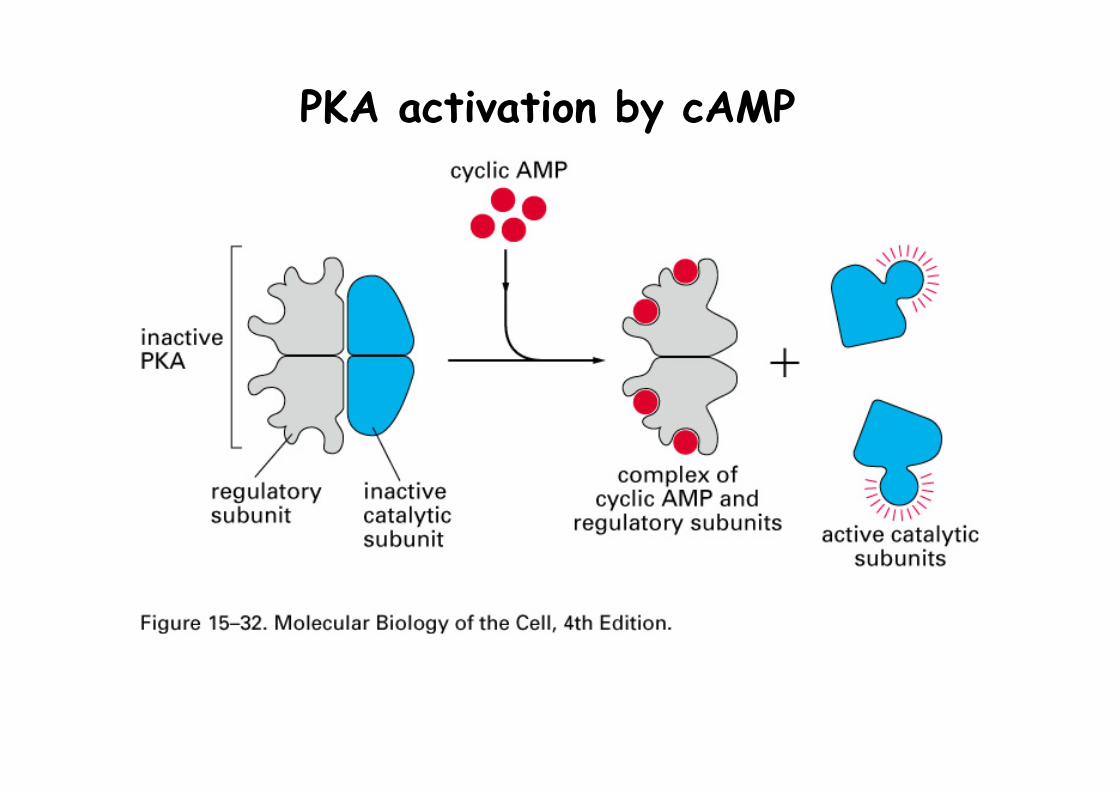
PKA activation by cAMP

PKA activates gene expression
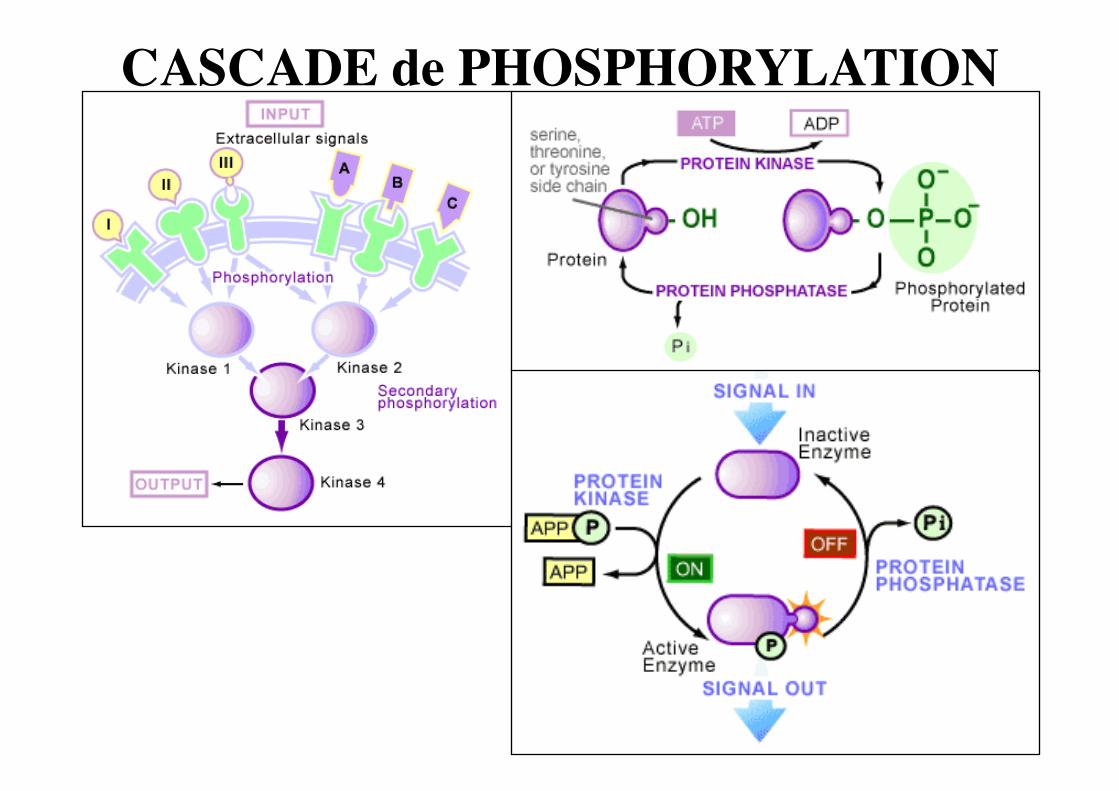
CASCADE de PHOSPHORYLATION
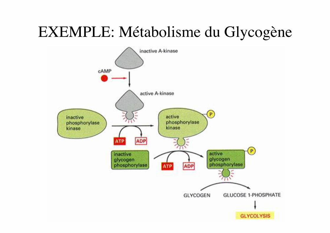
EXEMPLE: Métabolisme du Glycogène
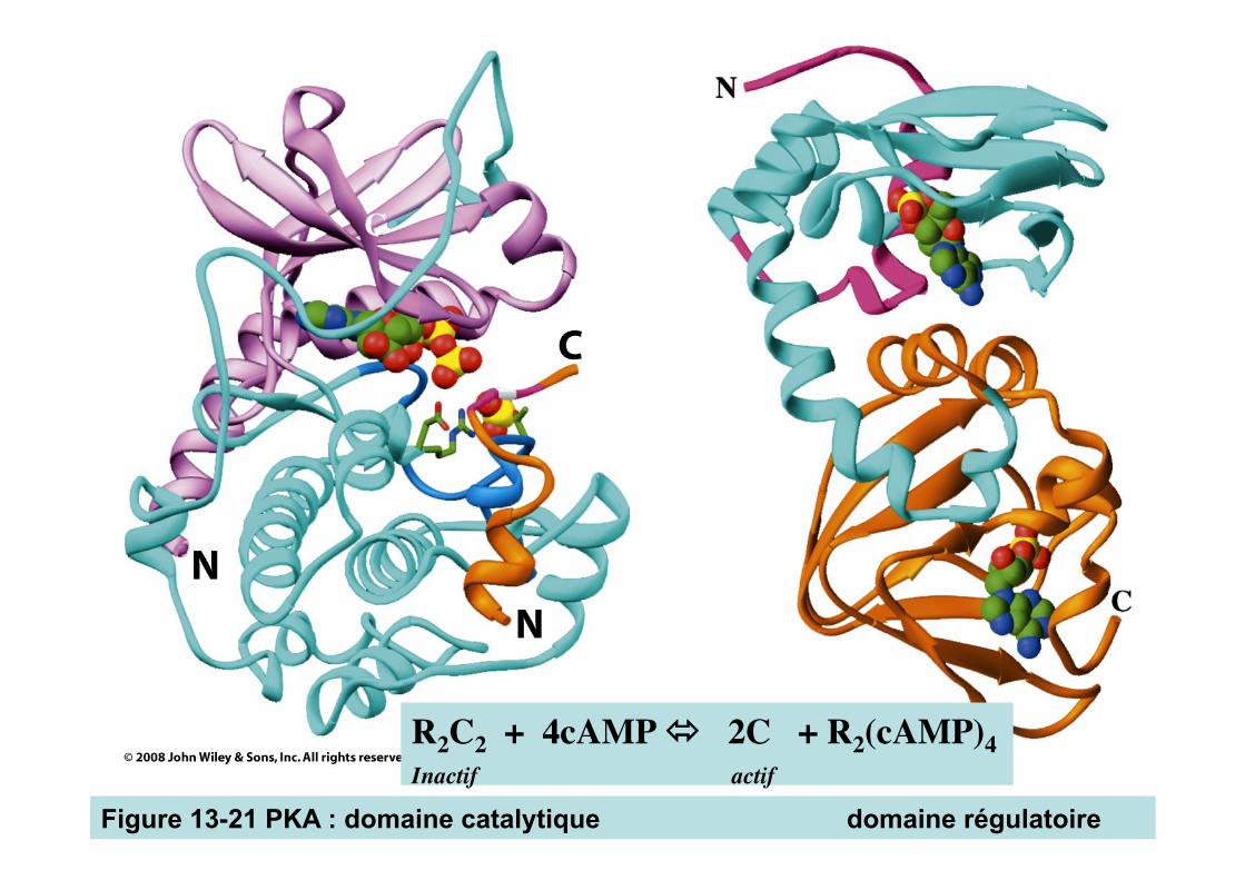
Figure 13-21 PKA : domaine catalytique domaine régulatoire
R2C2 + 4cAMP 2C + R2(cAMP)4 Inactif actif
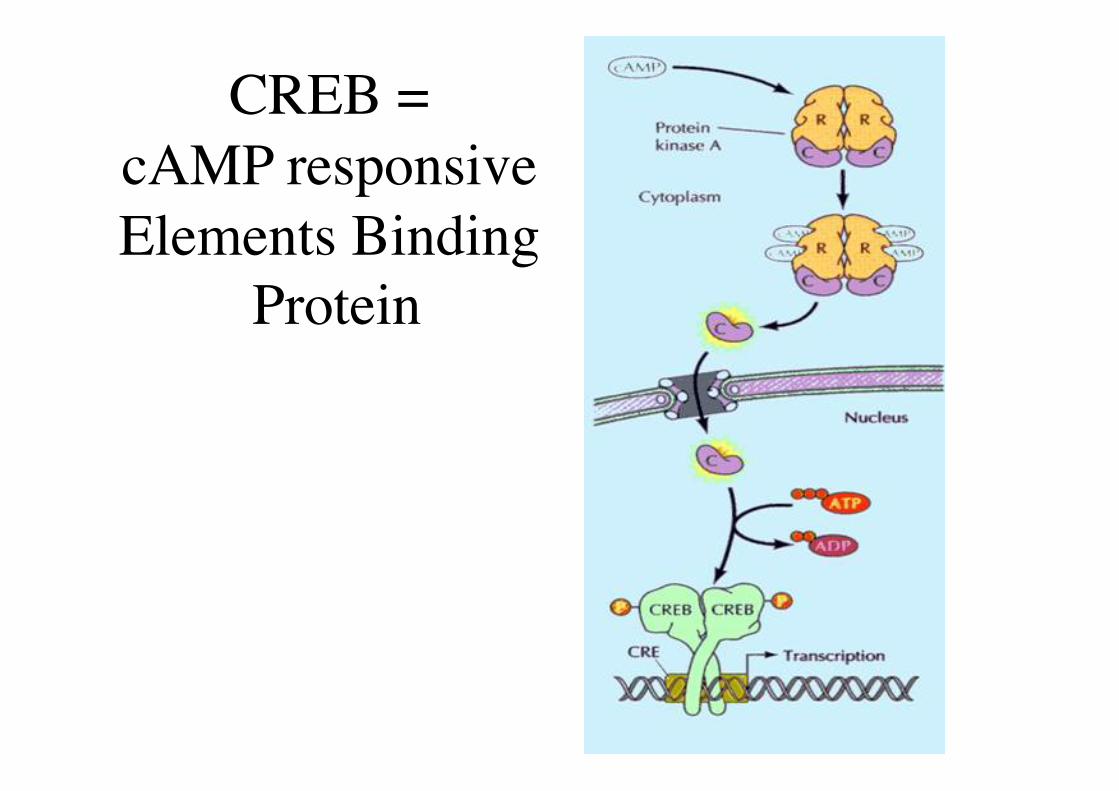
CREB = �cAMP responsive Elements Binding
Protein
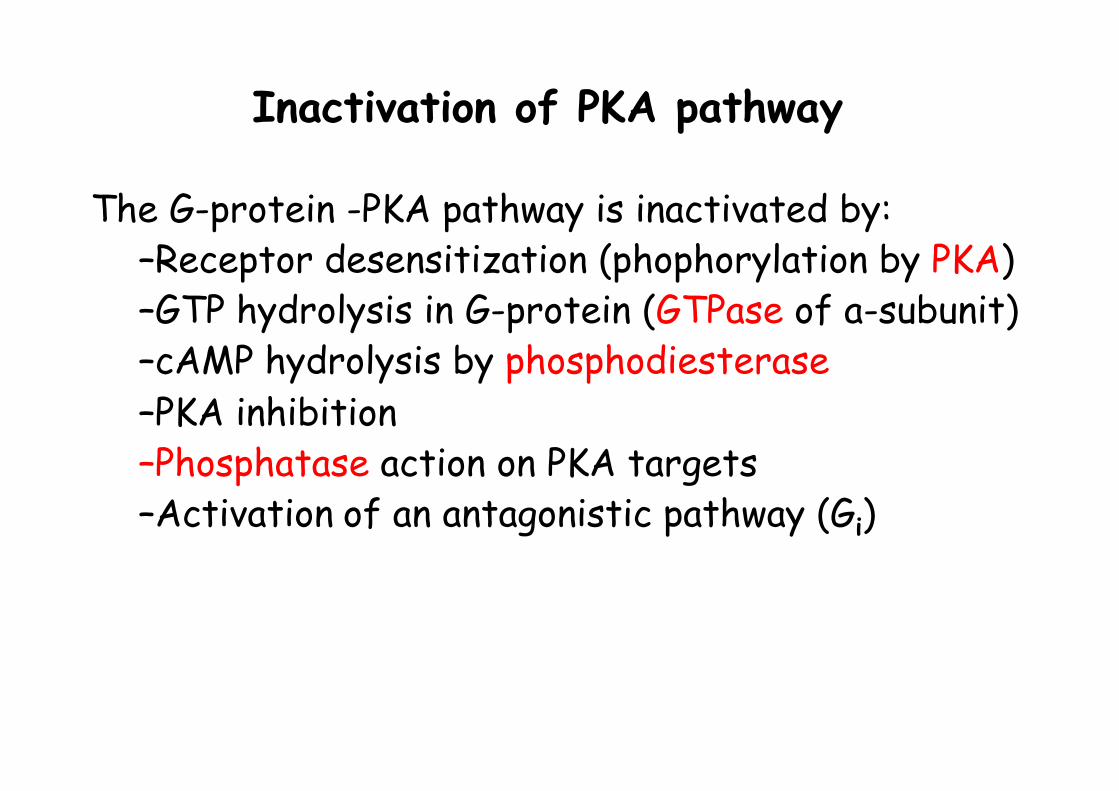
Inactivation of PKA pathway
The G-protein -PKA pathway is inactivated by: – Receptor desensitization (phophorylation by PKA) – GTP hydrolysis in G-protein (GTPase of a-subunit) – cAMP hydrolysis by phosphodiesterase – PKA inhibition – Phosphatase action on PKA targets – Activation of an antagonistic pathway (Gi)
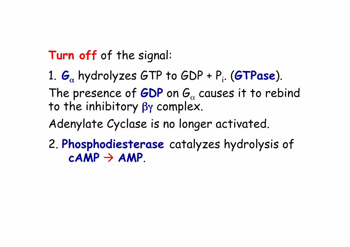
Turn off of the signal:
1. Gα hydrolyzes GTP to GDP + Pi. (GTPase). The presence of GDP on Gα causes it to rebind to the inhibitory βγ complex. Adenylate Cyclase is no longer activated.
2. Phosphodiesterase catalyzes hydrolysis of cAMP AMP.
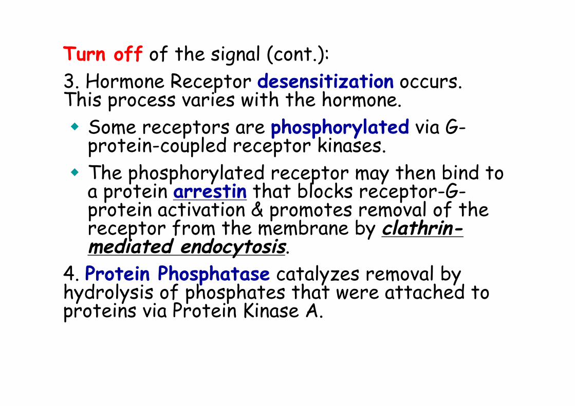
Turn off of the signal (cont.): 3. Hormone Receptor desensitization occurs. This process varies with the hormone. Some receptors are phosphorylated via G-
protein-coupled receptor kinases. The phosphorylated receptor may then bind to
a protein arrestin that blocks receptor-G-protein activation & promotes removal of the receptor from the membrane by clathrin-mediated endocytosis.
4. Protein Phosphatase catalyzes removal by hydrolysis of phosphates that were attached to proteins via Protein Kinase A.
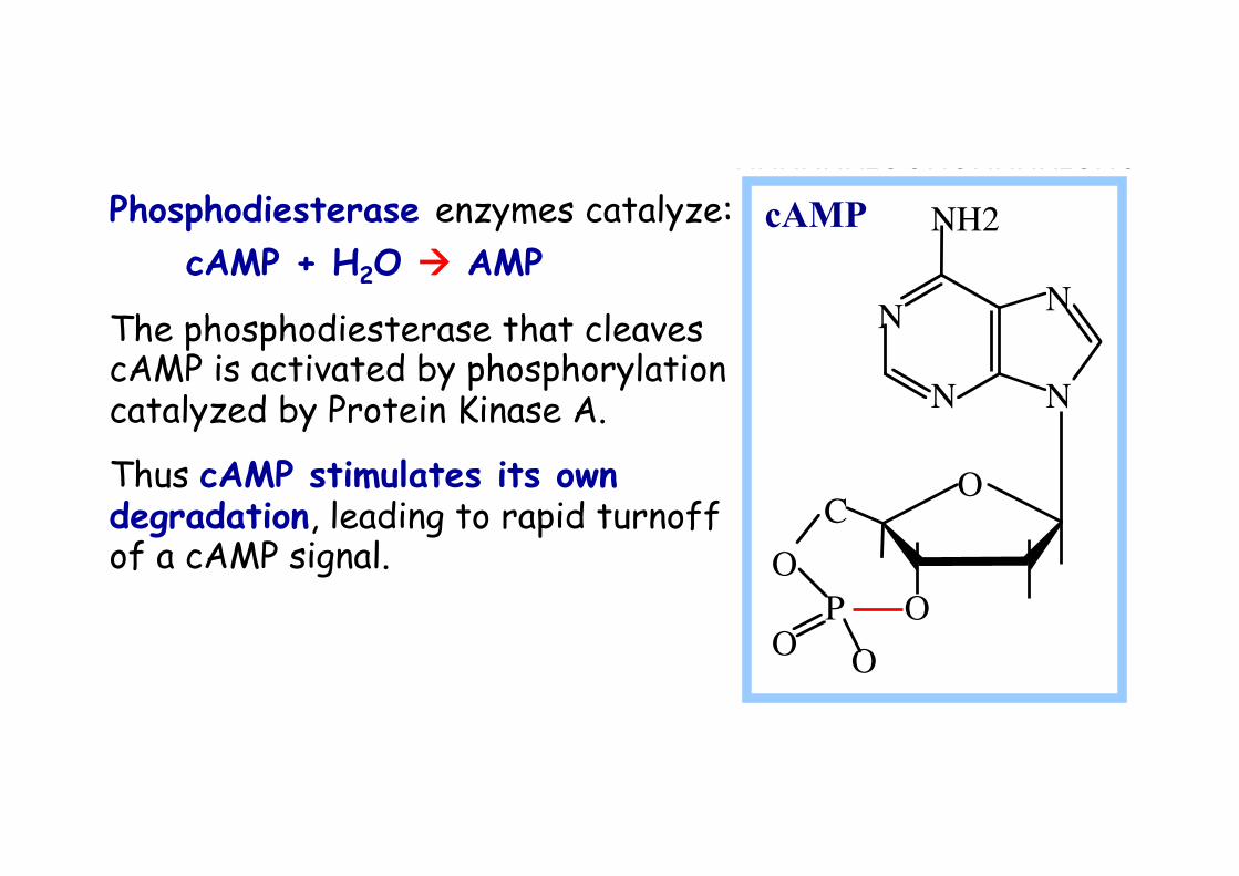
Phosphodiesterase enzymes catalyze: cAMP + H2O AMP
The phosphodiesterase that cleaves cAMP is activated by phosphorylation catalyzed by Protein Kinase A.
Thus cAMP stimulates its own degradation, leading to rapid turnoff of a cAMP signal.
NNNNNH2OOHOHHHH2CHOPOO-1'3'5'4'2'
cAMP
O
O O O
C O
N
N
N
N
NH2
P
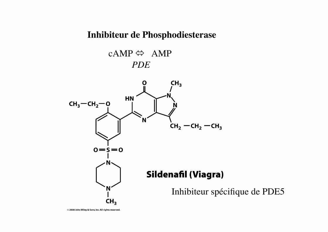
Inhibiteur de Phosphodiesterase
Inhibiteur spécifique de PDE5
cAMP AMP PDE
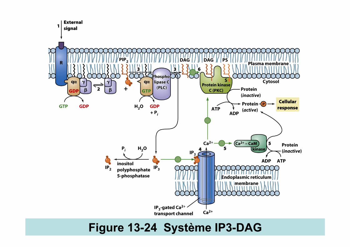
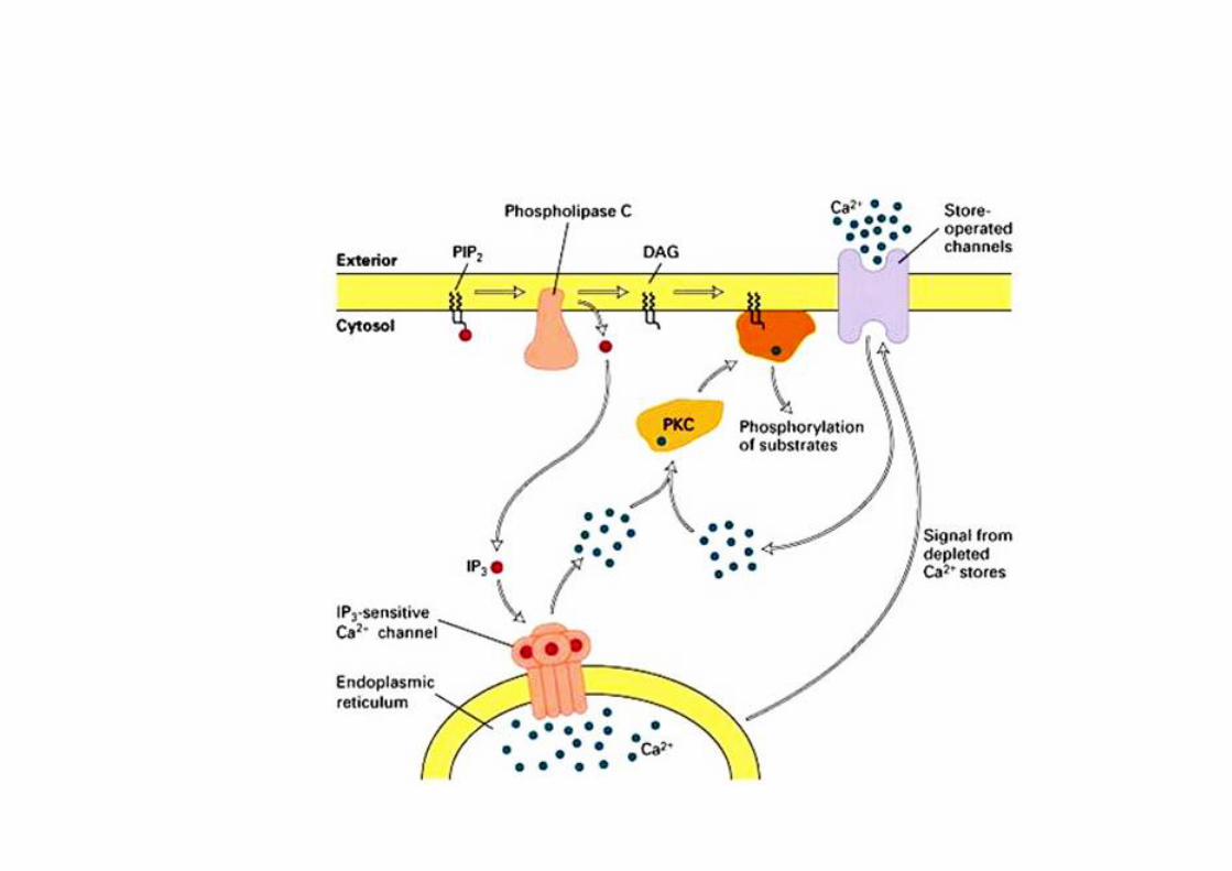
Figure 13-24 Système IP3-DAG
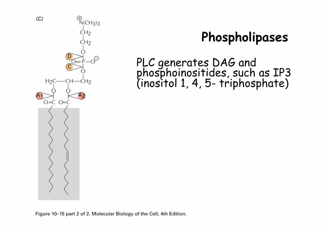
Phospholipases
PLC generates DAG and phosphoinositides, such as IP3 (inositol 1, 4, 5- triphosphate)

Figure 13-25
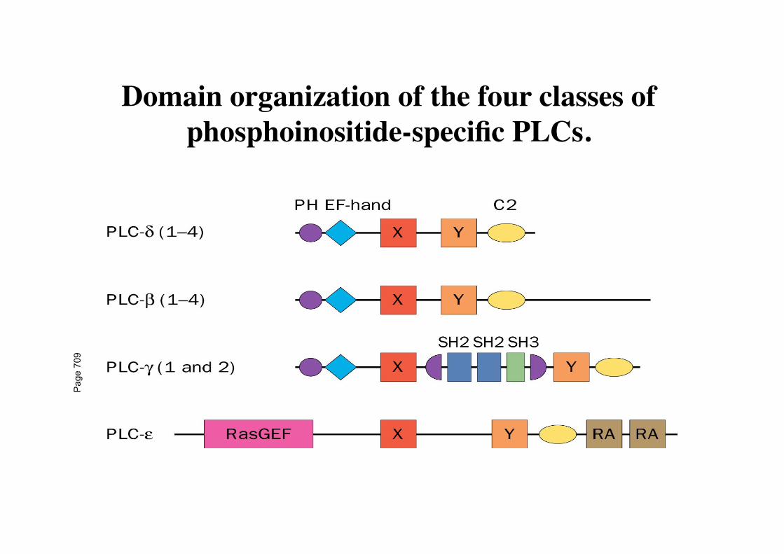
Domain organization of the four classes of phosphoinositide-specific PLCs.
Pag
e 70
9

PKC Inhibiteur de PKC
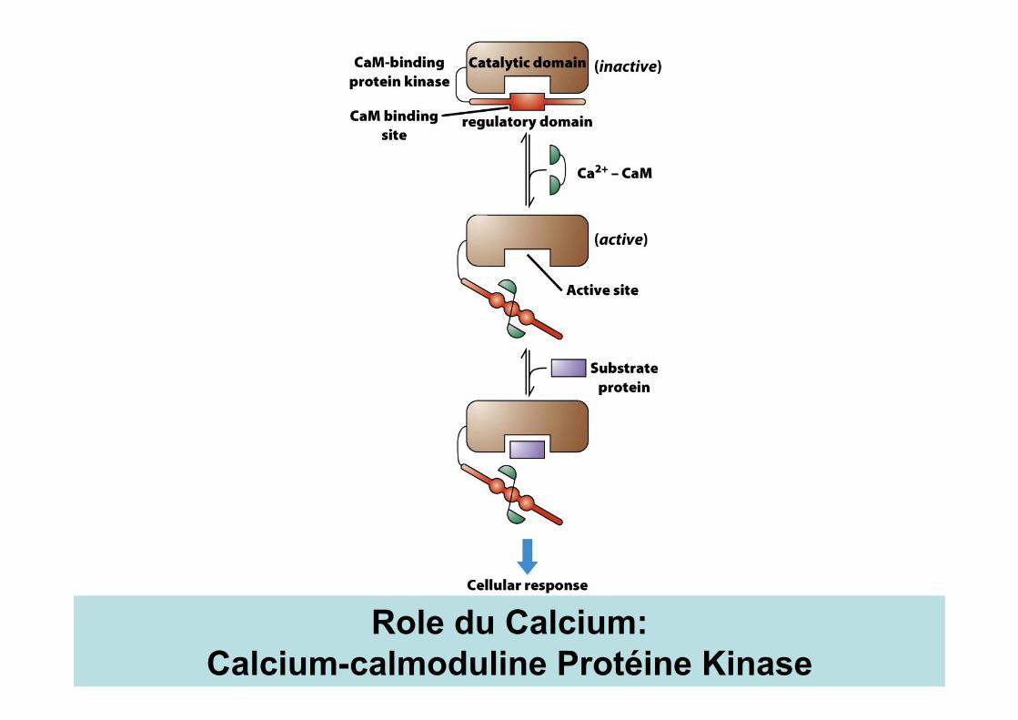
Role du Calcium: Calcium-calmoduline Protéine Kinase
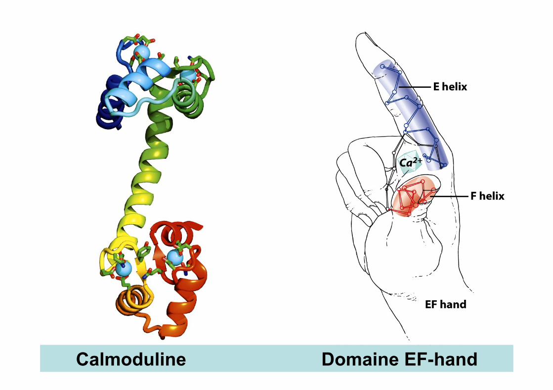
Calmoduline Domaine EF-hand
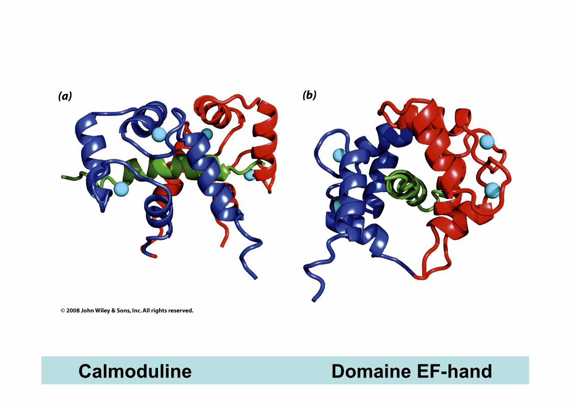
Calmoduline Domaine EF-hand
Ca2+
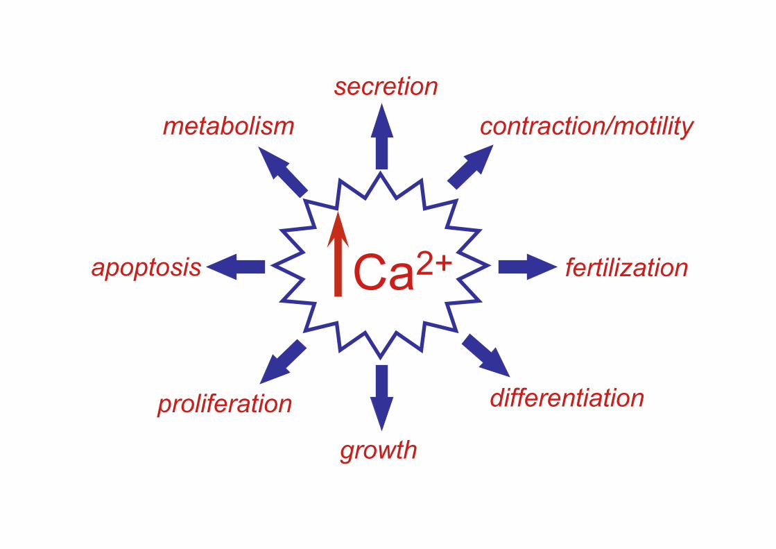
growth
secretion
differentiation proliferation
contraction/motility
fertilization apoptosis
metabolism
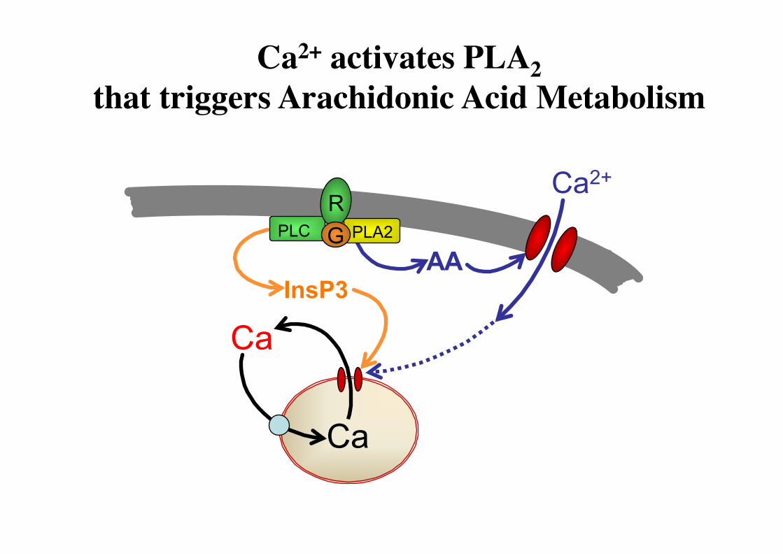
InsP3 AA
Ca
Ca
PLC R
Ca2+ PLA2 G
Ca2+ activates PLA2 that triggers Arachidonic Acid Metabolism
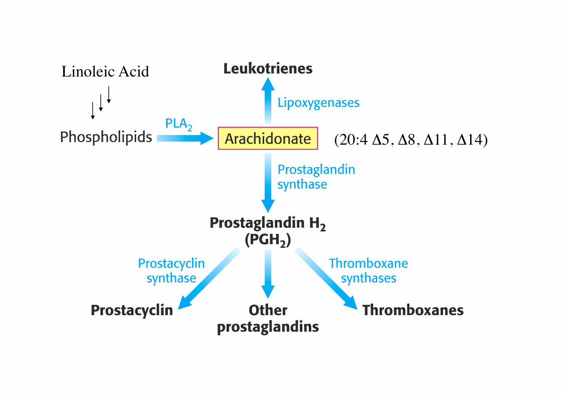
(20:4 Δ5, Δ8, Δ11, Δ14)
Linoleic Acid

Prostagandin biosynthesis pathways are a common drug target.
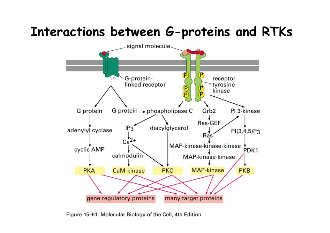
Interactions between G-proteins and RTKs
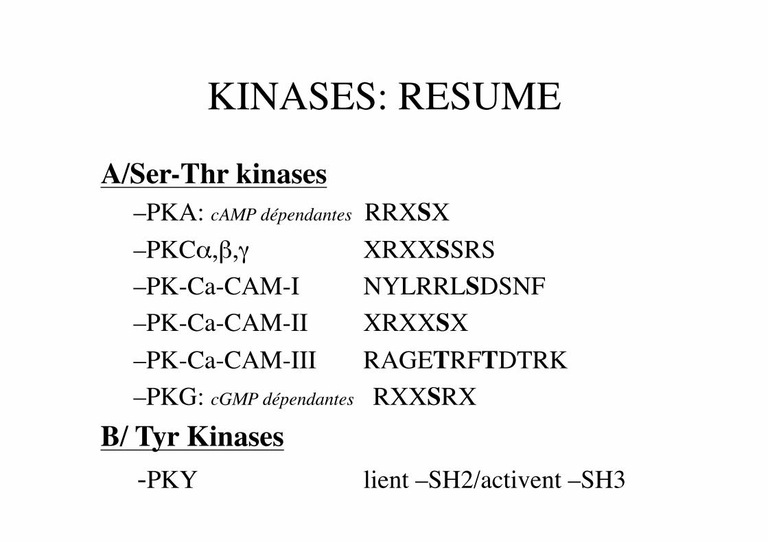
KINASES: RESUME
A/Ser-Thr kinases – PKA: cAMP dépendantes RRXSX – PKCα,β,γ XRXXSSRS – PK-Ca-CAM-I NYLRRLSDSNF – PK-Ca-CAM-II XRXXSX – PK-Ca-CAM-III RAGETRFTDTRK – PKG: cGMP dépendantes RXXSRX
B/ Tyr Kinases -PKY lient –SH2/activent –SH3

INSULIN SIGNAL TRANSDUCTION
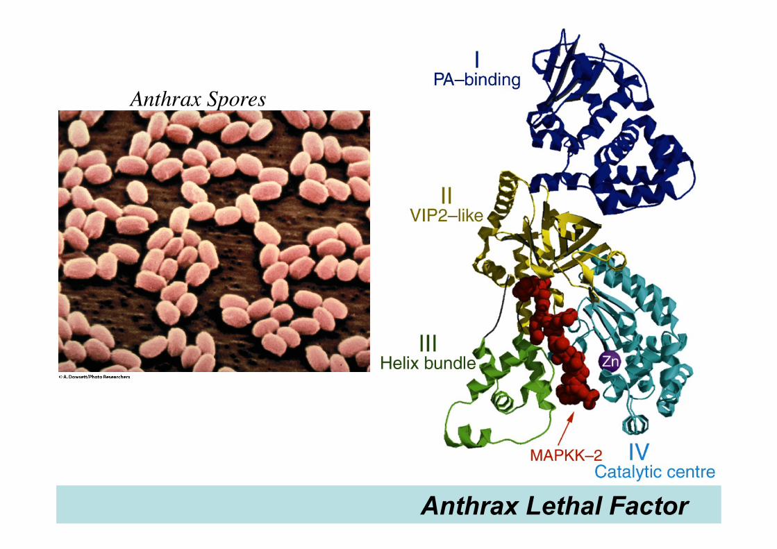
Anthrax Lethal Factor
Anthrax Spores