Molecular Characterization of Porcine Reproductive and ...
13
Page 1/13 Molecular Characterization of Porcine Reproductive and Respiratory Syndrome Virus (PRRSv) Isolated From Slaughtered Pigs in Northern Uganda Peter Oba ( [email protected] ) International Livestock Research Institute Michel M. Dione International Livestock Research Institute Joseph Erume Makerere University Barbara Wieland University of Bern Christine Mutisya International Livestock Research Institute Linnet Ochieng International Livestock Research Institute Elizabeth A. J. Cook International Livestock Research Institute Frank N. Mwiine Makerere University Research Article Keywords: PRRSv, genotypes, porcine, pigs, respiratory, Lira, Uganda Posted Date: June 9th, 2021 DOI: https://doi.org/10.21203/rs.3.rs-579302/v1 License: This work is licensed under a Creative Commons Attribution 4.0 International License. Read Full License
-
Upload
khangminh22 -
Category
Documents
-
view
2 -
download
0
Transcript of Molecular Characterization of Porcine Reproductive and ...

Page 1/13
Molecular Characterization of Porcine Reproductiveand Respiratory Syndrome Virus (PRRSv) IsolatedFrom Slaughtered Pigs in Northern UgandaPeter Oba ( [email protected] )
International Livestock Research InstituteMichel M. Dione
International Livestock Research InstituteJoseph Erume
Makerere UniversityBarbara Wieland
University of BernChristine Mutisya
International Livestock Research InstituteLinnet Ochieng
International Livestock Research InstituteElizabeth A. J. Cook
International Livestock Research InstituteFrank N. Mwiine
Makerere University
Research Article
Keywords: PRRSv, genotypes, porcine, pigs, respiratory, Lira, Uganda
Posted Date: June 9th, 2021
DOI: https://doi.org/10.21203/rs.3.rs-579302/v1
License: This work is licensed under a Creative Commons Attribution 4.0 International License. Read Full License

Page 2/13
Abstract
BackgroundA cross sectional study was conducted to determine genotypes of porcine reproductive and respiratorysyndrome virus (PRRSv) isolated from slaughtered pigs in Lira district, northern Uganda. The study wasconducted from March to September 2019 in three selected slaughter slabs. Pigs brought for slaughterwere randomly sampled. At necropsy, lungs were extracted from the thoracic cavity and examined forpneumonic lesions. Seventy-three (73) pigs with gross lung lesions were sampled, from which onehundred and one (101) tissue samples were taken. A real-time reverse transcriptase PCR (RT-qPCR) wasused to characterize PRRSv genotypes.
ResultsA total of 20 samples tested positive for PRRSv. The respective prevalence of PRRSv type 1 and type 2were 24.65% (n = 18) and 2.73% (n = 2) respectively. Of the pigs sampled (n = 73), only two pigs, 2.73% (n = 2) tested positive to both strains. The likelihood of PRRSv detection decreased with pig age, butincreased with gross pneumonic pathology.
ConclusionsThis study demonstrated dual circulation of both genotypes in northern Uganda. The associationbetween PRRSv and lung pathology suggests that it may be an important cause of lung disease in pigs inUganda and hence loss of production. This calls for further investigations on potential economic impactsof PRRSv on pig productivity. These �ndings contribute to discussions about the need of surveillance andpossible vaccination strategies against PRRSv in Uganda.
BackgroundIn Uganda, pig production has increased over the last few years, from approximately 0.7 million in 1990to 4.2 million pigs in 2018 due to a rising demand for pork [1, 2]. Pig production in Uganda is increasinglybecoming an important economic activity for many households, providing a reliable source of livelihoods.However, disease constraints hinder pig production and productivity in the country [3]. Recent multi-pathogen studies reveal occurrence of economically important respiratory pathogens such as porcinereproductive and respiratory syndrome virus (PPRSv), Mycoplasma hyopneumoniae (M. hyo),Actinobacillus pleuropneumoniae (App), Leptospira spp. and porcine circovirus (PCV2) type 2 [4–6].
Of the pathogens reported, PRRSv is known to be associated with high economic losses from mortalities,reproductive losses and increased costs of control [7–9]. In general, two genetically diverse strains ofPRRSv have been described worldwide, with the European genotype (EU) designated as type 1 (PRRSv-1)

Page 3/13
and the north American genotype, designated as type 2 (PRRSv-2) [10]. Furthermore, there is markedgenetic diversity in each of these two genotypes, leading to further classi�cation into virus subtypes [11,12]. These two genotypes are distinct in their virulence, antigenic characteristics and nucleotidesequences [13, 14]. This has important implications for immunological responses and vaccine selection,as only incomplete protection can be achieved from heterologous �eld strains [15]. This diversity of thevirus also compounds the challenges of disease control, due to differences in transmission rates, strainpathogenicity and its tendency to persist in infected herds.
In the US, PRRSv is reported to cost the swine industry up to $560 million annually, with up to 45% ofthese losses due to reduced growth and feed e�ciency [7]. Overrall, losses due to PRRSv vary widelydepending on epidemiological factors, production systems and farm characteristics. In a Dutch study,losses were found to range from €3 to €160 per sow per year [16]. The economic impacts of PRRSv onswine productivity are justi�cation for epidemiologic studies to generate knowledge to guideinterventions.
In Uganda, no vaccines are currently in use for control or prevention of PRRSv. In particular, few studieson PRRSv in Uganda have mainly focused on serologic assays, providing evidence for past exposure ofpigs to the virus and possible virus circulation. Recent developments in the pig sector in Uganda showincreased imports of breeder pigs from countries such as South Africa, where PRRSv has been reported[17]. This poses a threat to the swine population, if no measures to contain virus spread are established.There is no information on the current epidemiological situation regarding PRRSv and its potentialimpacts on swine productivity in Uganda, due to lack of surveillance.
Despite the availability of several commercial vaccines in Europe, America and Asia to control PRRSv [18,19], the apparent lack of information on the identity of current PRRSv strains circulating in Ugandan pigslimits their use as effective tools for control and prevention. The aim of this study was to determineprevalence and characterize genotypes of PRRSv isolated from slaughtered pigs in northern Uganda.
Methods
Study designA cross-sectional study was conducted from March to September 2019 in three selected slaughter slabsin Lira district, northern Uganda. Slaughter slabs with the highest daily slaughter capacity (≥ 8 pigs) wereselected for the study. Pigs slaughtered in Lira district were sourced from within the district (~ 60%), whilethe rest were sourced from neighboring districts of Apac, Kole, Amolatar and Pader. Figure 1 below showsa map of Lira district in Uganda where the study was conducted.
Sampling of slaughter slabs and pigsDuring this survey, three slaughter slabs: Teso Bar (Adyel division), Adekokwok (Adekokwok subcounty)and Amach market (Amach subcounty) were selected based on high daily slaughter capacity (≥ 8 pigs).

Page 4/13
In each slab, approx. 40% of pigs brought for slaughter were randomly selected on each day of sampling.At each slab on average, between 8 to 20 pigs were brought for slaughter per day, which representedapprox. 8–12 farms. On each day, a list of all pigs brought for slaughter was made and each allocated anumber, which were written on a piece of paper and folded. From this list, random sampling was done.Pig biodata was recorded at ante mortem (sex, age), while gross pathology (postmortem) was recordedas described in a related study (Oba et al., forthcoming). Traders were asked about the source(s) of thepigs, which were recorded.
Sample size determinationThe number of pigs sampled represented approximatly 40% of all pigs slaughtered monthly in the district(DVO, pers comm). The sample size required for a related slaughter slab survey was determined to be 139pigs (Oba et al., forthcoming). In a previous study, the seroprevalence of PRRSv in Lira district was foundto be 1.7% [4]. Using this �gure, the expected minimum number of infected pigs is three (3). To detect thepresence of PRRSv, the following equation was used to calculate sample size [20]:
Where α = 0.05, D is the estimated minimum number of diseased/infected pigs (3), and N is the numberof pigs sampled in a previous related study (139). Using this equation, the computed sample size was 87pigs. A sample of seventy three (73) pigs was attained, from which 101 tissue samples were taken. Onlylungs with gross pathologic lesions were sampled, normal lungs were not sampled.
Examination of lungs and other tissues for grosspneumonic lesions and sample collectionAt necropsy, the carcass was placed on a clean table, opened with knives to expose lungs and the pleura.The lungs were carefully extracted from the thoracic cavity and placed on a �at, clean surface.Examination of lungs for gross pneumonic lesion scoring is described in a previous related study (Oba etal. forthcoming). Lesion samples were taken and cut into ~ 0.5-gram pieces, placed in a 2 ml cryovial(Sarstaedt®, Germany) containing RNAlater® (Thermo Scienti�c®, USA) tissue stabilization solution.Other observed gross lesions were also recorded.. The cryovial was labelled and then placed in an ice boxcontaining ice packs at 4°C. To prevent cross contamination, a new sterile surgical blade was used foreach pig lung, with disinfection of gloved hands and collection tools using 70% ethanol betweensamplings. Hand gloves were frequently changed to minimize the risk of cross contamination.
Tissue sample transport and storageAfter collection, tissue samples were immediately (within 2 hours) transported to the district (Lira)veterinary laboratory for temporary storage in a fridge at 4°C. Later, samples were transported (in anicebox at 4°C) to Makerere College of Veterinary Medicine (CoVAB), Department of Biosecurity,Ecosystems and Veterinary public health laboratory and stored in a -20°C fridge. An export permit wassecured from the Commisioner Animal Health, Uganda and an import permit from the Directorate of

Page 5/13
Veterinary Services of the Republic of Kenya to transfer samples to International Livestock ResearchInstitute (ILRI) Kenya for molecular analysis. Upon receipt of an authorization to export samples, tissuesamples were shipped by air in October 2019 to ILRI Nairobi, Kenya. The samples were packaged in anice box containing ice packs at 4 ℃, where upon arrival they were placed in a -80°C fridge for subsequentRNA extraction and complementary DNA synthesis.
PRRSv RNA extraction and real-time reverse transcriptasePCR (RT-qPCR)RNA extraction was done using the AllPrep DNA/RNA Mini Kit (cat. no. 80204) according to themanufacturer’s protocol (Qiagen®, Denmark). A real-time (quantitative) reverse transcriptase PCR wasperformed in the same laboratory, in March 2020 using the KiCqStart(R) One-Step Probe RT-qPCRReadyMix™ Low ROX™ [21]. Real-time RT-qPCR and complementary DNA synthesis were performed in aGeneAmp® PCR System 7500 Fast version 2.3 (Applied Biosystems®). The sequences of primers(Macrogen Europe, cat. no. OG200117-237) for full length cDNA synthesis and the dual-labeled Taq-Manprobes are as shown in Table 1 below [22].
Table 1Sequences of primers and dual-labeled probes used in the assay
Genotype Name Orientation Sequence
PRRSv-1 Primer 1 Forward 5’-CGA CCA CCT CAC CCA GAC-3’
Primer 1 Reverse 5’-CAG TTC CTG CGC CTT GAT-3’
Probe Genomic 5’-6-FAM-CCT CTG CTT GCA ATC GAT CCA GAC-BHQ1-3’
PRRSv-2 Primer 2 Forward 1 5’-ATG ATG RGC TGG CAT TCT-3’
Primer 2 Reverse 5’-ACA CGG TCG CCC TAA TTG-3’
Probe Genomic 5’-HEX-TGT GGT GAA TGG CAC TGA TTG ACA-BHQ2-3’
A qPCR master mix was made up of 4 µl molecular biology grade water, 1 µl of 10 µM Forward, 1 µl of 10µM reverse primers, 1 µl of 10 µM probe and 10 µl of KICqStart Master mix (Sigma Aldrich, UK). Themaster mix was completely mixed by tapping the tube and a quick short spin. This master mix cocktailwas adequate for one reaction. The components of the master mix were adjusted to suit the number ofsamples. The contents of the master mix tube were mixed thoroughly and dispensed 17 µl to eachlabeled sample and control tubes. An RNA template of 3 µl was then dispensed to each tube with amaster mix. The tubes were placed in a 7500 Fast Thermalcycler and the program which includes aReverse Transcriptase (RT) at 50°C for 10 min, pre-heating at 95°C for 10 min, denaturation at 95°C for 30seconds and annealing at 60°C for 1 minute was started. This was repeated for 45 cycles with the RT andpreheating occurring just once.
Data analysis
Page 6/13
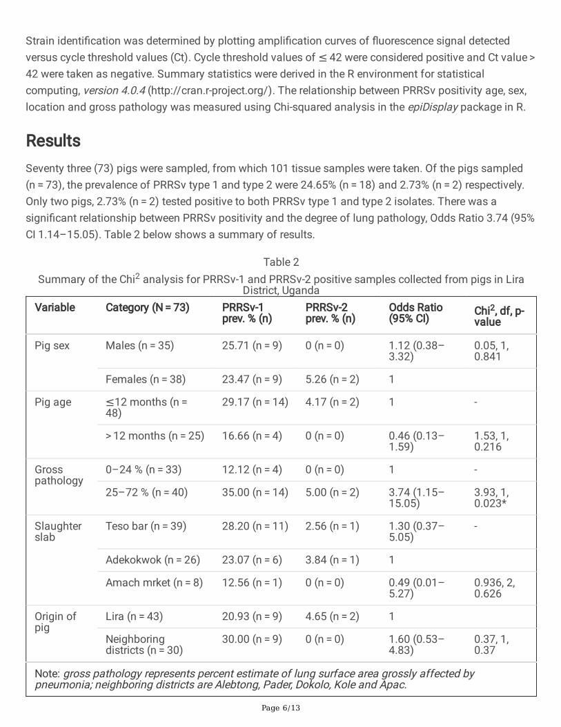
Strain identi�cation was determined by plotting ampli�cation curves of �uorescence signal detectedversus cycle threshold values (Ct). Cycle threshold values of ≤ 42 were considered positive and Ct value > 42 were taken as negative. Summary statistics were derived in the R environment for statisticalcomputing, version 4.0.4 (http://cran.r-project.org/). The relationship between PRRSv positivity age, sex,location and gross pathology was measured using Chi-squared analysis in the epiDisplay package in R.
ResultsSeventy three (73) pigs were sampled, from which 101 tissue samples were taken. Of the pigs sampled(n = 73), the prevalence of PRRSv type 1 and type 2 were 24.65% (n = 18) and 2.73% (n = 2) respectively.Only two pigs, 2.73% (n = 2) tested positive to both PRRSv type 1 and type 2 isolates. There was asigni�cant relationship between PRRSv positivity and the degree of lung pathology, Odds Ratio 3.74 (95%CI 1.14–15.05). Table 2 below shows a summary of results.
Table 2Summary of the Chi2 analysis for PRRSv-1 and PRRSv-2 positive samples collected from pigs in Lira
District, UgandaVariable Category (N = 73) PRRSv-1
prev. % (n)PRRSv-2prev. % (n)
Odds Ratio(95% CI)
Chi2, df, p-value
Pig sex Males (n = 35) 25.71 (n = 9) 0 (n = 0) 1.12 (0.38–3.32)
0.05, 1,0.841
Females (n = 38) 23.47 (n = 9) 5.26 (n = 2) 1
Pig age ≤12 months (n = 48)
29.17 (n = 14) 4.17 (n = 2) 1 -
> 12 months (n = 25) 16.66 (n = 4) 0 (n = 0) 0.46 (0.13–1.59)
1.53, 1,0.216
Grosspathology
0–24 % (n = 33) 12.12 (n = 4) 0 (n = 0) 1 -
25–72 % (n = 40) 35.00 (n = 14) 5.00 (n = 2) 3.74 (1.15–15.05)
3.93, 1,0.023*
Slaughterslab
Teso bar (n = 39) 28.20 (n = 11) 2.56 (n = 1) 1.30 (0.37–5.05)
-
Adekokwok (n = 26) 23.07 (n = 6) 3.84 (n = 1) 1
Amach mrket (n = 8) 12.56 (n = 1) 0 (n = 0) 0.49 (0.01–5.27)
0.936, 2,0.626
Origin ofpig
Lira (n = 43) 20.93 (n = 9) 4.65 (n = 2) 1
Neighboringdistricts (n = 30)
30.00 (n = 9) 0 (n = 0) 1.60 (0.53–4.83)
0.37, 1,0.37
Note: gross pathology represents percent estimate of lung surface area grossly affected bypneumonia; neighboring districts are Alebtong, Pader, Dokolo, Kole and Apac.

Page 7/13
DiscussionThis study revealed circulation of both type 1 and 2 PRRSv genotypes in northern Uganda. However,PRRSv type 1 was found to be the more predominant genotype detected. These �ndings also indicatethat PRRSv-1 likely to be prevalent elsewhere in Uganda, where its occurrence has not yet beeninvestigated properly. This situation could have adverse implications for swine productivity in the country,herd economic performance and consequently livelihoods, if the virus becomes established incommercial breeding herds. Information about the predominant virus is important for implementingsuccessful interventions for controlling the spread of the virus given the potential economic impacts ofPRRSv on swine productivity.
These results showed the likelihood of PRRSv-1 detection decreased with pig age. While this wasstatistically non signi�cant, it suggested a trend that needs further exploration with a larger sample size.This �nding is consistent with the observation that the immune system of swine is able to completelyeliminate PRRSv infection over prolonged periods of time, leading to sterilizing immunity [19]. Pigsexposed to PRRSv become resistant to reinfection with a homologous strain, although the level ofprotection was incomplete [23]. This was also corroborated by a study which found age-dependentresistance to infection, shown by reduced viremia and viral load in the blood of adult pigs compared toyounger pigs [24]. In contrast, other studies revealed that PRRSv tends to persist in infected herds [25, 26],suggesting increased likelihood of detection in older pigs. However, this �nding was speci�c for largerherds and where there were increased re-introductions of infected gilts [27]. In the smallholder pigproduction systems in northern Uganda, most farms were generally small in size (1–3 sows) and thereplacements were infrequent, as highlighted in a related study (Oba et al. forthcoming).
The increase in PRRSv detection rates associated with gross pathologic lesions conforms to previousstudies. The ability of PRRSv to induce clinical and macroscopic pneumonia, often as a co-infection withother pathogens such as M. hyo has been documented [28]. No differences in detection rates betweenmale and female pigs were observed in this study.
Our results are comparable to other studies which reported simultaneous circulation of both PRRSv type1 and type 2 genotypes in various regions and show increased circulation of PRRSv type 1. In Europe,both types circulate but there is a predominance of type 1, with marked genetic variation among thegenotypes [11]. In Asia, studies report the predominance of PRRSv type 1 in China, although the Americantype 2 has also been documented [29]. In the Republic of Korea, it was found that both type 1 and type 2strains circulated in pig farms during the period between 2013–2016. However, type 1 PRRSv wasreportedly predominant [30].
The information on PRRSv in African countries is limited but there are o�cial reports submitted to OIE bya few countries in Africa (DR Congo, Benin, Burkina Faso, Egypt, Ivory Coast, Nigeria) that documentoccurrence of PRRSv, although none of these studies reported its genetic diversity or molecular identity[17]. The current situation regarding the PRRSv genotypes circulating on the continent is largely unknown,as the few studies undertaken were based on serologic assays. In southwest Nigeria, a study reported a

Page 8/13
high seroprevalence of PRRSv of 53.8%, suggesting widespread exposure of pigs to the virus [31].However, the genotype of the virus was not determined.
In South Africa, the PRRSv strain responsible for the 2004 outbreaks was identi�ed by RT-PCR as type 2[32]. Our results are contrary to expected since a large number of pigs are imported from South Africa andsuggest a different source of the virus in Uganda, since PRRSv type 1 has not been reported in SouthAfrica. The lack of reliable data on pig imports into Uganda limits our understanding of the likely sourcesof PRRSv introduction into the country. Further studies to understand the introduction and maintenanceof PRRSv into Uganda are required. Knowledge gaps remain on the potential distribution of PRRSvstrains in other regions of Uganda especially in high pig dense areas, which justify further studies.
The method used to detect PRRSv in this study utilised primers that were designed to simultaneouslydetect both PRRSv-1 and PRRSv-2. This approach is reported to have high speci�city and sensitivity, atdifferentiating PRRSv-1 from PRRSv-2 isolates [22, 33]. This method is reportedly e�cient and rapid forlarge scale detection and differentiation of PRRSv strains. However, this study was limited by the smallsample size used and by the fact that the study was undertaken in only one region, implying that resultscannot be extrapolated to other regions of the country. Because we sampled only pigs that presented withgross lung lesions, the true prevalence of PRRSv and the distribution of strains in all slaughtered pigs andin the general pig population still remains unknown and possibly is higher to what has been reportedhere.
ConclusionsThis is the �rst study to document dual circulation of PRRSv type 1 and 2 genotypes in pigs in Uganda.The relation between PRRSv and severe lung pathology suggests it may be an important and increasingcause of lung disease in pigs in Uganda and hence loss of production. This study reveals PRRSv-1 is thepredominant genotype in circulation in northern Uganda. However, in view of its reported genetic diversity,further characterization of possible PRRSv-1 subtypes and evaluation of their pathogenicity in pigs isjusti�ed, as well as investigating circulation of PRRSv in other parts of the country with the aim toestablish surveillance. In addition, studies to evaluate e�cacy of different control measures, such asvaccination, considering dual circulation of the two genotypes and to quantify their economic e�caciesin Uganda are recommended.
AbbreviationsApp - Actinobacillus pleuropneumoniae
CoVAB – College of Veterinary Medicine, Animal Resources and Biosecurity
DVO – District Veterinary O�cer
M. hyo – Mycoplasma hyopneumoniae

Page 9/13
PCV2 – Porcine circovirus type 2
PRRSv – Porcine reproductive and respiratory syndrome virus
RT-qPCR – real-time reverse transcriptase polymerase chain reaction
DeclarationsEthics approval and consent to participate
This study received ethical approvals from the following institutions: Institutional Review Board (IRB),College of Veterinary Medicine, Animal Resources and Biosecurity, Makerere University (IRB #SBLS/REC/18/008), Uganda National Council of Science and Technology (UNCST reg no. A590); ILRI’sInstitutional Research Ethics Committee (IREC no. IREC2018-23) and ILRI’s Institutional Animal Care andUse Committee (IACUC2018-22). Prior informed consent was obtained from district local authorities andowners of slaughter slabs before the study commenced.
Consent for publication
Not applicable
Availability of data and materials
The datasets used and/or analysed during the current study are available from the corresponding authoron reasonable request. Both tissue samples and PCR products for this paper are stored at ILRI laboratory(Lab 5), ILRI campus, Nairobi, Kenya and can be obtained upon request.
Competing interests
The authors declare that they have no competing interests.
Funding
The funding received from the German Academic Exchange Service (DAAD) through the InternationalLivestock Research Institute (ILRI) for Peter Oba’s PhD research program at Makerere University is greatlyappreciated. This research was conducted as part of the CGIAR Research Program on Livestock and issupported by contributors to the CGIAR Trust Fund (www.cgiar.org/URL:http://www.cgiar.org/about-us/our-funders/). The funding agency had no role in the study design, analysis or preparation of thismanuscript.
Authors' contributions
PO, MD, JE and FNM designed the study, PO collected the data, PO, CM, LO & EAJC performed molecularassays; PO, MD, FNM, JE, BW & EAJC contributed to interpretation of results and wrote the manuscript; allauthors reviewed and approved the �nal manuscript for submission.

Page 10/13
Acknowledgements
We acknowledge the support received from the district veterinary o�cer Lira district, Dr Anthony Ogwaland the extension o�cers, Podpodo Cecil and Benard Okello during data collection. We thank owners ofslaughter slabs and the butchers for their voluntary consent in this study.
Authors' information
PO is a veterinarian with a MSc and a graduate fellow at ILRI Kampala. MMD holds a PhD in veterinaryepidemiology. He is a scientist and team leader, herd health at ILRI, based at Dakar, Senegal. JE holds aPhD in veterinary medicine with over 20 years experience in research and training. JE is a Professor ofVeterinary Microbiology, Dept of Biomolecular Resources & Biolab Sciences, CoVAB, Makerere University.BW holds a PhD in veterinary epidemiology with 20 years experience in research and developmentcooperation. BW is a team leader at the Institute of Virology and Immunology (IVI), Switzerland. CM andLO are research associates at ILRI Nairobi, Kenya. EAJC holds a PhD and is a scientist, epidemiology atILRI Nairobi, Kenya. FNM holds a PhD in Veterinary Medicine. FNM is Associate Professor and Chair,Department of Biomolecular Resources & Biolaboratory Sciences (BBS), CoVAB, Makerere University.
References1. UBOS. Statistical Abstract [Internet]. Entebbe; 2019. Available from: https://www.ubos.org/wp-
content/uploads/publications/01_20202019_Statistical_Abstract_-Final.pdf
2. Ouma E, Ochieng J, Dione M, Pezo D. Governance structures in smallholder pig value chains inUganda: Constraints and opportunities for upgrading. Int Food Agribus Manag Rev. 2017;20:307–19.
3. Muhanguzi D, Lutwama V, Mwiine FN. Factors that in�uence pig production in Central Uganda - Casestudy of Nangabo Sub-County, Wakiso district. Vet World. 2012;5:346–51.
4. Dione M, Masembe C, Akol J, Amia W, Kungu J, Lee HS, et al. The importance of on-farm biosecurity:Sero-prevalence and risk factors of bacterial and viral pathogens in smallholder pig systems inUganda. Acta Trop. Netherlands; 2018;187:214–21.
5. Jonsson L. Emerging Infectious Diseases: using PCV2 as a model of disease transmission dynamicsat the livestock-wildlife interface in Uganda. 2013.
�. Eneku W, Mutebi F, Mwiine F, Okwee-Acai J, Ojok L. Porcine Circovirus type 2 – Systemic disease onpig farms and associated knowledge of key players in the pig industry in Central Uganda. Int J VetSci Med [Internet]. Elsevier B.V.; 2018;6:178–85. Available from:https://doi.org/10.1016/j.ijvsm.2018.08.004
7. Neumann E, Kliebenstein J, Johnson C, Mabry J, Bush E, Seitzinger A, et al. Assessment of theeconomic impact of porcine reproductive and respiratory syndrome on swine production in theUnited States. J Am Vet Med Assoc. 2005;227:385-392.
�. Zimmerman J, Ben�eld D., Murtaugh M., Osorio F, Stevenson G., Torremorell M. Porcine reproductiveand respiratory syndrome virus (Porcine arterivirus). In: Straw BE, Zimmerman JJ, D’Allaire S, Taylor

Page 11/13
DJ, editors. Dis Swine. Ames, Iowa: Iowa State University Press; 2006. p. 387–418.
9. Holtkamp DJ, Kliebenstein JB, Neumann EJ, Zimmerman JJ, Rotto HF, Yoder TK, et al. Assessmentof the economic impact of porcine reproductive and respiratory syndrome virus on United States porkproducers. J Swine Heal Prod. 2013;21:72–84.
10. Faaberg K, Balasuriya U, Brinton M, Gorbalenya A, Leung F, Nauwynck H, et al. Family arteriviridae.Virus taxonomy. Ninth report of the international committee on taxonomy of viruses. Amsterdam;2012.
11. Stadejek T, Larsen L, Podgórska K, Bøtner A, Botti S, Dolka I, et al. Pathogenicity of three geneticallydiverse strains of PRRSV Type 1 in speci�c pathogen free pigs. Vet Microbiol. 2017;209:13–9.
12. Kuhn J, Lauck M, Bailey A, Shchetinin A, Vishnevskaya T, Bao Y, et al. Reorganization and expansionof the nidoviral family Arteriviridae. Arch Virol. 2016;161:755–68.
13. Meng X. Heterogeneity of porcine reproductive and respiratory syndrome virus: implications forcurrent vaccine e�cacy and future vaccine development. Vet Microbiol. 2000;74:309–29.
14. Kapur V, Elam M, Pawlovich T, Murtaugh M. Genetic variation in porcine reproductive and respiratorysyndrome virus isolates in the midwestern United States. J Gen Virol. 1996;77:1271–6.
15. Osorio F, Galeota J, Nelson E, Brodersen B, Doster A, Wills R, et al. Passive transfer of virus-speci�cantibodies confers protection against reproductive failure induced by a virulent strain of porcinereproductive and respiratory syndrome virus and establishes sterilizing immunity. Virology.2002;302:9–20.
1�. Nieuwenhuis N, Duinhof T, van Nes A. Economic analysis of outbreaks of porcine reproductive andrespiratory syndrome virus in nine sow herds. Vet Rec. 2012;170.
17. OIE. Porcine reproductive and respiratory syndrome in South Africa. Follow-up report No. 2. DiseaseInformation (Weekly info), 21 September 2006, Vol. 19, No. 38. 2005.
1�. Renukaradhya G, Meng X, Calvert J, Roof M, KM. L. Live porcine reproductive and respiratorysyndrome virus vaccines: Current status and future direction. Vaccine. 2015;33:4069–80.
19. Murtaugh M, Genzow M. Immunological solutions for treatment and prevention of porcinereproductive and respiratory syndrome (PRRS). Vaccine. 29:8192–204.
20. Dohoo IR, Martin W, Stryhn H. Veterinary Epidemiologic Research. 2nd ed. Charllotetown, PrinceEdward Island, Canada: AVC Inc; 2003.
21. Bustin S, Nolan T. RT-qPCR Testing of SARS-CoV-2: A Primer. Int J Mol Sci. 21:3004.
22. Kleiboeker SB, Schommer SK, Lee SM, Watkins S, Chittick W, Polson D. Simultaneous detection ofNorth American and European porcine reproductive and respiratory syndrome virus using real-timequantitative reverse transcriptase-PCR. J Vet Diagnostic Investig. 2005;17:165–70.
23. Shibata I, Moriy M, Yazawa S. Experimental reinfection with homologous porcine reproductive andrespiratory syndrome virus in SPF pigs. J Vet Med Sci. 2000;62:105–8.
24. Klinge K, Vaughn E, Roof M, Bautista E, Murtaugh M. Age-dependent resistance to Porcinereproductive and respiratory syndrome virus replication in swine. Virology. 2009;6:177.
Page 12/13
25. Wills RW, Doster AR, Galeota JA, Jung-Hyang S, Osorio FA. Duration of Infection and Proportion ofPigs Persistently Infected with Porcine Reproductive and Respiratory Syndrome Virus. J ClinMicrobiol. 41:58–62.
2�. Nathues H, Alarcon P, Rushton J, Jolie R, Fiebig K, Jimenez M, et al. Cost of porcine reproductive andrespiratory syndrome virus at individual farm level – An economic disease model. Prev Vet Med.Elsevier B.V.; 2017;142:16–29.
27. Evans C, Medley G, Creasey S, Green L. A stochastic mathematical model of the within-herdtransmission dynamics of Porcine Reproductive and Respiratory Syndrome Virus (PRRSV): fade-outand persistence. Prev Vet Med. 2010;93:248–57.
2�. Thacker E, Halbur P, Ross R, Thanawongnuwech R, Thacker B. Mycoplasma hyopneumoniaePotentiation of Porcine Reproductive and Respiratory Syndrome Virus-Induced Pneumonia. J ClinMicrobiol. 1999;37:620–7.
29. Wang X, Yang X, Zhou R, Zhou L, Ge X, Guo X, et al. Genomic characterization and pathogenicity of astrain of type 1 porcine reproductive and respiratory syndrome virus. Virus Res. 225:40–9.
30. Kang H, Yu JE, Shin JE, Kang A, Kim W Il, Lee C, et al. Geographic distribution and molecular analysisof porcine reproductive and respiratory syndrome viruses circulating in swine farms in the Republicof Korea between 2013 and 2016. BMC Vet Res. 2018;14:1–11.
31. Aiki-Raji CO, Adebiyi AI, Abiola JO, Oluwayelu DO. Prevalence of porcine reproductive and respiratorysyndrome virus and porcine parvovirus antibodies in commercial pigs, southwest Nigeria. Beni-SuefUniv J Basic Appl Sci [Internet]. Beni-Suef University; 2017; Available from:http://linkinghub.elsevier.com/retrieve/pii/S2314853517301695
32. Oosthuizen C. A restrospective study of a Porcine Reproductive and Respiratory Syndrome outbreakin South Africa in 2004. Prod Anim Stud [Internet]. 2010;MMedVet:42. Available from:http://upetd.up.ac.za/thesis/available/etd-06102011-155507/
33. Lurchachaiwong, W Payungporn, S Srisatidnarakul U, Mungkundar C, Theamboonlers A, PoovorawanY. Rapid detection and strain identi�cation of porcine reproductive and respiratory syndrome virus(PRRSV) by real‐time RT‐PCR. Lett Appl Microbiol. 2008;46:55–60.
Figures

Page 13/13
Figure 1
Map of Lira district Uganda showing study sites: Amach, Adekokwok and Adyel division (Teso bar) Note:The designations employed and the presentation of the material on this map do not imply the expressionof any opinion whatsoever on the part of Research Square concerning the legal status of any country,territory, city or area or of its authorities, or concerning the delimitation of its frontiers or boundaries. Thismap has been provided by the authors.