Keratinocyte-conditioned media regulate collagen expression in dermal fibroblasts
Upload
khangminh22Category
view
0download
0
EVALUATION OF PORCINE ACELLULARIZED DERMAL
MATRIX AS A BIOMATERIAL: IN VITRO FIBROPLASIA
Alexis Devon Amour
A thesis submitted in confonnity with the requirements for the degree of
Master of Science
Department of Surgery, Institute of Medical Science
University of Toronto
8 COPYRIGHT BY ALEXIS ARlMOUR 2001
The author has granted a non- exclusive kence allowing th3 Natiod Li* of Canada to reprodwe, 10- distriie or seii copies of' this thesis in microform, paper or electronic formats.
The author retains ownefsbip of the copyright in this thesis. Neither the thesis nor substantial extracts fkom it may be printed or othefwise reproduced without the author's permissionC
L'auteur a accordé une licence non exc1usi've permettant h la BiôliotMque nafiode du Canada de reproduire, prêter, distribuer ou vendre des copies de cette thèse sous la forme de microfiche/film, de reproduction sur papier ou sur format 6lectronique.
L'auteur conserve la propriété du droit d'auteur qui protège cette thèse. Ni la thése ni des extraits substantiels de celle-ci ne doivent être imprim6s ou autrement reproduits sans son aumkatim

Alexis Devon Amour
Department of Surgery, Institute of Medical Science, University of Toronto
Abstract
Acellularized pig dermis (pig ACM) is a biomaterial under study for use as a xenograft
dermal substitute, and for modeling demal maaix biology in vitro. The specific
objectives of the pnsent project were threefold (1) To chmcterize the structure and
acellularity of pig ACM, in cornparison with pig demis and human ACM. (2) To study
the effects of pig ACM on fi broplasia in vitro in compatison with other in vitro models of
fibroplasia. (3) To evaluate the effects of hyaluronan and heparin on fibroplasia in vitro,
af'ter incorporation into pig ACM. Pig ACM was found to be inferior to human ACM as
a scaffold for human fibroblasts. Fibroblasts proliferated mon rapidly on pig ACM and
di fferentiated into myofi broblasts to a greater degree. Hyaluronan-enriched pig ACM
was associated with decreased myofibroblast differentiation, whereas a greater
percentage of fibroblasts differentiated to myofibroblasts on heparin-emiched ACM.
Hyaluronan, unlike heparin, may therefore be considered a useful ACM dermal substitute
component. Pig ACM was found to be a promising matenal for in vitro modeling of
myofibroblast-mediated contraction of dermis. Structural modification of pig ACM
would likely impmve its clinical suitability as a xenograft dennal substitute.

1 would like to thank the following contributors to this project:
Audny Robinson-Seurig - for her dedication and perseverance in initiating this project. Dr. Eva Turley - for her insights and thoughffil discussions. James Ho - for skilled and prompt execution of endless histology samples. Dr. Ron Levine - for his support and assistance in obtaining surgical specimens. Carrie Purcell - for her help in providing pigs used in other research studies. Dr. Cho Pang - for his interest and readiness to help with this project. Dr. Chris Fomst - for his guidance and rnentorship regaràing a career as a surgeon- scienti st. Dr. Peter Neligan - for advice, encouragement and his cornmitment to the Surgeon- Scientist program in Plastic Surgery. Robert Chemecki - for technical assistance with scanning electron microscoyy, and for hours of delighthil conversation. Prof. Maurice Ringuette - for introducing me to the mystenes of matrix biology. David Giewercer - for technical assistance with confocal rnicroscopy. Trudey Nicklee - for her enthusiasm and help with histological analysis. Ryan Lausman, Tracy Yeung, Tamara Allred - for their commitment to parts of this project as undergraduate students. Jennifer Hansen, Michael May, Gary Skarja, Ali Khademhosseini - for their friendship and research discussions over the last two years. Xudong Cao - for knowing how to help with any problem. Robert Shenker, Zvi Margaliot, Peter Bray, Nancy de Kleer, Melinda Musgrave - for their advice, camaraderie and support during my time away from nsidency. Dr. Joel Fish - for introducing me to this project and his practical input throughout its progression. Dr. John Semple - for his fiendly and professional support as a supervisor, despite many other cornmitments. Rof. Kimberiy Woodhouse - for her guidance and suggestions throughout this project and thesis report, and her dedication to teaching. Sandra Elliot, Rebecca Lawson-Smith, Allison Brown, Richard Stahl - for their fiiendship and help in the lab. Joanna Fromstein - for her help with everything from cornputers to poster printing, coursework to baking, laser pointers to brainstorming. Catherine Bellingham - for sharing so many things with me and in doing so, malcing difficult experiences fun and great experiences memorabk. Cheryl, Jessica Trygve, Quintin and Jocelyne Armour - for their infinite help, patience and caring, regardless of how busy 1 am. Peter Jamieson - for his commitment, humour, and invaluable support.
iii

lm AC ACM BSA CEA CPm DMEM DMSO DNase EBM-2 ECM EDTA FBS FD FITC FTSG GAG G ~ Y H&E HA HA-ACM fiACM HBSS HCl HP HP-ACM MCDB-131 MEC M m
MW, II NaOH O.C.T. 0.d. P# PACM PBS PG PMSF
primary or passaged human dermal fi broblasts acetyl group acellular dennal matrix bovine serum albuniin cultund epidemal autograft counts per minute Du belcco ' s Modi fied Eagle Medium dimeth ylsulfoxide deoxyribonuclease endothelial ceIl basal medium extracellular matrix eth ylene diamine tetra-acetate fetd bovine serum fkeeze-dried f luorescei n full thickness skin graft glycosaminogi ycan glycine hematoxylin and eosin hyaluronan hyaluronan-enrichcd acellular demal matrix human acellular dermal matrix Hank' s balanced salt solution hydmchloridic acid heparîn heparin-emiched acellular dermal rnauix Molecular Cellular Developmental Biology medium #13 1 microvascular endothelial cells f(4.5-dimethyl thiazol-2-y1)-2,bdiphenyl tetrazolium bromiâe average molecular weight total sample size sodium hydroxide trade name, not an abbrevation outer diameter passage number pig acellular dennal maeix phosphate b u f f e ~ d saline proteogl ycan phen yl-methyl-su1 fon yI-fluoride
RHPS RTF 8
SEM SMA STSG TCA TEM TGW
rat tail fibroblasts sample size per group, per npeat scanning electron microscopy alpha-smooth muscle actin spli t-thickness s kin graft trichloroacetic acid transmission electron microscopy transforming growth factor-beta
Absrnet ~ a ~ ~ a ~ e a ~ ~ a a a a a a a a e e a a a o m a e e o a a a e e a a a a a a a o a a a a a o a a a a a o a a a e o a a e a o a a o a a a a a a a a a e a a a a a o a a a a a a a a o a a a a o a e a a a a a a o m ii A ~ ~ ~ ~ l ~ g ~ c n t s ~ a a ~ o e m a a e a e a a e a a e a a a e e a e a a a a a a a o a e a a e o a a a a a a a a a a a a a a a o e a a a a a o o a m a a o a e a a a a o a e a o a a o m m a a a a a o a a a a a a a a a iii List OI A b b ~ ~ v i l l f l ~ ~ a o o e e a a a a o e a a o a o a a a a a e a e a a e a a a a a a e a o a a a a a a a e a a o a a e a e o a a o a o a o o a a a a a a a e a a a a a a a a a a a a a e a o a a a a a a e a a a a a a a IV Lht of Tables ea~~oae~oaaaaemoeaaaaaoaaaaaoaaaaaaoeaaaeaeoeeamaaaoaaaaaaoooaaaaaaaaaaaaaaoaameaaaaaaaaaaaaaaemaaaaoaaaoaaaaam x List of Fiwro~ ~ ~ ~ ~ a a ~ ~ e ~ o ~ o ~ o a ~ a o a e o a e e ~ a a a o a e a a a a a a ~ a e a a a o m m a a a a a a a o a a a a a a m a a a a a e a a a a o o a e a o a e a a a a a m a a a a a a a a a a Xi
.................................................................. Chapter One: INTRODUCTION 1 1.1 CLIN~CALPROBLEM .......................................................................................................... 1
................................................................................................................ 1.1.1 Burns 1 ............................................................................................... 1.1.2 Chronic Wounds 2
................................................................................ 1.2 DERMAL S m s m ENGINEERING 2 ........................................................................................ 1.3 HYPOTHESES AND OBJECTIVES 4
Chapter TWO: B A C K G R O ~ o o o o ~ o ~ ~ ~ o o o o o o o o o o o o o o o o o o o o o ~ o o o o o o o o 6 ........................................................................................................... 2.1 ANAT~MY OF SKIN 6
........................................................................... 2.1.1 Dermal extracellular matrix 6 2.1.1.1 Collagen ........................................................................................... 7
........................................................................................ 2.1.1.2 Hyalumnan 8 2.1.1.3 Heparan sulfates ................................................................................ 9
.................................................................. 2.1.2 Comparison of pig and human skin 10 ......................................................... 2.2 CEU-hhm LNTEaAcrro~s m WOUND HEALING 11
................................................................................... 2.2.1 Stages of wound healing Il .................................................................................................... 2.2.2 Inflammation 12 .................................................................................................... 2.2.3 Angiogenesis 12
2.2.4 Epithelialization ............................................................................................... 13 2.2.5 Fibroplasia ........................................................................................................ 13
........................................................................................... 2.2.6 Wound contraction 16 .......................................................................................................... 2.2.7 Sumrnary 18
................................................................................. 2.3 HOST R E S ~ N S E TO B I O M A ~ 19 ...................................................................................... 2.3.1 Foreign body nsponse 19
........................................................... 2.3.2 Biomaterial integration into the wound 19 2.4 EX~MELLL~LAR MATRRC-DERIVED BIOMATER~ALS .- ................................................... 20
......................................................................................................... 2.4.1 Collagen 2 1 ................................................................................ 2.4.1.1 Irnmunogenicity 2 1
..................................................................... 2.4.1.2 Structure modification 2 3 2.4.2 Hyaluronan .................................................................................................... 23
........................................................................................ 2.5 DERMAL S m s m DESIGN 2 4 .................................................................... 2.5.1 Hwnan acellular dermal matrix 2 5
.............................................................................. 2.5.2 Pig acellular demiai matrix 26 ..................................................... 2.5.2.1 Pig dermis as a wound cûessing 2 6
................................................ 2 S.2.2 Immunogenicity and vascularization 2 7 ............................. 2.5.2.3 Development of cumnt acellularization protocol 28
2.5.2.4 Acellular pig demal matrix as a dermal substitute ........................... 29 ................................................ 2.5.2.5 Ce11 seeding of acellular pig dermis 3 1
2.6 PIBROKASIA IN Vmo .................................................. . .............................. . 3 1 . . . . . ....... A LW . .... .. - A- . a..-- ............................................................................. ... . Z6TTwo fimensional modêfs, 32
..............*...................... ............................... 2.6.2 Three dimensionai models ... . 3 3 ..................................................................................... 2.6.2.1 Collagen gels 34
2.6.2.2 Collagen sponges .......................................................................... 36 2.6.2.3 Fibrin gels .......................................................................................... 37 2.6.2.4 Acellularized human dermis ..................................... .................... 37
.................... 2.6.3 Sumrnary .. ............. . . . . . . . . . . . . . . ................................. 38 Chapter Three: ~IATERIALs AND METHODS aaaoaaaoaoaaooaaa~aaaoa.aaaaaaaamoaaama39
........................... 3.1 ACELLULAR DERMAL MATRIX ~ODUCTION AND CHARACTERIZATION 39 . . .
3.1.1 Demal acellular matrix (ACM) production ..................................................... 39 ........................................................................ 3.1.1.1 Pig demis harvesting 39
.................................................................. 3.1.1.2 Human dermis harvesting 40 ..................................................................... 3.1.1.3 Dermal acellularization 40
........................................................ 3.1.2 Demal acellular maaix characterization 4 2 ........................................................................................ 3.1.2.1 Acellularity 42
............................................................... 3.1.2.2 Acellular matrix structure 4 2 .............................................. 3.1.2.3 Glycosaminoglycan content of ACM 4 3
..................................................... 3.2 FIBROPLASIA IN A PIG ACELLULAR DERMAL MATRIX 4 4 3.2.1 CeH culture ....................................................................................................... 44
3.2.1.1 Human dermal fibroblast culture ...................................................... 44 .................................................... 3.2.1.2 ACM preparation for ce11 seeding 4 5
.......................................................... 3.2.1.3 Fibroblast seeding ont0 ACM 4 5 3.2.1.4 Numbering of experiments ........................................................... 46 3.2.1.5 Experimental controls ....................................................................... 46
3.2.2 Cell morphology ............................................................................................... 47 .......................................................................... 3.2.2.1 PhalIoidin staining 4 7
........................................................................ 3.2 .2.2 Confocal micmscopy 4 7 ........................................................................................ 3.2.3 Celi proliferation 4 7
3.2.3.1 MTï assay ...................................................................................... 47 ................................................................................... 3.2.3.2 CyQuant assay 49
............................................................................................ 3.2.4 Collagen synthesis 51 ................................................................................................. 3.2.5 Cell infiltration 52
............................................................ 3.2.5.1 Cell infiltration expefiments 5 4 3.2.5.2 Seeding &nsity .................................................................................. 55 3.2.5.3 Level of demis .................................................................................. 55
..................................................................................... 3.2.5.4 Freeze-drymg 56 ..................................................................... 3.2.6 Myofibroblast differentiation 5 6
3.2.7 Contraction ....................... .... ................................................................ 56 3.3 F'ROPLASIA IN GLYCOSAM~NOGLYCAN-ENRICHED RG ACEUULAR DERMAL MATU .. 57
3.3.1 Glycosaminoglycan incorporation ................................................................... 57 .......................................................................................... 3.3.1.1 Materials 5 7
.................................................................. 3.3.1.2 Hyalumnan incorporation 58 ..................................................................... 3.3. L . 3 Heparin incorporation 5 9
.......................................... 3.3.2 Glycosaminoglycan localization & quantification 5 9 .............................................................................................. 3.3.3 Cell proli feration 61

............................................................................................ 3.3.4 Collagen synthesis 61 -.....A- .......... ~- . . . . ,L- . -" - . - - -. ............................ . . .................................................................. ........................... ii'X5:Fa1nfiTtrati~n .I 6 2
.......................................................................... 3.3.6 Myofibroblast difienntiation 6 2 3.3.7 Statistics ................... ..............,.... .. ................................................................... 63 ....................................... Cbapter Four: RESULTS AND DISCUSSION .., 64
............................................................... 4.1 PIG ACEUULAR MATRIX CHARACTERIZATION 6 4 4.1.1 Acellularity ....................................................................................................... 64
4.1.1.1 Pig ACM ........................................ .. . .. ....................................... 64 4.1.1.2 Hwnan ACM ........................................ . . . .............................. 68
........................................ 4.1.2 Acelliilar rnatrix structure ............................ ......... 7 0 ................................................. 4.1.2.1 Pig demal ACM versus pig dermis 7 0
4.1.2.2 Pig ACM versus human ACM ....................................................... 7 4 ...................................................................................................... 4.1.3 Sumrnary 7 9
...................................................... 4.2 F~BROPLASIA IN A PIG ACELLULAR DERMAL MATRIX 81 4.2.1 Cell morphology .................................................................................... 8 1
............................................................................................ 4.2.2 Ce11 proli feration 8 4 4.2.2.1 MTI' assay ......................................................................................... 84 4.2.2.2 CyQuant assay ................................................................................. 86
4.2.3 Collagen synthesis ........................................ .. .................... . . . . . . . .. 8 9 4.2.4 Ce11 infiltration .......................................... . . . ..................... 91 4.2.5 Cornparison of pig ACM to human ACM as a scaffold ................................... 93
4.2.5.1 Time in culture ............................................................................... 9 7 ............................................................................... 4.2.5.2 Level of demis 9 7
.................................................................................... 4.2.5.3 Freeze-drying 9 9 4.2.6 Myofibroblast differentiation ........................................................................... 100 4.2.7 Contraction ........................................................................... .. . . . . . . . . 103 4.2.8 Sumrnary ........................................................................................................ 109
4.3 RBROPLASIA IN GLYCOSAMINOGLYCAN-ENR~CHED PIG ACELLULAR DERMAL MATRIX .. 1 1 1 ...... .......................... . **..........*......... 4.3.1 Glycosaminoglycan incorporation .. .. .. Ill
................................................................... 4.3.2 Gl ycosaminogl ycan quantification 113 .............................................................................................. 4.3.3 Cell proli feration 118
4.3.4 Collagen synthesis ...................................................................................... 119 4.3.5 Ce11 infiltration .......................................................................... .. . + ......... 120 4.3.6 Myofibroblast diffmntiation ........................................................................ 124 4.3.7 Summary .......................................................................................................... 125 ............... Chapter Five: CONCLUSIONS AND RECOMMENDATIONS 127
............................................................................................................... 5.1 CONCLUSIONS 127 5.1.1 Acellular pig demal matrix as a mode1 of cell-matrix interactions ................. 127 5.1.2 Acellular pig dermal matrix as a dermal substitute .......................................... 128 5.1.3 Hyaluronan and heparin as demal substitute components .............................. 130
................................. ...................................................*.................. 5.2 RECOMMENDATIONS , 131 5.2.1 Dermal substitute development ....................................................................... 131 5.2.2 In vitro modeiing ............................................................................................ 132
5.3 SUMMARY ....................................................................................................................... 133 REFERENCES ................................................................................................ 135 APPENDIX

Culture of human dermal micmvascular endotheüal cells on pig ACM ............................... A 4 p-- -. ---- x -----aL. + - - - --

Table 1 . Hypotheses and objectives ........................................................................................ 5 Table 2 . Design requirements of wound dressings and skin substitutes in the treatment of full-thickness wounds ...................................................................................................... 2 5 Table 3 . Anas of cell-matrix interactions in cutaneous wound healing studied in 2D ........ 33 Table 4 . Acellularized pig and human dermis batches used in experiments ........................ 41 Table 5 . Acellularization technique for pig and human dermis ............................................ 41 Table 6. ACM sterilization technique ................................................................................ 41 Table 7 . Antibodies used for immunohistochemical staining of the ACM .......................... 42
..................... Table 8 . Donor information for primary human dermal fibroblast harvesting 47 Table 9 . Summary of cell proliferation experiments for CyQuant assay .............................. 50
................................................................................. Table 10 . Cell infiltration experiments 54 ............................................................... Table 11 . Cell infiltration experiments continued 54
Table 12 . Levels of harvested dermis. defined by depth from surface ................................. 55 ............................................................... Table 13 . Sumary of contraction experiments 5 7
................. Tabk 14 . Effect of diffusion on the quantity of HA and HP in ACM over time 61 Table 15 . Effect of HA or HP on fibroblast proliferation in PACM .................................... 61
.............................. Table 16 . Stnictural propertits of pig and human dermal ACM PACM 78 Table 17 . Comparison of human fibroblast infiltration in fresh pig ACM. freeze-dried pig
..................................................... .......................... ACM and freeze-dried human ACM ... 9 3 Table 18 . Distance of cells from monolayer surface. in npresentative fields of vimentin- stained sections of cell-see&d ACM samples at 3 weeks ................................................. 9 4 Tabk 19 . Effect of culture time on primary human fibroblast infiltration into pig ACM .... 97 Table 2û . Effect of demal level on fibroblast infiltration of pig ACM .............................. 98 Tabk 21 . Effect of freeze-drymg on fibroblast infiltration of pig ACM .............................. 99 Tabk 22 . Expression of a-smwth muscle actin by fibroblasts cultured on pig ACM after different times in culture ................................................................................................... 101 Table 23 . Percentage of fibroblasts on each ACM type expressing a-smooth muscle actin (SMA) at 2 weeks .................................................................................................................. 102
.............................. Table 24 . Effect of ACM thickness on fibroblast-mediated contraction 107 - Table 2 5. Evaluation of GAG incoqmation methods ...................................................... 115
Table 26 . Effect of HA and HP on distance of fibroblast migration from the cell-seeded surface into the pig ACM ...................................................................................................... 124 Table 27 . Effect of HA and HP on fibroblast smooth muscle actin expression at 2 weeks . 124 Tabk 28 . Cornparison of pig ACM with existing in vitro models of fibroplasia ................ 130

List of-Figures .
Figure 1 . Structural levds of collagen ................................................................................. 7 Figure 2 . Chernical structure of h yalmnan ........................................................................... 9 Figure 3 . Chemicai structure of heparin ................................................................................ 9 Figure 4 . Temporal sequence of wound heaiing in adult humans ........................................ 12 Figure 5 . Histological processing of an ACM sample ........................................................ 53 Figure 6. Diagram of ACM and HA in centrifuge filter tube for HA incorporation ............ 58 Figure 7 . Pig dermal ACM. Ha. 10x ................................................................................. 65 Figure 8 . Pig ACM. stained for pankeratin. 10x ............................................................. 6 5 Figure 9 . Pig demal ACM and dermis stained for vimentin. 10x ...................................... 67 Figure 10 . Pig ACM and âermis staining with smwth muscle actin. 10x .......................... 67 Figure 11 . Pig ACM stained for smwth muscle actin. showing non-specific background staining. 20x .......................................................................................................................... 68 Figure 12 . Human fibroblast-seeded pig ACM. smwth muscle actin stain. with hematoxylin counter~stain. 20x and 40x ....................................................................... 6 8 Figure 13 . Human dermal ACM acellularity with different stains ....................................... 69 Figure 11 . Pig ACM. Movats stain. 1Ox ................. ...... ............................................ 7 0
... Figure 15 . A cornparison of pig ACM and pig dermis by scanning electron microscopy 71 Fipre 16 . Pig ACM by transmission electron microscopy (16. 000~) ............................... 7 2 Fipre 17 . Pig ACM senichue under different conditions. by histolugy .............................. 73 Figure 18 . Rg ACM stained with alcian blue .................................................................... 74 Figure 19 . Gross appearance of pig and human dennal ACM ........................................... 74 Figure 20 . Histological comparison of pig and human demal ACM collagen matrix structure ................................................................................................................................ 75 Figure 21 . Pig and human ACM. scanning electron micrographs of surface structure of tangentially cut surface. 500x ................................................................................................ 77 Figure 22 . Pig and human ACM. ke-fracture scanning electron micmgraphs of ACM in cross-sec tion. lOOx ....................................................................................................... 7 7 Fipre 23 . Confocai micmscopy of passaged human dermal fibroblasts. 1Ox ..................... 82 Figure 24 . Confocal microscopy of passaged human dermal fibroblasts. 63x .................... 83 Figuc25, Pig ACM without cells. after four hour incubation with MIT and extraction with isopropanol ........................... ... ................................................................................. 8 5 Figure 26 . Caiibration cuves with the MTT assay of passaged human demal fibroblasts in the pnsence and absence of ACM .................................................................................... 85 Figure 27 . Passaged human demal fibroblast viability on diffemt substrates over time. by the hîTT assay ................................................................................................................ 8 6 Figure 28 . Caiibration curves with the CyQuant assay of human demiai fibroblasts in the presence and absence of digested ACM ............................................................................... 87 Figure 29 . Fibroblast proliferation on different substrates as determined by the CyQuant assay ..................................................................................................................................... 88 Figure JO . Protein synthesis by human fibroblasts seeded on different substrates .............. 89 Figure 31 . Percentage of radiolabeled-proîine incorporated into newly synthesized
................. collagen by human fibroblasts culnued on different substrates ...................... .. 90

.................... . . AL-. - . Figure 32 Pig ACM seeded with human fibroblasts, after 4 weeks; Ha, 10x 91 .............. Figure ~3XPigAA~~CrG"Efi~KmbE~er~4-wYv&ks; vimentin, TOx 9Z
................................... Figure 34 . Fibroblast infiltration of pig and human ACM at 4 weeks 93 Figure 35 . Typical appearance of human ACM seeded with fibroblasts afier 3 weeks.
........................................................................................................................... viwntin, 10x 95 Figure 36 . Freeze-fracture SEM. pig and human ACM at day O and at day 28 after ce11
..................................................................................................................................... d n g 96 Figura 37 . Pig ACM for full-thickness pig demis from 30- 1 1-00 batc h. HM. 5x .............. 98 Figure 38 . Fibroblast-seeded pig ACM. stained for smooth muscle actin ........................... 101 Figure 39 . Pig ACM after 4 weeks. hematoxylin. IOx ......................................................... 103 Figure 40 . Concnicted collagen gels ................................................................................... 104 Figure 41 . Fibroblast-seeded collagen gel in cross.section. hematoxylin. 10x .................... 105 Figure 42 . Fibroblast-mediated contraction of thin (0.3 mm in thickness) pig ACM after 2 weeks in culture ........................................................................................................... 105
................................ Figure 43 . Fibroblast-seeded pig ACM at 4 weeks. hematoxylin. 10x 106 Figure 44 . Schematic of the mechanism of fibroblast-meàiated contraction of pig ACM .. 106 Figure 45 . Fibroblast-mediated contraction of pig ACM (0.6 mm in thickness) after 4 weeh in culture .................................................................................................................. 107 Figure 46 . Distribution of fluorescently-labeied HA and HP in pig ACM. frozen unstained sections. Sx ............................................................................................................ 112 Figure 47 . Distri bution of heparin in pig ACM. alcian blue staining. pH 2.5. 5x ................ 112 Figure 48 . Calibration curve of FiTC-HA at high concentrations ....................................... 113 Figure 49 . Calibration c w e of FITC-HA at low concentrations ........................................ 114
........................................................................ Figure 50 . Calibration c w e of dansyl-HP 114 Figure 51 . Qualitative histological assessment of effects of diffusion on GAG-enriched ACM ...................................................................................................................................... 116
.............. Figure 52 . Quantitative assessment of effects of difision on FITC-HA in ACM 117 Fi y r e 53 . Quantitative assessrnent of effects of ciiffision on dansyl-HP in ACM ............. 117 Figure 54 . Effect of HA and HP on fibroblast pmliferation on pig ACM. by the CyQuant assay ...................................................................................................................................... 118 Figun 55 . Effect of HA and HP in ACM on collagen synthesis by fibroblasts on pig ACM ...................................................................................................................................... 119 Figure 56 . Effect of HA or HP on fibroblast infiltration into pig ACM samples ................. 121
. Fllpire 57 Wect of HA and HP on fihroblart innltration into pig ACM &ter 4 weeks ...... 122
xii

1.1 Clinicat Problem In many clinical situations encountered in plastic siugery, it is necessary to replace lost
skin. Clinically, the epidermis can be easily replaced. Thin split-thickness skin grafis
are comrnonly tnuisplanted to deliver intact epidemis to the wound. Altematively, in
the absence of available donor skin, the patient's keratinocytes can be expanded in
culture to fom a multilayered, cultured epidermal autograft (CEA). However, the
epidermis represents only a small percentage of normal skin. The demis is responsible
for the skin's strength and durability, as well as for scar formation in cutaneous wounds.
Despite the functional importance of the demis, dermal replacement presents an
ongoing challenge in the treatment of full thickness wounds (Le. burns and chronic
wounds).
1.1.1 Burns In North America, approximately 107,000 people are victims of severe bum injury every '
year. The cost of their treatment is estimated to be twice that of other hospitalized
patients (Dimick, Potts et al., 1986). The variables most commonly related to length of
care for burn injury are: size of the bum, patient age, inhalation injury, infection and the
length of time the wounds are left open (Bowser, Caldwell et al., 1983). In addition, a
prolongation of the time before wound debridement and healing cm foster complications
such as pneumonia .and multiple organ faifure (Demiing ancf Lalonde, 1 9W), (Stratta,
Warden et al., 1986), as well as increases the risk of hypertrophie scar formation
(Deitch, Wheelahan et al., 1983). A particular challenge in the area of burns lies
therefore in the inadequate availability of autograft donor sites. It has become
increasingly necessary to seek a solution in the use of temporary or permanent skin
substitutes.

1. L2 Chmnic Wounds --_p ?-.-TL&- _-a-- L- - -- - -- -- -- --A - - - - -,.
For chronic wounds, which may take months or even years to close, the impact on
patients' quality of life during this time is significant. Furthemore, systemic
complications such as septicemia, anemia and failure to thrive, as well as the enonnous
costs of nursing care, antibiotics, and specialized are , are additional hstrations in the
management of chronic skin wounds. In the case of wounds such as these, the healing
prmess itself is impaired for a variety of underlying reasons. As a result, skin grafting
ofien fails to achieve the desired level or quality of wound closure. A growing area of
research is concemed wi th designing skin substitutes speci fically to enhance the heal ing
process in chronic wounds.
Further to the acute problems associateâ with full thickness wounds, keloids,
hypertrophic scars and contractures in inadequately treated wounds can lead to
significant fiuictional and cosmetic morbidity in the long term. Effective dermal
replacement can play a major role in improving the final scarring outcome.
1.2 Dennal Substitute Engineering Dennal replacement allom the sicin's cwmetic and pmtective functions to be restored.
Specifically, dennal substitutes may be designed to (1) replace lost tissue, (2) accelerate
healing and (3) reduce scarring, thereby decreasing the personal and financial costs of
full thickness wounds. The field of demial substitute engineering encompasses both
matenals science and ce11 biology. The tenn tissue engineering refers to the
development and application of materials which maintain, restore or improve tissue
function (Langer and Vacanti, 1993). These biomaterials rnay be naturally occurring or
synthetic materiais. Naturally occurring matenais have a number of advantages in skin
engineering applications, as we have l m e d fiorn the success of cadaveric allograft
dermai transplants. Unfortunately the supply of allognft (human source) materials can
be limited. An attractive alternative lies in the use of xenograft (non-human source)
biological matenals, given their greater availability and m u e n t homology 4 t h human
tissues.

For each of these q p m h e s , it is important to appreciate the influence of the materials a--.. - - - d -- & -- - - &% 2- -- 2 - L --
themselves on ceil behaviour in wound healing. Specific healing processes occur in the
presence of a foreign material or device in the body. The ideal dennal substitute
biomatend would not only minimize a foreign body reaction, but also modulate other
wound healing events. Candidates for biologically active, natural materials are largely
found in the extracellular matrix. Many matrix components have been shown to
beneficially influence wound healing in specific clinical situations (Roy, DeBlois et al.,
1993), (Harris, di Francesca et ai., 1999), (Dawson, Goberdhan et al., 19%).
A thorough understanding of the manner in which the ultrastructure of the extracellular
matrix modulates function, and its applications in wound healing, is fiindamental to
tissue engineering. It is also necessary to understand the effect of different matrix
components on the various and precise responses in the wound matrix environment.
M e r the initial inflammatory phase, wound fibroblasts are the dennal cells
predominantly responsible for the regeneration and remodeling of skin. The specific
effects on fibmblasts, of acellular pig dermal matrix and the glycosaminoglycans
hyalunman and hepin, are of the focus of this thesis
Acellularized pig demal matrix is evaluated as a biomaterial for two possible
applications. First, it is investigated as a scaffold for modeling dennal fibroplasia in
vitro. By isolating particular interactions during wound healing, in vitro experiments
attempt to explain the mechanism of events observed in vivo. Acellular pig dennal
matnx offers a usehl opportunity to investigate how fibroblasts are influenced by
dennal collagen in its originalarchitecture. This material would be expected to be more
representative of human dermis than the current models of fibroplasia, particularly in
ternis of contraction. As well, this novel material is used here to study the effects of
matrix glycosaminoglycans on fibroplasia in a dermal matrix. It is hoped that, in this
way, specific mahix molecules could be evaluated as potentid demal substitute
components. Hyaluronan may potentially facilitate cell infiltration into an acellular
dennal matn'x, while heparin has pro-angiogenic effects.

Second, the in vitro methods in this shidy are htended to complement in vivo research - A---- - -- . . L - - - - - - - - A - - -
on acellular pig dennal matrix as a xenografi demal substitute. Acellularized pig
demis is currently not used as a dermal substiMe in humans. There are a nurnber of
possible limitations with its use, which have not ken fully elucidated. The acellular
xenograft demis may elicit a signifiant immune response, causing delayed take and
chronic inflammation. Altematively, the structure of xenografi demis may not provide
the appropriate microenvironment to support host cell infiltration. Another option is that
both these factors are at play in the limited clinical effectiveness of acellular xenograft
dermal substitutes.
This study focuses on the scaffold properties of and fibroblast responses to acellular pig
dermal matrix. By using simplified in vitro conditions, these fibroblast-matrix
interactions cm be studied in isolation fiom any idammatory or immune-mediated
effects. The results of this study may therefore help sort out the relative contribution of
the material's structure to its effectiveness as a dermal replacement. Immunogenicity of
the material will be examined in a separate study, either under in vitro conditions or in
an animal model. To evaluate its potential both as a dermal substitute and as an in vitro
model, acellular pig demis wiil be compared to human demis in ternis of its matrix
structure and fibroblast-matrix interactions.
1.3 Hypotheses and Objectives The overall objective of this project is to evaluate the acellular pig demis produced in
our laboratory, as a scaffold for human fibroblasts in vitro. Where the results of this
project may be relevant to the development of a xenograft dennal substitute, it is hoped
they will, as well, lay the groundwork for an in vitro model of dermal cell-matrix
interactions, including contraction (Table 1 ).

Pig and human demis may be To acellularize pig and human acellularized without significant dermis modification of the collagen To evaluate acellularity and matrix matrix structure structure
-- - -
FIBROPLASIA Acellular pig dermis is To compare acellular pig demis to equivalent to acellular human acellular human demis and bovine dermis as a three-dimensional collagen gel as a scaffold for scafEold for human fibrobtast human fibroblasts. culîwe To characterize aspects of in vitro
fibroplasia in acellular pig and human dermal matrices: - Fibroblast morphology - Proliferation - Collagen synthesis - Infiltration of matrix - Myofibroblas t di fferentiation
GLYCOSAMINOCLYCANS The glycosaminoglycans To develop methods for hyaluronan and heparin incorporating and quantieing influence fibroplasia in a demial hyaluronan and h e p a ~ into the maaix, as demonstrated using pig dermal matrix acellular pig demis as a mdel 0 To evaluate the effects of
hyaluronan and heparin in the matnx on fibroblast proliferation, infiltration, differentiation and collagen synthesis
CONTRACTION Acellular pig demis may be To determine in vitro conditions used as an in vitro mode1 for that will allow fibroblast-mediated studying the mechanisms of contraction of the ACM to occur fibroblast-mediated contraction TO mmpare acellular pig d e d s of the dermis with bovine collagen gel as a
mdel of contraction
Table 1. Hypotheses and objectives of this projet.

Chapter Ttvar BACKGROUND
2.1 Anatomy of Skin The skin is a highly specialized bilaminate stmcture which serves as an organ of
protection to the body. It controls the invasion of microorganisms, regulates fluid loss or
infiltration, monitors temperature, protects against injury fiom radiation and electricity
and provides immunologie surveillance (Wilkins, Chung et al., 1996). Human skin is
generally 1.2 mm thick but varies fkom 0.5 to 6 mm. The outer highly cellular epidemal
layer measures 0.06 to 0.8 mm in thickness and is in contact with the demis through
irregular interpapillary ridges and grooves. The demis is 20 to 30 times thicker than the
epidermis (Wilkins, Chung et al., 1996). While the demis also contains the nervous,
vascular, lymphatic and epidermal appendage structures, the functiûns of the demis are
mainly attributable to its extracellular matrix.
2.1.1 Demal extraceIIuIar matrlx The dermal extracellular matrix contains fibrous and nonfibrous matrix molecules. The
fibrous collagen and elastin proteins impart bulk, density and tensile properties to skin
and also allow for pliancy and elasticity. The papillary and reticular demis comprise the
two main demal zones. The papillary demis is only slightly thicker than the epidemis.
It is composed primarily of type III collagen. The collagen in the reticular demis, of
which the majority of the demis is composed, is primarily type 1. In addition. the blood
vessels, pibsebaceous units, eccrine and apocrine glands of the demis are encircled by a
thin meshwork of type III collagen fibres similar to those present in the papillary demis
(Junqueira, Montes et al., 1983).
The elastic fibres of the demis are much h e r than its collagen fibres. The fùnction of
elastin fibres is to restore the collagen network to its relaxed condition after deformation
(Elder, Elenitsas et al., 1997). The elastic network is in part responsible for natural skin
tension. The nonfibrous matrix molecules fom the ground substance, which influences
the osmotic properties of the skin, promotes cellular migration in a more fluid milieu,
-. - and serves as a continuous medium for the other maaix elements (Mac Neil, 1994). The - - ------- -- - -- - - - A=- & *-- % - - -
ground substance includes glycosaminoglycans, proteoglycans and certain glycoproteins
2.1.1.1 Colbgen Type 1 collagen is the predominant protein within adult pig and human dennal
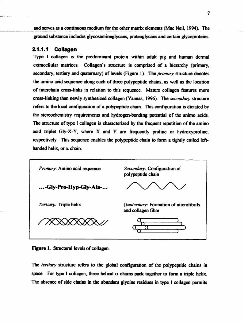
extracellular matrices. Collagen's structure is comprised of a hierarchy (primary,
secondary, ter&iary and quatemary) of levels (Figure 1). The primrrry structure denotes
the amino acid sequence dong each of three polypeptide chahs, as well as the location
of interchain cross-links in relation to this sequence. Mature collagen features more
cross-linking than newly synthesized collagen (Ymas, 1996). The seconduty structure
refers to the local configuration of a polypeptide chain. This configuration is dictated by
the stereochemis~ requirements and hydrogen-bonding potential of the anho acids.
The structure of type 1 collagen is characterized by the muent repetition of the amino
acid triplet Gly-X-Y, where X and Y are frequently proline or hydroxyproline,
respectively. This sequence enables the polypeptide chah to fom a tightly coiled lefi-
handed helix, or a chain.
Primury: Amino acid sequence Secondary: Configuration of polypeptide chain
..- Gly-PrwHyp-Gly-A h-...
I Tertiury: Triple helix Quutemry: Formation of microfibnls anà collagen fibre
Figure 1. Structural levels of collagen.
The terfiury structure refers to the global configuration of the polypeptide c h a h in
space. For type 1 collagen, three helical a chains pack together to form a triple helix.
The absence of side chains in the abuadant glycine midues in type 1 collagen permits

the - close - alignment - . - of polypeptide chahs in a triple helix arrangement. The peptide _ _ - _ dU 1_.1__ ____dC % _ i-- ---l- f % C - -
bonds linking adjacent amino acids are oriented tow8fds the interior of the helix,
rendering the triple-helix highly resistant to general pmtease attack. The quatemary
structure denotes the repeating supernoleculor unit structure, comprising several
molecules packed in a specific lanice, i.e. a microfibril. Adjacent molecules in the
microfibril are approximately parallel to the fibre axis; they al1 point in the sarne
direction along the fibre and are staggered regularly. This Oves rise to the well-known
D-period of about 64 nm, which is visible by transmission electron microscopy
(Linsenmayer, 199 1 ).
Collagen synthesis is a cornplex, multi-step progression through each of these four
levels. The collagen molecules aquire their primary, secondary and tertiary structures
intracellularly, and are secreted as triple-helical, procollagen molecules. Ext~acellular
processing involves proteolytic cleavage of terminal propeptides as wzll as self-
assembly into microfibrils and fibres. Collagen fibres becorne fiirther stabilized by inter-
fibre cross-linking of lysine residues by the enzyme lysyl oxidase (Linsenma yer, 1 99 1 ).
Type 1 collagen is characterized by its stability as a molecule; this stability is enhanced
over time by the ongoing formation of intermolecular cross-links in mature collagen.
2.1.1.2 Hyaluronan Hyal uronan (HA) is a linear polysaccharide of up to 25,000 disaccharide uni ts composed
of Dglucuronic acid and N-acetyl-D-glucosamine (Figure 2). Its high molecular mass
and numerous mutually repelling anionic carôoxyl groups on the glucuronic acid
residues make HA a rigid and hydrated molecule. Accordin&, HA occupies a volume
in solution approximately 1000 times greater than in its dry state (Voet and Voet, 1990).
Besides providing structural support within the extracellular d x , HA maintains water
homeostasis of the tissues. Even at low concentrations, the individual HA c h a h
enangle and fomi a continuous network in the solution, causing a pmnounced
viscoelasticity (Laurent, Fraser et al., 1986). HA is found in variable amounts in al1
tissues and fluids of adult anbals and is especially abundant in soft connective tissues
(Alberts, Bray et ai., 1994). HA occm bund to plasma membranes, aggregated with
other macromolecules, or as a free polysaccharide (Laurent, Fraser et al., 1986).

Figure 2. Chemical structure of hyaluronan. Abbreviations: Ac = acetyl group.
2A.l.3 Hepamnsuhbs The heparan sulfates, composed of heparin and heparan sulfate, are both sulfated
glycosaminoglycans composed of glucuronic acid or iduronic acid and K
acetylglucosamine with an a1,4 linkage (Wight, Heinegard et al., 1991). The degree of
sulfation differs between heparin and heparan sulfate; less than 50% of the N-acetyl
groups in heparan sulfate are sulfated, while more than 70% are sulfated in heprin
(Figure 3) (Wight, Heinegard et al., 1991). Heparin is the major intracellular heparan
sulfate, found within mast cells and basophils. Commercial heparin, clinically used as
an anticoagulant, is a degradative product of mast cell heparin. b
Figure 3. Chemical structure of heparin, indicating the target side groups for sulfation.
Heprin is released from mast cells and basophils in early wounds and has a nwnber of
important effats on cells during wound healing (Section 2.2). Heparin and other
sulfated glycosaminoglycans (GAGs) anchor soluble proteins in the matrix by their
highly anionic sulfate groups. These soluble proteins may be growth factors, proteinases
or proteinase inhibitors. It is mt clear whether GAGs act as reservoirs to keep these
growth factors in the vicinity of the cells, help prevent their proteolytic breakdown or
actually present the growth factors to the celis in a more efficient manner. However, the
association between small soluble growth factors and &se extracellular matrix
molecules is effective in modulating cell behaviour (Roy, DeBlois et al., 1993).

2.-1.2-Compcrrian of Pi&andtfurnannSkin_ -- The pig is widely used in research as a representative mode1 of human wound healing
(Sullivan, Eaglstein et ai., 200 1 ). Given the many morphological similarities between
pig and human skin, acellularized pig skin may be a suitable, less expensive alternative
to burnan demis for dermal replacement. niere are many similarities between the
morphological and functional characteristics of pig and human skin. These features
include relatively sparse hair, a thick epidennis with distinct rete pegs and corresponding
dermal pepillae, as well as similarities in the stnicture of the collagenous tissue
framework and the adipose chambers of the subcutis (Meyer, Schwarz et al., 1978). Pig
dermal collagen is similar to human demal collagen biochemically, accounting for its
use in a number of wound healing products (Heinrich, Lange et al., 1971). There are
signifiant puaîlels in the composition of the lipid film of the skin sdhce, and both
similarities in epidemd tissue turnover time and the character of keratinous proteins
(Meyer, Schwarz et al., 1978). Like human demis, pig demis has a well-defined
papillary component, consisting of delicate collagen fibres intermingled with comective
tissue cells. This layer is continuous with the b r d micular demis which shows a
welldeveloped interlacing of connective tissue fibres.
There are also several differences to note behveen pig and human demis. In contrast to
man, the elastic fibre content of pig demis is relatively low but is still higher than in
other species (Meyer and Neuimd, 1987). As well, pig hair is coarser than human hair
(Hinrichsen, Birk-Sorensen et al., 1998). The subcutis of the pig is similar to that of the
human, with some differences. In the most superficial layer, the stratum adiposum
subcutis, elastic and collagen fiben show pocket formation in which adipose tissue is
deposited abwdantly, much Iike in humaas. The deeper stratum fibrosum subcutis of
the pig is represented by a connective tissue sheet which, in contrast to man, contains the
paaaiculus carnosus (Rose, Vistnes et al., 1978). The pattern of vascularization in the
pig displays subepidermal, lower and mid-demal networks. It differs h m man in that
the sukpidennal network is less dense in the pig. However the vascularization of the
lower region of the hair follicle including the hair pepilla, parallels that in humans. Pig
skin contains no eccrine glands, and unlike man, apociine glands are distributeci through
the skin surface (Meyer, Schwarz et al., 1978).

-=a- - -- - ---...a -- -- - - - - - - - - - - A - .. Given the success of acellÛlar human demis as a dermal s u b h ~ t e (Section 2.5.1 ), the
similarities between pig and human demis suggest that acellular pig dermis may too
have potential as a &mal substitute. It is hypothesized in this thesis that acellular pig
demis is equivalent to acellular human demis as a scaffold for human fibroblasts in
vitro.
2.2 Cell-Matrix Interactions in Wound Healing For al1 stages of wound healing, cellular responses are to a large extent dictated by their
interaction with the sunounding extracellular matrix. Cells are found to bind to
extracellular matrices as a means of anchoring themselves to the scaffold and to denve
traction for migration (Martin, 1997). Ongoing research into the structure and
composition of the extracellular matrix now strongly inâicates that a major role of the
matrix is to regulate cell behaviour, rather than to passively support cells. Matrices also
impart signals for growth and diffenntiation. Extracellular matrix components involved
in scar regdation and neovascularization, in peiticular, are desirable as dermal substiMe
matenals. This section will highlight some of the ways in which the cell-rnatrix
interactions of collagen, hyaluronan and proteoglycans can influence specific wound
healing events.
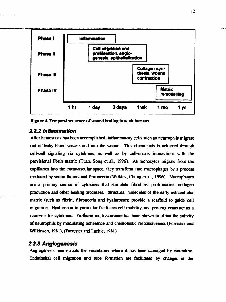
2.2. i Stages of wwnd heaIing The healing pnness can be broken down into early, intemediate, late and terminal
phases (Figure 4). Specific biologic processes characterize each phase. The primary
activities involved in the eariy phase of healing are hemostasis and inflammation.
Mesenchymal cell proliferation and migration, epithelialization and angiogenesis are the
primary events of the intermedia% phase. In the late phase (der one week), the
synthesis of collagen and other matrix pmteins begin. As well, wound contraction can
be oôserved h m this point on. The terminal phase of wound healing is characterized by
wound remodeling to fonn a scar. The mie of the fibroblast in wound healing will be
summarizeâ here, as well as the influence of the extracellular matrix on cellular
responses in wound healing.
Phas01
Phase II
Pham III
Phase IV
--
Colrgen syn- thesk, wound contnction
- - - - - - - - - - - -- - - - - -- -- -
1 hr 1 day 3 days 1 wk 1 mo 1 yr
Figure 4. Temporal sequence of w o d healing in ahilt humans.
22.2 lnflamrnatm'on M e r hemostasis has been accomplished, idammatory cells such as neutrophils migrate
out of leaky blood vessels and into the wound. This chernotaxis is achieved through
cell-cell signaling via cytokines, as well as by cell-matrix interactions with the
provisional fibrin rnatrix (Tuan, Song et al., 1996). As monocytes migrate from the
capillaries into the extravascular space, they transfomi into macrophages by a process
mediated by serum factors and fibronectin (Wilkins, Chung et al., 19%). Macrophages
are a primary source of cytokines that stimulate fibroblast proliferation, collagen
production and other healing processes. Structural molecules of the early extracellular
matrix (such as fibrin, fibroneain and hyaluronan) provide a scaffold to guide cell
migration. Hyaluronan in particular facilitates cell mobility, and proteoglycans act as a
reservoir for cytokines. Furthemore, hyalun>aan has been shown to affect the activity
of neutrophils by modulating adherence and chemotactic responsiveness (Forrester and
Wilkinson, 198 1 ), (Forrester and Lackie, 198 1 ).
2.2.3 Angiogenesis Angiogenesis reconstructs the vasculatm where it has been damaged by wounding.
Endothelid cell migration and tuôe fornation are facilitated by changes in the

extracellular rnatrix. The synthesis and degradation of type 1 and type N collagen - N_ -_u.iLL- _ - _ -- -. 4 - - - ---- -A_ _ _ _ _ - _ _ -_ - _ - -
smunding the capillary sprouts are tightly regdated to provide support and guidance to
the newly fomed capillaries (Clark, 19%). Cytokines directly and indirectly stimulate
endothelial cell migration and proliferation. Many angioge~c cytokines have been
identified, and include basic fibroblast growth factor (bFGF), of which heparin is a CO-
factor, as well as vascular endothelial growtb factor and transfonning growth factor+
(Wilkins, Chung et al., 1996). The progngiogenic effects of hepmin are believed to be
attributable to its delivery of bFGF and acidic FGF to the cells (Muellet, Thomas et al.,
1989), (Folkman and Sking, 1992), but this has not been conclusively shown
(Sasisekharan, Moses et al., 1994). Heparin has been investigated in cardiovascular
applications for its pro-angiogenic properties, which have been shown to be molecuiar
weightdependent (Bornbardini and Picano, 1997). The degradative products of
hyalmnan, specificaliy fragments 4 to 25 disaccharides in length, also have angiogenic
properties (Cockerill, Gamble et al., 1 995).
2.2.4 EpîtheIiaIIzatlon The sequence of events which Oves rise to epithelialization include cellular detachment,
migration, prd i feration and di fferentiation. Marginal basal cells elongate, detach fiom
the underl ying basement membrane and migrate into the wound. Wi thout col lagenol ytic
activity, keratinocytes do not migrate within a dermal collagen matrix (Pilcher, Sudbeck
et al., 1998). It is hypothesized that keratinocytes use collagenase- 1 to cleave collagen to
gelatin, thereby providing a substrate which is more conducive to migration. It is
believed that altered cell-matrix interactions in wound healing regulate the spatially -
precise pattern of collagenase production in rnigrating keratinocytes (Clark, 1996).
Evidence shows that keratinocytes lose contact with the basement membrane and instead
bind to the provisional maaix and die type 1 collagen-rich demial matrix. This transfer
is a critical determinant of increesed collagenase production, and initiation of migration
(Pilcher, Sudbeck et al., 1998).
2.2.5 FibmpIasia Fibroplasia denotes fibroblast cellular activities during tissue repair. These include
migration, prolifmtion, differentiation, organization and degradation of the transient
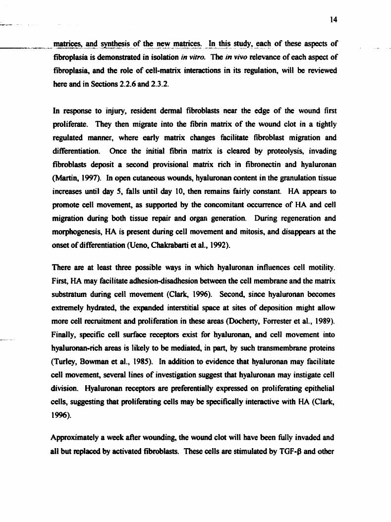
motrices, - . LL-- and synthesis %-As -- of the ---Lw new -A matrices. -a -- --?--- In Uiis - study, - -- - - each - . of these aspects of
fibroplasia is demomtrated in isolation in vitro. The in vivo relevance of each aspect of
fibmplasia, and the role of cell-matrix interactions in its regdation, will be reviewed
here and in Sections 2.2.6 and 2.3.2.
In response to injury, resident demai fibroblasts near the edge of the wound fint
proliferate. They then migrate into the fibrin matrix of the wound clot in a tightly
regdated manner, where early matnx changes facilitate fibroblast migration and
differentiation. Once the initial fibrin matrix is cleared by proteolysis, invading
fibroblasts deposit a second provisional matrix rich in fibronectin and hyaluronan
(Martin, 1997). In open cutaneous wounds, hyaluronan content in the granulation tissue
increases until &y 5, falls until day 10, then remains fairly constant. HA appears to
promote ceIl movement, as supported by the concomitant occurrence of HA and ceIl
migration during both tissue repair and organ generation. During regeneration and
morphogenesis, HA is present dwing ce11 movement and mitosis, and disappears at the
onset of differentiation (Ueno, Chakrabarti et al., 1992).
There are at least three possible ways in which hyaluronan infiuences cell motility.
First, HA may facilitate adhesion-cüsadhesion between the ceIl membrane and the matrix
substratum during ceIl movement (Clark, 1996). Second, since hyalmnan becomes
extremely hydrated, the expanded interstitial spece at sites of deposition might allow
more ceil recruitment and proliferation in these areas (Docherty, Forrester et al., 1989).
Finally, specific cell surface receptors exist for hyaluronan, and ceIl rnovement into
hyaluronan-rich areas is likely to be mediated, in part, by such transmembrane proteins
(Turley, Bowman et al., 1985). In addition to evidence that hyaluronan may facilitate
cell movement, several lines of investigation suggest that hyalmnan rnay instigate ce11
division. Hyalutonan receptors are peferentially expressed on proliferating epithelial
ce14 suggesting that proliferating cells may be specifically interactive with HA (Clark,
19%).
Approximately a week after wounding, the wound clot will have been N l y invaded and
dl but replaced by activated fïbroblasts. These cells are stimula&$ by TGF-B and other

growth fiictors to synthesk and remodel a new collagen-rich matrix (Martin, 1997). L-L --- - - L - - - - - -- - L - - - - - - - A - -
The rate of oollagei, synthesis increases rapidly and continues at an accelerated rate for 2
to 4 weeks. Collagen makes up more than 50% of the protein found in scar tissue. Mer
four weeks, collagen synthesis rates decline, eventuaily balancing the rate of collagen
destruction by collagenase (Wilkins, Chung et al., 1996).
The dynarnic between extracellular rnatrix and fibroblasts is especially evident during
granulation tissue development. It has been shown that the provisional matrix promotes
an early granulation tissue fibroblast phenotype. In contrast, collagen matnx promotes a
relatively quiescent dermal fibroblast phenotype. Extracellular matrix fibrils strongly
influence the direction of fibroblast migration, since the cells tend to align and migrate
along discontinuities in substrats to wtiich they are attached (Clark, 1996). Type 1
collagen appears to cause directed migration of cells through this process (Postlethwaite,
Seyer et al., 1978). Thus chemotactic, adhesion and contact guidance signals may al1
influence fibroblast migration into the provisional matrix.
The eff'ts of extracellular matnx collagen on fibroplasia are mediated in part through
activation of the integrin collagen recepton al P 1 and a2p 1. Collagen matrices reduce
fibroblast proliferation and collagen synthesis, but induce procollagenase and a2Pl
integrin expression (Mac Neil, 1994). It is likely that the collagen-rich extracellular
matrix- which accumulates as granulation tissue matures- at the same time reduces the
ability of wound fibroblasts to prduce M e r collagenous matrix, and promotes the
ability of these d h to mnodet the dlagm-rich matrix alredy p n s r t (Grimntt and
Lamke, 1984). While the specificity of the cell-maüix interactions seems to corne from
the integrin binding, other adhesive matrix molecules also contain sites that can interact
with the glycosaminoglycan component of proteogiycans. The binding of cell sdace
proteoglycans to such sites is likely to also play a regdatory role in cell adhesion (Clark,
19%).
In open cutaneous womds the level of gaiactosaminoglycans, i.e. chondroitin-4-sulfate
and dematan sulfate, begins to increase at approximately one week pst-wounâing
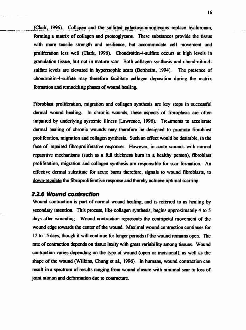
(Clark, 1996). Collagen and the sulfhted ~actosaminoglycans replace hyaluronan, L-- 3- A - - - - - A- - -A - -- -- - - --
foming a matnx of collagen and proteoglycans. These substances provide the tissue
with more tensile s~rength and resilience, but accommodate ceIl movement and
proli feration less well (Clark, 1996). Chondroitin-4-sulfate occurs at high levels in
granulation tissue, but not in mature scar. Both collagen synthesis and chondmitin-4-
sulfate levels are elevated in hypertrophie scars (Bertheirn, 1994). The presence of
chonâroitin4sulfate may therefore facilitate dlagen deposition dwing the matrix
fonnation and remodeling phases of wound healing.
Fibroblast proliferation, miwtion and collagen synthesis are key steps in successfûl
demai wound healing. In chronic wounds, these aspects of fibroplasia are oflen
impaiced by underlying systemic illness (Lawrence, 19%). Tmbnents to accelerate
dermai healing of chronic wounds may therefore be designad to prumote fibroblast
proliferation, migration and collagen synthesis. Such an effect would be desirable, in the
face of impaired fibroproliferative responses. However, in acute wounds with normal
reparative mechanisms (such as a full thickness bum in a healthy person), fibroblast
pliferetion, migration and collagen synthesis an responsible for scar formation. An
effective demal substitute for acute bums therefore, signals to wound fibroblasts, to
dom-remlate the fibroproli ferative response and thereby achieve optimal scamng.
2.2.6 Wound contraction Wound contraction is part of normal wound healing, and is referred to as healing by
secondaiy intention. This process, like collagen synthesis, begins approximately 4 to 5
days after wounding. Wound contraction represents the centripetal movement of the
wound edge towards the center of the wound. Maximal wound contraction continues for
12 to 15 days, though it will continue for longer periods if the wound nmains open. The
rate of contraction depends on tissue laxity with great variability among tissues. Wound
contraction varies depending on the type of wound (open or incisional), as well as the
shape of the wound (Wilkins, Chung et al., 1996). In humans, wound contraction can
result in a spctnun of results ranging from wound closure with minimal scar to loss of
joint motion and deformation due to contracture.
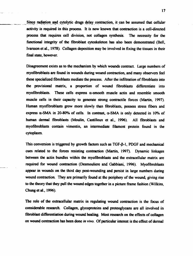
Since - radiation -u---=ya--- and cytolytic -- - dnigs - - delay -A -..- contraqtion, -- - - it - - - - be - assumed that cellular
activity is required in this process. It is now known that contraction is a cell-directed
process that nquires ceil division, not collagen synthesis. The necessity for the
fiinctional integrity of the fibmblast cytoskeleton has aiso been demonstrated (Bell,
Ivarsson et al., 1978). Collagen deposition may be involved in fixing the tissues in their
final state, however.
Disagreement exists as to the mechanism by which wounds contract. Large numbers of
myofibroblasts are found in wounds during wound contraction, and many observers feel
these specialized fibroblasts rnediate the process. After the infiltration of fibroblasts into
the provisional maaix, a proportion of wound fibroblasts differentiate into
myofibmblasts. These cells express a-smooth muscle actin and resemble smooth
muscle cells in their capcity to generate strong contractile forces (Martin, 1997).
Human myofibroblasts grow more slowly than fibroblasts, possess stress fiben and
express a-SMA in 20-80% of cells. In contrast, a-SMA is ody detected in 10% of
h u m dermal fibroblasts (Moulin, Castillowt et al., 19%). All fbroblasts and
myofibroblasts contain vimentin, an intemediate filament protein found in the
cytoplasm.
This conversion is triggered by growth factors such as TGF-P-1, PDGF and mechanical
cues reiated to the forces resisting contraction (Martin, 1997). Dynamic linkages
between the actin bundles within the myofibroblasts and the extracellular matrix are
required for wound contraction (Desmouliere and Gabbiani, 1996). Myofibroblasts
appear in wounds on the third day post-wounding end persist in large numben during
wound contraction. They are primarily found at the periphery of the wound, giving rise
to the theory that they pull the wound edges together in a picture fnune fashion (Wilkins,
Chung et al., 19%).
The role of the extracellular rnatrix in regulating wound contraction is the focus of
considerable research. Collagen, glycoproteins and proteoglycans are al1 involved in
fibmblast differentiation during wound healing. Most research on the effécts of collagen
on wund contraction has been Qne in vivo. ûfpanicular interest is the effect of dennal
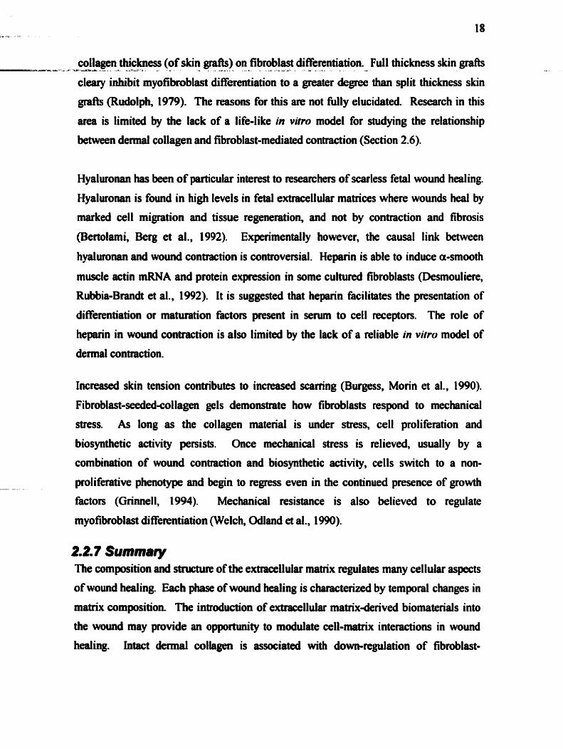
collagen thickness (of skin graAs) on fibroblast differentiation. Full thickness skin &rafts ---*--A >---=2 - .-- - - . - d-.. - -A - -- - - - *- - - -
cleary inhibit rnyofibroblast differentiation to a greater &gree than split thickness skin
gr* (Rudolph, 1979). The reasons for this are not Mly elucidated. Research in this
atea is limited by the lack of a life-like in viho model for studying the relationship
between demal coilagen and fibroblast-mediated contraction (Section 2.6).
Hyalwonan has been of psrticuiar interest to researchers of scarless fetal wound healing.
Hyaluronan is found in high levels in fetal extracellular matrices where wounds heal by
marked cell migration and tissue regeneiation, and not by contraction and fibrosis
(Benohmi, Berg et al., 1992). Experimentally Lwever, the causal link between
hyalwonan and wound contraction is wntroversial. Heparin is able to induce a-smwth
muscle actin mRNA and protein expression in some cultured fibroblasts (Desmouliere,
Rubbia-Brandt et al., 1992). It is suggested that heparin facilitates the presentation of
diflerentiation or maturation factors present in serum to cell receptors. The role of
heparin in wound contraction is also limited by the lack of a diable in vitro model of
demal contraction.
Increaseà skin tension contributes to increased scaning (Burgess, Morin et al., 1990).
Fibroblast-Seededeollagen gels demonstrate how fibroblasts respond to mechanical
stress. As long as the collagen material is under stress, cell proliferation and
biosynthetic activity persists. Once mechanical stress is relieved, usually by a
combination of wound contraction and biosynthetic activity, cells switch to a non-
proliferative phenotype and begin to regress even in the continued presence of growth
factors (Gfi~ell, 1994). Mechanical resistance is ais0 believed to regulate
myofibmblast di fferentiation (Welch, Odland et al., 1 WO).
t t 7 Summaty The composition and stnicnup of the extrawiiular matrix regdates many cellular aspects
of wound healing. Each phase of wound heaîing is characterized by temporal changes in
matrix composition The introduction of exttacellular matrixderived biomaterials into
the wound may provide an opportunity to modulate cd-matrix interactions in wound
healing. Intact dermal collagen is associated with dom-regdation of fibroblast-

mediated contraction. Hyaluronan fscilitates cell migration, and may play a role in ------ - -- - 2 - -= -- - >- - - -L--- A---- LA 2 -
scarless wound healiag. Hyaluronan's degradation pducts, as well as heparin, are pro-
angiogenic. The significance of these matrix moleailes as components of an acellular
dermal matrix, in ternis of their effects on fibroplasia, is the focus of this thesis.
2.3 Host Response to Biomaterials Biomaterials for dermal substitutes may be designed to replace Iost tissue or to M e r
alter the host's healing response. To effatively replace the demis, the material must
become vascul- and intiltrated by the host's dermal cells in order to become
successfùily integrated into the wound. The design of biomaterials must take into
account wound healing mechanisms involved in biomaterial integration into the body.
2.3. f Foiieign body msponse The occumnce of a foreip body reaction is considered a typical wound healing
response to implanted biomaterials (Anderson, 1996). This is typified by the presence of
foreip body giant cells and the hown components of granulation tissue. Generally, the
foreign body reaction results in fibrosis at the surface, which isolates the implant from
the host tissue (Yannas, 1996). With biocompatible materials, the foreip body reaction
in the implant site may be controlled by the properties of the biomaterial. The degiee of
fibroproliferative response in most cases parallels the extent of inflammatory response of
a material. Materials which elicit a marked inflammatory reaction become quickly
walled off with a thick fibrous capsule. For demiel substitute matenals, where the goal
is to achieve minimal scarring, it is ctitically important that fibrous capsule formation is
prevented. Materials for this application must therefore be specitically designed to
modulate fibroblast responses such as fibroblast prdiferabion and cdlagen synthesis, and
to provoke a minimal inflarnmatory reaction.
2.3.2 BiomateriTaI Integrniion in& the Wound Re@ of implanted biomaterials can involve two distinct processes: replacement of the
implant by fibrous capsule co~ective tissue, or regeneration, Le. the replacement of
injured tissue by parenchymal cells of the same type. niese processes are generally
controlled by (1) the proliferative capacity of the cells in the recipieat tissue, and (2) the
extet of injury as it relates to the destruction or persisteme of the original tissue

The condition of the underlyhg framework or supporting - - -,=-- -A -- %.-- - -- - 2 - .- - - -
cells following an injury plays an important mle in the
mtoration of nomal tissue structure. Retention of the framework may lead to
restitution of the normal tissue structure, while its destruction most commonly leads to
fibrosis (Yannas, 1996).
Biomaterials may be desiped as scafYolds for host cell infiltration, thus replacing the
supporting fnuaework of the injured tissue. Biomatenals intended for integration must
be designed to modulate cellular responses. This is necessary to prevent fibrous capsule
formation and its sequestration as an avascular implant in the wound. The material must
possess the necessary fatwes for ce11 infiltration to occu: adequate pore size for ceIl
migration and nutrient diffusion, pangiogenic properties, anà suitable adhesiveness
for migrating cells. A key step in the integration of foreign materials intu the wound is
the ingrowth of host capillaries. As the material becomes vascularized, the rnigrating
endothelial cells signal to smunding fibroblasts and macrophages to begin migration
into the material (Yannas, 1997). Ideally, the material's degradation rate is matched to
the biosynthetic rates of the infiltrating host cells, thus remodeling a new extracellular
matrix. Upon integration of the ideal biomaterial, the typical fibrotic response of wound
healing would be dom-regulated, so as to instead fàvour a regenerative response.
2.4 Extracellular Matrix-Derived Biomaterials Most of the naturally occurring materiais in use as biomatenals today are constituents of
the extracellular matrix (Griffith, Osborne et al., 1999). Natural allograft and xenografi
materials offer the advantage of king similar or identical to those found within the
human body, enabling their recognition and degradation in the biological environment.
The problems of toxicity and chronic inflammation, which are frequently provoked by
synthetic polymers, may therefore be avoided (Y-, 19%). The pmmising
comptibiüty offered by natural materials allows the possibility of desiping
biomaterials which fiuiction biologically at die molecular, rather than at the
macroscopic, level.

An intriguizig characteristic of these materials lies in their high degradation level by A %.-Lu - -*--.. - A L - - - -- --
natumlly occwing enzymes. It is therefore possible to control the degradation rate of
the implanteâ materiai, by specifically modiQing its structure with the enzyme
specificity in mind (Mauch, Adelmann-Grill et al., 1989). A disadvantage, however, in
the use of biomaterials presents itself in a sometimes significant level of
immunogenicity, which of course derives precisely fiom their simi larity to biological
materials. The imrnunological reaction of the host to the implant is directeci against
selected antigenic determinants in the protein molecule, with eventual degradation or
encapsdation of the implant (Anderson, 1996). It mut be noted that, in some cases, the
immune reactivity of proteins can be virtually eliminated by chernical or physical
modification of the antigenic detenninants. The immunogenicity of polysaccharides is
typically far lower than that of proteins. As well, collagens are generally weak
immunogens relative to the majonty of proteins (Yannas, 1996).
2.4. f Co//agen Collagen fiom both animal and human sources is widely used as a biomaterid. Type I
collagen is the most abundant and important type for use as a biomaterial (Bailey, 2000).
Collagen-based materials can be separateci into two categories: (1) biomaterials retaining
the original tissue structure (used as sutures, cardiac valves, ligamentary protheses), and
(2) biomaterials prepared from pinfied collagen, with or without additives, and prepared
in different fonns depending on their applications. These in tuni are used as sponges,
tubes, spheres, membranes etc. Examples of applications of collagen-based biornaterials
include: injectable collagen for cosmetic skin defects, vitreois replacement and coating
of bioprostheses, collagen sponge for wound dtessings, hemostatic agents, sponge
scafKolds for tissue regeneration of peri pheral nerves, demis collagen microspheres for
Qug delivery and ceIl culture, collagen membrane as dialysis membranes and comeal
shields, and couagen powder for bone filling and repair (Chevallay and Herbage, 2000).
The immuwgenicity of collagen has been extensively studied. The clinid significance
of collagen's Unmunogenicity has been shown to be very low, and is often considered to
be negligible. The apparent reason for the low antigenicity of type I wllagen stems from
the small species difference arnong type 1 collagens, as in cow and human (Yannas. 2 --L--&.-- - -- - - . - - --= --..-- -- ----- &%--. - - A--- - -
1996). Such similarity is, in tuni, borne out by the inability of the triple helical
configuration to incorporate the substantial amino acid substitutions characteristic of
other proteins (Linsenmayer, 199 1 ).
In order to modify the immunogenicity of collagen, it is usefil to consider the location
of its antigenic deteminants, i.e. the specific chemical groups which are recognized as
foreip by the immune system. In general, the procollagen form is more antigenic.
However, when native collagen molecules are used as immunogens, antibodies can still
be elicited. When prmllagen is used as an imrnunogen, most of the antibodies are
directecl against epitopes within the propeptide extensions at the terminal ends. These
antigenic determinants include both sequential (i.e. dependant on amino acid sequence)
and wnfonnational (i .e. de pendant on tedary or quaternary structure) deteminants.
The conformational deteminants of collagen depend on the presence of the intact triple
helix and, consequently, are abolished when collagen is denatured into gelatin. The
pmess of gelatinization briefly ex- the sequential deteminant of collagen before it
is cleared away by enzymatic digestion (Clark, 1996). Conüolling the stability of the
triple helix during processing of collagen, therefore, would prewnt the display of the
sequential deteminants.
Sequential deteminants exist in the nonhelical end (telopeptide region) of the collagen
molecule. This region has been associated with most of the immunogenicity of
collagen-basai implants (Bailey, 2000). Several enzymatic tfeatments have been
devised to cleave the telopeptide region without destroying the triple helix. Treating
collagen with cross-linking agents also pennits a reduction in antigenicity (Weadock,
Olsen et al., 1984). intermolecuiar cross-linking of collagen by physical treatment or
chemical agents modifies other propecties of biomateriais, as well as their antigenicity.
For example, cross-linking can reduce solubility, water absorption and biodegraâability
of the coliagen biomaterial. Its mechanical properties are also enhanceci (Chevallay and
Herbage, 2000).

exposm to dhaviola or gamma rays, es well as by themial dehydration. Chernical
cross-links may also be f o d by chernical treatment with dialdehydes such as
glutaraldehyde, or with carboâiimide (Chevallay and Herbage, 2000). Changes in pH
can be used to selectively abolish the quaternary structure of collagen while maintaining
the tertiary structure. The lanice of aggregated triple helices may be lost reversibly
when the pH of a suspension of collagen fibres in acid is lowered below 4.25. During
this transformation, the triple helical structure remains unchanged. The triple helix may
be converted to random coi1 gelatin by heeting above the helix-coi1 temperature, or by
exceeding a critical concentration of anions in a sdution of collagen molecules (Yannas,
19%). Collagen may also be processeà through solubilization and freeze-drying to
achieve a porous sponge-like structure, with mean pore diameters ranging frwn 1 to 800
um. Collagen gels used in this stuây are p r e p d fiom a solution of acid-denatured
bovine dermal collagen. The suspension of non-aggregated triple-helical molecules is
neutralized witb base and heated to 3TC, to allow the molecules to quickly re-aggregate
into microfibrils. This fonn of collagen diffea fkom intact dermal collagen in the extent
of intemolecular cross-linking, as well as in the fibril size (Chevallay and Herbage,
2000).
2.4.2 Hyalumnan The structure of hyaluronan (HA) is con- across species, and therefore is not
antipnic (Chen and Abatangelo, 1999). The fact that HA elicits no immune response
makes it a desirable biomaterial. The high molecular weight and hydtophilicity of HA
fom the b i s for most of its uses as a biomaterial. Almost al1 applications make use of
the exceptionally high viscosity and facility to fom gels, characteristic of large
polysaccharides. HA is used in ophthalmologic applications such as in catatact surgery
and retinal reattachment. It is also used as an injectable synovial fluid additive in the
treatment of ostemduitis. Benzylesterified HA has been used to mate Laserskina, a
biocompatible delivery system for keratinocytes to impve cultured epidemal graft
take rates. The keratinocyte-seeded laser micmperfomted HA membranes are coupled to
fibmblast-Seeded meshes in order to obtain a derd-epidennal composite structure
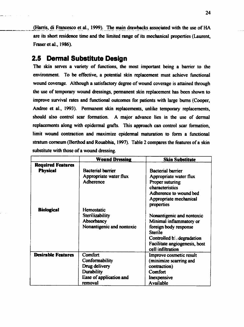
@uns, di Fmcesco et al., 1999). The main drawbacks associated with the use of HA ---- 6 - - - L a - A- - - - - - ..----- - --- - - -- - - - --
are its short tesidence time and the limited range of its mechanical properties (Laurent,
Fraser et al., 1986).
2.5 Dennal Substitute Design The skin serves a variety of fùnctions, the most important king a barrier to the
environment. To be effective, a potential skin replacement must achieve functional
wound coverage. Although a satisfactory degree of wowid coverage is attained through
the use of temporary wound dressings, permanent skin replacement has been show to
improve sunival rates and fiinctiod outcornes for patients with large burns (Cooper,
Andree et al., 1993). Pemiamnt skin replacements, unlike temporary replacements,
shodd also control scat formation. A major advance lies in the use of dermal
replacements dong with epidemial grafts. This approach can control scar formation,
limit wound contraction and maximize epidermal maturation to fonn a hctional
stratum conieum (Berthod and Rouabhia, 1997). Table 2 cornparcs the featms of a skin
substitute with those of a wound dressing.
Desirable Fea tu res
Wound Dresainp:
Bacterial M e r Appropriate water flux Adherence
Hemostatic Sterilizability Absorbancy Nonantigenic and nontoxic
Comfort Conformabiiity h g delivery Durability Ease of apptication and removal
Bacterial barrier Appropriate water flw Proper suturing c haracteristics Adherence to wound bed Appropriate mechanical properties
Nonantigenic and nontoxic Minimal inflammatory or foreip body response Sterile Controlled bi..; degradation Facilitate angiogenesis, hoa ce11 infiltration Improve cosmetic result (minhnize scarring and contraction) Comfort inexpensive Available
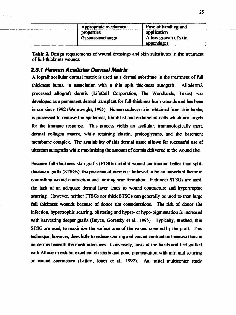
Table 2. Design requirements of wound dressings and skin substitutes in the treatment of full-thickness wounds.
2.5. f Human AcelIuI8r Detm8I Mat& Allograft acellular dermal d x is used as a dermal substitute in the treatment of fùll
thickness bums, in association with a thin split thickness autograft. Allodetm@
processed allograft demis (LifeCell Cotporation, The Woodlands, Texas) was
developed as a permanent dennal transplant for full-thickness bum wounds and has been
in use since 1992 (Wainwright, 1995). Human cadaver skin, obtained fiom skin banks,
is processed to remove the epidemal, fibroblast and endothelial cells which are targets
for the immune response. This pracess yields an acellular, Unmunologically inert,
demal collagen matrix, while retaining elastin, proteoglycans, and the basement
membrane cornplex. The availability of this dermal tissue allows for successful use of
ultrathin autograh while maximizing the arnount of demis delivered to the wound site.
Because NI-thickness skin &rafts (FTSGs) inhibit womd contraction better than split-
thickwss grafts (STSGs), the presence of &mis is believed to be an important fâctor in
controlling wound contraction and limiting scar formation. If thimer STSGs are used,
the lack of an adequate demal layer leads to wound contracture and hypertrophic
schng. However, neither FTSGs nor thick STSGs can generally be used to tteat large
fuii thiclcness wounds because of donor site considerati011~, The risic of donor site
infection, hypertrophic scarring, blistering and hyper- or hypo- pigmentation is increased
with harvesting deeper &rafts (Boyce, Goretsky et al., 1995). Typically, meshed, thin
STSG are used, to maximize the surface area of the wound covered by the gr&. This
technique. however. does little to reduœ sçarring and wound contraction because there is
no demis beneath the mesh interstices. Conversely, areas of the han& and feet grafled
with Alloderrn exhibit excellent elasticity and good pigmentation with minimal scarring
or wound contracture (Lattari, Jones et al., 1997). An initial multicenter study

documents that resuits obtained with acellular dennis and ultrathin STSG grah were at ---- . - - A - d - & -VA-& -- -a A+- --- - - & 2 -
least as equal to those of standard thickness STSGs (Waiawright, 1995).
The transmission of infection rernains a concem in the use of allograft materials. As a
result, al1 allograft d o m tissue is well screeneà. In addition, the acellularization process
safeguards against the transmission of viral disease. Because replication of human
pathogenic vinises occurs only intracellularly, the removal of cells fiom the extracellular
matrix elhinates sites for viral propagation. in addition, the process includes treatrnent
of the allograft tissue with antiviral agents (Hemdon, 1997).
2.5.2 AceIIuIar Mddk Tissues of animal origin have been d for thousands of years as dressings for extensive
bums. Porcine skin is the most common source of xenognifi skin because of its
simiiarity to human skin. Of the non-primate species, porcine dennis is probably the
most structurally and immunologically similar to human dennis (Srivastava, Jennings et
al., 1999). There exists the potential for cross-species transmission of pathogens,
apecially Mmes and prions, when xenografts are used. Xenografl demis can be
treateâ with ionizing radiation, freeze-dried or soaked in glycerin for steriiization
(Berthod and Rouabhia, 1997).
2.5.2.1 Pig demis as a wound dressing Pig skin can be used to âress second degree burns, especially afier early excision (Gao,
Hao et al., 1992). In addition, pig skin provides a suitable overlay to cover widely
meshed (1 :8 to 1 : 12) autogiafts. Two studies have analysed the cost-effectiveness of pig
skin as a temporary dressing (<4 day s between changes) for partial thickness and excised
full thiclmess wounds (Basile, 1982). (Wood and Hale, 1972). These studies concluded
that rehyârated fieezedried pig skin adhered firmly to the wound surface with
diminution of fluid, electrolyte, protein and heat loss. With muent dressing changes to
assist in wound debndement and to lower the degree of immune rejection, the authors
also described a reduction in pain and bacterial growth. Pig dermis was not however
found to be advantageous over petrolanni gauze, in the treatment of donor sites (Breach,
Davies et al., 1979). Biological skin dressings are also of value in cleaning leg ulcers,
decubitus ulcers and mmy types of i d 4 wounds (Artz, Rittenbuy et al., 1972).

For most types of xenograft transplants, immediate hyperacute pft rejection becomes
increasingly likely as the phylogenetic distance between the donor and recipient grows.
Interestingly, hyperacute rejection does aot seem to be a probkm with pig skin grafts,
but such g d b are susceptible to both cell-mediated and late antibody-mediated rejection
after revascularization (Heslop and Shaw, 1986). This rejection is hypothesized to be
due to the presence of xenogeneic endothelid cells, other ce11 types, or extracellular
matrix materials present in the graft (Wang, Chen et al., 1997). There is some debiite as
to the imrnunogenicity of acellularized porcine demis. Research in this ana has
focused on either ftesh pig skin or nonviable derrnis without the cells removed. It has
not been clearly demonstrated which aspect of the groA elicits antibody production, and
whether vascularization is necessary for this sensitization to occm Acellularized pig
dennal substitute, however, mut support the ingrowth of host capillaries to remain
viable and achieve wound closure.
There is experimental evidence on both sides, indicating that fresh xenografts do and do
not acquire a b l d supply. One study demonstrated, through the use of
microattgiograrns, vascularization of fresh full-thickness pig skin in rabbits (Toranto,
Salyer et al., 1974). Preserved split-thicicness pig skin was useci to treat partial thickness
wounds in humans. The pig skin did not become viable, although vessels were seen in
the grafi at 7 days. Using microangiographic techniques, the vessels could not be
distinguished fiom those accompuiying the invasion of the grah by granulation tissue
(Toranto, Salyer et al., 1974). Another approach to accccurately estimate blood flow
through the graft was to quanti6 the degree of radioactivity four days after injecting
hosts with radiolabelleci red cells. Here, it was fond that autogenic, allogenic and
mammalian xenogenic skn gr& contained approximately the same quantity of blood
when examineà four days after grafàng in the rat (Heslop and Shaw, 1986). These
results are pomising for the use of acellular pig demis as a dermal substitute, where a
material must be able to support host blood vesse1 inpwth.

For biological materials to be suitable for implantation in an alloge~c or xenogenic host,
the material's immumgenicity and potential for infection must be modified.
Acellularization techniques have been desigwd to conserve the tissue architecture of the
extracellular matrix while removing the more antigenic cellular and soluble protein
components. Most acellularization protocols are also successful in sterilizing the tissue
for implantation. There are many variations in acellularization techniques, Qpending on
the desired féatures of the resulting acellular müix.
The acellularization protocol used in this project was originally developed by Courtman
to proâuce acellular vascular allografh (Courtman, Pereira et al., 1994). The tissue is
first immersed in a hypotonic solution, which causes cells to lyse as they absorb water
thugh osmosis. The next solution is isotonie, but contains a mild detergent which
fùrther dissolves the phospholipid-based cellular membranes. These two solutions
contain protease inhibitors, to prevent enymatic degradation of the collagen matrix by
the released cellular material. The only enzymatic step involves a combination of
DNAse and RNAse to destroy the genetic material of the cells. The next rinsing step
uses a strong anionic detergent (sodium dodecyl sulfate, or SDS) to remove residual
soluble proteinaceous and phospholipid matenal. Adipose tissue is not removed in this
step. Finally, the acellular materiai is S08ked in 70% ethanol to remove the residual
SDS, anâ rehydrated in PBS prior to sterilization. The process causes ce11 disniption
with a leaching of ce11 contents, cellular antigens and lipid dissolution, yet inhibits
- - collagen and elastin matrix proteolysis (Wilson, Yeger et al., 1990).
This acellularization protocol was designed for tissues where preserved mechanical
properties and the sbility of the resulting material to support host ce11 infiltration are
critically important. The acellularization process was applied to the production of
acellular xenograft percaridium and hemt valves. Mechanical testing confhed that the
acellular pericardim possesd mechanid characteristics almost identical to those of
fksh pericardium (Courtman, Pereira et al., 1994). implantation of acellularized
allografl carotid arteries resulted in poor cellular ingrowth into the acellular material
after one month. It was suggested that this may be due to the retention of SDS in the

vesse1 extraction p c e s s (Wilson, Courtme $-al.,J995)l The dmtion of soalsng of -*--a- :-=--- - -- - - 4 .- - - - - - - --Lu - + .
the acelluiarized tissw in ethano1 was therefore extended to achieve complete removal
of SDS, in order to allow cellular ingrowth (Robinson-Seurig, 1999).
2.5.2.4 Acellular pig dermal matrix as a dennal substïtute in ment years, many attempts have been made to produce a nonimmunogenic,
xenografk dermal substitute which is capable of supporting thin split-thickness skin
grafts (STSG) or cultured epithelid autografts. Dermal substitutes composed of
denatured xenogenic collagen gels have been studied extensively in humans. Gels
composed of bovine type I collagen have been found to have some efficacy in the
treatment of Ml thickness bum injuries. Few adverse reactions directly related to the
implanted materials have been reported following those gr&. Anti-bovine col lagen
and serum antibody titers do not increase in recipients of these grah, and no evidence of
immediate or delayed immune reactivity to xenogenic collagen has been observed
(Boyce, Goretsky et al., 1995). However, the implanted gels degrade too rapidly to
prevent contraction and fibrosis (Compton, Butler et al., 1998).
The efficacy of undenatured xenograft collagen for the treatment of skin wounds has
also been examined. Xenograft porcine and human skins, both with the epidemis
removeà, have been found to have modest value in the treatment of tùll-thickness skin
wounds in conjunction with immediate STSG autografhng. When trypsinized porcine
skin or cryopreserved dispase-treated human skin was Unplanted into full-thickness
wounds on rats, acute inflammation, some loss of the overlying autograft, and variable - - - amounts of womid contraction were observeù (Wang, Chm et at., 1997), (Fan& Robb et
al., 1990). Lyophilizeâ rehydrated porcine and bovine demis have gîven poor results in
human s u e s (Breach, Davies et ai., 1979). Trypsinized acellular porcine skin was
W y rejected when it was implanted into human cutsneous wounds. Trypsinization
effoctively removes the cellular rnatter h m the demis, but alters the structure of dermal
collagen by cleaving collagen telopeptides. Dispase treatment of skin does not remove
al1 ce11 debris fiom the demis (Srivastava, Jenniags et al., 1999). Dermai matrices
produced by these methoâs may therefore exhibit antigenicity which would account for
their imsatisfactory biomaterial performance.

- -- - A - A recent study exaihinid "~lulaiJ'~p>icine déiiïis, cove& with thin autograb,
implanted in full thickness wounds in rats (Srivastava, Jennings et al., 1999). The
acellulmity of the xenograft material was not venfied histologically Sefore implantation,
however. Wounds treated with xenografb contracted to 46% of the initial 1.5 x 1.5 cm
square wound size, while allogdls treated with the same acellularization process
maintaineci 74% of the initial surfece area. Some gr& s d v a l was noted in the
pnsence of xenografi dennal matrix (52% of grail area survived), but was less than thai
of al lografts (82%). Histologicall y, the xenografts showed signi ficant inflammatory
response, occasional foreign body giant cells, swelling and disorganization of collagen
bundles at 10,14,20 and 30 days post implantation. Some inflammatory cell infiltration
was also noted in the allograft-treated wounds. It is not lrnown whether similar results
would be found if porcine demis, acellularized in the same mariner, were to be
implanted in humans. Histologically however, studies of this type have revealed a
clearly intense local immune response, with significant disruption of the dermal collagen
meshwork. It is suggested that this response was induced by cellular material present in
the xenogenic dermal matrix.
Using the same animal mode1 with a larger wound s ix (5 x 9 cm), another study
compared the contraction and epithelialization rates of wounds treated with three types
of skin replacements (Wang, Chen et al., 1997). The groups compareci in this study were
meshed acel lular allograft demis with autograit epi&rmis, meshed acel lular pig
xeno@ dermis with autogratt and finally, split thickness autografi alone. In that
study, the aceiïuiarity of the a b - and xenografb was confirmeci histologically prior to
implantation. Wou& in al1 three groups had completely epithelialized by three weeks.
Post-operative contraction was followed for 6 week. Allograft d e m i s - ~ t e d wounds
contracted to 79 f 1% of the original wound size; xenograft aeated-wounds. to 76 I 6%;
and split-thiçkness autografted wounds, to 58 f 4% by 6 weeks. The histological
responses to the grafls were monitoreâ up to 4 months pst-@ng. At 3 weeks,
xenograft-treated m>u& demonstrateci acute inflammation between the porcine dermal
elements, and the collagen fibres were swollen and inegularly armged. Mast cell
infiltration with dense granules was also identifid These findings were in contrast to

the nonnd appearance of granulation tissue seen in the allograft- and autogdbtreated - -- - - a---- -*-.- -- -- -- - . - - - E L - - - ---- L --
wounds. Interestingly, followvp studies at 4 months showed that the infiammatory
process and mast ceUs faded away completely in dl groupa H&E stains showed normal
scarring in d l three groups. It was therefore concluded that both allodemis anâ
xenodemis were equivalent, long term, at enhancing the healing of widely expanded
meshed skin grafts.
2.5.2.5 Cell seeâing of acelluhr pig demis Matouskova et al describe a method of preparing recombined humadpig skin (RHPS)
(Matouskova, Vogtova et al., 1993). Human keratiwytes are cultured on the epidennal
side of the ârieâ cell-free pig demis, using lethally irdiated 3T3 cells as feeders,
according to the method of Rheinwald and Green (Rheinwald and Green, 1975). Mer
reaching confîuency, the matrix was tumeci over and fibroblasts seeded on the demal
side. Fibroblasts were seeded at a density of 2 x lo4 celldcm2 for 1-3 days. The
fibroblasts grew as a monolayer, and did not migrate into the interna1 stnicture of the
demis.
In order to evaluate the suitability of animal dermal matrix preparations for human use,
Oliver tried to detennine whether they could support the growth of human cells in vitro.
Adult human skin fibroblasts were cultured on sheets of rat and pig acellular dennal
matrices. The results showed that the cells grew and survived for at least seven weeks,
and that the majority of cells forrned a nem-confluent layer on the surface with "some"
(Oliver, Batker et al., 1982) œll infiltration into the collagen matrix. No histological
dsta was p i d e c i in this sh<dy. Clesrty, many questions main ull~swcreâ in the
development of acellularized pig demis as a dennai substitute in humans.
2.6 Fibroplasia In WaP This topic is relevant to both biomaterial development and wound healing research. The
many variables, which must be taken into account in the design of biomaterials, allow
for a large number of possible outcornes. Several design features may therefore be more
conveniently optimized through the use of in vitro ce11 culture methods. For example, as
discussed in the previous section, the effkxt of acellularhtion on subsequent ce11

infiltration can be studied in vitro. Ideally, the in vitro conditions reproduce in vivo L-----L-d---- % S. 2 --a-- - - - -- -- ----- --- - & - & --
wound healing events as closely as possible. In this way, any information obtained from
the experimental mode1 would be reliable in pedicting the actual result were the
biomaterial placeci in the body.
For al1 aspects of biomaterial design and wound healing research, it is desirable to
experimentally model each wound healing event in isolation. Such mudels allow for
direct interpetation of cause and effect relationships, provided al1 the experimental
variables are accounted for. This type of in vitro model is o k n used to Qtennine the
underlying mechankm of an effect seen in an in vivo model. For understanding the
effect of specific extracellular matrix molecules on specific cells, in vitro methods allow
for a simplifieû system where rnost of the variables are known and controlled. The
information gained from these simplifieci in vitro models could in tum be applied to the
development of more life-like in vitro models. Research in this area has contributed a
great deal towarâs the development of sophisticated in vitro tissue equivalents (Berthod
and Auger, 1997), (Griffith, Osbow a al., 1999).
Ongoing development and refinement of experimental methodologies allow for a
growing understanding of cell-matrix interactions. The bulk of our current knowledge of
cell-matrix interactions in wound healing has been denved fiom various experimental
moâels. Each of these models has demonstrated patticular strengths and limitations. In
this section, experimental approac hes to study ce1 1-matrix interactions in cutaneous
wound healing will be categorized and briefly d i s c d . The emphasis will be on
fibmb1ast migration, proliferation, collagen synthesis, contraction and differentiation.
2.6.1 Tuio dimensionai maleIs The most commonly used technique to study specific aspects of ce11 biology, including
matrix biology, is two-dimensional in vitro ceIl culture. Coating a tissue culture dish
with a specific matrix mokcule, and subsequently culhiriag cells in a monolayer on the
coated dish, allows for the focused investigation of many aspects of ceIl biology. Some
aspects of cutaneous wound M i n g that have been studied in 2D are listed in Table 3.

.r=-----rc- -- 2. - --- u a.- - - -- -- a ---- - - --L - - - - - -
a Fibroblast and keratinocyte attachment, migration, intracellular protein expression and poliferaîion Cell pdiferation "post-injury" Infiammatory ce11 activation Endothelial cell migration, proliferation and lumen formation Intracellular signalling after matrix binding Matrix degradation Cytotoxicity of modifieci matrix molecules Matrix-specific cell surface receptors Cell morphology
a Cell growth rate or protein synthesis
Table 3. Anas of dl-matrix interactions in cutsneous wound healing studied with hvo- dimensional cell culture techniques.
Existing two-dimensional designs have attempted to explain the role of fibroblasts in
wound heeling. Soluble matrix molecules can be dissolved in the cell culture medium,
bathing fibroblasts grown as a monolayer. This design allows for the study of specific
receptor-mediated interactions between matrix molecules and cells. However, the
structural and physical roles of matrix molecules in cell-matrix interactions cannot be
aâdresseâ in this way. Fibmblasts display an intimate awareness of the structure and
composition of their surrormding environment (Coulomb, Dubertet et al., 1984). In fact,
studies suggest that the growth cycle and phenotype of fibroblasts in three-dimensional
scaffolds in vitro more closely approximates that of cells grown in vivo (Greco, Iocono
et al., 1998), @oane and Birk, 1991).
28.2 ThCPOdimension81 rnodèk As a result of in vivo research into matrix biology, the importance of structural
relationships among the matnx components has become recopized The limitations of
twwlirnensional ceIl culture, as well as the cost and ethicai issues involved with in vivo
resmch, have s p d the development of three-dimensional in vitro moâels for ceIl
biology research. The goal of 3D models is to provide a simplified environment in
which cells behave as they would in the body. When cultured in monolayer, fibroblasts
display high proliferation rates and bigh wllagen synthesis, while their rnitotic
behanour and collagen production are inhibited if placed in a collagen gel. Fibroblast

behaviour may differ in different models. It i s hypothesized that a mature d e m l matrix ----c----- - --.a - - - - = - - =- --a---- -- - - A -
would present a more life-like s d o l d for studying fibroplasia, and by extension, the
process of contraction.
A number of in vitro models have been developed to study the mechanisms of fibroblast
infiltration into a matrix and the resulting contraction. In vitro contraction models are
usually based on various purifieci extracellular mattix components such as collagen,
M n , glycosaminoglycans or acellularized demal matrices from allogenic or xenogenic
sources. However, a weakness exists with these models in their applicability to in vivo
wound contraction. Wound contraction is a steady, active process that transpires over a
relatively long period of time. An in vitro model whete rapid lattice contraction occurs
does not correctly teplicste the slow steady pocess of in vivo wound contraction
underway during wound healing. The collagen gel, collagen sponge, fibrin gel and
acellular demis as in v i m models of fibroplasia will be reviewed in this section.
2.6.2.1 Collagen gels To snidy wound contraction in vitro, several models using fibroblasts cultured in
collagen matrices have been developed. These fibroblast-laden collagen gels cm be
either anchored to the culture substratum, or lefi fiee-floating on culture media. The
anchored model has been shown to more closely resemble granulation tissue, whereas
the floating mode1 more closely membles the normal resting demis (Frey, Chamson et
al., 1995). The collagen gel system has been extensively studied and is the most popular
demal quivalent used since it is easy to prduce. Production methods take advantage
of the spontmeow ability of odlagen nioaomm tv m g e into W b at neunat pH,
and of the contractile properties of fibroblasts upon these collagen fibrils. The ability of
fibroblasts to contract type 1 collagen gels is also reliant on the pl subfmily of the
integrins, puhcularly a 2 P 1 (Klein and Nagy, 198 1).
It has been shown that collagen gels made with a higher wllagen concentration or
seeâed with more fibroblasts contract fiutet. The mechanism of fibroblast contraction of
wllagen gel has been dcmonstrated to occw by a gathenng of the collagen fibres by the
f5broblasts as they migrate dong the gel sufhce, resulting in the exclusion of water h m

between the fibnls (Bell, Ivarsson al., 1978). By testing various substances for their % - - -- --. - - - - -- A= -- . - -
ability to inhibit or stimulate fibroblast-mediateci contraction of collagen gel, the
mechanism of contraction may be investigated. For example, cytochalasin B is known
to inhibit translocation ad conttactility of non-muscle cells. In the presence of this
compound, the fibroblasts do not contract collagen gel, indicating that their translocation
and contractility are required for this process (Bell, lvarsson et al., 1978).
Collagen gel has been widely used by many investigators to compare the contractile
properties of different cells. Steinberg et al fond that established and transfomiad
fibroblasts are less able to contract collagen gels than normal precrisis cells (Steinberg,
Smith et al., 1980). Nedelec et al compared the ability of normal and hypertrophie scar
fibroblasts to induce lanice contraction (Nedelec, Shen et al., 1995). This study showed
no differenœ in contraction between the two fibroblast types, and was M e r able to
demonstrate differences in contraction by the hypemophic scar fibroblasts &er
incubation with a-2b-interferon. The fibroblast-populated collagen gel with, and
without, epidemal coverage has been used to moâel wound contraction by making a
punch biopsy in the centre (Ehrlich and Rajaratnam, 1990). Repopulation of the wound
space and re-epithelialization was then monitored.
Morewer, collagen gels cm be used to study synthesis of extracellular matrix molecules
by fibroblasts in a life-like environment. Fibroblasts cultured in collagen gels have been
shown to synthesize less collagen and fewer glycosaminoglycans than fibroblasts grown
as a monolayer (Edward, 1995). The smmding collagen matrix is believed to induce a
feedback inhibition on col lagen synthesis, as well as an inhibition of the mitotic activity
of fibroblasts within the gel.
Stimulants and inhibitors of contraction may be added to the medium or to the collagen
suspension prior to gelling, with a view to studying their effect on the cell-mediateâ
contraction. Among the modulators tested, DNA synthesis inhibitors, corticosteroids,
TGFg. PDGF, interteron gamma, retinoic aciâ, es weîl as agents prewating the
organization of actin filaments such as cytochalasin B, have been studied in the collagen
gel mode1 (Coleman, T m et al., 1998).

-A=- -
n i e use of &-;dimeniiiod in virio-de& iiio allows for easier study of cell-
ceIl interactions in dermal healing. Using a collagen gel model, Berthod et al showed
that dermal fibroblasts activate keratinocyte outgrowth on collagen gels, as well as
pomote the differentiation of a multilayered stratified epithelium covered with a
compact stratum comeum (Berthod and Rouabhia, 1997). In contras< in the absence of
fibroblasts, the keratinocytes form disorganized epithelial cell layers. Guidry et al
demonstrated that the presence of endothelid cells stimulates collagen gel contraction by
fibroblasts (Guiâry and Hook, 199 1).
2.6.2.2 Collagen sponges Collagen sponges are obtained by the lyophilisation of a collagen solution. The size and
structure of the pores in the sponge are related to the growth of ice crystals during the
h z i n g process (Doillon and Silver, 1986). Unlike collagen gels, porous col lagen
sponges permit seeded cells to proliferate. Doillon et al showed that fibroblasts placed
on a sponge of crosslinked type 1 collagen, multiplied and migrated thioughout the
sponge (Doillon, Wasserman et al., 1988). Berthod et al ohined a sponge of collagen-
GAG-chitosan entinly colonised by human fibroblasts after 10 days of culture (Berthod
and Auger, 1997). The fibroblasts' rate of growth was less rapid however than in
monolayer culture.
Dwing the first weeks of culture in a collagen sponge, fibroblasts exhibit moderate
mitotic activity and high protein synthesis approaching the level found in the healing
process. Mer one month their protein production and proliferation profile is markedly
reduced, mimicking the in vivo situation. A major advantage of this mode1 is the
proâuction of a hi& amount of newly synthesized extracellular matrix which fills the
porous spoage d e r a month. Collagen synthesizeà by fibroblasts in a collagen sponge
is arraaged in fibrils associated in interwoven bundles that mimic the fibrillar
organization of the demis. Integra@ consists of a collagen sponge enriched with
chondroitin sulfàte. Meaard et al used the Integram coUagmGAG sponge as a scaffold
to study the contractile behaviour of osteoblasts in a 3D matrix (Menarâ, Mitchell et al.,

2000). The celis were seen to migrate throughout the sponge, resulting in contraction &-a -a- - - . - =.- . - - -- - - - - -- A-.- ..-- - .- - -3
and bone extracellular matrix formation in the pores.
212.3 Fibrin gek T m et al proposed a fibnn gel mode1 of fibroblast-mediated contraction, cell
proliferation, fibrin degradation and collagen synthesiddeposition. Fibroblasts cultured
in fibrin gels display a high rate of collagen synthesis and mitotic activity. This occurs
aithough the fibroblasts are closely surroundeà by fibrin fibrils, in con- with the
behaviour of fibroblasts in a collagen gel (Tuan, Song et al., 1996). This fibrin gel
model is able to model the behaviour of fibroblasts in an early wound.
2D6.2D4 ~cdlularizd human demis Care should be taken in assuming clinical application fiom the ability of fibroblasts to
contract collagen gels in vitro. Since collagen gels are too simple to mimic the mature
extracellular matrix structure of the demis, recognition of this has led to the
development of more complex dennal equivalents. Normal demis has a par(icu1ar
threedimensional geography. This overall structure comprises nlatively open collagen
fibres in the deep reticular demis, more compact and ordered collagen fibres in the
upper papillary demis, and the complex basement membrane region containing several
extracellular matrix proteins in a unique conformation. The development of a skin
composite model, consisting of an acellularized human dennsl matrix seeded with
fibroblasts and keratinocytes, allows for the study of epidemal-mesenchymal
interactions in wound healing (Ralston, Layton et al., 1997). This composite model did
not however demonstrate any fibroblast-meâiated contraction of acellular human demis
in vitro.
Ohosh et a) sndied fibroblast ingrowth within acellular human demal matrix in an effort
to develop a fibroblast-sewled composite skin substitute (Ghosh, Boyce et al., 1997).
The matrix was processeâ by one of several mediods. It was soaked in glycerol, or
lyophilized follomd by ethylene oxide treatment. Both lyophilized and ethylene oxide-
treated and glycerol-treated demis accepted infiltration by fibroblasts. In total, 75% of
d l samples seeded on the reticular surf8ce were successfully infiltraed by fibroblasts
within ten days. No fibroôlast ingrowth o c c d when the cells were seeded on the

ppillary surfiace of the &mis, d e s s keratinocytes were cwultured with the - u-JwL --- - . --A A -- - - - -
fibroblasts.
In an attempt to study the stimulatory effects of fibroblasts on epithelialization of the
demis by kmtinocytes, Krejci et al seeded human fibroblasts on scellularizeâ, second-
cut reticular human demis (Krejci, Cuono et ai., 1991). Fresh acellularized demis was
compared with a lyophilized and rehyhteâ acellular demis. The results of the
fibroblast infiltration were not reportai; only the degree of epithelialization was
descri bed.
2.6.3 SuI))rnaty The development of the-dimensional in vitro models makes possible the study of
contraction and matrix invasion by capillaries, without animal experimentation.
Information lined h m these models may contribute to knowledge in a variety of
fiel&. Specific mechanisms of dermal wound healing biology, such as epithelialization,
contraction, and angiogenesis may be studied. Potential therapies for excessive
scaning, poor healing due to inadequate microvsscular blood supply, and tumor
vascularization and metastases, an a few long-tem objectives of such research. The
understanding and development of the cell-seeding techniques used in these models will
also prove helpful in the production of composite skin substitutes and other engineered
tissues. The availability of novel biocompatible materials for tissue engineering
scaffolds has allowed for considerable progress in the development of sophistiatted in
vitro models of skin as well as other tissues of interest in the field of bioengineered
materiats.

-=--2 . , -. - - Chapfer Three: WATERtAtS AND METHODS
3.1 Acellular Dermal Matrix Production and C haracterization
3. i. 1 bermal A cellular Mafrix (A CM) Production
3.l.l.l Pig dermis hawesting Al1 materials were obtained from Sigma-Aldrich Canada (Oakville, Ont.) unless
otherwise specified. Porcine demis was obtained from freshly sacrificed York pigs
(Riemens Fur Ranch, Guelph, Ont.) used for surgical demonstration. The use of pigs for
these applications was conducted according to protocols approved by the Animal
Research Ethics Board of the University of Toronto. The skin was harvested up until 8
houa post-mortem. Prior to harvesting, the pig's back, neck, flanks and thighs were
shaved with an electric razor and cleaned with soap and water. The depilatory Nair
(Carter-Homer Inc., Mississauga, Ont.) was then applied to the pig skin, and left on for
at least 20 minutes, until the hair was easily wiped off with a cloth. AAer rernoving the
Nair and loose hair, the skin was washed with betadine solution (Purdue Frederick Inc.,
Pickering, Ont.). The skin was rinsed with 70% ethanol (Commercial Alcohols Inc.,
Brampton, Ont.) two or three times to further decontaminate the skin.
An electroderrnatome (Padgett Instruments, Kansas City, MO) was used to harvest split-
thickness second-cut demis. With the dermatome set at an initial thickness of 0.02", the
top layer containing epidermis and papillary demis was tangentially excised and
discarded. The remaining demis was then harvested with a 10 cm yard, at a
dematome thickness set of 0.02". The level of dermis from the surface was recorded.
Only tissue harvested 0.02 to 0.10" below the epidermal surface was used for cell culture
experiments. Five levels of demis were obtained within this depth range (Table 12). In
order to produce ACM in a range of thicknesses, the ACM was thinned by cutting
tangentially with a dermatome blade (Padgett Instruments, Kansas City, MO) while
clamped to a cutting board for resistance. Biopunches of 8 or 16 mm in diameter were
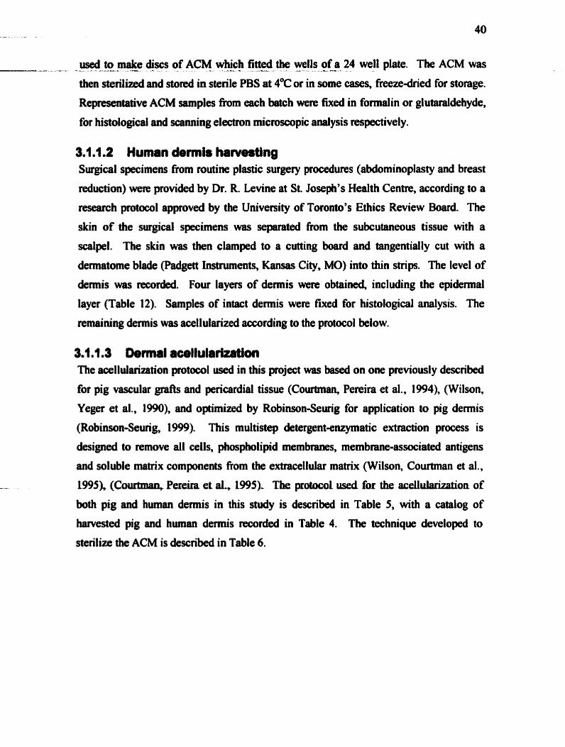
useâ to make @ses of ACM which fitted the wells of a 24 well plate. The ACM was - A----- - - = -a- - - - - - - - - y -- -
then sterilizeâ and stmd in sterile PBS at 4*C or in some cases, freeze-âried for storage.
Re~resentative ACM samples k m each batch were fixed in formalin or glutaraldehyde,
for histologicai and scanning electron micmswpic d y s i s respectively.
Surgical specimens from routine plastic surgery procedures (abdominoplasty and breast
reduction) were provided by Dr. R. Levine at St. Joseph's Health Centre, according to a
nsearch pmtocol appmved by the University of Toronto's Ethics Review Board. The
skin of the surgical specirnens was separated fiom the subcutaneous tissue with a
scalpel. The skin was then clamped to a cutting board and tangentially cut with a
dennatome blade (Padgett instruments, Kansas City, MO) into thin strips. The level of
demis was recorded. Four layers of demis were obtaineà, including the epidennal
layer (Table 12). Samples of intact demis were fuced for histological analysis. The
remaining demis was acellularized according to the protocol below.
3.4. t .3 Dermal acellularizaüon The acellularization pmtocol used in this project was based on one previously described
for pig vascular gr& and pericardial tissue (Courtman, Pereira et al., 1994), (Wilson,
Yeger et al., 1990). and optimized by Robinson-Sewig for application to pig demis
(Robinson-Seurig, 1999). This multistep detergentenzymatic extraction process is
designed to remove al1 ceus, phospholipid membranes, membrane-assaciated antigens
and soluble maaix compnents from the extracellular matrix (Wilson, Couttman et al.,
-- W95X (Co- Pereira et aL, 1995). The protocol used for the acellulaazation of
both pig and human dermis in this study is described in Table 5, with a catalog of
harvested pig and hwnm dennis recorded in Table 4. The technique developed to
sterilize the ACM is described in Table 6.

'7 weeks 7 weeks 3 months 7 weeks
Pig
Tabk A Acellularized pig and human dermis batches used in expenments.
(batch) 1842-oO
Human
hrnerse in 10 m M Tris buffet with EDTA, pH 8.0 containing 5% PMSF and antibiotics. Rinse with shaking for three days with fresh solution twice daily. Rinse with shaking in 1% Triton-X in 50 mM Tris bufTer, pH 8.0 with EDTA and PMSF for four days, with fresh solution twice daily. Rinse with Sorensen's phosphate bder. Cut skin into smaller pieces, and incubate with 6000 U DNAse and 5 mg RNAse for 24 hours in 40 ml Sorensen's phosphate buffet Rinse with swing in 1% sodium dodecyl sulfate in 50 mM Tris bufTer, pH 9.0, for four days, with fresh solution twice daily. Rime with shaking in water, then in 70% ethanol ovemight. Rehydrate in sterile PBS. Store at 4OC. Re-sterilize in 70% ethanol prior to use.
Table 5. Acellularization technique for pig and human demis.
7 week
30-1 1-00 23-1 1-00
- - - - - - - - -
SoaL in 70% ethano1 in sterile container overnight. . .
Aspirate using steri k technique. a Soak in b h 70% ethanol in a new sterile container, for 30 minutes. Aspirate and
replace with b h 70% ethanol for 30 minutes, twice. a Rinse in sterile PBS ovemight. a Aspirate and =place with fresh sterile PBS for 30 minutes, twice. a Transfer to a new sterile container. Store in sterile PBS or media at 4°C until
m e r use.
Table 6. ACM sterilization technique.
04-03-00
3 months 26 Y
May-00, July-00
14-12-00 7-1 2-00
Jan-01, Feb-01 Jan4 1, Feb-O 1

t- - 42
--=A- &~._?-ûe@gI AceUu!ar Mat@Cha6~cte@zatfon -
3.1.2.1 Acellularity For each batch of acellularized demis, a sample was fixed in formalin. The samples
were cut in cross-section and stained with hematoxylin-eosin, anti-vimentin antibodies
and anti-smooth muscle actin (SMA) antibodies. The antibodies were visualized using
the aviàin-biotin system. Fixed pig and human demis were useâ as positive controls.
Successfid binding of the anti-vimentin and anti-SMA antibodies (both fiom Dako,
Glostnip, Denmark) was âemonstrated in tissue sections fiom both species. A few
preliminary samples were also stained for pankeratin @ako) to ensure complete de-
epithelialization of superficial dermal layers. Table 7 summarizes the antibodies used
for immunohistochemistry in this study.
Antibody Vimentin, monoclonal goat anti-hurnan, clone 1A4
-- --
Tabk 7. Antibodies used for immunohistochemical staining of the ACM.
3m1m2m2 Acellular matrix structure
Description Cytoplasrnic intermediate filament in mesenchymal cells (including fi broblasts, myofi broblasts)
a-Smooth muscle actin, monoclonal goat anti-human Pankeratin, AEl/AE3, monoclonal rabbit anti-human
- Freeze-fhcttrre scmtng dection rnicro~eo~ (SEM) and trammission electt~n
microscopy (TEM) were performed on ~presentative samples of fresh pig ACM. Each
sample was examid at low (5Ox and lûûx) and high (5ûûx and lûûûx) magnification
for collagen buncüe size and arrangement as well as ACM surface chmcteristics. SEM
was used to compare the matrix structure of pig ACM and human ACM, fieezedried
and h h ACM, hyaiwnan- and heparim~ched pig and human ACM and finally,
cell-seeâed pig and human ACM samples Level2 samplcs (see Table 12) of the 1 7- 1 1-
00 pig ACM batch and level3 samples of the 23-1 1-00 human ACM batch were cuîhired
Cytoplasmic actin isoform expressed by contractile vascular smooth muscle cells and wound myofibroblasts
Cytoplasmic proteins expressed by keratinocytes

with primary human fibroblasts for four weeks, and wmpared in structure by SEM to - ~ --..---A------ ---- - -- . - --A .. - --: quivalent, non-seeâed samples.
Samples were fixed in 2% glutaraldehyde and dehydrated in a graded series of ethan01
concentrations7 followed by cntical point Qying. Some samples were then k z e -
h tu red with a scalpel before mounting. The freeze-dried samples were affixed to
alwninum stubs with double-sided electrical tape. The edges of the samples were then
coated with silver paint to improve electrical conductivity. The mounted samples were
sputter-coated with platinum. The samples were examined under a scaaning electron
microscope (mode1 S-2500, Hitachi, Japan) and the images were captured using a
passive image capture system (Quartz PCI, Quartz, Vancouver, B. C.).
TEM was done to ver@ the quaternary collagen structure in the pig ACM, afler the
acellularization process. TEM samples were fixed in 2% glutaraldehyde for 24 hours,
then post-fixed in 2% osmium tetroxide for 2 hours. Dehydration was through a graded
series of ethm01 followed by propylene oxide. Samples were embedded in Spurr's resin,
sectioneâ, and counterstained with uranyl acetate and lead citrate.
3.1.2.3 Glycorrminogîycan content of ACM The relative glycosaminoglycan content of the ACM was assessed qualitatively by alcian
blue steining of fieeze-fixed sections. Fmhly acellularized samples of pig and human
ACM were embedded in O.C.T. embedding wmpound@ ( S a b Finetek U.S.A. Inc.,
Tomce, CA) without fixation and immediately fiozen at -70°C to preserve the matrix
gly~saminoglycans, which tend to wash out with formalin fixation. Sections were cut
and stained with alcian blue at pH 1.0 and 2.5. The lower pH is better suited to detect
the sulfated glycosaminoglycans, and the higher pH allows some visualitaton of
hyalurotüiû (Elder, Elenitsas et al., 1997). Fresfily harvested pig aud human demis were
pmcessed and stained in the same manner. The relative intensity of alcian blue staining
was compared between fksh and acellularized pig demis.

3.2.1 Cell Cukm Ail ceIl culture experiments were perfonned in a tissue culture fàcility approved by the
University of Toronto Safety Cornmittee as a Biohazard Level Two facility. The r w m
was equipped with several laminar flow biological safety cabinets (NSF accredited, H.
E. P. A. Filter Services Inc, Toronto). All metal and giass equipment was sterilized with
a stem steriüzer (SM 300, Yarnato Scientific Amencan inc., Orangeburg, NY) prior to
contact with ce11 culture materials. A standard ce11 culture incubator, set at 3TC with
5% C a iafuseâ, was used for al1 experiments (Sanyo Elecüic Company Ltd, Japan).
3.2.1.1 Human demal fibrobhst cukum Subsequent cell culture experiments used passageci human dennal fibroblasts. These
were isolateû fiom fresh adult surgical specimens by explant culture (Nishiyama,
Tsunenaga et al., 1989). The subjects' age and medical history are listed in Table 6.
Briefly, the demis was obtained h m discardeci surgical specimens (fiom breast
reduction or abdominoplasty) in a sterile field and cut into small pieces (less than 2 mm
x 1 mm x 1 mm) with a sharp scalpel. nie tissue pieces were transportecl on ice from
the operating room to the tissue culture room, in sterile PBS with 1 mM calcium
chloride. The tissue was rinsed with sterile PBS several times. The fragments were then
placed in sterile 35 mm petri dishes with a &op of media to hydrate them, but without
causing the pieces to float. 7'he hgments were kep attachecl to the petri dish for up to
three weeks with minimal media, until outgrowth of fibroblasts fiom the fragments were
- seen. At this tirne the tissue was carefuly removed and the fibroblasts imnaersed in 1-2
ml of media until contluency was reached The cells were then trypsinized using 0.25%
trypsin/EDTA and plated in T7S flesks (Becton Dickinson, F d i n Lakes, NJ) for
expuision. At 80400% wnfîuency, the cells were harvested and fiozen as passage 2
cells or used in ce11 seeding experiments. Prior to seeding on the ACM, a sample of the
cells were stained with trypan blue and counteâ with a hemocytometer (Hausmann
Scientific, Horsham, PA). Dulbecco's Modified Eagles Medium (DMEM) with 10%
fetal bovine senmi, 1000 U penicillin and 100 pg streptomycin was used for al1 explant
and expansion culturing. The fibmblasts wwe used in ail experiments at passages 2 to 4.

1 P.H. 1 26 1 F 1 iiorie 1 23-11-00 1 26-12-00 1
Tibk IL Donor infocmation for primary hinnan dennal fibrobiast hvesthg.
3m2.1.2 ACM pnprmüon for ceIl seedlng Pig anâ human âermai aceîîuk maüix w m cut to fit the weUs on either 24 or 96 weU
tissue culture plates (Ndge Nunc Intemational, Donmark) with 8 mm or 16 mm
biopunches (Fray Roducts Corp., Arnhem, NY). The thickness of the ACM samples was mtztsureâ using a micrometer, anâ recordeci. The ACM samples w m tben stèriiized
in staire tissue cuiaae plates or eppendorf tubes by seskiag in a saies of 70% ethanol riases aa descni m Table 4. AU himicip ACM and haif of the pig ACM samples were
hm-dried for storsgc. Rior to cell sceding, the ACM worr rehydrated in PBS and tmnsfmcd to stmk tissue culture plates and soaked in the appropriate media for 48
hours. As a fûrthcr stedi ty check, the hirbidity and colour of the media in wâich the
ACM wae d e d was noted prior to ce11 secding.
&2m1.3 Fibfobl~~t onto ACM For al1 expaiments, the day of ceil seediag was condeted Day O. Prior to hypsiaizing
the cefls, the pbte coatvining the ACM was rcmovect h m the mcuôator. The media in
the mils was aspiratad and the ACM presscd dom against the baom of the weU to
m w v e any air buôblcs or excess media. The flash containbg the co&ent cells were
removcâ h m the inoubator and iinsed with warmeâ PBS. T~ypsinhDTA (5 ml) was dded to acb T75 flask for five minutes at 37°C. The seîected media contaiimig 10% scrum was addcd (5 ml), rinsing the ceUs off the ride of the nksk. The resulting 10 ml
WU suspwion was transfemd to two sterile 15 ml tubs and cmmifbged for 10 minutes
at 500 rpm (50 x g). Ibe supernatant was asphteci and the celî pllet resuspmded in the
desireci vduw to yieM an appqriatc ctn co~lcclltration fa counting ( 1 0 ~ 1 0 ~ ceusiml).

An aliquot of the resuspe.nd@ cells was diluted with tqqgn blue, and counted using a ---- - A--- - - - - A- - -- --2 -= --- L - - --
hemocytometer. The cells were then diluteâ to the required concentration to aîlow for
seeding with a desired amount of cells in an appropriate volume. The plate was retumed
to the incubator, the tirne noted, and the wells top@ up to the required volume of
wmned media 24 hours later. For al1 cell-seeded ACM samples, the media was changed
every 2-3 days. Fresh media was added slowly to the side of the well to avoid washing
the cells off the ACM surface.
The experiments described in this section were numbend according to the conditions of
the experiment. If a variable was changeâ, the experiment was assigned a sequentially
different nuniber. For each objective several experiments were done, each under slightly
different conditions. Each expriment is therefore numbered iodependently within the
same section. The number of times each experhent was repeated under identical
conditions (r), is listeâ at the top of each column in Tables 9, 10, 1 1, 13 and 15. The
number of experimentd samples in each grwp (a) is listed in the last row of the table,
for each repeat. in the statistical analyses of the results, the sample size (a) used in each
set of expenmental conditions correspondecl to the following equation:
Simple size (n) = r x s
3.2.l.S Experimental controls To detemine how the presence of pig dermal matrix influenced fibroplasia in an
expenmental setting, a number of contmls were included. Human dermal ACM was
used in some experiments to determine whether the effects of pig ACM were species-
specific. Bovine collagen gel was used as a cornparison to detemine whether the eRéct
of a collagen matrix on fibroblasts is structuredependent. To this end, collagen gels
were prepared in the wells of the tissue culture plate the day of the experiment. Briefly,
8 ml of chilled acidified bovine dermal collagen (Cohesion Corp., Pa10 Alto, CA) were
mixed with 1 ml of 10x PBS. The pH mis djusted to 7.4 with 1 ml of 0.1M NaOH.
Different volumes (400, 500 or 700 pl) of neutraiid, isotonic collagen solution were
placeâ in the wlls and incubated at 37°C for at least an hou. The resulting gel was
adherent to the sides and bottom of the well. As a final control, fibroblasts were also
grown âirectiy on a polystyrene culture dish itself.
- 3 . 2 ! 2 S ~ I / + W ~ h - o ~ - - _ . - _ - - d -
Specific morphological fïndings have been associated with fibroblast contraction in
different matrix envimnments (Ehrlich and Rajaratnam, 1990). To observe whether the
collagen substraae intluenced the morphology of the seeded fibroblasts, confocal
microscopy of phalloidin-stained samples was perfomed.
3.2.2.1 PhalIoidin staining The appearance of human fibroblasts on pig ACM was compareâ to that of fibroblasts
cultured on human ACM, collagen gel and coverslips. After 4 to 6 days in culture, the
cell-seeded samples were fixed in fornuilin for 30 minutes. Alexa F l w 488 phalloidin
(Molecuiar Probes, Oregon) was use& at a 1:40 dilution with 0.1% bovine serum
albumin (BSA) in PBS, to stain the F-actin fibres of the fibroblast's cytoskeleton. The
samples were rinsed in PBS. The samples were immersed in 1:l PBS:glycerol in 24
well plates and wrapped in foi1 for transport. The coverslips were mounted on a slide
and sealed with clear mil pdish.
3.2.2.2 Confoccil micmscopy nie confod microscope system useâ was the Zeiss LSM 5 10, with an argon laser at a
wavelength of 488 m. The morphology of the fibroblasts was exarnined with a 10x dry
objective and a 63x water-immersion objective, with an optical slice thickness of 2 um.
The morphology of the cells on pig ACM, human ACM, collagen gel and polystyrene
coverslips were c o m p d . Three samples were viewed for each group, over twelve
optical fields of view at 10x magnification, and five fields at 63x magnification.
32.31 MTT assay The MIT (3-(4,5dimethyl thiaÿol-2-y1)-2 Jdipheny l tetrazolim bromide) assay was
used to deterxnine how rnany cells were viable d e r seeding ont0 ACM, as compared to
cells plated in the absence of ACM. First, a calibration cuve was generated to compare
the accuracy of the assay when used in the preseace of ACM. Hs68 fibroblasts were
seeded et d-erent deasities (0,5000, 10,000 and 20,000 celldcm2) in 2 x 24 well plates
either in the absence or in the presence of 15 mm diameter ACM samples. Each curve

was perfomed in quadruplicate. AAer 24 and 48 hours incubation in DMEM with 10% A =--- 2L- - -. - - - - . - a. - - a -- - - -
senun and antibiotics, the MIT assay was performed as outlined below. The average
sbsorbance of the two reaâings pr sample was plotted against the ceIl concentration.
The calibration curves for c e l l - d e d ACM were compand to that of cells only, and the
24- and 48-hou data were compared.
The MïT assay was used to detemine the number of viable cells on pig ACM (PACM)
aiter différent lengths of time in culture. Cell numbers on PACM samples were
compred to those on collagen gel and on polystyrene tissue plates. PACM samples (16
mm in diamter) were seeded with d e d fibroblasts at a density of 2500 cells/cm2, and
incubated for 24 hours, 1 week and 2 weeks. A total of 18 samples were seeded, for 6
samples at each time point (Experiment 1).
In preparation for the MTT assay, the media was aspirated fiom the wells and the cell-
seeded ACM rinsed with warmed PBS. AAer aspirathg the PBS, the cell-seeded ACM
sampies were t ransfed to a new 24 well plate to avoid counting the cells adherent to
the culture dish. Steriie 5 mg/ml Mï"ï (3-(4,5dimethyl thiazol-2-yl)-2,5-diphenyl
tetramhm bromide) solution, in serum-fm RPMI-1640 medium, was added to each
well (300 pl), and the plates retwned to the incubator for 4 hours. M e r addition of
MTT to the wells, the ACM samples were flipped cell-side down to fully immerse the
cells adherent to the ACM in the MTT solution. At the end of the incubation pend, the
hnIT solution was aspirated fiom each well and acidified isopropanol(4% 1 N HCI) was
added (300 uüwell). The lid was secured with patafilm, and the plate gently shaken for
1 hour on a rotomix. Each sample was mixed by pipetting up and dom. A 100 pl
aliquot of each sample was transferred to two wells on a 96 well plate. Fresh MTT
solution was used as a blank. The plate was then nad spectrophotometricaily at 570 nm
with a 630 nrn reference filter. In one repeat of the expiment, aiter solubilization with
isopropanol, the ACM samples and coatrols were photographed with a digital camera to
demonstrate the MïT colour change.

3.2.3.2 CyQuant aissay ---- --LzL -- -- - - -- - - -A- - - L - - --
in order to verify the reliability of the MTT assay as a measure of cell proliferation on
the ACM, a second assay for cell proliferaton was also used The basis of the CyQuent
kit (Molecular Probes, Oregon) is the use ofa proprietary green fluorescent dye, which
exhibits strong fluorescence enhancernent when bound to cellular nucleic acids. Unlike
the MTT assay, seru. components (which are dificult to wash out of the ACM) do not
appreciably interfere with the CyQuant assay. By measuring the degree of fluorescence
after adding the dye to cell-seeded ACM, the assay provides a means of quantifying the
relative amount of cells bound to the ACM. While the cells are not viable at the time of
the dye binding, the assay provides an indirect measure of cell viability, as only viable
cells will rernain adheremt to the ACM with repeated changes of media.
Human dennal fibroblasts were grown to confluence in a T75 flask under standard
culture conditions. The cells were trypsinizeâ, collected by centrifugation and
resuspended in 2 ml of phenol r & f k media The ce11 concentration was detennined by
counting live cells (as show by trypan blue staining). The cell suspension was diluted
to a known concentration of around 2 x 10' celldml. The diluted cell suspension was
transferred to sterile eppndorf tubes in exactly 1 ml aliquots. The tubes were
centrifugexi for 5 minutes at 200 x g. The supernatant was carefully removed and
discarded without disturbing the ceIl pellet. The cells were placed immediately in a -
70°C fieezer.
Two caliôration curves were generated: one for cells only, and one for the sarne number
of alb in the presence of a digest& 8 mm ACM sampte. The second calibration curve
was done to account for the emr in the experimental samples, which contained cells and
digested ACM. Since digested ACM collagen adds to the turôidity of the solution, the
possibility of falsely elewted fluorescence values had to be considend. Once the ACM
haâ been digested for 48 hours, the plate was centrifiigeà at 400 x g for 5 minutes, and
gently inverted to blot the media off The digested ACM was stored at -70°C until the
cens were ready for âüuting. The cell pellet was thawed at rmm temperature. A 1 ml
volume of the preparrd CyQuant dyelcell lysis buffcr (sec below) was added and the
cells were resuspended by vortexing. A dilution series was generated in the wells of a

--A--. microphte - -A.-= ----- by diluting -.- - -- the - cell = suspension - -A .- with . - --- more --- CyQuant - - - - buffet. The range for this -
series comprised 0, 1000, 5000, 10,000, 15,000, 20,000 and 25,000 cells per well. The
samples were incubatecl for 2-5 minutes, with the plate wrapped in foil. Two calibration
cums were made in botb the presence and absence of digested ACM (fou c w e s in
total).
The CyQuartt assay was used to detemine cell proliferation on the pig ACM (PACM),
humm ACM (HACM) and polystyrene plates. The assay allows for spectrofluorometric
analysis directly in the cell-seeded well. 8 mm ACM samples were used. Table 9
sumrnarized the different experimental conditions under which the sarnples were
prepared for the CyQuant assay.
Variable 1 Experiment 2 1 Experiment 3 1 Experiment 4 1
Groups
Seeâing density (celldcm2) Time points
performed; s = number of samples in each group.
(r= 1) PACM, HACM,
#ofsamplesper group per repeat
For cell proliferation experiments, controls consisted of cells on polystyrene wells
collagen gel, ceils on1 y 20,000
ld,1,2,3,4wks
-- witbout the presence of ACM were &id on separate ptafes for each time point. At
(r = 3) PACM, collagen
Table 9. Smmary of ce11 proliferation expenments for CyQuant assay. Abbreviations: PACM = pig ACM; HACM = human ACM; r = number of times the experiment was
s=3or4
each time point, the cell-seeded ACM samples were transferred to unused wells on the
(r = 2) PACM
gel, cells only
20,000
Iâ,1,3wks
plate designated for that time point. This transfer prevented cells growing in the same
well as the ACM, but not - the ACM, from king included in ce11 proliferation
20,000
id, 24 34 5d, 1,
s = 3
determinations The media was aspirateci h m the wells with cells only, and the wells
filled with watmeci HBSS. Type H collagenase (200 ul of5 mgml solution) was added
2 ,4 wks 1
s = 3
to the cell-seeded A C U The plate was renirned to the incubator for 24 hours. Each cell-ACM solution was pipetteci up and dom to bnak up tbe ACM collagen. The plate
was rehimed to the incubator for another 24 hours. By th t the, the ACM had been

completely - digested, -- - - and the releaseâ -.-- cells --- could -- be easily seen - by microscopy. The
plate was centrifiiged at 400 x g for 5 minutes and gently inverted ont0 paper towels to
blot the supernatant. The plate was placed in a -70°C fieezer to lyse the cells prior to
quantification.
For the purpose of quantification, the plate was first thawed at rwm temperature. An
adequate volume of CyQuant dyehll lysis b a e r was preparwl with the following ratios
of reagents: 380x distilled water, 20x cell lysis stock, l x CyQuant dye stock. A 200 ul
volume of the prepared buffer was added to each well. Samples of 200 ul of buffer
without cells served as controls. All oaniples were incubrrted at m m temperature for 2-
5 minutes. Fluorescence was measured using a spectrofluorometer with filters
appropriate for 485 nrn excitation and 538 nrn emission maxima. The appropriate
cslibration curve was used to determine ce11 number values.
3.2.4 CoIIagen Synthesis Radiolabeled-proline incorporation is a common method for quanti@ ng collagen or
protein synthesis by cells in vitro. in this study, collagen synthesis by fibroblasts seeded
on the PACM was compared to the background radioactivity of PACM without cells.
This procedure was adapted from that of Freiberger et al (Freiberger, Grove et al., 1980).
The samples were seeded with humsn dermal fibroblasts at a density of 30,000
cells/crn2. They were culturd in DMEM with 10% FBS and penicillin/streptomycin for
4 days, with a change of media at day 2. in each repeat, the sarnples were pe$onned in
quadrupIicate, as were the cell-seeded sampks to be used for ce11 proliferation
measuremettt. The experimmt was repeatect three h e s (Expriment 5). On &y 4, the
samples were washed widi PBS and changed to pre-labeling medium. The pre-labeling
medium consisted of the same medium witb 50 pgml ascorbic acid and 50 &ml P- aminoproprionitrile funiarae. The samples were incubated in pre-labelling medium for
7 hours, and then labeled with 200 pl of labeling medium for an additional 23 hours.
This medium was identicai to the pre-labeling medium, except that it contained 2 pCi of
~ f - ~ ~ - j m l i n e .
At the end of the ipcubation,@ medium and a 200 pl P B s - y ~ h were collected into --------a.- - ----a-- A:---- &--A --
labeled eppendorf tubes. The ACM were then dissolved in 100 pl of 10 rnM HCl for 1
hour at 37°C. The aciddissolved ACM was collecteci in separate tubes. One of each the
medium and the ACM were digesteci with collagenase in the following manner. The
samples were made up to 1 ml with 50 mM tris-HC1, 10 rnM calcium acetate, 2.5 rnM
N-ethylmaleimide and 10 U of type W collagenase and incubated at 37°C for 3 hours.
Half of the samples were made up to 2.5 m M N-ethylmaleimide and 0.5 mglm1 BSA in
the meantirne. Once the collagenase digestion was complete, 0.5 mglm1 BSA was added
to these samples as well. All samples were then made up to 10% TCA, and left
ovemight at 4OC to precipitate the protein. The next dry, the eppendorf tubes were
centrifuged at 12,000 x g for 3 minutes and tbe supernatant discarded. The pellet was
resuspended in 5% TCA and centrifuged; this was repeated twice. The pellet was
solubilized by sotking in 10 mM HCl ovemight. The acidified protein solution was
transferred to numbered scintillation rials and 1 0 ml of scintillation fluid (Ready-Safe,
Beckman instruments Ltd., Fullenon, CA) wss added for counting in a scintillation
counter.
The fiaction of collagenase soluble counts (cpm) wss calculatecl fiom the formula:
(Collagenase-digested cpm - blank cpm) / (Total epm - blan k cpm)
The percentage of radiolabelebproliw incorporatecl into newly synthesized collagen (as
opposed to al1 other protein synthesis) could then be detemiined:
[1 - (Collagenasedigested epm - Blank cpm)/(Total epm - Blaak cpm)] x 100
The relative amount of collagen synthesis per sample in erçh group was nonnalized by
the cell numkrs in each group. The number of cells per semple was determineâ by the
CyQuant assay of designated samples.
3.2.5 Cell lnîihtion Infiltration of hwnan fibroblasts into pig demai ACM was determined by cross-
sectional histology of cd-seeded semples. Fibroblasts were seeded on one surface of
the ACM, as in the ce11 prolifedon experiments. At each time point, the cell-seeded
ACM samples were rernoved from the plate and fixed in f o d i n for 24 hours. The

samges were quyfidiy etion-@ yitJ a #IO scalpel blade into three pieces (see -AL----= - - I L - - - -
Figure 5). Each section was blotted dry, and the cut edge was marked with
merc~f~~hrorne. The tissue pieces were put back into fonnalin until they could be
embedded in Iioraffln on edge. Al1 thne pieces were embedded in a single paraffin
block. One section was made fiom each block, mounted on a slide and stained with
hematoxylin and eosin. There were three cross-sectional pieces of tissue per sample.
ACM umpk ACM pieces Pamflin bkck Hhbologicrt slMe
Figure S. Histological processing of an ACM sample.
The slides were reviewed to find seçtions containing a monolayer of fibroblasts at the
top of the tissue section; this ensured that the tissue was in fact cut pcrpendicular to the
cell-seeded surface. All H&E stained sections contsining a monolayer were analysed for
cell infiltration of the matrix. In some experiments, new slides were made from the
sample blocks for Mmentin staining. Cell infiltration was assessed in three ways. First,
the prcentage of sections in each experimental group demonstrating ce11 infiltration was
determined. A section was considered positive for ce11 infiltration if at least 50 cells
wen sceit, at k t 50 pm below the surfhce. Second, the numôer of cells per positive
section was deterrnined by m a n d wunting. The cells located in the monolayer, at the
surface of the masix, were not included in the count. Automated counts were done on
sorne samples to correlate with manual count results. Thirâ, the distance of each ceIl
h m the surface was recorded using digitai photography and MCID image analysis
software. Again, the cells of the monolayer were not included in the analysis. Distance
measurements were recorded for three microscopie fields per section, at 10x
magni fication.

A-. - . . 3.2.S21 _Cmnfilon Expriment8 - ------ Several experirnents were done to assess pig ACM as a s d o l d for human fibroblast
infiltration. Many expimental variables were investigated for any possible influence
on ce11 infiltration. Human ACM samples were seeded under the sarne conditions, to
allow a comparative assessrnent of pig ACM infiltration. Tables 10 and 1 1 sumrnarize
the cell infiltration experiments.
1 Variable 1 Exp6 1 Exp7 1 Exp 8 1 Exp9 1 Exp 10
Table la Ceil infiltration experiments. Expiments 610 used human derrnal fibroblasts at passage 24, cul- in DMEM with 10% FBS. Abbreviations: PACM= pig ACM; HACM= human ACM; F D k z e dried; r= nurnber of times the expriment
ACM type and batch tested
Seeding densiîy (ceiis/crn2) Time points
# of samples
was perfomed; s= number of samples in each group.
(r = 2) PACM 18-02-00
2500
2,4 wks
s = 5
Variable ACM type and batch tested
Ce11 type
# of samples per 1 s=20 (s=4 for each 1 s=5 to 6 1 ~3
(r = 2) PACM 10-08- 00
2500, 25,000
4 wks
s = 2
Expl1 (r=1) PACM 30- 1 1-00, 10-8-00, HACM 23-1 1-00
1
1"HFb-P#2 Seeding density (ce1 ls/cm2) Time mints
(r = 2) FD-PACM 1 7-1 1-00, FD-HACM 23-1 1-00 3000, 30,000
1,2,3,4 wks s = 4
5000,50,000
3 wks. 5 wks
30,000,50,000
4 wks
group Media type
Table 11. Ce11 infiltration expenments (continued). AbbteM'ations: 1- human demal fibroblasts; P#== passage aumber; FBS= 10% fetal bovine serum.
Exp 12 (r = 1) PACM 10-8-00
1"HFb-P#3 50,000
4 wks
ACM diameter and setup
(r = 3) FD-PACM 2- 1 1 - 00, 17- 1 1-00,30- 1 1-00, FD- HACM 24-1 1-00 30,000
1,3 wks
s = 4
Exp 13 (r=2) PACM 2 1-8-00, FD-PACM 2 1-8- 00, FD-HACM 24- 1 LOO 1-P#4
levei) DMEM+FBS
(r = 2) FD- PACM 2- t 1-00, 30-1 1-00 30,000
1,2,4 wks s = 3
8 mm, 16 mm DMEM+FBS MCDB 131+ FBS 16mmACMwit.h staidess steel rings
-- - -
16mm
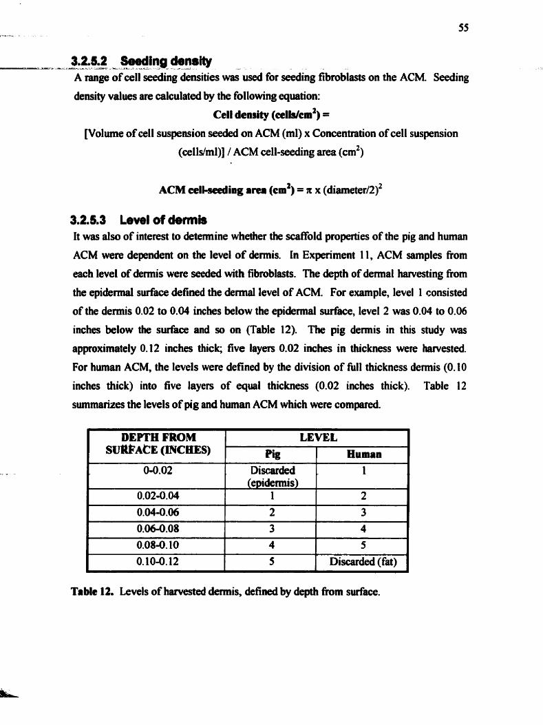
3.2.5.2 Seeding density -a------ -.=..a-. - - - 4 - - - A L -
A range of ce11 seeding densities w&used fibroblasts on the ACM. Seeding
density values are calculated by the following equation:
Cell demity (ceudcm2) =
[Volume of ce11 suspension seeded on ACM (ml) x Concentration of ce11 suspension
(celldml)] / ACM cell-seeding area (cm2)
ACM celi-seeding a m (cm2) = n x (dia1neter/2)~
3.2.5.3 Level of demis It was also of interest to detennine whether the scaffold properties of the pig and human
ACM were dependent on the level of demis. In Experiment 11, ACM samples from
each level of demis were seeded with fibroblasts. The depth of demial harvesting from
the epidermal surface defined the demal level of ACM. For example, level 1 consisted
of the demis 0.02 to 0.04 inches below the e p i d e d surface, level 2 was 0.04 to 0.06
inches below the surface and so on (Table 12). The pig dennis in this study was
approximately 0.12 inches thicl<; five layen 0.02 inches in thickness were harvested.
For human ACM, the levels were defined by the division of N1 thickness demis (0.10
inches thick) into five layen of equal thickness (0.02 inches thick). Table 12
summaiues the levels of pig and human ACM which were compareà.
1 O. 10-0.12 1 5 1 Discarded (fat) 1
DEPTH FROM S V ~ A ~ E (n~cms)
Table 12. Levels of harvested demis, defined by depth h m swface.
LEVEL
ph! 1 Human

polypmpylene tubes. The tubes were without lids and wrapped in foi1 perforated with
small holes. The tubes were immersed in Iiquid nibogen for one minute to fieeze the
coneained ACM. The tubes were placed in a fieeze-dryer flask and co~ected to a
vacuum freeze-dryer (Labconw Corp., Kansas City, MO) ovemight. The ACM was
rehydrated in sterile media ovemight.
3.2. 6 MjtoSbmbIast DinremnOatkM Histological sections of fibroblast-seeded PACM, HACM and collagen gel were stained
with antibodies to the myofibroblast marker a-smooth muscle actin. Samples were
seeded with 30,000 cellslcm2 and incubated until fixation of three sarnples per group at
each of the following time points: 3 days, 1,2, and 4 weeks (Experiment 14). These
samples were cut in ctosslsection, stained for a-smooth muscle actin (SMA) and
counterstained with hematoxylin. Each slide was analyzed for percentage of cells
stained for SMA, with the total nurnkr of cells demonstrated by hematoxylin. Non-
specific background staining was observed on some sections of pig ACM. These
sections were excluded fiom analysis. Hwnan fibroblasts and rat-tail fibroblasts
cultured on coverslips were used as conmls for SMA staining.
3.2.7 ConCracffon The prepared ACM and collagen gel samples were seeded with hurnan dermal
fibroblasts, and incuôated for the lengths of time inàicated in Table 13. Al1 sarnples
were seeded with 25,000 fibrobhsttscm2. H u m a fibmMirsts between passages 2 and 3
were used. The collagen gel was detached fiom the sides and bottom of the plate 24
hours &et seeding. This allowed the gel to float in the culture medium and contract
b l y . The medium was changed every 2 to 3 days. DMEM with 10% FBS and
antibiotics was used in ExpeRment 15. d e EBM-2 with 5% FBS was used in
Experheats 16 and 17. In the latter two experiments, the fibroblast-seeded pig ACM
samples were incubated in wells seeded with human microvascular endothelial cells.
Once the time point was rericheû, the plate was photographecl and the image importeû
into a GelDoc Multianaiyst p o g n a The surface a m of the cuntracted sample was
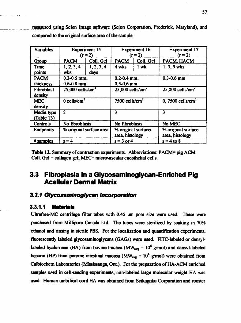
measured using Scion Image sqftware (Scion Corporatio~ Fredenck, Maryland), and - - ----*-A = - - - - ---- - --- --A2 - comparexi to the original surface ara of the sample.
Variables
Group Time points PACM thickness Fibroblast density MEC density
Table 13. Summary of contraction experiments. Abbreviations: PACM= pig ACM; Coll. Gel = collagen gel; MEC= micmvascular endothelid cells.
Experiment 15 (r = 2)
0.3-0.6 mm, 0.648 mm 25,000 cellslcm2
Media type (Table 13) Controls Endpoints
#sam~les
3.3 Fibroplasia in a Glycosaminoglycan-Enriched Pig Acellular Dennal Matrix
PACM 1,2,3,4 wks
O cells/cmz
3.3.1.1 Materials Ultra&-MC centrifuge filter tubes with 0.45 um pore size were used. These were
purchased from Millipore Canada Ltd. The tubes were sterilized by soaking in 70%
ethan01 and rinsing in sterile PBS. For the localkation and quantification experiments,
fluorescently labeled glycosaminoglycans (GAGS) were used FITC-labeled or dansyl-
labeled hyaluronaa (HA) fkom bovine trachea (MW, = 106 g/mol) and dansyl-labeled
heparin (HP) fiom porcine intestinai mucosa (MW, = lo4 g/mol) were obtained from
Calbiochem Laboratories (Mississauga, ûnt.). For the preparation of HA-ACM e ~ c h e d
samples used in cell-Seeding experiments, non-labeled large molecular weight HA was
use& Huinan umbilicrl cord HA was obtained h m Seikagaku Corporation end rooster
Experiment 17 (r = 2)
PACM, HACM 1,3,5 wks
Coll. Gel 1,2,3,4 days
Experiment 1 6 (r = 2)
0.24.4 mm, 0.5-0.6 mm 25,000 celldcm2
2
No fibmblasts % original surface area
s=4
PACM 4wùs
0.34.6 mm
25,000 cells/cmz
7500 cells/cmz
Coll. Gel 1 wk
O, 7500 cells/cm2
3
No fibroblasts ?4 original suface area, histology s=3or4
3
No MEC % original surface area, histology s = 4 t o 8

comb - - ~ a s d o ~ t o b ~ Y - H Y ~ Phan?-iuti@~ Ltd @th m a , = 106 g/mol)- Al1 - P z & --- ----- - - a----
chernicals and ce11 culture supplies were obtained h m Sigma-Aldrich Canada Ltd
d e s s otherwise specified. An EC Micmmax microcentrifuge (International
Equipment Company, Kansas City, MO) was used in d l HA incorporation experiments.
3.3.1.2 H yaluronan incorporation Pig ACM was cut into 8 mm outer diameter discs using a biopunch. Care was taken to
ensure consistent thic kness. Sample disc thic kness was measured using a micrometer;
samples of greater than 1.5 mm or less than 0.7 mm in thichess were not used. The
selected ACM sarnpIes were sterilized as previously described and dl procedures were
carried out in a biological cabinet. The inner diameter of the centrifuge filter tubes was
7 mm. The ACM discs were plaoed at the bottom of the filter insert (Figure 6) in a
concave-down position. Each experimental sample disc was then covered with 100 ul of
a sterile 5 mgml solution of HA in DMEM. The tubes were centrifiiged at 1900 x g for
10 x 30 minute intervals. At the end of each 30 minute centrifugation, any HA solution
which haâ passed into the bottom of the tube was collected and replaced above the
ACM. This was done to prevent the centrifiigation of dry ACM samples. The tubes
were also rotated 180°C afier each 30 minute interval (Lausman, 2000). The ACM
samples enriched with HA are referred to as HA-ACM.
I
F i n 6. Diagram of ACM and HA in centrifuge filter tube for HA incorporation.

3.3. 1.3 --- Hepdn ---A- incorpontion ----- - - - - -
ACM samples (8 mm 0.d.) of comparable thicknesses were placed in the wells of a
sterile % well plate. A 100 pl volume of a 5 m&l solution of heparin (HP) was added,
the plate wmpped with parafilm and placed in a 37°C incubator for 48 hours. The ACM
samples enriched with heparin are referred to as HP-ACM.
3.3.2 Gi'osaminoglycan LocaIIzatlon 6 Quanaffcation In order to visualite the GAGs in tissues, samples cannot be formalin-fixed (Lin, Shuster
et al., 1997). Al1 HA-ACM and HP-ACM samples were therefore fiozen at -70°C and
embedded in O.C.T. cornpouna prior to sectioning. Frozen samples of the unmodified
ACM HA-ACM and HP-ACM were stained with alcian blue (at pH 1 .O and 2.5). While
the histology did demonstrate some différentiai staining between the sarnples (Section
4.3. l), it did not adequately delineate the level of HA or HP incorporation. A further
method was undertaken to facilitate GAG localization in the ACM. Solutions of 1,s and
10 mg/ml FITC-labeled HA were centnfuged into ACM discs in the same rnanner as
described above. Dansyl-labeled HP was dissolved in PBS as a 5 mg/ml solution, and
incorporated accordhg to the method described (FK-labeled HP was not available).
The samples were frozen, and unstained cross-sections were obtained. A fluorescent
hiovert microscope (Zeiss, Gemany) connected to a computer and a digital camera
was used to demonstrate the anas of fluorescence in the ACM. A 450 nm blue light
filter was used. The images were viewed by Spot Advanced version software
(Diagnostic Instruments Inc, Sterling Heights, MI). A solution of fluorescein in water of
equivalent concentration was centrifiiged into control ACM samples, to demonstrate the
specificity of the fluorescence in the ACM to the FITC-HA. Al1 fluorescent solutions
and tissues were kept fiom light during the experiment.
Existing methods to quantify GAGs in tissues are limited to some extent. The alcian
blue assay has high errer, and is too insensitive to detect microgram quantities. An
antiibody baseci on hyaluronan-binding potein was developed, but has been shown to
cross-mwt with pig wllagen (Tengblad, 1980). To ovemme these problems, the
fluorescently labeled GAGs inwrporated into the ACM were quantifid
spectrofluorometrically. Once fluorescein-labeled HA or dsnsyl-labeled HP was

inwrporated - - --- into ACM samples, - - - the L-----A ~amples were - - y rinsed - - off . --- in PBS. They were then - --
placed in a microplate, and 200 pl of 5 mglml type H collagenase were added to the
wells. The ACM was digested for 48 hours in the dark. Equal volumes of each sample
were transfened to separate wells for quantification. Calibration curves were prepared
using equal volumes of standards containing digested ACM, and either fluorescein-HA
or dansyl-HP. Calibration curves covering a wide range of concentrations were us&
and prepared in duplicate or triplicate. Fluorescein-labeled HA was quantified using a
spectrofluorometer with 485 nm excitation and 538 m emission filters. Dansyl-labeled
heptuin samples were measured at 265 nm excitation and 5 10 nm emission wavelengths.
In this study, 100 pl of 1 rnglml, 5 mg/ml and 10 mglml each of fluorescein-HA and
dansyl-HP were incorporated into three ACM samples at each concentration. The
amount of GAG incorporated into the ACM was quantified spectrofluorometrically
using calibration curves of both FITC-HA and dansyl-HP in the presence of digested
ACM. The volume of the digested GAG-enriched ACM was measured in order to
calculate the mass of GAG in the ACM fiom the concentration. The percentage of
available GAG incorporated into the ACM, at each concentration, was analyzed. The
concentration of GAG in the ACM was estimated, by using an approximate ACM
volume (Section 4.3.2).
In order to detemiine the longevity of the HA and HP in the ACM immersed in solution,
HA-ACM and HP-ACM samples were quantified a k r making in HBSS for different
times at 3î"C (Table 14). The solution of HBSS was replenished every 3 days. The - - -
mass of GAG present in the ACM at each time point was compared to the m a s of GAG
in the ACM at &y 0. A sarnple at each time point was fiozen for histological analysis,
to comlate with the quantitative data. Due to the cost of the fluorexently labeled
GAGS only a few repeats were possible.

, incorporation (ug) Time points 0, t2,24,36,48, 0, 1,3,7, 14
days Endpoints Histology Histology Histology
- --AL . Variables Amount available for
Controls
Table 14. Effkct of diffusion on the quantity of HA and HP in ACM over time: qualitative and quantitative essessrnent
# of sarnples pet time point
3.3.3 Cell PmIihmtUon Non-fluorescently labeled HA and HP were incorporated into sterile ACM samples in
FITC-EA 190 1 500
FITC-ACM, at the
the manner described above. These were seeded with human dennal fibroblasts (pessage
same tirne points 1
3 or 4) to detennine their effats as components of a dennal matrix structure. Mer
DansvCHP 500
Quantification ACM without
different lengths of times in culture, the cell-seeded samples were analysed for ce11
-. .. --
Quantification ACM without
FITC-HA Histology: 1 Quantification: 2
number by the CyQuant assay (Section 3.2.3.2). The relative proliferation rates of
dansyl-HP Histology: 1 Quantification: 2
fibroblasts on the different ACM materials were detennined in this way. Table 15 lists
the experimental conditions of these cell proliferation experiments.
Experimental group
Variables
ACM HA-ACM HP-ACM
- - - -- - -
Expriment 18 (r = 3)
[ # of simples per group 1 s=3or4 1
; Celttype Seeding density (cellslcm2) Time mints
Table 15. Effect of HA or HP on fibroblast proliferation in PACM.
ACM without cells t O Wb-P#3 or 4
2
30,000
2 6 1.2.4 wks
Samples of ACM, HAenriched ACM and HPeMched ACM were seeded with 30,000
celldcm2. Hurnan dennal fibroblasts at pessage 2 were us4 (Experiment 5). In each
repeat, the samples were performed in quadniplicate, as were the ceIl-seeûed samples to
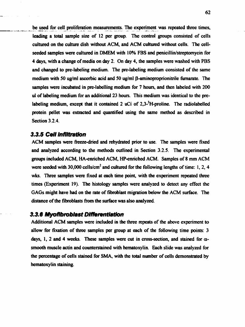
be used for cell proliferation measurernents. The experiment was repeated three times, -a--- A- - A--* - -------- - --A- A---- ----- ----A
leadhg a total sample size of 12 per group. The control groups consisted of cells
cultured on the culture dish without ACM, and ACM cultured without cens. The ceIl-
seeded samples were cultured in DMEM with IO% FBS and penicillin/streptomycin for
4 days, with a change of media on day 2. On day 4, the samples were washed with PBS
and changeâ to pre-labeling medium. The pre-labeling medium consisted of the same
medium with 50 ug/ml ascorbic acid and 50 ug/ml P-aminoproprionitrile fumarate. The
samples were incubated in pre-labelling medium for 7 hours, and then labeled with 200
ul of labeling medium for an additional 23 hours. This medium was identical to the pre-
labeling medium, excep that it contained 2 uCi of 2,3-3~-proline. The radiolabelled
protein pellet was extracted and quantifiad using the same method as described in
Section 3.2.4.
3.3.5 Cell InfJldration ACM samples were freeze-dried and rehydrated prior to use. The samples were fixed
and andyzed according to the methods outlineâ in Section 3.2.5. The experimental
groups included ACM, HA-e~ched ACM, HP-enriched ACM. Samples of 8 mm ACM
were seedeâ with 30,000 cells/cm2 and cultureâ for the following lengdis of time: 1,2,4
wks. Three sarnples were fixed at each time point, with the experiment repeated three
times (Expriment 19). The histology samples were analyzed to detect any effect the
GAGS might have had on the rate of fibmblast migration below the ACM surface. The
distance of the fibmblasts h m the siirface was also analyzed.
33.6 MyoffbriDbHDMmnti~n Additional ACM samples were included in the three repeats of the above experiment to
allow for fixation of three samples per group at each of the following time points: 3
days, 1, 2 and 4 weeks. These samples were cut in cross-section, and stained for a-
smooth muscle actin and counterstained with hematoxylin. Each slide was analyzed for
the percentage of cells stained for S M . witb the total number of cells demonstrated by
hematoxy lin staining.
3.3.7 S t s i i ' s --=-- -- -3 -- - - .- - -
Cell viability, proliferation and infiltration were anaiyzed for differences between pig
and human ACM. nie statistical analyses for this study were performed using SSPS
software. Al1 data was tested for normality using the Shapiro-Wilk and Kolmogorov-
Smimov tests. Non-parametric âata was analyzed for signticance using non-parametric
chi-squareci, Kruskal-Wallis, Fisher's exact test, Mann-Whitney U or Wilcoxon W tests.
Normal1 y distributed data was analyseà for si gni ficant differences by mu1 tivariate
ANOVA and posthoc Tukey HSD test. Significance was defined as ~ 0 . 0 5 .
Differences between ACM acellularity and structure were detemined qualitatively, and
therefore were not analyzed statistically.
Ctrap€er Four: RESUtTSAND DtSCUSStON
4.1 Pig Acellular Matrix Characterization
4.1.1 Acellularity The acellularization protocol has not been applied to the acellularization of human
demis pior to this study. No previous immunohistochemistry has been done to
determine its effectiveness in removing al1 cellular material fiom pig or human dermal
tissue. Samples of fieshly acellularized pig and human demis were formalin-fixed for
analysis of acellularity. Samples fiom each batch of acellularized demis were stained
with H&E, as well as with antibodies to vimentin, pankeratin and a-smooth muscle
actin.
4.1.1.1 Pig ACM Hematoxylin and eosin (H&E) staining of a sarnple from each batch of pig ACM
revealed consistent removal of al1 nuclear material. Hematoxylin is a cationic dye; it
binds to the negatively charged genetic material concentrated in the ce11 nucleus and
stains it bluish-purple. Eosin, an anionic dye, imparts a pink to red color to the other
dennal tissue components (Cormack, 1993). The presence of basophilic nuclear material
can therefore be easily seen with H&E staining. Figure 7 shows the appearance of pig
dermal ACM after staining with H&E. No basophilic (or blue) staining c m be seen,
indicating the removal of cellular nuclear material.

Figure 7. Pig demal ACM, H&E, 10x.
Epithelial cells of the demis (in the epidemis and demal appendages) contain
cytokeratins in their cytoplasm. The most superficial, epidennis-bearing layer of pig
demis was not acellularized in this study; afier harvesting it with a dermatome. it was
discarded. Only the deeper dermal layers were acellularized for ce11 seeding. Pankeratin
staining, however, showed residual keratin proteins around some hair follicles in the
ACM (Figure 8).
Figure 8. Pig ACM, stained for pankeratin, 10x. Positive (pink) staining around hair follicle indicates residual epithelial cellular material &er acellularization.

Since the nuclei of these cells were not seen by hematoxylin staining, the nuclear - - - --
material appears to have ken successfully digested despite the incomplete removal of
the cytoplasmic keratin. The presence of hairs likely prevents the complete removal of
the follicular epithelial cells by this acellularization protocol.
A limitation of the acellularization protocol with respect to pig demis lies in the failure
of the acellularization process to solubilize hairs. These are only loosened, such that
they might easily be removed with minimal friction at the end of the acellularization
process. The hairs were most easily removed in very thin sarnples of ACM, where they
separated fiom the matrix with mild shaking. It is believed that complete removal of
proteins around the hair shaft, could be accomplished with a repeat of the last detergent
step of the acellularization process after complete hair removal. This would allow
greater access by the detergent to the proteins lining the hair follicle, and thus optimize
removal of the proteins. This technique was not tested in this poject however; the
residual pig epithelial cellular material did not interfere with the ability to identify
human fibroblasts in the pig ACM.
The most important stains for this project were the mesenchymal-ce11 cytoplasmic
proteins, vimentin and a-smooth muscle actin. Complete removal of pig mesenchymal
cells from the pig demis is required for pig ACM to be used as an in vitro scaffold for
human fibroblasts. This allows for the seeded fibroblasts within the matrix to be clearly
identified. The degree of acellularity within ACM batches is demonstrated by
immunohistochemistry using these antibodies in Figure 9 (vimentin) and Figure 10
(SMA). Negative vimentin staining demonstrates complete removal of this protein From
the ACM; in contrast, the typical distribution of vimentin-staining in the demis is shown
in Figure 9 (b).

a) Pig ACM, 17- 1 1-00 batch, stained for vimentin, 20x.
b) Pig dermis, stained for vimentin, 20x.
Figure 9. Pig demal ACM and demis stained for vimentin (brown).
Smooth muscle cells in the demis stain positively for a-SMA. These surround blood
vessels, hair follicles and dermal appendages, as shown in Figure 10 (b).
a) Pig ACM, stained for srnooth muscle actin, 1 OX.
b) Pig demis, stained for smooth muscle actin, I ox.
Figure 10. Pig ACM and demis staining with smooth muscle actin (brown).
With the complete removal of native vimentin- and SMA-positive cells from the pig
demis, newly seeded human fibroblasts may be easily visualized on the pig ACM by
vimentin or SMA staining.
SMA staining of ACM samples occasionally showed some irregular staining of the
demis, possibly due to residual protein adsorbed to the dermal matrix andlor to non-
specific binding of the antibody to the matrix (Figure 11). This non-specific staining of

the ACM is distinguished fiom that of myofibroblasts by several features. Diffise weak -- -A--- - . . - - - - .-- - - - -
staining, seen in areas where the sectioned tissue is folded over, and not in association
with a ce11 nucleus, was considered non-specific staining. Figure 12 shows a typical
SMA-stained sarnple of fibroblast-seeded ACM, demonstrating the contrast between
background SMA staining and seeded fibroblast staining.
Figure I l . Pig ACM stained for smooth muscle actin, showing non-specific background staining (brown), 20x.
Figure t2. Human fibrobtasts (arrow) seeded on pig ACM stain for smooth muscle actin (red-brown) with hematoxylin counter-stain (blue-purple), 40x (lefi), 20x (right).
The presence of the background SMA staining in some ACM samples made the
automated quantification of SMA-positive fibroblasts dificult. These sections were
therefore excluded fiom ce11 counting analysis (Section 3.2.5).
4.1.1.2 Human ACM Human demis, obtained from two reduction mammaplasty donors, was acellularized for
this study. Staining with H&E, vimentin and SMA revealed no background staining
(Figure 13). In contrast to pig ACM preparation, the epidermal layer of the human
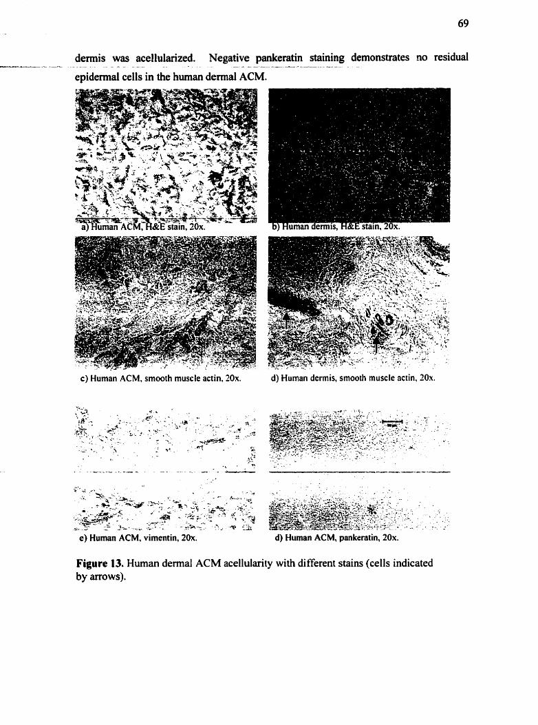
demis was acellularized. Negative pankeratin staining demonstrates no residual ---..--- - - . - . =--
epidemal cells in the human demal ACM.
c) Human ACM, smooth muscle actin, 30x.
e) Human ACM, vimentin, 20x.
d) Human dermis, smooth muscle actin, 20.u.
d) Human ACM, pankeratin, 20x.
Figure 13. Human dermal ACM acellularity with different stains (cells indicated by arrows).

-
--- --- - 4.1.2 - Acellular - - - Matri' Structure a-S - --
4.1.2.1 Pig demial ACM venus pig dermis Compared with whole demis, the extracellular matrix of demal ACM is devoid of cells.
A thorough evaluation of the pig ACM matrix composition is beyond the scope of this
project, and has not yet been done. A preliminary histological survey shows that most
anionic matrix components (Le. glycosaminoglycans) are removed in the acellularization
process. The structure of the insoluble matrix is preserved, however. Figure 14 shows
the preserved structure of the collagen and elastin matrix in the ACM.
a) Pig ACM, Movats, 20x. b) Pig demis, Movats, 20x.
Figure 14. Movats staining shows the preserved collagen (orange) and elastin (black) matrix structure of the pig demal ACM (a), in comparison with that of pig demis (b).
A comparison of the surface and cross-section of pig ACM and demis by SEM,
confirms the histological findings of preserved collagen bundle structure and residual
hairs (Figure 15). In Figure 15 (a) and (b), the collagen bundles are seen in cross-section
at high magnification. Both pig ACM and pig demis show a tightly packed network of
thick collagen bundles (circle).
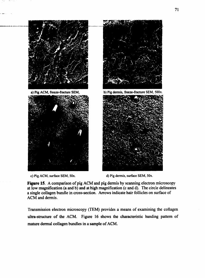
a) Pig ACM, fieeze-fiachire SEM, b) Pig dermis, fieeze-fracture SEM, 500x.
c) Pig ACM, surface SEM, 50x. d) Pig demis, surface SEM, 50x.
Figure 15. A cornparison of pig ACM and pig demis by scanning eIectron microscopy at low magnification (a and b) and at high magnification (c and d). The circle delineates a single collagen bundle in cross-section. Arrows indicate hair follicles on surface of ACM and dermis.
Transmission electron microscopy (TEM) provides a means of examining the collagen
ultra-structure of the ACM. Figure 16 shows the characteristic banding pattern of
mature demal collagen bundles in a sample of ACM.

Figure 16. Pig ACM by transmission electron microscopy (1 6,000~) showing the typicai appearance of type I collagen fibrils.
OAen experimental conditions cm change the structure of a material under investigation,
thereby introducing additional variables. The structure of pig dermal ACM however,
appeared very stable. This can be hypothesized to be due to the tightly packing of the
dense collagen network. It is known that collagen-based materials are more stable with a
higher degree of crosslinking; the extent of crosslinking in pig demis is not known
however. Figure 17 shows how the ACM collagen matrix structure appears relatively
unchanged histologically after fieeze-drying, centrifkging, or after several weeks of
soaking in culture media at 37OC. Preservation of pig ACM structure under these
conditions was further confirmed by fieeze-fracture SEM examination. These results
suggest that, under the conditions used in this study, pig ACM may be a consistent
mode1 for studying fibroblast interactions in an intact dermal collagen matrix.

a) Pig ACM after fieeze-drying and rehydration, hematoxylin, 20x.
b) Pig ACM afler centrifugation with media, hematoxy lin, 20x.
C) Pig ACM after four weeks under cell culture conditions, hematoxylin, 20x.
Figure 17. Pig ACM structure under different conditions, as demonstrated by histology.
It was desirable as well to have a dermal matrix scaffold which could be selectively
e ~ c h e d with certain soluble matrix molecules. In this way, the specific effects of
exogenous matrix molecules could more easily be detennined. The presence of
glycosarninoglycans in pig ACM was determined qualitatively by alcian blue staining of
freeze-fixed sections. Figure 18 demonstrates alcian blue staining of the anionic
components of pig demis and the ACM. Alcian blue binds primarily to sulfated
glycosarninoglycans such as heparan sulfate and chondroitin sulfate, but also binds to
hyduronan. These histology slides suggest that glycosaminoglycans appear to be
removed in the acellularization process. For alcian blue staining, the tissue was
embedded in O.C.T. compound without fixation, which prevented the extraction of
glycosaminoglycans fiom the matrix that occurs with formalin fixation.

a) Freeze-fixed pig ACM, alcian blue at pH b) Freeze-fixed pig skin, alcian blue at pH 2.5,20x. 2.5, 20x.
Figure 18. Pig ACM stained with alcian blue, showing minimal residual anionic matrix components after acellularization (compared with alcian blue staining of pig demis (bN.
4.1 -2.2 Pig ACM versus human ACM Pigs are widely used as animal models of wound healing because of the many
similarities between pig and human demis. Grossly, pig and human ACM have several
features in common (Figure 19). Both materials appear white, given the absence of
cells, blood and fat in the tissue. Since pig and human ACM are both predominantly
composed of collagen, they have similar handling properties.
Figure 19. Gross appearance of pig and hurnan dermal ACM.
There are, however, differences in the physical properties of pig and human ACM. Pig
ACM is qualitatively more rigid, and thus maintains its shape better than hurnan ACM

- when cut. Human ACM appears more extensible than pig ACM, and exhibits more
".-- - * - - - L M . - - - - - &
primary contraction, subjectively. Moreover, human ACM is more dificult to cut
tangentially, than pig ACM; the extensible human dermal tissue does not provide as
much resistance to the dermatome blade.
These differences may limit the relevance of pig ACM as a mode1 of hurnan dermal cell-
rnatrix interactions. However, its ease of handling and processing make pig ACM an
attractive material for studying certain aspects of dermal wound healing. For example,
the study of regulatory cytokines in fibroplasia would likely require many experimental
samples to investigate al1 possible concentrations of different cytokines. Pig ACM may
be reliably harvested in large amounts to provide hundreds of samples of equal
dimensions. Another example is the study of dermal contraction. This study shows that
this phenomenon is highly dependent on the dimensions of the contracting material
(Section 4.2.8). With pig ACM, it is much easier to control the dimensions of the ACM
samples.
The histological similarities of pig and human dennal ACM include the interlacing
network of collagen bundles of varying thickness, and the apparent spaces between the
collagen bundles. These spaces appear to be large enough in both species to
accommodate the migration of dermal cells such as fibroblasts into the matrix. These
features can be seen in the H&E stained
Figure 20.
sections of pig and human dermal ACM in
a) Pig ACM, H&E, 10x. b) Human ACM, H&E, 10x.
Figure 20. Histological cornparison of pig and human dermal ACM collagen matrix structure by histology.
Another difference is the relatively higher density of collagen in the pig ACM; the E - -
human collagen matrix appears to be less tightly packed. A discrepancy in the density of
collagen in the dermal matrix may explain the apparent difference in "rigidity" between
pig and human ACM. This tight packing of collagen may be a result of a greater
proportion of collagen in the maûix, as well as more cross-linking between the collagen
bundles, compared with human ACM. These aspects of pig demis have not been
investigated. The apparent difference in extensibility may be due to higher amounts of
elastin in the human ACM (Meyer and Neurand, 1987). Furthemore, the human ACM
samples used in this study were obtained from hypertrophie breasts requiring reduction
marnmaplasty. The dermal collagen fibres have therefore been previously stretched for
long petiods of time, which is known to result in lengthening of collagen fibres (Elder.
Elenitsas et al., 1997), and thus loosening of the collagen network. In future work, it
may be of interest to quantitatively compare the density of pig and human demis, as
well as to quanti@ the elastinîollagen ratio in the two materials.
Relatively minor structural differences rnay influence cell-matrix interactions (Yannas,
1997). For pig demis to be used as an in vitro mode1 of fibroplasia or as a dermal
substitute, its dermal matrix structure must allow human fibroblast infiltration. Since
histological processing can modifi the dermal collagen matrix structure (Meyer and
Neurand. 1987), scanning electron microscopy is believed to provide a more accurate
representation of biological structures. SEM pictures were examined for any differences
in demal ACM structure between the two species. The surfaces of fieshly acellularized
pig and human ACM were compared by SEM in Figure 21. By freeze-fracture SEM,
cross-sectional views were compared (Figure 22).

Figure 21. Pig (a) and human (b) ACM, scanning electron micrographs of tangentially cut surface, 500x. Collagen bundles (armws) are loose and irregular on surface of human ACM (b).
Figure 22. Pig (a) and human (b) ACM, freeze-fracture scanning electron micrographs of ACM in cross-section, 100x. Pig and human ACM were taken fiom demis 0.04- 0.06" below the epidennal surface. Arrows point to cross-sectional aspect.
In both surface and cross-sectional SEM, the pig ACM appears denser than the human
ACM. This difference in cross-sectional matrix structure may be significant in terms of
the scaffold properties of pig ACM. Migrating cells may less easily infiltrate the pig
matrix structure. As well, the surface of the human ACM appears more rough and
uneven than that of the pig ACM. This may be due to differences in (1) collagen mahix -,--A. SL .--- - - - . -
structure specific to anatornic location, (2) how the fibrillar collagen is altered by
soaking in PBS for a few weeks, or (3) cuning techniques (Le. electric dematome for
pig demis, hand-held dermatome blade for human demis). The slight differences
between the surface characteristics of pig and hurnan ACM may also affect the
behaviour of fibroblasts on the surface. The effect of these discrepancies in matrix
structure on in vitro fibroplasia will be investigated in Section 4.2. The structural
properties of pig and hurnan ACM found in this evaluation are summarized in Table 16.
1 Gross 1 Conformable 1 Conformable 1
Human ACM Lateral poles of breast Hand-held dermatome blade
1 Pig ACM , Source
Processing
(qualitative) Hhtology
[ SEM 1 interwoven collagen bundles 1 interwoven collagen bundles
Table 16. Structural properties of pig and human dermal ACM.
Paravertebral area of back Electric dermatome
Surface SEM
Cross-sectioion
An obvious variable in this cornparison of pig and hurnan dermal ACM is their
discrepant anatomic locations. One might think that a cornparison of pig and human
ACM structure should be performed on demis from equivûlent anatomic locations in
each species. AAer all, back and breast demis may differ in thickness, mechanical
properties and ultrastructure. However, an adequate supply of human demis for in vitru
modeling could realistically only be obtained from abdominoplasty or reduction
mammaplasty surgical specimens. In addition, pig demis can only be harvested
efficiently from the rigid back and flank areas, where an electric dematome c m be used.
As the objective of this investigation is to compare two potential materials for in vitro
modeling, human breast demis and pig back demis were evaluated in this study.
More rigid than human ACM Densely packed network of
Moreover, the structure of pig back demis is relevant to its potential application as a
More clastic than pig ACM Loosely packed network of
interwoven collagen bundles Smooth, cleanly cut surface wi th residual hairs Densely packed network of
derrnal substitute material sis well.
interwoven collagen bundles Irregular surfacc with loose collagen bundles, no hair Looseiy packed network of

4.1.3 Summaty A major challenge in the development of biomaterials is the modulation of the host
infiammatory and ensuing fibrotic responses. An effective dermal replacement may be
derîved from non-antigenic, naturally occurring materials already present in the demis.
The restoration of the normal extracellular matrix architecture is believed to facilitate
regeneration of the injured tissue. Despite ongoing research in this area, not enough is
known to allow the reconstruction of a synthetic three-dimensional fibrous collagen
matrix with sufficient resemblance to normal derrnal collagen. Allograft and xenograft
tissues are therefore the focus of considerable interest as starting materials for dermal
replacement.
Our acellularization protocol appears to remove cells fiom pig demis while preserving
an intact dermal collagen and elastin matrix. This protocol has not been fully
characterized in tenns of its effect on basement membrane and specific soluble matrix
components, and needs optimization for complete removal of al1 cellular proteins and
hain. However, the resulting three-dimensional scaffold has the potential to more
closely replicate the structure of acellular human demis, without the associated
problems of limited availability and high cost. The ability of this scaffold to support
human ce11 ingrowth will be critical to its use as a dermal substitute. For this in vino
evaluation of demal fibroplasia on pig and human ACM, the current acellularization
protocol was adequate. For future in vivo evaluations of acellularized pig demis
however, better removal of hairs and hair follicle proteins will be required.
It is essential in this study to produce a dermal matrix fiom which al1 pig fibroblasts
have been removed, and preferably al1 pig hyaluronan and sulfated glycosarninoglycans
as well. Vimentin staining clearly shows a complete removal of al1 pig mesenchyrnal
cells, including fibroblasts, fiom the demis (Figure 9). Alcian blue staining shows a
decrease in staining for glycosaminoglycans in the matrix (Figure 18). This method
however is not quantitative. In future studies, glycosarninoglycan extraction from the
matrix, electrophoretic separation and quantification with alcian blue standards will be
performed (Freund, Siebert et al., 1993).

--A -L .. - The matrix structure of pig ACM was evaluated
microscopy and transmission electron microscopy.
80
by histology, scanning electron
The structure of pig ACM was
evaluated for several reasons: (1) To determine if the acellularization process or the
expenmental conditions modify the collagen and elastin matrix structure. (2) To
determine if the structure of pig ACM is similar enough to that of hurnan ACM, to be
suitable for use as an in vitro mode1 of human dermal fibroplasia. (3) To evaluate its
suitability as a xenografi dermal substitute scaffold.
The collagen matrix structure appeared unchanged after acellularization, centrifugation,
fieeze-drying or culture with hurnan fibroblasts. It remains to be investigated whether
prolonged storage of pig ACM in sterile PBS at 4OC affects the tightness of the collagen
bundle network. Grossly, the tissue did not appear changed, even after four months of
storage.
Several differences were seen, subjectively, between the structure of pig and human
ACM. It is not known whether these differences in collagen bundle packing, overall
density of collagen, rigidity and extensibility are significant. Their significance will be
determined by testing the relative effectiveness of pig and human ACM as scaffolds for
human fibroblast infiltration. According to Yannas, the pore size of a scaffold for ce11
infiltration is important to the difhsion of nutrients through the scaffold's pores to the
infiltrated cells, as well as the provision of sufficient surface area for cell binding
(Yannas, 1997). Both these parameters are critical to permit ce11 infiltration into a
scaffold. The degradation properties of a scaffold can also influence its interactions with
cells. It is possible that fibroblasts secrete collagenases to digest the ACM as they
migrate into the collagen matrix (Mauch, Adelmann-Grill et al., 1989). The relatively
tight collagen matrix of the pig ACM may therefore still be effective as a scaffold,
provided it c m be progressively digested by migrating fibroblasts.
n ie most compelling reason for testing pig ACM as a potential derrnal substitute is the
success of acellularized human demis in vivo as a dermal substitute. Allograft human
ACM becomes infiltrated with host cells and well-vascularized, supports overlying
keratinocyte viability and decreases the extent of wound contraction in full thickness

---Eu -2- - - wounds (Wainwright, A - - - -
following design~. (1)
cells. (2) They can be
1995). A dermal substitute may fit in to one or more of the
They can be acellular but designed to become infiltrated by host
pre-seeded with cells normally found in the wounded tissue. (3)
They can be fabricated with degradable biopolymers, into which infiltrating cells create
a tissue specific extracellular matrix. (4) They cm consist of an enriched scaffold
designed specifically to influence cellular activities during the healing process. The rapid
integration of human acellular dermal natrix into the wound, despite its acellularity,
demonstrates the tasibility of using a dermal matrix as a dermal substitute.
4.2 Fibroplasia in an Acellular Pig Dermal Matrix
4.2.1 Ce// Morphology It is important to characterize the relative effects of pig ACM on fibroblast morphology,
in order to determine the suitability of pig ACM as a dermal substitute, as well as an in
vitro mode1 of fibroplasia. The morphological appearance of firmly adherent cells
differs from that of migrating cells as well as contracting cells (Ruoslahti, 1991). Ce11
morphology and behaviour are known to be influenced by a number of factors related to
the extracellular envimnment. Certain materials (such as collagen) trigger the
expression of surface receptors (such as integrins) to allow for specific material-receptor
binding. The number and type of receptors expressed help to regulate the degree of
adherence to and migration on the surface (Chevallay and Herbage, 2000). A difference
in adherence is reflected in the cell morphology (Doillon, Silver et al., 1987). With the
difference in collagen architecture and surface structure between the collagen gel and the
ACM, it was hypothnized that fibroblasts may be diffmntially adherent to the two
materials, and that the effects of the materials would be reflected in the ce11 morphology.
Confocal laser microscopy allows for the visualization of fluorescently-stained
fibroblasts adherent to the ACM surface. While regular or phase-contrast microscopy is
typically used to visualize cells cultured on coverslips or on the semi-translucent
collagen gel, cells grown on the dense dermal ACM must be stained and viewed under
fluorescence. Figures 23 and 24 show the appearance of primary human fibroblasts on
pig ACM, human ACM, collagen gel and coverslips after five days in culture.
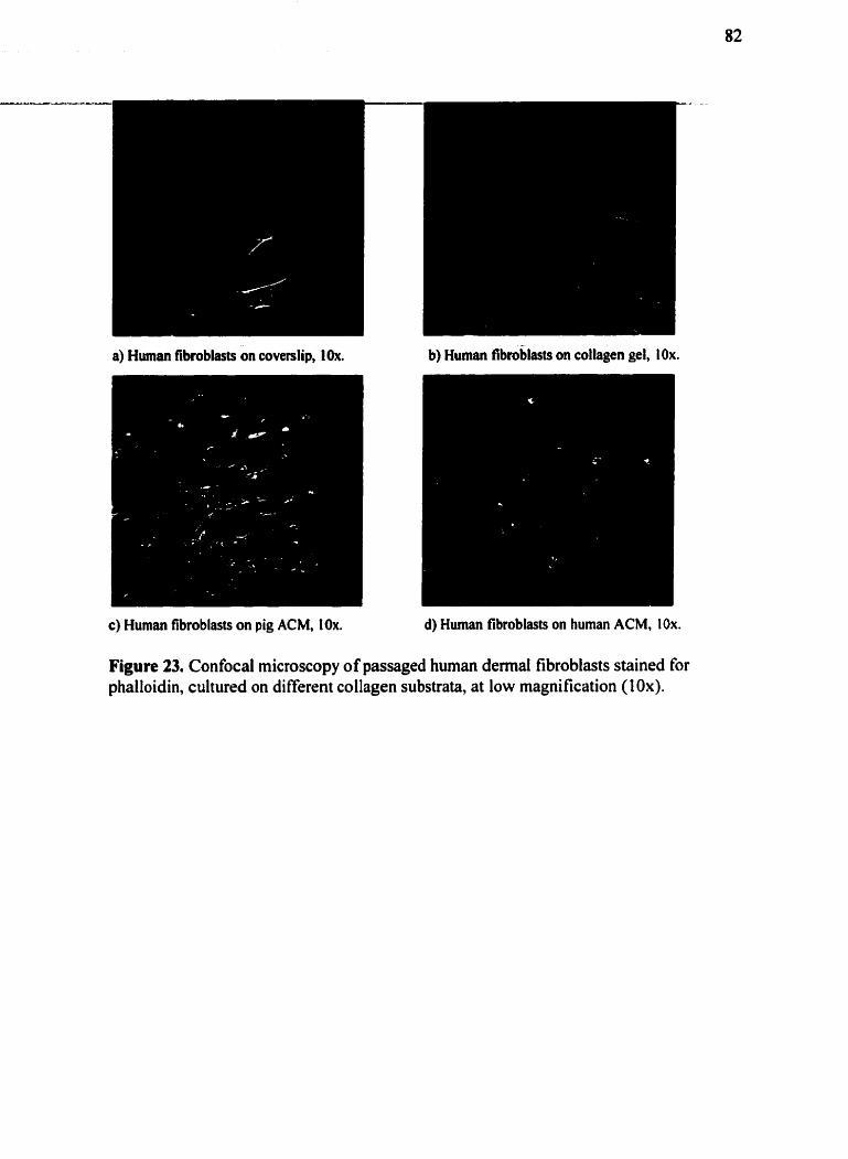
a) Human fibroblasts on coverslip, 1Ox. b) Human fibtoblasts on collagen gel, IOx.
c) Human fibroblasts on pig ACM, 10x. d) Hurnan fibroblasts on human ACM, 10x.
Figure 23. Confocal microscopy of passaged human dermal fibroblasts stained for phalloidin, cultured on different collagen substrata, at low magnification (10x).

a) Human tibroblast on coverslip, 63x. b) Hurnan fibroblast on collagen gel, 63x.
c) Human fibroblast on pig ACM, 63x. d) Human tibroblast on human ACM, 63x.
Figure 24. Confocal microscopy of passaged human demal fibroblasts stained for phalloidin, on diffierent collagen substrata, at high magnification (63x).
The fibroblasts were clearly smaller on the collagen substrates, especially human ACM,
relative to fibroblasts grown on coverslips. The spread-out, planar rnorphology of the
coverslip fibroblasts contrasts starkly with the small, spindle-shaped morphological
appearance of dennal fibroblasts in vivo (Grime11 and Lamke, 1984). On collagen gel,
the fibroblasts are also large, but appear more linear, with a "bipolar" appearance
(Nedelec, Ghahary et al., 2000). In general, fibroblasts on the pig and human ACM
were smaller than those on collagen gel or polystyrene. The majority of fibroblasts on
pig ACM were spindle-shaped (Figure 23c), while a wide range of morphologies was
displayed on the human ACM (Figure 23d). This suggests perhaps a response of the

fibroblasts to the different material properties of pig and human ACM. Fibroblast -.
morphology cm be afTected by surface roughness, ce11 surface receptor binding, collûgen
composition and mechanical tension (Guidry and Grinnell, 1985), (Nedelec, Ghahary et
al., 2000). Pig ACM has both a smoother surface and is more tigid than human ACM,
thereby possibly resulting in altered fibroblast morphology. The relevance of ce11
morphology to fibroblast behaviour on these materials in vitro will be discussed in later
sections,
4.2.2 Ce11 Proliferation It was not known prior to this study whether or not human fibroblasts would remain
viable if cultured on the pig dermal ACM prepared in our laboratory. It was therefore
necessary to develop methods for determining the viability of cells cultured on the ACM
over time. Results from the MTT and CyQuant assay will be presented here. Other
methods attempted included: trypsinization and ce11 counting with a hemocytometer, and
digestion and spectrophotometric quantification of methylene blue-stained cells. niese
methods were unsuccessfùl for a number of reasons. The fibroblasts did not separate
easily from the ACM with trypsinization. Moreover, given the small size of the ACM
and the relatively few cells on its surface, the number of cells available for counting was
below the sensitivity of the hernocytometer. There was also significant non-specific
staining of the ACM tissue with methylene blue, despite multiple rinses in PBS.
4.2.2.1 MTT Assay The MTT assay provided the first confirmation that fibroblasts seeded on the ACM
remained viable under culture conditions. The yellow tetrazalium dye (MTT) is cleaved
to a purple formazan by the succinate-tetrazolium reductase system, which belongs to
the mitochondrial respiratory chain. Only viable cells are believed to be capable of this
enzymatic cleavage. When the MTT solution is incubated with viable cells, the yellow
solution becomes purple, in proportion to the number of cells present. However, serum
enzymes are believed to interfere with this assay (Denizot and Lang, 1986). S e m
components in the culture media adsorb to the ACM and may not be completely washed
out prior to the addition of the MTT. It is therefore not surprising that the MTT is

converted to its purple product when placed on ACM sarnples even in the absence of c s-. - - - - - .-
tells (Figure 25).
Figure 25. Pig ACM without cells, afler four hour incubation with MTT and extraction with isopropanol. MTT is converted to purple formazan dye in absence of cells on the ACM. As well, the purple dye is incompletely extracted fiom the ACM with isopropanol.
Figure 26 shows a cornparison of the MTT caiibration curves with cells only and with
cells cultured on the ACM. Background is increased in the presence of ACM due to
non-specific conversion of MTT. It was therefore difficult to show a lack of ce11
viability for cells seeded on ACM, given the falsely elevated results in the presence of
ACM. - -- . - - - - - -- - - - - - - - - -
MTT Calibration Cuwes 0.8 7 ---
O 5000 10000 15000 20000 25000 Cell number
ACM+ cells Cells
Figure 26. Calibration curves with the MTT assay of passaged human dermal fibroblasts in the presence and absence of ACM.
A preliminary assessrnent of ce11 viability over time on the pig ACM, as compared to
cells grown directly on polystyrene wells, is shown in Figure 27. The results are
difficult to interpret, due to the poor specificity of the MTT assay on the cell-seeded
ACM. The rate of ce11 proliferation on the ACM appears to be low, accounting for the
lack of increase in absorbante over tirne. It was observed that the cells floated off the
culture dish with isopropanol extraction, but the cells seeded on the ACM did not. This
may have resulted in a greater proportion of the formazan being measured in the cells
only group. To clari@ these potential erroa with the use of the MTT assay with cells
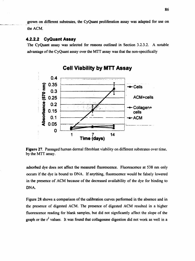
. grown on different substrates, the CyQuant proliferation assay was adapted for use on
- - -
the ACM.
4.2.2.2 CyQuant Assay The CyQuant assay was selected for reasons outlined in Section 3.2.3.2. A notable
advantage of the CyQuant assay over the MTT assay was that the non-specifically
Cell Viability by MTT Assay
+ Cells I
1 7 14 Time (days)
Figure 27. Passaged human dermal fibroblast viability on different substrates over time. by the MTT assay .
adsorbed dye does not affect the measured fluorescence. Fluorescence at 538 nm only
occurs if the dye is bound to DNA. If anything, fluorescence would be falsely lowered
in the presence of ACM because of the decreased availability of the dye for binding to
DNA.
Figure 28 shows a cornparison of the calibration curves perhrmed in the absence and in
the presence of digested ACM. The presence of digested ACM resulted in a higher
fluorescence reading for blank samples, but did not signficantly affect the dope of the
graph or the r2 values. It was found that collagenase digestion did not work as well in a

37OC incubator without COz or humidification, and that calibration curves prepared in PA--- - - - * - - - -
this manner had poor r' values (data not shown).
ACM Cells
.
+cells l only
O 10000 2MKX) 30000 - .
Figure 28. Calibration curves with the CyQuant assay of human dermal fibroblasts in the presence and absence of digested ACM.
Given the reliability of the CyQuant assay in assessing cell numbers in the presence of
ACM, this assay was used to quantitate fibroblast proliferation on pig ACM. Their
proliferation on pig ACM was compared to that of fibroblasts cultured on human ACM,
collagen gel and directly on the culture dish. In Figure 29, fluorescence is proportional
to ce11 number. The percent change fiom day O fluorescence, for cell-seeded sample
d e r different lengths of time in culture, is shown.

PACM rn Cells a Coll gel A HACM - . - - - - . . - . - . . .
Figure 29. Fibroblast proliferation on different substrates as determined by the CyQuant assay @ooled data fiom Experiments 2,3,4 - Table 9). Abbreviations: PACM= pig ACM, HACM= human ACM, Coll gel = collagen gel, Cells= cells on culture dish.
The error bars in Figure 29 represent standard deviations. As expected, the cells
proliferated when cultured on the culture dish only (brown squares). The fibroblasts
were also found to proliferate on the pig ACM (purple circles). A cornparison of Figures
27 and 28 suggests that the MTT assay is unreliable in measuring cell viability on the
ACM. The proliferation rate can be estimated by looking at the dope of the trendlines
for each group. It can be seen that the proliferation rates for the fibroblasts are similar
on the culture dish and on the pig ACM. However, the fibroblasts proliferated
significantly more slowly on the collagen gel and human ACM, i.e. the two substrates
which supported more ce11 infiltration (Section 4.2.4 and 4.2.6). These results suggest
that human fibroblasts behave differently on pig ACM than on human ACM. Pig demal
ACM may therefore not be as similar to human dermal ACM as expected. This
surprishg difference between the pig and human ACM will be discussed more in later
sections.
A source of error in the CyQuant assay was likely the incomplete digestion of the human
ACM by collagenase. The thicker hurnan ACM samples required more time to be
digested than the pig ACM and the collagen gel samples, resulting in a higher

- - - 3-
background reading for the human ACM. To account for this, the measured A - . -A . -
fluorescence for each sample was reduced by the average background reading for each
substrate type.
4.2.3 Collagen Synfhesis New collagen synthesis by fibroblasts cultured on pig ACM was compared to that of
fibroblasts cultured on collagen gel and in the absence of collagen. It was of interest to
determine how the extracellular matrix environment influenced cellular behaviour in this
aspect of fibroplasia. The amount of radiolabelled proline uptake by the cells
corresponds to the total protein synthesis during the incubation tirne (23 hours). Figure
30 shows the results for the different groups. The figures for each sample are
nonnalized for the ceIl number, as determined by the CyQuant assay. -- - -- - - - - a - -
Figure 30. Protein synthesis by human fibroblasts seeded on different substrates. * indicates significance < 0.05 between groups s h o w by bracket, as determined by the Mm-Whitney and Wilcoxon tests.
By comparing the total cpm with the cpm remaining after digestion with collagenase and
removd of the collagenase-soluble radioactivity, it was possible to determine what
percentage of total protein synthesis was devoted to collagen synthesis. Figure 3 1 shows
the percentage of collagenase-soluble radiolabeled protein, i.e. newly synthesized
collagen, by the fibroblasts in each group. The standard deviation in each group is
indicated by error bars.

- - - A - - - - . - - - - - - -- - - - - - - - - .-
Figure 31. Collagen synthesis by human fibroblasts cultured on different substrates. Collagen synthesis by monolayer fibroblasts was significantly different that of fibroblasts grown on the other two media (chi-squared test).
In gels of contracted collagen, the synthesis of collagen by fibroblasts is strongly
inhibited (six to eight times less than in monolayer culture by both pre-transcriptional
(Mauch, Hatamochi et al., 1988) and post-transcriptional mechanisms) (Eckes. Mauch et
al., 1993). Reduction in collagen synthesis could be the result of negative feedback
induced by biochemical confinement, as observed in contracted collagen gels containing
fibroblasts (Nakagawa, Pawelek et al., 1989). Collagen synthesis in this study was
measured four days afier seeding the fibroblasts on the ACM and collagen gel; no
infiltration would be expected at that point. The fibroblasts would be expected to have
formed a monolayer on the ACM or gel surface, and to be in contact with the collagen
matrix but not surrounded by it. The results suggest that collagen synthesis by
fibroblasts is decreased even if the cells are not confined by collagen.
Dermal ACM and collagen were compared to determine whether the nature of the
collagen in contact with the fibroblasts had any effect on their collagen biosynthesis.
Collagen synthesis was less on the collagen gel than on the ACM. The inhibitory effect
of extracellular collagen on fibroblast collagen synthesis may therefore be influenced by
collagen structure. The percentage of new protein synthesis dedicated to newly
synthesized collagen appeared consistent between the three groups. There was however
considerable variability in these cdculated values. The radiolabelled proline assay was

---- .- usefùl for measuring protein synthesis by cells cu1huu:d on ACM or collagen gel, but was - r
less reliable at determinhg the collagenasr-sü:übie portion of the total protein. An
alternative collagen synthesis assay invcives measuring radiolabelled hydroxyproline
incorporation.
4.2.4 Cell Infiltration The study of cell-matrix interactions in vitro allows for the isolation of specific wound
healing events. In this study. the effect of the preserved pig dermal matrix structure on
fibroblast infiltration is of interest for a variety of reasons. A potential dermal substitute
n w t support rapid host cell infiltration to achieve incorporation into the wound bed and,
in the case of fibroblasts, to begin matrix remodeling and wound closure. Previous
studies demonstrate that acellular human dennis acts as an effective scaffold for ce11
infiltration in vivo. Acellular pig dennis however is not incorporated as effectively into
full thickness wounds (Section 2.5.2.4). While the reason for this may be immune
rejection, it is also possible that its structure is different enough from that of human
demis to prevent effective host ce11 infiltration.
Using the culture conditions described in the Methods section, minimal fibroblast
intiltration was seen in the majority of pig ACM samples by H&E staining (Figure 32).
Fi y r e 32. Pig ACM seeded with human fibroblasts, after 4 weeks; H&E, 10x. There is considerable proliferation of the fibroblasts at the surface (arrow), with few cells present within the matrix.

Vimentin staining of the cell-seeded ACM sections allowed greater visualization of the
fibroblasts. The entire cytoplasm of the cells was stained, as opposed to just the nucleus.
Even with vimentin staining however, very few cells were seen below the surface in the
majority of pig ACM samples (Figure 33). A total of 75 cell-seeded ACM samples.
cultured for over 3 weeks, were analyzed, histologically, representing six different
batches of pig ACM and primary human dennal fibroblasts fiom five different donors.
Of al1 the samples examined, 50/75 (67%) sarnples showed surface infiltration only, Le.
infiltration to only within 100 Pm of the surface.
Figure 33. Pig ACM seeded with human fibroblasts, afier 4 weeks; vimentin, 10x.
In a minority of samples. (25/75; 33%) deeper fibroblast infiltration coutd be seen on at
least one section. These results were initially difficult to interpret, without appropriate
controls. The inconsistent fibroblast infiltration of pig ACM was hypothesized to be due
to either (1) the in vitro culture conditions not favouring fibroblast migration, or to (2)
poor scaffold properties of pig ACM. Since human ACM has been shown to support
human fibroblast infiltration in vitro, this material was used in this study as a positive
control for fibroblast infiltration in vitro.

Acellular human demis has been show in previous studies to act as an effective
scafTold for Uifiltrating fibroblasts ni vitro (Ghosh, Boyce et al., 1997), (Ralston, Layton
et al.. 1997). Fibroblast infiltration in vitro is therefore a convenient basis of cornparison
for the structural similarity between pig and human demis. The influence of various
culture conditions on fibroblast infiltration of acellular dermis has not been studied
previously. However it is known that fibroblasts do not infiltrate the dermal basernent
membrane in the absence of kemtinocytes. Al1 demis used in this study was therefore
second-cut demis, without basement membrane.
Table 17 and Figure 34 compare pig and human ACM in terms of the percentage of
samples with infiltration by human fibroblasts, and the nurnber of cells per section,
respectively. ACM samples in both groups were selected fiom al1 levels of dermis,
fieeze-dried, cut to fit the culture dish, seeded with 25,000 or 30,000 cells/cm2 and
incubated for 3 to 5 weeks. Equivalent freeze-dried and non-freeze-dried samples of pig
ACM are included in the table for cornparison.
Table 17. Comparison of hurnan fibroblast infiltration in fiesh pig ACM, freeze-dried pig ACM and fieeze-dried human ACM. There was a çignificant difference between the groups indicated by *, by the chi-squared test.
Signi ficance
Pig FD-Pig FD-Human
-- ---- 1
Fibroblast Infiltration of ACM
% of samples
ACM
49 17 24
Total number of samples
Number of samples with ceIl infiltration in at least
one section 15 9 20
31% 53% 83%
*Human *Human *Pin, FD-~ig;

Fiare 34. Fibroblast ifiltration o f pig and h m a n AC-M @ 4 weeks, determined by automated ce11 counting. The number of cells per section was nonnalized for section size, then averaged for 16 samples, with three sections each. The bars represent standard deviation. The human ACM group had more samples with significant cell infiltration and more
cells per section than did the pig ACM group. The substantial variability between
samples is represented by the high standard deviation. It was also noted that the
distribution of fibroblasts in the matrix differed considerably between pig and human
ACM. The distance of each ce11 fiom the surface was therefore measured to
quantitatively compare the ce11 distribution. In Table 18. the distance of the infiltrated
cells fiom the cell-seeded surface is shown. Freeze-dried samples of cell-seeded pig and
human ACM at 3 weeks were analyzed. To calculate the chi-squared statistic, the actual
distance measurements for each ce11 were used to determine if the distribution of the
cells in the two groups was statistically different.
I 1
Table 18. Distance of cells fiom monolayer surface, in representative fields of vimentin-stained sections of cell-seeded ACM samples at 3 weeks. n = number of samples per group. Distances of over 100 cells per field were recorded.
Human ACM (n = 5) 34-492 pm
38.3 16.5
29 k 6.2 33 I 6 . 6
p < 0.05
Range of ce11 distance from surface % of infiltrated cells:
0-150 um 151-300 UII
301 -500 ~m
Human ACM appeared to support fibroblast migration fùrther fiom the surface.
Pin ACM (n = 5) 15-434 pm
78.8 f 8.3
14.2 I 7.9 8 k 1.2
Although a few fibroblasts did migrate a considerable distance fiom the surface in pig
Chi-squared test on individual ce11 distance measurements (n = 300 per sample)
ACM, the vast majority were within 1 50 um from the surface. The celis in the human
ACM appeared to have migrated more quickly throughout the matrix (Figure 35).

t
Figure 35. Typical appearance of human ACM seeded with fibroblasts after 3 weeks, vimentin, 10x. The cell-seeded surface is at the top of the image.
In pig ACM, the fibroblasts were observed to migrate below the cell-seeded surface
along the empty hair follicle. but without migrating into the actual collagen matrix. This
was not seen in the human ACM, due to the paucity of hair follicles. The successful
infiltration of human ACM demonstrates the effectiveness of the in vitro methods at
modeling in vivo host ce11 infiltration. It seems likely therefore that the poor infiltration
of pig ACM by human fibroblasts is mainly attributable to its scaffold properties. The
increased fibroblast infiltration in the human ACM suggests that human and pig demis
may differ in structure.
The structure of human and pig demis were therefore compared, before and after
fibroblast seeding. Figure 36 shows, by freeze-fracture SEM, a c o m p ~ s o n of the
structure of cell-free, and cell-seedeû, pig and human ACM, afier 4 weeks in culture.
Al1 samples were fiom demis taken 0.02-0.04 inches below the epidemis, and freeze-
dried prior to incubation.

Day O Day 28 - Day O 60 prn
Day 28
Figure 36. Pig and human ACM at day O and at day 28 &er cell seeding. Cross-sectional scanning electron micrographs of the matrix structure at the surface, 500x. Scale bar: 60 Fm.
A greater change in matrix structure with cell seeding is seen in human ACM. The
structure of pig ACM appears well preserved in the presence of human fibroblasts. In
contrast, the collagen bundles in human ACM appear to be even more loosely packed
after cell seeding. This change in matrix structure with ce11 culture may be attributable to
the effect of matrix collagen digestion by fibroblasts, or to the effect of prolonged
soaking in culture media at 3 7 ' ~ . It is not surprising that the looser matrix structure of
the human ACM is more easily modified. The dense pig ACM, on the other hand,
appeared more resistant to structural change under the culture conditions.
The mature collagen bundles and porous nature of pig ACM are favourable features in a
potential dermal substitute. However, the results of fibroblast-seeding experiments show
that pig ACM is not equivalent to human ACM, with respect to hurnan fibroblast
infiltration in vitro. The densely packed nature of pig dermal collagen matrix may delay
its infiltration by host cells. Future work in this area should focus on the modification of
the aceflular pig dermal matrix structure, to improve its scaffold properties. This will be
discussed in more detail in Section 5.1.2.

To determine whether the variability in fibroblast infiltration of pig ACM could be
reduced, and infiltration optimized, each of the experimental variables hypothesized to
be a factor in ce11 infiltration was analyzed for its effect. The conditions of samples with
ce11 infiltration were examined to determine what factors influenced fibroblast
infiltration into the pig ACM. The variables examined included fibroblast type, seeding
density, ce11 culture setup, culture media, microvascular endothelial ce11 CO-culture, time
in culture, level of demis and sample freeze-drying. Only time in culture showed a
statistically significant effect, while a trend was seen towards greater infiltration with
fieeze-drying of samples.
4.2.5.1 Time in culture
Table 19. Effect of culture time on primary human fibroblast infiltration into pig ACM. ACM samples of 16 mm diameter and seeded with 25,000 cells/cm2 only were compared. n = number of samples examined per time point. By one-way ANOVA, it was determined that at least one of the four groups was significantly different. Post-hoc Tukey HSD allowed painvise cornparisons of al1 the groups for significant differences.
Time in culture Number of infiiltrated cells per section Significance Average Standard deviation between groups
1 week (n = 8) 15.63 18.98 * 5,7 weeks
The length of time in culture had a clear effect on fibroblast infiltration of the pig ACM.
3 weeks (n = 8) 5 weeks (n = 16) 7 weeks (n = 8)
Presumably the process of infiltration requires a period of time for fibroblast migration
and possibly digestion of the matrix by secreted collagenolytic enzymes.
6 t .25 94.3 8 203.13
4.2.5.2 Level of dermis It was hypothesized that the ultrastructure of the pig ACM varîed with its depth from the
epidennal surface. Histologically, only very minor structurai differences were observed
55.56 45.52 22.34
across a cross-section of full-thickness pig ACM (Figure 37).
* 5,7 weeks * 1,3,7 weeks * 1,3,5 weeks
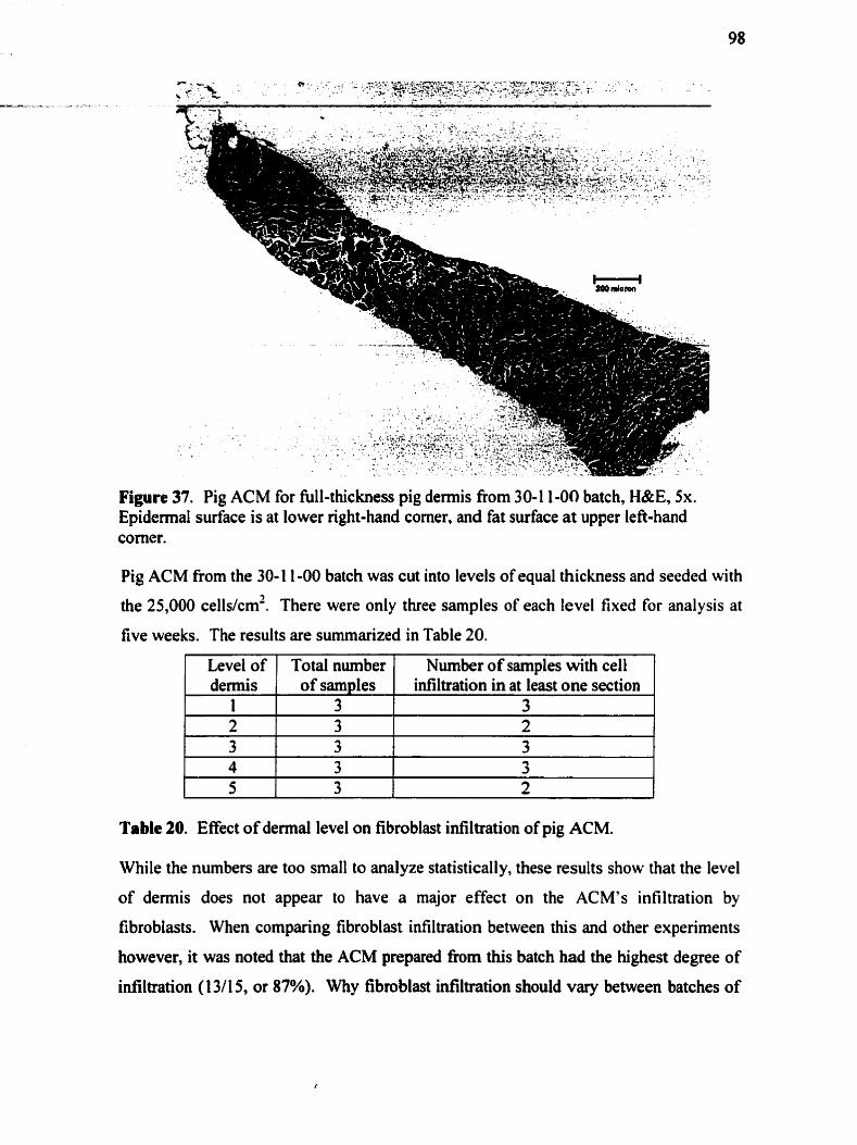
Figure 37. Pig ACM for full-thickness pig demis fiom 30-1 1-00 batch, H&E, 5x. Epidermal surface is at lower right-hand corner, and fat surface at upper lefi-hand corner.
Pig ACM fiom the 30-1 1-00 batch was cut into levels of equal thickness and seeded with
the 25,000 cells/cm2. There were only three samples of each level fixed for analysis at
five weeks. The results are summarized in Table 20.
Table 20. Effect of dennal level on fibroblast infiltration of pig ACM.
Level of dermis
1 2
While the numbers are too small to analyze statistically, these results show that the level
of demis does not appear to have a major effect on the ACM's infiltration by
Total nurnber of samples ,
3 3
fibroblasts. When comparing fibroblast infiltration between this and other experiments
Number of samples with ce11 infiltration in at least one section
3 2
however, it was noted that the ACM prepared fiom this batch had the highest degree of
infiltration (1 3/I 5, or 87%). Why fibroblast infiltration should Vary between batches of

pig ACM, cannot be conclusively explained fiorn this work. Possible reasons for this, - - -
which will be investigated for thoroughly in future work, include the effect of Freeze-
drying, the age of the pig, the length of time in storage in PBS, and the inter-experiment
variability in ce11 adherence to the ACM.
4.2.5.3 Freeze-drying Freeze-drying is a standard pmcedure for preparing biological materials for storage.
Many potential biomaterials require fieeze-drying before being stored and transported.
and therefore before being used in the body. Previous studies suggest that the structure
of a dermal collagen matrix is not significantly modified by fieeze-drying (Ghosh,
Boyce et al., 1997). However these studies employed histological methods for assessing
the collagen matrix structure, which can in itself result in alteration in structure.
Collagen may shrink slightly with freezing, but it is not known whether this significantly
alters cellular responses to the material (Doillon, Brandwein et al., 1986). Freeze-dried
samples were therefore compared to samples stored in PBS at 4°C for any effect on
fibroblast infiltration. The use of freeze-drying was not required for fibroblast
infiltration of the pig ACM to occur, but did not impair infiltration (Table 21). Freeze-
dried samples were more convenient to store and handie, and easily rehydrated. In
subsequent experiments, pig ACM sarnples were freeze-dried and rehydrated prior to
cell seeding.
1 Freeze- 1 Total number / Number of samples with ce11 1 % of sarnples with 1
Table 21. Effect of fieeze-drying on fibroblast infiltration of pig ACM. ACM samples seeded with 25,000 or 30,000 cells/cm2 and cultured for > 3 weeks. No significant association was found between keze-drying and ce11 infiltration of pig ACM (two-sided Fisher's exact test).
drying Yes
nie examination of the experimental conditions in the ce11 infiltration experiments
revealed some interesting information. Of al1 the variables studied in a controlled
manner, only time had a statistically significant effect on ce11 infiltration. However, not
al1 the ACM samples cultured for five weeks had fibroblast infiltration. There are
of samples 17
infiltration in at least one section 9
cell infiltration 53%
clearly additional variables at play which were not controlled for in this study. For
exarnple, it may be that the differences in the extent of ce11 infiltration arnong the pig
ACM were due to diffenng lengths of storage time in PBS. Slight differences in ce11
seeding technique rnay also have occurred unknowingly. Alternatively, fibroblasts in
culture may infiltrate the ACM scaffold to a greater degree afier a certain nurnber of
passages than others. In future work, al1 ACM samples will be fieeze-dried immediately
&er acellularization. In this way, the variable of storage time can be eliminated, and
any variability of the pig ACM itself could be discemed.
4.2.6 Myofibroblast Ditferentiation During wounding, fibroblasts at the wound edge receive signals from cytokines and
matrix molecules to begin migrating into the wound dot. A proportion of these wound
fibroblasts then differentiates to myofibroblasts. The forces generated by rnyofibroblasts
adherent to the underlying matrix are believed to be responsibie for wound contraction.
Decreased wound contraction is associated with a more rapid disappearance of
myofibroblasts from the wound (Rudolph, 1979), (Berry, Hardiiig et al., 1998). It is
known that wounds treated with intact demal collagen contract significantly less than
wound treated with denatured autografi collagen (Brown, Garner et al., 1990). Little is
understood about the effect of dermal substitute structure on full thickness wound
contraction. It was of interest therefore to determine whether the intact dermal collagen
structure of allograft or xenograft demis has an effect on myofibroblast differentiation.
Previous studies of the effect of acellular pig demis on wound contraction suggest that
the xenografi cm d u c e contraction, provided giafc teke is echieved quickly. (Wang,
Chen et al., 1997), (Fang, Robb et al., 1990). It is therefore possible that pig dermal
ACM may be associated with a reduced expression of myofibroblast markers by seeded
fibroblasts, in cornparison with fibroblasts cultured on the culture dish or a collagen gel.
Another question that was addressed in this section was whether any diflerences in
myofibroblast differentiation were seen between the pig and hurnan ACM, to suggest a
fiirther evidence for a difference in the microenvironment of the two matrices. These
experiments represent preliminary investigations into these questions, and are primarily
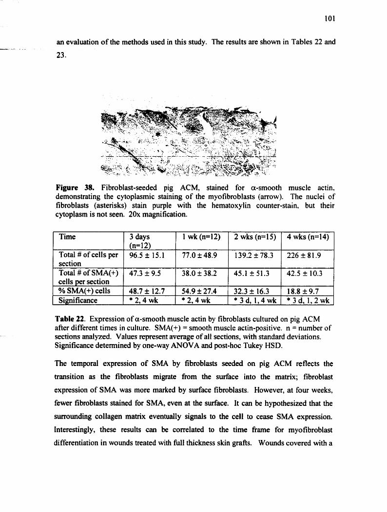
an evaluation of the methods used in this study. The results are shown in Tables 22 and
Figure 38. Fibroblast-seeded pig ACM, stained for a-smooth muscle actin, demonstrating the cytoplasmic staining of the myofibroblasts (arrow). The nuclei of fibroblasts (asterisks) stain purple with the hematoxylin counter-stain, but their cytoplasm is not seen. 20x magnification.
Tirne
Total # of cells per section
. - [ Signi ficance
I
1 *2 ,4wk 1 *2,4wk 1 * 3 d , 1 , 4 w k ( *3d ,1 ,2wk 1
Total # of SMA(+) cells per section % SMA(+) cells
Table 22. Expression of a-smooth muscle actin by fibroblasts cultured on pig ACM afier different times in culture. SMA(+) = smooth muscle actin-positive. n = number of sections analyzed. Values represent average of al t sections, with standard deviations. Significance determined by one-way ANOVA and post-hoc Tukey HSD.
3 days (n= 1 2) 96.5 f 15.1
The temporal expression of SMA by fibroblasts seeded on pig ACM reflects the
47.3 f 9.5
48.7 i 12.7
transition as the fibroblasts migrate from the surface into the matrix; fibroblast
lwk(n=12)
77.0 I 48.9
expression of SMA was more marked by surface fibroblasts. However, at four weeks,
38.0 I 38.2
54.9 f 27.4
fewer fibroblasts stained for SM& even at the surface. It can be hypothesized that the
2wks(n=15)
139.2 I 78.3
surroundhg collagen matrix eventually signais to the cell to cease SMA expression.
4wks(n=14)
226 k 8 1.9
45.1 -t 51.3
32.3 k 16.3
Interestingly, these results can be correlated to the tirne fiame for myofibroblast
42.5 + 10.3
18.8 f 9.7
differentiation in wounds treated with N1 thickness skin grafts. Wounds covered with a

full thickness skin gr& showed a more rapid decrease in myofibroblasts than untreated -a.. - --+
wounds. Myofibroblasts were found in untreated wounds from 4 to 42 days post-
wounding, and from 4 to 2 1 days in wounds treated with full thickness grafis (Rudolph,
1979). This interesting cowlation suggests the possibility that the effects of a dermal
collagen matrix on fibroblast differentiation may, at least in part, be demonstrable in
viiro.
The effect of pig ACM, human ACM and collagen gel on fibroblast SMA expression
was then investigated using these methods. The results are surnmarized in Table 23.
Table 23. Percentage of fibroblasts on each ACM type expressing a-smooth muscle actin at 2 weeks. SMA(+) = smooth muscle actin-positive. n= number of sections
Collagen gel (n=9) 172.6 I 55.2
26.8 k 19.7
Substrate Total # of cells per section Total # of SMA(+) cells per section % SMA(+) cells Si nificance
analysed. Values represent average of al1 sections. Significance determined by one-way ANOVA and post-hoc Tukey HSD.
Surprisingly, frwer fibroblasts expressed SMA when cultured on human ACM or on
collagen gel, relative to those on pig ACM. It c m be hypothesized that the human ACM
and collagen gel are more quickly id~ltrated by fibroblasts, thus dispersing the SMA-
positive fibroblasts from the rnonotayer. On the pig ACM by cornparison, multiple
layers of fibroblasts are present around most samples at two weeks, in some ways
resembling a fibrous capsule (Figure 39). A significant proportion of these monolayer
fibroblasts expresses SMA.
Pig ACM (n=9) 139.2 f 78.3
45.1 '51.3
32.3 I 26.3
g g e l
Mechanicd tension is believed to play a role in regulating myofibroblast differentiation.
In vivo, the timing of the switch from fibroblast to myofibroblast phenotype appears to
depend in part on the extent to which the wound resists contraction (Darby and
Gabbiani, 1990). Wounds under tension are commody observed to heal with more
Human ACM (n=9) 204.5 I 97.6
21.2 k 16.4
10.3 k 8.9
* PACM
15.5 + 12.6
*PACM
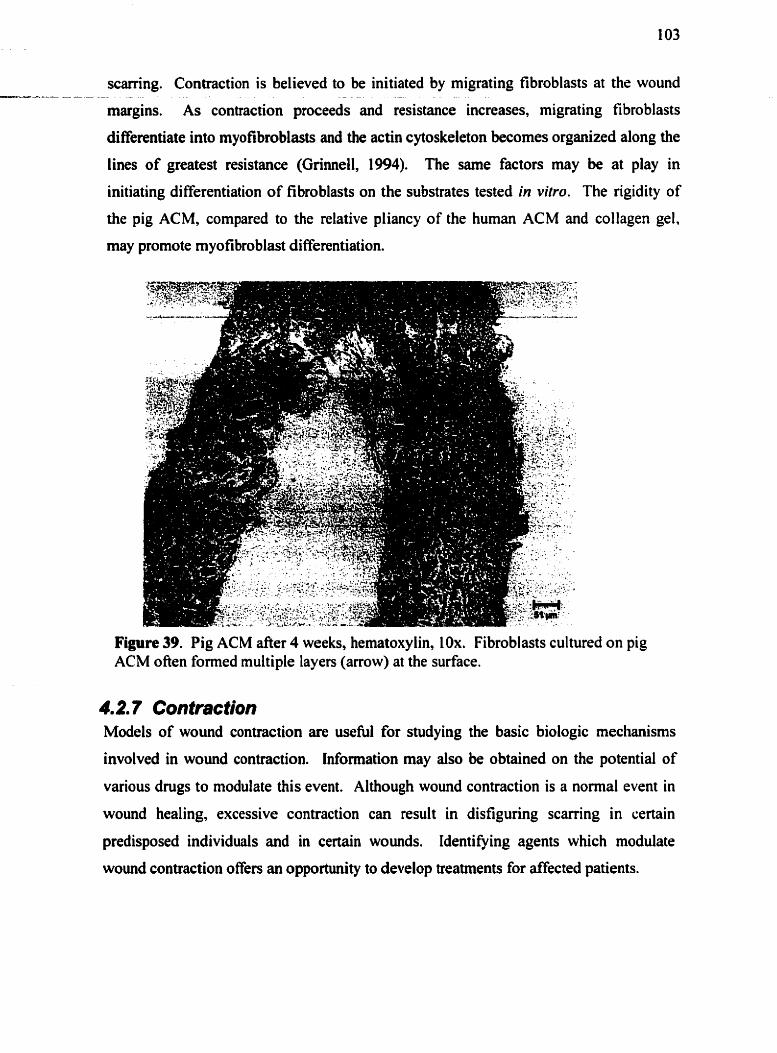
scarring. Contraction is believed to be initiated by migrating fibroblasts at the wound -----= - - -- -
margins. As contraction proceeds and resistance increases, migrating fibroblasts
differentiate into myofibroblasts and the actin cytoskeleton becomes organized dong the
lines of greatest resistance (Grinnell, 1994). The sarne factors may be at play in
initiating differentiaiion of fibroblasts on the substrates tested in vitro. The rigidity of
the pig ACM, compared to the relative pliancy of the human ACM and collagen gel,
may promote myofibroblast differentiation.
Figure 39. Pig ACM after 4 weeks, hematoxylin, IOx. Fibroblasts cultured on pig ACM often formed multiple layers (arrow) at the surface.
4m2m 7 Contraction Models of wound contraction are useful for studying the basic biologic mechanisms
involved in wound contraction. Information may also be obtained on the potential of
various drugs to modulate this event. Although wound contraction is a normal event in
wound healing, excessive contraction c m result in disfiguring scarring in certain
predisposed individuals and in certain wounds. Identifying agents which modulate
wound contraction offers an opportunity to develop treatments for affected patients.

Collagen gel as a mode1 for fibroblast-mediated contraction has been well characterized. - - - . - - -
While the contraction of collagen gel occurs too q&kly to be related to fibroblast
migration into the gel matrix, the collagen solution can be mixed with cells pnor to
gelation, thus distrïbuting the fibroblasts throughout the collagen matrix. In this study,
fibroblasts were seeded on the surface of the gel only, to allow a better comparison with
pig ACM.
As expected, in this shidy, the collagen gel contracted within 24 hours to 60% of its
original surface area, and within 72 hours, to 15% of its original surface area (Figure
40). An interesting observation of the contracted gels was that the gels were not fixed in
a contracted state; the curied up edges of the gels could be restored to their original
position with the removal of the media. For thick gels (>3 mm), the cell-seeded surface
and the lower portion of the gel contracted to different degrees. Because of the soft
flexible nature of the gel, the thickness of the celi-fiee lower portion of the gel did not
inhibit the upper surface of the gel fiom contracting. The rate of contraction of the cell-
seeded surface was not decreased when thicker gels were used. However. the
visualization of the outer diameter of the cell-seeded surface was difficult; the thicker
gels appeared to have a greater diameter than the thinner gels.
a) 1 mm thick gel at 24 hours b) I mm thick gel at 72 hours c) 3 mm thick gel at 72 hours
Figure 40. Contracted collagen gels. Al1 samples were onginally the sarne diameter as the well(201 mm2). The degree of contraction was determined by measuring the change in surface of the cell-seeded surface (outlined in dotted line).
On cross-section histology. the extent of fibroblast infiltration into the collagen gel cm
be seen. Figure 41 shows minimal infiltration of human fibroblasts into a collagen gel 3
weeks after seeding.

Figure 41. Fibroblast-seeded collagen gel in cross-section, hematoxylin, 1Ox. The upper border of the scale bar indicates the cell-seeded edge of the collagen gel. which is pooriy visualized with this stain.
Fibroblast-mediated contraction of collagen gels therefore occurred without minimal
fibroblast infiltration below the surface. A similar phenornenon of contraction was seen
with the fibroblast-seeded pig ACM. Over a variable period of time, depending on the
thickness of the ACM, the cell-seeded surface slowly curled up, thereby reducing the
surface area of the entire ACM. To confirm that this change in appearance of the ACM
was in fact due to fibroblast-mediated contractile forces, ACM samples of equal
thickness were incubated with or without cells and assessed for evidence of contraction.
None of the 9 cell-fiee samples (0%) decreased in surface area, while 36 of 46 cell-
seeded ACM (78%) decreased by more than a fifth of their original surface area. Figure
42 illustrates the degree of contraction noted in the cell-seeded ACM samples. The
percent change in surface area measwments are shown in Table 24.
I Cell-seeded ACM I I CelCfree ACM I Figure 42. Fibroblast-mediated contraction of thin (0.3 mm in thickness) pig ACM after 2 weeks in culture (Experiment 15). Scale: each well is 16 mm in diameter.
The same samples were examined histologically, and found to have minimal fibroblast
infiltration into the dermal matrix (Figure 43). It was surprising that fibroblast-mediated

contraction of pig ACM, like the collagen gel, occurred in the absence of significant ce11 - - - - 2 - -
hfiltration.
Figure 43. Fibroblast-seeded pig ACM at 4 weeks, hematoxylin, 10x. This sample contracted d o m to 40% of its original surface area, presumably by the forces generated by the monolayer of fibroblasts at the surface (dark blue ce11 layer on top surface). Very few cells infiltrated below the surface; a few are indicated by red circles.
Fibroblast-mediated contraction of collagen materials has been hypothesized to occur by
two possible mechanisms (Ehrlich and Rajaratnam, 1990). The fibroblasts may attach to
the collagen surface and subsequently contract, causing a reduction in the distance
between collagen bundles. Alternatively, the fibroblasts may migrate a certain distance
along the collagen bundles and in doing so, bring the different collagen bundles closer
together. Both of these mechanisms seem plausible for the contraction of pig ACM.
Regardless of the mechanism, the resulting contraction is restricted to one surface only,
causing the ACM to curl up towards the cell-seeded surface as illustrated in Figure 44.
Cel l seeded ACM disc Tractional forces on cell- ACM rcmains folded afler prior to contraction seeded surface curl edges prolonged incubation with
fibroblasts . p.
Figure 44. Schematic of the mechanism of fibroblast-mediated contraction of pig ACM.

P- - It should be noted that no contraction at al1 was noted in ACM above a thickness of 0.7
- - - - - - - - - - - - -
mm. Table 24 shows the relationship between ACM thickness and contraction.
contraction, by the chi-squared test.
Approximate Thickness*
0.5 mm 0.6 mm >0.7 mm
This study attempted to study the effects of the CO-culture of MECs on contraction.
Number of samples with contraction+
Number of simples
Fibroblast-seeded samples of equal thickness were either CO-cultured with MECs or
Average % original surface area in contracted samples*
Table 24. Effect of ACM thickness on fibroblast-mediated contraction. tcontraction defined as greater than 15% decrease in surface area. Al1 samples were seeded with 25,000 cellslcm2, incubated for 4 weeks in EBM-2 with 5% FBS and CO-culiured with MEC. Initial surface area: 20 1 mm2. *There was a significant effect of thickness on
IO 16 40
cultured alone, both in EBM-2 media. A representative set of samples and controls are
shown in Figure 45.
7 9 O
78% i 10% 79% + 6% 100% + 4%
Figure 45. Fibroblast-mediated contraction of pig ACM (0.6 mm in thickness) afier 4 weeks in culture (Experiment 17). Scale: each well is 16 mm in diameter.
Fibroblasts & MEC CO-
cultured
There was no clear difference in contraction associated with the CO-culture of MEC.
These results were not convincing however; the small degree of contraction seen in the
thicker 0.6 mm samples did not leave much room for differences between groups. The
effect of even minor differences in thickness, among samples in the same group, was far
Fibroblasts only
greater than any stimulatory effect by endothelial cells.

The finding that fibroblast-mediated contraction of a cross-linked dermal collagen -r.---e- - - - - - -
matrix can be seen in vitro is quite surprising. A previous study, in which no contraction
of fibroblast-seeded human acellular demis was seen, suggested even that the role of
fibroblasts in contraction was over-emphasized (Ralston, Layton et al., 1997). The same
study did however demonstrate contraction of the human dermal matrix by
keratinocytes, which did not infiltrate below the surface. Our results confirm their
findings in one respect; contraction of the dermal matrix, whether by keratinocytes or
fibroblasts, does not require infiltration of the matrix. Moreover, these results suggest
new possibilities as to the mechanism of fibroblast-mediated contraction of skin grafls.
The dennal component of skin grafts appears to exert the main influence on wound
contraction. One study of rat wound healing found that grafts with an intact collagen
matrix produce lasting inhibition of wound contraction. In contrast, grafls with a
denatured collagen matrix limit the process to a lesser degree (Browri, Garner et al..
1990). This, and other data in the study, suggests that the structure of collagen is
important in regulating contraction. The exact mechanism by which this inhibition is
expressed is as yet unknown. Wounds covered with full-thickness skin grafts had a
minimum of contraction with a very rapid decrease in the number of myofibroblasts. In
wounds treated with split thickness skin grafls, myofibroblasts remained in the wound
for an average of 40 days, compared with 21 days with full thickness grafls (Rudolph
and Ballantyne, 1990). Full-thickness skin grafts thus appeared to influence contracting
wounds not by preventing myofibroblast differentiation, but by speeding up completion
of their life cycle.
The direct effect of thickness on ACM contraction by fibroblasts correlates well with
clinical observations of contraction in grafled wounds. Thin split-thickness grafts
contract more quickly and to a greater degree than thicker g r a s . The effect of thickness
on contraction may be mediated by a greater mechanical resistance of thicker gr&s to
the contractile forces of the fibroblasts. Several researchers have found that it is not the
absolute thickness of the gr& which affects contraction, but rather the relative thickness
of the demis ba t is included (Rudolph, 1979). An interesting topic for m e r research
may be to examine whether ACM thickness has any effect on myofibroblast

differentiation in vitro. The reduced proportion of myofibroblasts on human ACM, - c
relative to pig ACM, may have contributed to the lack of observed contraction of human
ACM in vitro. Given the very thin saniples that are required to demonstrate contraction,
pig ACM may be more suitable than hurnan ACM as a model of fibroblast-mediated
dermal contraction. Human ACM exhibits considerable primary contraction, which is
associated with a marked change in observed thickness and diarneter after cutting. Pig
ACM has practically no primary contraction, and therefore maintains its desired
dimensions after cutting into small pieces.
The fact that contraction of collagen gel occurs rapidly without significant SMA
expression by fibroblasts suggests that collagen gel models the early contraction of
wound healing by wound margin fibroblasts. Pig ACM may enable the study of
myofibroblast-mediated contraction that occurs later in wound healing, such as in full
thickness wounds treated with split-thickness skin grafis. It has k e n suggested that
collagen gels are too simple in structure to rnimic the mature extracellular matrix
structure of the demis, and that there may be problems in extrapolating from the ability
of fibroblasts to contract collagen gels in vitro to the clinical situation (Ralston, Layton
et al., 1997). Pig ACM may provide an alternative model in which to study this aspect
of wound healing.
4.2.8 Summary In this section, pig ACM was evaluated as a model for demal fibroplasia in vitro.
Fibroblast morphology, proliferation, collagen synthesis, ceIl infiltration, expression of
myofibroblast smooth muscle actin end contraction were studied. In al1 these aspects of
fibroplasia, pig ACM was compared to human ACM. Several differences were noted in
the interactions of hurnan fibroblasts with pig ACM as opposed to with human ACM.
Fibroblasts adhered well to the pig ACM surface, as evidenced by the fibroblast
morphology. Fibroblasts on the human ACM appeared rounder and less spindle-shaped,
consistent with the greater infiltration of human ACM by the fibroblasts. Fibroblasts
proliferated on pig ACM almost as much as they proliferated on the culture dish, but
synthesized less collagen on pig ACM. These results are only partially consistent with

-- . =
those of studies on fibroplasia in a collagen gel.
proliferation of the fibroblasts seeded within the gel is
(Bell, Ivarsson et al., 1978), (Ehrlich and Rajaratnam,
In an unattached collagen gel,
inhibited by contraction of the gel
1990), (Nakagawa, Pawelek et al.,
1989). It has been shown that growth is not inhibited among the fibroblasts grown on
the surface of the gel (Tornasek, Hay et al., 1982). It appears therefore that ce11 growth
inhibition is a result of the number and distribution of contact sites between collagen and
cells rather than an inhibition by cell-to-ce11 contact. In Guidry and Grinnell's attached
gel model however, fibroblasts do multiply within the collagen matrix (Guidry and
GrinneIl, 1985). An explanation for this has not yet been determined. In our study,
collagen synthesis seemed to be inhibited by the adherence of the fibroblasts to a
collagen substrate, whereas proliferation rates were minimally affected, even after four
weeks in culture.
Pig ACM may be a suitable model for studying certain dermal cell-matrix interactions
only, such as contraction. Fewer fibroblasts infiltrated the pig ACM compared to the
human ACM, suggesting that pig ACM may not be equivalent to human ACM as a
scaffold for human cells. However, this may provide an opportunity to demonstrate the
pro-migratory effects of incorporated matrix molecules, such as hyaluronan. The
influence of culture conditions on fibroblast infiltration were studied to deterrnine how
to best achieve fibroblast infiltration into pig ACM in vitro. Of al1 the variables studied.
only time showed a significant effect on infiltration into pig ACM. However, the
considerable variability among samples suggests that there are still unrecognized
experimental variables influencing fibroblast infiltration.
Pig ACM may be a useful model of fibroblast-mediated, dermal contraction. The results
of this investigation clearly demonstrate the ability of fibroblasts to contract a dennal
collagen matrix in vitro. For a number of practical reasons, pig ACM has several
advantages over human ACM for this application. Improved harvesting techniques
however would be required to obtain pig ACM sarnples of standard dimensions. This
would help ensure greater reliability of this model. Fibroblast-mediated contraction in
this model occurred independently of fibroblast infiltration below the ACM surface.

-. - - This was consistent with observations of collagen gel contraction without fibroblast
-a- L - infiltration.
The results of this section are also pertinent to the evaluation of pig ACM as a potential
xenograft demal substitute. Successful infiltration of human ACM demonstrated that
the culture conditions used are effective in allowing fibroblast infiltration of a matrix.
Sarnples of pig ACM which had been stored in PBS for long periods of time and were
then freeze-dried, appeared to have a greater degree of fibroblast infiltration. The reason
for this is not known. Storage time was not controlled for in this study. These results
suggest however, that the dense pig dennal matrix supports more ce11 infiltration after it
may have been slightly altered. The variability in fibroblast infiltration between pig
ACM samples indicates that the structure of pig ACM must be modified to ensure
consistent and rapid ceIl infiltration. It is particular important in vivo, that pig ACM as a
scaffold does not require degradation by macrophages before it is infiltrated by cells.
Such a response would promote inflammation and scarring in a cutaneous wound.
4.3 Fibroplasia in a Glycosaminoglycan-Enriched Acellular Pig Dermal Matrix
4.3.1 Glycosaminoglycan Incorporation in most studies on the effects of these matrix molecules on cells in vitro.
glycosaminoglycans (GAGs) are either coated on the culture dish or added to the culture
medium bathing the cells. In these ways, the effects of GAGs are determined on cells
grown in two dimensions. By using collagen gels or sponges or an even more natural
material such as acellular dermal matrix, as scaffolds for 3D ce11 culture, it is possible to
snidy the efiects of GAGs on fibroblasts in a more "life-like" environment. GAGs can
be easily incorporated into a collagen gel by mixing the GAGs with the denatured
collagen solution pnor to gelation or fieeze-drying. This however altered the structure
of the collagen gel or sponge, confounding any effect attributable to the presence of the
GAG itself. The incorporation of solubilized GAGs into dermal ACM must of course
employ a different method. It was therefore necessary to develop methods for

incorporating GAGs into the acellular demal matrix without modiming the tissue - - - - - - - - - . -
arc hi tecture.
In order to evaluate the effectiveness of the incorporation, fluorescently-labeled HA and
HP were incorporated into the pig ACM. Figure 46 (a) shows the distribution of
fluorescein-HA and dansyl-HP in the pig ACM afler incorporation, viewed with a UV
filter of 450 nrn. This allowed for optimal visualization of fluorescein-HA. However,
the dansyl-labelled HP was not as visible, as it excites at 265 nm and emits at 510 nm.
The samples enriched with HP were therefore also stained for alcian blue (pH 2.5) to
confirm the presence of HP in the ACM (Figure 47).
a) Pig ACM centrifuged with 5 mg/ml FlTC-HA b) Pig ACM soaked in 5 mg/ml dansyl-HP
Figure 46. Distribution of fluorescently-labeled HA and HP in pig ACM, fiozen unstained sections, 5x.
a) Pig ACM soaked in 5 mg/ml heparin b) Pig ACM without heparin
Figure 47. Distribution of heparin in pig ACM, as seen by alcian blue staining, pH 2.5, 5x. Background alcian blue staining of pig ACM (b) is show for cornparison.
This mode1 allows a unique oppomuiity to study the effects of matrix
glycosaminoglycans on fibroblasts cultured within a natural dermal collagen
environment. Moreover, the incorporation process does not appear to modify the
collagen matrix structure (Section 4.1.2.1). Previous studies incorporated GAGs into a
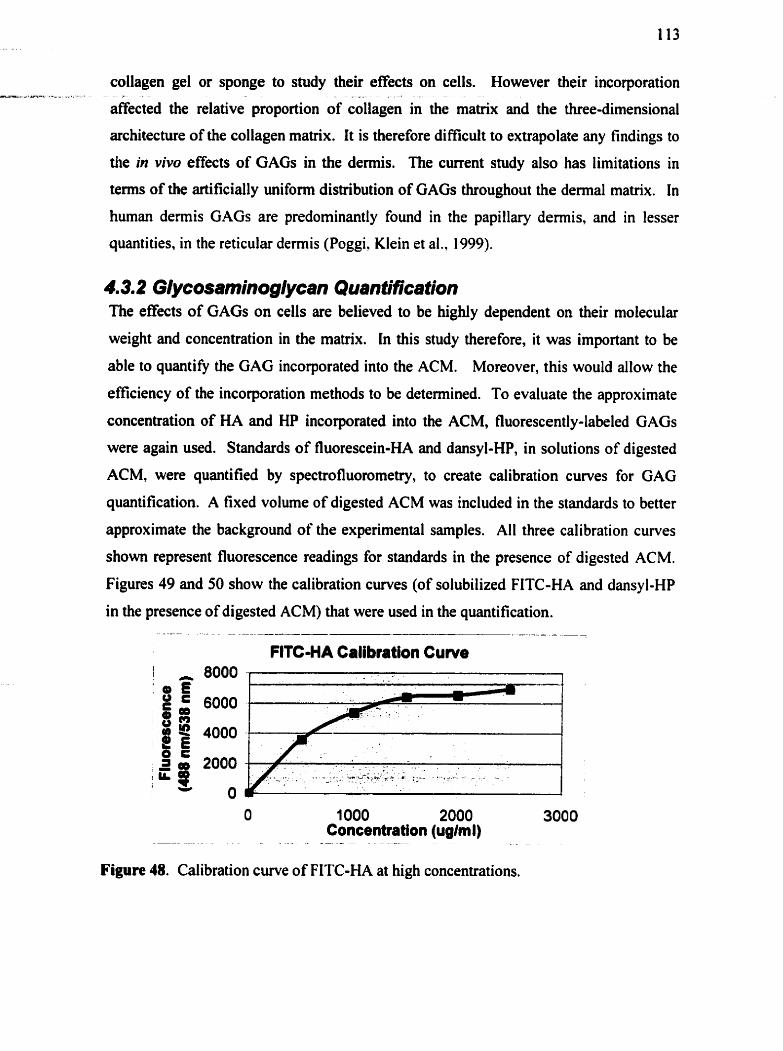
collagen gel or sponge to study their effects on cells. However their incorporation ---* - a -.
affected the relative proportion of &lagen in the matrix and the three-dimensional
architecture of the collagen matrix. It is therefore dificult to extrapolate any findings to
the in vivo effects of GAGs in the dermis. The current study also has limitations in
terms of the artificially unifonn distribution of GAGs throughout the dermal matrix. In
human demis GAGs are predorninantly found in the papillary demis, and in lesser
quantities, in the reticular demis (Poggi, Klein et al., 1999).
4.3.2 Glycosaminoglycan Quantification The effects of GAGs on cells are believed to be highly dependent on their molecular
weight and concentration in the matrix. In this study therefore, it was important to be
able to quantifi the GAG incorporated into the ACM. Moreover, this would allow the
efficiency of the incorporation methods to be determined. To evaluate the approximate
concentration of HA and HP incorporated into the ACM, fluorescently-labeled GAGs
were again used. Standards of fluorescein-HA and dansyl-HP, in solutions of digested
ACM, were quantified by spectrofluorornetry, to create calibration curves for GAG
quantification. A fixed volume of digested ACM was included in the standards to better
approximate the background of the experimental samples. All three calibration curves
shown represent fluorescence readings for standards in the presence of digested ACM.
Figures 49 and 50 show the calibration curves (of solubilized FITC-HA and dansyl-HP
in the presence of digested ACM) that were used in the quantification. -- - . - - -- - - - - -
FlTC-HA Calibtation Cuwe
O 1 O00 2000 3000 --
Concentration (ug/m l) . - - - -. - - .- -
Figure 48. Calibration cuve of FITC-HA at high concentrations.

=, -
---- ---.-- A * - - .. L - - -A- A--
114
At higher FiTc-HA concentratiolls, as Figure 48 shows, the fluomscena was w longer
pqmticmal to concc~ttration. Maxmisl fluorescence appearrd to k reached at 150
Wml FITC-HA. For quantification of experimental samples, a caiiibration cuve of
10- concentrations was used to obtain a linear equation (Figure 49).
I FITC-HA cdibritkn Cuwe
Figare 49. Cal'bration c w e of FiTC-HA at kw concentrations.
The f l u ~ r t ~ ~ e n t mark= of heparin, dansy1, doss not produce as much fluorescence as the
fluorescein. A cal'btaticm cuwe at hi& concentrations thdore shows a linear
relationsbip between hionscence and dsnsy l-HP concenttation (Figure 50).
Figure 50. Cal'btation cuve of dan@-HP @lue), with trcadline indicatd m black.
The amount of HA th t was be barporatd into the ACM using our centrifugation
m(hd is listsd m Tabk 25. The amwit of HP iacorporabed into the ACM by 48 boum
of soelring is show as weU.
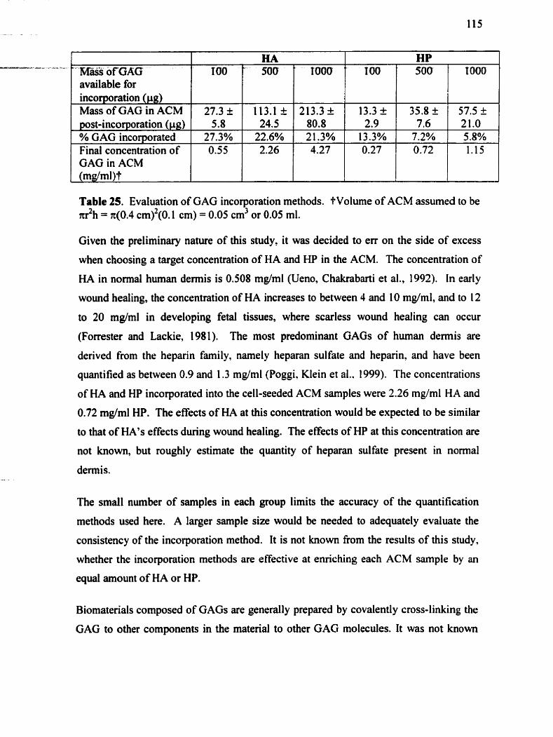
"' KaSs of GAG available for incorporation (pg) MassofGAGinACM pst-incorporation (pg) % GAG incorporated
Table 25. Evaluation of GAG incorporation methods. tVolume of ACM assumed to be d h = n(0.4 C ~ ) ~ ( O . 1 cm) = 0.05 cm3 or 0.05 mi.
HA
27.3+
Final concentration of GAG in ACM (rndml)?
Given the preliminary nature of this study, it was decided to err on the side of excess
when choosing a target concentration of HA and HP in the ACM. The concentration of
HA in normal human demis is 0.508 mg/ml (Ueno, Chakrabarti et al., 1992). In early
wound healing, the concentration of HA increases to between 4 and 10 mg/ml, and to 12
to 20 mg/ml in developing fetal tissues, where scarless wound healing can occur
(Forrester and Lackie, 1981). The most predominant GAGs of human demis are
derived from the heparin family, namely heparan sulfate and heparin, and have been
quantified as between 0.9 and 1.3 mg/ml (Poggi, Klein et al.. 1999). The concentrations
of HA and HP incorporated into the cell-seeded ACM samples were 2.26 mg/ml HA and
0.72 mg/ml HP. The effects of HA at this concentration would be expected to be similar
to that of HA'S effects during wound healing. The erects of HP at this concentration are
not known, but roughly estimate the quantity of heparan sulfate present in normal
100
5.8 27.3%
dermis.
' HP
113.1I
0.55
The small number of samples in each group limits the accuracy of the quantification
methods used here. A larger sample size would be needed to adequately evaluate the
consistency of the incorporation method. It is not known fiom the results of this study,
whether the incorporation methods are effective at enriching each ACM sample by an
equal amount of HA or HP.
100 ' 500 " 500
24.5 22.6%
Biomaterials cornposed of GAGs are generdly prepared by covalently cross-linking the
GAG to other components in the material to othet GAG molecules. It was not known
1000 ' ~ ~ 0 0
2 1 3 . 3 i
2.26
80.8 21.3%
13.3 t
4.27
2.9 13.3%
35.8 k
0.27
57.5 2 7.6
7.2% 21.0 5.8%
0.72 1.15
how long the non-crosslinked GAGs such as HA and HP would remain in the d e m l - - - - - - - - - -
matrix after i&orporation: This was-a factor in the decision to incorporate more rather
than fewer GAGs into the ACM. In order to evaluate HA or HP's eflects on long term
aspects of fibroplasia such as ce11 infiltration, it was hoped that the GAGs would remain
in the ACM for as long as possible. It was expected that the higher molecular weight
HA (1,000,000 Da versus 10,000 Da for HP) would d i f i e out of the ACM more
slowly. HA-enriched ACM was therefore soaked in HBSS for various time points, up to
28 days, while the HP-enriched ACM was soaked for up to 14 days. The effect of
diffbsion on GAGs in the ACM was assessed qualitatively and quantitatively. Figure 5 1
shows the histological appearance of fluorescently labeled HA-ACM and HP-ACM after
soaking for 28 and 14 days, respectively. AI1 photographs are taken with the same
exposure times and gain.
a) FITC-HA in pig ACM, after 28 days in WBSS b) Dansyl-HP in pig ACM, afler 14 at 37°C. days in HBSS at 37°C.
Figure 51. Qualitative histological assessrnent of effects of diffusion on GAG- e ~ c h e d ACM. Frozen, unstained sections of pig ACM are viewed under fluorescence at 450 nm, 5x.
Once it was established that some HA and HP was present in the ACM at these later
time points, samples of ACM with 113.1 pg of HA, or with 35.8 pg of HP, were
quantified spectrophotometrically af?er soaking for different lengths of time. Fresh
cdibration curves were prepared at each time point, of both FITC-HA and dansyl-HP.
Figure 52 shows that even after 28 days, HA maintained its original mass. For HP, 20
ug have difised out, leaving 44% of the original mass at day 14 (Figure 53).

Figure 52. Quantitative assessrnent of effects of diffusion on FITC-HA in ACM. Error bars represent standard deviation
l
Maas of DansybHP in ACM over T ime
Figure 53. Quantitative assessrnent of effects of diffusion on dansyl-HP in ACM. Error bars represent standard deviation.
As expected, the amount of HP in the ACM decreased significantly due to rapid
diffusion of the small rnolecule. The lack of difhision of HA out of the ACM was very
surprising. Previous reports found 60% of GAG leaching within 1 week of immersion in
isotonic crystalloid (Poggi, Klein et al., 1999), as well as a half-Iife of one day for newly
synthesized HA in dermal tissue, in the presence of cells (Tammi, Tuhkanen-
Martikainen et al., 1996). It is possible that the values in Figure 52 were not accurately
quantified by spectrofluorometry, given the high concentration of HA (>2000 pg/ml).
As s h o w in Figure 48, fluorescence is not linearly proportional to concentration above
1500 pg/rnl. This was not a problem for the dansyl-labeled HP, which was less
fluorescent. It is likely however that a significant portion of the HA did remain in the
ACM despite some diffusion, given the very large arnount of HA originally
incorporated. A second experiment was therefore performed, in which lower

concentrations of FITC-HA were incorporated into the ACM (0.001 to 1 mg/ml). --A- --& -. L - A- . - - -
Samples in this experiment were only soaked for 1 week. Even with smaller
concentrations of HA, at least 50% of the original HA mass remained in the ACM afier a
week in HBSS (results not shown).
There are a number of possible explanations for the longevity of FITC-HA in the ACM.
The HA may adsorb to the collagen matrix, despite soaking in aqueous solution.
Altematively, HA may self-aggregate at the relatively high concentration present in the
matrix; these aggregates may not be as soluble in water. These results can only estimate
the duration of HA'S presence in the ACM under actual ce11 culture conditions, given the
absence of cells in this expriment.
4.3.3 Cell Proliferation No significant difference was seen between the number of cells cultured on ACM, HA-
ACM or HP-ACM (Experiment 18- Figure 54). r-- -- - I
Fibroblast Proliferation 1000 , 8
-
+ ACM HA-ACM HP-ACM
l O 10 20 30 I
I Days
Figure 54. Effect of HA and HP on fibroblast proliferation on pig ACM. by the CyQuant assay. There was no significant difference between groups at each time point, by the Mann-Whitney test. For al1 groups, Day 2 values were significantly different from Day 28 values by the Mann-Whitney test.
These results were encouraging in ternis of the low toxicity of the GAG concentrations
selected. HA has been previously show to inhibit fibroblast proliferation at high
concentrations (Huang-Lee, Wu et al., 1994), but has also been reported by other
investigators to increase as well as to have no effect on proliferation (Pieper, van
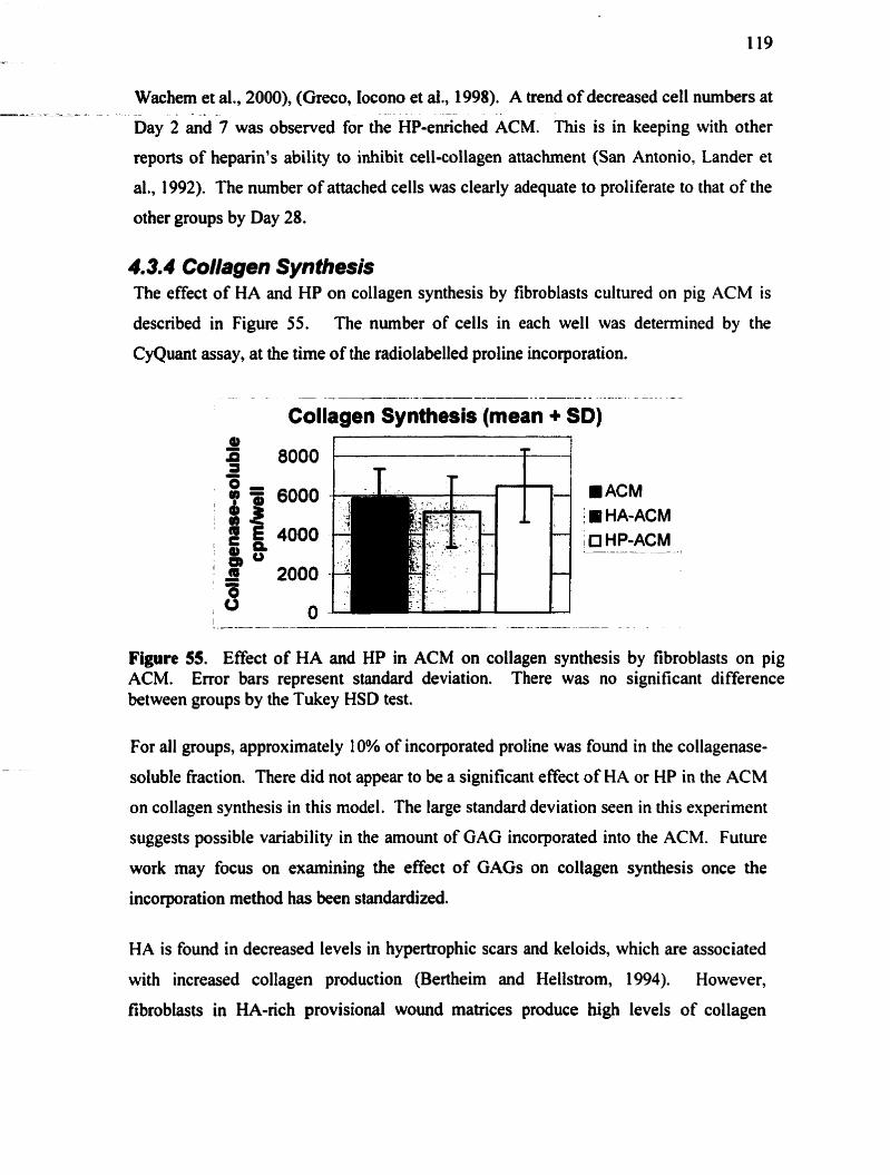
Wachem et al., 2000), (Greco, Iocono et ai., 1998). A trend of decreased cell numbers at
Day 2 i d 7 was observed for the ~ p - e i c h e d ACM. This is in keeping with other
reports of heparin's ability to inhibit cell-collagen attachment (San Antonio, Lander et
al., 1992). The number of attached cells was clearly adequate to proliferate to that of the
other groups by Day 28.
4.3.4 Collagen Syn thesis The effect of HA and HP on collagen synthesis by fibroblasts cultured on pig ACM is
described in Figure 55. The number of cells in each well was deterrnined by the
CyQuant assay, at the time of the radiolabelled proline incorporation.
. .- - -- -- - - - ----- - -
Collagen Synthesis (mean + Sb)
ACM I IP HA-ACM 1 0 HP-ACM
/
Figure 55. Effect of HA and HP in ACM on collagen synthesis by fibroblasts on pig ACM. Error bars represent standard deviation. There was no significant difference between groups by the Tukey HSD test.
For al1 groups, approximately 10% of incorporated proline was found in the collagenase-
soluble fraction. There did not appear to be a significant effect of HA or HP in the K M
on collagen synthesis in this model. The large standard deviation seen in this experiment
suggests possible variability in the amount of GAG incorporated into the ACM. Future
work may focus on examinhg the effect of GAGS on collagen synthesis once the
incorporation method bas k e n standardized.
HA is found in decreased levels in hypertrophie scars and keloids, which are associated
with increased collagen production (Bertheim and Hellstrom, 1994). However,
fibroblasts in HA-rich provisional wound matrices produce high levels of collagen

(Clark, 1996). HA'S influence on collagen synthesis is therefore not clear. A direct -
influence of HA on fibroblast synthesis of collagen remains to be shown. Little is
likewise known about HP's effects on collagen synthesis. The concentration of HP in
the matrix at the tirne collagen synthesis was measured (day 4), may not have been high
enough to demonstrate an effect. Future experiments may require a higher concentration
of HP at day 0.
4.3.5 Ceîî infiltration In view of the variability seen in tems of ce11 infiltration into pig ACM, care was taken
to ensure homogeneity in ACM characteristics and consistent seeding techniques
between groups. Groups (ACM, HA-ACM, HP-ACM) were only compared within each
experiment repeat, not between repeats. Freeze-dried ACM sarnples were rehydrated
and seeded with second passage hurnan dermal fibroblasts at a density of 30,000
cells/cm2. Sarnples of ACM 8 mm in diameter were fitted into the wells of a 96 well
plate for seeding. Figure 56 shows representative histological sections of each group
after four weeks in culture, at 20x magnification.

a) Pig ACM, vimentin, IOx (Repeat 1). b) HA-ACM, vimentin, 1Ox (Repeat I ).
c) HP-ACM, vimentin, 1 Ox (Repeat 1). d) Pig ACM, vimentin, IOx (Repeat 3).
Figure 56. Effect of HA or HP on fibroblast infiltration into pig ACM samples in Repeat 1 (a, b and c) and Repeat 3 (d).
Qualitatively, the presence of HA or HP did not appear to have any effect in tems of
preventing or facilitating fibroblast infiltration into the pig ACM. Mltration occurred
in the majority of Repeat 1 and 2 samples (416 for ACM and HA-ACM, 5/6 for HP-
ACM). Minimal infiltration was noted in repeat 3 samples (1/3 for ACM and HP-ACM.
0/3 for HA-ACM). The number of cells per section were counted to see if the presence
of HA or HP quantitatively intluenced the ability of the cells to infiltrate the pig ACM.
Samples fiom Repeats 1 and 2 only were used. Figure 57 shows the average number of
cells seen per section in each group, after 4 weeks in culture. The cells on the surface of
the ACM (seen as a monolayer in sections) were not included in the count. The presence
of HA or HP in the matrix did not significantly change the nurnber of infiiltrated
fibroblasts. In fact, the presence of HA appeared to have a slight inhibitory effect on
fibroblast infiltration into the pig ACM.

ACM *
HA-ACM HP-ACM
Figure 57. Effect of HA and HP on fibroblast infiiltration into pig ACM after 4 weeks. Results represent mean of Repeat I and 2 samples (1 8 sections per group). There was no significant difference between the three groups by the Kniskal-Wallis test.
The distance of each cell from the surface was also analysed to see if HA or HP had any
effect on ce11 migration. The results are show in Table 26.
1 Distance from 1 % of total cells per section 1 surface
ACM (n = 9 ) 1 HA-ACM (n = 9) 1 HP-ACM (n = 9)
Table 26. Effect of HA and HP on distance of fibroblast migration fiom the cell-seeded surface into the pig ACM (Repeat 1 samples). The number of sections analyzed in each group is indicated by n. There was no significant effect of HA or HP on the distance of the cells fiom the surface, by the chi-squared test.
The results for this section demonstrate how the effect of the ACM batch was much
greater than the effect of the matrix GAGS, on fibroblast infiltration. For these batches
of ACM, infiltration was independent of the presence of HA or HP. It was surprising
that HA in particular did not facilitate fibroblast infiltration into the matrix. HA is
known for its stimulatory effects on ce11 migration during wound healing and
morphogenesis. Its presence in a collagen sponge âramatically improved fibroblast
intiiltration in vi t ro (Doillon and Silver, 1986). in this study, embryonic chick tendon
fibroblasts were cultured on collagen sponges for 9 days. By day 9, only about 25% of

the collagen sponge was infïltrated with fibroblasts based on histological observation. In -
collagen sponges prepared with 5% (wlw) hyaluronic acid, fibroblasts infiltrated the
entire width of the sponge in 9 days. The effect of heparin could not be tested in this
model because the resulting collagcn-heparin mixture failed to form a sponge structure.
Another study (Reid and Newman, 1991) showed that the incorporation of HA and HP
into collagen gels also stimulated infiltration by leukocytes. However, this pro-
migratory effect may arise from the altered collagen sponge structure, rather than from
any direct effect of HA on the cells. In contrast, the structure of HA-enriched ACM
appears unchanged fiom that of ACM, by histology and SEM.
Neither HA nor HP had any significant effect on modulating fibroblast infiltration into
the pig ACM. The lack of effect by HP on fibroblast infiltration is not surprising given
the rapid diffusion of incorporated HP out of the ACM, which occurs long before the
fibroblasts begin itafiltrating the matrix. In the case of HA however, significant levels
were s h o w to remain, despite diffùsion, up to four weeks following incorporation. It
may be that HA'S lack of effect was particular to the molecular weight and concentration
chosen in this study. High molecular weight HA, such as that used in this study, has
k e n shown to stimulate ce11 migration in other models (Laurent and Fraser, 1992),
(Hayen, Goebeler et al., 1999). There is controveny however regarding the relationship
between the concentration of HA and its effects on ce11 migration (Laurent, Helistrorn et
al., 1988).
The most surprising finding in this section was the degree of infiltration which occurred
into the pig ACM siimpies in general. An interesting observation is that these
experiments were done later, using pig ACM sarnples which had been stored in PBS for
a relatively longer time, prior to fieeze-drying and cell-seeding. The ACM batch used
for Repeats 1 and 2 in particular, had been stored for several months pior to use.
Unfortunately these sarnples were not evaluated by SEM to determine the effect of
prolonged storage on the ACM structure. The histology results suggest that the ACM
structure is loose enough for fibroblasts to migrate fiom the surface into the matrix.
Clearly more work needs to be done on this model, to optimize the pig ACM structure
and allow for rapid ce11 infiltration in al1 sarnples.

4.3.6 Myoflbmblast Differentiation -su----:- - -- - -
Human fibioblasts were found to express SMA when-seeded on pig ACM, as s h o w in
Section 4.2.7. To determine the effect of HA and HP on fibroblast differentiation to
myofibroblasts, al1 SMA-positive cells per section, included those at the monolayer,
were counted. Table 27 shows the percentage of SMA-positive cells per section at the
different time points exarnined.
1 % SMA positive 1 3 1.2 12.5% 1 17.2 t 15.7% 1 44.1 +21.2% 1 celis I I
Substrate Total # of cells per section Total # of SMA positive cells per section
1 Significance 1 *HA 1 *Pig ACM, HP 1 *HA Table 27. Effect of HA and HP on fibroblast smooth muscle actin expression at 2
ACM (n = 12) 90.6 + 3 1.7
28.3 I 19.3
HA-ACM (n = 9) 95.1 t 23.5
16.4 f 23.2
weeks. Significance determined by one-way ANOVA and post-hoc Tukey HSD.
HP-ACM (n = 9) 1 1 O. 1 5 27.0
48.5 k 17.1
Heparin stimulates SMA expression by fibroblasts in vitro and in vivo, by selecting for
SMA-positive cells among replicating fibroblasts (Desmouliere, Rubbia-Brandt et al.,
1992). The results of the CyQuant assay clearly show continued proliferation of the
fibroblasts on the ACM. The mechanism of HA'S effects on myofibroblast
differentiation is less clear. Research on the mechanisms of fetal scarless wound healing
in sheep shows that the transition fiom scarless tissue repair (at 75 days gestation) to
healing with scar formation (at 120 days gestation) coincides with the expression of
SMA by myofibroblasts (Desmouliere and Gabbiani, 1996). Since fetal repair is also
characterized by persistently elevated levels of hyaluronan in cutaneous wounds, it could
be hypothesized that HA may play a regulatory role in fibroblast-mediated contraction,
and possibly myofibroblast differentiation. Another interesting observation is that the
timing of HA'S disappearance fiom adult wounds (around 3 to 5 days, through the
degradative action of hyaluronidase) corresponds somewhat to the timing of the
emergence of myofibroblasts in wounds (Weigel, Fuller et al., 1986). HA'S role in
regulating myofibroblast differentiation has otherwise not been directly studied.

An objective for future work in this area should include the development of methods to - z - - - -
assess SMA expression by confocal microscopy, rather than by cross-sectional
microscopy. With phalloidin counterstaining, the percentage of SMA-positive cells on
the surface of the ACM could be determined. This method would likely address the
large standard deviation caused by significant variation in the number of cells seen on
each section.
4.3.7 Summary Although a prototypic dennal replacement consisting of bovine collagen and a single
xenogenic GAG (chondroitin sulfate) was described more than 15 years ago, this
constnict has not achieved widespread use in the field of bum care. This may suggest
that collagen alone, or even in combination with a single matrix GAG, is probably
insuficient to replace missing demis. With an increasing recognition of the role of
GAGs in the modulation of cytokine signalling and other wound healing events, it is
reasonable to hypothesize that an aceilular dermal substitute with a GAG composition
that more closely resembles that of normal demis might fùnction more effectively.
Acellular dermal matrix may be processed to specifically conserve the GAGs of the
dermal matrix. An objective of this study was to determine the value of preserving
dermal GAGs in the acellularization process, in tems of their influence on fibroplasia.
The re-incorporation of specific GAGs (HA and HP) attempted to demonstrate the effect
of these molecules in the ACM.
Methods of GAG quantification in bovine and porcine tissues are limited by practical
dificutties in GAG extraction techniques and GAG visualization (Robinson-Seurig,
1999). For large tissue samples, alcian blue staining of electrophoretically separated
GAGs with standards, dlows for accurate quantification (Freund, Siebert et al., 1993).
In the current study, this method was unsuitable for quantification of small K M
samples with nanogram amounts of GAGs. A more thorough evaluation of the GAG
incorporation methods developed in this project should include a scaling-up of the
sarnple volume, to allow more accurate quantification of matrix GAGs.

It was expected that the incorporated GAGS in the ACM would quickly diffise out under - culture conditions, even without cells. As expected, the mass of HP in the ACM tended
to decrease over time. It was therefore not surprising that HP only had an effect on
fibroblast proliferation early on. This inhibitory effect on fibroblast infiltration was not
long lasting. HP-ACM was associated with SMA expression by 44% of seeded
fibroblasts, compared with only 3 1% of fibroblasts on the ACM at 2 weeks. The role of
cell-matrix interactions in regulating myofibroblast differentiation rnay therefore not
require prolonged contact between the matrix and the cells. However, HP had no effect
on fibroblast infiltration into pig ACM, nor on collagen synthesis.
Surprisingly, HA appeared to remain in the ACM, despite soaking, for up to 28 days.
Pig ACM therefore seems to be a promising mode1 for studying the effects of HA on
fibroblast proliferation, collagen synthesis and SMA expression. HA did not influence
fibroblast proliferation, infiltration or collagen synthesis. At the high concentration
tested in this study, HA would be expected to increase fibroblast infiltration into the
ACM. It may be that HA'S role in facilitating fibroblast migration in demis may not be
as significant as previously believed. HA did significantly decrease SMA expression by
fibroblast cultured on pig ACM for 2 weeks. Hyaluronan may be a desirable additive for
rnany reasons, including its inhibitory effect on myofibroblast differentiation seen in this
study. However, to more accurately evaluate their role as dermal substitute components,
a range of GAG molecular weights and concentrations shouid be tested in the ACM.
The results of this preliminary investigation show only that hyaluronan and heparin, at
the concentrations tested here, had minimal effects on fibroplasia in an acellular pig
dermal matrix.
Chapterfive:
CONCLUSIONS AND RECOMM ENDATIONS
5.1 Conclusions
5.1. f Acellular pig de& matrix as a dermal substitute Xenograft materials are an attractive alternative to human-derived materials, which are
limited by their high cost and short supply. The acellular pig dermal matrix has
considerable potential as a dermal substitute for use in burns and chronic wounds. Pig
ACM consists of a mature demal collagen network, which has been shown in allografts,
to decrease contraction and improve keratinocyte take as an allograft (Livesay, Hemdon
et al., 1995). Grossly, pig demis has many similarities to human demis, and was
hypothesized to interact with human cells in a beneficial way. Collagen as a xenografi is
relatively non-irnmunogenic; the complex cornformational structure of collagen
demands a certain amount of conservation of its primary sequence across species. The
acellularization process further reduces the immunogenicity of pig ACM, by removing
the cellular targets of xenograft rejection responses.
Pig ACM has been extensively tested as an allo- and xenograft dermal substitute in
animal models, with varying results. However, pig ACM has been tested in human
wounds without success. Possible reasons for its failure include inadequate
acellularization, clinically significant immunogenicity of pig collagen andor
unsuccessful infiltration by host cells. A major objective of this project was to
investigate the third possibility. By seeding pig ACM with human fibroblasts in an in
vitro environment, the scaffold properties of the material could be tested in isolation, in
the absence of any immunological issues.
Pig and human ACM have many similarities in their structure and composition, as well
as several differences. Both consist of an interwoven network of cross-linked collagen
bundles. However, pig ACM appears denser and more resistant to structural alteration
under culture conditions. In viiro, pig ACM supported human fibroblast proliferation --- L
and enabled fibroblast infiltration under certain conditions. In comparison to human
ACM, pig ACM was infiltrated more slowly and in fewer samples. In the presence of
pig ACM, fibroblasts synthesized less collagen than controls grown directly on the
culture dish. Above a thickness of 0.7 mm, contraction of the pig ACM did not occur.
A percentage of fibroblasts cultured on the ACM expressed the myofibroblast marker
srnooth muscle actin, in a time-dependent manner; by four weeks, only 19% of
fibroblasts expressed SMA on pig ACM. However, fewer myofibroblasts were found
among ceils seeded on human ACM at al1 time points. The results of this in vitro
investigation suggest that as a dennal substitute, pig ACM may have some beneficial
effects on fibroplasia but would likely produce results inferior to that of hurnan ACM.
Pig ACM was not found to be equivalent to human ACM as a scafTold for human
fibroblasts. Its structure should be modified to ensure adequate infiltration by host cells
as a dermal substitute.
5.1.2 Acellular pig dennal mattf" as a mode1 of cell-matrlx interactions and contraction
Interactions between cells and their environment play an important role in many
biological processes. Three-dimensional culture systems have been developed to mimic
natural interactions between cells and the extracellular matrix. These models recreate
the natural extracellular matrix environment more closely than the traditional monolayer
culture cm. Acellulat pig dermal matrix was tested as a convenient scaffold for the
three-dimensional study of hurnan fibroplasia. As most soluble matrix components are
removed in the acellularization process, specific matrix molecules can be re-incorporated
and studied in a dermal matrix. The original tissue architecture of acellular demis offers
a unique opportunity to study fibroblast-mediated demal contraction.
Pig ACM was compared to human ACM with respect to its effects on certain aspects of
fibroplasia. Pig ACM is easily obtained from the paravertebral areas of the animal and
can be processed to yield many experimental samples. With the complete removal of
pig fibroblasts from the matrix, seeded human fibroblasts can be visualized
histologically. Subjectively, pig ACM feels more rigid and is less extensible than human

ACM. Its ultrastructure is denser, with less elastin than human ACM. Human - -4
fibroblasts behave similarly on pig and human ACM in terms of ce11 morphology.
However, the differences in ACM structure do seem to have an influence on other
aspects of fibroplasia in vitro. Specificall y, fibroblasts pro1 i ferate more on pig ACM
than on human ACM. Fibroblasts on pig ACM express smooth muscle actin in greater
proportions than on human ACM. Fibroblasts fom a multi-layered monolayer on the
surface of pig ACM with some infiltration below the surface, whereas they form a
minimal monolayer on and quickly infiltrate into human ACM. Fibroblasts can contract
thin samples of pig ACM ( ~ 0 . 7 mm thin) in two to four weeks, whereas human ACM is
not contracted by fibroblasts in vitro.
Contraction, as a normal feature of wound healing, is an important element in cutaneous
repair. Pathologic or excessive wound contraction is a major factor leading to loss of
hc t ion as a result of hypertrophic scarring, scleroderma, Dupuytren's disease and other
systemic disorders. Pig ACM was found to be effective as an in vitro model of dermal
contraction, and offers several advantages to the collagen gel contraction model. Pig
ACM provides a three-dimensional substrate in which the interaction of demial
fibroblasts with a mature demal collagen matrix can be examined and, hopefully, the
effects of potential treatments quantified. Also, with optimization and standardization of
its processing and structure, pig ACM may be developed into a representative model of
human demis for studying cell-matrix interactions. Table 28 summarizes the unique
features of pig ACM as an in vitro model of fibroplasia, in cornparison with other
proposed models.

Contraction
Pig ACM
+++ - ---A-=
Collagen synthesis M~"!broblast diflerentiation Fibroblast infiltrat ion
Stage of wound healing
1 +++ Clot
remodeling and early
matrix de~osition
ttt
Nat tested
++
Early wound contraction and matrix remodeling
- Humaa - ACM
+ CoUagen - gel .
+ b -. .-- - .-
Fibroblast proli$eration
+
-
+
1 formation
Fibrin gel
+++
Late matrix remodeling
Table 28. Comparison of pig ACM with existing in vitro models of fibroplasia. Syrnbols refer to qualitative representation of the quantitative results presented in previous sections.
Not tested
+
*
mediated contraction and fibrous capsule
5.1.3 HyaIumnan and heperin as dermal substitute components Glycosaminoglycans are cornmon constituents of cell surfaces and extracellular
matrices. Biological functions of GAGs include the facilitation of ce11 migration,
+
++
++
proliferation and differentiation, the binding and modulation of enzymes, protease
inhibitors and cytokines as well as tissue organization. The presence of GAGs in
biomaterials offers an opportunity to exploit the many biocharacteristics of these
polysaccharides and possibly to rnodulate the healing responses elicited by the
biomaterial. As components of an acellular dermal matrix, it was hypothesized that
hyaluronan and heparin may influence fibroblast responses to the matrix. Other
desirable effects of hyaluronan and heparin on dermal substitute healing may include
stimulation of angiogenesis, modulation of inflammation and delivery of covalently-
linked substances (such as antibiotics, analgesics or growth factors) to the wound.
Hyaluronan and heparin were successfully incorporated into the acellular pig dermal
matrix. Each GAG was distributed throughout the matrix. In this study, a near-
physiological concentration of HP and a supra-physiological concentration of HA in the
matrix were achieved. This facilitated an evaluation of the incorporation methods and to
allow for leaching of the GAGs fiom the matrix over the course of the experiment. Most

of the incorporated HA remained in the matrix despite soaking for 28 days, while most &
of the HP diffked out of the ACM in 3 days. The presence of the GAGS did not
interfere with fibroblast proliferation on the ACM, and did not alter collagen synthesis.
Fibroblast ifiltration into the GAG-enriched ACM was equivalent to that of the
unmodified ACM. A greater proportion of fibroblasts expressed a-smooth muscle actin
on the pig ACM in the presence of HP, while HA was found to have an opposite effect.
The effects of heparin on fibroblast SMA expression and contraction should be further
elucidated. Hyaluronan does not appear to interfere with normal healing as a dermal
substitute cornponent, at the concentration and molecular weight evaluated in this study.
5.2 Recommendations A number of possible projects related to this study may be pursued. These relate to the
application of pig ACM as either a dermal substitute or an in vitro model material.
5.2. f Dennal substitute deve/opment
(1) Modification of xenograft dermal collagen matrix structure The results of this study suggest that the effectiveness of pig ACM as a demal substitute
scaffold may be limited by its structural characteristics. The major difference between
the structure of pig and human ACM lies in the relative tightness of the collagen bundle
network. The collagen matrix of pig ACM could be modified therefore, through
mechanical, enzymatic or physico-chernical methods. The in vitro methods developed in
this study of fibroplasia may be applied to a modified pig ACM substrate, in order to
determine the effect of the structural modification on its suitability as a scaffold for
human cells.
(2) Evaluation of acellular pig dermal maMx from different anatomic locations It is known that the structure of the dermis varies according to its anatomic location
(Elder, Elenitsas et al., 1997). In this study, only pig demis taken from the donor's back
was evaluated as an in vitro scaffold. In the funw, improved techniques may allow for
convenient harvesting of pig dermis fiom other anatomic locations. It may be
worthwhile therefore to evaluate samples of acellularized pig demis fiom different

anatomic locations as a scaffold for human cells. It is suggested that abdominal demis .- -
may be a desirable option.
(3) Hyaluronan for drug delivery in acellular allograft dermal substitutes
Hyaluronan was shown in this study to become incorporated into the pig ACM, and to
remain in the matrix at high levels despite predicted outward diffusion. The presence of
HA in pig ACM was not associated with fibroplasia characteristic of excessive scarring.
HA could therefore hold promise as a component in an acellular dermal matrix demal
substitute. The HA incorporation methods used in this study could be applied to the
incorporation of HA bound to other therapeutic substances, in order to develop a kind of
dmg delivery system for the wound. Furthemore, an evaluation of HA-enriched ACM
in vivo may also demonstrate the known water-absorbing and anti-inflammatory
properties of HA.
5.2.2 In vitn, modelling
(1) Correlation of findings with in vivo fibroplasia (including contraction)
An effective in viîro mode1 should generate results in keeping with the in vivo situation.
Some in vitro aspects of fibroplasia on pig ACM could be correlated with in vivo
experimentation of pig ACM grafted ont0 full thickness cutaneous wounds. There are
many challenges in finding a xenogenic animal model for studying the effects of pig
ACM on wound contraction. While pigs are a good model of human wound contraction,
the use of domestic pigs in this study would constitute an allogenic, and not xenogenic,
animal model. Loose-skinned animais such as rats or guinea pigs, while xenogenic, bear
less resemblance to humans with respect to wound contraction. These smaller animals
however have been used and validated in several studies. Although caution must be
used in extrapolating information about human wound healing fiom this model, future in
vivo testing would likely employ guinea pigs.
(2) Enect of glycosaminoglycans on contraction of ACM
It was shown using this model that HA and HP have effects on myofibroblast
differentiation. The next logical step is to correlate these findings with the effects of
matrix HA and HP on contraction of pig ACM in vitro. Pig ACM would offer a

consiâerabk aâvantage ovw ocher in vitro modtls of contraction in îhis a m : its maîrk stmoha is not grossly modificd by the incarparation of HA or HP. Any effcct on
aiatrix contraction would tberefore k specific to the GAGS' effects on cells. Additional work is needcd to adspt HA and HP i n c o ~ m mctbods to vcry thm ACM simples,
as weU a9 to improve the longenty of HP m the ma(rV<.
(3) Human ACM u r modd of cd-mitris intemcîiom In îhis shdy, effective metbods of GAG incorporation and quantifiication in an a c e b damil mstrix wen devtbpsd. These metùoâs d d be applied to the use of humen
ACM Eutead of pig ACM, as a t h m d m m i d substrate fa modehg ceU-maîrix
interactions in Bkoplasia in vüro. Human ACM may bt a sable model for studying
cknnai fikoblast prolif;cration, smootb muscle actm expression, cobgen synthesis aad fibroblast iaEihrati011. A stuày of fiôrobiast-rnodiated contraction with human ACM
woulà not k fcasiblt, shce tbis matcrisl is difficult to cut to the repuired âimensioas.
(4) Pig ACM as a mdcl of contncüon
Fi'brobtast-mediated contraction of a mature d d matrix has not been pwiously
demonstrated in vitro. It was sbown in chis shidy thaî îhis type of contraction is
dcpcndmt on fibroblasts, but the mechanian is unclear. The dect of ceU seedmg
dmsity on contraction would k of intaest, as well as the effcct of selective inhibitory
agents on ACM contraction. It would k uscfui to chatacfenze whether dK contraction of the matrix is reversMe witb ceU remod , or is âependent on particular serutn
5.3 Summary This thesis provides an evaluatioa of acelkilrir pig damal matrix as a biomatd. Acelluiar pig d d maüîx was mvestigated for use as a possiile xcnograft dermai subBtitube material as weli as an in vitro moâel of dormal ceIl-nrat+;x Uiteractim. The
extraceMar mMix compcmcnts, hyahironan snd hqmb, were cvaluatd for then &cts on fibroplasia ih vitrw using pig ACM as a model H o l d . The nsuhs of this
tbesisdemaastrstetbebeaentsaidInmbiti~tl~ofpieACMhtermsofihefoUoWing
parrimeters:

- -.-- -a-
(1) similarity in structure to human demis
(2) ability to be infiltrated by human cells
(3) effects on the behaviour of seeded fibroblasts
(4) ability to be processed and e ~ c h e d by glycosaminoglycans without gross
modification of its structural integrity . This project did not address any quality control issues, i.e. the variability in structure
between samples. The structural analysis of the dermal matrix was not quantitative.
Only one concentration and molecular weight of each glycosaminoglycan was tested.
Each of these rnethodological aspects could be improved in future research. The most
valuable contributions of this thesis are (1) the characterization and'application of an in
vitro mode1 of fibroplasia in a dermal matrix and (2) the cornparison of pig to human
dermai matrix as a scaffold for human fibroblasts.

Alberts, B., D. Bray, et al. (1994). Molecular Bioloay of the Cell. New York, Garland Publishing.
Anderson, J. M. (1996). Inflammation, wound healing and the foreign body response. Biomaterials Science. B. D. Ratner, A. S. Hoffman, F. J. Schoen and J. E. Lemons. San Diego, Academic Press: 1 65- 1 72.
Artz, C. P., M. S. Rittenbury, et al. (1972). "An appraisal of allografts and xenografts as biological dressings for wounds and bums." Amais of Sureew 175(6): 934-938.
Bailey, A. J. (2000). "The fate of collagen implants in tissue defects." Wound Rewir and Reeeneration S(1): 5- 12.
Basile, A. (1982). "A comparative study of glycerinized and lyophilized porcine skin in dressings for third-degree burns." Plastic and Reconstructive Sur~ew 69(6): 969-972.
Bell, E., B. Ivarsson, et al. (1978). "Production of a tissue-like structure by contraction of collagen lattices by human fibroblasts of different proliferative potential in vitro." Cell Biolop 76(3): 1274- 1278.
Berry, D. P., K. G. Harding, et al. (1998). "Human wound contraction: Collagen organization, fibroblasts and myofibroblasts." Plastic and Reconstnictive Suraew 102(1): 124-131.
Bertheim, U. and S. Hellstrom (1994). "The distribution of hyaluronan in hurnan skin and mature, hypertrophie and keloid scars." British Journal of Plastic Sur~erv 47: 483- 489.
Berthod, F. and F. A. Auger (1 997). In vitro applications of skin substitutes for dermatological purposes. ski* Substitute ~roduction bv Tissue Engineering: Clinical and Fundamental A~plications. M. Rouabhia, R O Landes Company.
Berthod, F. and M. Rouabhia (1997). Exhaustive review of clinical alternatives for damaged skin replacement. Skin Substitute Production by Tissue Engineering: - Clinical and Fundamental Amlications. M. Rouabhia, R G Landes Company.
Bertolami, C. N., S. Berg, et al. (1992). Fetal Wound Healinq. New York, Elsevier.
Bowser, B. H., F. T. Caldwell, et al. (1983). "Statistical method to predict morbidity and mortality: self-assessrnent techniques for burn units." Burns 9: 3 18-326.

Boyce, S. T., M. J. Goretsky, et al. (1995). "Comparative assessrnent of cultured skin -..---L & - -_A-:-_
su€ïXftif 1:s-id naf ive -sHn aufograft for üeafmenro f fùli-thickness b u s . " Annah of Sur~ery 222(6): 743-752.
Breach, N., D. Davies, et al. (1979). "Study of effects of porcine skin and bovine demis on the healing of split-skin gr& donor sites in humans." Plastic and Reconstructive Swgery 63(4): 546-549.
Brown, D., W. Garner, et al. (1990). "Skin grafiing: dermal components in inhibition of wound contraction." Southern Medical huma1 83(7): 789-795.
Burgess, L., G. Morin, et al. (1 990). "Wound healing: relationship of wound closing tension to scar width in rats." Archives of Otolarvngoioav, Head and Neck Surgerv 116: 798-803.
Chen, C. S., 1. V. Yannas, et al. (1 995). "Pore strain behaviour of collagen- glycosarninoglycan analogues of extracellular matrix." Biomaterials 16: 777-783.
Chen, W. Y. and G. Abatangelo (1 999). "Functions of hyaluronan in wound repair." Wound Repair and Regeneration 7(2): 79-89.
Chevallay, B. and D. Herbage (2000). "Collagen-based biomaterials as 3D scaffold for ceIl cultures: applications for tissue engineering and gene therapy." Medical & Bioloaical Engineering & Comnutina 2000 38: 2 1 1-2 18.
Clark, R. A. (1 996). Wound Repair: Overview and General Considerations. The molecular and cellular biolow of wound repair. R. A. F. Clark. New York, Plenum Press: 3-35.
Coleman, C., T. Tuan, et al. (1 998). "Contractility, transforming growth factor beta, and plasmin in fetal skin fibroblasts: role in scarless wound healing." Pediatric Research 43(3): 403-409.
Compton, C. C., C. E Butler, et ai. (1998)+ "Organized skin structure is regenerated in vivo fiom collagen-GAG matrices seeded with autologous keratinocytes." The Journal of Investigative Dermatologv 1 lO(6): 908-9 16.
Cormack, D. H. (1993). Essential Histolow. Philadelphia, J. B. Lippincott Company.
Coulomb, B., L. Dubertet, et al. (1984). "The collagen lattice: a mode1 for studying the physiology, biosynthetic fùnction and phannacology of the skin." British Joumal of Dermatology III(Supp1ement 27).
Courtman, D., C. Pereira, et al. (1994). "Development of a pericardial acellular matrix biomaterial: Biochemical and mechanical effects of ce11 extraction." Journal of Biomedical Materiais Research 28: 655-666.

Courfman, D., C. Pereira, et ai. (1995). "Biomechanical and ultrastructurat cornparison of cryopreservation and a novel cellular extraction of porcine aortic valve leaflets." Journal of Biomedical Materials Research 29: 1 507- 15 16.
Darby, 1. and G. Gabbiani (1990). "Alpha-smooth muscle actin is transiently expressed by myofibroblasts during experimental wound healing." Laboratory Investiaation 63: 2 1- 29.
Dawson, R. A., N. J. Goberdhan, et al. (1996). "Influence of extracellular matrix proteins on human keratinocyte attachent, proliferation and transfer to a dermal wound model." Burns 22(2): 93- 100.
Deitch, E. A., T. M. Wheelahan, et al. (1983). "Hypertrophie burn scars: analysis of variables." Journal of Trauma 23: 895-898.
Demling, R. H. and C. Lalonde (1990). "Early bum excision attenuates the post bum lung and systemic response to endotoxin." Sur~ew 108: 229-237.
Denizot, F. and R. Lang (1 986). "Rapid colorimetric assay for ceIl growth and survivai." Journal of Immunolonical Methods 89: 27 1-277.
Desmouliere, A. and G. Gabbiani (1996). The role of the myofibroblast in wound healing and fibrocontractive diseases. The Molecular and Cellular Biologv of Wound Re~air. R. Clark. New York, Plenum Press: 391-414.
Desmouliere, A., L. Rubbia-Brandt, et al. (1992). "Alpha-smooth muscle actin is expressed in a subpopulation of cultured and cloned fibroblasts and is modulated by gamma-interferon." Exwrimental Ce11 Research 201: 64-73.
Desmouliere, A., L. Rubbia-Brandt, et al. (1992). "Heparin induces alpha-smooth muscle actin expression in cultured fibroblasts and in granulation tissue myofibroblasts." Laboratorv Investiaation 67: 7 1 6-726.
Dimick, A. R., L. H. Potts, et al. (1986). "The cost of burn care and implications for the future on quality of care." Journal of Trauma 26: 260-265.
Doane, K. J. and D. E. Birk (1991). "Fibroblasts retain their tissue phenotype when grown in three-dimensional collagen gels." Experimental Cell Research 195: 432-442.
Docherty, R., J. V. Forrester, et al. (1989). "Glycosaminoglycans facilitate the movement of fibroblasts through three-dimensional collagen matrices." Journal of Ce11 Science 92: 263-270.
Doillon, C. J., S. Brandwein, et al. (1986). "Collagen-based wound dressings: control of Xhe pore sfructure and rnorpho~ogy.'~So~atof BiomedicafMaterids Resemh 28: 121% 1228.
Doillon, C. J. and F. H. Silver (1 986). 46Coliagen-based wound dressing: Effects of hyaluronic acid and fibronectic on wound healing." Biomaterials 7: 3-8.
Doillon, C. J., F. H. Silver, et al. (1987). "Fibroblast growth on a porous collagen sponge containing hyaluronic acid and fibronectin." Biomaterials B(May): 195-200.
Doillon, C. S.. A. J. Wasserman, et al. (1988). "Behaviour of fibroblasts and epidermal cells cutlivated on analogues of extracellular matrix." Biomaterials 9: 9 1-96.
Eckes, B., C. Mauch. et al. (1 993). "Down-regulation of collagen synthesis in fi broblasts within three-dimensional collagen lattices involves transcriptional and post- transcriptional mechanisms." FEBS Letters 318: 129- 1 33.
Edward, M. (1995). "Effects of retinoids on glycosarninoglycan synthesis by human skin fibroblasts grown as monolayers and within contracted collagen lattices." British Journal of Dermatoloey Ils: 223-230.
Ehrlich, H. P. and J. B. M. Rajaratnarn (1990). "Cell locomotion forces versus ce11 contraction forces for collagen lattice contraction: an in vitro mode1 of wound contraction." Tissue and Ce11 22(4): 407-4 17.
Elder, D., R. Elenitsas, et al., Eds. (1997). Lever's Histo~atholonv of the Skin. Philadelphia, Lippincott-Raven.
Fang, C. H., E. C. Robb, et al. (1990). "Observations on stability and contraction of composite skin grafts: xenodermis or allodermis with an isograft onlay." Journal of Burn Care and Rehabilitation 11 : 538-542.
Forrester, J. V. and J. M. Lackie (1981). "Effect of hyaluronic acid on neutrophil adhesion," Journal of CeU Science 50: 329-344.
Forrester, J. V. and P. C. Wilkinson (1 98 1). "Inhibition of leukocyte locomotion by hyaluronic acid." Journal of Ce11 Science 48: 3 1 5-33 1.
Freiberger, H., D. Grove, et al. (1980). "Procollagen 1 synthesis in human skin fibroblasts: effect of culture conditions on biosynthesis." Journal of Investieative Dermatology 75: 425-430.
Freund, R. M., J. W. Siebert, et al. (1 993). "Serial quantitation of hyaluronan and sulfated glycosarninoglycans in fetal sheep skin." Biochemistw and Molecular Biology International 29(4): 773-783.

Gao, 2.-R., 2.-Q. Hao, et al. (1992). "Porcine dermai collagen as a wound dressing for smiIonor sites anddeep partid slUn thickn~s~burns."Bums f 8(6): 492-496.
Ghosh, M. M., S. Boyce, et al. (1997). "A Cornparison of Methodologies for the Preparation of Human Epidennal-Demal Composites." Annals of Plastic Sur~ery 39: 3 90-404.
Greco, R. M., J. A. Iocono, et al. (1 998). "Hyaluronic acid stimulates human fibroblast proliferation within a collagen matrix." Journal of Cellular Physiolony 177: 465-473.
Griffith, M., R. Osborne, et ai. (1 999). "Functional human corneal equivalents constnicted fiom ceil lines." Science 286(lO December): 2 169-2 1 72.
Grimeil, F. (1994). "Fibroblasts, myofibroblasts and wound contraction." Jomai of Ce11 Bioloev 124(4): 40 1-404.
GinneIl, F. and C. R. Lamke (1 984). "Reorganization of hydrated collagen lattices by human skin fibroblasts." Journal of Ce11 Science 66: 5 1-63.
Guidry, C. and F. Ginneil (1985). "Studies on the mechanism of hydrated collagen gel reorganization by human skin fibroblasts." Journal of Cell Science 79: 67-8 1.
Guidry, C. and M. Hook (1 991 ). "Endothelins produces by endothelial cells promote collagen gel contraction by fibroblasts." The Journal of Ce11 Biolonv 1 lS(3): 873-880.
Harris, P. A., F. di Francesca, et al. (1999). "Use of hyaluronic acid and cultured autologous keratinocytes and fibroblasts in extensive bms." The Lancet 353(9146): 35- 36.
Hayen, W., M. Goebeler, et al. (1999). "Hyaluronan stimulates tumor ce11 migration by modulating the fibrin fiber architecture." Journal of Ce11 Science 112: 224 t -225 1 .
Herndon, D. N. (1997). "Perspectives in the use of allograft." kumal of Bum Care Rehabilitation 18: S6.
Heslop, B. F. and J. H. F. Shaw (1986). "Early vascularization of syngeneic, allogenic and xenogeneic skin grafts." Australian and New Zeaiand Journal of Suraerv 56: 357- 362.
Himichsen, N., L. Birk-Sorensen, et al. (1998). "Wound contraction in an experirnental porcine model." Scandinavian Journal of Plastic and Reconstructive Hand Sureerv 32: 243-248.
Huang-Lee, L. L. H., J. H. Wu, et ai. (1994). "Effects of hyaluronan on collagen fibrillar matrix contraction by fibroblasts." Journal of Biomedical Materials Research 28: 123- 132.

-- -. .-L% - -- J Ü E q u ë ~ L. C. U., G. S. Montes, et&-(r983). "Demat coltagen distribution." Histochemistry 79: 397-403.
Klein, J. and 2. Nagy (1981). "Physiology of alloreactivity: an interpretation." Transplantation Proceedings 13: 9 18-92 1.
Krejci, N. C., C. B. Cuono, et al. (1991). "In vitro reconstitution of skin: Fibroblasts facilitate keratinocyte growth and differentiation on acellular reticular demis." Journal of Investigational Dermatology 97: 843-848.
Langer, R. and S. P. Vacanti (1993). "Tissue engineering." Science 260: 920-926.
Lattari, V., L. M. Jones, et al. (1997). "The use of a permanent dermal allograft in full- thickness bums of the hand and foot: a report of three cases." Journal of Bum Care and Rehabilitation 18: 147-55.
Laurent, C., S. Hellstrom, et al. (1 988). "Hyaluronan improves the healing of experimental tyrnpanic membrane perforations." Arch of Otolarvn~oloev and Head and Neck surnery 114: 143 5- 144 1.
Laurent, T. C., J. Fraser, et al. (1986). The properties and turnover of hyaluronan. Functions of the Proteoalvcans. Chichester, Ciba Foundation Symposium. 124: 9-29.
Laurent, T. C. and J. R. E. Fraser (1992). "Hyaluronan." The FASEB Journal 6: 2397- 2403.
Lausman, R. G. (2000). Incorporation, localization and quantification of hyaluronan in porcine dermal acellular matrix. Engineering Science. Facult~ of Amlied Science and Engineering. Toronto, University of Toronto: 3 1.
Lawrence, W. T. (1996). Wound Healing Biology and Application to Wound Management. The Ph~sioloaic Basis of Sureerv. J. P. O'Leary . Baltimore, Wi 1 liams and Wilkins: 11 8-140,
Lin, W., S. Shuster, et al. (1997). "Patterns of hyaluronan staining are modified by fixation techniques." The Journal of Histochemis~ and Cvtochemistry 45(8): 1 157- 1163.
Linsenmayer, T. (1991). Collagen. Ce11 Biolonv of Extracellular Bioloav. E. Hay. New York, Plenum Press: 7-44.
Mac Neil, S. (1994). "What rote does the extracellular matrix serve in skin grafting and wound healing?" Burns 20(1): S67-570.

Martin, P. (1997). "Wound Healing- Aiming for Perfect Skin Regeneration." Science Z?6(lXpriT 4): 75-81.
Matouskova, E., D. Vogtova, et al. (1993). "A recombined skin composed of human keratinocytes cultured on cell-free pig demis." Burns 19(2): 1 18-1 23.
Mauch, C., B. Adelmann-Grill, et al. (1989). "Collagenase gene expression in fibroblasts is regulated by a three-dimensional contact with collagen." FEBS Letters 250(2): 30 1 - 305.
Mauch, C., A. Hatamochi, et al. (1988). "Regulation of collagen synthesis in fibroblasts within a three-dimensional collagen gel." Exwrimental Ce11 Research 178: 493-503.
Menard, C., S. Mitchell, et al. (2000). "Contractile behaviour of smooth muscle actin- containing osteoblasts in collagen-GAG matrices in vitro: implant-related ce11 contraction." Biomaterials 2 1 : 1 867- 1 877.
Meyer, W. and K. Neurand (1987). "A comparative scanning electron rnicroscopic view of the integument of domestic mamrnals." Scannine Microscorw l(1): 169- 1 80.
Meyer, W., R. Schwarz, et al. (1 978). ''The skin of domestic mammals as a model for the human skin, with special reference to the domestic pig." Current Problems in Dermatoloav 7: 39-52.
Moulin, V., G. Castilloux, et al. (1996). "ln vitro models to study wound healing fibroblast." Burns 22(5): 359-362.
Nakagawa, S., P. Pawelek, et al. (1989). "Long terni culture of fibroblasts in contracted collagen gels: effect on ce11 growth and biosynthetic activity." Journal of Investieative Dermatoloqy 93: 792-798.
Naughton, G. and J. Mansbridge (1 999). "Human-based tissue-engineered implants for plastic and reconstructive surgery." Clinics in Plastic Sureerv 26(4): 579-586.
Nedelec, B., A. Ghahary, et al. (2000). "Control of wound contraction." Hand Clinics 16(2): 289-302.
Nedelec. B., Y. J. Shen, et al. (1995). "The effect of interferon alpha 2b on the expression of cytoskeletal proteins in an in vitro model of wound contraction." Journal of Laboratow Clinical Medicine 126(5): 474-484.
Nishiyarna, T., M. Tsunenaga, et al. (1 989). "Growth rate of hurnan fibroblasts is repressed by the culture within reconstituted collagen matnx but not by the culture on the matrix." Matrix 9: 193- 199.
Oliver, R., H. Baker, et al. (1982). "Dermal collagen implants." Biomaterials 3: 38-40.

Piepër, S. S., P. van Wachem, et a1;-(2000E *Attachent of glycosaminoglycans to collagenous matrices modulates the tissue response in rats." Biomaterials 21 : 1 689- 1699.
Pilcher, B. K., B. D. Sudbeck, et al. (1998). "Collagenase-1 and collagen in epidermal repair." Archives of Dermatoloeical Research 290(Suppl): S37446.
Poggi, M. M., M. Klein, et al. (1 999). "Effects of cryopreservation and deconstruction on the Dermai Glycoaminoglycan Content of uman skin." Joumal of Burn Care and Rehabilitation 20(3): 20 1-206.
Postlethwaite, A. E., J. M. Seyer, et ai. (1978). "Chemotactic attraction of hurnan fibroblasts to type 1, II and III collagens and collagen-derived peptides." Proceedines of the National Academy of Sciences 75(2): 87 1-875.
Ralston, D. R., C. Layton, et al. (1997). "Keratinocytes contract human dermal extracellular rnatrix and reduce soluble fibronectin production by fibroblasts in a skin composite model." British Journal of Plastic Sureery 50: 408-4 1 5.
Reid, G. G. and 1. Newman (1 991). "Hurnan leucocute migration through collagen matrices containing other extracellular matrix components." Ce11 Bioloev Intemation Reports 15(8): 771 -720.
Rheinwald, J. G. and H. Green (1 975). "Serial cultivation of strains of hurnan epidermal keratinocytes: the formation of keratinizing colonies from the single cells." Cell6: 33 1- 44.
Robinson-Sewig, A. A. (1999). Development of dermal acellular matrix and acellular matrix-hyaluronan constructs for chronic skin wound applications. Chemical Engineering & Amlied Chemistry. Toronto, University of Toronto: 162.
Rose, E. H., L. M. Vistnes, et al. (1978). "A Microarchitectural Mode1 of Regional Variations in Hypodermal Mobility in Porcine and Human Skin." Original article.
Roy, F., C. DeBlois, et al. (1993). cbExtracellular matrix analogs as carrien for growth factors: In vitro fibroblast behaviour." Journal of Biomedical Materials Research 27: 389- 397.
Rudolph, R. (1979). "Inhibition of myofibroblasts by skin grafts." Plastic and Reconstmctive Surgery 63(4): 473-480.
Rudolph, R. and D. L. Ballantyne (1 990). Skin Grafts. Plastic Sureery. J. G. McCarthy. Philadelphia, W. B. Saundea Company. 1: 244.
Ruoslahti, E. (1991). integrins as receptors for extracellular matrix. Ce11 Bioloev of Extracellular Matrix. E. D. Hay. New York, Plenum Press: 343-364.

San Antonio, J. D., A. D. Lander, et at (t992):"Heparin- inhibits the attachment and growth of Balblc-3T3 fibroblasts on collagen substrata." Journal of Cellular Phvsioloev 150: 8-1 6.
Sasiselcharan, R., M. A. Moses, et al. (1994). "Hepainase inhibits neovascularization." Proceedines of the National Academ~ of Sciences U.S.A. 91(Cell Biology): 1524- 1528.
Souren, J. E., M. Ponec, et al. (1989). "Contraction of collagen by human firbroblasts and keratinocytes." In Vitro Cellular and Develo~mental Biology 25( 1 1): 1039- 1 1 03.
Sivastava, A., L. Jennings, et al. (1999). "Xenogenic acellular derrnal matrix as a derrnal substitute in rats." Journal of Burn Care and Rehabilitation 20: 382-390,
Steinberg, B. M., K. Smith, et al. (1980). "Establishment and transformation diminish the ability of fibroblasts to contract a native collagen gel." The Journal of Ce11 Biolow 87: 304-308.
Stratta, R. H., G. D. Warden, et al. (1986). "Immunologic parameters in burned patients: effect of therapeutic interventions." Journal of Trauma 26: 7- 17.
Tammi, R., A.-L. Tuhkanen-Martikainen, et al. (1996). Whole skin organ as a tool to study hyaluronan and proteoglycan rnetabolism. Dermatoloeic Research Techniaues. H. 1. ~ L b a c h . Boca ~a ton . CRC Press: 9 1 - 1 1 1.
Tengblad, A. (1 980). "Quantitative analysis of hyaluronate in nanogram amounts." Biochemistw Journal 185: 10 1 - 105.
Tomasek, J. J., E. D. Hay, et al. (1982). "Collagen modulates ce11 shape and cytoskeleton of embryonic comeal and fibroma fibroblasts: distribution of actin. alpha-actinin and myosin." Develo~mental Biology 92: 107- 122.
Toranto, I., K. Salyer, et al. (1 974). "Vascularization of porcine skin heterografls." Plastic and Reconstructive Surnery 54(2): 1% -200.
Tuan, T.-L., A. Song, et al. (1 996). "In vitro fibroplasia: Matrix contraction, ce11 growth and collagen production of fibroblasts cultured in fibrin gels." Exwrimental Cell Research 223: 127- t 34.
Turley, E. A., P. Bowman, et al. (1985). "Effects of hyaluronate and hyaluronate binding proteins on ce11 rnotile and contact behaviour." Journal of Ce11 Science 78: 133- 145.
Ueno, N., B. Chakrabarti, et al. (1 992). "Hyaluronic acid of human skin and post-burn scar: heterogeneity in primary structure and molecular weight." Biochemistrv International 26(5): 787-796.

Ueno, N., B. Chakrabarti, et al. (1 992). "Hylaluronic acid of human skin and post-burn scar : Heterogeneity in primary structure-an& motecu tarweigk'' Biochemistrv International 26(5): 787-796.
Voet, D. and J. G. Voet (1990). Biochemistw. New York, Wiley.
Wainwright, D. (1995). "Use of an acellular allograft derrnal matrix (Allodem) in the managrnent of full-thickness bums." Burns 21: 243-248.
Wang, H.-S., T.-M. Chen, et al. (1997). "Use of a porcine demis template to enhance widely expanded mesh autologous split-thiclcness skin gr& growth: preliminary report." Journal of Trauma: Infection. I n i w and Critical Care 42(2): 177- 1 83.
Weadock, K., R. M. Olsen, et al. (1984). "Evaluation of collagen cross-linking techniques." Biomaterials. Medical Devices and Artificial Orpans 1 1 : 294-3 18.
Wight, T. N., D. K. Heinegard, et al. (1991). Proteoglycans: Structure and Function. Ce11 Bioloav of Extracellular Matrix. E. D. Hay. New York, Plenum Press: 45-78.
Wilkins, E. O., K. C. Chung, et al. (1996). Skin and Subcutaneous Tissue Review in General Surgery. The Phvsioloaic Basis of Sureerv. J. P. O'Leary. Baltimore, Williams & Wilkins: 56 1-580.
Wilson, O., D. Courtman, et al. (1995). '4Acellular matrix: a biomaterials approach for coronary artery bypass and heart valve replacement." Annals of Thoracic Surgew 60: S353-358.
Wilson, G., H. Yeger, et al. (1990). "Acellular matrix allograft small caliber vascular prostheses." Transactions of the American Society of Artificial Intemal Oreans 36: M340-M343.
Wood, M. and H. Hale (1972). "The use of pigskin in the treatment of thermal burns." American Journal of Surgery 124: 720-723.
Yannas, 1. V. (1 996). Natural Materiah. Biomaterials Science. B. D. Ratner, A. S. H o h a n , F. J. Schoen and J. E. Lemons. San Diego, Academic Press: 84-93.
Yannas, 1. V. (1997). In Vivo Synthesis of Tissues and Organs. Princi~les of Tissue Enpineerinq. - R. P. Lanza, R. Langer and W. L. Chick. Austin, R. G. Landes Company: 169- 177.

-- _ = _ A L _ Appe-n~ixxClJLTURE -OF HUMAN DERMAL MICROVASCULAR ENDOTHELIAL CELLS ( M E ~ O % - P ~ A C M -
Introduction
Angiogenesis, the formation of new blood vessels, is an Unportant component of various
physiologicd and pathological processes, including wound healing. Little is known about the
specific conditions under which wound healing angiogenesis rnay be stimulated. increased
knowledge of these mechanisms would certainly prove wfbl in the treatment of chronic
wounds, the inflammation of rheurnatoid arthritis, capillary hemangiomas as well as
malignant transformation, tumour growth and metastasis (as in melanoma). Research has
been limited by the lack of reliable. reproducible assays of the angiogenic response. In view
of both the ethical and cost issues inherent in in vivo angiogenesis models, several groups
have established microvascular endothelial cell culture techriques. The important role
played by microvascular endothelial cells (MECs) in a wide range of normal and
pathological processes has stimulated the development of in vi;ro models for capillary
formation.
MECs tend to grow as monolayers on two-dimensional substrata, but have been show to
form capillaries in artificial type 1 collagen and fibrin matrices under certain conditions
(Folkman and Haudenschild, 1980). The lack of correlation between two-dimensional
and three-dimensional cultures establishes the need to understand why cells behave
differently in a more natural three-dimensional matrix configuration. The challenge in
designing an in vitro model of dermal capillary formation lies in recreating the structure and
composition of the connective tissue matrix which surrounds btood vessets in the body. We
propose to develop a three-dimensional model of capillary formation based on an
acellularized dermal matrix. This model would allow us to study the effects of the
interactions between MECs and the mature collagen and elastin network of the demis.
Our acellularization protocol removes most of the soluble matrix molecules of the
demis. We have developed methods to reincorporate these molecules individual 1 y. in
order to study their effects on capillary formation in this model.
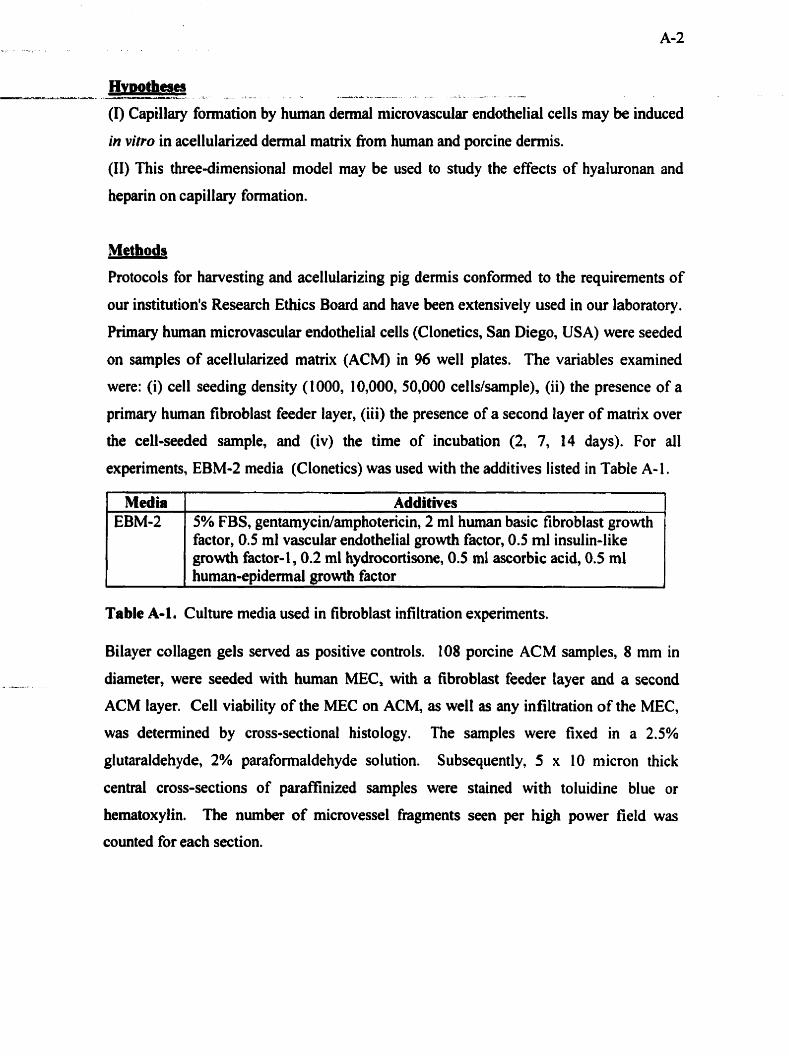
Hvwtheses - .-__ A---- L -- - - - - - - - (1) Capillary formation by human dermal n~icrovascular endothelial cells may be induced
in vitro in acellularized demal matrix fiom human and porcine dermis.
(II) This three-dimensional mode1 may be used to study the effects of hyaluronan and
heparin on capillary formation.
Methods
Protocols for harvesting and acellularizing pig demis confomed to the requirements of
our institution's Research Ethics Board and have been extensiveiy used in our laboratory.
Primary human microvascular endothelial cells (Clonetics, San Diego, USA) were seeded
on samples of acellularized matrix (ACM) in 96 well plates. The variables examined
were: (i) ceIl seeding density (1000, 10,000, 50,000 cells/sample), (ii) the presence of a
primary human fibroblast feeder layer, (iii) the presence of a second layer of matrix over
the cell-seeded sample, and (iv) the time of incubation (2, 7, 14 days). For al1
experiments, EBM-2 media (Clonetics) was used with the additives listed in Table A-1 .
factor, 0.5 ml vascular endothelial growth factor, 0.5 ml insulin-like growth factor-1 ,0.2 ml hydrocortisone, 0.5 ml ascorbic acid, 0.5 ml human-epiderrnal growth factor
Media EBM-2
Table A-1. Culture media used in fibroblast infiltration experirnents.
- - ---
Additives 5% FBS, gentamycin/amphotericin, 2 ml human basic fibroblast growth
Bilayer collagen gels served as positive controls. 108 porcine ACM samples, 8 mm in
--. diameter, were seeded with hurnan MEC, with a fibroblast feeder layer and a second
ACM layer. Ce11 viability of the MEC on ACM, es well as any infiltration of the MEC,
was detemined by cross-sectional histology. The samples were fixed in a 2.5%
glutaraldehyde, 2% paraformaldehyde solution. Subsequently, 5 x 10 micron thick
central cross-sections of paraffinized sarnples were stained with toluidine blue or
hematoxylin. The number of microvessel fragments seen per high power field was
counted for each section.

In the second set of experiments, samples of pig ACM enriched with hyaluronan or -.. ..- - -L-a-A2-----rL- 2 *--.-a .: - - . - - 4 - - S . -A - - - - L . A - F
heparin were seeded with MEC (50,000 cells/sarnple, for 14 days, in a bilayer model).
Control ACM samples contained no hyaluronan or heparin. Histology was performed on
triplicate samples in each group in three separate experiments.
Resulîs
Cross-sectional histology of MEC-seeded collagen gels and ACM mple s showed the
presence of adherent MEC at different time points (Figure A- 1).
Figure A-1. Adherent MEC on (a) collagen gel and (b) pig ACM at 2 days, hematoxylin, 20x. MEC are stained dark blue, and are indicated by m w s .
Under every condition examina including the coliagen gel ssmples intended as positive
controls, no capillary formation below the surface was obsewed. However, the MEC
srirvived at the surface, but were rarely seen on cross-sectional histology below the matrix
surface (Figure A-2).

Figure A-2. Lack of MEC infiltration below the surface of pig ACM at (a) 7 day days, hematoxylin, 20x. MEC are stained dark blue, and are indicated by arrows.
,s and (b) 14

Figure A-& Surface MEC (mm) on pig ACM at 14 days, toluidine blue, 40x.
Some infiltration of MEC was seen in the collagen gel sarnples, but no capillary form
was observed.
ation

' . . . O - ' . - . . . . L -:*--:, . . - i l , I 7 1 -.
Figure A 4 MEC on surface of collagen gel at 14 days, hematoxylin, 20x.
Although not al1 the histological results are presented here, the variables examined (cell
seeding density, unilayer or bilayer, fibroblast cotulture, duration of culture) had no effect
on celi infiltration or capillary formation. Moreover, neither hyaluronan- nor heparin-
enriched pig ACM samples showed any MEC infiltration.
Discussion
The ability to model angiogenesis in tissues in vitro would provide an alternative to animal
experimentation. Also, the use of simplified experirnental conditions in vitro may allow for
more control over experimental variables. in this study, acellular pig dermal matrix was
hvestigated as a possible scaffold for human demal micmvascular endothelid cells. These
ceils were selected based on their tissue of origin, given that demal endothelial cells are
responsible for capillary formation in dermal wounds. Unfortunately, no previous studies
have demonstrated in vitro capillary formation with these cells. In fùture work, the more
commonly used human umbilical vein endothelial cells will be studied in this model.
Capillary formation is a cornplex, multifactorial process which is tightly regulated in vivo. -%.--A-*- - L ---- ----L - = - A - - ' - - - - L - - : - -
For the phenomenon of angiogenesis to occur in an ex vivo enviment , certain cues must be
providecl to the cells artificially. The content of the culture media is therefore supplemented
with pro-angiogenic factors. As well, the cells require a three-dimensional fiarnework for
actual capillaries to take shape. The extracellular matrix must also provide an appropnate
stimulus to the endothelial cell. The use of Matrigel is an example of how a matrix can be
specifically designed to stimulate angiogenesis. Matrigel is denved fiom the basement
membrane matrix of Englebreth-Holm-Swami tumors taken fkom lathrytic mice. The major
components of this matenai are laminin, collagen IV, entactinhidogen, he paran su1 fate
proteogl ycan and growth factors (Cockerill, Garnble et al ., 1 995). This model demonstrates
the importance of the basement membrane matrix as a biological mediator of angiogenesis.
Within 18 hours of culture on Matrigel human urnbilical endothelial cells fom a network of
capillary-like structures with well-defined lumens (Cockenll, Gamble et al., 1 995).
As with Matrigel, capillary-like networks in collagen gels occur mostly on the surface of the
material (Montesano, 1992). As the collagen gel is semi-transparent, the cells can be
visualized in the culture dish under rnicroscopy. The difficulty with using a more dense
tissue, such as pig ACM, arises during its removal fiom the culture dish and histological
analysis. This process tends to disrupt delicate cellular arrangements at the surface; only cells
which have infiltrated below the matrix surface can be visualized in this model.
Unfortunately, the endothelial cells did not infiltrate into the pig ACM. in future work, a
tissue which supports more ce11 infiltration would be more advisable. An obvious choice for
a materid for diis puipose is acellufm human demat matnx, &ch appeared to be a better in
v i ~ o scafTold for human fibroblast intiltration than pig ACM. This material may therefore be
more suitable for histological analysis of capillary formation within the matrix. Cells present
below the surface to be identified histologically. Another long-terni objective of this study is
to develop a better xenograft-derived scafl'old material. Structurai modifications to pig ACM
may improve its effectiveness at supporthg MEC ingrowth, which couid then be detedned
histologically .

Endothelial ce11 migration into acellular tissue in vitro is not well understood. This process is --- c L - -% -- 1 > A - -- - - - - -
stirnulated in vivo by specific cytokines and local matrix changes, and is thecefore highly
dependent on cell-to-cell communication. in this study, MEC-seeded ACM samples in one
group were CO-cultureci with human fibroblasts at the bottom of the well. This CO-culture
arrangement proved insufficient to stimulate MEC infiltration into the ACM. Perhaps the
MEC might be seeded on the sarne side of the ACM as the fibroblasts, in order to better
reproduce multicellular in vivo conditions. In this study design, it would be necessary to use
cell-specific stains to distinguish cell types (i.e. von Willebrand factor
immunohistochemistry).
Hyaluronan- and heparin-e~ched ACM samples were also investigated for MEC
infiltration. Unfortunately, one cannot conclude any effect of HA or HP on MEC behaviour,
given the lack of infiltration in the control group. To evaluate the effect of HA and HP on
capillary formation within a demal matnx, different experimental conditions would be
required:
(1) Human urnbilical vein endothelial cells (HUVECs) might be used on human ACM in
order to determine whether endothelial cells can infiltrate a demal matrix scafTold in
vitro
(2) HA and HP incorporation techniques developed for pig ACM may be applied to
human ACM. The HA- or HPsnriched human ACM may then be seeded with
MECS.
(3) Altematively, pig ACM enriched with HA or HP may be tested as a potential mode1
ushg W C s .
(4) Finally, in an effort to develop a xenograft material as a potentiai demai substitute,
the structure of pig ACM rnight be specifically modified to allow for easier cellular
intiltration. The modified xenograft material might then be enriched with HA or HP
and seeded with MECs.
It would be expected that as HA is degraded to HA oligosaccharides, a possible pro-
angiogenic effect might be seen. HP's role in delivering bFGF (present in the media) may
a h result in a pro-angiogenic effect, aithough HP wouid be without i t . usual proteoglycan
configuration. HP alone has been show to stimulate endotheiial cell proliferation in vitro

(Thomton, Muelier et al., 1983). However this effèct on angiogenesis has not been studied _- --=---A _ __ _ _-_ - _ X i - 3 ----a . - - - - - -
with HP as a maîrix component.
Conclusions
We proposed an in vitro model of hurnan capillary formation in a mature porcine dermal
maûix. The goal here was to develop a model which more closely reproduced the cell-
matrix interactions of angiogenesis in the demis. This preliminory study employed a
new dermal materiai, a new MEC line, and new in vitro conditions. Since the cells did
not form capillary structures, even on the established collagen gel model, the ceil type is
clearly not suitable for studying angiogenesis in vitro. As such, pig ACM may not be a
suitable material for studying capillary formation, given that fibroblasts take several
weeks to infiltrate below the surface. Without infiltration, the dense ACM material,
which requires histological processing to visualize capillary formation, becomes
impractical as a model. There were clearly too many variables for this study to be
conclusive within its existing methodology. Due to time restrictions, the other
approaches proposed here were not pursued in this thesis. Further methodological
research is encouraged to develop an in vitro model of demal angiogenesis.
References
Cockerill, G. W., J. R. Gamble, et al. (1995). Angiogenesis: models and modulators.
International review of cytology 159: 1 1 3 - 1 60.
Folkman, J. and C. Haudenschild (1980). Angiogenesis in vitro. Nature 288(December
- - 1 1, 1980): 55 1-556,
Montesano, R. (1 992). Regulation of angiogenesis in vitro. European Journal of Clinical
Investigution 22: 504-5 15.
Thomton, S. C., S. N. Mueller, et al. (1983). Human endothelial cells: use of heparin in
cloning and long term serial cultivation. Science 222(November 1 1 , 1 983): 623-625.
Copyright © 2022 FDOKUMEN