Evaluation of in vitro vs. in vivo methods for assessment of dermal absorption of organic flame...
10
Review Evaluation of in vitro vs. in vivo methods for assessment of dermal absorption of organic flame retardants: A review Mohamed Abou-Elwafa Abdallah a,b, ⁎, Gopal Pawar a , Stuart Harrad a a Division of Environmental Health and Risk Management, College of Life and Environmental Sciences, University of Birmingham, Birmingham B15 2TT, United Kingdom b Department of Analytical Chemistry, Faculty of Pharmacy, Assiut University, 71526 Assiut, Egypt abstract article info Article history: Received 30 May 2014 Accepted 23 September 2014 Available online xxxx Keywords: Flame retardants Dermal absorption Human exposure Human skin equivalents Bioavailability There is a growing interest to study human dermal exposure to a large number of chemicals, whether in the in- door or outdoor environment. Such studies are essential to predict the systemic exposure to xenobiotic chemicals for risk assessment purposes and to comply with various regulatory guidelines. However, very little is currently known about human dermal exposure to persistent organic pollutants. While recent pharmacokinetic studies have highlighted the importance of dermal contact as a pathway of human exposure to brominated flame retar- dants, risk assessment studies had to apply assumed values for percutaneous penetration of various flame retar- dants (FRs) due to complete absence of specific experimental data on their human dermal bioavailability. Therefore, this article discusses the current state-of-knowledge on the significance of dermal contact as a path- way of human exposure to FRs. The available literature on in vivo and in vitro methods for assessment of dermal absorption of FRs in human and laboratory animals is critically reviewed. Finally, a novel approach for studying human dermal absorption of FRs using in vitro three-dimensional (3D) human skin equivalent models is present- ed and the challenges facing future dermal absorption studies on FRs are highlighted. © 2014 Elsevier Ltd. All rights reserved. Contents 1. Introduction . . . . . . . . . . . . . . . . . . . . . . . . . . . . . . . . . . . . . . . . . . . . . . . . . . . . . . . . . . . . . . . 14 2. Human exposure to FRs . . . . . . . . . . . . . . . . . . . . . . . . . . . . . . . . . . . . . . . . . . . . . . . . . . . . . . . . . . 14 3. Skin as a barrier for systemic exposure to xenobiotic chemicals . . . . . . . . . . . . . . . . . . . . . . . . . . . . . . . . . . . . . . . . 15 4. Significance of dermal absorption as a pathway of human exposure to FRs . . . . . . . . . . . . . . . . . . . . . . . . . . . . . . . . . . . 16 5. Transdermal metabolism of xenobiotics . . . . . . . . . . . . . . . . . . . . . . . . . . . . . . . . . . . . . . . . . . . . . . . . . . . 16 6. In vivo dermal bioavailability studies . . . . . . . . . . . . . . . . . . . . . . . . . . . . . . . . . . . . . . . . . . . . . . . . . . . . 16 7. Paradigm shift — in vivo to in vitro dermal bioavailability studies . . . . . . . . . . . . . . . . . . . . . . . . . . . . . . . . . . . . . . . . 18 8. Human skin equivalent models (HSE) . . . . . . . . . . . . . . . . . . . . . . . . . . . . . . . . . . . . . . . . . . . . . . . . . . . . 18 8.1. Rationale . . . . . . . . . . . . . . . . . . . . . . . . . . . . . . . . . . . . . . . . . . . . . . . . . . . . . . . . . . . . . 18 8.2. Composition . . . . . . . . . . . . . . . . . . . . . . . . . . . . . . . . . . . . . . . . . . . . . . . . . . . . . . . . . . . 18 8.3. General protocol for in vitro percutaneous absorption studies . . . . . . . . . . . . . . . . . . . . . . . . . . . . . . . . . . . . . 19 9. Future perspectives and challenges facing dermal absorption studies of FRs . . . . . . . . . . . . . . . . . . . . . . . . . . . . . . . . . . . 19 Acknowledgement . . . . . . . . . . . . . . . . . . . . . . . . . . . . . . . . . . . . . . . . . . . . . . . . . . . . . . . . . . . . . . . 20 Appendix A. Supplementary data . . . . . . . . . . . . . . . . . . . . . . . . . . . . . . . . . . . . . . . . . . . . . . . . . . . . . . . 20 References . . . . . . . . . . . . . . . . . . . . . . . . . . . . . . . . . . . . . . . . . . . . . . . . . . . . . . . . . . . . . . . . . . 20 Environment International 74 (2015) 13–22 Abbreviations: BFRs, brominated flame retardants; BPA, bisphenol A; BTBPE, 1,2-bis(2,4,6-tribromophenoxy)ethane; DBDPE, decabromodiphenylethane; EU, European Union; EVCAM, European Centre for Validation of Alternative Methods; FRs, flame retardants; FT, full-thickness skin; HBCD, hexabromocyclododecane; HSE, human skin equivalent; KC, keratinocytes; K OW , octanol/water partition coefficient; LCs, Langerhans cells; NBFRs, novel brominated flame retardants; OATP, organic anion transporting polypeptides; OECD, organisation for economic co- operation and development; PA, percutaneous absorption; PBDEs, polybrominated diphenyl ethers; PBT, persistent, bioaccumulative and toxic; PCBs, polychlorinated biphenyls; PFRs, organ- ophosphate flame retardants; PK, pharmacokinetic; POPs, persistent organic pollutants; RDP, resorcinol bis-diphenylphosphate; SC, stratum corneum; RHE, reconstructed human epidermis; TBB, 2-ethylhexyl-2,3,4,5-tetrabromobenzoate; TBBPA, tetrabromobisphenol A; TBPH, bis(2-ethylhexyl)tetrabromophthalate; TCEP, tris(2-chloroethyl) phosphate; TCIPP, tris(2-chloro-1- methylethyl) phosphate; TDCPP, tris(1,3-dichloro-2-propyl) phosphate; TRIS, tris(dibromopropyl) phosphate; USEPA, United States Environment Protection Agency. ⁎ Corresponding author at: Division of Environmental Health and Risk Management, College of Life and Environmental Sciences, University of Birmingham, Birmingham B15 2TT, United Kingdom. Tel.: +44 121 414 7297; fax: +44 121 414 3078. E-mail address: [email protected] (M.A.-E. Abdallah). http://dx.doi.org/10.1016/j.envint.2014.09.012 0160-4120/© 2014 Elsevier Ltd. All rights reserved. Contents lists available at ScienceDirect Environment International journal homepage: www.elsevier.com/locate/envint
-
Upload
birmingham -
Category
Documents
-
view
0 -
download
0
Transcript of Evaluation of in vitro vs. in vivo methods for assessment of dermal absorption of organic flame...
Environment International 74 (2015) 13–22
Contents lists available at ScienceDirect
Environment International
j ourna l homepage: www.e lsev ie r .com/ locate /env int
Review
Evaluation of in vitro vs. in vivo methods for assessment of dermalabsorption of organic flame retardants: A review
Mohamed Abou-Elwafa Abdallah a,b,⁎, Gopal Pawar a, Stuart Harrad a
a Division of Environmental Health and Risk Management, College of Life and Environmental Sciences, University of Birmingham, Birmingham B15 2TT, United Kingdomb Department of Analytical Chemistry, Faculty of Pharmacy, Assiut University, 71526 Assiut, Egypt
Abbreviations:BFRs, brominatedflame retardants; BPAEuropeanCentre forValidationofAlternativeMethods; FRsoctanol/water partition coefficient; LCs, Langerhans cells; Noperationanddevelopment;PA,percutaneousabsorption;ophosphateflameretardants;PK,pharmacokinetic;POPs,pTBB, 2-ethylhexyl-2,3,4,5-tetrabromobenzoate; TBBPA, tetmethylethyl) phosphate; TDCPP, tris(1,3-dichloro-2-propy⁎ Corresponding author at: Division of Environmental
United Kingdom. Tel.: +44 121 414 7297; fax: +44 121E-mail address: [email protected] (M.A.-E. A
http://dx.doi.org/10.1016/j.envint.2014.09.0120160-4120/© 2014 Elsevier Ltd. All rights reserved.
a b s t r a c t
a r t i c l e i n f oArticle history:Received 30 May 2014Accepted 23 September 2014Available online xxxx
Keywords:Flame retardantsDermal absorptionHuman exposureHuman skin equivalentsBioavailability
There is a growing interest to study human dermal exposure to a large number of chemicals, whether in the in-door or outdoor environment. Such studies are essential to predict the systemic exposure to xenobiotic chemicalsfor risk assessment purposes and to comply with various regulatory guidelines. However, very little is currentlyknown about human dermal exposure to persistent organic pollutants. While recent pharmacokinetic studieshave highlighted the importance of dermal contact as a pathway of human exposure to brominated flame retar-dants, risk assessment studies had to apply assumed values for percutaneous penetration of various flame retar-dants (FRs) due to complete absence of specific experimental data on their human dermal bioavailability.Therefore, this article discusses the current state-of-knowledge on the significance of dermal contact as a path-way of human exposure to FRs. The available literature on in vivo and in vitromethods for assessment of dermalabsorption of FRs in human and laboratory animals is critically reviewed. Finally, a novel approach for studyinghuman dermal absorption of FRs using in vitro three-dimensional (3D) human skin equivalentmodels is present-ed and the challenges facing future dermal absorption studies on FRs are highlighted.
© 2014 Elsevier Ltd. All rights reserved.
Contents
1. Introduction . . . . . . . . . . . . . . . . . . . . . . . . . . . . . . . . . . . . . . . . . . . . . . . . . . . . . . . . . . . . . . . 142. Human exposure to FRs . . . . . . . . . . . . . . . . . . . . . . . . . . . . . . . . . . . . . . . . . . . . . . . . . . . . . . . . . . 143. Skin as a barrier for systemic exposure to xenobiotic chemicals . . . . . . . . . . . . . . . . . . . . . . . . . . . . . . . . . . . . . . . . 154. Significance of dermal absorption as a pathway of human exposure to FRs . . . . . . . . . . . . . . . . . . . . . . . . . . . . . . . . . . . 165. Transdermal metabolism of xenobiotics . . . . . . . . . . . . . . . . . . . . . . . . . . . . . . . . . . . . . . . . . . . . . . . . . . . 166. In vivo dermal bioavailability studies . . . . . . . . . . . . . . . . . . . . . . . . . . . . . . . . . . . . . . . . . . . . . . . . . . . . 167. Paradigm shift — in vivo to in vitro dermal bioavailability studies . . . . . . . . . . . . . . . . . . . . . . . . . . . . . . . . . . . . . . . . 188. Human skin equivalent models (HSE) . . . . . . . . . . . . . . . . . . . . . . . . . . . . . . . . . . . . . . . . . . . . . . . . . . . . 18
8.1. Rationale . . . . . . . . . . . . . . . . . . . . . . . . . . . . . . . . . . . . . . . . . . . . . . . . . . . . . . . . . . . . . 188.2. Composition . . . . . . . . . . . . . . . . . . . . . . . . . . . . . . . . . . . . . . . . . . . . . . . . . . . . . . . . . . . 188.3. General protocol for in vitro percutaneous absorption studies . . . . . . . . . . . . . . . . . . . . . . . . . . . . . . . . . . . . . 19
9. Future perspectives and challenges facing dermal absorption studies of FRs . . . . . . . . . . . . . . . . . . . . . . . . . . . . . . . . . . . 19Acknowledgement . . . . . . . . . . . . . . . . . . . . . . . . . . . . . . . . . . . . . . . . . . . . . . . . . . . . . . . . . . . . . . . 20Appendix A. Supplementary data . . . . . . . . . . . . . . . . . . . . . . . . . . . . . . . . . . . . . . . . . . . . . . . . . . . . . . . 20References . . . . . . . . . . . . . . . . . . . . . . . . . . . . . . . . . . . . . . . . . . . . . . . . . . . . . . . . . . . . . . . . . . 20
, bisphenolA; BTBPE, 1,2-bis(2,4,6-tribromophenoxy)ethane; DBDPE, decabromodiphenylethane; EU, EuropeanUnion; EVCAM,,flame retardants; FT, full-thickness skin;HBCD,hexabromocyclododecane;HSE, humanskinequivalent;KC,keratinocytes;KOW,BFRs, novel brominatedflame retardants; OATP, organic anion transporting polypeptides; OECD, organisation for economic co-
PBDEs,polybrominateddiphenylethers;PBT,persistent,bioaccumulativeandtoxic;PCBs,polychlorinatedbiphenyls;PFRs,organ-ersistentorganicpollutants;RDP, resorcinolbis-diphenylphosphate;SC, stratumcorneum;RHE,reconstructedhumanepidermis;rabromobisphenol A; TBPH, bis(2-ethylhexyl)tetrabromophthalate; TCEP, tris(2-chloroethyl)phosphate; TCIPP, tris(2-chloro-1-l) phosphate; TRIS, tris(dibromopropyl) phosphate; USEPA, United States Environment Protection Agency.Health and Risk Management, College of Life and Environmental Sciences, University of Birmingham, Birmingham B15 2TT,414 3078.bdallah).
14 M.A.-E. Abdallah et al. / Environment International 74 (2015) 13–22
1. Introduction
Organic flame retardants (FRs) are a diverse group of chemicals usedto prevent or reduce theflammability and combustibility of polymers andtextiles. The major members of this group are polybrominated diphenylethers (PBDEs), hexabromocyclododecane (HBCD), tetrabromobisphenolA (TBBPA), novel brominated flame retardants (NBFRs), as well as organ-ophosphate flame retardants (PFRs) (Ghosh et al., 2011; van der Veenand de Boer, 2012).
Although polychlorinated biphenyls (PCBs) were mainly applied asheat transfer fluids in electric equipment, capacitors and transformers,one of their major advantages as heat transfer fluids was flame-retardancy. Thus, PCBs were highly desirable for applications wherefire was a threat to life and property, such as in electrical equipmentin commercial buildings, in hospitals, in hydraulic systems in foundries,and in heat transfer systems. Furthermore, PCBs were also applied toflame-proof polyimide (nylon-type) and polyolefin yarns. Due to theirpersistent, bioaccumulative and toxic (PBT) properties, the productionand usage of PCBs were banned throughout most of the industrializedworld in the 1970s (Erickson and Kaley, 2011; Fiedler, 2001).
PBDEs have found wide application as FRs for plastics, textiles, elec-tronic casings and circuitry. The fully brominated product (DecaBDE)dominated worldwide production with a global demand of 56,100 t in2001, compared to 7500 and 3790 t for the less brominated PentaBDEand OctaBDE formulations, respectively (BSEF, 2013). In 2001, the worldmarket demand for HBCD was 16,700 t, 57% of which was in Europe(Covaci et al., 2006). The principal application of HBCD is in expandedand extruded polystyrene foams used for building insulation, but it hasalso been used to flame retard textiles and housing for electrical items(KEMI (National Chemicals Inspectorate), 2008). TBBPA is themostwide-ly used BFRwith a production volume of 170,000 t in 2004, appliedmain-ly for epoxy resins used in printed circuit boards of electric and electronicequipments (Covaci et al., 2009). As PBDEs, HBCD, and ~20% of the pro-duction of TBBPA are blended physically within (and referred to as “addi-tive” FRs) rather than bound chemically (and known as “reactive” FRs)to polymeric materials, they migrate from products, following whichtheir persistence and bioaccumulative character leads to contaminationof the environment including humans (Harrad et al., 2010a). This is ofconcern owing to their potential environmental and toxicological risksincluding: endocrine disruption, neurodevelopmental and behaviouraldisorders, hepatotoxicity and possibly cancer (Darnerud, 2008; Hakk,2010; Wikoff and Birnbaum, 2011). Moreover, the few data availablefrom human epidemiological studies imply effects on: male reproductivehormones (Johnson et al., 2013; Meeker et al., 2009), semen quality(Akutsu et al., 2008), thyroid hormone homeostasis (Turyk et al., 2008),cryptorchidism (Main et al., 2007), hormone levels and fecundability inadult women (Harley et al., 2010), as well as lower birth weight andlength (Chao et al., 2007; Lignell et al., 2013). Such evidence has contrib-uted to complete EU bans for the Penta- and Octa-BDE formulations, andrestrictions on the use of Deca-BDE (Roberts et al., 2012). In addition,PBDEs associated with Penta- and Octa-BDEs are listed under the UNEPStockholm Convention on POPs, while Deca-BDE is currently under con-sideration for listing under Annexes A, B and/or C of the convention(Stockholm convention on POPs, 2009). Furthermore, HBCD will bephased out following its recent listing under Annex A of the StockholmConvention (Stockholm convention on POPs, 2013). Despite such restric-tions on their production and use, human exposure to PBDEs andHBCD islikely to continue for some time, given the ubiquity of flame retardedproducts remaining in use and entering the waste stream, coupled withthe environmental persistence of these BFRs (Harrad and Diamond,2006).
These restrictions on the use of PBDEs and HBCD have pavedthe way for the use of NBFRs as replacements with an estimatedglobal production volume of 100,000 t in 2009 (Harrad and Abdallah,2011). Major NBFRs are: DBDPE (decabromodiphenylethane),BTBPE (1,2-bis(2,4,6-tribromophenoxy)ethane), TBB (2-ethylhexyl-
2,3,4,5-tetrabromobenzoate), and TBPH (bis(2-ethylhexyl)tetrabromo-phthalate) (further details are provided in Table SI-1). While informa-tion regarding the environmental occurrence of several NBFRs hasbecome available recently (Covaci et al., 2011), very little is knownabout their toxicological properties and the pathways and magnitudeof human exposure to these chemicals. Nevertheless, several NBFRsbear striking structural similarity to PBDEs (e.g. DBDPE is a very closeanalogue of BDE-209) and are reported to have similarly low vapourpressures and water solubilities, as well as high KOW values, and PBTcharacteristics (Covaci et al., 2011; Harrad and Abdallah, 2011).
In addition to BFRs, PFRs have been associated with a wide range ofapplications (Table SI-1). Likely linked to the aforementioned restric-tions on PBDEs, EU market demand for PFRs increased from 83,700 tin 2004 to 91,000 t in 2006 (EFRA, 2007). Tris(2-chloroethyl) phosphate(TCEP), tris(2-chloro-1-methylethyl) phosphate (TCIPP) and tris(1,3-dichloro-2-propyl) phosphate (TDCPP)were all subject to an EU risk as-sessment process under an Existing Substances Regulation (EEC 793/93) (Regnery and Puttmann, 2010). Despite less stability and overall en-vironmental persistence than PBDEs, they were classified as persistentorganic compounds in the aquatic environment and reported to fulfilPBT criteria. In addition, several studies have reported them to displayadverse effects including reproductive toxicity and carcinogenic effectson lab animals (Regnery et al., 2011). Hence TCEP is classified by theEU as a “potential human carcinogen” (Regnery and Puttmann, 2010),while TDCPP is classified under regulation EC 1272/2008 as a category2 carcinogen (ECHA, 2010).
2. Human exposure to FRs
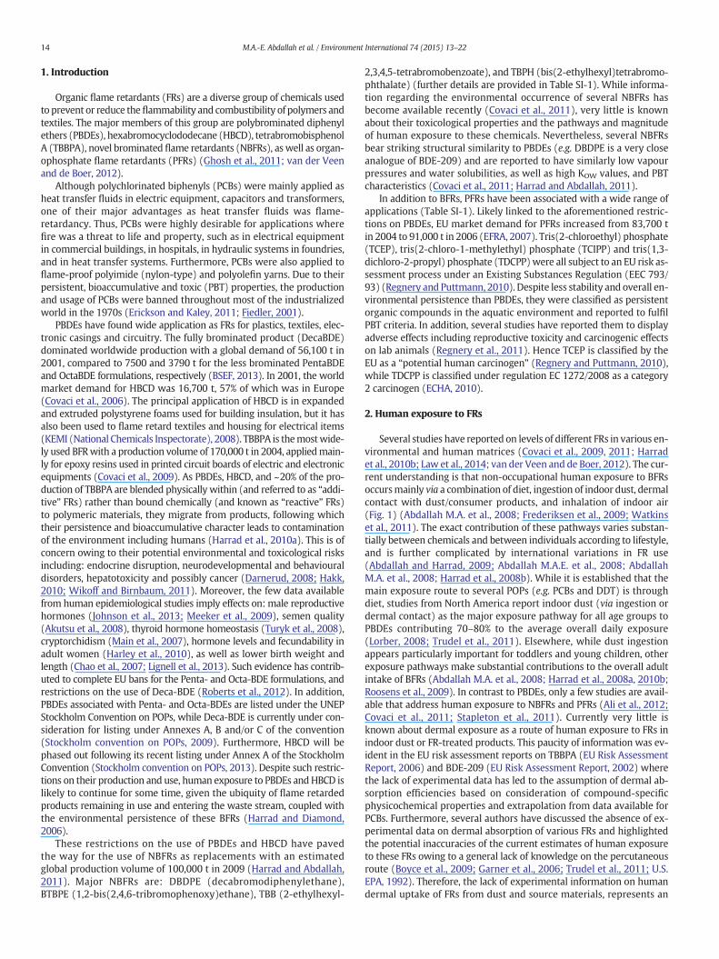
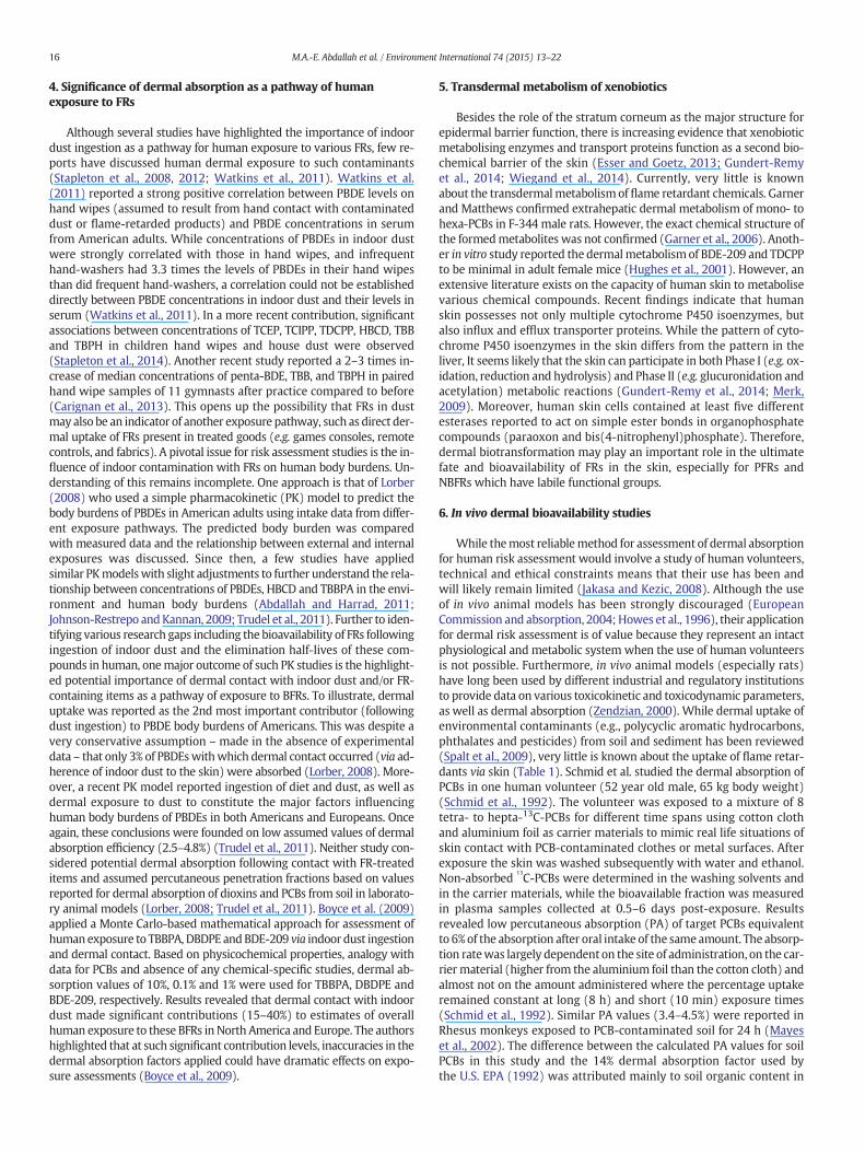
Several studies have reported on levels of different FRs in various en-vironmental and human matrices (Covaci et al., 2009, 2011; Harradet al., 2010b; Law et al., 2014; van der Veen and de Boer, 2012). The cur-rent understanding is that non-occupational human exposure to BFRsoccursmainly via a combination of diet, ingestion of indoor dust, dermalcontact with dust/consumer products, and inhalation of indoor air(Fig. 1) (Abdallah M.A. et al., 2008; Frederiksen et al., 2009; Watkinset al., 2011). The exact contribution of these pathways varies substan-tially between chemicals and between individuals according to lifestyle,and is further complicated by international variations in FR use(Abdallah and Harrad, 2009; Abdallah M.A.E. et al., 2008; AbdallahM.A. et al., 2008; Harrad et al., 2008b). While it is established that themain exposure route to several POPs (e.g. PCBs and DDT) is throughdiet, studies from North America report indoor dust (via ingestion ordermal contact) as the major exposure pathway for all age groups toPBDEs contributing 70–80% to the average overall daily exposure(Lorber, 2008; Trudel et al., 2011). Elsewhere, while dust ingestionappears particularly important for toddlers and young children, otherexposure pathways make substantial contributions to the overall adultintake of BFRs (Abdallah M.A. et al., 2008; Harrad et al., 2008a, 2010b;Roosens et al., 2009). In contrast to PBDEs, only a few studies are avail-able that address human exposure to NBFRs and PFRs (Ali et al., 2012;Covaci et al., 2011; Stapleton et al., 2011). Currently very little isknown about dermal exposure as a route of human exposure to FRs inindoor dust or FR-treated products. This paucity of information was ev-ident in the EU risk assessment reports on TBBPA (EU Risk AssessmentReport, 2006) and BDE-209 (EU Risk Assessment Report, 2002) wherethe lack of experimental data has led to the assumption of dermal ab-sorption efficiencies based on consideration of compound-specificphysicochemical properties and extrapolation from data available forPCBs. Furthermore, several authors have discussed the absence of ex-perimental data on dermal absorption of various FRs and highlightedthe potential inaccuracies of the current estimates of human exposureto these FRs owing to a general lack of knowledge on the percutaneousroute (Boyce et al., 2009; Garner et al., 2006; Trudel et al., 2011; U.S.EPA, 1992). Therefore, the lack of experimental information on humandermal uptake of FRs from dust and source materials, represents an
Ingestion Inhalation Dermal absorption
Diet Dust Indoorair
Outdoorair Dust Consumer
products
Body Burden
Bioavailability
Metabolism
Fig. 1.Major pathways of human exposure to FRs.
15M.A.-E. Abdallah et al. / Environment International 74 (2015) 13–22
important research gap that hampers accurate assessment of humanexposure to FRs. However, efforts to fill this gap are hindered by sev-eral difficulties including: ethical issues encountered with humanstudies, inter-species variation in dermal structure and uptake thatcast doubt on the accuracy of extrapolation or allometric scaling ofanimal data to humans, and tighter regulations on in vivo tests in-volving animals.
Against this backdrop, this paper: (a) provides a critical reviewof thecurrent state-of-knowledge on dermal absorption of FRs, (b) discussesthe paradigm shift in toxicity testing from in vivo to in vitro dermalbioavailability studies and (c) suggests effective novel approaches tostudying human dermal uptake of FRs, with special emphasis onin vitro 3D human skin percutaneous assays, that are finding increasingapplication in the pharmaceutical and cosmetics sectors (Gibbs et al.,2013; Kandarova et al., 2013; Tornier et al., 2010).
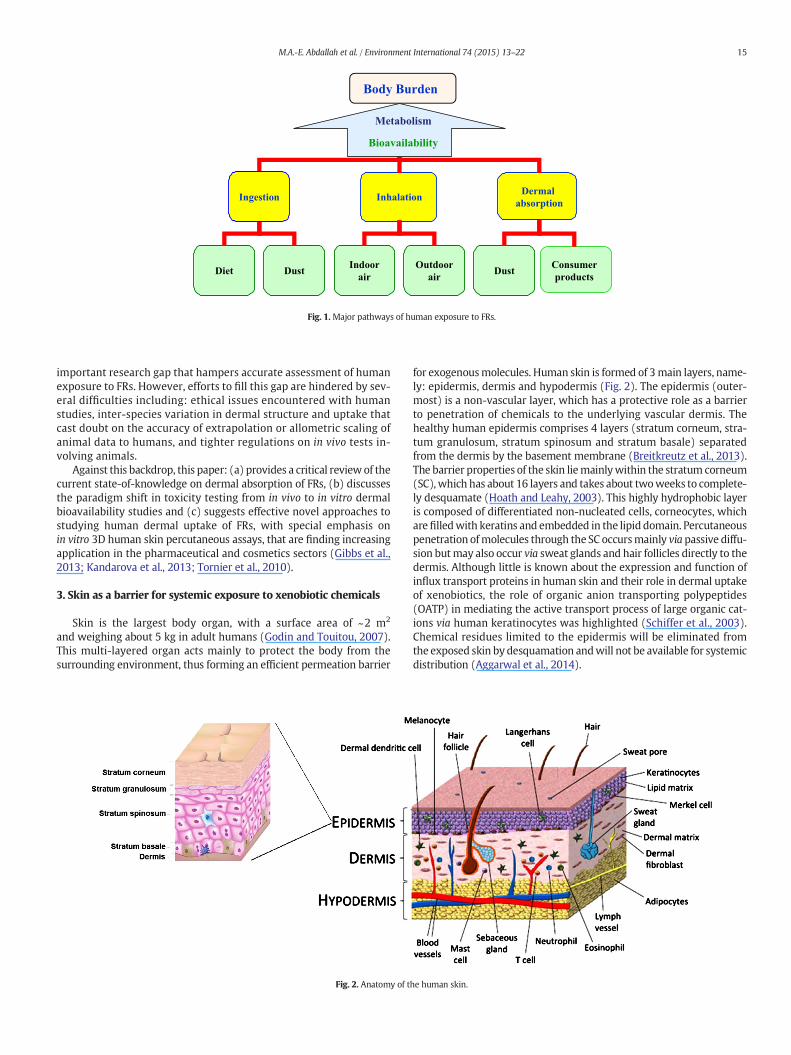
3. Skin as a barrier for systemic exposure to xenobiotic chemicals
Skin is the largest body organ, with a surface area of ~2 m2
and weighing about 5 kg in adult humans (Godin and Touitou, 2007).This multi-layered organ acts mainly to protect the body from thesurrounding environment, thus forming an efficient permeation barrier
Fig. 2. Anatomy of t
for exogenousmolecules. Human skin is formed of 3main layers, name-ly: epidermis, dermis and hypodermis (Fig. 2). The epidermis (outer-most) is a non-vascular layer, which has a protective role as a barrierto penetration of chemicals to the underlying vascular dermis. Thehealthy human epidermis comprises 4 layers (stratum corneum, stra-tum granulosum, stratum spinosum and stratum basale) separatedfrom the dermis by the basement membrane (Breitkreutz et al., 2013).The barrier properties of the skin liemainlywithin the stratum corneum(SC),which has about 16 layers and takes about twoweeks to complete-ly desquamate (Hoath and Leahy, 2003). This highly hydrophobic layeris composed of differentiated non-nucleated cells, corneocytes, whicharefilledwith keratins and embedded in the lipid domain. Percutaneouspenetration ofmolecules through the SC occursmainly via passive diffu-sion butmay also occur via sweat glands and hair follicles directly to thedermis. Although little is known about the expression and function ofinflux transport proteins in human skin and their role in dermal uptakeof xenobiotics, the role of organic anion transporting polypeptides(OATP) in mediating the active transport process of large organic cat-ions via human keratinocytes was highlighted (Schiffer et al., 2003).Chemical residues limited to the epidermis will be eliminated fromthe exposed skin by desquamation andwill not be available for systemicdistribution (Aggarwal et al., 2014).
he human skin.
16 M.A.-E. Abdallah et al. / Environment International 74 (2015) 13–22
4. Significance of dermal absorption as a pathway of humanexposure to FRs
Although several studies have highlighted the importance of indoordust ingestion as a pathway for human exposure to various FRs, few re-ports have discussed human dermal exposure to such contaminants(Stapleton et al., 2008, 2012; Watkins et al., 2011). Watkins et al.(2011) reported a strong positive correlation between PBDE levels onhand wipes (assumed to result from hand contact with contaminateddust or flame-retarded products) and PBDE concentrations in serumfrom American adults. While concentrations of PBDEs in indoor dustwere strongly correlated with those in hand wipes, and infrequenthand-washers had 3.3 times the levels of PBDEs in their hand wipesthan did frequent hand-washers, a correlation could not be establisheddirectly between PBDE concentrations in indoor dust and their levels inserum (Watkins et al., 2011). In a more recent contribution, significantassociations between concentrations of TCEP, TCIPP, TDCPP, HBCD, TBBand TBPH in children hand wipes and house dust were observed(Stapleton et al., 2014). Another recent study reported a 2–3 times in-crease of median concentrations of penta-BDE, TBB, and TBPH in pairedhand wipe samples of 11 gymnasts after practice compared to before(Carignan et al., 2013). This opens up the possibility that FRs in dustmay also be an indicator of another exposure pathway, such as direct der-mal uptake of FRs present in treated goods (e.g. games consoles, remotecontrols, and fabrics). A pivotal issue for risk assessment studies is the in-fluence of indoor contamination with FRs on human body burdens. Un-derstanding of this remains incomplete. One approach is that of Lorber(2008) who used a simple pharmacokinetic (PK) model to predict thebody burdens of PBDEs in American adults using intake data from differ-ent exposure pathways. The predicted body burden was comparedwith measured data and the relationship between external and internalexposures was discussed. Since then, a few studies have appliedsimilar PKmodelswith slight adjustments to further understand the rela-tionship between concentrations of PBDEs, HBCD and TBBPA in the envi-ronment and human body burdens (Abdallah and Harrad, 2011;Johnson-Restrepo andKannan, 2009; Trudel et al., 2011). Further to iden-tifying various research gaps including the bioavailability of FRs followingingestion of indoor dust and the elimination half-lives of these com-pounds in human, onemajor outcome of such PK studies is the highlight-ed potential importance of dermal contact with indoor dust and/or FR-containing items as a pathway of exposure to BFRs. To illustrate, dermaluptake was reported as the 2nd most important contributor (followingdust ingestion) to PBDE body burdens of Americans. This was despite avery conservative assumption – made in the absence of experimentaldata – that only 3% of PBDEswithwhich dermal contact occurred (via ad-herence of indoor dust to the skin) were absorbed (Lorber, 2008). More-over, a recent PK model reported ingestion of diet and dust, as well asdermal exposure to dust to constitute the major factors influencinghuman body burdens of PBDEs in both Americans and Europeans. Onceagain, these conclusions were founded on low assumed values of dermalabsorption efficiency (2.5–4.8%) (Trudel et al., 2011). Neither study con-sidered potential dermal absorption following contact with FR-treateditems and assumed percutaneous penetration fractions based on valuesreported for dermal absorption of dioxins and PCBs from soil in laborato-ry animal models (Lorber, 2008; Trudel et al., 2011). Boyce et al. (2009)applied a Monte Carlo-based mathematical approach for assessment ofhuman exposure to TBBPA, DBDPE andBDE-209 via indoor dust ingestionand dermal contact. Based on physicochemical properties, analogy withdata for PCBs and absence of any chemical-specific studies, dermal ab-sorption values of 10%, 0.1% and 1% were used for TBBPA, DBDPE andBDE-209, respectively. Results revealed that dermal contact with indoordust made significant contributions (15–40%) to estimates of overallhuman exposure to these BFRs in North America and Europe. The authorshighlighted that at such significant contribution levels, inaccuracies in thedermal absorption factors applied could have dramatic effects on expo-sure assessments (Boyce et al., 2009).
5. Transdermal metabolism of xenobiotics
Besides the role of the stratum corneum as the major structure forepidermal barrier function, there is increasing evidence that xenobioticmetabolising enzymes and transport proteins function as a second bio-chemical barrier of the skin (Esser and Goetz, 2013; Gundert-Remyet al., 2014; Wiegand et al., 2014). Currently, very little is knownabout the transdermalmetabolism of flame retardant chemicals. Garnerand Matthews confirmed extrahepatic dermal metabolism of mono- tohexa-PCBs in F-344 male rats. However, the exact chemical structure ofthe formedmetabolites was not confirmed (Garner et al., 2006). Anoth-er in vitro study reported the dermalmetabolismof BDE-209 and TDCPPto be minimal in adult female mice (Hughes et al., 2001). However, anextensive literature exists on the capacity of human skin to metabolisevarious chemical compounds. Recent findings indicate that humanskin possesses not only multiple cytochrome P450 isoenzymes, butalso influx and efflux transporter proteins. While the pattern of cyto-chrome P450 isoenzymes in the skin differs from the pattern in theliver, It seems likely that the skin can participate in both Phase I (e.g. ox-idation, reduction and hydrolysis) and Phase II (e.g. glucuronidation andacetylation) metabolic reactions (Gundert-Remy et al., 2014; Merk,2009). Moreover, human skin cells contained at least five differentesterases reported to act on simple ester bonds in organophosphatecompounds (paraoxon and bis(4-nitrophenyl)phosphate). Therefore,dermal biotransformation may play an important role in the ultimatefate and bioavailability of FRs in the skin, especially for PFRs andNBFRs which have labile functional groups.
6. In vivo dermal bioavailability studies
While themost reliablemethod for assessment of dermal absorptionfor human risk assessment would involve a study of human volunteers,technical and ethical constraints means that their use has been andwill likely remain limited (Jakasa and Kezic, 2008). Although the useof in vivo animal models has been strongly discouraged (EuropeanCommission and absorption, 2004; Howes et al., 1996), their applicationfor dermal risk assessment is of value because they represent an intactphysiological and metabolic system when the use of human volunteersis not possible. Furthermore, in vivo animal models (especially rats)have long been used by different industrial and regulatory institutionsto provide data on various toxicokinetic and toxicodynamic parameters,as well as dermal absorption (Zendzian, 2000). While dermal uptake ofenvironmental contaminants (e.g., polycyclic aromatic hydrocarbons,phthalates and pesticides) from soil and sediment has been reviewed(Spalt et al., 2009), very little is known about the uptake of flame retar-dants via skin (Table 1). Schmid et al. studied the dermal absorption ofPCBs in one human volunteer (52 year old male, 65 kg body weight)(Schmid et al., 1992). The volunteer was exposed to a mixture of 8tetra- to hepta-13C-PCBs for different time spans using cotton clothand aluminium foil as carrier materials to mimic real life situations ofskin contact with PCB-contaminated clothes or metal surfaces. Afterexposure the skin was washed subsequently with water and ethanol.Non-absorbed
13C-PCBs were determined in the washing solvents and
in the carrier materials, while the bioavailable fraction was measuredin plasma samples collected at 0.5–6 days post-exposure. Resultsrevealed low percutaneous absorption (PA) of target PCBs equivalentto 6%of the absorption after oral intake of the sameamount. The absorp-tion ratewas largely dependent on the site of administration, on the car-rier material (higher from the aluminium foil than the cotton cloth) andalmost not on the amount administered where the percentage uptakeremained constant at long (8 h) and short (10 min) exposure times(Schmid et al., 1992). Similar PA values (3.4–4.5%) were reported inRhesus monkeys exposed to PCB-contaminated soil for 24 h (Mayeset al., 2002). The difference between the calculated PA values for soilPCBs in this study and the 14% dermal absorption factor used bythe U.S. EPA (1992) was attributed mainly to soil organic content in
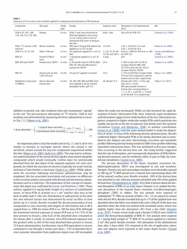
Table 1Summary of in vivo and in vitro methods applied for studying dermal absorption of FR chemicals.
Compound Skin type Studytype
Dosing Exposure time Absorption (% of administereddose)
Ref.
PCBs # 52, 101, 108,118, 138, 153, 170, 180
Human In vivo PCBs (5 mg) were dissolved inDCM and applied to the carrier(4 cm2 cotton cloth or 28 cm2
aluminium foil) prior to fixing tothe skin
0.66–1 day Up to 6% for PCB-153 Schmid et al. (1992)
PCBs (14C-Aroclor 1260mixture)
Rhesus monkeys In vivo 500 mg of 70 μg/g PCB-spiked soilapplied to 12 cm2 of skin
12–24 h 3.43 ± 0.35% for 12 h and4.26 ± 0.52% for 24 h
Mayes et al. (2002)
PCBs # 4, 15, 47, 155 Male F-344 rats In vivo 0.4 mg/kg bw applied to 1 cm2 ofskin
1, 4, 8, 12, 24, 48,72, 96, and 336 h
From ca. 100% for PCB-4 to ca. 30%for PCB-155.
Garner and Matthews(1998)
BDE-47 Female C57BL/6mice
In vivo 1 mg/kg bw applied to 2 cm2 ofskin
5 days 62% Staskal et al. (2005)
BDE-209 and TDCPP Female mice (SKH1) In vitro 6, 30 and 60 nmol in THF for BDE-209; 20, 100 and 200 pmol inacetone for TDCPP
24 h 2–20% in skin, 0.07–0.34% inreceptor fluid for BDE-209.39–57% in skin and 28–35% inreceptor fluid for TDCPP
Hughes et al. (2001)
BDE-47 Human and rat skin(350–410 μm)
In vitro 10 mg/cm2 applied in acetone 24 h 2–15% in 0.9% NaCl receptor fluid;57% and 33% remained in cells forhuman and rat skin, respectively.
Roper et al. (2006)
Bisphenol-A (precursorto TBBPA)
Pig ear skin andhuman skin
In vitro 50, 100, 200, 400 and 800 nmolwere applied in 60 μL ethanol/phosphate buffer (pH 7.4)
24, 48 and 72 h Human skin (45.6 ± 6.2%), pigskin (65.3 ± 8.2%)BPA-glucuronide formed inhuman skin, corresponding to7 ± 2, 16 ± 3 and 30. ± 3 nmolat 24, 48 and 72 h, respectively.
Zalko et al. (2011)
17M.A.-E. Abdallah et al. / Environment International 74 (2015) 13–22
addition to particle size, skin residence time and contaminant “ageing”in the soil. The percutaneous absorption of 14C-Aroclor 1260 in testmonkeyswas determined bymeasuring the feces radioactivity in excre-ta (Eq. (1)) (Mayes et al., 2002).
%dose absorbed ¼% topical dose excreted 14C−urineþ14C faecesð Þ
% intravenous dose excreted 14C−urineþ14C faecesð Þ
!� 100
ð1Þ
An important point is that themodel used in Eq. (1) and in all in vivostudies in humans or surrogate species where the animal is notsacrificed, cannot account for any test compounds sequestered withinthe skin (Mayes et al., 2002; Spalt et al., 2009). Thismay lead to substan-tial underestimation of the actual dermal uptake of persistent lipophiliccompounds which would eventually (within days) be systemicallyabsorbed from the skin depot of the exposed organism. For such com-pounds, for which the outcome of concern is typically not acute toxicity,inclusion of skin burden is necessary (Spalt et al., 2009). While adjust-ment for excretion following intravenous administration may beemployed, this has associated uncertainty and presumes no differencein the excretory pattern associatedwith dermal and intravenous admin-istration used as a reference. The importance of this concept of contam-inant skin depot was confirmed by Garner andMatthews (1998). Theseauthors applied 0.4 mg/kg body weight of a mixture of radiolabeledmono- to hexa-PCBs in acetone to a 1 cm2 hairless skin area at theback of adult male F-344 rats. Distribution of radioactivity in the dosesite and selected tissues was determined by serial sacrifice at timepoints up to 2 weeks. Results revealed the dermal penetration of testcompounds to vary inversely with degree of chlorination and at 48 hranged from ca. 100% for mono-PCB to ca. 30% for hexa-PCB. Althoughthe maximum internal exposure to mono-PCB was at 4 h (37% of thedose present in tissues), only 0.2% of the absorbed dose remained inthe tissues after 2 weeks. In contrast, tetra-PCB internal exposure wasthe greatest with ca. 85% of the total absorbed dose present in tissues72 h post-administration. Furthermore, hexa-PCB equivalents in tissuescontinued to rise through 2 weeks post-dose (~15% of absorbed dose)since systemic absorption from epidermis depots was still incomplete
when the study was terminated. While rat skin favoured the rapid ab-sorption of lower chlorinated PCBs, their relatively rapid metabolismand elimination suggest lower body burdens of the less chlorinated con-geners compared to highermolecular weight PCBs which penetrate lessrapidly, but persist at the site of exposure and slowly enter the systemiccirculation (Garner and Matthews, 1998). In another contribution,Garner et al. (2006) used the same animal model to study the disposi-tion of mono- to hexa-PCBs following dermal administration. Resultsconfirmed higher chlorinated PCBs to be slowly absorbed and accumu-lated in the adipose and skin. Interestingly, excretion and metabolicprofiles following dermal dosing tended to differ fromprofiles followingequivalent intravenous doses. This was attributed to first pass metabo-lism occurring at the dermal dose site. The study further suggestedthat the rate of absorption, and consequently disposition of PCBs follow-ing dermal exposure, may be mediated, either in part or fully, by trans-dermal metabolism (Garner et al., 2006).
The dermal absorption of the flame retardant resorcinol bis-diphenylphosphate (RDP) was investigated in rats and monkeys.Sprague–Dawley rats and cynomolgusmonkeyswere dermally exposedto 100 mg of 14C-RDP spread over a shaved area representing about 20%of the animal's surface area. Results revealed ~20% of the dermal dosewas absorbed in rats, whereas primates absorbed only 10% of the applieddermal dose (Freudenthal et al., 2000). Very little is known about the der-mal absorption of BFRs. In an early report, Ulsamer et al. studied the der-mal absorption of the banned flame retardant tris(dibromopropyl)phosphate (TRIS) in rabbits. The test animals were exposed toradiolabeled 14C-TRIS via sections of fabric (10× 12 cm) placed in contactwith skin for 96 h. Results revealed that up to 17% of the applied dosewasabsorbedwhen the fabricwaswettedwith urine. Only 6% of the dosewasabsorbed when the cloth was wetted with simulated sweat, which wasslightly higher than the absorption (4%) from a dry cloth (Ulsameret al., 1978). A more recent study used a female C57BL/6 mice model toassess the dermal bioavailability of BDE-47. Test animals were exposedto 1 mg/kg body weight of 14C-BDE 47 in acetone applied to a hairless2 cm2 skin patch. Results revealed ~62% absorption of the administereddose after 5 days while 15% remained at the site of application whereskin and adipose were reported as the major depot tissues (Staskalet al., 2005).
18 M.A.-E. Abdallah et al. / Environment International 74 (2015) 13–22
7. Paradigm shift— in vivo to in vitro dermal bioavailability studies
Due to the ethical and technical issues arising from the use of labanimals in toxicology studies, the use of in vivo animal models is in-creasingly strongly discouraged (Jakasa and Kezic, 2008). Therefore,focus has shifted to developing and validating alternative in vitro testmethods, which also provide a better platform for development ofpredictive pharmacokinetic models. Several guidance documents forconducting in vitro skin absorption studies (OECD, 2004; U.S. EPA,2004; WHO, 2006) are currently available rendering the application ofin vitro skin models increasingly acceptable for research and regulatorypurposes.
Different types of skinmay be used, for example, human excised skinfrom surgery or from cadavers (ex vivo skin) or animal (e.g. pig) skin.Various types of diffusion cells have been employed in in vitro studiesto date, and the composition of receptor fluids may vary. All these fac-tors can influence the results of in vitro experiments (Jakasa and Kezic,2008).While several papers have reported on in vitrodermal absorptionof environmental contaminants such as: polycyclic aromatic hydrocar-bons, phthalates, as well as organochlorine and organophosphate pesti-cides (Hopf et al., 2014; Hughes and Edwards, 2010; Spalt et al., 2009),very few in vitro studies of the dermal absorption of FRs exist. In onesuch study, Hughes et al. (2001) used skin from adult hairless femalemice (SKH1) mounted in flow-through diffusion cells to study theabsorption of 14C-BDE-209 and 14C-TDCPP at 3 concentration levels.HEPES ((4-(2-hydroxyethyl)-1-piperazineethanesulfonic acid))-bufferedHanks' balanced salt solution (pH 7.4) with 10% foetal bovine serumwas used as receptor fluid. Following 24 h exposure, the skin patcheswere washed with solvent prior to analysis of receptor fluid, skinwash and skin for chemical-derived radioactivity. BDE-209 showedlow penetration (0.3%) into the receptor fluid while up to 20% of thedose remained in skin after 24 h. TDCPP displayed higher penetration(39–57%) to the receptor fluid, while 28–35% of administered doseremained in the skin. This was mainly attributed to its lower molecularweight and KOW than BDE-209 (Hughes et al., 2001). The dermal ab-sorption of BDE-47 was studied using in vitro split-thickness skin mem-branes (350–410 μm, stratum corneum uppermost) of human and ratskin exposed to a single dose of ca. 10 mg/cm2 of 14C-BDE-47 for 24 h.The skin patches were mounted in flow-through cells while receptorfluid (NaCl, 0.9%, w/v in water) was pumped through the receptorchambers at ca. 1.5 ml/h (Roper et al., 2006). The dose recovered fromthe receptor fluid was 2% and 15% of administered BDE-47 to humanand rat skin, respectively. The difference between the results of thisin vitro study (Roper et al., 2006) and the higher (62%) sorption ob-served in an in vivo study of dermal absorption in mice (Staskal et al.,2005) (Table 1) may be attributed mainly to the use of 0.9% NaCl solu-tion in water as a receptor fluid, as this may greatly reduce diffusion ofthe lipophilic BDE-47 to the receptor fluid (Wilkinson and Williams,2002) and does not accuratelymimic actual biological conditions. Possi-ble evidence of this is provided by the high residual levels of BDE-47 de-tected in the cells (57% and 33% for human and rat skin, respectively)that appeared not to diffuse to the receptor fluid (Roper et al., 2006).While no data exists on dermal absorption of TBBPA, a recent in vitrostudy reported on the percutaneous bioavailability of its precursor,bisphenol A (BPA) from human and pig skin (Zalko et al., 2011). Viablehuman and pig skin patches (500 μm thickness) weremaintained at theair/liquid interface using Transwell inserts while dermal/epidermalfeeding was achieved via diffusion of nutrients from a modifiedDulbecco's Eagle culture medium which kept the cells alive during 72h exposure experiments. BPA was efficiently absorbed (65% and 46%frompig andhuman skin, respectively) andmetabolised by the culturedskin indicating the trans-dermal route contributes substantially tohuman exposure to BPA (Zalko et al., 2011). However, it should benoted that TBBPA has amuch highermolecularweight and consequent-ly, different physico-chemical properties (e.g.water solubility, partitionco-efficient and vapour pressure) than BPA. Furthermore, the lack of
halogen atoms in BPA is likely to enhance the rate of its percutaneousabsorption compared to its tetra-brominated derivative (Garner andMatthews, 1998).
Given the growing evidence that suggests dermal absorption to be apotentially significant pathwayof humanexposure to FRs, the paucity ofdata on dermal bioavailability of such ubiquitous contaminants may beattributed to a combination of ethical, technical and economic issues.One alternativemethodwith the potential to overcome such difficultiesis the use of 3D human skin equivalent (HSE) models which provide arelatively cheap, commercially available, ethical, and reliable methodfor dermal absorption studies that is capable of producing data of rele-vance to human exposure.
8. Human skin equivalent models (HSE)
8.1. Rationale
Although the Organisation for Economic Co-operation and Develop-ment (OECD) and the European Centre for Validation of AlternativeMethods (ECVAM) describe methods for assessing dermal absorptionusing excised in vitro human and animal skin, the lack of correlation intransdermal permeation of chemicals across species imparts a high de-gree of uncertainty when extrapolating results from animal models tohumans. This is mainly due to variations in the stratum corneum thick-ness, intercellular subcutaneous lipids and/or between-species differ-ences in metabolic enzymes and their activity (Schafer-Korting et al.,2008a). Therefore, excised in vitro human skin is preferable to animalskin (e.g. rat or pig skin) for dermal absorption testing, but is clearlyless available. To overcome this shortage, HSE have been developed toprovide an alternative to human skin in testing of compounds for trans-dermal permeability (Mertsching et al., 2008). A protocol was devel-oped and validated according to the OECD guidelines for percutaneousabsorption by using commercially available HSE models (Table 2). Thepermeability of tested HSE models was compared to that of excisedhuman epidermis, pig skin and bovine udder skin, using 9 compoundswidely varying in physicochemical characteristics, including the OECDstandards: testosterone, caffeine and benzoic acid. Results revealedthat HSE models closely mimic the histological and physiological char-acteristics of viable human skin, allowing their use for in vitro skin pen-etration studies, taking product-specific over predictability into account(Hartung et al., 2004; Schafer-Korting et al., 2008a). Consequently, sev-eral validatedmethods using HSEmodels have been approved by OECDand ECVAM for testing skin absorption, phototoxicity, corrosion andirritation by xenobiotic chemicals (Ackermann et al., 2010; Buist et al.,2010).
8.2. Composition
HSE models can be generally classified into 2 main types:1 — Reconstructed human epidermis (RHE): RHE is a human skin
tissue obtained from human keratinocytes cultured on an inert polycar-bonate medium. One key advantage is that it permits growth of donorepidermal cells in a serum-free culture environment. After rapidlyproliferating preparative keratinocyte cultures have been obtained,the epidermal cells yielded are seeded on inert filter substrates, whichare then raised to the air–liquid interface in a humidified-air incubator.A fully-defined nutrient medium feeds the basal cells through the filtersubstratum. After 14 days, a stratified epidermis is formed that closelyresembles human epidermis in vivo (Fig. 3) (Boelsma et al., 2000).
Morphologically, these cultures exhibit a well-stratified epitheli-um and cornified epidermis with significantly improved barrierfunction and metabolic activity (Boelsma et al., 2000). Differentia-tion markers such as suprabasal keratins, integrin b4, integrin a6,fibronectin, involucrin, filaggrin, trichohyalin, type I, III, IV, V and VII col-lagen, laminin, heparan sulfate and membrane-bound transglutaminase
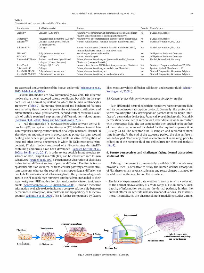
Table 2Characteristics of commercially available HSE models.
Brand name Scaffold material Source Dermis Manufacturer
Episkin™ Collagen (0.38 cm2) Keratinocytes (mammary/abdominal samples obtained fromhealthy consenting donors during plastic surgery)
No L'Oreal, Nice,France
Skinethic™ Polycarbonate membrane (0.5 cm2) Keratinocytes (neonatal foreskin tissue or adult breast tissue) No L'Oreal, Nice,FranceEpidermTM Collagen coated polycarbonate
(9 mm diameter)Human keratinocytes (neonatal foreskin adult breast skin) No MatTek Corporation, MA, USA
EpidermFT™ Collagen Human keratinocytes (neonatal foreskin adult breast skin),human fibroblasts (neonatal skin, adult skin)
Yes MatTek Corporation, MA, USA
EST-1000 Polycarbonate membrane Keratinocytes (neonatal foreskin) No CellSystems, Troisdorf GermanyAST-2000 Collagen Human keratinocytes Yes CellSystems, Troisdorf GermanyPhenion® FT Model Bovine, cross linked, lyophilized
collagen (1.3 cm diameter)Primary human keratinocytes (neonatal foreskin), humanfibroblasts (neonatal foreskin)
Yes Henkel, Duesseldorf, Germany
StrataTest® Collagen I (0.6 cm2) Immortalized, human NIKS® keratinocytes dermal fibroblasts Yes Stratatech Corporation Madison WI, USAEpistem ® LSE Collagen Primary human keratinocytes and dermal fibroblasts. Yes Epistem limited, Manchester, UK.StratiCell® EPI/001 Polycarbonate membrane Primary human keratinocytes No Straticell Corporation, Gembloux, Belgium.StratiCell® Mel/001 Polycarbonate membrane Primary human keratinocytes and melanocytes. No Straticell Corporation, Gembloux, Belgium.
19M.A.-E. Abdallah et al. / Environment International 74 (2015) 13–22
are expressed similar to those of the human epidermis (Brinkmann et al.,2013; Mehul et al., 2004).
Several RHE models are now commercially available. The differentmodels share the air-exposed culture conditions, but differ in the sup-port used as a dermal equivalent on which the human keratinocytesare grown (Table 2). Numerous histological and biochemical featuresare shared by these models, in particular epidermal stratification anddifferentiation, and all produce a well-defined stratum corneum as a re-sult of tightly regulated expression of differentiation-related genes(Boelsma et al., 2000; Zhang and Michniak-Kohn, 2012).
2— Full-thickness skin (FT): Paracrine signalling between dermal fi-broblasts (FB) and epidermal keratinocytes (KC) is believed tomodulateskin responses during contact irritant or allergic reactions. Dermal FBalso plays an important role in photo-ageing, photo-damage, woundhealing and cancer progression. To enable in vitro investigation ofthese and other dermal phenomena inwhich FB–KC interactions are im-portant, FT skin models composed of a FB-containing dermis/KC-containing epidermis have been developed (Schafer-Korting et al.,2008b; Semlin et al., 2011). In order to test possible immunological re-actions on skin, Langerhans cells (LCs) can be introduced into FT skinsubstitutes (Regnier et al., 1997). Percutaneous absorption of chemicalsis due to two different routes of passive diffusion. The first is trans-epidermal diffusion via inter- or trans-cellular pathway across the stra-tum corneum, whereas the second is trans-appendageal diffusion viahair follicles and associated sebaceous glands. The presence of append-ages in the FT models may represent another advantage added to theirsuperiority over RHE models for biotransformation-linked toxic end-points (Ackermannet al., 2010; Curren et al., 2006). However, the scarceinformation available to date indicates a complex relationship betweenpercutaneous absorption, skin thickness and lipophilicity of test com-pounds (Wilkinson et al., 2006). This is further compounded by factors
Fig. 3. General stages of deve
like: exposure vehicle, diffusion cell design and receptor fluid (Schafer-Korting et al., 2008b).
8.3. General protocol for in vitro percutaneous absorption studies
EachHSEmodel is suppliedwith its respective receptor/culture fluidand its percutaneous absorption protocol. Generally, the protocol in-volvesmounting the fully-developed skin patches at the air-liquid inter-face of a permeation device (e.g. Franz-cell type diffusion cells, Mattek®permeation device, see SI section for further details) while in contactwith the receptor fluid. The test compound is then applied to the surfaceof the stratum corneum and incubated for the required exposure time(usually 24 h). The receptor fluid is sampled and replaced at fixedtime intervals. At the end of the exposure period, the skin surface iswashed/wiped clean of any residual contaminant remaining, prior tocollection of the receptor fluid and cell culture for chemical analysis(Fig. 4).
9. Future perspectives and challenges facing dermal absorptionstudies of FRs
Although the current commercially available HSE models mayprovide a useful alternative to study the human dermal absorptionof FRs, there remain several challenges and research gaps that need tobe addressed in the near future. These include:
• The lack of experimental data – either in vivo or in vitro – relevantto the dermal bioavailability of a wide range of FRs in human. Suchpaucity of information regarding the dermal pathway hinders thecurrent efforts for accurate risk assessment of various FRs. Further-more, it complicates the pharmacokinetic modelling studies aiming
lopment of HSE model.

Fig. 4. General protocol for percutaneous absorption studies using in vitro HSE models.
20 M.A.-E. Abdallah et al. / Environment International 74 (2015) 13–22
to understand the relationship between external exposure andhuman body burdens of FRs.
• The diverse nature and wide range of physico-chemical parametersof organic FR chemicals (Table SI-1). Contaminant propertieslike: Log KOW, molecular weight, size and water solubility werereported to affect the dermal absorption of PCBs (Garner andMatthews, 1998). Furthermore, the difference in protein binding af-finities of various FRs may also influence their permeation throughthe skin barrier. This will be of particular interest if OATPs wereinvolved in mediating the active transport process of FRs acrossthe human epidermis. Therefore, the chemical diversity and co-existence of various BFRs and PFRs in different environmental samplesare likely to present a challenge to environmental scientists trying tomimic in vivo scenarios.
• FR chemicals with similar/comparable molecular weight, size andKow can exist in different isomeric forms (e.g. HBCD isomers),which might adopt various structural characteristics (e.g. planarity)and exhibit different physico-chemical properties (e.g.water solubili-ty). This is also likely to constitute an important factor influencing thedermal bioavailability of such iso-baric compounds.
• Despite the huge advances in production and validation of HSEmodels in the past few years, further improvements are still requiredto closely mimic the in vivo situation. The presence of hair follicles,sweat and sebaceous glands provides further potential pathwaysfor percutaneous penetration. The dermis in vivo is continuously per-fused by the subcutaneous vasculature, which can rapidly removepermeants reaching the epidermal–dermis interface, allowing forfurther diffusion of the permeant through the skin layers. Thissystem can be mimicked in vitro via the use of dynamic in-line flowthrough diffusion cells (Table SI-2). However, further validation andstandardisation of test protocols using this model are still requiredto gain the approval of the regulatory bodies and research organisa-tions.
• Transdermal metabolism has been reported as a major mediator forpercutaneous absorption of PCBs (Garner et al., 2006). Currently,
very little is known about the dermal biotransformation of BFRs andPFRs (Hughes et al., 2001). Enhanced understanding of percutaneousmetabolic pathways and identification of themetabolites thus formedin humans thus appear important, if the reliability of risk assessmentof these contaminants is to be improved.
• The excretion of xenobiotic chemicals and their metabolites insweat and hair follicles has been well documented in literature (DeGiovanni and Fucci, 2013; Parle and Jadhav, 2007). Therefore, bio-transformation may not be the only dermal contaminant-removalmechanism in human. Further research is required to understandthe role of eccrine sweat and hair follicles as excretion routes forFRs. Consequently, the in vitro human skin models may consider thedermal bioavailability of FRs as an equilibrium process.
• While HSE models have been widely exploited in the pharmaceuticaland cosmetic fields, to the authors' knowledge, they are yet to beapplied for studying dermal absorption of FRs or any other organiccontaminants. This is likely to create several challenges for analyticalmethod development, exposure protocols and modelling of theresults. Furthermore, in vitro dermal studies carried out for the pur-pose of risk assessment should also include scenarios that mimicreal life exposure to the test compounds. This includes exposure toenvironmentally-relevant concentrations via appropriate exposuremedia. Previous studies have shown that dermal absorption of PCBsfrom contaminated soils was different from direct application ofPCBs in solution to the skin (Mayes et al., 2002). In addition, dermalbioavailability has also been shown as influenced by the age of thecontaminant in soil and its organic content (Spalt et al., 2009). Similarfactors are likely to affect percutaneous absorption of BFRs and PFRs.Therefore, several exposure scenarios addressing dermal uptakefrom a range of environmental media (e.g. indoor dust, soil, sweatand consumer products) will be needed for full characterisation ofthe exposure arising from human dermal exposure to FRs.
Acknowledgement
The authors gratefully acknowledge funding from the EuropeanUnion Seventh Framework Programme FP7/2007–2013 under grantagreement no. 316665 (A-TEAM project) and grant agreement no.PIIF-GA-2012-327232 (ADAPT project).
Appendix A. Supplementary data
Specific details on physico-chemical parameters, uses, toxicokineticprofiles and main exposure pathways of key brominated and phospho-rous flame retardants in addition to different in vitro dermal absorptionprotocols are available as supplementary data. Supplementary data tothis article can be found online at doi:10.1016/j.envint.2014.09.012.
References
Abdallah MA-E, Harrad S. Personal exposure to HBCDs and its degradation products viaingestion of indoor dust. Environ Int 2009;35:870–6.
Abdallah MA, Harrad S. Tetrabromobisphenol-A, hexabromocyclododecane and itsdegradation products in UK human milk: relationship to external exposure. EnvironInt 2011;37:443–8.
Abdallah MA, Harrad S, Covaci A. Hexabromocyclododecanes and tetrabromobisphenol-Ain indoor air and dust in Birmingham, U.K.: implications for human exposure. EnvironSci Technol 2008a;42:6855–61.
Abdallah MAE, Harrad S, Ibarra C, Diamond M, Melymuk L, Robson M, et al. Hexabro-mocyclododecanes in indoor dust from Canada, the United Kingdom, and theUnited States. Environ Sci Technol 2008b;42:459–64.
Ackermann K, Borgia SL, Korting HC, Mewes KR, Schafer-Korting M. The phenion full-thickness skin model for percutaneous absorption testing. Skin Pharmacol Physiol2010;23:105–12.
Aggarwal M, Battalora M, Fisher P, Huser A, Parr-Dobrzanski R, SoufiM, et al. Assessmentof in vitro human dermal absorption studies on pesticides to determine defaultvalues, opportunities for read-across and influence of dilution on absorption. RegulToxicol Pharmacol 2014;68:412–23.
Akutsu K, Takatori S, Nozawa S, Yoshiike M, Nakazawa H, Hayakawa K, et al.Polybrominated diphenyl ethers in human serum and sperm quality. Bull EnvironContam Toxicol 2008;80:345–50.

21M.A.-E. Abdallah et al. / Environment International 74 (2015) 13–22
Ali N, Van den Eede N, Dirtu AC, Neels H, Covaci A. Assessment of human exposure toindoor organic contaminants via dust ingestion in Pakistan. Indoor Air 2012;22:200–11.
Boelsma E, Gibbs S, Faller C, Ponec M. Characterization and comparison of reconstructedskin models: morphological and immunohistochemical evaluation. Acta DermVenereol 2000;80:82–8.
Boyce C, Sax S, Dodge D, Pollock M, Goodman J. Human exposure to decabromodiphenylether, tetrabromobisphenol A, and decabromodiphenyl ethane in indoor dust. J Envi-ron Prot Sci 2009;3:75–96.
Breitkreutz D, Koxholt I, Thiemann K, Nischt R. Skin basement membrane: the foundationof epidermal integrity-BM functions and diverse roles of bridging molecules nidogenand perlecan. Biomed Res Int 2013:16. http://dx.doi.org/10.1155/2013/179784.
Brinkmann J, Stolpmann K, Trappe S, Otter T, Genkinger D, Bock U, et al. Metabolicallycompetent human skin models: activation and genotoxicity of benzo[a]pyrene.Toxicol Sci 2013;131:351–9.
BSEF. Bromine science and environmental forum. www.bsef.com, 2013. [accessed 17-15-2013].
Buist HE, van Burgsteden JA, Freidig AP, Maas WJ, van de Sandt JJ. New in vitro dermalabsorption database and the prediction of dermal absorption under finite conditionsfor risk assessment purposes. Regul Toxicol Pharmacol 2010;57:200–9.
Carignan CC, Heiger-Bernays W, McClean MD, Roberts SC, Stapleton HM, Sjodin A, et al.Flame retardant exposure among collegiate United States gymnasts. Environ SciTechnol 2013;47:13848–56.
Chao HR, Wang SL, Lee WJ, Wang YF, Papke O. Levels of polybrominated diphenyl ethers(PBDEs) in breast milk from central Taiwan and their relation to infant birth outcomeand maternal menstruation effects. Environ Int 2007;33:239–45.
Covaci A, Gerecke AC, Law RJ, Voorspoels S, Kohler M, Heeb NV, et al. Hexabro-mocyclododecanes (HBCDs) in the environment and humans: a review. Environ SciTechnol 2006;40:3679–88.
Covaci A, Voorspoels S, Abdallah MA, Geens T, Harrad S, Law RJ. Analytical and environ-mental aspects of the flame retardant tetrabromobisphenol-A and its derivatives.J Chromatogr A 2009;1216:346–63.
Covaci A, Harrad S, Abdallah MA, Ali N, Law RJ, Herzke D, et al. Novel brominated flameretardants: a review of their analysis, environmental fate and behaviour. EnvironInt 2011;37:532–56.
Curren RD, Mun GC, Gibson DP, Aardema MJ. Development of a method for assessing mi-cronucleus induction in a 3D human skin model (EpiDerm). Mutat Res 2006;607:192–204.
Darnerud PO. Brominated flame retardants as possible endocrine disrupters. Int J Androl2008;31:152–60.
De Giovanni N, Fucci N. The current status of sweat testing for drugs of abuse: a review.Curr Med Chem 2013;20:545–61.
ECHA. European Chemicals Agency: Annex 1—document to RAC opinion on TDCP. http://echaeuropaeu/documents/10162/0410f4e3-7838-4819-b321-f9d75d3a9cce, 2010.[accessed 19-6-2012].
EFRA. (European Flame Retardants Association). Market statistics. http://wwwflameretardantseu/DocShareNoFrame/docs/6/KHAIJIBBHOBJOKBOHNNFGAJL53V443HA4YW3PDB348BT/EFRA/docs/DLS/EFRA_web_11-2007_Market_statistics-1_pdf, 2007.[Accessed 17 May 2013].
Erickson MD, Kaley II RG. Applications of polychlorinated biphenyls. Environ Sci PollutRes Int 2011;18:135–51.
Esser C, Goetz C. Filling the gaps: need for research on cell-specific xenobiotic metabolismin the skin. Arch Toxicol 2013;87:1873–5.
EU Risk Assessment Report. European Union Risk Assessment Report onBIS(PENTABROMOPHENYL) ETHER, vol. 17. European Commission, Joint ResearchCentre, European Chemicals Bureau; 2002. [EUR20402EN].
EU Risk Assessment Report. European Union Risk Assessment Report on 2,2′,6,6′-tetrabromo-4,4′-isopropylidenediphenol(tetrabromobisphenol-A or TBBP-A). Part II,Human health, vol. 63. European Commission, Joint Research Centre, EuropeanChemicals Bureau; 2006. [EUR22161E].
European Commission. Guidance document on dermal absorption. Guidance documenton dermal absorption, directorate E–Sanco/333/2000, Rev 7. http://eceuropaeu/food/plant/protection/evaluation/guidance/wrkdoc20_rev_enpdf, 2004.
Fiedler H. Polychlorinated biphenyls (PCBs): uses and environmental releases. http://wwwchemunepch/pops/pops_inc/proceedings/cartagena/FIEDLER1html, 2001.
Frederiksen M, Vorkamp K, Thomsen M, Knudsen LE. Human internal and external expo-sure to PBDEs—a review of levels and sources. Int J Hyg Environ Health 2009;212:109–34.
Freudenthal RI, McDonald LJ, Johnson JV, McCormick DL, Henrich RT. Comparativemetabolism and toxicokinetics of C-14-resorcinol bis-diphenylphosphate (RDP) inthe rat, mouse, and monkey. Int J Toxicol 2000;19:233–42.
Garner CE, Matthews HB. The effect of chlorine substitution on the dermal absorption ofpolychlorinated biphenyls. Toxicol Appl Pharmacol 1998;149:150–8.
Garner CE, Demeter J, Matthews HB. The effect of chlorine substitution on the disposition ofpolychlorinated biphenyls following dermal administration. Toxicol Appl Pharmacol2006;216:157–67.
Ghosh R, Hageman KJ, Bjorklund E. Selective pressurized liquid extraction of three classesof halogenated contaminants in fish. J Chromatogr A 2011;1218:7242–7.
Gibbs S, Corsini E, Spiekstra SW, Galbiati V, Fuchs HW, DeGeorge G, et al. An epidermalequivalent assay for identification and ranking potency of contact sensitizers. ToxicolAppl Pharmacol 2013;272:529–41.
Godin B, Touitou E. Transdermal skin delivery: predictions for humans from in vivo,ex vivo and animal models. Adv Drug Deliv Rev 2007;59:1152–61.
Gundert-Remy U, Bernauer U, Bloemeke B, Doering B, Fabian E, Goebel C, et al. Extrahe-patic metabolism at the body's internal–external interfaces. Drug Metab Rev 2014;46:291–324.
Hakk H. Different HBCD stereoisomers are metabolized differently. Toxicol Lett 2010;196:S33–4.
Harley KG, Marks AR, Chevrier J, Bradman A, Sjodin A, Eskenazi B. PBDE concentrations inwomen's serum and fecundability. Environ Health Perspect 2010;118:699–704.
Harrad S, Abdallah MAE. New directions: what do we need to know about brominatedflame retardants in indoor dust? Atmos Environ 2011;45:5652–3.
Harrad S, Diamond M. New directions: exposure to polybrominated diphenyl ethers(PBDEs) and polychlorinated biphenyls (PCBs): current and future scenarios. AtmosEnviron 2006;40:1187–8.
Harrad S, Ibarra C, Abdallah MA, Boon R, Neels H, Covaci A. Concentrations of brominatedflame retardants in dust fromUnited Kingdom cars, homes, and offices: causes of var-iability and implications for human exposure. Environ Int 2008a;34:1170–5.
Harrad S, Ibarra C, Diamond M, Melymuk L, Robson M, Douwes J, et al. Polybrominateddiphenyl ethers in domestic indoor dust from Canada, New Zealand, UnitedKingdom and United States. Environ Int 2008b;34:232–8.
Harrad S, de Wit CA, Abdallah MA, Bergh C, Bjorklund JA, Covaci A, et al. Indoor contam-ination with hexabromocyclododecanes, polybrominated diphenyl ethers, andperfluoroalkyl compounds: an important exposure pathway for people? Environ SciTechnol 2010a;44:3221–31.
Harrad S, Goosey E, Desborough J, Abdallah MA, Roosens L, Covaci A. Dust from U.K.primary school classrooms and daycare centers: the significance of dust as a pathwayof exposure of young U.K. children to brominated flame retardants and polychlorinatedbiphenyls. Environ Sci Technol 2010b;44:4198–202.
Hartung T, Bremer S, Casati S, Coecke S, Corvi R, Fortaner S, et al. A modular approach tothe ECVAM principles on test validity. Altern Lab Anim 2004;32:467–72.
Hoath SB, Leahy DG. The organization of human epidermis: functional epidermal unitsand phi proportionality. J Invest Dermatol 2003;121:1440–6.
Hopf NB, Berthet A, Vernez D, Langard E, Spring P, Gaudin R. Skin permeation andmetab-olism of di(2-ethylhexyl) phthalate (DEHP). Toxicol Lett 2014;224:47–53.
Howes D, Guy R, Hadgraft J, Heylings J, Hoeck U, Kemper F. Methods for assessing percu-taneous absorption. ECVAM workshop report 13. Altern Lab Anim 1996;24:81–106.
Hughes MF, Edwards BC. In vitro dermal absorption of pyrethroid pesticides in humanand rat skin. Toxicol Appl Pharmacol 2010;246:29–37.
Hughes MF, Edwards BC, Mitchell CT, Bhooshan B. In vitro dermal absorption of flameretardant chemicals. Food Chem Toxicol 2001;39:1263–70.
Jakasa I, Kezic S. Evaluation of in-vivo animal and in-vitromodels for prediction of dermalabsorption in man. Hum Exp Toxicol 2008;27:281–8.
Johnson PI, Stapleton HM, Mukherjee B, Hauser R, Meeker JD. Associations between bro-minated flame retardants in house dust and hormone levels inmen. Sci Total Environ2013;445:177–84.
Johnson-Restrepo B, Kannan K. An assessment of sources and pathways of human expo-sure to polybrominated diphenyl ethers in the United States. Chemosphere 2009;76:542–8.
Kandarova H, Letasiova S, Milasova T, Klausner M. Analysis of the validated epiderm skincorrosion test (EpiDerm SCT) and a prediction model for sub-categorization accord-ing to the UN GHS and EU CLP. Toxicol Lett 2013;221:S141.
KEMI (National Chemicals Inspectorate). EU risk assessment report on hexabro-mocyclododecane R044_0710_env_hh.doc. R044_0710_env_hhdoc; 2008 [Sundbyberg,Sweden].
Law RJ, Covaci A, Harrad S, Herzke D, Abdallah MAE, Fernie K, et al. Levels and trends ofPBDEs and HBCDs in the global environment: status at the end of 2012. Environ Int2014;65:147–58.
Lignell S, Aune M, Darnerud PO, Hanberg A, Larsson SC, Glynn A. Prenatal exposure topolychlorinated biphenyls (PCBs) and polybrominated diphenyl ethers (PBDEs)may influence birth weight among infants in a Swedish cohort with background ex-posure: a cross-sectional study. Environ Health 2013;12.
Lorber M. Exposure of Americans to polybrominated diphenyl ethers. J Expo Sci EnvironEpidemiol 2008;18:2–19.
Main KM, Kiviranta H, Virtanen HE, Sundqvist E, Tuomisto JT, Tuomisto J, et al. Flame re-tardants in placenta and breast milk and cryptorchidism in newborn boys. EnvironHealth Perspect 2007;115:1519–26.
Mayes BA, Brown GL, Mondello FJ, Holtzclaw KW, Hamilton SB, Ramsey AA. Dermal ab-sorption in rhesus monkeys of polychlorinated biphenyls from soil contaminatedwith Aroclor 1260. Regul Toxicol Pharmacol 2002;35:289–95.
Meeker JD, Johnson PI, Camann D, Hauser R. Polybrominated diphenyl ether (PBDE) con-centrations in house dust are related to hormone levels in men. Sci Total Environ2009;407:3425–9.
Mehul B, Asselineau D, Bernard D, Leclaire J, Regnier M, Schmidt R, et al. Gene expressionprofiles of three different models of reconstructed human epidermis and classicalcultures of keratinocytes using cDNA arrays. Arch Dermatol Res 2004;296:145–56.
Merk HF. Drug skin metabolites and allergic drug reactions. Curr Opin Allergy ClinImmunol 2009;9:311–5.
Mertsching H, Weimer M, Kersen S, Brunner H. Human skin equivalent as an alternativeto animal testing. GMS Krankenhaushygiene interdisziplinar; 2008 [3:Doc11].
OECD. Guideline for the testing of chemicals. Skin absorption: in vitro method. Organisa-tion for Economic Cooperation and Development; 2004 [TG 428].
Parle M, Jadhav MP. Hair analysis: a novel technique for tracing drugs. Indian J PharmEduc Res 2007;41:73–7.
Regnery J, Puttmann W. Occurrence and fate of organophosphorus flame retardants andplasticizers in urban and remote surface waters in Germany. Water Res 2010;44:4097–104.
Regnery J, PuettmannW, Merz C, Berthold G. Occurrence and distribution of organophos-phorus flame retardants and plasticizers in anthropogenically affected groundwater.J Environ Monit 2011;13:347–54.
Regnier M, Staquet MJ, Schmitt D, Schmidt R. Integration of Langerhans cells into apigmented reconstructed human epidermis. J Invest Dermatol 1997;109:510–2.

22 M.A.-E. Abdallah et al. / Environment International 74 (2015) 13–22
Roberts SC, Macaulay LJ, Stapleton HM. In vitro metabolism of the brominated flame re-tardants 2-ethylhexyl-2,3,4,5-tetrabromobenzoate (TBB) and bis(2-ethylhexyl)2,3,4,5-tetrabromophthalate (TBPH) in human and rat tissues. Chem Res Toxicol2012;25:1435–41.
Roosens L, Abdallah MA, Harrad S, Neels H, Covaci A. Exposure to hexabro-mocyclododecanes (HBCDs) via dust ingestion, but not diet, correlates with concen-trations in human serum: preliminary results. Environ Health Perspect 2009;117:1707–12.
Roper CS, Simpson AG, Madden S, Serex TL, Biesemeier JA. Absorption of [C-14]-tetrabromodiphenyl ether (TeBDE) through human and rat skin in vitro. DrugChem Toxicol 2006;29:289–301.
Schafer-Korting M, Bock U, Diembeck W, Dusing HJ, Gamer A, Haltner-Ukomadu E, et al.The use of reconstructed human epidermis for skin absorption testing: results of thevalidation study. Altern Lab Anim 2008a;36:161–87.
Schafer-Korting M, Mahmoud A, Lombardi Borgia S, Bruggener B, Kleuser B, Schreiber S,et al. Reconstructed epidermis and full-thickness skin for absorption testing: influ-ence of the vehicles used on steroid permeation. Altern Lab Anim 2008b;36:441–52.
Schiffer R, NeisM, Holler D, Rodriguez F, Geier A, Gartung C, et al. Active influx transport ismediated by members of the organic anion transporting polypeptide family inhuman epidermal keratinocytes. J Invest Dermatol 2003;120:285–91.
Schmid P, Buhler F, Schlatter C. Dermal absorption of Pcb in man. Chemosphere 1992;24:1283–92.
Semlin L, Schafer-KortingM, Borelli C, Korting HC. In vitromodels for human skin disease.Drug Discov Today 2011;16:132–9.
Spalt EW, Kissel JC, Shirai JH, Bunge AL. Dermal absorption of environmental contami-nants from soil and sediment: a critical review. J Expo Sci Environ Epidemiol 2009;19:119–48.
Stapleton HM, Kelly SM, Allen JG, Mcclean MD, Webster TF. Measurement of poly-hrominated diphenyl ethers on hand wipes: estimating exposure from hand-to-mouth contact. Environ Sci Technol 2008;42:3329–34.
Stapleton HM, Klosterhaus S, Keller A, Ferguson PL, van Bergen S, Cooper E, et al. Identi-fication of flame retardants in polyurethane foam collected from baby products.Environ Sci Technol 2011;45:5323–31.
Stapleton HM, Eagle S, Sjodin A, Webster TF. Serum PBDEs in a North Carolina toddler co-hort: associations with handwipes, house dust, and socioeconomic variables. EnvironHealth Perspect 2012;120:1049–54.
Stapleton HM, Misenheimer J, Hoffman K, Webster TF. Flame retardant associationsbetween children's handwipes and house dust. Chemosphere 2014;116:54–60.
Staskal DF, Diliberto JJ, DeVito MJ, Birnbaum LS. Toxicokinetics of BDE 47 in female mice:effect of dose, route of exposure, and time. Toxicol Sci 2005;83:215–23.
Stockholm convention on POPs. Governments unite to step-up reduction on global DDTreliance and add nine new chemicals under international treaty. http://chmpopsint/
Convention/Pressrelease/COP4Geneva8May2009/tabid/542/language/en-US/Defaultaspx, 2009. [accessed 5-6-2009].
Stockholm convention on POPs. New POPs: decisions & recommendations. http://chmpopsint/Implementation/NewPOPs/DecisionsRecommendations/tabid/671/Defaultaspx, 2013. [(accessed 24-11_2013). Directions SC-4/14, SC-4/18 and SC-6/13].
Tornier C, Amsellem C, de Fraissinette AD, Alepee N. Assessment of the optimizedSkinEthic (TM) reconstructed human epidermis (RHE) 42 bis skin irritation protocolover 39 test substances. Toxicol In Vitro 2010;24:245–56.
Trudel D, Scheringer M, von Goetz N, Hungerbuhler K. Total consumer exposure topolybrominated diphenyl ethers in North America and Europe. Environ Sci Technol2011;45:2391–7.
Turyk ME, Persky VW, Imm P, Knobeloch L, Chatterton R, Anderson HA. Hormone disrup-tion by PBDEs in adult male sport fish consumers. Environ Health Perspect 2008;116:1635–41.
U.S. EPA. Dermal exposure assessment: principles and applications. EPA/600/8–91/011BOffice of Health and Environmental Assessment. Washington, DC: USEPA; 1992.
U.S. EPA. In vitro dermal absorption rate testing of certain chemicals of interest to the oc-cupational safety and health administration; final rule. Fed Regist 2004;69:22402–41.
Ulsamer AG, PorterWK, Osterberg RE. Percutaneous absorption of radiolabeled TRIS fromflame-retarded fabric. J Environ Pathol Toxicol 1978;1:543–9.
van der Veen I, de Boer J. Phosphorus flame retardants: properties, production, environ-mental occurrence, toxicity and analysis. Chemosphere 2012;88:1119–53.
Watkins DJ, McCleanMD, Fraser AJ, Weinberg J, Stapleton HM, Sjodin A, et al. Exposure toPBDEs in the office environment: evaluating the relationships between dust,handwipes, and serum. Environ Health Perspect 2011;119:1247–52.
WHO. Dermal absorption. Environmental Health crieteria, 235. ; 2006.Wiegand C, Hewitt NJ, Merk HF, Reisinger K. Dermal xenobiotic metabolism: a compari-
son between native human skin, four in vitro skin test systems and a liver system.Skin Pharmacol Physiol 2014;27:263–75.
Wikoff DS, Birnbaum L. Human health effects of brominated flame retardants. In: EljarratE, Barcelo D, editors. Brominated flame retardants; 2011.
Wilkinson SC, Williams FM. Effects of experimental conditions on absorption of glycolethers through human skin in vitro. Int Arch Occup Environ Health 2002;75:519–27.
Wilkinson SC, Maas WJM, Nielsen JB, Greaves LC, van de Sandt JJM, Williams FM. Interac-tions of skin thickness and physicochemical properties of test compounds in percuta-neous penetration studies. Int Arch Occup Environ Health 2006;79:405–13.
Zalko D, Jacques C, Duplan H, Bruel S, Perdu E. Viable skin efficiently absorbs and metab-olizes bisphenol A. Chemosphere 2011;82:424–30.
Zendzian RP. Dermal absorption of pesticides in the rat. AIHAJ 2000;61:473–83.Zhang Z, Michniak-Kohn BB. Tissue engineered human skin equivalents. Pharmaceutics
2012;4:26–41.