Skin Barriers in Dermal Drug Delivery - MDPI
31
pharmaceutics Review Skin Barriers in Dermal Drug Delivery: Which Barriers Have to Be Overcome and How Can We Measure Them? Christian Gorzelanny, Christian Mess, Stefan W. Schneider, Volker Huck and Johanna M. Brandner * Department of Dermatology and Venerology, Center for Internal Medicine, University Medical Center Hamburg-Eppendorf, 20246 Hamburg, Germany; [email protected] (C.G.); [email protected] (C.M.); [email protected] (S.W.S.); [email protected] (V.H.) * Correspondence: [email protected]; Tel.: +49-40-7410-55158 Received: 5 June 2020; Accepted: 14 July 2020; Published: 20 July 2020 Abstract: Although, drugs are required in the various skin compartments such as viable epidermis, dermis, or hair follicles, to efficiently treat skin diseases, drug delivery into and across the skin is still challenging. An improved understanding of skin barrier physiology is mandatory to optimize drug penetration and permeation. The various barriers of the skin have to be known in detail, which means methods are needed to measure their functionality and outside-in or inside-out passage of molecules through the various barriers. In this review, we summarize our current knowledge about mechanical barriers, i.e., stratum corneum and tight junctions, in interfollicular epidermis, hair follicles and glands. Furthermore, we discuss the barrier properties of the basement membrane and dermal blood vessels. Barrier alterations found in skin of patients with atopic dermatitis are described. Finally, we critically compare the up-to-date applicability of several physical, biochemical and microscopic methods such as transepidermal water loss, impedance spectroscopy, Raman spectroscopy, immunohistochemical stainings, optical coherence microscopy and multiphoton microscopy to distinctly address the different barriers and to measure permeation through these barriers in vitro and in vivo. Keywords: skin barrier; drug delivery; stratum corneum; tight junctions; claudin; microscopy; spectroscopy; tomography; TEER; TEWL; atopic dermatitis 1. Introduction Optimization of drug delivery to the exact compartment and biophase where the drug is needed is an important goal to increase effectivity and to decrease side effects. To achieve this, it has to be known which mechanical barriers the drug and its delivery system have to cross. In the skin there are the mechanical barriers of the stratum corneum (SC) [1] and the tight junctions (TJs) in the interfollicular epidermis [2,3], and in hair follicles (HFs) [4]. Furthermore, there is putatively a mechanical barrier at the basement membrane at the dermal–epidermal junction and barriers in glands and blood vessels. In addition, barrier function in particular skin conditions should be known as mechanical barriers are often affected in the course of skin diseases such as atopic dermatitis. Therefore, advanced methods to denote these barriers and to determine their exact localization as well as their tightness or leakiness to (marker) molecules or ions of different sizes and charges are necessary. Ideally, the methods can also measure concentrations of the (marker) molecules/ions overcoming the barrier. The markers can be taken as surrogate molecules for topically applied drugs. Optimally, the drugs themselves (or their delivery system) can be detected by the method. This is especially true for the investigation Pharmaceutics 2020, 12, 684; doi:10.3390/pharmaceutics12070684 www.mdpi.com/journal/pharmaceutics
-
Upload
khangminh22 -
Category
Documents
-
view
2 -
download
0
Transcript of Skin Barriers in Dermal Drug Delivery - MDPI

pharmaceutics
Review
Skin Barriers in Dermal Drug Delivery:Which Barriers Have to Be Overcome and How CanWe Measure Them?
Christian Gorzelanny, Christian Mess, Stefan W. Schneider, Volker Huckand Johanna M. Brandner *
Department of Dermatology and Venerology, Center for Internal Medicine, University Medical CenterHamburg-Eppendorf, 20246 Hamburg, Germany; [email protected] (C.G.); [email protected] (C.M.);[email protected] (S.W.S.); [email protected] (V.H.)* Correspondence: [email protected]; Tel.: +49-40-7410-55158
Received: 5 June 2020; Accepted: 14 July 2020; Published: 20 July 2020�����������������
Abstract: Although, drugs are required in the various skin compartments such as viable epidermis,dermis, or hair follicles, to efficiently treat skin diseases, drug delivery into and across the skin isstill challenging. An improved understanding of skin barrier physiology is mandatory to optimizedrug penetration and permeation. The various barriers of the skin have to be known in detail,which means methods are needed to measure their functionality and outside-in or inside-outpassage of molecules through the various barriers. In this review, we summarize our currentknowledge about mechanical barriers, i.e., stratum corneum and tight junctions, in interfollicularepidermis, hair follicles and glands. Furthermore, we discuss the barrier properties of the basementmembrane and dermal blood vessels. Barrier alterations found in skin of patients with atopicdermatitis are described. Finally, we critically compare the up-to-date applicability of several physical,biochemical and microscopic methods such as transepidermal water loss, impedance spectroscopy,Raman spectroscopy, immunohistochemical stainings, optical coherence microscopy and multiphotonmicroscopy to distinctly address the different barriers and to measure permeation through thesebarriers in vitro and in vivo.
Keywords: skin barrier; drug delivery; stratum corneum; tight junctions; claudin; microscopy;spectroscopy; tomography; TEER; TEWL; atopic dermatitis
1. Introduction
Optimization of drug delivery to the exact compartment and biophase where the drug is needed isan important goal to increase effectivity and to decrease side effects. To achieve this, it has to be knownwhich mechanical barriers the drug and its delivery system have to cross. In the skin there are themechanical barriers of the stratum corneum (SC) [1] and the tight junctions (TJs) in the interfollicularepidermis [2,3], and in hair follicles (HFs) [4]. Furthermore, there is putatively a mechanical barrier atthe basement membrane at the dermal–epidermal junction and barriers in glands and blood vessels.In addition, barrier function in particular skin conditions should be known as mechanical barriers areoften affected in the course of skin diseases such as atopic dermatitis. Therefore, advanced methods todenote these barriers and to determine their exact localization as well as their tightness or leakinessto (marker) molecules or ions of different sizes and charges are necessary. Ideally, the methods canalso measure concentrations of the (marker) molecules/ions overcoming the barrier. The markers canbe taken as surrogate molecules for topically applied drugs. Optimally, the drugs themselves (ortheir delivery system) can be detected by the method. This is especially true for the investigation
Pharmaceutics 2020, 12, 684; doi:10.3390/pharmaceutics12070684 www.mdpi.com/journal/pharmaceutics

Pharmaceutics 2020, 12, 684 2 of 31
of molecules on the way from skin surface into the skin (outside-in). However, also the passage oftracer molecules from inside-out and the relevant barriers on their path are of interest to understandthe various barriers of the skin in more detail. In addition, because the SC, which is the first barrierto molecules from outside-to inside, is often the rate-limiting barrier for uptake of topically appliedmolecules (see below) and thus prevents these molecules from reaching further barriers in the viableepidermis and below, it can only be seen by inside-out barrier function assays to what extent the otherskin structures provide additional mechanical barriers. This is important to understand the complexskin barrier system in general and to comprehend what limits the loss of water and solutes fromthe body. Furthermore, the additional barriers may be of importance when the SC is impaired, e.g.,in certain skin diseases, or for molecules which are not primarily stopped in the SC.
The methods to investigate skin barriers and their relevance for outside-in and inside-out passageof molecules can be divided into (electro) physical, chemical and microscopic methods. Importantly,it is not only necessary to know the potential of a method but also to be aware of its (current) limitations.Is a method e.g., able to measure the passage through a specific barrier or does it ‘only’ measure theflow through the whole epidermis/skin? Can it be applied merely in vitro/ex vivo, which means in(3D) cell cultures or excised skin or also in vivo?
In this review, we give an overview about the various mechanical barriers in the skin and describemethods used to measure barrier function/molecular flow such as transepidermal water loss (TEWL)measurements, Raman spectroscopy and multiphoton microscopy. As an example for a skin diseasewith altered barrier function, we describe changes in mechanical barriers in atopic dermatitis (AD).
The knowledge of the composition and structure of the skin barriers and their barrier function tosurrogate markers is important for the development of new drug delivery systems, especially also whenplanning to deliver the drugs to certain compartments of the skin and for drug delivery in diseasedskin. Of course, for this planning and optimization also detailed knowledge of the drug and its deliverysystem itself is important. Physicochemical properties such as diffusion and partition properties,as well as pharmacokinetics and pharmacodynamics independent from skin barrier interactions arecrucial. In addition, the right choice of the biological and mathematical experimental model, the properadministration and sampling of the drug as well as sample preparation/detection techniques to quantifythe drug are of high relevance. However, these topics are beyond the scope of this review. For a goodoverview of many of these aspects see [5]. Moreover, we will focus here on mechanical barriers of theskin, even though the microbiome barrier and the immunological barrier also play an important role indrug delivery, especially also concerning side effects.
2. Which Skin Barriers Have to Be Overcome?
2.1. Interfollicular Epidermis
2.1.1. Stratum Corneum (SC)
The SC is the first mechanical barrier bordering the environment. It is highly relevant for theabsorption process of the vast majority of drugs by passive diffusion. It consists of corneocytes whichare connected via corneodesmosomes and TJ remnants, and intercellular lipids [1,6] (see Figure 1A).
Corneocytes are terminally differentiated keratinocytes. They are characterized by a lack of cellnuclei and organelles, an accumulation of cytokeratin filaments which are bundled, among others, byfilaggrin, and by the presence of a rigid cornified envelope (CE). The CE is built by several proteinssuch as involucrin, loricrin, small proline-rich proteins (SPRs), envoplakin, periplakin, filaggrinand cysteine protease inhibitor A (cystatin A), which are cross linked by transglutaminases [1,6,7](see Figure 1B).
Corneodesmosomes are composed, among others, by desmoplakin, desmoglein 1, desmocollin 1and corneodesmosin. Corneodesmosin is delivered to the extracellular space by lamellar bodies at thegranular cell layer and is then integrated into desmosomes [8] which are transformed step by stepto corneodesmosomes (see Figure 1A). Proper degradation of corneodesmosomes is essential for the

Pharmaceutics 2020, 12, 684 3 of 31
desquamation of corneocytes and thus for an ordered turnover of the epidermis. The degradationof corneodesmosomes is performed by proteases such as Kallikrein-related peptidases (KLKs) andcathepsins (see Figure 1A). They are controlled by protease inhibitors, e.g., lymphoepithelial-Kazal-type5 inhibitor (LEKTI), cholesterol sulfate and pH [1]. In addition, it is thought that TJ remnants (seebelow) restrict the access of proteases to corneodesmosomes [6,9] (see Figure 1A). This might explainwhy corneodesmosomes at the lateral sides of the cells, where TJ remnants are present, are degradedlater than corneodesmosomes at the basal and apical sides.
The extracellular area of the SC is filled with densely packed lipid layers, so called lipid lamellae.They consist of cholesterol, free fatty acids and ceramides. They form two crystalline lamellar phases,the long periodicity phase (LPP) with a repeat distance of ca. 13 nm and the short periodicityphase (SPP) with a repeat distance of about 6 nm [10,11]. Furthermore, also the lateral packing is ofimportance. In healthy skin, most SC lipids are present in a dense orthorhombic packing, while asubpopulation adopts a less dense hexagonal packing. In addition, a liquid-crystalline packing existswhich allows the lipids the greatest freedom of movement [12–14] (see Figure 1C). The precise 3Dorganization depends on the composition of lipids, especially ceramides, which in turn defines barrierfunction [14–16]. Furthermore, skin hydration and temperature (as well as solvents and penetrationenhancers, see below) influence the lateral packing of the SC lipids [17].
The lipids are mainly derived as precursors delivered by lamellar bodies (see Figure 1A). Yet,also sebaceous glands and extracutaneous sources have been described to contribute lipids to theSC [16,18]. Additionally, the lipid processing enzymes are mainly secreted via lamellar bodies.
The importance of the SC as a whole for skin barrier function has been shown for decades inmany reports. First of all, removal of several layers of the SC, e.g., by tape stripping, results in impairedskin barrier function, shown by e.g., increased TEWL and enhanced uptake of externally appliedsubstances [23,24]. Thus, removal of the SC by tape stripping or laser abrasion is also a methodto enhance drug delivery [25–27]. In addition, several further methods to open the SC have beenapplied to improve transdermal drug delivery, such as thermal ablation, electroporation, sonophoresis,iontophoresis, fractional laser ablation, microneedles, and high velocity jets [25,27,28]. However,many of these methods do not only open the SC but also involve deeper layers of the epidermis orhair follicles.
Various distinct components of the SC such as filaggrin, CE-proteins and corneodesmosin havebeen shown to be involved in skin barrier function.
Filaggrin mutations have been related to defective barrier function in patients with ichthyosisvulgaris and AD [29,30]. Filaggrin-deficient mice exhibit increased desquamation under mechanicalstress and increased antigen penetration [31]. The absence of filaggrin impairs corneocyte surfacetexture and stiffness [32]. However, basal TEWL is not elevated in filaggrin knock-out mice [31] andepidermal equivalents of filaggrin-null keratinocytes do not show impaired outside- in and inside-outskin barrier function [33]. Therefore, absence of filaggrin appears to only predispose the skin forbarrier impairment whereas further damages (e.g., mechanical stress) are required to induce a relevantbarrier disruption. It also has to be taken into account that filaggrin is not only involved in structuralorganization of the corneocytes but its degradation products urocanic acid and pyrrolidine carboxylicacid contribute to an acidic skin pH and retention of water [34,35], which are important for properbarrier formation (see below).

Pharmaceutics 2020, 12, 684 4 of 31Pharmaceutics 2020, 12, x FOR PEER REVIEW 4 of 32
Figure 1. Overview of epidermal molecular structures important for skin barrier function. (A) Schematicdrawing of the uppermost stratum granulosum and stratum corneum. KHG: keratohyalin granula KLK:

Pharmaceutics 2020, 12, 684 5 of 31
kallikrein-like kinases, LB: lamellar body, TGN: trans-golgi-net TJ: tight junctions. (B) Schematicdrawing of the cornified envelope. SPR: small proline-rich. (C) Organization of the intercellular lipidsof the stratum corneum. LPP: long periodicity phase, SPP: short periodicity phase. (D) Tight Junction(TJ) structure and TJ proteins in the epidermis. Cldn: claudin, JAM: junctional adhesion molecule, Ocln:Occludin, ZO: zonola occludens protein. (E) Composition and structure of the basement membrane,(A) modified from [19], (B) from [20], (C) from [21], (D) from [2], (E) from [22].
Several proteins involved in CE formation have been shown to be associated with skin diseasescharacterized by impairment of skin barrier function. Mutations or down-regulation of SPR, SPR3and loricrin have been linked to AD [36–38]. Mutations in loricrin are associated with loricrinkeratoderma [38] loss-of-function mutation in SCTA (cystatin A) can induce autosomal recessiveexfoliative ichthyosis and acral peeling skin syndrome [39,40]. Studies in mice show that knock-downof single components of the CE such as envoplakin, periplacin, involucrin and loricin often haveno or only a very mild impact on CE structure and skin phenotype, hinting for strong redundancyand thus for the importance of the CE itself [41,42]. Consequently, loss of the crosslinking enzymestransglutaminase 1 and 5 which affect several CE proteins results in perturbation of CE’s and barrierfunction in mice [43] and have been associated with Acral peeling skin syndrome (missense mutationin TGM5) and lamellar ichthyosis (mutation in TGM1 resulting in deficiency) [44–46].
Mutations in genes coding for the corneodesmosomal proteins corneodesmosin and desmoglein1 result in skin barrier diseases such as generalized inflammatory types of peeling skin syndrome andsevere dermatitis, multiple allergies, and metabolic wasting (SAM) syndrome [6,47]. A knock-out ofcorneodesmosin results in impairment of epidermal barrier function and is lethal in neonatal mice [48].
The importance of lipids for skin barrier function can be seen by the fact that removal of lipidsfrom the SC, e.g., by acetone, increases TEWL [49]. In addition, it was shown that abnormal lipidorganization due to changes in lipid composition result in impaired epidermal barrier function [15].Changes in free fatty acids and cholesterol seem to have only a minor influence on the barrierfunction, whereas ceramides have been described to be most relevant [1]. For example, changesin ceramide levels, composition and chain lengths were found in AD and were associated withbarrier impairment [50,51]. Many drug delivery enhancers address the lipid compartment of the SC.For instance, chemical penetration enhancers such as fatty acids (e.g., oleic acid and linoleic acid) andsurfactants such as sodium lauryl sulphate are used [27,52–54]. Drug delivery formulation can perturbthe SC lipids and therefore SC barrier e.g., by having high solvent concentrations which remove skinlipids. Furthermore, components of the formulation can intercalate into the structured lipids andtherefore decrease diffusional resistance. In addition, they can modify the solubility parameter of theskin lipids [55].
Besides these structural components of the SC, also SC hydration has to be considered formaintenance of skin barrier function. In addition to SC lipid organization and the size of corneocytes,natural moisturizing factor (NMF) is a major determinant of SC hydration [15]. NMF is derived by thedegradation of filaggrin (see above) but also sweat contains NMF [56]. SC hydration is important forskin plasticity and SC morphology [57]. Furthermore, it influences the activities of various proteasesinvolved in desquamation and lipid synthesis. Thus, alteration of skin hydration during short- andlong-term drug delivery may influence SC barrier function.
Finally, also skin pH affects several factors regulating epidermal barrier integrity, such as proteasesimportant for desquamation and enzymes important for lipid synthesis [35,58]. But also in deeperlayers, microenvironmental pH is important for proper folding of molecules and therefore optimalinteraction, e.g., in TJs [59]. Thus, next to its direct physicochemical effect on the applied drug or drugcarrier system itself, changes in pH during drug delivery may be exploited to change skin barrierfunction but may also cause side effects.

Pharmaceutics 2020, 12, 684 6 of 31
2.1.2. Tight Junctions
TJs form a continuous barrier in the stratum granulosum (SG) of the epidermis. They are thesecond barrier to molecules on their paracellular passage from outside to inside.
TJs in the epidermis form a barrier to molecules of different sizes with the smallest tested moleculesbeing Biotin-SH with 556 Da. Dependent on their composition, TJs and especially claudins can blockthe passage of compounds in a charge-selective way. Therefore, they reduce the paracellular passageof ions such as chloride, sodium and calcium [60,61].
TJs consist of three families of transmembrane proteins: claudins, TJ associated MARVEL-proteins(including occludin and tricellulin) and junctional adhesion molecules. Especially claudins areimportant for defining the barrier function of the TJs. In human epidermis, predominantly the presenceof claudin-1 and claudin-4 has been demonstrated (see Figure 1D). These are barrier tighteningclaudins [62]. In addition, TJs contain a variety of plaque proteins, such as ZO-1, -2, cingulin,and atypical proteinkinase C, which are important for scaffolding, regulation and signaling andestablish TJs as a signaling platform [63] (see Figure 1D).
The importance of TJs to skin barrier was impressively shown by the death of claudin-1 knock-outmice at the first day of birth due to excessive water loss [64]. More detailed investigations have shownthat the absence of claudin-1 results in a leakage of the TJs present in the SG for small molecular tracersup to 5000 Da but not for larger molecules (approx. 30 kDa) [64,65]. Interestingly, increased loss ofwater was due to an impaired SC and not primarily due to increased TJ water permeability [60,66].Nonetheless, this also indicated that dysregulation of TJs affects the formation of the SC [67].
Complete loss of claudin-1 in human results in the neonatal ichthyosis sclerosing cholangitis(NISCH) syndrome. The disease is characterized by vulgar type ichthyosis, hypotrichosis with alopeciaand sparse eyelashes/eyebrows with varying extents. In some patients, changes in the SC wereshown [68–72]. To our knowledge, skin barrier function tests addressing this rare disease have notbeen published yet.
In AD lesional skin there is a strong downregulation of claudin-1 [73–75] and, depending on thecohort investigated, no, mild or medium downregulation in non-lesional skin [74–77]. Concerningbarrier function, there is strong impairment of TJ barrier in lesional and no impairment in ADnon-lesional skin with no or only mild downregulation of claudin-1 [74], reflecting the dose dependencyof TJ barrier function on claudin-1 levels [74,78]. There is a slight upregulation of occludin and claudin-4in non-lesional AD skin [74,75] while there is an upregulation of occludin and an altered localization ofclaudin-4 in lesional skin [74].
Several approaches to enhance drug delivery via modification of TJ proteins, especially claudin-1,have been described. Application of a TJ-disrupting peptide patch addressing claudin-1 results inbarrier disruption as measured by increased TEWL [79]. When using theses peptides in combinationwith an epicutaneous influenza vaccination patch, immune response was increased.
The C-terminal part of Clostridium perfringens enterotoxin (cCPE) removes claudin-3, -4, -6 and -9from TJs and was shown to be effective in impairing TJ ion [80] and molecular tracer [67,80] barriers inreconstructed human epidermis. In addition, it weakened also the SC barrier [67].
m19, a TJ binding peptide addressing claudin-1, -2, -4, and -5 reduces the transepithelial electricalresistance (TEER) (increases ion permeability) in normal human epidermal keratinocytes (NHEKs).7A5, a monoclonal antibody directed to claudin-1 reduces TEER and increases 4 kDa Dextran flux.3B11, a monoclonal antibody directed to claudin-4 also increases ion permeability in NHEKs [81].
The AT1002 peptide consists of six amino acids (FCIGRL) and can open TJs of the granular celllayer by leading to phosphorylation of the TJ structural protein ZO-1. AT1002 enhances the delivery oftopically applied siRNA in mice, and its efficacy at treating skin diseases such as AD by deliveringrelevant siRNAs was tested in AD mouse models [82,83].

Pharmaceutics 2020, 12, 684 7 of 31
A broader approach by using sodium caprate, which opens TJs but also changes the SC results indecrease of TEER of reconstructed human epidermis [84].
These examples show that addressing TJs is a promising approach to enhance drug delivery. But italso indicates that unintended alterations of TJs by drugs or their carrier systems might be relevant.
2.1.3. Basement Membrane (Basal Lamina)
The basement membrane (BM) is localized at the basal side of the stratum basale at thedermo–epidermal junction. It is an assembly of different matrix proteins and carbohydrates.Major components are e.g., laminins and collagens, proteoglycans such as perlecan, and hyaluronicacid. They form, together with a variety of further molecules, a cross-linked mat-like structure [22,85](see Figure 1E) which is important for proper formation of the epidermis and consequently also forbarrier formation. Auto-antibodies directed to laminin result in blistering pemphigoid diseases [86,87].In atopic dermatitis, it was shown that thickness of the BM is significantly reduced [88].
The extent to which the BM can be considered as a barrier is largely unknown. The mesh-structureof the BM suggests that the exchange of substances between the epidermis and the dermis is attenuated.However, in inside-out barrier experiments, the intended transport of proteins till 40 kDa (HRP) wasapparently not affected [89]. Nonetheless, the epidermal uptake of particles with a size of about 8 nmwas significantly decreased [90,91]. Furthermore, the passage of virus particles, e.g., herpes simplexvirus was stopped by the BM [92]. In addition, it has been reported that due to its strong negativecharge the BM acts as a charge-selective barrier for larger (approx. 450 kDa) molecules [93].
2.2. Hair Follicles
Hair follicles (HFs) are complex structures present all over the human body except for glabrousskin. HFs undergo cycles with a consecutive sequence of anagen (growth phase), catagen (regressionphase) and telogen/exogen (resting phase) [94]. The majority of HFs are in anagen, while fewer are incatagen and telogen. Even though flux in the HF is predominantly from inside-out, uptake of substancesvia HFs was shown and drug delivery by the route of HFs is of considerable interest [4,95,96].
Human anagen HFs contain two main barriers. Barrier-forming TJs are continuously presentfrom the infundibulum down to the lower central part of the outer root sheath of the HF [97](see Figure 2A). In the infundibulum, these TJ-containing layers are covered by a SC which iscontinuous to the SC of the epidermis. However, composition is slightly different [98]. In addition,there exist barrier-forming TJs between Henle and Huxley’s layers [97] (see Figure 2A). Concerning drugdelivery, especially the upper part of the HF is accessible for drugs. Therefore, SC and TJs in this areamay be predominantly relevant for drug uptake. Whether TJs between Henle and Huxley’s layer are ofsignificance for outside-in barrier is a challenging question for future investigations with sophisticatedmicroscopical tools.
TJ proteins/mRNAs demonstrated in human and porcine HFs up to now are ZO-1, ZO-2, occludin,claudin-1, and claudin-4 with also minor mRNA expression of claudin-12 and claudin-17 [97,99–101].The distribution of TJ proteins depends on the specific areas of the HF from distal to proximal(infundibulum, isthmus region, central region, suprabulbar region and bulb) as well as from outside(bordering the environment) to inside (inner root sheath, outer root sheath) [97]. Barrier-formingTJs are found in layers with a colocalization of all investigated TJ proteins, e.g., in the SG of theinfundibulum and in the companion cell layer of the outer root sheath of isthmus, central regionand upper suprabulbar region [97]. TJ structures were confirmed in the companion cell layer inultrastructural experiments [102].
In AD, Cldn-1 expression is decreased in HFs of lesional skin. In addition, down-regulation ofCldn-1 in keratinocytes derived from HFs results in decreased barrier function to ions as well as to4 kDa Dextrans [97]. NISCH syndrome, a rare human genetic disease due to complete absence ofclaudin-1, is often accompanied by alopecia and reduced eyebrows/lashes (see Section 2.1.2). However,

Pharmaceutics 2020, 12, 684 8 of 31
this is likely not only caused by changes in HF barrier function since Cldn-1 also regulates proliferationand apoptosis in HF keratinocytes [97].
In catagen and telogen HFs, TJs are found in the outermost living layer bordering the environment.Here, they are present all around the club hair [97] (see Figure 2B). In addition, in the infundibulum aSC is present [97].
Drug delivery into and via hair follicles seems especially promising for drugs loaded intonanocarriers [4].
Pharmaceutics 2020, 12, x FOR PEER REVIEW 8 of 32
barrier-forming TJs between Henle and Huxley’s layers [97] (see Figure 2A). Concerning drug delivery, especially the upper part of the HF is accessible for drugs. Therefore, SC and TJs in this area may be predominantly relevant for drug uptake. Whether TJs between Henle and Huxley’s layer are of significance for outside-in barrier is a challenging question for future investigations with sophisticated microscopical tools.
TJ proteins/mRNAs demonstrated in human and porcine HFs up to now are ZO-1, ZO-2, occludin, claudin-1, and claudin-4 with also minor mRNA expression of claudin-12 and claudin-17 [97,99–101]. The distribution of TJ proteins depends on the specific areas of the HF from distal to proximal (infundibulum, isthmus region, central region, suprabulbar region and bulb) as well as from outside (bordering the environment) to inside (inner root sheath, outer root sheath) [97]. Barrier-forming TJs are found in layers with a colocalization of all investigated TJ proteins, e.g., in the SG of the infundibulum and in the companion cell layer of the outer root sheath of isthmus, central region and upper suprabulbar region [97]. TJ structures were confirmed in the companion cell layer in ultrastructural experiments [102].
In AD, Cldn-1 expression is decreased in HFs of lesional skin. In addition, down-regulation of Cldn-1 in keratinocytes derived from HFs results in decreased barrier function to ions as well as to 4 kDa Dextrans [97]. NISCH syndrome, a rare human genetic disease due to complete absence of claudin-1, is often accompanied by alopecia and reduced eyebrows/lashes (see Section 2.1.2). However, this is likely not only caused by changes in HF barrier function since Cldn-1 also regulates proliferation and apoptosis in HF keratinocytes [97].
In catagen and telogen HFs, TJs are found in the outermost living layer bordering the environment. Here, they are present all around the club hair [97] (see Figure 2B). In addition, in the infundibulum a SC is present [97].
Drug delivery into and via hair follicles seems especially promising for drugs loaded into nanocarriers [4].
Figure 2. Schematic drawing of a hair follicle in anagen (A) and catagen (B) phase denoting stratum corneum (SC) and tight junction (TJ) barriers. More intense color of SC denotes SC similar to
Figure 2. Schematic drawing of a hair follicle in anagen (A) and catagen (B) phase denoting stratumcorneum (SC) and tight junction (TJ) barriers. More intense color of SC denotes SC similar to epidermalSC. Less intense color denotes infundibular SC with slightly different properties. Please note that thecompanion cell layer in the central and isthmus region is overlayed by the TJ barrier labelling.
2.3. Glands
In general, also skin glands form barriers. Because of the inside-out flux direction of dermalglands these routes are not preferably addressed for drug delivery, however, uptake into glands can bederived via certain techniques, e.g., iontophoresis.
Due to the presence of TJs (see below) in glands, improved transepidermal drug delivery by e.g.,TJ barrier modulating enhancers may also attenuate the barriers in glands. Accordingly, this mightresult in side effects such as increased or decreased sweat flow or disturbed secretion of lipids viasebaceous glands.

Pharmaceutics 2020, 12, 684 9 of 31
2.3.1. Sebaceous Glands
Sebaceous glands are multicellular exocrine glands that end in the hair shaft. They were shown tocontain TJ proteins claudin-1, claudin-4, claudin-7, tricellulin and ZO-1. The sebaceous gland TJs forma barrier to intradermal injected anti-desmoglein-1 single chain variable fragment and lanthanum inmouse skin [103]. Holocrine secretion occurs outside of the TJ barrier. Knock-out of claudin-1 causesleakage of the TJ barrier in sebaceous glands and incomplete degradation of the plasma membraneand nuclei during holocrine secretion [103].
2.3.2. Sweat Glands
Additionally, sweat glands contain a TJ barrier. On the ultrastructural level, the presenceof barrier-forming TJs in sweat glands of human skin has already been shown in the 1970s and1980s [104–106]. Sweat glands express occludin, claudin-1, claudin-3, claudin-4, claudin-10b andclaudin-15 as well as ZO-1 and ZO-2 in human skin with varying expression patterns depending onthe localization within the gland [107–109]. Claudin-1 is often absent from the barrier-forming TJs,while claudin-3, claudin-10 and claudin-15 seem to be more prominent. Of note, in mouse skin, onlyclaudin-3, claudin-4 and claudin-10 are present [109].
Claudin-3 knock-out mice show TJ leakage and—putatively due to decreased retention of thesweat in the lumen of the upper sweat gland—less sweat [109]. Humans with less claudin-10b in sweatglands due to a missense mutation in the CLDN10B gene, exhibit anhidrosis. Related experiments in 3Dcultures modelling sweat secretion showed that an impaired TJ strand formation perturbs paracellularsodium transport [108]. Patients with AD show decreased sweat volume and decreased expression ofclaudin-1, claudin-3, and claudin-15 [109].
Jajack et al. [110] showed that flux of biomarkers into sweat (e.g., glucose) can be increased bymore than 10 times by using citrate, a calcium chelator which opens TJs, in combination with reverseiontophoresis to drive the negatively charged chelator down into the lumen of the sweat gland [110].
2.4. Blood Vessels
The ‘last barrier’ of the skin is its vasculature. Ending up in the papillary loops of the superficialarteriovenous plexus nearby to the dermo–epidermal junction in the upper dermis, a one-cell thickendothelial cell layer represents the interface between the surrounding tissue of the skin and the humanvascular system. The role of the endothelium in the skin is analogue to its role in the whole body:it actively responds to pressure, shear forces, osmolarity, heat, chemokines and cytokines by modulationof its permeability and induction of also vegetatively controlled vasodilatation or constriction [111].
Next to the direct impact of e.g., inflammatory stimuli on the permeability, the skin vasculatureexhibits a physiologically relevant noteworthiness: it constitutes the major effector component ofthermoregulation by opening up vascular loops usually closed under resting conditions. Thus, thetotal skin ranges from a perfusion of 0.05 L/min upon cold stress via 0.25 mL/min in the mean undernormo-thermic resting conditions to more than 5.00 L/min upon hyperthermia [112]. In addition tothe thermoregulatory function, this meaningful modification of local perfusion rates affects the fluxrate of substances outside-in and inside-out and therefore the barrier function of the skin [113,114].For transdermal drug delivery, a plethora of clinical trials have already been successfully performedfocusing on the heat induced increase of systemic plasma concentrations of topically applied substancessuch as fentanyl [115], clonidine [116], testosterone [117] or nicotine [118].
Summarized, contiguous to the epidermal components of the skin barrier, the blood vessel systemof the skin has to be taken into consideration for an appraisal of the entire skin barrier function.

Pharmaceutics 2020, 12, 684 10 of 31
3. And How Can We Measure Them?
As outlined above, skin barrier depends on a large variety of distinct structures such as lipids,structural proteins and protein assemblies including corneodesmosomes and TJs. The barrier ofthe skin is therefore largely dependent on a plethora of different factors with different physical andchemical properties. Prerequisite for optimized delivery of drugs or carrier systems is therefore detailedknowledge on the molecular and structure-molecular composition of the skin barriers. In addition,monitoring of the flux of the drug/carrier system in relation to the various barriers is desirable. Althoughhigh-resolution microscopy (e.g., electron microscopy) offers the maximal information on barrierstructures and localization of certain tracers, its application is currently limited to the analysis of certaintime points in thin tissue sections sliced from skin biopsies without being able to measure dynamicchanges. The same is true for approaches addressing single barrier-forming structures combined withtracer assays in light microscopy (e.g., TJ protein staining combined with biotinylation assays) whichcan only be performed ex vivo/in vitro in processed tissues. In contrast, low resolution imaging (e.g.,optical coherence tomography, ultrasound), spectroscopic approaches (e.g., Raman spectroscopy) ortransepidermal water loss (TEWL) measurements allow the investigation of the native skin barrieras a whole in vivo and at different time points whereas the contributions of distinct barrier-formingcomponents such as SC or TJs cannot specifically be analyzed. Recent developments in the field ofmultiphoton microscopy offer high-resolution imaging in vivo which allows the longitudinal analysisof the skin with a micrometer resolution. Moreover, multiphoton tomography could be combinedwith fluorescence life-time imaging or Coherent anti-Stokes Raman spectroscopy combining highresolution imaging with powerful spectroscopy. Similarly, progress in optical coherence tomographyand optoacoustic imaging envisions sophisticated tools that enable morphological examination of theskin and the tracking of drugs at high resolution.
In the following section, we briefly present the use of common methods to investigate skin barrierfunction. We will highlight their usability to address the different barriers of the skin for outside-inand for inside-out permeation. In addition, the possibility of flux-quantification and the in vivoapplicability will be summarized (see also Table 1).

Pharmaceutics 2020, 12, 684 11 of 31
Table 1. Summary of analytical tools to investigate skin barrier and their potential use to measure flux.
Method
Can Distinguishbetween
InterfollicularEpidermis,
Hair-Follicles andGlands
Can Distinguishbetween Barriers
in SC, ViableEpidermis,
Dermis
Can SpecificallyAddress Tjs in the
ViableEpidermis/HairFollicles/Glands
Measurement ofInside-Out or
Outside-In Flux?
Flux Can BeQuantified?
Measurement atDifferent TimePoints Possible
[Permeation-PenetrationKinetics]?
Human In Vivo 1
ApplicationPossible?
TEWL − − − Inside-out + ++ ++
TEER − − − No directionality ++ ++ −
Advanced electrical impedancespectroscopy − + − No directionality ++ + (+)
(Radio)Chemical analyses ofdrugs/tracers applied onto the skin inacceptor compartment/blood (e.g., by
UPLC, UHPLC-MS/MS etc.)
− − − Outside-in +++ ++ +
Chemical analysis of drugs/tracersapplied onto the skin in different skin
layers after separation 2 andsubsequent extraction (analyses e.g.,
by UPLC, UHPLC-MS/MS etc.)
−(+) (with certain
limitations) − Outside-in +++ − [++] 3 −
Dye permeation analysis − − − Outside-in ++ ++ −
Raman Spectroscopy/microscopy + + 4/++ 5 −Outside-in and
inside-out 6 + ++ ++
(ATR-)FTIR spectroscopy (+) 7 (+) 7 −Outside-in and
inside-out 6 + ++ ++
Transmission electron microscopycombined with tracers +++ +++ +++
Outside-in andinside-out 6 − − −
Immuno-histochemistry combinedwith tracers (e.g., biotinylation assay)(Fluorescence microscopy or CLSM)
++ ++ ++Outside-in and
inside-out 6 (+) − −
OCT/optoacoustic imaging + + −Outside-in and
inside-out 6 + ++ ++
In vivo CLSM/reflectance confocalmicroscopy (RCM)
+ (HF)− (Glands) − −
Outside-in andinside-out 6 − ++ +
MPM/FLIM + ++(−) except for intrinsic
fluorescent TJsOutside-in and
inside-out 6 (+) ++ +
1 in vivo means measurement in vivo is possible without taking biopsies, 2 e.g., by tape stripping, horizontal sectioning or heat treatment; 3 determination of penetration kinetics possibleby e.g., evaluation of consecutive tape strips and constructing curves of absorption, 4 Raman spectroscopy, 5 Raman microscopy, 6 depending on the site of application, 7 normally used forSC only.

Pharmaceutics 2020, 12, 684 12 of 31
3.1. Physical/Chemical Methods
The various methods described in this chapter are summarized in Table 1. Figure 3 gives anoverview of the areas of skin comprised by the measurements with the various physical/chemicalmethods and the distinct barriers localized in these areas.
3.1.1. Transepidermal Water Loss (TEWL)
The measurement of the TEWL is a well-established method to determine the amount of waterthat permeates through the skin, which means through interfollicular epidermis, HFs and glands. Therecent review by Alexander et al. [119] summarizes the principles of TEWL measurements. TEWLmeasurements were frequently applied to quantify disease-related skin dehydration occurring e.g., inatopic skin. In this context, increased TEWL is mechanistically linked to an impaired skin barrier. Forinstance, the molecular correlation between skin barrier impairment and TEWL has been shown in theskin of AD patients with mutated filaggrin [120]. Other reports suggest a correlation between the skinlipid composition and the regulation of the TEWL [15]. Recently, TEWL measurements were used toquantify the opening of the skin barrier by penetration enhancers and the clear correlation betweenincreased TEWL and enhanced accumulation of the model drug cidofovir was shown [121]. However,TEWL is of course not a direct measurement of the drug.
This method is very valuable to measure bulk changes of the skin barrier for inside-out movementof water as a surrogate for other molecules over time. It can easily be used in vivo when environmentalinfluences are controlled.
Yet, TEWL measurements cannot be connected to single molecular structures but reflect the sum ofwater loss via distinct skin components (interfollicular epidermis, HFs and sweat glands) and variousskin-barrier related features (e.g., filaggrin expression, lipid composition or tight junctions). This lackof specificity may also prevent detailed insights into the molecular action of penetration enhancers orother barrier disrupting treatments.
Pharmaceutics 2020, 12, x FOR PEER REVIEW 13 of 32
3.1. Physical/Chemical Methods
The various methods described in this chapter are summarized in Table 1. Figure 3 gives an overview of the areas of skin comprised by the measurements with the various physical/chemical methods and the distinct barriers localized in these areas.
3.1.1. Transepidermal Water Loss (TEWL)
The measurement of the TEWL is a well-established method to determine the amount of water that permeates through the skin, which means through interfollicular epidermis, HFs and glands. The recent review by Alexander et al. [119] summarizes the principles of TEWL measurements. TEWL measurements were frequently applied to quantify disease-related skin dehydration occurring e.g., in atopic skin. In this context, increased TEWL is mechanistically linked to an impaired skin barrier. For instance, the molecular correlation between skin barrier impairment and TEWL has been shown in the skin of AD patients with mutated filaggrin [120]. Other reports suggest a correlation between the skin lipid composition and the regulation of the TEWL [15]. Recently, TEWL measurements were used to quantify the opening of the skin barrier by penetration enhancers and the clear correlation between increased TEWL and enhanced accumulation of the model drug cidofovir was shown [121]. However, TEWL is of course not a direct measurement of the drug.
This method is very valuable to measure bulk changes of the skin barrier for inside-out movement of water as a surrogate for other molecules over time. It can easily be used in vivo when environmental influences are controlled.
Yet, TEWL measurements cannot be connected to single molecular structures but reflect the sum of water loss via distinct skin components (interfollicular epidermis, HFs and sweat glands) and various skin-barrier related features (e.g., filaggrin expression, lipid composition or tight junctions). This lack of specificity may also prevent detailed insights into the molecular action of penetration enhancers or other barrier disrupting treatments.
Figure 3. Localization of mechanical barriers in the skin (light red: stratum corneum (SC), Tight junctions (TJs) and basement membrane (BM)) and graphical representations of the various physical/chemical methods described in this review: (A) Fourier transform infrared (FTIR) spectroscopy (B) electrical impedance spectroscopy, (C) chemical analyses, (D) transepidermal water
Figure 3. Localization of mechanical barriers in the skin (light red: stratum corneum (SC), Tight junctions

Pharmaceutics 2020, 12, 684 13 of 31
(TJs) and basement membrane (BM)) and graphical representations of the various physical/chemicalmethods described in this review: (A) Fourier transform infrared (FTIR) spectroscopy(B) electrical impedance spectroscopy, (C) chemical analyses, (D) transepidermal water loss (TEWL),(E) transepithelial electrical resistances (TEER), (F) dye permeation analyses, and (G) Ramanspectroscopy. Brackets denote the area measured by a specific method not discriminating between thedifferent barriers within this area. (B) and (C) can be used to measure complete skin but can also beused—by additional preparation steps or more sophisticated methods—to discriminate between SC,viable epidermis and dermis. BV: blood vessel, HF: hair follicle, SbG: sebaceous gland, SwG: sweatgland. (A) from [122], (C) from [123], (F) from [74], (G) from [124].
3.1.2. Transepithelial Electrical Resistance (TEER) and Electrical Impedance Spectroscopy (EIS)
TEER measures the resistance at direct current. It is commonly used in monolayer cell culturesgrown on transwell filters [60,125] applying “chopstick” electrodes or EndOhm chambers. In recentyears it was also used for 3D cultures of reconstructed human epidermis/skin [74,84,126,127] or humanskin [128]. While this is a good tool to measure overall barrier function to ions, which means comprisingtranscellular and paracellular barrier as well as barrier of the SC and the viable epidermis includingTJs, it is not able to distinguish between these barriers. To this end, more sophisticated EIS methodsare needed.
EIS measures the resistance at alternating currents of different frequencies. Accessible aretwo-dimensional cell cultures and the measured impedance provide information on the paracellularpassage of ions commonly referred to as barrier resistance, the transcellular capacity coupled current andthe ventral distance between the cell and the cell binding substrate [129,130]. This technique enables thelongitudinal recording of the epidermal barrier in real time. By using two-path impedance spectroscopyit is also possible to differentiate between paracellular and transcellular epithelial resistance [131].Recently, EIS was used to measure the ion barrier of murine skin in vivo. Papain induced destructionof the skin barrier, as indicated by an impaired SC and disrupted TJs, resulted in a reduced resistance.Interestingly, the measured impedance correlated clearly with TEWL measurements [132]. However,the three-dimensional epidermis complicates the interpretation of the measured impedance anddistinction between SC and TJ (and further barrier components) is not possible with the usualtechniques. Additional mathematic modelling [133] and/or advanced techniques [134] are required tofully understand the obtained results. Drug or penetration enhancer-related changes of epidermal ionbarrier are accessible by EIS. However, changes in EIS do not always correlate with the permeation rate ofthe drug/substance, as was e.g. shown by using different penetration enhancers to deliver theophylline,or determination of p-chloronitrobenzene flux after DMSO-induced skin damage [135,136].
3.1.3. Chemical and Radiochemical Analyses
Several marker molecules to test skin barrier function as well as drugs can be detectedand quantified by chemical analyses such as high performance liquid chromatography(HPLC), ultra-performance liquid chromatography (UPLC), liquid chromatography tandem-massspectrometry (LC-MS/MS), ultra-high performance liquid chromatography tandem mass spectrometry(UHPLC-MS/MS) or high performance thin layer chromatography [137–139]. In addition, alsoradioactively labelled surrogates for hydrophilic (caffeine) or lipophilic (testosterone) molecules areoften used [140]. To this end, the molecular markers/drugs are applied in vitro (reconstructed humanepidermis/skin, e.g., [141,142]), ex vivo (e.g., excised skin in horizontal or vertical diffusion cells—e.g.,Franz cells—, Saarbrücken penetration model, Hamburg model of penetration, [138,143,144]) orin vivo [145,146] onto the skin.
Often, molecular markers/drugs are detected after complete permeation of the skin by investigatingblood levels or concentrations in the acceptor compartment of the diffusion (Franz) cell or in culturemedium. Although measurements can be conducted at different time points, thus resulting in the

Pharmaceutics 2020, 12, 684 14 of 31
ability to perform permeation kinetics, there is no information where drug diffusion was eventuallydecelerated within the skin.
To get more information about this item, the skin samples can be separated into differentcompartments by tape-stripping (allowing detailed analysis of the various SC layers), or sectioning(e.g., SC, epidermis and dermis) or heating (separation epidermis/dermis). The segregatedcompartments can be investigated independently [137,145,147,148]. This also allows the calculation ofpenetration kinetics, especially when using tape strips [149].
Even though these approaches to separate different skin compartments are already a big stepforward, they still have several limitations: In vivo testing of drug delivery is not possible becausea skin biopsy is needed for evaluation after drug application. In addition, these tests can only beperformed at one point in time per tissue sample. Furthermore, barrier structures within a compartment(e.g., TJs within the epidermis) cannot be specifically addressed. Finally, it is difficult to distinguishbetween interfollicular epidermis and hair follicles and glands.
3.1.4. Dye Permeation Analyses
Topical application of usually fluorescent dyes, such as Lucifer yellow and fluoresceinisothocyanates (FITCs) of different sizes are used to measure kinetics of dye permeation in 2Dcell cultures but also in reconstructed human epidermis and in skin [60,74,125,128,150] to determineskin barrier function. To this end, specimens are taken from the acceptor/basal compartment andmeasured using fluorescent readers. This method can also be used for fluorescent-(labelled) drugsor drug delivery systems, e.g., 5-amino-levulinic acid [121,151]. This is a very useful technique toquantify penetration over time. Yet, it does not discriminate between the various barriers of the skin.Combining this technique with microscopical techniques can visualize local concentrations of thedye [33,152] (see also Section 3.2).
3.1.5. Raman Spectroscopy
Vibrational spectroscopy techniques such as Raman spectroscopy are common analytical tools inskin research [153,154]. They can also provide insights into molecular changes which are potentiallyrelated to skin barrier functions such as skin hydration [155]. Beside the examination of theskin physiology, Raman spectroscopy appears to be also suited for the tracking of drugs and todetermine drug penetration depth and quantitative information on drug accumulation in the skin [156].In principle, the collected data give information on chemical compounds within the skin and theycan provide a means to track characteristic drugs without the need of labelling. Every substancecould provide characteristic spectra, especially large delocalized electron systems produce comparablestrong Raman signals. This has e.g., enabled the detection of ibuprofen, lidocaine or caffeineskin permeation [154,157,158]. However, the high complexity of skin generates also very complexRaman spectra blurring drug related signals and prevent a straightforward interpretation of theobtained results [156]. To overcome these limitations, the combination of Raman spectroscopy withhigh resolution microscopy techniques such as confocal laser scanning microscopy or multiphotonmicroscopy is in the focus of recent research [159]. The outstanding advantage of such Ramanmicroscopy is the high spatial resolution in the µm range in all three dimensions and the non-invasive,label free profiling of the chemical skin composition [160,161]. What is more, the acquired intensitysignals connected to the concentration of the probed molecules allowing quantitative analysis of theskin [162]. However, long acquisition times, high levels of background noise and time-consumingimage analysis limit the widespread use of Raman microscopy in dermatological research. Coherentanti-Strokes Raman spectroscopy (CARS) is a modified technical approach that significantly increasesthe imaging speed. The development of the combined multiphoton/CARS microscope could beconsidered as an important step towards a clinical application of Raman spectroscopy [163]; yet,further quenching of background signals and automatized image analysis is required to grant a closeimplementation into routine examination in dermatology. Further progress in the development of

Pharmaceutics 2020, 12, 684 15 of 31
user-friendly devices and automatized image acquisition and analysis will open a broader usage ofRaman spectroscopy in (trans)dermal drug delivery research [164].
3.1.6. Fourier Transform Infrared (FTIR) Spectroscopy
Another vibrational technique often used in combination with Raman spectroscopy is Fouriertransform infrared (FTIR) spectroscopy. Compared to other spectroscopic methods, FTIR spectroscopysamples the absorbance of molecular vibrations resulting in characteristic fingerprints for manybiomolecules. FTIR spectroscopy has numerous advances compared to classical dispersivespectroscopy [165,166]. First, the light yield is increased by up to factor 200 and therefore amuch better signal-to-noise ratio can be achieved (the so called “Jacquinot” advantage). Second,the emission/absorption spectrum is not measured sequentially with regard to the wavelength but inparallel over the whole frequency range which, too, increases the signal-to-noise ratio (“Fellgett” ormultiplex advantage). Third, the wavelength scale can be calibrated by a Helium-neon (HeNe) laser asa reference beam resulting in much better wavelength accuracy (“Connes” advantage). For real-timemeasurements of highly dynamic processes, fast scanning FTIR spectrometers with microsecondtime-resolution are currently in development [167].
Attenuated total reflection (ATR) can be used as an extension to standard transmission FTIRspectroscopy enabling direct measurements of samples in a solid or liquid state without any precedingsample preparation and a penetration depth of 0.5 to 2 µm. For using ATR, the sample has to bebrought in direct contact with special ATR crystals with a high refractive index (e.g., diamond).
Concerning the skin barrier, the molecular conformation of the lipid matrix within the SC is ofspecial interest. Boncheva et al. provide a method for estimating the molecular lipid structure by usingATR-FTIR spectroscopy for the measurement of the CH2 scissoring bandwidth. Besides comparingthe molecular organization of SC lipids in human, porcine and reconstructed skin, they estimate thechanges following the topical application of penetration enhancers such as oleic acid [13]. In general,the method allows conclusions whether the intercellular lipids in the SC are mainly arranged in theorthorhombic, hexagonal or liquid lateral packing. In addition, the secondary structure of keratin canbe described. Finally also water content in the skin can be delineated [168].
3.2. Microscopical Methods
The various methods described in this chapter are summarized in Table 1. Figure 4 givesan overview of the skin compartments addressed by the various microscopical methods and thebarriers/skin areas that can be visualized.
3.2.1. Immunohistochemical Analysis
To investigate TJ-related barrier function, the simultaneous analysis of molecular tracers which canbe primarily or secondarily detected by fluorescence microscopy and that were injected into the dermis(or applied to the basal compartment of 3D cell cultures), in combination with labelling of distinctTJ proteins by antibody-linked immunofluorescence, have frequently been used in skin samples ofdifferent species, applying normal fluorescence microscopy or confocal laser scanning microscopy(CLSM) [64,65,74,97,109,169–171]. This method which is called “biotinylation assay” when usingEZ-LinkTM-Sulfo-NHS-LC-Biotin (Biotin-SH) gives insight into barrier function of TJs in interfollicularepidermis of healthy and diseased/knock-out skin [64,65,74,169,170], HFs [97,171] and glands [109].This method warrants high molecular resolution of the localization of the stop of tracers of differentsizes (up to now 556 Da (“Biotin-SH”), 1500 Da, 5000 Da, 31 kDa) in correlation with the TJ barrier.By using antibodies directed to SC components this could also be used to determine barriers within theSC from outside-in when tracers are applied topically or from inside-out in situations with impairedTJ barrier
However, even though this method is very valuable in basic research, it has several disadvantagesto investigate drug delivery and thus plays, up to now, only a marginal role in drug formulation design.

Pharmaceutics 2020, 12, 684 16 of 31
First of all, it can only be performed ex vivo or in vitro with processed tissue samples with processingfor immunohistochemical stainings always including the possibility of changing the position of thedrug investigated. In addition, it is not possible to perform successive measurements but only onepoint in time per sample can be investigated. Furthermore, quantification is, at least at the moment,time consuming [74]. Finally, the drug or the delivery system must be fluorescent or detectableby antibodies. Tagging the drug by a fluorescence dye might change the penetration behavior of thedrug by changing its size and polarity and therefore the results may only give limited information onthe penetration behavior of the drug itself. Thus, this technique might only be utilizable for very largedrugs where the addition of the tag only results in minor changes. For 3D imaging, one could combine3D tissue antibody staining [170] with CLSM/multiphoton microscopy (MPM), optimally with drugsthat can be detected by fluorescence lifetime imaging (see Sections 3.2.4 and 3.2.5).
Pharmaceutics 2020, 12, x FOR PEER REVIEW 17 of 32
only be utilizable for very large drugs where the addition of the tag only results in minor changes. For 3D imaging, one could combine 3D tissue antibody staining [170] with CLSM/multiphoton microscopy (MPM), optimally with drugs that can be detected by fluorescence lifetime imaging (see Sections 3.2.4 and 3.2.5).
Figure 4. Localization of mechanical barriers in the skin (light red: stratum corneum (SC), Tight junctions (TJs) and basement membrane (BM)) and graphical presentation of the various microscopical methods described in this review: (A1–A6) Multiphoton microscopy with fluorescence lifetime imaging and second harmonic generation signal detection (B) transmission electron microscopy (C) confocal laser scanning microscopy of immunofluorescence stainings (D) Immunohistochemical analysis of a biotinylation assay (E) optical coherence tomography (OCT). The bracket denotes the area measured by OCT not discriminating between the different barriers within this area. Dotted black lines denote specific barriers/skin areas addressed by a method. Arrow in E denotes a hair follicle. BV: blood vessel, HF: hair follicle, SbG: sebaceous gland, SwG: sweat gland, (B) from [172], (C) from [170], (E) from [173].
3.2.2. Transmission Electron Microscopy
Another approach to investigate detailed localization of the barrier function of the skin to tracers is to use electron dense tracers like lanthanum combined with ultrastructural demonstration of TJ and SC structures in electron microscopy [174]. However, this method includes even more processing and is more time consuming than the light microscopical approach mentioned above. Nonetheless, transmission electron microscopy was used for monitoring dermal penetration of e.g., nanoparticles with different outcomes [175,176].
3.2.3. Optical Coherence Tomography (OCT) and Optoacoustic Imaging
OCT has been introduced in dermatology more than 20 years ago and proven as a useful tool for non-invasive morphological skin analysis [177,178]. It provides a large field of view (6 × 6 mm) and an optical penetration depth of up to 2 mm with a lateral and axial resolution in the 10 µm range
Figure 4. Localization of mechanical barriers in the skin (light red: stratum corneum (SC), Tightjunctions (TJs) and basement membrane (BM)) and graphical presentation of the various microscopicalmethods described in this review: (A1–A6) Multiphoton microscopy with fluorescence lifetime imagingand second harmonic generation signal detection (B) transmission electron microscopy (C) confocallaser scanning microscopy of immunofluorescence stainings (D) Immunohistochemical analysis of abiotinylation assay (E) optical coherence tomography (OCT). The bracket denotes the area measuredby OCT not discriminating between the different barriers within this area. Dotted black lines denotespecific barriers/skin areas addressed by a method. Arrow in E denotes a hair follicle. BV: blood vessel,HF: hair follicle, SbG: sebaceous gland, SwG: sweat gland, (B) from [172], (C) from [170], (E) from [173].
3.2.2. Transmission Electron Microscopy
Another approach to investigate detailed localization of the barrier function of the skin to tracersis to use electron dense tracers like lanthanum combined with ultrastructural demonstration of TJand SC structures in electron microscopy [174]. However, this method includes even more processing
Pharmaceutics 2020, 12, 684 17 of 31
and is more time consuming than the light microscopical approach mentioned above. Nonetheless,transmission electron microscopy was used for monitoring dermal penetration of e.g., nanoparticleswith different outcomes [175,176].
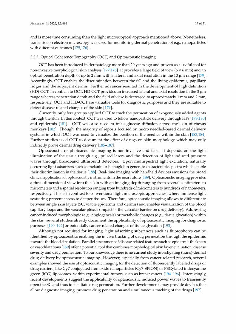
3.2.3. Optical Coherence Tomography (OCT) and Optoacoustic Imaging
OCT has been introduced in dermatology more than 20 years ago and proven as a useful tool fornon-invasive morphological skin analysis [177,178]. It provides a large field of view (6 × 6 mm) and anoptical penetration depth of up to 2 mm with a lateral and axial resolution in the 10 µm range [179].Accordingly, OCT enables the discrimination between the SC and the living epidermis, papillaryridges and the subjacent dermis. Further advances resulted in the development of high definition(HD)-OCT. In contrast to OCT, HD-OCT provides an increased lateral and axial resolution in the 3 µmrange whereas penetration depth and the field of view is decreased to approximately 1 mm and 2 mm,respectively. OCT and HD-OCT are valuable tools for diagnostic purposes and they are suitable todetect disease-related changes of the skin [179].
Currently, only few groups applied OCT to track the permeation of exogenously added agentsthrough the skin. In this context, OCT was used to follow nanoparticle delivery through HFs [173,180]and epidermis [181]. OCT was also used to track glucose diffusion across the skin of rhesusmonkeys [182]. Though, the majority of reports focused on micro needled-based dermal deliverysystems in which OCT was used to visualize the position of the needles within the skin [183,184].Further studies used OCT to document the effect of drugs on skin morphology which may onlyindirectly prove dermal drug delivery [185–187].
Optoacoustic or photoacoustic imaging is non-invasive and fast. It depends on the lightillumination of the tissue trough e.g., pulsed lasers and the detection of light induced pressurewaves through broadband ultrasound detectors. Upon multispectral light excitation, naturallyoccurring light adsorbers such as melanin or hemoglobin generate characteristic spectra which enabletheir discrimination in the tissue [188]. Real-time imaging with handheld devices envisions the broadclinical application of optoacoustic instruments in the near future [189]. Optoacoustic imaging providesa three-dimensional view into the skin with an imaging depth ranging from several centimeters tomicrometers and a spatial resolution ranging from hundreds of micrometers to hundreds of nanometers,respectively. This is in contrast to conventional light microscopic approaches, where immense lightscattering prevent access to deeper tissues. Therefore, optoacoustic imaging allows to differentiatebetween single skin layers (SC, viable epidermis and dermis) and enables visualization of the bloodcapillary loops and the vascular plexus (impact of the vascular barrier on drug delivery). Addressingcancer-induced morphologic (e.g., angiogenesis) or metabolic changes (e.g., tissue glycation) withinthe skin, several studies already document the applicability of optoacoustic imaging for diagnosticpurposes [190–192] or potentially cancer-related changes of tissue glycation [193].
Although not required for imaging, light adsorbing substances such as fluorophores can beidentified by optoacoustics enabling the in vivo tracking of drug permeation through the epidermistowards the blood circulation. Parallel assessment of disease related features such as epidermis thicknessor vasodilatations [159] offer a potential tool that combines morphological skin layer evaluation, diseaseseverity and drug permeation. To our knowledge there is no current study investigating (trans)-dermaldrug delivery by optoacoustic imaging. However, especially from cancer-related research, severalexamples showed the use of optoacoustic imaging for the detection of fluorescently labelled drugs ordrug carriers, like Cy7 conjugated iron oxide nanoparticles (Cy7-SPION) or PEGylated indocyaninegreen (ICG) liposomes, within experimental tumors such as breast cancer [194–196]. Interestingly,recent developments suggest the applicability of optoacoustic induced power waves to transientlyopen the SC and thus to facilitate drug permeation. Further developments may provide devices thatallow diagnostic imaging, promote drug penetration and simultaneous tracking of the drugs [197].

Pharmaceutics 2020, 12, 684 18 of 31
3.2.4. Confocal Laser Scanning Microscopy (CLSM)
CLSM and especially confocal fluorescence microscopy can be a valuable tool to investigatethe skin barrier and the delivery of drugs in vitro, ex vivo and in vivo. It can be very beneficialfor the investigation of immunofluorescence stainings in 3D samples [170] (see also Section 3.2.1).Compared to classical bright field and fluorescence microscopy, CLSM uses a focused laser beam toexcite the sample point-wise on a raster in x and y direction. The emission is then spatially filtered bya confocal pinhole to block out-of-focus light, sacrificing image intensity in exchange for improvedimage contrast compared to bright field microscopy. Image acquisition at multiple steps in z directionand subsequent three-dimensional reconstruction allows for investigation of skin structures in all threespace dimensions.
Besides imaging of endogenous fluorophores like reduced nicotinamide adenine dinucleotide(NADH), collagen or melanin, to illuminate the spatial structure of the skin, dermal drug delivery canbe probed by fluorescence labelling of the drug or—in special cases, if the drug itself has fluorescentproperties—without labelling [198].
For example, Alvarez-Román et al. investigated the skin penetration and distribution of polymericnanoparticles in porcine skin ex vivo by CLSM and observed an accumulation in HFs and skinfurrows [199].
Another application of CLSM is reflectance confocal microscopy (RCM), which uses backscatteredlight instead of fluorescence emission for imaging. In vivo RCM is frequently used as a diagnostic toolin dermatological routine [200], but has also many potential use cases in skin barrier research due toalmost real-time imaging capabilities and relatively modest acquisition costs.
3.2.5. Multiphoton Microscopy (MPM)
After its first operability demonstration 30 years ago in a laboratory setting [201], MPM overcomesthe limit of explorative practice and developed as a powerful instrument in the area of skin basicresearch and increasingly clinical applications [202,203].
Three photonic component parts define MPM’s applicability for the characterization of human skin.Based on the nonlinear excitation of naturally occurring endogenous fluorophores such as
NADH, flavin adenine dinucleotide, tryptophan, porphyrins and melanin [204], and protein structures(e.g., elastin, collagen) [205,206], the morphological pattern and layering of the skin can be evaluatedon a subcellular level. Beyond the reflection in the outermost barrier of the epidermis—the SC—thefirst living cell in the viable epidermis can be assessed in detail with the incidental finding of aprecise discrimination of the SC/SG transition. The principle of two-photon excitation overcomes thelimitations of fluorescence imaging, e.g., operation of CLSM in fluorescence mode, for innocuous in vivoutilization. Here, with a negligible linear energy transfer at the boundary layer, the excitation almostexclusively takes place within the target volume of examination without damaging the surroundingtissue [207].
The additional utilization of the nonlinear optical process second harmonic generation (SHG),observed in noncentrosymmetric molecular structures such as collagen [163,208], allows, next to itsapplication in the area of dermal fibril characterization up to a penetration depth of approximately200 µm [209–211], the exact discrimination of the dermo–epidermal junction and consecutively theepidermal thickness.
Third, MPM additionally offers the applicability of fluorescence lifetime imaging microscopy(FLIM) [163,212]. Since the lifetime of a fluorophore—the time it spends in the excited state beforereturning to the ground state by photon emission—is characteristic for a fluorophore in its biochemicalenvironment [213,214], FLIM facilitates a distinct in vivo identification of fluorescent molecules thatshare an overlap in their morphological or spectroscopic pattern within human skin. By time-correlatedsingle photon counting [213] or phasor analysis [215] selective visualization of endogenous fluorophoresor topical drug uptake was successful at high spatial resolutions [216].

Pharmaceutics 2020, 12, 684 19 of 31
Therefore, MPM FLIM enables for non-invasive high-resolution examination of human skinin vitro, ex vivo and in clinical in vivo applications. On this basis, clinical studies focused on bothhigh-resolution morphological patterns and pathophysiological variances on a cellular level e.g., in theprocess of skin wound healing [217,218]. Next to MPM FLIM application in advanced skin cancerdiagnosis [203,219], also the skin barrier function itself became the focus of attention: Volz as well asFrombach used high-resolution Cluster-FLIM in in vitro- and ex vivo-approaches to explicitly inspectthe dynamics of both drug penetration and the penetrated skin area itself [163,216,220–222].
Transferring skin barrier analysis to ‘patient’s bedside’, Ogawa-Fuse examined skin hydrationutilizing a combination of already mentioned techniques such as CARS/Raman and MPM FLIM [220].Moreover, the same technical combination facilitated the measurement of pharmaceutical and cosmeticdrug penetration and delivery through the skin, e.g., minocycline and tazarotene [163,216,221,222].
The combination of mentioned non-invasive spectroscopic and microscopic techniques suchas CARS/Raman spectroscopy, MPM and associated techniques provide a huge potential for theinvestigation of the skin barrier and the related impact on dermal or even transdermal drug delivery.Sophisticated image analysis through artificial intelligence may open up the broad application ofhigh-resolution skin microscopy in skin research and clinics [164].
4. Outlook/Conclusions
The present review highlights current knowledge on skin barriers that have to be overcome bya drug or its carrier system and tools that allow the localization and quantification of skin barrierfunctions, skin barrier components or tracking of tracer molecules on their way through the skin fromoutside to inside and from inside-out. The SC is considered as the most efficient barrier that controls theentrance and permeation of topically applied substances. TJs are a second barrier, especially importantwhen SC is impaired—either by e.g., skin diseases or due to penetration enhancers—or for drugsnot limited by the SC. However, also other parts of the skin may limit or decelerate drug diffusionand systemic delivery. For example, the basement membrane at the interface between dermis andepidermis as dense mesh of structural proteins and carbohydrates or the blood vessels underneath themembrane may play a role in the overall barrier system of the skin. To investigate the skin barrier andto discriminate between different barrier-forming components various tools are available. Immanentlimitations of every method have to be considered for proper interpretation of obtained researchdata. Recent advances in high resolution imaging (e.g., Raman microscopy, optoacoustic imaging andMPM-FLIM) provides pioneering technologies for an improved understanding of the skin barrier onthe one hand and dermal drug delivery on the other hand. Broad application of those novel techniquesis currently limited because of sophisticated image analysis required for accurate data interpretations.One solution for most of these hurdles can be the application of advanced machine learning techniques.Especially automatic image segmentation and structure recognition based on supervised learning ofdeep neural networks show promising results on many tasks if the required amount of annotatedtraining data is available or can be generated without too much effort. Steady progress in skin barrierresearch and increasing interest in (trans) dermal drug delivery will push forward the development ofeven more delicate instruments that extend the toolbox of dermatological and pharmaceutical research.Among many other interesting methods, advances in electrical impedance spectroscopy, Cluster-FLIM,X-ray or stimulated Raman spectromicroscopy [134,223,224] envision an improved view on skin barrierfunctions and associated structures as well as the simultaneous tracking of distinct molecules withinthe skin.
Author Contributions: All authors contributed to conception and writing of the manuscript as well as designingthe figures. All authors have read and agreed to the published version of the manuscript.
Funding: This research was funded by the Deutsche Forschungsgemeinschaft Grant No: BR 1982-4/1 (JMB).

Pharmaceutics 2020, 12, 684 20 of 31
Acknowledgments: We sincerely thank Dorothee Günzel, and Roman Mannweiler, Charité Berlin and HartmutSchlüter, University Hospital Hamburg-Eppendorf, Hamburg, for pictures Figure 3B,C respectively. In addition,we highly appreciate the photographic assistance of Antonia Burmeister, Sabine Vidal and Germar Schüring.
Conflicts of Interest: The authors declare no conflict of interest.
References
1. Lee, A.Y. Molecular Mechanism of Epidermal Barrier Dysfunction as Primary Abnormalities. Int. J. Mol. Sci.2020, 21, 1194. [CrossRef] [PubMed]
2. Basler, K.; Bergmann, S.; Heisig, M.; Naegel, A.; Zorn-Kruppa, M.; Brandner, J.M. The role of tight junctionsin skin barrier function and dermal absorption. J. Control. Release 2016, 242, 105–118. [CrossRef] [PubMed]
3. Yokouchi, M.; Kubo, A. Maintenance of tight junction barrier integrity in cell turnover and skin diseases.Exp. Dermatol. 2018, 27, 876–883. [CrossRef] [PubMed]
4. Patzelt, A.; Lademann, J. Recent advances in follicular drug delivery of nanoparticles. Expert Opin. Drug Deliv.2020, 17, 49–60. [CrossRef]
5. Dragicevic, N.; Maibach, H. Percutaneous Penetration Enhancers Drug Penetration Into/Through the Skin:Methodology and General Considerations, 1st ed.; Springer: Berlin, Germany, 2017; p. 414.
6. Ishida-Yamamoto, A.; Igawa, S.; Kishibe, M.; Honma, M. Clinical and molecular implications of structuralchanges to desmosomes and corneodesmosomes. J. Dermatol. 2018, 45, 385–389. [CrossRef]
7. Matsui, T.; Amagai, M. Dissecting the formation, structure and barrier function of the stratum corneum.Int. Immunol. 2015, 27, 269–280. [CrossRef]
8. Jonca, N.; Leclerc, E.A.; Caubet, C.; Simon, M.; Guerrin, M.; Serre, G. Corneodesmosomes and corneodesmosin:From the stratum corneum cohesion to the pathophysiology of genodermatoses. Eur J. Dermatol.2011, 21, 35–42. [CrossRef]
9. Haftek, M. ‘Memory’ of the stratum corneum: Exploration of the epidermis’ past. Br. J. Dermatol.2014, 171, 6–9. [CrossRef]
10. Bouwstra, J.A.; Gooris, G.S.; van der Spek, J.A.; Bras, W. Structural investigations of human stratum corneumby small-angle X-ray scattering. J. Invest. Dermatol. 1991, 97, 1005–1012. [CrossRef]
11. Bouwstra, J.A.; Gooris, G.S.; Bras, W.; Downing, D.T. Lipid organization in pig stratum corneum. J. Lipid Res.1995, 36, 685–695.
12. Bouwstra, J.A.; Thewalt, J.; Gooris, G.S.; Kitson, N. A model membrane approach to the epidermalpermeability barrier: An X-ray diffraction study. Biochemistry 1997, 36, 7717–7725. [CrossRef] [PubMed]
13. Boncheva, M.; Damien, F.; Normand, V. Molecular organization of the lipid matrix in intact Stratum corneumusing ATR-FTIR spectroscopy. Biochim. Biophys. Acta (BBA) Biomembr. 2008, 1778, 1344–1355. [CrossRef][PubMed]
14. Mojumdar, E.H.; Kariman, Z.; van Kerckhove, L.; Gooris, G.S.; Bouwstra, J.A. The role of ceramide chain lengthdistribution on the barrier properties of the skin lipid membranes. Biochim. Biophys. Acta (BBA) Biomembr.2014, 1838, 2473–2483. [CrossRef] [PubMed]
15. van Smeden, J.; Bouwstra, J.A. Stratum Corneum Lipids: Their Role for the Skin Barrier Function inHealthy Subjects and Atopic Dermatitis Patients. In Skin Barrier Function; Agner, T., Ed.; Karger: Basel,Switherland, 2016; Volume 49, pp. 8–26.
16. Wertz, P.W. Lipids and the Permeability and Antimicrobial Barriers of the Skin. J. Lipids 2018, 2018. [CrossRef][PubMed]
17. Naik, A.; Guy, R. Infrared spectroscopic and differential scanning calorimetric investigations of the stratumcorneum barrier function. Drugs Pharm. Sci. 1997, 83, 87–162.
18. Ludovici, M.; Kozul, N.; Materazzi, S.; Risoluti, R.; Picardo, M.; Camera, E. Influence of the sebaceous glanddensity on the stratum corneum lipidome. Sci. Rep. 2018, 8, 11500. [CrossRef]
19. Ishida-Yamamoto, A.; Igawa, S.; Kishibe, M. Molecular basis of the skin barrier structures revealed byelectron microscopy. Exp. Dermatol. 2018, 27, 841–846. [CrossRef]
20. Candi, E.; Schmidt, R.; Melino, G. The cornified envelope: A model of cell death in the skin. Nat. Rev. Mol.Cell Biol. 2005, 6, 328–340. [CrossRef]

Pharmaceutics 2020, 12, 684 21 of 31
21. Janssens, M.; van Smeden, J.; Gooris, G.S.; Bras, W.; Portale, G.; Caspers, P.J.; Vreeken, R.J.; Hankemeier, T.;Kezic, S.; Wolterbeek, R.; et al. Increase in short-chain ceramides correlates with an altered lipid organizationand decreased barrier function in atopic eczema patients. J. Lipid Res. 2012, 53, 2755–2766. [CrossRef]
22. Breitkreutz, D.; Koxholt, I.; Thiemann, K.; Nischt, R. Skin basement membrane: The foundation of epidermalintegrity–BM functions and diverse roles of bridging molecules nidogen and perlecan. Biomed. Res. Int.2013, 2013, 179784. [CrossRef] [PubMed]
23. Monash, S.; Blank, H. Location and reformation of the epithelial barrier to water vapor. AMA Arch. Derm.1958, 78, 710–714. [CrossRef] [PubMed]
24. Rancan, F.; Giulbudagian, M.; Jurisch, J.; Blume-Peytavi, U.; Calderon, M.; Vogt, A. Drug deliveryacross intact and disrupted skin barrier: Identification of cell populations interacting with penetratedthermoresponsive nanogels. Eur. J. Pharm. Biopharm. 2017, 116, 4–11. [CrossRef] [PubMed]
25. Parhi, R.; Suresh, P.; Patnaik, S. Physical means of stratum corneum barrier manipulation to enhancetransdermal drug delivery. Curr. Drug Deliv. 2015, 12, 122–138. [CrossRef] [PubMed]
26. Sklar, L.R.; Burnett, C.T.; Waibel, J.S.; Moy, R.L.; Ozog, D.M. Laser assisted drug delivery: A review of anevolving technology. Lasers Surg. Med. 2014, 46, 249–262. [CrossRef] [PubMed]
27. Asfour, M.H. Advanced trends in protein and peptide drug delivery: A special emphasis on aquasomes andmicroneedles techniques. Drug Deliv. Transl. Res. 2020. [CrossRef]
28. Del Rio-Sancho, S.; Lapteva, M.; Sonaje, K.; Bohler, C.; Ling, V.; Boehncke, W.H.; Kalia, Y.N. Targetedcutaneous delivery of etanercept using Er:YAG fractional laser ablation. Int. J. Pharm. 2020, 580, 119234.[CrossRef]
29. Kezic, S.; Jakasa, I. Filaggrin and Skin Barrier Function. Curr. Probl. Dermatol. 2016, 49, 1–7. [CrossRef][PubMed]
30. McLean, W.H. Filaggrin failure-from ichthyosis vulgaris to atopic eczema and beyond. Br. J. Dermatol.2016, 175, 4–7. [CrossRef]
31. Kawasaki, H.; Nagao, K.; Kubo, A.; Hata, T.; Shimizu, A.; Mizuno, H.; Yamada, T.; Amagai, M. Alteredstratum corneum barrier and enhanced percutaneous immune responses in filaggrin-null mice. J. AllergyClin. Immunol. 2012, 129, 1538–1546. [CrossRef]
32. Thyssen, J.P.; Jakasa, I.; Riethmuller, C.; Schon, M.P.; Braun, A.; Haftek, M.; Fallon, P.G.; Wroblewski, J.;Jakubowski, H.; Eckhart, L.; et al. Filaggrin Expression and Processing Deficiencies Impair CorneocyteSurface Texture and Stiffness in Mice. J. Investig. Dermatol. 2020, 140, 615–623. [CrossRef]
33. Niehues, H.; Schalkwijk, J.; van Vlijmen-Willems, I.; Rodijk-Olthuis, D.; van Rossum, M.M.; Wladykowski, E.;Brandner, J.M.; van den Bogaard, E.H.J.; Zeeuwen, P. Epidermal equivalents of filaggrin null keratinocytesdo not show impaired skin barrier function. J. Allergy Clin. Immunol. 2017, 139, 1979–1981 e1913. [CrossRef][PubMed]
34. Kezic, S. Loss-of-function mutations in filaggrin gene and malignant melanoma. J. Eur. Acad.Dermatol. Venereol. 2018, 32, 193. [CrossRef] [PubMed]
35. Proksch, E. pH in nature, humans and skin. J. Dermatol 2018, 45, 1044–1052. [CrossRef] [PubMed]36. Sharma, S.; Kaur, T.; Malhotra, S.K.; Rai, J.; Chaudhari, S. Correlation of Vitamin D3 Levels and SCORAD
Index in Atopic Dermatits: A Case Control Study. J. Clin. Diagn. Res. 2017, 11, WC01–WC03. [CrossRef][PubMed]
37. Marenholz, I.; Rivera, V.A.; Esparza-Gordillo, J.; Bauerfeind, A.; Lee-Kirsch, M.A.; Ciechanowicz, A.;Kurek, M.; Piskackova, T.; Macek, M.; Lee, Y.A. Association screening in the Epidermal DifferentiationComplex (EDC) identifies an SPRR3 repeat number variant as a risk factor for eczema. J. Investig. Dermatol.2011, 131, 1644–1649. [CrossRef] [PubMed]
38. Catunda, R.; Rekhi, U.; Clark, D.; Levin, L.; Febbraio, M. Loricrin downregulation and epithelial-relateddisorders: A systematic review. J. Dtsch. Dermatol. Ges. 2019, 17, 1227–1238. [CrossRef] [PubMed]
39. Moosbrugger-Martinz, V.; Jalili, A.; Schossig, A.S.; Jahn-Bassler, K.; Zschocke, J.; Schmuth, M.; Stingl, G.;Eckl, K.M.; Hennies, H.C.; Gruber, R. Epidermal barrier abnormalities in exfoliative ichthyosis with a novelhomozygous loss-of-function mutation in CSTA. Br. J. Dermatol. 2015, 172, 1628–1632. [CrossRef]
40. Muttardi, K.; Nitoiu, D.; Kelsell, D.P.; O’Toole, E.A.; Batta, K. Acral peeling skin syndrome associated with anovel CSTA gene mutation. Clin. Exp. Dermatol. 2016, 41, 394–398. [CrossRef]
41. Rice, R.H.; Durbin-Johnson, B.P.; Ishitsuka, Y.; Salemi, M.; Phinney, B.S.; Rocke, D.M.; Roop, D.R. ProteomicAnalysis of Loricrin Knockout Mouse Epidermis. J. Proteome Res. 2016, 15, 2560–2566. [CrossRef]

Pharmaceutics 2020, 12, 684 22 of 31
42. Boczonadi, V.; Maatta, A. Functional Analysis of Periplakin and Envoplakin, Cytoskeletal Linkers, andCornified Envelope Precursor Proteins. In Methods in Enzymology; Elsevier: Amsterdam, The Netherlands,2016; Volume 569, pp. 309–329. [CrossRef]
43. Cangkrama, M.; Darido, C.; Georgy, S.R.; Partridge, D.; Auden, A.; Srivastava, S.; Wilanowski, T.; Jane, S.M.Two Ancient Gene Families Are Critical for Maintenance of the Mammalian Skin Barrier in Postnatal Life.J. Investig. Dermatol. 2016, 136, 1438–1448. [CrossRef]
44. van der Velden, J.J.; van Geel, M.; Nellen, R.G.; Jonkman, M.F.; McGrath, J.A.; Nanda, A.; Sprecher, E.;van Steensel, M.A.; McLean, W.H.; Cassidy, A.J. Novel TGM5 mutations in acral peeling skin syndrome.Exp. Dermatol. 2015, 24, 285–289. [CrossRef] [PubMed]
45. Karim, N.; Phinney, B.S.; Salemi, M.; Wu, P.W.; Naeem, M.; Rice, R.H. Human stratum corneum proteomicsreveals cross-linking of a broad spectrum of proteins in cornified envelopes. Exp. Dermatol. 2019, 28, 618–622.[CrossRef] [PubMed]
46. Takeda, M.; Nomura, T.; Sugiyama, T.; Miyauchi, T.; Suzuki, S.; Fujita, Y.; Shimizu, H. Compound heterozygousmissense mutations p.Leu207Pro and p.Tyr544Cys in TGM1 cause a severe form of lamellar ichthyosis. J.Dermatol. 2018, 45, 1463–1467. [CrossRef]
47. Has, C. Peeling Skin Disorders: A Paradigm for Skin Desquamation. J. Investig. Dermatol.2018, 138, 1689–1691. [CrossRef] [PubMed]
48. Leclerc, E.A.; Huchenq, A.; Mattiuzzo, N.R.; Metzger, D.; Chambon, P.; Ghyselinck, N.B.; Serre, G.;Jonca, N.; Guerrin, M. Corneodesmosin gene ablation induces lethal skin-barrier disruption and hair-follicledegeneration related to desmosome dysfunction. J. Cell Sci. 2009, 122, 2699–2709. [CrossRef]
49. Grubauer, G.; Elias, P.M.; Feingold, K.R. Transepidermal water loss: The signal for recovery of barrierstructure and function. J. Lipid Res. 1989, 30, 323–333.
50. Sajic, D.; Asiniwasis, R.; Skotnicki-Grant, S. A look at epidermal barrier function in atopic dermatitis:Physiologic lipid replacement and the role of ceramides. Skin Ther. Lett. 2012, 17, 6–9.
51. Kihara, A. Synthesis and degradation pathways, functions, and pathology of ceramides andepidermal acylceramides. Prog. Lipid Res. 2016, 63, 50–69. [CrossRef]
52. Bouwstra, J.A.; Honeywell-Nguyen, P.L.; Gooris, G.S.; Ponec, M. Structure of the skin barrier and itsmodulation by vesicular formulations. Prog. Lipid Res. 2003, 42, 1–36. [CrossRef]
53. Williams, A.C.; Barry, B.W. Penetration enhancers. Adv. Drug Deliv. Rev. 2004, 56, 603–618. [CrossRef]54. Dragicevic, N.; Atkinson, J.P.; Maibach, H.I. Chemical penetration enhancers: Classification and mode
of action. In Percutaneous Penetration Enhancers Chemical Methods in Penetration Enhancement; Springer:Berlin/Heidelberg, Germany, 2015; pp. 11–27.
55. Hadgraft, J. Passive enhancement strategies in topical and transdermal drug delivery. Int. J. Pharm.1999, 184, 1–6. [CrossRef]
56. Shiohara, T.; Sato, Y.; Komatsu, Y.; Ushigome, Y.; Mizukawa, Y. Sweat as an Efficient Natural Moisturizer.Curr. Probl. Dermatol. 2016, 51, 30–41. [CrossRef] [PubMed]
57. Gorzelanny, C.; Goerge, T.; Schnaeker, E.M.; Thomas, K.; Luger, T.A.; Schneider, S.W. Atomic force microscopyas an innovative tool for nanoanalysis of native stratum corneum. Exp. Dermatol. 2006, 15, 387–391. [CrossRef][PubMed]
58. Wohlrab, J.; Gebert, A.; Neubert, R.H.H. Lipids in the Skin and pH. Curr. Probl. Dermatol. 2018, 54, 64–70.[CrossRef] [PubMed]
59. Piontek, J.; Krug, S.M.; Protze, J.; Krause, G.; Fromm, M. Molecular architecture and assembly of the tightjunction backbone. Biochim. Biophys. Acta (BBA) Biomembr. 2020, 1862, 183279. [CrossRef] [PubMed]
60. Kirschner, N.; Rosenthal, R.; Furuse, M.; Moll, I.; Fromm, M.; Brandner, J.M. Contribution of Tight JunctionProteins to Ion, Macromolecule, and Water Barrier in Keratinocytes. J. Investig. Dermatol. 2013, 133, 1161–1169.[CrossRef] [PubMed]
61. Adams, M.P.; Mallet, D.G.; Pettet, G.J. Towards a quantitative theory of epidermal calcium profile formationin unwounded skin. PLoS ONE 2015, 10, e0116751. [CrossRef]
62. Gunzel, D.; Yu, A.S. Claudins and the modulation of tight junction permeability. Physiol. Rev.2013, 93, 525–569. [CrossRef]
63. Brandner, J.M.; Zorn-Kruppa, M.; Yoshida, T.; Moll, I.; Beck, L.A.; De Benedetto, A. Epidermal tight junctionsin health and disease. Tissue Barriers 2015, 3, e974451. [CrossRef]

Pharmaceutics 2020, 12, 684 23 of 31
64. Furuse, M.; Hata, M.; Furuse, K.; Yoshida, Y.; Haratake, A.; Sugitani, Y.; Noda, T.; Kubo, A.;Tsukita, S. Claudin-based tight junctions are crucial for the mammalian epidermal barrier: A lessonfrom claudin-1-deficient mice. J. Cell Biol. 2002, 156, 1099–1111. [CrossRef]
65. Yokouchi, M.; Kubo, A.; Kawasaki, H.; Yoshida, K.; Ishii, K.; Furuse, M.; Amagai, M. Epidermal tightjunction barrier function is altered by skin inflammation, but not by filaggrin-deficient stratum corneum.J. Dermatol. Sci. 2015, 77, 28–36. [CrossRef] [PubMed]
66. Sugawara, T.; Iwamoto, N.; Akashi, M.; Kojima, T.; Hisatsune, J.; Sugai, M.; Furuse, M. Tightjunction dysfunction in the stratum granulosum leads to aberrant stratum corneum barrier functionin claudin-1-deficient mice. J. Dermatol. Sci. 2013, 70, 12–18. [CrossRef] [PubMed]
67. Yuki, T.; Komiya, A.; Kusaka, A.; Kuze, T.; Sugiyama, Y.; Inoue, S. Impaired tight junctions obstruct stratumcorneum formation by altering polar lipid and profilaggrin processing. J. Dermatol. Sci. 2013, 69, 148–158.[CrossRef] [PubMed]
68. Hadj-Rabia, S.; Baala, L.; Vabres, P.; Hamel-Teillac, D.; Jacquemin, E.; Fabre, M.; Lyonnet, S.; De Prost, Y.;Munnich, A.; Hadchouel, M.; et al. Claudin-1 gene mutations in neonatal sclerosing cholangitis associatedwith ichthyosis: A tight junction disease. Gastroenterology 2004, 127, 1386–1390. [CrossRef]
69. Feldmeyer, L.; Huber, M.; Fellmann, F.; Beckmann, J.S.; Frenk, E.; Hohl, D. Confirmation of the origin ofNISCH syndrome. Hum. Mutat. 2006, 27, 408–410. [CrossRef]
70. Nagtzaam, I.F.; van Geel, M.; Driessen, A.; Steijlen, P.M.; van Steensel, M.A. Bile duct paucity is part of theneonatal ichthyosis-sclerosing cholangitis phenotype. Br. J. Dermatol. 2010, 163, 205–207. [CrossRef]
71. Paganelli, M.; Stephenne, X.; Gilis, A.; Jacquemin, E.; Henrion Caude, A.; Girard, M.; Gonzales, E.; Revencu, N.;Reding, R.; Wanty, C.; et al. Neonatal ichthyosis and sclerosing cholangitis syndrome: Extremely variableliver disease severity from claudin-1 deficiency. J. Pediatr. Gastroenterol. Nutr. 2011, 53, 350–354. [CrossRef]
72. Kirchmeier, P.; Sayar, E.; Hotz, A.; Hausser, I.; Islek, A.; Yilmaz, A.; Artan, R.; Fischer, J. Novel mutation inthe CLDN1 gene in a Turkish family with neonatal ichthyosis sclerosing cholangitis (NISCH) syndrome.Br. J. Dermatol. 2014, 170, 976–978. [CrossRef]
73. Batista, D.I.; Perez, L.; Orfali, R.L.; Zaniboni, M.C.; Samorano, L.P.; Pereira, N.V.; Sotto, M.N.; Ishizaki, A.S.;Oliveira, L.M.; Sato, M.N.; et al. Profile of skin barrier proteins (filaggrin, claudins 1 and 4) and Th1/Th2/Th17cytokines in adults with atopic dermatitis. J. Eur. Acad. Dermatol. Venereol. 2015, 29, 1091–1095. [CrossRef]
74. Bergmann, S.; von Buenau, B.; Vidal, Y.S.S.; Haftek, M.; Wladykowski, E.; Houdek, P.; Lezius, S.; Duplan, H.;Basler, K.; Dahnhardt-Pfeiffer, S.; et al. Claudin-1 decrease impacts epidermal barrier function in atopicdermatitis lesions dose-dependently. Sci. Rep. 2020, 10, 2024. [CrossRef]
75. Gruber, R.; Bornchen, C.; Rose, K.; Daubmann, A.; Volksdorf, T.; Wladykowski, E.; Vidal, Y.S.S.; Peters, E.M.;Danso, M.; Bouwstra, J.A.; et al. Diverse regulation of claudin-1 and claudin-4 in atopic dermatitis.Am. J. Pathol. 2015, 185, 2777–2789. [CrossRef]
76. De Benedetto, A.; Slifka, M.K.; Rafaels, N.M.; Kuo, I.H.; Georas, S.N.; Boguniewicz, M.; Hata, T.;Schneider, L.C.; Hanifin, J.M.; Gallo, R.L.; et al. Reductions in claudin-1 may enhance susceptibilityto herpes simplex virus 1 infections in atopic dermatitis. J. Allergy Clin. Immunol. 2011, 128, 242–246 e245.[CrossRef]
77. Yoshida, K.; Kubo, A.; Fujita, H.; Yokouchi, M.; Ishii, K.; Kawasaki, H.; Nomura, T.; Shimizu, H.; Kouyama, K.;Ebihara, T.; et al. Distinct behavior of human Langerhans cells and inflammatory dendritic epidermal cells attight junctions in patients with atopic dermatitis. J. Allergy Clin. Immunol. 2014, 134, 856–864. [CrossRef][PubMed]
78. Tokumasu, R.; Yamaga, K.; Yamazaki, Y.; Murota, H.; Suzuki, K.; Tamura, A.; Bando, K.; Furuta, Y.;Katayama, I.; Tsukita, S. Dose-dependent role of claudin-1 in vivo in orchestrating features of atopic dermatitis.Proc. Natl. Acad. Sci. USA 2016, 113, E4061–E4068. [CrossRef] [PubMed]
79. Brewer, M.G.; Anderson, E.A.; Pandya, R.P.; De Benedetto, A.; Yoshida, T.; Hilimire, T.A.; Martinez-Sobrido, L.;Beck, L.A.; Miller, B.L. Peptides Derived from the Tight Junction Protein CLDN1 Disrupt the Skin Barrierand Promote Responsiveness to an Epicutaneous Vaccine. J. Investig. Dermatol. 2020, 140, 361–369 e363.[CrossRef] [PubMed]
80. Beier, L.S.; Rossa, J.; Woodhouse, S.; Bergmann, S.; Kramer, H.B.; Protze, J.; Eichner, M.; Piontek, A.;Vidal, Y.S.S.; Brandner, J.M.; et al. Use of Modified Clostridium perfringens Enterotoxin Fragments forClaudin Targeting in Liver and Skin Cells. Int. J. Mol. Sci. 2019, 20, 4774. [CrossRef] [PubMed]

Pharmaceutics 2020, 12, 684 24 of 31
81. Nakajima, M.; Nagase, S.; Iida, M.; Takeda, S.; Yamashita, M.; Watari, A.; Shirasago, Y.; Fukasawa, M.;Takeda, H.; Sawasaki, T.; et al. Claudin-1 Binder Enhances Epidermal Permeability in a HumanKeratinocyte Model. J. Pharmacol. Exp. Ther. 2015, 354, 440–447. [CrossRef]
82. Uchida, T.; Kanazawa, T.; Takashima, Y.; Okada, H. Development of an efficient transdermal delivery systemof small interfering RNA using functional peptides, Tat and AT-1002. Chem. Pharm. Bull. 2011, 59, 196–201.[CrossRef]
83. Ibaraki, H.; Kanazawa, T.; Kurano, T.; Oogi, C.; Takashima, Y.; Seta, Y. Anti-RelA siRNA-Encapsulated FlexibleLiposome with Tight Junction-Opening Peptide as a Non-invasive Topical Therapeutic for Atopic Dermatitis.Biol. Pharm. Bull. 2019, 42, 1216–1225. [CrossRef]
84. Abdayem, R.; Callejon, S.; Portes, P.; Kirilov, P.; Demarne, F.; Pirot, F.; Jannin, V.; Haftek, M. Modulation oftransepithelial electric resistance (TEER) in reconstructed human epidermis by excipients known to permeateintestinal tight junctions. Exp. Dermatol. 2015, 24, 686–691. [CrossRef]
85. Nystrom, A.; Bruckner-Tuderman, L. Matrix molecules and skin biology. In Seminars in Cell and DevelopmentalBiology; Elesvier: Amsterdam, The Netherlands, 2019; Volume 89, pp. 136–146. [CrossRef]
86. Witte, M.; Zillikens, D.; Schmidt, E. Diagnosis of Autoimmune Blistering Diseases. Front. Med. 2018, 5, 296.[CrossRef] [PubMed]
87. Stemmler, S.; Parwez, Q.; Petrasch-Parwez, E.; Epplen, J.T.; Hoffjan, S. Association of variation in theLAMA3 gene, encoding the alpha-chain of laminin 5, with atopic dermatitis in a German case-control cohort.BMC Dermatol. 2014, 14, 17. [CrossRef] [PubMed]
88. Kim, T.I.; Park, H.J.; Won, Y.Y.; Choi, H.; Jeong, K.H.; Sung, J.Y.; Shin, M.K. Basement Membrane StatusIs Intact in Urticarial Dermatitis vs. Adult-Onset Atopic Dermatitis. Ann. Dermatol. 2018, 30, 258–261.[CrossRef] [PubMed]
89. Schreiner, E.; Wolff, K. The permeability of the intercellular space of the epidermis for low molecular weightprotein. Electron microscopic cytochemical studies with peroxidase as a tracer substance. Archiv fur Klinischeund Experimentelle Dermatologie 1969, 235, 78–88. [CrossRef] [PubMed]
90. Wolff, K.; Honigsmann, H. Permeability of the epidermis and the phagocytic activity of keratinocytes.Ultrastructural studies with thorotrast as a marker. J. Ultrastruct. Res. 1971, 36, 176–190. [CrossRef]
91. Briggaman, R.A.; Wheeler, C.E., Jr. The epidermal-dermal junction. J. Investig. Dermatol. 1975, 65, 71–84.[CrossRef]
92. Weeks, B.S.; Ramchandran, R.S.; Hopkins, J.J.; Friedman, H.M. Herpes simplex virus type-1 and -2pathogenesis is restricted by the epidermal basement membrane. Arch. Virol. 2000, 145, 385–396. [CrossRef]
93. Kazama, T.; Yaoita, E.; Ito, M.; Sato, Y. Charge-selective permeability of dermo-epidermal junction: Tracerstudies with cationic and anionic ferritins. J. Investig. Dermatol. 1988, 91, 560–565. [CrossRef]
94. Oh, J.W.; Kloepper, J.; Langan, E.A.; Kim, Y.; Yeo, J.; Kim, M.J.; Hsi, T.C.; Rose, C.; Yoon, G.S.; Lee, S.J.; et al.A Guide to Studying Human Hair Follicle Cycling In Vivo. J. Investig. Dermatol. 2016, 136, 34–44. [CrossRef]
95. Lademann, J.; Knorr, F.; Richter, H.; Blume-Peytavi, U.; Vogt, A.; Antoniou, C.; Sterry, W.;Patzelt, A. Hair follicles–an efficient storage and penetration pathway for topically applied substances.Skin Pharmacol. Physiol. 2008, 21, 150–155. [CrossRef]
96. Vogt, A.; Blume-Peytavi, U. Selective hair therapy: Bringing science to the fiction. Exp. Dermatol.2014, 23, 83–86. [CrossRef] [PubMed]
97. Zorn-Kruppa, M.; Vidal, Y.S.S.; Houdek, P.; Wladykowski, E.; Grzybowski, S.; Gruber, R.; Gorzelanny, C.;Harcup, J.; Schneider, S.W.; Majumdar, A.; et al. Tight Junction barriers in human hair follicles-roleof claudin-1. Sci. Rep. 2018, 8, 12800. [CrossRef]
98. Blume-Peytavi, U.; Vogt, A. Human hair follicle: Reservoir function and selective targeting. Br. J. Dermatol.2011, 165, 13–17. [CrossRef] [PubMed]
99. Morita, K.; Itoh, M.; Saitou, M.; Ando-Akatsuka, Y.; Furuse, M.; Yoneda, K.; Imamura, S.; Fujimoto, K.;Tsukita, S. Subcellular distribution of tight junction-associated proteins (occludin, ZO-1, ZO-2) in rodent skin.J. Investig. Dermatol. 1998, 110, 862–866. [CrossRef] [PubMed]
100. Tebbe, B.; Mankertz, J.; Schwarz, C.; Amasheh, S.; Fromm, M.; Assaf, C.; Schultz-Ehrenburg, U.; SanchezRuderish, H.; Schulzke, J.D.; Orfanos, C.E. Tight junction proteins: A novel class of integral membraneproteins. Expression in human epidermis and in HaCaT keratinocytes. Arch. Dermatol. Res. 2002, 294, 14–18.[CrossRef] [PubMed]

Pharmaceutics 2020, 12, 684 25 of 31
101. Brandner, J.M.; McIntyre, M.; Kief, S.; Wladykowski, E.; Moll, I. Expression and localization of tightjunction-associated proteins in human hair follicles. Arch. Dermatol. Res. 2003, 295, 211–221. [CrossRef][PubMed]
102. Raymond, K.; Richter, A.; Kreft, M.; Frijns, E.; Janssen, H.; Slijper, M.; Praetzel-Wunder, S.; Langbein, L.;Sonnenberg, A. Expression of the orphan protein Plet-1 during trichilemmal differentiation of anagenhair follicles. J. Investig. Dermatol. 2010, 130, 1500–1513. [CrossRef] [PubMed]
103. Atsugi, T.; Yokouchi, M.; Hirano, T.; Hirabayashi, A.; Nagai, T.; Ohyama, M.; Abe, T.; Kaneko, M.;Zouboulis, C.C.; Amagai, M.; et al. Holocrine Secretion Occurs outside the Tight Junction Barrier inMulticellular Glands: Lessons from Claudin-1-Deficient Mice. J. Investig. Dermatol. 2020, 140, 298–308 e295.[CrossRef]
104. Hashimoto, K. Demonostration of the intercellular spaces of the human eccrine sweat gland by lanthanum. I.The secretory coil. J. Ultrastruct. Res. 1971, 36, 249–262. [CrossRef]
105. Hashimoto, K. Demonstration of the intercellular spaces of the human eccrine sweat gland by lanthanum. II.The duct. J. Ultrastruct. Res. 1971, 37, 504–520. [CrossRef]
106. Briggman, J.V.; Bank, H.L.; Bigelow, J.B.; Graves, J.S.; Spicer, S.S. Structure of the tight junctions of the humaneccrine sweat gland. Am. J. Anat. 1981, 162, 357–368. [CrossRef]
107. Wilke, K.; Wepf, R.; Keil, F.J.; Wittern, K.P.; Wenck, H.; Biel, S.S. Are sweat glands an alternatepenetration pathway? Understanding the morphological complexity of the axillary sweat gland apparatus.Skin Pharmacol. Physiol. 2006, 19, 38–49. [CrossRef] [PubMed]
108. Klar, J.; Piontek, J.; Milatz, S.; Tariq, M.; Jameel, M.; Breiderhoff, T.; Schuster, J.; Fatima, A.; Asif, M.;Sher, M.; et al. Altered paracellular cation permeability due to a rare CLDN10B variant causes anhidrosisand kidney damage. PLoS Genet. 2017, 13, e1006897. [CrossRef] [PubMed]
109. Yamaga, K.; Murota, H.; Tamura, A.; Miyata, H.; Ohmi, M.; Kikuta, J.; Ishii, M.; Tsukita, S.; Katayama, I.Claudin-3 Loss Causes Leakage of Sweat from the Sweat Gland to Contribute to the Pathogenesis ofAtopic Dermatitis. J. Investig. Dermatol. 2018, 138, 1279–1287. [CrossRef] [PubMed]
110. Jajack, A.; Brothers, M.; Kasting, G.; Heikenfeld, J. Enhancing glucose flux into sweat by increasing paracellularpermeability of the sweat gland. PLoS ONE 2018, 13, e0200009. [CrossRef]
111. Petrofsky, J.S. Control of Skin Blood Flow. In Textbook of Aging Skin; Farage, M.A., Miller, K.W.,Maibach, H.I., Eds.; Springer: Berlin/Heidelberg, Germany, 2017; pp. 1091–1104. [CrossRef]
112. Kaiyala, K.J. Energy Homeostasis: Thermoregulation. In Encyclopedia of Neuroscience; Squire, L.R., Ed.;Elsevier: Oxford, UK, 2009; pp. 1043–1052. [CrossRef]
113. Hao, J.; Ghosh, P.; Li, S.K.; Newman, B.; Kasting, G.B.; Raney, S.G. Heat effects on drug delivery acrosshuman skin. Expert Opin. Drug Deliv. 2016, 13, 755–768. [CrossRef] [PubMed]
114. Nanjwade, B.K. Novel Drug Delivery Systems and Regulatory Affairs; CBS PUBL & DIST PVT Limited I: Delhi,India, 2019.
115. Shomaker, T.S.; Zhang, J.; Ashburn, M.A. Assessing the impact of heat on the systemic delivery of fentanylthrough the transdermal fentanyl delivery system. Pain Med. 2000, 1, 225–230. [CrossRef] [PubMed]
116. Fujimura, A.; Sasaki, M.; Harada, K.; Kumagai, Y.; Ohashi, K.; Ebihara, A. Influences of bathing andhot weather on the pharmacokinetics of a new transdermal clonidine, M-5041T. J. Clin. Pharmacol.1996, 36, 892–896. [CrossRef]
117. Shomaker, T.S.; Zhang, J.; Ashburn, M.A. A pilot study assessing the impact of heat on the transdermaldelivery of testosterone. J. Clin. Pharmacol. 2001, 41, 677–682. [CrossRef]
118. Petersen, K.K.; Rousing, M.L.; Jensen, C.; Arendt-Nielsen, L.; Gazerani, P. Effect of local controlled heat ontransdermal delivery of nicotine. Int. J. Physiol. Pathophysiol. Pharmacol. 2011, 3, 236–242.
119. Alexander, H.; Brown, S.; Danby, S.; Flohr, C. Research Techniques Made Simple: Transepidermal Water LossMeasurement as a Research Tool. J. Investig. Dermatol. 2018, 138, 2295–2300 e2291. [CrossRef] [PubMed]
120. Jungersted, J.M.; Scheer, H.; Mempel, M.; Baurecht, H.; Cifuentes, L.; Hogh, J.K.; Hellgren, L.I.; Jemec, G.B.;Agner, T.; Weidinger, S. Stratum corneum lipids, skin barrier function and filaggrin mutations in patientswith atopic eczema. Allergy 2010, 65, 911–918. [CrossRef] [PubMed]
121. Kopecna, M.; Machacek, M.; Novackova, A.; Paraskevopoulos, G.; Roh, J.; Vavrova, K. Esters of terpenealcohols as highly potent, reversible, and low toxic skin penetration enhancers. Sci. Rep. 2019, 9, 14617.[CrossRef]

Pharmaceutics 2020, 12, 684 26 of 31
122. Kazarian, S.G.; Chan, K.L. Applications of ATR-FTIR spectroscopic imaging to biomedical samples.Biochim. Biophys. Acta (BBA) Biomembr. 2006, 1758, 858–867. [CrossRef] [PubMed]
123. Kiani, P.; Schlüter, H. Mass Spectrometric Investigation and Determination of Proteome Composition ofHuman Skin Tissues Ablated Using Picosecond Infrared Laser (PIRL) in a “layer by Layer” Approach.Ph.D. Thesis, University of Hamburg, Hamburg, Germany, 2019.
124. Franzen, L.; Anderski, J.; Windbergs, M. Quantitative detection of caffeine in human skin by confocal Ramanspectroscopy-A systematic in vitro validation study. Eur. J. Pharm. Biopharm. 2015, 95, 110–116. [CrossRef][PubMed]
125. De Benedetto, A.; Rafaels, N.M.; McGirt, L.Y.; Ivanov, A.I.; Georas, S.N.; Cheadle, C.; Berger, A.E.; Zhang, K.;Vidyasagar, S.; Yoshida, T.; et al. Tight junction defects in patients with atopic dermatitis. J. AllergyClin. Immunol. 2011, 127, 773–786. [CrossRef] [PubMed]
126. Celli, A.; Zhai, Y.; Jiang, Y.J.; Crumrine, D.; Elias, P.M.; Feingold, K.R.; Mauro, T.M. Tight junction propertieschange during epidermis development. Exp. Dermatol. 2012, 21, 798–801. [CrossRef]
127. Schimek, K.; Hsu, H.H.; Boehme, M.; Kornet, J.J.; Marx, U.; Lauster, R.; Portner, R.; Lindner, G. Bioengineeringof a Full-Thickness Skin Equivalent in a 96-Well Insert Format for Substance Permeation Studies andOrgan-On-A-Chip Applications. Bioengineering 2018, 5, 43. [CrossRef]
128. Kuo, I.H.; Carpenter-Mendini, A.; Yoshida, T.; McGirt, L.Y.; Ivanov, A.I.; Barnes, K.C.; Gallo, R.L.;Borkowski, A.W.; Yamasaki, K.; Leung, D.Y.; et al. Activation of epidermal toll-like receptor 2 enhancestight junction function: Implications for atopic dermatitis and skin barrier repair. J. Investig. Dermatol.2013, 133, 988–998. [CrossRef]
129. Giaever, I.; Keese, C.R. Micromotion of mammalian cells measured electrically. Proc. Natl. Acad. Sci. USA1991, 88, 7896–7900. [CrossRef] [PubMed]
130. Kaiser, M.; Pereira, S.; Pohl, L.; Ketelhut, S.; Kemper, B.; Gorzelanny, C.; Galla, H.J.; Moerschbacher, B.M.;Goycoolea, F.M. Chitosan encapsulation modulates the effect of capsaicin on the tight junctions of MDCK cells.Sci. Rep. 2015, 5, 10048. [CrossRef] [PubMed]
131. Krug, S.M.; Amasheh, S.; Richter, J.F.; Milatz, S.; Gunzel, D.; Westphal, J.K.; Huber, O.; Schulzke, J.D.;Fromm, M. Tricellulin forms a barrier to macromolecules in tricellular tight junctions without affectingion permeability. Mol. Biol. Cell 2009, 20, 3713–3724. [CrossRef]
132. Rinaldi, A.O.; Morita, H.; Wawrzyniak, P.; Dreher, A.; Grant, S.; Svedenhag, P.; Akdis, C.A. Direct assessmentof skin epithelial barrier by electrical impedance spectroscopy. Allergy 2019, 74, 1934–1944. [CrossRef][PubMed]
133. Gerasimenko, T.; Nikulin, S.; Zakharova, G.; Poloznikov, A.; Petrov, V.; Baranova, A.; Tonevitsky, A.Impedance Spectroscopy as a Tool for Monitoring Performance in 3D Models of Epithelial Tissues.Front. Bioeng. Biotechnol. 2019, 7, 474. [CrossRef] [PubMed]
134. Mannweiler, R.; Bergmann, S.; Vidal-y-Sy, S.; Brandner, J.M.; Günzel, D. Direct assessment of individual skinbarrier components by electrical impedance spectroscopy. Biosens. Bioelectron. 2020, submitted.
135. Kopecna, M.; Machacek, M.; Prchalova, E.; Stepanek, P.; Drasar, P.; Kotora, M.; Vavrova, K. DodecylAmino Glucoside Enhances Transdermal and Topical Drug Delivery via Reversible Interaction with SkinBarrier Lipids. Pharm. Res. 2017, 34, 640–653. [CrossRef]
136. White, E.A.; Orazem, M.E.; Bunge, A.L. Characterization of damaged skin by impedance spectroscopy:Chemical damage by dimethyl sulfoxide. Pharm. Res. 2013, 30, 2607–2624. [CrossRef] [PubMed]
137. Kandekar, S.G.; Singhal, M.; Sonaje, K.B.; Kalia, Y.N. Polymeric micelle nanocarriers for targeted epidermaldelivery of the hedgehog pathway inhibitor vismodegib: Formulation development and cutaneousbiodistribution in human skin. Expert Opin. Drug Deliv. 2019, 16, 667–674. [CrossRef] [PubMed]
138. Herbig, M.E.; Houdek, P.; Gorissen, S.; Zorn-Kruppa, M.; Wladykowski, E.; Volksdorf, T.; Grzybowski, S.;Kolios, G.; Willers, C.; Mallwitz, H.; et al. A custom tailored model to investigate skin penetration in porcineskin and its comparison with human skin. Eur. J. Pharm. Biopharm. 2015, 95, 99–109. [CrossRef]
139. Rancan, F.; Contardi, M.; Jurisch, J.; Blume-Peytavi, U.; Vogt, A.; Bayer, I.S.; Schaudinn, C. Evaluation of DrugDelivery and Efficacy of Ciprofloxacin-Loaded Povidone Foils and Nanofiber Mats in a Wound-InfectionModel Based on Ex Vivo Human Skin. Pharmaceutics 2019, 11, 527. [CrossRef] [PubMed]
140. Hausmann, C.; Hertz-Kleptow, D.; Zoschke, C.; Wanjiku, B.; Wentzien-Odenthal, A.; Kerscher, M.;Schafer-Korting, M. Reconstructed Human Epidermis Predicts Barrier-Improving Effects of Lactococcuslactis Emulsion in Humans. Skin Pharmacol. Physiol. 2019, 32, 72–80. [CrossRef] [PubMed]

Pharmaceutics 2020, 12, 684 27 of 31
141. Zoschke, C.; Schilrreff, P.; Romero, E.L.; Brandner, J.M.; Schafer-Korting, M. Dendritic nanoparticlesfor cutaneous drug delivery–testing in human skin and reconstructed human skin. Curr. Pharm. Des.2015, 21, 2784–2800. [CrossRef] [PubMed]
142. Beissner, N.; Bolea Albero, A.; Fuller, J.; Kellner, T.; Lauterboeck, L.; Liang, J.; Bol, M.; Glasmacher, B.;Muller-Goymann, C.C.; Reichl, S. Improved in vitro models for preclinical drug and formulation screeningfocusing on 2D and 3D skin and cornea constructs. Eur. J. Pharm. Biopharm. 2018, 126, 57–66. [CrossRef][PubMed]
143. Bartosova, L.; Bajgar, J. Transdermal drug delivery in vitro using diffusion cells. Curr. Med. Chem.2012, 19, 4671–4677. [CrossRef]
144. Wagner, H.; Kostka, K.H.; Lehr, C.M.; Schaefer, U.F. Interrelation of permeation and penetration parametersobtained from in vitro experiments with human skin and skin equivalents. J. Control. Release 2001, 75, 283–295.[CrossRef]
145. Herkenne, C.; Naik, A.; Kalia, Y.N.; Hadgraft, J.; Guy, R.H. Effect of propylene glycol on ibuprofen absorptioninto human skin in vivo. J. Pharm. Sci. 2008, 97, 185–197. [CrossRef]
146. Rancan, F.; Volkmann, H.; Giulbudagian, M.; Schumacher, F.; Stanko, J.I.; Kleuser, B.; Blume-Peytavi, U.;Calderon, M.; Vogt, A. Dermal Delivery of the High-Molecular-Weight Drug Tacrolimus by Means ofPolyglycerol-Based Nanogels. Pharmaceutics 2019, 11, 394. [CrossRef]
147. Esim, O.; Savaser, A.; Karadurmus, L.; Bakirhan, N.K.; Ozkan, S.A.; Ozkan, Y. Current Status at Drug DeliveryApproaches and Assay of Anti-Migraine Drugs. Curr. Drug Deliv. 2020. [CrossRef]
148. Carrer, V.; Alonso, C.; Oliver, M.A.; Coderch, L. In vitro penetration through the skin layers of topicallyapplied glucocorticoids. Drug Test. Anal. 2018, 10, 1528–1535. [CrossRef]
149. Lau, W.M.; Ng, K.W. Finite and infinite dosing. In Percutaneous Penetration Enhancers Drug PenetrationInto/Through the Skin; Springer: Berlin/Heidelberg, Germany, 2017; pp. 35–44.
150. Kurasawa, M.; Kuroda, S.; Kida, N.; Murata, M.; Oba, A.; Yamamoto, T.; Sasaki, H. Regulation of tight junctionpermeability by sodium caprate in human keratinocytes and reconstructed epidermis. Biochem. Biophys. Res.Commun. 2009, 381, 171–175. [CrossRef]
151. Auner, B.G.; Petzenhauser, E.; Valenta, C. Influence of 6-ketocholestanol on skin permeation of5-aminolevulinic acid and evaluation of chemical stability. J. Pharm. Sci. 2004, 93, 2780–2787. [CrossRef][PubMed]
152. Volz, P.; Schilrreff, P.; Brodwolf, R.; Wolff, C.; Stellmacher, J.; Balke, J.; Morilla, M.J.; Zoschke, C.;Schafer-Korting, M.; Alexiev, U. Pitfalls in using fluorescence tagging of nanomaterials: Tecto-dendrimers inskin tissue as investigated by Cluster-FLIM. Ann. N. Y. Acad. Sci. 2017, 1405, 202–214. [CrossRef] [PubMed]
153. Falcone, D.; Uzunbajakava, N.E.; Varghese, B.; de Aquino Santos, G.R.; Richters, R.J.; van de Kerkhof, P.C.;van Erp, P.E. Microspectroscopic Confocal Raman and Macroscopic Biophysical Measurements in the in vivoAssessment of the Skin Barrier: Perspective for Dermatology and Cosmetic Sciences. Skin Pharmacol. Physiol.2015, 28, 307–317. [CrossRef] [PubMed]
154. Franzen, L.; Windbergs, M. Applications of Raman spectroscopy in skin research–From skin physiologyand diagnosis up to risk assessment and dermal drug delivery. Adv. Drug. Deliv. Rev. 2015, 89, 91–104.[CrossRef]
155. Wang, H.; Zhang, Q.; Mao, G.; Conroy, O.; Pyatski, Y.; Fevola, M.J.; Cula, G.O.; Maitra, P.; Mendelsohn, R.;Flach, C.R. Novel confocal Raman microscopy method to investigate hydration mechanisms in human skin.Skin Res. Technol. 2019, 25, 653–661. [CrossRef]
156. Choe, C.; Schleusener, J.; Choe, S.; Lademann, J.; Darvin, M.E. A modification for the calculation of waterdepth profiles in oil-treated skin by in vivo confocal Raman microscopy. J. Biophotonics 2020, 13, e201960106.[CrossRef]
157. Mateus, R.; Abdalghafor, H.; Oliveira, G.; Hadgraft, J.; Lane, M.E. A new paradigm indermatopharmacokinetics-Confocal Raman spectroscopy. Int. J. Pharm. 2013, 444, 106–108. [CrossRef]
158. Bakonyi, M.; Gacsi, A.; Kovacs, A.; Szucs, M.B.; Berko, S.; Csanyi, E. Following-up skin penetration oflidocaine from different vehicles by Raman spectroscopic mapping. J. Pharm. Biomed. Anal. 2018, 154, 1–6.[CrossRef]

Pharmaceutics 2020, 12, 684 28 of 31
159. Yew, Y.W.; Dinish, U.S.; Choi, E.C.E.; Bi, R.; Ho, C.J.H.; Dev, K.; Li, X.; Attia, A.B.E.; Wong, M.K.W.;Balasundaram, G.; et al. Investigation of morphological, vascular and biochemical changes in the skin ofan atopic dermatitis (AD) patient in response to dupilumab using raster scanning optoacoustic mesoscopy(RSOM) and handheld confocal Raman spectroscopy (CRS). J. Dermatol. Sci. 2019, 95, 123–125. [CrossRef]
160. Choe, C.; Ri, J.; Schleusener, J.; Lademann, J.; Darvin, M.E. The non-homogenous distribution and aggregationof carotenoids in the stratum corneum correlates with the organization of intercellular lipids in vivo.Exp. Dermatol. 2019, 28, 1237–1243. [CrossRef]
161. Darvin, M.E.; Choe, C.S.; Schleusener, J.; Lademann, J. Non-invasive depth profiling of the stratumcorneum in vivo using confocal Raman microscopy considering the non-homogeneous distribution ofkeratin. Biomed. Opt. Express 2019, 10, 3092–3103. [CrossRef] [PubMed]
162. Lohan, S.B.; Saeidpour, S.; Solik, A.; Schanzer, S.; Richter, H.; Dong, P.; Darvin, M.E.; Bodmeier, R.; Patzelt, A.;Zoubari, G.; et al. Investigation of the cutaneous penetration behavior of dexamethasone loaded tonano-sized lipid particles by EPR spectroscopy, and confocal Raman and laser scanning microscopy. Eur. J.Pharm. Biopharm. 2017, 116, 102–110. [CrossRef] [PubMed]
163. Konig, K.; Breunig, H.G.; Batista, A.; Schindele, A.; Zieger, M.; Kaatz, M. Translation of two-photonmicroscopy to the clinic: Multimodal multiphoton CARS tomography of in vivo human skin. J. Biomed. Opt.2020, 25, 1–12. [CrossRef] [PubMed]
164. Guimaraes, P.; Batista, A.; Zieger, M.; Kaatz, M.; Koenig, K. Artificial Intelligence in Multiphoton Tomography:Atopic Dermatitis Diagnosis. Sci. Rep. 2020, 10, 7968. [CrossRef]
165. Griffiths, P.R.; De Haseth, J.A. Fourier Transform Infrared Spectrometry; John Wiley & Sons: New York, NY,USA, 2007; Volume 171.
166. Perkins, W.D. Fourier-Transform Infrared-Spectroscopy 2. Advantages of Ft-Ir. J. Chem. Educ.1987, 64, A269–A271. [CrossRef]
167. Suss, B.; Ringleb, F.; Heberle, J. New ultrarapid-scanning interferometer for FT-IR spectroscopy withmicrosecond time-resolution. Rev. Sci. Instrum. 2016, 87, 063113. [CrossRef] [PubMed]
168. Covi-Schwarz, J.; Klang, V.; Valenta, C. ATR-FTIR Spectroscopy and the Skin Barrier: Evaluation ofPenetration-Enhancement Effects. In Percutaneous Penetration Enhancers Drug Penetration Into/Through the Skin;Springer: Berlin/Heidelberg, Germany, 2017; pp. 247–254.
169. Kirschner, N.; Houdek, P.; Fromm, M.; Moll, I.; Brandner, J.M. Tight junctions form a barrier inhuman epidermis. Eur. J. Cell Biol. 2010, 89, 839–842. [CrossRef] [PubMed]
170. Yoshida, K.; Yokouchi, M.; Nagao, K.; Ishii, K.; Amagai, M.; Kubo, A. Functional tight junction barrierlocalizes in the second layer of the stratum granulosum of human epidermis. J. Dermatol. Sci. 2013, 71, 89–99.[CrossRef]
171. Mathes, C.; Brandner, J.M.; Laue, M.; Raesch, S.S.; Hansen, S.; Failla, A.V.; Vidal, S.; Moll, I.; Schaefer, U.F.;Lehr, C.M. Tight junctions form a barrier in porcine hair follicles. Eur. J. Cell Biol. 2016, 95, 89–99. [CrossRef]
172. Gruber, R.; Elias, P.M.; Crumrine, D.; Lin, T.K.; Brandner, J.M.; Hachem, J.P.; Presland, R.B.; Fleckman, P.;Janecke, A.R.; Sandilands, A.; et al. Filaggrin genotype in ichthyosis vulgaris predicts abnormalities inepidermal structure and function. Am. J. Pathol. 2011, 178, 2252–2263. [CrossRef] [PubMed]
173. Zaytsev, S.M.; Svenskaya, Y.I.; Lengert, E.V.; Terentyuk, G.S.; Bashkatov, A.N.; Tuchin, V.V.; Genina, E.A.Optimized skin optical clearing for optical coherence tomography monitoring of encapsulated drug deliverythrough the hair follicles. J. Biophotonics 2020, 13, e201960020. [CrossRef] [PubMed]
174. Hashimoto, K. Intercellular spaces of the human epidermis as demonstrated with lanthanum.J. Investig. Dermatol. 1971, 57, 17–31. [CrossRef]
175. Baroli, B.; Ennas, M.G.; Loffredo, F.; Isola, M.; Pinna, R.; Lopez-Quintela, M.A. Penetration of metallicnanoparticles in human full-thickness skin. J. Investig. Dermatol. 2007, 127, 1701–1712. [CrossRef] [PubMed]
176. Hsiao, P.F.; Peng, S.; Tang, T.C.; Lin, S.Y.; Tsai, H.C. Enhancing the in vivo transdermal delivery of goldnanoparticles using poly(ethylene glycol) and its oleylamine conjugate. Int. J. Nanomed. 2016, 11, 1867–1878.[CrossRef]
177. Pan, Y.; Engelhardt, R.; Rosperich-Palm, J.; Huettmann, G.; Birngruber, R. Measurement of optical-transportcoefficients of Intralipid in visible and NIR range. In Laser-Tissue Interaction V; and Ultraviolet Radiation Hazards;International Society for Optics and Photonics: Bellingham, WA, USA, 1994; Volume 2134, pp. 354–363.
178. Welzel, J.; Lankenau, E.; Birngruber, R.; Engelhardt, R. Optical coherence tomography of the human skin.J. Am. Acad. Dermatol. 1997, 37, 958–963. [CrossRef]

Pharmaceutics 2020, 12, 684 29 of 31
179. Cao, T.; Tey, H.L. High-definition optical coherence tomography-an aid to clinical practice and researchin dermatology. J. Dtsch. Dermatol. Ges. 2015, 13, 886–890. [CrossRef]
180. Fuchs, C.S.K.; Ortner, V.K.; Mogensen, M.; Philipsen, P.A.; Haedersdal, M. Transfollicular delivery of goldmicroparticles in healthy skin and acne vulgaris, assessed by in vivo reflectance confocal microscopy andoptical coherence tomography. Lasers Surg. Med. 2019. [CrossRef]
181. Xu, Q.; Jalilian, E.; Fakhoury, J.W.; Manwar, R.; Michniak-Kohn, B.; Elkin, K.B.; Avanaki, K. Monitoring thetopical delivery of ultrasmall gold nanoparticles using optical coherence tomography. Skin Res. Technol.2020, 26, 263–268. [CrossRef]
182. Ghosn, M.G.; Sudheendran, N.; Wendt, M.; Glasser, A.; Tuchin, V.V.; Larin, K.V. Monitoring of glucosepermeability in monkey skin in vivo using Optical Coherence Tomography. J. Biophotonics 2010, 3, 25–33.[CrossRef]
183. Chen, C.H.; Shyu, V.B.; Chen, C.T. Dissolving Microneedle Patches for Transdermal Insulin Delivery inDiabetic Mice: Potential for Clinical Applications. Materials 2018, 11, 1625. [CrossRef]
184. Carcamo-Martinez, A.; Anjani, Q.K.; Permana, A.D.; Cordeiro, A.S.; Larraneta, E.; Donnelly, R.F. Coatedpolymeric needles for rapid and deep intradermal delivery. Int. J. Pharm. 2020, 2, 100048. [CrossRef]
185. Hermsmeier, M.; Sawant, T.; Chowdhury, K.; Nagavarapu, U.; Chan, K.F. First Use of Optical CoherenceTomography on In Vivo Inflammatory Acne-Like Lesions: A Murine Model. Lasers Surg. Med.2020, 52, 207–217. [CrossRef] [PubMed]
186. Larsen, H.F.; Ahlstrom, M.G.; Gjerdrum, L.M.R.; Mogensen, M.; Ghathian, K.; Calum, H.; Sorensen, A.L.;Forman, J.L.; Vandeven, M.; Holerca, M.N.; et al. Noninvasive measurement of reepithelializationand microvascularity of suction-blister wounds with benchmarking to histology. Wound Repair Regen.2017, 25, 984–993. [CrossRef] [PubMed]
187. Odorici, G.; Losi, A.; Ciardo, S.; Pellacani, G.; Conti, A. Non-invasive evaluation of Secukinumab efficacy insevere plaque psoriasis with confocal microscopy and optical coherence tomography: A case report. SkinRes. Technol. 2018, 24, 160–162. [CrossRef] [PubMed]
188. Schwarz, M.; Buehler, A.; Aguirre, J.; Ntziachristos, V. Three-dimensional multispectral optoacousticmesoscopy reveals melanin and blood oxygenation in human skin in vivo. J. Biophotonics 2016, 9, 55–60.[CrossRef]
189. Moustakidis, S.; Omar, M.; Aguirre, J.; Mohajerani, P.; Ntziachristos, V. Fully automated identificationof skin morphology in raster-scan optoacoustic mesoscopy using artificial intelligence. Med. Phys.2019, 46, 4046–4056. [CrossRef]
190. Chuah, S.Y.; Attia, A.B.; Long, V.; Ho, C.J.; Malempati, P.; Fu, C.Y.; Ford, S.J.; Lee, J.S.; Tan, W.P.;Razansky, D.; et al. Structural and functional 3D mapping of skin tumours with non-invasive multispectraloptoacoustic tomography. Skin Res. Technol. 2017, 23, 221–226. [CrossRef]
191. Attia, A.B.E.; Chuah, S.Y.; Razansky, D.; Ho, C.J.H.; Malempati, P.; Dinish, U.S.; Bi, R.; Fu, C.Y.; Ford, S.J.;Lee, J.S.; et al. Noninvasive real-time characterization of non-melanoma skin cancers with handheldoptoacoustic probes. Photoacoustics 2017, 7, 20–26. [CrossRef]
192. Garza-Morales, R.; Rendon, B.E.; Malik, M.T.; Garza-Cabrales, J.E.; Aucouturier, A.; Bermudez-Humaran, L.G.;McMasters, K.M.; McNally, L.R.; Gomez-Gutierrez, J.G. Targeting Melanoma Hypoxia with the Food-GradeLactic Acid Bacterium Lactococcus Lactis. Cancers 2020, 12, 438. [CrossRef]
193. Ghazaryan, A.; Omar, M.; Tserevelakis, G.J.; Ntziachristos, V. Optoacoustic detection of tissue glycation.Biomed. Opt. Express 2015, 6, 3149–3156. [CrossRef]
194. Anani, T.; Brannen, A.; Panizzi, P.; Duin, E.C.; David, A.E. Quantitative, real-time in vivo tracking of magneticnanoparticles using multispectral optoacoustic tomography (MSOT) imaging. J. Pharm. Biomed. Anal.2020, 178, 112951. [CrossRef] [PubMed]
195. Chen, Z.; Dean-Ben, X.L.; Liu, N.; Gujrati, V.; Gottschalk, S.; Ntziachristos, V.; Razansky, D. Concurrentfluorescence and volumetric optoacoustic tomography of nanoagent perfusion and bio-distribution insolid tumors. Biomed. Opt. Express 2019, 10, 5093–5102. [CrossRef] [PubMed]
196. Beziere, N.; Lozano, N.; Nunes, A.; Salichs, J.; Queiros, D.; Kostarelos, K.; Ntziachristos, V. Dynamic imagingof PEGylated indocyanine green (ICG) liposomes within the tumor microenvironment using multi-spectraloptoacoustic tomography (MSOT). Biomaterials 2015, 37, 415–424. [CrossRef] [PubMed]
197. Sa, G.F.; Serpa, C.; Arnaut, L.G. Stratum corneum permeabilization with photoacoustic waves generated bypiezophotonic materials. J. Control. Release 2013, 167, 290–300. [CrossRef] [PubMed]

Pharmaceutics 2020, 12, 684 30 of 31
198. Cristina, F.; Vieira, L.; Badra Bentley, M.V.L. Confocal Laser Scanning Microscopy as a Tool for the Investigationof Skin Drug Delivery Systems and Diagnosis of Skin Disorders. In Confocal Laser Microscopy-Principles andApplications in Medicine, Biology, and the Food Sciences; Lagali, N., Ed.; Intech: London, UK, 2013. [CrossRef]
199. Alvarez-Roman, R.; Naik, A.; Kalia, Y.N.; Fessi, H.; Guy, R.H. Visualization of skin penetration using confocallaser scanning microscopy. Eur. J. Pharm. Biopharm. 2004, 58, 301–316. [CrossRef]
200. Huzaira, M.; Rius, F.; Rajadhyaksha, M.; Anderson, R.R.; Gonzalez, S. Topographic variations in normal skin,as viewed by in vivo reflectance confocal microscopy. J. Investig. Dermatol. 2001, 116, 846–852. [CrossRef]
201. Denk, W.; Strickler, J.H.; Webb, W.W. Two-photon laser scanning fluorescence microscopy. Science1990, 248, 73–76. [CrossRef]
202. Yew, E.; Rowlands, C.; So, P.T. Application of Multiphoton Microscopy in Dermatological Studies:A Mini-Review. J. Innov. Opt. Health Sci. 2014, 7, 1330010. [CrossRef]
203. Patalay, R.; Talbot, C.; Alexandrov, Y.; Lenz, M.O.; Kumar, S.; Warren, S.; Munro, I.; Neil, M.A.; Konig, K.;French, P.M.; et al. Multiphoton multispectral fluorescence lifetime tomography for the evaluation of basalcell carcinomas. PLoS ONE 2012, 7, e43460. [CrossRef]
204. Obeidy, P.; Tong, P.L.; Weninger, W. Research Techniques Made Simple: Two-Photon Intravital Imaging ofthe Skin. J. Investig. Dermatol 2018, 138, 720–725. [CrossRef] [PubMed]
205. Le Digabel, J.; Houriez-Gombaud-Saintonge, S.; Filiol, J.; Lauze, C.; Josse, G. Dermal fiber structuresand photoaging. J. Biomed. Opt. 2018, 23, 1–12. [CrossRef] [PubMed]
206. Puschmann, S.; Rahn, C.D.; Wenck, H.; Gallinat, S.; Fischer, F. Approach to quantify human dermal skinaging using multiphoton laser scanning microscopy. J. Biomed. Opt. 2012, 17, 036005. [CrossRef] [PubMed]
207. Fischer, F.; Volkmer, B.; Puschmann, S.; Greinert, R.; Breitbart, E.; Kiefer, J.; Wepf, R. Assessing the risk of skindamage due to femtosecond laser irradiation. J. Biophotonics 2008, 1, 470–477. [CrossRef]
208. Kiss, N.; Fesus, L.; Bozsanyi, S.; Szeri, F.; Van Gils, M.; Szabo, V.; Nagy, A.I.; Hidvegi, B.; Szipocs, R.;Martin, L.; et al. Nonlinear optical microscopy is a novel tool for the analysis of cutaneous alterations inpseudoxanthoma elasticum. Lasers Med. Sci. 2020. [CrossRef]
209. Springer, S.; Zieger, M.; Hipler, U.C.; Lademann, J.; Albrecht, V.; Bueckle, R.; Mess, C.; Kaatz, M.; Huck, V.Multiphotonic staging of chronic wounds and evaluation of sterile, optical transparent bacterial nanocellulosecovering: A diagnostic window into human skin. Skin Res. Technol. 2019, 25, 68–78. [CrossRef] [PubMed]
210. Chen, W.L.; Hu, P.S.; Ghazaryan, A.; Chen, S.J.; Tsai, T.H.; Dong, C.Y. Quantitative analysis of multiphotonexcitation autofluorescence and second harmonic generation imaging for medical diagnosis. Comput. Med.Imaging Graph. 2012, 36, 519–526. [CrossRef] [PubMed]
211. Sugata, K.; Osanai, O.; Sano, T.; Takema, Y. Evaluation of photoaging in facial skin by multiphoton laserscanning microscopy. Skin Res. Technol. 2011, 17, 1–3. [CrossRef] [PubMed]
212. Pena, A.M.; Chen, X.; Pence, I.J.; Bornschlogl, T.; Jeong, S.; Gregoire, S.; Luengo, G.S.; Hallegot, P.; Obeidy, P.;Feizpour, A.; et al. Imaging and quantifying drug delivery in skin-Part 2: Fluorescence andvibrationalspectroscopic imaging methods. Adv. Drug Deliv. Rev. 2020. [CrossRef]
213. Becker, W.; Bergmann, A.; Hink, M.A.; Konig, K.; Benndorf, K.; Biskup, C. Fluorescence lifetime imaging bytime-correlated single-photon counting. Microsc. Res. Tech. 2004, 63, 58–66. [CrossRef] [PubMed]
214. Lakowicz, J.R.; Szmacinski, H.; Nowaczyk, K.; Johnson, M.L. Fluorescence lifetime imaging of free andprotein-bound NADH. Proc. Natl. Acad. Sci. USA 1992, 89, 1271–1275. [CrossRef]
215. Krasieva, T.B.; Stringari, C.; Liu, F.; Sun, C.H.; Kong, Y.; Balu, M.; Meyskens, F.L.; Gratton, E.; Tromberg, B.J.Two-photon excited fluorescence lifetime imaging and spectroscopy of melanins in vitro and in vivo.J. Biomed. Opt. 2013, 18, 31107. [CrossRef]
216. Jeong, S.; Greenfield, D.A.; Hermsmeier, M.; Yamamoto, A.; Chen, X.; Chan, K.F.; Evans, C.L. Time-resolvedfluorescence microscopy with phasor analysis for visualizing multicomponent topical drug distributionwithin human skin. Sci. Rep. 2020, 10, 5360. [CrossRef]
217. Schaefer, P.M.; Kalinina, S.; Rueck, A.; von Arnim, C.A.F.; von Einem, B. NADH Autofluorescence-A Markeron its Way to Boost Bioenergetic Research. Cytometry A 2019, 95, 34–46. [CrossRef]
218. Huck, V.; Gorzelanny, C.; Thomas, K.; Getova, V.; Niemeyer, V.; Zens, K.; Unnerstall, T.R.; Feger, J.S.;Fallah, M.A.; Metze, D.; et al. From morphology to biochemical state-intravital multiphoton fluorescencelifetime imaging of inflamed human skin. Sci. Rep. 2016, 6, 22789. [CrossRef]

Pharmaceutics 2020, 12, 684 31 of 31
219. Seidenari, S.; Arginelli, F.; Bassoli, S.; Cautela, J.; French, P.M.; Guanti, M.; Guardoli, D.; Konig, K.; Talbot, C.;Dunsby, C. Multiphoton laser microscopy and fluorescence lifetime imaging for the evaluation of the skin.Dermatol. Res. Pract. 2012, 2012, 810749. [CrossRef]
220. Ogawa-Fuse, C.; Morisaki, N.; Shima, K.; Hotta, M.; Sugata, K.; Ichihashi, T.; Oguri, M.; Yoshida, O.;Fujimura, T. Impact of water exposure on skin barrier permeability and ultrastructure. Contact Dermat.2019, 80, 228–233. [CrossRef]
221. Lademann, J.; Meinke, M.C.; Schanzer, S.; Richter, H.; Darvin, M.E.; Haag, S.F.; Fluhr, J.W.; Weigmann, H.J.;Sterry, W.; Patzelt, A. In vivo methods for the analysis of the penetration of topically applied substances inand through the skin barrier. Int. J. Cosmet. Sci. 2012, 34, 551–559. [CrossRef]
222. Mohammed, Y.H.; Holmes, A.; Haridass, I.N.; Sanchez, W.Y.; Studier, H.; Grice, J.E.; Benson, H.A.E.;Roberts, M.S. Support for the Safe Use of Zinc Oxide Nanoparticle Sunscreens: Lack of Skin Penetrationor Cellular Toxicity after Repeated Application in Volunteers. J. Investig. Dermatol. 2019, 139, 308–315.[CrossRef]
223. Brodwolf, R.; Volz-Rakebrand, P.; Stellmacher, J.; Wolff, C.; Unbehauen, M.; Haag, R.; Schafer-Korting, M.;Zoschke, C.; Alexiev, U. Faster, sharper, more precise: Automated Cluster-FLIM in preclinical testing directlyidentifies the intracellular fate of theranostics in live cells and tissue. Theranostics 2020, 10, 6322–6336.[CrossRef]
224. Wanjiku, B.; Yamamoto, K.; Klossek, A.; Schumacher, F.; Pischon, H.; Mundhenk, L.; Rancan, F.; Judd, M.M.;Ahmed, M.; Zoschke, C.; et al. Qualifying X-ray and Stimulated Raman Spectromicroscopy for MappingCutaneous Drug Penetration. Anal. Chem. 2019, 91, 7208–7214. [CrossRef]
© 2020 by the authors. Licensee MDPI, Basel, Switzerland. This article is an open accessarticle distributed under the terms and conditions of the Creative Commons Attribution(CC BY) license (http://creativecommons.org/licenses/by/4.0/).