Complexin Has Opposite Effects on Two Modes of Synaptic Vesicle Fusion

www.elsevier.com/locate/jconrel
Journal of Controlled Releas
Liposomes as carriers for dermal delivery of tretinoin: in vitro
evaluation of drug permeation and vesicle–skin interaction
Chiara Sinicoa, Maria Manconia, Marcello Peppib, Francesco Laia,
Donatella Valentia, Anna Maria Faddaa,*
aDipartimento Farmaco Chimico Tecnologico, Universita di Cagliari, Via Ospedale 72, 09124 Cagliari, ItalybDipartimento di Citomorfologia, Laboratorio di Microscopia Elettronica e Microanalisi e Centro per lo studio dei metalli pesanti in medicina,
Universita di Cagliari, Cittadella Universitaria di Monserrato, 09042 Monserrato, Cagliari, Italy
Received 27 July 2004; accepted 17 November 2004
Available online 16 December 2004
Abstract
The influence of liposome composition, size, lamellarity and charge on the (trans)dermal delivery of tretinoin (TRA) was
studied. For this purpose we studied both multilamellar (MLV) or unilamellar (UV) liposomes. Positively or negatively charged
liposomes were obtained using either hydrogenated (PhospholiponR90H) or non-hydrogenated soy phosphatidylcholine
(PhospholiponR90) and cholesterol, in combination with stearylamine or dicetylphosphate. Liposomal formulations were
characterized by transmission electron microscopy (TEM) and optical and light polarized microscopy for vesicle formation and
morphology, and by dynamic laser light scattering for size distribution. In order to obtain more information about the stability
and the thermodynamic activity of the liposomal tretinoin, TRA diffusion through a lipophilic membrane was investigated. The
effect of the vesicular incorporation of tretinoin on its accumulation into the newborn pig skin was also studied. The
experiments were performed in vitro using Franz cells in occlusive conditions and were compared to three different controls.
The tretinoin amount delivered through and accumulated in the several skin layers was detected by HPLC. Furthermore, TEM
in combination with osmium tetroxide was used to visualize the skin structure after the liposomal administration. Overall
obtained results showed that liposomes may be an interesting carrier for tretinoin in skin disease treatment, when appropriate
formulations are used. In particular, negatively charged liposomes strongly improved newborn pig skin hydration and TRA
retention, though no evidence of intact vesicle penetration was found.
D 2004 Elsevier B.V. All rights reserved.
Keywords: Liposomes; Tretinoin; Pig skin; Topical application; Transmission electron microscopy
0168-3659/$ - see front matter D 2004 Elsevier B.V. All rights reserved.
doi:10.1016/j.jconrel.2004.11.020
* Corresponding author. Tel.: +39 70 6758565; fax: +39 70
6758553.
E-mail address: [email protected] (A.M. Fadda).
1. Introduction
During recent years trans retinoic acid or tretinoin
(TRA), a natural retinoid, has been the subject of
growing interest because of its ability to regulate
e 103 (2005) 123–136

C. Sinico et al. / Journal of Controlled Release 103 (2005) 123–136124
epithelial cell growth and differentiation, sebum
production and collagen synthesis. These qualities
have led to its use in the treatment of various
proliferative and inflammatory skin diseases, such as
psoriasis, acne, epidermotropic T-cell lymphomas or
epithelial skin cancer [1]. Due to the severe side
effects when systemically administered [2], trans
retinoic acid is almost exclusively topically employed,
resulting in little if any TRA being absorbed systemi-
cally [2,3]. However, even its topical use is limited by
several disadvantages, such as skin irritation, very low
water solubility, and high instability in the presence of
air, light and heat. The low solubility may limit its
incorporation in a suitable vehicle, while its photol-
ability may render the topically applied drug ineffec-
tive [4]. Moreover, when topically applied trans
retinoid acid can lead to local irritation such as
erythema, peeling and burning at the application site
and increased susceptibility to sunlight. In order to
overcome all these drawbacks, tretinoin liposomal
formulations have been studied by several authors. In
particular, the incorporation of TRA in liposome
formulations has been selected in order to circumvent
the undesirable effects of the drug on the skin surface,
to maximize its accumulation into the skin, and to
prevent its fast degradation. Moreover, the use of
liposomes for targeting drugs into the pilosebaceous
structures has shown that liposomal incorporation
could be beneficial for treating hair follicle-associated
disorders, such as acne and alopecia. Different
investigators reported an increased skin accumulation
of TRA in vitro and a reduced irritancy in vivo after
treatment with these carriers. Foong et al. [5] reported
to find greater TRA concentration in the epidermis
and in the dermis after application of liposomal
formulations compared to conventional creams. In
one report, Masini et al. [6] found that the percentages
of tretinoin in the epidermis and dermis (in vitro
studies) were significantly higher with liposomes than
with gel formulations. Furthermore, Sch7fer-Kortinget al. [7] demonstrated that liposomal formulations
which were less concentrated in TRA than commer-
cial topical preparations had the same efficacy and
lower skin irritancy in man. Results of our previous
studies demonstrated that TRA can be incorporated in
high yields both in liposomes and niosomes, which
are also able to reduce the photodegradation rate of
this drug [8,9].
As liposomes have been reported to be able to give
slow drug release, cutaneous targeting and low trans-
dermal delivery of a drug, in this work we have studied
the influence of liposomal incorporation on the (trans)-
dermal delivery of tretinoin. The aim of this study is to
find new dermatological formulations suitable for
tretinoin. Therefore, we investigated the effect of
TRA incorporation on the deposition of the drug into
the different skin strata by using in vitro diffusion
experiments through newborn pig skin. In order to find
the optimal formulation for the topical delivery of
tretinoin, the influence of several parameters such as
vesicle size and composition, surface charge, and
vesicle stability were investigated. Moreover, in the
attempt to elucidate liposome–skin interactions, we
compared results of the permeation experiments with a
visualization study of the newborn pig skin using
transmission electron microscopy (TEM).
Many factors control the delivery of bioactive
molecules into the skin by using liposomal carriers.
They include drug physicochemical properties
(molecular weight, lipophilicity) as well as type of
liposomal formulations (lamellarity, lipid composi-
tion, surface charge, lipid concentration, size)
[10,11]. Several authors have studied the mecha-
nisms used by liposomes which can improve
(trans)dermal drug delivery. These studies were
carried out by using several techniques such as
diffusion experiments [12–14], visualization studies
using transmission electron microscopy (TEM)
[15,16], freeze fracture electron microscopy (FFEM)
[15–19] and/or fluoromicrography [20–22] as well as
confocal laser scanning microscopy (CLSM) [23–25],
X-ray diffraction and electron spin resonance (ESR)
[18,26,27]. Therefore, different results were obtained
which led to different interpretations on vesicle–skin
interactions. This can be summarized as follows: (1)
liposomes may penetrate intact into the skin
[11,28,29]; (2) vesicles do not penetrate intact but
they disintegrate on the skin surface and individual
lipid molecules can penetrate the stratum corneum
(SC) with consequent fluidization and modification of
the SC lipids [25]; (3) adsorption and fusion of
liposomes on the skin surface may occur with a
consequent mixing of liposomal bilayer with inter-
cellular skin lipids [18,23] and/or direct transfer of the
drug to the SC [30]; and (4) liposomes may exert an
occlusive effect [31].

C. Sinico et al. / Journal of Controlled Release 103 (2005) 123–136 125
These different interpretations reported in literature
can be explained by the fact that vesicle–skin
interactions are powerfully dependent on the phys-
icochemical properties of the vesicular systems. In
fact, it has been shown that vesicle–skin interactions
are strongly affected by vesicle composition, and in
particular by their phase state and elasticity. Liquid-
state vesicles have demonstrated higher capability to
interact with human skin than gel-state vesicles
[16,19,30].
During this work we studied TRA delivery from
two different soy phosphatydilcholine-based lip-
osome formulations. In fact, in order to study the
influence of phase transition temperature (Tc) of
the main liposomal component, we prepared multi-
lamellar (MLV) or unilamellar (UV) liposomes
using two different commercial mixtures of hydro-
genated (PhospholiponR90H, P90H) or non-hydro-
genated soy phosphatydilcholine (PhospholiponR90,P90) as the main bilayer component. The head
groups of these phospholipids are consistent, but
the acyl chain components vary considerably. P90H
have fully saturated acyl chains and a high gel–
liquid Tc (80 8C, 52 8C in water). P90 is a
mixture of pure soy phosphatydilcholine, rich in
unsaturated and polyunsaturated fatty acids and
with a low Tc (b2 8C). Therefore, at the temper-
ature of the skin (i.e. 32 8C), P90H and P90
liposomal bilayers should be in a different thermo-
dynamic state.
Moreover, positively or negatively charged lip-
osomal formulations were obtained using either
P90H and P90 in combination with stearylamine
(SA) or dicetylphosphate (DCP), respectively. All
formulations also contained cholesterol. The effect of
the vesicular incorporation of tretinoin on its
accumulation into the newborn pig skin was also
investigated. The experiments were performed in
vitro using vertical diffusion Franz cells in occlusive
conditions and in comparison with three different
controls: a hydroalcoholic solution, an oil solution
and a commercial formulation of TRA (Retin-AR).At the end of the experiments, the tretinoin amount
accumulated into the several skin layers was detected
by HPLC. Furthermore, we used TEM in combina-
tion with osmium tetroxide (OsO4) to visualize the
newborn pig skin structure after the liposomal
administration.
2. Experimental methods
2.1. Materials
Soy phosphatidylcholine (PhospholiponR90, P90)and hydrogenated soy phosphatidylcholine (Phos-
pholiponR90H, P90H) were kindly obtained from
Natterman Phospholipids, Gmb. Cholesterol (Chol),
trans retinoic acid (TRA), dicetylphosphate (DCP),
stearylamine (SA) and all the other products were
analytical grade and were purchased from Aldrich,
Milan, Italy. Silicon membrane, PertheseR, was
purchased from Electromedics, Florence, Italy.
Retin-AR is a commercial preparation produced by
Janssen-Cilag, Milan, Italy.
2.2. Vesicle preparation
Liposomes were prepared from P90 or P90H,
cholesterol and TRA (5:0.6:2 molar ratio). All
vesicle formulations contained a constant amount
of DCP (2:1 TRA/DCP molar ratio) or SA (2:1
TRA/SA molar ratio) to obtain negatively or
positively charged vesicles. Multilamellar vesicles
(MLVs) were prepared according to the film
hydration method. The surfactant, Chol, TRA and
DCP or SA in chloroform solution were mixed in
the appropriate ratio. The lipid–drug mixture was
deposited as a thin film in a round-bottom flask by
roto-evaporating the chloroform under vacuum. The
vacuum was applied for 1 h to ensure total removal
of trace solvent. The film was hydrated at a
temperature above the gel–liquid transition temper-
ature of the amphiphiles (Tc) with phosphate-
buffered saline solution (PBS). Sonicated unilamel-
lar vesicles (UVs) were prepared starting from MLV
dispersion by sonication in a Soniprep 150 appara-
tus (MSE, Crowley), under a nitrogen stream, for 30
min (30 times for 1 min), at a temperature above
the Tc. Each vesicle suspension was purified from
non-incorporated TRA by gel chromatography on
Sephadex G50. Incorporation efficiencies (E%),
expressed as a percentage of the total amount of
TRA found in the studied formulations at the end of
the preparation procedure, were determined by
HPLC after disruption of vesicles with Triton X-
100. Tretinoin content of samples was analyzed at
350 nm using a liquid chromatograph Alliance 2690

C. Sinico et al. / Journal of Controlled Release 103 (2005) 123–136126
(Waters), equipped with a photodiode array detector
and a computer integrating apparatus (Millennium
32). The column was a Nova-Pack C18 (60 A, 4
Am, Waters). The mobile phase was a mixture of
acetonitrile, water and acetic acid (84.5:15:0.5, v/v),
at a flow rate of 1.2 ml/min. All suspensions were
prepared under yellow light and kept in the dark at
all times. All formulations were diluted with PBS in
order to achieve the same TRA concentration (0.2
mg/ml). Diluted liposomal suspensions were stable
for at least 24 h without any appreciable vesicle
sedimentation.
2.3. Vesicle characterization
Vesicles were characterized by TEM and optical
and light polarized microscopy for vesicle formation
and morphology and by dynamic laser light scattering
(DLLS) for mean size and polydispersivity index (PI).
A drop of vesicle dispersion was applied to a carbon
film-covered copper grid and was stained with a 1%
phosphotungstic acid. Then samples were examined
and photographed with Zeiss EM 109 transmission
electron microscope at an accelerating voltage of 80
kV.
Optical and light polarized micrographs were
obtained using an optical microscope Zeiss Axioplan
2, at 25 8C. The temperature was kept constant F1 8Cby a hot stage device connected to a thermostatic bath.
Liposome size distribution was determined by
DLLS (N4 plus, Beckman Coulter) at 25 8C. Samples
were scattered (633 nm) at an angle of 908. Data werefitted by the method of inverse bLaplace trans-
formationQ and Contin.
2.4. Diffusion studies through silicone membrane
In vitro diffusion studies of TRA in liposomes
were performed through PertheseR using vertical
Franz diffusion cells (Rofarma, Milan). The receiver
compartment had a volume of 5.5 ml and an effective
diffusion area of 0.636 cm2. The receiver compart-
ment was filled with a hydroalcoholic solution
(ethanol/water 50:50) which was constantly stirred
with a small magnetic bar and thermostated at 37F1
8C throughout the experiments. 1 ml of each lip-
osomal suspension with or without (control) TRA
incorporated was placed on the silicone membrane
surface and then the diffusion cells were covered with
aluminum foil to prevent light exposure. A hydro-
alcoholic solution of TRA was also studied as a
reference. Samples of the receiver compartment were
extracted after elapsed times of 2, 4, 6, 8 and 24 h,
replaced with an equal volume of hydroalcoholic
solution to ensure sink conditions, and analyzed by
HPLC. At the end of the experiments, samples of the
donor phase were analyzed by HPLC for tretinoin
content. TRA recovery from the donor and receiver
compartments was always more than 96% of the
applied dose. Samples of the receiver compartment
were also analyzed for phospholipid content, using
thin-layer chromatography on high performance-TLC
silica gel uniplates (Analtech), a mixture of choro-
form/methanol/water (75:23:2, v/v) as eluant, and
iodine vapor for visualization.
2.5. Skin permeation studies
In vitro skin permeation studies were performed
using vertical diffusion Franz cells with an effective
diffusion area of 0.636 cm2. The experiments were
carried out using newborn pig skin. The skin,
previously frozen at �18 8C, was pre-equilibrated in
PBS solution at +25 8C 2 h before the experiments. A
circular piece of this skin was sandwiched securely
between the two halves of the cells with the SC side
facing the donor compartment. The receiver compart-
ment was filled with 5.5 ml of a hydroalcoholic
solution (ethanol/PBS 50:50) which was continuously
stirred and thermostated at 378F1 8C throughout the
experiments. 1 ml of TRA incorporated vesicle
suspensions (0.2 mg/ml) or control preparations were
placed on the membrane surface. As controls, a
hydroethanolic (W/Et) solution and an almond oil
solution of TRA (0.2 mg/ml) were used. To compare
the results we also tested a commercial cream, Retin-
AR (RetA, 0.025%). Before starting the experiments
the donor cell was sealed with parafilm and the cells
were covered with aluminum foil to prevent exposure
to light. At hourly intervals up to 9 h the receiver
compartment was removed and replaced with an equal
volume of pre-thermostated (37 8C) fresh hydro-
alcoholic solution. The complete substitution of the
receiver compartment was needed to ensure sink
conditions and quantitative determination of the small
amounts of TRA permeated.
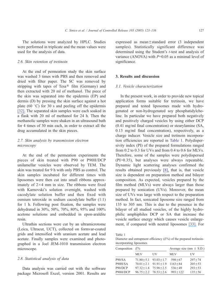
Table 1
Diameter and entrapment efficiency (E%) of the prepared tretinoin
incorporating liposomes
Composition E% Average size (nm F S.D.
MLV UV MLV UV
P90/SA 71.80F5.1 93.03F1.7 598F67 297F74
P90H/SA 70.01F2.1 91.91F1.9 1163F84 205F53
P90/DCP 97.32F1.8 75.98F2.5 536F49 293F53
P90H/DCP 96.75F2.2 78.53F2.6 993F122 135F56
C. Sinico et al. / Journal of Controlled Release 103 (2005) 123–136 127
The solutions were analyzed by HPLC. Studies
were performed in triplicate and the mean values were
used for the analysis of data.
2.6. Skin retention of tretinoin
At the end of permeation study the skin surface
was washed 3 times with PBS and then removed and
dried with filter paper. The SC was removed by
stripping with tapes of TesaR film (Germany) and
then extracted with 20 ml of methanol. The piece of
the skin was separated into the epidermis (EP) and
dermis (D) by pressing the skin surface against a hot
plate (60 8C) for 30 s and peeling off the epidermis
[32]. The separated skin samples were each soaked in
a flask with 20 ml of methanol for 24 h. Then the
methanolic samples were shaken in an ultrasound bath
for 4 times of 30 min each, in order to extract all the
drug accumulated in the skin pieces.
2.7. Skin analysis by transmission electron
microscopy
At the end of the permeation experiments the
pieces of skin treated with P90 or P90H/DCP
unilamellar vesicles were observed by TEM. The
skin was treated for 9 h with only PBS as control. The
skin samples incubated for different times with
liposomes were then cut into small ribbons approx-
imately of 2�4 mm in size. The ribbons were fixed
with Karnovski’s solution overnight, washed with
cacodylate solution buffer and then fixed with
osmium tetroxide in sodium cacodylate buffer (1:1)
for 1 h. Following post fixation, the samples were
dehydrated in 30%, 50%, 70%, 80%, 95% and 100%
acetone solutions and embedded in epon-araldite
resin.
Ultrathin sections were cut by an ultramicrotome
(Leica, Ultracut, UCT), collected on formvar-coated
grids and intensified with uranium acetate and lead
acetate. Finally samples were examined and photo-
graphed in a Joel JEM-1010 transmission electron
microscope.
2.8. Statistical analysis of data
Data analysis was carried out with the software
package Microsoft Excel, version 2001. Results are
expressed as meanFstandard error (3 independent
samples). Statistically significant difference was
determined using the Student’s t-test and analysis of
variance (ANOVA) with P=0.05 as a minimal level of
significance.
3. Results and discussion
3.1. Vesicle characterization
In the present work, in order to provide new topical
application forms suitable for tretinoin, we have
prepared and tested liposomes made with hydro-
genated or non-hydrogenated soy phosphatidylcho-
line. In particular we have prepared both negatively
and positively charged vesicles by using either DCP
(0.41 mg/ml final concentration) or stearylamine (SA,
0.13 mg/ml final concentration), respectively, as a
charge inducer. Vesicle size and tretinoin incorpora-
tion efficiencies are reported in Table 1. Polydisper-
sivity index (PI) of the prepared formulations ranged
from 0.2 to 0.3 for UVs and from 0.4 to 0.6 for MLVs.
Therefore, some of the samples were polydispersed
(PIN0.35), but analyses were always repeatable.
Dynamic light scattering analyses confirmed the
results obtained previously [8], that is, that vesicle
size is dependent on preparation method and bilayer
composition. As expected, vesicles prepared by the
film method (MLVs) were always larger than those
prepared by sonication (UVs). Moreover, the mean
size of UVs was large with respect to the preparation
method. In fact, sonicated liposome size ranged from
135 to 305 nm. This is due to the presence in the
bilayer of all studied vesicles, of the highly hydro-
philic amphiphiles DCP or SA that increase the
vesicle surface energy which causes vesicle enlarge-
ment, if compared with neutral liposomes [33]. For
-
)

C. Sinico et al. / Journal of Controlled Release 103 (2005) 123–136128
the same reason, vesicle formulations containing SA
were always larger in size than those containing DCP.
As shown in Table 1, all studied formulations
presented a high entrapment efficiency (E%). It is
interesting to note that SA multilamellar liposomes
showed an incorporation efficiency that was lower
than that of the unilamellar vesicles. This could be due
to the higher energy required for TRA-incorporating
vesicle formation when SA was present in the lipid
phase. In fact, in the preparation of SA-containing
liposomes, sonication was more efficient than vortex-
ing, especially in comparison with the preparation of
negatively charged MLVs.
3.2. Diffusion studies through silicone membrane
In order to obtain more information about the
stability and the thermodynamic activity of tretinoin
in liposomal formulations, we studied the TRA
diffusion through a polymeric lipophilic membrane.
This silicone membrane, which offers a non-porous,
hydrophobic, inert and reproducible barrier, has been
used by several authors to evaluate factors such as
drug activity on permeation. The percentages of
Fig. 1. Cumulative amounts (%) of tretinoin permeated through a silicone
TRA-loaded liposomes. For each formulation, main component and cha
separately in triplicate in vertical Franz diffusion cells through a silicon m
samples of three separate experiments.
tretinoin permeated after 24 h from our liposomal
formulations and from a hydroalcoholic solution of
the free drug (control) are reported in Fig. 1. As may
be seen, the percentage of drug diffused from the
control solution was generally smaller than that from
the liposomal formulations. We had already obtained
similar results studying the diffusion of tretinoin and
8-methoxsalen (8-MOP) from liposomes and nio-
somes [8,34]. Our previous studies on the in vitro
diffusion of TRA and 8-MOP had shown that the
delivery of a lipophilic drug can be modulated by
varying the structure and/or bilayer composition of the
vesicular carriers. Moreover, these studies had shown
that vesicular formulations enhanced the lipophilic
drug diffusion if compared with a hydroalcoholic
solution of the free drug and that this result could be
the consequence of a higher thermodynamic activity
of the vesicle-incorporated drug with respect to the
control [34]. Results obtained during this study, using
liposomal formulations and a control with the same
drug concentration (i.e. 0.2 mg/ml), support this
explanation. In fact, in the control solution the drug
concentration was a lot less than the solute saturation
solubility, therefore the thermodynamic activity of the
membrane from a bfreeQ TRA hydroalcoholic solution (W/Et) and
rge inducer are reported. Diffusion experiments were carried out
embrane (PertheseR). Data represent the means for three replicate

C. Sinico et al. / Journal of Controlled Release 103 (2005) 123–136 129
drug is most probably less than the activity in
liposomal formulations. Our results also show that
tretinoin diffusion is affected by the liposome
composition and structure. In fact, as previously
observed with negatively charged vesicular systems,
UVs always gave a higher drug release than MLVs
and P90 liposomes released greater TRA amounts
than P90H liposomes. Moreover, except for P90H
MLVs, negatively charged liposomes showed a higher
release rate than positively charged liposomes. This
result could be explained as a consequence of ion pair
formation between the drug (pKa=7.85) and stearyl-
amine counter ions. In fact, tretinoin is partially
ionized at pH=7 and therefore it could be more
strongly retained in the positively charged vesicles.
Finally, in our early work we suggested that
vesicles do not interact with Perthese membrane and
that the incorporated drug cannot diffuse through the
membrane. Indeed, we did not find phospholipids in
the receiver compartment at the end of the experi-
ments. However, we cannot exclude that the presence
of ethanol as a cosolvent in the donor compartment
could have produced a polarity of the membrane
resulting in a reduced partitioning and flux of the free
tretinoin.
Fig. 2. In vitro diffusion of liposomal and free tretinoin through newborn
are reported. Data represent the means for three replicate samples of three
3.3. In vitro skin permeation study
In order to evaluate the influence of composition,
structure and surface charge of the studied liposomes
on the TRA accumulation into and diffusion through
the skin, we carried out in vitro permeation studies,
using newborn pig skin and vertical Franz diffusion
cells. During this study we compared permeation data
obtained from liposomal tretinoin with those of
control formulations (oil and hydroalcoholic solution)
having the same drug concentration (i.e. 0.2 mg/ml).
Furthermore, as a control we also used a commercial
topical formulation of tretinoin (Retin-AR, 0.25 mg/
ml), because it was stated that liposomes less TRA
concentrated than commercial formulations had
shown the same efficacy [7].
The in vitro permeation study was carried out
through the whole newborn pig skin and in occlusive
conditions because, according to several authors ([16],
for instance) occlusive conditions are likely to
enhance drug accumulation into the skin layers. The
mean amount of TRA permeated per unit of surface
area from vesicles or control formulations was
determined during 9 h experiments. In Fig. 2,
permeation profiles (cumulative amounts of TRA
pig skin. For each formulation, main component and charge inducer
separate experiments.

C. Sinico et al. / Journal of Controlled Release 103 (2005) 123–136130
permeated versus time) of tretinoin through the skin
from the different studied liposomes and controls are
shown. As can be seen, permeation curves do not
show a classic profile with a steady state phase.
Tretinoin flux is higher in the initial period of the
permeation experiments (0–3 h). Afterwards, for all
liposomal formulations, TRA permeation rate was
almost constant in all sampling times. Results seem to
indicate that TRA release and skin permeation
occurred rapidly especially when the drug was
delivered from positively charged liposomes. In fact,
after 1 h from the beginning of the experiments,
positively charged formulations delivered 0.463–
0.641 Ag/cm2 while negatively charged vesicles
delivered 0.098–0.180 Ag/cm2 of the drug. Then, a
plateau is obtained from the negatively charged P90
liposomes that showed the lowest TRA diffusion rate.
Table 2 shows the cumulative amounts of tretinoin
delivered in the receiver compartment 3 and 9 h after
the beginning of the experiments. It is evident that
there are some differences in the total amount of TRA
permeated.
In the first analysis, the dose permeated of
liposomal TRA was dependent on the main bilayer
component. In fact, this value increased from P90 to
P90H liposomes both in MLV and UV dispersions.
Results of our study show that vesicle size and
lamellarity did not affect TRA delivery through the pig
skin. In fact for each composition the permeation
profile was very similar for both multi- or unilamellar
vesicle dispersions. These results seem to confirm Du
Table 2
Results of in vitro permeation study from vesicular formulations and contro
the experiments (9 h); amount of TRA accumulated into the skin at th
Efficiency (LAC) value: TRA accumulated into SC/TRA delivered throug
Composition Structure TRA delivered (Ag/cm2FS.
3 h 9 h
P90/SA MLV 0.599F0.07 0.8
UV 0.544F0.04 0.7
P90H/SA MLV 0.897F0.06 1.5
UV 0.758F0.05 1.3
P90/DCP MLV 0.330F0.06 0.5
UV 0.263F0.04 0.4
P90H/DCP MLV 0.677F0.08 1.1
UV 0.565F0.05 1.0
W/Et 0.631F0.08 1.7
Oil 0.457F0.07 1.1
Retin-AR 0.404F0.08 0.9
Plessis et al. [13] suggesting that intact penetration of
liposomes does not occur. Some interesting data related
to vesicle composition can be pointed out. In fact,
surprisingly we have obtained higher drug diffusion
when TRA was delivered in vesicles made from
phospolipids with the highest Tc (P90H, Tc=52 8C)than in P90 vesicles (Tcb2 8C). TRA delivery through
the skin was enhanced by a factor of 2–2.35 in P90H
vesicles. It is well recognized that the thermodynamic
state of liposomal bilayers plays a fundamental role on
the drug transport rate through the skin in vitro.
Incorporation of drugs in gel-state liposomes is known
to result in a slower skin permeation rate than in fluid-
state vesicles [16]. It has also been suggested that the
fluid-state of the lipids is the main aspect involved in
the fluidity of the vesicle bilayers [19]. However, it is
also well known that cholesterol content may play a
crucial role for the effective delivery of liposomal
substance into the skin. The optimal amount of
cholesterol is between 30% and 50% of the mass of
the liposomal membrane [27]. Moreover, when lip-
osomal bilayer is heterogeneous (i.e. composed of
different phospholipids, cholesterol and other compo-
nents), regions of different ordering and dynamic of
phospholipid chains are formed in the liposomal
bilayer [27]. Results obtained during this work could
be explained as the consequence of the presence in the
liposomal bilayers of both cholesterol and the amphi-
patic drug tretinoin, which is present in high molar
ratio. Here, in the occlusive conditions, both choles-
terol and TRA could have affected the Tc of soy
ls: amount of TRA delivered through the skin at 3 h and at the end of
e end of the permeation experiments; and Locally Accumulation
h the skin ratio
D.) TRA accumulated (AgFS.D.) LAC
9 h 9 h
88F0.06 13.56F1.33 5.3
99F0.04 23.47F2.55 12.9
41F0.09 17.45F3.00 3.3
31F0.04 29.83F4.25 8.4
55F0.08 30.45F7.58 26.6
52F0.02 32.67F8.98 34.8
44F0.07 30.37F7.12 9.6
25F0.09 34.13F8.28 14.9
56F0.11 15.33F2.43 2.4
73F0.10 16.83F2.52 6.5
17F0.09 12.03F0.41 4.2

C. Sinico et al. / Journal of Controlled Release 103 (2005) 123–136 131
hydrogenated phosphatidylcholine in such a way that
the fluidity of the P90H liposomal membrane increased
at the temperature of the experiments. A detailed study
of the thermal behavior of these formulations has just
started using Differential Scanning Calorimetry (DSC).
On the other hand, the differences in the TRA flux
could be simply related to a lower stability of the TRA-
incorporated P90 liposomes.
Obtained results also showed that positively
charged vesicles gave higher tretinoin transport rate
than negatively charged liposomes. In addition,
statistical comparison of the TRA amounts permeated
throughout 3–9 h showed that P90 liposomes pro-
vided drug permeation generally lower than the
controls. On the contrary, drug delivery from P90H/
SA liposomes was similar or slightly higher than
Retin-A, which was the control with the lowest TRA
diffusion through the pig skin. The highest drug
transport was obtained with the water/ethanol mixture
as a consequence of the enhancing property of ethyl
alcohol.
It is well known that the skin may act as a
negatively charged membrane [35,36] and it has been
generally reported that the presence of a charge on the
vesicle surface affects the drug diffusion through the
skin [32]. Negatively charged vesicles generally give
a higher flux than positively charged counterparts,
which in turn can improve drug accumulation in the
superficial skin strata. We also observed this behavior
studying the in vitro skin permeation of 8-MOP-
loaded vesicles [34]. However, in the present inves-
tigation, positively charged vesicles provided a
tretinoin permeation similar or even statistically
higher (Pb0.05, P90H formulations) than negatively
charged liposomes. As reported above for in vitro
TRA diffusion through the synthetic membrane, the
enhanced TRA diffusion through the pig skin
observed here with the SA-containing formulations
could be explained as the consequence of ion pair
formation between the organic acid tretinoin and SA
counter ions. The ion pair skin transport of TRA in
microemulsions, using various counter ions, has been
reported recently [37]. Here, the partially ionized TRA
(pKa=7.85; pH=7) and SA could behave as ion pairs
thus promoting tretinoin delivery through the skin. In
conclusion, overall results of the in vitro release and
skin permeation study indicated that TRA flux is
strongly affected by the liposome stability.
3.4. In vitro TRA regional distribution on the skin
The amounts of drug accumulated in the whole pig
skin are reported in Table 2. As can be seen, liposomal
formulations enhanced TRA accumulation into new-
born pig skin by a factor of 1.45–2.83, 1.14–2.22 and
1.04–20.03 over Retin-AR, hydroalcoholic and oil
solution, respectively. TRA oil solution gave the
highest accumulation compared the other controls,
while the lowest TRA accumulation was obtained
with Retin-AR. Results showed that the accumulation
of TRA provided by the liposomal formulations was
related both to vesicle structure and composition. In
fact, UVs always showed to be a better carrier than
MLVs for the delivery of TRA locally to the skin.
Moreover, this result was statistically important only
for SA-containing liposomes (Pb0.01). The highest
accumulation values were obtained from the nega-
tively charged vesicles and size and lamellarity of
negative liposomes did not greatly affect the TRA
delivery to the skin.
Fig. 3 shows the percentage of TRA delivered
through the skin and accumulated into the stratum
corneum (SC), epidermis (E) and dermis (D) for all
the studied liposomal formulations and controls at the
end of the experiments. As can be seen from Fig. 3,
the highest retention of tretinoin was always found in
the SC and in particular when negatively charged
liposomes were used. When carriers for TRA were
positively charged MLVs, the amount of drug
accumulated in the SC was very similar to that
obtained with the hydroalcoholic solution or Retin-
AR and even lower than that provided by the oil
solution.
To evaluate vesicular formulations as topical
carriers for TRA, it is interesting to compare the ratio
of TRA accumulated into SC versus TRA delivered
through the skin, which gives a dimensionless number
for the quantification of the Locally Accumulation
Efficiency (LAC) from the several formulations. LAC
values are listed in Table 2. The LAC value of
negatively charged liposomes was higher by a factor
of 2.3–8.3 than that of the commercially available
Retin-AR (LAC=4.2), suggesting that the main effect
of these systems was to accumulate the drug into the
skin. The only exceptions were positively charged
multilamellar liposomes whose LAC value was very
close to that of Retin-AR (P90/SA, LAC=5.3) or even
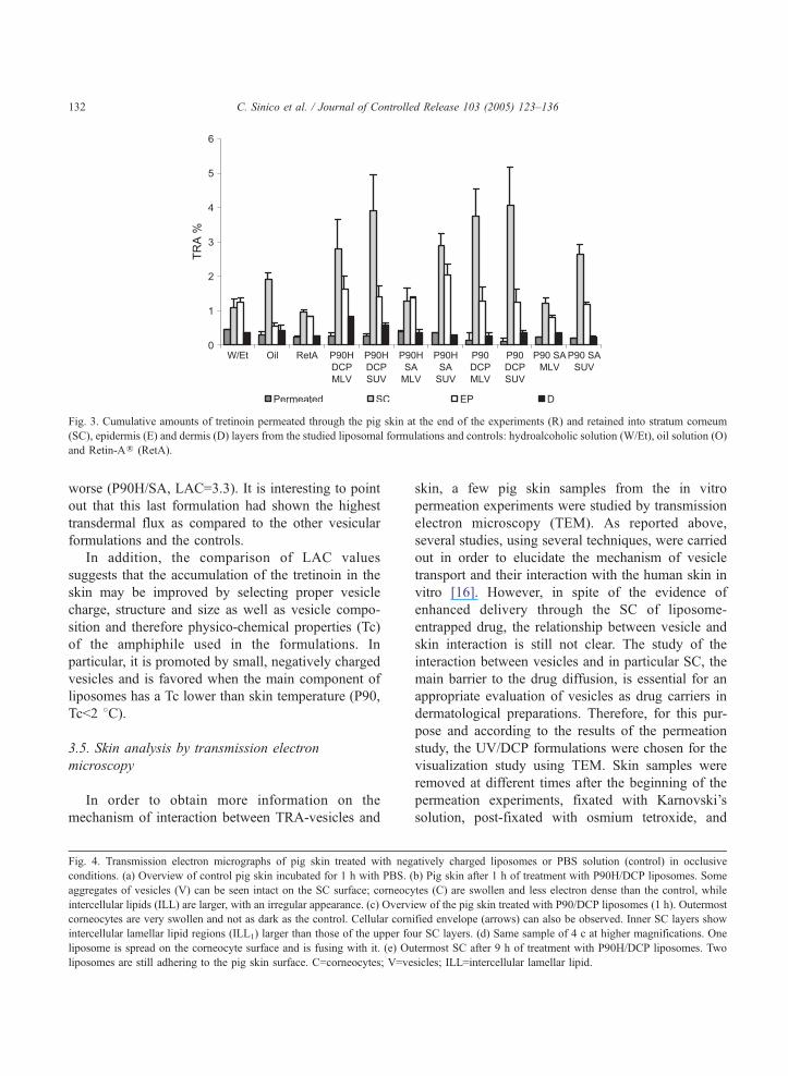
Fig. 3. Cumulative amounts of tretinoin permeated through the pig skin at the end of the experiments (R) and retained into stratum corneum
(SC), epidermis (E) and dermis (D) layers from the studied liposomal formulations and controls: hydroalcoholic solution (W/Et), oil solution (O)
and Retin-AR (RetA).
C. Sinico et al. / Journal of Controlled Release 103 (2005) 123–136132
worse (P90H/SA, LAC=3.3). It is interesting to point
out that this last formulation had shown the highest
transdermal flux as compared to the other vesicular
formulations and the controls.
In addition, the comparison of LAC values
suggests that the accumulation of the tretinoin in the
skin may be improved by selecting proper vesicle
charge, structure and size as well as vesicle compo-
sition and therefore physico-chemical properties (Tc)
of the amphiphile used in the formulations. In
particular, it is promoted by small, negatively charged
vesicles and is favored when the main component of
liposomes has a Tc lower than skin temperature (P90,
Tcb2 8C).
3.5. Skin analysis by transmission electron
microscopy
In order to obtain more information on the
mechanism of interaction between TRA-vesicles and
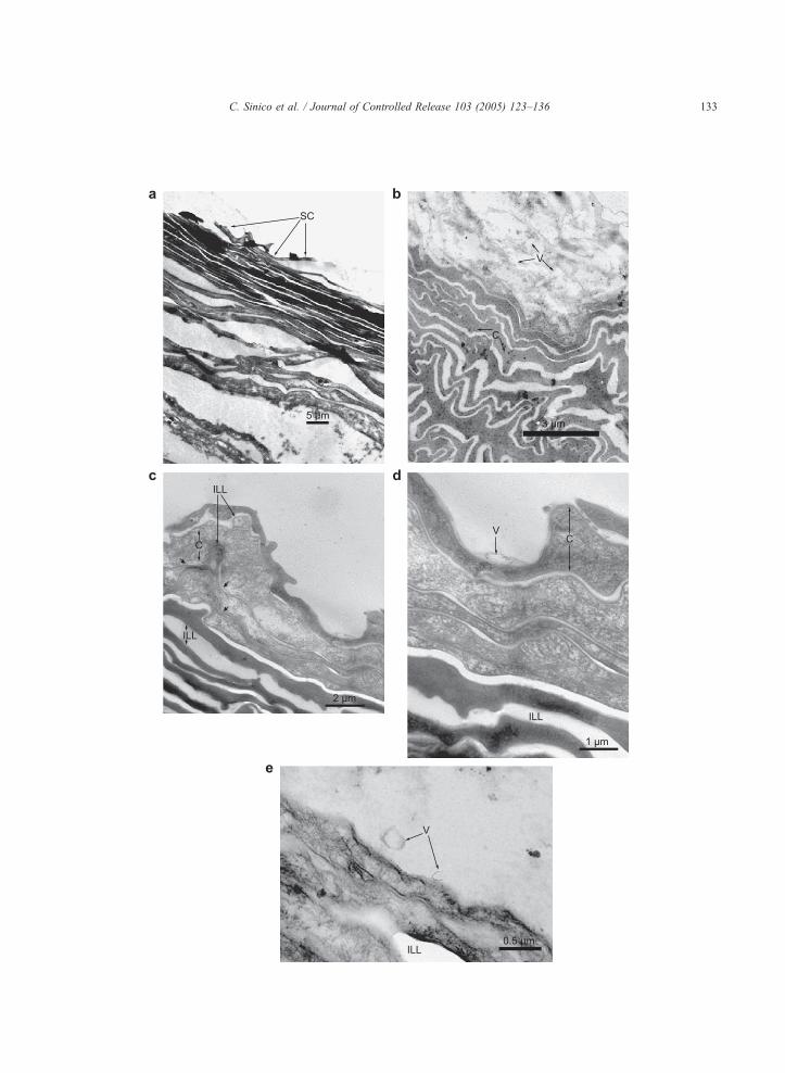
Fig. 4. Transmission electron micrographs of pig skin treated with neg
conditions. (a) Overview of control pig skin incubated for 1 h with PBS. (
aggregates of vesicles (V) can be seen intact on the SC surface; corneocy
intercellular lipids (ILL) are larger, with an irregular appearance. (c) Overv
corneocytes are very swollen and not as dark as the control. Cellular corn
intercellular lamellar lipid regions (ILL1) larger than those of the upper fo
liposome is spread on the corneocyte surface and is fusing with it. (e) Ou
liposomes are still adhering to the pig skin surface. C=corneocytes; V=ve
skin, a few pig skin samples from the in vitro
permeation experiments were studied by transmission
electron microscopy (TEM). As reported above,
several studies, using several techniques, were carried
out in order to elucidate the mechanism of vesicle
transport and their interaction with the human skin in
vitro [16]. However, in spite of the evidence of
enhanced delivery through the SC of liposome-
entrapped drug, the relationship between vesicle and
skin interaction is still not clear. The study of the
interaction between vesicles and in particular SC, the
main barrier to the drug diffusion, is essential for an
appropriate evaluation of vesicles as drug carriers in
dermatological preparations. Therefore, for this pur-
pose and according to the results of the permeation
study, the UV/DCP formulations were chosen for the
visualization study using TEM. Skin samples were
removed at different times after the beginning of the
permeation experiments, fixated with Karnovski’s
solution, post-fixated with osmium tetroxide, and
atively charged liposomes or PBS solution (control) in occlusive
b) Pig skin after 1 h of treatment with P90H/DCP liposomes. Some
tes (C) are swollen and less electron dense than the control, while
iew of the pig skin treated with P90/DCP liposomes (1 h). Outermost
ified envelope (arrows) can also be observed. Inner SC layers show
ur SC layers. (d) Same sample of 4 c at higher magnifications. One
termost SC after 9 h of treatment with P90H/DCP liposomes. Two
sicles; ILL=intercellular lamellar lipid.
C. Sinico et al. / Journal of Controlled Release 103 (2005) 123–136 133
C. Sinico et al. / Journal of Controlled Release 103 (2005) 123–136134
finally examined by TEM. This analysis has provided
a reliable microscopic estimation of the skin mod-
ifications induced by TRA-incorporating vesicles as
compared with skin treated with PBS solution
(control). Representative electron micrographs of pig
skin 1 or 9 h after incubation in occlusive conditions
with PBS solution and P90/DCP or P90H/DCP
liposomes are depicted in Fig. 4. As can be seen
(Fig. 4a), the pig skin treated with PBS is mainly
organized in an alternating pattern of electron dense
and light bands. Intercellular spaces (light bands) are
very sharp and linear whereas corneocytes (black
bands) are flat and dark because of the presence of the
electron dense fibrous protein keratin. In Fig. 4b, an
overview of the ultrastructure of the pig skin after 1 h
of incubation with P90H/DCP liposomes is shown.
Aggregates of liposomes are still present on the skin
surface showing that vesicles close to the SC layers
were not rinsed off during the fixation procedure.
Some morphological alterations in the SC layers are
evident with respect to the control: corneocytes (C)
are very swollen, less electrondense; intercellular
spaces (ILL) are larger, wavy and more irregular than
PBS-treated skin. In fact, several folds, which increase
the intercellular pathway, are present.
Fig. 4c shows a micrograph of newborn pig skin
treated for 1 h with P90/DCP liposomes. As in Fig.
4b, the upper corneocytes appear swollen and not as
dark as the control. The four outermost layers of
corneocytes show quite clearly their boundary with
the intercellular lipids: the so-called cornified enve-
lope. Here, this cellular envelope has a highly
irregular appearance and it seems to create finger-like
structures within the intercellular lamellar lipids
(ILL).
The inner SC layers show a different organization
with intercellular lipid regions (ILL1) larger than in
the upper SC layers. No aggregates of vesicles are
present on the skin surface, but a single P90/DCP
vesicle tight to the external surface of the SC can be
seen. Fig. 4d, which shows the same sample at higher
magnifications, allows us to notice that the P90
liposome is spread on the corneocyte surface and is
fusing with it.
In Fig. 4e, a micrograph of the SC after 9 h of
treatment with P90H/DCP liposomes can be observed.
Two liposomal vesicles are still present intact on the
SC surface at the end of the experiment. They have a
spheroidal shape and one of them is close to the skin
surface while the other one is approaching the horny
layer. Moreover, in this micrograph the upper corneo-
cyte layer shows a swollen and flexible cytoplasm,
less electron dense than in the control: the SC treated
with liposomes appears very hydrated and elastic.
TEM analyses in this work have not shown any
evidence of vesicular structures in the pig skin.
Therefore, we can conclude that liposomes do not
seem to penetrate intact into deeper skin layers. On
the other hand, micrographs have given the evidence
of vesicle adsorption (both P90 and P90H liposomes)
and mixing or fusion (P90 vesicles) with the SC
surface. The main result of TEM analyses is that
liposomes may strongly affect the ultrastructure of the
intra- and intercellular regions of the horny layer.
On the basis of TEM images, it is evident that the
main advantage of liposomes is to improve the drug
residence time on the skin surface. This is particularly
true for P90H liposomes which are still present on the
pig skin surface at the end of the experiments (9 h;
Fig. 4e). Furthermore, liposomes enhance TRA
bioavailability because after their topical application,
they settle down on the skin surface close to the
outermost corneocyte layer. In these conditions,
material exchange between vesicles and intercellular
lipids may occur, allowing the diffusion of free
molecules as well as small membranal fragments into
the SC. Resulting perturbation of SC intercellular
lipids modifies the enthalpy of the SC lipid transition
giving rise to a new phase transition associated to the
applied lipids. The result is an increase of the
membrane permeability and a different partition of
lipophile molecules (TRA) in the intercellular spaces
[38,39]. In the end, liposomes form a lipid film on the
skin which retains water, both endogen and liposome-
entrapped, causing an improvement of SC intra- and
intercellular hydration. Vesicular components (i.e.
water and lipids) may join lipid bilayers in the SC
intercellular regions causing the swelling of the
intercellular lipids. Moreover, water also causes the
swelling of intracellular fibrous protein and hence of
corneocytes. Therefore, the compact structure of the
SC is opened and the barrier permeability is increased.
All these factors cause the retention of tretinoin in the
horny layer. However, they cannot improve TRA
diffusion through the inner more hydrophile skin
layers because of the lipophilicity of the drug which

C. Sinico et al. / Journal of Controlled Release 103 (2005) 123–136 135
shows higher affinity for the SC lipid matrix. Only
when TRA was incorporated into positively charged
liposomes a slightly improved diffusion through the
skin was obtained. This behavior can be explained as
a consequence of a better diffusion pattern of the more
hydrophile ionized TRA electrostatically linked to the
counter ion stearylamine.
4. Conclusions
Overall results obtained during this work have
shown that liposomes may be an interesting carrier for
tretinoin in skin disease treatment, when appropriate
formulations are used. In fact, tretinoin dermal
delivery was found to be affected by several factors
including vesicle composition, morphology and size.
In particular, it has been shown that negatively
charged liposomes strongly improved newborn pig
skin hydration and TRA retention, though no evi-
dence of intact vesicle penetration was found. Further
in vivo studies have to be carried out in order to
obtain more information on vesicle–skin interactions.
In addition, in order to find the best topical
application forms suitable for liposomal tretinoin, a
study on liposomes formulated in gels and poly-
ethylene glycol ointment is being carried out. These
studies are being performed both in occlusive and
non-occlusive conditions.
References
[1] R.W. Lucek, W.A. Colburn, Clinical pharmacokinetics of the
retinoids, Clin. Pharmacokinet. 10 (1985) 38–62.
[2] P. Fenaux, C. Chomienne, L. Degos, Treatment of acute
promyelocytic leukaemia, Best Pract. Res. Clin. Haematol. 14
(2001) 153–174.
[3] J.G. Allen, D.P. Bloxham, The pharmacology and pharmaco-
kinetics of the retinoids, Pharmacol. Ther. 40 (1989) 1–27.
[4] P.A. Lehman, J.T. John, T.J. Franz, Percutaneous absorption of
retinoids: influence of vehicle, light exposure and dose, J.
Invest. Dermatol. 91 (1990) 56–61.
[5] W.C. Foong, B.B. Harsanyi, M. Mezei, Biodisposition and
histological evaluation of topically applied retinoic acid in
liposomal, cream and gel dosage forms, in: I. Hanin, G.
Pepeu (Eds.), Phospholipids, Plenum Press, New York, 1990,
pp. 139–154.
[6] V. Masini, F. Bonte, A. Meybeck, J. Wepierre, Cutaneous
bioavailability in hairless rats of tretinoin in liposomes or gel,
J. Pharm. Sci. 82 (1993) 17–21.
[7] M. Sch7fer-Korting, H.C. Korting, E. Ponce-Pfschl, Lip-
osomal tretinoin for uncomplicated acne vulgaris, Clin. Invest.
72 (1994) 1086–1091.
[8] M. Manconi, D. Valenti, C. Sinico, G. Loy, A.M. Fadda,
Niosomes as carriers for tretinoin: I. Preparation and proper-
ties, Int. J. Pharm. 234 (2002) 237–248.
[9] M. Manconi, D. Valenti, C. Sinico, F. Lai, G. Loy, A.M.
Fadda, Niosomes as carriers for tretinoin: II. Influence of
vesicular incorporation on tretinoin photostability, Int. J.
Pharm. 260 (2003) 261–272.
[10] N. Weiner, F. Martin, M. Riaz, Liposomes as a drug delivery
system, Drug Dev. Ind. Pharm. 15 (1989) 1523–1524.
[11] G. Cevc, G. Blume, Lipid vesicles penetrate into intact
skin owing to the transdermal osmotic gradients and
hydration force, Biochim. Biophys. Acta 1004 (1992)
226–232.
[12] J. Du Plessis, K. Egbaria, N. Weiner, Influence of formulation
factors on the deposition of liposomal components into the
different strata of the skin, J. Soc. Cosmet. Chem. 43 (1992)
93–100.
[13] J. Du Plessis, C. Ramachandran, N. Weiner, D.G. Muller, The
influence of particle size of liposomes on the deposition of
drug into skin, Int. J. Pharm. 103 (1994) 277–282.
[14] H.Y. Yu, H.M. Liao, Triamcinolone permeation from different
liposome formulations through rat skin in vitro, Int. J. Pharm.
127 (1996) 1–7.
[15] B.A.I. Van den Bergh, J. Vroom, H. Gerritsen, E. Junginger,
J.A. Bouwstra, Interactions of elastic and rigid vesicles with
human skin in vitro: electron microscopy and two-photon
excitation microscopy, Biochim. Biophys. Acta 1461 (1999)
155–173.
[16] J.A. Bouwstra, P.L. Honeywell-Nguyen, G.S. Gooris, M.
Ponec, Structure of the skin barrier and its modulation by
vesicular formulations, Prog. Lipid Res. 42 (2003) 1–36.
[17] B.A.I. Van den Bergh, I. Salomons-de Vrie, J.A. Bouwstra,
Interactions between liposomes and human stratum corneum
studied by freeze-substitution electron microscopy, Int. J.
Pharm. 167 (1998) 57–67.
[18] H.E.J. Hofland, J.A. Bouwstra, H.E. Bodde, F. Spies, H.E.
Junginger, Interactions of liposomes with human skin in vitro:
freeze fracture electron microscopial visualization and small
angle X-ray scattering studies, Br. J. Dermatol. 132 (1995)
853–866.
[19] P.L. Honeywell-Nguyen, A.M. de Graff, H.W. Wouter
Groenink, J.A. Bouwstra, The in vivo and in vitro interactions
of elastic and rigid vesicles with human skin, Biochim.
Biophys. Acta 1573 (2002) 130–140.
[20] J. Lasch, R. Laub, W. Wohlrab, How deep do intact
liposomes penetrate into human skin? J. Control. Release 18
(1991) 55–58.
[21] K. Kriwet, C.C. Muller-Goymann, Diclofenac release
from phospholipid drug systems and permeation through
excised human stratum corneum, Int. J. Pharm. 125
(1995) 231–242.
[22] D. Yarosh, C. Bucana, P. Cox, et al., Localization of liposomes
containing a DNA repair enzyme in murine skin, J. Invest.
Dermatol. 103 (1994) 461–468.

C. Sinico et al. / Journal of Controlled Release 103 (2005) 123–136136
[23] M. Kirjavainen, A. Urtti, I. Jaaskelainen, T.M. Suhonen, P.
Paronen, R. Valjakka-Koskela, J. Kiesvaara, J. Mfnkkfnen,Interaction of liposomes with human skin in vitro—the
influence of lipid composition and structure, Biochim. Bio-
phys. Acta 1304 (1996) 179–189.
[24] E. Touitou, N. Dayan, L. Bergelson, B. Godin, M. Eliaz,
Ethosomes—novel vesicular carriers for enhanced delivery:
characterization and skin penetration properties, J. Control.
Release 65 (2000) 403–418.
[25] S. Zellmer, W. Pfiel, J. Lasch, Interaction of phosphatidylcho-
line liposomes with the human stratum corneum, Biochim.
Biophys. Acta 237 (1995) 176–182.
[26] V. Gabrijelcic, M. Sentjurc, J. Kristl, Evaluation of liposomes
as drug carriers into the skin by one-dimensional EPR
imaging, Int. J. Pharm. 62 (1990) 75–79.
[27] K. Vrhovnik, J. Kristl, M. Sentjurc, J. Smid-Korbar, Influence
of liposome bilayer fluidity on the transport of encapsulated
substance into the skin as evaluated by EPR, Pharm. Res. 15
(1998) 525–530.
[28] M. Mezei, V. Gulasekharam, Liposomes—a selective drug
delivery system for the topical route of administration: I.
Lotion dosage form, Life Sci. 26 (1980) 1473–1477.
[29] M. Foldvari, A. Gesztes, M. Mezei, Dermal drug delivery by
liposome encapsulation clinical and electron microscopic
study, J. Microencapsul. 7 (1990) 479–489.
[30] M. Jacobs, G.P. Martin, C. Marriott, Effects of phosphatidyl-
choline in the topical bioavailability of corticosteroids assessed
by the human skin blanching assay, J. Pharm. Pharmacol. 40
(1988) 829–833.
[31] M.E.M.J. Van Kuijk-Meuwissen, H.E. Junginger, J.A.
Bouwstra, Interactions between liposomes and human skin
in vitro, a confocal laser scanning microscopy study,
Biochim. Biophys. Acta 1371 (1998) 31–39.
[32] N. Katahira, T. Murakami, S. Kugai, N. Yata, M. Takano,
Enhancement of topical delivery of a lipophilic drug from
charged multilamellar liposomes, J. Drug Target. 6 (1999)
405–414.
[33] J.-Y. Fang, C.-T. Hong, W.-T. Chiu, Y.-Y. Wang, Effect of
liposomes and niosomes on skin permeation of enoxacin, Int.
J. Pharm. 219 (2001) 61–72.
[34] C. Sinico, D. Valenti, M. Manconi, F. Lai, A.M. Fadda, Octyl/
decyl polyglucoside niosomes as potential carriers for the
cutaneous delivery of 8-methoxypsoralen, J. Liposome Res.
(2004) (submitted for publication).
[35] R.R. Burnette, B. Ongipattanakul, Characterization of the
permeselective properties of excise human skin during
ionophoresis, J. Pharm. Sci. 76 (1987) 765–773.
[36] L. Montenegro, A.M. Panico, A. Ventimiglia, F.P. Bonina,
In vitro retinoic acid release and skin permeation from
different liposome formulations, Int. J. Pharm. 133 (1996)
89–96.
[37] M. Trotta, E. Ugazio, E. Peira, C. Pulitano, Influence of ion
pairing on topical delivery of retinoic acid from micro-
emulsions, J. Control. Release 86 (2003) 315–321.
[38] K. Egbaria, C. Ramachandram, N. Weiner, Topical application
of liposomally entrapped cyclosporin evaluated by in vitro
diffusion studies with human skin, Skin Pharmacol. 4 (1991)
21–28.
[39] D.D. Verma, S. Verma, G. Blume, A. Fahr, Particle size of
liposomes influences dermal delivery of substances into skin,
Int. J. Pharm. 258 (2003) 141–151.
Copyright © 2022 FDOKUMEN